Recombinant Adeno-Associated Viral Vectors in the Nervous System
Upload
khangminh22Category
view
4download
0
Nervous System
© 2009 Ebneshahidi
Dr. Ali Ebneshahidi
Nervous System
Nervous system and endocrine system are thechief control centers in maintaining bodyhomeostasis.
Nervous system uses electrical signals (nerve
© 2009 Ebneshahidi
Nervous system uses electrical signals (nerveimpulses) which produce immediate (but short -lived) responses; endocrine system useschemical signals (hormones) that produce slower(but long lasting) responses.

Nervous system has 3 major functions
Sensory input – sensory or afferent neutron detect internal or externalchanges (stimuli) and send the message to the brain or spinal cord.
Integration – interneurons in the brain or spinal cord interpret themessage from & relay the massage back to body parts.
Motor output – motor or efferent neurons receive the message frominterneuron and produce a response at the effector organ (a muscle ora gland).
© 2009 Ebneshahidi
a gland).

Neurons All neurons have a cell body called soma. Although there is DNA in
the neuron, somehow DNA replication and mitosis do not occur,resulting in the neurons lack of ability to reproduce or regenerate.Extensions of the soma form nerve such as dendrites which conductnerve impulses toward the soma, and axon which conducts nerveimpulses away from the some (to another neuron, or to an effectororgan). The number of dendrites ranges from 1 to thousands (inmultipolar neurons). All neurons only contain1axon.
© 2009 Ebneshahidi
multipolar neurons). All neurons only contain1axon.

Longer axons are enclosed by a lipoprotein substance calledmyelin sheath produced by a type of neuroglial cell calledschwann cells.
This myelin sheath insulates the axon against depolarization, andforces action potential to occur in the gaps (Node of Ranvier) inbetween the myelin sheath. This type of nerve impulsepropagation where action potential jumps from one gap to the
© 2009 Ebneshahidi
propagation where action potential jumps from one gap to thenext, is called saltatory conduction.
g) axons enclosed by myelin sheath are called myelinated axonswhich make up the white matter in the nervous system; whileaxons that have no myelin sheath are called unmyelinated axonswhich make up the gray matter in the nervous system.

© 2009 Ebneshahidi

A synapse is the junction between two neurons, or between aneuron and an effecter organ (muscle or gland). Each synapseconsists of:
Presynaptic neuron – the neuron that sends an impulse to thesynapse.
Axon – the nerve fiber extends from the presynaptic neuron,that propagates the impulse to the synapse.
Synapse
© 2009 Ebneshahidi
that propagates the impulse to the synapse.
Synaptic knobs- the round endings of the axon.
Synaptic vesicles- membranous sacs that contain aneurotransmitter (e.g. acetylcholine, norepinephrine,dopamine), located in the synaptic knobs.

Synapse
© 2009 Ebneshahidi
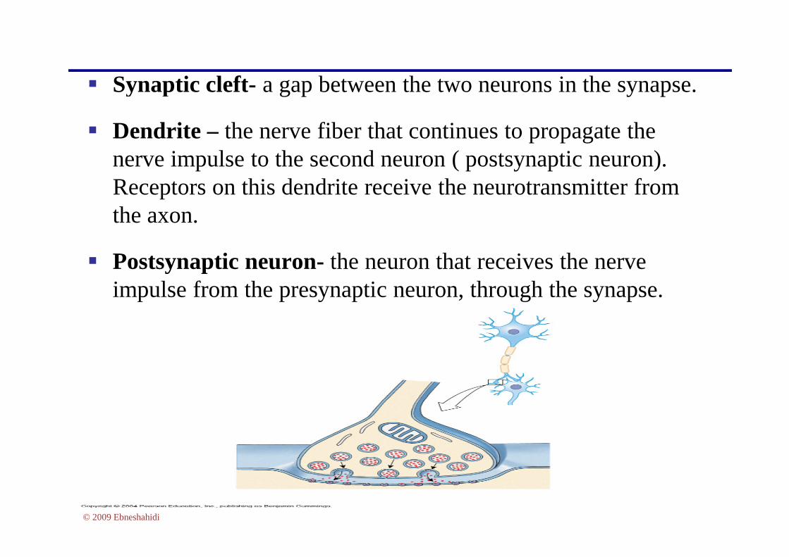
Synaptic cleft- a gap between the two neurons in the synapse.
Dendrite – the nerve fiber that continues to propagate thenerve impulse to the second neuron ( postsynaptic neuron).Receptors on this dendrite receive the neurotransmitter fromthe axon.
Postsynaptic neuron- the neuron that receives the nerveimpulse from the presynaptic neuron, through the synapse.
© 2009 Ebneshahidi
impulse from the presynaptic neuron, through the synapse.

1. The membrane is semi-permeable some things get through,while others do not get through. Important ions to beconcerned with are Na+, K+, Cl- ,and anions-.
2. There are differences in concentration of these various ionsbetween the inside and outside of the cell, so there are conc.gradients for each of these ions across the cell membrane.
Excitable membrane
© 2009 Ebneshahidi
gradients for each of these ions across the cell membrane.
3. There is electrical potential differences across themembrane, such that the inside is negative with respect to theoutside in the resting state. The production of activity innerve cells are due to changes in this membrane potential.

Cell membrane is usually polarized as a result of unequaldistribution of ions on either side.
A high concentration of Na+ is on the outside and a highconcentration of K+ is inside. The outside of the membrane ismore positive relative to the inside of cell (inside is negative).
The resting membrane potential (-70 mv)
© 2009 Ebneshahidi

Polarized membrane:
© 2009 Ebneshahidi Figure 11.3b, c

stimulation of a membrane affects its resting potential in a localregion.
a. The membrane is depolarized if it becomes less negative.
b. The membrane is hyperpolarized if it becomes more negative.
Resting membrane potential is maintained by Na – K pump
© 2009 Ebneshahidi
(pumps 3 Na+ outside , 2 K+ inside).
Membrane potential – summery:
1. [Na+] outside > [Na+] inside.
2. [K+] inside > [K+] outside.
3. [(Na+) outside + (K+) outside > [(Na+) inside + (K+) inside].

Depolarization
© 2009 Ebneshahidi

Depolarization vs. Hyperpolarization
© 2009 Ebneshahidi

1. Electrical messages – conduction
a. Graded – travels short distances in intensity,subject to summation (ex. Receptor potential).
b. Action – travels long distances intensity isunchanged.
Communication within the CNS
© 2009 Ebneshahidi
unchanged.
2. chemical messages – Neurotransmitters
a. Excitatory: through membrane depolarization.
b. Inhibitory: through membrane hyperpolarization.

Events leading to nerve impulse cnduction
1. Nerve fiber membranemaintains resting potential bydiffusion of Na+ and K+ downtheir concentration gradientsas the cell pumps them up thegradients.
© 2009 Ebneshahidi
2. Neurons receivestimulation, causing localpotentials, which may sum toreach threshold.
3. Sodium channels in a localregion of the membrane open.

4. sodium ions diffuseinward, depolarizing themembrane. Na+ rushes infollowing its concentrationgradient.
5. potassium channels in themembrane open.
© 2009 Ebneshahidi
membrane open.
6. potassium ions diffuseoutward, repolarizing the
membrane.
© 2009 Ebneshahidi

In response to a nerve impulse, the end of a motor nerve fiber secretes aneurotransmitter, which diffuses across the junction and stimulates themuscle fiber.
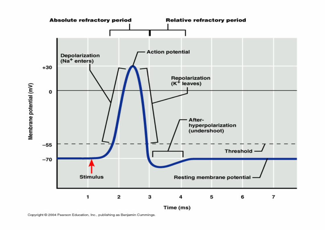
Action potential: Electrical changes that occurs along the sarcolemma.
1. Membrane Depolarization – Na+ entering the cell.
© 2009 Ebneshahidi
1. Membrane Depolarization – Na entering the cell.
2. Action potential is propagated as the move of depolarization spreads.
3. Repolarization – Na+ channels close and K+ opens, and K+ diffuse out.
4. Refractory period: cell can not be stimulated during this phase.(Na+ - K+ pump restores the electrical condition) [pumps 3 Na+ outside,2 K+ inside].

Action Potential
© 2009 Ebneshahidi

Action potential – characteristic
-Maybe excitatory or inhibitory.
-Travels down the axon (not in dendrites).
-They are non–decremental.
-They may carry sensory or motor information.
© 2009 Ebneshahidi
-They may carry sensory or motor information.
-Generated by neurons and muscle cells.
-All – or – None response.
-Impulse conduction is more rapid in myelinated fibers (saltatoryconduction).

Synaptic transmission – Neurotransmitter release
1. Action potential passesalong a nerve fiber and overthe surface of its synapticknob.
2. synaptic knob membranebecomes more permeable to
© 2009 Ebneshahidi
becomes more permeable toCa+ ions, and they diffuseinward.
3. In the presence of Ca+ ions, synaptic vesicles fuse tosynaptic knob membrane.

4. synaptic vesicles release their neurotransmitter by exocytosis into thesynaptic cleft.5. synaptic vesicles become part of the membrane.6. the added membrane provides material for endocytotic vesicles.
© 2009 Ebneshahidi

Action potential in presynaptic cell.
Depolarization of the cell membrane of the presynaptic axonterminal.
Release of the chemical transmitter by the presynaptic terminal.
Binding of the transmitter to specific receptons on the plasma
Summary
© 2009 Ebneshahidi
Binding of the transmitter to specific receptons on the plasmamembrane of the post synaptic cells.
Transient changes in the conductance of the postsynaptic plasmamembrane to specific ions.
Transient change in the membrane potential of the postsynapticcell (excitatory or inhibitory).

Acetylcholine – stimulates the postsynaptic membrane.
Epinephrine & norepinephrine – main signal in sympathetic NS.
Dopamine – used in mid. brain.
Serotonine – mostly inhibitory. Plays a role in sleep, appetite, andregulation of mood.
Neurotransmitters
© 2009 Ebneshahidi
regulation of mood.
GABA (gama-aminobutiric acid) – Inhibitory.
Endorphins – Inhibitory.
Substance "p" – Excitatory.
Somatostatin – Inhibitory.

Alzheimer's Disease – Low (Deficient)acetylcholine.
Depression - Deficient norepinephrine, serotonin.
Huntington disease – Deficient GABA.
Disorders
© 2009 Ebneshahidi
Huntington disease – Deficient GABA.
Parkinson's disease – Deficient dopamine.
Schizophrenia – deficient GABA and excessivedopamine.

a. Neurons are organized into nueronal pools within the CNS.
b. Each pool receives / processes, and conducts away impulses.
Facilitation: each neuron in a pool may receive excitatory orinhibitory stimuli. A neuron is facilitated when it receives (excitatoryor inhibitory) subthreshold stimuli and becomes more excitable.
Integration: Organization of neurons
© 2009 Ebneshahidi

Divergence:
a. impulses leaving a poolmay diverge by passing ontoseveral output fibers.
b. Divergence amplifiesimpulses.
© 2009 Ebneshahidi
Convergence:
a. incoming impulses mayconverge on a single neuron.
b. convergence enables aneuron to summate impulsesfrom different sources.

The largest organ in the nervous system; composed of about 100billion neurons (interestingly, although the neurons contain DNA,there is no DNA replication or mitosis in the brain, as a result thenumber of neurons decreases as a person ages).
Divided into 3 main regions: cerebrum, cerebellum, and thebrain stem.
Brain
© 2009 Ebneshahidi
brain stem.
Contains spaces called ventricles where choroids plexuses of piamater produce cerebrospinal fluid (CSF), and these ventriclesallow CSF to circulate around the brain and into the spinal cord(through the central canal).

Regions of the Brain
© 2009 Ebneshahidi

cerebral cortex (outer region) is made of gray matter(unmyelinated neurons) which contains up to 75% of allneurons in the nervous system, while cerebral medulla (innerregion) is made of white matter (myelinated neurons).
Consists of left and right hemispheres, created by thelongitudinal fissure at the center of cerebrum, and are
Regions of the Brain
© 2009 Ebneshahidi
longitudinal fissure at the center of cerebrum, and areconnected by the corpus callosum.
Its surface is marked by ridges called convolutions (or gyri)which are separated by grooves called sulcus (or fissure, if thegrooves are deeper).

© 2009 Ebneshahidi

Lobes of the Brain Frontal lobe controls skeletal muscle movement and
intellectual processes.
Parietal lobe controls sensations and speech.
Temporal lobe controls hearing and memory.
Occipital lobe controls vision.
© 2009 Ebneshahidi

Motor areas - located in frontal lobe, to control voluntarymuscles.Motor speech area ("Broca’s area") - Located in frontal lobe,to control muscles of mouth, tongue, and larynx for speech.Frontal eye field - located in frontal lobs just above the Broca’sarea, to control muscles of the eye and eyelid.Auditory area - located in temporal lobe, to control hearing.
Functional regions of the cerebral cortex
© 2009 Ebneshahidi
Auditory area - located in temporal lobe, to control hearing.Visual area - located in occipital lobe, to control visualrecognition of objects and combine visual images.Sensory areas - located in parietal lobe, to be involved incutaneous sensations of temperature, touch, pressure and pain.Association areas - located in all of cerebral cortex, tointerconnect sensory and motor functions of all lobes of thecerebrum.

Functional Regions of the cerebral cortex
© 2009 Ebneshahidi

Cerebellum
Coordinates andcontrols muscularmovement and muscletone.
Maintains body posture,by working with theequilibrium receptors in
© 2009 Ebneshahidi
equilibrium receptors inthe inner ear.
New data suggest that italso functions as thespeech area that isinvolved with findingthe right words to use.

Brain Stem
Made up of braintissue at the base ofcerebrum,connecting thecerebrum to thespinal cord.
Functions largely
© 2009 Ebneshahidi
Functions largelyfor autonomousactivities.
Subdivided intodiencephalons,midbrain, pons,and medullaoblongata.

Diencephalon, Midbrain, and Pons
Diencephalon: consists of thalamus (a major relaycenter to direct nerve impulses from various sources tothe proper destinations) and hypothalamus (animportant area for regulating homeostatic activities, suchas hunger, thirst, sex drive, and even addictions).
Midbrain: serves as a major cerebral reflex center, andalso helps direct CSF from the third Ventricle to Fourth
© 2009 Ebneshahidi
also helps direct CSF from the third Ventricle to FourthVentricle.
Pons: contains at least 2 "respiratory centers" (groups ofspecialized neurons) which regulate the duration anddepth of breathing.

Medulla Oblongata
at the base of base ofbrain stem andcontinuous tobecome spinal cord.
contains specializedneurons that form"cardiac centers" (to
© 2009 Ebneshahidi
"cardiac centers" (tocontrol heart rate)"vasomotor centers"(to control bloodflow and bloodpressure), and"respiratory centers"(to controlrespiratory rhythms).

Spinal Cord
A long nerve cord that beginsat the foramen magnum andends at the first or secondlumbar vertebrae. Divided into31 segments (named after thevertebral regions), eachsegment gives rise to a pair ofspinal nerves ( part of the
© 2009 Ebneshahidi
spinal nerves ( part of thePNS).
In general, the location of thespinal nerve corresponds withthe location of the effectororgan (e.g. cervical nervesconnect to muscles and glandson the head, face, and neck).

© 2009 Ebneshahidi

Peripheral nervous system Consists of 12 pairs of cranial nerves and 31 pairs of spinal
nerves.
Serves as a critical link between the body and the centralnervous system.
© 2009 Ebneshahidi

Cranial nerves
Nerve I (olfactory) - for the sense of smell.
Nerve II (optic) - for the sense of vision.
Nerve III (occulomotor) - for controlling muscles andaccessory structures of the eyes.
Nerve IV (trochlear) - for controlling muscles of the eyes.
© 2009 Ebneshahidi
Nerve V (trigeminal) - for controlling muscles of the eyes,upper and lower jaws, and tear glands.
Nerve VI (abducens) - for controlling extrinsic eyemuscles.
Nerve VII (facial) - for the sense of taste and controllingfacial muscles, tear glands, and salivary glands.

Cranial nerves
Nerve VIII (vestibulocochlear) - for the senses of hearingand equilibrium.
Nerve IX (glossopharyngeal) - for controlling muscles inthe pharynx and to control salivary glands.
Nerve X (vagus) - for controlling muscles used in speech,swallowing, and the digestive tract, and controls cardiac
© 2009 Ebneshahidi
swallowing, and the digestive tract, and controls cardiacand smooth muscles.
Nerve XI (accessory) - considered part of the vagus nerve.
Nerve XII (hypoglossal) - for controlling muscles thatmove the tongue.

Cranial Nerves
© 2009 Ebneshahidi

Electroencephalogram
Electrical changes generated by neurons in the cerebral cortex canbe recorded as "brain waves" which indicate relationshipsbetween cerebral actions and body functions.
alpha waves (8-13 cycles per second) are produced when aperson is a wake but resting, with eyes closed.
beta waves (13 cps ) are produced when a person is actively
© 2009 Ebneshahidi
beta waves (13 cps ) are produced when a person is activelyengaged in mental activity.
Theta waves (4-7 cps ) are normally produced by children; inadults, these may be related to early stages of sleep of emotionalstress.
Delta waves (4 cps ) are produced during sleep.

EEG
© 2009 Ebneshahidi
Copyright © 2022 FDOKUMEN