ANTIOXIDANT PROPERTIES OF OREGANO (ORIGANUM VULGARE) LEAF EXTRACTS
Upload
independentCategory
view
0download
0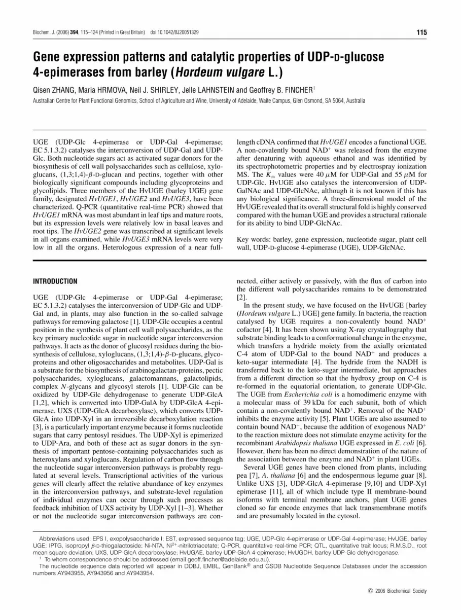
Biochem. J. (2006) 394, 115–124 (Printed in Great Britain) doi:10.1042/BJ20051329 115
Gene expression patterns and catalytic properties of UDP-D-glucose4-epimerases from barley (Hordeum vulgare L.)
Qisen ZHANG, Maria HRMOVA, Neil J. SHIRLEY, Jelle LAHNSTEIN and Geoffrey B. FINCHER1
Australian Centre for Plant Functional Genomics, School of Agriculture and Wine, University of Adelaide, Waite Campus, Glen Osmond, SA 5064, Australia
UGE (UDP-Glc 4-epimerase or UDP-Gal 4-epimerase;EC 5.1.3.2) catalyses the interconversion of UDP-Gal and UDP-Glc. Both nucleotide sugars act as activated sugar donors for thebiosynthesis of cell wall polysaccharides such as cellulose, xylo-glucans, (1,3;1,4)-β-D-glucan and pectins, together with otherbiologically significant compounds including glycoproteins andglycolipids. Three members of the HvUGE (barley UGE) genefamily, designated HvUGE1, HvUGE2 and HvUGE3, have beencharacterized. Q-PCR (quantitative real-time PCR) showed thatHvUGE1 mRNA was most abundant in leaf tips and mature roots,but its expression levels were relatively low in basal leaves androot tips. The HvUGE2 gene was transcribed at significant levelsin all organs examined, while HvUGE3 mRNA levels were verylow in all the organs. Heterologous expression of a near full-
length cDNA confirmed that HvUGE1 encodes a functional UGE.A non-covalently bound NAD+ was released from the enzymeafter denaturing with aqueous ethanol and was identified byits spectrophotometric properties and by electrospray ionizationMS. The Km values were 40 µM for UDP-Gal and 55 µM forUDP-Glc. HvUGE also catalyses the interconversion of UDP-GalNAc and UDP-GlcNAc, although it is not known if this hasany biological significance. A three-dimensional model of theHvUGE revealed that its overall structural fold is highly conservedcompared with the human UGE and provides a structural rationalefor its ability to bind UDP-GlcNAc.
Key words: barley, gene expression, nucleotide sugar, plant cellwall, UDP-D-glucose 4-epimerase (UGE), UDP-GlcNAc.
INTRODUCTION
UGE (UDP-Glc 4-epimerase or UDP-Gal 4-epimerase;EC 5.1.3.2) catalyses the interconversion of UDP-Glc and UDP-Gal and, in plants, may also function in the so-called salvagepathways for removing galactose [1]. UDP-Glc occupies a centralposition in the synthesis of plant cell wall polysaccharides, as thekey primary nucleotide sugar in nucleotide sugar interconversionpathways. It acts as the donor of glucosyl residues during the bio-synthesis of cellulose, xyloglucans, (1,3;1,4)-β-D-glucans, glyco-proteins and other oligosaccharides and metabolites. UDP-Gal isa substrate for the biosynthesis of arabinogalactan-proteins, pecticpolysaccharides, xyloglucans, galactomannans, galactolipids,complex N-glycans and glycosyl sterols [1]. UDP-Glc can beoxidized by UDP-Glc dehydrogenase to generate UDP-GlcA[1,2], which is converted into UDP-GalA by UDP-GlcA 4-epi-merase. UXS (UDP-GlcA decarboxylase), which converts UDP-GlcA into UDP-Xyl in an irreversible decarboxylation reaction[3], is a particularly important enzyme because it forms nucleotidesugars that carry pentosyl residues. The UDP-Xyl is epimerizedto UDP-Ara, and both of these act as sugar donors in the syn-thesis of important pentose-containing polysaccharides such asheteroxylans and xyloglucans. Regulation of carbon flow throughthe nucleotide sugar interconversion pathways is probably regu-lated at several levels. Transcriptional activities of the variousgenes will clearly affect the relative abundance of key enzymesin the interconversion pathways, and substrate-level regulationof individual enzymes can occur through such processes asfeedback inhibition of UXS activity by UDP-Xyl [1–3]. Whetheror not the nucleotide sugar interconversion pathways are con-
nected, either actively or passively, with the flux of carbon intothe different wall polysaccharides remains to be demonstrated[2].
In the present study, we have focused on the HvUGE [barley(Hordeum vulgare L.) UGE] gene family. In bacteria, the reactioncatalysed by UGE requires a non-covalently bound NAD+
cofactor [4]. It has been shown using X-ray crystallography thatsubstrate binding leads to a conformational change in the enzyme,which transfers a hydride moiety from the axially orientatedC-4 atom of UDP-Gal to the bound NAD+ and produces aketo-sugar intermediate [4]. The hydride from the NADH istransferred back to the keto-sugar intermediate, but approachesfrom a different direction so that the hydroxy group on C-4 isre-formed in the equatorial orientation, to generate UDP-Glc.The UGE from Escherichia coli is a homodimeric enzyme witha molecular mass of 39 kDa for each subunit, both of whichcontain a non-covalently bound NAD+. Removal of the NAD+
inhibits the enzyme activity [5]. Plant UGEs are also assumed tocontain bound NAD+, because the addition of exogenous NAD+
to the reaction mixture does not stimulate enzyme activity for therecombinant Arabidopsis thaliana UGE expressed in E. coli [6].However, there has been no direct demonstration of the nature ofthe association between the enzyme and NAD+ in plant UGEs.
Several UGE genes have been cloned from plants, includingpea [7], A. thaliana [6] and the endospermous legume guar [8].Unlike UXS [3], UDP-GlcA 4-epimerase [9,10] and UDP-Xylepimerase [11], all of which include type II membrane-boundisoforms with terminal membrane anchors, plant UGE genescloned so far encode enzymes that lack transmembrane motifsand are presumably located in the cytosol.
Abbreviations used: EPS I, exopolysaccharide I; EST, expressed sequence tag; UGE, UDP-Glc 4-epimerase or UDP-Gal 4-epimerase; HvUGE, barley
UGE; IPTG, isopropyl β-D-thiogalactoside; Ni-NTA, Ni2+-nitrilotriacetate; Q-PCR, quantitative real-time PCR; QTL, quantitative trait locus; R.M.S.D., root
mean square deviation; UXS, UDP-GlcA decarboxylase; HvUGAE, barley UDP-GlcA 4-epimerase; HvUGDH, barley UDP-Glc dehydrogenase.1 To whom correspondence should be addressed (email [email protected]).The nucleotide sequence data reported will appear in DDBJ, EMBL, GenBank® and GSDB Nucleotide Sequence Databases under the accession
numbers AY943955, AY943956 and AY943954.
c© 2006 Biochemical Society

116 Q. Zhang and others
Table 1 Q-PCR primer sequences, product sizes and optimal acquisition temperatures
Barley GAPDH (glyceraldehyde-3-phosphate dehydrogenase), HSP70 (heat-shock protein 70), α-tubulin, cyclophilin and CesA 1 (cellulose synthase 1) were used as control genes. T , the meltingtemperature.
Gene Forward primer Reverse primer Size (bp) T (◦C)
GAPDH GTGAGGCTGGTGCTGATTACG TGGTGCAGCTAGCATTTGAGAC 198 80HSP70 CGACCAGGGCAACCGCACCAC ACGGTGTTGATGGGGTTCATG 108 83α-Tubulin AGTGTCCTGTCCACCCACTC AGCATGAAGTGGATCCTTGG 248 80Cyclophilin CCTGTCGTGTCGTCGGTCTAAA ACGCAGATCCAGCAGCCTAAAG 122 79CesA 1 TGTGGCATCAACTGCTAGGAAA CGTACAAAGTGCCTCATAGGAAA 267 75HvUGE1 CGCAGAGACAATGAAGGAGGAAGA CAAGGCAAGGGAACCAACCTTAT 176 75HvUGE2 GATCGGTCCGTGTGGACTCACC GCTGCTAGGCAATGCAACTCAA 268 77HvUGE3 TCGTCGTGGCGTCTCTTCTG ATGCGTGGAGCCCATACTGAC 241 78
Although it is assumed that UGE plays an important rolein galactose metabolism and in the biosynthesis of galactose-containing polysaccharides, the extent and breadth of its physio-logical functions are unclear in plants. Mutation of the correspond-ing genes through transposon insertions causes a loss of thepathogenicity for Erwinia amylovora, the causative agent of‘fire blight’ in rosaceous plants [12]. In Rhizobium meliloti, theformation of nodules on legumes is also affected by mutation ofthe exoB gene, a homologue of the E. coli UGE, and the mutationsresult in an inability of the plant to synthesize the galactose-containing capsular polysaccharides EPS I (exopolysaccharide I)and EPS II [13]. In A. thaliana, a mutation in one of the five UGEgenes results in abnormal root growth in a mutant (rhd1) [14],which is attributed to abnormal biosynthesis of xyloglucans andarabinogalactans in root epidermal cells, as well as to the down-regulation of some arabinogalactan-proteins. Ethylene suppressesall rhd1 phenotypes by restoring the biosynthesis of galactose-containing xyloglucans and arabinogalactans. However, ethylenedoes not up-regulate expression of other UGE isoforms [2,14].A possible function of UGE in plant root growth has alsobeen reported in rice, where drought stress induces increasedexpression of a UGE gene, which maps to a root thickness QTL(quantitative trait locus) region [15].
Here, we have examined the barley HvUGE gene familythrough analyses of extensive barley EST (expressed sequencetag) databases. Three members of the HvUGE gene family havebeen cloned and their sequences were used to design gene-specificprimers for Q-PCR (quantitative real-time PCR) for comparisonsof relative transcriptional activities of individual members of thegene family in a range of barley organs. One near full-lengthcDNA has been expressed in E. coli to confirm that HvUGE activ-ity is associated with the protein product of the cDNA. Theexpressed enzyme was characterized biochemically and alsoexamined for the presence of a bound NAD+. A three-dimensionalmodel of HvUGE1 was also constructed using human UDP-Galepimerase as a template, to establish a structural basis forthe enzyme’s substrate specificity. The results from enzymaticassays and the three-dimensional modelling have demonstratedthat the HvUGE also catalyses the reversible interconversionof UDP-GlcNAc and UDP-GalNAc. Thus the enzyme mighthave several functions in plants, including the synthesis of bothpolysaccharides and glycoproteins.
EXPERIMENTAL
Materials
UDP-Glc, galactose dehydrogenase, NADH and NAD+ werepurchased from Sigma–Aldrich (Australia). Superscript II RNase
H reverse transcriptase, Gateway protein expression system andTRIzol® reagent were from Invitrogen Australia (MountWaverley, Victoria, Australia).
Isolating HvUGE cDNAs
After searching barley EST databases, which contain approx.360000 entries, with a sequence of the A. thaliana UGE gene(accession number Z54214), approx. 90 cDNA clones wereidentified. Analysis of these clones with ContigExpress software(Informax, Frederick, ML, U.S.A.) generated three consensus se-quences. Three gene-specific oligonucleotide primers (ATG-GTTTCTGCTGTTCTTC, CCGCTCCGCTCGCTCGCTCCCTand ATGGCGATCGGCGGCGTT) were used with an oligo-dT primer to amplify barley leaf cDNA by PCR. Three barleycDNAs were obtained and were designated HvUGE1, HvUGE2and HvUGE3.
cDNA preparations and Q-PCR analysis of transcript levels
Total RNA preparation, cDNA synthesis and Q-PCR followed thesame procedures described by Zhang et al. [3]. The preparationof barley leaf and root segments was done as described by Burtonet al. [16]. The primer pairs and optimal acquisition temperaturesfor Q-PCR are shown in Table 1.
Heterologous expression
The HvUGE1 cDNA was cloned into E. coli using the Gatewayprotein expression system following the same procedures asthose described for HvUXS1 expression [3], after the near full-length barley HvUGE1 cDNA was amplified by PCR using theprimer pair GGGGACAAGTTTGTACAAAAAAGCAGGCT-TCGAAGGAGATAGAACCATGGGTCATCACCATCACCAT-CACCAGATGGTTTCTGCTGTTCTTCGTACGA/GGGGACC-ACTTTGTACAAGAAAGCTGGGTCCTATCAGTGGCCGTT-GTCGCTGGATCCG. The underlined nucleotides indicate gene-specific regions. Recombinant protein was induced with 0.5 mMIPTG (isopropyl β-D-thiogalactoside) and purified using Ni-NTA(Ni2+-nitrilotriacetate) resin. Expressed proteins were analysed bySDS/PAGE on 12% (w/v) denaturing gels as described previously[3]. The N-terminal amino acid sequence of the recombinantprotein was determined using a Hewlett–Packard G1000Aautomated amino acid sequencer (Hewlett–Packard, Palo Alto,CA, U.S.A.).
Enzyme assay and NAD+ determination
Procedures similar to those used for HvUXS1 [3] were used for thedemonstration of non-covalently bound NAD+ in the recombinant
c© 2006 Biochemical Society

Barley UDP-D-glucose 4-epimerase 117
HvUGE1 enzyme, including HPLC and MS analyses. HvUGE1activity was assayed in a reaction mixture (75 µl) containing20 mM sodium phosphate (pH 8.0), 2 mM NAD+, 0.05 mM UDP-Gal or UDP-Glc and purified recombinant HvUGE1 (12 ng ofprotein). The assay was performed at 25 ◦C for 10 min unlessotherwise specified and terminated by quickly transferring thereaction tube to a boiling-water bath or by addition of 4 vol. of100% ethanol. The reaction mixture was dried and analysed byHPLC on a Hewlett–Packard 1090 LC. UDP-Glc and UDP-Galwere separated using a reversed phase column (Hypersil ODS,5 µm, 2.1 mm × 250 mm; Agilent Technologies, Wilmington,DE, U.S.A.) and eluted isocratically with 0.1 M triethylamineacetate and 0.1 mM EDTA (pH 6.8) at a flow rate of 0.2 ml/min.UDP-GlcNAc and UDP-GalNAc were separated on a PrevailCarbohydrate ES column (150 mm × 4.6 mm, 5 µm; Alltech,Deerfield, IL, U.S.A.) using a gradient of 25 mM NaB4O7/25 mMNaClO4 (0–15 min) to 75 mM NaB4O7/75 mM NaClO4 (15–25 min) at 0.6 ml/min. The elution was monitored at 262 nm.Various concentrations of UDP-Glc, UDP-Gal (6–250 µM) andUDP and UMP (0–125 µM) were used for determinations ofkinetic constants, estimated from the Lineweaver–Burk (Km, kcat)and Dixon (K i) plots. Care was taken to ensure that initial reactionrates were measured. Substrate consumption never exceeded10% of initial concentrations. For assessment of the equilibriumconstants (Keq), the reaction mixture (75 µl) contained thestandard reaction ingredients specified above except that 360 ngof protein of the recombinant HvUGE1 (instead of 12 ng) wasadded, and the reactions were sampled at 5, 15, 40 and 60 mintime intervals.
Construction of a three-dimensional model for HvUGE1
A three-dimensional molecular model of HvUGE1 was con-structed using the Modeler 7v7 program [17,18], which uses thesatisfaction of empirical spatial restraints and statistical analysisof the relationships between pairs of homologous structures inorder to produce a protein fold. The CHARMM22 force-fieldenergy function enforcing proper stereochemistry was imple-mented during energy minimization of models to produce an ob-jective function, which represents a combined term with the spatialrestraints. The first step of the experimental approach was theidentification of a known three-dimensional structure (template)related to the target HvUGE1 sequence. The modelling uses co-ordinates of the template protein as a basis for further modell-ing. To identify the most suitable template for HvUGE1, searchesthrough the Structure Prediction Meta-Server ([19]; http://bioinfo.pl/meta/pdb-test.pl), including SeqAlert (Bioinformatics and Bio-logical Computing, Weizmann Institute of Science, Rehovot,Israel), Protein Data Bank (http://www.rcsb.org/pdb/) and 3D-PSSM Server (Imperial College of Science, Technology andMedicine, London, U.K.) were performed. The best template wasfound to be the human UGE with the PDB entries 1ek5, 1ek6and 1hzj, which represent binary complexes of human UGE withNAD+, NAD+ plus UDP-Glc, and NAD+ plus UDP-GlcNAc re-spectively. The second step in modelling process was the align-ment of the template with the target sequence, using the T-COFFEE server. The alignment contained four 1–6-amino-acid-long gap insertions and deletions (results not shown). Thealignment was further checked manually using hydrophobiccluster analysis to maintain integrity of hydrophobic cluster pat-terns and the distribution of secondary structural elements. Thepositional sequence identity and similarity scores were calculatedby the Bestfit program from the University of Wisconsin GCGsoftware package [20], with the implemented gap penalty functionand dynamic programming algorithm of Smith and Waterman
[21]. The identity and similarity scores between the human UGEand HvUGE enzymes were 60.5 and 73.2% respectively. In thefinal step, the structurally aligned HvUGE1 and human UGEsequences containing the ligands NAD+, NAD+ plus UDP-Glc,and NAD+ plus UDP-GlcNAc were used as input parameters tobuild three-dimensional molecular models on a Silicon Graphicsmodel O2 workstation, running IRIX 6.5. The final three-dimensional molecular model of HvUGE1 was selected from 40models; the model with the lowest value of the Modeller objectivefunction was chosen for further refinement. This was done byenergy minimization using the GROMOS96 implementation inSwiss-PdbViewer [22] and SYBYL 6.9.1 [23].
The stereochemical quality and overall G-factors of the finalHvUGE1 model were calculated with PROCHECK [24]. Z-score values were calculated by ProsaIIv3; the plots weresmoothed using a window size of 50 amino acid residues. Theroutine ‘compare.top’ implemented in the Modeller 7v7 [17] wasused to determine the R.M.S.D. (root mean square deviation)values in the Cα positions and distance R.M.S.D. values inatom distances between the two three-dimensional structures.The molecular graphics were generated with PyMol graphicalsoftware (http://www.pymol.org).
The cDNA sequences determined here for the HvUGE1,HvUGE2 and HvUGE3 genes have been lodged in the GenBank®
and EMBL databases under the accession numbers AY943955,AY943956 and AY943954 respectively.
RESULTS
HvUGE cDNA clones
Analysis of barley EST sequences revealed three consensussequences. PCR amplification from barley leaf cDNA usingprimers designed from the consensus sequences produced threeHvUGE cDNA clones. The cDNA sequences were 1317, 1642 and1475 bp in length and were designated HvUGE1, HvUGE2and HvUGE3 respectively. The open reading frames of HvUGE1,HvUGE2 and HvUGE3 encoded proteins consisting of 353,374 and 370 amino acid residues respectively. HvUGE proteinsshowed moderate sequence identities (54–72 %) with human andA. thaliana UGEs at the amino acid level (Figure 1, Table 2), buta much higher sequence identity with the orthologous UGE fromrice (>79%; Table 2). They all contained GXXGXXG NAD+-binding motifs and conserved Ser, Tyr and Lys amino acid residuesthat are believed to be located in the catalytic site (Figure 1).
The sequence identities between HvUGE1, HvUGE2 andHvUGE3 were between 60 and 74% at the amino acidlevel (Table 2). The UGEs were clearly distinguishable fromother barley nucleotide sugar interconverting enzymes such asthe HvUXSs [3], HvUGDH (barley UDP-Glc dehydrogenase)and HvUGAE (barley UDP-GlcA 4-epimerase), with which therewas less than 30% sequence identity at the amino acid level(Table 2). However, the HvUGE shared more than 37% sequenceidentity with the UDP-Xyl epimerase.
Biochemical properties of HvUGE1
To confirm that the cloned HvUGE cDNAs encoded enzymes withUGE activity, the HvUGE1 cDNA was selected for heterologousexpression in E. coli. The selection was based on observationsthat HvUGE1 mRNA was relatively abundant in many barleyorgans. A recombinant protein induced with 0.5 mM IPTG hada molecular mass of 39 kDa (Figure 2), as predicted from theHvUGE1 cDNA. N-terminal amino acid sequencing showedthat the recombinant protein contained the amino acid residues
c© 2006 Biochemical Society

118 Q. Zhang and others
Figure 1 Alignment of amino acid sequences of UGEs
Amino acid sequences of UGEs from barley (HvUGE1, HvUGE2 and HvUGE3), A. thaliana (AtUGE1, Swiss-Prot Data Bank entry Q42605), rice (TIGR locus: Os05g51670) and human (PDB entry1hzj) were aligned using the GeneDoc program (www.psc.edu/biomed/genedoc). The conserved motifs GXXGXXG (NAD+-binding) and catalytic amino acid residues Ser and Tyr-Xaa-Xaa-Xaa-Lysof the active site are underlined.
Table 2 Comparison of sequence identities
Amino acid sequence identities (%) of HvUGE1, HvUGE2 and HvUGE3 are compared with barleyUDP-Xyl synthases (HvUXS1–HvUXS4) [3], barley UDP-Xyl 4-epimerases (HvUXE1–HvUXE3),HvUGDH, HvUGAE (Q. Zhang and G. B. Fincher, unpublished work), rice UGEs (TIGR Data Bankloci Os05g51670, Os09g15420, Os09g35800 and Os08g28730), a human UGE (PDB entry1hzj) and A. thaliana UGE1 (Swiss-Prot Data Bank entry Q42605).
Sequence identity (%)
HvUGE1 HvUGE2 HvUGE3
HvUGE1 100HvUGE2 74 100HvUGE3 63 60 100HvUXS1 26 27 20HvUXS2 28 27 26HvUXS3 28 29 27HvUXS4 29 28 24HvUXE1 41 41 37HvUXE2 42 42 40HvUXE3 40 39 39HvUGDH 21 17 21HvUGlcAE 25 27 21Rice Os05g51670 90 76 63Rice Os09g15420 74 87 60Rice Os09g35800 61 62 79Rice Os08g28730 76 82 63Human UGE 59 55 54A. thaliana UGE1 66 61 72
Figure 2 SDS/PAGE of barley HvUGE1 expressed in E. coli
The barley HvUGE1 cDNA was expressed in E. coli and proteins were separated by SDS/PAGE.Control, cell extracts without addition of IPTG; IPTG, protein expression following induction with0.5 mM IPTG; Ni-NTA, the purified fraction following Ni-NTA affinity chromatography. Proteinmolecular mass standards (in kDa) are indicated on the left.
MVSAVLRTILVT after the His tag, and this sequence was asexpected from the HvUGE1 cDNA sequence (Figure 1).
The recombinant protein catalysed the interconversion of UDP-Glc and UDP-Gal. It displayed a broad pH optimum in the rangepH 8–9.5. The bivalent cations Mg2+ and Ca2+ neither stimulatednor inhibited the activities of the recombinant HvUGE1 (results
c© 2006 Biochemical Society

Barley UDP-D-glucose 4-epimerase 119
Table 3 Kinetic constants of HvUGE1
The values are means for three experiments and the standard deviations are indicated forUDP-Gal and UDP-Glc. The values for UDP-GalNAc and UDP-Glc are estimates only becauseof difficulties in the HPLC separation of the two compounds.
Substrates K m (µM) k cat (s−1) k cat/K m (s−1 ·µM−1)
UDP-Gal 40 +− 4 21 +
− 3 550 +− 80
UDP-Glc 55 +− 1 11 +
− 2 220 +− 30
UDP-GalNAc 160 0.12 0.8UDP-GlcNAc 200 0.07 0.4
not shown). As expected from these results, the addition of 2 mMEDTA had no effect on enzyme activity (results not shown).
The recombinant HvUGE1 had Km values of 40 µM for UDP-Gal and 55 µM for UDP-Glc, and kcat values of 21 s−1 for UDP-Galand 11 s−1 for UDP-Glc (Table 3). The efficiency of the reactioncatalysed by HvUGE1 was approx. 2.5-fold higher for UDP-Galthan for UDP-Glc (Table 3). Standard deviations were generallyless than 15%. After the reaction reached equilibrium, UDP-Galand UDP-Glc concentrations were approx. 23 and 77 % of totalnucleotide sugars in the reaction system respectively. The Keq
obtained from the ratio of substrate to product was 0.30, whilethat calculated from kcat/Km values for the forward and reversereactions was 0.40. These values can be compared with a Keq of0.38 reported by Piller et al. [25].
The heterologously expressed HvUGE1 also catalysed theinterconversion of UDP-GalNAc and UDP-GlcNAc. The finalequilibrium proportions were 26 and 74% of UDP-GalNAcand UDP-GlcNAc respectively, and Keq values were thereforevery similar to those calculated for the UDP-Glc/UDP-Galreaction (Table 3). The Km values for UDP-GalNAc and UDP-GlcNAc were 160 and 200 µM respectively (Table 3). The ratiosof catalytic efficiencies (kcat/Km) for the substrate pairs UDP-Glc/UDP-GlcNAc and UDP-Gal/UDP-GalNAc were 550 and688 respectively, and these values indicated that the enzyme is500–600 times more efficient with the UDP-Glc and UDP-Galsubstrates.
UDP and UMP strongly inhibited the interconversion of UDP-Gal and UDP-Glc, with K i values of 36 and 32 µM respectively. Asimilar K i value (20 µM) has been reported for UGE from porcine
submaxillary glands [25]. Glucose and galactose had little effecton the reaction rate of the barley enzyme, such that K i values forthese sugars could not be calculated. The barley enzyme appears tohave very low levels of activity on UDP-Xyl (results not shown),but this observation has not been examined in detail.
Characterization of bound NAD+
Addition of exogenous NAD+, NADH, NADP+ or NADPH tothe reaction mixture did not significantly stimulate the catalyticrate (results not shown), although it is known that NAD+ playsa critical role in the reaction mechanism [4,26]. To confirm thatthe heterologously expressed enzyme contained bound NAD+, therecombinant HvUGE1 was treated with aq. 80% (v/v) ethanol. Anethanol-soluble compound was released, and was shown to havean HPLC retention time and an absorbance spectrum similar tostandard NAD+ (results not shown). When the ethanol extractablecomponent was incubated with galactose dehydrogenase in thepresence of galactose, a compound that had the same HPLCprofile and absorbance spectrum of standard NADH was produced(results not shown). It was therefore concluded that the extractedcompound from the recombinant HvUGE1 was NAD+, which hadbeen non-covalently associated with the enzyme before ethanolextraction. Purification of the expressed HvUGE1 on the Ni-NTAresin had no effect on the bound NAD+. Analysis of the 80%
ethanol-soluble component by electrospray ionization MS showedthat the component had a mass of 664 Da, which is the mass ofstandard NAD+ (results not shown). The molar ratio of NAD+ inthe recombinant HvUGE1 protein was estimated to be 0.85. It islikely therefore that each protein molecule contains one moleculeof NAD+.
Transcript levels of HvUGE genes in different organs
Transcript levels of the three individual HvUGE genes were deter-mined by Q-PCR using gene-specific primers designed from3′-untranslated sequences. The HvUGE1 mRNA was most abun-dant in leaf tip and mature roots, where HvUGE1 mRNA levelswere 300- and 10-fold higher than the HvUGE1 mRNA levels inbasal leaves and root tips respectively (Figure 3). In contrast,HvUGE1 mRNA levels in developing grains, coleoptiles andstems were at least 15-fold lower than the HvUGE1 mRNA level
Figure 3 Normalized expression levels of HvUGE genes in different barley organs
Levels of mRNA are presented as the number of copies per µg of total RNA after normalization. Organs were as described by Burton et al. [16] and included leaf tips, leaf bases, root tip and thematuration zone (m/zone) of roots, flowers just before anthesis (FL early), flowers at anthesis (FL anthesis), developing grain 3 and 13 days post-anthesis (Grain early and Grain mid), 3-day-oldcoleoptiles (Col 3 day) and the stem of mature plants just below the emerging ear (Stem). The results shown are means for four separate evaluations and S.D.s are indicated.
c© 2006 Biochemical Society

120 Q. Zhang and others
Figure 4 Normalized expression levels of HvUGE genes in young barleyleaves, roots and coleoptiles
Barley leaves were sectioned into five segments and roots were cut into four segments asdescribed by Burton et al. [16]. Units and statistical treatments were as described in the legendto Figure 3.
in leaf tips. The HvUGE2 gene was transcribed in all organs ex-amined at approximately similar levels, but the mRNA abundancewas only approx. 15% of HvUGE1 mRNA levels in leaf tips.Levels of HvUGE2 mRNA were higher than the levels of HvUGE1mRNA in leaf bases, root tips, early developing grains, coleoptilesand stems (Figure 3). Levels of HvUGE3 mRNA were lowest inall organs and generally accounted for less than 5 % of HvUGE1mRNA levels in leaf tips.
Transcript levels of HvUGE genes in young leaves,coleoptiles and roots
In young barley leaves, expression of the HvUGE1 gene washighest in leaf tips, but rapidly decreased to very low levels inthe basal region of the leaves. The HvUGE1 mRNA level wasapprox. 100-fold lower in leaf bases than in leaf tips (Figure 4A).Both HvUGE2 and HvUGE3 mRNA levels were low in all leafsegments.
In young barley roots, mRNA levels of HvUGE1 were veryhigh in the maturation zone compared with other regions of theroot (Figure 4B). The HvUGE1 transcript levels were approx. 10-fold higher in maturation zones than in root tips. Similar levels ofHvUGE2 transcripts were detected in all root segments, but theywere at least 5-fold lower than the levels of HvUGE1 mRNA inthe maturation zone. The HvUGE2 mRNA level was 2.5-foldhigher than HvUGE1 transcript level in root tips. Transcriptsof the HvUGE3 gene were not detectable in the root segments(Figure 4B). Comparison of mRNA levels between leaves androots shows that the expression levels were 4-fold higher in thematuration zones of roots than in leaf tips (Figures 4A and 4B).
In coleoptiles, levels of HvUGE1 mRNA were low in day 1and day 3 coleoptiles (Figure 4C), but increased approx. 28-fold inday 5 coleoptiles. In contrast, the level of HvUGE2 mRNA re-mained essentially unchanged from day 1 to day 5. The HvUGE3
mRNA was undetectable in day 1 and day 3 coleoptiles, butshowed a 20-fold increase by day 5. Overall, the level of HvUGEmRNA was over 10-fold lower in coleoptiles than in matureroots.
Molecular modelling of HvUGE1
The human UGE structure (PDB entry 1hzj), which represents amixed α/β protein fold based on the CATH (Class, Architecture,Topology and Homologous superfamily) classification [27],served as an excellent structural template for modelling theHvUGE1. The solved crystal structure of the human UGEalso included the binding positions of NAD+ and UDP-GlcNAcand the template therefore represented a ternary complex of thehuman UGE enzyme, with bound substrate and cofactor [28].The stereochemical quality and overall G-factors of the finalHvUGE1 enzyme/substrate/cofactor model [29] showed that noneof the amino acid residues in the modelled structure were posi-tioned in disallowed regions, and the overall G-factors were0.18 and 0.16 for the template and modelled three-dimensionalstructures respectively. The Z-score values, which reflect thecombined statistical potential energy of the modelled HvUGE1and template three-dimensional structures and the estimate com-patibility of the sequence with an overall fold and conservationof the key functional and structural residues, were −10.96 and−10.07 for 345 and 343 residues of the template and modelledstructures respectively. The R.M.S.D. values in the Cα positionsand distance R.M.S.D. values in atom distances between the twothree-dimensional structures were 0.951 and 1.903 Å respect-ively (1 Å = 0.1 nm) for 343 amino acid residues. The modelledHvUGE1 structure was further evaluated using the pG server [18];the pG score of 1.0 again indicated that the HvUGE1 model couldbe classified as highly reliable.
The overall three-dimensional shape and the distributionof the secondary structure elements of the HvUGE1 were highlyconserved in comparison with the human UGE crystal structure(results not shown), and the overall geometry of the active site wasalso conserved. However, there were differences in insertions anddeletions of loops that connect the individual secondary structureelements in the HvUGE1, although these insertions and deletionswere always positioned at the surface of the enzyme (results notshown). Insertions and deletions of 1, 2, 3 and 6 amino acidresidues were located in the loop regions at amino acid positions152, 57, 261 and 43 respectively. The two highly conserved, keycatalytic amino acid residues in the human UGE, namely Ser132
and Tyr157, are 4.7 Å apart in the human enzyme. In the HvUGE1enzyme, these residues corresponded to Ser132 and Tyr156, whichwere 4.8 Å apart. The relatively close proximity of these two keyresidues is a characteristic feature for UGEs [28].
The molecular modelling of the HvUGE1 also allowed thelocations of the NAD+ cofactor and the UDP-GlcNAc substratein the active site region to be defined (Figure 5A). The reason forthe choice of UDP-GlcNAc as a nucleotide sugar substrate donorfor the molecular modelling was to investigate if the HvUGE1enzyme could actually accommodate UDP-GlcNAc in its activesite [28]. The modelling clearly showed that there were no inter-atomic clashes among amino acid residues that participate in bind-ing of the cofactor NAD+ and the substrate UDP-GlcNAc (Fig-ure 5B). The triplet of catalytic amino acid residues Ser, Tyr andLys was positioned at the interface between bound NAD+
and UDP-GlcNAc in both human UGE and HvUGE (results notshown). Analysis of the model of the barley enzyme showedthat only a few favourable hydrogen-bonding interactions wereformed among Tyr225, Asp305, Arg302, Asn206 and Asn186, and thecorresponding groups of the UDP moiety of UDP-GlcNAc. From
c© 2006 Biochemical Society

Barley UDP-D-glucose 4-epimerase 121
Figure 5 Molecular model of the HvUGE1
(A) Stereo view of the ribbon representation of the HvUGE1 model shows secondary structural elements of the enzyme folded into a mixed α/β protein fold. The R.M.S.D. value between the templatestructure (PDB entry 1hzj) and the modelled HvUGE1 is 0.951 A over 343 amino acid residues. The N- and C-termini of the enzyme are shown in the lower and upper parts of the panel respectively.Spheres in atomic colours represent van der Waals radii of bound UDP-GlcNAc and NAD+ ligands. (B) Stereo representation of the molecular surface area depicts the active site region with boundNAD+ and UDP-GlcNAc. The molecular surface was generated with a probe radius of 1.4 A. The Figure was prepared with PyMol.
these interactions, the shortest hydrogen bond of 2.7 Å was madebetween Asp305 and the C-4′ OH group of the ribosyl moiety ofUDP-GlcNAc. It was also apparent from the model that the N-acetyl group at the C-2′ atom of UDP-GlcNAc was not bondedto amino acid residues that were positioned nearby. Instead, ahydrogen bond of 3.3 Å was formed between Ser132 and the C-4′
OH group of the GlcNAc moiety of UDP-GlcNAc. However, itmust be emphasized that the distances between the active siteresidues of the modelled HvUGE1 structure and UDP-GlcNAcshould be treated as estimates only, because of interpretativeproblems associated with molecular modelling. It was thereforerationalized in silico that the HvUGE1 enzyme would be able toaccommodate UDP-GlcNAc in its active site, and hence wouldhave the potential to epimerize this nucleotide sugar.
DISCUSSION
Database searches and Q-PCR analyses indicate that the UGEenzymes of barley are encoded by a small gene family of atleast three members. In rice, there are at least four UGE genes,while in A. thaliana the UGE family consists of five individualgenes [1,2]. Near full-length cDNAs encoding the three barleyisoenzymes, designated HvUGE1, HvUGE2 and HvUGE3, wereamplified by PCR from cDNA preparations from young barleyleaves and characterized. The HvUGE1 cDNA was expressed inE. coli to confirm the substrate specificity of the encoded enzymeand to define its kinetic and chemical properties.
The UGEs belong to a superfamily of dehydrogenases/re-ductases [1]. They contain an absolutely conserved GXXGXXG
c© 2006 Biochemical Society

122 Q. Zhang and others
NAD+-binding motif and a triplet of amino acid residues, Ser,Tyr and Lys, that is positioned at the interface between NAD+ andUDP-GlcNAc in the crystal structure of the human UGE [28].The three HvUGE isoenzymes also have the conserved NAD+-binding motif and the triplet of catalytic site amino acid residues(Figure 1). The barley enzymes contain 353–370 amino acidresidues and therefore have molecular masses similar to those forthe human and A. thaliana UGE enzymes (Figure 1). The HvUGEslack any obvious organellar targeting signals or transmembranehelices (Figure 1), and are therefore predicted to be located in thecytosol. Similarly, all five isoforms of AtUGE from A. thalianalack membrane-binding motifs and are believed to be cytosolicenzymes [2]. This is in contrast with other nucleotide sugarinterconverting enzymes such as UDP-Xyl synthases, for whichboth cytosolic and membrane-bound isoforms are found [3,30].
The cytosolic location for plant UGEs is consistent with theobservation that most of the cellular UDP-Glc and UDP-Gal, asmeasured by [31P]NMR, is located in the cytosol [31]. However,UDP-Glc and UDP-Gal are also used for biosynthesis of non-cellulosic polysaccharide and glycoproteins in the Golgi apparatus[2,32] and specific transporters are required for transferringnucleotide sugars from the cytosol to the lumen of the Golgicisternae. A nucleotide sugar transporter gene has been cloned inA. thaliana, and the gene product transports both UDP-Gal andUDP-Glc into the Golgi [33]. A near full-length cDNA sequenceencoding a putative barley UDP-Gal transporter with 79% aminoacid sequence identity to the A. thaliana UDP-Gal transporter hasalso been cloned (Q. Zhang and G. B. Fincher, unpublished work).
Following heterologous expression of the barley HvUGE1cDNA and affinity purification of the expressed protein, UGEactivity was confirmed. The expressed enzyme exhibited bio-chemical properties that were similar to those of UGE enzymesfrom other plants, insofar as it was not activated by Mg+ or Ca+
ions and it had a broad pH optimum of 8.0–9.5 [34]. Furthermore,it was shown that the expressed HvUGE1 enzyme contained anon-covalently bound NAD+ cofactor, as suggested by proposedreaction mechanisms and inhibition studies, but not previouslydemonstrated in plant UGEs [1,2]. Certain nucleotide sugarinterconverting enzymes can be activated or inhibited by cofactorsor products. Thus a UDP-D-apiose/UDP-Xyl synthase from A.thaliana is activated by NAD+ and inhibited by NADH [35]. UDP-Glc dehydrogenases from soya bean and sugarcane are subject tofeedback inhibition by UDP-Xyl [36,37], as are the UDP-Xylsynthases from A. thaliana and barley [3,6]. However, activityof the HvUGE1 enzyme is not affected by the cofactor NAD+,consistent with the conclusion that this cofactor is bound withinthe enzyme molecule, and we could find no kinetic evidence forproduct inhibition of reactions catalysed by HvUGE1.
The expressed HvUGE1 enzyme showed a relatively highaffinity for its UDP-Gal and UDP-Glc substrates compared withbacterial and human UGEs. Thus the Km values for HvUGE1 were40 and 55 µM, depending on the substrate, while those for humanand bacterial UGEs are generally approx. 200 µM [38]. It is worthnoting that the concentration of UDP-Glc in endosperm cells fromLolium multiflorum is approx. 30 µM [39]. The kinetic analysisalso revealed that the HvUGE1 has a higher catalytic efficiencyfor UDP-Gal (kcat/Km ∼ 550 s−1 ·µM−1) than UDP-Glc (kcat/Km∼220 s−1 · µM−1). Thus HvUGE1 could play a role in removinggalactose, which can have inhibitory effects on cell growth[40,41]. In the recycling process, free galactose is phosphorylatedto galactose 1-phosphate by galactose kinase and subsequentlyconverted into UDP-Gal by UDP-Gal pyrophosphorylase [42].
The heterologous expression of HvUGE1 enabled the substratespecificity of the enzyme to be examined in more detail. Threegroups of UGEs with slightly different substrate specificities
have been identified [28,43–45]. The first group is representedby bacterial UGEs, which only catalyse the interconversion ofUDP-Glc and UDP-Gal [43]. The second group is represented byhuman and some bacterial UGEs, which epimerize not onlyUDP-Gal and UDP-Glc, but also UDP-GalNAc and UDP-GlcNAc [28,44]. A third group of UGEs, found in Pseudomonasaeruginosa, epimerizes only UDP-GalNAc and UDP-GlcNAc[45]. In the present study, it has been shown that the expressedHvUGE1 enzyme catalyses not only the interconversion ofUDP-Gal and UDP-Glc, but also that of UDP-GalNAc and UDP-GlcNAc. The catalytic efficiencies with the latter two sub-strates are much lower than those for UDP-Gal and UDP-Glc(Table 3).
The broader substrate specificity of the HvUGE1 can berationalized through molecular modelling (Figure 5). The modelsof the barley enzyme confirmed that UDP-GalNAc could serve asa substrate, because there were no inter-atomic clashes betweenthe acetyl group on the C-2′ atom of the UDP-GalNAc moleculeand neighbouring amino acid residues of the enzyme or theNAD+ cofactor. Indeed, there were no steric clashes predictedfor any atom of the UDP-GlcNAc substrate. Furthermore, thejuxtaposition of NAD+ cofactor and C-4′ of the UDP-GalNAcsubstrate can be clearly seen (Figure 5). The results from themolecular modelling indicated that at least five critical hydrogenbonds were formed between the UDP moiety of UDP-GlcNAcand the enzyme, while only one such bond was detected betweenthe GlcNAc moiety of UDP-GlcNAc and the enzyme. The latterinteraction is also critical for binding of the Glc moiety of UDP-Glc to the human UGE [46]. When comparing binding modes ofUDP-Glc and UDP-GlcNAC in the human UGE crystal structures,it is apparent that substantial rotations, of approx. 90◦, of the Glcand GlcNAc moieties relative to UDP occur [28,38], while thehydrogen bonds between Glc and GlcNAc of the nucleotide sugarsubstrates and the corresponding amino acid residues are main-tained. It is expected that similar rotations could take place inthe barley enzyme, because secondary structural elements ofHvUGE1 and amino acid residues that participate in the formationof critical interactions among cofactors and substrates are con-served in both structures. We could therefore conclude that UDP-GlcNAc could be accommodated in the active site of the barleyenzyme and that the UDP component of UDP-GlcNAc is expectedto be bound more tightly than the GlcNAc moiety.
Despite the relatively low catalytic efficiency of the reaction, theability of the HvUGE1 to interconvert UDP-GalNAc and UDP-GlcNAc raises more general questions about the functions ofUGEs in plants. Although GlcNAc will be a common constituentof plant glycoproteins, GalNAc has also been detected in plantpolymers [47,48]. Thus it is possible but not proven thatplant UGEs might participate in the biosynthesis of glycolipidsor glycoproteins.
A combined analysis of the kinetic, substrate specificity andstructural modelling data provided some insight into enzyme–substrate binding reactions. Thus the similarities of the Km valuefor UDP-Glc (55 µM) and the K i values for UDP and UMP(36 and 32 µM respectively) indicated that the UDP region ofthe nucleotide sugar is likely to be a key determinant of initialsubstrate binding to the enzyme. The inability of Glc to inhibitthe reaction to any significant extent would be consistent with thisconclusion. This is not to say that the sugar region of the substrateis not involved in binding once the enzyme–substrate complex isformed. Structural information from the human UGE [28,30]clearly shows interactions between the enzyme and the sugarmoiety of the substrate, but the observation that five hydrogenbonds are formed between the enzyme and the UDP moiety ofUDP-GlcNAc, while only one hydrogen bond is formed between
c© 2006 Biochemical Society
Barley UDP-D-glucose 4-epimerase 123
the sugar moiety and the enzyme, is consistent with the importanceof the UDP moiety in substrate binding, as indicated by our kineticdata. If it is assumed that Km (k−1 + k2)/k1 is indicative of Ks
(k−1/k1, when k2 ≪ k−1), it could be concluded that the N-acetylgroup at the C-2′ carbon of the UDP-GlcNAc substrate has agreater effect on catalytic rate than on substrate binding.
It is worth noting that the concentration of UDP in potato tubersis approx. 3 µM [49], which is approx. 10 times lower than theK i value of HvUGE1 for UDP, and UDP is therefore unlikelyto directly affect activity of UGE enzymes under physiologicalconditions.
The availability of the three HvUGE cDNA sequences enabledthe design of specific PCR primers for the quantitative analysis ofrelative mRNA levels in a range of barley organs (Figures 3 and 4).In many organs, HvUGE1 mRNA is more abundant than mRNAsencoding the other two isoenzymes, although HvUGE2 mRNApredominates in some organs (Figure 3). In mature vegetativeorgans such as leaf tip and the maturation zone of roots, HvUGE1mRNA levels are relatively high compared with other organs, butin developing and dividing organs including basal leaves, root tipsand young coleoptiles, the levels of HvUGE1 mRNA were lower(Figures 3 and 4).
The precise physiological functions of UGEs in plants remainunclear. In A. thaliana, expression of UGE genes is relativelyhigh in roots and a mutation of the AtUGE4 gene, which isorthologous to the barley HvUGE1 gene, results in abnormal roothair formation [50]. In rice, expression of a UGE gene is inducedby drought treatment and this gene has been mapped to a rootthickness QTL region [15]. In pea roots, UGE gene expressionincreases 5-fold in 3-week-old plants, compared with 4-day-oldseedlings [7]. These observations suggest that the UGE enzymesplay a role in plant root development and/or growth.
It has also been suggested that nucleotide sugar intercon-versions and hence the composition of the nucleotide sugar poolin plant cells might reflect the compositions of cell walls inthose cells and might also represent a point of regulation ofwall composition [3]. It is not possible at this stage to determinewhether or not levels of HvUGE mRNA or enzyme might be cor-related with wall composition or with regulatory controlmechanisms, but the cloning of genes encoding the key enzymesinvolved in nucleotide sugar interconversions in plants [1,2],together with emerging techniques for metabolite profiling and fordetecting individual wall polysaccharides in vitro, has providedthe tools and resources necessary for a detailed analysis of linksbetween nucleotide sugar pools and cell wall biosynthesis.
We thank Rachel Burton (School of Agriculture and Wine, University of Adelaide, GlenOsmond, SA, Australia) for providing organ mRNA samples and for helpful discussions.We are also grateful to Deb Mohnen from the Complex Carbohydrate Research Center atthe University of Georgia (Athens, GA, U.S.A.) for providing the UDP-Xyl. This work wasfunded by grants from the Grains Research and Development Corporation (to G. B. F.) andthe Australian Research Council (to G. B. F. and M. H.).
REFERENCES
1 Reiter, W. D. and Vanzin, G. F. (2001) Molecular genetics of nucleotide sugar
interconversion pathways in plants. Plant Mol. Biol. 47, 95–113
2 Seifert, G. J. (2004) Nucleotide sugar interconversions and cell wall biosynthesis: how to
bring the inside to the outside. Curr. Opin. Plant Biol. 7, 277–284
3 Zhang, Q., Shirley, N., Lahnstein, J. and Fincher, G. B. (2005) Characterization and
expression patterns of UDP-D-glucuronate decarboxylase genes in barley. Plant Physiol.
138, 131–141
4 Bauer, A. J., Rayment, I., Frey, P. A. and Holden, H. M. (1992) The molecular structure of
UDP-galactose 4-epimerase from Escherichia coli determined at 2.5 A resolution.
Proteins 12, 372–381
5 Barat, B. and Bhattacharyya, D. (2001) UDP-galactose 4-epimerase from Escherichia coli:
formation of catalytic site during reversible folding. Arch. Biochem. Biophys. 391,
188–196
6 Dormann, P. and Benning, C. (1996) Functional expression of uridine 5′-diphospho-
glucose 4-epimerase (EC 5.1.3.2) from Arabidopsis thaliana in Saccharomyces
cerevisiae and Escherichia coli. Arch. Biochem. Biophys. 327, 27–34
7 Lake, M. R., Williamson, C. L. and Slocum, R. D. (1998) Molecular cloning and
characterization of a UDP-glucose 4-epimerase gene (gaZE) and its expression in pea
tissues. Plant Physiol. Biochem. 36, 555–562
8 Joersbo, M., Pedersen, S. G., Nielsen, J. E., Marcussen, J. and Brunstedt, J. (1999)
Isolation and expression of two cDNA clones encoding UDP-glucose epimerase
expressed in developing seeds of the endospermous legume guar. Plant Sci. 142,
147–153
9 Gu, X. and Bar-Peled, M. (2004) The biosynthesis of UDP-galacturonic acid in plants.
Functional cloning and characterization of Arabidopsis UDP-D-glucuronic acid
4-epimerase. Plant Physiol. 136, 4256–4264
10 Mølhøj, M., Verma, R. and Reiter, W. D. (2004) The biosynthesis of D-galacturonate in
plants. Functional cloning and characterization of a membrane-anchored
UDP-D-glucuronate 4-epimerase from Arabidopsis. Plant Physiol. 135, 1221–1230
11 Burget, E. G., Verma, R., Molhoj, M. and Reiter, W. D. (2003) The biosynthesis of
L-arabinose in plants: molecular cloning and characterization of a Golgi-localized
UDP-D-xylose 4-epimerase encoded by the MUR4 gene of Arabidopsis. Plant Cell 15,
523–531
12 Metzger, M., Bellemann, P., Bugert, P. and Geider, K. (1994) Genetics of galactose
metabolism of Erwinia amylovora and its influence on polysaccharide synthesis and
virulence of the fire blight pathogen. J. Bacteriol. 176, 450–459
13 Buendia, A. M., Enenkel, B., Koplin, R., Niehaus, K., Arnold, W. and Puhler, A. (1991) The
Rhizobium meliloti exoZl exoB fragment of megaplasmid 2: ExoB functions as a
UDP-glucose 4-epimerase and ExoZ shows homology to NodX of Rhizobium
leguminosarum biovar viciae strain TOM. Mol. Microbiol. 5, 1519–1530
14 Seifert, G. J., Barber, C., Wells, B., Dolan, L. and Roberts, K. (2002) Galactose
biosynthesis in Arabidopsis: genetic evidence for substrate channeling from
UDP-D-galactose into cell wall polymers. Curr. Biol. 12, 1840–1845
15 Nguyen, T. T., Klueva, N., Chamareck, V., Aarti, A., Magpantay, G., Millena, A. C., Pathan,
M. S. and Nguyen, H. T. (2004) Saturation mapping of QTL regions and identification of
putative candidate genes for drought tolerance in rice. Mol. Genet. Genomics 272,
35–46
16 Burton, R. A., Shirley, N. J., King, B. J., Harvey, A. J. and Fincher, G. B. (2004) The CesA
gene family of barley. Quantitative analysis of transcripts reveals two groups of
co-expressed genes. Plant Physiol. 134, 224–236
17 Sali, A. and Blundell, T. L. (1993) Comparative protein modelling by satisfaction of spatial
restraints. J. Mol. Biol. 234, 779–815
18 Sanchez, R. and Sali, A. (1998) Large-scale protein structure modeling of the
Saccharomyces cerevisiae genome. Proc. Natl. Acad. Sci. U.S.A. 95, 13597–13602
19 Ginalski, K., Elofsson, A., Fischer, D. and Rychlewski, L. (2003) 3D-Jury: a simple
approach to improve protein structure predictions. Bioinformatics 19, 1015–1018
20 Devereux, J., Haeberli, P. and Smithies, O. (1984) A comprehensive set of sequence
analysis programs for the VAX. Nucleic Acids Res. 12, 387–395
21 Smith, T. F. and Waterman, M. S. (1981) Identification of common molecular
subsequences. J. Mol. Biol. 147, 195–197
22 Guex, N. and Peitsch, M. C. (1997) SWISS-MODEL and the Swiss-PdbViewer: an
environment for comparative protein modeling. Electrophoresis 18, 2714–2723
23 Clark, M., Cramer, R. D. and Van Opdenbosch, N. (1989) Validation of the general
purpose Tripos 5.2 force field. J. Comp. Chem. 10, 982–1012
24 Laskowski, R. A., MacArthur, M. W., Moss, D. S. and Thornton, J. M. (1993) PROCHECK:
a program to check the stereochemical quality of protein structures. J. Appl. Crystallogr.
26, 283–291
25 Piller, F., Hanlon, M. H. and Hill, R. L. (1983) Co-purification and characterization of
UDP-glucose 4-epimerase and UDP-N-acetylglucosamine 4-epimerase from porcine
submaxillary glands. J. Biol. Chem. 258, 10774–10778
26 Shaw, M. P., Bond, C. S., Roper, J. R., Gourley, D. G., Ferguson, M. A. and Hunter, W. N.
(2003) High-resolution crystal structure of Trypanosoma brucei UDP-galactose
4′-epimerase: a potential target for structure-based development of novel trypanocides.
Mol. Biochem. Parasitol. 126, 173–180
27 Orengo, C. A., Michie, A. D., Jones, S., Jones, D. T., Swindells, M. B. and Thornton, J. M.
(1997) CATH – a hierarchic classification of protein domain structures. Structure 5,
1093–1108
28 Thoden, J. B., Wohlers, T. M., Fridovich-Keil, J. L. and Holden, H. M. (2001) Human
UDP-galactose 4-epimerase. Accommodation of UDP-N-acetylglucosamine within the
active site. J. Biol. Chem. 276, 15131–15136
29 Engh, R. A. and Huber, R. (1991) Accurate bond and angle parameters for X-ray protein
structure refinement. Acta Crystallogr. A 47, 392–400
c© 2006 Biochemical Society
124 Q. Zhang and others
30 Pattathil, S., Harper, A. D. and Bar-Peled, M. (2005) Biosynthesis of UDP-xylose:
characterization of membrane-bound AtUxs2. Planta 221, 538–548
31 Bligny, R., Gardestrom, P., Roby, C. and Douce, R. (1990) 31P NMR studies of spinach
leaves and their chloroplasts. J. Biol. Chem. 265, 1319–1326
32 Moore, P. J., Swords, K. M., Lynch, M. A. and Staehelin, L. A. (1991) Spatial organization
of the assembly pathways of glycoproteins and complex polysaccharides in the Golgi
apparatus of plants. J. Cell Biol. 112, 589–602
33 Norambuena, L., Marchant, L., Berninsone, P., Hirschberg, C. B., Silva, H. and
Orellana, A. (2002) Transport of UDP-galactose in plants. Identification and functional
characterization of AtUTr1, an Arabidopsis thaliana UDP-galactose/UDP-glucose
transporter. J. Biol. Chem. 277, 32923–32929
34 Fan, D. F. and Feingold, D. S. (1969) Nucleoside diphosphate sugar 4-epimerases. I.
Uridine diphosphate glucose 4-epimerase of wheat germ. Plant Physiol. 44, 599–604
35 Molhoj, M., Verma, R. and Reiter, W. D. (2003) The biosynthesis of the branched-chain
sugar D-apiose in plants: functional cloning and characterization of a UDP-D-
apiose/UDP-D-xylose synthase from Arabidopsis. Plant J. 35, 693–703
36 Hinterberg, B., Klos, C. and Tenhaken, R. (2002) Recombinant UDP-glucose
dehydrogenase from soybean. Plant Physiol. Biochem. 40, 1011–1017
37 Turner, W. and Botha, F. C. (2002) Purification and kinetic properties of UDP-glucose
dehydrogenase from sugarcane. Arch. Biochem. Biophys. 407, 209–216
38 Liu, Y., Thoden, J. B., Kim, J., Berger, E., Gulick, A. M., Ruzicka, F. J., Holden, H. M. and
Frey, P. A. (1997) Mechanistic roles of tyrosine 149 and serine 124 in UDP-galactose
4-epimerase from Escherichia coli. Biochemistry 36, 10675–10684
39 Smith, M. M. and Stone, B. A. (1973) β-Glucan synthesis by cell-free extracts from
Lolium multiflorum endosperm. Biochim. Biophys. Acta 313, 72–94
40 Roberts, R. M., Heishman, A. and Wicklin, C. (1971) Growth inhibition and metabolite
pool levels in plant tissues fed D-glucosamine and D-galactose. Plant Physiol. 48, 36–42
41 Yamamoto, R. and Masuda, Y. (1984) Galactose inhibition of auxin-induced cell
elongation in oat coleoptile segments. Physiol. Plant. 61, 321–326
42 Feingold, D. S. and Avigad, G. (1980) Sugar nucleotide transformations in plants. In The
Biochemistry of Plants: A Comprehensive Treatise (Stumpf, P. K. and Conn, E. E., eds.),
pp. 101–170, Academic Press, New York
43 Thoden, J. B., Henderson, J. M., Fridovich-Keil, J. L. and Holden, H. M. (2002) Structural
analysis of the Y299C mutant of Escherichia coli UDP-galactose 4-epimerase. Teaching
an old dog new tricks. J. Biol. Chem. 277, 27528–27534
44 Soldo, B., Scotti, C., Karamata, D. and Lazarevic, V. (2003) The Bacillus subtilis Gne
(GneA, GalE) protein can catalyse UDP-glucose as well as UDP-N-acetylglucosamine
4-epimerisation. Gene 319, 65–69
45 Ishiyama, N., Creuzenet, C., Lam, J. S. and Berghuis, A. M. (2004) Crystal structure of
WbpP, a genuine UDP-N-acetylglucosamine 4-epimerase from Pseudomonas
aeruginosa : substrate specificity in UDP-hexose 4-epimerases. J. Biol. Chem. 279,
22635–22642
46 Thoden, J. B., Wohlers, T. M., Fridovich-Keil, J. L. and Holden, H. M. (2000)
Crystallographic evidence for Tyr157 functioning as the active site base in human
UDP-galactose 4-epimerase. Biochemistry 39, 5691–5701
47 Hanisch, F. G. (2001) O-glycosylation of the mucin type. Biol. Chem. 382, 143–149
48 Masuda, Y., Ohnuma, S., Kawagoe, M. and Sugiyama, T. (2003) A glycoside of Nicotiana
tabacum affects mouse dopaminergic behavior. Methods Find. Exp. Clin. Pharmacol. 25,
41–43
49 Geigenberger, P., Fernie, A. R., Gibon, Y., Christ, M. and Stitt, M. (2000) Metabolic activity
decreased as an adaptive response to low internal oxygen in growing potato tubers.
Biol. Chem. 381, 723–740
50 Schiefelbein, J. W. and Somerville, C. (1990) Genetic control of root hair development in
Arabidopsis thaliana . Plant Cell 2, 235–243
Received 15 August 2005/1 November 2005; accepted 3 November 2005Published as BJ Immediate Publication 3 November 2005, doi:10.1042/BJ20051329
c© 2006 Biochemical Society
Copyright © 2022 FDOKUMEN