Trypanosoma cruzi: Insights into naphthoquinone effects on growth and proteinase activity
Upload
independentCategory
view
0download
0
Functional Genomic Characterization of mRNAs Associatedwith TcPUF6, a Pumilio-like Protein from Trypanosoma cruzi*□S
Received for publication, April 12, 2007, and in revised form, December 4, 2007 Published, JBC Papers in Press, December 4, 2007, DOI 10.1074/jbc.M703097200
Bruno Dallagiovanna‡§, Alejandro Correa‡, Christian M. Probst‡§, Fabiola Holetz‡, Pablo Smircich¶,Alessandra Melo de Aguiar‡§, Fernanda Mansur‡, Claudio Vieira da Silva�, Renato A. Mortara�, Beatriz Garat¶,Gregory A. Buck**, Samuel Goldenberg‡§, and Marco A. Krieger‡§1
From the ‡Instituto de Biologia Molecular do Parana, Rua Professor Algacyr Munhoz Mader 3775, Curitiba 81350-010 PR, Brazil,the §Fundacao Oswaldo Cruz, Avenida Brasil 4365, Rio de Janeiro 21040-900 RJ, Brazil, the ¶Laboratorio de InteraccionesMoleculares, Facultad de Ciencias, Igua 4225, 11400 Montevideo, Uruguay, the �Departamento de Microbiologia, Imunologia eParasitologia, Rua Botucatu 862-8a, UNIFESP, Sao Paulo 04023-062 SP, Brazil, and the **Center for the Study of BiologicalComplexity, Virginia Commonwealth University, Richmond, Virginia 23284
Trypanosoma cruzi is the protozoanparasite that causesChagasdisease or American trypanosomiasis. Kinetoplastid parasitescould be considered as model organisms for studying factorsinvolved in posttranscriptional regulation because they controlgene expression almost exclusively at this level. ThePUF (Pumilio/FBF1) protein family regulates mRNA stability and translation ineukaryotes, and several members have been identified in trypano-somatids. We used a ribonomic approach to identify the putativetarget mRNAs associated with TcPUF6, a member of the T. cruziPUF family. TcPUF6 is expressed in discrete sites in the cytoplasmat various stages of the parasite life cycle and is not associatedwiththe translationmachinery.Theoverexpressionof a tandemaffinitypurification-tagged TcPUF6 protein allowed the identification ofassociated mRNAs by affinity purification assays and microarrayhybridization yieldingnineputative targetmRNAs.Whole expres-sionanalysis of transfectedparasites showed that themRNAsasso-ciated with TcPUF6 were down-regulated in populations overex-pressing TcPUF6. The association of TcPUF6 with the TcDhh1helicase in vivo and the cellular co-localization of these proteins inepimastigote forms suggest that TcPUF6 promotes degradation ofits associated mRNAs through interaction with RNA degradationcomplexes. Analysis of the mRNA levels of the putative TcPUF6-regulated genes during the parasite life cycle showed that theirtranscripts were up-regulated in metacyclic trypomastigotes.In these infective forms no co-localization between TcPUF6 andTcDhh1 was observed. Our results suggest that TcPUF6 regulatesthehalf-livesof itsassociatedtranscriptsviadifferentialassociationwithmRNAdegradation complexes throughout its life cycle.
The kinetoplastid parasite Trypanosoma cruzi is the causa-tive agent of Chagas disease or American trypanosomiasis,which affects several million people in South and CentralAmerica. During its life cycle, T. cruzi invades two differenthosts: a reduviid insect and amammal. The parasite has at leastfour distinct and well defined developmental stages: replicativeepimastigotes, the infective metacyclic and cellular trypomas-tigotes, and replicative intracellular amastigotes (1). The devel-opmental forms differ in terms of the environment they inhabit,metabolic activity, and ability to invade different host cells.Consequently, tight regulation of gene expression is required toallow the rapid adaptation essential for parasite survival. Geneexpression in trypanosomes differs from that of highereukaryotes and involves unusual mechanisms, such as polycis-tronic transcription, the editing of mitochondrial transcripts,and the addition of a 5� mini exon sequence by trans-splicingand of the 3� poly(A) tail to mRNA in a coupled reaction. Nocanonical RNA polymerase II promoter has yet been identified,and there is no clear evidence of transcriptional regulation forprotein coding genes (2). Because the individual genes presentin a given polycistronic unit may show different temporalexpression patterns, the regulation of gene expression intrypanosomes is thought to be posttranscriptional (2, 3).In eukaryotes, posttranscriptional regulation mechanisms
allow rapid and efficient responses to environmental changesand physiological conditions. Detailed characterization of theregulatory processes involved and of the various components ofthe posttranscriptional machinery is needed. However, suchstudies have been hampered by the need to separate the effectsof posttranscriptional control from the mechanisms regulatingtranscription initiation. Trypanosomatids, which lack tran-scriptional regulation, provide an excellent noise-free modelfor studying the posttranscriptional control of gene expression,the gene regulatory networks controlled at this level, and themacromolecular machineries involved. Posttranscriptionalregulation involves specific interactions between regulatorytrans-acting factors and conserved cis-elements present in the5�- and 3�-untranslated regions (UTR)2 of the transcripts (4).RNA-binding proteins bind to sequence-specific and/or struc-
* This work was supported by Fundacao Oswaldo Cruz, Programa de Nucleosde Excelencia (Fundacao Araucaria), Conselho Nacional de Desenvolvi-mento Cientifico e Tecnologico (Conselho Nacional de DesenvolvimentoCientifico e Technologico (CNPq), PROSUL), and National Institutes ofHealth Grant 5R01AI050196-02. This work was also supported by CNPqresearch fellowships (to B. D., A. C., C. M. P., M. P. M., S. G., and M. A. K.) anda Programa de Estudio y Desarrollo de Ciencias Basicas fellowship and anAMSUD/Pasteur training fellowship (to P. S.). The costs of publication ofthis article were defrayed in part by the payment of page charges. Thisarticle must therefore be hereby marked “advertisement” in accordancewith 18 U.S.C. Section 1734 solely to indicate this fact.
□S The on-line version of this article (available at http://www.jbc.org) containssupplemental Tables S1 and S2 and supplemental Figs. S1–S4.
1 To whom correspondence should be addressed: Rua Professor AlgacyrMunhoz Mader 3775, Curitiba 81350-010 PR, Brazil. Tel.: 5541-3316-3232;Fax: 5541-3316-3267; E-mail: [email protected].
2 The abbreviations used are: UTR, untranslated region; TAP, tandem affinitypurification; PEPCK, phosphoenolpyruvate carboxykinase.
THE JOURNAL OF BIOLOGICAL CHEMISTRY VOL. 283, NO. 13, pp. 8266 –8273, March 28, 2008© 2008 by The American Society for Biochemistry and Molecular Biology, Inc. Printed in the U.S.A.
8266 JOURNAL OF BIOLOGICAL CHEMISTRY VOLUME 283 • NUMBER 13 • MARCH 28, 2008
by Sam
uel Goldenberg on M
arch 24, 2008 w
ww
.jbc.orgD
ownloaded from
http://www.jbc.org/cgi/content/full/M703097200/DC1
Supplemental Material can be found at:
tural elements in the UTR regions of functionally relatedmRNAs, modulating their expression (5, 6). RNA-binding pro-teins can be classified into different families according to theirstructural features. The PUF (Pumilio/FBF1) family of post-transcriptional regulators is widespread among eukaryotes andhas been the focus of intense studies in recent years. PUF pro-teins are defined by the presence of a Pumilio domain consist-ing of eight imperfect repeats and carboxyl- and amino-termi-nally flanking regions. Each repeat folds into three �-helices,which face the internal side of a rainbow-shaped structure.RNA binds to the �-helices present on the concave surface ofthe protein, this RNA recognition system being highly modular(7). The recognition motif has been identified for Drosophila,yeast, Caenorhabditis elegans, mouse, and human PUF pro-teins. Most PUF family proteins bind sequences containing anUGUR core motif, with the flanking sequences controlling thetarget specificity of each PUF protein (7–9). PUF proteins reg-ulate mRNA stability and translation by enhancing the dead-enylation and subsequent degradation of mRNAs or repressingtranslation initiation (8, 10). Interactions between the yeastPUF protein Mpt5p (or PUF5) and deadenylating complexeshave recently been described, and such interactions are con-served throughout evolution in eukaryotes (11). Mpt5p inter-acts directly with the Pop2p nuclease, which is part of theCcr4p-Pop2p-Not deadenylase complex. This protein bridgesthe interactionwith theDhh1 helicase, which is also involved inmRNA storage and degradation (12). Affinity tag purificationand microarray analysis have shown that each PUF proteinbinds and regulates a functionally and cytotopically related sub-population of mRNAs in yeast (9).Several conserved PUF proteins have been identified in the
genomes of kinetoplastida. In T. cruzi, the PUF protein familyhas 10 members. Based on in silico analysis, they can beassigned to three groups according to putative binding specific-ity (13). We have previously described and characterizedTcPUF6, amember of this family (14). TcPUF6 is an ortholog ofthe Trypanosoma brucei protein TbPUF1 (15). T. cruzi PUF6protein is produced constitutively, throughout the life of theparasite. The protein is present in the cytoplasm of the replica-tive epimastigote forms in multiple discrete foci, a characteris-tic pattern also observed for members of the yeast PUF family(9) and other trypanosome PUF proteins (13, 15). We used aribonomic approach (16, 17) to identify the putative targetmRNAs associated with TcPUF6. The overexpression of aTAP-tagged TcPUF6 protein did not affect growth rate ormor-phology. Microarray analysis of affinity-purified transcriptsand whole expression of the transfected parasites showedthat mRNAs associated with TcPUF6 were down-regulatedin parasites overexpressing TcPUF6. We assessed the in vivoassociation of TcPUF6 with the TcDhh1 helicase by immu-noprecipitation, TAP-Tag assays, and immunofluorescenceco-localization assays.
EXPERIMENTAL PROCEDURES
Parasites—T. cruzi clone Dm28c (18) was used throughoutthis work. Epimastigote forms weremaintained at 28 °C in liverinfusion tryptose medium supplemented with 10% heat-inacti-vated fetal bovine serum. Metacyclic trypomastigotes and
extracellular amastigotes were prepared as described by Con-treras et al. (19, 20).Immunofluorescence and Imaging—Immunofluorescence
assays were performed as previously described by Holetz et al.(21). Serumdilutionswere: rabbit anti-TcPUF6 1:50 andmouseanti-TcDhh1 1:20. Alexa Fluor 488-conjugated anti-mouse andAlexa Fluor 546-conjugated anti-rabbit secondary antibodies(1:400) (Molecular Probes, Invitrogen) were used. The cellswere stained with 4�,6�-diamino-2-phenylindole (1:2000;Sigma). Subcellular localization images were acquired using aNikon Eclipse E600 microscope coupled to a Cool SANP-PROcolor camera (Media Cybernetics). The merged images wereobtained by superimposing image files in the Image-PROPLUSsoftware V6.2 (Media Cybernetics).Sucrose Density Gradient Separation and Polysome Analysis—
T. cruzi polysomes were purified and separated on sucrose gra-dients, using a modified version of the method described byBrecht and Parsons (22). Exponentially growing cultures of 5�108 epimastigotes were lysed in buffer A (300 mM KCl, 10 mMMgCl2, 10 mM Tris-HCl, pH 7.4, 10% Nonidet P-40, and 2 Msucrose) and centrifuged at 16,000 � g for 5 min at 4 °C. Thesupernatant (400�l) was layered onto 15 to 55% sucrose densitygradients prepared in buffer B plus inhibitors (300 mM KCl, 10mMMgCl2, 10mMTris-HCl, pH 7.4, 100 �g/ml cycloheximide,10�ME-64, 1mM phenylmethylsulfonyl fluoride, 1mg/ml hep-arin) and centrifuged at 4 °C for 2 h at 39,000 rpm in a BeckmanSW40 rotor. As a control, the cells were preincubated with 2mM of Puromycin for 1 h at 28 °C before being lysed. Gradientswere fractionated using an ISCOFoxy Jr. collection systemwithpump speed set to 1ml/min, a fraction timeof 30 s/fraction, anda chart speed of 60 cm/h. For Western blot analysis, 30-�l ali-quots of the indicated fractions were boiled for 5minwith SDS-PAGE loading buffer.RNA Preparations—Total RNA was extracted from 5 � 108
exponentially growing epimastigote forms, using the RNeasymini kit (Qiagen) according to themanufacturer’s instructions.Linearly amplified RNA was generated from 1 �g of total RNA(single round), using the MessageAmp amplified RNA kit(Ambion) according to the manufacturer’s manual. cDNA wassynthesized from 1 �g of total or affinity-purified RNAs usingan oligo(dT) primer (US Biochemical Corp.) and reverse tran-scriptase (IMPROM II, Promega) as recommended.MicroarrayHybridization andAnalysis—Fluorescent cyanin
(Cy) dyesCy3 orCy5, as appropriatewere incorporated into thesecond strand of the cDNA, using 2 �g of amplified RNA as thestarting material for each sample. The labeled cDNA was puri-fied with aMicrocom30 device (Millipore).Microarray hybrid-izations and washes were carried out in a GeneTac automatedhybridization station (Genomic Solutions). The Cy3- and Cy5-labeled cDNAs were mixed and added to 120 �l of hybridiza-tion solution and allowed to hybridize for 14–16 h at 42 °C. Themicroarray slides were then washed in buffers of increasingstringency (0.5� and 0.05� SSC) and dried by centrifugation at280 � g for 5 min. The dried slides were scanned in a 428 ArrayScanner (Affymetrix). The images were analyzed with Spotsoftware. The resulting datawere corrected for background andnormalized, using the normexp and PrintTip-Loess methods,respectively, within the Limma package (24). Microarray
Functional Genomics of TcPUF6-associated mRNAs
MARCH 28, 2008 • VOLUME 283 • NUMBER 13 JOURNAL OF BIOLOGICAL CHEMISTRY 8267
by Sam
uel Goldenberg on M
arch 24, 2008 w
ww
.jbc.orgD
ownloaded from
results were confirmed by quantitative PCR, as previouslydescribed (25). Two-step real time reverse transcription-PCRassays were carried out with an ABI PRISM 7000 sequencedetection system (Applied Biosystems).Tandem Affinity Purification and Immunoprecipitation
Assays—For TAP-Tag assays, the coding sequence of theTcPUF6 gene inserted into a pTEXTAPTAG vector3 encodingthe amino acid sequences of the IgG-binding domain of Staph-ylococcus aureus protein A and the human calmodulin-bindingpeptide as a double tag (26). T. cruzi epimastigotes (5 � 107)were transfected with 50 �g of vector DNA by electroporationin 4-mm cuvettes, using the Bio-Rad GenePulser II electropo-rator, with two pulses of 450 V and 500 millifarads. We added250�g/ml geneticin 24 h after electroporation, for the selectionof transfected parasites. After 72 h, the cultures were diluted1:4, and the concentration of geneticin was increased to 500�g/ml. Transfectants were cloned by serial dilution in 24-wellplates and cultured in the presence of 500 �g/ml of geneticin.TAP-Tag assays of bound proteins and RNAs were performedas previously described (9, 17). The soluble cytosolic fractionwas prepared from 1010 exponentially growing epimastigoteforms at a final concentration of 109/ml of polysome lysis buffer(100 mMKCl, 5 mMMgCl2, 10 mMHepes, pH 7.0, and 1%Non-idet P-40). The parasites were lysed for 1 h at 4 °C (17). Lysedparasites were centrifuged at 10,000 � g for 20 min at 4 °C, andthe supernatants were immediately used in affinity purificationassays. The tagged proteins were purified on IgG-Sepharosecolumns (Amersham Biosciences). They were washed threetimes and treated with 200 units/ml of TEV protease (Invitro-gen), as previously described (26). Eluted RNAs were purifiedusing the RNeasy mini kit (Qiagen) and amplified using thecRNA amplification kit (Ambion), following the manufactur-er’s instructions.Immunoprecipitation assays were performed as previously
described (17). Antiserum against TcPUF6 or TcDhh1 (50 �l)was incubated with 100 �l of protein A-Sepharose or proteinG-agarose beads (Sigma), respectively, overnight at 4 °C. Thebeads were blocked by incubation in 5% skim milk powder inphosphate-buffered saline. The soluble cytosolic fraction wasprepared from 109 exponentially growing epimastigote forms.Conjugated beads were incubated for 1 h at room temperaturein the presence of the soluble protein fraction. Bound proteinswere washed three times with ten column volumes of polysomelysis buffer and eluted with one volume of 0.2 M glycine, pH 2.3.For RNA immunoprecipitation assays 50 �l of anti-TcPUF6and preimmune serum were used as previously described.Eluted RNAs were purified using the RNeasy mini kit (Qiagen)concentrated on microcon devices and immediately convertedto cDNA as described. Similar volumes of the resulting cDNAswere subjected to PCR amplification. PCR conditions were asfollows: 95 °C for 1 min, followed by 26 cycles of 58 °C for 45 s,and 72 °C for 1 min for all the genes studied. Primer sets arelisted in supplemental Table S2.Western Blot Analysis—Protein extracts from epimastigotes
in the exponential growth phase were prepared and separated
by SDS-PAGE in 10% acrylamide gels, electrotransferred ontoHybond-C membranes (Amersham Biosciences), and incu-bated with the different polyclonal sera as previously described(14). Bound antibodies were detected using alkaline phospha-tase-conjugated goat anti-rabbit IgG (H�L) (Sigma) diluted1/7.500. For relative protein quantification, band densitometrywas performed with One DScan E X 3.1 software (Scanalytic),using wild-type TcPUF6 protein levels as a reference. Beforeeach microarray hybridization or affinity purification assay,Western blots were performed to confirm the overexpressionof the tagged protein.Quantitative PCR—Two-step real time reverse transcrip-
tion-PCR assays were performed using the ABI PRISM 7000sequence detection system (Applied Biosystems) as previouslydescribed (25). In brief, amplifications were carried out in trip-licatewith 10 ng of cDNAand the recommended concentrationof SYBR green master mix (Applied Biosystems). Primer setsare listed in supplemental Table S2. PCR conditions were asfollows: 50 °C for 2 min and 95 °C for 10 min, followed by 45cycles of 95 °C for 15 s, 60 °C for 30 s, and 72 °C for 30 s for allgenes studied or 45 cycles of 95 °C for 15 s and 70 °C for 1 minfor control genes. Thermal dissociation confirmed that reversetranscription-PCR generated a single amplicon. For relativequantification, the standard curve method was used, based oncycle threshold values. 1:5 dilutions of known concentrations ofcDNAwere used to generate curves extending from 80 pg to 50ng of cDNA. A standard curve was generated for each of thegenes studied and both control genes. Gene expression wasnormalized against TcL9 or TcH2B control genes (25). Theresults are expressed as fold changes of gene expression inmetacyclic trypomastigotes using epimastigotes as the refer-ence population.
RESULTS
Cellular Localization of TcPUF6 in the Different Stage Forms—We have previously shown that TcPUF6 is present in multiplediscrete foci in the cytoplasm of the replicative epimastigoteforms. However, given the differences between the develop-mental stages of T. cruzi, we conceived that TcPUF6 might bedistributed differently in nonreplicative, infective metacyclictrypomastigotes and in replicative intracellular amastigoteforms.Weused a previously described polyclonal TcPUF6 anti-serum (14) to determine the distribution of the protein in cells.In both cellular forms, TcPUF6 was localized in discrete cyto-plasmic foci (supplemental Fig. S1).TcPUF6 Is Localized in Ribosome-free Regions of the
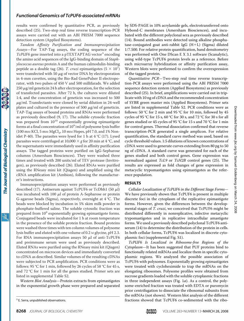
Cytoplasm—It has been suggested that PUF proteins bind tofunctionally related mRNAs and localize them in specific cyto-plasmic regions. We analyzed the possible association ofTcPUF6 with polysomes. Exponentially growing epimastigoteswere treated with cycloheximide to trap the mRNAs on theelongating ribosomes. Polysome profiles were obtained fromsucrose gradients loaded with the soluble cytoplasmic fractionsin three independent assays (Fig. 1a). As a control, the poly-some-enriched fraction was treated with EDTA or puromycinprior centrifugation to dissociate the ribosomal subunits fromthe mRNAs (not shown). Western blot analysis of the differentfractions showed that TcPUF6 co-sedimented with the ribo-3 E. Serra, unpublished observations.
Functional Genomics of TcPUF6-associated mRNAs
8268 JOURNAL OF BIOLOGICAL CHEMISTRY VOLUME 283 • NUMBER 13 • MARCH 28, 2008
by Sam
uel Goldenberg on M
arch 24, 2008 w
ww
.jbc.orgD
ownloaded from
some-free fractions in all three experiments (collected fractions1–3). No signal was detected with polysomes or monosomes(Fig. 1b). An antiserum against T. cruzi eIF3 translation initia-tion factor subunit Tif34 was used as a control of a polysomeassociated protein.4 A signal corresponding to the TcTif34 pro-tein was found in fractions along the entire gradient in all of theexperiments (Fig. 1c). As negative control, an anti-PEPCKserumwas tested, and the corresponding signal was found up tofractions 6–9 depending on the assay analyzed (Fig. 1d). Fur-thermore, we used confocalmicroscopy to perform co-localiza-tion assayswith the anti-TcPUF6 serumand a commercial anti-serum against human ribosomal P-proteins (Immunovision).Our results showed that TcPUF6 was not present in the ribo-some-rich regions of the cytoplasm (supplemental Fig. S2g).Wealso investigated possible associations between TcPUF6 andactively translating ribosomes. Co-localization assays wereperformed with two different antisera raised against twosubunits, Tif34 and Ptr1, of T. cruzi eIF3. No clear co-local-ization was observed with either the Ptr1 subunit (supple-mental Fig. S2h) or the Tif34 subunit (not shown). Thus,TcPUF6 is present in the cytoplasm and is not associatedwith ribosomes or polysomes.Overexpression of a TAP-tagged TcPUF6 Protein Does Not
Affect Differentiation Phenotype—A TAP-tagged TcPUF6 pro-tein was overexpressed in epimastigote forms to identify themRNAs associatedwithTcPUF6 and to obtain additional infor-mation about protein function. We used the pTEX vector,
which is maintained as a low copy number episomal DNAwithdiscrete overexpression (27), to facilitate the purification ofmRNP complexes without affecting the cellular distribution ofthe protein. Two clonal populations derived from independenttransfection assays were selected. These populations, PUF6Aand PUF6B, overexpressed a protein of about 90 kDa in sizeresulting from the fusion of TcPUF6 with the 30-kDa tag. Thelevel of overexpressionwas analyzed byWestern blotting, usingdifferent concentrations of total protein extract, with in silicoquantification by peptide densitometry, using the wild-typeprotein as a reference. The fusion protein was overexpressed byfactors of three and five in the PUF6 A and B populations,respectively (Fig. 1e). Immunofluorescence microscopy wasperformed with both the anti-TcPUF6 and an anti-tag serumand showed that the characteristic cellular distribution ofTcPUF6 was maintained in the transfected parasites (Fig. 1h).Parasites overexpressing the TcPUF6-tagged protein showedno significant change in cell growth rate or morphology; para-sites transfected with the pTEX-TAPTAG vector were used asthe reference population. The ability of the TcPUF6-overex-pressing parasites to differentiatewas also analyzed in vitro, andno significant difference in differentiation rates followingmeta-cyclogenesis and amastigogenesis in vitro were observed (notshown).Microarray Analysis of TcPUF6-overexpressing Parasites—
We investigated the phenotypic changes caused by TcPUF6 atthe molecular level, using DNA microarray hybridization tostudy the steady-state mRNA levels of parasites transfectedwith vector alone and epimastigotes overexpressing TcPUF6. AT. cruzi cDNA microarray containing �6,200 independentprobes was analyzed by competitive hybridization, using anamplified cRNA derived from the total RNA of both popula-tions (supplemental Figs. S3 and S4). TcPUF6 overexpressionhad a considerable effect on global gene expression.We simpli-fied the analysis by considering only genes showing changes inmRNA levels by at least a factor of three. Overall, 269 genesfulfilled this condition, corresponding to about 4% of thesequences present in the microarray. From these genes, 232were up-regulated in the TcPUF6 transfectants, whereas 37were clearly down-regulated (supplemental Table S1). TheTcPUF6 transcript itself increased in abundance by a factor of4.5, consistent with Western blotting data (Fig. 1e). The genesselected were identified as putative RNA-binding proteins (9),proteases (11), protein kinases (18), transporters (13), and 48enzymes involved in central metabolism. However, about 50%of the sequences analyzed were classified as hypothetical pro-teins (122 sequences) (supplemental Table S1). An analysis ofthe biological process potentially affected by changes inTcPUF6 levels showed that the up-regulated probes includedgenes encoding proteins involved in DNA metabolism, stressresponses, cytoskeleton and mitochondrial proteins, and sev-eral surface antigens. An analysis of the down-regulated genesshowed that they included one encoding a putative cyclin andsix genes encoding proteins involved in RNA processing,including the TcPUF7 protein. However, most of the down-regulated genes were identified as hypothetical proteins. Theseresults were corroborated by quantitative PCR.4 S. P. Fragoso, personal communication.
FIGURE 1. TcPUF6 is present in the ribosome-free fractions of sucrose gradi-ents. a, the positions of the 40 and 60 S subunits, the 80 S ribosome monomer,and polysomes are indicated in the sucrose density gradient. b–d, Western blotsof the collected fractions (30 �l) probed with anti-TcPUF6 (1/500) (b), anti-TcTif34 (1/500) (c), and anti-PEPCK (1/250) sera (d). Transfected parasitesoverexpress a TcPUF6-tagged protein. e, Western blot analysis of proteinextracts from epimastigotes (lane 1), epimastigotes transfected with thepTEX-TAPTAG-transfected clones (lane 2), and the PUF6 A- and (4) B-trans-fected clones (lane 3), with antiserum against TcPUF6 protein (1/500 dilution).f, protein extracts were also tested with an anti-CBP serum (1/1000) (Upstate).g, the same blot was re-probed with an anti-PEPCK serum (1/500). h, immu-nolocalization of T. cruzi TcPUF6 in TcPUF6-TAPTAG-transfected epimastig-ote forms with anti-CBP antiserum (1/100). Bars, 5 �m.
Functional Genomics of TcPUF6-associated mRNAs
MARCH 28, 2008 • VOLUME 283 • NUMBER 13 JOURNAL OF BIOLOGICAL CHEMISTRY 8269
by Sam
uel Goldenberg on M
arch 24, 2008 w
ww
.jbc.orgD
ownloaded from
Identification of Putative TcPUF6 Target mRNAs—Ribonu-cleoprotein complexes formed by mRNAs, and the taggedTcPUF6 protein were purified from soluble protein lysates pre-pared from transfected exponentially growing epimastigoteforms by affinity chromatography on IgG-Sepharose columns,as previously described (9). RNA was purified from the elutedfractions and amplified; the resulting cRNA was labeled andhybridized to the T. cruzi cDNA microarray. Parasites trans-fected with vector alone were used as a control in affinity puri-fication experiments; however, the levels of RNAobtainedwerealmost undetectable, even after amplification (not shown).Total epimastigote RNA was used as a control in microarraycompetitive hybridization analyses to eliminate nonspecifichybridization signals with the most abundant mRNAs. Eightindependent affinity purifications were performed, only threeof which generated enough RNA for microarray hybridization,including dye swap controls.Nine probes were selected based on increased signals
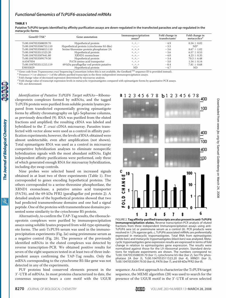
obtained in at least two of three experiments (Table 1). Fivecorresponded to genes encoding hypothetical proteins. Theothers corresponded to a serine-threonine phosphorilase, theXRND1 exonuclease, a putative amino acid transporter(PAT6), and the 69-kDa PFR2 (paraflagellar rod protein 2). Adetailed analysis of the hypothetical proteins showed that twohad predicted transmembrane domains and one had a signalpeptide.One of the proteinswith transmembrane domains pre-sented some similarity to the cytochrome B5 protein.Alternatively, to confirm theTAP-Tag results, the ribonucle-
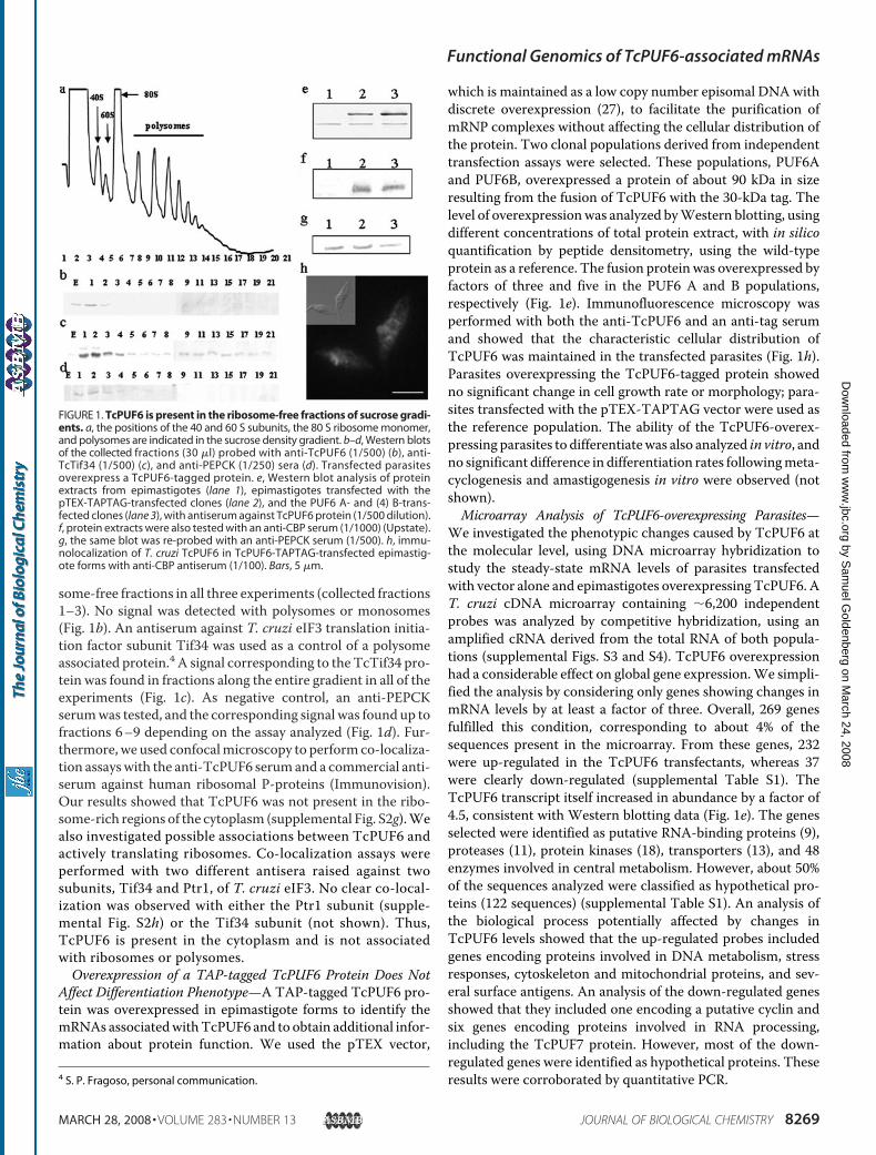
oprotein complexes were purified by immunoprecipitationassays using soluble lysates prepared fromwild-type epimastig-ote forms. The anti-TcPUF6 serum was used in the immuno-precipitation experiments (Fig. 2a) using preimmune serum asa negative control (Fig. 2b). The presence of the previouslyidentified mRNAs in the eluted complexes was detected byreverse transcription-PCR. We obtained positive results forseven of the eight sequences tested in at least two of three inde-pendent assays confirming the TAP-Tag results. Only themRNA corresponding to the cytochrome B5-like gene was notdetected in any of the experiments.PUF proteins bind conserved elements present in the
3�-UTR of mRNAs. In most proteins characterized to date, theconsensus sequence bears a core motif with the UGUR
sequence. As a first approach to characterize theTcPUF6 targetsequence, the MEME algorithm (28) was used to search for thepresence of the UGUR motif in the 3�-UTR of seven selected
FIGURE 2. Tag affinity-purified transcripts are also present in anti-TcPUF6immunoprecipitation elutes. Reverse transcription-PCR analysis of elutedfractions from three independent Immunoprecipitation assays using anti-TcPUF6 sera (a) or preimmune serum as a control (b). PCR products wereresolved in 1.2% agarose gels. c, TcPUF6 associated mRNAs are preferentiallyexpressed in metacyclic trypomastigotes. Total RNA from epimastigotes(white bars) and metacyclic trypomastigotes (black bars) was analyzed. Meta-cyclic trypomastigotes gene expression results are expressed in terms of foldchange in relation to epimastigotes gene expression. The results werenormalized against those for the L9 ribosomal protein. Standard devia-tions for triplicate experiments are shown. The numbers correspond to:Tc00.1047053508039.70 (bar 1), cytochrome b5-like (bar 2), Ser/Thr phos-phatase 2A (bar 3), Tc00.1047053511523.20 (bar 4), XRND1 (bar 5),Tc00.1047053509179.50 (bar 6), PAT6 (bar 7), and 69-kDa PFR2 (bar 8).
TABLE 1Putative TcPUF6 targets identified by affinity purification assays are down-regulated in the transfected parasites and up-regulated in themetacyclic forms
GeneID TSKa Gene annotation Immunoprecipitationassaysb
Fold change intransfectantsc
Fold change inmetacyclicsd
Tc00.1047053508039.70 Hypothetical protein �,�,� �4.9 0.34 � 0.02Tc00.1047053506753.110 Hypothetical protein (cytochrome b5-like) �,�,� �3.5 NDe
Tc00.1047053504013.110 Serine/threonine-protein phosphatase 2A �,�,� �3.6 8.67 � 1.02Tc00.1047053511523.20 Hypothetical protein �,�,� �3.6 6.57 � 0.53Tc00.1047053507641.80 XRND1 exonuclease �,�,� �4.0 4.23 � 0.35Tc00.1047053509179.50 Hypothetical protein �,�,� �3.4 70.3 � 6.48AAS47054 PAT6 amino acid transporter �,�,� �3.0 1.54 � 0.14Tc00.1047053511215.119 69 kDa paraflagellar rod protein putative �,�,� �8.4 7.81 � 0.68EH035829 Hypothetical protein ND �4.7 ND
a Gene code from Trypanosoma cruzi Sequencing Consortium (when this is not available, the GenBankTM sequence ID is provided instead).b Presence (�) or absence (�) of the affinity purified transcripts in the three independent immunoprecipitation assays.c Fold change value of decreased expression determined by microarray analysis.d Fold change value of transcript expression levels in metacyclic trypomastigotes compared with epimastigote forms by quantitative PCR assays.e ND, not determined.
Functional Genomics of TcPUF6-associated mRNAs
8270 JOURNAL OF BIOLOGICAL CHEMISTRY VOLUME 283 • NUMBER 13 • MARCH 28, 2008
by Sam
uel Goldenberg on M
arch 24, 2008 w
ww
.jbc.orgD
ownloaded from
genes annotated in the T. cruzi GeneDB. Because we have noinformation about the 3�-UTR of the mRNAs studied, we ana-lyzed 200 nucleotides downstream from the stop codon as theputative UTR of these genes. At least one UGUR sequence wasfound, though no clear motif could be determined in silico (notshown).Transcripts Associated with TcPUF6 Are Down-regulated in
Transfected Parasites—It can be inferred that in the case oftranscripts bound and regulated by TcPUF6, its overexpressionshould affect their expression levels in a coordinated manner.Analysis of the microarray data showed significantly lowermRNA levels in the transfected parasites than in control para-sites for all of the selected genes (Table 1). Microarray resultswere confirmed by quantitative PCR using total RNA preparedfrom control and TcPUF6-transfected parasites. The quantita-tive PCR assays confirmed the microarray data for all theselected genes, although differences in expression levels weresmaller (not shown). Thus, overexpression ofTcPUF6 results indown-regulation of the transcript levels of its putative mRNAtargets.TcPUF6 andTcDhh1Are Present in the Same Ribonucleopro-
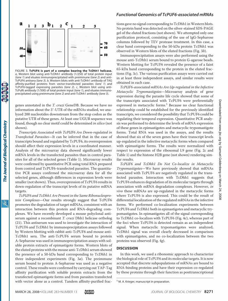
tein Complexes—Our results strongly suggest that TcPUF6promotes the degradation of target mRNAs, consistent with aninteraction between this protein and RNA-degrading com-plexes. We have recently developed a mouse polyclonal anti-serum against a recombinant T. cruzi Dhh1 helicase ortholog(21). This antiserum was used to investigate the interaction ofTcPUF6 and TcDhh1 by immunoprecipitation assays followedby Western blotting with rabbit anti-TcPUF6 and mouse anti-TcDhh1 sera. The anti-TcPUF6 serum bound to proteinA-Sepharose was used in immunoprecipitation assays with sol-uble protein extracts of epimastigote forms. Western blots ofthe eluted proteinswith themouse anti-TcDhh1 serum showedthe presence of a 50-kDa band corresponding to TcDhh1 inthree independent experiments (Fig. 3a). The preimmuneserum bound to protein A-Sepharose was used as a negativecontrol. These resultswere confirmedby carrying outTAP-Tagaffinity purification with soluble protein extracts from thetransfected epimastigote forms and from parasites transfectedwith vector alone as a control. Tandem affinity-purified frac-
tions gave no signal corresponding toTcDhh1 inWestern blots.No protein band was detected on the silver-stained SDS-PAGEgel of the eluted fractions (not shown). We attempted only onepurification protocol, consisting of the use of IgG-Sepharosecolumns followed by TEV protease treatment. In this case, aclear band corresponding to the 50-kDa protein TcDhh1 wasobserved in Western blots of the eluted fractions (Fig. 3b).Immunoprecipitation assays were also performed using the
mouse anti-TcDhh1 serum bound to protein G-agarose beads.Western blotting for TcPUF6 revealed the presence of a faint65-kDa band corresponding to the protein in the eluted frac-tions (Fig. 3c). The various purification assays were carried outin at least three independent assays, and similar results wereobtained in each case.TcPUF6-associated mRNAs Are Up-regulated in the Infective
Metacyclic Trypomastigotes—Microarray analysis of geneexpression during the parasite life cycle showed that some ofthe transcripts associated with TcPUF6 were preferentiallyexpressed in metacyclic forms.5 Because no clear functionalrelationship could be established for the previously identifiedtranscripts, we considered the possibility that TcPUF6 could beregulating their temporal expression. Quantitative PCR analy-sis was performed to determine the levels of mRNA expressionof these genes in epimastigotes and metacyclic trypomastigoteforms. Total RNA was used in the assays, and the resultsshowed that six of the seven genes have their transcript levelsup-regulated in the infective metacyclic forms when comparedwith epimastigote forms. The results were normalized withrespect to expression of the ribosomal L9 gene (Fig. 2c andTable 1) and the histone H2B gene (not shown) rendering sim-ilar results.TcPUF6 and TcDhh1 Do Not Co-localize in Metacyclic
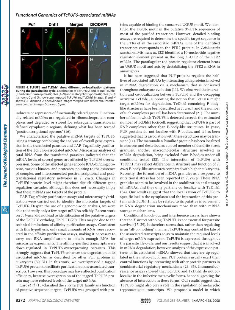
Trypomastigotes—We have previously showed that mRNAsassociated with TcPUF6 are negatively regulated in the trans-fected parasites. Interaction with TcDhh1 suggests thatTcPUF6 enhances degradation of its molecular targets throughassociation with mRNA degradation complexes. However, invivo these mRNAs are up-regulated in the metacyclic formswhere TcPUF6 is also expressed. This could be the result ofdifferential localization of the regulatedmRNAs in the infectiveforms. We performed co-localization experiments betweenTcPUF6 andTcDhh1 both in epimastigotes andmetacyclic try-pomastigotes. In epimastigotes all of the signal correspondingto TcDhh1 co-localizes with TcPUF6 (Fig. 4c), whereas part ofthe foci where TcPUF6 is detected remain as an independentsignal. When metacyclic trypomastigotes were analyzed,TcDhh1 signal was overall clearly decreased in comparisonwith epimastigotes, and no co-localization between the twoproteins was observed (Fig. 4g).
DISCUSSION
In this work, we used a ribonomic approach to characterizethe biological role ofTcPUF6 and itsmolecular targets. It is nowaccepted that discrete subpopulations of mRNAs are bound toRNA-binding proteins and have their expression co-regulatedby these proteins through their function as posttranscriptional
5 M. A. Krieger, manuscript in preparation.
FIGURE 3. TcPUF6 is part of a complex bearing the TcDhh1 helicase.a, Western blot using anti-TcDhh1 antibody (1/250) of total protein input(lane 1) and eluates immunoprecipitated with preimmune (lane 2) and anti-TcPUF6 antisera (lane 3). b, Western blots with anti-TcDhh1 antibody of TAGaffinity-purified proteins from vector-transfected parasites (lane 1) andTcPUF6-tagged expressing parasites (lane 2). c, Western blot using anti-TcPuf6 antibody (1/500) of total protein input (lane 1), and eluates immuno-precipitated using preimmune (lane 2) and anti-TcDhh1 antibody (lane 3).
Functional Genomics of TcPUF6-associated mRNAs
MARCH 28, 2008 • VOLUME 283 • NUMBER 13 JOURNAL OF BIOLOGICAL CHEMISTRY 8271
by Sam
uel Goldenberg on M
arch 24, 2008 w
ww
.jbc.orgD
ownloaded from
inducers or repressors of functionally related genes. Function-ally related mRNAs are regulated in ribonucleoprotein com-plexes and degraded or stored for subsequent translation indefined cytoplasmic regions, defining what has been termed“posttranscriptional operons” (16).We characterized the putative mRNA targets of TcPUF6,
using a strategy combining the analysis of overall gene expres-sion in the transfected parasites and TAP-Tag affinity purifica-tion of the TcPUF6-associated mRNAs. Microarray analyses oftotal RNA from the transfected parasites indicated that themRNA levels of several genes are affected by TcPUF6 overex-pression. Some of the affected genes encode RNA-binding pro-teins, various kinases, and proteases, pointing to the existenceof complex and interconnected posttranscriptional and post-translational regulatory networks in T. cruzi. Changes inTcPUF6 protein level might therefore disturb different generegulation cascades, although this does not necessarily implythat these mRNAs are targets of the protein.TAP-Tag affinity purification assays and microarray hybrid-
ization were carried out to identify the molecular targets ofTcPUF6. Despite the use of a genome-wide analysis, we wereable to identify only a few target mRNAs reliably. Recent workonT. brucei did not lead to identification of the putative targetsof the TcPUF6 ortholog, TbPUF1 (29). This may be due to thetechnical limitations of affinity purification assays. Consistentwith this hypothesis, only small amounts of RNA were recov-ered in the affinity purification assays, making it necessary tocarry out RNA amplification to obtain enough RNA formicroarray experiments. The affinity-purified transcripts weredown-regulated in TcPUF6-overexpressing parasites. Thisstrongly suggests that TcPUF6 enhances the degradation of itsassociated mRNAs, as described for other PUF proteins ineukaryotes (30, 31). In this work, we overexpressed a taggedTcPUF6 protein to facilitate purification of the associated tran-scripts. However, this proceduremay have affected purificationefficiency, because overexpression of the tagged TcPUF6 pro-tein may have reduced levels of the target mRNAs.Caro et al. (13) classified theT. cruzi PUF family as a function
of putative sequence targets. TcPUF6 was grouped with pro-
teins capable of binding the conserved UGUR motif. We iden-tified the UGUR motif in the putative 3�-UTR sequences ofmost of the purified transcripts. However, detailed bindingassays are required to determine the specific target sequence inthe UTRs of all the associated mRNAs. One of the identifiedtranscripts corresponds to the PFR2 protein. In Leishmaniamexicana, Mishra et al. (32) identified a 10-nucleotide negativeregulator element present in the long 3�-UTR of the PFR2mRNA. The paraflagellar rod protein regulator element bearsan UGUR motif and acts by destabilizing the PFR2 mRNA inamastigotes.It has been suggested that PUF proteins regulate the half-
lives of associatedmRNAsby interactingwith proteins involvedin mRNA degradation via a mechanism that is conservedthroughout eukaryote evolution (11).We observed the interac-tion and co-localization between TcPUF6 and the decappingactivator TcDhh1, supporting the notion that TcPUF6 mighttarget mRNAs for degradation. TcDhh1-containing P body-like structures have been described in T. cruzi, and the numberof such complexes per cell has been determined (21). The num-ber of foci in which TcPUF6 is detected exceeds the estimatednumber of TcDhh1 foci/cell, suggesting that TcPUF6 is part ofRNP complexes other than P-body-like structures. In yeast,PUF proteins do not localize with P-bodies, and it has beensuggested that its associationwith these structuresmay be tran-sient (11). Themammalian Pumilio2 protein has been localizedin neurons and described as a novel member of dendrite stressgranules, another macromolecular structure involved inmRNA degradation, being excluded from P-bodies under allconditions tested (33). The interaction of TcPUF6 withTcDhh1 may reflect differences in structure and function of T.cruzi P-body-like structures compared with higher eukaryotes.Recently, the formation of mRNA granules as a response tonutritional stress has been reported in T. cruzi. These RNAgranules seem to be involved in the stabilization and storageof mRNAs, and they only partially co-localize with TcDhh1(34). Our results suggest that the localization of TcPUF6 tospecific loci in the cytoplasm and the interaction of this pro-tein with TcDhh1 may be related to its putative involvementin RNA degradation mechanisms more than with mRNAstorage mechanisms.Conditional knock-out and interference assays have shown
that theT. brucei ortholog, TbPUF1, is not essential for parasitesurvival (15, 29). It therefore seems that, rather than regulatingin an “all-or-nothing” manner, TcPUF6 may control the fate ofthe associated transcripts so as to maintain the required levelsof target mRNA expression. TcPUF6 is expressed throughoutthe parasite life cycle, and our results suggest that it is involvedin mRNA degradation; however, analysis of the expression pat-terns of its associated mRNAs showed that they are up-regu-lated in the metacyclic forms. PUF proteins usually exert theircontrol functions by interacting with other protein partners incombinatorial regulatory mechanisms (23, 35). Immunofluo-rescence assays showed that TcPUF6 and TcDhh1 do not co-localize in the infective metacyclic forms, hence suggesting theabsence of interaction in these forms. Our results suggest thatTcPUF6 might also play a role in the regulation of metacyclictrypomastigote transcripts. We propose a model in which
FIGURE 4. TcPUF6 and TcDhh1 show different co-localization patternsduring the parasite life cycle. Localization of TcPUF6 (A and E) and TcDhh1(B and F) in T. cruzi epimastigotes (A–D) and metacyclic trypomastigotes (E–H)is shown. C and G show superimposed TcPUF6 and TcDhh1 images. D and Hshow 4�,6�-diamino-2-phenylindole images merged with differential interfer-ence contrast images. Scale bar, 5 �m.
Functional Genomics of TcPUF6-associated mRNAs
8272 JOURNAL OF BIOLOGICAL CHEMISTRY VOLUME 283 • NUMBER 13 • MARCH 28, 2008
by Sam
uel Goldenberg on M
arch 24, 2008 w
ww
.jbc.orgD
ownloaded from
TcPUF6 regulates the half-lives of its associated transcripts byits stage-dependent association with TcDhh1 containing com-plexes involved in mRNA degradation. The characterization ofmRNA subsets and proteins interacting with TcPUF6 in para-sites of different developmental stages might shed light on themechanisms involved in posttranscriptional regulation intrypanosomes.
Acknowledgments—We thank Nilson Fidencio for skilful technicalassistance and Paulo Arauco for DNA sequencing. We also thank Dr.Esteban Serra for providing the pTEX-TAPTAG vector.
REFERENCES1. De Souza, W. (2002) Kinetoplastid Biol. Dis. 31, 1–32. Clayton, C. E. (2002) EMBO J. 21, 1881–18883. Teixeira, S. M. (1998) Braz. J. Med. Biol. Res. 31, 1503–15164. Mignone, F., Gissi, C., Liuini, S., and Pesole, G. (2002). Genome Biol. 3,
0004.1–0004.105. Auweter, S. D., Oberstrass, F. C., and Allain, F. H. T. (2006) Nucleic Acids
Res. 34, 4943–49596. Keene, J. D., and Tenenbaum, S. A. (2002)Mol. Cell 9, 1161–11677. Wang, X., McLachlan, J., Zamore, P. D., and Tanaka Hall, T. M. (2002).
Cell 110, 501–5128. Wickens, M., Bernstein, D. S., Kimble, J., and Parker, R. (2002) Trends
Genet. 18, 150–1579. Gerber, A. P., Herschlag, D., andBrown, P.O. (2004)PLoSBiol.2, 342–35410. Wharton, R. P., and Aggarwal, A. K. (2006). Sci. STKE 2006, pe3711. Goldstrohm, A. C., Hook, B. A., Seay, D. J., and Wickens, M. (2006) Nat.
Struct. Mol. Biol. 13, 533–53912. Coller, J. M., Tucker, M., Sheth, U., Valencia-Sanchez, M. A., and Parker,
R. (2001) RNA 7, 1717–172713. Caro, F., Bercovich, N., Atorrasagasti, C., Levin, M. J., and Vazquez, M. P.
(2006) Exp. Parasitol. 113, 112–12414. Dallagiovanna, B., Perez, L., Sotelo-Silveira, J., Smircich, P., Duhagon,
M. A., and Garat, B. (2005) Exp. Parasitol. 109, 260–26415. Hoek, M., Zanders, T., and Cross, G. A. (2001) Mol. Biochem. Parasitol.
57, 706–716
16. Keene, J. (2001) Proc. Natl. Acad. Sci. U. S. A. 98, 7018–702417. Tenenbaum, S. A., Lager, P. J., Carson, C. C., and Keene, J. D. (2002)
Methods 26, 191–19818. Contreras, V. T., Araujo-Jorge, T. C., Bonaldo, M. C., Thomaz, N., Bar-
bosa, H. S., Meirelles, M., de, N., and Goldenberg, S. (1988). Mem. Inst.Oswaldo Cruz 83, 123–133
19. Contreras, V. T., Salles, J. M., Thomas, N., Morel, C. M., and Goldenberg,S. (1985).Mol. Biochem. Parasitol. 16, 315–327
20. Contreras, V. T., Navarro, M. C., DeLima, A. R., Arteaga, R., Duran, F.,Askue, J., and Franco, Y. (2002). Mem. Inst. Oswaldo Cruz. 97,1213–1220
21. Holetz, F., Correa, A., Avila, A. R., Nakamura, C. V., Krieger, M. A., andGoldenberg, S. (2007) Biochem. Biophys. Res. Commun. 356, 1062–1067
22. Brecht, M., and Parsons, M. (1998)Mol. Biochem. Parasitol. 97, 189–19823. Sonoda, J., and Wharton, R. P. (1999) Genes Dev. 13, 2704–271224. Smyth, G. K. (2004). Stat. App. Genet. Mol. Biol. 3, article 325. Nardelli, S. C., Avila, A. R., Freund, A.,Motta,M. C.,Manhaes, L., de Jesus,
T. C., Schenkman, S., Fragoso, S. P., Krieger, M. A., Goldenberg, S., andDallagiovanna, B. (2007) Eukaryot. Cell 6, 337–345
26. Puig,O., Caspary, F., Rigaut, G., Rutz, B., Bouveret, E., Bragado-Nilsson, E.,Wilm, M., and Seraphin, B. (2001).Methods 24, 218–229
27. Kelly, J. M., Ward, H. M., Miles, M. A., and Kendall, G. (1992) NucleicAcids Res. 20, 3963–3969
28. Bailey, T. L., and Elkan, C. (1994). Proceedings of the Second InternationalConference on Intelligent Systems for Molecular Biology, pp. 28–36, AAAIPress, Menlo Park, CA
29. Luu, V. D., Brems, S., Hoheisel, J. D., Burchmore, R., Guilbride, D. L., andClayton, C. (2006)Mol. Biochem. Parasitol. 150, 340–349
30. Olivas, W., and Parker, R. (2000) EMBO J. 19, 6602–661131. Wreden, C., Verrotti, A. C., Schisa, J. A., Lieberfarb, M. E., and Strickland,
S. (1997). Development 124, 3015–302332. Mishra, K. K., Holzer, T. R., Moore, L. L., and LeBowitz, J. H. (2003)
Eukaryot. Cell 2, 1009–101733. Vessey, J. P., Vaccani, A., Xie, Y., Dahm, R., Karra, D., Kiebler, M. A., and
Macchi, P. (2006) J. Neurosci. 26, 6496–650834. Cassola, A., De Gaudenzi, J. G., and Frasch, A. C. (2007) Mol. Microbiol.
65, 655–67035. Jaruzelska, J., Kotecki, M., Kusz, K., Spik, A., Firpo, M., and Reijo-Pera,
R. A. (2003) Dev. Genes Evol. 213, 120–126
Functional Genomics of TcPUF6-associated mRNAs
MARCH 28, 2008 • VOLUME 283 • NUMBER 13 JOURNAL OF BIOLOGICAL CHEMISTRY 8273
by Sam
uel Goldenberg on M
arch 24, 2008 w
ww
.jbc.orgD
ownloaded from
Copyright © 2022 FDOKUMEN