Carbon nanotube bundles and thin layers probed by micro-Raman spectroscopy
Upload
independentCategory
view
3download
0
Biochem. J. (2002) 365, 379–389 (Printed in Great Britain) 379
Function of the 90-loop (Thr90–Glu100) region of staphylokinase inplasminogen activation probed through site-directed mutagenesisand loop deletionGovindan RAJAMOHAN, Monika DAHIYA, Shekhar C. MANDE and Kanak L. DIKSHIT1
Institute of Microbial Technology (IMTECH), Sector 39 A, Chandigarh-160036, India
Staphylokinsae (SAK) forms a bimolecular complex with human
plasmin(ogen) and changes its substrate specificity by exposing
new exosites that enhances accession of substrate plasminogen
(PG) to the plasmin (Pm) active site. Protein modelling studies
indicated the crucial role of a loop in SAK (SAK 90-loop;
Thr*!–Glu"!!) for the docking of the substrate PG to the SAK–Pm
complex. Function of SAK 90-loop was studied by site-directed
mutagenesis and loop deletion. Deletion of nine amino acid
residues (Tyr*#–Glu"!!) from the SAK90-loop, resulted inE 60%
reduction in the PG activation, but it retained the ability to
generate an active site within the complex of loop mutant
of SAK (SAK∆90) and Pm. The preformed activator complex of
SAK∆90 with Pm, however, displayed a 50–60% reduction in
substrate PG activation that remained unaffected in the presence
of kringle domains (K1K2K3K4) of PG, whereas PG
activation by SAK–Pm complex displayed C 50% reduction in
the presence of kringles, suggesting the involvement of the
kringle domains in modulating the PG activation by native SAK
INTRODUCTION
Staphylokinase (SAK), a 16 kDa profibrinolytic protein from
the Staphylococcus aureus, has been demonstrated to induce
highly fibrin-specific thrombolysis in both human plasma [1,2]
and in limited clinical trials [3,4,5]. Recent studies on the
thrombolytic potential of recombinant SAK in achieving early
perfusion in myocardial infarction and in the dissolution of
platelet-rich clot [4,6] have clearly established its immense utility
in clinical medicine as a thrombolytic agent and suggested that it
can be developed as a potent clot-dissolving agent. Unlike some
other plasminogen (PG) activators, e.g. tissue PG activator and
urokinase, SAK has no proteolytic properties by itself, but acts
by forming a 1:1 stoichiometric complex with plasmin (Pm),
which, in turn, activates other molecules [7–11]. In this bimol-
ecular complex, SAK acts as a cofactor and provides substrate-
binding exosites for docking and enhanced presentation of the
PG ‘substrate ’ to the Pm active site.
Pm is an 85 kDa serine protease that plays a central role
in haemostasis by degrading fibrin clots into soluble products.
In human plasma, it circulates as a proenzyme, PG, which is
activated by proteolytic cleavage of the Arg&'"–Val&'# peptide
bond [12]. Human PG contains five kringle domains that serve
as functional binding loci for other plasma proteins [13]. The
activation of truncated PG derivatives, miniplasminogen (mini-
PG, carrying only kringle 5 and protease domain) and microplas-
minogen (µPG, carrying only protease domain) is slower than
Abbreviations used: SAK, staphylokinase ; SK, streptokinase ; PG, plasminogen; miniPG, miniplasminogen; µPG, microplasminogen; Pm, plasmin ;SAK–µPm, SAK, staphylokinase–microplasmin complex ; SKα, α-domain of streptokinase ; SAK–Pm complex, staphylokinase–plasmin complex ; NPGB,p-nitrophenyl p«-guanidinobenzoate ; SAK∆90, loop mutant of SAK; SAKK97A (etc.), a mutant of SAK in which Lys97 has been mutated to alanine.
1 To whom correspondence should be addressed (e-mail kanak!imtech.res.in).
but not by SAK∆90. Lysine residues (Lys*%, Lys*', Lys*( and
Lys*)) of the SAK 90-loop were individually mutated into alanine
and, among these four SAK loop mutants, SAKK*(A
and SAKK*)A
exhibited specific activities about one-third and one-quarter
respectively of the native SAK. The kinetic parameters of PG
activation of their 1:1 complex with Pm indicated that the Km
values of PG towards the activator complex of these two SAK
mutants were 4–6-fold higher, suggesting the decreased accessi-
bility of the substrate PG to the activator complex formed
by these SAK mutants. These results demonstrated the involve-
ment of the Lys*( and Lys*) residues of the SAK 90-loop in
assisting the interaction with substrate PG. These interactions of
SAK–Pm activator complex via the SAK 90-loop may provide
additional anchorage site(s) to the substrate PG that, in turn,
maypromote the overall process of SAK-mediatedPGactivation.
Key words: kringle domain, molecular modelling, site-directed
mutagenesis.
for the full-length PG, suggesting a role for kringle domains in
the PG activation process [14]. The molecular mechanism by
which these kringle structures modulate overall processing of PG
by various PG activators is not very well known at present. The
three-dimensional structures of SAK and the SAK–µPm complex
have been elucidated by X-ray-diffraction studies [11,15]. In the
heterotrimer complex, consisting of two µPm molecules and one
SAK molecule [15], SAK binds one µPm molecule in the
proximity of its active site. Formation of this bimolecular
complex presents a slightly concave surface on to which the
second molecule of µPm docks in a substrate-like manner. Of
the amino acids in SAK, 30% are charged, and these amino
acids, including 20 lysine residues, are critical for its PG-activator
activity. A number of SAK mutants [16–18] have been generated
in order to investigate the determinants of SAK for PG binding
and activation. It has been shown that the positively charged
N-terminal region of SAK is necessary if it is to achieve its full
activation function. Recent studies conducted in our laboratory
[19] using site-directed mutagenesis of SAK have indicated that
the positively charged N-terminal region of SAK may be involved
in the interaction and}or stabilization of substrate PG during the
process of PG activation. Previous studies [16] on alanine
scanning mutagenesis of SAK have indicated that four clustered
charged segments of SAK are important for the functional
properties of SAK. Apart from the positively charged N-
terminus, two discrete segments of SAK, spanning Glu%%–Lys&!
and Glu'&–Asp'*, form the core region of SAK and may be
# 2002 Biochemical Society

380 G. Rajamohan and others
involved in PG binding and activation. Another highly charged
region of SAK (Lys*'–Lys*)) that significantly affect the func-
tionality of SAK, is a part of its surface-exposed loop structure.
The three-dimensional structure of the SAK–µPm complex [15]
has indicated that this charged loop region of SAK is away from
the interaction sites of SAK–Pm activator complex and substrate
µPG. Computer-based modelling studies with kringle domain of
PG indicate that the surface-exposed positively charged residues
of the SAK 90-loop may provide potential docking sites for the
kringle domains of the substrate PG. The aim of the present
study was to probe the functional role of the positively charged
loop region of SAK in the PG activation process through site-
directed and deletion mutagenesis. In the present work we have
shown that a SAK mutant deleted in the charged loop region
retains its native SAK-like capability for generating a bimolecular
complex with PG that exhibits highly reduced capability for
substrate PG processing. Structural modelling, supported by
biochemical studies reported in the present work, have provided
the experimental evidence that the 90-loop region of SAK may
interact with the kringle domains of substrate which, in turn,
may potentiate}stabilize the interaction of substrate PG with the
SAK–Pm enzyme complex and facilitate the PG activation
process.
MATERIALS AND METHODS
Bacterial strains, plasmids and reagents
S. aureus (local isolate, designated as SAK 11), was used for
retrieving the gene (sak) encoding SAK as described previously
[19]. Bluescript KS+ expression plasmid, pET9b (Promega) and
Escherichia coli host strains JM109 and BL21DE3 (Promega)
were routinely utilized for cloning and expression of recombinant
genes. All restriction and DNA-modifying enzymes were ob-
tained from Promega or New England Biolabs. Chromozym
PL and human PG were from Boehringer Mannheim. Pm was
obtained from KabiVitrum. All biochemicals were of the highest
grade commercially available.
Structural analysis and molecular modelling of the SAK–µPmcomplex and kringle domain
Co-ordinates of the SAK–µPm ternary complex, as well as those
of the kringle 5 domain of human PG, were retrieved from the
Protein Data Bank (Research Collaboratory for Structural
Bioinformatics Consortium). Molecular surfaces of both the sets
of co-ordinates were calculated by using GRASP [20] with a
probe radius of 0.14 nm (1.4 AI ). Electrostatic calculations were
then performed with default parameters in GRASP, and the
potentials were mapped on to the respective surfaces. Residues
occurring on distinctly positive regions of SAK and distinctly
negative regions of kringle domain were identified as putative
interaction sites. Kringle 5 domain, taken as a representative
structure of kringle domains of human PG, was manually docked
on to the SAK–Pm ternary complex using Insight II package.
Construction of loop-deletion and site-directed mutants of SAK
Recombinant plasmid, pRM1, encoding wild type SAK in E. coli
[19] was utilized for generating various SAK mutants. The SAK
90-loop deletion mutant, carrying deletion of nine amino acid
residues (Tyr*#–Glu"!!) of SAK were generated by an overlap-
PCR method [21] using the following sets of oligonucleotide
primers :
(1) 5«-GCAGGAGGATCCACGAAGTCTTTCCCTATAA-
CAGAA-3«
(2) 5«-TCGCGTGGATCCTCCTGCAGTGACT TCGATCTT -
3«
The specific primers for generating site-directed mutants of SAK
are as follows:
SAKK*%A
: 5«-TATTATGATGCGAATAAGAAAAAAGAAG-
AAACGAAG-3«
SAKK*'A
: 5«-TATGATAAGAATGCGAAAAAAGAAGAAA-
CGAAGTCT-3«
SAKK*(A
: 5«-GATAAGAATAAGGCGAAAGAAGAAACG-
AAGTCT-3«
SAKK*)A
: 5«-GATAAGAATAAGAAAGCGGAAGAAACG-
AAGTCT-3«
Incorporation of mutation in the sak gene was confirmed by
DNA sequencing. All SAK mutants were cloned in pET9b and
expressed in E. coli under the T7 promoter.
Purification of SAK mutants
E. coli cells carrying recombinant plasmids for the expression of
respective target genes were induced with 0.1 mM isopropyl β--
thiogalactoside during the mid-exponential phase of growth
[attenuance (D'!!
) 0.6] and harvested after 7–10 h of further
incubation at 37 °C. A two-step purification protocol, involving
an ion-exchange [sulphopropyl (SP)-Sepharose] and hydro-
phobic-interaction chromatography (Phenyl-Sepharose), was
used to obtain a purified protein preparation essentially as
described elsewhere [22]. The specific PG activator activity of a
purified preparation of recombinant SAK was found to be
comparable (160000 units}mg of protein) with the standard
SAK preparation isolated from its natural host (a gift from
Professor Patrick J. Gaffney, National Institute of Biological
Standards and Control, South Mimms, Herts., U.K.).
PG purification and preparation of kringle domains and miniPm
Human PG was isolated from fresh frozen plasma using lysine–
Sepharose affinity chromatography following the published pro-
cedure [23]. Separation of kringle domains (K1K2K3K4)
and miniPm (kringle 5 and protease domain) from the intact PG
moiety was achieved by limited digestion of full-length PG with
pancreatic elastase in 0.2 M Tris}HCl, pH 8.8, for 10 min at
37 °C using the enzyme}substrate ratios 1:50–1:100 as described
elsewhere [24,25].
SAK assay and PG activation reaction
The specific PG activator activity of native and mutant SAK was
determined by using previously published PG-coupled chromo-
genic substrate assay [26,27]. Briefly, purified SAK or SAK
mutant protein (5 nM) was added to PG (1.5 µM) in a quartz
cuvette carrying 1.0 mM Chromozym PL (a chromogenic sub-
strate from Boehringer-Mannheim) in 0.1 M phosphate buffer,
pH 7.5, containing 0.1% BSA and 0.01% Tween 80. The change
in A%!&
was then measured as a function of time in a Shimadzu
(UV-1601) spectrophotometer at 25 °C. Purified native SAK
(obtained from Professor Patrick J. Gaffney) was used as
standard to determine PG activator activity in the unknown
protein samples. PG activation and processing of substrate PG
by SAK–Pm complex was monitored following the published
procedure [28,29]. The molar concentration of full-length SAK
and SAK loop-deletion mutant was calculated by taking their
molecular masses as 16.2 kDa and 15.1 kDa respectively. Briefly,
equimolar amounts of SAK or SAK mutant and PG (1.5 µM
each) were combined in 100 mM Tris}HCl, pH 7.5, and incubated
# 2002 Biochemical Society

381The 90-loop region of staphylokinase in plasminogen activation
at 25 °C. After 5 min, a 7.5 µl aliquot was removed and subjected
to SDS}12%-(w}v)-PAGE for the cleavage of substrate PG. The
kinetic constants of PG activation by SAK–Pm complexes were
calculated from Lineweaver–Burk plots [30,31]. To construct
these plots, equimolar mixtures of SAK or SAK mutant and Pm
were preincubated for 5 min at 37 °C in 100 mM phosphate
buffer, pH 7.4, containing 0.01% Tween 80 and left on ice. These
preformed complexes (5 nM) were mixed with PG (1.0–10 µM)
and the generation of Pm was measured by the ∆A%!&
with
Chromozym PL as substrate.
Complex-formation of SAK mutants with Pm and generation ofactive site within the SAK–PG (Pm) bimolecular complex
The time course of active-site generation in equimolar complexes
of Pm with wild-type SAK or SAK mutants was monitored by
titration with p-nitrophenyl p«-guanidinobenzoate (NPGB) at
room temperature, as described previously [8,32]. Concentrated
stock solutions of PG and SAK–Pm complex were diluted in
0.1 M veronal buffer, pH 8.3, containing 0.1 M arginine, to final
concentrations of 4.0 µM and 4.0 µM respectively. At different
time points (0–20 min) after mixing of PG and SAK–Pm or loop
mutant of SAK (SAK∆90)–Pm complex, NPGB was added to a
final concentration of 100 µM. The concentration of active site
was determined from the ‘burst ’ of p-nitrophenol, using a molar
absorption coefficient of 16700 M−" [ cm−" [32].
PG activation by catalytic amounts of preformed equimolarcomplexes of SAK or SAK mutants with Pm
Equimolar mixtures of SAK or SAK mutants and Pm were
preincubated in 0.1 M phosphate buffer, pH 7.5, containing
0.1% BSA and 0.01% Tween 80 at 37 °C for 5 min to generate
the SAK–Pm bimolecular complex. These preformed activator
complexes (5 nM) were then mixed with PG (1.5 µM), and
generation of Pm was measured at 25 °C from ∆A%!&
with
Chromozym PL.
Measurement of PG binding
Purified protein preparation of native SAK or SAK∆90 (1.5 ng)
was suspended in binding buffer (50 mM Na#CO
$}NaHCO
$, pH
9.5) andwas immobilized on themicrotitre plates or nitrocellulose
strips. Blotted protein was air-dried and blocked with 1% BSA
for 1 h at 4 °C. Different concentrations of "#&I-Pm, prepared by
the Iodogen method [33], were then added to the immobilized
SAK proteins and incubated at 4 °C for 1 h. Excess radioactivity
was removed by repeated washing with the same buffer. The
amount of labelled Pm, bound with the SAK, was estimated by
the amount of radioactivity (measured by using a γ-radiation
counter) retained on the blot. BSA was used in place of SAK to
check the background level of non-specific binding that was
subtracted from the total level in order to determine specific Pm
binding.
Casein/PG overlay and radial caseinolytic assay
Bacterial colonies overexpressing SAK were detected by over-
laying 0.8% agarose}1% skim milk}human PG (500 µg}plate)}150 mM NaCl}50 mM Tris}HCl, pH 8.0, on top of the
agar plates carrying recombinant E. coli colonies as described
previously [34]. The plates were incubated at 37 °C for 2–6 h.
SAK-positive clones indicated a clearing zone around the col-
onies. Functional activity of purified SAK or mutant SAK
protein was also estimated by radial caseinolytic assay. Petri
dishes containing 1.2% agarose, 1% skim milk and 10 µg}ml
PG were prepared. On this solidified agarose plate, wells of equal
diameter were bored and an equal quantity of purified native
SAK or mutant SAK protein was added and kept at 37 °C for
6–7 h. The diameter of the halo around the well was measured to
check the functional activity of SAK proteins.
Sandwich binding assay
The formation of ternary complex between Pm and native SAK
or SAK∆90 was checked by means of the sandwich binding assay
described previously [19,35]. Briefly, 0.01–0.5 ml of Pm (10 µg}ml
in 50 mM Tris}HCl, pH 7.5) was immobilized on nitrocellulose
strips or microtitre plates, air-dried and blocked with 1%
BSA in PBS for 1–2 h. Native SAK or SAK∆90 protein was
added in excess to the immobilized Pm and incubated for
30–60 min at 4 °C. Unbound SAK was removed after washing
with 0.05 M Tris}HCl, pH 7.4, containing 0.1% BSA and
0.01% Tween 80. Thereafter "#&I-PG was added at various
concentrations and incubated further for another 30–60 min at
4 °C. Excess radioactivity was removed by means of repeated
washing with the same buffer, and the amount of substrate PG
bound to immobilized Pm–SAK complex was determined by the
level of radioactivity incorporated (measured using a γ-radiation
counter). Non-specific binding was determined by using BSA
instead of SAK protein. This was subtracted from the total
radioactivity to obtain a value for specific protein binding.
RESULTS
Characteristics of 90-loop region (amino acids Thr90–Glu100)of SAK
SAK is a single-domain PG activator that is folded into a
compact and flattened structure that consists of a five-stranded
β-sheet packed on a single 12-residue α-helix [11]. It exhibits
significant three-dimensional structural similarity with the
α-domain of streptokinase (SKα), including similar β-grasp fold-
ing characteristics [15,36]. Structure-based sequence alignment
of SAK with SKα indicates that SAK 90-loop region, carrying
several surface-exposed charged residues, is unique in SAK
structure. Protein modelling and structural overlay of SAK and
SKα domain indicates that SAK 90-loop is extended out as
compared with the corresponding SKα loop region and probably
can be deleted without major structural perturbations. SAK 90-
loop is hydrophilic in nature and contains four positively charged
residues (Lys*%, Lys*', Lys*( and Lys*)) at the tip and three
negatively charged (Asp*$, Glu** and Glu"!!) residues at the stem,
along with two aromatic residues, Tyr*# and Tyr*$. The crystal
structure of the µPm–SAK–µPm ternary complex suggests that
this loop region of SAK is away from the interface between SAK
and µPm in the SAK–Pm enzyme complex and does not have
any direct structural interaction with the substrate µPG (Fig-
ure 1, upper panel). The orientation of the substrate µPm on the
SAK–Pm enzyme complex suggests that it may be possible for
the kringle domains of substrate PG to make contact with this
surface-exposed loop structure. Since the crystal structure of
ternary complex of SAK and µPm provides only a partial picture
of protein–protein interaction, owing to a lack of kringle domains
in µPm, computer modelling, based on the SAK–µPm crystal
structure and other available information, including the position
of substrate µPm relative to bimolecular complex of SAK–Pm,
docking of kringle on SAK 90-loop, etc., was carried out to look
for the possible targets of the SAK 90-loop. The result (Figure 1,
lower panel) indicates that it is feasible to dock kringle 5 of
human PG on the SAK 90-loop. A search for geometric and
electrostatic complementary regions of both the protein struc-
# 2002 Biochemical Society

382 G. Rajamohan and others
Figure 1 For legend see facing page.
# 2002 Biochemical Society

383The 90-loop region of staphylokinase in plasminogen activation
Figure 2 Characteristics of loop-deletion mutant (SAK∆90) of SAK
(A) Sequence of 90-loop region of SAK (‘SAK WT ’) and SAK∆90 (‘SAK.del 90 ’). Amino acid residues and nucleotide sequence of the SAK 90-loop region are shown. Amino acid residues from
Tyr92 to Glu100 have been deleted from the SAK∆90 and a small linker (shown in bold letters) has been incorporated to facilitate the folding of the loop. Amino acid sequence of deleted region
of SAK∆90 is shown. (B) Expression of SAK∆90 in E. coli. Recombinant plasmids, pRM1 and pSAK∆90, encoding native SAK and SAK∆90, respectively, were transformed in E. coli BL21DE3
for the overexpression of recombinant SAK proteins. A 1% inoculum of overnight-grown culture of these cells were inoculated into 50 ml of Luria broth and grown at 37 °C for 3 h at 180 rev./min ;
after that, 0.1 mM isopropyl β-D-thiogalactoside was added to the flasks and cells were further grown for another 6 h. Cells were then harvested by centrifugation and subjected to SDS/15%-
PAGE. Lane 1, molecular-mass markers (‘Kd ’¯ kDa) ; lane 2, control E. coli cells ; lane 3, E. coli expressing native SAK ; lane 4, E. coli expressing SAK∆90. (C) PG activation activity of SAK∆90
as determined by the radial caseinolysis assay. A 50 ng portion each of native and mutant SAK purified proteins in the individual wells were added and plates were incubated at 37 °C for 6 h.
1, Wild-type SAK ; 2, SAK∆90.
tures indicated that Lys*', Lys*( and Lys*) of SAK 90-loop align
and orient towards the cationic centres of kringle 5 and may be
the probable target for the kringle interaction. Considering the
Figure 1 The ternary complex of SAK and µPm (upper panel) and stereoscopic view of the manually docked complex of kringle 5 domain with the ternarycomplex of SAK and µPm (lower panel)
Upper panel : this was generated by using the co-ordinates of the ternary complex of SAK and µPm submitted by Parry et al. [15] to the Protein Data Bank. The position of the SAK 90-loop
with respect to the SAK–Pm enzyme complex [SAK in blue, Pm (‘Plasmin-A ’) in green] and substrate µPm (‘Plasmin-B ’, in red) is shown in black. It is clearly seen that this loop makes no
direct interaction with the substrate µPm. Orientation of the N-terminus of the substrate µPm is indicated. Lower panel : the anionic centre of the kringle 5 domain (red) carrying negatively charged
patches docks well with positively charged residues on the SAK 90-loop which contributes to the major positively charged surface of the SAK molecule. The position of the specific region of
kringle 5 with the SAK 90-loop is shown. The grey structure represents SAK, whereas green and blue structures represent partner and substrate plasmin respectively.
similarity between the three-dimensional structure and the basic
conformation of the anionic centre of the lysine-binding sites of
kringle domains 1–5 of human PG [14], it may be envisaged that
# 2002 Biochemical Society
384 G. Rajamohan and others
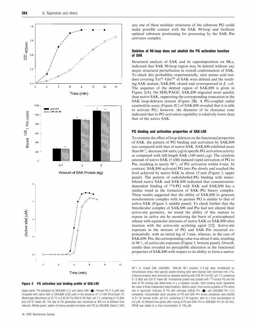
Figure 3 PG activation and binding profile of SAK∆90
Upper panel : PG activation by SAK∆90 (D) and native SAK (E). Human PG (1.5 µM) was
incubated with native SAK or SAK∆90 (0.05 µM) in the presence of 1.2 mM Chromozym PL
(Boehringer-Mannheim) at 25 °C in 0.05 M Tris/HCl/0.1M NaCl, pH 7.4, containing 0.1% BSA
and 0.01% Tween 80. The rate of Pm generation was monitored at 405 nm at different time
intervals. Middle panel : pattern of binary-complex formation with PG by SAK∆90. Native (‘ SAK
any one of these modular structures of the substrate PG could
make possible contact with the SAK 90-loop and facilitate
optimal substrate positioning for processing by the SAK–Pm
activator complex.
Deletion of 90-loop does not abolish the PG activation functionof SAK
Structural analysis of SAK and its superimposition on SKα,
indicated that SAK 90-loop region may be deleted without any
major structural perturbation in overall conformation of SAK.
To check this probability experimentally, nine amino acid resi-
dues covering Tyr*#–Glu"!! of SAK were deleted and the result-
ing SAK mutant, SAK∆90, cloned and overexpressed in E. coli.
The sequence of the deleted region of SAK∆90 is given in
Figure 2(A). On SDS}PAGE, SAK∆90 migrated more quickly
than native SAK, supporting the corresponding truncation in the
SAK loop-deletion mutant (Figure 2B). A PG-coupled radial
caseinolytic assay (Figure 2C) of SAK∆90 revealed that it is able
to activate PG; however, the diameter of its clearance zone
indicated that its PG activation capability is relatively lower than
that of the native SAK.
PG binding and activation properties of SAK∆90
To examine the effect of loop deletion on the functional properties
of SAK, the pattern of PG binding and activation by SAK∆90
was compared with that of native SAK. SAK∆90 exhibited more
than 60% decrease (64 units}µg) in specific PG activation activity
as compared with full-length SAK (160 units}µg). The catalytic
amount of native SAK (5 nM) induced rapid activation of PG to
Pm, resulting in nearly 90% of PG activation within 6 min. In
contrast, SAK∆90 activated PG into Pm slowly and reached the
level achieved by native SAK in about 15 min (Figure 3, upper
panel). The pattern of radiolabelled-PG binding with immo-
bilized native SAK and SAK∆90 indicated that concentration-
dependent binding of "#&I-PG with SAK and SAK∆90 has a
similar trend in the formation of SAK–PG binary complex.
These results suggested that the ability of SAK∆90 to generate
stoichiometric complex with its partner PG is similar to that of
native SAK (Figure 3, middle panel). To check further that the
bimolecular complex of SAK∆90 and Pm had not altered their
active-site geometry, we tested the ability of this mutant to
expose its active site by monitoring the burst of p-nitrophenol
release with equimolar mixtures of native SAK or SAK∆90 after
reaction with the active-site acylating agent [32]. Active-site
exposure in the mixture of PG and SAK–Pm occurred ex-
ponentially, with an initial lag of 3 min, whereas, in the case of
SAK∆90–Pm, the corresponding value was about 4 min, resulting
in 98% of active-site exposure (Figure 3, bottom panel). Overall,
results thus revealed no perceptible alteration in the functional
properties of SAK∆90 with respect to its ability to form a native-
WT’) or mutant SAK (SAK∆90 ; ‘ SAK.del 90 ’) proteins (1.5 ng) were immobilized on
nitrocellulose strips. Non-specific protein-binding sites were blocked with skimmed milk (1%).
Unbound proteins were removed by repeated washing with 0.05 M Tris/HCl, pH 7.5, containing
0.1% BSA and 0.01% Tween 80. Immobilized protein was probed with 125I-human PG and the
level of PG binding was determined in a γ-radiation counter. Each binding result represents
the mean of three independent determinations. Bottom panel : time-course acylation of Pm active
site in equimolar mixtures of PG with wild-type SAK∆–Pm (E) and SAK∆90–Pm (D)
complexes. Concentrated stock solutions of PG and SAK–Pm binary complexes were diluted
in 0.1 M Veronal buffer, pH 8.3, containing 0.1 M arginine, both to a final concentration of
4.0 µM. At different time points after mixing of PG and SAK–Pm or SAK∆90–Pm (0–20 min),
NPGB was added to a final concentration of 100 µM.
# 2002 Biochemical Society

385The 90-loop region of staphylokinase in plasminogen activation
14.0
21.0
31.0
42.7
66.2
97.4
Kd M PG
SA
K
0 1 5 10 SA
K.d
el90SAK:PG
Reaction Time (min)
0 1 5 10
SAK:PGReaction Time (min)
15
Figure 4 Substrate PG binding and activation by preformed complex of Pm with native SAK and SAK∆90
Top left panel : activation of substrate PG by preformed complex of Pm with native SAK (E) and SAK∆90 (D). Pm (1.5 µM) and SAK (1.5 µM) were preincubated for 10 min at 37 °C in Tris/HCl,
pH 7.4, containing 0.1 M NaCl, 0.1% BSA and 0.01% Tween 80. This complex was subsequently diluted (final concentration 5 nM) and incubated with PG (1.5 µM) and generated Pm activity
was measured at different time intervals in the presence of Chromozym PL. Top right panel : Lineweaver–Burk plots of amidolysis by SAK and SAK mutants. Various concentrations (1.0–10 µM)
of substrate PG were mixed with the activator complex (5 nM) formed between Pm and SAK (E) or SAK mutants (D, SAK∆90 ; y, SAKK97A ; x, SAKK98A) in a quartz cuvette containing assay
buffer. The absorption at 405 nm was continuously recorded for 60 min to obtain velocities. Lineweaver–Burk plots of 1/v (‘ V−1 ’) [(∆A405/min)−1] against 1/s (‘ s−1’ ; µM−1) from typical experiments
are shown. Bottom left panel : formation of ternary complex (PG–SAK–Pm) with substrate PG by immobilized binary complex formed with native SAK (‘SAK WT ’) or SAK∆90 (‘SAK.del 90 ’) with
Pm. Different concentrations of human Pm were immobilized and non-specific sites were blocked with BSA (1%) as described in the Materials and methods section. Native or mutant SAK (SAK∆90)
proteins (2 µM) were added in 2-fold excess to form binary complex with the bound Pm. After repeated washing with 0.05 M Tris/HCl, pH 7.4, containing 0.1% BSA and 0.01% Tween 80, 125I-
PG (2–3 µM) was added. Unbound PG was removed after three to five washings with the same buffer and the level of bound PG was determined in a γ-radiation counter. Each binding result
represents the mean of three independent measurements. Lower-right panel : cleavage of PG by equimolar complex of Pm with native SAK and SAK∆90 (‘SAK.del 90 ’). Equimolar amounts of
SAK or SAK∆90 and PG (1.5 µM each) were mixed in 100 mM Tris/HCl, pH 7.5, and incubated at 25 °C for 15 min. At each time point, a 7.5 µl aliquot was removed and analysed for the
conversion of PG into Pm by SDS/12.5%-PAGE. Lane M, Molecular-mass markers (Kd¯ kDa) ; lane 1, PG ; lane 2, purified SAK ; lanes 3–6, PG–SAK (‘SAK :PG ’) reaction mixtures incubated
for 0, 1, 5 and 10 min respectively ; lane 7, purified SAK∆90 ; lanes 8–12 are PG–SAK∆90 (‘SAK.del 90 :PG ’) reaction mixtures incubated for 0, 1, 5, 10 and 15 min respectively. The arrow
indicates the cleavage products of PG to Pm.
SAK-like bimolecular complex with Pm and exposure of the
active site within the activator complex.
Catalytic efficiency of preformed SAK∆90–Pm complex
Although SAK∆90 exhibited native-SAK-like ability to generate
activator complex with its partner Pm, its PG activation rate was
7–8-fold slower than that of wild-type SAK. These results
prompted us to explore whether this deficiency in PG activation
is due to the alteration in the interaction of SAK∆90–Pm
complex with the substrate PG. Therefore we compared the
pattern of PG activation by preformed complexes of Pm with
native SAK and SAK∆90 (Figure 4, top-left panel). Activation
of PG by preformed SAK–Pm complex occurred progressively,
with a brief lag phase followed by an exponential phase. The
overall activation pattern obeyed Michaelis–Menton kinetics, as
# 2002 Biochemical Society

386 G. Rajamohan and others
Figure 5 Effect of kringle domains of human PG on PG activation bypreformed activator complex formed by Pm with native SAK (upper panel)and SAK∆90 (lower panel)
Preformed complex of Pm (1.5 µM) with native SAK or SAK∆90 (1.5 µM) were generated
by incubating this mixture at 37 °C for 10 min. The mixture was then diluted 50-fold
and incubated with PG (1 µM) in the presence of different concentration of kringles (0–
20 µM) and generated Pm activity was measured at different time intervals in the presence of
Chromozym PL. Upper panel : E, SAK wild-type ; x, SAK wild-type5 ng of K(1–4) ;
+, SAK wild-type10 ng of K(1–4) ; V, SAK wild-type20 ng of K(1–4). Lower panel :
E, SAK∆90 ; D, SAK∆905 ng of K(1–4) ; y, SAK∆9010 ng of K(1–4) ; x,
SAK∆9020 ng of K(1–4). ‘ OD405 nm ’¯ A405.
Table 1 Functional properties of SAK mutants carrying alteration within the 90-loop region
Sequence of the loop region*
Specific activity
(units/µg)†SAK mutant Position … 92 93 94 95 96 97 98 99 100 101
Wild-type Tyr Asp Lys Asn Lys Lys Lys Glu Glu Thr 160
SAKK94A Tyr Asp Ala Asn Lys Lys Lys Glu Glu Thr 140
SAKK96A Tyr Asp Lys Asn Ala Lys Lys Glu Glu Thr 155
SAKK97A Tyr Asp Lys Asn Lys Ala Lys Glu Glu Thr 49
SAKK98A Tyr Asp Lys Asn Lys Lys Ala Glu Glu Thr 55
SAK∆90 Gly Gly Ser ∆‡ Thr 45
* Underlined amino acid residues indicate substitution with alanine.
† Average value of three individual determinations.
‡ Deletion.
revealed by linear double-reciprocal plots of the initial activation
rate versus the PG concentration (Figure 4, top-right panel). The
catalytic efficiency and Km
of preformed SAK∆90–Pm complex
was about 50% lower and about 5-fold higher respectively than
the preformed SAK–Pm complex (Table 2 below). Figure 4, top-
right panel, shows Lineweaver–Burk plots obtained for the
activator complex formed by SAK and SAK mutants in enzyme
kinetic studies. To further compare the capability of ternary-
complex formation with the activator complex formed with Pm
and native SAK or SAK∆90, the interaction of substrate PG
with the bimolecular complex formed with SAK∆90 and Pm, a
sandwich-binding assay was performed. Immobilized bimol-
ecular complex formed with SAK∆90 and Pm exhibited a more-
than-40% decrease in the interaction with the substrate PG in
the formation of ternary complex as compared with native SAK
(Figure 4, bottom-left panel). Cleavage of substrate PG to Pm by
an equimolar complex of Pm with SAK or SAK∆90 was
monitored by SDS}PAGE to further compare the catalytic
efficiency of SAK∆90 with native SAK (Figure 4, bottom-right
panel). Densitometric analysis of the protein profile indicated
that more than 90% of the PG is converted into Pm within 5 min
by SAK–Pm complex. Under similar conditions, nearly 25% of
the substrate PG was cleaved into Pm in about 15 min by a
preformed complex of SAK∆90 and Pm.
Effect of kringle (K1K2K3K4) domains on the activation ofPG by preformed complex of SAK∆90 and Pm
Since PG activator complex formed with the loop-deletion
mutant of SAK (SAK∆90) exhibited slower processing of sub-
strate PG, and protein-modelling studies indicated possible
interaction of kringle domain of substrate PG with the SAK 90-
loop region, we studied the pattern of PG activation by preformed
SAK–Pm complex in the presence of isolated kringle domains
(K1K2K3K4) of human PG. A time-course study on PG
activation by pre-formed SAK–Pm complex indicated that, in
the presence of 5 µM kringles, activation of PG was inhibited by
30% which was further increased to about 50% when the
amount of kringles were increased to 20 µM (Figure 5, upper
panel), indicating that the kringle domains are competing with
full-length substrate PG molecules for the interaction with the
SAK–Pm enzyme complex. Interestingly, preformed SAK∆90–
Pm complex exhibited very little difference in the PG activation
process and exhibited only 10% inhibition in the presence of
20 µM of kringles (Figure 5, lower panel), suggesting that the
kringle domains are not effectively competing with the substrate
PG during activation via SAK∆90–Pm enzyme complex.
# 2002 Biochemical Society

387The 90-loop region of staphylokinase in plasminogen activation
Table 2 Kinetic constants for the PG activation properties of equimolarcomplexes of SAK mutants and Pm
The results are means³S.E.M. of three determinations.
Bimolecular complex Km (µM) kcat (s−1) kcat/Km (s−1 [µM−1)
SAK–Pm 2.5³0.12 2.0³0.09 0.80
SAK∆90–Pm 14.4³0.20 1.9³0.05 0.14
SAKK97A–Pm 10.4³0.16 2.1³0.10 0.21
SAKK98A–Pm 11.8³0.18 1.8³0.10 0.16
Site-directed mutagenesis of 90-loop region of SAK and functionalproperties of SAK mutants
With a view to further explore whether the charged residues of
SAK 90-loop structure provide any contribution to substrate PG
processing and activation, we attempted to delineate the probable
target of 90-loop region of SAK by site-directed mutagenesis.
Four SAK mutants, namely K94A, K96A, K97A and K98A,
were created by individually converting lysine into alanine. Out
of these four SAK mutants, SAKK*%A
and SAKK*'A
exhibited PG
activator activity comparable with that of native SAK, implying
that these lysine residues may not be important for modulating
the PG activation function of SAK. By contrast, the other two
mutants, SAKK*(A
and SAKK*)A
, exhibited about a 4–5-fold
reduction in their specific activities for PG activation (Table 1).
All these mutants retained near-native-SAK-like binding capa-
bility for the Pm binding as determined by solid-phase radioassay
employing "#&I-Pm. Additionally, these SAK mutants exhibited a
4–6-fold reduction in their affinity for the substrate PG (Table 2),
which was evident from their higher Km
values for the substrate
PG (Figure 4, top-right panel). These observations clearly
indicated that mutations in lysine residues of the 90-loop region
of SAK had led specifically to a reduction in the ability of these
SAK mutants to interact with the substrate PG.
DISCUSSION
Although three-dimensional structures of SAK and its ternary
complex with µPG [15] have been resolved, the molecular
mechanism whereby SAK brings about structural changes after
association with its partner PG or Pm and interacts with free PG
to generate Pm activity is still largely unknown. From a mech-
anistic perspective, two distinct protein–protein interactions
occur during the SAK-mediated PG activation process. SAK
generates an inactive 1:1 bimolecular complex with Pm which
requires removal of a decapeptide from the N-terminus of SAK
[17,18] and the conversion of substrate PG into Pm [6,36,37].
SAK does not directly induce conformational changes in the
active-site residues, but alters the substrate specificity of Pm in-
directly by creating new subsites on to which substrate PG docks
for the enhanced presentation of the PG activation loop
towards the enzyme [11]. Precise molecular interactions that
occur in between the SAK–Pm enzyme complex and the substrate
PG are far from clear at present. The crystal structure of the ter-
nary complex µPm–SAK–µPm reveals only a partial picture,
owing to the absence of auxillary domains (kringle structures)
in µPm as compared with the full-length PG molecule. Recent
biochemical studies [38] indicate the participation of kringle
structure in SAK-mediated PG activation. In the present study
we have demonstrated that the SAK–Pm enzyme complex may
make an additional interaction with kringle domains of the sub-
strate PG via a charged loop region of SAK (SAK 90-loop) that
may facilitate interaction of substrate PG during PG activation.
The SAK 90-loop region is at a position totally distant from
the protein–protein interaction sites in the three-dimensional
structure of µPm–SAK–µPm ternary complex (Figure 1, upper
panel). Retention of PG activation capability in the SAK mutant,
lacking this loop region, suggests that the SAK loop-deletion
mutant is still able to fold back and achieve its native-SAK-like
conformation. SAK exhibits closest structural similarity with
SKα, and it has been suggested that SAK and SK perform
similar function(s) in the PG activator complex [15]. Structural
overlay and amino acid alignment of SAK and SKα, based on
their three-dimensional structural homology, indicate that there
are two distinct mobile regions that do not align when the
structures of these two protein units are superimposed. One of
these region spans positions 44–64 in SKα, and another distinct
region is in SAK, covering the 90-loop region that is absent
in SKα domain. Involvement of the Asp%"–His%) region of SK in
interaction with the substrate PG has been recently suggested on
the basis of site-directed mutagenesis within this mobile region of
SK [39]. Deletion of the SAK 90-loop region results in a 5–6-fold
decrease in specific activity of SAK and generates significant
reduction in the PG activation properties of the molecule. In
principle, SAK could modify the specificity of the Pm active site
by altering its conformation or by changing substrate PG
accession through altered docking of PG on the SAK–Pm
enzyme complex. SAK∆90 forms a 1:1 SAK–Pm complex which
displays an amidolytic activity comparable with that of the
native SAK–Pm binary complex. Progressive exposure of active
site within the SAK∆90–Pm complex clearly indicated that the
SAK loop-deletion mutant is capable of generating activator
complex with Pm very similar to that of native SAK. Therefore
the slow activation kinetics of PG activation by SAK∆90 may
not be due to alteration in activator complex formation with Pm.
Deletion of the SAK 90-loop led to a perceptible increase in
Km
for the substrate PG. The slow kinetics of PG activation by
the preformed activator complex of SAK∆90 and Pm suggested
that this bimolecular complex is not able to interact optimally
with the substrate PG. These results were further substantiated
by slow cleavage of PG into Pm by activator complex of SAK∆90
with Pm. Furthermore, site-directed mutagenesis within the SAK
90-loop region revealed that substitution of Lys*( or Lys*) with
alanine within this loop region results in similar lesion in substrate
PG activation by these SAK mutants. These results provided the
evidence that Lys*( and Lys*) of the SAK 90-loop may be
assisting in the sequestering of substrate PG. These results
conform with those obtained in previous studies on alanine-
clustered charge mutagenesis of SAK [16]. Overall, results thus
suggest that the SAK 90-loop may not be very important in
maintaining the functional conformation of SAK, but may be
involved in accession}interaction of substrate PGvia its positively
charged residues, Lys*( and Lys*).
In an effort to determine the probable target of the SAK 90-
loop on substrate PG, we analysed the orientation of substrate
PG with respect to the SAK–Pm enzyme complex in a three-
dimensional model of the µPm–SAK–µPm complex. It indicated
that the SAK 90-loop may provide an ideal docking site for
the kringle structure of substrate PG. Free PG binds with the
activator complex of SAK–Pm with 3-fold higher affinity com-
pared with miniPG [40], suggesting that the presence of kringle
structures are crucial for optimal interaction of substrate PG. In
an attempt to evaluate further the probable interaction of kringle
structures and the SAK 90-loop, competition between substrate
PG and kringle was examined during PG activation by SAK∆90–
Pm activator complex and compared with that of native SAK–Pm
complex. Kringle domains of PG (K1K2K3K4) effectively
competed with PG for interaction with SAK–Pm activator
# 2002 Biochemical Society

388 G. Rajamohan and others
complex, whereas the SAK∆90–Pm complex did not exhibit any
significant change in PG activation pattern in the presence of
kringles. Conceptually, kringles could modulate the interaction
of substrate PG by providing more optimal docking of substrate
on SAK–Pm activator complex through additional interaction
site(s) via their lysine-binding sites. Biochemical studies on SAK
have indicated that the lysine-binding sites in K1K2K3K4
kringle domain of PG may play a role in the activation of PG by
SAK–Pm complex [38]. In this process, the SAK 90-loop may be
the ideal target for this interaction, owing to the presence of
several surface-exposed clustered lysine residues.Molecular dock-
ing of kringle 5 on SAK 90-loop indicates that Lys*( and Lys*)
are facing the anionic centre of the kringle. A drastic decrease in
the PG activation properties of SAK mutants SAKK*(A
and
SAKK*)A
supports this structural prediction. Since the presence of
kringle structure (K1K2K3K4) effectively compete with
full-length PG, any one of these kringle domains may possibly
show an interaction with the SAK 90-loop region. The overall
basic conformations of the kringle domains are more or less
similar, including kringle 5, which we have tried to dock on the
SAK 90-loop in our modelling studies.
In conclusion, the present study on site-directed and loop-
deletion mutagenesis of SAK has provided novel information on
SAK–PG interaction and provided the experimental evidence for
the participation of the SAK 90-loop region in facilitating the
interaction(s) of substrate PG with the SAK–Pm enzyme com-
plex. Lys*( and Lys*) of the SAK 90-loop region may be
specifically involved in this process. These additional interactions
between free PG substrate and bimolecular SAK–Pm complex
thus may potentiate the SAK-mediated PG activation process.
We thank Professor Patrick J. Gaffney for providing purified preparation of naturalSAK protein. Technical assistance was provided by Mr. Muthukrishnan. Financialsupport provided by The Council of Scientific and Industrial Research, Governmentof India, in the form of research fellowship to M.D., is gratefully acknowledged.
REFERENCES
1 Collen, D. and Lijnen, H. R. (1994) Staphylokinase, a fibrin-specific plasminogen
activator with therapeutic potential. Blood 84, 680–686
2 Collen, D. (1998) Staphylokinase : a potent, uniquely fibrin-selective thrombolytic
agent. Nat. Med. 3, 279–284
3 Collen, D. and Van de Werf, E. (1993) Coronary thromobolysis with recombinant
staphylokinase in patients with evolving myocardial infarction. Circulation 87,1850–1853
4 Vanderschueren, S., Barrios, L., Kerdsinchai, P., Van den Heuvel, P., Hermann, L.,
Vrolix, M., DeMan, F., Benit, E., Muyldermans, L., Collen, D. and Van del Werf, F.
(1995) A randomized trial of recombinant staphylokinase versus alteplase for coronary
artery patency in acute myocardial infaction. Circulation 92, 2044–2049
5 Vanderschueren, S., Collen, D. and Van de Werf, F. (1996) A pilot study on bolus
administration of recombinant staphylokinase for coronary artery thrombolysis.
Thromb. Haemostasis 76, 541–544
6 Collen, D., De Cock, F. and Stassen, J. M. (1993) Comparative immunogenicity and
thrombolytic properties toward arterial and venous thrombi of streptokinase and
recombinant staphylokinase in baboons. Circulation 87, 996–1006
7 Lijnen, H. R., Van Hoef, B., De Cock, F., Okada, K., Ueshima, S., Matsuo, O. and
Collen, D. (1991) On the mechanism of firin-specific plasminogen activation by
staphylokinase. J. Biol. Chem. 266, 18, 11826–11832
8 Lijnen, H. R., De Cock, F., Van Hoef, B., Schlott, B. and Collen, D. (1994)
Characterization of the interaction between plaminogen and staphylokinase. Eur. J.
Biochem. 224, 143–149
9 Jespers, L., Vanuetswinkel, S., Lijnen, H. R., Van Herzeele, N., Van Hoef, B.,
Demarsh, E. and Collen, D. (1999) Structural and functional basis of plasminogen
activation by staphylokinase. Thromb. Haemostasis 4, 479–485
10 Collen, D., VanHoef, B., Schlott, B., Hartmann, M., Guhrs, K. H. and Lijnen, H. R.
(1993) Mechanisms of activation of mammalian plasma fibrinolytic systems with
streptokinase and with recombinant staphylokinase. Eur. J. Biochem. 216, 307–314
11 Rabijns, A., Hendrik, L., De Bondt, H. L. and De Ranter, C. (1997) Three-
dimensional structure of staphylokinase, a plasminogen activator with therapeutic
potential. Nat. Struct. Biol. 4, 357–360
12 Castellino, F. J. (1981) Recent advances in the chemistry of the fibrinolytic system.
Chem. Rev. 81, 431–446
13 Sugiyama, N., Sasakai, T., Iwamoto, M. and Abiko, Y. (1988) Binding site of
α2-plasmin inhibitor to plasminogen. Biochim. Biophys. Acta 952, 1–7
14 Chang, Y., Mochalkin, I., McCance, S. G., Cheng, B., Tulinsky, A. and Castellino, F. J.
(1998) Structure and ligand binding determinants of the recombinant kringle 5
domain of human plasminogen. Biochemistry 37, 3258–3271
15 Parry, M. A., Fernandez-Catalan, C., Bergner, A., Huber, R., Hopfner, K. P.,
Scholott, B., Guhrs, K. H. and Bode, W. (1998) The ternary microplasmin–
staphylokinase–microplasmin complex is a protease–cofactor–substrate complex in
action. Nat. Struct. Biol. 10, 917–923
16 Silence, K., Hartmann, M., Guhrs, K. H., Gase, A., Schlott, B., Collen, D. and Lijnen,
H. R. (1995) Structure–function relationships in staphylokinase as revealed by
‘‘ clustered charge to alanine ’’ mutagenesis. J. Biol. Chem. 270, 27192–27198
17 Schlott, B., Guhrs, K. H., Hertmann, M., Rocker, A. and Collen, D. (1997)
Staphylokinase requires NH2-terminal proteolysis for plasminogen activation. J. Biol.
Chem. 272, 6067–6072
18 Schlott, B., Guhrs, K. H., Hartmann, M., Rocker, A. and Collen, D. (1998)
NH2-terminal stuctural motifs in staphylokinase required for plasminogen activation.
J. Biol. Chem. 273, 22346–22350
19 Rajamohan, G. and Dikshit, K. L. (2000) Role of the N-terminal region of
staphylokinase (SAK) : evidence for the participation of the N-terminal region of SAK
in the enzyme–substrate complex formation. FEBS Lett. 474, 151–158
20 Nicholls, A., Sharp, K. A. and Honig, B. (1991) Protein folding and association :
insights from the interfacial and thermodynamic properties of hydrocarbons.
Proteins Struct. Funct. Genet. 11, 282–296
21 Vallejo, A. N., Pogulis, R. J. and Pease, L. R. (1995) PCR. In PCR Primer :
A Laboratory Manual. (Dieffenbach, C. W. and Dveksler, G. S., eds.), pp. 603–612,
Cold Spring Harbor Laboratory Press, Cold Spring Harbor, NY
22 Schlott, B., Hartmann, M., Guhrs, E., Birch-Hirschfeld, H., Pohl, S., Vanderschueren,
S., Van de Werf, F., Michoel, A., Collen, D. and Behnke, D. (1994) High level
production and purification of recombinant staphylokinase for thrombolytic therapy.
Biotechnology 12, 185–189
23 Deutsch, D. G. and Mertz, E. T. (1970) Plasminogen purification from human plasma
by affinity chromatography. Science 170, 1095–1096
24 Sottrup-Jenson, L., Claeys, H., Zajdel, M., Petersen, T. E. and Magnusson, S. (1978)
The primary structure of human plasminogen : isolation of two lysine binding
fragments and one miniplasminogen (MW, 38,000) by elastase-catalysed-specific
limited proteolysis. In Progress in Chemical Fibrinolysis and Thrombosis (Davidson,
J. F., Rowan, R. M., Samama, M. M. and Desnoyers, P. C., eds.), vol. 3,
pp. 191–209, Raven Press, New York
25 Shi, G. Y. and Wu, H. L. (1988) Isolation and characterization of microplasminogen.
J. Biol. Chem. 263, 17071–17075
26 Jackson, K. W., Esmon, N. and Tang, T. (1981) Streptokinase and staphylokinase.
Methods Enzymol. 80, 387
27 Trieu, T., Behnke, D., Gerlach, D. and Tang, J. (1993) Activation of human
plasminogen by recombinant staphylokinase. Methods Enzymol. 223, 156–167
28 Ueshima, S., Silence, K., Collen, D. and Lijnen, H. R. (1993) Molecular conversions
of recombinant staphylokinase during plasminogen activation in purified systems and
in human plasma. Thromb. Haemostasis 70, 495–499
29 Szarka, S. J., Sihota, E. G., Habibi, H. R. and Wong, S. L. (1999) Staphylokinase as a
plaminogen activator component in recombinant fusion proteins. Appl. Environ.
Microbiol. 65, 506–513
30 Wohl, R. C., Summaria, L. and Robbins, K. C. (1980) Kinetics of activation of human
plasminogen by different activator species at pH 7.4 and 37 °C. J. Biol. Chem. 255,2005–2013
31 Shibata, H., Nagaoka, M., Sakai, M., Sawada, H., Watanabe, T. and Yokokura, T.
(1994) Kinetic Studies on the plasminogen activation by the staphylokinase–Plasmin
complex. J. Biochem. 115, 738–742
32 Chase, T. J. and Shaw, E. (1969) Comparison of the esterase activites of trypsin,
plasmin and thrombin on generation on guanidinobenzoate esters – titration of the
enzymes. Biochemistry 8, 2212–2224
33 Fraker, P. J. and Speck, J. C. (1978) Protein and cell membrane iodination with a
sparingly soluble choramide 1,3,4,6-tetrachloro-3a,6a-diphenylglycorilBiochem.
Biophys. Res. Commun. 80, 849–857
34 Pratap, J., Kaur, J., Rajamohan, G., Singh, D. and Dikshit, K. L. (1996) Role of
N-terminal domain of streptokinase in protein transport. Biochem. Biophys. Res.
Commun. 227, 303–310
35 Young, K. C., Shi, G. Y., Wu, D. H., Chang, B. I., Ou, C. P. and Wu, H. L. (1998)
Plasminogen activation by streptokinase via a unique mechanism. J. Biol. Chem. 273,3110–3116
36 Parry, M. A., Zhang, X. C. and Bode, I. (2000) Molecular mechanism of plasminogen
activation : bacterial cofactor provide clues. Trends Biochem. Sci. 25, 53–59
# 2002 Biochemical Society
389The 90-loop region of staphylokinase in plasminogen activation
37 Grella, D. K. and Castellino, F. J. (1997) Activation of human plaminogen by
staphylokinase direct evidence that preformed plasmin is necessary for activation to
occur. Blood 89, 1585–1589
38 Arai, K., Modoiwa, S., Mimuro, J., Asakura, S., Matsuda, M., Sako, T. and Sakata, Y.
(1998) Role of the kringle domain in plasminogen activation with staphylokinase.
J. Biochem. (Tokyo) 123, 71–77
Received 12 November 2001/28 March 2002 ; accepted 8 April 2002
Published as BJ Immediate Publication 8 April 2002, DOI 10.1042/BJ20011647
39 Kim, D. M., Lee, S. J., Kim, I. C., Kim, S. T. and Byun, S. M. (2000) Asp41–His48
region of streptokinase is important in binding to a substrate plasminogen. Thromb.
Res. 99, 93–98
40 Lijinen, H. R., Van Hoef, B., Schlott, B. and Collen, D. (1993) Interaction of
staphylokinase with different molecular forms of Plasminogen. Eur. J. Biochem. 211,91–97
# 2002 Biochemical Society
Copyright © 2022 FDOKUMEN