Left Ventricular End Diastolic Loading: Comparison of Cardiac ...
Upload
independentCategory
view
3download
0
Journal of Cardiovascular Magnetic Resonance@’, 2( I ) , 7- 14 (2000)
Function
Magnetic Resonance Real-Time Imaging for the Evaluation of Left Ventricular Function
Eike Nagel,’ Uta Schneider,’ Simon Schalla,l Tareq Ibrahim,’ Bernhard Schnackenburg,2 Axel Bornstedt,l Christoph Klein,l Hans B. Lehmkuhl, and Eckart Fleck1 Department of Internal Medicine/Cardiology, German Heart institute Berlin and
Chariie Campus Virchow, Humboldt University, Berlin, Germany * Philips Medical Systems, Hamburg, Germany
ABSTRACT
New ultrafast gradient systems and hybrid imaging sequences make it possible to acquire a complete image in real time, without the need for breathholding or electrocardiogram (ECG) triggering. In 21 patients, left ventricular function was assessed by the use of a turbo-gradient echo technique, an echo- planar imaging (EPI) technique, and a new real-time imaging technique. End-diastolic and end- systolic volumes, left ventricular muscle mass, and ejection fraction of the ultrafast techniques were compared with the turbo-gradient echo technique. Inter- and intraobserver variability was determined for each technique. Image quality was sufJicient for automated contour detection in all but two patients in whom foldover occurred in the real-time images. Results of the ultrafast imaging techniques were comparable with conventional turbo-gradient echo techniques. There was a tendency to overestimate the end-diastolic volume by 3.9 and 1.3 ml with EPI real-time imaging, the end-systolic volume by 0.9 and 5.0 ml, and the left ventricular mass by 2.4 and 23.8 g. Ejection fraction showed a tendency to be overestimated by 1.1 % with EPI and underestimated by 4.5% with real-time imaging. Correlation between EPI real-time imaging and turbo-gradient echo were 0.94 and 0.95, respectively, for end- diastolic volumes, 0.98 and 0.94, respectively, for end-systolic volumes, and 0.94 and 0.89, respec- tively, for left ventricular mass. Inter- and intraobserver variability was low with all three techniques. Real-time imaging allows an accurate determination of left ventricular function without ECG trig- gering. Scan times can be reduced signijcantly with this new technique. Further studies will have to assess the value of real-time imaging for the detection of wall motion abnormalities and the imaging of patients with atrial jibrillation. KEY WORDS: kft ventricular function; Magnetic resonance: Real-time imaging.
Received December 23, 1998; Accepted June 20, 1999 Address reprint requests to Eike Nagel.
Copyright 0 2000 by Marcel Dekker, Inc. www.dekker.com

8 Nagel et al.
INTRODUCTION
Magnetic resonance (MR) imaging of the heart has been shown to be highly accurate and reproducible for the evaluation of left ventricular (LV) function and mus- cle mass (LVM) (1-5). In contrast to angiography or scintigraphy, no geometric assumptions for the determi- nation of volumes are needed because three-dimensional data sets are available. The feasibility and accuracy of turbo-gradient echo (6) breathhold imaging for evalua- tion of LV volume and mass with low interstudy variabil- ity have been demonstrated (7-9). Recently, it has been shown that good accuracy can also be attained with recti- linear echo-planar imaging (EPI) (10) and spiral echo- planar imaging (1 1-14). However, even with these ul- trafast breathhold techniques, a major limitation of MR tomography when compared with echocardiography is the acquisition of images during several heart beats. Real- time planning or adapting of imaging planes is impossi- ble, and breathholding is required to suppress breathing motion artifacts. In addition, the assessment of the com- plete left ventricle during stress examiantions may not be possible because the acquisition duration may exceed the longest acceptable duration of the stress test. This may be of special importance because high-dose dobutamine stress MR imaging has been shown to be highly superior to dobutamine stress echocardiography for the noninva- sive detection of myocardial ischemia (15).
The development of high-performance gradient sys- tems and optimized hybrid sequences, which combine turbo-gradient echo and EPI (16,17), today enables the acquisition of complete cardiac images in real time with high temporal resolution. This tremendous speed has sev- eral advantages when compared with conventional acqui- sition techniques. For example, electrocardiogram (ECG) triggering will become obsolete because the complete data set is acquired during a single measurement interval and not during several heart beats as with older tech- niques. In combination with interactive planning tools, real-time planning and adaptation of imaging planes can be performed (18). The reduction of measurement time to one or two heart beats per slice makes the imaging of the complete left ventricle feasible within a single breathhold. However, to reach this acquisition speed, spa- tial resolution needs to be reduced and high EPI factors have to be accepted, which may lead to image distortion and reduce accuracy. Recently, it was reported that image quality of real-time scans is sufficient for an assessment of LV function (1 8), and a close correlation between real- time and EPI was reported in a preliminary study (19).
The aim of this study was to analyze the accuracy and
reproducibility of real-time imaging and EPI techniques for the evaluation of LV function, including end-diastolic volume (EDV), end-systolic volume (ESV), and left ven- tricular ejection fraction (EF), and LVM when compared with ultrafast turbo-gradient echo techniques.
METHODS
Patients
Twenty-one patients (15 men, 6 women, aged 60 ? 9 yr, heart rate 75 2 19 beatshin) were included in the study after informed consent, of which 3 patients had LV hypertrophy due to long-standing arterial hypertension, 4 patients had myocardial infarction, and 1 patient had dilative cardiomyopathy. Patients with contraindications for MR examinations or high-grade ventricular arrhyth- mias or atrial flutter/fibrillation were excluded from the study.
MR Imaging
All patients were examined in the supine position with a 1.5-T whole body MR scanner (ACS NT, Philips, Best, The Netherlands, CPR6), which was equipped with ul- trafast gradients (21 mT m-' amplitude, 100 T m-' sec-' slew rate) that used a five-element phased array cardiac coil that was placed around the thorax of the patient. To avoid foldover, only the two anterior segments of the coil were used for data acquisition. All images were acquired during breathholding at end expiration. Respiration was checked with a strain gauge, and ECG triggering was used for the turbo-gradient and EPI image acquisition. After two rapid surveys to determine the exact axis of the left ventricle, 7- 12 continuous short-axis planes (slice thickness 8 mm, no gap), which covered the complete left ventricle, were planned.
Turbo-field echo imaging (TFE = TI-weighted turbo- gradient echo) was performed with a segmented k-space technique over 16 heart beats (Fig. 1). The details of the sequence are shown in Table 1.
EPI was performed with the identical geometry that used a segmented k-space technique over 16 heart beats (Fig. I ) . For details see Table 1. In two patients, no EPI measurements were performed to shorten the duration of the session.
Real-time imaging was performed with a hybrid turbo-gradient echo-EPI sequence (Fig. 1) (16,17). A temporal resolution of 62 msec (= 16 images per second) was achieved by reducing spatial resolution to 2.2 X 4.4 mm (Table 1). Forty consecutive images were acquired

MR Real-Time Imaging of LV Function
Image Analysis
9
In the end-diastolic and end-systolic images, the endo- and epicardial borders were traced by the use of a com- mercially available software with automated contour de- tection and manual correction, if necessary, on a Sparc 5 work station (Easyvision 4, Cardiac Package, Philips). For the TFE and EPI image series, end-diastolic images were chosen as the first phase after triggering of the R- wave, for the real-time scans the frame with the largest LV cavity area. End-systolic images were defined as the images with the smallest cavity area (Fig. 1). The most basal slice to be included had to cover >50% of the cir- cumference. The volume of each slice was determined from the area within the endocardial tracing (excluding both papillary muscles) multiplied by the slice thickness. EDV and ESV were calculated by summing the volumes of all short-axis slices (Simpson’s method). EF was cal- culated as (ESV - EDV)/EDV and LVM at end-diastole by subtracting the EDV from the end-diastolic epicardial volume multiple by 1.05 g/cm3 (specific myocardial gravity) and adding the mass of the papillary muscles. All scans were analyzed by two independent observers (U.S. and T.I.). Analysis of 10 patients was repeated after
without re- viewing the first analysis to determine intraobserver Vari-
Figure 1. End-diastolic and end-systolic equatorial short-axis views of a turbo-gradient echo image (top row), echo-planar image (middle row), and real-time image (bottom row) of the same patient. ability.
weeks by One Of the examiners
Statistical Analysis to cover at least two complete heart beats. To ensure a similar geometry when compared with the other two im- aging techniques, real-time imaging was also performed during breathholding at end expiration, even though this was not required to preserve image quality.
For all parameters, means ?SD are given. An AN- OVA for repeated measurements was performed for all measured parameters for the comparison of different im- aging techniques. If significant differences occurred ( p
Table 1
Scan Parameters
TFE EPI Real Time
TE, msec TR, msec Flip angle, degree K-lines per shot EPI factor Matrix Temporal resolution, msec Spatial resolution, mm
2.1 5.9 25 4
121 X 256* 50
1.3 X 2.6
-
5.6 20 30 7 7
96 X 128* 20
2.6 X 3.4
6.8 15.5 20 36 9
64 X 128* 62
2.2 x 4.4
All measurements were performed with flow compensation. *Raw data were filtered and zero filled to 256 points.

10 Nagel et al.
< 0.05), a Scheffk procedure was applied for post-hoc testing of individual groups. The results from the real- time and EPI technique were linearly correlated with the turbo-gradient echo technique. Linear correlation was also performed for interobserver and intraobserver vari- ability. Absolute and relative differences between the dif- ferent techniques (difference of two techniques divided by their mean value), different observers, and repeated measurements of one observer were calculated (20).
RESULTS
Image quality was sufficient for automated contour detection with all techniques in all but two patients (Fig. 2). In these patients, foldover occurred in the real-time images due to the small field of view chosen; these pa- tients were excluded from the analysis. Only minimal user interference was needed in any image. Contrast be-
Figure 2. Real-time image series in an equatorial short-axis view. ES, end-systolic; ED, end-diastole.
150
P 100
6 $ 50
0 0 50 100 150
ESV-TFE [mll
200 1 EPI=11.5+.97*TFE
150 I
6 100
b 50
0
r =.92 tJ, 0 50 100 150 200
EDV-TFE [mll
BPI = 12.4 + .93 * TFE r -.94
0 100 200 300 400 LVM-TFE lmgl
RT=3.7+1.14*TFE
0 50 100 150 ESV-TFE Imll
400
- p 300
2 200
I
t 100
0
0 50 100 150 200 EDV-TFE [mll
RT = 24.1 + 1.0 *
0 100 200 300 400 LVM-TFE lmgl
0 20 40 60 80 100 EF-TFE [%I
0 20 40 60 80 100 EF-TFE [%I
Figure 3. Correlation between the EPI and real-time (RT) techniques when compared with the turbo-gradient echo tech- nique.
tween blood and endocardium was good with all scan techniques.
There was a correlation of EPI and real-time with the turbo-gradient echo technique for EDV, ESV, and EF (Figs. 3 and 4). The correlation factor for the determina- tion of LVM with real-time or turbo-gradient echo tech- niques was only 0.88. The absolute and relative differ- ences between EPI, real-time, and turbo-gradient echo
MR Real-Time Imaging of LV Function I I
A EDV
20 1 n
0
-40 - 40 60 80 100 120 140 160
TFE [mll
B ESV
-20 0 20 40 60 80 100
TFE [mll
4 0 40 60 80 100 120 140 160
TFE Id1
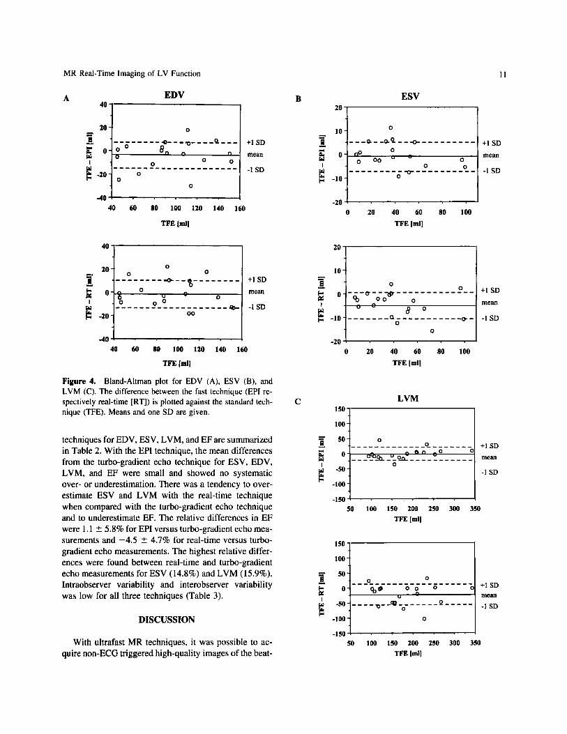
Figure 4. Bland-Altman plot for EDV (A), ESV (B), and LVM (C). The difference between the fast technique (EPI re- spectively real-time [RT]) is plotted against the standard tech- nique (TFE). Means and one SD are given.
techniques for EDV, ESV, LVM, and EF are summarized in Table 2. With the EPI technique, the mean differences from the turbo-gradient echo technique for ESV, EDV, LVM, and EF were small and showed no systematic over- or underestimation. There was a tendency to over- estimate ESV and LVM with the real-time technique when compared with the turbo-gradient echo technique and to underestimate EF. The relative differences in EF were I . I 2 5.8% for EPI versus turbo-gradient echo mea- surements and -4.5 2 4.7% for real-time versus turbo- gradient echo measurements. The highest relative differ- ences were found between real-time and turbo-gradient echo measurements for ESV (14.8%) and LVM (15.9%). Intraobserver variability and interobserver variability was low for all three techniques (Table 3).
DISCUSSION
With ultrafast MR techniques, it was possible to ac- quire non-ECG triggered high-quality images of the beat-
10 i I
0 20 40 60 80 100
TFE [mll
C LVM
0
-100
+ I SD mean
-1 SD
50 100 150 200 250 300 350 TFE [mll
100 i I
I 50 100 150 200 250 300 350
TFE [ml]
-1 SD

12
Table 2
Mean and Relative Differences According to Bland and Altman (in parentheses) Between the Ultrafast Techniques (EPI and Real Time) when Compared With
the Standard Technique (Turbo-Gradient Echo)
EPI Real Time
Nagel et al.
EDV; ml ESV. ml LVM, g EF; %
3.9 -t 12.5 (10.3 -t 11.6%) 0.9 -t 6.2 (14.4 t 15.0%) 2.6 +- 19.3 (9.7 t 11.0%) 1 . 1 ? 5.8 (6.0 t 4.7%)
13.3 t 12.2 ( 1 1.3 t 8.8%) 5.0 2 5.5 (14.8 t 10.6%)
23.8 t 35.7 (15.9 t 14.1%) -4.5 t 4.7 (10.3 t 5.3%)
Table 3
Inter- and Intraobserver Variability
TFE EPI Real Time
EDV Difference, ml r
ESV Difference, ml r
LVM Difference, g r
EF Difference, % r
- 1 . 1 (2.61) 0.96 (0.98)
-3.4 (0.38) 0.98 (0.99)
-7.2 (-2.9) 0.94 (0.99)
4.8 ( 1 . 1 ) 0.93 (0.97)
-7.2 (-2.6) 0.94 (0.96)
-2.6 (- 1.57) 0.98 (0.99)
-2.9 (-1.7) 0.97 (0.99)
1.8 (0.2) 0.97 (0.99)
0.8 (-2.3) 0.95 (0.98)
-7.1 (-1.4) 0.97 (0.99)
- 10.6 (3.7) 0.91 (0.94)
6.2 (0.9) 0.94 (0.97)
The difference and correlation factor between the two observers (two measure- ments) are given.
ing heart in real time with a temporal resolution of 62 msec. Two patients had to be excluded due to insufficient image quality caused by foldover. Spatial resolution and contrast between intracavitary blood and endocardium were sufficient to allow a, quantitative image analysis. With real-time imaging, a tendency to overestimate LV mass and ESVs was found, which resulted in a tendency to underestimate EF. All techniques were highly repro- ducible with a low intra- and interobserver variability.
Two factors may explain the differences between turbo-gradient echo, EPI, and real-time techniques. First, spatial resolution is decreased from 1.3 X 2.6 mm for TFE to 2.2 X 4.4 mm for real-time imaging, which de- creases the accuracy for the delineation of the endo- and epicardial borders. However, zero filling of raw data by a factor of 2 was used, which reduces partial volume ef- fects (21,22) and leads to a reduction of edge definition artifacts (23). Second, chemical shift artifacts in EPI se- quences may be very pronounced and may lead to a su-
perposition of fat signals on parts of the myocardium. These superpositions introduce an error in the determina- tion of LVM as the epicardial border may not be visible.
Temporal resolution was maximal with EPI (20 msec), less so with turbo-gradient echo (50 msec), and minimal with real-time imaging (62 msec). Thus, temporal resolu- tion should be sufficient with EPI and turbo-gradient echo to accurately detect EDV and ESV which remain stable during the isovolumetric phase that lasts approxi- mately 50-80 msec at end-systole. The mild overestima- tion of ESVs with the real-time technique may be ex- plained by a mildly insufficient temporal resolution. In addition, further improvements of temporal resolution are needed to assess hemodynamic parameters such as peak filling rate.
Recently, two possibilities were suggested to solve these problems: either the use of partial k-space acquisi- tion techniques (24) for spiral imaging or coil sensitivity encoding (25). The use of ultrafast gradient systems (2 1

MR Real-Time Imaging of LV Function 13
mT m-’, 100 T m-’ sec-I), which shorten echo times sig- nificantly when compared with conventional systems, leads to a reduction of many problems that may otherwise occur with EPI sequences, such as blurring due to T2* decay, displacement due to phase errors, and systolic loss of blood signal due to intravoxel dephasing. However, these effects are more pronounced with the real-time technique when compared with the other techniques used, due to higher EPI factors and longer echo times.
In previous studies (14), a dark ring between blood and myocardium at times of high flow was observed. In the current study, all measurements were performed us- ing flow compensation to eliminate these problems.
Some errors may have occurred with all techniques due to missed slices or slice overlap caused inconsistent breathhold positions. The use of navigator echoes to determine the diaphragmatic position, combined with feedback mechanisms for reproducible and constant breathholding (26) or respiratory gating during free respi- ration (27), may help to solve this problem. In addition, multislice real-time imaging allows us to cover the com- plete heart within a single breathhold.
Another method, which is successfully used for func- tional cardiac imaging, is spiral k-space acquisition (14.18). The advantages of this method are a high tempo- ral resolution and robustness against motion. However, there are known problems with magnetic field inhomoge- neities and off-resonance effects.
EF was more than 70% in three patients. These pa- tients had LV hypertrophy due to severe arterial hyper- tension and thus very small ESV.
Real-time imaging offers a completely new quality to cardiac MR imaging as ECG triggering becomes dispens- able. This enables us to reduce the time for patient setup and scanning with low-quality ECG tracings within the magnet. Imaging is extremely fast, which can be used to reduce examination time, and costs and patient discom- fort are minimized. In addition, ultrashort scan times allow the combination of different MR techniques with- out exceeding patient tolerance, thus making the “one- stop shop” more and more feasible. Also, it is possible to image the complete left ventricle within one breathhold, which might further improve the diagnostic accuracy of MR stress testing. No data averaging of several heart beats is needed. This makes it possible to scan patients with absolute arrhythmia or frequent premature ventricu- lar complexes or to assess the influence of hemodynamic maneuvers. In combination with interactive planning tools, real-time planning and adaptation of imaging planes is also feasible (1 8) and enables us to correct for patient motion or changes of cardiac position, which may
occur during stress testing due to increased inotropy or perfusion. Breathholding is no longer required or can be reduced to a minimum (e.g., two heart beats) to ensure optimal imaging planes. Thus, recovery periods between subsequent breathholds are obsolete, which further de- creases scan time.
CONCLUSION
Real-time imaging allows an accurate and reproduc- ible determination of LV function and volumes without ECG triggering. Results are Comparable with conven- tional ECG triggered turbo-gradient echo and EPI tech- niques. For a reliable assessment of LVM with real-time imaging, further improvements in spatial resolution are needed. Scan time can be reduced significantly with this new technique. Further studies will have to assess the value of real-time imaging for the detection of wall mo- tion abnormalities and the imaging of patients with atrial fibrillation. In addition, the use of contrast agents has to be assessed, which will most probably enhance edge de- tection quality.
ACKNOWLEDGMENT
Supported by a research grant from Philips Medical Systems, Best, The Netherlands.
REFERENCES
I .
2.
3.
4.
5 .
Longmore DB, Klipstein RH, Underwood SR, Firmin DN, Hounsfield GN, Watanabe M, Bland C, Fox K, Poole Wilson PA and Rees RS. Dimensional accuracy of mag- netic resonance in studies of the heart. Lancet, 1985; 1:
Stratemeier EJ, Thompson R, Brady TJ, Miller SW, Saini S, Wismer GL, Okada RD and Dinsmore RE. Ejection fraction determination by MR imaging: comparison with left ventricular angiography. Radiology, 1986; 158:775- 777. Sechtem U, Pflugfelder PW, Gould RG. Cassidy MM and Higgins CB. Measurement of right and left ventricular volumes in healthy individuals with cine MR imaging. Radiology. 1987; 167:425-430. Semelka RC, Tomei E, Wagner S, Mayo J, Caputo G, O’Sullivan M, Parmley WW, Chatterjee K. Wolfe C and Higgins CB. Interstudy reproducibility of dimensional and functional measurements between cine magnetic res- onance studies in the morphologically abnormal left ven- tricle. Am Heart J, 1990; 1 19: 1367- 1373. Semelka RC, Tomei E, Wagner S, Mayo J, Kondo C, Su-
1360-1362.

14 Nagel et al.
6.
10.
1 I .
12.
13.
14.
15.
16.
17.
zuki J, Caputo GR and Higgins CB. Normal left ventricu- lar dimensions and function: interstudy reproducibility of measurements with cine MR imaging. Radiology, 1990;
Frahm J, Haase A and Matthaei D. Rapid NMR imaging of dynamic processes using the FLASH technique. Magn Reson Med, 1986; 3:321-327. McDonald KM, Parrish T, Wennberg P, Stillman AE, Francis GS, Cohn JN and Hunter D. Rapid, accurate and simultaneous noninvasive assessment of right and left ventricular mass with nuclear magnetic resonance im- aging using the snapshot gradient method. J Am Coll Cardiol, 1992; 19:1601-1607. Sakuma H, Fujita N, Foo TK, Caputo GR, Nelson SJ, Hartiala J, Shimakawa A and Higgins CB. Evaluation of left ventricular volume and mass with breath-hold cine MR imaging. Radiology, 1993; 188:377-380. Bogaert JG, Bosmans HT, Rademakers FE, Bellon EP, Herregods MC, Verschakelen JA, Van de Werf F and Marchal GJ. Left ventricular quantification with breath- hold MR imaging: comparison with echocardiography.
Mansfield P. Multi-planar image formation using NMR spin echoes. Journal of Physics C , 1977; 10:L55-L58. Ahn CB, Kim JH and Cho ZH. High speed spiral scan imaging. ITMI, 1986; 5:2-7. Hunter GJ, Hamberg LM, Weisskoff RM, Halpern EF and Brady TJ. Measurement of stroke volume and cardiac output within a single breath hold with echo-planar MR imaging. J Magn Reson Imaging. 1994; 4:5 1-58. Unterweger M, Debatin JF, Leung DA, Wildermuth S, McKinnon GC and von Schulthess GK. Cardiac vol- umetry. Comparison of echoplanar and conventional cine-magnetic resonance data-acquisition strategies. In- vest Radiol, 1994; 29:994- IOOO. Wiesmann F, Gatehouse PD. Panting JR, Taylor AM, Fir- min DN and Pennell DJ. Comparison of fast spiral, echo planar and FLASH magnetic resonance imaging for car- diac volumetry at 0.5T. J Magn Reson Imaging, 1998; 8:
Nagel E, Lehmkuhl HB, Boksch W, Klein C, Vogel U, Frantz E, Ellmer A. Dreysse S and Fleck E. High dose do- butamine magnetic resonance imaging for the detection of myocardial ischemia. Circulation 1999; 99:763-770. McKinnon GC. Ultrafast interleaved gradient-echo-pla- nar imaging on a standard scanner. Magn Reson Med,
Fischer SE, Wickline SA and Lorenz CH. Multiple slice
174:763-768.
MAGMA, 1995; 3:5-12.
1033- 1039.
1993; 30:609-616.
18.
19.
20.
21.
22.
23.
24.
25.
26.
27.
hybrid imaging sequence for myocardial perfusion mea- surement [abstract]. Proceedings of the International So- ciety for Magnetic Resonance in Medicine, New York. 1996, p. 682. Yang P, Ken A, Liu A, Liang DH, Hardy C, Meyer C. Macovski A, Pauly J and Hu B. New real-time interactive cardiac magnetic resonance imaging system [abstract]. J Am Coll Cardiol, 1998; 31(Suppl A):3A. Scheidegger MB, Spiegel M, Stuber M, Bonetti P, Dubach P and Boesiger P. Assessment of cardiac wall thickening and ejection fraction from real time car- diac MR images in patients with left ventricular dysfunc- tion [abstract]. Proceedings of the International Society of Magnetic Resonance in Medicine, Sydney, 1998;
Bland JM and Altman DG. Statistical method for assess- ing agreement between two methods of clinical measure- ment. Lancet, 1986; 1 :307-3 10. Parker DL, Yiping DP and Davis DL. The voxel sensitiv- ity function in fourier transform imaging: applications to magnetic resonance angiography. Magn Reson Med,
Parker DL, Parker DJ. Blatter DD, Yiping PD and Good- rich KC. The effect of image resolution on vessel signal in high-resolution magnetic resonance angiography. J Magn Reson Imaging, 1996; 6:632-641. van Tyen R and Saloner D. Reduction of edge definition artifacts from object and image misregistration [abstract]. Proceedings of the Society of Magnetic Resonance in Medicine, New York, 1993; p. 678. Ken AB, Pauly JM and Nishimura DG. Partial k-space acquisition for real-time MR imaging [abstract]. Proceed- ings of the International Society of Magnetic Resonance in Medicine, Sydney, 1998, p. 1945. Weiger M, Scheidegger MB, Pruessmann KP and Boe- siger P. Cardiac real-time acquisition using coil sensitiv- ity encoding [abstract]. Proceedings of the International Society of Magnetic Resonance in Medicine, Sydney, 1998, p. 803. Liu YL, Riederer SJ, Rossman PJ, Grimm RC, Debbins JP and Ehman RL. A monitoring, feedback, and trig- gering system for reproducible breath-hold MR imaging. Magn Reson Med, 1993; 30507-5 I I , Wang Y, Rossman PJ, Grimm RC, Riederer SJ and Eh- man RL. Navigator-echo-based real-time respiratory gat- ing and triggering for reduction of respiration effects in three-dimensional coronary MR angiography. Radiology,
p. 554.
1995; 33:156-162.
1996; 19855-60.
Copyright © 2022 FDOKUMEN