Hypercapnia attenuates ventilator-induced lung injury through ...
Upload
independentCategory
view
5download
0
Journal of Molecular and Cellular Cardiology 53 (2012) 599–608
Contents lists available at SciVerse ScienceDirect
Journal of Molecular and Cellular Cardiology
j ourna l homepage: www.e lsev ie r .com/ locate /y jmcc
Original article
Transgenic overexpression of matrix metalloproteinase-9 in macrophagesattenuates the inflammatory response and improves left ventricular functionpost-myocardial infarction
Rogelio Zamilpa a,b,c, Jessica Ibarra d, Lisandra E. de Castro Brás a,b,c, Trevi A. Ramirez a,b,c,Nguyen Nguyen a,e, Ganesh V. Halade a,b,c, Jianhua Zhang a,b,c, Qiuxia Dai a,b,c,Tariq Dayah a,b,c, Ying Ann Chiao a,b,c, Wesley Lowell a,b,c, Seema S. Ahuja a,d,Jeanine D'Armiento f, Yu-Fang Jin a,e, Merry L. Lindsey a,b,c,⁎a San Antonio Cardiovascular Proteomics Center, The University of Texas Health Science Center at San Antonio, USAb Barshop Institute of Longevity and Aging Studies, The University of Texas Health Science Center at San Antonio, USAc Division of Geriatrics, Gerontology and Palliative Medicine, The University of Texas Health Science Center at San Antonio, USAd Division of Nephrology, Department of Medicine, The University of Texas Health Science Center at San Antonio, USAe Department of Electrical and Computer Engineering, University of Texas at San Antonio, USAf Department of Medicine, Columbia University, USA
⁎ Corresponding author at: Department ofMedicine, Diand Palliative Medicine, The University of Texas Health15355 Lambda Drive, MC 7755, San Antonio, TX 78245fax: +1 210 562 6110.
E-mail address: [email protected] (M.L. Lindsey
0022-2828/$ – see front matter © 2012 Elsevier Ltd. Allhttp://dx.doi.org/10.1016/j.yjmcc.2012.07.017
a b s t r a c t
a r t i c l e i n f oArticle history:Received 25 June 2012Accepted 24 July 2012Available online 3 August 2012
Keywords:Myocardial infarctionMatrix metalloproteinase-9Extracellular matrixInflammationCardiac remodelingMacrophage
Followingmyocardial infarction (MI), activatedmacrophages infiltrate into the necroticmyocardium as part of arobust pro-inflammatory response and secrete matrix metalloproteinase-9 (MMP-9). Macrophage activation, inturn, modulates the fibrotic response, in part by stimulating fibroblast extracellular matrix (ECM) synthesis. Wehypothesized that overexpression of human MMP-9 in mouse macrophages would amplify the inflammatoryand fibrotic responses to exacerbate left ventricular dysfunction. Unexpectedly, at day 5 post-MI, ejectionfraction was improved in transgenic (TG) mice (25±2%) compared to the wild type (WT) mice (18±2%;pb0.05). By gene expression profiling, 23 of 84 inflammatory genes were decreased in the left ventricle in-farct (LVI) region from the TG compared to WT mice (all pb0.05). Concomitantly, TG macrophages isolatedfrom the LVI, as well as TG peritoneal macrophages stimulated with LPS, showed decreased inflammatorymarker expression compared to WT macrophages. In agreement with attenuated inflammation, only 7 of84 cell adhesion and ECMgeneswere increased in the TG LVI compared toWT LVI, while 43 geneswere decreased(all pb0.05). These results reveal a novel role for macrophage-derived MMP-9 in blunting the inflammatoryresponse and limiting ECM synthesis to improve left ventricular function post-MI.
© 2012 Elsevier Ltd. All rights reserved.
1. Introduction
After myocardial infarction (MI), circulating monocytes in thebloodstream infiltrate into the injured myocardium and differentiateinto macrophages [1]. Macrophage infiltration into the necroticmyocar-dium can be detected by day 3 and peaks by day 5 post-MI [2,3]. The de-gree of macrophage activation plays a critical role in the cell-mediatedinflammatory response that follows MI. Macrophage activation post-MIis biphasic and consists of an initial pro-inflammatory phase that overlapswith an anti-inflammatory phase [2]. Pro-inflammatory macrophages,
vision of Geriatrics, GerontologyScience Center at San Antonio,, USA. Tel.: +1 210 562 6051;
).
rights reserved.
also known as M1 or classically activated macrophages, have increasedproduction of inflammatory mediators such as interleukin-1 beta(IL-1β), interleukin-6 (IL-6), tumor necrosis factor alpha (TNFα), andMMP-9. Pro-inflammatory macrophages promote extracellular matrix(ECM) degradation [4]. In contrast, anti-inflammatory macrophages,also referred to as M2 or alternatively activated macrophages, have in-creased production of anti-inflammatory mediators such as interleukin-10 (IL-10), transforming growth factor beta (TGFβ), and vascular endo-thelial growth factor (VEGF). Anti-inflammatory macrophages stimulateECM synthesis and scar formation [5].
Matrix metalloproteinase (MMP)-9 is a member of the MMP fam-ily of enzymes and is classified as a gelatinase for its ability to degradedenatured collagen. MMP-9 is robustly secreted by macrophages anddegrades myocardial ECM to facilitate inflammatory cell infiltration,among other functions [6]. In the absence of MMP-9, macrophage in-filtration into the infarct region is reduced, and remodeling of the leftventricle (LV) post-MI is attenuated [3,7]. Therefore, we hypothesized

600 R Zamilpa et al. / Journal of Molecular and Cellular Cardiology 53 (2012) 599–608
that overexpression of human MMP-9 in mouse macrophages wouldamplify the inflammatory and fibrotic responses to exacerbate LVdysfunction. Accordingly, we examined the effect of transgenic overex-pression of human MMP-9 in mouse macrophages after experimentalMI, focusing on LV function, LV inflammatory gene expression, macro-phage infiltration and phenotype, and LV ECM gene expression at day5 post-MI.
2. Materials and methods
2.1. Mice
All animal procedures were conducted according to the “Guide forthe Care and Use of Laboratory Animals” (Eighth Edition, 2011, Na-tional Research Council) and were approved by the Institutional Ani-mal Care and Use Committee at the University of Texas Health ScienceCenter at San Antonio. The mice used in this study were male and fe-male, 3–6 month old, WT littermates (n=16) and TG mice (n=34)overexpressing human MMP-9. Both groups had equal distributionof sex and equal average ages. TG mice carried the human MMP-9cDNA (2.4 kb) transgene driven by the scavenger receptor A enhanc-er/promoter (4.5 kb) as described previously [8]. MI was induced bysurgical ligation of the left anterior descending (LAD) coronary artery,as described previously [3]. The copy number for the TG day 0 groupwas 3.8±1.0 copies and for the day 5 TG MI group was 3.1±0.9 cop-ies. In the post-MI LV macrophages, total MMP-9 gene expressionincreased 15±2-fold compared to WT levels.
2.2. Echocardiography and necropsy evaluations
Echocardiograms were acquired under spontaneous respirationwith 0.5–2% isoflurane in a 100% oxygen mix. Electrocardiogramand heart rate were monitored throughout the imaging procedureusing a surface electrocardiogram. All images were acquired withthe use of a Vevo 770™ High-Resolution In Vivo Imaging System (Vi-sual Sonics) and were taken at a heart rate >400 bpm to achievephysiologically relevant measurements. Measurements were takenfrom the two-dimensional parasternal long-axis and short axis(m-mode) recordings of the LV. For each parameter, 3 images fromconsecutive cardiac cycles were measured and averaged.
At 5 days post-MI, LV tissue was collected. The mice were anesthe-tized with 2% isoflurane, and the coronary vasculature was flushedwith cardioplegic solution [9]. The hearts were excised and the LV andright ventricle were separated and weighed individually. The LV wassectioned into three transverse sections and stained with 1% 2, 3, 5 tri-phenyltetrazolium chloride (Sigma) for infarct area determination. Theinfarct (LVI) and remote (LVC) regions were separated, individuallysnap frozen, and stored at−80 °C for biochemical analysis. Themid sec-tion was fixed in zinc-formalin (Fisher Scientific), paraffin-embedded,and sectioned at 5 μm for histological examination. The lungs werealso removed and weighed. For controls, day 0 WT (n=20) and TG(n=19) mice were sacrificed as described above.
2.3. Histological evaluation
Immunohistochemistry was performed with the use of theVectastain ABC Kit (Vector Laboratories). HistoMark Black (KPL54-75-00) was used to visualize positive staining, with eosin as acounterstain. An antibody specific for neutrophils (anti-neutrophil,mouse monoclonal from Cedarlane, #CL8993AP; 1:100 dilution)was used to selectively detect neutrophils. Picrosirius red staining wasused to visualize collagen content. Staining levels were quantifiedusing Image-Pro software (Media Cybernetics) to calculate percentageof total area stained positive. Negative controls included no primaryantibody and IgG isotype matched controls.
2.4. LV tissue inflammatory and ECM arrays
LV tissue from WT and TG day 0 controls (3F/3M each for bothgroups) and WT and TG day 5 post-MI (3F/3M each for both groups)were collected. The infarct and remote regions from the post-MI micewere analyzed separately. RNA extraction was performed using Trizol®Reagent (Invitrogen 15596‐026), and cDNA was synthesized using RT2
First Strand Kit (Qiagen 330401). The expression of inflammation andECM genes was measured using the inflammatory cytokine and recep-tor array and the ECM and adhesion molecule array following themanufacturer's recommendations (Qiagen PAMM-011 and PAMM-013, respectively). Gene expression was normalized to the hypoxan-thine guanine phosphoribosyl transferase 1 (Hprt1) gene.
2.5. LV infarct macrophage isolations and qRT-PCR
To isolate macrophages from 5 day LVI, we followed the prepara-tion of single-cell suspension from the mouse heart protocol fromMiltenyi Biotech, with slight modifications. Briefly, LVI tissue fromWT (n=4) and TG (n=4) was minced and dissociated into single-cell suspension using collagenase II (600 U/mL, Worthington) andDNase I (60 U/mL, AppliChem) in Hank's Balanced Salt Solution(HBSS, Gibco). After 45 min incubation at 37 °C, with mechanical dis-sociation applied every 15 min, the cell suspension was centrifuged,resuspended in cold HBSS and applied over pre-separation filters toremove non-dissociated clumps (Miltenyi Biotec 130-041-407). Thecells were pelleted by centrifugation at 300 ×g for 10 min andresuspended in MACS separation buffer and Red Blood Cell Lysis Solu-tion (Miltenyi Biotec). The cell suspension was purified using themouse anti-LY-6G microbead kit (Miltenyi Biotec 130-092-332) andapplied over a magnetic MS column (Miltenyi Biotec 130-042-201)to remove neutrophils, which were not abundant at day 5 post-MI.The cells in the flow-through were resuspended in fresh separationbuffer, and the macrophages were sorted using the mouse/humanCD11b Microbead kit (Miltenyi Biotec 130-049-601). The CD11b pos-itive cells were plated on Thermanox® coverslips or in a 24-well tis-sue culture plate and incubated in RPMI 1640 media (Gibco)supplemented with 10% fetal bovine serum. After overnight incuba-tion, the adherent cells (i.e., macrophages) were washed using Hank'sBalanced buffered saline solution, and were fixed with 4% paraformal-dehyde (Sigma) for 25 min and stored in phosphate buffered saline at4 °C for immunocytochemistry, or were collected for RNA isolation.RNA extraction was performed by lysing the cells directly in thewell using RNeasy (Qiagen 12183-018A), and cDNA was synthesizedusing High Capacity RNA to cDNA Kit (ABI 4387406). Quantitative real-time PCR (qRT-PCR) was performed using Taqman gene expressionassays (Applied Biosystems) for human MMP-9 (Hs00234579_m1)and mouse MMP-9 (Mm00442991_m1). For M1 macrophage acti-vation, IL-1β (Mm01336189_m1), IL-6 (Mm00446190_m1), IFN-γ(Mm01168134_m1), and TNFα (Mm00443258_m1) levels wereassessed. For M2 macrophage activation, the levels of Arg1(Mm00475988_m1), CD163 (Mm00474091_m1), mannose receptor1 (Mm00485148_m1) and TGFβ1 (Mm01178820_m1)weremeasured.Gene expression was normalized to Gapdh (Mm99999915_g1).
2.6. Peritoneal macrophage isolation and qRT-PCR
Peritoneal macrophages were isolated from WT littermate (n=3F/3M) and TG (n=3F/3M) peritoneum, and 2×10⁶ cells were plat-ed on 25 cm2 cell culture flasks in RPMI 1640 media supplementedwith 10% fetal bovine serum. After overnight incubation at 37 °C,the cells were serum starved for 24 h and treated using serum-freemedium with and without LPS (100 ng/mL, Sigma) for 24 h. Thecells were washed with PBS and lysed directly in the plate, RNA wasextracted, and expression of inflammatory cytokine and receptorswas analyzed using the array described above.
601R Zamilpa et al. / Journal of Molecular and Cellular Cardiology 53 (2012) 599–608
2.7. Protein extraction and immunoblotting
Total protein was extracted from the day 0 controls, the LVC regions,and the LVI regions by homogenizing the sample with Sigma Reagent 4(7 M urea, 2 M thiourea, 40 mM Trizma® base and the detergent 1%C7BzO) and 1× Complete Protease Inhibitor Cocktail (Roche). Proteinconcentrations were determined using the Bradford assay (BioRad).Due to the high urea content in Reagent 4, protein extracts were diluted1:40withwater for compatibilitywith Bradford assay reagents. The totalprotein (10 μg) for each fraction of all samples was run on 26-well, 4–12% criterion Bis–Tris gels (Bio-Rad), transferred onto nitrocellulosemembranes (Bio-Rad) and stained with MemCode™ Reversible ProteinStain Kit (Pierce) to verify protein concentration and loading accuracy.Protein levels were quantified by immunoblotting using the followingprimary antibodies: alpha smooth muscle actin (Abcam ab5694), anti-human MMP-9 (Epitomics 1939‐1), anti-mouse MMP-9 (Abcamab38898), Mac 3 (Cedarlane CL8943AP), galectin 3 (R&D AF1197), andvimentin (Abcamab8545). Immunoblottingwas performed as previous-ly described [10].Molecular Imaging Software (Kodak) and Image Jwereused to measure densitometry. Densitometric units were normalized tothe densitometry of the total protein stain for the entire lane. The groupswere sub-analyzed, to determine if there were any differences based onsex, forMMP-9, galectin-3, andMac-3. There were no differences withinthe groups based on sex. Male and female results, therefore, werecombined.
2.8. Statistical analyses
Data are reported as mean±SEM. Two group comparisons ofuntreated and treated cells were performed using paired t-tests.One-way ANOVA followed by the Student Newman–Keuls post-hoctest was used for comparisons of more than two groups. A pb0.05was considered significant.
3. Results
3.1. 5-Day post-MI mortality
During the 5 day post-MI protocol, WT mice had a mortality rateof 0% (0 of 16 mice), and TG mice had a mortality rate of 14.7% (5 of34 mice; p=0.16). For the TG mortalities, 1 of the 5 deaths (20%)was a confirmed cardiac rupture. Non-rupture related deaths, likelythe result of acute heart failure or arrhythmias, accounted for 80% ofthe deaths (4 of 5 mice).
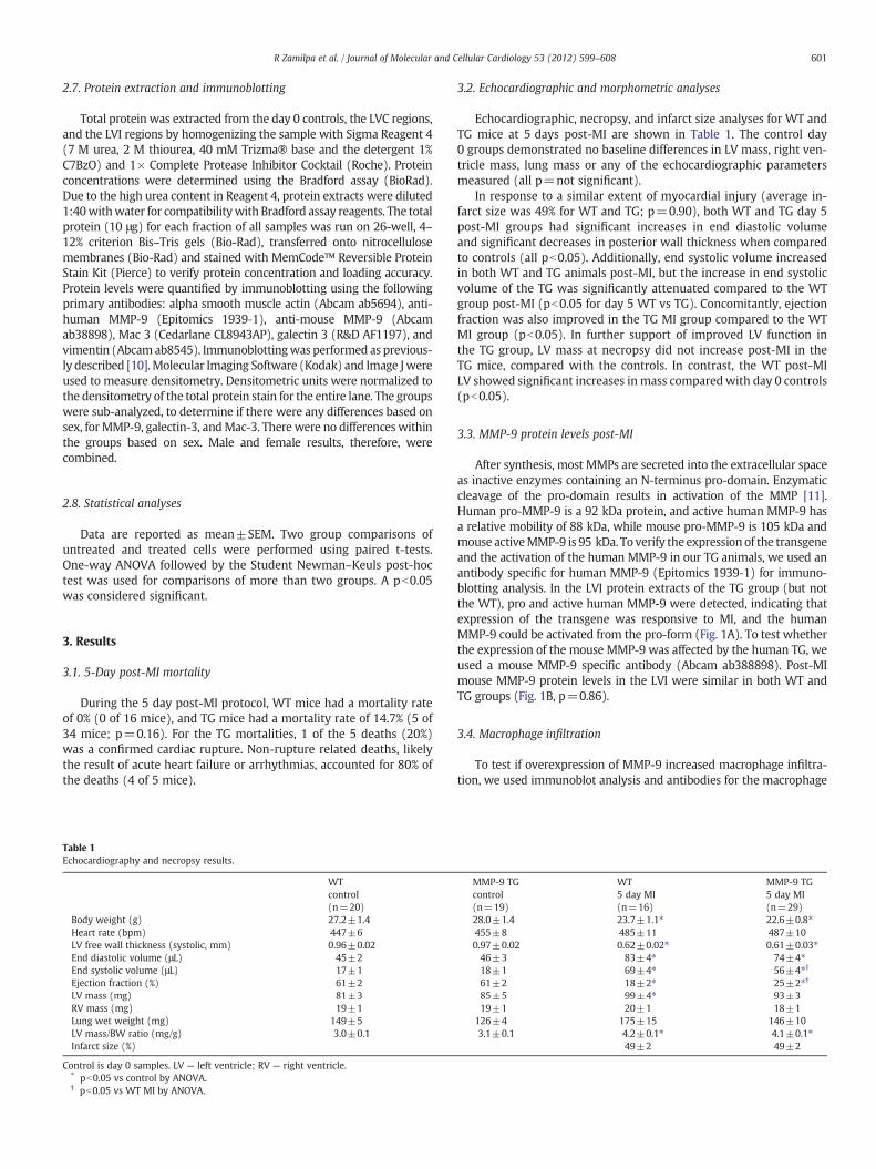
Table 1Echocardiography and necropsy results.
WTcontrol(n=20)
Body weight (g) 27.2±1.4Heart rate (bpm) 447±6LV free wall thickness (systolic, mm) 0.96±0.02End diastolic volume (μL) 45±2End systolic volume (μL) 17±1Ejection fraction (%) 61±2LV mass (mg) 81±3RV mass (mg) 19±1Lung wet weight (mg) 149±5LV mass/BW ratio (mg/g) 3.0±0.1Infarct size (%)
Control is day 0 samples. LV — left ventricle; RV — right ventricle.⁎ pb0.05 vs control by ANOVA.† pb0.05 vs WT MI by ANOVA.
3.2. Echocardiographic and morphometric analyses
Echocardiographic, necropsy, and infarct size analyses for WT andTG mice at 5 days post-MI are shown in Table 1. The control day0 groups demonstrated no baseline differences in LV mass, right ven-tricle mass, lung mass or any of the echocardiographic parametersmeasured (all p=not significant).
In response to a similar extent of myocardial injury (average in-farct size was 49% for WT and TG; p=0.90), both WT and TG day 5post-MI groups had significant increases in end diastolic volumeand significant decreases in posterior wall thickness when comparedto controls (all pb0.05). Additionally, end systolic volume increasedin both WT and TG animals post-MI, but the increase in end systolicvolume of the TG was significantly attenuated compared to the WTgroup post-MI (pb0.05 for day 5 WT vs TG). Concomitantly, ejectionfraction was also improved in the TG MI group compared to the WTMI group (pb0.05). In further support of improved LV function inthe TG group, LV mass at necropsy did not increase post-MI in theTG mice, compared with the controls. In contrast, the WT post-MILV showed significant increases in mass comparedwith day 0 controls(pb0.05).
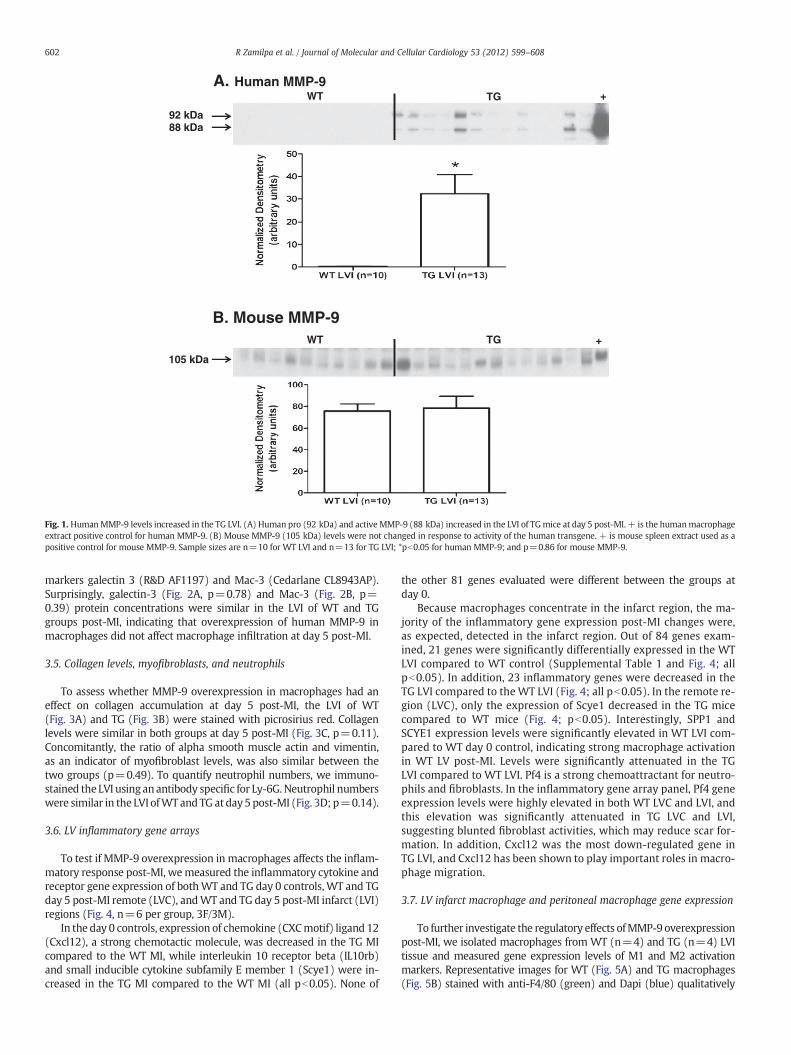
3.3. MMP-9 protein levels post-MI
After synthesis, most MMPs are secreted into the extracellular spaceas inactive enzymes containing an N-terminus pro-domain. Enzymaticcleavage of the pro-domain results in activation of the MMP [11].Human pro-MMP-9 is a 92 kDa protein, and active human MMP-9 hasa relative mobility of 88 kDa, while mouse pro-MMP-9 is 105 kDa andmouse activeMMP-9 is 95 kDa. To verify the expression of the transgeneand the activation of the human MMP-9 in our TG animals, we used anantibody specific for human MMP-9 (Epitomics 1939‐1) for immuno-blotting analysis. In the LVI protein extracts of the TG group (but notthe WT), pro and active human MMP-9 were detected, indicating thatexpression of the transgene was responsive to MI, and the humanMMP-9 could be activated from the pro-form (Fig. 1A). To test whetherthe expression of the mouse MMP-9 was affected by the human TG, weused a mouse MMP-9 specific antibody (Abcam ab388898). Post-MImouse MMP-9 protein levels in the LVI were similar in both WT andTG groups (Fig. 1B, p=0.86).
3.4. Macrophage infiltration
To test if overexpression of MMP-9 increased macrophage infiltra-tion, we used immunoblot analysis and antibodies for the macrophage
MMP-9 TGcontrol(n=19)
WT5 day MI(n=16)
MMP-9 TG5 day MI(n=29)
28.0±1.4 23.7±1.1⁎ 22.6±0.8⁎455±8 485±11 487±100.97±0.02 0.62±0.02⁎ 0.61±0.03⁎46±3 83±4⁎ 74±4⁎18±1 69±4⁎ 56±4⁎†
61±2 18±2⁎ 25±2⁎†
85±5 99±4⁎ 93±319±1 20±1 18±1
126±4 175±15 146±103.1±0.1 4.2±0.1⁎ 4.1±0.1⁎
49±2 49±2
92 kDa88 kDa
+TGWT
105 kDa
TGWT
A. Human MMP-9
B. Mouse MMP-9+
Fig. 1. HumanMMP-9 levels increased in the TG LVI. (A) Human pro (92 kDa) and activeMMP-9 (88 kDa) increased in the LVI of TGmice at day 5 post-MI. + is the humanmacrophageextract positive control for human MMP-9. (B) Mouse MMP-9 (105 kDa) levels were not changed in response to activity of the human transgene. + is mouse spleen extract used as apositive control for mouse MMP-9. Sample sizes are n=10 for WT LVI and n=13 for TG LVI; *pb0.05 for human MMP-9; and p=0.86 for mouse MMP-9.
602 R Zamilpa et al. / Journal of Molecular and Cellular Cardiology 53 (2012) 599–608
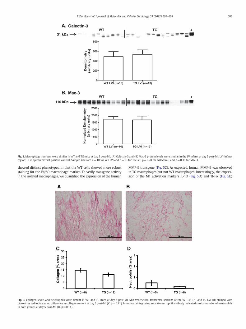
markers galectin 3 (R&D AF1197) and Mac-3 (Cedarlane CL8943AP).Surprisingly, galectin-3 (Fig. 2A, p=0.78) and Mac-3 (Fig. 2B, p=0.39) protein concentrations were similar in the LVI of WT and TGgroups post-MI, indicating that overexpression of human MMP-9 inmacrophages did not affect macrophage infiltration at day 5 post-MI.
3.5. Collagen levels, myofibroblasts, and neutrophils
To assess whether MMP-9 overexpression in macrophages had aneffect on collagen accumulation at day 5 post-MI, the LVI of WT(Fig. 3A) and TG (Fig. 3B) were stained with picrosirius red. Collagenlevels were similar in both groups at day 5 post-MI (Fig. 3C, p=0.11).Concomitantly, the ratio of alpha smooth muscle actin and vimentin,as an indicator of myofibroblast levels, was also similar between thetwo groups (p=0.49). To quantify neutrophil numbers, we immuno-stained the LVI using anantibody specific for Ly-6G. Neutrophil numberswere similar in the LVI ofWT and TG at day5 post-MI (Fig. 3D; p=0.14).
3.6. LV inflammatory gene arrays
To test if MMP-9 overexpression in macrophages affects the inflam-matory response post-MI, wemeasured the inflammatory cytokine andreceptor gene expression of bothWT and TG day 0 controls, WT and TGday 5 post-MI remote (LVC), andWT and TG day 5 post-MI infarct (LVI)regions (Fig. 4, n=6 per group, 3F/3M).
In the day 0 controls, expression of chemokine (CXCmotif) ligand 12(Cxcl12), a strong chemotactic molecule, was decreased in the TG MIcompared to the WT MI, while interleukin 10 receptor beta (IL10rb)and small inducible cytokine subfamily E member 1 (Scye1) were in-creased in the TG MI compared to the WT MI (all pb0.05). None of
the other 81 genes evaluated were different between the groups atday 0.
Because macrophages concentrate in the infarct region, the ma-jority of the inflammatory gene expression post-MI changes were,as expected, detected in the infarct region. Out of 84 genes exam-ined, 21 genes were significantly differentially expressed in the WTLVI compared to WT control (Supplemental Table 1 and Fig. 4; allpb0.05). In addition, 23 inflammatory genes were decreased in theTG LVI compared to theWT LVI (Fig. 4; all pb0.05). In the remote re-gion (LVC), only the expression of Scye1 decreased in the TG micecompared to WT mice (Fig. 4; pb0.05). Interestingly, SPP1 andSCYE1 expression levels were significantly elevated in WT LVI com-pared to WT day 0 control, indicating strong macrophage activationin WT LV post-MI. Levels were significantly attenuated in the TGLVI compared to WT LVI. Pf4 is a strong chemoattractant for neutro-phils and fibroblasts. In the inflammatory gene array panel, Pf4 geneexpression levels were highly elevated in both WT LVC and LVI, andthis elevation was significantly attenuated in TG LVC and LVI,suggesting blunted fibroblast activities, which may reduce scar for-mation. In addition, Cxcl12 was the most down-regulated gene inTG LVI, and Cxcl12 has been shown to play important roles in macro-phage migration.
3.7. LV infarct macrophage and peritoneal macrophage gene expression
To further investigate the regulatory effects ofMMP-9overexpressionpost-MI, we isolated macrophages from WT (n=4) and TG (n=4) LVItissue and measured gene expression levels of M1 and M2 activationmarkers. Representative images for WT (Fig. 5A) and TG macrophages(Fig. 5B) stained with anti-F4/80 (green) and Dapi (blue) qualitatively
31 kDa+TGWT
110 kDa+TGWT
A. Galectin-3
B. Mac-3
Fig. 2.Macrophage numbers were similar inWT and TGmice at day 5 post-MI. (A) Galectin-3 and (B) Mac-3 protein levels were similar in the LV infarct at day 5 post-MI. LVI-infarctregion; + is spleen extract positive control. Sample sizes are n=10 for WT LVI and n=13 for TG LVI; p=0.78 for Galectin-3 and p=0.39 for Mac-3.
603R Zamilpa et al. / Journal of Molecular and Cellular Cardiology 53 (2012) 599–608
showed distinct phenotypes, in that the WT cells showed more robuststaining for the F4/80 macrophage marker. To verify transgene activityin the isolated macrophages, we quantified the expression of the human
A B
C D
0
5
10
15
20
25
30
WT (n=8) TG (n=12)
Co
llag
en (
% a
rea)
Fig. 3. Collagen levels and neutrophils were similar in WT and TG mice at day 5 post-MIpicrosirius red indicated no difference in collagen content at day 5 post-MI (C, p=0.11). Immin both groups at day 5 post-MI (D, p=0.14).
MMP-9 transgene (Fig. 5C). As expected, human MMP-9 was observedin TG macrophages but not WT macrophages. Interestingly, the expres-sion of the M1 activation markers IL-1β (Fig. 5D) and TNFα (Fig. 5E)
0
1
2
3
Neu
tro
ph
ils (
% a
rea)
WT (n=5) TG (n=8)
. Mid-ventricular, transverse sections of the WT LVI (A) and TG LVI (B) stained withunostaining using an anti-neutrophil antibody indicated similar number of neutrophils

Fig. 4. Expression of inflammatory cytokine and receptor genes was decreased in the left ventricle infarct (LVI) post-MI of TG compared to WT mice. The expression of 84 cytokineand receptor genes was quantified in the wild type (WT) and transgenic (TG) day 0 controls and in the WT remote region (WT LVC), WT infarct region (WT LVI), TG remote (TGLVC), and TG infarct (TG LVI) at day 5 post-MI. By ANOVA, pb0.05 for 1-WT vs WT LVC or TG vs TG LVC; 2-WT vs WT LVI or TG vs TG LVI; 3-WT vs TG; 4-WT LVC vs TG LVC; and5-WT LVI vs TG LVI.
604 R Zamilpa et al. / Journal of Molecular and Cellular Cardiology 53 (2012) 599–608
was decreased (pb0.05) in the TG macrophages isolated from theLVI, indicating that MMP-9 overexpression served to dampen thepro-inflammatory response. Interestingly, the expression of other
hMMP-9
BA
C D
WT (n=4) TG(n=4) WT (n=4
Fig. 5.Macrophages isolated from the infarct region of TG mice at day 5 post-MI have reduce(A)WT macrophages and (B) TG macrophages isolated from the LVI stained with F4/80 (gremacrophage isolated from the LV infarct at day 5 post-MI in TG but not WT mice. (D) IL-1βmacrophages. Sample sizes are n=4 for both groups, *pb0.05 vs WT LVI macrophages.
M1 activation markers (IL-6 and IFN-γ) and M2 activation markers(arginase 1, CD163, mannose receptor 1 and TGFβ1) was similar inboth WT and TG macrophages isolated from the LVI at day 5
ETNFαα
) TG (n=4) WT (n=4) TG (n=4)
d expression of pro-inflammatory markers IL-1β and TNFα compared toWT post-MI LV.en) and Dapi (blue). Scale bar is 20 μm. (C) Human MMP-9 (hMMP-9) was expressed inand (E) TNFα levels were significantly decreased in TG macrophages compared to WT

605R Zamilpa et al. / Journal of Molecular and Cellular Cardiology 53 (2012) 599–608
post-MI (all p=not significant). These results suggest that MMP-9overexpression has specific regulatory effects on IL-1β and TNFα.
As a complement experiment, we used the inflammatory gene andreceptor array to measure gene expression of peritoneal macrophagesfromWT(n=6) and TGmice (n=6) treatedwith LPS for 24 h (Supple-mental Table 2). Of the 84 genes evaluated, 27 showed significant differ-ences (Fig. 6). Similar to the response observed in the LVI tissue andisolated LVI macrophages, reduced inflammatory gene expression (in-cluding IL-1β and TNFα) was detected in the isolated TG peritonealmacrophages treatedwith LPS, compared to theWTmacrophages treat-ed with LPS.
3.8. LV extracellular matrix gene arrays
To measure the effects of a reduced inflammatory response on theECM response post-MI, we measured the expression of 84 ECM andadhesion molecules of matched WT and TG day 0 controls, WT andTG day 5 post-MI remote (LVC), and WT and TG day 5 post-MI infarct(LVI) regions (Supplemental Table 3 and Fig. 7). In the day 0 controls,laminin beta 2 and MMP-15 were the only two genes altered in theTG LV and both were decreased in the TG compared to the WT con-trols (pb0.05).
Post-MI, gene expression levels of Cd44, ctnna1, itgb2, lama2,MMP-2, Sgce, and Vcam1 were decreased in the TG LVC, compared tothe WT LVC (all pb0.05). In line with a decreased inflammation in theinfarct region, 43 of 84 ECM and adhesion genes decreased in the TGLVI compared to the WT LVI (all pb0.05). Interestingly, Adamts2,
Fig. 6. TG peritoneal macrophages have decreased gene expression of inflammatory cytokimatory cytokine and receptor genes of untreated WT and TG peritoneal macrophages and LPt-test, pb0.05 for a-WT vs WT+LPS or TG vs TG+LPS; and by ANOVA, pb0.05 for 1-WT v
ECM1, fibronectin, TIMP-2, MMP-14, periostin, and tenascin were in-creased in the TG LVI compared to the WT LVI (all pb0.05), suggestingthat these proteins may play an important role in the reparative pro-cess. Additionally, these results indicate that decreased inflammationdampened the fibrotic response post-MI.
4. Discussion
MMP-9 is a critical enzyme affiliatedwith post-MI remodeling [12]. Inthis study, we examined the effect of overexpression of humanMMP-9 inmouse macrophages, with respect to left ventricular function, inflamma-tory gene and receptor expression, macrophage infiltration and activa-tion, and ECM and adhesion molecule expression. The significantfindings of this study were that MMP-9 overexpression in macrophagesresulted in 1) improved left ventricular function, 2) reduced inflamma-tory responses in the LV infarct tissue and in isolated macrophages,and 3) decreased the fibrotic response at day 5 post-MI. These resultssuggest that MMP-9 regulates the inflammatory response to MI,which indirectly influences the fibrotic response.
Optimal infarct healing relies on the timely resolution of the in-flammatory response. At the same time, macrophage depletion dur-ing the first week after MI results in defective myocardial healing[13,14]. Interestingly, the degree of macrophage activation and theheterogeneity of macrophage subsets regulate chemotaxis, phagocy-tosis of necrotic myocytes and apoptotic neutrophils, angiogenesis,ECM turnover, and fibroblast activation [4,15].
ne and receptors after treatment with LPS. We quantified the expression of 84 inflam-S-treated WT (WT+LPS) and transgenic peritoneal macrophages (TG+LPS). By paireds WT+LPS or TG vs TG+LPS and 2-WT+LPS vs TG+LPS.

Fig. 7. Expression of ECM and adhesion molecule genes was decreased in the TG mice left ventricle infarct (LVI) at day 5 post-MI compared to WT LVI. The expression of 84 ECMmolecule and adhesion genes was quantified in the wild type (WT) and transgenic (TG) day 0 controls and in the WT remote region (WT LVC), WT infarct region (WT LVI), TGremote region (TG LVC) and TG infarct region (TG LVI). By ANOVA, pb0.05 for 1-WT vs WT LVC or TG vs TG LVC; 2-WT vs WT LVI or TG vs TG LVI; 3-WT vs TG; 4-WT LVC vsTG LVC; and 5-WT LVI vs TG LVI.
606 R Zamilpa et al. / Journal of Molecular and Cellular Cardiology 53 (2012) 599–608
MMP-9 is elevated early in the plasma and myocardium of post-MIpatients and multiple animal models, and targeted deletion of MMP-9in mice has been shown to protect against cardiac rupture and adverseventricular remodeling post-MI [7,16–18]. Because of the beneficialeffects observed in the MMP-9 null mice post-MI, we predicted thattransgenic overexpression of human MMP-9 in mouse macrophageswould result in adverse remodeling and an increased incidence ofrupture. At 5 day post-MI, however, survival was not affected byoverexpression of MMP-9 in macrophages. Further, LV function wasalso improved.
Based on previous results with the MMP-9 null mice showing im-paired macrophage infiltration at day 5 post-MI, we hypothesized thatincreased MMP-9 would stimulate macrophage infiltration and ECMdegradation [3,6]. Our results indicate that MMP-9 overexpression in
macrophages did not affect macrophage infiltration. As neutrophils areanother source of MMP-9, our results suggest that neutrophil-derivedMMP-9 may play a more important role in macrophage migration thanmacrophage-derived MMP-9. Our results agree with previous literatureusing thesemice in amodel of asthma,wheremacrophage numbers thatinfiltrated into the lung were similar between WT and TG [19].
Our findings indicate that MMP-9 overexpression in macrophagesreduces the expression of inflammatory genes and receptors in the LVinfarct region post-MI. Moreover, MMP-9 overexpression regulatedthe inflammatory response of isolated macrophages from the LV in-farct and LPS-stimulated peritoneal macrophages. It is important tomention that mRNA levels in isolated macrophages were evaluatedafter plating the cells, which could modify expression levels. As a con-sequence of reduced inflammation, the gene expression level of ECM

607R Zamilpa et al. / Journal of Molecular and Cellular Cardiology 53 (2012) 599–608
and adhesion molecules was decreased in the TG mice post-MI. Ofnote, macrophage MMP-9 overexpression significantly decreased ex-pression of mouse MMP‐2, -3, -7, -8, -9, -10, -12, and ‐13, indicatingthat MMP-9 may be an important upstream mediator of these otherMMPs. Interestingly, Adamts2, ECM1, fibronectin, MMP-14, periostin,tenascin, and TIMP-2 were upregulated in the TG compared to WTpost-MI, indicating a beneficial role in cardiac wound healing at day5 post-MI by stimulating a more robust scar formation. The fact thatMMP-14 increased with MMP-9 overexpression suggests that MMP-14maybe upstreamofMMP-9 or thatMMP-14may share a commonpath-waywithMMP-9. Fibronectin expression during cardiac repair has beenshown to serve as a scaffold for reparative ECM deposition and pluripo-tent cell migration [20–23]. In the post-MI setting, periostin null micehave been shown to have increased rupture rates as a result of decreasedmyocardial stiffness and collagen accumulation [24,25]. Interestingly,tenascin C null animals have a phenotype similar to that seen in theTG mice; namely, attenuated LV remodeling post-MI [26]. Moreover,TIMP-2 null mice have severe inflammation and greater infarct expan-sion that exacerbated ventricular dilation [27]. These seven genesprovide a targeted list for further studies into MMP-9 dependentmechanisms of LV remodeling.
Traditionally, MMP-9 is considered an enzyme that can degradeECM; however, our findings indicate that MMP-9 has a central rolein the regulation of a multitude of cytokines and chemokines, includ-ing the pro-inflammatory genes IL-1β and TNFα. Consistent with ourresults, Cabrera and colleagues reported reduced pulmonary fibrosisin the bleomycin treated TG MMP-9 mice that was due to increasedMMP-9 activity and cleavage of the MMP-9 substrate insulin-likegrowth factor binding protein-3 [8]. Interestingly, in our inflammato-ry array, the gene expression of small inducible cytokine subfamily E,member 1 (SCYE1), a known MMP-9 substrate, was elevated at base-line and its expression was lower in both the LV remote and infarctregions of TG compared to the WT post-MI regions [28]. SCYE1, alsoknown as EMAPII, is a proinflammatory cytokine whose expressionis induced by apoptosis [29–31]. Although the mechanisms bywhich SCYE1 regulates inflammation are not well understood, ourfindings suggest that overexpression of MMP-9 may regulate the in-flammatory response in part through SCYE1.
In comparison to the MMP-9 null mice, which demonstrate reducedmacrophage infiltration and collagen accumulation to improve LV func-tion post-MI, our findings indicate that macrophage-overexpression ofMMP-9 has a similar functional end product of reduced inflammationand fibrosis, albeit through different mechanisms.
In conclusion, this study demonstrated that human MMP-9overexpression in mouse macrophages imparts a positive conse-quence on LV remodeling at day 5 post-MI. The MMP-9 transgenicmice showed improved left ventricular function and reduced in-flammatory response with a concomitant decrease in ECM proteinexpression. Our data suggest that MMP-9 overexpression impartsa beneficial consequence in the setting of acute inflammation andpost-MI remodeling.
Disclosure statement
None.
Acknowledgments
We acknowledge support from T32 (HL07446), American Heart As-sociation (09POST2150178), and HL075360S1 to RZ; NSF 0649172, NIHEB009496, and NIH 1SC2 HL101430 to Y-FJ; and from NHLBI HHSN268201000036C (N01-HV-00244) for the San Antonio CardiovascularProteomics Center and R01 HL075360, the Max and Minnie TomerlinVoelcker Fund, and the Veteran's Administration (Merit) to MLL.
Appendix A. Supplementary data
Supplementary data to this article can be found online at http://dx.doi.org/10.1016/j.yjmcc.2012.07.017.
References
[1] Swirski FK, Nahrendorf M, Etzrodt M, Wildgruber M, Cortez-Retamozo V, PanizziP, et al. Identification of splenic reservoir monocytes and their deployment to in-flammatory sites. Science 2009;325:612-6.
[2] Nahrendorf M, Pittet MJ, Swirski FK. Monocytes: protagonists of infarct in-flammation and repair after myocardial infarction. Circulation 2010;121:2437-45.
[3] Lindsey ML, Escobar GP, Dobrucki LW, Goshorn DK, Bouges S, Mingoia JT, et al.Matrix metalloproteinase-9 gene deletion facilitates angiogenesis after myocardialinfarction. Am J Physiol Heart Circ Physiol 2006;290:H232-9.
[4] Lambert JM, Lopez EF, Lindsey ML. Macrophage roles following myocardial infarc-tion. Int J Cardiol 2008;130:147-58.
[5] Ricardo SD, van Goor H, Eddy AA. Macrophage diversity in renal injury and repair.J Clin Invest 2008;118:3522-30.
[6] Gong Y, Hart E, Shchurin A, Hoover-Plow J. Inflammatory macrophage migrationrequires MMP-9 activation by plasminogen in mice. J Clin Invest 2008;118:3012-24.
[7] Ducharme A, Frantz S, Aikawa M, Rabkin E, Lindsey M, Rohde LE, et al. Targeteddeletion of matrix metalloproteinase-9 attenuates left ventricular enlargementand collagen accumulation after experimental myocardial infarction. J Clin Invest2000;106:55-62.
[8] Cabrera S, Gaxiola M, Arreola JL, Ramirez R, Jara P, D'Armiento J, et al.Overexpression of MMP9 in macrophages attenuates pulmonary fibrosis inducedby bleomycin. Int J Biochem Cell Biol 2007;39:2324-38.
[9] Michael LH, Ballantyne CM, Zachariah JP, Gould KE, Pocius JS, Taffet GE, et al.Myocardial infarction and remodeling in mice: effect of reperfusion. Am J Physiol1999;277(2 Pt 2):H660-8.
[10] Zamilpa R, Lopez EF, Chiao YA, Dai Q, Escobar GP, Hakala K, et al. Proteomic anal-ysis identifies in vivo candidate matrix metalloproteinase-9 substrates in the leftventricle post-myocardial infarction. Proteomics 2010;10:2214-23.
[11] Nagase H. Activation mechanisms of matrix metalloproteinases. Biol Chem1997;378:151-60.
[12] Lindsey ML, Zamilpa R. Temporal and spatial expression of matrix metalloproteinasesand tissue inhibitors ofmetalloproteinases followingmyocardial infarction. CardiovascTher 2012;30:31-41.
[13] Tao ZY, Cavasin MA, Yang F, Liu YH, Yang XP. Temporal changes in matrixmetalloproteinase expression and inflammatory response associated with cardiacrupture after myocardial infarction in mice. Life Sci 2004;74:1561-72.
[14] van Amerongen MJ, Harmsen MC, van Rooijen N, Petersen AH, van Luyn MJ. Mac-rophage depletion impairs wound healing and increases left ventricular remodel-ing after myocardial injury in mice. Am J Pathol 2007;170:818-29.
[15] Zamilpa R, Kanakia R, Jt Cigarroa, Dai Q, Escobar GP, Martinez H, et al. CC chemo-kine receptor 5 deletion impairs macrophage activation and induces adverse re-modeling following myocardial infarction. Am J Physiol Heart Circ Physiol2011;300:H1418-26.
[16] Heymans S, Luttun A, Nuyens D, Theilmeier G, Creemers E, Moons L, et al. Inhibi-tion of plasminogen activators or matrix metalloproteinases prevents cardiac rup-ture but impairs therapeutic angiogenesis and causes cardiac failure. Nat Med1999;5:1135-42.
[17] Kelly D, Cockerill G, Ng LL, Thompson M, Khan S, Samani NJ, et al. Plasma matrixmetalloproteinase-9 and left ventricular remodelling after acute myocardial in-farction in man: a prospective cohort study. Eur Heart J 2007;28:711-8.
[18] Sundstrom J, Evans JC, Benjamin EJ, Levy D, Larson MG, Sawyer DB, et al. Relationsof plasma matrix metalloproteinase-9 to clinical cardiovascular risk factors andechocardiographic left ventricular measures: the Framingham Heart Study. Circu-lation 2004;109:2850-6.
[19] Mehra D, Sternberg DI, Jia Y, Canfield S, Lemaitre V, Nkyimbeng T, et al. Alteredlymphocyte trafficking and diminished airway reactivity in transgenic mice ex-pressing human MMP-9 in a mouse model of asthma. Am J Physiol Lung CellMol Physiol 2010;298:L189-96.
[20] van Dijk A, Niessen HW, Ursem W, Twisk JW, Visser FC, van Milligen FJ. Accumu-lation of fibronectin in the heart after myocardial infarction: a putative stimulatorof adhesion and proliferation of adipose-derived stem cells. Cell Tissue Res2008;332:289-98.
[21] Ulrich MM, Janssen AM, Daemen MJ, Rappaport L, Samuel JL, Contard F, et al.Increased expression of fibronectin isoforms after myocardial infarction in rats.J Mol Cell Cardiol 1997;29:2533-43.
[22] Willems IE, Arends JW, Daemen MJ. Tenascin and fibronectin expression inhealing human myocardial scars. J Pathol 1996;179:321-5.
[23] Cleutjens JP, Verluyten MJ, Smiths JF, Daemen MJ. Collagen remodeling after myo-cardial infarction in the rat heart. Am J Pathol 1995;147:325-38.
[24] Shimazaki M, Nakamura K, Kii I, Kashima T, Amizuka N, Li M, et al. Periostin is es-sential for cardiac healing after acute myocardial infarction. J Exp Med 2008;205:295-303.
[25] Kuhn B, del Monte F, Hajjar RJ, Chang YS, Lebeche D, Arab S, et al. Periostin in-duces proliferation of differentiated cardiomyocytes and promotes cardiac repair.Nat Med 2007;13:962-9.

608 R Zamilpa et al. / Journal of Molecular and Cellular Cardiology 53 (2012) 599–608
[26] Nishioka T, Onishi K, Shimojo N, Nagano Y, Matsusaka H, Ikeuchi M, et al. Tenascin-Cmay aggravate left ventricular remodeling and function after myocardial infarctionin mice. Am J Physiol 2010;298:H1072-8.
[27] Kandalam V, Basu R, Abraham T, Wang X, Soloway PD, Jaworski DM, et al.TIMP2 deficiency accelerates adverse post-myocardial infarction remodelingbecause of enhanced MT1-MMP activity despite lack of MMP2 activation.Circ Res 2010;106:796-808.
[28] Liu J, Schwarz MA. Identification of protease-sensitive sites in human endothelial-monocyte activating polypeptide II protein. Exp Cell Res 2006;312:2231-7.
[29] Yao C, Williams AJ, Ottens AK, Lu XC, Liu MC, Hayes RL, et al. P43/pro-EMAPII:a potential biomarker for discriminating traumatic versus ischemic brain injury.J Neurotrauma 2009;26:1295-305.
[30] Thompson JL, Ryan JA, Barr ML, Franc B, Starnes VA, Schwarz MA. Potential role forantiangiogenic proteins in the myocardial infarction repair process. J Surg Res2004;116:156-64.
[31] Kayton ML, Libutti SK. Endothelial monocyte activating polypeptide II (EMAP II)enhances the effect of TNF on tumor-associated vasculature. Curr Opin InvestigDrugs 2001;2:136-8.
Copyright © 2022 FDOKUMEN