Fast preparation of nano-hydroxyapatite/superhydrophilic reduced graphene oxide composites for...
9
Fast preparation of nano-hydroxyapatite/ superhydrophilic reduced graphene oxide composites for bioactive applications Hudson Zanin, a Eduardo Saito, a Fernanda Roberta Marciano, b Helder Jose Ceragioli, c Alessandro Eustaquio Campos Granato, d Marimelia Porcionatto d and Anderson Oliveira Lobo * b A method for the direct electrodeposition of globular nano-hydroxyapatite (nHAp) onto reduced graphene oxide (RGO) is presented and a model for the specific growth preference is discussed. Results show that the carboxyl (carboxylic acid)/carboxylate functional groups attached directly to the RGO after oxygen plasma treatment were essential to accelerate the OH– formation and the deposition of globular nHAp crystals. High resolution scanning electron microscopy, energy dispersive X-ray and X-ray diffraction showed that homogeneous, highly crystalline, stoichiometric nHAp crystals, with preferential growth in the (002) plane direction, were formed without any thermal treatment. The nHAp/RGO composites were shown to be an appropriate surface for mesenchymal stem cell adhesion with active formation of membrane projections. 1 Introduction Hydroxyapatite (HAp, Ca 10 (PO 4 ) 6 (OH) 2 ) is a form of calcium phosphate that bears close chemical likeness with the mineral part of bone and teeth. 1 It promotes tissue attachment and bone growth by spontaneously forming a biologically active bone-like apatite layer on its surface. 2 Thus, HAp is classied as a biocompatible and bioactive material, and crystalline HAp has been found in many biological applications such as dental or skeletal implants and bone repair scaffolds. 3 There is a resur- gent interest in controlling HAp crystal nucleation, crystallinity, and growth for use in composite materials, analogous to those produced in nature, involving the biomineralization process. 4 Template-induced HAp has broad prospects in the applied elds of regenerative medicine and bone repair. Recent advances in biomaterials research have suggested that the best osteocompatibility of HAp could be achieved if the structure, size, and morphology of the crystal were closer to those of biological apatite. As a result, nano-hydroxyapatite (nHAp) is of great interest. Poor crystallinity can directly affect the natural precipitation of apatite that promotes natural osteointegration. 5 Traditional clinical applications of calcium phosphates have mainly focused on highly crystalline ceramics. 5 HAp has better osteo- conductivity if the characteristics of the crystal are closer to the structure, size and morphology of biological apatite. For this reason, reducing the size of the HAp crystal to the nanoscale is of current interest. Although plasma spraying is the most common method used in commercial deposition of HAp, electrodeposition has advantages such as low temperature synthesis with high crys- tallinity and adherence to substrates, uniform deposition with control of thickness and microstructure even onto complex geometries, and principally the use of low cost equipment and materials. 6,7 Electrodeposition is an advantageous method for producing a thin, crystalline, homogeneous and adherent lm. It is a simple, fast, reproducible, efficient and low cost process. 6–8 The composition and the control of the coating structure are possible in part due to the low processing temperature. The ability to coat irregular surfaces is also important. Therefore, electrodeposition of HAp coatings has attracted noticeable attention in recent years. We have shown in a recent publication that superhydrophilic vertically aligned multi-walled carbon nanotube (VAMWCNT) lms efficiently grow plate-like nHAp crystals directly on their surface. 9 a Associated Laboratory of Sensors and Materials of National Institute for Space Research, Av. dos Astronautas 1758, Sao Jose dos Campos, CEP: 12227-010, SP, Brazil. E-mail: [email protected]; [email protected]; Tel: +55 12 32086576 b Laboratory of Biomedical Nanotechnology/Institute of Research and Development at Universidade do Vale do Paraiba, Av. Shishima Hifumi, 2911, CEP: 12244-000, Sao Jose dos Campos, SP, Brazil. E-mail: [email protected]; *[email protected]; [email protected]; Fax: +55 12 39471149; Tel: +55 1239471100 c Departamento de Semicondutores, Instrumentos e Fotonica, Universidade Estadual de Campinas, Av. Albert Einstein 400, Campinas, CEP: 13083-852, SP, Brazil. E-mail: [email protected] d Neurobiology Lab, Research Building II, Universidade Federal de S~ ao Paulo, Rua Pedro de Toledo, 669, CEP: 04039-032, S~ ao Paulo, SP, Brazil. E-mail: [email protected]; [email protected]; Tel: +55 1155764969 Cite this: DOI: 10.1039/c3tb20550a Received 17th April 2013 Accepted 25th July 2013 DOI: 10.1039/c3tb20550a www.rsc.org/MaterialsB This journal is ª The Royal Society of Chemistry 2013 J. Mater. Chem. B Journal of Materials Chemistry B PAPER Published on 29 July 2013. Downloaded by Universidade Federal de Sao Paulo on 16/08/2013 20:39:32. View Article Online View Journal
Transcript of Fast preparation of nano-hydroxyapatite/superhydrophilic reduced graphene oxide composites for...
Journal ofMaterials Chemistry B
PAPER
Publ
ishe
d on
29
July
201
3. D
ownl
oade
d by
Uni
vers
idad
e Fe
dera
l de
Sao
Paul
o on
16/
08/2
013
20:3
9:32
.
View Article OnlineView Journal
aAssociated Laboratory of Sensors and M
Research, Av. dos Astronautas 1758, Sao
Brazil. E-mail: [email protected];
32086576bLaboratory of Biomedical Nanotechnology/
Universidade do Vale do Paraiba, Av. Shish
Jose dos Campos, SP, Brazil. E-mail: fermar
[email protected]; Fax: +55 12 39471149;cDepartamento de Semicondutores, Instrume
Campinas, Av. Albert Einstein 400, Campin
[email protected] Lab, Research Building II, U
Pedro de Toledo, 669, CEP: 04039-0
[email protected]; marime
1155764969
Cite this: DOI: 10.1039/c3tb20550a
Received 17th April 2013Accepted 25th July 2013
DOI: 10.1039/c3tb20550a
www.rsc.org/MaterialsB
This journal is ª The Royal Society of
Fast preparation of nano-hydroxyapatite/superhydrophilic reduced graphene oxidecomposites for bioactive applications
Hudson Zanin,a Eduardo Saito,a Fernanda Roberta Marciano,b Helder Jose Ceragioli,c
Alessandro Eustaquio Campos Granato,d Marimelia Porcionattod
and Anderson Oliveira Lobo*b
A method for the direct electrodeposition of globular nano-hydroxyapatite (nHAp) onto reduced
graphene oxide (RGO) is presented and a model for the specific growth preference is discussed. Results
show that the carboxyl (carboxylic acid)/carboxylate functional groups attached directly to the RGO
after oxygen plasma treatment were essential to accelerate the OH– formation and the deposition of
globular nHAp crystals. High resolution scanning electron microscopy, energy dispersive X-ray and X-ray
diffraction showed that homogeneous, highly crystalline, stoichiometric nHAp crystals, with preferential
growth in the (002) plane direction, were formed without any thermal treatment. The nHAp/RGO
composites were shown to be an appropriate surface for mesenchymal stem cell adhesion with active
formation of membrane projections.
1 Introduction
Hydroxyapatite (HAp, Ca10(PO4)6(OH)2) is a form of calciumphosphate that bears close chemical likeness with the mineralpart of bone and teeth.1 It promotes tissue attachment and bonegrowth by spontaneously forming a biologically active bone-likeapatite layer on its surface.2 Thus, HAp is classied as abiocompatible and bioactive material, and crystalline HAp hasbeen found in many biological applications such as dental orskeletal implants and bone repair scaffolds.3 There is a resur-gent interest in controlling HAp crystal nucleation, crystallinity,and growth for use in composite materials, analogous to thoseproduced in nature, involving the biomineralization process.4
Template-induced HAp has broad prospects in the appliedelds of regenerative medicine and bone repair. Recentadvances in biomaterials research have suggested that the best
aterials of National Institute for Space
Jose dos Campos, CEP: 12227-010, SP,
[email protected]; Tel: +55 12
Institute of Research and Development at
ima Hifumi, 2911, CEP: 12244-000, Sao
[email protected]; *[email protected];
Tel: +55 1239471100
ntos e Fotonica, Universidade Estadual de
as, CEP: 13083-852, SP, Brazil. E-mail:
niversidade Federal de S~ao Paulo, Rua
32, S~ao Paulo, SP, Brazil. E-mail:
[email protected]; Tel: +55
Chemistry 2013
osteocompatibility of HAp could be achieved if the structure,size, and morphology of the crystal were closer to those ofbiological apatite. As a result, nano-hydroxyapatite (nHAp) is ofgreat interest.
Poor crystallinity can directly affect the natural precipitationof apatite that promotes natural osteointegration.5 Traditionalclinical applications of calcium phosphates have mainlyfocused on highly crystalline ceramics.5 HAp has better osteo-conductivity if the characteristics of the crystal are closer to thestructure, size and morphology of biological apatite. For thisreason, reducing the size of the HAp crystal to the nanoscale isof current interest.
Although plasma spraying is the most commonmethod usedin commercial deposition of HAp, electrodeposition hasadvantages such as low temperature synthesis with high crys-tallinity and adherence to substrates, uniform deposition withcontrol of thickness and microstructure even onto complexgeometries, and principally the use of low cost equipment andmaterials.6,7
Electrodeposition is an advantageous method for producinga thin, crystalline, homogeneous and adherent lm. It is asimple, fast, reproducible, efficient and low cost process.6–8 Thecomposition and the control of the coating structure arepossible in part due to the low processing temperature. Theability to coat irregular surfaces is also important. Therefore,electrodeposition of HAp coatings has attracted noticeableattention in recent years. We have shown in a recent publicationthat superhydrophilic vertically aligned multi-walled carbonnanotube (VAMWCNT) lms efficiently grow plate-like nHApcrystals directly on their surface.9
J. Mater. Chem. B

Journal of Materials Chemistry B Paper
Publ
ishe
d on
29
July
201
3. D
ownl
oade
d by
Uni
vers
idad
e Fe
dera
l de
Sao
Paul
o on
16/
08/2
013
20:3
9:32
. View Article Online
Recently, several papers have examined the development ofHAp/reduced oxide graphene (RGO) composites,10,11 withrespect to the contribution of surface functionalization assites for nHAp nucleation,12 or method of precipitation,13 andhave presented exceptional mechanical properties.
For the rst time, the direct electrodeposition of nHApcrystals on superhydrophilic-RGO with a high crystallinity,reproducibility, and homogeneity is presented. The use of thesuperhydrophilic-RGO lms was shown to be essential to obtainsuch exceptional characteristics. The unique high crystallinityof the nHAp obtained using the electrodeposition processappears to be responsible for all of the other propertiesobserved. The differences in nHAp crystal growth were evalu-ated by high-resolution scanning electron microscopy (HR-SEM), transmission electron microscopy (TEM), energy-disper-sive X-ray analysis (EDX), X-ray diffraction (XRD), Raman andFourier transform infrared (FTIR) spectra and contact angle(CA) measurements.
2 Materials and methods2.1 Synthesis of RGO
The RGO samples were prepared on titanium 10 mm � 10 mm� 1 mm substrates. Prior to the deposition, the substrates wereimmersed in polyaniline diluted in N,N-dimethylformamideand then dried at room temperature for 2 hours. Aer that, thesubstrates were immersed again, but this time in nickel nitratediluted in acetone and then dried again at room temperature. Ahot lament chemical deposition vapor system (HFCVD) wasused in these experiments. The carbon source was a mixture ofpropanone (acetone), camphor, and citric acid. This carbonsource is carried to a HFCVD by hydrogen gas ux (15 sccm).The HFCVD chamber is maintained at 10 Torr with constantows of 65 sccm of nitrogen and 20 sccm of oxygen. A hot spiraltungsten lament heated to 1500 �C dissociates gases andvapours into radicals, depositing RGO thin lms on Tisubstrates aer 30 min.
2.2 RGO functionalized by polar groups
Functionalization of the RGO samples was carried out incor-porating oxygen-containing groups using a pulsed-directcurrent plasma reactor with an oxygen ow rate of 1 sccm, at apressure of 85 mTorr, �700 V and with a frequency of 20 kHz.14
The total time of the plasma etching was 120 seconds and thistreatment changes RGO surface hydrophobicity.
2.3 The globular-like nHAp crystal electrodeposition processon superhydrophilic-RGO lms
The electrodeposition of the nHAp crystals onto the super-hydrophilic-RGO lms was performed using 0.042 mol L�1
Ca(NO3)2$4H2O + 0.025 mol L�1 (NH4)$2HPO4 electrolytes (pH4.7). The electrochemical measurements were carried out usinga three-electrode cell coupled to an Autolab: PGSTAT128Ninstrument equipped with pH and temperature module control.Superhydrophilic-RGO lms were used as the working electrodeand the area in contact with the electrolytic solution was
J. Mater. Chem. B
0.27 cm2. A high-purity platinum coil wire served as the auxiliaryelectrode, and an Ag/AgCl(3 M) electrode was used as thereference electrode. The nHAp lms were produced by applyinga constant potential of �2.0 V for 30 min while the solutiontemperature was maintained at 70 �C.
2.4 Sample characterization
The as-grown and superhydrophilic-RGO lms were character-ized by HR-SEM; TEM, Raman, FTIR and EDX spectroscopies,CA measurements and XRD.
Morphological RGO lm images were generated using a FEIInspect F50 microscope to evaluate the structural arrangementsand to monitor modications of the surface morphology. Thesecond electron mode was employed and the magnication,accelerating voltage, work distance, as well as the spot size arepresented in each image. SEM images (model: JEOL-JSM 5610VPI) were captured to observe the morphology of nHAp/RGOcomposites. Semi-quantitative elemental analyses of calcium(Ca) and phosphorus (P) were carried out using an energydispersive spectrometer coupled to a SEM. HR-TEM imagingwas performed using a JEOL 3010 at 300 kV with a LaB6
lament.We employed Raman scattering spectroscopy for the struc-
tural evaluation of RGO lms (Renishaw 2000 system), withexcitation using an Ar+-ion laser (l ¼ 514.5 nm) in backscat-tering geometry. The diamond peak at 1332 cm�1 calibrated theRaman shi with all measurements carried out in air at roomtemperature. The curve tting and data analysis soware Fityk15
assigned the peak locations and corresponding tting of allspectra. Surface chemical compositions of the nHAp/RGOcomposites were investigated by Raman spectroscopy using twosystems (Renishaw micro-Raman model 2000 with an Ar ionlaser, l ¼ 514.5 nm) and (Bruker Raman model RFS100 withNd:Yag 1064 nm).
The oxygen content of the as-grown and superhydrophilic-RGO samples in chemical bonds was investigated by FTIRattenuated total reection spectroscopy (Spectrum Spotlight-400, Perkin Elmer).
A Kruss Easy Drop system was used to measure the CA by thesessile drop method using high purity deionized water at roomtemperature to evaluate the wettability of the as-grown andsuperhydrophilic-RGO lms.
The structural analysis of nHAp/RGO composites was per-formed by X-ray diffractometry (X-Pert Philips) with Cu K-aradiation generated at 40 kV and 50 mA. The crystal size of thenHAp phase (thkl) was calculated using Scherrer's formula. ThenHAp plane's preferential growth was calculated using theequation suggested by Hu et al.16 All the results were comparedto the HAp powder sample standard (JCPDS 9-432).
2.5 Cellular adhesion
Bone marrow-derived stem cells were obtained from BALB/cmice leg bones (Ethics Committee Approval 0397/11). Briey,mice were euthanized by injection of xylazine/ketamine; tibiaand femurs were dissected, and the epiphyses were cut. Thebone marrow was extracted from the bones under sterile
This journal is ª The Royal Society of Chemistry 2013

Paper Journal of Materials Chemistry B
Publ
ishe
d on
29
July
201
3. D
ownl
oade
d by
Uni
vers
idad
e Fe
dera
l de
Sao
Paul
o on
16/
08/2
013
20:3
9:32
. View Article Online
conditions using a syringe needle 26G (13 mm/24, 5 mm, BDBiosciences, NJ, USA) to inject 2 ml of DMEM (Dubelcco'sModied Eagle's Medium, Invitrogen, San Francisco, USA).DMEM was injected at one end of the bone and the marrow wascollected at the other end. The cell suspension was centrifugedat 400 � g for 10 min at room temperature. The cells weresuspended in DMEM with 10% fetal bovine serum (FBS; Culti-lab, Campinas, Brazil), 1% L-glutamine (Invitrogen) and 1%penicillin/1% streptomycin (GibcoBRL). The number of cellswas quantied in a Neubauer chamber, and diluted with thesame medium to achieve 5 � 106 cells per mL. Cells were platedon P60 dishes and kept in a humidied incubator, at 37 �C, 5%CO2. The change of the culture medium was performed every 72h for the removal of non-adherent hematopoietic cells. Whenthe plates reached 90% conuence, subcultures were performedusing trypsin (Cultilab) diluted 10 times in 10 mM phosphatebuffered saline (PBS). Aer the second subculture adherent cellswere considered mesenchymal stem cells (MSCs), according tothe standardization used internationally.17 The capacities ofcellular adhesion of MSCs on nHAp/RGO composites wereevaluated 48 h aer plating. Cells attached to the substrate werexed with a 3% glutaraldehyde/0.1 M sodium cacodylatebuffer for 1 h and dehydrated in a graded ethanol solutionseries (30%, 50%, 70%, 95%, 100%) for 10 min each. The dryingstage used a 1 : 1 solution of ethanol with hexamethyldisilazane(HMDS), and the samples were dried with pure HMDS at roomtemperature. Aer deposition of a thin gold layer, the speci-mens were examined using an XL30 FEG scanning electronmicroscope (SEM).
3 Results and discussions
Fig. 1 shows the morphology and structure of the RGO beforeand aer exposure to the oxygen plasma. Fig. 1a shows a top-view image of the high density of the as-grown RGO on titaniumsubstrates. The top view of RGO lms shows that the sample hasporous microstructures composed of entangled wires. Fig. 1bshows a HR-SEM image of the as-grown sample, revealing that
Fig. 1 SEM images of RGO films (a and b) before and (c) after oxygen plasmatreatment. Typical HR-SEM (d) and TEM (e) images of these samples.
This journal is ª The Royal Society of Chemistry 2013
those wires have an outer diameter of �1 mm and graphenenanosheets along the tube surface. Fig. 1c shows the HR-SEMimage aer the oxygen plasma etching showing no appreciablestructural changes. Visually, no signicant morphological orstructural changes could be observed on converted super-hydrophilic-RGO lms. Fig. 1(d) shows a typical image of highresolution of these samples (650 thousand times highermagnication). With higher magnication we can see moreclearly the details of the structure of RGO. In Fig. 1(e) a typicalTEM image of these samples is presented. It can conrm thepresence of graphene and the interplanar spacing of around0.35 nm can be observed.
Fig. 2 shows the oxygen functional groups aer oxygenplasma etching of superhydrophilic RGO lms evaluated byFTIR. For comparison, the RGO lms without oxygen plasmatreatment were added. As expected, there are signicantdifferences between superhydrophilic and as-grown RGOs. Ahigher intensity of two peaks associated with the C]O and C–Ostretching vibrations of carboxyl groups at �1760 and�1400 cm�1 occurred aer oxygen plasma treatment.18 Theband at 1720 cm�1 corresponds to the vibration of C]O incarboxylic or carbonyl groups.19,20 The band area of the regionaround 1720 cm�1 was calculated for the as-grown and super-hydrophilic RGO. The FWHMmeasurement was�33.5 cm�1 foras-grown and 51.57 cm�1 for superhydrophilic RGO lms.Based on this analysis, we have shown the higher presence ofthe carboxyl and carboxylate groups (data collected from threesample and three different points).
We performed a comparison between the CA of a drop ofwater on the as-grown and superhydrophilic-RGOs in order totest the wettability of their surfaces. Both are dynamic andchange over time, due to the higher porosity of the samples.With the drop of water on the surface, in just a few seconds, theCA became zero for RGO lms. However, when the same test isapplied to the surface of the superhydrophilic-RGO lms the CAbecame zero instantly (no data shown).
Raman spectroscopy is a powerful tool for structural analysisof both nHAp crystals and RGO lms. The evaluation of the
Fig. 2 ATR-FTIR absorbance spectra of RGO films (a) before and (b) after oxygenplasma treatment.
J. Mater. Chem. B
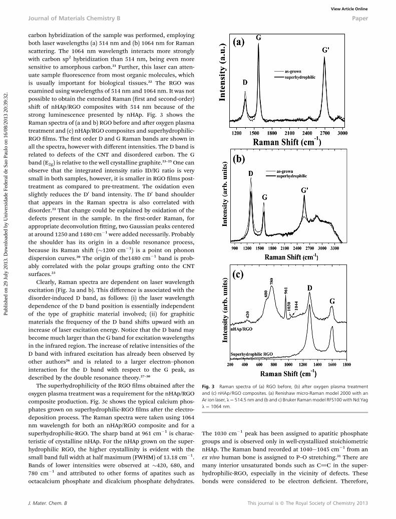
Fig. 3 Raman spectra of (a) RGO before, (b) after oxygen plasma treatmentand (c) nHAp/RGO composites. (a) Renishaw micro-Raman model 2000 with anAr ion laser, l¼ 514.5 nm and (b and c) Bruker Ramanmodel RFS100 with Nd:Yagl ¼ 1064 nm.
Journal of Materials Chemistry B Paper
Publ
ishe
d on
29
July
201
3. D
ownl
oade
d by
Uni
vers
idad
e Fe
dera
l de
Sao
Paul
o on
16/
08/2
013
20:3
9:32
. View Article Online
carbon hybridization of the sample was performed, employingboth laser wavelengths (a) 514 nm and (b) 1064 nm for Ramanscattering. The 1064 nm wavelength interacts more stronglywith carbon sp2 hybridization than 514 nm, being even moresensitive to amorphous carbon.21 Further, this laser can atten-uate sample uorescence from most organic molecules, whichis usually important for biological tissues.22 The RGO wasexamined using wavelengths of 514 nm and 1064 nm. It was notpossible to obtain the extended Raman (rst and second-order)shi of nHAp/RGO composites with 514 nm because of thestrong luminescence presented by nHAp. Fig. 3 shows theRaman spectra of (a and b) RGO before and aer oxygen plasmatreatment and (c) nHAp/RGO composites and superhydrophilic-RGO lms. The rst order D and G Raman bands are shown inall the spectra, however with different intensities. The D band isrelated to defects of the CNT and disordered carbon. The Gband (E2g) is relative to the well crystalline graphite.23–25 One canobserve that the integrated intensity ratio ID/IG ratio is verysmall in both samples, however, it is smaller in RGO lms post-treatment as compared to pre-treatment. The oxidation evenslightly reduces the D0 band intensity. The D0 band shoulderthat appears in the Raman spectra is also correlated withdisorder.23 That change could be explained by oxidation of thedefects present in the sample. In the rst-order Raman, forappropriate deconvolution tting, two Gaussian peaks centeredat around 1250 and 1480 cm�1 were added necessarily. Probablythe shoulder has its origin in a double resonance process,because its Raman shi (�1200 cm�1) is a point on phonondispersion curves.20 The origin of the1480 cm�1 band is prob-ably correlated with the polar groups graing onto the CNTsurfaces.23
Clearly, Raman spectra are dependent on laser wavelengthexcitation (Fig. 3a and b). This difference is associated with thedisorder-induced D band, as follows: (i) the laser wavelengthdependence of the D band position is essentially independentof the type of graphitic material involved; (ii) for graphiticmaterials the frequency of the D band shis upward with anincrease of laser excitation energy. Notice that the D band maybecomemuch larger than the G band for excitation wavelengthsin the infrared region. The increase of relative intensities of theD band with infrared excitation has already been observed byother authors26 and is related to a larger electron–phononinteraction for the D band with respect to the G peak, asdescribed by the double resonance theory.27–30
The superhydrophilicity of the RGO lms obtained aer theoxygen plasma treatment was a requirement for the nHAp/RGOcomposite production. Fig. 3c shows the typical calcium phos-phates grown on superhydrophilic-RGO lms aer the electro-deposition process. The Raman spectra were taken using 1064nm wavelength for both an nHAp/RGO composite and for asuperhydrophilic-RGO. The sharp band at 961 cm�1 is charac-teristic of crystalline nHAp. For the nHAp grown on the super-hydrophilic RGO, the higher crystallinity is evident with thesmall band full width at half maximum (FWHM) of 13.18 cm�1.Bands of lower intensities were observed at �420, 680, and780 cm�1 and attributed to other forms of apatites such asoctacalcium phosphate and dicalcium phosphate dehydrates.
J. Mater. Chem. B
The 1030 cm�1 peak has been assigned to apatitic phosphategroups and is observed only in well-crystallized stoichiometricnHAp. The Raman band recorded at 1040�1045 cm�1 from anex vivo human bone is assigned to P–O stretching.31 There aremany interior unsaturated bonds such as C]C in the super-hydrophilic-RGO, especially in the vicinity of defects. Thesebonds were considered to be electron decient. Therefore,
This journal is ª The Royal Society of Chemistry 2013

Fig. 5 pH and temperature behavior during the nHAp formation on super-hydrophilic-RGO.
Paper Journal of Materials Chemistry B
Publ
ishe
d on
29
July
201
3. D
ownl
oade
d by
Uni
vers
idad
e Fe
dera
l de
Sao
Paul
o on
16/
08/2
013
20:3
9:32
. View Article Online
bonds between Ca2+ and superhydrophilic-RGO defects arealmost impossible. On the other hand, the oxygen atoms of the–C–O–C– and –COO– in the superhydrophilic-RGO possessmany lone pairs of electrons and thus could easily create theionic interaction with Ca2+. Moreover, we have shown in arecent publication that there may exist a hydrogen bond inter-action between the –COO in functionalized composites and–OH in nHAp, which could also enhance the nHAp growth.9
Similar behaviour was observed during the electrodeposition ofnHAp on superhydrophilic-RGO.
Fig. 4 shows the (a) cathodic current density and (b) thecharge density (DQ) accumulation during nHAp crystal forma-tion on the superhydrophilic-RGO. The linear response ofcharging indicates the predominance of a Faradaic reaction.27
The rapid increase of the cathodic-current is related to theelectric double-layer charging when the reaction begins. Thecurrent immediately reaches 3.6 � 0.05 mA cm2 and remainsconstant, because of OH� generation, which has a mass trans-port limitation. This OH� generation is the most importantcontrol parameter for nHAp crystal formation, because theacid–base reactions form (PO4)
3� and (HPO4)2�, which leads to
calcium phosphate precipitation.6–8
Fig. 5 shows the pH and temperature behavior during theelectrodeposition process. The temperature was maintained at374.8 � 0.075 K using a thermostated recirculation water bath.The temperature chosen for the experiment was dened basedon the trial that yielded the best result. The pH was free tochange and it reduced from 5.88 to 5.2. The increase in positiveions in the solution is expected32 due to OH� generation andnHAp formation.33
Fig. 6 shows SEM images of the morphology (a and b) andenergy dispersive spectrum (c) of globular-like nHAp electro-deposited onto superhydrophilic RGO aer 30 min of electro-deposition. The SEM images show that the nHAp crystals arehomogeneously distributed on the superhydrophilic-RGOsurface (Fig. 6a). The nHAp has globular-like and high level ofporosity, essential for bioactivity in bone regeneration (Fig. 6b).The porosity formation could be explained by bubble formationduring the electrodeposition process and the 3D structures ofsuperhydrophilic-RGO.
Fig. 4 The (a) cathodic current density and (b) the charge density (DQ) accu-mulation during nHAp formation on the superhydrophilic-RGO.
This journal is ª The Royal Society of Chemistry 2013
In Fig. 6(c) the elemental composition of the coating wasquantied by mapping the nHAp/RGO composite surface. Weanalyzed the Ca and P content proles (% wt) for each group.The Ca/P ratio determined from the analysis was �1.96 � 3, avalue near that of the stoichiometric nHAp (1.67) present inbone tissue.33
Fig. 7 shows the X-ray diffraction pattern of the nHAp crys-tals grown on superhydrophilic-RGO lms. The diffractionpeaks of the substrate are also shown for comparison. Note thatthe apatite formation is deduced from the presence of severalcharacteristic X-ray reection peaks in the diffraction patternshown. The principal diffraction peaks of nHAp appear at 2qvalues of 25.9� for reection (002); at 31.9� (triplet) for reec-tions (211), (112), and (300); and at 34.0� for reection (200)(JCPDS 9-432).9
The broadening of a diffraction peak can be related to themean crystallite size via the Scherrer equation (t ¼ 0.89l/B cos qB),34 where t is the mean crystallite size, B is the peakline-width at half maximum (in radians), qB is the Braggdiffraction angle, and l is the X-ray wavelength (CuKa radiationin our case).
Fig. 6 (a and b) SEM images of the morphology and (c) energy dispersivespectrum of globular-like nHAp electrodeposited onto superhydrophilic-RGO.Collected from N ¼ 3 and five different points.
J. Mater. Chem. B

Fig. 7 XRD analyses of nHAp/RGO composites. Data compared to nHAp stan-dard powder (JCPDS 9-432). Titanium (substrate) and nHAp crystal planes arespecified.
Journal of Materials Chemistry B Paper
Publ
ishe
d on
29
July
201
3. D
ownl
oade
d by
Uni
vers
idad
e Fe
dera
l de
Sao
Paul
o on
16/
08/2
013
20:3
9:32
. View Article Online
A preference calculated from the XRD data was furtherexplored as a structure indicator.16 The relative intensity of the(002) peak (RI) to the intensities of the three strongest peaks forthe standard powder HA sample (JCPDS 9-432) is dened as:
RI ¼ I(002)/I(211) + I(112) + I(300) (1)
where I(002), I(211), I(112), and I(300) are the XRD peak intensities of(002), (211), (112), and (300) planes respectively. For the stan-dard powder, the RI of the HAp was calculated using JCPDS 9-432, RIs ¼ 0.1818 for the (002) peak. A similar value wasobtained by Hu et al.16 Each peak has its own Ri and RIs. In thiswork, the preference, P, is dened as the relative difference of RIfrom RIs:
P ¼ RI � 1 (2)
Table 1 shows all the data used to calculate the crystallite sizeand growth preference. These data were collected from threedifferent points and samples. The mean was expressed forcomparison.
The HAp has a hexagonal space group of p63/m with a ¼0.9430 nm and c ¼ 0.6891 nm. The O–H groups are ordered onthe c-axis or in the (002) plane. For HAp with a hexagonalstructure, the [001] direction is the usual direction for preferredgrowth, along which the crystal planes are most densely
Table 1 Parameters collected from deconvolution of the X-ray diffractogram ofglobular-like nHAp crystals electrodeposited onto superhydrophilic-RGO films. Allthe crystallite sizes and growth preferences determined for the planes are shown.The JPCDS 9-432 was used for comparison. *Data collected for the HAp standardpowder (JPCD 9-432) for preference growth computation
PlanePosition(2-theta) FWHM
Crystallites(A)
Preferencegrowth, P
(002) 25.98 0.3646 223 2.61(211) 31.90 0.3816 216 0.45(112) 32.33 0.3376 245 0.71(300) 33.03 0.6210 133 0.02
J. Mater. Chem. B
populated with atoms. Calculations identied preferentialgrowth along the (002) plane, and secondly, along the (112)plane. The negative value was attributed to suppression of the(211) and (300) planes due to higher growth preference for the(002) plane (data shown in Table 1). In the case of super-hydrophilic-RGO, the thickness of the globular-like structure isvery close to the crystallite size obtained using the Scherrerformula from the X-ray diffractogram (Table 1). This indicatesthat each globule is a single globular-like nHAp crystal. There-fore, the crystalline (002) planes whose axes are along in the[001] direction grow preferentially.6,7,35 The higher intensity ofthe (002) peak shows a standard nHAp growth pattern onsuperhydrophilic-RGO. The high frequency of these planes maybe related to a globular-like morphology, as evident in Fig. 2band c. Probably, the expression of previous globular-likemorphology of nHAp indicates some selectivity in surfacebinding. Filgueiras et al.,36 using computational modeling,attributed the globular-like morphology of nHAp crystals to thesurface energy. The surface energy of the (100) planes is higherthan that of the (001) planes.36 In other words, the (100) planesare comparatively less stable. The less-stable terminations willbe more reactive, and may quickly accumulate more HApmaterial which may inhibit adsorbates from forming morestable terminations.36 In addition, the (100) planes contain lociof positive charges centered on calcium ions.36
A similar phenomenon of preferred orientation of apatitecoatings on titanium substrates was reported earlier,6,8,30 butthey were obtained only aer thermal annealing around 900 �C.
Nucleation, in general, represents an activation energybarrier to the spontaneous formation of a solid phase from asupersaturated solution. This kinetic constraint may be suffi-cient to offset the thermodynamic driving force for precipita-tion, resulting in metastable solutions, which do not undergophase transformations over a long period of time. The activa-tion energy for nucleation (DGN) is related to g and DGB by theequation:37
DGN ¼ 16pg33(DGB)2 (3)
and
DGB ¼ kTlnSy, (4)
where k is the Boltzmann constant, T is the nucleationtemperature and S is the relative supersaturation of the solu-tion, g is the solid–liquid interfacial energy, y is the molecularvolume, and DGB is the energy released in the formation ofbonds in the bulk of the aggregate. The –COOH groups formedon superhydrophilic RGO lms aer the oxygen plasma treat-ment (Fig. 2) constructed ordered “recognized sites” with highpolarity and charged density, which could draw the directelectrodeposition of the nHAp on them.
Other effects can also be directly attributed to electrodepo-sition of nHAp crystals.38,39 Ban and Maruno38 demonstratedthat the precipitation of calcium phosphate on the cathode isinduced by the super-saturation of calcium phosphate salts dueto the local increase in pH and accumulation of both calciumand phosphate ions. The authors suggested that the needles
This journal is ª The Royal Society of Chemistry 2013

Fig. 8 Schematic illustration of plate-like nHAp crystal formation and dissolutionduring direct electrodeposition on superhydrophilic RGO surfaces based on theSaffar et al. model.48
Paper Journal of Materials Chemistry B
Publ
ishe
d on
29
July
201
3. D
ownl
oade
d by
Uni
vers
idad
e Fe
dera
l de
Sao
Paul
o on
16/
08/2
013
20:3
9:32
. View Article Online
grew on the nuclei and that the number of nuclei decreased as afunction of the electrolyte temperature. These results imply thatthe hydrothermal–electrochemical deposition consists of twoprocesses: nucleation and crystal growth. In this paper, we haveshown that this property was essential for the direct growth ofnHAp crystals on superhydrophilic-RGO.
Ban and Hasegawa39 showed that the surface reaction forHAp crystal growth depends on whether the crystal faces arerough or smooth at the atomic level. The crystal tip formed at100 �C had a smooth, at, and sharp edged apatite, possiblyformed due to the lateral growth mode at this temperature(globular-like nHAp crystals). Aer the bath temperature wasadjusted to 200 �C, apatite grew by both adhesive and lateralgrowth modes, because the tips and edges were round, andsome planes were smooth and at (needle-like nHAp crystals).These authors concluded that both the precipitation and thecrystal growth morphology of apatite strongly depend on theelectrolyte temperature and only slightly on the currentdensity.38,39 In the present work, in addition to investigating theinuence of electrolyte temperature and current density, wehave shown that the growth of globular-like nHAp crystals onsuperhydrophilic-RGO lms is highly inuenced by the nano-topography and wettability properties. The reason for thisaffirmation is a simple comparison between the same electro-deposition process applied using NiTi alloys40 and VAMWCNTlms9 in terms of the temperature and current density. In thiscomparison, the nHAp crystal growth is only possible on NiTialloys aer roughening of the surface and thermal treatment.Aer roughening treatment, the NiTi alloys have a circularmorphology, which results in nHAp crystals with an acicularmorphology. In the VAMWCNT lms, plate-like crystals areobserved aer only thirty minutes without thermal treatment.9
These crystals are different from the globular nHAp crystals thatare obtained on the superhydrophilic-RGO lms presentedhere. Aer these considerations were all taken into account, theproposed model of globular-like nHAp growth presented here ishighly inuenced by the nanotopology, current density, andmost of all by the carboxylic groups directly attached to thesuperhydrophilic-RGO lms aer the oxygen plasma treatment.
The mechanism for nHAp crystal formation on super-hydrophilic-RGO lms is proposed and discussed earlier.8,9
Manso et al. showed that the difference between depositionsobtained with electrical activation as compared to depositionwithout it demonstrates that the crystallization process at theinterface is encouraged by electrical activation.6 In this case, thedefects on the superhydrophilic-RGOs can be considered asactive sites for nHAp crystal nucleation, and subsequently tolm formation.
Three hypotheses about the HAp electrodeposition processhave been proposed by other authors.8,41,42 The rst one is thatno charge evolution has been reported for nHAp crystals inbasic media,43 which excludes electrostatic forces from beingresponsible for a hypothetical indirect deposition. The secondone is attributed to adherence of the coatings obtained at lowvoltages and high temperature, which is related to the diffusionof Ca2+ and especially P species through the Ti substrate.44
The nHAp coatings obtained by indirect deposition at higher
This journal is ª The Royal Society of Chemistry 2013
voltages with sequential sintering temperatures also producednHAp lms with good adhesion properties. The third one statesthat the low voltages applied in our experiment lead to currentdensities comparable to those used by other authors in acidicelectrolytes. However, longer times for indirect deposition havebeen reported.8
Our work presented a fast method to obtain globular-likenHAp crystals on superhydrophilic-RGO lms. From this, thepH increase at the interface does not change the nucleation andpromotes direct nHAp deposition44 on the surface of thesuperhydrophilic RGO lms. In this case, the electrostaticattraction of OH� triggers the precipitation of nHAp nuclei atthe interface and leads to direct deposition. Previous investi-gations of the nucleation sites of nHAp crystals on functional-ized collagen bers have suggested that the binding of calciumions on the negatively charged carboxylate groups of collagen isone of the key factors for initiating the nucleation of HApcrystals.45
Fig. 8 illustrates the establishment of ionic interactionbetween carboxylate groups and calcium ions, which are beingelectrodeposited onto the HAp solid structure during electro-deposition of plate-like nHAp crystals on superhydrophilic RGOlms. The atomic Ca/P ratio of calcium phosphates directlyelectrodeposited onto superhydrophilic RGO lms was �1.96from the EDS analysis (data obtained aer the quantitativeanalysis of Fig. 6), slightly higher than that of stoichiometric HA(1.67). It has been suggested that the carboxylate groups facili-tate the initial electrodeposition of calcium ions, and theattraction of calcium ions is an important initial step in calciumphosphates formation46 and the size of nHAp crystals is relatedto the available nucleation sites.47 The large amount of nucleifacilitates the homogeneous nucleation during electrodeposi-tion, leading to precipitation and formation of apatite on thesuperhydrophilic RGO lms. It is possible to revise the mech-anism reported by Saffar et al.48 and the revised one can beproposed as presented in Fig. 8.
This mechanism is believed to be responsible for the initi-ation of nHAp precipitation on the superhydrophilic-RGOsurface.
Fig. 9 shows the SEM images of murine MSC adhered tonHAp/RGO composite surfaces aer 48 h of culture. The MSCsused in this study were obtained from adult animals and havethe ability to differentiate into a variety of cell types, such as
J. Mater. Chem. B

Fig. 9 (a–e) SEM image of the murine MSC adhesion on nHAp/RGO compositesafter 48 h.
Journal of Materials Chemistry B Paper
Publ
ishe
d on
29
July
201
3. D
ownl
oade
d by
Uni
vers
idad
e Fe
dera
l de
Sao
Paul
o on
16/
08/2
013
20:3
9:32
. View Article Online
osteoblasts49 and osteocytes.50 The cells spread with no prefer-ential direction, acquiring a at (9d) roughly circular (9a and b)form over the surface. A healthy cell pattern is observed on thenHAp/RGO composites showing that the cells present activeformation of membrane projections all over the cell surface(Fig. 9c). The cells occupy a considerable area of nHAp/RGOcomposite surface. The cell attachment is very similar to whatwas observed in another work of our group, where humanosteoblast cells were able to promote adhesion on a hydroxy-apatite/superhydrophilic vertically aligned multi-walled carbonnanotube composite.9 Further in vitro and in vivo assays areunder development to better characterize nHAp/RGO compos-ites bioactivity.
4 Conclusions
The great novelty of the current paper is the direct growth ofcrystalline nHAp on superhydrophilic-RGO, without anythermal treatment, in only thirty minutes. In addition, we pre-sented a mechanism for the preferential growth of globularnHAp crystals directly electrodeposited onto superhydrophilic-RGO lms. The globular nHAp crystal formation is due to the–COOH terminations that were attached to superhydrophilic-RGO tips aer oxygen plasma etching. The deprotonation ofcarboxylic terminations resulted in preferential sites to nucleate(2D) nHAp crystals and promote further growth. A preferentialgrowth of globular nHAp crystals in the (002) plane wasobserved. This new method of nHAp formation may providean alternative way to design and prepare bioactive nano-materials with improved mechanical properties and tailored
J. Mater. Chem. B
microstructure and macrodimensional control. Also thenHAp/RGO composite showed excellent biocompatibility sinceMSCs were able to adhere and proliferate on the surface of thecomposite aer 48 h of culture.
Acknowledgements
The authors are very grateful to the Fundaç~ao de Amparo a Pes-quisa do Estado de S~ao Paulo (2011/17877-7) and (2011/20345-7),for nancial support. Special thanks to Priscila Leite and EricaFreire Antunes for scanning electron microscopy images. Theauthors are very grateful to our colleagues Alene Alder-Rangeland Nigel Roderick Messmer for the English revision.
Notes and references
1 R. Z. LeGeros, Clin. Orthop. Relat. Res., 2002, 395, 81.2 A. Oyane, K. Onuma, A. Ito, H. M. Kim, T. Kokubo andT. Nakamura, J. Biomed. Mater. Res., Part A, 2003, 64(2), 339.
3 M. Vallet-Regi and J. M. Gonzalez-Calbet, Prog. Solid StateChem., 2004, 32, 1.
4 A. Berman, D. J. Ahn, A. Lio, M. Salmeron, A. Reichert andD. Charych, Science, 1995, 269, 515.
5 G. Balasundaram and T. J. Webster, Nanomedicine, 2006, 1,169.
6 M. Manso, C. Jimenez, C. Morant, P. Herrero andJ. M. Martinez-Duart, Biomaterials, 2000, 21, 1755.
7 N. Eliaz, W. Kopelovitch, L. Burstein, E. Kobayashi andT. Hanawa, J. Biomed. Mater. Res., Part A, 2009, 89, 270.
8 N. Eliaz and M. Eliyahu, J. Biomed. Mater. Res., Part A, 2007,80, 621.
9 A. O. Lobo, M. A. F. Corat, S. C. Ramos, J. T. Matsushima,A. E. C. Granato, C. Pacheco-Soares and E. J. Corat,Langmuir, 2010, 26, 18308.
10 M. Li, Y. Wang, Q. Liu, Q. Li, Y. Cheng, Y. Zheng, et al.,J. Mater. Chem. B, 2013, 1, 475.
11 P. A. A. P. Marques, G. Goncalves, M. K. Singh and J. Gracio,J. Nanosci. Nanotechnol., 2012, 12, 6686.
12 H. Liu, P. Xi, G. Xie, Y. Shi, F. Hou, L. Huang, et al., J. Phys.Chem. C, 2012, 116, 3334.
13 G. M. Neelgund, A. Okia and Z. Luo, Mater. Res. Bull., 2013,48, 175.
14 F. R. Marciano, J. S. Marcuzzo, L. F. Bonetti, E. J. Corat andV. J. Trava-Airoldi, Surf. Coat. Technol., 2009, 204, 64.
15 M. Wojdyr, J. Appl. Crystallogr., 2010, 43, 1126.16 R. Hu, C. Lin, H. Sh and H. Wang, Mater. Chem. Phys., 2009,
115, 718.17 L. S. Meirelles and N. B. Nardi, Murine marrow-derived
mesenchymal stem cell: isolation, in vitro expansion, andcharacterization, Br. J. Haematol., 2003, 123, 702.
18 J. Shen, M. Shi, N. Li, B. Yan, H. Ma, Y. Hu, et al., Nano Res.,2010, 3, 339.
19 O. C. Compton, B. Jain, D. A. Dikin, A. Abouimrane,K. Amine and S. T. Nguyen, ACS Nano, 2011, 5, 4380.
20 M. J. Fernandez-Merino, L. Guardia, J. I. Paredes, S. Villar-Rodil, P. Solis-Fernandez, A. Martinez-Alonso, et al., J. Phys.Chem. C, 2010, 114, 6426.
This journal is ª The Royal Society of Chemistry 2013

Paper Journal of Materials Chemistry B
Publ
ishe
d on
29
July
201
3. D
ownl
oade
d by
Uni
vers
idad
e Fe
dera
l de
Sao
Paul
o on
16/
08/2
013
20:3
9:32
. View Article Online
21 E. F. Antunes, A. O. Lobo, E. J. Corat, V. J. Trava-Airoldi,A. A. Martin and C. Verissimo, Carbon, 2006, 44, 2202.
22 E. B. Hanlon, R. Manoharan, T. W. Koo, K. E. Shafer,J. T.Motz,M. Fitzmaurice, et al., Phys.Med. Biol., 2000, 45, R1.
23 J. Tsukada, H. Zanin, L. C. A. Barbosa, G. A. da Silva,H. J. Ceragioli, A. C. Peterlevitz, et al., J. Electrochem. Soc.,2012, 159, D59.
24 H. Zanin, A. C. Peterlevitz, H. J. Ceragioli, A. A. Rodrigues,W. D. Belangero and V. Baranauskas, Mater. Sci. Eng., C,2012, 32, 2340.
25 H. G. Zanin, A. C. Peterlevitz, R. F. Teolo, H. J. Ceragioli andV. Baranauskas, Ferroelectrics, 2012, 436, 96.
26 S. Choi, K. H. Park, S. Lee and K. H. Koh, J. Appl. Phys., 2002,92, 4007.
27 S. Reich and C. Thomsem, Philos. Trans. R. Soc., A, 2004, 362,2271.
28 R. P. Vidano, D. B. Fischbach, L. J. Willis and T. M. Loehr,Solid State Commun., 1981, 39, 341.
29 I. Pocsik, M. Hundhausen, M. Koos and L. Ley, J. Non-Cryst.Solids, 1998, 230, 1083.
30 A. K. Sood, R. Gupta and S. Asher, J. Appl. Phys., 2001, 90,4494.
31 S. Kale, S. Biermann, C. Edwards, C. Tarnowski, M. Morrisand M. W. Long, Nat. Biotechnol., 2000, 18, 954.
32 N. Eliaz, Isr. J. Chem., 2008, 48, 159.33 A. O. Lobo, F. R. Marciano, I. Regiani, S. C. Ramos,
J. T. Matsushima and E. J. Corat, Theor. Chem. Acc., 2011,130, 1071.
34 A. Patterson, Phys. Rev., 1939, 56, 978.35 T. V. Vijayaraghavan and A. Bensalem, J. Mater. Sci. Lett.,
1994, 13, 1782.
This journal is ª The Royal Society of Chemistry 2013
36 M. R. T. Filgueiras, D. Mkhon and N. H. de Leeuw, J. Cryst.Growth, 2006, 294, 60.
37 K. Zhang and L. Zhang, Science and Technology ofCrystal Growth, Press: Science Publication, 1997, p. 89 inChinese.
38 S. Ban and S. Maruno, Jpn. J. Appl. Phys., 1993, 32, L1577.39 S. Ban and Hasegawa, Biomaterials, 2002, 23, 2965.40 A. O. Lobo, J. Otubo, J. T. Matsushima and E. J. Corat,
J. Mater. Eng. Perform., 2011, 20, 793.41 P. Patnaik, Handbook of inorganic chemicals, McGraw-Hill,
2003, ISBN:0070494398.42 S. Pezzatini, R. Solito, L. Morbidelli, S. Lamponi, E. Boanini,
A. Bigi, et al., J. Biomed. Mater. Res., Part B, 2006, 76, 656.43 R. Damodaran and B. M. Moudgil, Colloids Surf., A, 1993, 80,
191.44 F. Barrere, M. M. E. Snel, A. Blitterswijka, G. de Klaas and
P. Layrolle, Biomaterials, 2004, 25, 2901.45 M. Kikuchi, S. Itoh, S. Ichinose, K. Shinomiya and J. Tanaka,
Biomaterials, 2001, 22, 1705.46 M. Kawashita, M. Nakao, M. Minoda, H. Kim, T. Beppu,
T. Miyamoto, et al., Biomaterials, 2003, 24, 2477.47 G. Toworfe, R. Composto, I. Shapiro and P. Ducheyne,
Biomaterials, 2006, 27, 631.48 K. P. Saffar, A. R. Arshi, N. Jamilpour, A. R. Naja, G. Rouhi
and L. Sudak, J. Biomed. Mater. Res., Part A, 2010, 94,594.
49 M. F. Pittenger, A. M. Mackay, S. C. Beck, R. K. Jaiswal,R. Douglas, J. D. Mosca, M. A. Moorman, D. W. Simonetti,S. Craig and D. R. Marshak, Science, 1999, 284, 143.
50 L. Mcfarlin, X. Gao and Y. B. Liu, Wound Repair andRegeneration, 2006, 14, 471.
J. Mater. Chem. B