Expression of the Mycobacterium tubercuolosis PPE37 protein in Mycobacterium smegmatis induces low...
10
Expression of the Mycobacterium tuberculosis PPE37 protein in Mycobacterium smegmatis induces low tumour necrosis factor alpha and interleukin 6 production in murine macrophages Sylvia Daim, Ikuo Kawamura, Kohsuke Tsuchiya, Hideki Hara, Takeshi Kurenuma, Yanna Shen, Sita R. Dewamitta, Shunsuke Sakai, Takamasa Nomura, Huixin Qu and Masao Mitsuyama Correspondence Ikuo Kawamura [email protected] u.ac.jp Received 9 September 2010 Accepted 10 January 2011 Department of Microbiology, Kyoto University Graduate School of Medicine, Yoshida Konoe-cho, Sakyo-ku, Kyoto 606-8501, Japan PPE37 is a member of the Mycobacterium tuberculosis proline-proline-glutamic acid (PPE) multigene family. Its expression is upregulated in bacteria that are phagocytosed by macrophages and is enhanced even more in bacteria isolated from the lungs of infected mice. This raises the possibility that PPE37 may play a role in the virulence of M. tuberculosis and led to this investigation of the function of PPE37. Recombinant bacterial strains, one expressing the M. tuberculosis PPE37 protein (Ms_ppe37) and another harbouring the vector alone (Ms_vec) were generated from the non-pathogenic Mycobacterium smegmatis. These bacterial strains were used to infect peritoneal exudate and bone marrow-derived macrophages. It was found that, despite the comparable intracellular survival between the two recombinant M. smegmatis strains, Ms_ppe37 induced a significantly lower level of tumour necrosis factor alpha and interleukin 6 in the infected macrophages compared with Ms_vec. Western blot analyses revealed that the activation levels of nuclear factor kappa B, mitogen-activated protein kinase (MAPK)/extracellular signal-regulated kinase and MAPK/p38 were lower in macrophages infected with Ms_ppe37 than in macrophages infected with Ms_vec. These results suggest that PPE37 may have a potential role in interfering with the pro-inflammatory cytokine response of infected macrophages. INTRODUCTION The existence of the proline-proline-glutamic acid (PPE) multigene family was revealed when the decoding of the Mycobacterium tuberculosis genome was completed (Cole et al., 1998). Members of this gene family were found to share a highly conserved N-terminal sequence of approxi- mately 180 aa that also contained the conserved PPE motif. In contrast, their C-terminal regions were highly heteroge- neous in both sequence and length (Cole et al., 1998). The ppe genes are not found outside the genus Mycobacterium and are highly distributed among the pathogenic species of mycobacteria (Gey van Pittius et al., 2006). For these reasons, they are speculated to contribute to the patho- genicity of M. tuberculosis. A possible functional role has been proposed for the PPE proteins, in which they serve as a source of antigenic variation that promotes antigenic diversity in M. tuberculosis (Cole et al., 1998). Indeed, the PPE proteins reported to date are immunogenic, eliciting either humoral or T-cell immune responses (Choudhary et al., 2003; Khan et al., 2008; Romano et al., 2008; Tundup et al., 2008; Wang et al., 2008). One of the PPE proteins, PPE37, may have a role in the virulence of M. tuberculosis. It has been reported that expression of the ppe37 gene is upregulated in M. tuberculosis during infection of murine bone marrow- derived macrophages (Schnappinger et al., 2003; Voskuil et al., 2004). Moreover, in M. tuberculosis isolated from the lungs of infected mice, expression of the ppe37 gene is enhanced even more (Schnappinger et al., 2003). Earlier studies have shown that an iron-dependent transcriptional regulator that is critical for proper iron homeostasis in M. tuberculosis regulates the expression of ppe37 (Rodriguez et al., 1999, 2002). When M. tuberculosis was exposed to iron-limiting conditions in in vitro culture, the expression of ppe37 increased greatly (Rodriguez et al., 2002; Schnappinger et al., 2003). In addition, in vitro exposure of M. tuberculosis to nitrosative and oxidative growth conditions also increased the expression of ppe37 (Schnappinger et al., 2003; Voskuil et al., 2004). Both of Abbreviations: ERK, extracellular signal-regulated kinase; IL, interleukin; LDH, lactate dehydrogenase; MAPK, mitogen-activated protein kinase; MHC, major histocompatibility complex; NF-kB, nuclear factor kappa B; TNF-a, tumour necrosis factor alpha; TLR, Toll-like receptor. Journal of Medical Microbiology (2011), 60, 582–591 DOI 10.1099/jmm.0.026047-0 582 026047 G 2011 SGM Printed in Great Britain
Transcript of Expression of the Mycobacterium tubercuolosis PPE37 protein in Mycobacterium smegmatis induces low...
Expression of the Mycobacterium tuberculosisPPE37 protein in Mycobacterium smegmatisinduces low tumour necrosis factor alpha andinterleukin 6 production in murine macrophages
Sylvia Daim, Ikuo Kawamura, Kohsuke Tsuchiya, Hideki Hara,Takeshi Kurenuma, Yanna Shen, Sita R. Dewamitta, Shunsuke Sakai,Takamasa Nomura, Huixin Qu and Masao Mitsuyama
Correspondence
Ikuo Kawamura
u.ac.jp
Received 9 September 2010
Accepted 10 January 2011
Department of Microbiology, Kyoto University Graduate School of Medicine, Yoshida Konoe-cho,Sakyo-ku, Kyoto 606-8501, Japan
PPE37 is a member of the Mycobacterium tuberculosis proline-proline-glutamic acid (PPE)
multigene family. Its expression is upregulated in bacteria that are phagocytosed by macrophages
and is enhanced even more in bacteria isolated from the lungs of infected mice. This raises the
possibility that PPE37 may play a role in the virulence of M. tuberculosis and led to this
investigation of the function of PPE37. Recombinant bacterial strains, one expressing the M.
tuberculosis PPE37 protein (Ms_ppe37) and another harbouring the vector alone (Ms_vec) were
generated from the non-pathogenic Mycobacterium smegmatis. These bacterial strains were
used to infect peritoneal exudate and bone marrow-derived macrophages. It was found that,
despite the comparable intracellular survival between the two recombinant M. smegmatis strains,
Ms_ppe37 induced a significantly lower level of tumour necrosis factor alpha and interleukin 6 in
the infected macrophages compared with Ms_vec. Western blot analyses revealed that the
activation levels of nuclear factor kappa B, mitogen-activated protein kinase (MAPK)/extracellular
signal-regulated kinase and MAPK/p38 were lower in macrophages infected with Ms_ppe37 than
in macrophages infected with Ms_vec. These results suggest that PPE37 may have a potential
role in interfering with the pro-inflammatory cytokine response of infected macrophages.
INTRODUCTION
The existence of the proline-proline-glutamic acid (PPE)multigene family was revealed when the decoding of theMycobacterium tuberculosis genome was completed (Coleet al., 1998). Members of this gene family were found toshare a highly conserved N-terminal sequence of approxi-mately 180 aa that also contained the conserved PPE motif.In contrast, their C-terminal regions were highly heteroge-neous in both sequence and length (Cole et al., 1998). Theppe genes are not found outside the genus Mycobacteriumand are highly distributed among the pathogenic species ofmycobacteria (Gey van Pittius et al., 2006). For thesereasons, they are speculated to contribute to the patho-genicity of M. tuberculosis. A possible functional role hasbeen proposed for the PPE proteins, in which they serve asa source of antigenic variation that promotes antigenicdiversity in M. tuberculosis (Cole et al., 1998). Indeed, the
PPE proteins reported to date are immunogenic, elicitingeither humoral or T-cell immune responses (Choudharyet al., 2003; Khan et al., 2008; Romano et al., 2008; Tundupet al., 2008; Wang et al., 2008).
One of the PPE proteins, PPE37, may have a role in thevirulence of M. tuberculosis. It has been reported thatexpression of the ppe37 gene is upregulated in M.tuberculosis during infection of murine bone marrow-derived macrophages (Schnappinger et al., 2003; Voskuilet al., 2004). Moreover, in M. tuberculosis isolated from thelungs of infected mice, expression of the ppe37 gene isenhanced even more (Schnappinger et al., 2003). Earlierstudies have shown that an iron-dependent transcriptionalregulator that is critical for proper iron homeostasis in M.tuberculosis regulates the expression of ppe37 (Rodriguezet al., 1999, 2002). When M. tuberculosis was exposed toiron-limiting conditions in in vitro culture, the expressionof ppe37 increased greatly (Rodriguez et al., 2002;Schnappinger et al., 2003). In addition, in vitro exposureof M. tuberculosis to nitrosative and oxidative growthconditions also increased the expression of ppe37(Schnappinger et al., 2003; Voskuil et al., 2004). Both of
Abbreviations: ERK, extracellular signal-regulated kinase; IL, interleukin;LDH, lactate dehydrogenase; MAPK, mitogen-activated protein kinase;MHC, major histocompatibility complex; NF-kB, nuclear factor kappa B;TNF-a, tumour necrosis factor alpha; TLR, Toll-like receptor.
Journal of Medical Microbiology (2011), 60, 582–591 DOI 10.1099/jmm.0.026047-0
582 026047 G 2011 SGM Printed in Great Britain
these conditions are reported to mimic the macrophagephagosomal environment that contains M. tuberculosis(Schnappinger et al., 2003). From these results, it seemsthat PPE37 is required for the adaptation of M. tuberculosisto the intracellular niche in macrophages.
In the present study, as a first step towards evaluating thepossible role of PPE37 in the virulence of M. tuberculosis,we took advantage of the lack of ppe genes inMycobacterium smegmatis and generated two recombinantbacterial strains using this non-pathogenic bacterium.Unlike the M. tuberculosis genome, which contains 69 ppeORFs, the M. smegmatis genome contains only 2.Furthermore, none of the M. smegmatis ppe genes areorthologues of the M. tuberculosis ppe37 gene (Gey vanPittius et al., 2006). We cloned the ppe37 gene from theM. tuberculosis strain H37Rv and expressed the gene inM. smegmatis strain mc2 155 (Ms_ppe37). The ability ofMs_ppe37 to survive inside macrophages was assessed in invitro infection of mouse macrophages. In addition, theeffect of PPE37 on the macrophage cytokine response wasalso investigated.
METHODS
Bacterial strains and growth conditions. Escherichia coli DH5a
was routinely grown in Lennox LB medium for use in DNA cloningprocedures. M. tuberculosis strain H37Rv and M. smegmatis strain mc2
155 were grown at 37 uC in Middlebrook 7H9 liquid medium or onMiddlebrook 7H10 agar (Difco) supplemented with 0.5 % (w/v)albumin fraction V, 0.2 % (w/v) glucose, 0.5 % (v/v) glycerol and0.05 % (v/v) Tween 80. For the preparation of culture filtrate fraction,recombinant M. smegmatis Ms_ppe37 was grown in Sauton mediumas described by Rosenkrands & Andersen (2001). When required,25 mg kanamycin ml21 was also added.
Macrophages. C57BL/6 female mice of 7 to 9 weeks old (Japan SLC)were used in experiments according to protocols approved by theAnimal Ethics and Research Committee of Kyoto University GraduateSchool of Medicine. Peritoneal exudate cells were harvested frommice 3–4 days after intraperitoneal injection with 2.5 ml 3 % (w/v)thioglycollate (Eiken Chemical). Cells were washed, seeded in tissueculture plates and cultured for 2 h in 5 % CO2 at 37 uC in RPMI 1640supplemented with 10 % (v/v) heat-inactivated fetal bovine serum.Adherent cells were used as peritoneal exudate macrophages after
non-adherent cells had been removed by washing. In otherexperiments, bone marrow cells were collected from the tibiae ofmice. Cells were cultured for 5–7 days in RPMI 1640 supplementedwith 10 % heat-inactivated fetal bovine serum, 20 mg gentamicin ml21
and 100 ng mouse macrophage colony-stimulating factor ml21 (R&DSystems). After removal of non-adherent cells, adherent cells wereused as bone marrow-derived macrophages.
Generation of recombinant M. smegmatis expressing PPE37.Chromosomal DNA was isolated from M. tuberculosis, and the ppe37gene was PCR-amplified with the use of forward primer 59-TTACTAGTcaccatcacACCTTCCCGAT-39 containing an SpeI site(underlined) and three His codons (lower-case letters), and reverseprimer 59-CCGTAAGCTTCTTCAACGTTTAATCTGACC-39 con-taining a HindIII site (underlined). The PCR product of approxi-mately 1.5 kb was cloned into the pEGFP vector (ClontechLaboratories), generating the recombinant plasmid pEGFP-his3ppe37. Three additional His codons were introduced at the 59
end of the his3ppe37 sequence with the use of the same reverse primerand a second forward primer, 59-TGAATTCATGcatcaccatcaccatc-acACC-39, containing an EcoRI site (underlined) and the His codons(lower-case letters). The resultant PCR product containing sixconsecutive His codons was inserted in frame into the cloning siteof pMV261, a mycobacterial expression vector (Stover et al., 1991),generating pMV261-his6ppe37. The recombinant plasmid or emptypMV261 was electroporated into M. smegmatis mc2 155 according tostandard procedures (Larsen, 2000). Recombinant M. smegmatisexpressing 6His-tagged PPE37 (Ms_ppe37) and M. smegmatisharbouring empty pMV261 alone (Ms_vec) were selected on Middle-brook 7H10 agar containing 25 mg kanamycin ml21.
Detection of ppe37 gene expression in recombinant M.
smegmatis. Recombinant M. smegmatis strains were cultured untilthey reached an OD600 of 0.6–1.0 in 100 ml Middlebrook 7H9 liquidmedium in the presence of 25 mg kanamycin ml21. Total bacterialRNA was isolated using Sepasol RNA I Super (Nacalai Tesque). AllRNA samples were treated with DNase I (Promega) and subjected toPCR to test for the complete removal of genomic DNA. cDNA wassynthesized from 1 mg total RNA in a 40 ml reaction mix containingreverse transcriptase buffer, 150 ng random primers, 2 ml 10 mMdNTP mix, 2 ml 0.1 M DTT and 400 U SuperScript III reversetranscriptase (Invitrogen). PCR was performed with a KOD-Plusenzyme kit (Toyobo) and the following primer pairs: (i) ppe37 gene –59-TGTTGGACTGGTTCATCTCG-39 (forward) and 59-CAGTCT-TGTTGCTTTGCTGG-39 (reverse), product size 500 bp; and (ii)aminoglycoside phosphotransferase (aph) gene – 59-AGGTAGC-GTTGCCAATGATG-39 (forward) and 59-CTCACCGAGGCAGTT-CCATA-39 (reverse), product size 540 bp.
Detection of His-tagged PPE37. Recombinant M. smegmatisstrains were cultured to an OD600 of 0.6–1.0 in 25 ml Middlebrook7H9 liquid medium or 50 ml Sauton medium in the presence of25 mg kanamycin ml21. Bacterial pellets were harvested, washed threetimes with ice-cold PBS and resuspended in extraction buffercontaining 20 mM Tris/HCl (pH 6.8), 4 mM EDTA, 0.6 % SDSand protease inhibitor cocktail (Nacalai Tesque). Bacterial cells weredisrupted and supernatants were collected after centrifugation at20 000 g for 20 min at 4 uC. For preparation of the culture filtratefraction, the culture supernatant of bacteria grown in Sauton mediumwas harvested by centrifugation at 2000 g for 15 min at 4 uC. Thesupernatant was filtered through a 0.2 mm syringe filter andconcentrated to approximately 150 ml using a centrifugal filter witha cut-off value of 5 kDa (Millipore). Samples were subjected to SDS-PAGE, and the His-tagged PPE37 protein was detected by Westernblotting and mouse anti-penta-His antibody (Qiagen). Chemilumin-escent images were captured with a luminescent image analyser LAS-4000mini (Fujifilm).
M. smegmatis infection of macrophages. Macrophages wereseeded at 16106 cells per well in 12-well tissue culture plates or at36105 cells per well in 24-well tissue culture plates. Cells wereinfected with Ms_ppe37 or Ms_vec at an m.o.i. of 20. At this m.o.i.,the resulting infection rate was greater than 80 % as estimated inpreliminary infection assays from microscopy evaluation of slidesstained according to the Kinyon method (Chapin & Lauderdale,2007). Four hours after infection, gentamicin was added to give a finalconcentration of 5 mg ml21. At 6, 24 and 48 h after infection,macrophages were washed and lysed in PBS containing 0.1 % (v/v)Triton X-100. Lysates were plated on Middlebrook 7H10 agar platescontaining 25 mg kanamycin ml21 and the number of intracellularbacteria was enumerated.
Assay for lactate dehydrogenase (LDH) release. Culture super-natants were harvested after infection of macrophages with Ms_ppe37or Ms_vec for 6, 24 or 48 h. LDH activity in the culture supernatants
PPE37 impairs the macrophage cytokine response
http://jmm.sgmjournals.org 583
was assayed with an LDH cytotoxicity detection kit (Takara Bio). The
percentage of LDH release was calculated as: percentage relea-
se51006(experimental LDH release2spontaneous LDH release)/
(maximal LDH release2spontaneous LDH release). A value of
maximal LDH release was obtained from culture supernatants of
macrophages that were lysed with 1 % (v/v) Triton X-100.
Assay for cytokine production. Culture supernatants were
harvested after infection of macrophages with Ms_ppe37 or Ms_vec
for 24 h. The concentrations of cytokines in the culture supernatants
were determined using commercially available ELISA kits for tumour
necrosis factor alpha (TNF-a), interleukin 6 (IL-6), IL-1b
(eBioscience) and IL-12p70 (Endogen). In some experiments, after
infection of macrophages with Ms_ppe37 or Ms_vec for 3, 6, 9, 12
and 18 h, total RNA was extracted with a Nucleospin RNA II kit
(Macherey-Nagel). RNA (250 ng) was treated with RNase-free DNase
(Promega) and subsequently reverse transcribed into cDNA using a
SuperScript VILO cDNA synthesis kit (Invitrogen). cDNAs were
diluted tenfold, and a PCR was performed in an equal reaction
volume using a KOD-Plus enzyme kit and the following primer pairs:
(i) tnf gene – 59-CATGAGCACAGAAAGCATGATCCG-39 (forward)
and 59-TCTGGGCCATAGAACTGATGAGAG-39 (reverse), product
size 230 bp; (ii) IL-6 gene – 59-TTCCTCTCTGCAAGAGACT-39
(forward) and 59-TGTATCTCTCTGAAGGACT-39 (reverse), product
size 432 bp; and (iii) Actb gene – 59-TGGAATCCTGTGGCA-
TCCATGAAAC-39 (forward) and 59-TAAAACGCAGCTCAGTAA-
CAGTCCG-39 (reverse), product size 350 bp. Equal volumes of the
PCR mixtures were electrophoresed in 1.5 % agarose gel, and DNA
bands were visualized with ethidium bromide (2 mg ml21) staining.
Flow cytometric analysis. Macrophages were harvested after a 24 h
infection with Ms_ppe37 or Ms_vec and treated with anti-CD16/
CD32 mAb (clone 93; eBioscience) for 10 min. This was followed by a
20 min incubation on ice with one of the following phycoerythrin-
conjugated antibodies against: major histocompatibility complex class
I (MHC-I; clone 28-14-8), MHC-II (clone M5/114.15.2), B7.1 (CD80
clone 16-10A1), B7.2 (CD86, clone GL1) or CD40 (clone 1C10), or
with an isotype control antibody (all from eBioscience). The intensity
of each cell-surface marker was analysed on a FACScalibur flow
cytometer equipped with CellQuest software (BD Biosciences).
Assay for nuclear factor-kappa B (NF-kB), extracellular signal-
regulated kinase (ERK) and p38 phosphorylation. Macrophages
were infected with Ms_ppe37 or Ms_vec for 0.5, 1, 2, 4, 6, 7 or 8 h.
After infection, macrophages were lysed in buffer containing 10 mM
Tris/HCl (pH 6.8), 1 % (v/v) NP-40 and proteinase/phosphatase
inhibitor cocktail (Nacalai Tesque). Cell lysates were harvested and
subjected to SDS-PAGE. Phosphorylated and unphosphorylated
ERK and p38, as well as the phosphorylated p65 subunit of NF-kB,
were detected in lots with specific antibodies (Cell Signaling
Technology). b-Actin was detected with anti-b-actin antibody
(Sigma-Aldrich). Chemiluminescent images were captured with a
luminescent image analyser LAS-4000mini. In another experiment,
macrophages were pre-treated with 20 or 40 mM U0126 (a MEK1/2
inhibitor; Cell Signaling Technology) or with 10 or 20 mM
Calbiochem SB202190 (a p38 inhibitor; EMD Biosciences). On the
basis of preliminary experiments, the inhibitors were used at the
concentrations required to inhibit ERK and p38 activities. One hour
after treatment, the macrophages were infected with Ms_ppe37 or
Ms_vec. The culture supernatants were harvested 24 h after
infection, and ELISA was performed to determine the concentrations
of TNF-a and IL-6.
Statistical analysis. Data were analysed using Student’s two-tailed
t-test. Statistical significance was defined as a P value ,0.05. Error
bars represent SD.
RESULTS
Ms_ppe37 constitutively expressesM. tuberculosis PPE37 protein
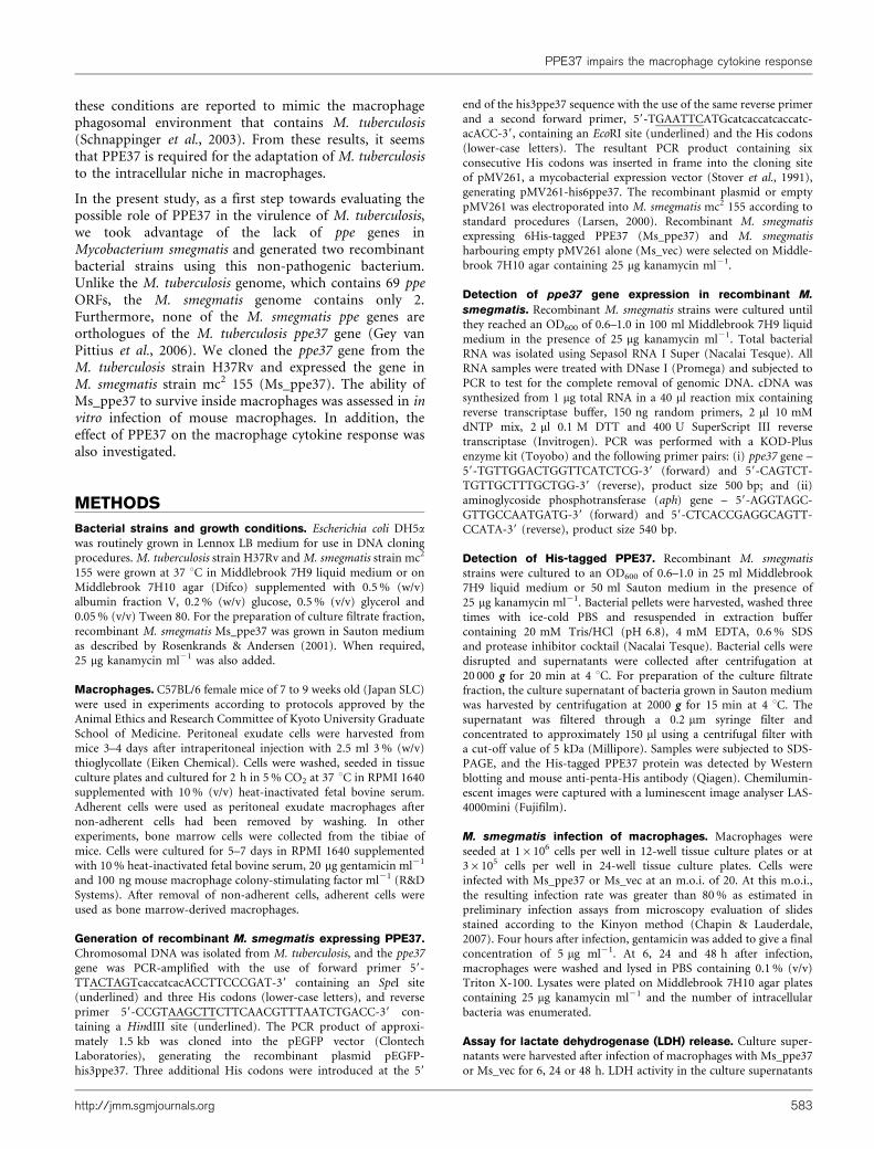
In this study, we generated two recombinant M. smegmatisstrains to investigate the effect of PPE37 on the macro-phage response to bacterial infection. The Ms_ppe37 strainwas engineered to express a 6His-tagged PPE37 proteinfrom a recombinant pMV261 vector, whilst the Ms_vecstrain harboured the vector alone. The pMV261 vectorcontains the kanamycin resistance gene aph for selection oftransformed bacteria (Stover et al., 1991). Both Ms_ppe37and Ms_vec, which were grown in Middlebrook 7H9medium in the presence of kanamycin, expressed the aphgene. However, only Ms_ppe37 was able to express theppe37 gene (Fig. 1a). Furthermore, Western blot analysiswith anti-penta-His antibody detected a protein bandrepresenting PPE37 in the total cell lysate prepared fromMs_ppe37 but not from Ms_vec (Fig. 1b). These resultsconfirmed that the transformation was successful and thatppe37 gene expression was detectable only in M. smegmatisthat had been electroporated with the recombinant vector.In addition, Western blot analysis also revealed that aprotein band representing PPE37 was detectable in thetotal cell lysate but not in the culture filtrate fractionsprepared from Ms_ppe37 grown in Sauton medium (Fig.1c). This was not due to the absence of proteins in theculture filtrate fraction, as Coomassie blue stainingrevealed the presence of many protein bands in both theculture filtrate and the cell lysate fractions. From the resultin Fig. 1(c), it could be suggested that PPE37 is not asecretory protein. We also compared the growth kinetics ofMs_ppe37 and Ms_vec in Middlebrook 7H9 medium, asexcess production of recombinant protein is known toexert a metabolic burden on recombinant bacteria,sometimes reducing the growth of these cells (Bentleyet al., 1990). We observed no marked difference in thegrowth kinetics (Fig. 1d), indicating that expression of theppe37 gene did not influence the growth of Ms_ppe37.
PPE37 does not contribute to the intracellularsurvival of M. smegmatis in macrophages
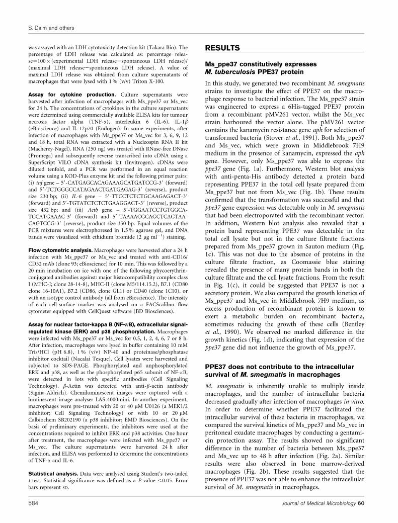
M. smegmatis is inherently unable to multiply insidemacrophages, and the number of intracellular bacteriadecreased gradually after infection of macrophages in vitro.In order to determine whether PPE37 facilitated theintracellular survival of these bacteria in macrophages, wecompared the survival kinetics of Ms_ppe37 and Ms_vec inperitoneal exudate macrophages by conducting a gentami-cin protection assay. The results showed no significantdifference in the number of bacteria between Ms_ppe37and Ms_vec up to 48 h after infection (Fig. 2a). Similarresults were also observed in bone marrow-derivedmacrophages (Fig. 2b). These results suggested that thepresence of PPE37 was not able to enhance the intracellularsurvival of M. smegmatis in macrophages.
S. Daim and others
584 Journal of Medical Microbiology 60
PPE37 does not affect macrophage cell deathduring infection with M. smegmatis
One of the consequences of infecting host cells withM. tuberculosis is cell death, with the possibility thatM. tuberculosis manipulates host-cell death as one of themechanisms of pathogenicity. Recently, emerging newevidence prompted the proposal of a new model on theinteraction between M. tuberculosis and its host cell (Beharet al., 2010). This model suggests that, as a pathogenicstrategy, virulent M. tuberculosis inhibits apoptosis whilstactively inducing necrosis in the infected host cell. Theoutcome of this strategy is a reduction in the efficiency of
cross-presentation of mycobacterial antigens leading to theimpairment in the initiation of T-cell immunity (Beharet al., 2010; Divangahi et al., 2010).
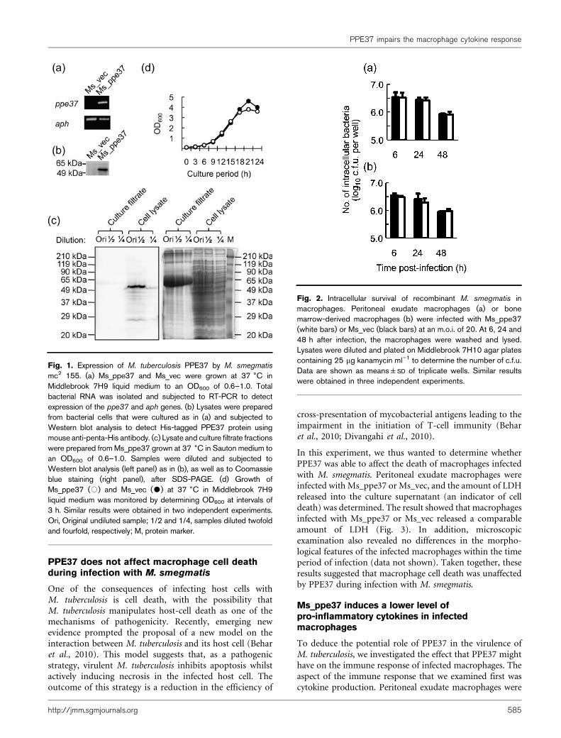
In this experiment, we thus wanted to determine whetherPPE37 was able to affect the death of macrophages infectedwith M. smegmatis. Peritoneal exudate macrophages wereinfected with Ms_ppe37 or Ms_vec, and the amount of LDHreleased into the culture supernatant (an indicator of celldeath) was determined. The result showed that macrophagesinfected with Ms_ppe37 or Ms_vec released a comparableamount of LDH (Fig. 3). In addition, microscopicexamination also revealed no differences in the morpho-logical features of the infected macrophages within the timeperiod of infection (data not shown). Taken together, theseresults suggested that macrophage cell death was unaffectedby PPE37 during infection with M. smegmatis.
Ms_ppe37 induces a lower level ofpro-inflammatory cytokines in infectedmacrophages
To deduce the potential role of PPE37 in the virulence ofM. tuberculosis, we investigated the effect that PPE37 mighthave on the immune response of infected macrophages. Theaspect of the immune response that we examined first wascytokine production. Peritoneal exudate macrophages were
Fig. 1. Expression of M. tuberculosis PPE37 by M. smegmatis
mc2 155. (a) Ms_ppe37 and Ms_vec were grown at 37 6C inMiddlebrook 7H9 liquid medium to an OD600 of 0.6–1.0. Totalbacterial RNA was isolated and subjected to RT-PCR to detectexpression of the ppe37 and aph genes. (b) Lysates were preparedfrom bacterial cells that were cultured as in (a) and subjected toWestern blot analysis to detect His-tagged PPE37 protein usingmouse anti-penta-His antibody. (c) Lysate and culture filtrate fractionswere prepared from Ms_ppe37 grown at 37 6C in Sauton medium toan OD600 of 0.6–1.0. Samples were diluted and subjected toWestern blot analysis (left panel) as in (b), as well as to Coomassieblue staining (right panel), after SDS-PAGE. (d) Growth ofMs_ppe37 (#) and Ms_vec ($) at 37 6C in Middlebrook 7H9liquid medium was monitored by determining OD600 at intervals of3 h. Similar results were obtained in two independent experiments.Ori, Original undiluted sample; 1/2 and 1/4, samples diluted twofoldand fourfold, respectively; M, protein marker.
Fig. 2. Intracellular survival of recombinant M. smegmatis inmacrophages. Peritoneal exudate macrophages (a) or bonemarrow-derived macrophages (b) were infected with Ms_ppe37(white bars) or Ms_vec (black bars) at an m.o.i. of 20. At 6, 24 and48 h after infection, the macrophages were washed and lysed.Lysates were diluted and plated on Middlebrook 7H10 agar platescontaining 25 mg kanamycin ml”1 to determine the number of c.f.u.Data are shown as means±SD of triplicate wells. Similar resultswere obtained in three independent experiments.
PPE37 impairs the macrophage cytokine response
http://jmm.sgmjournals.org 585
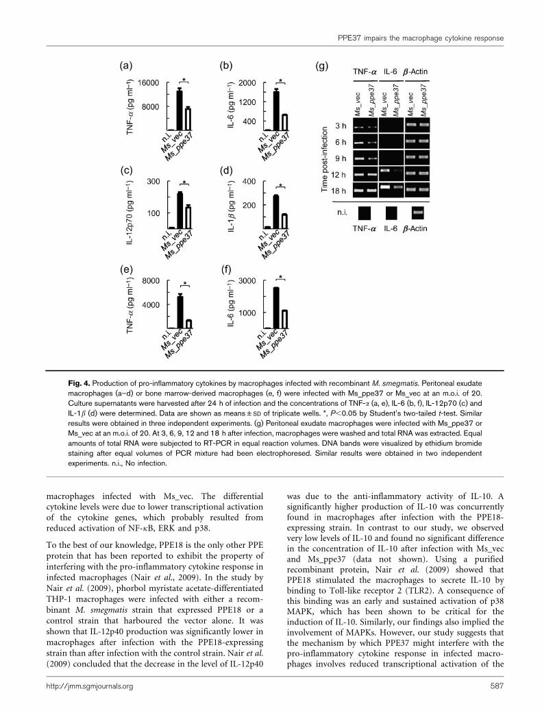
infected with Ms_ppe37 or Ms_vec for 24 h, and theconcentrations of cytokines in the culture supernatants weredetermined. Consistent with the cytokine profiles reportedelsewhere for macrophages infected with M. smegmatis (Lee& Schorey, 2005; Post et al., 2001; Roach & Schorey, 2002),peritoneal exudate macrophages infected with Ms_vec orMs_ppe37 also produced TNF-a, IL-6, IL12p70 and IL-1b
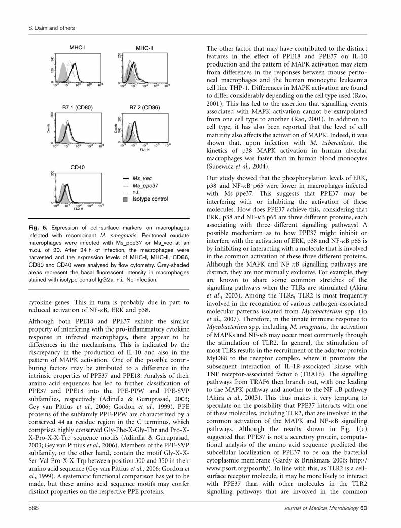
(Fig. 4a–d). However, we discovered that Ms_ppe37 induceda significantly lower level of these cytokines in infectedmacrophages compared with Ms_vec. We repeated theinfection experiment in bone marrow-derived macrophages,and measured the production of TNF-a and IL-6. Similar toinfection in peritoneal exudate macrophages, Ms_ppe37 alsoinduced a significantly lower level of TNF-a and IL-6 in theinfected bone marrow-derived macrophages compared withMs_vec (Fig. 4e, f). RT-PCR analyses showed that theexpression of TNF-a mRNA in macrophages infected withMs_ppe37 was delayed compared with the expression inmacrophages infected with Ms_vec. The difference in theexpression level was observed until 9 h after infection andthereafter became comparable (Fig. 4g). Although expressionof IL-6 mRNA occurred much later, from 12 h afterinfection, the mRNA levels were also lower in macrophagesinfected with Ms_ppe37 than in macrophages infected withMs_vec. However, despite the differential cytokine response,flow cytometric analysis revealed no significant differences inthe expression levels of MHC-I, MHC-II, CD80, CD86 andCD40 between macrophages infected with Ms_ppe37 andmacrophages infected with Ms_vec (Fig. 5). Taken together,these results suggested that PPE37 might possess a functionalproperty that interferes with the pro-inflammatory cytokine
production of macrophages infected with M. smegmatis butdoes not affect the expression of surface markers on thesemacrophages.
PPE37 alters the activation levels of NF-kB, ERKand p38 in macrophages infected withM. smegmatis
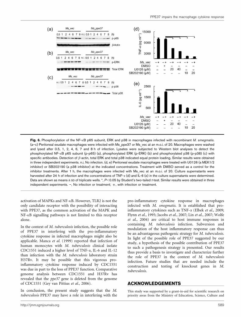
NF-kB is a major transcription factor responsible for theexpression of both TNF-a and IL-6 mRNAs (Collart et al.,1990; Faggioli et al., 2004; Kuprash et al., 1999; Libermann& Baltimore, 1990; Zhang et al., 1994). It has been reportedthat NF-kB activation is needed to induce the expression ofTNF-a and IL-6 mRNAs in macrophages infected with M.smegmatis (Gutierrez et al., 2008). In addition, a previousstudy showed that M. smegmatis infection is also able toinduce phosphorylation of the NF-kB p65 subunit (Lee &Schorey, 2005). The altered TNF-a and IL-6 mRNAexpression shown in Fig. 4(g) therefore raised thepossibility that NF-kB activation might be altered inmacrophages infected with Ms_ppe37. To clarify thispossibility, Western blot analysis was performed and thelevel of phosphorylated NF-kB p65 subunit in the infectedmacrophages was assessed. It was observed that Ms_ppe37induced a relatively lower level of p65 phosphorylation inthe infected macrophages compared with Ms_vec (Fig. 6a).
In addition to NF-kB, it has been shown that the ex-pression of TNF-a mRNA in macrophages infected with M.smegmatis also requires the activation of p38 and ERK (Lee& Schorey, 2005; Roach et al., 2005). Pharmacologicalinhibition experiments confirmed the requirement for ERKand p38 activities in the production of TNF-a and IL-6 inmacrophages infected with Ms_vec (Fig. 6d, e). Theseresults (Figs 4g and 6d, e) thus led us to investigate whetherthe activation of these mitogen-activated protein kinases(MAPKs) might also be affected in macrophages infectedwith Ms_ppe37. Western blot analysis was performed andthe levels of phosphorylated ERK and p38 in the infectedmacrophages were assessed. Fig. 6(b, c) showed thatMs_ppe37 also induced a relatively lower level of ERKand p38 phosphorylation in infected macrophages com-pared with Ms_vec. The lower transcriptional activation ofthe TNF-a and IL-6 genes in macrophages infected withMs_ppe37 therefore might have been due to a lower levelof activation of NF-kB, ERK and p38. Taken together, theseresults further argue for the possibility of PPE37 interferingwith the pro-inflammatory cytokine response of macro-phages infected with M. smegmatis.
DISCUSSION
In the present study, the results suggested the possibilitythat the M. tuberculosis PPE37 protein might interfere withthe pro-inflammatory cytokine response in infectedmacrophages. We found that TNF-a, IL-6, IL-12p70 andIL-1b were produced at significantly lower concentrationsby macrophages infected with Ms_ppe37 compared with
Fig. 3. Assay of cell death in macrophages infected withrecombinant M. smegmatis. Peritoneal exudate macrophages wereinfected with Ms_ppe37 (white bars) or Ms_vec (black bars) at anm.o.i. of 20. At 6, 24 and 48 h after infection, culture supernatantswere harvested. Release of LDH as a measure of macrophage celldeath was estimated by assaying LDH activity in the culturesupernatants. Data are shown as means±SD of triplicate wells.Similar results were obtained in three independent experiments.
S. Daim and others
586 Journal of Medical Microbiology 60
macrophages infected with Ms_vec. The differentialcytokine levels were due to lower transcriptional activationof the cytokine genes, which probably resulted fromreduced activation of NF-kB, ERK and p38.
To the best of our knowledge, PPE18 is the only other PPE
protein that has been reported to exhibit the property of
interfering with the pro-inflammatory cytokine response in
infected macrophages (Nair et al., 2009). In the study by
Nair et al. (2009), phorbol myristate acetate-differentiated
THP-1 macrophages were infected with either a recom-
binant M. smegmatis strain that expressed PPE18 or a
control strain that harboured the vector alone. It was
shown that IL-12p40 production was significantly lower in
macrophages after infection with the PPE18-expressing
strain than after infection with the control strain. Nair et al.
(2009) concluded that the decrease in the level of IL-12p40
was due to the anti-inflammatory activity of IL-10. Asignificantly higher production of IL-10 was concurrentlyfound in macrophages after infection with the PPE18-expressing strain. In contrast to our study, we observedvery low levels of IL-10 and found no significant differencein the concentration of IL-10 after infection with Ms_vecand Ms_ppe37 (data not shown). Using a purifiedrecombinant protein, Nair et al. (2009) showed thatPPE18 stimulated the macrophages to secrete IL-10 bybinding to Toll-like receptor 2 (TLR2). A consequence ofthis binding was an early and sustained activation of p38MAPK, which has been shown to be critical for theinduction of IL-10. Similarly, our findings also implied theinvolvement of MAPKs. However, our study suggests thatthe mechanism by which PPE37 might interfere with thepro-inflammatory cytokine response in infected macro-phages involves reduced transcriptional activation of the
Fig. 4. Production of pro-inflammatory cytokines by macrophages infected with recombinant M. smegmatis. Peritoneal exudatemacrophages (a–d) or bone marrow-derived macrophages (e, f) were infected with Ms_ppe37 or Ms_vec at an m.o.i. of 20.Culture supernatants were harvested after 24 h of infection and the concentrations of TNF-a (a, e), IL-6 (b, f), IL-12p70 (c) andIL-1b (d) were determined. Data are shown as means±SD of triplicate wells. *, P,0.05 by Student’s two-tailed t-test. Similarresults were obtained in three independent experiments. (g) Peritoneal exudate macrophages were infected with Ms_ppe37 orMs_vec at an m.o.i. of 20. At 3, 6, 9, 12 and 18 h after infection, macrophages were washed and total RNA was extracted. Equalamounts of total RNA were subjected to RT-PCR in equal reaction volumes. DNA bands were visualized by ethidium bromidestaining after equal volumes of PCR mixture had been electrophoresed. Similar results were obtained in two independentexperiments. n.i., No infection.
PPE37 impairs the macrophage cytokine response
http://jmm.sgmjournals.org 587
cytokine genes. This in turn is probably due in part toreduced activation of NF-kB, ERK and p38.
Although both PPE18 and PPE37 exhibit the similarproperty of interfering with the pro-inflammatory cytokineresponse in infected macrophages, there appear to bedifferences in the mechanisms. This is indicated by thediscrepancy in the production of IL-10 and also in thepattern of MAPK activation. One of the possible contri-buting factors may be attributed to a difference in theintrinsic properties of PPE37 and PPE18. Analysis of theiramino acid sequences has led to further classification ofPPE37 and PPE18 into the PPE-PPW and PPE-SVPsubfamilies, respectively (Adindla & Guruprasad, 2003;Gey van Pittius et al., 2006; Gordon et al., 1999). PPEproteins of the subfamily PPE-PPW are characterized by aconserved 44 aa residue region in the C terminus, whichcomprises highly conserved Gly-Phe-X-Gly-Thr and Pro-X-X-Pro-X-X-Trp sequence motifs (Adindla & Guruprasad,2003; Gey van Pittius et al., 2006). Members of the PPE-SVPsubfamily, on the other hand, contain the motif Gly-X-X-Ser-Val-Pro-X-X-Trp between position 300 and 350 in theiramino acid sequence (Gey van Pittius et al., 2006; Gordon etal., 1999). A systematic functional comparison has yet to bemade, but these amino acid sequence motifs may conferdistinct properties on the respective PPE proteins.
The other factor that may have contributed to the distinctfeatures in the effect of PPE18 and PPE37 on IL-10production and the pattern of MAPK activation may stemfrom differences in the responses between mouse perito-neal macrophages and the human monocytic leukaemiacell line THP-1. Differences in MAPK activation are foundto differ considerably depending on the cell type used (Rao,2001). This has led to the assertion that signalling eventsassociated with MAPK activation cannot be extrapolatedfrom one cell type to another (Rao, 2001). In addition tocell type, it has also been reported that the level of cellmaturity also affects the activation of MAPK. Indeed, it wasshown that, upon infection with M. tuberculosis, thekinetics of p38 MAPK activation in human alveolarmacrophages was faster than in human blood monocytes(Surewicz et al., 2004).
Our study showed that the phosphorylation levels of ERK,p38 and NF-kB p65 were lower in macrophages infectedwith Ms_ppe37. This suggests that PPE37 may beinterfering with or inhibiting the activation of thesemolecules. How does PPE37 achieve this, considering thatERK, p38 and NF-kB p65 are three different proteins, eachassociating with three different signalling pathways? Apossible mechanism as to how PPE37 might inhibit orinterfere with the activation of ERK, p38 and NF-kB p65 isby inhibiting or interacting with a molecule that is involvedin the common activation of these three different proteins.Although the MAPK and NF-kB signalling pathways aredistinct, they are not mutually exclusive. For example, theyare known to share some common stretches of thesignalling pathways when the TLRs are stimulated (Akiraet al., 2003). Among the TLRs, TLR2 is most frequentlyinvolved in the recognition of various pathogen-associatedmolecular patterns isolated from Mycobacterium spp. (Joet al., 2007). Therefore, in the innate immune response toMycobacterium spp. including M. smegmatis, the activationof MAPKs and NF-kB may occur most commonly throughthe stimulation of TLR2. In general, the stimulation ofmost TLRs results in the recruitment of the adaptor proteinMyD88 to the receptor complex, where it promotes thesubsequent interaction of IL-1R-associated kinase withTNF receptor-associated factor 6 (TRAF6). The signallingpathways from TRAF6 then branch out, with one leadingto the MAPK pathway and another to the NF-kB pathway(Akira et al., 2003). This thus makes it very tempting tospeculate on the possibility that PPE37 interacts with oneof these molecules, including TLR2, that are involved in thecommon activation of the MAPK and NF-kB signallingpathways. Although the results shown in Fig. 1(c)suggested that PPE37 is not a secretory protein, computa-tional analysis of the amino acid sequence predicted thesubcellular localization of PPE37 to be on the bacterialcytoplasmic membrane (Gardy & Brinkman, 2006; http://www.psort.org/psortb/). In line with this, as TLR2 is a cell-surface receptor molecule, it may be more likely to interactwith PPE37 than with other molecules in the TLR2signalling pathways that are involved in the common
Fig. 5. Expression of cell-surface markers on macrophagesinfected with recombinant M. smegmatis. Peritoneal exudatemacrophages were infected with Ms_ppe37 or Ms_vec at anm.o.i. of 20. After 24 h of infection, the macrophages wereharvested and the expression levels of MHC-I, MHC-II, CD86,CD80 and CD40 were analysed by flow cytometry. Grey-shadedareas represent the basal fluorescent intensity in macrophagesstained with isotype control IgG2a. n.i., No infection.
S. Daim and others
588 Journal of Medical Microbiology 60
activation of MAPKs and NF-kB. However, TLR2 is not theonly candidate receptor with the possibility of interactingwith PPE37, as the common activation of the MAPK andNF-kB signalling pathways is not limited to this receptoralone.
In the context of M. tuberculosis infection, the possible roleof PPE37 in interfering with the pro-inflammatorycytokine response in infected macrophages might also beapplicable. Manca et al. (1999) reported that infection ofhuman monocytes with M. tuberculosis clinical isolateCDC1551 induced a higher level of TNF-a, IL-6 and IL-12than infection with the M. tuberculosis laboratory strainH37Rv. It may be possible that this vigorous pro-inflammatory cytokine response induced by CDC1551was due in part to the loss of PPE37 function. Comparativegenome analysis between CDC1551 and H37Rv hasrevealed that the ppe37 gene is deleted from the genomeof CDC1551 (Gey van Pittius et al., 2006).
In conclusion, the present study suggests that the M.tuberculosis PPE37 may have a role in interfering with the
pro-inflammatory cytokine response in macrophagesinfected with M. smegmatis. It is established that pro-inflammatory cytokines such as TNF-a (Elbek et al., 2009;Flynn et al., 1995; Jacobs et al., 2007; Lin et al., 2007; Wolfeet al., 2004) are critical to host immune responses incontaining M. tuberculosis infection. Subversion andmodulation of the host inflammatory response can thusbe an advantageous pathogenic strategy for M. tuberculosis.In light of the possible role of PPE37 suggested by ourstudy, a hypothesis of the possible contribution of PPE37to such a pathogenesis strategy is presented. Our resultsthus provide a basis to investigate and characterize furtherthe role of PPE37 in the context of M. tuberculosisinfection. Future studies that are needed include theconstruction and testing of knockout genes in M.tuberculosis.
ACKNOWLEDGEMENTS
This study was supported by a grant-in-aid for scientific research onpriority areas from the Ministry of Education, Science, Culture and
Fig. 6. Phosphorylation of the NF-kB p65 subunit, ERK and p38 in macrophages infected with recombinant M. smegmatis.(a–c) Peritoneal exudate macrophages were infected with Ms_ppe37 or Ms_vec at an m.o.i. of 20. Macrophages were washedand lysed after 0.5, 1, 2, 4, 6, 7 and 8 h of infection. Lysates were subjected to Western blot analyses to detect thephosphorylated NF-kB p65 subunit (p-p65) (a), phosphorylated ERK (p-ERK) (b) and phosphorylated p38 (p-p38) (c) withspecific antibodies. Detection of b-actin, total ERK and total p38 indicated equal protein loading. Similar results were obtainedin three independent experiments. n.i, No infection. (d, e) Peritoneal exudate macrophages were treated with U0126 (a MEK1/2inhibitor) or SB202190 (a p38 inhibitor) at the indicated concentrations. Treatment with DMSO served as a control for theinhibitor treatments. After 1 h, the macrophages were infected with Ms_vec at an m.o.i. of 20. Culture supernatants wereharvested after 24 h of infection and the concentrations of TNF-a (d) and IL-6 (e) in the culture supernatants were determined.Data are shown as means±SD of triplicate wells. *, P,0.05 by Student’s two-tailed t-test. Similar results were obtained in threeindependent experiments. ”, No infection or treatment; +, with infection or treatment.
PPE37 impairs the macrophage cytokine response
http://jmm.sgmjournals.org 589
Sports of Japan, grants-in-aid for scientific research (B and C), agrant-in-aid for young scientists (B) from the Japan Society for thePromotion of Science, a grant-in-aid for research on emerging and re-emerging infectious disease from the Ministry of Health, Labour andWelfare of Japan, and a grant-in-aid from the Waksman Foundationof Japan.
REFERENCES
Adindla, S. & Guruprasad, L. (2003). Sequence analysis correspond-ing to the PPE and PE proteins in Mycobacterium tuberculosis andother genomes. J Biosci 28, 169–179.
Akira, S., Yamamoto, M. & Takeda, K. (2003). Role of adapters inToll-like receptor signalling. Biochem Soc Trans 31, 637–642.
Behar, S. M., Divangahi, M. & Remold, H. G. (2010). Evasion of innateimmunity by Mycobacterium tuberculosis: is death an exit strategy?Nat Rev Microbiol 8, 668–674.
Bentley, W. E., Mirjalili, N., Andersen, D. C., Davis, R. H. & Kompala,D. S. (1990). Plasmid-encoded protein: the principal factor in the‘‘metabolic burden’’ associated with recombinant bacteria. BiotechnolBioeng 35, 668–681.
Chapin, K. C. & Lauderdale, T.-L. (2007). Reagents, stains, and media:bacteriology. In Manual of Clinical Microbiology, 9th edn, pp. 334–364. Edited by P. R. Murray, E. J. Baron, J. H. Jorgensen, M. L. Landry& M. A. Pfaller. Washington, DC: American Society for Microbiology.
Choudhary, R. K., Mukhopadhyay, S., Chakhaiyar, P., Sharma, N.,Murthy, K. J. R., Katoch, V. M. & Hasnain, S. E. (2003). PPE antigenRv2430c of Mycobacterium tuberculosis induces a strong B-cellresponse. Infect Immun 71, 6338–6343.
Cole, S. T., Brosch, R., Parkhill, J., Garnier, T., Churcher, C., Harris, D.,Gordon, S. V., Eiglmeier, K., Gas, S. & other authors (1998).Deciphering the biology of Mycobacterium tuberculosis from thecomplete genome sequence. Nature 393, 537–544.
Collart, M. A., Baeuerle, P. & Vassalli, P. (1990). Regulation of tumornecrosis factor alpha transcription in macrophages: involvement offour kB-like motifs and of constitutive and inducible forms of NF-kB.Mol Cell Biol 10, 1498–1506.
Divangahi, M., Desjardins, D., Nunes-Alves, C., Remold, H. G. &Behar, S. M. (2010). Eicosanoid pathways regulate adaptive immunityto Mycobacterium tuberculosis. Nat Immunol 11, 751–758.
Elbek, O., Uyar, M., Aydin, N., Borekci, S., Bayram, N., Bayram, H. &Dikensoy, O. (2009). Increased risk of tuberculosis in patients treatedwith antitumor necrosis factor alpha. Clin Rheumatol 28, 421–426.
Faggioli, L., Costanzo, C., Donadelli, M. & Palmieri, M. (2004).Activation of the interleukin-6 promoter by a dominant negativemutant of c-Jun. Biochim Biophys Acta 1692, 17–24.
Flynn, J. L., Goldstein, M. M., Chan, J., Triebold, K. J., Pfeffer, K.,Lowenstein, C. J., Schreiber, R., Mak, T. W. & Bloom, B. R. (1995).Tumor necrosis factor-a is required in the protective immuneresponse against Mycobacterium tuberculosis in mice. Immunity 2,561–572.
Gardy, J. L. & Brinkman, F. S. L. (2006). Methods for predictingbacterial protein subcellular localization. Nat Rev Microbiol 4, 741–751.
Gey van Pittius, N. C., Sampson, S. L., Lee, H., Kim, Y., van Helden,P. D. & Warren, R. M. (2006). Evolution and expansion of theMycobacterium tuberculosis PE and PPE multigene families and theirassociation with the duplication of the ESAT-6 (esx) gene clusterregions. BMC Evol Biol 6, 95.
Gordon, S. V., Eiglmeier, K., Brosch, R., Garnier, T., Honore, N.,Barrell, B. & Cole, S. T. (1999). Genomics of Mycobacterium
tuberculosis and Mycobacterium leprae. In Mycobacteria MolecularBiology and Virulence, pp. 93–109. Edited by C. Ratledge & J. Dale.Oxford: Blackwell Science.
Gutierrez, M. G., Mishra, B. B., Jordao, L., Elliott, E., Anes, E. &Griffiths, G. (2008). NF-kB activation controls phagolysosome fusion-mediated killing of mycobacteria by macrophages. J Immunol 181,2651–2663.
Jacobs, M., Togbe, D., Fremond, C., Samarina, A., Allie, N., Botha, T.,Carlos, D., Parida, S. K., Grivennikov, S. & Nedospasov, S.(2007). Tumor necrosis factor is critical to control tuberculosisinfection. Microbes Infect 9, 623–628.
Jo, E.-K., Yang, C.-S., Choi, C. H. & Harding, C. V. (2007). Intracellularsignalling cascades regulating innate immune responses to Myco-bacteria: branching out from Toll-like receptors. Cell Microbiol 9,1087–1098.
Khan, N., Alam, K., Nair, S., Valluri, V. L., Murthy, K. J. R. &Mukhopadhyay, S. (2008). Association of strong immune responsesto PPE protein Rv1168c with active tuberculosis. Clin VaccineImmunol 15, 974–980.
Kuprash, D. V., Udalova, I. A., Turetskaya, R. L., Kwiatkowski, D.,Rice, N. R. & Nedospasov, S. A. (1999). Similarities and differencesbetween human and murine TNF promoters in their response tolipopolysaccharide. J Immunol 162, 4045–4052.
Larsen, M. H. (2000). Some common methods in mycobacterialgenetics. In Molecular Genetics of Mycobacteria, pp. 319–320. Editedby G. F. Hatfull & W. R. Jacobs, Jr. Washington, DC: AmericanSociety for Microbiology.
Lee, S.-B. & Schorey, J. S. (2005). Activation and mitogen-activatedprotein kinase regulation of transcription factors Ets and NF-kB inmycobacterium-infected macrophages and role of these factors intumor necrosis factor alpha and nitric oxide synthase 2 promoterfunction. Infect Immun 73, 6499–6507.
Libermann, T. A. & Baltimore, D. (1990). Activation of interleukin-6gene expression through the NF-kB transcription factor. Mol Cell Biol10, 2327–2334.
Lin, P. L., Plessner, H. L., Voitenok, N. N. & Flynn, J. L. (2007). Tumornecrosis factor and tuberculosis. J Investig Dermatol Symp Proc 12, 22–25.
Manca, C., Tsenova, L., Barry, C. E., III, Bergtold, A., Freeman, S.,Haslett, P. A. J., Musser, J. M., Freedman, V. H. & Kaplan, G. (1999).Mycobacterium tuberculosis CDC1551 induces a more vigorous hostresponse in vivo and in vitro, but is not more virulent than otherclinical isolates. J Immunol 162, 6740–6746.
Nair, S., Ramaswamy, P. A., Ghosh, S., Joshi, D. C., Pathak, N.,Siddiqui, I., Sharma, P., Hasnain, S. E., Mande, S. C. &Mukhopadhyay, S. (2009). The PPE18 of Mycobacterium tuberculosisinteracts with TLR2 and activates IL-10 induction in macrophage.J Immunol 183, 6269–6281.
Post, F. A., Manca, C., Neyrolles, O., Ryffel, B., Young, D. B. & Kaplan, G.(2001). Mycobacterium tuberculosis 19-kilodalton lipoprotein inhibitsMycobacterium smegmatis-induced cytokine production by humanmacrophages in vitro. Infect Immun 69, 1433–1439.
Rao, K. M. K. (2001). MAP kinase activation in macrophages. J LeukocBiol 69, 3–10.
Roach, S. K. & Schorey, J. S. (2002). Differential regulation of themitogen-activated protein kinases by pathogenic and nonpathogenicmycobacteria. Infect Immun 70, 3040–3052.
Roach, S. K., Lee, S.-B. & Schorey, J. S. (2005). Differentialactivation of the transcription factor cyclic AMP response elementbinding protein (CREB) in macrophages following infection withpathogenic and nonpathogenic mycobacteria and role for CREB intumor necrosis factor alpha production. Infect Immun 73, 514–522.
S. Daim and others
590 Journal of Medical Microbiology 60
Rodriguez, G. M., Gold, B., Gomez, M., Dussurget, O. & Smith, I.(1999). Identification and characterization of two divergentlytranscribed iron regulated genes in Mycobacterium tuberculosis.Tuber Lung Dis 79, 287–298.
Rodriguez, G. M., Voskuil, M. I., Gold, B., Schoolnik, G. K. & Smith, I.(2002). ideR, an essential gene in Mycobacterium tuberculosis: role ofIdeR in iron-dependent gene expression, iron metabolism, andoxidative stress response. Infect Immun 70, 3371–3381.
Romano, M., Rindi, L., Korf, H., Bonanni, D., Adnet, P. Y., Jurion, F.,Garzelli, C. & Huygen, K. (2008). Immunogenicity and protectiveefficacy of tuberculosis subunit vaccines expressing PPE44 (Rv2770c).Vaccine 26, 6053–6063.
Rosenkrands, I. & Andersen, P. (2001). Preparation of culture filtrateproteins from Mycobacterium tuberculosis. In Mycobacterium tuber-culosis Protocols, pp. 205–215. Edited by T. Parish & N. G. Stoker.Totowa, NJ: Humana Press.
Schnappinger, D., Ehrt, S., Voskuil, M. I., Liu, Y., Mangan, J. A.,Monahan, I. M., Dolganov, G., Efron, B., Butcher, P. D. & otherauthors (2003). Transcriptional adaptation of Mycobacterium tuber-culosis within macrophages: insights into the phagosomal envir-onment. J Exp Med 198, 693–704.
Stover, C. K., de la Cruz, V. F., Fuerst, T. R., Burlein, J. E., Benson,L. A., Bennett, L. T., Bansal, G. P., Young, J. F., Lee, M. H. & otherauthors (1991). New use of BCG for recombinant vaccines. Nature351, 456–460.
Surewicz, K., Aung, H., Kanost, R. A., Jones, L., Hejal, R. & Toossi, Z.(2004). The differential interaction of p38 MAP kinase and tumornecrosis factor-a in human alveolar macrophages and mono-cytes induced by Mycobacterium tuberculosis. Cell Immunol 228,34–41.
Tundup, S., Pathak, N., Ramanadham, M., Mukhopadhyay, S.,Murthy, K. J. R., Ehtesham, N. Z. & Hasnain, S. E. (2008). The co-operonic PE25/PPE41 protein complex of Mycobacterium tuberculosiselicits increased humoral and cell mediated immune response. PLoSONE 3, e3586.
Voskuil, M. I., Schnappinger, D., Rutherford, R., Liu, Y. & Schoolnik,G. K. (2004). Regulation of the Mycobacterium tuberculosis PE/PPEgenes. Tuberculosis (Edinb) 84, 256–262.
Wang, J., Qie, Y., Zhang, H., Zhu, B., Xu, Y., Liu, W., Chen, J. & Wang, H.(2008). PPE protein (Rv3425) from DNA segment RD11 ofMycobacterium tuberculosis: a novel immunodominant antigen ofMycobacterium tuberculosis induces humoral and cellular immuneresponses in mice. Microbiol Immunol 52, 224–230.
Wolfe, F., Michaud, K., Anderson, J. & Urbansky, K. (2004).Tuberculosis infection in patients with rheumatoid arthritis and theeffect of infliximab therapy. Arthritis Rheum 50, 372–379.
Zhang, Y., Broser, M. & Rom, W. N. (1994). Activation of theinterleukin 6 gene by Mycobacterium tuberculosis or lipopolysacchar-ide is mediated by nuclear factors NF-IL6 and NF-kB. Proc Natl AcadSci U S A 91, 2225–2229.
PPE37 impairs the macrophage cytokine response
http://jmm.sgmjournals.org 591