Toxin gene profiling of enterotoxic and emetic Bacillus cereus
Upload
independentCategory
view
5download
0
This article appeared in a journal published by Elsevier. The attachedcopy is furnished to the author for internal non-commercial researchand education use, including for instruction at the authors institution
and sharing with colleagues.
Other uses, including reproduction and distribution, or selling orlicensing copies, or posting to personal, institutional or third party
websites are prohibited.
In most cases authors are permitted to post their version of thearticle (e.g. in Word or Tex form) to their personal website orinstitutional repository. Authors requiring further information
regarding Elsevier’s archiving and manuscript policies areencouraged to visit:
http://www.elsevier.com/copyright

Author's personal copy
The vapBC Operon from Mycobacterium smegmatis IsAn Autoregulated Toxin–Antitoxin Module That ControlsGrowth via Inhibition of Translation
Jennifer Robson1, Joanna L. McKenzie2, Ray Cursons2,Gregory M. Cook1 and Vickery L. Arcus2!1Department of Microbiologyand Immunology, Otago Schoolof Medical Sciences, Universityof Otago, P.O. Box 56, Dunedin9054, New Zealand2Department of BiologicalSciences, University of Waikato,Private Bag 3105, Hamilton3240, New Zealand
Received 4 February 2009;received in revised form4 May 2009;accepted 7 May 2009Available online13 May 2009
The largest family of bacterial toxin–antitoxin (TA) modules is formed bythe vapBC operons, and these are grouped together by virtue of their toxincomponents belonging to the PilT N-terminal domain family of proteinsthat are thought to function as ribonucleases. We have identified a singlevapBC operon in the genome of Mycobacterium smegmatis and herein reportthe molecular and biochemical characterisation of this TA module. In M.smegmatis, the vapBC genes are transcribed as a leaderless mRNA that isconstitutively synthesised throughout the growth cycle. The vapBC operonis autoregulated by the VapBC protein complex as demonstrated by athreefold increase in vapBC expression (promoter-vapB-lacZ) in a !vapBCmutant. Electrophoretic mobility shift assays using purified VapBC proteincomplex show that the complex binds to inverted repeat DNA sequences inthe vapBC promoter region that overlap the !35 and !10 promoterelements, thus explaining the autoregulation and the low-level constitutiveexpression of this operon in M. smegmatis. Neither a !vapBC nor a !vapBmutant strain exhibited any phenotypic deviation to that of the isogenicwild-type parent strain under normal laboratory growth conditions, butconditional overexpression of VapC in M. smegmatis inhibited growth by abacteriostatic mechanism and this phenotype is exacerbated in a !vapBCmutant. This effect is mediated through VapC-dependent inhibition oftranslation, not inhibition of DNA replication or transcription. The growthinhibitory effect of VapC was neutralised when co-expressed with itscognate antitoxin VapB. Western blot analysis revealed the overproductionof VapC under inducing conditions and that the VapC protein is notproduced in the !vapB mutant despite the presence of mRNA transcript.Taken together, these data demonstrate that VapBC from M. smegmatis hasall the hallmarks of a TA module with the capacity to cause growthinhibition by regulating translation.
© 2009 Elsevier Ltd. All rights reserved.
Edited by J. KarnKeywords: toxin–antitoxin; translation; RNase; mycobacteria; growthregulation
Introduction
Bacterial toxin–antitoxin (TA) modules are gener-ally bicistronic operonswhose protein products forma tight heterodimeric protein complex.1–4 Onemember of the protein complex is toxic, and itscognate antitoxin acts as an efficient inhibitor. TheseTA operons are abundant and can be found in nearlyevery sequenced bacterial and archaeal genome.1,5The toxic components of these operons can bedivided into seven families,3 and in the case of four
!Corresponding author. E-mail address:[email protected] used: TA, toxin–antitoxin; PIN domain,
PilT N-terminal domain; RT, reverse transcriptase; IR,inverted repeat; PvapB-lacZ, promoter-vapB-lacZ; DLS,dynamic light scattering; EMSA, electrophoretic mobilityshift assay; DIG, digoxigenin-11-ddUTP; LF, left flank; RF,right flank.
doi:10.1016/j.jmb.2009.05.006 J. Mol. Biol. (2009) 390, 353–367
Available online at www.sciencedirect.com
0022-2836/$ - see front matter © 2009 Elsevier Ltd. All rights reserved.

Author's personal copy
well-characterised families, their toxicity arises fromeither the poisoning of DNA gyrase activity or theinhibition of translation (via ribonuclease activityand/or interaction with the ribosomal S30 subunit).6The antitoxins are more difficult to classify but ingeneral have the characteristics of a range of smalltranscription factors with an additional peptidedomain that binds to the toxin and acts as an efficientinhibitor. For example, many antitoxins fall into theCopG family of transcription factors and have C-terminal peptides that function as the inhibitorytoxin-binding motif.The largest family of TA modules is formed by the
vapBC operons.2,7–9 These are grouped together byvirtue of their toxin components belonging to thePilT N-terminal domain (PIN domain) family ofproteins that are thought to act as ribonucleases.10Thus, the vapBC operons transcribe a “toxic”ribonuclease whose upstream antitoxin is a tran-scription factor that binds tightly to the ribonucleaseand inhibits its activity. The distribution of thevapBC operons across species is intriguing. In manysequenced genomes, there are between one and fivecopies of vapBC TAs. However, in several unrelatedorganisms, the vapBC family of TAs is greatlyexpanded in number. For example, the genome ofthe important human pathogen Mycobacteriumtuberculosis harbours 45 putative vapBC operons2,3and the unrelated archaeon Pyrococcus kodakaraensiscontains 29 putative vapBC operons.7 Strikingly,among the mycobacteria, this expansion of TAoperons is confined to just the mammalian patho-gens M. tuberculosis and Mycobacterium bovis. Incontrast, Mycobacterium smegmatis and Myco-bacterium avium subsp. paratuberculosis have a singlevapBC TA operon, and Mycobacterium leprae hasnone.2 These bioinformatic observations have ledmany investigators to speculate about the possiblecontemporary biological roles of vapBC operons, aswell as their evolutionary origins. Indeed, ninepotential functions for TA modules have recentlybeen suggested, ranging from purely selfish geneticelements to metabolic regulators of growth and/oragents of bacterial programmed cell death.11The biological roles of a vapBC operon in the
pathogenic bacterium Neisseria gonorrhoeae havebeen experimentally investigated.12,13 The operonis called fitAB (fast intracellular trafficking locus)and was discovered in a mutant with an acceleratedintracellular replication rate, which signalled it totraverse the mucosal epithelium more quickly thanthe wild type.14 The role of fitB (the vapC PINdomain toxin) is to slow bacterial growth in theintracellular environment. Another vapBC operonfrom the unrelated soil symbiont Sinorhizobiummeliloti (named ntrPR) has been characterised, andthe ntrPR knockout strain shows increased rates ofnitrogen fixation and biomass production in the hostplant. Similar to fitAB from N. gonorrhoeae, thefunction of ntrPR is to regulate metabolic rates inspecific environments.15In Leptospira interrogans, the biological role of
vapBC is not well defined, but expression of L.
interrogans VapC in Escherichia coli arrests bacterialgrowth, and this can be rescued by co-expression ofthe antitoxin VapB.16 In addition, if L. interrogansVapBC is encoded on an unstable plasmid, theplasmid instability can be rescued, suggesting thatthis module has characteristics similar to those ofother plasmidmaintenance TAs. RNase activity for aVapC protein has been demonstrated for VapC-1from nontypeable Haemophilus influenzae,17 andmore recently, low levels of RNase activity havebeen shown in vitro for Rv0627 (vapC5) from M.tuberculosis.18The abundance of the vapBC operons in the
genome of M. tuberculosis has led us to focus onthe biological roles of vapBC operons in themycobacteria.2 In this study, we chose to focus onthe single vapBC operon from M. smegmatis on thebasis that deciphering its biological role will lead toclues about the corresponding functions for the closehomologues in M. tuberculosis. Elucidating bio-logical roles for the array of more than 60 putativeTAmodules inM. tuberculosis presents a challengingtask, and so the greatly simplified system for M.smegmatis, which contains single copies of vapBC,mazEF and doc TAs, is a potentially fruitful startingpoint.We report here the molecular and biochemical
characterisation of vapBC from M. smegmatis. Thisis the first systematic analysis of a vapBC operonfrom mycobacteria where the expansion of thisfamily in M. tuberculosis may have very significantimplications for the biology of this importanthuman pathogen.
Results
Bioinformatic analysis of the completeM. smegmatismc2155 genome sequence revealed the presence of aputative vapBC operon.7 VapC from M. smegmatis(MSMEG_1284) is a member of the PIN domainsuperfamily (NCBI annotation, PilT-domain-containing protein; Pfam number PF01850), whichcurrently contains 1879 proteins from 467 speciescovering all three kingdoms. The PIN domainproteins share a common three-dimensional struc-ture that places four conserved acidic residues inclose proximity, and these residues constitute theactive site of the protein (likely binding Mn2+ orMg2+ ions).19,20 Identification of the four acidicresidues can be difficult from sequence alignmentsalone, and in the case of M. smegmatis VapC and itsclosely related family members, likely candidatesfor three of four acidic residues can be identified(Asp4, Asp99 and Glu118). Proteins that share thehighest amino acid sequence identity with M.smegmatis VapC are found in the related myco-bacterium M. tuberculosis (Rv0624, 81% sequenceidentity), the methylotrophic symbiont Methylo-bacterium nodulans (MnodDRAFT_6624, 65%sequence identity) and Nitrobacter hamburgensis(Nham_4577, 63% sequence identity), another soilbacterium.
354 VapBC from M. smegmatis

Author's personal copy
The antitoxin VapB from M. smegmatis(MSMEG_1283) belongs to the Rv0623-like familyof transcription factors (Pfam number PF07704), andthe conservation of the cognate VapB along withVapC in M. smegmatis, M. tuberculosis, M. nodulansand N. hamburgensis (Rv0623, 83% sequence iden-tity; MnodDRAFT_6625, 59% sequence identity;Nham_4578, 51% sequence identity) suggests thatthe vapBC operon has been co-inherited possibly viahorizontal gene transfer.
Construction of vapBC and vapB deletionmutants
In order to identify a physiological role for thevapBC module in M. smegmatis, we created a vapBCdeletion mutant by allelic exchange mutagenesis(Fig. 1a). Ninety-six percent of the vapBC operonwas replaced with a nonpolar kanamycin resistancecassette (aphA-3), which introduced two new SmaIrestriction sites into this region of chromosomalDNA. Genotypic analysis by Southern hybridisationof SmaI-digested genomic DNA revealed a bandshift from 12.1 kb in the wild type to 4.5 kb in thedeletion mutant when probed with the vapBC left
flanking region, confirming that the correct integra-tion event had taken place (Fig. 1b). This mutant wasdesignated JR121. Other TA systems studied haveshown that the antitoxin is essential, given its role inneutralising the toxin, and unable to be deleted in atoxin-producing strain.21,22 To test this premise forthe VapBC TA system inM. smegmatis, we attemptedto create a vapB deletion strain. Using allelicexchange mutagenesis, we replaced 88% of theantitoxin gene vapB with the nonpolar kanamycincassette and confirmed this deletion via Southernhybridisation (Fig. 1c). This indicates that under thelaboratory conditions used here, an antitoxin dele-tion mutant was obtained and was viable. Thismutant strain was designated JR171. To ensure thatthe vapC mRNA was still being transcribed in the!vapB mutant, we performed reverse-transcriptase(RT) PCR using vapC-specific primers and RNAextracted from both the vapB deletion mutant andwild-type mc2155. We obtained a 316-bp RT-PCRproduct (Fig. 1d, lane 3), and a product of the samesize was obtained for the wild-type mc2155 strain(Fig. 1d, lane 2). No product was obtained with PCRusing mRNA as a template in the absence of an RTstep,which indicates that therewasno contaminating
Fig. 1. Allelic replacement of M. smegmatis vapBC and vapB. (a) Schematic diagram of allelic exchange of both vapBCand vapB with aphA-3 (not drawn to scale). The diagram shows the genomic region flanking the vapBC operon and theexpected SmaI-digested wild-type mc2155 DNA fragment of 12.1 kb. The LF and RF PCR products were used as probes(bold line) to identify the correct deletion mutant and are shown labelled along with their associated probe-bound SmaIfragment of sizes 4.5 and 7.3 kb for each correct vapBC and vapB deletion mutant, respectively. (b) The replacement of thevapBC operon was screened by Southern hybridisation analysis. SmaI-digested genomic DNA from wild-type strainmc2155 and !vapBC deletion strain JR121 was probed with 32P-labelled LF PCR product, which bound to a 12-kbfragment and a 4.5-kb fragment, respectively. (c) The SmaI-digested genomic DNA from mc2155 and !vapB deletionmutant JR171 was probed with 32P-labelled RF PCR product, which resulted in a band shift from 12 to 7.3 kb detected bySouthern hybridisation. (d) RT-PCR was performed to verify that the toxin vapC was still being transcribed in !vapBdeletion mutant JR171. RT-PCR products (316 base pairs) representing the vapC toxin transcript were present in total RNAobtained from both mc2155 and JR171.
355VapBC from M. smegmatis
Author's personal copy
genomic DNA present (data not shown). Sequencingof the RT-PCR product obtained from vapB deletionmutant JR171 revealed a sequence identical with thatof the vapC gene from M. smegmatis mc2155.
M. smegmatis vapBC is transcribed as a singleleaderless mRNA
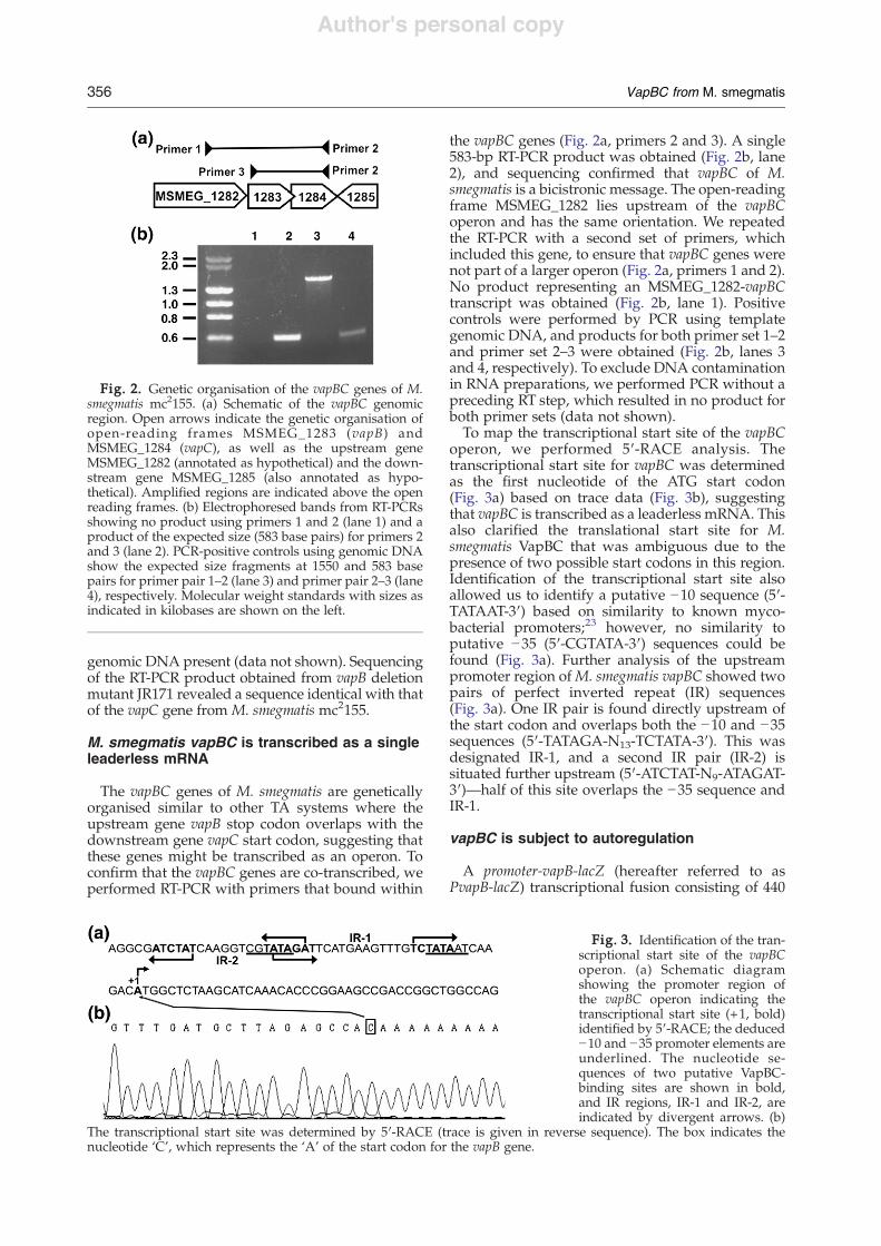
The vapBC genes of M. smegmatis are geneticallyorganised similar to other TA systems where theupstream gene vapB stop codon overlaps with thedownstream gene vapC start codon, suggesting thatthese genes might be transcribed as an operon. Toconfirm that the vapBC genes are co-transcribed, weperformed RT-PCR with primers that bound within
the vapBC genes (Fig. 2a, primers 2 and 3). A single583-bp RT-PCR product was obtained (Fig. 2b, lane2), and sequencing confirmed that vapBC of M.smegmatis is a bicistronic message. The open-readingframe MSMEG_1282 lies upstream of the vapBCoperon and has the same orientation. We repeatedthe RT-PCR with a second set of primers, whichincluded this gene, to ensure that vapBC genes werenot part of a larger operon (Fig. 2a, primers 1 and 2).No product representing an MSMEG_1282-vapBCtranscript was obtained (Fig. 2b, lane 1). Positivecontrols were performed by PCR using templategenomic DNA, and products for both primer set 1–2and primer set 2–3 were obtained (Fig. 2b, lanes 3and 4, respectively). To exclude DNA contaminationin RNA preparations, we performed PCR without apreceding RT step, which resulted in no product forboth primer sets (data not shown).To map the transcriptional start site of the vapBC
operon, we performed 5!-RACE analysis. Thetranscriptional start site for vapBC was determinedas the first nucleotide of the ATG start codon(Fig. 3a) based on trace data (Fig. 3b), suggestingthat vapBC is transcribed as a leaderless mRNA. Thisalso clarified the translational start site for M.smegmatis VapBC that was ambiguous due to thepresence of two possible start codons in this region.Identification of the transcriptional start site alsoallowed us to identify a putative !10 sequence (5!-TATAAT-3!) based on similarity to known myco-bacterial promoters;23 however, no similarity toputative !35 (5!-CGTATA-3!) sequences could befound (Fig. 3a). Further analysis of the upstreampromoter region ofM. smegmatis vapBC showed twopairs of perfect inverted repeat (IR) sequences(Fig. 3a). One IR pair is found directly upstream ofthe start codon and overlaps both the !10 and !35sequences (5!-TATAGA-N13-TCTATA-3!). This wasdesignated IR-1, and a second IR pair (IR-2) issituated further upstream (5!-ATCTAT-N9-ATAGAT-3!)—half of this site overlaps the !35 sequence andIR-1.
vapBC is subject to autoregulation
A promoter-vapB-lacZ (hereafter referred to asPvapB-lacZ) transcriptional fusion consisting of 440
Fig. 3. Identification of the tran-scriptional start site of the vapBCoperon. (a) Schematic diagramshowing the promoter region ofthe vapBC operon indicating thetranscriptional start site (+1, bold)identified by 5!-RACE; the deduced!10 and !35 promoter elements areunderlined. The nucleotide se-quences of two putative VapBC-binding sites are shown in bold,and IR regions, IR-1 and IR-2, areindicated by divergent arrows. (b)
The transcriptional start site was determined by 5!-RACE (trace is given in reverse sequence). The box indicates thenucleotide ‘C’, which represents the ‘A’ of the start codon for the vapB gene.
Fig. 2. Genetic organisation of the vapBC genes of M.smegmatis mc2155. (a) Schematic of the vapBC genomicregion. Open arrows indicate the genetic organisation ofopen-reading frames MSMEG_1283 (vapB) andMSMEG_1284 (vapC), as well as the upstream geneMSMEG_1282 (annotated as hypothetical) and the down-stream gene MSMEG_1285 (also annotated as hypo-thetical). Amplified regions are indicated above the openreading frames. (b) Electrophoresed bands from RT-PCRsshowing no product using primers 1 and 2 (lane 1) and aproduct of the expected size (583 base pairs) for primers 2and 3 (lane 2). PCR-positive controls using genomic DNAshow the expected size fragments at 1550 and 583 basepairs for primer pair 1–2 (lane 3) and primer pair 2–3 (lane4), respectively. Molecular weight standards with sizes asindicated in kilobases are shown on the left.
356 VapBC from M. smegmatis

Author's personal copy
base pairs upstream of the vapBC transcriptionalstart site and 106 base pairs of the vapB coding regionfused to lacZ was created (pJR174) (Fig. 4a). Thisintegrative construct was transformed into the wild-type strain mc2155 (JR176) and into the two knock-out strains!vapBC (JR175) and!vapB (JR223).WhenJR176 was grown under normal laboratory condi-tions, PvapB-lacZ expression increased throughoutthe growth curve and the "-galactosidase activitywas in the range 5–15 MU (Miller units) (Fig. 4b). Incontrast, for the deletion strain JR175 (!vapBC), theactivity of PvapB-lacZwas constitutive and increasedthreefold as compared with the wild-type strain,with values between 30 and 50 MU (Fig. 4b),indicating that one or both TA proteins are requiredfor repression of the vapBC operon. The threefold
increase in "-galactosidase activity of PvapB-lacZwas also observed for JR223 (!vapB) (data notshown). Complementation of the !vapBC deletionstrain with the vapBC operon under the control of itsnative promoter restored "-galactosidase activity tolow levels (1–3 MU; data not shown), below that ofthe wild type, which also increased during growth.This reduced activity is attributed to multicopyeffects of the plasmid-borne complementing con-struct pJR221, and similar results have been reportedpreviously in experiments using pOLYG for com-plementing repressor deletions.24
vapBC expression is regulated by the VapBCprotein complex
To overexpress and purify VapBC from M.smegmatis, we initially used Escherichia coli as anexpression host and attempted to express VapB andVapC proteins individually, as well as in a co-expression construct, using a pET-DUET system(Invitrogen). These approaches were unsuccessful,and, in general, protein was poorly expressed andinsoluble. Successful expression of the VapBCcomplex utilised M. smegmatis mc24517 as theexpression host.25 This contains a chromosomalcopy of the T7 RNA polymerase, and this allowsoverexpression vectors that contain the T7 promoterto be used. The M. smegmatis vapBC operon wascloned into the pYUB1049 expression vector, andthis was transformed into M. smegmatis mc24517.25Expression of the VapBC proteins was via auto-induction,25,26 and the protein complex was ex-pressed at high levels with no effect on bacterialgrowth. The complex was purified by Ni2+ affinity(the expression construct includes a C-terminal His6tag) followed by size-exclusion chromatography(Fig. 5). The expected molecular masses for theindividual proteins are 9.3 and 17.4 kDa for VapB andVapC, respectively, and these are evident in the SDSdenaturing gel in Fig. 5. There is a slight discrepancyin themolecularmass for VapB,which appears to runat "11 kDa in Fig. 5, and this is due to the poorresolving power of the gel close to the dye front.VapBC forms a protein complex of unknown
stoichiometry. We used dynamic light scattering(DLS) and a solution of purified VapBC, whichshowed that the VapBC protein complex wasmonodisperse with a hydrodynamic radius of51 Å. From this, the calculated molecular mass ofthe VapBC complex was "152 kDa. This isconsistent with a protein complex similar to thatseen for FitAB from N. gonorrhoeae, which has anaverage radius of 51.1 Å and a molecular mass of121 kDa.13 FitAB from N. gonorrhoeae is a tetramer ofdimers that binds to double-stranded DNA (at IRsequences) approximately 36 base pairs in length.The discrepancy in molecular masses between thatof FitAB and that calculated for VapBC from DLS islikely due to the assumption of a spherical particle inthe DLS molecular mass calculation coupled withthe unknown size of DNA to which the purified M.smegmatis VapBC is bound.
Fig. 4. Transcriptional activity of vapBC promoter.Transcriptional expression of the vapBC operon wasmeasured using an integrative vector with the upstreampromoter region fused to a promoterless lacZ. (a) Diagramof the PvapB-lacZ fusion construct pJR174, indicating theposition of the 546-bp region cloned into the integrativeplasmid pSM128. The vapB (MSMEG_1283) and vapC(MSMEG_1284) open-reading frames are indicated byopen arrows, and the black bar indicates the position ofthe region cloned, with numbers indicating base pairsrelative to the transcriptional start site (+1) identified by5!-RACE. (b) M. smegmatis strain mc2155 (JR176) and thevapBC deletion strain JR175 were grown in LBT, and "-galactosidase activity was measured at different parts ofthe growth curve over time (hours). Growth as measuredby optical density is shown as closed circles. "-Galacto-sidase activity is shown as vertical bars and given as themean±SEM of triplicate technical assays performed oneach sample. Results shown are representative of threeindependent experiments.
357VapBC from M. smegmatis

Author's personal copy
Electrophoretic mobility shift assays (EMSAs)with purified VapBC protein complex and vapBCpromoter DNA were performed in order to deter-mine which of the IR sequences identified in Fig. 3aare involved in VapBC–DNA binding. A 127-bpdigoxigenin-11-ddUTP (DIG)-labelled PCR productdesignated ‘Bind 1’ that encompassed the vapBCpromoter region and contained both IR-1 and IR-2motifs was used (Fig. 6a). The VapBC proteincomplex caused a mobility shift of ‘Bind 1’ atprotein concentrations of 2.4 nM and above, with acomplete shift of all labelled ‘Bind 1’DNA occurringat 24 nM (Fig. 6b). The specificity of the VapBCprotein complex for binding to this region wasdemonstrated by using an unlabelled PCR fragment(named mazE660) containing a 660-bp productamplified from the promoter region upstream fromthe open-reading frame MSMEG_4447. This open-reading frame is annotated as mazE and is theantitoxin component of the MazEF TAmodule inM.smegmatis. The presence of the mazE660 PCRproduct at 250-fold excess did not affect the mobilityshift of ‘Bind 1’, whereas the addition of the sameamount of unlabelled ‘Bind 1’ completely abolishedthe ability of VapBC to shift DIG-labelled ‘Bind 1’.This demonstrates that the DNA retardationobserved in the gel-shift assay is specific for theupstream promoter region of the vapBC operon.To investigate which of the two IR sequence pairs
was responsible for recognition and binding by theVapBC complex, we mutated each half site of bothIR-1 and IR-2, resulting in PCR products ‘Bind 2’ (5!-ATCTAT-3! to 5!-CCCCCC-3!), ‘Bind 3’ (5!-ATAGAT-3! to 5!-CCCCCC-3!) and ‘Bind 4’ (5!-TCTATA-3! to5!-CCCCCC-3!) (Fig. 6a). These three sequences,along with the wild-type sequence ‘Bind 1’, werethen used in gel-shift assays with the VapBC
complex as above (Fig. 6c). The resulting band-shift pattern for ‘Bind 2’ was identical with that forthe wild-type ‘Bind 1’ fragment. However, the band-shift pattern associated with VapBC binding waslost when sequences ‘Bind 3’ and ‘Bind 4’were used,both of which contain components of IR-1 andoverlap the !35 and !10 promoter elements,respectively. Band shifts were observed at 48 nMVapBC for ‘Bind 3’ and ‘Bind 4’, and this probablyreflects much weaker binding to an IR sequencewhere half the repeat is missing. VapBC-bindingcompetition assays were performed using unla-belled DNA sequences ‘Bind 1’–‘Bind 4’ at 25! and50! that of DIG-labelled ‘Bind 1’ (0.4 ng) withVapBC at a concentration of 9.6 nM. These resultsshowed that 25! and 50! excesses of unlabelledwild-type sequence ‘Bind 1’ were sufficient tooutcompete the DIG-labelled ‘Bind 1’ sequence(Fig. 6d), resulting in a near-complete loss ofretardation compared with the control (no excess‘Bind 1’). A similar result occurred with unlabelledsequence ‘Bind 2’, which also abolished the shift,thereby confirming previous results that the 5! halfof IR-2 is not required for VapBC promoter binding(Fig. 6c). However, 25! and 50! excess levels ofunlabelled ‘Bind 3’ and ‘Bind 4’ sequences could notaffect the shift created by VapBC binding to thewild-type DIG-labelled ‘Bind 1’ sequence.
Conditional expression of VapC inhibitstranslation
Under normal laboratory conditions, both !vapdeletion strains JR121 and JR171 had growth ratessimilar to those of the wild-type strain mc2155 inboth rich [Luria–Bertani (LB) medium supplemen-ted with 0.05% (wt/vol) Tween-80 (LBT)] andminimal [Hartman's–de Bont (HdB)] media. InLBT medium, the doubling times for mc2155 andthe deletion strains JR121 (!vapBC) and JR171(!vapB) were approximately 2.7 h. In HdB minimalmedium, the doubling times were approximately 3 hfor all strains. Overexpression of the toxin compo-nent of TA systems has often been shown to result ineither bacteriostasis or cell death.16,27 To determinewhether the VapC toxin had a similar effect in M.smegmatis, we constructed pJR29, which containedthe vapC gene under the control of a tetracycline-inducible promoter,28 and measured whether induc-tion of VapC influenced cell growth and/or survi-val. To ensure that the effect observed was due to theproduction of VapC alone, we also cloned vapBC inthe same manner, creating pJR230, thus abrogatingthe effect of the toxin by co-expressing the cognateantitoxin. This also removes the possibility that theinhibitory effect seen for the conditional expressionof VapC is simply due to general overexpression of aprotein. Both constructs were placed into the back-grounds of the !vapBC strain (JR121) and wild-typeM. smegmatismc2155. As a control, the empty vectorpMind was used in both strains. Cultures wereinduced at an optical density (OD600) between 0.10to 0.20 [between 5!106 and 1!107 CFU (colony-
Fig. 5. Overexpression and purification of the VapBCprotein complex. Shown is a chromatogram from a size-exclusion purification showing the progress of elution. Alarge peak elutes in the void volume of the column, andthis is then followed by a smaller peak that contains theVapBC protein complex. Fractions between 59- and 75-mlelution volumes were collected and run on an SDS-PAGEgel (shown above the chromatographic trace). Amolecularweight marker at the left side of the gel shows molecularmasses in kilodaltons, and bands corresponding to toxinand antitoxin proteins are labelled at the right (expectedprotein sizes of 17.4 and 9.3 kDa, respectively; see the text).
358 VapBC from M. smegmatis

Author's personal copy
forming units) ml!1] and were then monitored byOD600 and cell viability (CFU) measurements.Blokpoel et al.28 have reported that the greatestexpression using the pMind vector following induc-tion with tetracycline at 20 ng ml!1 occurs after 24 h.We therefore monitored optical density and viabilityover 96 h post-induction. M. smegmatis mc2155expressing VapC exhibited a 10-fold reduction inCFUs per millilitre compared with that of the strainexpressing either the VapBC complex or the emptyvector control strain (Fig. 7a). A 100-fold differencewas observed in the !vapBC deletion strain whenVapC expression was induced (Fig. 7a). In order tovalidate that cells were being inhibited, we platedcells onto plates that were supplemented either withhygromycin B only or with hygromycin B and 20 ngml!1 tetracycline. Results showed that for both!vapBC strain JR121 (Fig. 7c) and mc2155 (data notshown), cultures that harboured either the vapBCoperon or the empty vector pMind had normalcolony growth on plates with and those withouttetracycline. However, the JR121 strain containingpMind-vapC only (grown under inducing condi-tions) formed visible colonies on plates without
tetracycline and “pinpoint”-sized colonies that werebarely visible in the presence of tetracycline (Fig. 7c).A similar result was observed in the wild-type strainwith very small colonies (data not shown).The presence of VapC protein was confirmed by
Western blot using polyclonal antibodies raisedagainst purified VapC. For the purposes of antibodyproduction, M. smegmatis VapC was overexpressedas inclusion bodies in E. coli and purified underurea-denaturing conditions (see Materials andMethods for details). In strain mc2155, endogenousVapC is detected in the empty pMind control (Fig.7b, E, lane 1) and the strain containing pMind-vapCwithout induction via tetracycline (Fig. 7b, !Tc, lane3). Significantly greater amounts of VapC protein areevident in the mc2155 strain containing pMind-vapCafter induction by tetracycline (Fig. 7b, +Tc, lane 2).In the !vapBC strain containing the empty pMindvector, no endogenous VapC was detected, which isexpected for the knockout strain (Fig. 7b, lane 4). Incontrast, significant amounts of VapC were inducedby tetracycline in the !vapBC mutant harbouringpMind-vapC (Fig. 7b, lane 5). No VapC protein isdetected in a !vapB mutant (Fig. 7b, lane 7),
Fig. 6. In vitroDNAbinding of purified VapBC protein complex. (a) Schematic of the vapBC promoter region indicatingthe sequence used for 126-bp fragment ‘Bind 1’ and base-pair changes overlapping potential VapBC-binding sites.Nucleotide changes (CCCCCC) for each half of IR-1 and IR-2 are indicated above the sequence, giving 126-bp mutatedfragments ‘Bind 2’, ‘Bind 3’ and ‘Bind 4’. Divergent arrows denote sequences for IR-1 and IR-2. Bold nucleotides indicatededuced !10 and !35 promoter elements. (b) Binding of VapBC complex to the vapBC promoter region. EMSAexperiments were performed with a DIG-labelled 126-bp promoter fragment, ‘Bind 1’. The concentrations of VapBC areshown above. Specific binding and nonspecific binding are shown using a 250-fold concentration (100 ng) of unlabelled‘Bind 1’ and an unlabelled 660-bp promoter frommazEFDNA obtained fromM. smegmatismc2155, respectively. (c) EMSAexperiments were performed using increasing concentrations of VapBCwith DIG-labelled fragments ‘Bind 1’–‘Bind 4’. (d)Competition experiments were performed with VapBC complex (9.6 nM) and DIG-labelled ‘Bind 1’ (0.4 ng). ‘Bind 1’ lanerepresents free DIG-labelled ‘Bind 1’ DNA. Increasing amounts of unlabelled competitor DNAwere used at 25! and 50!that of DIG-labelled Bind 1. The unlabelled fragments used for each shift are written above along with the concentrationsof competitor DNA used for each lane. VapBC–DNA complex and free unbound DNA are indicated by arrows.
359VapBC from M. smegmatis

Author's personal copy
suggesting that VapB is required for VapC produc-tion when expressed from the chromosome (i.e., thatthere is translational coupling for this operon). Therescue of growth inhibition by the expression ofVapBC protein complex was also followed byWestern blot (Fig. 7b, right panel) at three timepoints: 0, 12 and 24 h post-induction. This demon-strates that the rescue of growth is the result of VapBinhibition of VapC.The mechanism of growth inhibition by VapC was
investigated by measuring the rates of DNA repli-cation, transcription and translation, which werefollowed for both JR121 and wild-type M. smegmatismc2155 containing the tetracycline-inducible pMindvector encoding VapC. Pulse-chase experimentswere conducted during exponential-phase growthusing rates of incorporation of 3H-labelled thymi-dine, 3H-labelled uridine and 35S-labelled methio-
nine to follow replication, transcription and transla-tion, respectively (Fig. 8). During the course of thisexperiment, cells are maintained in exponentialphase by dilution of cultures at 12, 18 and 21 h ofgrowth (post-induction) in order to remove con-founding factors associated with the cells reachingstationary phase. Inhibition of translation was seenfor pMind-vapC in both wild-type and !vapBCbackgrounds, with a more marked inhibition oftranslation in the latter knockout strain (Fig. 8a).This is consistent with endogenous VapB in thewild-type strain offsetting the effect of inducedVapC from the pMind vector. It follows that thiseffect would be exacerbated in the absence ofendogenous VapB in the !vapBC strain, and thiswas observed. There was no effect seen for theconditional expression of VapC for any of the strainswhen monitoring DNA replication or transcription(Fig. 8b and c, respectively).
Discussion
The contemporary biological functions of TAmodules in bacteria are the subject of a great dealof current research and debate. One of the centralquestions is the role of the TA modules inbacteriostasis, bacterial persistence and bacterial
Fig. 7. Effect of VapC on growth and viability of M.smegmatis. M. smegmatis strain mc2155 and !vapBCdeletion JR121 harbouring plasmid pMind (vector only),pMind-vapC (expressing the VapC toxin) or pMind-vapBC(expressing the VapBC operon) were grown in HdBmedium supplemented with hygromycin at 37 °C untilan OD600 of "0.1–0.2. Induction of expression was by theaddition of tetracycline (final concentration of 20 ng ml!1).(a) Cellular growth was monitored over 100 h bymeasuring optical density. Tetracycline (final concentra-tion of 20 ng ml!1) was added, and cell viability was thenmonitored for M. smegmatis mc2155 and JR121 harbouringcontrol (empty pMind vector; dark circles and squares,respectively), VapC expression constructs (pMind-vapC;open circles and squares, respectively) and VapBCexpression constructs (pMind-vapBC; open triangles).Cellular viability was determined by CFUs by platingout appropriate dilutions onto LBT agar supplementedwith hygromycin and enumerating them after 3 days ofgrowth at 37 °C. Results are shown as the mean±SEM oftechnical replicates of each time point. (b) AWestern blotshowing VapC protein from mc2155 and JR121 (!vapBC).The left panel shows mc2155 containing pMind (emptyvector, denoted E), pMind-vapC after induction withtetracycline (denoted +Tc) and pMind-vapC withoutinduction (denoted !Tc). The central panel shows thesame for the !vapBC strain. In addition, lanes are shownfor JR171 (!vapB strain), along with a positive control(purified VapBC protein complex). For JR171 (!vapBstrain), there is no VapC detected despite the presence ofmRNA transcript. The right panel shows detection ofVapC protein in JR121 (!vapBC) containing pMind-vapBC.The numbers beneath each lane indicate the time post-induction. (c) A mosaic of agar plates showing indicativegrowth of JR121 containing pMind (empty vector) orpMind-vapC with and that without tetracycline.
360 VapBC from M. smegmatis

Author's personal copy
programmed cell death.3 Recent reports have shownthat theMazEF TAmodule in E. coli causes cell deathin response to quorum-sensing signals.29 MazF hasalso been shown to mediate programmed cell deathin Myxococcus xanthus.30 In contrast, protein expres-sion systems, which utilise MazF mRNase activity toeliminate translation of nearly all but the desiredtranscript, are available, and this clearly precludesMazF being a cell-death factor.31 In several publica-tions, both the MazEF and RelBE TA modules havebeen described as mRNA interferases32,33 withpossible roles in response to various stressors, suchas nutritional stress and antibiotic stress. The VapBCTA modules are more widely spread than MazEF
and RelBE and in several organisms are dramati-cally expanded in number.2,3 Unlike MazEF andRelBE, the VapBC modules have only recently beenidentified and so are less well studied, and theircontemporary biological functions remain unchar-acterised in most organisms.In this article, we demonstrate that the open-
reading frames MSMEG_1283 and MSMEG_1284from M. smegmatis mc2155 encode a bona fide vapBCTAmodule. Deletions in either vapB or vapBC had noeffect on the growth of M. smegmatis mc2155 undernormal laboratory conditions, and the vapBC operonwas expressed at low levels throughout the growthcycle. In a !vapBC mutant, PvapB-lacZ activity wasthreefold higher than the wild-type strain, indicatingthat the vapBC operon was subject to autoregulationby the VapBC protein complex. Regulation of vapBCexpression was mediated through VapBC binding toan IR sequence in the vapBC promoter that overlapsthe !35 and !10 sequences (IR-1 in Fig. 6a), thusexplaining the repression of vapBC transcriptionunder physiological conditions. These data areconsistent with the notion that vapBC expression istightly regulated inM. smegmatis and is not inducedunder normal growth conditions.In some TA systems studied, the antitoxin is
essential, given its role in neutralising the toxin, andis unable to be deleted in a toxin-producingstrain.21,22 In the wild-type strain under physio-logical conditions, VapC was detected by Westernblot analysis, but no growth phenotype wasobserved, suggesting that VapB was neutralisingthe toxin under these conditions. We successfullyconstructed a !vapB knockout, and this mutantcontained elevated expression levels of vapC tran-script, but no phenotype (i.e., growth inhibition) wasevident. Western blot analysis indicates that despiteelevated vapC transcription in the !vapB mutant, noVapCprotein could be detected, suggesting that RNAprocessing and translational coupling are importantin VapC expression. This has been reported for theparD operon that encodes the Kis/Kid system.Synthesis of Kid (toxin) is greatly reduced whenuncoupled from the synthesis of Kis (antitoxin).34A number of studies have used the overexpression
of the toxin, often in a heterologous host, such as E.coli, to demonstrate toxicity of the toxin module. Theconditional (tetracycline-regulated) overexpressionof VapC in M. smegmatis resulted in inhibition ofgrowth, and the effect of VapC was neutralisedwhen co-expressed with its cognate antitoxin VapB.The ability of VapC to inhibit the growth of M.smegmatis was due to its ability to inhibit translationbut not DNA replication or transcription. This isconsistent with a postulated biochemical activity forthe VapC proteins as RNases and with what havebeen observed in H. influenzae,17 M. tuberculosis18and Pyrobaculum aerophilum (J.L.M. & V.L.A., unpub-lished results). Growth inhibition by VapC wasreversed through the removal of tetracycline,indicating that the effect of VapC on translationwas bacteriostatic. Even in the !vapBC mutant,the VapC-dependent growth inhibition could be
Fig. 8. Effects of VapC on translation, transcription andDNA replication. M. smegmatis strain mc2155 harbouringplasmid pMind (vector only; closed circles) or pMind-vapC (expressing VapC toxin; open circles) and !vapBCstrain harbouring the same plasmid pMind (closedsquares) or pMind-vapC (open squares) were grown inHdB medium supplemented with hygromycin. Exponen-tially growing cultures were induced with tetracycline(20 ng ml!1) at time zero, and the rates of (a) translation,(b) transcription and (c) DNA replication were determinedby the incorporation of 35S-labelled methionine, 3H-labelled uridine and 3H-labelled thymidine, respectively.Chloramphenicol, an inhibitor of translation, wasincluded in (a) as a positive control (open triangles). Thisexperiment was carried out as independent duplicates.
361VapBC from M. smegmatis

Author's personal copy
overcome by tetracycline removal, suggesting thatthe antitoxin was not responsible for this reversi-bility. Korch et al.35 have recently demonstrated thatoverexpression of rel toxin genes fromM. tuberculosisin M. smegmatis induced growth arrest and that thiswas reversed via expression of the cognate antitoxinrel genes. Interestingly, the growth-inhibited pheno-type was also reversible in the absence of thecognate antitoxin. These observations for both RelEand VapC suggest that the cells must possessmechanisms to degrade the toxin in the absence ofde novo protein synthesis.The role of VapBC in the biology of mycobacteria
remains unknown. VapBC modules have beenimplicated in controlling bacterial growth in theintracellular environment14 and regulating rates ofnitrogen fixation and biomass production duringsymbiosis in a host plant.15 In addition, in a numberof cases, the toxic effect of VapC has only beengleaned through its overexpression in a hetero-logous host.16–18,36 Our results infer a physiologicalrole for vapBC in M. smegmatis insofar as over-expressed VapC in the parent showed growthinhibition via disruption of translation, and thiseffect was most pronounced in rapidly growing cells(i.e., in exponential phase). The reversibility of thisphenomenon in the absence of the cognate antitoxinVapB (i.e., in the !vapBC background) is intriguing.These data suggest that M. smegmatis may use theVapBC module to trigger reversible bacteriostasisunder conditions where the cells have an imbalance
of anabolic and catabolic reactions. Using aninhibitor of protein synthesis (i.e., an RNase) tocontrol metabolic activity when required wouldseem advantageous when one considers that 70%–90% of cellular ATP consumption by a cell is forprotein synthesis.37
Materials and Methods
Bacterial strains, media and growth conditions
E. coli DH10B was grown in LB medium at 37 °C withagitation (200 rpm) or on LB agar plates. M. smegmatismc2155 and derived strains (Table 1) were grown in LBTor HdB minimal medium supplemented with 0.2%glycerol, 50 mM 4-morpholinoethanesulfonic acid and0.05% (wt/vol) Tween-80 at 37 °C with agitation(200 rpm). Growth curves were performed in triplicatein both LBT and HdB media. Bacterial cell viability wasmonitored by cell counts based on CFUs per millilitrewhere serial dilutions of bacterial cell culture inphosphate-buffered saline with 0.05% Tween-80 wereplated on LBT agar plates supplemented with appro-priate antibiotics. Selective media contained ampicillin(100 #g ml!1 for E. coli), kanamycin (50 #g ml!1 for E.coli, 20 #g ml!1 for M. smegmatis), gentamicin (20 #g ml!1for E. coli, 5 #g ml!1 for M. smegmatis), hygromycin B(200 #g ml!1 for E. coli, 50 #g ml!1 for M. smegmatis),streptomycin (20 #g ml!1 for E. coli and M. smegmatis) orspectinomycin (20 #g ml!1 for E. coli). All plasmids andstrains used in this study are listed in Table 1.
Table 1. Bacterial strains and plasmids used in this study
Bacterial strains Descriptiona Source or reference
E. coliDH10B F-mcrA!(mrr-hsdRMS-mcrBC) $80 dlacZ!M15!lacX74 deoR recA1 araD139!(ara leu)7697 galU
galK rpsL endA1 nupGRef. 44
M. smegmatismc2155 Electrocompetent wild-type strain of M. smegmatis Ref. 45mc24517 M. smegmatis expression strain with T7 RNA polymerase, Kmr Ref. 25JR121 mc2155 !vapBC ::aphA-3; Kmr This studyJR171 mc2155 !vapB ::aphA-3; Kmr This studyJR176 mc2155 with pJR174 integrated in attB; Strepr This studyJR175 JR121 with pJR174 integrated in attB; Kmr Strepr This studyJR223 JR171 with pJR174 integrated in attB; Kmr Strepr This study
PlasmidspBluescript II KS Cloning vector; Apr StratagenepUC18K E. coli plasmid containing an excisable, nonpolar kanamycin resistance cassette; Kmr Apr Ref. 39pX33 E. coli mycobacterium shuttle vector for allelic exchange mutagenesis in mycobacteria, pPR23,
carrying a constitutive xylE marker; Gmr Sacs tsRefs. 39 and 40
pSM128 lacZ transcriptional fusion vector derived from pYUB295, with mycobacteriophage L5 integraseand attP for integration into attB of mycobacteria; Specr Strepr
Ref. 46
pOLYG E. coli mycobacterium shuttle vector; Hygr Ref. 47pMind Tetracycline-inducible expression vector; Kmr Hygr Ref. 28pYUB1049 E. coli–M. smegmatis shuttle vector with T7 promoter and encoding a C-terminal His6 tag: Hygr Ref. 25pJR113 pX33 harbouring !vapBC ::aphA-3; Kmr Gmr Sacs ts This studypJR139 pX33 harbouring !vapB ::aphA-3; Kmr Gmr Sacs ts This studypJR174 pSM128 harbouring a 546-bp PvapB-lacZ fusion; Strepr Specr This studypJR221 pOLYG harbouring vapBC genes with its native promoter region; Hygr This studypJR29 pMind harbouring vapC with RBS from kanamycin marker; Kmr Hygr This studypJR230 pMind harbouring vapBC with RBS from kanamycin marker; Kmr Hygr This study
a Gmr, gentamicin resistance; Hygr, hygromycin B resistance; Kmr, kanamycin resistance; Apr, ampicillin resistance; Strepr,streptomycin resistance; Specr, spectinomycin resistance; Sacs, sucrose sensitivity; ts, temperature sensitivity.
362 VapBC from M. smegmatis

Author's personal copy
DNA manipulation and cloning of constructs
All primer sequences used are listed in Table 2. GenomicDNA from M. smegmatis was isolated using the cetyltri-methylammonium bromide method adapted from themethod of Bull et al.38To create a construct for the deletion of the vapBC genes,
we amplified from pUC18K39 a kanamycin resistancecassette encoded by aphA-3 by PCR using primers 5!mcspUC F and 3!mcspUC R. The resulting 850-bp productwas digested with BamHI and Asp719. The regionsflanking either side of vapBC genes were amplified usingprimer pairs vapBCKOLF F and vapBCKOLF R to obtainthe left flank (LF; 990 base pairs) region and vapBCKORF Fand vapBCKORF R to obtain the right flank (RF; 1010 basepairs) region. The LF product was digested with SpeI andAsp718, and the RF was digested with BamHI and SpeI.Both the flanking DNA fragments and the kanamycincassette were ligated into SpeI-digested pBluescript. Theassembled insert containing LF/kanamycin/RF DNAwasthen sub-cloned into pX33,40 generating pJR113, whichwas electroporated into M. smegmatis mc2155 background.To create a construct that would result in a vapB deletion
mutant, we took the same approach. Primers vapBKOLF Fand vapBCKOLF R were used to amplify the left flankingregion (1360 base pairs) of the antitoxin vapB gene andright flanking primers vapBKORF F and vapBKORF R,which were designed to amplify the right flanking region,including the toxin vapC gene, resulting in a 1391-bpfragment. These DNA fragments were ligated to eitherside of the kanamycin cassette in vector pBluescript. Theresulting assembled insert was sub-cloned into pX33 anddesignated pJR139. This vapB deletion construct waselectroporated into M. smegmatis mc2155.Allelic replacement of vapBC and vapB was carried out
as described previously.41 In brief, two M. smegmatismc2155 cultures carrying pJR113 and pJR139 were grownat 28 °C with agitation (200 rpm) to an optical density(OD600) of approximately 0.6 to 0.8. These were platedonto low-salt (2 g NaCl l!1) LBT plates containingkanamycin and 10% sucrose at 40 °C, selecting fordouble-recombinant events. For Southern hybridisation
analysis, SmaI-digested genomic DNAwas obtained fromputative mutants and separated on 1% agarose Tris–acetate–EDTA (ethylenediaminetetraacetic acid) gel andtransferred to a nylon membrane by vacuum blotting.Probes were radiolabelled by random priming using[%-32P]dCTP (Amersham) and Ready-To-Go DNALabelling Beads (Amersham).A PvapB-lacZ transcriptional fusion was constructed by
amplification of a 546-bp fragment containing the first 106base pairs of vapB coding region and 440 base pairsupstream of the transcriptional start site identified by 5!-RACE using primers TAp+440 F and TAp!106 R. ThisPCR product was blunt end cloned into ScaI-digestedpSM128, which is an integrative promoter probe vectorderived from pYUB295, which contains a promoterlesslacZ. PCR was performed on clones with the correct insertto ensure that the 546-bp upstream promoter fragmentwas in the correct orientation to the promoterless lacZusing primers TAp+440 F and 3!mcspUC R. One clonewith the correct sequence was designated pJR174. Emptyvector pSM128 and the recombinant plasmid pJR174 wereintroduced into wild-type strain M. smegmatis mc2155,JR121 and JR171 by electroporation, followed by screeningfor streptomycin resistance. For complementation of JR121and JR171 containing the integrated vapB-lacZ construct, aPCR product that encompassed both the vapBC genes and440 base pairs of upstream DNA was obtained usingprimers TAp+440 F and toxin R and digested withBamHI/EcoRI followed by ligation into the BamHI/EcoRI sites of E. coli/mycobacteria shuttle vectorpOLYG, generating pJR221.In order to create the VapC tetracycline-inducible
expression construct pJR29, we amplified from M.smegmatis mc2155 a PCR product containing the vapCgene (390 base pairs) using primers vapCpMind F andvapCpMind R and ligated it into the BamHI/SpeI sites ofthe tetracycline-inducible vector pMind. This approachwas repeated to obtain construct pJR230 that contains thevapBC operon (645 base pairs) amplified using primersvapBCpMind F and vapCpMind R. Sequence analysisconfirmed that both inserts were in the correct orientationto the tetRO promoter region. Both forward primers,
Table 2. Oligonucleotides
Name Sequence 5!–3! Enzyme
TAp+440 F AAATTTGGATCCAACGAGATCAGCTATCTCAG BamHITAp!106 R AAATTTGGTACCGCTCCCGCAGTGCCATCACC Asp718vapBCKOLF F AAATTTACTAGTGGTACATGCTGGAATGGAAG SpeIvapBCKOLF R AAATTTGGTACCATGCTTAGAGCCATGTCTTG Asp718vapBCKORF F AAATTTGGATCCCCACTGAACGAGGTCAGAAT BamHIvapBCKORF R AAATTTACTAGTGATGACCTCCACGACGCAAT SpeIvapBKOLF F AAATTTACTAGTTCCAACTGTTGATCGTCACA SpeIvapBKORF F AAATTTGGATCCCGGTCTGCCGTCCTGATGGT BamHIvapBKORF R AAATTTACTAGTCGTTGGTGATGGATGCTCTG SpeIvapBRT-PCR R TCGTCGTAGCCCAGGATCGAvapCRT-PCR F TTATCGACACTTCTGCCCTCvapBCRT-PCR F CAAGACATGGCTCTAAGCATvapBCRT-PCR R TTGGCGAGCGCATAGGAAAAToxin R AAATTTGAATTCCCATGGTGAATTCTGACCTCGTTCAG EcoRI/NcoIvapCpMind F AAATTTGGATCCGGAGGAATAATGGTTATCGACACTTCTGC BamHIvapCpMind R AAATTTACTAGTTGAATTCTGACCTCGTTCAG SpeIvapBCpMind F AAATTTGGATCCGGAGGAATAATGGCTCTAAGCATCAAACAC BamHIBind1-126bp F GAACAGGAGCGGATCGACTGBind1-126bp R CGGCTTCCGGGTGTTTGATG3!mcspUC R GTTTTCCCAGTCACGACGTT5!mcspUC F CACACAGGAAACAGCTATGAVapB-forward TCAGCTCCATGGCTCTAAGCATCAAACA NcoIVapC-reverse TATTTAGGATCCGCGTGGACCGCAGCG BamHI
363VapBC from M. smegmatis

Author's personal copy
vapCpMind F and vapBCpMind F, introduced a syntheticribosome-binding sequence (GGAGG) upstream of thevapC and vapBC genes, respectively, which was obtainedfrom nonpolar kanamycin resistance cassette (aphA-3) toensure translation.
RNA extraction and reverse-transcriptase PCR
For RNA preparation, appropriate strains were grownto an OD600 between 0.5 and 0.7 in LBT medium, and 5 to10 ml of culture was harvested by centrifugation. TotalRNA was extracted using TRIzol reagent (Invitrogen)according to the manufacturer's instructions. Cells wereruptured with two rounds of bead beating in a MiniBeadBeater (Biospec) (5000 rpm, 1 min). To remove anycontaminating DNA present in samples, we treatedextracted RNA with 2 U of RNase-free DNase using aTurbo RNase-free kit (Ambion) according to the manufac-turer's instructions. RNA concentrations were determinedusing a Nanodrop ND-1000 spectrophotometer.RT-PCR was performed using a Titan One Tube RT-
PCR system (Roche) according to the manufacturer'sinstructions. The RT reactions were carried out using200 ng of RNA template. To detect whether the vapBCgenes were co-transcribed, we used gene-specific primers(vapBCRT-PCR F, primer 3; vapBCRT-PCR R, primer 2)that bind within the 645-bp vapBC operon in an RT-PCRcycle with an RT step at 42 °C for 30 min, followed by 30cycles of PCR. This RT-PCR was also used to detectwhether an MSMEG_1282-vapBC amplicon was presentusing primers vapBCKOLF F (primer 1) and vapBCRT-PCR R (primer 2).To detect the presence of toxin mRNA using RNA
prepared from the antitoxin deletion mutants, we per-formed RT-PCR using primers vapCRT-PCR F andvapBCRT-PCR R. For all RT-PCR experiments, controlreactions to exclude DNA contamination were carried outas above for each sample using RNA as the templatewithout the RT step. For positive controls, PCR using thegenomic DNA of M. smegmatis mc2155 as template wasperformed using Taq DNA polymerase (Roche) accordingto the manufacturer's instructions.
Mapping of the transcriptional start site
The transcriptional start site for the vapBC operon wasmapped by 5!-RACE using components of the 3!/5!-RACE kit (Roche) according to the manufacturer'sinstructions. First-strand cDNA was obtained using RTsynthesised from 4 #g of total RNA of M. smegmatis andvapC-specific primer vapBCRT-PCR R. The resulting cDNAwas purified, and a dA tail was added following the kitinstructions. Before the addition of terminal transferase,cDNA was heated (3 min, 94 °C) and then placedimmediately on ice to remove any possible secondarystructures. Purified dA-tailed cDNA was used as atemplate for PCR using the oligo-dT anchor forwardprimer and vapBRT-PCR R reverse gene-specific primer,and the resulting PCR product (ca 250 base pairs) was gelpurified with a Qiaex® II gel extraction kit (Qiagen). Toincrease the yield of this product, we performed a secondround of PCR using the PCR anchor primer and vapB-specific primer TAp!106 R. The single PCR product waspurified (High Pure PCR Product Purification Kit, Roche)and ligated into pGEM-T easy vector (Promega) accordingto the manufacturer's instructions. Three clones con-taining the correctly sized insert were sequenced using
primer TAp!106 R, and the last nucleotide before thepoly-A tail to align with the genome sequence was chosenas the most likely transcriptional start site. The sequencesof all primers used in this protocol are listed in Table 2.
!-Galactosidase assay
"-Galactosidase activity was measured for wild-typeM.smegmatis mc2155 and both deletion strains containingempty vector pSM128 and integrated fusion constructpJR129 in LBT. Overnight starter cultures were grown toan OD600 between 0.3 and 0.5 in LBT containingstreptomycin, which was then diluted in a volume of50 ml of LBT with streptomycin in a 250-ml flask to anOD600 of 0.005. Samples were collected over time todetermine the optical density and "-galactosidase activity."-Galactosidase activity was assayed as previouslydescribed40 and expressed in Miller units,42 calculated asthe increase in A420 per minute per 1 ml of cell suspension(normalised to an OD600 of 1.0) used and multiplied by afactor of 1000. Results shown are representative of threeindependent experiments.
Expression and purification of VapBC inM. smegmatis mc24517
The ORF encoding the vapBC operon was amplifiedfrom M. smegmatis genomic DNA using the followingprimers: VapB-forward containing 16 base pairs of gene-specific sequence, 5!-TCAGCTCCATGGCTCTAAGCAT-CAAACA-3! (NcoI restriction site underlined), and VapC-reverse containing 15 base pairs of gene-specific sequence,5!-TATTTAGGATCCGCGTGGACCGCAGCG-3! (BamHIrestriction site underlined). The amplified product wasdigested with NcoI/BamHI restriction enzymes andinserted into the pYUB1049 shuttle vector25 between therestriction sites, enabling expression of a C-terminal Histag attached to VapC. The construct was transformed intoE. coli TOP10 cells and plated on low-salt LB agar mediumsupplemented with 0.05% Tween-80 and 50 #g ml!1hygromycin B to select for positive transformants. Con-structs were sequenced prior to transformation into M.smegmatis mc24517 following previously publishedmethods.25,43 Positive colonies were selected by platingthe transformants on 7H10 agar medium supplementedwith ADC (albumin, dextrose and catalase supplement),0.05% Tween-80 and 50 #g ml!1 kanamycin and hygro-mycin B.Expression of the VapBC complex was performed in
autoinduction medium26 supplemented with 0.05%Tween-80 and 50 #g ml!1 kanamycin and hygromycin B.A single colony was selected and used to inoculate a PA-0.5G seeder culture. This culture was grown for 36 h at37 °C and was used at a 1:100 dilution to inoculate a ZYP-5052/Tween expression culture and grown at 37 °C for96 h for maximal protein expression.Cells from the expression culture were harvested by
centrifugation at 7000g for 25 min at 4 °C. Cell pellets wereresuspended in 50 mM phosphate buffer, pH 7.4, and200 mM NaCl with the addition of an EDTA-free proteaseinhibitor tablet (RocheApplied Science). Resuspended cellswere then sonicated on ice. The cell lysate was centrifugedat 16,000g for 20 min at 4 °C to separate the soluble andinsoluble protein fractions. The soluble fraction containingthe His-tagged VapBC protein complex was loaded onto aHisTrap HP column (GE Healthcare Life Sciences), and thecaptured VapBC complex was eluted with an imidazole
364 VapBC from M. smegmatis

Author's personal copy
gradient over a concentration of 0–500 mM imidazole.Fractions containing VapBC protein complex were concen-trated and purified further by size-exclusion chromato-graphyusing a Superdex 200 16/60 column (GEHealthcareLife Sciences) in the same buffer (50 mM phosphate buffer,pH 7.4, and 200 mM NaCl).
Expression and purification of VapC for antibodyproduction
VapC from M. smegmatis was expressed as an N-terminal His6-tagged fusion protein in E. coli using apPROEX expression vector (Invitrogen). Briefly, theoverexpression of VapC in E. coli DH5% cells was inducedusing isopropyl-",D-thiogalactopyranoside at an OD600 of0.5. Cells were grown overnight and then harvested bycentrifugation and lysed using sonication. The cell lysatewas centrifuged at 16,000g for 20 min at 4 °C, and theinsoluble fraction was resuspended in 50 mM phosphatebuffer, pH 7.4, 200 mM NaCl and 8 M urea and gentlystirred for 1.5 h. Following this, particulates wereremoved by centrifugation and the clarified supernatantwas loaded onto a HisTrap HP column (GE HealthcareLife Sciences). VapC protein was eluted with an imidazolegradient of 0–500 mM. Denatured VapC was dialysedagainst 50 mM phosphate buffer, pH 7.4, and 200 mMNaCl to remove urea and imidazole and then concen-trated to 1.0 mg ml!1.Antibody production was in 4-month-old New Zealand
white rabbits. A pre-immune serum sample was takenfrom each rabbit. The primary immunisation for VapCantibody production (0.2 ml of 1 mg ml!1 VapC protein,0.3 ml of Freund's complete adjuvant) was injectedsubcutaneously. Secondary immunisations were carriedout every 2 weeks for 6 weeks. Rabbits were anaesthetisedprior to the terminal bleed, and serum was diluted 1:750for Western blots. In all cases, 250 #g of total protein wasloaded in each lane to allow for qualitative comparisons ofVapC levels.
Electrophoretic mobility shift assay
Purified M. smegmatis VapBC complex was used inEMSAs using a DIG Gel-Shift Kit, 2nd Generation (Roche)to 3!-end label target DNAwith DIG. The DNA fragmentswere labelled and used in gel-shift reactions according tothe manufacturer's instructions. For use in the bindingassay, purified VapBC complex was diluted to appropriateconcentrations in 50 mM potassium phosphate buffer,pH 7.3, containing 200 mMNaCl and 0.05 mgml!1 bovineserum albumin. Protein/DNA molar ratios were calcu-lated to achieve different protein concentrations requiredfor each gel-shift assay. Target DNA and VapBC proteinwere added at different concentrations in 10-#l reactionvolumes. To each 10-#l sample, binding buffer (2 #l), polydI.dC (1 mg ml!1) (0.5 #l) and DIG-labelled target DNA(0.4 ng) were added as per the manufacturer's instruc-tions. All binding reactions were incubated at roomtemperature for 20 min. A 6% native acrylamide (19:1)TBE (Tris–borate–EDTA) gel in 0.5! TBE buffer was pre-run at 300 V for 5 min, followed by electrophoresis of thegel-shift reactions at 300 V for 20 min. Electroblotting wasperformed using Xcell II Blot Module (Invitrogen)according to the manufacturer's instructions. Protein-bound DIG-labelled DNA was fixed to nylon membraneand was detected by a chemiluminescent immunoassay,which was recorded using autoradiography.
The wild-type 126-bp fragment designated ‘Bind 1’corresponding to the vapBC promoter was amplified byPCR using primers Bind1-126bp F and Bind1-126bp Rfrom M. smegmatis genomic DNA. The mutated 126-bpDNA fragments ‘Bind 2’, ‘Bind 3’ and ‘Bind 4’ containingspecific base-pair changes overlapping presumptive IRVapBC-binding sequences were obtained syntheticallythrough GeneArt (Regensburg, Germany), which weresupplied in a standard GeneArt plasmid. These plasmidclones were used as templates for PCR to amplifyfragments for EMSA using primers Bind1-126bp F andBind1-126bp R.
Conditional expression of VapC in M. smegmatis
Initial starter cultures of appropriate strains were grownin LBT to an OD600 between 0.2 and 0.4 and used toinoculate a second starter culture in HdB medium. HdBstarter cultures were then grown to an OD600 between 0.1and 0.2 and used to dilute 200 ml of HdB medium in a 1-lflask to an OD600 of 0.0025. All media were supplementedwith hygromycin B. For all strains, growth was monitoreduntil an OD600 of between 0.10 and 0.20 was reached andVapC expression was induced with tetracycline (20 ngml!1). Aliquots of culture were taken over time to monitoroptical density of culture and cell viability. For cellviability, each dilution was spread on LBT plates contain-ing hygromycin B and incubated at 37 °C for 3 days.Samples of each strain were taken and dilutions werespread onto plates containing LBT hygromycin B only andLBT hygromycin B and tetracycline (20 ng ml!1) and wereincubated for 3 days at 37 °C tomonitor the effects of VapCoverexpression on mycobacterial growth. Each experi-ment was performed at least three times using triplicateplates at each time point.For the determination of rates of translation, transcrip-
tion and replication, M. smegmatis cultures were grownat 37 °C in 100 ml of HdB minimal medium. Cells wereinduced with 20 ng #l!1 tetracycline at an OD600 of 0.2.Cultures were diluted at 12, 18 and 21 h post-inductionto maintain the cells in exponential phase, thus remov-ing confounding factors associated with the cells reach-ing stationary phase. Samples were taken at 3-h intervalsafter induction for 24 h, along with a pre-inductionsample. Samples (0.5 ml) were removed and pulsed witha 20-#l solution containing 1 #Ci of 35S-labelledmethionine, 3H-labelled uridine or 3H-labelled thymi-dine (for investigation of inhibition of translation,transcription or replication, respectively) and 6.25 #l of10 ng ml!1 unlabelled methionine, uridine or thymidine.These samples were incubated at 37 °C for 15 min withshaking and then chased with 50 #l of 10 mg ml!1unlabelled methionine, uridine or thymidine for 60 min.Samples were then centrifuged, supernatant wasremoved and the cell pellet was frozen at !80 °C. Apositive control included the addition of 50 #g ml!1chloramphenicol as a global inhibitor of translation. Cellpellets were resuspended in 100 #l of water, and 100 #lof 40% trichloroacetic acid was then incubated on ice for60 min to ensure denaturation of proteins. Samples werecollected on a 25-mm-diameter membrane filter (poresize of 0.45 #m; Advantec). Filters were washed with20% trichloroacetic acid, followed by 5 ml of coldethanol. The filters were removed from the filtering unitand added to a scintillation vial with 5 ml of scintillationbuffer (Packard). Radioactivity was counted in a Tricarb2700TR Liquid Scintillation Analyser (United Technolo-gies Packard).
365VapBC from M. smegmatis

Author's personal copy
Acknowledgements
This research was funded by the Health ResearchCouncil of New Zealand. J.R. and J.L.M. are fundedby PhD scholarships from the Tertiary EducationCommission and the University of Waikato, respec-tively. We thank Kenn Gerdes and KristofferWinther for their generous help with translationinhibition experiments and for helpful discussionsregarding TAs. We thank Valerie Mizrahi for criticalreading of the manuscript.
References
1. Anantharaman, V. & Aravind, L. (2003). New connec-tions in the prokaryotic toxin–antitoxin network:relationship with the eukaryotic nonsense-mediatedRNA decay system. Genome Biol. 4, R81.
2. Arcus, V. L., Rainey, P. B. & Turner, S. J. (2005). ThePIN-domain toxin–antitoxin array in mycobacteria.Trends Microbiol. 13, 360–365.
3. Gerdes, K., Christensen, S. K. & Lobner-Olesen, A.(2005). Prokaryotic toxin–antitoxin stress responseloci. Nat. Rev. Microbiol. 3, 371–382.
4. Hayes, F. (2003). Toxins–antitoxins: plasmid main-tenance, programmed cell death, and cell cycle arrest.Science, 301, 1496–1499.
5. Pandey, D. P. & Gerdes, K. (2005). Toxin–antitoxinloci are highly abundant in free-living but lost fromhost-associated prokaryotes. Nucleic Acids Res. 33,966–976.
6. Liu, M., Zhang, Y. L., Inouye, M. & Woychik, N. A.(2008). Bacterial addiction module toxin Doc inhibitstranslation elongation through its association with the30S ribosomal subunit. Proc. Natl Acad. Sci. USA, 105,5885–5890.
7. Sevin, E. & Barloy-Hubler, F. (2007). RASTA-Bacteria:a web-based tool for identifying toxin–antitoxin loci inprokaryotes. Genome Biol. 8, R155.
8. Radnedge, L., Davis,M. A., Youngren, B. &Austin, S. J.(1997). Plasmid maintenance functions of the largevirulence plasmid of Shigella flexneri. J. Bacteriol. 179,3670–3675.
9. Pullinger, G. D. & Lax, A. J. (1992). A Salmonella dublinvirulence plasmid locus that affects bacterial growthunder nutrient-limited conditions. Mol. Microb. 6,1631–1643.
10. Clissold, P. M. & Ponting, C. P. (2000). PIN domains innonsense-mediated mRNA decay and RNAi. Curr.Biol. 10, R888–R890.
11. Magnuson, R. D. (2007). Hypothetical functions oftoxin–antitoxin systems. J. Bacteriol. 189, 6089–6092.
12. Wilbur, J. S., Chivers, P. T., Mattison, K., Potter, L.,Brennan, R. G. & So, M. (2005). Neisseria gonorrhoeaeFitA interacts with FitB to bind DNA through itsribbon–helix–helixmotif. Biochemistry, 44, 12515–12524.
13. Mattison, K., Wilbur, J. S., So, M. & Brennan, R. G.(2006). Structure of FitAB from Neisseria gonorrhoeaebound to DNA reveals a tetramer of toxin–antitoxinheterodimers containing PIN-domains and ribbon–helix–helix motifs. J. Biol. Chem. 281, 37942–37951.
14. Hopper, S., Wilbur, J. S., Vasquez, B. L., Larson, J.,Clary, S., Mehr, I. J. et al. (2000). Isolation of Neisseriagonorrhoeae mutants that show enhanced traffickingacross polarized T84 epithelial monolayers. Infect.Immun. 68, 896–905.
15. Bodogai, M., Ferenczi, S., Bashtovyy, D., Miclea, P.,Papp, P. & Dusha, I. (2006). The ntrPR operon ofSinorhizobium meliloti is organized and functions as atoxin–antitoxin module. Mol. Plant-Microbe Interact.19, 811–822.
16. Zhang, Y. X., Guo, X. K., Wu, C., Bi, B., Ren, S. X.,Wu, C. F. & Zhao, G. P. (2004). Characterization of anovel toxin–antitoxin module, VapBC, encoded byLeptospira interrogans chromosome. Cell Res. 14,208–216.
17. Daines, D. A., Wu, M. H. & Yuan, S. Y. (2007). VapC-1of nontypeable Haemophilus influenzae is a ribo-nuclease. J. Bacteriol. 189, 5041–5048.
18. Miallau, L., Faller, M., Chiang, J., Arbing, M., Guo, F.,Cascio, D. & Eisenberg, D. (2008). Structure andproposed activity of a member of the VapBC family oftoxin–antitoxin systems: VapBC-5 fromMycobacteriumtuberculosis. J. Biol. Chem. 284, 276–283.
19. Arcus, V. L., Backbro, K., Roos, A., Daniel, E. L. &Baker, E. N. (2004). Distant structural homology leadsto the functional characterization of an archaeal PINdomain as an exonuclease. J. Biol. Chem. 279,16471–16478.
20. Bunker, R., Mckenzie, J., Baker, E. N. & Arcus, V. L.(2008). Crystal structure of PAE0151 from Pyrobaculumaerophilum, a PIN-domain (VapC) protein from atoxin–antitoxin operon. Proteins, 72, 510–518.
21. Black, D. S., Irwin, B. & Moyed, H. S. (1994).Autoregulation of HIP, an operon that affects lethalitydue to inhibition of peptidoglycan or DNA synthesis.J. Bacteriol. 176, 4081–4091.
22. Budde, P. P., Davis, B. M., Yuan, J. & Waldor, M. K.(2007). Characterization of a higBA toxin–antitoxinlocus in Vibrio cholerae. J. Bacteriol. 189, 491–500.
23. Manganelli, R., Proveddi, R., Rodrigue, S., Beaucher,J., Gaudreau, L. & Smith, I. (2004). Sigma factors andglobal gene regulation in Mycobacterium tuberculosis.J. Bacteriol. 186, 895–902.
24. Zahrt, T. C. & Deretic, V. (2001). Mycobacteriumtuberculosis signal transduction system required forpersistent infections. Proc. Natl Acad. Sci. USA, 98,12706–12711.
25. Goldstone, R. M., Moreland, N. J., Bashiri, G., Baker,E. N. & Shaun Lott, J. (2008). A new Gateway vectorand expression protocol for fast and efficient recom-binant protein expression in Mycobacterium smegmatis.Protein Expression Purif. 57, 81–87.
26. Studier, F. W. (2005). Protein production by auto-induction in high-density shaking cultures. ProteinExpression Purif. 41, 207–234.
27. Engelberg-Kulka, H., Hazan, R. & Amitai, S. (2005).mazEF: a chromosomal toxin–antitoxin module thattriggers programmed cell death in bacteria. J. Cell Sci.118, 4327–4332.
28. Blokpoel, M. C. J., Murphy, H. N., O'Toole, R., Wiles,S., Runn, E. S. C., Stewart, G. R. et al. (2005).Tetracycline-inducible gene regulation in myco-bacteria. Nucleic Acids Res. 33, e22.
29. Kolodkin-Gal, I., Hazan, R., Gaathon, A., Carmeli, S. &Engelberg-Kulka, H. (2007). A linear pentapeptide is aquorum-sensing factor required for mazEFmediatedcell death in Escherichia coli. Science, 318, 652–655.
30. Nariya, H. & Inouye, M. (2008). MazF, an mRNAinterferase, mediates programmed cell death duringmulticellular Myxococcus development. Cell, 132,55–66.
31. Suzuki, M., Mao, L. & Inouye, M. (2007). Singleprotein production (SPP) system in Escherichia coli.Nat. Protocols, 2, 1802–1810.
366 VapBC from M. smegmatis

Author's personal copy
32. Overgaard, M., Borch, J., Jørgensen, M. & Gerdes, K.(2008). Messenger RNA interferase RelE controlsrelBE transcription by conditional cooperativity. Mol.Microbiol. 69, 841–857.
33. Inouye, M. (2006). The discovery of mRNA inter-ferases: Implication in bacterial physiology andapplication to biotechnology. J. Cell. Physiol. 209,670–676.
34. Ruizechevarria, M. J., Gimenezgallego, G., Sabarie-gosjareno, R. & Diazorejas, R. (1995). Kid, a smallprotein of the ParD stability system of plasmid R1,is an inhibitor of DNA replication acting at theinitiation of DNA synthesis. J. Mol. Biol. 247,568–577.
35. Korch, S. B., Contreras, H. & Clark-Curtiss, J. E. (2008).Three Mycobacterium tuberculosis Rel toxin:antitoxinmodules inhibit mycobacterial growth and areexpressed in human-infectedmacrophages. J. Bacteriol.191, 1618–1630.
36. Gupta, A. (2008). Killing activity and rescue functionof genome-wide toxin–antitoxin loci of Mycobacteriumtuberculosis. FEMS Microbiol. Lett. 290, 45–53.
37. Cox, R. A. & Cook, G. M. (2007). Growth regulation inthe mycobacterial cell. Curr. Mol. Med. 7, 231–245.
38. Bull, T. J., Hermon-Taylor, J., Pavlik, I., El-Zaatari, F. &Tizard, M. (2000). Characterization of IS900 loci inMycobacterium avium subsp. paratuberculosis anddevelopment of multiplex PCR typing. Microbiology,146, 3285.
39. Menard, R., Sansonetti, P. J. & Parsot, C. (1993).Nonpolar mutagenesis of the ipa genes defines IpaB,IpaC and IpaD as effectors of Shigella flexneri entryinto epithelial cells. J. Bacteriol. 175, 5899–5906.
40. Gebhard, S., Tran, S. L. & Cook, G. M. (2006). The Phnsystem of Mycobacterium smegmatis: a second high-affinity ABC-transporter for phosphate. Microbiology,152, 3453–3465.
41. Pelicic, V., Jackson, M., Reyrat, J. M., Jacobs, W. R.,Jr, Gicquel, B. & Guilhot, C. (1997). Efficient allelicexchange and transposon mutagenesis in Myco-bacterium tuberculosis. Proc. Natl Acad. Sci. USA, 94,10955–10960.
42. Miller, J. H. (1972). Experiments in Molecular Genetics.Cold Spring Laboratory Press, Cold Spring Harbor,NY.
43. Bashiri, G., Squire, C. J., Baker, E. N. &Moreland, N. J.(2007). Expression, purification and crystallization ofnative and selenomethionine labeled Mycobacteriumtuberculosis FGD1 (Rv0407) using a Mycobacteriumsmegmatis expression system. Protein Expression Purif.54, 38–44.
44. Hanahan, D., Jessee, J. & Bloom, F. R. (1991). Plasmidtransformation of Escherichia coli and other bacteria.Methods Enzymol. 204, 63–113.
45. Snapper, S. B., Melton, R. E., Mustafa, S., Kieser, T. &Jacobs, W. R., Jr (1990). Isolation and characterizationof efficient plasmid transformation mutants of Myco-bacterium smegmatis. Mol. Microbiol. 4, 1911–1919.
46. Dussurget, O., Timm, J., Gomez, M., Gold, B., Yu, S.,Sabol, S. Z. et al. (1999). Transcriptional control of theiron-responsive fxbA gene by the mycobacterialregulator IdeR. J. Bacteriol. 181, 3402–3408.
47. Garbe, T. R., Barathi, J., Barnini, S., Zhang, Y., Abou-Zeid, C., Tang, D. et al. (1994). Transformation ofmycobacterial species using hygromycin resistance asselectable marker. Microbiology, 140, 133–138.
367VapBC from M. smegmatis
Copyright © 2022 FDOKUMEN