Oxygen binding and NO scavenging properties of truncated hemoglobin, HbN, of Mycobacterium smegmatis
11
Oxygen binding and NO scavenging properties of truncated hemoglobin, HbN, of Mycobacterium smegmatis Amrita Lama, Sudesh Pawaria, Kanak L. Dikshit * Institute of Microbial Technology, Sector 39 A, Chandigarh 160036, India Received 28 April 2006; revised 2 June 2006; accepted 13 June 2006 Available online 22 June 2006 Edited by Stuart Ferguson Abstract Unraveling of microbial genome data has indicated that two distantly related truncated hemoglobins (trHbs), HbN and HbO, might occur in many species of slow-growing patho- genic mycobacteria. Involvement of HbN in bacterial defense against NO toxicity and nitrosative stress has been proposed. A gene, encoding a putative HbN homolog with conserved fea- tures of typical trHbs, has been identified within the genome sequence of fast-growing mycobacterium, Mycobacterium smegmatis. Sequence analysis of M. smegmatis HbN indicated that it is relatively smaller in size and lacks N-terminal pre-A re- gion, carrying 12-residue polar sequence motif that is present in HbN of M. tuberculosis. HbN encoding gene of M. smegmatis was expressed in E. coli as a 12.8 kD homodimeric heme protein that binds oxygen reversibly with high affinity (P 50 0.081 mm Hg) and autooxidizes faster than M. tuberculosis HbN. The cir- cular dichroism spectra indicate that HbN of M. smegmatis and M. tuberculosis are structurally similar. Interestingly, an hmp mutant of E. coli, unable to metabolize nitric oxide, exhibited very low NO uptake activity in the presence of M. smegmatis HbN as compared to HbN of M. tuberculosis. On the basis of cellular heme content, specific nitric oxide dioxygenase (NOD) activity of M. smegmatis HbN was nearly one-third of that from M. tuberculosis. Additionally, the hmp mutant of E. coli, carry- ing M. smegmatis HbN, exhibited nearly 10-fold lower cell sur- vival under nitrosative stress and nitrite derived reactive nitrogen species as compared to the isogenic strain harboring HbN of M. tuberculosis. Taken together, these results suggest that NO metabolizing activity and protection provided by M. smegmatis HbN against toxicity of NO and reactive nitrogen is significantly lower than HbN of M. tuberculosis. The lower efficiency of M. smegmatis HbN for NO detoxification as compared to M. tuber- culosis HbN might be related to different level of NO exposure and nitrosative stress faced by these mycobacteria during their cellular metabolism. Ó 2006 Published by Elsevier B.V. on behalf of the Federation of European Biochemical Societies. Keywords: Hemoglobin; HbN; HbO; Nitric oxide; Acidified nitrite; Nitrosative stress; Oxygen uptake; Mycobacterium smegmatis 1. Introduction Exploration of mycobacterial genome sequences in recent years has brought many novel insights into the biology of this fascinating group of organisms, leading to the identification of several previously unknown genes, including presence of genes for novel hemoglobins (Hbs) [1,2]. Two genes, glbN and glbO, encoding truncated hemoglobins (trHbs), HbN and HbO, respectively, have been first detected in Mycobacterium tuber- culosis and, subsequent unraveling of mycobacterial genome data suggested that these Hbs may be ubiquitous in mycobac- teria. Three distinct types of trHbs (HbN, HbO and HbP) have been identified within the mycobacterial genome [3]. The ex- tent of amino acid identity between members of these three groups is less than 18% suggesting that these hemoglobins are distinct from each other and may be playing different func- tion(s) in mycobacterial cellular metabolism. The opportunis- tic pathogen, M. avium, carries all three types of trHbs (HbN, HbO and HbP), whereas, intracellular pathogens like, M. tuberculosis, M. bovis, M. marinum, etc. carry two trHbs, HbN and HbO. Interestingly, the obligate intracellular patho- gen, M. leprae, that has extensive reduction of its genome [2] and carries a minimum set of genes for its survival and patho- genicity, has retained at least one hemoglobin (HbO), suggest- ing that Hb-like proteins may be vital for the intracellular regime of pathogenic mycobacteria. Functions of these myco- bacterial hemoglobins (Hbs) are not very well understood at present and may be diverse. Studies on mycobacterial Hbs have been mainly concen- trated on HbN and HbO of M. tuberculosis [4–8]. Physiological studies performed on M. bovis demonstrated that trHbO is ex- pressed during all growth phases, whereas, HbN expression is induced only during stationary phase [4,6] indicating that these oxygen-binding proteins are required at different growth stages of mycobacteria. Although both HbN and HbO display char- acteristic 2-over-2 alpha-helical globin fold, there are distinct structural differences between these two trHbs. The three dimensional structure of M. tuberculosis HbN is characterized by the presence of an extra N-terminal pre-A helical region and extended apolar tunnel/cavity connecting the heme distal pocket to two distinct protein surface sites [9]. It has been spec- ulated that these unique cavities present in HbN may provide an alternative port for the diffusion of ligands towards the distal site where solvent access through the classical E7 gate path is completely impaired due to orientation of E-helix and packing of the pocket by side chains of distal site residues [9,10]. Struc- tural and biochemical characteristics of HbN of M. tuberculosis suggest that its oxygen binding stereochemistry, B10 hydrogen * Corresponding author. Fax: +91 172 2690585. E-mail addresses: [email protected], [email protected] (K.L. Dikshit). 0014-5793/$32.00 Ó 2006 Published by Elsevier B.V. on behalf of the Federation of European Biochemical Societies. doi:10.1016/j.febslet.2006.06.037 FEBS Letters 580 (2006) 4031–4041
-
Upload
independent -
Category
Documents
-
view
0 -
download
0
Transcript of Oxygen binding and NO scavenging properties of truncated hemoglobin, HbN, of Mycobacterium smegmatis

FEBS Letters 580 (2006) 4031–4041
Oxygen binding and NO scavenging properties of truncated hemoglobin,HbN, of Mycobacterium smegmatis
Amrita Lama, Sudesh Pawaria, Kanak L. Dikshit*
Institute of Microbial Technology, Sector 39 A, Chandigarh 160036, India
Received 28 April 2006; revised 2 June 2006; accepted 13 June 2006
Available online 22 June 2006
Edited by Stuart Ferguson
Abstract Unraveling of microbial genome data has indicatedthat two distantly related truncated hemoglobins (trHbs), HbNand HbO, might occur in many species of slow-growing patho-genic mycobacteria. Involvement of HbN in bacterial defenseagainst NO toxicity and nitrosative stress has been proposed.A gene, encoding a putative HbN homolog with conserved fea-tures of typical trHbs, has been identified within the genomesequence of fast-growing mycobacterium, Mycobacteriumsmegmatis. Sequence analysis of M. smegmatis HbN indicatedthat it is relatively smaller in size and lacks N-terminal pre-A re-gion, carrying 12-residue polar sequence motif that is present inHbN of M. tuberculosis. HbN encoding gene of M. smegmatiswas expressed in E. coli as a 12.8 kD homodimeric heme proteinthat binds oxygen reversibly with high affinity (P50 � 0.081 mmHg) and autooxidizes faster than M. tuberculosis HbN. The cir-cular dichroism spectra indicate that HbN of M. smegmatis andM. tuberculosis are structurally similar. Interestingly, an hmpmutant of E. coli, unable to metabolize nitric oxide, exhibitedvery low NO uptake activity in the presence of M. smegmatisHbN as compared to HbN of M. tuberculosis. On the basis ofcellular heme content, specific nitric oxide dioxygenase (NOD)activity of M. smegmatis HbN was nearly one-third of that fromM. tuberculosis. Additionally, the hmp mutant of E. coli, carry-ing M. smegmatis HbN, exhibited nearly 10-fold lower cell sur-vival under nitrosative stress and nitrite derived reactive nitrogenspecies as compared to the isogenic strain harboring HbN of M.tuberculosis. Taken together, these results suggest that NOmetabolizing activity and protection provided by M. smegmatisHbN against toxicity of NO and reactive nitrogen is significantlylower than HbN of M. tuberculosis. The lower efficiency of M.smegmatis HbN for NO detoxification as compared to M. tuber-culosis HbN might be related to different level of NO exposureand nitrosative stress faced by these mycobacteria during theircellular metabolism.� 2006 Published by Elsevier B.V. on behalf of the Federation ofEuropean Biochemical Societies.
Keywords: Hemoglobin; HbN; HbO; Nitric oxide; Acidifiednitrite; Nitrosative stress; Oxygen uptake; Mycobacteriumsmegmatis
*Corresponding author. Fax: +91 172 2690585.E-mail addresses: [email protected], [email protected](K.L. Dikshit).
0014-5793/$32.00 � 2006 Published by Elsevier B.V. on behalf of the Feder
doi:10.1016/j.febslet.2006.06.037
1. Introduction
Exploration of mycobacterial genome sequences in recent
years has brought many novel insights into the biology of this
fascinating group of organisms, leading to the identification of
several previously unknown genes, including presence of genes
for novel hemoglobins (Hbs) [1,2]. Two genes, glbN and glbO,
encoding truncated hemoglobins (trHbs), HbN and HbO,
respectively, have been first detected in Mycobacterium tuber-
culosis and, subsequent unraveling of mycobacterial genome
data suggested that these Hbs may be ubiquitous in mycobac-
teria. Three distinct types of trHbs (HbN, HbO and HbP) have
been identified within the mycobacterial genome [3]. The ex-
tent of amino acid identity between members of these three
groups is less than 18% suggesting that these hemoglobins
are distinct from each other and may be playing different func-
tion(s) in mycobacterial cellular metabolism. The opportunis-
tic pathogen, M. avium, carries all three types of trHbs
(HbN, HbO and HbP), whereas, intracellular pathogens like,
M. tuberculosis, M. bovis, M. marinum, etc. carry two trHbs,
HbN and HbO. Interestingly, the obligate intracellular patho-
gen, M. leprae, that has extensive reduction of its genome [2]
and carries a minimum set of genes for its survival and patho-
genicity, has retained at least one hemoglobin (HbO), suggest-
ing that Hb-like proteins may be vital for the intracellular
regime of pathogenic mycobacteria. Functions of these myco-
bacterial hemoglobins (Hbs) are not very well understood at
present and may be diverse.
Studies on mycobacterial Hbs have been mainly concen-
trated on HbN and HbO of M. tuberculosis [4–8]. Physiological
studies performed on M. bovis demonstrated that trHbO is ex-
pressed during all growth phases, whereas, HbN expression is
induced only during stationary phase [4,6] indicating that these
oxygen-binding proteins are required at different growth stages
of mycobacteria. Although both HbN and HbO display char-
acteristic 2-over-2 alpha-helical globin fold, there are distinct
structural differences between these two trHbs. The three
dimensional structure of M. tuberculosis HbN is characterized
by the presence of an extra N-terminal pre-A helical region
and extended apolar tunnel/cavity connecting the heme distal
pocket to two distinct protein surface sites [9]. It has been spec-
ulated that these unique cavities present in HbN may provide
an alternative port for the diffusion of ligands towards the distal
site where solvent access through the classical E7 gate path is
completely impaired due to orientation of E-helix and packing
of the pocket by side chains of distal site residues [9,10]. Struc-
tural and biochemical characteristics of HbN of M. tuberculosis
suggest that its oxygen binding stereochemistry, B10 hydrogen
ation of European Biochemical Societies.

4032 A. Lama et al. / FEBS Letters 580 (2006) 4031–4041
bonding and unstrained heme-iron proximal coordination
[11,12] may favor oxygen and nitric oxide (NO) interactions.
Studies conducted in our laboratory have established that M.
tuberculosis HbN has a potent oxygen dependent NO dioxygen-
ase (NOD) activity and relieves nitrosative stress in heterolo-
gous hosts [7], very similar to flavohemoglobins [13,14]. Being
single domain hemoglobin, how HbN is able to display an effi-
cient NOD activity is not known at present. Integration of both
globin and reductase domains together is essential for the NOD
function of flavohemoglobin [15]. An electron donating partner
protein, involved in modulating the function of M. tuberculosis
HbN, has not been identified so far. However, potent NO-scav-
enging activity displayed by HbN of M. tuberculosis indicates
that its primary function may be to protect the bacilli against
toxic NO produced by the host macrophages during intracellu-
lar infection and latency that may be vital for the pathogenicity
of M. tuberculosis [16].
Computational and sequence analysis of available mycobac-
terial genome data indicated the presence of Hb encoding
genes in the non-pathogenic, fast growing, saprophytic Myco-
bacterium smegmatis as well. M. smegmatis neither enters epi-
thelial cells nor persist in professional phagocytes although it
has been known to cause soft tissue and bone infections in rare
cases [17]. Hemoglobins of any fast-growing mycobacteria
have not been studied so far. New insight into the functionality
of trHbs can be gained if comparative data on functionality of
this interesting group of small hemoglobins of slow- and fast-
growing mycobacteria are available. In the present study, we
have reported spectral and ligand-binding characteristics of
truncated hemoglobin, HbN, of M. smegmatis and demon-
strated that its functional properties are distinct from HbN
of M. tuberculosis.
2. Materials and methods
2.1. Bacterial strains, plasmids and culture conditionsE. coli JM109 and E. coli BL21DE3 strains were utilized for routine
cloning and expression of recombinant genes. E. coli RB9060 (Dhmp)strain was kindly provided by Prof. Ninfa (University of Michigan).Cultures of E. coli strains were grown in Luria-Bertani (LB) or TerrificBroth (containing 24 g of Yeast Extract, 12 g of Bacto-Tryptone,12.3 g of K2HPO4, 2.3 g of KH2PO4) medium at 37 �C at 180 r.p.m.unless mentioned otherwise. Mycobacterium smegmatis mc2 155 [18]was grown in Middlebrook 7H9 (Difco) supplemented with ADC(10% bovine serum albumin fraction V, dextrose and sodium chloride).When required, ampicillin and kanamycin (Sigma) were added at aconcentration of 100 lg/ml and 30 lg/ml, respectively. Plasmids,pBluescript (Stratagene), pET9b (NEB) and pUC8:16 [19] were usedfor cloning and expression of recombinant genes as described earlier[7]. The oligonucleotides were custom synthesized by IntegratedDNA Technologies Inc. Nitric oxide (NO; 98.5%) was obtained fromAldrich or saturated NO was prepared as described earlier [20] follow-ing the published procedure [21].
2.2. Cloning and expression of the glbN gene of M. smegmatisThe nucleotide sequence of glbN gene of M. smegmatis was retrieved
after BLAST search from the unfinished genome sequence data of M.smegmatis available at TIGR site. The HbN encoding gene of M.smegmatis was expressed under the control of the Vitreoscilla hemoglo-bin (VHb) gene promoter following the strategy used earlier for theexpression of HbN encoding gene of M. tuberculosis [7]. The forward(5 0-GATCCTTAAGATGACGAGCATCTAAGAGCAGATCGGC-GGC-3 0) and reverse (5 0-GAAGGGATCCTCACGACGTGCGGGC-CGAGGCGATCTC-3 0) primers were designed on the basis of genesequence of HbN of M. smegmatis. The forward and reverse primerscontained AflII and BamHI site, respectively. The HbN encoding gene
was retrieved from the genomic DNA of M. smegmatis mc2 155 afterpolymerase chain reaction (PCR) amplification and sequenced com-pletely to verify its authenticity. The vgb gene promoter, carried onplasmid pUC8:16 [19], was selectively separated by isolating 2.8 kbAflII–BamHI fragment and ligated with the AflII–BamHI digestedPCR product of glbN gene of M. smegmatis. The resulting HbN express-ing plasmid was designated as pSGN. The HbN encoding genes werealso overexpressed in mycobacteria under the constitutive promoterof 19 kD antigen of M. tuberculosis present on E. coli-mycobacteriashuttle vector, p19Kpro following the strategy described earlier [7].Briefly, HbN encoding gene of M. tuberculosis and M. smegmatis wereamplified by PCR using gene specific primers carrying BamHI restric-tion site at the 5 0 end and PstI site at the 3 0 end and cloned at Bam-HI–PstI site of p19Kpro. The resulting plasmids, carrying HbN geneof M. tuberculosis and M. smegmatis, were designated as pRPN-2 andpSGN-2, respectively.
2.3. Isolation, purification and characterization of HbN of M. smegmatisFor protein purification, cell culture of E. coli JM109 overexpressing
glbN gene of M. smegmatis, was harvested by centrifugation at14000 · g for 10 min at 4 �C and resuspended in 10 mM Tris Æ Cl (pH8.0) having 10 mM dithiothreitol, 1 mM EDTA, 45 lg/ml phenylmeth-ylsulphonyl flouride (PMSF), 500 lg/ml RNase and 10 U of DNase I.Cells were lysed by sonication and subjected to ultracentrifugation at170000 · g at 4 �C for 2 h. The clear reddish brown cell lysate, thus, ob-tained was loaded on Ion-Exchange Column (DEAE-Sepharose CL4B,Pharmacia), equilibrated with 10 mM Tris Æ Cl (pH 8.0) and elutedusing 0.12 M NaCl. It resulted in nearly 80% pure preparation of hemo-globin exhibiting distinct reddish brown color. This fraction was furtherpurified by Gel filtration chromatography on to a Superdex 75 columnequilibrated in 10 mM Tris Æ Cl (pH 8.0) and protein was eluted in0.15 M NaCl in 10 mM Tris Æ Cl (pH 8.0). The protein and hemoglobinprofile was monitored at 280 and 414 nm, respectively.
2.4. Spectral and ligand binding studiesAbsorption spectra of whole cells or protein preparation were re-
corded using Shimadzu or Perkin Elmer Lambda 35 spectrophotome-ter. NO binding spectra were recorded by using anoxic preparation ofHbN. Briefly, 6–8 lM solutions of different species of HbN in 0.1 Msodium phosphate buffer (pH 7.0) was placed in a rubber sealed cuvetteand bubbled with nitrogen gas (99.9%, Sigma Gas Ltd.) for 10 minwith gentle agitation to remove oxygen. Saturated solution of NOwas then injected anoxically and spectra of NO-bound HbN were re-corded. The oxygen affinity of HbN was checked using the tonometeras mentioned earlier [6] or Hemox Analyzer (TCS scientific Corp.).
2.5. Heme assaysTotal cellular heme content was determined according to the method
of Appleby [22]. Briefly, approximately 2–3 · 109 cells were washed bycentrifugation with minimal salt medium (60 mM K2HPO4, 33 mMKH2PO4, 7.6 mM (NH4)2SO4 and 1.7 mM sodium citrate) and weresuspended in 0.6 ml of alkaline pyridine reagent containing 2.2 M pyr-idine and 0.1 N NaOH and lysed with 30 s sonic burst with a micro-probe sonicator. The resulting lysate was then clarified bycentrifugation for 10 min at 25000 · g to remove insoluble debris.Heme concentration was calculated from absorption difference at556 and 539 nm for the dithionite-reduced and ferricyanide-oxidizedsample.
2.6. Western blottingFor Western blotting, purified protein or cell extracts (10 to 15 lg of
protein/slot) were resolved on 15% SDS–PAGE and transferred onto anitrocellulose membrane (0.45 lM) in a mini trans-blot apparatus(Bio-Rad). Immobilized proteins were probed with primary (Poly-clonal antisera raised against HbN of M. tuberculosis) and secondary(horseradish peroxidase-conjugated goat anti-rabbit IgG) antibodiesand developed using diaminobenzidine and hydrogen peroxide.
2.7. NO consumption assay and determination of cell survival against
NO donor and nitrosative stressNO consumption activity of cells was monitored polarographically
as described previously [20]. NO consumption buffer assay contained60 mM K2HPO4, 33 mM KH2PO4, 7.6 mM (NH4)2SO4, 1.7 mM so-

A. Lama et al. / FEBS Letters 580 (2006) 4031–4041 4033
dium citrate, 10 mM glucose and 200 lg/ml chloramphenicol. Sodiumnitroprusside (SNP) and acidified nitrite were used to generate nitrosa-tive stress to check cell survival against these NO releasing agents. Tocheck the response of M. smegmatis HbN against reactive nitrogenspecies, growth profile of E. coli expressing HbN of M. smegmatis,was determined at pH 6.0 and 7.5 in buffered LB medium supple-mented with 10 and 30 mM sodium nitrite and compared with isogenicstrain carrying HbN of M. tuberculosis by observing OD600 periodi-cally. Survival of recombinant E. coli cells against SNP was determinedas described previously [7]. Briefly, control and recombinant E. coliRB9060 cells, expressing hemoglobin, were grown aerobically in LBmedium up to an optical density at 600 nm of 0.6. The cells were thentreated with different concentrations of SNP for 60 min, serially dilutedand plated to check the survival of cells. Results were expressed as thepercentage of viable cells present in the control culture without anytreatment.
3. Results
3.1. Structural features of M. smegmatis HbN and its
comparison with other HbN type truncated hemoglobins
Truncated hemoglobin, HbN, homologs are less repre-
sented in bacterial genome sequences compared to its other
molecular relative, HbO, that are present widely in bacterial
and plant species. Exploration of genome data of M. smegma-
tis indicated that it might also carry HbN encoding gene very
similar to the pathogenic strains of mycobacteria. Structure
based sequence alignment of M. smegmatis along with other
mycobacterial HbN (Fig. 1) revealed that it is relatively smal-
ler in size than its counterpart present in M. tuberculosis and
lacks 12-residue long positively charged N-terminal sequence
motif constituting the pre-A region of HbN, although struc-
Fig. 1. Structure-based multiple sequence alignment of HbN of M. smegmatispositions are shown on the top of the aligned sequences. Important residuesare marked and conserved residues of trHbs family are highlighted on blackboxes and bold letters.
tural features, e.g., Tyr-B10, Phe-CD1, Leu-E7, Phe-E14,
His-F8, three Gly motifs, crucial for attaining the trHb fold,
and residues defining the protein matrix tunnel, are all con-
served in this mycobacterial Hb. Preservation of trHbs signa-
ture sequences in M. smegmatis HbN indicates that it can
attain two-over-two alpha-helical structure and may function
as hemoglobin. The highly polar and charged N-terminal mo-
tif is present in all known mycobacterial HbN except M.
smegmatis, however, its relevance in protein function is cur-
rently unknown. Detailed sequence comparison of HbN type
[7] trHbs revealed that other HbN type trHbs do not carry
such additional secondary structure at the N-terminus sug-
gesting that it may not be crucial for the structural integrity
of trHb fold and may be specific to certain mycobacterial
HbN.
3.2. Expression and purification of recombinant trHbN of
M. smegmatis
Our initial attempt to express M. smegmatis HbN encoding
gene under T7 promoter did not result in good yield of soluble
protein. Therefore, the Vgb gene promoter, which has been uti-
lized earlier to express HbN encoding gene of M. tuberculosis
[7], was used to clone and express HbN encoding gene of M.
smegmatis in E. coli. It resulted in overexpression of M.
smegmatis HbN as a soluble protein, constituting nearly 5–
6% of total cellular protein that imparted a reddish brown
tinge to the recombinant E. coli cells. SDS–PAGE analysis
of these cells confirmed the presence of a 12.8 kD protein cor-
responding to the predicted size of HbN of M. smegmatis
(Fig. 2A, Panel I) which is nearly 1kD shorter than HbN of
with other mycobacterial HbN type trHbs. The globin fold topologicalwith respect to coordination of the heme and ligand binding propertiesbox. Conserved and similar residues of HbN are shown in light shaded

Fig. 2A. Expression of M. smegmatis HbN in E. coli. The total cellularprotein content (10–15 lg) of recombinant cells expressing trHbN wasresolved on 15% SDS–PAGE as described under Section 2 and proteinbands were visualized after Coomassie blue staining and after Westernblotting using HbN-specific polyclonal antibodies. Panel I. (1) E. coliJM109-pUC 18; (2) E. coli JM109 expressing HbN of M. tuberculosis;(3) E. coli JM109 expressing HbN of M. smegmatis; (4) Molecularweight marker. Panel II. Western blot analysis of HbN expressing cellsof E. coli. Lanes 1–4 have the same samples as given in panel I.
4034 A. Lama et al. / FEBS Letters 580 (2006) 4031–4041
M. tuberculosis. Recombinant M. smegmatis HbN was purified
to near homogeneity using Ion Exchange, Phenyl Sepharose
and Gel filtration chromatography (Fig. 2B) that revealed its
molecular mass corresponding to 25 ± 1 kD, which is close
to that expected for the dimeric form of 12.8 kD protein.
Fig. 2B. Size exclusion chromatography of trHbN of M. smegmatis. The
3.3. Spectral and functional properties of M. smegmatis HbN
The absolute absorption spectra of the E. coli cells, carrying
M. smegmatis HbN, exhibited the presence of a Soret peak at
416 nm along with alpha and beta peaks at 580 and 543 nm
(Fig. 3A) which is very similar to oxygenated form of M. tuber-
culosis HbN and other oxyhemoglobins and myoglobins, sug-
gesting that predominant form of M. smegmatis HbN remains
oxygenated in vivo. The deoxygenated ferrous derivative of M.
smegmatis HbN shows a soret absorbance band at 430 nm and
CO difference (reduced CO–reduced) spectra had maxima at
570, 533 and 419 nm, which is typical of hemoglobin exhibiting
reversible ligand binding. Optical spectrum of the ferric species
of M. smegmatis HbN at pH 7.5 exhibited absorption maxima
at 406, 501 and 573 nm that is typical of hexa-coordinate high
spin form of hemoglobin. The calculated oxygen concentration
at 50% saturation of M. smegmatis HbN (Table 1) was found
to be slightly higher (P50 = 0.081 ± 0.015 mm Hg) than that of
HbN of M. tuberculosis (P50 = 0.013 mm Hg) but much lower
than that of sperm whale Mb (P50 = 0.51 mm Hg). Upon
standing at room temperature, oxygenated hemoglobins and
myoglobins autooxidize to the met form. However, oxyform
of M. tuberculosis HbN is highly stable and displays very slow
rate of autooxidation [3]. In comparison, autooxidation rate of
M. smegmatis HbN was nearly one and a half-fold higher (Ta-
ble 1). Exposure of anoxic solution of both species of HbN to
NO resulted in immediate reaction with these hemoproteins.
Optical spectra of NO-bound proteins suggest that both
HbN are able to interact with NO (Fig. 3B). M. smegmatis
HbN exhibited strong cross reactivity (Fig. 2A, Panel II) with
monospecific polyclonal antisera raised against HbN of M.
tuberculosis, indicating serological similarity between these
two HbN type mycobacterial trHbs.
3.4. CD spectra of HbN of M. smegmatis
Since HbN of M. smegmatis lacks pre-A, N-terminal region
that is present in HbN of M. tuberculosis, we compared the CD
spectra of these two HbN type hemoglobins to check if they
have any structural differences. Near-UV CD spectra of both
elution profile of HbN and molecular weight standard are shown.
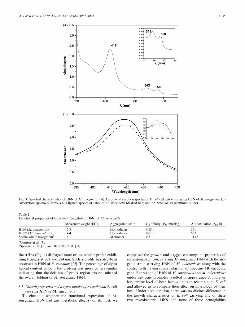
Fig. 3. Spectral characteristics of HbN of M. smegmatis. (A) Absolute absorption spectra of E. coli cell extract carrying HbN of M. smegmatis. (B)Absorption spectra of ferrous NO ligated species of HbN of M. smegmatis (dashed line) and M. tuberculosis (continuous line).
Table 1Functional properties of truncated hemoglobin, HbN, of M. smegmatis
Molecular weight (kDa) Aggregation state O2 affinity (P50 mm/Hg) Autooxidation (t1/2 h)
HbN (M. smegmatis) 12.8 Homodimer 0.18 361HbNa (M. tuberculosis) 14.4 Homodimer 0.013 537Sperm whale myoglobinb 14 Monomer 0.51 13.8
aCouture et al. [4].bSpringer et al. [30] and Barantly et al. [31].
A. Lama et al. / FEBS Letters 580 (2006) 4031–4041 4035
the trHbs (Fig. 4) displayed more or less similar profile exhib-
iting troughs at 208 and 224 nm. Such a profile has also been
observed in HbN of N. commune [23]. The percentage of alpha
helical content of both the proteins was more or less similar
indicating that the deletion of pre-A region has not affected
the overall folding of M. smegmatis HbN.
3.5. Growth properties and oxygen uptake of recombinant E. coli
carrying HbN of M. smegmatis
To elucidate whether the functional expression of M.
smegmatis HbN had any metabolic effect(s) on its host, we
compared the growth and oxygen consumption properties of
recombinant E. coli, carrying M. smegmatis HbN with the iso-
genic strain carrying HbN of M. tuberculosis along with the
control cells having similar plasmid without any Hb encoding
gene. Expression of HbN of M. smegmatis and M. tuberculosis
under vgb gene promoter resulted in appearance of more or
less similar level of both hemoglobins in recombinant E. coli
and allowed us to compare their effect on physiology of their
host. Under high aeration, there was no distinct difference in
the growth characteristics of E. coli carrying any of these
two mycobacterial HbN and none of these hemoglobins
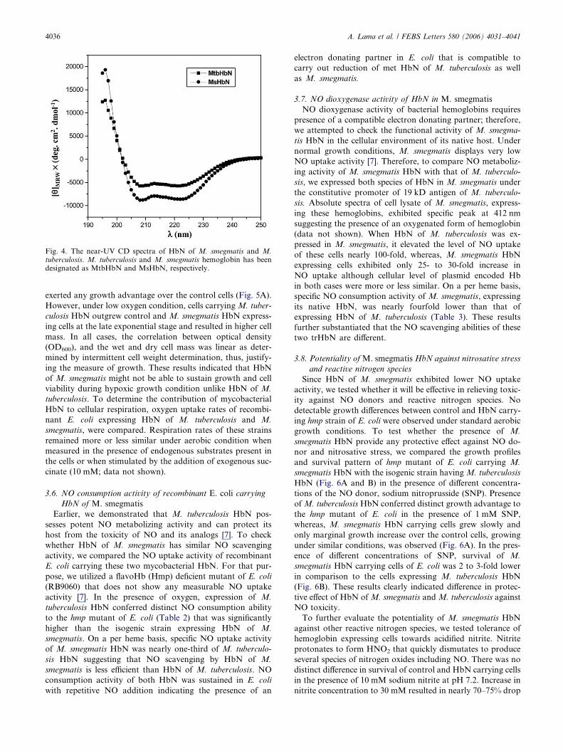
Fig. 4. The near-UV CD spectra of HbN of M. smegmatis and M.tuberculosis. M. tuberculosis and M. smegmatis hemoglobin has beendesignated as MtbHbN and MsHbN, respectively.
4036 A. Lama et al. / FEBS Letters 580 (2006) 4031–4041
exerted any growth advantage over the control cells (Fig. 5A).
However, under low oxygen condition, cells carrying M. tuber-
culosis HbN outgrew control and M. smegmatis HbN express-
ing cells at the late exponential stage and resulted in higher cell
mass. In all cases, the correlation between optical density
(OD600), and the wet and dry cell mass was linear as deter-
mined by intermittent cell weight determination, thus, justify-
ing the measure of growth. These results indicated that HbN
of M. smegmatis might not be able to sustain growth and cell
viability during hypoxic growth condition unlike HbN of M.
tuberculosis. To determine the contribution of mycobacterial
HbN to cellular respiration, oxygen uptake rates of recombi-
nant E. coli expressing HbN of M. tuberculosis and M.
smegmatis, were compared. Respiration rates of these strains
remained more or less similar under aerobic condition when
measured in the presence of endogenous substrates present in
the cells or when stimulated by the addition of exogenous suc-
cinate (10 mM; data not shown).
3.6. NO consumption activity of recombinant E. coli carrying
HbN of M. smegmatis
Earlier, we demonstrated that M. tuberculosis HbN pos-
sesses potent NO metabolizing activity and can protect its
host from the toxicity of NO and its analogs [7]. To check
whether HbN of M. smegmatis has similar NO scavenging
activity, we compared the NO uptake activity of recombinant
E. coli carrying these two mycobacterial HbN. For that pur-
pose, we utilized a flavoHb (Hmp) deficient mutant of E. coli
(RB9060) that does not show any measurable NO uptake
activity [7]. In the presence of oxygen, expression of M.
tuberculosis HbN conferred distinct NO consumption ability
to the hmp mutant of E. coli (Table 2) that was significantly
higher than the isogenic strain expressing HbN of M.
smegmatis. On a per heme basis, specific NO uptake activity
of M. smegmatis HbN was nearly one-third of M. tuberculo-
sis HbN suggesting that NO scavenging by HbN of M.
smegmatis is less efficient than HbN of M. tuberculosis. NO
consumption activity of both HbN was sustained in E. coli
with repetitive NO addition indicating the presence of an
electron donating partner in E. coli that is compatible to
carry out reduction of met HbN of M. tuberculosis as well
as M. smegmatis.
3.7. NO dioxygenase activity of HbN in M. smegmatis
NO dioxygenase activity of bacterial hemoglobins requires
presence of a compatible electron donating partner; therefore,
we attempted to check the functional activity of M. smegma-
tis HbN in the cellular environment of its native host. Under
normal growth conditions, M. smegmatis displays very low
NO uptake activity [7]. Therefore, to compare NO metaboliz-
ing activity of M. smegmatis HbN with that of M. tuberculo-
sis, we expressed both species of HbN in M. smegmatis under
the constitutive promoter of 19 kD antigen of M. tuberculo-
sis. Absolute spectra of cell lysate of M. smegmatis, express-
ing these hemoglobins, exhibited specific peak at 412 nm
suggesting the presence of an oxygenated form of hemoglobin
(data not shown). When HbN of M. tuberculosis was ex-
pressed in M. smegmatis, it elevated the level of NO uptake
of these cells nearly 100-fold, whereas, M. smegmatis HbN
expressing cells exhibited only 25- to 30-fold increase in
NO uptake although cellular level of plasmid encoded Hb
in both cases were more or less similar. On a per heme basis,
specific NO consumption activity of M. smegmatis, expressing
its native HbN, was nearly fourfold lower than that of
expressing HbN of M. tuberculosis (Table 3). These results
further substantiated that the NO scavenging abilities of these
two trHbN are different.
3.8. Potentiality of M. smegmatis HbN against nitrosative stress
and reactive nitrogen species
Since HbN of M. smegmatis exhibited lower NO uptake
activity, we tested whether it will be effective in relieving toxic-
ity against NO donors and reactive nitrogen species. No
detectable growth differences between control and HbN carry-
ing hmp strain of E. coli were observed under standard aerobic
growth conditions. To test whether the presence of M.
smegmatis HbN provide any protective effect against NO do-
nor and nitrosative stress, we compared the growth profiles
and survival pattern of hmp mutant of E. coli carrying M.
smegmatis HbN with the isogenic strain having M. tuberculosis
HbN (Fig. 6A and B) in the presence of different concentra-
tions of the NO donor, sodium nitroprusside (SNP). Presence
of M. tuberculosis HbN conferred distinct growth advantage to
the hmp mutant of E. coli in the presence of 1 mM SNP,
whereas, M. smegmatis HbN carrying cells grew slowly and
only marginal growth increase over the control cells, growing
under similar conditions, was observed (Fig. 6A). In the pres-
ence of different concentrations of SNP, survival of M.
smegmatis HbN carrying cells of E. coli was 2 to 3-fold lower
in comparison to the cells expressing M. tuberculosis HbN
(Fig. 6B). These results clearly indicated difference in protec-
tive effect of HbN of M. smegmatis and M. tuberculosis against
NO toxicity.
To further evaluate the potentiality of M. smegmatis HbN
against other reactive nitrogen species, we tested tolerance of
hemoglobin expressing cells towards acidified nitrite. Nitrite
protonates to form HNO2 that quickly dismutates to produce
several species of nitrogen oxides including NO. There was no
distinct difference in survival of control and HbN carrying cells
in the presence of 10 mM sodium nitrite at pH 7.2. Increase in
nitrite concentration to 30 mM resulted in nearly 70–75% drop
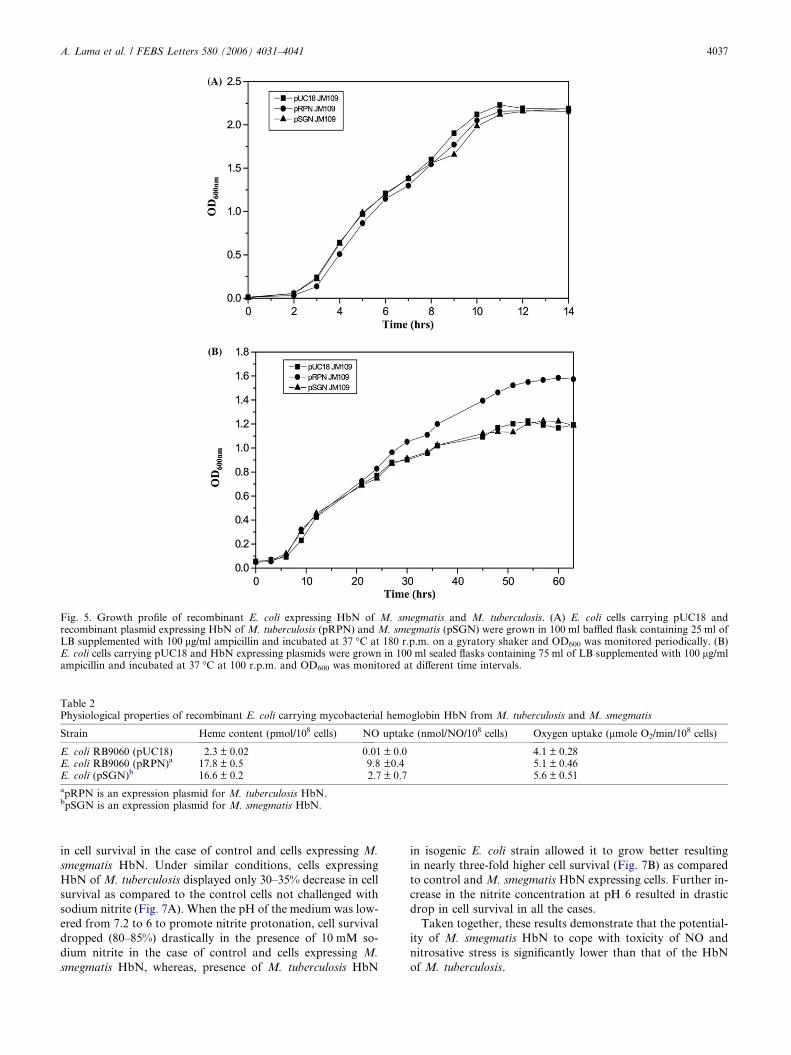
Fig. 5. Growth profile of recombinant E. coli expressing HbN of M. smegmatis and M. tuberculosis. (A) E. coli cells carrying pUC18 andrecombinant plasmid expressing HbN of M. tuberculosis (pRPN) and M. smegmatis (pSGN) were grown in 100 ml baffled flask containing 25 ml ofLB supplemented with 100 lg/ml ampicillin and incubated at 37 �C at 180 r.p.m. on a gyratory shaker and OD600 was monitored periodically. (B)E. coli cells carrying pUC18 and HbN expressing plasmids were grown in 100 ml sealed flasks containing 75 ml of LB supplemented with 100 lg/mlampicillin and incubated at 37 �C at 100 r.p.m. and OD600 was monitored at different time intervals.
Table 2Physiological properties of recombinant E. coli carrying mycobacterial hemoglobin HbN from M. tuberculosis and M. smegmatis
Strain Heme content (pmol/108 cells) NO uptake (nmol/NO/108 cells) Oxygen uptake (lmole O2/min/108 cells)
E. coli RB9060 (pUC18) 2.3 ± 0.02 0.01 ± 0.0 4.1 ± 0.28E. coli RB9060 (pRPN)a 17.8 ± 0.5 9.8 ±0.4 5.1 ± 0.46E. coli (pSGN)b 16.6 ± 0.2 2.7 ± 0.7 5.6 ± 0.51
apRPN is an expression plasmid for M. tuberculosis HbN.bpSGN is an expression plasmid for M. smegmatis HbN.
A. Lama et al. / FEBS Letters 580 (2006) 4031–4041 4037
in cell survival in the case of control and cells expressing M.
smegmatis HbN. Under similar conditions, cells expressing
HbN of M. tuberculosis displayed only 30–35% decrease in cell
survival as compared to the control cells not challenged with
sodium nitrite (Fig. 7A). When the pH of the medium was low-
ered from 7.2 to 6 to promote nitrite protonation, cell survival
dropped (80–85%) drastically in the presence of 10 mM so-
dium nitrite in the case of control and cells expressing M.
smegmatis HbN, whereas, presence of M. tuberculosis HbN
in isogenic E. coli strain allowed it to grow better resulting
in nearly three-fold higher cell survival (Fig. 7B) as compared
to control and M. smegmatis HbN expressing cells. Further in-
crease in the nitrite concentration at pH 6 resulted in drastic
drop in cell survival in all the cases.
Taken together, these results demonstrate that the potential-
ity of M. smegmatis HbN to cope with toxicity of NO and
nitrosative stress is significantly lower than that of the HbN
of M. tuberculosis.

Table 3NO consumption activity of M. smegmatis, expressing HbN of M.tuberculosis and M. smegmatis
Strains Heme content(pmol/108 cells)
NO consumptionactivity(nmole of NOheme�1 s�1 )
M. smegmatis (p19Kpro) 1.6 ± 0.5 0.09 ± 0.7M. smegmatis (pRPN-2) 4.8 ± 0.9 18.6 ± 0.5M. smegmatis (pSGN-2) 6.4 ± 0.1 5.1 ± 0.7
M. smegmatis cultures were grown in Middlebrook 7H9 broth (Difco)supplemented with ADC (10% bovine serum albumin fraction V,dextrose and sodium chloride), 2% glycerol, 0.05% Tween 80 and50 lg/ml Hygromycin (wherever required) at 37 �C with vigorousshaking (250 r.p.m) for 10 h. Cells were then harvested by centrifu-gation (at 8000 · g for 30 min at 4 �C), washed with NO consumptionassay buffer (see Section 2) and resuspended in the same buffer at a celldensity of nearly 3 · 107 cells/ll and NO consumption activities ofcontrol M. smegmatis (p19Kpro) and that carrying HbN of M.tuberculosis (pRPN-2) and M. smegmatis (pSGN-2) were determined.
Fig. 6. Effect of sodium nitroprusside (SNP) on growth and survival of reco(A) Growth characteristics of E. coli expressing HbN of M. tuberculosis andexpressing E. coli in the presence of different concentrations of SNP. Surviv
4038 A. Lama et al. / FEBS Letters 580 (2006) 4031–4041
4. Discussion
Slow growing pathogenic mycobacteria, such as M. tubercu-
losis, M. bovis, M. avium, etc. carry genes encoding HbN and
HbO type truncated hemoglobins that may play vital role in
their cellular metabolism. Functional relevance of these trun-
cated hemoglobins in mycobacterial cellular metabolism is
not conclusively known. Biochemical and structural studies
conducted on hemoglobins of M. tuberculosis in our labora-
tory and of others [4,5,7,8] suggest that HbN may be involved
in oxygen-sustained detoxification of NO that may provide a
defense mechanism to the tubercle bacillus against macro-
phage-generated reactive nitrogen species. In the present work,
we have shown that truncated hemoglobin, HbN, of fast-grow-
ing, non-pathogenic mycobacterium, M. smegmatis, has func-
tional characteristics that are distinct from HbN of M.
tuberculosis.
mbinant E. coli expressing HbN of M. tuberculosis and M. smegmatis.M. smegmatis in the presence of SNP (1 mM). (B) Survival of HbN
al is represented as percentage of the values for the untreated cells.

Fig. 7. Survival of HbN expressing recombinant E. coli in the presenceof acidified nitrite. (A) Survival pattern of control and HbN expressingE. coli cells in the presence of different concentrations of sodium nitriteat pH 7.2. (B) Survival pattern of control and HbN expressing E. colicells under different levels of sodium nitrite at pH 6.0. Survival isrepresented as the percentage value of the untreated cells.
A. Lama et al. / FEBS Letters 580 (2006) 4031–4041 4039
M. smegmatis HbN is smaller in size than its counterpart
present in other mycobacterial species and lacks N-terminal
pre-A region, however, conservation of structural features
crucial for the stabilization of the truncated globin fold sug-
gests that M. smegmatis HbN may be able to function as
hemoglobin. Near-UV CD spectra of HbN of M. smegmatis
and M. tuberculosis is very similar indicating the presence of
high degree of alpha-helical structure as has been observed in
HbN of N. commune [23]. The spectral and biochemical char-
acteristics show that M. smegmatis HbN is a cooperative
homodimer and contains a five-coordinated ferrous heme
group when the protein has no bound ligand and that CO
and oxygen bind to the heme moiety in a reversible manner,
very similar to other functional Hbs. Oxygen affinity of M.
smegmatis HbN is relatively lower than HbN of M. tubercu-
losis, despite the fact that both display TyrB10 and GlnE7
distal residues and share a comparable distal site sequence
composition. This suggests that other factors may play a role
in modulating the overall ligand affinity of M. smegmatis
HbN. Lack of pre-A region, that makes contact with the
pre-F loop and G-helix in HbN of M. tuberculosis, may
affect access and kinetics of ligand access through pro-
tein motion [9,10] resulting in differences in oxygen binding
of M. smegmatis HbN. Autooxidation properties of HbN
of M. tuberculosis and M. smegmatis further suggest that
interactions of oxygen with these two trHbs may have some
differences.
Although HbN of M. smegmatis and M. tuberculosis share
close structural similarity, their ability to scavenge NO and
provide protection from the toxicity of NO and nitrosative
stress is quite different. M. smegmatis HbN exhibits lower
NO metabolizing activity in heterologous host, E. coli, as
well as in its native host as compared to HbN of M. tuber-
culosis. Molecular basis of this functional difference between
these two mycobacterial HbN is currently unknown. Oxygen
dependent NO consumption by single domain bacterial
hemoglobin,VHb, has been demonstrated [20] and it has
been suggested that structural features of VHb allows it to
associate with a partner flavoreductase in vivo under certain
physiological conditions to create a transient flavoHb like
structure that may participate in NO metabolism. Optical
spectra of NO-bound HbN of M. tuberculosis overlaps with
that of M. smegmatis indicating that both species are able to
bind NO. Thus, the distinct difference in NO metabolizing
activity of these two hemoglobins may be due to their cata-
lytic efficiency for NO dioxygenation. HbN exhibits potent
NO metabolizing activity and protects heterologous hosts
from the toxicity of NO and nitrosative stress, therefore, it
is quite possible that a partner flavoreductase may associate
with it in vivo to attain NOD function. NO dioxygenase
activity of M. smegmatis HbN is significantly lower than
HbN of M. tuberculosis. HbN of M. smegmatis has nearly
70% sequence similarity with HbN of M. tuberculosis and
it does not carry any charged pre-A region, therefore, the
coupling efficiency of these two hemoglobins with its elec-
tron-donating partner may differ resulting in differences in
their ability to interact with NO and reactive nitrogen spe-
cies. Difference in functionality of these two mycobacterial
trHbN can be correlated with their functional relevance with-
in the cellular environment of their native host. M. tubercu-
losis is an intracellular pathogen that resides and replicates
within the hypoxic and NO-enriched environment of macro-
phages, thus, may require a strong NO detoxification system
to cope with the toxic level of its intracellular environment.
In comparison, M. smegmatis is not naturally exposed to
such hazardous conditions and oxygen and NO binding
properties of its HbN may provide sufficient protection from
the level of stress faced by its host. Alternatively, some other
NO-scavenging system may be operative in M. smegmatis. It
is notable that M. tuberculosis genome carries gene encoding
for a putative flavohemoglobin (flavoHb) and similar fla-
voHb homolog has also been identified within the genome
sequence of M. smegmatis (unpublished observation). Role
of flavoHbs in protection from the toxicity of NO and nitro-
sative stress has been well documented in the literature [24–
26]. It is quite possible that M. tuberculosis requires multiple
systems for relieving stress against high level of NO and reac-
tive nitrogen species generated within the macrophages,

4040 A. Lama et al. / FEBS Letters 580 (2006) 4031–4041
whereas, flavoHb may be sufficient to take care of the phys-
iological stress encountered by M. smegmatis.
The pre-A region, carrying highly polar sequence motif, is
conserved in HbN of other pathogenic mycobacteria; however,
the relevance of this additional secondary structure in protein
function is not known at present. HbN of M. smegmatis does
not carry this polar motif at its N-terminus. It is quite possible
that the presence of this additional secondary structure at the
N-terminus of HbN may have some functional significance for
pathogenic mycobacterium, which possibly may not be re-
quired for the function of HbN of M. smegmatis.
The data reported here provides first report on truncated
hemoglobin, HbN, of a fast-growing mycobacterium, M.
smegmatis, and demonstrates that its functional properties
are significantly different from its counterpart present in path-
ogenic mycobacterium, M. tuberculosis. The major difference
in functionality of these two HbN type truncated hemoglobins
lies in their capability to metabolize NO and provide protec-
tion against NO and nitrosative stress. Physiological func-
tion(s) of M. smegmatis HbN is not very obvious at present.
M. smegmatis HbN binds oxygen with reasonably high affinity,
thus, there is a possibility that it may also participate in other
metabolic reactions involving oxygen as has been envisaged for
the protozoan and cyanobacterial HbN [27–29].
Acknowledgements: We are thankful to Prof. N. Ninfa for makingavailable E. coli RB9060 strain for our work. Technical assistance pro-vided by Mr. S. Muthukrishnan is gratefully acknowledged. This workhas been supported by research grants provided by Council of Scien-tific and Industrial Research (SMM003) and the Department ofBiotechnology, Govt. of India.
References
[1] Cole, S.T., Brosch, R., Parkhill, J., Garnier, T., Churcher, C.,Harris, D., Gordon, S.V., Eiglmeier, K., Gas, S., Barry, C.E.,Tekaia, F., Badcock, K., Basham, D., Brown, D., Chillingworth,T., Connor, R., Davies, R., Devlin, K., Feltwell, T., Gentles, S.,Hamlin, N., Holroyd, S., Hornsby, T., Jagels, K., Krogh, A.,Mclean, J., Moule, S., Murphy, L., Oliver, K., Osborne, J., Quail,M.A., Rajandream, M.A., Rogers, J., Rutters, S., Seeger, K.,Skelton, J., Squares, R., Squares, S., Sulston, J.E., Taylor, K.,Whitehead, S. and Barrell, B.G. (1998) Deciphering the biology ofMycobacterium tuberculosis from the complete genome sequence.Nature 393, 537–544.
[2] Cole, S.T., Eiglmeier, K., Parkhill, J., James, K.D., Thomson,N.R., Wheeler, P.R., Honore, N., Garnier, T., Churcher, C.,Harris, D., Mungall, K., Basham, D., Brown, D., Chillingworth,T., Connor, R., Davies, R.M., Devlin, K., Duthoy, S., Feltwell,T., Fraser, A., Hamlin, N., Holroyd, S., Hornsby, T., Jagels, K.,Lacroix, C., Maclean, J., Moule, S., Murphy, I., Oliver, K., Quail,M.A., Rajandream, M.A., Rutherford, K.M., Rutter, S., Seeger,K., Simon, S., Simmonds, M., Skelton, J., Squares, R., Squares,S., Stevens, K., Taylor, K., Whitehead, S., Woodward, J.R. andBarrel, B.G. (2001) Massive gene decay in the leprosy bacillus.Nature 409, 1007–1011.
[3] Wittenberg, J.B., Bolognesi, M., Wittenberg, B.A. and Guertin,M. (2001) Truncated hemoglobins: a new family of hemoglobinswidely distributed in bacteria, unicellular eukaryotes, and plants.J. Biol. Chem. 277, 871–874.
[4] Couture, M., Yeh, S.R., Wittenberg, B.A., Wittenberg, J.B.,Ouellet, Y., Rousseau, D.L. and Guertin, M. (1999) A cooper-ative oxygen-binding hemoglobin from Mycobacterium tubercu-losis. Proc. Natl. Acad. Sci. USA 96, 11223–11228.
[5] Ouellet, H., Juszczak, L., Dantsker, D., Samuni, U., Ouellet,Y.H., Savard, P.Y., Wittenberg, J.B., Wittenberg, B.A., Fried-man, J.M. and Guertin, M. (2003) Reaction of Mycobacteriumtuberculosis truncated hemoglobin O with ligand reveal a novel
ligand inclusive hydrogen bond network. Biochemistry 42, 5764–5774.
[6] Pathania, R., Navani, N.K., Rajamohan, G. and Dikshit, K.L.(2002) Mycobacterium tuberculosis Hemoglobin HbO associateswith membranes and stimulates cellular respiration of recombi-nant E. coli. J. Biol. Chem. 277, 15293–15302.
[7] Pathania, R., Navani, N.K., Gardner, A.M., Gardner, P.R. andDikshit, K.L. (2002) Nitric oxide scavenging and detoxification bythe Mycobacterium tuberculosis haemoglobin, HbN in Escherichiacoli. Mol. Microbiol. 45, 1303–1314.
[8] Ouellet, H., Ouellet, Y., Richard, C., Labarre, M., Wittenberg, B.,Wittenberg, J. and Guertin, M. (2002) Truncated hemoglobinHbN protects Mycobacterium bovis from nitric oxide. Proc. Natl.Acad. Sci. USA 99, 5902–5907.
[9] Milani, M., Pesce, A., Ouellet, Y., Ascenzi, P., Guertin, M. andBolognesi, M. (2001) Mycobacterium tuberculosis hemoglobin Ndisplays a protein tunnel suited for O2 diffusion to the heme.EMBO J. 20, 3902–3909.
[10] Milani, M., Pesce, A., Ouellet, Y., Dewilde, S., Friedman, J.,Ascenzi, P., Guertin, M. and Bolgnesi, M. (2004) Heme-ligandtunneling in group I truncated hemoglobins. J. Biol. Chem. 279,21520–21525.
[11] Yeh, S.R., Couture, M., Ouellet, Y., Guertin, M. and Rousseau,D.L. (2000) A cooperative oxygen binding hemoglobin fromMycobacterium tuberculosis. Stabilization of heme ligands by adistal tyrosine residue. J. Biol. Chem. 275, 1679–1684.
[12] Samuni, U., Ouellet, Y., Guertin, M., Friedman, J.M. and Yeh,S.R. (2004) The absence of proximal strain in the truncatedhemoglobins from Mycobacterium tuberculosis. J. Am. Chem.Soc. 126, 2682–2683.
[13] Poole, R.K. and Hughes, M.N. (2000) New functions for theancient globin family,bacterial response to nitric oxide andnitrosative stress. Mol. Microbiol. 36, 775–783.
[14] Gardner, P.R. (2005) Nitric oxide dioxygenase function andmechanism of flavohemoglobin, hemoglobin, myogloin and theirassociated reductases. J. Inorg. Biochem. 99, 247–266.
[15] Hernandez-Urzua, E., Mills, C.E., White, G.P., Contreras-Zen-tella, M.L., Escamilla, E., Vasudevan, S.G., Membrillo-Hernan-dez, J. and Poole, R.K. (2003) Flavohemoglobin Hmp, but not itsindividual domains, confers protection from respiratory inhibi-tion by nitric oxide in Escherichia coli. J. Biol. Chem. 278, 34975–34982.
[16] MacMicking, J.D., North, R.J., LaCourse, R., Mudgett, J.S.,Shah, S.K. and Nathan, C.F. (1997) Identification of nitric oxidesynthase as a protective locus against tuberculosis. Proc. Natl.Acad. Sci. USA 94, 5243–5248.
[17] Turenne, C.Y., Suchak, A.A., Wolfe, J.N., Kabani, A. andNicolle, L.E. (2003) Soft tissue infection caused by a novelpigmented, rapidly growing mycobacterium species. J. Clin.Microbiol. 41, 2779–2782.
[18] Snapper, S.B., Melton, R.E., Mustafa, S., Kieser, T. and JacobsJr., W.R. (1990) Isolation and characterization of efficient plasmidtransformation mutants of Mycobacterium smegmatis. Mol.Microbiol. 4, 1911–1919.
[19] Dikshit, K.L. and Webster, D.A. (1988) Cloning, characterizationand expression of the bacterial globin gene from Vitreoscilla inEscherichia coli. Gene 70, 377–386.
[20] Kaur, R., Pathania, R., Sharma, V., Mande, S.C. and Dikshit,K.L. (2002) Chimeric Vitreoscilla hemoglobin (VHb) carrying aflavoreductase domain relieves nitrosative stress in Escherichiacoli: new insight into the functional role of VHb. Appl. Environ.Microbiol. 68, 152–160.
[21] Poole, R.K., Anjum, M.F., Membrillo-Hernandez, J., Kim, S.O.,Hughes, M.N. and Stewart, V. (1996) Nitric oxide, nitrite and Fnrregulation of hmp (flavohemoglobin) gene expression in Esche-richia coli K-12. J. Bacteriol. 178, 5487–5492.
[22] Appleby, C.A. (1978) Purification of Rhizobium cytochromes P-450. Methods Enzymol. 52, 157–166.
[23] Thorsteinsson, M.V., Bevan, D.R., Ebel, R.E., Weber, R.E. andPotts, M. (1996) Spectroscopical and functional characterizationof the hemoglobin of Nostoc commune UTEX 584 (Cyanobacte-ria). Biochim. Biophys. Acta 1292, 133–139.
[24] Hausladen, A., Gow, A.J. and Stamler, J.S. (1998) Nitrosativestress: metabolic pathway involving the flavohemoglobin. Proc.Natl. Acad. Sci. USA 95, 14100–14105.

A. Lama et al. / FEBS Letters 580 (2006) 4031–4041 4041
[25] Liu, J., Zeng, M., Hausladen, A., Heitman, J. and Stamler, J.S.(2000) Protection from nitrosative stress by yeast flavohemoglo-bin. Proc. Natl. Acad. Sci. USA 97, 4672–4676.
[26] Stevanin, T.M., Ioannidis, N., Mills, C.E., Kim, S.O., Hughes,M.N. and Poole, R.K. (2000) Flavohemoglobin Hmp affordsinducible protection for Escherichia coli respiration, catalyzed bycytochromes bo 0 or bd, from nitric oxide. J. Biol. Chem. 275,35868–35875.
[27] Yamauchi, K., Tada, H. and Usuki, I. (1995) Structure andevolution of Paramecium hemoglobin genes. Biochim. Biophys.Acta 1264, 53–62.
[28] Couture, M., Das, T.K., Lee, H.C., Peisach, J., Rousseau, D.L.,Wittenberg, B.A., Wittenberg, J.B. and Guertin, M. (1999)Chlamydomonas chloroplast ferrous hemoglobin: heme pocket
structure and reactions with ligands. J. Biol. Chem. 274, 6898–6910.
[29] Das, T.K., Weber, R.E., Dewilde, S., Wittenberg, J.B., Witten-berg, B.A., Yamauchi, K., VanHauwaert, M.L., Moens, L. andRousseau, D.L. (2000) Ligand binding in the ferric and ferrousstates of Paramecium hemoglobin. Biochemistry 39, 14330–14340.
[30] Springer, B.A., Egeberg, K.D., Sligar, S.G., Rohlfs, R.J.,Mathews, A.J. and Olson, J.S. (1989) Discrimination betweenoxygen and carbon monoxide and inhibition of autooxidation bymyoglobin. site directed mutagenesis of the distal histidine. J.Biol. Chem. 264, 3057–3060.
[31] Brantley Jr., R.E., Smerdon, S.J., Wilkinson, A.J., Singleton,E.W. and Olson, J.S. (1993) The mechanism of autooxidation ofmyoglobin. J. Biol. Chem. 268, 6995–7010.