estimation of haemoglobin
20
HAEMOGLOBIN ESTIMATION
-
Upload
khangminh22 -
Category
Documents
-
view
1 -
download
0
Transcript of estimation of haemoglobin

HAEMOGLOBINESTIMATION
STRUCTURE OF HAEMOGLOBIN:
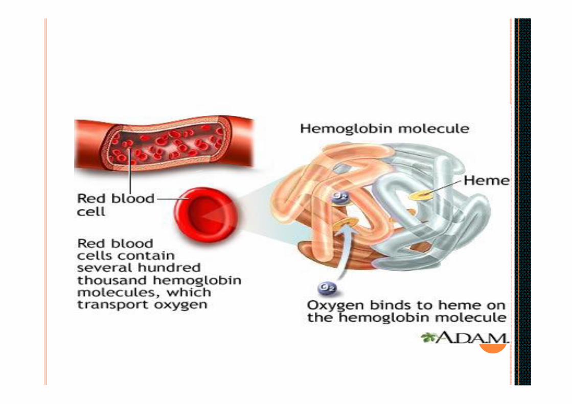
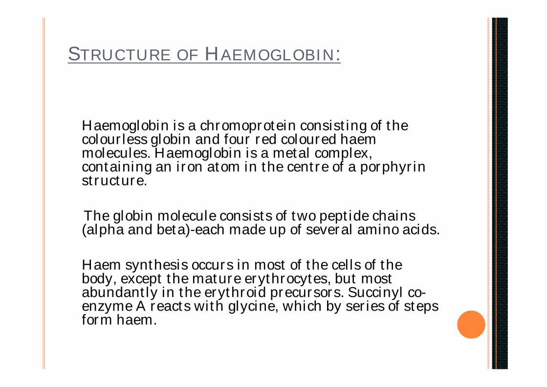
Haemoglobin is a chromoprotein consisting of the colourless globin and four red coloured haemmolecules. Haemoglobin is a metal complex, containing an iron atom in the centre of a porphyrinstructure.
The globin molecule consists of two peptide chains (alpha and beta)-each made up of several amino acids.
Haem synthesis occurs in most of the cells of the body, except the mature erythrocytes, but most abundantly in the erythroid precursors. Succinyl co-enzyme A reacts with glycine, which by series of steps form haem.

Globin synthesis occurs in the cytoplasm of the normoblastand reticulocyte.
The polypeptide chains are manufactured on the ribosomes. In each hemoglobin molecule, one haem group is inserted into a hydrophobic pocket of one folded -polypeptide chain.
Normal adult HbA consists of four haem groups and four polypeptide chains ( 2 alpha & 2 beta ).
RBCs have 120-day life span. As the cell ages, activity of certain glycolytic enzymes is diminished and the cells assume a smaller surface area.. These are lysed by the RE system and are removed from circulation. Hemoglobin also degrades and the components are recycled.

ESTIMATION OF HAEMOGLOBIN :
Many methods are available:-1. Colorimetric method:-
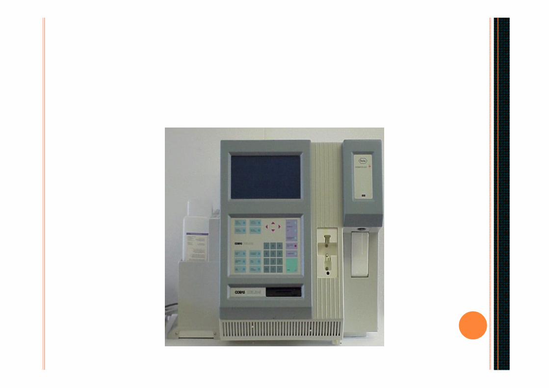
Cyanmethaemoglobin method :-
A aliquot of well mixed whole blood is taken and it is diluted in a solution of Potassium ferricyanide and potassium cyanide. The potassium ferricyanideoxidizes hemoglobin to hemiglobin to form hemiglobincyanide ( HiCN ) which has a broad absorption maximum at a wavelength of 540nm. It is measured in a photometer or a spectro-phototmeter at 540nm and compared with that of a standard HiCN solution.
2. Acid hematin ( Sahli’s method ) and alkali hematinmethod :
Principle :-Hemoglobin is converted to acid hematin or alkaline hematin and colour is compared with glass standards in a comparator.
3. In the gasometric method, the oxygen carrying capacity of the blood is measured with the Vonslyke apparatuasand hemoglobin is estimated indirectly.
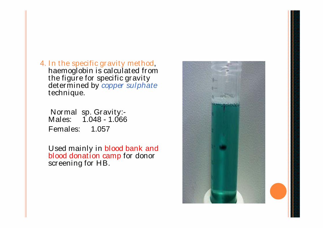
4. In the specific gravity method, haemoglobin is calculated from the figure for specific gravity determined by copper sulphatetechnique.
Normal sp. Gravity:-Males: 1.048 - 1.066Females: 1.057
Used mainly in blood bank and blood donation camp for donor screening for HB.
5. Haemoglobin is determined by chemical method by measuring iron content of blood. Iron content of Hb is 0.347%. It is a complex method.
Very accurate methods are cynamethaemoglobin and chemical method, but not used in routine practice. Cyanmethhaemoglobin method is recommended by International committee for standardisation in haemotology & is widely used in laboratories the world over.
Acid hematin method is simple and accurate technique for routine clinical use.
Acid Hematin Method ( Sahli’s ):
Principle :- Hemoglobin is converted into acid hematin by diluting with weak
acids and further diluted until its colour matches with that of the comparator.
Apparatus:-Sahli type of hemoglobinometer with comparator with glass standardsHemoglobin square tube marked both in grams and percentages.Hemoglobin pipette marked at 20c.m.0.1 N HClDistilled water
Specimen :- Double oxalated or EDTA venous blood or capillary blood. Anticoagulated sample must be thoroughly mixed by gentle shaking.

Method :-1. Fill Hb square tube upto mark 20% with 0.1 N HCl.2. Fill Hb pipette with the blood upto mark 20 micro lit Excess blood should be
removed with wet gauze.3. Empty pipette into the acid in the square tube. Rinse pipette atleast three times
by drawing in and discharging the blood acid mixture. Take care to prevent air bubble formation in acid hematin mixture.
4. Mix the acid-hematin mixture in the tube with the glass-rod and allow the tubeto stand for 20minutes.
5. Now dilute the solution by adding distilled water dorp by drop and stirring the mixture all the time with glass rod. The comparator is held against good day light and distilled water is added till the colour of the solution matches perfectly with that of standards. Record the reading in GRAMS. Read the result at the bottom of the meniscus.
6. Clean all the apparatus. The hemoglobin pipette is cleaned by sucking and then expelling water several times.


Errors :-1. Technique of collecting blood sample.2. Manual error: In labelling, in method, in visual
comparision. 3. Instrumental : Comparator glass standards may fade with time, so
dilution is more in the specimen hence higher values of Hb are obtained.Calibration errors.Improper light can lead to variation in result. 4. All forms of Hb are not measured as only Hb and HbO2 can be converted to acid hematin. So it does not give the true value of hemoglobin.

Range of Hemoglobin in healthy individuals:-
Adult males :- 14.0 to 17.5 gm/dl.Adult female:- 12.3 to 15.3gm/dl.Cord blood or new born :- 13.5 to 19.5 gm/dl.At 1 yr :- 11 gm/dlChildren upto 10yrs :- 12 gm/dl
Abnormal values:-Increased - Polycythemia Vera
DehydrationPoorly compensated heart disease
Decreased - Anemia

Abnormal hemoglobin :-The normal adult hemoglobin is hemoglobin A (alpha2 beta2) and foetal hemoglobin is hemoglobin F (alpha2 gamma2 ). At birth HbFis present in the proportion of 60-80% and gradually decreases to 0.5 -2% in adults.
HbA2 (alpha2 delta2) is in the proportion of 1.5 to 3.5% of the normal adult hemoglobin. HbA2 levels are increased in the range of 3.5% to 8.0% in Beta Thalassaemia Minor. HbA2 levels are decreased in iron deficiency anemia.
The abnormalities of Hb may be associated either with Haemmolecule or Globin molecule. In haem molecule disorders, formation of abnormal pigments e.g. carboxyhemoglobin, methemoglobin , sulphemoglobin and porphyrins, due todisorderedporphyrin metabolism occurs. Except some porphyrias, these abnormalities are acquired. Detection of abnormal pigment is done by chemical method & spectroscopic examination.

Hemoglobinopathies refer to the abnormal globin molecule disorders. In these disorders the alpha or the beta peptide chains are abnormal due to constituent amino acids being substituted by another forming different alpha or beta chains. They are differentiated by electrophoretic and chromatographic techniques. They are designated as hemoglobin S,C,D,E,G,H,I,K. These disorders are hereditary. The presence of abnormal hemoglobin may lead to changes in the shape of the red cell e.g. Abnormal hemoglobin S, which makes the RBC sickle shaped. This leads to intracorpuscular defects which reduce their life span.
If the rate of synthesis of polypeptide chains of Hb is diminished but structure is normal, then it causes Thalassaemia. If alpha chains are reduced or absent it is called alpha-thalassaemia. If beta chains are reduced or absent it is called beta-thalassaemia. In case of abnormalities involving both the genes homozygous thalassaemias result Homozygous beta thalassaemia (beta thalassaemia major) is a serious condition affecting infants or children in their early years of life producing transfusion dependent anemia. In B thalassaemias, HbF and HbA2 are increased in quantity and HbA is reduced.
Heterozygous thalassaemias (alpha or beta) serve as carriers in population & usually are asymptomatic.
Beta thalassaemias are more prevalant in Mediterranean countries and parts of Africa and South East Asia, which includes India.