Tristetraprolin targets Nos2 expression in the colonic epithelium
Upload
sorbonne-frCategory
view
2download
0
Eur. J . Immunol. 1996.26: 1807-1815
Yves Modigliani’, Antonio Coutinho’, Pablo Pereira’, Nicole Le Douarin’, VCronique Thomas-Vaslin’, Odile Burlen-Defranoux’, Josselyne Salaun’ and Antonio Bandeira’
UnitC d’Immunobiologie, CNRS URA 1961, Institut Pasteur, Paris, France ’ Institut d’Embryologie Cellulaire
et MolCculaire du CNRS et du Collkge de France, Nogent-sur- Marne, France
Dominant mechanisms in thymic epithelium-induced tolerance 1807
Establishment of tissue-specific tolerance is driven by regulatory T cells selected by thymic epithelium
Grafts of thymic epithelium (TE) rudiments restore T cell development and function in allogeneic athyrnic mice. These TE chimeras are specifically tolerant to grafts of peripheral tissues (e .g . skin and heart) from the TE donor strain, although they harbor peripheral immunocompetent T cells capable of rejecting those grafts. Initial analysis has shown that TE chimeras also harbor TE-selected CD4 T lymphocytes that inhibit graft rejection by tissue-reactive T cells in immu- nocompetent recipients. Peripheral tolerance in TE chimeras is thus maintained by dominant mechanisms dependent on regulatory CD4 T lymphocytes. Here we show that TE-selected regulatory T cells recruit nontolerant tissue-reactive CD4 and CD8T cells to express similar regulatory functions. Only recent thymic emigrants, but not peripheral resident mature T cells are susceptible to this pro- cess of functional education, which also requires exposure to specific antigens and occurs entirely in the periphery. We propose that these mechanisms play a major role in establishing and maintaining natural self tolerance to tissue- specific antigens.
1 Introduction
The thymus plays an essential role in the establishment of self tolerance to antigens expressed on its stroma by clonal elimination [ 1-71 or functional inactivation [8-101 of self- reactive T cells. In the establishment of tolerance to peripheral tissue-specific antigens, the role of the thymus is much less clear, since recessive mechanisms of periph- eral tolerance (ignorance, deletion, anergy, immunodevi- ation) do not require thymic selection. Active suppression [ll] or infectious tolerance [12], on the other hand, can accomodate a role for the thymus in this process. Thymic- dependent specific suppression of aggressive responses to autoantigens that are not expressed intrathymically, how- ever, is a highly controversial concept and the respective mechanisms are difficult to envisage.
Experiments in birds have shown that embryonic grafts of xenogeneic peripheral tissues in age-matched recipients are rejected later in life [13-151. In contrast, similar exper- iments performed with allogeneic peripheral tissues led to tolerance induction in most, but not all, cases: many ani- mals showed mild, recurrent, but reversible signs of rejec- tion [16]. In mammals, allogeneic grafts of peripheral tis- sues performed in newborn [17, 181 or embryonic recipi- ents [ 191 were never tolerized. These observations indicate that post-thymic T cell recognition of peripheral tissue
[I 151681
Received December 20, 1995; in final revised form May 13, 1996, accepted May 14, 1996.
Correspondence: Antonio Bandeira, UnitC d’Immunobiologie, CNRS URA 1961, Institut Pasteur, 25 rue du Docteur Roux, F-75724 Pans Cedex 15, France Fax: +33-1-45-68-89-21
Abbreviations: TE: Thymic epithelium RTE: Recent thymic emigrant PRM: Peripheral resident mature HC: Hemato- poietic cell TE-T cells: T cells from thymic epithelium- reconstituted mice ST: Survival time MST: Mean ST
Key words: Tolerance I Thymic epithelium I CD4 I CD8 / T cell
antigens during development is not sufficient for full toler- ance induction. In contrast, grafts of a variety of periph- eral tissues, in both xeno- or allogeneic combinations, are readily accepted if the recipient has also received thymic epithelium (TE) of the donor type [14-16,20-251. In fact, the establishment of TE-induced tolerance can operate in the absence of those peripheral tissues [14, 15,21-23,251. This property is also shared by hemopoietic cells (HC) which are able to induce tolerance to all other tissues, as first shown by Medawar and colleagues [26]. As previously discussed [ 191, these findings are compatible with toler- ance models based on dominant suppression, rather than with recessive processes of deletion or anergy of self- reactive cells.
In mice, TE chimeras can be generated by grafting athymic recipients with allogeneic TE rudiments obtained from day 10 (E10) embryos, before colonization by hemopoietic cells [22]. These chimeras are permanently tolerant to skin or heart grafts of TE donor strain, but reject third-party grafts [22]. Furthermore, in agreement with independent observations on the relative inefficiency of TE to mediate clonal deletion [8, 23, 27, 281, transfer experiments revealed that tolerant TE chimeras harbor immunocom- petent, tissue-reactive T cells that can readily reject the tolerized grafts [29]. In support of the notion that TE toler- ance is mediated by regulatory mechanisms, CD4 T lym- phocytes selected by TE were thereafter shown to specifi- cally inhibit, in a quantitative manner, the rejection of the tolerized grafts by tissue-reactive T cells from immuno- competent donors [29, 301. Since in this experimental sys- tem allo-antigen expression is strictly limited to the TE, these results also demonstrated that peripheral tolerance is centrally selected.
Based on these observations, we postulated that in TE chi- meras regulatory T cells are positively selected by the allo- geneic TE, escape deletion because hemopoietic antigen- presenting cells (APC) express a distinct MHC, and can thus be produced throughout life. This would contrast with the situation in normal mice, where the progressive colon- ization of the thymus by HC would restrict the putative generation of regulatory T cells to the perinatal period of
0 VCH Verlagsgesellschaft mbH, D-69451 Weinheim, 1996 0014-298Ol96lO808-1807$10 .OO + ,2510

1808 Y. Modigliani et al.
thymic development. If the observations on TE chimeras were to be transposed to normal tissue-specific tolerance, other mechanisms would have to be invoked. Thus, since the numbers of peripheral T cells increase 1000-fold in the first 3-4 weeks of life by accumulation of recent thymic emigrants (RTE) [31], putative regulatory T cells pro- duced around birth would be diluted out in the periphery by high numbers of tissue-specific T cells produced throughout life. We therefore hypothesized that TE- selected regulatory T cells peripherally recruit newly formed tissue-reactive T cells to a similar activity. Such a mechanism would also serve to progressively bias regula- tory T cell repertoires towards tissue-specific antigens. The present report addresses this issue.
Eur. J. Immunol. 1996.26: 1807-1815
2 Materials and methods
2.1 Mice
C57BL/6 (Thy-1.2 Ly-5.2) (B6), BALB/c and C3WHe mice were obtained from IFFA-Credo (EAbresle, France). C57BL/6 nude, C57BW6.Ba.Thy-1.1 (B6.Thy-l.l), c57BW6.L~-5.1 (B6.Ly-5.1) were obtained from the CNRS, CSAL (OrlCans, France). Rag-1-deficient mice were kindly provided by Prof. S. Tonegawa.
2.2 Antibodies and cell surface stainings
The following FITC-, PE- or biotin-labeled monoclonal antibodies were used: 145-2C11, anti-CD3; A20.1, anti-Ly- 5.1; 104.2, anti-Ly-5.2; 19x5, anti-Thy-1.1; 30H12, anti- Thy-1.2. For biotinylated antibodies, PE-labeled streptavi- din or Streptavidin-Tri-Color were used in two- or three- color analyses, respectively. Cell surface staining for flow cytometric analysis or sorting was done as described [30].
2.3 Thymic epithelium chimeras and T cell chimeras
2.3.1 B6(BALBE10) chimeras
The third branchial pouches of El0 (24-28 somites) BALBk embryos were grafted into 6-8-week-old B6 nude mice under the kidney capsule. At this developmental stage, TE grafts are not yet colonized by HC. The degree of T cell reconstitution in the chimeras used in these exper- iments ranged between 30% and 100% of that of euthymic mice, and were never skin grafted.
2.3.2 TE/Bone marrow (TE/BM) chimeras
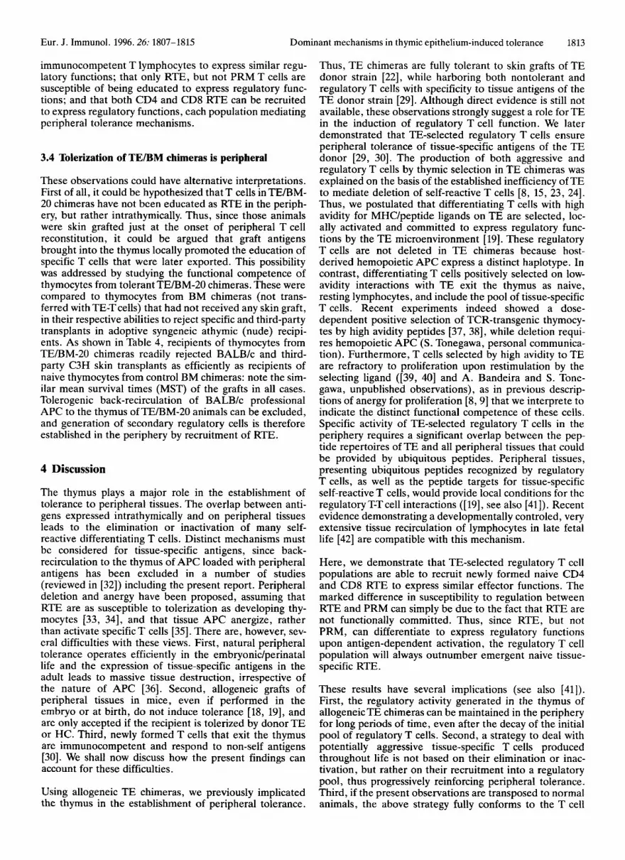
B6.Thy-l.l(Ly-5 . l) were whole-body irradiated (1100 rad) and injected intravenously on the same day with 10’ T cell- depleted BM cells from B6.Ly-5.1(Thy-1.2) mice. After 1 week, animals received the indicated numbers of B6(BAL- BE10) T cells. The generation of these chimeras is explained in detail in Fig. 2 and its legend.
For transfers of tolerance, recipients were either nude mice or animals lethally irradiated and reconstituted with lo7 BM cells from Rag-1-deficient mice using the congenic strain combinations as described in Fig. 2.
2.4 Cell transfers
T cells were purified from mixed spleen and mesenteric lymph node cell suspensions by wheat germ agglutinin treatment [30]. T cell purity was 90-97 YO. CD4 and CD8 T cell populations were positively identified with the appropriate fluorochrome-labeled antibodies and purified by cell sorting with a FACStarPIUs (Becton Dickinson, Mountain View, CA). Cell surface staining and FACS anal- ysis were done as described [30] using the combinations of monoclonal antibodies indicated in the legends of the figu- res and tables.
2.5 Skin grafts
Fragments of about 8 x 8 mm of donor tail skin (epider- mis and dermis) were sutured on the dorsal thorax of anes- thetized recipients. Grafts were considered rejected the day the entire transplant disappeared. Accepted grafts were healthy for 4-6 months, when animals were killed.
3 Results
3.1 RTE are more susceptible to tolerization by TE- selected regulatory T cells than peripheral resident mature (PRM) T cells
We sought to compare quantitatively the susceptibility of naive RTE and PRM T cells to the activity of TE-selected
5 :I 0 0
/
Days after irradiation
Figure 1. Kinetics of donor mature T cell expansion and BM- derived T cell reconstitution in peripheral lymphoid organs of irradiated, syngeneic recipients. B6.Thy-l.l(Ly-5.2) recipients (0) were lethally irradiated (1200 rad) and reconstituted with lo7 T cell-depleted B6.Ly-5.1 (Thy-1.2) BM cells (0). After 1 week, animals received 1 X lo6 (*), 3 x lo6 (W) or 8 x lo6 (A) purified B6 (Ly-5.2, Thy-1.2) peripheral T cells as equivalents of TE-T cells. At the indicated time points, T cell chimerism in the spleen and lymph nodes of recipient mice was studied by flow cytofluoro- metric analyses of CD3’ cells by double- or triple-staining with the respective congenic markers: radiation resistant, host T cells (0): anti-Thy-1.1; newly formed, donor BM-derived T cells (0): anti-Ly-5.1; donor mature T cells (+, W, A): anti-Ly-5.2 and anti- Thy-1.2. The total number of pooled splenic and mesenteric lymph node T cells was determined for each T cell population in individual mice. Each point represents the mean of at least five animals; the SD were always less than 15 % of the mean.

Eur. J. Immunol. 1996.26: 1807-1815 Dominant mechanisms in thymic epithelium-induced tolerance 1809
mmy-i.i [Ly-5.2)
9 f f 86
BALB Ic C3WHe
skin grafts
86 Thy1 .l T cdl d w e d
hahD
/ io\ I I
BM-T cells I I I I I I I I
BB my-i .i (Ly-5.2)
BALWc skin gran I
Figure 2. The experimental model and adoptive cell transfers. (a) Generation of TEIBM chimeras. B6.Thy-l.l(Ly-5.2) mice were lethally irradiated (1200 rad) and reconstituted with lo7 T cell- depleted B6.Ly-5.1 (Thy-1.2) BM cells (0). After 1 week, groups of 12-16 animals received 1 X lo6, 3 X lo6 or 8 X lo6 purified B6 (Ly-5.2, Thy-1.2) T cells obtained from B6(BALBE10) chimeras (denoted TE-T cells; 0) as a source of regulatory T cells. Then 20, 40 or 60 days after irradiation and BM reconstitution, four to six mice of each group of recipients of the same number of TE-T cells were skin grafted. Each individual TEIBM chimera was transplanted with B6 (syngeneic), BALB/c (experimental) and C3H/He (third-party) skin grafts. (b) Transfer of tolerance induced in TE/BM chimeras. Four to six months after skin trans- plantation, 2 x 10⁶-3 x lo6 purified, peripheral BM-derived T cells, denoted BM-T cells (0), of TElBM chimeras were trans- ferred into lethally (1200 rad) irradiated B6.Thy-1.1 recipients. These recipients were reconstituted with lo7 BM cells of Rag-1 KO mice; thus, de n o w T and B cell development was impaired. After 2 weeks, each recipient received the three skin grafts indi- cated in (a). (c) Development of regulatory cell function in BM-T cells of tolerant T E B M chimeras. Purified CD3, CD4 or CD8 BM-T cells (2 x 10⁶-3 x 10') (0) were cotransferred with 2 X 10⁶-3 X lo', purifiedT cells from normal B6.Thy-1.1 mice. Recipi- ents and skin grafts are as indicated in (b).
regulatory T cells when challenged by skin grafts of the TE donor type. To this end, peripheral T cell reconstitution of naive irradiated recipients was allowed to occur in the presence of adoptively transferred syngeneic regulatory T cells from TE chimeras. A group of these animals was skin grafted at the onset of reconstitution, such that all naive T cells met antigen (and regulatory T cells) as RTE.
Another group of animals received skin grafts after the peripheral T cell pool had been entirely reconstituted, such that almost all naive T cells were confronted with antigen as PRM cells. Adult irradiated recipients that are reconstituted with syngeneic BM are permissive to periph- eral expansion and persistence of donor T cells at levels which are directly related to the number of injected T cells [30]. As shown in Fig. 1, by combining irradiation and BM reconstitution with transfers of graded numbers of mature T cells, variable degrees of T cell chimerism (donor T cells versus newly formed host T lymphocytes) can therefore be generated and provide quantitative information on the regulatory phenomena.
B6(H-2b) nude mice were reconstituted with BALB/c (H- 2d) El0 TE. These TE chimeras, denoted B6(BALBE10), are tolerant to BALB/c skin and heart transplants [25], and harbor TE-selected CD4 T cells capable of specifically suppressing nontolerant, graft-reactive T cells [29, 301. Purified T cell populations from these chimeras, denoted hereafter as TE-T cells, were used in transfer experiments as a source of regulatory cells (Fig. 2a). All donor B6(BALBE10) chimeras used here had not received any graft, such that antigen can virtually be excluded from the transfers. Taking into consideration the kinetics of host T cell reconstitution and donor T cell expansion shown in Fig. 1, graded numbers of TE-T cells were transferred into syngeneic mice 1 week after irradiation and BM recon- stitution, using combinations of congeneic B6 strains (Thy- 1.1; Ly-5.2) that allow the identification of each cell type.
/'/A A
A 20 4 0 0 6 0 A
I I 1 0 10 20 30 40
YO Of TE-T Cells
Figure 3. Ratios of regulatory TE T cellslBM T cells required for tolerance induction are dependent on the time of implantation of specific allografts during development of BM-T cells. TElBM chi- meras were transplanted with BALBlc, C3WHe and B6 skin grafts (see Fig. 2a) 20 (O) , 40 (0) or 60 (A) days after irradiation and BM reconstitution. For each time point of skin transplanta- tion, control BM chimeras ( n = 5 for each time point) that did not receive TE-T cells are plotted as 0 % TE-T cell chimerism. After 4-6 months, the recipients were killed and T cell chimerism was evaluated by FACS analysis of peripheral lymphoid organs (as in Fig. 2a). Syngeneic B6 grafts were indefinitely accepted in all cases. Third-party C3H grafts were all rejected with a survival time (ST) ranging between 11 and 17 days, depending on the total number of peripheral T cells in the recipient at the time of the graft (MST = 17 days for TEIBM-20 injected with 1 X lo6 T E T cells; 11 I MST 5 14 days in all the other cases). Control BM- 20, -40 and -60 chimeras rejected BALB/c grafts with MST of 20, 13 and 11 days, respectively. The ST of BALB/c grafts in TE/ BM chimeras was substracted by the ST of BALB/c skin in the cor- responding control BM chimeras; thus, the y axis indicates the increase in ST.

1810 Y. Modigliani et al. Eur. J. Immunol. 1996.26: 1807-1815
Table 1. Exposure to tolerized BALBlc skin does not alter the frequency of regulatory TE-T cells parked in TEIBM-20 chimeras"'
State of tolerance in recipients
TElBM donor Days between skin Number of MST (days)h Fraction of chimeras graft and transfer recipients nontolerant recipi-
of TE-T cells ents (%) Tolerogen Third party (BALBk) (C3HlHe)
Ungrafted TEBM -
TE/BM-20 10
11
9
7
9
8
1 0 TEIBM-20 60
>120 16.6 f 4.1
17.9 k 4.8 45.0
30.4 * 9.5
> 120 12.0 f 3.6
13.1 k 3.5 56.3
32.7 ? 8.2
>120 13.9 k 3.6
12.4 k 3.7 55.6
26.3 f 7.8
a) TE-Tcells were purified from pools of 3 donor TEIBM-20 chimeras, killed 10 or 60 days after skin transplantation. Control TElBM donors were left ungrafted and killed 2-3 months later. For each group of donors, aliquots of lO5T cells were injected in 16-20 B6 nude recipients. Three weeks later, three skin transplants were grafted. Syngeneic grafts were indefinitly accepted.
b) Mean survival time of graft.
At the indicated days, these animals (denoted TE/BM chi- meras) received skin grafts of B6 (syngeneic), BALB/c (tolerogen) and C3H (third party), simultaneously per- formed in individual chimeras. According to the kinetics of reconstitution by mature T cells (taken as equivalents of TE-T cells) and recipient (BM-T) cells (see Fig. l), respec- tively, 100 % ,SO % or 1 YO of naive BM-Tcells will encoun- ter antigen as RTE in animals grafted at days 20, 40 or 60 after irradiation. In all animals, naive T cells meet antigen in the presence of an established pool of TE-T cells and of a residual number (<lo6) of radiation resistant mature cells (see Fig. 2a for the allotypic markers discriminating the three T cell populations). Control recipients that did not receive TE-T cells were also transplanted as indicated in Fig. 3 (see legend). Graft survival was recorded, and 4 to 6 months later, T cell chimerism was studied in both central and peripheral lymphoid organs of these TE/BM chimeras and the respective controls.
As can be seen in Fig. 3, specific tolerance induction was entirely dependent on the presence of TE-T cells (see 0 % chimerism and legend to Fig. 3), regardless of the time point of the graft implant. Hence, naive RTE are already fully competent to reject antigenic tissues they first meet in the periphery. Furthermore, the results in Fig. 3 demon- strate the correlation between BALB/c graft survival and the degree of TE-T cell chimerism in individual chimeras. Interestingly, however, all animals that received skin grafts at the onset of peripheral BM-T cell reconstitution (TE/ BM-20 chimeras) were specifically tolerant to BALB/c transplants, even when TE-T cells only accounted for 5 YO of the total T cell compartment. In contrast, complete tolerance was never achieved in TE/BM-60 animals: all animals in this group showed a significant increase of graft survival that was linearly related to the relative representa- tion of TE-T cells. This is to be expected, since in none of these animals did TE-T cell chimerism reach the minimal value of 50 % that was previously shown to be required for complete tolerance in PMR T cells [30]. The quantitative
aspect of the TE-T cell-dependent suppression was also apparent in the intermediate group of TE/BM-40 chime- ras, where life-long tolerance was achieved with lcvels of TE-T cell chimerism of 20-25 % . In all three groups, tolerance was specific to the TE donor haplotype since third-party (C3WHe) skin grafts were appropriately rejected in all animals with survival times ranging from 11 to 17 days, depending on the total number of peripheral T cells in the animal at the time of the graft (see legend to Fig. 3). Furthermore, independent experi- ments confirmed that tolerance or delayed rejection were dependent on CD4, but not CD8 TE-T cells (data not shown).
We conclude from these observations that both RTE and PRM T cells display the ability to reject tissue grafts in the absence of TE-T cells; that the peripheral encounter with antigenic tissue per se does not tolerize RTE, demonstrat- ing both the immunocompetence of cells at this differen- tiation stage and imunogenic antigen presentation by the grafted tissue; and that there is an exquisite sensitivity of RTE to dominant tolerance mechanisms, as they require tenfold fewer TE-selected regulatory T cells to establish antigen dependent tolerance compared to PRM T cells.
3.2 Stability of pool sizes in peripheralized regulatory
One alternative explanation to account for the apparent higher susceptibility of RTE, compared to PRM, to antigen-dependent TE-T cell suppression is that the three types of TE/BM chimeras contained vastly different pro- portions of BALB/c-specific TE-T cells. Thus, since the three groups of TE/BM chimeras were grafted at 20-day intervals, it could be argued that antigen stimulation led to an expansion of specific TE-T cells in TE/BM-20 chimeras that was higher than in the other two groups. Conversely, specific TE-T cells transferred into TE/BM-60 chimeras remained for 60 days in the absence of BALB/c antigen,
TE-T cells

Eur. J. Immunol. 1996.26: 1807-1815 Dominant mechanisms in thymic epithelium-induced tolerance 1811
Table 2. BM-T cells from tolerant TEIBM-20 and -40 chimeras cannot reject tolerized BALB/c skin grafts”)
Tolerant state Group TEIBM donor Tolerance in Transferred Number of MST (days)
chimeras TE/BM donors BM-T cells recipients Tolerogen Third party (BALB/c) (C3H/He)
A B C Dh’ E”
TE/BM-20 Yes CD3 8 >120 13.4 f 2.9 TEIBM-40 Yes CD3 8 > 120 13.0 ? 2.4 TE/BM-60 no CD3 7 13.1 k 3.4 12.6 f 2.1
BM-20 no CD3 7 12.5 f 2.6 15.7 f 3.3 - - - 8 29.4 ? 2.8 29.2 f 3.0
a) TE/BM chimeras were donors of 2 X 10⁶-3 X lo6 purified CD3 BM-T cells which were transfered into lethally (1200 rad) irradiated
b) BM-20 donor chimeras promptly rejected BALB/c skin grafts. c) About 2 % of control T cell numbers are left in the recipients after lethal irradiation.
B6.Thy-1.1 mice. Contamination by TE-T cells was never detected in any recipient. Syngeneic B6 skin grafts were never rejected.
and this could have led to a significant decay. None of these possibilities would be reflected in the total numbers of TE-T cells (that were comparable in the three groups), because expansion or decay exclusively concerned the spe- cific population of BALB/c-reactive cells which, we assume, is probably a minor population. We performed experiments, therefore, to address the questions of whether TE/BM-20 chimeras contained an increased fre- quency of BALB/c-specific regulatory cells in the TE-T cell population, and whether the frequency of such specific regulatory TE-T cells decreased throughout a 60-day per- iod in the absence of specific antigen. The experimental protocol was based on previous observations [29] showing that adoptive transfers of low, but not high numbers of peripheral B6(BALBE10) T cells into B6 nude hosts result in a number of recipients rejecting BALB/c graft. The frac- tion of recipients that break tolerance is determined by the frequency of specific regulatory T cells in the original TE-T cell population: this experimental protocol provides, therefore, information on the relative frequencies of spe- cific regulatory cells in T cell populations.
TE-T cells were isolated by FACS sorting from pools of three donor TE/BM-20 chimeras that were killed 10 or 60 days after skin transplantation. Control TEBM donors were left ungrafted and killed 2-3 months later for TE-T cell purification. We could thus compare populations of TE-T cells that had either never been exposed to periph- eral BALB/c skin grafts, or else that were allowed a puta- tive antigen-dependent expansion for 10 or 60 days, respectively. For each group of donors, aliquots of lo5 TE- T cells were injected in 16-20 B6 athymic (nude) recipi- ents. After 3 weeks, all animals received three skin trans- plants as before. Table 1 shows that adoptive transfers of low numbers (10’) of TE-T cells recovered from all three groups of donors resulted in comparable fractions of recipients breaking tolerance. This result shows that spe- cific regulatory T cells were present at comparable fre- quencies, and thus similarly diluted out in the transfers in all experimental groups. Furthermore, recipient animals that did break specific tolerance did so with comparable rejection times. In contrast, C3H (third-party) skin rejec- tion was accelerated in recipients of primed (donor TE/ BM-20 chimeras had been grafted with BALB/c and C3H skin) compared to nongrafted TE/BM donors, while syn- geneic B6 grafts were indefinitely accepted in all cases.
Incidentally, these results also confirm and extend previ- ous observations on the presence of nontolerant T cells in the original B6(BALBE10) chimeras, as well as the finding that animals breaking tolerance display prolonged graft survival times [29].
3.3 TE-selected regulatory T cells recruit RTE into similar effector functions
The reasons for the marked quantitative difference between RTE and PRM T cells in their susceptibility to dominant tolerance mechanisms operating in TEBM chi- meras were investigated in a series of transfer experi- ments. The idea was to evaluate the performance of BM-T cells from those animals once released from the influence of TE-T cells. We demonstrated previously that PRM T cells from animals that accept skin grafts under suppres- sion by TE-T cells retain full competence for graft rejection once isolated from regulatory TE-T cells [30]. The present experiments, therefore, were conducted to distinguish whether RTE were also subject to suppressive mechanisms similar to PRM, or whether they were irreversively inactiv- ated.
Peripheral BM-T cells were isolated by FACS sorting from tolerant TE/BM-20 and TEBM-40 chimeras 4-6 months after skin grafting, as well as from TEBM-60 mice that showed delayed graft rejection. In the former groups, the isolated T cells had been exposed to antigen (and regula- tory TE-T cells) as RTE, while the latter group contains T cells that were PRM at the time of antigen contact. A control group ofT cells was isolated from nontolerant BM- 20 donors that had been grafted in the absence of regulatory TE-T cells, and thus also contains T cells that encountered antigen as RTE, but under nontolerizing con- ditions. All four populations of T cells were then trans- ferred into lethally irradiated syngeneic recipients in the absence of TE-T cells, and these mice were skin grafted 2 weeks later (see Fig. 2b). As shown in Table 2, recipients of BM-20T cells promptly rejected specific and third-party grafts compared to control irradiated mice that received no T cell transfer (groups D and E in Table 2). Furthermore, as expected from previous results [30], transferred BM-T cells from nontolerant TEBM-60 chimeras (group C) rejected BALB/c and C3H skin grafts equally well. Thus,

1812 Y. Modigliani et al. Eur. J. Immunol. 1996.26: 1807-1815
Table 3. RTE, but not PRM are recruted to express regulatory functions"'
Tolerant state MST (days) Group TEIBM donor Tolerance Transferred Co-transfer % of BM-T Number of
chimeras in TE/BM BM-T cells of normal non- cells among recipients donors tolerant T cells total T cells in
recipients Tolerogen Third party (BALBk) (C3WHe)
- + 0 7 16.0 f 3.2 16.1 f 3.5 A B TEIBM-20 Yes CD3 + 28-67 8 >120 11.3 f 2.0 C TEIBM-40 Yes CD3 + 44-54 4 > 120 10.8 f 1.7 I 33-41 4 52; 54; 57; 60 12.1 f 2.6
>120 11.8 f 2.5 E TEIBM-20 Yes CD8 + 23-60 8 >120 12.7 k2.6
- -
D TEIBM-20 Yes CD4 + 23-62 7
a) Purified CD3, CD4 or CD8 BM-Tcells (2 X 10⁶-3 x lo6) fromTE/BM-20 chimeras were co-transfered with similar numbers of puri- fied T cells from normal ungrafted B6.Thy-1.1 donors. The MST of BALB/c (tolerogen) and C3WHe (third-party) grafts are indi- cated. The ratios BM-Tcellshontolerant T cells in transferred recipients at the day of skin transplantation (blood analyses) were sim- ilar to those determined 4 months later at sacrifice (peripheral lymphoid organs).
Table 4. Tolerance induction in TEIBM-20 chimeras is peripheral")
State of tolerance in recipients ~~~
Thymocyte donor State of tolerance Number of recipients MST chimeras in donor chimeras
Tolerogen (BALBk) Third party (C3WHe)
Ungrafted BM Nontolerant 19 18.7 f 3.1 20.6 f 4.3 chimeras
Tolerant 16 20.2 f 3.4 19.0 k 3.8 TEIBM-20 chimeras
a) B6 nude mice received simultaneously B6, BALBlc and C3WHe skin grafts. After 1 month, they received 2 x lo7 thymocytes from the indicated chimeras. Syngeneic B6 grafts were never rejected. Nude recipients that did not receive thymocytes (n = 5 ) never rejected the two allogeneic skin grafts (MST = 120 days).
contrast, T cells from TE/BM-20 and -40 chimeras main- tain tolerance to BALB/c skin, even when isolated from TE-T cells, such that the respective recipients permanently accept specific, but not third-party grafts (groups A and B).
These experiments show that peripheral BM-T cell popula- tions that encountered antigen (and TE-T cells) as RTE in TEBM chimeras lost the ability to reject the tolerized skin. This could be due to several mechanisms, such as the deletion or irreversible inactivation of specific T cells, or reflect the secondary induction of regulatory mechanisms. The latter possibility, that RTE exposed to antigen and TE-T cells mediate dominant tolerance, was already sug- gested in the above experiments. Thus, as shown in group E of Table 2, radiation-resistant T cells in animals receiv- ing no T cell transfer are capable of rejecting BALB/c skin grafts in around 30 days. This ability of naive residual T cells to reject grafts is specifically suppressed in recipi- ents of TE/BM-20 or -40 T cells that now indefinitely accept BALB/c skin. We therefore directly addressed the possibility that tolerized BM-T cells had acquired the abil- ity to specifically suppress BALB/c graft rejection in another set of experiments. These involved co-transferring normal, nontolerant B6 T lymphocytes with BM-T cells obtained from tolerant chimeras into irradiated hosts (see Fig. 2c). The results of these experiments are shown in Table 3. As can be seen, BM-T cells from TE/BM-20
donors could now prevent rejection of BALB/c, but not C3H grafts by normal syngeneic T cells that otherwise readily reject BALB/c and C3H grafts equally well (com- pare groups A and B). Interestingly, in co-transfers involv- ing BM-T cells from tolerant TE/BM-40 chimeras, delayed rejection rather than full tolerance to BALB/c skin was observed in half of the animals (group C). Since tolerance versus delayed rejection correlate with the ratio of BM-T/ naive B6T cells in the recipients, this suggests that the fre- quency of regulatory T cells in TE/BM-40 chimeras is lower than that in TE/BM- 20 animals. Thus, BM-T cells from TE/BM-20 chimeras transfer full tolerance at ratios that only result in partial tolerance when TE/BM-40T cells are transferred. This interpretation is in agreement with the fact that in TE/BM-40 animals, only a part of BM-T cells met antigen as RTE.
Having established that secondary regulatory T cells had been induced in naive RTE populations in the presence of antigen and TE-T cells, we next analyzed their respective phenotype. As shown in Table 3 (groups D and E), in con- trast to the original regulatory TE-T cell population in B6(BALBE10) chimeras that exclusively consists of CD4 T cells, regulatory BM-T cells are now found in both the CD4 and the CD8 T cell subsets.
We conclude from these results that TE-selected regulatory CD4 T cells are capable, with specific antigen, to recruit
Eur. J. Immunol. 1996.26: 1807-1815 Dominant mechanisms in thymic epithelium-induced tolerance 1813
immunocompetent T lymphocytes to express similar regu- latory functions; that only RTE, but not PRM T cells are susceptible of being educated to express regulatory func- tions; and that both CD4 and CD8 RTE can be recruited to express regulatory functions, each population mediating peripheral tolerance mechanisms.
3.4 Tolerization of TEBM chimeras is peripheral
These observations could have alternative interpretations. First of all, it could be hypothesized that T cells in TE/BM- 20 chimeras have not been educated as RTE in the periph- ery, but rather intrathymically. Thus, since those animals were skin grafted just at the onset of peripheral T cell reconstitution, it could be argued that graft antigens brought into the thymus locally promoted the education of specific T cells that were later exported. This possibility was addressed by studying the functional competence of thymocytes from tolerant TE/BM-20 chimeras. These were compared to thymocytes from BM chimeras (not trans- ferred with TE-Tcells) that had not received any skin graft, in their respective abilities to reject specific and third-party transplants in adoptive syngeneic athymic (nude) recipi- ents. As shown in Table 4, recipients of thymocytes from TE/BM-20 chimeras readily rejected BALB/c and third- party C3H skin transplants as efficiently as recipients of naive thymocytes from control BM chimeras: note the sim- ilar mean survival times (MST) of the grafts in all cases. Tolerogenic back-recirculation of BALB/c professional APC to the thymus of TE/BM-20 animals can be excluded, and generation of secondary regulatory cells is therefore established in the periphery by recruitment of RTE.
4 Discussion
The thymus plays a major role in the establishment of tolerance to peripheral tissues. The overlap between anti- gens expressed intrathymically and on peripheral tissues leads to the elimination or inactivation of many self- reactive differentiating T cells. Distinct mechanisms must be considered for tissue-specific antigens, since back- recirculation to the thymus of APC loaded with peripheral antigens has been excluded in a number of studies (reviewed in [32]) including the present report. Peripheral deletion and anergy have been proposed, assuming that RTE are as susceptible to tolerization as developing thy- mocytes [33, 341, and that tissue APC anergize, rather than activate specific T cells [35]. There are, however, sev- eral difficulties with these views. First, natural peripheral tolerance operates efficiently in the embryonidperinatal life and the expression of tissue-specific antigens in the adult leads to massive tissue destruction, irrespective of the nature of APC [36]. Second, allogeneic grafts of peripheral tissues in mice, even if performed in the embryo or at birth, do not induce tolerance [18, 191, and are only accepted if the recipient is tolerized by donor TE or HC. Third, newly formed T cells that exit the thymus are immunocompetent and respond to non-self antigens [30]. We shall now discuss how the present findings can account for these difficulties.
Using allogeneic TE chimeras, we previously implicated the thymus in the establishment of peripheral tolerance.
Thus, TE chimeras are fully tolerant to skin grafts of TE donor strain [22], while harboring both nontolerant and regulatory T cells with specificity to tissue antigens of the TE donor strain [29]. Although direct evidence is still not available, these observations strongly suggest a role for TE in the induction of regulatory T cell function. We later demonstrated that TE-selected regulatory T cells ensure peripheral tolerance of tissue-specific antigens of the TE donor [29, 301. The production of both aggressive and regulatory T cells by thymic selection in TE chimeras was explained on the basis of the established inefficiency of TE to mediate deletion of self-reactive T cells [8, 15, 23, 241. Thus, we postulated that differentiating T cells with high avidity for MHC/peptide ligands on TE are selected, loc- ally activated and committed to express regulatory func- tions by the TE microenvironment [19]. These regulatory T cells are not deleted in TE chimeras because host- derived hemopoietic APC express a distinct haplotype. In contrast, differentiating T cells positively selected on low- avidity interactions with TE exit the thymus as naive, resting lymphocytes, and include the pool of tissue-specific T cells. Recent experiments indeed showed a dose- dependent positive selection of TCR-transgenic thymocy- tes by high avidity peptides [37, 381, while deletion requi- res hemopoietic APC (S. Tonegawa, personal communica- tion). Furthermore, T cells selected by high avidity to TE are refractory to proliferation upon restimulation by the selecting ligand ([39, 401 and A. Bandeira and S. Tone- gawa, unpublished observations), as in previous descrip- tions of anergy for proliferation [8,9] that we interprete to indicate the distinct functional competence of these cells. Specific activity of TE-selected regulatory T cells in the periphery requires a significant overlap between the pep- tide repertoires of TE and all peripheral tissues that could be provided by ubiquitous peptides. Peripheral tissues, presenting ubiquitous peptides recognized by regulatory T cells, as well as the peptide targets for tissue-specific self-reactive T cells, would provide local conditions for the regulatory T-T cell interactions ([ 191, see also [41]). Recent evidence demonstrating a developmentally controled, very extensive tissue recirculation of lymphocytes in late fetal life [42] are compatible with this mechanism.
Here, we demonstrate that TE-selected regulatory T cell populations are able to recruit newly formed naive CD4 and CD8 RTE to express similar effector functions. The marked difference in susceptibility to regulation between RTE and PRM can simply be due to the fact that RTE are not functionally committed. Thus, since RTE, but not PRM, can differentiate to express regulatory functions upon antigen-dependent activation, the regulatory T cell population will always outnumber emergent naive tissue- specific RTE.
These results have several implications (see also (411). First, the regulatory activity generated in the thymus of allogeneic TE chimeras can be maintained in the periphery for long periods of time, even after the decay of the initial pool of regulatory T cells. Second, a strategy to deal with potentially aggressive tissue-specific T cells produced throughout life is not based on their elimination or inac- tivation, but rather on their recruitment into a regulatory pool, thus progressively reinforcing peripheral tolerance. Third, if the present observations are transposed to normal animals, the above strategy fully conforms to the T cell

1814 Y. Modigliani et al. Eur. J. Immunol. 1996.26: 1807-1815
population dynamics in development. Thus, in contrast to TE chimeras, a normal thymus is expected to delete (on hemopoietic APC) most TE-selected regulatory T cells, such that their production and export must be restricted predominantly to the perinatal period if we postulate that full HC colonization is still incomplete at this time point. Although no direct evidence is available in this respect, it has been demonstrated that superantigen-mediated dele- tion is absent or reduced during the neonatal period [43-461; one explanation for this might be delayed HC colonization. The present observations, however, indicate that a small number of TE-selected regulatory T cells pro- duced early in ontogeny can ensure the peripheral func- tional education of all tissue-specific T cells, given that the whole T cell pool is postnatally built up by accumulation of RTE [31]. Since this process explains the establishment, maintenance and renewal of the peripheral pool of self- specific regulatory activities, it may represent a fundamen- tal aspect of natural self tolerance: thus, in spite of the life- long production of newly formed T cells in the thymus, acquisition of tolerance is restricted to the perinatal period [26, 361, such that a developmental shift from tolerance to immunity occurs in normal animals. Thus, the present results predict [19,41] that the regulatory T cell repertoire progressively shifts from TE-specific to tissue-specific through the local recruitment of tissue-specific RTE by TE-selected T cells recognizing ubiquitous peptides that can prevent destruction of a variety of tissues [22,25]. Pre- liminary results involving both skin and heart grafts sup- port this assumption. The present finding that functional recruitment of RTE requires antigen recognition also explains why newly formed T cells with specificities towards non-self antigens differentiate as naive resting cells to recruitment-resistant PRM. It follows that antigens introduced in adult life will induce conventional immune responses that are no longer subject to regulation, provi- ding the basis for the developmental shift from tolerance to immunity.
A similar process of education was previously described in mice tolerized to minor histocompatibility antigens after treatment in vivo with nondepleting anti-CD4 and anti- CD8 mAb [12]. This process, designated infectious toler- ance, was shown to operate on CD4 T cells of thymectom- ized mice, hence on non-recent thymic emigrants. The apparent discrepancy between this work and the present report could simply reflect quantitative differences in the number of tissue-reactive T cells to be educated. Similarly, this possibility could explain the failure to tolerize to major histocompatibility antigens in that system [ 121. Alterna- tively, regulatory T cells generated by positive selection on TE might display properties different from those induced by antibody treatments.
Available observations suggest a major role of regulatory T cells in the establishment and maintenance of natural tolerance in normal animals. Neonatal thymectomy indu- ces T cell-mediated organ-specific autoimmune diseases [47] that can be prevented by transfer of normal adult CD4 T cells [47, 481, suggesting the operation of thymus- selected regulatory cells during normal development. Nor- mal adult rats and mice contain subsets of T cells that can induce autoimmune disease if transferred into immuno- deficient recipients, but also harbor other T cell subsets which confer protection from these autoaggressive T cell
activities [49, 501. The repeated findings of spontaneous autoimmune disease in interleukin- [51-531 or T cell- deficient [54] mice is also most compatible with dominant tolerance mechanisms. Finally, Lafaille et al. [55] have recently shown that mice transgenic for an autoreactive tissue-specific TCR will only develop disease at high fre- quency if crossed into a Rag-1-deficient background.
The functional education of RTE described here may have implications in therapeutic approaches to autoimmune dis- eases and organ transplantation. Thus, the acquisition of disease resistance by normal animals that have undergone a course of autoimmunity after challenge with autoantigen [56] is likely to reflect priming of regulatory T cells. Given that self ligands have been identified as targets of anti- tumor CD8 T cells [57-591, the present observations on the recruitment of regulatory autoreactive CD8 T lympho- cytes suggest that such cells could contribute to inhibit aggressive T cell responses to tumors, and thus explain the apparent lack of antigenicity of many tumors [60].
This work was supported by grants from CNRS, INSERM, EEC and A R C .
5 References
1 Kappler, J. W., Roehm, N. and Marrack, P., Cell 1987.49: 273. 2 Fowlkes, B. J., Schwartz, R. H. and Pardoll, D. M., Nature
1988. 334: 620. 3 Hengartner, H., Odermatt, B., Schneider, R., Schreyer, M.,
Walle, G., MacDonald, H. R. and Zinkernagel, R. M., Nature 1988. 336: 388.
4 Kappler, J. W., Staerz, U., White, J. and Marrack, P., Nature 1988. 332: 35.
5 Kisielow, P., Bluthmann. H., Staerz, U., Steinmetz, M. and von Boehmer, H., Nature 1988. 333: 742.
6 Sha, W. C., Nelson, C. A., Newberry, R. D., Kranz, D. M.. Russel, J . H. and Loh, D. Y., Nature 1988. 336: 73.
7 Pircher, H., Biirki, K., Lang, R., Hengartner, H. and Zinker- nagel, R. M., Nature 1989.342: 559.
8 Ramsdell, F., Lantz, T. and Fowlkes, B. J . , Science 1989.246: 1038.
9 Roberts, J. L., Sharrow, S. 0. and Singer, A., J. Exp. Med. 1990. 171: 935.
10 Schonrich, G., Momburg, F., Hammerling, G. J. and Arnold, B., Eur. J . Immunol. 1992.22: 1687.
11 Roser, B. J., Immunol. Rev. 1989. 107: 179. 12 Qin, S., Cobbold, S. P., Pope, H., Elliott, J., Kioussis, D.,
Davies, J. and Waldmann, H., Science 1993. 259: 974. 13 Kinutani, M., Coltey, M. and Le Douarin, N., Cell 1986. 45:
307. 14 Ohki, H., Martin, C., Corbel, C., Coltey, M. and Le Douarin,
N. M., Science 1987.237: 1032. 15 Ohki, H., Martin, C., Coltey, M. and Le Douarin, N . M.,
Development 1988. 104: 619. 16 Corbel, C., Martin, C.. Ohki, H., Coltey, M., Hlozanek, I.
and Le Douarin, N., Int. Immunol. 1990. 2: 33. 17 Medawar, P. B. and Woodruff, M. F. A., Immunology 1958. I:
27. 18 Billingham, R. and Silvers, W., J. Immunol. 1960. 85: 14. 19 Coutinho, A., Salaun, J., Corbel, C., Bandeira. A., Le Doua-
rin, N. M., Immunol. Rev. 1993. 133: 225. 20 Houssaint, E., Torano, A. and Ivanyi, I., J. Imrnunol. 1986.
136: 3155. 21 Belo, M., Corbel, C., Martin, C., Le Douarin, N. M., Int.
Immunol. 1989. I: 105. 22 Salaun, J . , Bandeira, A., Khazaal, I . , Calman, F., Coltey, M.,
Coutinho, A., Le Douarin, N., Science 1990. 247: 1471.

Eur. J . Immunol. 1996.26: 1807-1815 Dominant mechanisms in thymic epithelium-induced tolerance 1815
23 Hoffmann, M. W., Allison, J. and Miller, J. A. P., Proc. Natl. Acad. Sci. USA 1992. 89: 2526.
24 Martin, C., Belo, M., Le Douarin, N. M. and Corbel, C., Int. Immunol. 1994. 6: 795.
25 Thomas-Vaslin, V., Salaun, J., Gajdos, B., Le Douarin, N., Coutinho, A. and Bandeira, A., Eur. J. Immunol. 1995. 25: 438.
26 Billingham, R. E., Brent, L. and Medawar, P. B., Nature 1953. 172: 603.
27 Marrack, P., Lo, D., Brinster, R., Palmiter, R. , Burkly, L., Flavell, R. H. and Kappler, J., Cell 1988. 53: 627.
28 Carlow, D. A., Teh, S. J. and Teh, H. S., J. Immunol. 1992. 148: 2988.
29 Modigliani, Y., Thomas-Valin, V., Bandeira, A., Coltey, M., Le Douarin, N., Coutinho, A. and Salaun, S., Proc. Natl. Acad. Sci. USA 1995. 92: 7555.
30 Modigliani, Y., Pereira, P., Thomas-Valin, V., Salaun, J., Burlen-Defranoux, O., Coutinho, A., Le Douarin, N. and Bandeira, A., Eur. J. Immunol. 1995. 25: 2563.
31 Modigliani, Y., Coutinho, G. , Burlen-Defranoux, O., Cou- tinho, A. and Bandeira, A., Eur. J. Immunol. 1994.24: 1223.
32 Moller, G. (Ed.), Immunol. Rev. 1993. 133. 33 Ramsdell, F., Jenkins, M., Dinh, Q. and Fowlkes, B. J., J.
34 Alters, S. E., Song, H. K. and Fathman, C. G., Transplantu-
35 Schwartz, R. H. , Cell 1992. 71: 1065. 36 Adams, T. E., Alpert, S. and Hanahan. D., Nature 1987. 325:
223. 37 Hogquist, K., Jameson, S., Heath, W., Howard, J., Bevan, M.
and Carbone, F., Cell 1994. 76: 17. 38 Ashton-Rickardt, P., Bandeira, A., Delaney, J., van Kaer, L.,
Pircher, H.-P., Zinkernagel, R. and Tonegawa, S., Cell 1994. 76: 651.
39 Jameson, S., Hoquist, K. and Bevan, M., Nature 1994. 369: 750.
40 Hogquist, K., Jameson, S. and Bevan, M., Immunity 1995.3: 79.
41 Modigliani, Y., Bandeira, A. and Coutinho, A., Immunol. Rev. 1996. 149: 155.
42 Kimpton, W., Washington, E. and Cahill, R., Int. Immunol. 1995. 7: 1567.
Immunol. 1991. 147: 1779.
tion 1993.56: 633.
43 Smith, H., Chen, I.-M., Kubo, R. andTung, K., Science 1989. 245: 749.
44 Schneider, R., Lees, R., Pedrazzini, T., Zinkernagel, R., Hen- gartner, H. and MacDonald, R., J. Exp. Med. 1989.169: 2149.
45 Jones, L., Chin, L., Merriam, G., Nelson, L. and Kruisbeck, A., J. Exp. Med. 1990. 172: 1277.
46 Bandeira, A., Coutinho, A., Burlen-Defranoux, O., Khazaal, I., Coltey, M., Jacquemart, F., Le Douarin, N. and Salaun, J., Eur. J. Immunol. 1992.22: 1397.
47 Sakaguchi, S. , Takahashi, T. and Nishizuka, Y., J. Exp. Med. 1982. 156: 1577.
48 Sakaguchi, S., Fukuma, K., Kuribayashi, K. and Masuda, T., J . Exp. Med. 1985. 161: 72.
49 Powrie, E and Mason, D., J. Exp. Med. 1990. 172: 1701. 50 Powrie, F., Leach, M., Mauze, S., Menon, S., Caddle, L. and
Coffman, R. , Immunity 1994. I: 553. 51 Shull, M., Ormsby, I., Kier, A., Pawlowski, S. , Diebold, R.,
Yin, M., Allen, R., Sidman, C., Proetzel, G., Calvin, D., Annunziata, N. and Doetschman, T., Nature 1992.359: 693.
52 Sadlack, B., Merz, H., Schorle, H., Schimpl, A., Feller, A. and Horak, I., Cell 1993. 75: 253.
53 Kuhn, R., Lohler, J., Rennick, D., Rajewsky, K. and Muller, W., Cell 1993. 75: 263.
54 Mombaerts, P., Mizoguchi, E., Grusby, M., Glimcher, L., Bhan, A. and Tonegawa, S., Cell 1993. 75: 275.
55 Lafaille, J., Nagashima, K., Katsuki, M. and Tonegawa, S. , Cell 1994. 78: 399.
56 Cohen, I., Immunol. Rev. 1986. 94: 5. 57 van der Bruggen, P., Traversari, C., Chomez, P., Lurquin, C.,
De Plaen, E., Van den Eynde, B., Knuth, A. and Boon, T., Science 1991. 254: 1643.
58 Brichard, V., Van Pel, A., Wolfel, T., Wolfel, A , , De Plaen, E., LethC, B., Coulie, P. and Boon,T., J. Exp. Med. 1993.178: 489.
59 Coulie, P., Brichard, V., Van Pel, A., Wolfel, T., Schneider, J., Traversari, C., Mattei, S., De Plaen, E., Lurquin, C., Szikora, J.-P., Renauld, J.-C. and Boon, T., J. Exp. Med. 1994.180: 35.
60 Boon, T., Cerottini, J.-C., van den Eynde, B., van der Brug- gen, P., and Van Pel, A., Annu. Rev. Immunol. 1994. 12: 337.
Copyright © 2022 FDOKUMEN