This book has been specifically designed to accomplish two things... #1
Upload
independentCategory
view
0download
0
Immunity, Vol. 7, 291–301, August, 1997, Copyright 1997 by Cell Press
TECK: A Novel CC Chemokine SpecificallyExpressed by Thymic Dendritic Cellsand Potentially Involved in T Cell Development
Alain P. Vicari,* David J. Figueroa,* Joseph A. Hedrick,* chemokine, the C or g family, is represented by lympho-tactin, which conserves two cysteines (1 and 3) insteadJessica S. Foster,* Komal P. Singh,* Satish Menon,*
N. G. Copeland,† D. J. Gilbert,† N. A. Jenkins,† of the original four (Kelner et al., 1994). Finally, a recentlyidentified chemokine with three amino acids separatingKevin B. Bacon,* and Albert Zlotnik*
*DNAX Research Institute the first two cysteines defines a fourth, CX3C family(Bazan et al., 1997).901 California Avenue
Palo Alto, California 94304-1104 Some of the new chemokines discovered show a rela-tively restricted pattern of expression (Imai et al., 1996;†Mammalian Genetics Laboratory
National Cancer Institute Hieshima et al., 1997). It is tempting to suggest thatthese new approaches may lead to the discovery ofFrederick Cancer Researchand Development Center
Frederick, Maryland 21702 tissue- or cell-specific chemokines. In addition, there isnew biological evidence that chemokines have impor-tant roles in hemopoiesis (Cook, 1996; Nagasawa et al.,1996) and control of viral infections, including infection
Summary by the human immunodeficiency virus (Cocchi et al.,1995; Cook et al., 1995). Thus, the molecular cloning of
A novel CC chemokine was identified in the thymus of novel chemokines through DNA-based strategies maymouse and human and was designated TECK (thymus- uncover novel proteins belonging to the chemokine su-expressed chemokine). TECK has weak homology to perfamily but whose physiological role extends beyondother CC chemokines and maps to mouse chromo- control of inflammation.some 8. Besides the thymus, mRNA encoding TECK In an attempt to identify novel genes involved in Twas detected atsubstantial levels in the small intestine cell development, we analyzed a cDNA library from theand at low levels in the liver. The source of TECK in thymus of recombinase activation gene-1 (RAG-1) defi-the thymus was determined to be thymic dendritic cient mice. We identified a novel CC chemokine, desig-cells; in contrast, bone marrow–derived dendritic cells nated TECK (thymus expressed chemokine), based ondo not express TECK. The murine TECK recombinant sequence homology to other known chemokines. Weprotein showed chemotactic activity for activated subsequently isolated the human homolog of TECK. Themacrophages, dendritic cells, and thymocytes. We pattern of expression of TECK mRNA is highly restrictedconclude that TECK represents a novel thymic den- to the thymus and small intestine in both human anddritic cell–specific CC chemokine that is possibly in- mouse. Moreover, in the mouse thymus, TECK proteinvolved in T cell development. is produced by dendritic cells, whereas splenic dendritic
cells do not express TECK mRNA. Recombinant TECKshowed chemotactic activity on thymocytes, macro-Introductionphages, THP-1 cells, and dendritic cells but was inactiveon peripheral blood lymphocytes (PBLs) and neutro-Chemokines belong to a family of small peptides (6–15phils. The restricted pattern of expression of TECK to-kDa) whose best described biological function is to con-gether with its biological properties suggests a role fortrol the migration of certain leukocyte populations tothis novel dendritic cell–specific chemokine in T celllocalized sites of inflammation. (Baggiolini et al., 1994;development.Schall and Bacon, 1994; Hedrick and Zlotnik, 1996). In
the past few years many new members of thechemokinesuperfamily have been characterized. Initially, new Resultschemokines were identified through their chemotacticeffects on leukocytes (Baggiolini et al., 1994; Schall and Cloning and Structural Analysis of Mouse TECK
A directional cDNA library was made from RAG-1–Bacon, 1994) and were isolated mainly from blood leuko-cytes or cell lines. More recently, approaches based on deficient mouse thymus and analyzed by random se-
quencing. One of the clones contained an open readingthe selective cloning of secreted molecules by signalsequence trap (Tashiro et al., 1993; Imai et al., 1996) or frame with significant homology to previously described
CC chemokines. The full-length cDNA contains 1037on the use of public and private databases of expressedsequence tags through bioinformatics (Hieshima et al., base pairs (bp), including an open reading frame of 426
bp encoding a protein of 142 aminoacids, and is referred1997; Patel et al., 1997; Rossi et al., 1997) have allowedthe rapid identification of novel chemokines based on to in this report as mTECK (Figure 1). In the 39 untrans-
lated region, there is one unique polyadenylation signalsequence and structural homologies.These approaches take advantage of the fact that consistent with the single mRNA species observed in
Northern blots. The mTECK cDNA does not contain anymost of the chemokines are secreted factors whoseprotein sequences contain four conserved cysteines ATTTA transcript destabilization motif (Shaw and Ka-
men, 1986). Comparison of the amino acid sequence of(Schall, 1994). In the CXC or a chemokine family, thetwo first N-terminal cysteines are separated by a non- mTECK with previously described murine CC chemo-
kines demonstrates conservation of the four cysteinesconserved amino acid. In the CC or b chemokine family,these two cysteines are consecutive. A third type of present in all of these chemokines (Figure 2A). However,
Immunity292
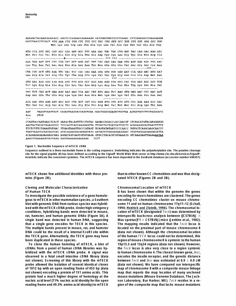
Figure 1. Nucleotide Sequence of mTECK cDNA
Sequence outlined in a three-nucleotide frame is the coding sequence. Underlining indicates the polyadenylation site. The putative cleavagesite for the signal peptide (#) has been defined according to the SignalP World Wide Web server at http://www.cbs.dtu.dk/services/SignalP.Asterisks indicate the conserved cysteines. The mTECK sequence has been deposited in the GenBank database (accession number U86357).
mTECK shows few additional identities with these pro- than toother knownCC chemokines and was thus desig-nated hTECK (Figures 2B and 3B).teins (Figure 2B).
Cloning and Molecular Characterization Chromosomal Location of mTECKIt has been shown that within the genome the genesof Human TECK
To investigate the possible existence of a gene homolo- encoding for most chemokines are clustered. The genesencoding CC chemokines cluster on mouse chromo-gous to mTECK in other mammalian species, a Southern
blot with genomic DNA from various species was hybrid- some 11 and on human chromosome 17q11–12 (Schall,1994; Hedrick and Zlotnik, 1996). The chromosomal lo-ized with themTECK cDNA probe. Under high stringency
conditions, hybridizing bands were detected in mouse, cation of mTECK (designated Teck) was determined byinterspecific backcross analysis between ([C57Bl/6J 3rat, hamster, and human genomic DNAs (Figure 3A). A
single band was detected in human DNA, suggesting Mus spretus]F1 3 C57Bl/6J) mice (Jenkins et al., 1982).The mapping results indicated that the Teck locus isthat a single gene encodes for TECK in this species.
The multiple bands present in mouse, rat, and hamster located on the proximal part of mouse chromosome 8(data not shown). Although the chromosomal locationDNA could be the result of a internal EcoRI site within
the TECK gene. Alternatively, the TECK gene may have of the human TECK locus could not be determined, thisregion of mouse chromosome8 is syntenic to the humanbeen duplicated in these species.
To clone the human homolog of mTECK, a blot of 19p13.3 and 13q34 regions (data not shown). However,the Teck locus is also very close to a region synteniccDNAs from a panel of human cDNA libraries was hy-
bridized with the mTECK cDNA probe. A signal was to human chromosome 4. The closest known gene, Insr,encodes the insulin receptor, and the genetic distanceobserved in a fetal small intestine cDNA library (data
not shown). Screening of this library with the mTECK between Teck and Insr was estimated at 0.9 6 0.9 cM(data not shown). We have compared our interspecificprobe allowed the isolation of several identical clones
of 1012 bp with an open reading frame of 453 bp (data map of chromosome 8 with a composite mouse linkagemap that reports the map location of many unclonednot shown) encoding a protein of 151 amino acids. This
protein had a much higher degree of homology at the mouse mutations (Mouse Genome Database, The Jack-son Laboratory, Bar Harbor, ME). Teck resides in a re-nucleic acid level (71% nucleic acid identity for the open
reading frame and 49.3% amino acid identity) to mTECK gion of the composite map that lacks mouse mutations

TECK: A Thymic Dendritic Cell–Specific Chemokine293
Figure 2. Comparison of the Amino Acid Sequence of TECK with Other CC Chemokines
(A) Multiple sequence alignment of mTECK with other murine CC chemokines. The protein sequences of eotaxin (Gonzalo et al., 1996), MCP-3(Thirion et al., 1994), MCP-5 (Jia et al., 1996), MIP-1b (GenBank accession number X62502), MIP-1a (Grove et al., 1990), and RANTES (Schallet al., 1992) were compared using the Clustal W program (Thompson et al., 1994). Dark-shaded boxes indicate conserved anino acids betweenat least four sequences, including the four cysteine residues characteristic of the CC chemokine family; light-shaded boxes indicateconservativesubstitutions.(B) Phylogenic tree of CC chemokines. According to their Clustal W alignment, evolutionary distances between mTECK and other chemokineswere estimated. The location of the branch points is not to scale. Numbers in parentheses are the percentage amino acid identities betweenmTECK and the other chemokines. The percentage identities of human TARC (Imai et al., 1996) was calculated in comparison with hTECK.
with a phenotype that might be expected for an alter- The distribution of hTECK mRNA was similarly ana-lyzed. As with mouse, hTECK mRNA expression wasation in this locus (data not shown).highly restricted to the thymus and small intestine (Fig-ure 5). Weak expression was also detected in inflamedAnalysis of mTECK and hTECK mRNA
Distribution in Cells and Tissues tonsil and fetal spleen, but at much lower levels thanthat observed in the thymus since this particular blotAn analysis of the distribution of mTECK mRNA in tis-
sues and cells by Northern blotting or by Southern blot- was exposed for a long time. Of note, hTECK mRNAwas absent from a series of cDNA libraries from dendriticting of mouse cDNA libraries revealed that mTECK was
expressed at significant levels only in the thymus and cells derived in vitro from bone marrow CD341 progeni-tor cells or peripheral blood monocytes (Figure 5A). Into a lesser extent in small intestine (Table 1 and Figure
4A). Weak expression of mTECK mRNA was observed addition to the dendritic cell cDNA libraries displayedin Figure 5, hTECK mRNA was absent from libraries ofin brain, testis, and liver RAG-12/2 cDNA libraries. Inter-
estingly, mTECK mRNA was detected in a cDNA library monocyte-derived dendritic cells stimulated with LPSor a combination of tumor necrosis factor-a (TNFa), in-of activated pro-T cells. Pro-T cells represent an early
stage of intrathymic T cell progenitors, not fully commit- terleukin 1a (IL-1a), and monocyte supernatant for 4 and16 hr (data not shown). Collectively, these data indicateted to the T cell lineage because they can give rise to
natural killer and dendritic cells (Moore and Zlotnik, that TECK mRNA is specifically expressed at high levelsin thymus and small intestine in vivo.1995; Wu et al., 1996). In contrast, mTECK mRNA was
undetectable in resting or activated thymocytes, periph-eral T or B cells, macrophages, PBLs, and splenic den- Identification of mTECK-Producing Cells In Vivo
The abundance of mTECK mRNA expression in RAG-dritic cells and in all other tissues tested, with the ex-ception of spleens recovered from mice injected with 1–deficient thymus and its absence in thymic T cells
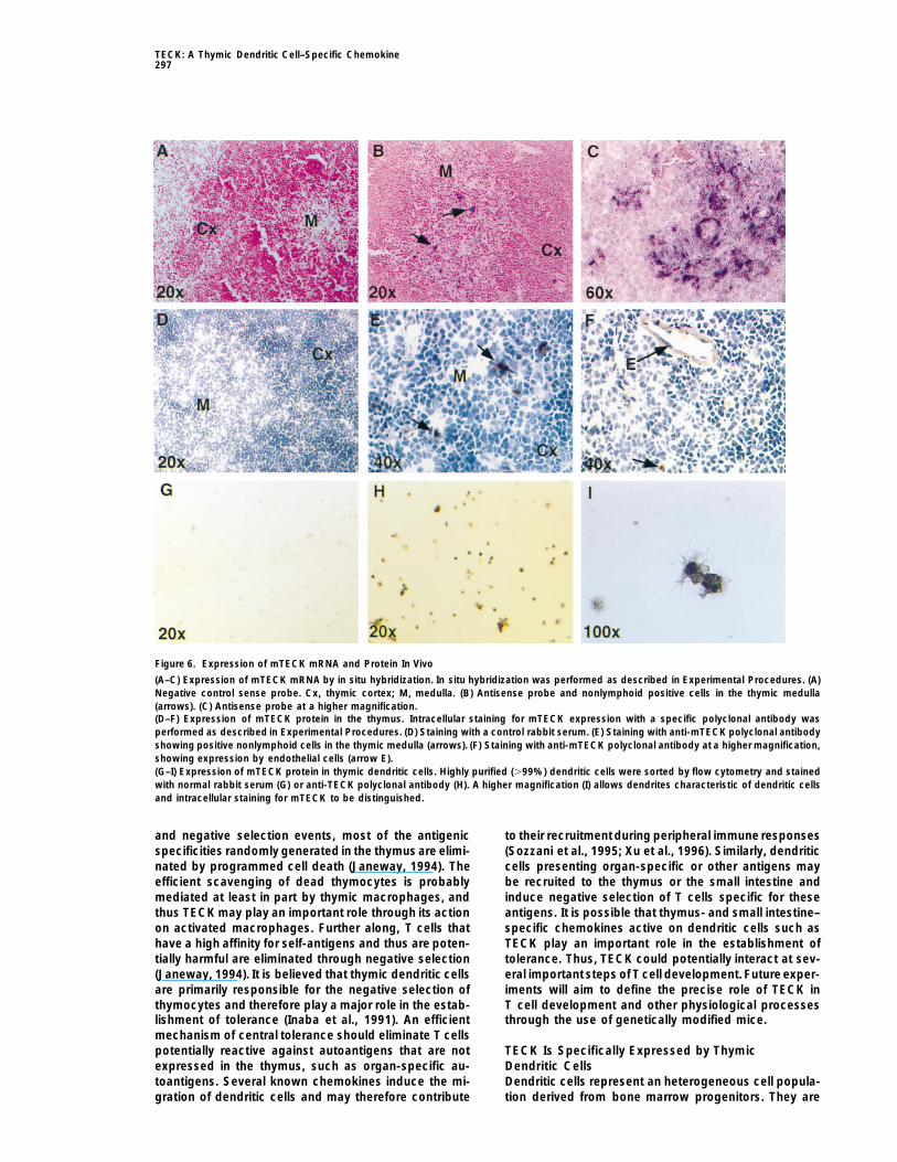
suggested that mTECK was expressed by a thymic stro-lipopolysaccharide (LPS) (Table 1 and Figure 4A).mTECK mRNA was detected by polymerase chain reac- mal component in normal mice. We performed in situ
mRNA hybridization with sense or antisense mTECKtion (PCR) in fetal thymi of day 14 of gestation (Figure4B), indicating that mTECK is expressed in the thymus probes generated by PCR. Thymic sections hybridized
with the sense probe (negative control) demonstratedat the earliest stages of T cell development.

Immunity294
was negative. In contrast, mTECK mRNA was undetect-able in a cDNA library made from freshly isolated splenicdendritic cells (Table 1).
We then performed immunostaining of thymic sec-tions and purified thymic dendritic cells with a polyclonalantibody raised against a decapeptide correspondingto the C-terminus of mTECK. This polyclonal antibodyreacted with recombinant mTECK in enzyme-linked im-munosorbent assay and Western blot, while normal rab-bit serum was negative (data not shown). In thymic sec-tions, the polyclonal anti-peptide antibody reacted witha stromal component of the thymic medulla, consistentwith the in situ hybridization data (Figures 6E and 6F),while staining with normal rabbit serum was negative(Figure 6D). The antibody also reacted weakly with someendothelial cells (Figure 6F), raising the possibility thatmTECK can be produced by the thymic endothelium.Finally, the anti-mTECK polyclonal antibody stainedsorted thymic dendritic cells, while the control serumwas negative (Figures 6G and 6H). High magnification(Figure 6I) clearly showed intracellular staining of cellswith characteristic dendritic morphology. Taken to-gether, these results indicate that thymic dendritic cellsand possibly thymic endothelial cells produce TECKin vivo.
Figure 3. Conservation of TECK in Mammalians and Structure ofChemotactic Activities of mTECK ProteinHuman TECKTo evaluate the biologic properties of mTECK, a recom-(A) Zooblot of genomic DNA hybridized with a mTECK probe. Abinant protein with a N-terminal FLAG peptide was ob-Southern blot of genomic DNA of different mammalian species di-
gested with EcoRI was hybridized with the mTECK probe. tained in a bacterial expression system (see Experimen-(B) Structure of hTECK and comparison of hTECK and mTECK tal Procedures). In some experiments, a recombinantgenes. Protein sequences of mTECK and hTECK were compared mTECK protein with a C-terminal FLAG was used andusing the Clustal W program (Thompson et al., 1994). Dark-shaded
similar results were obtained (data not shown). mTECKboxes indicate conserved amino acids; light-shaded boxes indicateinduced the migration of mouse thymocytes (Figure 7A).conservative substitutions. The protein and amino acid homologiesThe optimal response was obtained with a dose of 10both were calculated for the coding sequence only. The hTECK
sequence has been deposited in the GenBank data base (accession ng/ml TECK. Cell migration was determined to be che-number U86358). motaxis and not chemokinesis through checkerboard
analysis (data not shown). Furthermore, it is establishedthat chemokines bind to specific receptors that are cou-pled through heterotrimeric G proteins to intracellularno specific staining (Figure 6A), while sections hybrid-
ized with the antisense probe at the same concentration signal-transducing pathways (Murphy, 1994). To deter-mine whether the chemotaxis of thymocytes involved ashowed specific staining in the thymic medulla (Figure
6B). At higher magnification (Figure 6C), positive cells G protein–coupled receptor, cells were incubated priorto the assay with 10 ng/ml pertussis toxin, which ADP-appeared to have a nonlymphoid morphology with pro-
cesses surrounding lymphoid cells. This experiment in- ribosylates Gai proteins (Katz et al., 1992). This pretreat-ment completely abrogated the chemotactic responsedicated that in vivo, mTECK mRNA is expressed by a
nonlymphoid component of the medullary stroma, pos- of thymocytes to mTECK (Figure 7A).The recombinant mTECK protein also induced migra-sibly dendritic cells.
The thymic stroma is composed mainly of epithelial tion of human monocytic THP-1 cells activated for 16hr with IFNg (Figure 7B), whereas it was not significantlycells, macrophages, dendritic cells, and fibroblasts, to-
gether with a network of vascular and nervous tissue active on resting THP-1 cells. This experiment showedthat mTECK is activeon human cells. In addition, mTECK(Boyd et al., 1993). Since we previously failed to detect
mTECK mRNA expression in thymic epithelial or macro- induced activated mouse peritoneal macrophages tomigrate as well as highly purified mouse splenic den-phage cell lines with or without activation with inter-
feron-g (IFNg) (Table 1), we sorted thymic dendritic cells dritic cells (Figure 7B). In all of these experiments, theoptimal dose of mTECK was 10 ng/ml. In contrast, nobased on their high expression of major histocompatibil-
ity (MHC) class II and CD11c (N-418 antibody). Analysis chemotaxis was observed with bone marrow cells, puri-fied neutrophils, splenic B cells, splenic T cells, or IL-of mTECK expression by reverse transcription PCR (RT-
PCR) revealed that freshly isolated MHC class II1 2–activated RAG-1–deficient mouse splenocytes lack-ing matureT and B lymphocytes (Mombaerts et al.,1992)CD11c1 thymic dendritic cells expressed mTECK mRNA
(Figure 4C), whereas the MHC class II1 CD11c2 subset and therefore enriched in natural killer cells (data not

TECK: A Thymic Dendritic Cell–Specific Chemokine295
Table 1. mTECK mRNA Expression in Tissues and Cells
cDNA Libraries Northern Blot
Cell Type or Tissue Negative Positive Cell Type or Tissue Negative Positive
Th2 CD41 T cells X Heart XTh1 CD41 T cells X Brain XLung X Spleen XL cells X Lung XRAG-1 KO lung X Liver XRAG-1 KO heart X Skeletal muscle XRAG-1 KO brain X (1) Kidney XRAG-1 KO spleen X Testis XRAG-1 KO kidney X Thymus X (111)RAG-1 KO testis X (1) Small intestine X (11)RAG-1 KO thymus X (111) CD41CD82 thymocytes R/A XRAG-1 KO liver X (1) CD42CD81 thymocytes R/A XCD42CD82 thymocytes X CD42CD82 thymocytes R/A XA20-J B cell lymphoma X B2201 splenocytes R/A XBW CD42CD82CD32 hybridoma X Thy-11 splenocytes R/A Xpro-T cells X (1) 1G18LA macrophages R/A Xpre-T cells X Primary thymic stroma R/A X30-R bone marrow stroma X 3D.1 thymic epithelial R/A XD10 T cell hybridoma X MTSC-C thymic epithelial XCTLL T cell clone X 30.R bone marrow stroma XPeritoneal macrophages XSplenic dendritic cells X
Analysis of mTECK mRNA was carried out as described in Experimental Procedures.1 to 111, relative intensity of the signal; R/A, resting or activated.
shown). These results are consistent with the absence 17 (Rossi et al., 1997), and the gene encoding the novelhuman CC chemokine MIP-3a/LARC (Rossi et al., 1997)of in vivo accumulation of neutrophils, monocytes, or
lymphocytes 2 and 5 hr following an intraperitoneal in- has been mapped on chromosome 2 (Hieshima et al.,1997). It is likely that the CC chemokines on chromo-jection of 10 mg of mTECK (data not shown). Collectively,
these data indicate that TECK is a chemotactic factor some 11 in the mouse and chromosome 17 in the humanhave been generated through gene duplication of a pri-for thymocytes, macrophages, and dendritic cells.mordial chemokine. Our results suggest that TECK mayhave been generated at an earlier stage during evolution.DiscussionIn this regard, the TECK gene may have evolved to en-sure functions similar to other CC chemokines with aTECK, a Distant Member of the CCdistant primary structure but through similar receptor(s)Chemokine Familyas dictated by its secondary and tertiary structures. Al-In this report, we describe the molecular isolation andternatively, the receptor(s) and physiological role ofcharacterization of TECK, a novel mouse and humanTECK may be unique among chemokines.CC chemokine. Analysis of its predicted amino acid se-
quence showed that TECK is distantly related to pre-viously described CC chemokines. Conservation of par- TECK Expression and Function Is Associated
with T Cell Developmentticular amino acids among most CC chemokines maybe related to their functional importance (Beall et al., We observed that TECK was strongly expressed in the
thymus, the primary lymphoid organ in which T cell1992; Lusti-Narasimhan et al., 1995). In particular, a tyro-sine residue between the second and third cysteines development takes place. Recently, another CC chemo-
kine highly expressed in the thymus, TARC (thymus-has been shown to be critical for monocyte chemotaxis(in position50 of the multiple alignment, Figure 2A) (Beall and activation-restricted chemokine), has been identi-
fied (Imai et al., 1996). However, TARC is also expressedet al., 1992). Although TECK does not have a tyrosineat this particular position, it has one inposition 52 (Figure in lung and colon as well as activated PBMCs (Imai et
al., 1996), whereas TECK was absent from these tissues.2A) that may have the same function, since TECK ischemotactic for activated monocytes. In addition to Besides the thymus, numerous reports indicate that T
cell development can occur in the small intestine (Pous-these differences in the primary structure, the gene en-coding TECK maps on chromosome 8 in the mouse, sier and Julius, 1994), where TECK is also expressed.
The liver has also been suggested to support T cellunlike most other CC chemokines, which are clusteredon chromosome 11. This is not the first report of an development to some extent (Abo et al., 1994), and we
observed a low TECK expression in a liver cDNA library.unusual chromosomal location for a CC chemokine. Wehave cloned the human CC chemokine MIP-3b (macro- These data show that TECK expression correlates with
organs that support T cell development.phage inflammatory protein-3b) and showed that itsgene was on chromosome 9 rather than chromosome While many molecular and cellular aspects of T cell

Immunity296
Figure 4. Expression of mTECK mRNA in Different Mouse Tissuesand Cell Types
(A) Northern blot of RNA from different organs hybridized with themTECK cDNA probe with or without in vivo LPS stimulation. RNAsfrom different organs were isolated 3 hr after intravenous injectionof PBS(lanes a) or 50 mg LPS (lanes b).Each lane contained approxi-mately 10 mg of total RNA. The hybridizing bands (top)correspondedto the predicted size of approximately 1040 bp for mTECK mRNA.Staining of the gel with ethidium bromide (bottom) showed a bandcorresponding to the ribosomal 28s RNA. Figure 5. Expression of hTECK mRNA in Different Human Tissues(B) mTECK mRNA expression in the mouse fetal thymus. RNAs from and Cell Typesfetal thymic lobes were extracted on days 14–17 of gestation, and (A) Expression of mTECK mRNA in human cDNA libraries. Southernexpression of mTECK mRNA was analyzed by RT-PCR. A unique blots of human cDNA libraries digested with the appropriate restric-band corresponding to the predicted size of the PCR product (z400 tion enzymes (10 mg/lane) were hybridized with the hTECK cDNAbp) was observed after ethidium bromide staining. The expression probe. A major hybridizing band corresponding to the predictedof HPRT mRNA in the same samples was analyzed by RT-PCR as length of hTECK mRNA (z1040 bp) was observed, sometimes witha control. some other bands that may represent incomplete cDNAs. RAG-KO(C) mTECK mRNA expression in thymic dendritic cells. A population thymus showed the cross-hybridization between hTECK cDNA andenriched in thymic dendritic cells was prepared from 15 pooled mouse RNA. NK cells correspond to pools of NK clones activatedadult thymi as described in Experimental Procedures. Dendritic cells with PMA and ionomycin for 12 hr. Splenocytes were activated with(.99% pure) were then sorted by flow cytometry based on their anti-CD40 antibody and IL-4 for 6 and 12 hr. PBMC were activatedMHC class II1 N-4181 phenotype. mTECK mRNA was then analyzed with anti-CD3 and PMA for 6, 12, and 24 hr. C1 monocytes consistedby RT-PCR, and an MHC class II1 N-4182 population sorted in the in elutriated monocytes cultured with IFNg and IL-10; C2 monocytessame experiment was used as a negative control. The expression consisted in elutriated monocytes cultured with IFNg and anti-IL-of HPRT mRNA in the same samples was analyzed by RT-PCR as 10 antibody. The designation “70% dendritic” corresponds to a 70%a control. CD1a1 dendritic cell population obtained by expansion of CD341
bone marrow cells with GM-CSF and TNF-a and resting; DC3 corre-sponds to a similar dendritic cell population stimulated with PMAand ionomycin for 1 and 6 hr. CD1a is similar to DC3 but the cellsdifferentiation are well documented, the precise role ofwere 95% CD1a1; DC5 corresponds to dendritic cells obtained bychemokines in T cell development is still unknown. Re-culturing peripheral blood monocytes in the presence of IL-4 and
cently, it has been shown that the bone marrow stroma– GM-CSF. U937 is a premonocytic cell line.derived CXC chemokine SDF-1 (stromal cell–derived (B) Expression of hTECK mRNA in human tissues. A northern blotfactor-1) is important for B lymphopoiesis and myelo- made with poly(A)1 RNA from different tissues (5 mg/lane) was hy-
bridized with the hTECK cDNA probe. The hybridizing band corre-poiesis since SDF-12/2 mice are impaired for these func-sponds to the predicted size of approximately 1040 bp for hTECKtions (Nagasawa et al., 1996). Similarly, it is likely thatmRNA.chemokines act at different stepsof T celldifferentiation.
Chemokines, together with the expression of appro-priate adhesion molecules, may dictate the migration of sensitivity of progenitor cells to TECK would increase as
these cells leave the bone marrow to colonize lymphoiduncommitted progenitors from thebone marrow to otheranatomic locations. Indeed, SDF-1 is chemoattractant organs. Of note, intrathymic maturation is also charac-
terized by a directional migration from the subcapsularfor human CD341 progenitor cells (Aiuti et al., 1997).The observation that TECK is chemoattractant for thy- region, which contains the earliest progenitors to the
cortex, and finally to the medulla, where thymocytesmocytes but not for mature peripheral T cells suggeststhat TECK may attract T cell progenitors to the thymus. finish their maturation (Boyd et al., 1993). It is possible
that the secretion of TECK by medullary dendritic cellsSuch populations are very difficult to isolate in sufficientnumbers to conduct in vitro chemotaxis experiments, may play a role in this directional migration. Yet another
possibility is that TECK may play a role in the organiza-but we arecurrently designing new strategies to addressthis question. In addition, we have not found significant tion and development of the thymic stroma.
We also showed that TECK is chemotactic for acti-chemotactic activity of TECK on bone marrow cells.SDF-1 was shown to be much less potent on CD341 vated macrophages and dendritic cells. These two cell
types also play important roles in T cell development.progenitors from the peripheral blood than those fromthe bone marrow (Aiuti et al., 1997). It is possible that the Through a complex screening process involving positive
TECK: A Thymic Dendritic Cell–Specific Chemokine297
Figure 6. Expression of mTECK mRNA and Protein In Vivo
(A–C) Expression of mTECK mRNA by in situ hybridization. In situ hybridization was performed as described in Experimental Procedures. (A)Negative control sense probe. Cx, thymic cortex; M, medulla. (B) Antisense probe and nonlymphoid positive cells in the thymic medulla(arrows). (C) Antisense probe at a higher magnification.(D–F) Expression of mTECK protein in the thymus. Intracellular staining for mTECK expression with a specific polyclonal antibody wasperformed as described in Experimental Procedures. (D) Staining with a control rabbit serum. (E) Staining with anti-mTECK polyclonal antibodyshowing positive nonlymphoid cells in the thymic medulla (arrows). (F) Staining with anti-mTECK polyclonal antibody at a higher magnification,showing expression by endothelial cells (arrow E).(G–I) Expression of mTECK protein in thymic dendritic cells. Highly purified (.99%) dendritic cells were sorted by flow cytometry and stainedwith normal rabbit serum (G) or anti-TECK polyclonal antibody (H). A higher magnification (I) allows dendrites characteristic of dendritic cellsand intracellular staining for mTECK to be distinguished.
and negative selection events, most of the antigenic to their recruitment during peripheral immune responses(Sozzani et al., 1995; Xu et al., 1996). Similarly, dendriticspecificities randomly generated in the thymus are elimi-
nated by programmed cell death (Janeway, 1994). The cells presenting organ-specific or other antigens maybe recruited to the thymus or the small intestine andefficient scavenging of dead thymocytes is probably
mediated at least in part by thymic macrophages, and induce negative selection of T cells specific for theseantigens. It is possible that thymus- and small intestine–thus TECK may play an important role through its action
on activated macrophages. Further along, T cells that specific chemokines active on dendritic cells such asTECK play an important role in the establishment ofhave a high affinity for self-antigens and thus are poten-
tially harmful are eliminated through negative selection tolerance. Thus, TECK could potentially interact at sev-eral important steps of T cell development. Future exper-(Janeway, 1994). It is believed that thymic dendritic cells
are primarily responsible for the negative selection of iments will aim to define the precise role of TECK inT cell development and other physiological processesthymocytes and therefore play a major role in the estab-
lishment of tolerance (Inaba et al., 1991). An efficient through the use of genetically modified mice.mechanism of central tolerance should eliminate T cellspotentially reactive against autoantigens that are not TECK Is Specifically Expressed by Thymic
Dendritic Cellsexpressed in the thymus, such as organ-specific au-toantigens. Several known chemokines induce the mi- Dendritic cells represent an heterogeneous cell popula-
tion derived from bone marrow progenitors. They aregration of dendritic cells and may therefore contribute

Immunity298
lymphoid dendritic cell precursor in thymus and bonemarrow that is able to derive both lymphocytes anddendritic cells in the absence of GM-CSF (Ardavin etal., 1993; Galy et al., 1995; Marquez et al., 1995; Wu etal., 1996). These lymphoid-derived dendritic cells mayhave different functional properties such as a negativeregulation of T cell responses, since they express FasLin the mouse (Suss and Shortman, 1996). We found thatTECK was expressed at high levels in mouse thymicdendritic cells but was absent in cDNA libraries frommouse splenic dendritic cells or from human dendriticcells generated in vitro from CD341 precursors or mono-cytes. mTECK mRNA was present at a low level in apopulation of early thymocyte progenitors still able toderive dendritic cells (Wu et al., 1996). Thus, it wouldbe tempting to suggest that TECK could be a specificmarker of lymphoid-derived dendritic cells. However,we observed that TECK was absent from splenic den-dritic cells that likely contain lymphoid-derived dendriticcells. The expression of TECK mRNA in the spleen ofmice injected with LPS would suggest that peripheraldendritic cells may express TECK upon activation, butwe found that TECK was not expressed in cDNA librariesof bone marrow–derived dendritic cells activated withLPS, phorbol myristate acetate (PMA), and ionomycinor IL-1a and TNFa. It is possible that the normal expres-sion of TECK is specific for lymphoid-derived dendriticcells or, alternatively, that it is up-regulated by very spe-cific stimuli present in the thymic and intestinal microen-vironment under physiological conditions. Consistentwith the latter hypothesis is our observation of specificstaining of thymic endothelial cells with anti-TECK anti-body, since we have not been able to find TECK expres-
Figure 7. Chemotactic Properties of mTECK Recombinant Protein sion in human HUVEC endothelial cells by Northern blot(A) Migration of mouse thymocytes to recombinant mTECK and analysis, without activation or following a 16 hr activa-effect of pertussis toxin. tion with various combinations of IL-1, TNFa, IL-4, IL-7,Chemotaxis assays were performed as described in Experimental
and oncostatin (data not shown), while some of theseProcedures. Recombinant mouse lymphotactin (Lptn) was used asstimuli induce the expression of other CC chemokinesa positive control. Data are expressed as the mean (6 SEM) of cellin endothelial cells (Rollins and Pober, 1991; Marfaing-counts obtained from three separate experiments in duplicate. In
one experiment,cells were preincubated 1 hr with 10 ng/ml pertussis Koka et al., 1995; Garcia-Zepeda et al., 1996a, 1996b).toxin (PTX) prior to the assay. Taken together, our data strongly suggest that TECK is(B) Migration of other leukocyte subsets to recombinant mTECK a novel chemokine specifically expressed by activatedMouse splenic dendritic cells and mouse activated macrophages
lymphoid-derived dendritic cells.were obtained as described in Experimental Procedures. THP-1Through their function of antigen presentation, den-human monocytic cells were used without or with a 16 hr activation
dritic cells play major roles in the establishment of toler-with IFNg. Results are obtained as the mean (6 SD) of the chemotac-tic index from three separate experiments per cell type in duplicate. ance and in the initiation of an antigen-specific immuneThe number of cells migrating to medium alone was greater than response. The use of purified dendritic cells has been40 cells per five high-power fields in each experiment. Recombinant recently proposed in different therapeutic protocolsMIP-1a was used as a positive control.
(Cella et al., 1997). The discovery of factors with a regu-lated expression in dendritic cells such as the novel
present in nonlymphoid organs as immature dendritic CC chemokine TECK will improve our knowledge of thecells (such as Langerhans cells in the skin), where they biology of dendritic cells and lead to the design of rele-display a high ability for antigen capture (Cella et al., vant in vivo applications.1997). Subsequent to antigen challenge, they migrateto secondary lymphoid organs and acquire a high ca- Experimental Procedurespacity to present processed antigens to naive T cells toinitiate a specific immune response (Cella et al., 1997). Mice and In Vivo Experimental Procedures
Four- to eight-week-old and time-pregnant BALB/c mice were pur-It has been shown that dendritic cells can derive fromchased from Simonsen Laboratories (Gilroy, CA). RAG-1–deficientCD341 progenitors cultured in the presence of granulo-mice (Mombaerts et al., 1992) were purchased from The Jacksoncyte/macrophage colony-stimulating factor (GM-CSF)Laboratory. To analyze TECK expression after in vivo activation,
and TNFa (Caux et al., 1992; Caux et al., 1996) or from various organs were recovered from pools of two mice 3 hr aftermonocytes in the presence of GM-CSF and IL-4 (Sallusto intravenous LPS injection (50 mg of LPS in 200 ml of phosphate-
buffered saline [PBS] or 200 ml of PBS for controls).and Lanzavecchia, 1994). There is also evidence for a

TECK: A Thymic Dendritic Cell–Specific Chemokine299
Cell Purification, Culture, and Stimulation gel electrophoresis, Southern blot transfer, and hybridization withthe full-length mTECK cDNA probe were performed as describedTHP-1 cells (TIB-202 from the American Type Culture Collection,
Rockville, MD) were cultured in complete medium that consisted in (Jenkins et al., 1982). Fragments of 7.5, 6.9, and 2.5 kb were detectedin HincII-digested C57Bl/6J DNA, and fragments of 8.8 and 5.4 kbRPMI 1640 medium (JRH BioSciences, Lenexa, KS) supplemented
with 10% fetal calf serum, 200 mM L-glutamine, 5 3 1025 M mercap- were detected in HincII digested M. spretus DNA. The presence orabsence of the 8.8 and 5.4 kb HincII M. spretus–specific fragments,toethanol, minimal essential medium amino acids and vitamins, so-
dium bicarbonate, penicillin, streptomycin (all from Sigma, St. Louis, which cosegregated, was followed in backcross mice. A descriptionof the probes and restriction fragment length polymorphisms forMO), and gentamycin (Boehringer, Indianapolis, IN). To obtain acti-
vated mouse macrophages, 10 ml of cold PBS was injected into two of the loci linked to Teck, including Insr, has been providedpreviously (Ceci et al., 1990). Recombination distances were calcu-the peritoneum and the collected cells allowed to adhere to plastic
for 24 hr in complete medium. The adherent fraction, mostly macro- lated as described (Green, 1981) using the computer programSpretus Madness.phages, was then collected. To obtain splenic dendritic cells, a
splenocyte cell suspension was prepared in RPMI 1640 Dutch modi-fied medium (Life Technologies, Paisley, Scotland) as described Measurement of TECK mRNA Expression by RT-PCRpreviously (Macatonia et al., 1987). Splenocytes were incubated at RNAs from sorted thymic dendritic cells or fetal thymi were prepared378C for 16 hr, and the cell suspension was collected and laid over with the RNeasy total RNA kit (Quiagen, Chatsworth, CA), followingMetrizamide (Nycomed Pharma, Oslo, Norway). After centrifugation the manufacturer’s instructions. First-strand cDNAs were generatedfor 10 min at 1700 3 g, the low interface was collected and stained by reverse transcription with a random hexamer in a 10 ml reaction,with anti-Mac-1 (Pharmingen, San Diego, CA) and the anti-CD11c and 1 ml of this reaction was used as a template for PCR. TECKN-418 antibodies (Macatonia et al., 1993). Splenic dendritic cells expression was compared to the expression of hypoxanthine-gua-were sorted by flow cytometry on a FACStar plus cell sorter (Becton nine phosphoribosyl transferase (HPRT). Primer sequences were asDickinson, Mountain View, CA) to a purity greater than 98% upon follows: TECK: 59 primer, 59CCTTCAGGTATCTGGAGAGGAGATC39reanalysis in all the experiments included in this report. To obtain and 39 primer, 59CACGCTTGTACTGTTGGGGTTC39; HPRT: 59 primer,thymic dendritic cells, thymi were cut in small fragments and resus- 59GTAATGATCAGTCAACGGGGGAC39 and 39 primer, 59CCAGCAApended in 10 ml of RPMI-1640 1 10% fetal calf serum containing GCTTGCAACCTTAACCA39. Samples were submitted to 25 cycles1 mg/ml collagenase and 0.02 mg/ml DNase I (both from Sigma) of amplification, each composed of 948C for 1 min, 578C for 30 s,and digested with continuous agitation at room temperature for 30 and 728C for 2 min. PCR products were then separated by electro-min (Shortman et al., 1995). One milliliter of 0.1 M EDTA (pH 7.2) was phoresis in 2% agarose gels and stained with ethidium bromide.added for an additional 5 min. Cells were then washed in completemedium, resuspended in completemedium, and overlaid ontoMetri- In Situ Hybridizationzamide. The thymic dendritic cell–enriched preparation was then Biotin-14-CTP–labeled sense and antisense riboprobes were gener-stained with anti-IAd and N-418 antibodies and the dendritic cells ated using a nonradioactive RNA labeling system (Gibco, Gaithers-sorted by flow cytometry. burg, MD) and the plasmid PCRII (InVitrogen, Carlsbad, CA) con-
taining a 400 bp TECK cDNA fragment inserted by PCR and clonedMolecular Cloning of Mouse and Human TECK into the PCRII vector using the TA cloning kit (InVitrogen). Paraffin-The cDNA encoding mouse TECK was obtained by random se- embedded tissues were cut in 3–5 mm sections, mounted on slides,quencing of a RAG-1 knockout mouse thymic directional cDNA li- baked at 608C for 1 hr, deparaffinized in xylene (Fisher Scientific,brary. In brief, mRNA was extracted using RNAzol B (Tel-Test, Pittsburgh, PA), and immersed in 100% ethanol. Sections were thenFriendswood, TX) and then oligotex-dT mRNA kit (Quiagen, Chats- incubated for 10 min at 378C in proteinase K solution (40 mg/ml)worth, CA) following the manufacturer’s instruction. A directional (Gibco) in PBS and rinsed for 2 min in PBS at room temperaturecDNA library was prepared using the Superscript Plasmid System before being refixed in 10% formalin (Fisher Scientific) in PBS for 1(Gibco–BRL, Grand Island, NY) and cloned into the pME18s plasmid min. Next, the sections were dehydrated through graded solutionsvector. Sequencing was done using the TaQ DyeDeoxy Terminator of ethanol and air dried. Hybridization was carried out using theCycle Sequencing kit (Applied Biosystems, Foster City, CA). To de- Gibco in situ hybridization and detection system kit. Vanadyl ribonu-termine whether TECK was present in other mammals, including cleoside complex (Gibco) was added to the hybridization solutionhuman, a Southern blot containing EcoRI-digested genomic DNA (39 mM final). A 0.1 mg/ml concentration of each probe was usedfrom different species (Bios Laboratories, New Haven, CT) was hy- during an 18 hr hybridization at 428C. Posthybridization washes usedbridized with the full-length mouse TECK cDNA. room temperature 0.23 sodium chloride/sodium citrate. Following
The cDNA encoding human TECK was found by screening of a detection and substrate visualization, the slides were counter-small intestine cDNA library using the full-length mouse TECK cDNA stained with 1% nuclear red stain (Sigma, St. Louis, MO).as a probe, following standard procedures.
ImmunohistochemistryNorthern Blot Analysis of RNA and Southern A polyclonal antibody specific of a synthetic decapeptide identicalBlot Analysis of cDNA Libraries to the C-terminus part of murine TECK (Figure 1) was prepared inAll RNAs were isolated from tissues or cells using RNAzol B (Tel- rabbits by Research Genetics (Huntsville, AL). Normal rabbit serumTest) and analyzed after electrophoresis in a 1% formaldehyde- from a pool of 50 different animals (Research Genetics) was usedagarose gel (10 mg/lane). RNAs were then blotted onto a Hybond-N1 as a negative control. To study TECK protein expression in thenylon membrane (Amersham, Arlington Heights, IL). Some Northern mouse thymus, 6 mm thick cryostat sections were thaw mountedblots of mRNA were bought from Clontech (Palo Alto, CA). To ana- on organosilicone subbed slides (American Histology Reagent,lyze the expression of TECK in cDNA libraries (obtained from T. Stockton, CA) and fixed in 3% formalin (Fisher Scientific, Springfield,MacClanahan, DNAX), 10 mg of DNA was digested with the appro- NJ) in Hank’s balanced salt solution (HBSS) with 0.01 M HEPESpriate restriction enzymes to release their inserts and analyzed by (HBSS–HEPES) (pH 7.4), for 15 min at room temperature. The sec-Southern blotting onto nylon membranes. Northern blots and blots tions were sequentially blocked for endogenous biotin binding usingof cDNA libraries were hybridized for 16 hr at 658C with a 32P-labeled the Vector blocking kit (Vector Laboratories, Burlingame, CA) andprobe consisting in the full-length cDNA encoding for mouse or for endogenous peroxidase activity with a 1% hydrogen peroxide,human TECK and then washed and exposed, according to standard 0.2 M sodium azide solution, in HBSS-HEPES with 0.1% saponinprotocols. (staining buffer). Nonspecific antibody binding sites were then
blocked with 10% normal goat serum (Sigma) in staining buffer.Sections prepared as above were first incubated for 18 hr at 258CInterspecific Mouse Backcross Mapping
Interspecific backcross progeny were generated by mating (C57Bl/ with 1:500 dilution of polyclonal antibody or control rabbit serum instaining buffer. In the second step, the sections were incubated for6J 3M. spretus)F1 femalesand C57Bl/6J males as described (Cope-
land and Jenkins, 1991). A total of 205 N2 mice were used to map 1 hr at room temperature with biotin-labeled goat anti-rabbit IgG (2mg/ml) (Vector Laboratories) in staining buffer and then for 30 minthe Teck locus. DNA isolation, restriction enzyme digestion, agarose

Immunity300
at room temperature with the Vectastain Elite ABC Kit (Vector Labo- Referencesratories) in staining buffer. The sections were then rinsed in HBSS-HEPES without saponin. Immunoenzyme tissue staining was revealed Abo, T., Watanabe, H., Iai, T., Kimura, M., Ohtsuka, K., Sato, K.,
Ogawa, M., Hirahara, H., Hashimoto, S., and Sekikawa, H. (1994).with 3,39-diaminobenzidine tetrahydrochloride (DAB) substrate (0.5mg/ml) (Sigma) in 0.05 M Tris (pH 7.4) containing 0.0075% hydrogen Extrathymic pathways of T-cell differentiation in the liver and other
organs. Int. Rev. Immunol. 11, 61–102.peroxide. The substrate reaction was stopped by rinsing the sec-tions in distilled water. The sections were then counterstained with Aiuti, A., Webb, I.J., Bleul, C., Springer, T., and Gutierrez-Ramos,Harris’ hematoxylin (Shandon Lipshaw, Pittsburg, PA). J.C. (1997). The chemokine SDF-1 is a chemoattractant for human
CD341 hematopoietic progenitor cells and provides a new mecha-Production of Recombinant Mouse TECK in Escherichia nism to explain the mobilization of CD341 progenitors to peripheralcoli and Other Chemokines blood. J. Exp. Med. 185, 111–120.Mouse recombinant TECK was produced in E. coli as a N-terminal Ardavin, C., Wu, L., Li, C.L., and Shortman, K. (1993). Thymic den-FLAG (DYKDDDDKL) fusion protein. In brief, the fusion construct dritic cells and T cells develop simultaneously in the thymus fromcontaining FLAG followed by the mTECK sequence minus the leader a common precursor population. Nature 362, 761–763.peptide (Figure 1) was obtained by PCR amplification of the TECK
Baggiolini, M., Dewald, B., and Moser, B. (1994). Interleukin-8 andcDNA to flank the coding sequence with HindIII and EcoRI sites andrelated chemotactic cytokines: CXC and CC chemokines. Adv. Im-
subsequent ligation in the pFLAG.1 vector, which contains the FLAGmunol. 55, 97–179.
sequence and an OmpA signal sequence. Electrocompetent UTBazan, F., Bacon, K., Hardiman, G., Wang, W., Soo, K., Rossi, D.,4400 E. coli were transformed with the pFLAG.1-mTECK plasmid.Greaves, D., Zlotnik, A., and Schall, T. (1997). A new class of mem-The cells were grown in 23 Luria broth plus 50 mg/ml ampicillin,brane-bound chemokine with a CX3C motif. Nature 385, 640–644.induced at an OD of 2.3 with 400 mM isopropyl-b-D-thiogalactopy-Beall, C.J., Mahajan, S., and Kolattukudy, P.E. (1992). Conversionranoside (IPTG) and harvested. The cell pellet was resuspended inof monocyte chemoattractant protein-1 into a neutrophil attractantcold lysis buffer (20 mM Tris [pH 8], 2 mM EDTA, 20% sucrose, 0.1by substitution of two amino acids. J. Biol. Chem. 267, 3455–3459.mg/ml lysozyme, 100 ml benzonase), homogenized, and allowed to
sit for 30 min. Then the same amount of a 1:4 dilution of cold lysis Boyd, R.L., Tucek, C.L., Godfrey, D.I., Izon, D.J., Wilson, T.J., David-buffer without lysozyme was added for an additional 10 min. The son, N.J., Bean, A.G., Ladyman, H.M., Ritter, M.A., and Hugo, P.solution was spun, and the supernatant was filtered through a 0.2 (1993). The thymic microenvironment. Immunol. Today 14, 445–459.mm membrane and then diluted 1:1 in 50 mM Tris (pH 7.5). The Caux, C., Dezutter-Dambuyant, C., Schmitt, D., and Banchereau,diluted osmotic extract was submitted to chromatography on a J. (1992). GM-CSF and TNF-alpha cooperate in the generation ofQ-sepharose column equilibrated with 50 mM Tris (pH 7.5) and dendritic Langerhans cells. Nature 360, 258–261.eluted with a linearsalt gradient. The fractions containing the recom-
Caux, C., Vanbervliet, B., Massacrier, C., Dezutter-Dambuyant, C.,binant protein were pooled. The fractions were then loaded onto a De Saint-Vis, B., Jacquet, C., Yoneda, K., Imamura, S., Schmitt, D.,S-sepharose column equilibrated with 20 mM acetate (pH 4.0). The and Banchereau, J. (1996). CD341 hematopoietic progenitors fromcolumn was eluted with a linear salt gradient and then with a 1.5 M human cord blood differentiate along two independent dendritic cellNaCl wash that contained the protein. Finally, the eluate was loaded pathways in response to GM-CSF 1 TNF-alpha. J. Exp. Med. 184,onto a reverse phase column. The column was eluted with a linear 695–706.gradient of 20%–80% acetonitrile 1 0.1% trifluoroacetic acid. The
Ceci, J.D., Justice, M.J., Lock, L.F., Jenkins, N.A., and Copeland,concentration of the mTECK protein was estimated by ComassieN.G. (1990). An interspecific backcross linkage map of mouse chro-blue staining and densitometric scanning of a 10% Nu–polyacryla-mosome 8. Genomics 6, 72–79.mide gel electrophoresis gel with lysozyme as a standard. The purityCella, M., Sallusto, F., and Lanzavecchia, A. (1997). Origin, matura-was estimated at 100% by sequencing of the N-terminus of thetion and antigen presenting function of dendritic cells. Curr. Opin.recombinant protein. Recombinant murine MIP-1a (R and D Sys-Immunol. 9, 10–16.tems, Minneapolis, MN) and lymphotactin (Hedrick et al., 1997) were
used as controls. Cocchi, F., DeVico, A.L., Garzino-Demo, A., Arya, S.K., Gallo, R.C.,and Lusso, P. (1995). Identification of RANTES, MIP-1a, and MIP-1b as the major HIV-suppressive factors produced by CD81 T cells.Assay for ChemotaxisScience 270, 1811–1815.The in vitro migration of cells isolated as described above in re-
sponse to TECK or other factors was assessed in a modified Boyden Cook, D.N. (1996). The role of MIP-1 alpha in inflammation andmicro chamber (Neuroprobe, Cabin John, MD) as described pre- hematopoiesis. J. Leukoc. Biol. 59, 61–66.viously (Kelner et al., 1994). In brief, factor dilutions in Dulbecco’s Cook, D.N., Beck, M.A., Coffman, T.M., Kirby, S.L., Sheridan, J.F.,modified Eagle’s medium (DMEM) (Gibco) were loaded in the lower Pragnell, I.B., and Smithies, O. (1995). Requirement of MIP-1a forcompartment in duplicate, and 105 cells in a 50 ml volume of DMEM an inflammatory response to viral infection. Science 269, 1583–1585.were loaded in the upper compartment. The two compartments
Copeland, N.G., and Jenkins, N.A. (1991). Development and applica-were separated by a 5 or 8 mm pore polycarbonate filter (Nucleopore,
tions of a molecular genetic linkage map of the mouse genome.Pleasanton, CA). After incubation at 378C for 80 min (or 120 min for
Trends Genet. 7, 113–118.lymphocytes), the filters were fixed in methanol and stained with
Galy, A., Travis, M., Cen, D., and Chen, B. (1995). Human T, B,fields A and B. Cell migrated on the other side of the membranenatural killer and dendritic cells arise from a common bone marrowwere counted per five high-power fields (1003) under a microscope.progenitor cell subset. Immunity 3, 459–473.The chemotactic index was calculated from the number of cellsGarcia-Zepeda, E., Combadiere, C., Rothenberg, M., Sarafi, M., La-counted with the test sample divided by the number of cells countedvigne, F., Hamid, Q., Murphy, P., and Luster, A. (1996a). Humanwith medium alone.monocyte chemoattractant protein (MCP)-4 is a novel CC chemo-kine with activities on monocytes, eosinophils, and basophils in-Acknowledgmentsduced in allergic and nonallergic inflammation that signals throughthe CC chemokine receptors (CCR)-2 and 23. J. Immunol. 157,Correspondence should be addressed to A. Z. (e-mail: zlotnik@5613–5626.dnax.org). We are grateful to T. MacClanahan, K. Franz-Bacon, andGarcia-Zepeda, E.A., Rothenberg, M.E., Ownbey, R.T., Celestin, J.,M. F. Sterling for the construction and availability of most of theLeder, P., and Luster, A.D. (1996b). Human eotaxin is specific che-cDNA libraries used in this report. We also thank J. Cupp, E. Callas,moattractant for eosinophil cells and provides a new mechanism toJ. Poliakoff, and E. Murphy for cell sorting and A. Helms and C.explain tissue eosinophilia. Nature Med. 4, 449–456.Huffine for DNA sequencing. DNAX Research Institute is supported
by Schering-Plough, Inc. Gonzalo, J.-A., Jia, G.-Q., Aguirre, V., Friend, D., Coyle, A.J., Jenkins,N.A., Lin, G.-S., Katz, H., Lichtman, A., Copeland, N., et al. (1996).Mouse eotaxin expression parallels eosinophil accumulation duringReceived April 22, 1997.

TECK: A Thymic Dendritic Cell–Specific Chemokine301
lung allergic inflammation but it is not restricted to a Th2-type re- Murphy, P.M. (1994). The molecular biology of leukocyte chemoat-sponse. Immunity 4, 1–14. tractant receptors. Annu. Rev. Immunol. 12, 593–633.
Green, E.L. (1981). Linkage, recombination and mapping. In Genetics Nagasawa, T., Hirota, S., Tachibana, K., Takakura, N., Nishikawa,and Probability in Animal Breeding Experiments. (New York: Oxford S., Kitamura, Y., Yoshida, N., Kikutani, H., and Kishimoto, T. (1996).University Press), pp. 77–113. Defects of B-cell lymphopoiesis and bone-marrow myelopoiesis in
mice lacking the CXC chemokine PBSF/SDF-1. Nature382, 635–638.Grove, M., Lowe, S., Graham, G., Pragnell, I., and Plumb, M. (1990).Sequence of the murine haematopoietic stem cell inhibitor/macro- Patel, V.K., Kreider, B.L., Li, Y., Li, H., Leung, K., Salcedo, T., Nardelli,phage inflammatory protein 1 alpha gene. Nucleic Acids Res. 18, B., Pippalla, V., Gentz, S., Thoatakura, R., et al., (1997). Molecular5561. and functional characterization of two novel human C-C chemokines
as inhibitors of two distinct classes of myeloid progenitors. J. Exp.Hedrick, J., and Zlotnik, A. (1996). Chemokines and lymphocyteMed. 185, 1163–1172.biology. Curr. Opin. Immunol. 8, 343–347.Poussier, P., and Julius,M. (1994). Thymus independent T cell devel-Hedrick, J., Saylor, V., Figueroa, D., Mizoue, L., Xu, Y., Menon, S.,opment and selection in the intestinal epithelium. Annu. Rev. Immu-Abrams, J., Handel, T., and Zlotnik, A. (1997). Lymphotactin attractsnol. 12, 521–553.NK and T cells in vivo. J. Immunol. 158, 1533–1540.
Rollins, B.J., and Pober, J.S. (1991). Interleukin-4 induces the syn-Hieshima, K., Imai, T., Opdenakker, G., Van Damme, J., Kusuda, J.,thesis and secretion of MCP-1/JE by human endothelial cells. Am.Tei, H., Sakaki, Y., Takatsuki, K., Miura, R., Yoshie, O., et al. (1997).J. Pathol. 138, 1315–1319.Molecular cloning of a novel human CC chemokine live and activa-
tion-regulated chemokine (LARC) expressed in liver. J. Biol. Chem. Rossi, D., Vicari, A., Franz-Bacon, K., McClanahan, T., and Zlotnik, A.272, 5846–5853. (1997). Identification through bioinformatics of two new macrophage
proinflammatory human chemokines: MIP-3alpha and MIP-3beta.Imai, T., Yoshida, T., Baba, M., Nishimura, M., Kakizaki, M., andYoshie, O. (1996). Molecular cloning of a novel T cell-directed CC J. Immunol. 158, 1033–1036.chemokine expressed in thymus by signal sequence trap using Ep- Sallusto, F., and Lanzavecchia, A. (1994). Efficient presentation ofstein-Barr virus vector. J. Biol. Chem. 271, 21514–21521. soluble antigen by cultured human dendritic cells is maintained byInaba, M., Inaba, K., Hosono, M., Kumamoto, T., Ishida, T., Mura- granulocyte/macrophage colony-stimulating factor plus interleu-matsu, S., Masuda, T., and Ikehara, S. (1991). Distinct mechanisms kin-4 and downregulated by tumor necrosis factor alpha. J. Exp.of neonatal tolerance induced by dendritic cells and thymic B cells. Med. 179, 1109–1118.J. Exp. Med. 173, 549–559. Schall, T.J. (1994). The chemokines. In The Cytokine Handbook, A.Janeway, C.A., Jr. (1994). Thymic selection: two pathways to life Thomson, ed. (New York: Academic Press), pp. 419–460.and two to death. Immunity 1, 3–6. Schall, T.J., and Bacon, K.B. (1994). Chemokines, leukocyte traffick-Jenkins, N.A., Copeland, N.G., Taylor, B.A., and Lee, B.K. (1982). ing, and inflammation. Curr. Opin. Immunol. 6, 865–873.Organization, distribution, and stability of endogenous ecotropic Schall, T.J., Simpson, N.J., and Mak, J.Y. (1992). Molecular cloningmurine leukemia virus DNA sequences in chromosomes of Mus and expression of the murine RANTES cytokine: structural and func-musculus. J. Virol. 43, 26–36. tional conservation between mouse and man. Eur. J. Immunol. 22,Jia, G.-Q., Gonzalo, J.A., LLoyd, C., Kremer, L., Lu, L., Martinez-A., 1477–1481.C., Wershil, B.K., and Gutierrez-Ramos, J.C. (1996). Distinct expres- Shaw, G., and Kamen, R. (1986). A conserved AU sequence fromsion and function of the novel mouse chemokine monocyte chemo- the 39 untranslated region of GM-CSF mRNA mediates selectivetactin protein-5 in lung allergic inflammation. J. Exp. Med. 184, 1939– mRNA degradation. Cell 46, 659–667.1951.
Shortman, K., Wu, L., Ardavin, C., Vremec, D., Stozik, F., Winkel,Katz, A., Wu, D., and Simon, M.I. (1992). Subunits beta gamma of K., and Suss, G. (1995). Thymic dendritic cells: surface phenotype,heterotrimeric G protein activate beta 2 isoform of phospholipase developmental origin and function. Adv. Exp. Med. Biol. 378, 21–29.C. Nature 360, 686–689.
Sozzani, S., Sallusto, F., Luini, W., Zhou, D., Piemonti, L., Allavena,Kelner, G.S., Kennedy, J., Bacon, K.B., Kleyensteuber, S., Largaes- P., Van Damme, J., Valitutti, S., Lanzavecchia, A., and Mantovani,pada, D.A., Jenkins, N.A., Copeland, N.G., Bazan, J.F., Moore, K.W.,
A. (1995). Migration of dendritic cells in response to formyl peptides,Schall, T.J., et al. (1994). Lymphotactin: a cytokine that represents
C5a and a distinct set of chemokines. J. Immunol. 155, 3292–3295.a new class of chemokine. Science 266, 1395–1399.
Suss, G., and Shortman, K. (1996). A subclass of dendritic cells killsLusti-Narasimhan, M., Power, C.A., Allet, B., Alouani, S., Bacon,
CD4 T cells via Fas/Fas ligand-induced apoptosis. J. Exp. Med. 183,K.B., Mermod, J.-J., Proudfoot, A.E.I., and Wells, T.N.C. (1995). Mu-1789–1796.tation of Leu25 and Val27 introduces CC chemokine activity intoTashiro, H., Tada, R., Heilker, M., Shirozu, T., Nakano, T., and Honjo,interleukin 8. J. Biol. Chem. 270, 2716–2721.T. (1993). Signal sequence trap: a cloning strategy for secreted
Macatonia, S.E., Knight, S.C., Edwards, A., Fryer, P., and Griffiths,proteins and type I membrane proteins. Science 261, 600–603.S. (1987). Localization of antigen on lymph node dendritic cells afterThirion, T., Nys, G., Fiten, P., Masure, S., Van Damme, J., and Opde-exposure to the contact sensitizer fluorescein isothiocyanate. J.nakker, G. (1994). Mouse macrophage derived monocyte chemotac-Exp. Med. 166, 1654–1667.tic protein-3: cDNA cloning and identification as MARC/FIC. Bio-Macatonia, S.E., Doherty, T.M., Knight, S.C., and O’Garra, A. (1993).chem. Biophys. Res. Commun. 201, 493–499.Differential effect of interleukin 10 on dendritic cell-induced T cellThompson, J., Higgins, D., and Gibson, T. (1994). CLUSTAL W: im-proliferation and interferon-g production. J. Immunol. 150, 3755–proving the sensitivity of progressive multiple sequence alignment3765.through sequence weighting, positions-specific gap penalties andMarfaing-Koka, A., Devergne, O., Gorgone, A., Proud, D., Schall,weight matrix choice. Nucl. Acids Res. 22, 4673–4680.T.J., Galanaud, P., and Emilie, D. (1995). Regulation of the productionWu, L., Li, C.L., and Shortman, K. (1996). Thymic dendritic cell pre-of the RANTES chemokine by endothelial cells. J. Immunol. 154,cursors: relationship to the T lymphocyte lineage and phenotype of1870–1878.the dendritic cell progeny. J. Exp. Med. 184, 903–911.Marquez, C., Trigueros, C., Fernandez, E., and Toribio, M.L. (1995).Xu, L.L., Warren, M.K., Rose, W.L., Gong, W., and Wang, J.M. (1996).The development of T and non-T cell lineages from CD341 humanHuman recombinant monocyte chemotactic protein and other c-cthymic precursors can be traced by the differential expression of
CD44. J. Exp. Med. 181, 475–483. chemokines bind and induce directional migration of dendritic cellsin vitro. J. Leukoc. Biol. 60, 365–371.Mombaerts, P., Iacomini, J., Johnson, R.S., Herrup, K., Tonegawa,
S., and Papaioannou, V.E. (1992). RAG-1–deficient mice have nomature B and T lymphocytes. Cell 68, 869–877.
Moore, T.A., and Zlotnik, A. (1995). T-cell lineage commitment andcytokine responses of thymic progenitors. Blood 86, 1850–1860.
Copyright © 2022 FDOKUMEN