Design and synthesis of heterocyclic compounds potentially ...
297
Design and synthesis of heterocyclic compounds potentially antitumor by enzymatic inhibition Enric Lizano Gispert Aquesta tesi doctoral està subjecta a la llicència Reconeixement- NoComercial – SenseObraDerivada 4.0. Espanya de Creative Commons. Esta tesis doctoral está sujeta a la licencia Reconocimiento - NoComercial – SinObraDerivada 4.0. España de Creative Commons. This doctoral thesis is licensed under the Creative Commons Attribution-NonCommercial- NoDerivs 4.0. Spain License.
-
Upload
khangminh22 -
Category
Documents
-
view
0 -
download
0
Transcript of Design and synthesis of heterocyclic compounds potentially ...
Design and synthesis of heterocyclic compounds potentially antitumor by enzymatic inhibition
Enric Lizano Gispert
Aquesta tesi doctoral està subjecta a la llicència Reconeixement- NoComercial – SenseObraDerivada 4.0. Espanya de Creative Commons. Esta tesis doctoral está sujeta a la licencia Reconocimiento - NoComercial – SinObraDerivada 4.0. España de Creative Commons. This doctoral thesis is licensed under the Creative Commons Attribution-NonCommercial-NoDerivs 4.0. Spain License.

UNIVERSITAT DE BARCELONA
FACULTAT DE FARMÀCIA I CIÈNCIES DE L’ALIMENTACIÓ
DEPARTAMENT DE FARMACOLOGIA, TOXICOLOGIA I QUÍMICA
TERAPÈUTICA
DESIGN AND SYNTHESIS OF HETEROCYCLIC COMPOUNDS POTENTIALLY ANTITUMOR BY ENZYMATIC INHIBITION
ENRIC LIZANO GISPERT
2020

UNIVERSITAT DE BARCELONA
FACULTAT DE FARMÀCIA I CIÈNCIES DE L’ALIMENTACIÓ
DEPARTAMENT DE FARMACOLOGIA, TOXICOLOGIA I QUÍMICA
TERAPÈUTICA
DESIGN AND SYNTHESIS OF HETEROCYCLIC COMPOUNDS POTENTIALLY ANTITUMOR BY ENZYMATIC INHIBITION
PROGRAMA DE DOCTORAT DE QUÍMICA ORGÀNICA
Memòria presentada per Enric Lizano Gispert per optar al títol de doctor per la Universitat de Barcelona
Directora de la tesis Doctorand
Dra. M. Dolors Pujol Dilmé Enric Lizano Gispert
ENRIC LIZANO GISPERT, 2020

DESIGN AND SYNTHESIS OF HETEROCYCLIC COMPOUNDS POTENTIALLY ANTITUMOR BY ENZYMATIC INHIBITION
Memòria presentada per Enric Lizano Gispert, graduat en Farmàcia per la Universitat
de Barcelona, per optar al grau de doctor per la Universitat de Barcelona
El projecte de tesi doctoral es troba inscrit en el Departament de Farmacologia,
Toxicologia i Química Terapèutica de la Facultat de Farmàcia. El treball experimental i
la redacció de la memòria que es presenta han estat dirigits per la doctora M. Dolors
Pujol Dilmé
Barcelona 5 de Març de 2020
Directora de la tesi Doctorand
Dra. M. Dolors Pujol Dilmé Enric Lizano Gispert


AGRAÏMENTS
Aquest treball és el resultat de quatre anys de perseverança i dedicació a un projecte que m’ha
aportat tan coneixements científics, propis de la recerca duta a terme, com coneixements
transversals i habilitats essencials com la constància, la comprensió, la empatia, el treball en equip
i sobretot molta paciència.
Tot i que al llarg d’aquests anys hi han hagut moments de patiment i de preocupació, aquesta
experiència em quedarà marcada pels grans moments de felicitat, d’alegria i per la gran família
que tenim dins del laboratori, que sense la seva col·laboració no hagués estat possible.
A la primera persona a la que voldria agrair per la seva dedicació i esforç, donant-me la
oportunitat de poder realitzar aquesta tesis doctoral i per tant, sense ella res hagués estat
possible: la Dra. Dolors Pujol. Gràcies a la seva gran capacitat investigadora i per la seva gran
qualitat humana, he pogut realitzar una enriquidora experiència tant científica com personal. Vull
transmetre-li de tot cor tota la meva admiració per la seva empatia, constància, paciència, el seu
esforç, per la capacitat d’escoltar, d’ensenyar i sobretot per no rendir-se mai. També voldria
agrair-li tots els consells i la ajuda que m’ha donat, sempre disposada a escoltar-me i a mirar el
millor per mi. Gràcies de tot cor per totes les oportunitats que m’has donat i per ajudar-me a
créixer professionalment i personalment.
En segon lloc, m’agradaria agrair-li al Dr. Francis S. Willard, a la Dra. Marta Piñeiro per donar-me
la oportunitat d’haver treballat als laboratoris Eli Lilly & Company a Indianapolis i posar a la meva
disposició els mitjans necessaris per formar-me en el camp de la biologia quantitativa. Al Dr.
Francis Willard, agrair-li la seva confiança i la seva actitud positiva, sempre obert a nous reptes. A
la Dra. Marta Piñeiro agrair-li el seu temps i la seva confiança.
Al llarg d’aquests anys de doctorat he tingut el plaer de compartir el dia a dia en el laboratori de
Química farmacèutica amb la Dra. Laura Grau, la Dra. Lorena Navarro, la Dra. Patricia Mateo, la
Dra. Lucia Acedo, la Marta Vilaplana, el Miquel Viñas i el Dani Jahani. A la Dra. Laura Grau agrair-
li tot el que m’ha ensenyat. A la Dra. Lorena Navarro, per ser un referent dins i fora del laboratori
i proporcionar sempre un ambient agradable, una gran professional i una gran persona. A la Dra.
Patricia Mateo, agrair-li totes les experiències viscudes i pels seus consells, ja que amb il·lusió,
motivació i el treball en equip, tot és més ame i gratificant. A la Dra. Lucia Acedo per la seva visió
de la vida i la tranquil·litat enfront als reptes. A la Marta Vilaplana, companya desde l’inici d’aquest
viatge, per la seva alegria, per la seva motivació i inquietuts, per tots els moments que hem viscut,
ajudant-nos a superar els obstacles quan ha estat necessari i sobretot a riure plegats, sense ella
al costat aquest viatge hagués estat ben diferent.
Al Miquel Viñas, animar-lo a seguir pel mateix camí, amb la seva intel·ligència i serenor segur que
arribaràs on et proposis. Al Dani, animar-lo també a continuar treballant ben dur en el laboratori
i donar-li els millors desitjos.
De la mateixa manera, m’agradaria recalcar la participació en aquest treball de Jorge Baladón,
Amelia Morantin, Ariadna Martin, Josep Grima, Marina Cervera, Alexandre Chabrit i a la Elena
Haro, sense els quals no hagués pogut obtenir tots els resultats inclosos en aquesta memòria.

Gràcies a la Dra. Núria Mur, al Dr. Arturo Vinuesa, a la Dra. Lorena Navarro i a la Dra. Vanessa
Prieur per haver realitzat el valuós treball previ, ajudant a encaminar aquest projecte.
Agrair a la meva família i en especial als meus pares, Marcel Lizano, Ana M. Gispert, als meus
germans, Marcel i Anna, i als que considero també germans, a la Maria i a l’Adrià, pels valors que
m’han transmès, la educació que m’han donat, estant sempre presents, ajudant-me a seguir
endavant amb optimisme, seguretat i sempre consicents de que voler és poder. Sou el far que
em guia.
A la Núria Boixareu, per la seva paciència, pel seu afecte, per estar sempre al meu costat i fer
d’aquesta vida una alegria constant. I a la Dolors Balagué, partícep d’aquest llarg camí, per cuidar-
me, escoltar-me i ajudar-me sempre a créixer.
Gràcies,

“Where there is a will, there is a way” Pauline Kael
ABBREVIATIONS ABC: ATP binding cassette AC: Adenylyl cyclase ACN: Acetonitril/ Acetonitrile AcOEt: Acetat d’etil / Ethyl acetate ADN/DNA: Àcid desoxiribonucleic / Deoxyribonucleic acid ALDH: Aldehyde dehydrogenase (ALDH) APTS: Àcid para-toluensulfònic / para-toluenesulfonic acid ATP: Adenosine triphosphate ARN/RNA: Àcid ribonucleic / Ribonucleic acid BIM: BCL2-like 11 BINAP: (2,2’-bis(difenilfosfino)-1,1’-binaftil /(2,2′-bis(diphenylphosphino)-1,1′-binaphthyl) BNIP3: BCL2 interacting protein 3 CAF: Cancer associated fibroblast CCs: Cancer cells CD73: Cluster of differentiation 73 CDI: 1,1'-Carbonyldiimidazol / 1,1'-Carbonyldiimidazole CDK: Cyclin dependent kinases CDR: Cancer drug resistance CRC: Concentration response curve CRE: cAMP response element CREB: cAMP response element binding CSA: Cyclosporine A CSC: Cancer stem cells CSF-1R: Colony stimulating factor-1 receptor CTF: C-terminal fragment DAG: Diacylglycerol DALYs: Disability-adjusted life years DCM: Diclorometà / Dichloromethane DDR: DNA damage repair DEAD: Dietilazodicarboxilat / Diethyl azodicarboxylate DHFR: Dihydrofolate reductase DMF: Dimetilformamida / Dimethylformamide DMSO: Dimetilsulfòxid / Dimethyl sulfoxide DOX: Doxorubicin ECD: Extracellular domain ECM: Extracellular matrix EGFR: Epidermal growth factor receptor EMT: Epithelial mesenchymal transition EOM: Etiloximetil / Ethyloximethyl Eq: Equivalents ESI: Ionització per electrosprai / Electrospray ionization EtOH: Etanol / Ethanol FDA: Food and Drug Administration GAIN: GPCR autoproteolytic-inducing GPCRs: G protein coupled receptors GPS: GPCR proteolytic site GRKs: GPCR kinases GTP: Guanosine triphosphate h: Hores / Hours HCC: Hepatocellular carcinoma HER2: Human epidermal growth factor receptor 2
HGNC: Human Gene Nomenclature Committee HIV/AIDS: Human immunodeficiency virus infection / acquired immune deficiency syndrome hNNMT: Human nicotinamide N-methyltransferase HRMS: High resolution mass spectrometry HUGO: Human Genome Organization IC50: Half maximal inhibitory concentration ID: Intracellular domain IP3: Inositol trisphosphate LDA: Diisopropilamida de liti / Lithium diisopropylamide
LT: Lymphotoxin- m-CPBA: àcid meta-chloroperoxibenzoic /meta-chloroperoxybenzoic acid MDR-1: Multidrug resistance 1 MeOH: Metanol / Methanol MNA: 1-Methylnicotinamide MOM: Metiloximetil/ Methyloximethyl Mp: Meltig point MTB: Mycobacterium tuberculosis MW: Microones / Microwave NAD: Nicotinamide adenine dinucleotide NBS: N-bromosuccinimida / N-bromosuccinimide NCS: N-clorosuccinimida / N-chlorosuccinimide ncRNA: non-codingRNA NIS: N-iodosuccinimida / N-iodosuccinimide NSCLC: Non-small-cell lung carcinoma NTF: N-terminal fragment PASS: Prediction of Activity Spectra for Substance P-gp: P-glycoprotein PIP2: Phosphatidylinositol 4,5-bisphosphate PKA: cAMP-dependent protein kinase PKC: Protein kinase C QSAR: Quantity Structure-Activity Relationship QSPR: Quantity Structure-Property Relationship QSRR: Quantity Structure-Reactivity Relationship RBP2: Retinoblastoma-binding protein 2 Rf: Factor de retenció / retention factor RGS: Regulators of G protein signaling RMN/NMR: Resonància magnètica nuclear / Nuclear magnetic resonance ROS: Reactive oxygen species SP: Single point t.a./r.t.: Temperatura ambient / room temperature TAM: Tumor associated macrophage TB: Tuberculosis TDs: Tropical diseases TFA: Àcid trifluoroacètic / Trifluoroacetic acid TGF: Tumor growth factor THF: Tetrahydrofuran / Tetrahidrofurà TKIs: Tyrosine kinase inhibitors TLC: Thin layer chromatography TM: Transmembrane TME: Tumor microenvironment TNF: Tumor necrosis factor WHO: World Health Organization

RESUM La present tesi doctoral està enfocada a la investigació i desenvolupament de nous compostos
dirigits a diferents dianes implicades en el càncer i en malalties tropicals. La resistència al
tractament, tant en càncer com en malalties tropicals, és una barrera que en els últims anys està
portant a la comunitat científica a investigar noves tècniques de diagnòstic, a millorar en la
seqüenciació genòmica, així com en el desenvolupament de nous tractaments com la síntesis de
noves petites molècules per exemple. L’entramat dels processos fisiològics que condueixen a la
resistència als fàrmacs i/o a la radioteràpia és complexa i per aquets motiu la personalització dels
tractaments és un concepte creixent i necessari per millorar el pronòstic dels pacients.
Per tant, es busca la síntesis de compostos que actuïn sobre més d’una diana i amb selectivitat
per disminuir els efectes adversos i augmentar l’eficàcia del tractament.
D’aquesta manera, s’han posat a punt dues rutes sintètiques diferents: la primera, agafant com
a nucli principal l’1-espiro[benzodioxole-2,4’-piperidina], a partir del qual s’obté una gran varietat
de compostos. La segona ruta, la preparació de derivats de pirrolo[2,3-b]pirazines, obtenint així
anàlegs de purines i pirimidines. Aquests compostos s’han desenvolupat mitjançant reaccions
clàssiques de la química orgànica, han estat purificats mitjançant tècniques de separació
(cromatografia) i s’ha dut a terme l’elucidació estructural corresponent per a cadascun d’ells.
Amb la finalitat d’avaluar els compostos en diferents dianes terapèutiques, els laboratoris Eli Lilly
han dut a terme un cribratge d’elevat rendiment en múltiples dianes implicades en càncer,
malalties tropicals, diabetis, entre d’altres. A més, s’han realitzat assajos biològics quantitatius
per tal de caracteritzar els aGPCRs, receptors involucrats en diferents desordres com el càncer i
malalties cardiovasculars, aportant informació necessària per a la seva cristal·lització i poder
estudiar-los com a noves dianes.
ABSTRACT
This doctoral thesis is focused on the investigation and development of new compounds directed
to different cancer and tropical diseases targets. Drug resistance, as well in cancer as in tropical
diseases, is a hurdle which in the latest years it is driving the scientific community to investigate
in new diagnostic techniques, to improve in genomic sequencing, as well as to develop new
treatments through the synthesis of new small molecules, for example. The physiologic
framework, that drives to resistance, is complex and for this reason, personalized treatments is
an increasing concept and needed in order to improve the prognostic of the patients.
Therefore, we are looking for compounds acting on multiple targets, with selectivity to diminish
the adverse effects and increase the treatments efficacy.
Two different synthetic routes have been carried out: the first, taking as a main nucleus the 1-
spiro[benzodioxole-2,4’-piperidine], from which a wide variety of derivatives was obtained. The
second route is the preparation of pirrolo[2,3-b]pyrazine derivatives, then obtaining a purine and
pyrimidine analogues. All compounds were developed through classical chemistry reactions,
purified by means of separation techniques (chromatography), and their structural elucidation
was accomplished for every one of them. In order to test the compounds in different
therapeutical targets, Eli Lilly laboratories have undertaken a high throughput screening in several
targets related with cancer, tropical diseases, diabetes, amongst others. In addition, quantitative
biological assays have been made to characterize aGPCRs, the receptors involved in different
disorders such as cancer or cardiovascular diseases, providing information needed for their
crystallization and their study as a new targets.

INDEX
1. INTRODUCTION .................................................................................................................................1
1.1. CANCER ......................................................................................................................................1
1.1.1. EPIDEMIOLOGY OF CANCER ....................................................................................................2
1.1.2. PREVENTION AND TREATMENT ...............................................................................................5
1.1.3. DIFFICULTIES IN CANCER TREATMENT ....................................................................................6
1.1.3.1. CELL MECHANISMS FOR DRUG RESISTANCE ........................................................................9
1.2. DRUGS REPURPOSING ............................................................................................................. 19
1.3. PREDICTION OF ACTIVITY PROGRAMS..................................................................................... 21
1.4. TROPICAL DISEASES (TDs) ........................................................................................................ 25
1.4.1. CHAGAS DISEASE .................................................................................................................. 26
1.4.2. MALARIA .............................................................................................................................. 28
1.4.3. TUBERCULOSIS ..................................................................................................................... 30
1.5. PREVIOUS WORK ..................................................................................................................... 32
1.5.1. SPIRO DERIVATIVES .............................................................................................................. 32
1.5.2. PIRROLOPYRAZINES .............................................................................................................. 33
2. OBJECTIVES..................................................................................................................................... 35
2.1. FIRST OBJECTIVE ...................................................................................................................... 35
2.2. SECOND OBJECTIVE ................................................................................................................. 37
2.3. THIRD OBJECTIVE ..................................................................................................................... 38
3. THEORICAL DISCUSSION ................................................................................................................. 39
3.1. CHEMICAL DISCUSSION ............................................................................................................... 39
3.1.1. Preparation of 1-spiro[benzodioxole-2,4’-piperidine] .............................................................. 39
3.1.1.1. Preparation of N-acetyl-spiro[1,3-benzodioxole-2,4'-piperidine] (1) ................................ 39
3.1.1.2. Preparation of 1',5-diacetyl-spiro[benzo[1,3]dioxole-2,4'-piperidine] (2) and 5-acetyl-spiro[benzo[1,3]dioxole-2,4'-piperidine] (3) .................................................................................. 40
3.1.1.3. Preparation of 1’-acetyl-5-(1-(hydroxyimino)ethyl)spiro[benzo[1,3]dioxole-2,4'-piperidine] (4) ................................................................................................................................................... 40
3.1.1.4. Preparation of 1’-acetyl-5-(1-hydroxyethyl)spiro[benzo[1,3]dioxole-2,4'-piperidine](5) .. 42
3.1.1.5. Preparation of 1’-acetyl-5-bromospiro[benzo[1,3]dioxole-2,4'-piperidine] (6) ................. 42
3.1.1.6. Preparation of 7 ................................................................................................................ 43
3.1.1.7. Preparation of 8 to 11 ....................................................................................................... 45
3.1.1.8. Preparation of 1-acetyl-5-nitro-spiro[benzodioxole-2,4’-piperidine] (12) and 13 ............. 49
3.1.1.9. Preparation of 5-nitro-1'-(4-nitrophenyl)spiro[benzo[1,3]dioxole-2,4'-piperidine] (14) ... 50
3.1.1.10. Preparation of spiro[benzo[1,3]dioxole-2,4'-piperidine] (16) and the 1'-(4-nitrophenyl)spiro[benzo[1,3]dioxole-2,4'-piperidine] (17) ............................................................. 51
3.1.1.11. Preparation of 18 and 20 ................................................................................................. 53
3.1.1.12. Preparation of 1-(1'-(4-nitrophenyl)spiro[benzo[1,3]dioxole-2,4'-piperidine]-5-yl)ethan-1-ol (21) and 1-(1'-(4-aminophenyl)espiro[benzo[1,3]dioxole-2,4'-piperidine]-5-yl)ethan-1-ol (22). 55
3.1.1.13. Preparation of 23 to 26 ................................................................................................... 57
3.1.1.14. Preparation of 27 to 36 ................................................................................................... 59
3.1.1.15. Preparation of ureas 37-42 ............................................................................................. 64
3.1.1.16. Preparation of 1’-acetyl-1-(3H-spiro[benzoxazole-2,4’-piperidine]) (43) and 1’,3,6-triacetyl-1-(spiro[benzoxazole-2,4’-piperidine]) (44) ..................................................................... 70
3.1.2. Preparation of pyrrole[2,3-b]pyrazines .................................................................................... 71
3.1.2.1. Preparation of 2-amino-3,5-dibromopyrazine (91c).......................................................... 74
3.1.2.2. Preparation of pyrrole[2,3-b]pyrazine ............................................................................... 78
3.1.2.3. Preparation of derivatives 2,6-diphenyl-5H-pyrrole[2,3-b]pyrazines 47-52 ...................... 81
3.1.2.4. Preparation of arylated derivatives at positions C-3 and C-7 ............................................ 83
3.1.2.5. Protection of pyrrole NH, arylation of pyrrole[2,3-b]pyrazines ......................................... 85
3.1.3. Characterization results of the synthetized compounds .......................................................... 93
3.1.3.1. Analysis of 1-spiro[benzodioxole-2,4’-piperidines] ........................................................... 93
3.1.3.1. Analysis of pyrrole[2,3-b]pyrazines ................................................................................... 97
3.2. BIOLOGICAL DISCUSSION .......................................................................................................... 100
3.2.1. ONCOLOGY ......................................................................................................................... 100
3.2.2. IMMUNOLOGY ................................................................................................................... 110
3.2.3. TROPICAL DISEASES AND NEGLECTED ................................................................................ 112
3.2.4. NEURODEGENERATION AND PAIN ..................................................................................... 119
3.2.5. ENDOCRINE/CARDIOVASCULAR ......................................................................................... 123
3.2.6. ELANCO ANIMAL HEALTH ................................................................................................... 126
3.3. DISCOVERING NEW TARGETS- Quantitative Biology in Eli Lilly & Company .............................. 127
3.3.1. G-PROTEIN COUPLED RECEPTORS (GPCRs) ........................................................................ 127
3.3.2. ADHESION GPCRs ............................................................................................................... 129
3.3.3. CARACTERIZATION of 33 ADHESION GPCRs ....................................................................... 136
4. EXPERIMENTAL PART ............................................................................................................... 141
4.1. ORGANIC CHEMISTRY LABORATORY MATERIALS AND METHODS ......................................... 141
4.2. Preparation of 1-spiro[benzodioxole-2,4’-piperidine] ........................................................... 142
4.2.1. Preparation of N-acetyl-spiro[1,3-benzodioxole-2,4'-piperidine] (1) ................................. 142
4.2.2. Preparation of 1',5-diacetyl-spiro[benzo[1,3]dioxole-2,4'-piperidine] (2) .......................... 143
4.2.3. Preparation of 5-acetyl-spiro[benzo[1,3]dioxole-2,4'-piperidine] (3) ................................. 144
4.2.4. Preparation of 1’-acetyl-5-(1-(hydroxyimino)ethyl)spiro[benzo[1,3]dioxole-2,4'-piperidine] (4) ................................................................................................................................................. 145
4.2.5. Preparation of 1’-acetyl-5-(1-hydroxyethyl)spiro[benzo[1,3]dioxole-2,4'-piperidine](5) ... 146
4.2.6. Preparation of 1’-acetyl-5-bromospiro[benzo[1,3]dioxole-2,4'-piperidine] (6) .................. 147
4.2.7. Preparation of 1’-acetyl-5-(p-tolyl)spiro[benzo[1,3]dioxole-2,4'-piperidine] (7) ................ 148
4.2.8. Preparation of 1’-acetyl-5-(4-nitrophenyl)spiro[benzo[1,3]dioxole-2,4'-piperidine] (8) .... 149
4.2.9. Preparation of 1’-acetyl-5-((4-aminophenyl)amino)spiro[benzo[1,3]dioxole-2,4'-piperidine] (9) ................................................................................................................................................. 150
4.2.10. Preparation of 4-((1'-acetylspiro[benzo[1,3]dioxole-2,4'-piperidine]-5-yl)amino) benzonitrile (10) ........................................................................................................................... 151
4.2.11. Preparation of 1’-acetyl-5-((3-phenoxyphenyl)amino)spiro[benzo[1,3]dioxole-2,4'-piperidine] (11) ............................................................................................................................ 152
4.2.12. Preparation of 1’-acetyl-5-nitrospiro[benzo[1,3]dioxole-2,4'-piperidine] (12) ................. 153
4.2.13. Preparation of 5-nitrospiro[benzo[1,3]dioxole-2,4'-piperidine] (13) ................................ 154
4.2.14. Preparation of 5-nitro-1'-(4-nitrophenyl)spiro[benzo[1,3]dioxole-2,4'-piperidine] (14) .. 155
4.2.15. Preparation of 1'-(4-aminophenyl)spiro[benzo[1,3]dioxole-2,4'-piperidine]-5-amine (15) ..................................................................................................................................................... 156
4.2.16. Preparation of 1'-(4-nitrophenyl)spiro[benzo[1,3]dioxole-2,4'-piperidine] (16) .............. 157
4.2.17. Preparation of 5-acetyl-1'-(4-nitrophenyl)spiro[benzo[1,3]dioxole-2,4'-piperidine] (17) 158
4.2.18. Preparation of 5-acetyl-1'-(4-nitrophenyl)spiro[benzo[1,3]dioxole-2,4'-piperidine] (17) 159
4.2.19. Preparation of 1-(1'-(4-nitrophenyl)spiro[benzo[1,3]dioxole-2,4'-piperidine]-5-yl)ethanone oxime (18) .................................................................................................................................... 159
4.2.20. Preparation of N-(1'-(4-nitrophenyl)spiro[benzo[1,3]dioxole-2,4'-piperidine]-5-yl)acetamide (19) ............................................................................................................................................... 160
4.2.21. Preparation of 1'-(4-nitrophenyl)spiro[benzo[1,3]dioxole-2,4'-piperidine]-5-amine (20) 161
4.2.22. Preparation of 1-(1'-(4-nitrophenyl)spiro[benzo[1,3]dioxole-2,4'-piperidine]-5-yl)ethanol (21) ............................................................................................................................................... 163
4.2.23. Preparation of 1-(1'-(4-aminophenyl)spiro[benzo[1,3]dioxole-2,4'-piperidine]-5-yl)ethanol (21) ............................................................................................................................................... 164
4.2.24. Preparation of 5-acetyl-1'-(4-aminophenyl)spiro[benzo[1,3]dioxole-2,4'-piperidine] (23) ..................................................................................................................................................... 165
4.2.25. Preparation of 2-(4-(5-acetylspiro[benzo[1,3]dioxole-2,4'-piperidine]-1'-yl)phenyl)-isoindoline-1,3-dione (24) ............................................................................................................ 166
4.2.26. Preparation of 2-(4-(5-(1-hydroxyethyl)spiro[benzo[1,3]dioxole-2,4'-piperidine]-1'-yl)phenyl)isoindoline-1,3-dione (25) ............................................................................................ 167
4.2.27. Preparation of 3-((4-(5-acetylspiro[benzo[1,3]dioxole-2,4'-piperidine]-1'-yl)phenyl)amino)furan-2,5-dione (26) .......................................................................................... 168
4.2.28. Preparation of 5-acetyl-1'-(phenylsulfonyl)spiro[benzo[1,3]dioxole-2,4'-piperidine] (27) 169
4.2.29. Preparation of 5-acetyl-1'-tosylspiro[benzo[1,3]dioxole-2,4'-piperidine] (28) ................. 170
4.2.30. Preparation of 1-(1'-(phenylsulfonyl)spiro[benzo[1,3]dioxole-2,4'-piperidine]-5-yl)ethan-1-ol (29) ........................................................................................................................................... 171
4.2.31. Preparation of 5-(1-chloroethyl)-1'-tosylspiro[benzo[1,3]dioxole-2,4'-piperidine] (32) ... 172
4.2.32. Preparation of N,N-diethyl-2-(1-(1'-(phenylsulfonyl)spiro[benzo[1,3]dioxole-2,4'-piperidine]-5-yl)ethoxy)ethan-1-amine (33) ................................................................................. 173
4.2.33. Preparation of N,N-dimethyl-N-(1-(1'-tosylspiro[benzo[1,3]dioxole-2,4'-piperidine]-5-yl)ethyl)ethane-1,2-diamine (34) ................................................................................................. 174

4.2.34. Preparation of N,N-dimethyl-N-(1-(1'-(phenylsulfonyl)spiro[benzo[1,3]dioxole-2,4'-piperidine]-5-yl)ethyl)ethane-1,2-diamine (34)............................................................................ 175
4.2.35. Preparation of N-(1-(1'-tosylspiro[benzo[d][1,3]dioxole-2,4'-piperidine]-5-yl)ethyl)hexan-1-amine (35) .................................................................................................................................... 176
4.2.36. Preparation of 2-((4-(5-(1-(2-hydroxyethoxy)ethyl)spiro[benzo[1,3]dioxole-2,4'-piperidine]-1'-yl)phenyl)amino)ethanol (36)................................................................................................... 177
4.2.37. Preparation of 1-(4-(5-acetylspiro[benzo[1,3]dioxole-2,4'-piperidine]-1'-yl)phenyl)-3-(2-fluoro-5-(trifluoromethyl)phenyl)urea (37) .................................................................................. 178
4.2.38. Preparation of 1-(2-fluoro-5-(trifluoromethyl)phenyl)-3-(4-(5-(1-hydroxyethyl)-spiro[benzo[1,3]dioxole-2,4'-piperidine]-1'-yl)phenyl)urea (38) .................................................. 179
4.2.39. Preparation of 1-ethyl-3-(4-(5-(1-hydroxyethyl)spiro[benzo[1,3]dioxole-2,4'-piperidine]-1'-yl)phenyl)urea (39) ....................................................................................................................... 180
4.2.40. Preparation of 1-(pyridin-3-yl)-3-(4-(5-(3-(pyridin-3-yl)ureido)spiro[benzo[1,3]-dioxole-2,4'-piperidine]-1'-yl)phenyl)urea (40) ................................................................................................ 181
4.2.41. Preparation of 1-ethyl-3-(4-(5-(3-ethylureido)spiro[benzo[1,3]dioxole-2,4'-piperidine]-1'-yl)phenyl)urea (41) ....................................................................................................................... 182
4.2.42. Preparation of 1-(4-((1'-acetylspiro[benzo[1,3]dioxole-2,4'-piperidine]-5-yl)-amino)phenyl)-3-(4-chloro-3-(trifluoromethyl)phenyl)urea (42) .......................................................................... 183
4.2.43. Preparation of 1’-acetyl-3H-spiro[benzoxazole-2,4'-piperidine] (43) ............................... 184
4.2.44. Preparation of 1’-acetyl-3H-spiro[benzoxazole-2,4'-piperidine] (43) ............................... 185
4.2.45. Preparation of 1,1'-(3H-spiro[benzoxazole-2,4'-piperidine]-1',5-diyl)bis(ethanone) (44) 185
4.2.46. Preparation of 1’-acetyl-5-(4-nitrophenoxy)spiro[benzo[1,3]dioxole-2,4'-piperidine] (64) ..................................................................................................................................................... 186
4.2.47. Preparation of 1’-acetyl-5-(piperazin-1-yl)spiro[benzod[1,3]dioxole-2,4'-piperidine] (67) ..................................................................................................................................................... 187
4.2.48. Preparation of 1’-acetyl-5-(3-acetyl-1H-indol-1-yl)spiro[benzo[1,3]dioxole-2,4'-piperidine] (69) ............................................................................................................................................... 188
4.2.49. Preparation of spiro[benzo[1,3]dioxole-2,4'-piperidine] (71) ........................................... 188
4.2.50. Preparation of 1'-(4-nitrophenyl)spiro[benzo[1,3]dioxole-2,4'-piperidine]-5-yl acetate (72) ..................................................................................................................................................... 189
4.2.51. Preparation of 5-(1-bromoethyl)-1'-(4-nitrophenyl)spiro[benzo[1,3]dioxole-2,4'-piperidine] (74) ............................................................................................................................................... 190
4.3. Preparation of pyrrolo[2,3-b]pyrazines ................................................................................. 192
General procedure A (Sonogoshira coupling) .............................................................................. 192
General procedure B (Cyclization) ................................................................................................ 192
General procedure C (Suzuki-Miyaura coupling) .......................................................................... 192
General procedure D (halogenation of C-7) ................................................................................. 192
4.3.1. Halogenation of 2-aminopyrazine ...................................................................................... 193
4.3.2. Preparation of 2-bromo-6-phenyl-5H-pyrrolo[2,3-b]pyrazine (45a) .................................. 195
4.3.3. Preparation of 2-bromo-6-(4-fluorophenyl)-5H-pyrrolo[2,3-b]pyrazine (45b) ................... 196
4.3.4. Preparation of 2-bromo-6-(4-chlorophenyl)-5H-pyrrolo[2,3-b]pyrazine (45c) ................... 196

4.3.5. Preparation of 6-phenyl-2-(p-tolyl)-5H-pyrrolo[2,3-b]pyrazine (47) .................................. 197
4.3.6. Preparation of 2-(4-methoxyphenyl)-6-phenyl-5H-pyrrolo[2,3-b]pyrazine (48) ................. 198
4.3.7. Preparation of 6-phenyl-2-(4-(trifluoromethyl)phenyl)-5H-pyrrolo[2,3-b]pyrazine (49) .... 198
4.3.8. Preparation of 6-(4-fluorophenyl)-2-(p-tolyl)-5H-pyrrolo[2,3-b]pyrazine (50) ................... 199
4.3.9. Preparation of 6-(4-fluorophenyl)-2-(m-tolyl)-5H-pyrrolo[2,3-b]pyrazine (51) .................. 200
4.3.10. Preparation of 6-(4-chlorophenyl)-2-(p-tolyl)-5H-pyrrolo[2,3-b]pyrazine (52) ................. 201
4.3.11. Preparation of 6-phenyl-7-(m-tolyl)-2-(p-tolyl)-5H-pyrrolo[2,3-b]pyrazine (53a) ............. 202
4.3.13. Preparation of 7-bromo-6-phenyl-2,3-di-p-tolyl-5H-pyrrolo[2,3-b]pyrazine (54)............. 203
4.3.14. Preparation of 7-bromo-6-phenyl-2,3-di-p-tolyl-5H-pyrrolo[2,3-b]pyrazine (54)............. 204
4.3.15. Preparation of 5-(ethoxymethyl)-6-(4-fluorophenyl)-2-(p-tolyl)-5H-pyrrolo[2,3-b]pyrazine (55a) ............................................................................................................................................. 204
4.3.16. Preparation of 5-(ethoxymethyl)-6-(4-fluorophenyl)-2-(p-tolyl)-5H-pyrrolo[2,3-b]pyrazine (55a) ............................................................................................................................................. 205
4.3.17. Preparation of 5-(ethoxymethyl)-6-(4-fluorophenyl)-2-(m-tolyl)-5H-pyrrolo[2,3-b]pyrazine (55b) ............................................................................................................................................. 206
4.3.18. Preparation of 5-benzyl-6-(4-fluorophenyl)-2,7-di-p-tolyl-5H-pyrrolo[2,3-b]pyrazine (56) ..................................................................................................................................................... 207
4.3.19. Preparation of 6-(4-fluorophenyl)-2,7-di-p-tolyl-5H-pyrrolo[2,3-b]pyrazine (57a)........... 208
4.3.20. Preparation of 6-(4-fluorophenyl)-2,7-di-p-tolyl-5H-pyrrolo[2,3-b]pyrazine (57a)........... 209
4.3.21. Preparation of 6-(4-fluorophenyl)-2-(m-tolyl)-7-(p-tolyl)-5H-pyrrolo[2,3-b]pyrazine (57b) ..................................................................................................................................................... 210
4.3.22. Preparation of 2-(4-methoxyphenyl)-5-(4-nitrophenyl)-6-phenyl-5H-pyrrolo[2,3-b]pyrazine (58) ............................................................................................................................................... 211
4.3.23. Preparation of 5-bromo-3-(phenylethynyl)pyrazin-2-amine (92) ..................................... 212
4.3.24. Preparation of 5-bromo-3-((4-fluorophenyl)ethynyl)pyrazin-2-amine (97) ..................... 213
4.3.25. Preparation of 5-bromo-3-((4-chlorophenyl)ethynyl)pyrazin-2-amine (98) ..................... 213
4.3.26. Preparation of 6-phenyl-2-(phenylethynyl)-5H-pyrrolo[2,3-b]pyrazine (100) .................. 214
4.3.27. Preparation of 7-iodo-6-phenyl-2-(p-tolyl)-5H-pyrrolo[2,3-b]pyrazine (101) ................... 215
4.3.28. Preparation of 7-bromo-6-phenyl-2-(p-tolyl)-5H-pyrrolo[2,3-b]pyrazine (102a) ............. 216
4.3.29. Preparation of 7-bromo-6-phenyl-2-(4-(trifluoromethyl)phenyl)-5H-pyrrolo[2,3-b]pyrazine (102b) ........................................................................................................................................... 217
4.3.30. Preparation of 3,7-dibromo-6-phenyl-2-(p-tolyl)-5H-pyrrolo[2,3-b]pyrazine and the 7-bromo-3-chloro-6-phenyl-2-(p-tolyl)-5H-pyrrolo[2,3-b]pyrazine (103 and 105) .......................... 218
4.3.31. Preparation of 7-bromo-3-chloro-6-phenyl-2-(p-tolyl)-5H-pyrrolo[2,3-b]pyrazine (105) . 218
4.3.32. Preparation of 7-bromo-5-(methoxymethyl)-6-phenyl-2-(4-(trifluoromethyl)-phenyl)-5H-pyrrolo[2,3-b]pyrazine (106) ........................................................................................................ 219
4.3.33. Preparation of 7-bromo-3-chloro-5-(ethoxymethyl)-6-phenyl-2-(p-tolyl)-5H-pyrrolo[2,3-b]pyrazine (107) ........................................................................................................................... 220
4.3.34. Preparation of 7-bromo-5-(ethoxymethyl)-6-phenyl-2,3-di-p-tolyl-5H-pyrrolo[2,3-b]pyrazine (108) ........................................................................................................................... 221

4.3.35. Preparation of 7-bromo-5-(ethoxymethyl)-6-(4-fluorophenyl)-2-(p-tolyl)-5H-pyrrolo[2,3-b]pyrazine (109a) ......................................................................................................................... 221
4.3.36. Preparation of 7-bromo-5-(ethoxymethyl)-6-(4-fluorophenyl)-2-(m-tolyl)-5H-pyrrolo[2,3-b]pyrazine (109b) ......................................................................................................................... 222
4.3.37. Preparation of 5-(ethoxymethyl)-6-(4-fluorophenyl)-2,7-di-p-tolyl-5H-pyrrolo[2,3-b]pyrazine (110a) ......................................................................................................................... 223
4.3.38. Preparation of 5-(ethoxymethyl)-6-(4-fluorophenyl)-2-(m-tolyl)-7-(p-tolyl)-5H-pyrrolo[2,3-b]pyrazine (110b) ......................................................................................................................... 224
4.3.39. Preparation of 5-benzyl-6-(4-fluorophenyl)-2-(p-tolyl)-5H-pyrrolo[2,3-b]pyrazine (111) 225
4.3.40. Preparation of 5-benzyl-6-phenyl-2-(phenylethynyl)-5H-pyrrolo[2,3-b]pyrazine (112) ... 226
4.3.41. Preparation of 5-benzyl-7-bromo-6-(4-fluorophenyl)-2-(p-tolyl)-5H-pyrrolo[2,3-b]pyrazine (113) ............................................................................................................................................. 227
4.3.42. Preparation of 5-benzyl-7-bromo-6-phenyl-2-(phenylethynyl)-5H-pyrrolo[2,3-b]pyrazine (114) ............................................................................................................................................. 228
4.3.43. Preparation of 5-benzyl-6-phenyl-2-(phenylethynyl)-7-(p-tolyl)-5H-pyrrolo[2,3-b]pyrazine (115) ............................................................................................................................................. 229
4.3.44. Preparation of 7-bromo-2-(4-methoxyphenyl)-5-(4-nitrophenyl)-6-phenyl-5H-pyrrolo[2,3-b]pyrazine (116) ........................................................................................................................... 230
4.4. MATERIALS AND METHODS OF QUANTITATIVE BIOLOGY LABORATORY ................................... 231
5. CONCLUSIONS .............................................................................................................................. 237
6. REFERENCES AND NOTES ............................................................................................................. 240
7. ANNEX 1 ....................................................................................................................................... 250
7.1. A REVIEW OF THE ADHESION GPCRS FROM THE AVAILABLE LITERATURE UNTIL NOW ............ 250
7.2. BIBLIOGRAPHY ....................................................................................................................... 270

1
1. INTRODUCTION
1.1. CANCER
Cancer is a wide term used to describe a collection of genetic diseases produced by different
causes. All these diseases share a common start characterized by an uncontrolled cell division
and growth (either the process is accelerated or slowed down). As a result, new cells are formed
unrequired, old and damaged cells survive, producing an accumulation of cells called tumor (in
tissues, they are able to form solid tumors (sarcoma) unlike blood cancers (leukemia)). There are
more than 100 types of cancer, usually they are named after the tissue or organ where it forms
(i.e. pancreatic or colon) or it can describe the kind of cell where cancer is formed (lymphoma-
lymphocytes).1
The meaning of malignant tumor entails a related characteristics, unlike normal cells, allowing
them to spread and become invasive into surrounding tissues or into other parts of the body
through blood or lymph nodes (metastasis). As well, these features enable cancer cells (CC) to
ignore signal pathways that normally keep cells differentiated, controlled and functionally and
also, to hide from immune system or using it to protect themselves against killing cells. Another
property is the microenvironment made around tumors, which helps them to arise in multiple
ways, for example creating new blood vessels (angiogenesis), keeping a lower pH or increasing
nutrients and oxygen. Besides, another important feature to highlight, is the capacity of some
cancer cells to revert to a cancer stem cell (CSC), that is, undifferentiated quiescent cells with
availability to differentiate into diverse cancer cells, increasing tumor heterogeneity and
complexity.2
The process called carcinogenesis, where normal cells become cancer cells, can be summarized
in three points. The starting point, due to mutations in DNA, cells get the properties commented
above. Then, the first accumulation of cells (primary tumor) and local invasion occurs. Finally, it
ends up with tumor progression (metastasis). The benign tumors do not produce metastasis, so
when they are removed, generally they do not grow up again, unlike malignant cancers.3
These genetic causes could be inherited from parents (hereditary cancer) and/or obtained along
lifetime due to errors in division process and/or DNA damage affected by environmental
exposures (i.e. radiations, tobacco smoke). However, not all genetic changes will conclude to
cancer, actually, three kind of genes can be sorted as principal drivers of cancer when they
become altered: proto-oncogenes, tumor suppressor genes and DNA repair genes.1
· Proto-oncogenes: genes encoding proteins with multiple cell functions, giving as a main result,
a set of proteins involved in cell division stimulation, inhibiting cell differentiation, and arresting
cell death. Therefore, when these genes are mutated, they can become oncogenes, hence, an
uncontrolled cell proliferation occurs.4 Currently 40 genes are known to be oncogenes, for
example, K-RAS is related with non-small cell lung cancer (NSCLC), colorectal cancer, and
pancreatic cancer.1
1 https://www.cancer.gov (07/08/2019) 2 C. Alibert; B. Goud; J. B. Manneville. Biol. Cell. 2017, 109, 167-189 3 https://www.who.int (07/08/2019) 4 https://www.cancer.org (07/10/2019)

2
· Tumor suppressor genes: They are the opposite to proto-oncogenes. Thus, encoding a set of
proteins involved in slowing down cell division, repairing DNA damages, or promoting
programmed cell death (apoptosis). When these genes are mutated, the break that controls the
pace of cell division is turned off. For example, if DNA is damaged, the tumor suppressor p53 will
stop cell cycle before S phase (replication of DNA), giving time to the cell to repair the error. When
this damage is extremely, p53 will drive cell to apoptosis process, ensuring tissues wellness.
Therefore, when this gene results mutated, all these protective functions can be switched off.4
· DNA repair genes: They are those genes encoding proteins whose function is to fix DNA damages
occurred previously to cell division. If mutations affect these genes, the cell capacity to repair
errors could be overwhelmed, triggering cancer.5
These brief reviewed cancer concepts have token more than centuries to understand for human
being. From dawn of history people have written about cancer. The first related manuscript
(Edwin Smith Papyrus) was found in Egypt and dates back to 3000 BC. It describes different kinds
of breast cancer and concludes with the quote ‘’there is no treatment’’.6 The word cancer was
created by the Greek Hippocrates, using in his texts the word ‘’carcinos’’ (that is, crab in Greek)
to describe tumors. Since then, the number of books, articles, manuscripts, texts, where cancer
is treated has increased along the history of humanity.4 This stands out the complexity and
specificity of carcinogenesis and remains a challenge to find an effective medicine for each tumor.
1.1.1. EPIDEMIOLOGY OF CANCER
INTERNATIONAL EXTENSION
Cancer is still one of the main causes of mortality and morbidity in the world, increasing from the
estimated amount of 14 million diagnosticated in 2012 to the most recent data from 2018 with
an amount of 18.1 millions of cancers detected and 9.6 millions of mortality (Figure 1). The
predictions made by GLOBOCAN project foresee an amount of 29.5 millions of cancers for 2040.7
5 R. D. Wood; M. Mitchell; J. Sgouros; T. Lindahl. Science 2001, 291, 1284-1289 6 S. I. Hajdu. Cancer 2011, 117, 1097-1102 7 https://gco.iarc.fr (08/10/2019)
7.3%
57.3%0.7%
20.3%
14.4%
5.8%
48.4%
1.4%
23.4%
21%
Africa Asia Oceania Europe Americas
Figure 1. Global estimated distribution of cancer incidence (out) and mortality (in) by continents
(2018)
3
18.4%6.6%
9.2%
3.8%
8.2%8.2%
5.3%3.3%
43.1%
11.6%
11.6%
10.2%
7.1%5.7%4.7%
3.2%3.2%
36.6%
Lung
Breast
Colorectum
Prostate
Stomach
Liver
Esophagus
Cervix uter
Other
Figure 2. Global estimated distribution, incidence (out) and mortality (in), for most common cancers (both sexes)
Figure 4. Global estimated distribution of incidence and mortality for most common cancers in females (2018)
24.2%
9.5%8.4%
6.6%
4.1%
15%
9.5%
13.8%
7.5%6.5%
0
5
10
15
20
25
30
Breast Colorectum Lung Cervix uteri Stomach
Females Incidence Females Mortality
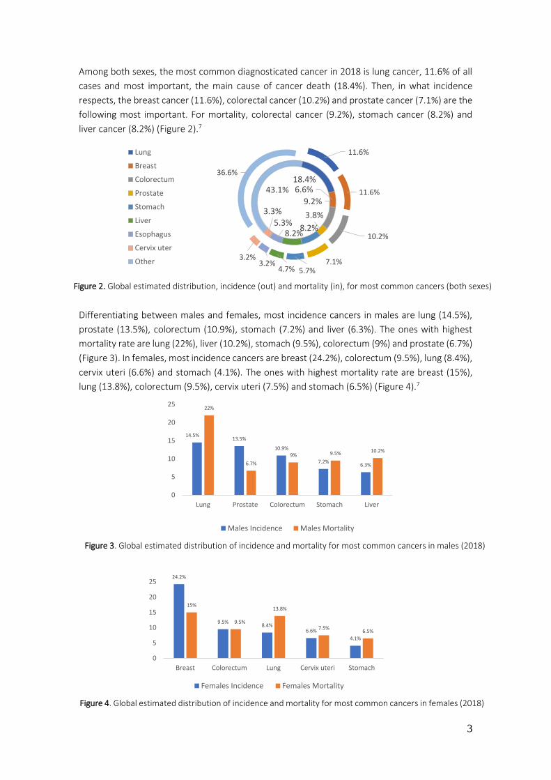
Among both sexes, the most common diagnosticated cancer in 2018 is lung cancer, 11.6% of all
cases and most important, the main cause of cancer death (18.4%). Then, in what incidence
respects, the breast cancer (11.6%), colorectal cancer (10.2%) and prostate cancer (7.1%) are the
following most important. For mortality, colorectal cancer (9.2%), stomach cancer (8.2%) and
liver cancer (8.2%) (Figure 2).7
Differentiating between males and females, most incidence cancers in males are lung (14.5%),
prostate (13.5%), colorectum (10.9%), stomach (7.2%) and liver (6.3%). The ones with highest
mortality rate are lung (22%), liver (10.2%), stomach (9.5%), colorectum (9%) and prostate (6.7%)
(Figure 3). In females, most incidence cancers are breast (24.2%), colorectum (9.5%), lung (8.4%),
cervix uteri (6.6%) and stomach (4.1%). The ones with highest mortality rate are breast (15%),
lung (13.8%), colorectum (9.5%), cervix uteri (7.5%) and stomach (6.5%) (Figure 4).7
14.5%13.5%
10.9%
7.2%6.3%
22%
6.7%
9% 9.5% 10.2%
0
5
10
15
20
25
Lung Prostate Colorectum Stomach Liver
Males Incidence Males Mortality
Figure 3. Global estimated distribution of incidence and mortality for most common cancers in males (2018)

4
According to a report, the therapeutic and supportive care cost in 2017 was 133 billion dollars
and it is foreseen to exceed 150 billion dollars by 2020.8 This data is related with therapies and it
is necessary, as well, to consider costs of hospitalizations, services out of hospital,
physician/supplier services, pharmacist, psychologist, medical equipment, home health care, and
others.
NATIONAL EXTENSION
In Spanish state, cancer is on top three of death causes, located after heart diseases.
In 2018 an amount of 270.363 incidences has been estimated (114.392 female and 155.971 male)
and 110.779 deaths (43.448 females and 67.331 males). In both sexes, most frequent cancer is
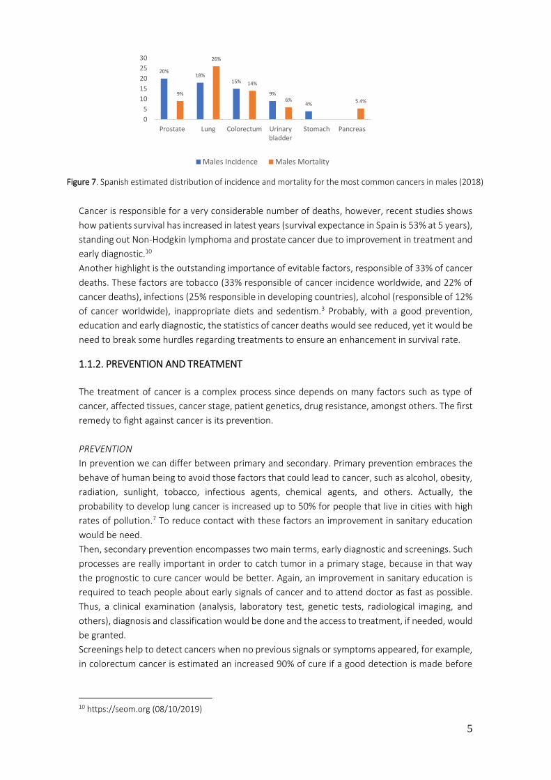
colorectum (14%), then prostate (12%), lung (10%), breast (12%) and skin cancers (8%). The
cancers with highest rate of death in 2018 are lung (20.3%), colorectum (14.1%), pancreas (6.3%),
breast (6%) and prostate (5.5%) (figure 5).9
If we differentiate between female and male, breast cancer (29%), colorectum (13%), lung (6%),
cervix uteri (6%) and ovarium (4.3%) are the most incidence cancers in females, and the ones
with most deaths rate are breast (15%), colorectum (15%), lung (11%), pancreas (7%) and
ovarium (5%) (Figure 6). While in males, most incident cancers in 2018 are prostate (20%), lung
(18%), colorectum (15%), urinary bladder (9%) and stomach (4%) and the ones with highest rate
of mortality are lung (26%), colorectum (14%), prostate (9%), urinary bladder (6%) and pancreas
(10.2%) (Figure 7).9
8 https://www.iqvia.com (08/10/2019) 9 https://www.aecc.es (08/10/2019)
14.1%
5.5%
20.3%
6%
6.3%
47.8%
14%
12%
10%
12%8%
44%
Colorectum Prostate Lung Breast Pancreas Others
Figure 5. Spanish estimated distribution of incidence (out) and mortality (in) for most common cancers (both sexes)
29%
13%
6% 6%4%
15% 15%
11%
7%5%
0
5
10
15
20
25
30
35
Breast Colorectum Lung Pancreas Cervix uteri Ovarium
Females Incidence Females Mortality
Figure 6. Spanish estimated distribution of incidence and mortality for most common cancers in females (2018)
5
Cancer is responsible for a very considerable number of deaths, however, recent studies shows
how patients survival has increased in latest years (survival expectance in Spain is 53% at 5 years),
standing out Non-Hodgkin lymphoma and prostate cancer due to improvement in treatment and
early diagnostic.10
Another highlight is the outstanding importance of evitable factors, responsible of 33% of cancer
deaths. These factors are tobacco (33% responsible of cancer incidence worldwide, and 22% of
cancer deaths), infections (25% responsible in developing countries), alcohol (responsible of 12%
of cancer worldwide), inappropriate diets and sedentism.3 Probably, with a good prevention,
education and early diagnostic, the statistics of cancer deaths would see reduced, yet it would be
need to break some hurdles regarding treatments to ensure an enhancement in survival rate.
1.1.2. PREVENTION AND TREATMENT
The treatment of cancer is a complex process since depends on many factors such as type of
cancer, affected tissues, cancer stage, patient genetics, drug resistance, amongst others. The first
remedy to fight against cancer is its prevention.
PREVENTION
In prevention we can differ between primary and secondary. Primary prevention embraces the
behave of human being to avoid those factors that could lead to cancer, such as alcohol, obesity,
radiation, sunlight, tobacco, infectious agents, chemical agents, and others. Actually, the
probability to develop lung cancer is increased up to 50% for people that live in cities with high
rates of pollution.7 To reduce contact with these factors an improvement in sanitary education
would be need.
Then, secondary prevention encompasses two main terms, early diagnostic and screenings. Such
processes are really important in order to catch tumor in a primary stage, because in that way
the prognostic to cure cancer would be better. Again, an improvement in sanitary education is
required to teach people about early signals of cancer and to attend doctor as fast as possible.
Thus, a clinical examination (analysis, laboratory test, genetic tests, radiological imaging, and
others), diagnosis and classification would be done and the access to treatment, if needed, would
be granted.
Screenings help to detect cancers when no previous signals or symptoms appeared, for example,
in colorectum cancer is estimated an increased 90% of cure if a good detection is made before
10 https://seom.org (08/10/2019)
20%18%
15%
9%
4%
9%
26%
14%
6% 5.4%
0
5
10
15
20
25
30
Prostate Lung Colorectum Urinarybladder
Stomach Pancreas
Males Incidence Males Mortality
Figure 7. Spanish estimated distribution of incidence and mortality for the most common cancers in males (2018)

6
late stages of disease, when symptoms comes out.11 These processes are more complex and
require a huge effort in organization and efficiency.
In both prevention stages there is one test getting relevance, genetic proves. In the beginning, to
read the genome costed around 2700 millions of dollars worldwide, nowadays it is possible to get
it for 600 dollars.12 Cancer is not inherited for the parents, is the genetic error what drives to
cancer, but the increased risk, the predisposition to get some type of cancers, is inherited. It is
not possible to cure cancer through this test but provides valuable knowledge about predisposed
genes, specifically for each human being (personalized therapy).
In the second stage, when cancer is already detected, it is also important to get back up from
genetic tests. In other words, if genetics of cancer cells is known, the efficiency of treatment
would be enhanced, choosing correctly through a wide breadth of drugs and therefore the
probability of survival would be improved as well.
TREATMENT
Based on the factors commented above, a specific treatment is chosen. Treatments can be sorted
by surgery (removing tumor from the body), radiation (high doses of radiation to kill cancer cells),
chemotherapy (drugs), immunotherapy (treatment to boost immune system to fight cancer),
targeted therapies (targeting changes that helps cancer cells to grow and progress), hormone
therapy (for breast or prostate), stem cell transplant (when radiotherapy and/or chemotherapy
has destroyed their own) and precision medicine (based on genetic knowledge).4 Even with this
wide portfolio of options, cancer is one of the biggest challenge due to its complexity.
1.1.3. DIFFICULTIES IN CANCER TREATMENT
One of the hardest difficulties is the similarity between normal cells and cancer cells, fact that
complicates the treatment (high rate of adverse effects) and therefore more selectivity is needed.
Probably, through a better conception of biology and tumor genetics, chasing those differences
present in cancer cells, will improve chemotherapeutic treatments. Taking this into account, what
makes cancer very tricky is his ability to spread (metastasis) and the resistance to treatments.
Linking these three factors gives as a result a cancer cells with an enhanced capacity to survive.
METASTASIS
Defined as the ability of cancer cells to spread in the body, to nearby tissues or to other parts
through blood or lymphatic system. When it occurs is called stage IV. The metastatic cancer cells
are the same than in primary tumor. It is well known that depending on the type of cancer it will
spread preferably to certain parts of the body than others. For example, breast cancer has
metastasis in bone, brain, liver and lung. Lung cancer in adrenal gland, bone, brain, liver and the
other lung.1
The mechanistic processes through which metastasis develops embrace some steps summarized
in:13
· Invading near tissue, blood vessels or/and lymph, changing epithelial cell structure (cell polarity
and cell adhesion)
11 F. T. Kolligs. Visc. Med. 2016, 32, 158–164 12 https://ghr.nlm.nih.gov (09/10/2019) 13 F. Zijl; G. Krupitza; W. Mikulitsa. Mutat. Res. 2011, 728, 23–34
7
· Moving to other parts of the body
· Stopping in small vessels, invading them and moving into the tissue. Their ability to promote
angiogenesis helps to improve these steps
· If the conditions are suitable, a new primary tumor grows
Not all cancer cells that undertake metastasis will survive, but if metastasis success, cells could
stay in a quiescent state for a long period of time, reappearing even when treatment is done.
Bearing in mind this processes, metastasis of cancer is one of the main factors leading to patient
death. When a stage IV is diagnosticated it is tricky to foresee the outcomes after treatment,
sometimes it would be a good prognostic but in other cases treatment will shrink the tumor but
not enough to cure it. Nowadays, treatments focused in metastasis have shown progress but
there are still some limitations such as the recurrence after treatment, the tough side effects and
most important, drug resistance. Hereto, new mechanism of action to prevent and to break
metastasis are needed. There are two concepts to stand out in metastasis, the epithelial
mesenchymal transition (EMT) and cancer stem cells, two concepts really linked and still in debate
to know which one precede the other.14
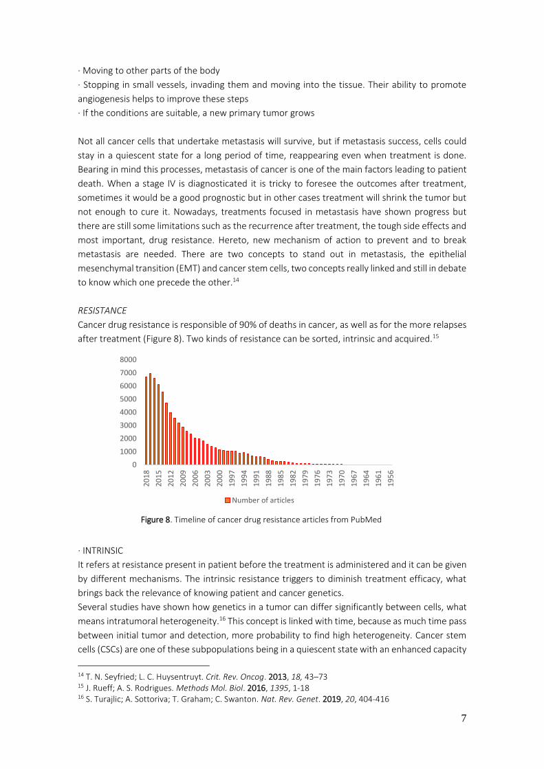
RESISTANCE
Cancer drug resistance is responsible of 90% of deaths in cancer, as well as for the more relapses
after treatment (Figure 8). Two kinds of resistance can be sorted, intrinsic and acquired.15
· INTRINSIC
It refers at resistance present in patient before the treatment is administered and it can be given
by different mechanisms. The intrinsic resistance triggers to diminish treatment efficacy, what
brings back the relevance of knowing patient and cancer genetics.
Several studies have shown how genetics in a tumor can differ significantly between cells, what
means intratumoral heterogeneity.16 This concept is linked with time, because as much time pass
between initial tumor and detection, more probability to find high heterogeneity. Cancer stem
cells (CSCs) are one of these subpopulations being in a quiescent state with an enhanced capacity
14 T. N. Seyfried; L. C. Huysentruyt. Crit. Rev. Oncog. 2013, 18, 43–73 15 J. Rueff; A. S. Rodrigues. Methods Mol. Biol. 2016, 1395, 1-18 16 S. Turajlic; A. Sottoriva; T. Graham; C. Swanton. Nat. Rev. Genet. 2019, 20, 404-416
0
1000
2000
3000
4000
5000
6000
7000
8000
20
18
20
15
20
12
20
09
20
06
20
03
20
00
19
97
19
94
19
91
19
88
19
85
19
82
19
79
19
76
19
73
19
70
19
67
19
64
19
61
19
56
Number of articles
Figure 8. Timeline of cancer drug resistance articles from PubMed

8
to resist therapy due to their characteristics. CSCs have a high tumorigenic potential since they
act as a normal stem cells, ergo they possess capacity of self-renewing and capacity to generate
differentiated subpopulations of cells, increasing heterogeneity.17
Another possibility that could confer intrinsic resistance is the pre-existence of mutations in a
gene(s) involved in cell division, proliferation and apoptosis. For example, in gastric cancer,
patients with HER2 overexpression showed a reduced outcome after treatment with cisplatin. The
HER2 gene upregulates the factor called Snail, which promote, the mentioned before, epithelial
mesenchymal transition (EMT), conferring more resistance to chemotherapy and radiotherapy.18
It has been mentioned that EMT and CSCs are linked with metastasis and now these two last
points relate them with the resistance as well. Finally, another mechanism that can decrease
efficiency of treatment are those pathways that organism undertakes to get rid of environmental
toxins or to detoxify cells. An example of this case is the ATP binding cassette (ABC), transporters
that regulate drug efflux, reducing drugs concentration inside the cells.19
· ACQUIRED
In acquired resistance the efficacy of treatment slows down along it due to new mutations in
proto-oncogenes, alterations in drug targets (mutations, up or down-regulation) or/and the
influence of tumor microenvironment. For example, methotrexate, which is amog the most used
known drugs for clinical cancer treatments, in acute leukemia treated with methotrexate an
amplification of dihydrofolate reductase (DHFR) gene was observed.22
DHFR is the target of methotrexate, then if the concentration of DHFR is higher than the inhibitor,
the cell cycle and the tumor growth will keep in progress.
In the treatment of osteosarcoma, the resistance is due to decreased transport through the
reduced folate carrier (RFC).23
17 M. Najafi; B. Farhood; K. Mortezaee. J. Cell. Physiol. 2019, 234, 8381-8395 18 D. Huang; H. Duan; H. Huang; X. Tong; Y. Han; G. Ru; L. Qu; C. Shou; Z. Zhao. Sci. Rep. 2016, 6, 1-12 19 M. Kartal-Yandim; A. Adan-Gokbulut; Y. Baran. Crit. Rev. Biotechnol. 2016, 36, 716-726 20 C. Holohan; S. Van Schaeybroeck; D.B. Longley; P.G. Johnston. Nat. Rev. Cancer 2013, 13, 714-726 21 M. Akada; T. Crnogorac-Jurcevic; S. Lattimore; P. Mahon; R. Lopes; M. Sunamura; S. Matsuno; N.R. Lemoine. Clin. Cancer. Res. 2005, 11, 3094-3101 22 J. R. Bertino; E. Göker; R. Gorlick; W. W. Li; D. Banerjee. Stem Cells 1996, 14, 5-9 23 W. Guo; J. H. Healey; P. A. Meyers; M. Ladanyi; A. G. Huvos; J. R. Bertino; R. Gorlick. Clin. Cancer Res. 1999, 51, 621-627
Drug Cancer Mutation Outcome
Cisplatin Gastric cancer HER2 +/Snail +
HER2 -/Snail +
HER2 -/Snail -
Patients with double positive genes have less survival
rate followed by -/+ and highest survival rate for -/-
profile18
TKIs Chronic myeloid
leukemia and EGFR
NSCLC
Polymorphism BCL2-
like 11(BIM)
Upregulation of BIM is required for TKIs to induce
apoptosis in kinase-driven cancers. The polymorphism
confers a lack in the pro-apoptotic domain20
Gemcitabine Pancreatic cancer Down regulation of
BCL2 interacting
protein 3 (BNIP3)
Overexpression of BNIP3 induces cell death21
Table 1. Examples of intrinsic resistance in cancer

9
Some mechanisms are shared by both types of resistance, actually, to differ between intrinsic
and acquired resistance is a temporal criteria, so may be, acquired resistance combines resistant
subpopulations prior to treatment (intrinsic resistance) with a new mechanisms adapted
throughout treatment.
1.1.3.1. CELL MECHANISMS FOR DRUG RESISTANCE
It is vital to understand the tumor resistance mechanism for each type of cancer in order to
optimize treatment. Here, these mechanisms are summarized in decreased intracellular drug
concentration, target alteration, epigenetic alterations, tumor microenvironment, tumor
heterogeneity, deregulation of cell death, and EMT-CSCs.
DECREASED INTRACELLULAR DRUG CONCENTRATION
The decreased drug uptake and the augmented drug efflux is responsible for diminishing
intracellular concentration of chemotherapeutics. Actually, it is the main reason of resistance.
As it is well known, to reach therapeutic effectiveness, anticancer drugs must get their targets
with an adequate concentration. The underlying mechanisms acting to decrease the final
concentration of drugs inside the cell has been studied for a long time and a clarification of them
is summarized in numerous articles.27,28
It is proposed that an alteration in plasmatic membrane acts as a hurdle, as a defense mechanism
to protect cancer cells. This alteration (changes in components and/or organization in membrane)
is caused with the malignant transformation in cancer cells, for example, glucosylceramide was
found increased in multidrug resistance cancer cell lines and absent in drug sensitive cells.29
Therefore, the quantity and the kind of transporters is linked with membrane components. In
one side, drug influx is reduced due to different factors, one of the most important are the
transporters such as solute carriers, for example, antifolates drugs enter inside cell through them
24 D. S. Hsu; W. L. Hwang; C. H. Yuh; C. H. Chu; Y. H. Ho; P. B. Chen; H. S. Lin; H. K. Lin; S. P. Wu; C. Y. Lin; W. H. Hsu; H. Y. Lan; H. J. Wang; S. K. Tai; M. C. Hung; M. H. Yang. Clin. Cancer Res. 2017, 23, 4388-4401 25 H. J. Choi; H. S. Joo; H. Y. Won; K. W. Min; H. Y. Kim; T. Son; Y. H. Oh; J. Y. Lee; G. Kong. J. Nat. Cancer Inst. 2018, 110, 400-410 26 D. Yan; J. Kowal; L. Akkari; A. J. Schuhmacher; J. T. Huse; B. L. West; J. A. Joyce. Oncogene 2017, 36, 6049-6058 27 R. R. Begicevic; M. Falasca. Int. J. Mol. Sci. 2017, 18, 2362-2386 28 Y. H. Choi; A. M. Yu. Curr. Pharm. Des. 2014, 20, 793–807 29 A. Lucci; W. I. Cho; T. Y. Han; A. E. Giuliano; D. L. Morton; M. C. Cabot. Anticancer Res. 1998, 18, 475-480
Drug Cancer Resistance Outcome
Cetuximab Head and neck
cancer
EMT process
through Snail
activation
Methylation of EGFR (target of cetuximab) and
dimerization with lymphotoxin-β (LTβ)24
Tamoxifen Breast cancer
(estrogen receptor
+ (ER+))
Epigenetic
alterations
Retinoblastoma-binding protein 2 (RBP2)
activates the PI3K/AKT pathway, promoting the
cell survival25
Dovitinib,
Vatalanib
Glioblastoma Tumor
microenvironment
Tumor associated macrophages (TAMs) increase
the secretion of colony stimulating factor-1
receptor (CSF-1R), then the survival and
progression is increased26
Table 2. Examples of acquired resistance in cancer

10
and it is been reported how in some cancers the alteration of this influx transporters decrease
antifolates efficacy.30 Another associated problem is the pH in tumor microenvironment, because
intracellular pH is increased and extracellular decreased down to 6.5-7.1, thus, antineoplastic
drugs with a weak basic character remain protonated outside the cell.31
In the other side, efflux transporters play a pivotal role in cancer drug resistance. The hugest
family is the ABC transporters (superfamily of 49 ABC genes) like the well described P-
glycoprotein (P-gp), also called multidrug resistance protein 1 (MDR-1).32 In kidney, colon, liver,
lung and rectum cancer an overexpression of P-gp has been described previous to treatment33
(intrinsic resistance) and after treatment like in chronic myelogenous leukemia (acquired
resistance).34 The P-gp contains two transmembrane domains, through which drugs and toxins
are removed from inside the cell, and two nucleotide binding domains to bind and hydrolyze the
ATP in order to change the conformation and pump off the substrates.
Consequently, the overexpression and mutations of P-gp has correlated with a poor prognosis
and poor therapeutic response, diminishing sensitivity to drugs. Then, in order to improve the
outcomes, it will be necessary to perform studies to recognize ABC cancer profile previous to
treat it. Also, new strategies like a combined chemotherapy (inhibitor of growth and inhibitor of
MDR-1) might rise in order to improve the treatment efficacy (Figure 9).35
TARGET ALTERATION
This mechanism of resistance appears for those treatments called target therapies, which acts
inhibiting the activity of specific target (enzymes, receptors, and others).
In the beginning of a treatment it can be an advantage to use the differences between normal
cells and CCs to decrease adverse effects, increasing selectivity, but it can draw CCs to CDR.
30 L. H. Matherly; M. R. Wilson; Z. Hou. Drug Metab. Dispos. 2014, 42, 632-649 31 D. T. Manallack. Perspect. Medicin. Chem. 2007, 17, 25-38 32 F. J. Sharom. Pharmacogenomics 2008, 9, 105-127 33 A. Adamska; M. Falasca. World J. Gastroenterol. 2018, 24, 3222–3238 34 X. X. Peng; K. T. Amit; H. C. Wu; Z. S. Chen. Chin. J. Cancer 2012, 31, 110–118 35 Z. Lin; M. Chen; X. Yang; Z. Cui; X. Zhang; L. Yuan; Q. Zhang. Int. J. Nanomedicine 2014, 9, 3425-3437
Figure 9. Illustration of possible advantages using Doxorubicin (DOX) + Cyclosporine A (CsA) (P-gp inhibitor) in a loaded liposomal (SSL) in a tumor treatment35

11
One of the best examples to understand this mechanism is resistance developed in treatments
with tyrosine kinase inhibitors of EGFR in non-small cell lung cancer (NSCLC). Since treatment with
erlotinib, gefitinib or afatinib was introduced into the market the efficacy in progression-free
survival and overall survival were increased compared with traditional treatment, but most of the
patients develop acquired resistance through mechanisms, such as mutations in target gene
(altering the binding site), epigenetic alterations (upregulation or downregulation) or/and
alterations in signal pathways.36 If methods of drug target alteration are known it could bring
support for diagnosis, for developing new strategies and drugs to treat resistant cancer cells.
EPIGENETIC ALTERATIONS
The epigenetic alterations refer to changes in gene expression that are heritable within cellular
division, then, no those alterations present in primary DNA. Epigenetic includes DNA methylation,
histone modification (acetylation or methylation) and non-coding RNA (miRNA and lnRNA) as
principals.37 The epigenetic changes control genes expression, for example when a histone is
acetylated the chromatin opens, giving as a result a transcriptionally accessible state, thus,
transcription factors between other proteins (inhibitors or activators) can regulate gene
expression. A good example is the downregulation of gene expression, through hypermethylation
of tumor suppressor genes or by contrast, the hypomethylation of oncogenes, ergo
overexpression.
· DNA damage repair (DDR)
The epigenetic alterations are also related with enhanced DNA damage repair (DDR). This process
of fixing damaged DNA is improved in cancer cells, reversing the efficacy of treatment. For
example, treatment with 5-fluorouracil induces upregulation of p53 target genes in damaged
DNA, thus the success to repair it is increased, diminishing cell arrest and programmed cell death.
It could be an appealing strategy to attack both, DDR and DNA, but it should contemplate the risk
of increased mutations due to genomic instability.38 Another related example is the
hypomethylation of MDR1 promoter, giving a P-gp overexpression in stomach cancer and in
invasive ductal carcinoma.39,40 Therefore, increasing their aggressiveness. This example helps to
emphasize the fact that resistance mechanisms are connected to each other, is a multifactorial
process that has to be studied in every cancer, in every patient, for every drug.
· Human Nicotinamide N-Methyltransferase (hNNMT)
The hNNMT is a phase II metabolizing enzyme, catalyzing the methylation of nicotinamide into 1-
methylnicotinamide (MNA) as a main reaction. Its expression is altered in some cancers
depending on stress situation, because in those tissues with reduced nutrient availability it is
found to be overexpressed, producing then more NAD+ and MNA41 (Figure 10), as well as, it has
36 J. Gao; H. R. Li; C. Jin; J. H. Jiang; J. Y. Ding. Clin. Transl. Oncol. 2019, 21, 1287-1301 37 M. Kacevska; M. Ivanov; M. Ingelman-Sundberg. Pharmacogenomics 2012, 13, 1373–1385 38 M. D. Wyatt; D. M. Wilson. Cell. Mol. Life Sci. 2009, 66, 788–799 39 G. Sharma; S. Mirza; R. Parshad; A. Srivastava; S. D. Gupta; P. Pandya; R. Ralhan. Clin. Biochem. 2010, 43, 373–379 40 A. A. Muggerud; J. A. Rønneberg; F. Wärnberg; J. Botling; F.Busato; J. Jovanovic; H. Solvang; I. Bukholm; A. L. Børresen-Dale; V. N. Kristensen; T. Sørlie; J. Tost. Breast Cancer Res. 2010, 12, 1-10 41 J. Jung; L. J. Kim; X. Wang; Q. Wu; T. Sanvoranart; C. G. Hubert; B. C. Prager; L. C. Wallace; X. Jin; S. C. Mack; J. N. Rich. J.C.I. Insight 2017, 2, e90019

12
found to be altered in some cancer stem cells.42 The aberrant expression of hNNMT is related
with poor prognosis in some cancers like in gastric, colorectal, gastric, prostate, breast and
liver.43,44 His activity in cancer drug resistance is linked with the inhibition of drug induced
apoptosis, increased proliferation, survival and cell cycle progression. For example, due to its
increased activity and expression, histones are hypomethylated rising protumorigenic genes.
The final product of the reaction, MNA, reduces reactive oxygen species (ROS) and apoptosis,
mechanism through which a resistance to 5-FU in colorectal cancer was described.45
Recently, hNNMt has been correlated with SIRT1 (sirtuins family are NAD+ dependent
deacetylases), which regulates cell functions like DNA repair, cell survival and metabolism via
deacetylation of histones and deacetylation of p53 (inhibition of p53, inhibition of apoptosis),
inducing cell survival in a stress situation. As nicotinamide is an inhibitor of SIRT1, an nicotinamide
concentration is decreased in those cancers where hNNMT is overexpressed, SIRT1 activity is
overstimulated.43 Finally, a recent study showed how hNNMT can be the key as a metabolic
regulator in cancers associated with fibroblasts, differentiating cells to cancer associated
fibroblast and therefore promoting cancer progression.46 All these examples lead to a conclusion
to undertake a new strategy in order to know the genomics of cancer and in those where hNNMT
is overexpressed to use a combined therapy against both, the specific cancer cell, plus inhibiting
hNNMt. Then, probably, prognosis and survival rate would arise.
42 F. P. Andrea; A. Safwat; M. Kassem; L. Gautier; J. Overgaard; M. R. Horsman. Radiother. Oncol. 2011, 99, 373-378 43 Y. Wang; J. Zeng; W. Wu; S. Xie; H. Yu; G. Li; T. Zhu; F. Li; J. Lu; G. Y. Wang; X. Xie; J. Zhang. Breast Cancer Res. 2019, 21, 1-17 44 https://www.proteinatlas.org (11/10/2019) 45 X. Xinyou; L. Huixing; W. Yanzhong; Z. Yanwen; Y. Haitao; L. Guiling; R. Zhi; L. Fengying; W. Xiuhong; Z. Jun. Oncotarget 2016, 7, 45837–45848 46 M. A. Eckert; F. Coscia; A. Chryplewicz; J. W. Chang; K. M. Hernandez; S. Pan; S. M. Tienda; D. A. Nahotko; G. Li; I. Blaženović; R. R. Lastra; M. Curtis; S. D. Yamada; R. Perets; S. M. McGregor; J. Andrade; O. Fiehn; R. E. Moellering; M. Mann; E. Lengyel. Nature 2019, 569, 723-728
Figure 10. Differences in hNNMT expression between a glioblastoma stem cell in an enriched niche and glioblastoma stem cell in a non-enriched niche 41

13
· Non-coding RNAs (ncRNAs)
There are five types of ncRNAs, microRNAs (miRNAs), longnoncoding RNAs(lncRNAs), small
interfering RNAs (siRNAs), antisense RNAs (asRNAs) and circular RNAs (circRNAs).47 About 98% of
transcriptional output is ncRNA in humans.48
Due to their regulation function in gene expression, ncRNAs play an important role in initiation,
metastasis, cancer stem cells and in latest years more evidence is coming up about its influence
in cancer drug resistance.49 The more related ones are lncRNA, miRNA and circRNA and the
mechanisms through which they regulate resistance is via ABC transporters, cell arrest and
apoptosis, CSC and EMT, autophagy and others.50
To understand how these RNA molecules affect in cancer, an example for each type is described.
The miRNAs (18-22 nt in length) acts as an inhibitor of post-transcriptional gene expression
binding to their complementary mRNAs, controlling different cellular process as apoptosis,
cellular differentiation and proliferation. The profile expression in tumor tissues is different
compared with normal tissues and their dysregulation has related with resistance and relapse.51,52
Before defining miRNAs as a tumor suppressor or oncogene it will be necessary to put in context
the biological cell environment, because it is what decant them to one or other profile.53
For example, miR663 was found to be upregulated in breast cancer and to create resistance to
doxorubicin by inhibiting apoptosis.54
The lncRNAs (200-10000 nt length) regulate gene expression through different mechanism, for
example blocking the binding site to transcriptional activators, chromatin modification, post-
translational regulation, and others. Their role in cells depends, like miRNAs, in the context.
An example is the resistance to trastuzumab in HER-2 positive breast cancer where it was found
an overexpression of lncRNA-ATB, which promotes epithelial mesenchymal transition and this
leads to drug resistance.55
The circRNAs unlike the already described RNAs makes a covalent closed loop. Their role also
depends on cellular context. For example, upregulation of circ_001569 in osteosarcoma has
linked with cisplatin resistance by activating Wnt/beta-catenin pathway and therefore promoting
cell proliferation.56
With these summarized implications it is visible how complex and wide can be the different roles
of ncRNA in cancer resistance. Therefore, a genomic studies and functional considerations will be
need it to understand the role of each ncRNA in each cancer, targeting them as a strategy in a
combined therapy.
47 S. Qu; Y. Zhong; R. Shang; X. Zhang; W. Song; J. Kjems; H. Li. RNA Biol. 2017, 14, 992-999 48 J. S. Mattick. EMBO Rep. 2001, 2, 986-991 49 E. Anastasiadou; L. S. Jacob; F. J. Slack. Nature Reviews Cancer 2018, 18, 5–18 50 W. Z. Hu; C. L. Tan; Y. J. He; G. Q. Zhang; Y. Xu; J. H. Tang. Onco. Targets Ther. 2018, 11, 1529-1541 51 I. Berindan-Neagoe; P. Monroig; B. Pasculli; G. A. Calin. C.A. Cancer J. Clin. 2014, 64, 311–336 52 H. Lan; H. Lu; X. Wang; H. Jin. Biomed. Res. Int. 2015, v2015, 1-17 53 M. Carlo; M. D. Croce. Nat. Rev. Genet. 2009, 10, 704–714 54 H. Hu; S. Li; X. Cui; X. Lv; Y. Jiao; F. Yu; H. Yao; E. Song; Y. Chen; M. Wang; L. Lin. J. Biol. Chem. 2013, 288, 10973-10985 55 S. J. Shi; L. J. Wang; B. Yu; Y. H. Li; Y. Jin; X. Z. Bai. Oncotarget 2015, 6, 11652-11663 56 H. Zhang; J. Yan; X. Lang; Y. Zhuang. Oncol. Lett. 2018, 16, 5856-5862

14
TUMOR MICROENVIRONMENT (TME)
The tumor microenvironment is not only made by homogenous cancer cells, it embraces vessels,
immune cells (as tumor associated macrophages), fibroblasts, extracellular matrix (ECM), signal
molecules, and others (Figure 11). All these factors aid the tumor to grow and survive.57
To keep this environment and the communication between cells a remodulation of ECM is
necessary. ECM is a complex network made by macromolecules which offers a structural and
biochemical support to cells, controlling cell growth, proliferation, motility, pH, hydration, and
others. Therefore, as an inflection point, ECM can contribute to cancer proliferation, evasion of
suppression, apoptosis resistance, induced angiogenesis, initiation of metastasis, dysregulation
of cellular homeostasis and avoidance of immune response.58 An example in CDR is described for
HER2 therapies in breast cancer with a PIK3CA mutation, where an upregulation of
collagen/integrin/Src signaling was found, endowing resistance to trastuzumab and
pertuzumab.59
Another example is the intrinsic resistance mechanism previously mentioned, the changes in
extracellular pH. In latest years a combined treatment including proton-pump inhibitors has
increased.60 When treatment starts, TME can change significantly giving as a result a resistance
to therapy (chemotherapy or targeted).
57 https://www.nature.com (14/10/2019) 58 M. W. Pickup; J. K. Mouw; V. M. Weaver. EMBO Rep. 2014, 15, 1243-1253 59 A. B. Hanker; M. V. Estrada; G. Bianchini; P. D. Moore; J. Zhao; F. Cheng; J. P. Koch; L. Gianni; D. R. Tyson; V. Sánchez; B. N. Rexer; M. E. Sanders; Z. Zhao; T. P. Stricker; C. L. Arteaga. Cancer Res. 2017, 77, 3280-3292 60 Z. N. Lu; B. Tian; X. L. Guo. Cancer Chemother. Pharmacol. 2017, 80, 925-937
Figure 11. Illustration of tumor microenvironment69
15
As an indispensable component of TME it is necessary to highlight myofibroblasts (or cancer
associated fibroblasts (CAFs)) and tumor associated macrophages (TAMs). The CAFs are able to
promote tumor growth and progression, to activate angiogenesis, to stimulate growth factors, to
promote ECM and to keep the stemness, between other important roles.61 An example of them
in CDR is the described in head and neck cancers with cisplatin resistance, where they produce
an exosomal miR-196a which promotes cell growth, survival and inhibition of apoptosis.62 In non-
small cell lung cancer cells, CAFs promote a signal pathway that upregulates P-gp and therefore
endowing resistance.63
In reference to TAMs, they are anti-inflammatory and protumorigenic cells, facilitating tumor
growth, metastasis and they can promote resistance by themselves or stimulating the growth of
CSC through inflammatory signals.64 An example of its role in CDR is found in hepatocellular
carcinoma, where TAMs by means of autophagy creates resistance to oxaliplatin treatment.65
Finally, it is necessary to describe the implications of ATP in TME. In cancer cells, generation of
energy, consuming high quantities of glucose and accumulation of lactate (aerobic glycolysis,
Warburg effect) differs compared with normal cells in which glucose is oxidized in the
mitochondria. Therefore, high levels of ATP have found to be essential for cancer cells and much
more in resistance cancer cells where energy is necessary to fight stress. In one study, ATP
concentration was found two times higher inside resistant cancer cells compared with primary
tumor cells.66 In intratumoral space, extracellular ATP (eATP) can be increased by apoptosis or
autophagy of cancer cells, thus eATP works as a signal (purinergic signal) with ability to promote
cell growth, proliferation and inducing EMT. This eATP can be internalized in cells increasing the
intracellular ATP, hence, increasing activity of ABC transporters.67
This ATP is the principal source of adenosine for CCs (CD73 catalyzes the conversion of ATP to
adenosine). Adenosine is essential in order to promote immunosuppression.68
All these summarized processes, underline the elaborated network that TME can be and actually
TME is only one aspect of the following point, the tumor heterogeneity, because TME enriches
the possible genetic differences in a tumor.69
TUMOR HETEROGENEITY
The heterogeneity of tumors embraces different concepts, the main one is probably genetic
differences between tumor subpopulations (different cancer cells) and also different kinds of cells
(immune cells, cancer stem cells, and others). Then, temporal heterogeneity, that is, temporal
61 M. Wang; J. Zhao; L. Zhang; F. Wei; Y. Lian; Y. Wu; Z. Gong; S. Zhang; J. Zhou; K. Cao; X. Li; W. Xiong; G. Li; Z. Zeng; C. Guo. J. Cancer 2017, 8, 761–773 62 X. Qin; H. Guo; X. Wang; X. Zhu; M. Yan; X. Wang; Q. Xu; J. Shi; E. Lu; W. Chen; J. Zhang. Genome Biol. 2019, 20, 1-21 63 Q. Zhang; J. Yang; J. Bai; J. Ren. Cancer Sci. 2018, 109, 944-955 64 A. J. Petty; Y. Yang. Immunotherapy 2017, 9, 289–302 65 X. T. Fu; K. Song; J. Zhou; Y. H. Shi; W. R. Liu; G. M. Shi; Q. Gao; X. Y. Wang; Z. B. Ding; J. Fan. Cancer Cell Int. 2019, 19, 1-11 66 Y. Zhou; F. Tozzi; J. Chen; F. Fan; L. Xia; J. Wang; G. Gao; A. Zhang; X. Xia; H. Brasher; W. Widger; M L. M. Ellis; Z. Weihua. Cancer Res. 2012, 72, 304-314 67 Y. Qian; X. Wang; Y. Liu; Y. Li; R. A. Colvin; L. Tong; S. Wu; X. Chen. Cancer Lett. 2014, 351, 242-251 68 X. Wang; Y. Li; Y. Qian; Y. Cao; P. Shriwas; H. Zhang; X. Chen. Oncotarget 2017, 8, 87860-87877 69 F. D. Virgilio; A. C. Sarti; S. Falzoni; E. De Marchi; E. Adinolfi. Nat. Rev. Cancer 2018, 10, 601-618

16
changes in cells composition. As well, it can be heterogeneity in nutrients and oxygen depending
on the angiogenesis and vessels surrounding the tumor.70
The heterogeneity of a tumor is one of the biggest hurdles for a good efficacy in a treatment. In
a cancer with a late diagnosis, the probability of high heterogeneity is increased, then
chemotherapy and/or radiotherapy will kill some subpopulations, but the resistance ones will
survive and they are able to communicate using extravascular vesicles containing ncRNAs and
pass those genetic characteristics that confers resistance to other CCs.71
To avoid these decreased outcomes in treatments, more genetic studies (sequential cell-studies,
samples of every part of tumor, and others) will afford good information about heterogeneity
and then a personalized therapy can be prepared.
DEREGULATION OF CELL DEATH
Three main paths of programmed death can be sorted: apoptosis, autophagic programmed cell
death and necrotic cell death.
Most of cancer therapies attack DNA or cell cycle (CDKs, p53, TNF family, and others) driving cells
to a programmed cell death. As it has been explained, in some cancers the tumor suppressors or
protooncogenes are altered and therefore they are able to avoid the apoptosis. Multiple
mechanisms are used by cancer cells to avoid all pathways that can draw them to apoptosis, such
as altering the expression of pro-apoptotic genes (downregulation) or anti-apoptotic genes
(upregulation) or post-translational regulation (altering protein functions, destroying pro-
apoptotic proteins).72
· Autophagy
The autophagy is an essential process, producing energy by means of degradation or recycling
dysfunctional cellular components and/or proteins, so it is a self-renewal process which helps to
keep homeostasis.73
In normal cells, autophagy can play a protective paper against cancer, destroying those cells,
toxins, altered proteins, carcinogens, that can promote cancer, but when autophagy is altered
(mutations) the energy in the cell is as well altered and processes as DNA replication can be
affected.74
In cancer, autophagy can be a doubled-edged sword as well, because it can promote tumor
growth and survival or either lead cancer cell to a programmed cell death. Since in cancer, stress
and inflammatory states are present, autophagy helps to produce the amount of energy
necessary to counter it.75
70 I. Dagogo-Jack; A. T. Shaw. Nat. Rev. Clin. Oncol. 2018, 15, 81-94 71 N. A. Yousafzai; H. Wang; Z. Wang; Y. Zhu; L. Zhu; H. Jin; X. Wang. Am. J. Cancer Res. 2018, 8, 2210–2226 72 K. Fernald; M. Kurokawa. Trends. Cell. Biol. 2013, 23, 620–633 73 N. Mizushima; M. Komatsu. Cell. 2011, 147, 728-741 74 Z. J. Yang; C. E. Chee; S. Huang; F. A. Sinicrope. Mol. Cancer. Ther. 2011, 10, 1533–1541 75 Y. J. Li; Y. H. Lei; N. Yao; C. R. Wang; N. Hu; W. C. Ye; D. M. Zhang; Z. S. Chen. Chin. J. Cancer 2017, 36, 1-10

17
The modulators of autophagy are called Atgs and in situations of stress, hypoxia they are
stimulated.76 It has been described how autophagy can drive to drug resistance in cancers like
hepatocellular carcinoma, where was found that downregulation of miR-199a-5p induced by
cisplatin treatment activates autophagy. Then, when autophagy is inhibited in MDR, the outcome
of the treatment is improved.77 Hence, autophagy can protect cancer cells destroying damaged
DNA and fighting against stressful situation.
This is the pro-survival edge of autophagy, in what pro-death activity respects, cell intrinsic
autophagy can help chemotherapeutics inhibiting genetic instability, because when DNA is
damaged, autophagy is able to destroy erroneous sequences or even lead to a cell death, since
controlling the protein quality is a described function of autophagy. Besides, in those cancer cells
where apoptosis is altered, the autophagic process is able to drive cells to death, thus, targeting
autophagy could be a resource to treat this type of cancers, due to autophagy overactivation
drives to a programmed cell death.78
The activity of autophagy, acting as a pro-death or pro-survival, it depends on tumor type and the
kind of treatment.79 In each cancer should be studied the altered pathways to know if it will be
useful to inhibit or stimulate autophagy in a combined therapy and much more in MDR càncers
(Figure 12).
CANCER STEM CELLS (CSCs) AND EPITHELIAL MESENCHYMAL TRANSITION (EMT)
The subpopulation of CSCs and the phenotypic plasticity conferred by EMT provides cancer more
aggressiveness with ability to decrease treatment efficacy, acquiring resistance to it.
To obtain these phenotypic characteristics, cancer cells undertake a reprogramming process.80
76 Y. Cao; Y. Luo; J. Zou. J. Ouyang; Z. Cai; X. Zeng; H. Ling; T. Zeng. Clin. Chim. Acta 2019, 489, 10-20 77 N. Xu; J. Zhang; C. Shen; Y. Luo; L. Xia; F. Xue; Q. Xia. Biochem. Biophys. Res. Commun. 2012, 423, 826-831 78 B. Y. K. Law; V. W. K. Wai. Intechopen 2016, 20, 435-454 79 F. Huang; B. R. Wang; Y. G. Wang. World J. Gastroenterol. 2018, 24, 4643-4651 80 X. Liu; D. Fan. Curr. Pharm. Des. 2015, 21, 1279-1291
Figure 12. Illustration of autophagy roles in hepatocellular carcinoma (HCC)76

18
· EMT
EMT is a highly conserved cellular process transforming epithelial cells to mesenchymal cells by
means of losing cell polarity and cell-cell adhesion. It is involved in embryogenesis, wound healing
and tumorigenesis (metastasis, recurrence, progression and resistance to therapy).
The mechanism used by EMT to endow cancer with drug resistance are not clear, but
morphological changes produce alteration of functional proteins related with epithelial cells and
mesenchymal cells. For example, it is known that some of ABC promoter genes have binding sites
for EMT transcription factors.81 The described miRNAs also contribute to EMT, regulating several
signals cascades.
These are some of transcription factors that can be a good target to treat or prevent drug
resistance: Snail, Slug, TGF-beta, ZEB, helix-loop-helix, amongst others. For example, in one study
was showed how inhibiting TGF-beta can reverse EMT phenotype and increase chemotherapy
efficacy (figure 13). 82,83,84
· CSCs
The CSCs are a subpopulation of tumor with the same characteristics of stem cells, that is,
capacity of self-renewal, quiescence, plasticity and potency to differentiate cell populations,
endowing CSCs with high tumorigenic potential. Besides, there are numerous studies showing
the capacity of this cells to resist radiotherapy and chemotherapy.85,86 Here we summarize some
described mechanism to get CDR by CSC.
81 Z. S. Jiang; Y. Z. Sun; S. M. Wang; J. S. Ruan. J. Cancer 2017, 8, 2319-2327 82 Y. T. Tang; Y. Y. Huang; J. H. Li; S. H. Qin; Y. Xu; T. X. An; C. C. Liu; Q. Wang; L. Zheng. BMC Genomics 2018, 19, 1-14 83 M. Mutlu; U. Raza; Ö. Saatci; E. Eyüpoğlu; E. Yurdusev; Ö. Şahin. J. Mol. Med. 2016, 94, 629-644 84 N. E. Bhola; J. M. Balko; T. C. Dugger; M. G. Kuba; V. Sánchez; M. Sanders; J. Stanford; R. S. Cook; C. L. Arteaga. J. Clin. Invest. 2013, 123, 1348-1358 85 T. B. Steinbichler; J. Dudás; S. Skvortsov; U. Ganswindt; H. Riechelmann; I. I. Skvortsova. Semin. Cancer Biol. 2018, 53, 156-167 86 E. Batlle; H. Clevers. Nat. Med. 2017, 23, 1124-1134
Figure 13. Illustration of connectivity network between EMT and CSC driving to resistance89
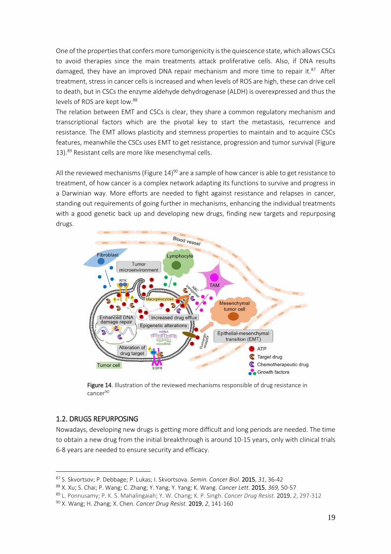
19
One of the properties that confers more tumorigenicity is the quiescence state, which allows CSCs
to avoid therapies since the main treatments attack proliferative cells. Also, if DNA results
damaged, they have an improved DNA repair mechanism and more time to repair it.87 After
treatment, stress in cancer cells is increased and when levels of ROS are high, these can drive cell
to death, but in CSCs the enzyme aldehyde dehydrogenase (ALDH) is overexpressed and thus the
levels of ROS are kept low.88
The relation between EMT and CSCs is clear, they share a common regulatory mechanism and
transcriptional factors which are the pivotal key to start the metastasis, recurrence and
resistance. The EMT allows plasticity and stemness properties to maintain and to acquire CSCs
features, meanwhile the CSCs uses EMT to get resistance, progression and tumor survival (Figure
13).89 Resistant cells are more like mesenchymal cells.
All the reviewed mechanisms (Figure 14)90 are a sample of how cancer is able to get resistance to
treatment, of how cancer is a complex network adapting its functions to survive and progress in
a Darwinian way. More efforts are needed to fight against resistance and relapses in cancer,
standing out requirements of going further in mechanisms, enhancing the individual treatments
with a good genetic back up and developing new drugs, finding new targets and repurposing
drugs.
1.2. DRUGS REPURPOSING
Nowadays, developing new drugs is getting more difficult and long periods are needed. The time
to obtain a new drug from the initial breakthrough is around 10-15 years, only with clinical trials
6-8 years are needed to ensure security and efficacy.
87 S. Skvortsov; P. Debbage; P. Lukas; I. Skvortsova. Semin. Cancer Biol. 2015, 31, 36-42 88 X. Xu; S. Chai; P. Wang; C. Zhang; Y. Yang; Y. Yang; K. Wang. Cancer Lett. 2015, 369, 50-57 89 L. Ponnusamy; P. K. S. Mahalingaiah; Y. W. Chang; K. P. Singh. Cancer Drug Resist. 2019, 2, 297-312 90 X. Wang; H. Zhang; X. Chen. Cancer Drug Resist. 2019, 2, 141-160
Figure 14. Illustration of the reviewed mechanisms responsible of drug resistance in cancer90

20
Then, the cost to produce it (measured by the Tufts University Center for the study of Drug
Development) is around 2.6 billion dollars (2013), growing 8.5% every year.91
Given this situation, it is increasing the process called drug repurposing, a strategy to identify new
activities and applications for already approved medicines or investigational medicines. This
entails some advantages in terms of cost, time and safe. Since some drugs will have passed the
clinical trials, they are concluded as safety for human use. The efficacy of these drugs has to be
proved for the new use, but at least, the risks of fail is lower. Also, those drugs that have failed
because of efficiency- efficacy but with a good safety profile, can be as well, repurposed.
In terms of cost, the investment it is also lower given that most of steps will be already done, so
it can decrease from 3 billion dollars to 200-300 million dollars.92 Indeed, time will be reduced as
well, depending if drug is already on the market or in preclinical stage.
Taking all of this together, the advantages of this approach are clear, such as quickest return of
benefits, low risk, and in research could bring new targets and new mechanisms of action. But
there are some hurdles that decreases efficiency of drug repurposing, the patents.
In what patents belong, industry can find some problems in patenting new activities due to the
main source of information will be already in the available literature. Only if new activity affords
differences compared with public data, then the company will be able to patent it.
Exists a special patent for those drugs in the market out of patent when a new activity has found
for them, the called new method-of-use. But it can be tricky to control it when generics of them
are already in the market.93
Repurposing drugs has been practicing along history of research and development, probably not
in a voluntary way, but affording good results as in Thalidomide case, which was used as a sedative
and was withdrawn off the market in 1961 due to birth skeletal defects. A few years after, was
found to be effective for erythema nodosum leprosum and finally, in 1999, for multiple
myeloma.94
In latest years more techniques and methods have been developed in order to enhance the
repurposing and activity prediction of thousand compounds. These tools can be computational,
experimental or mixed, but the aim is the same, find a relationship drug-disease.
The most common computational approaches are based on comparison between genetic
expression after treatment and genetics of diseases.95
Therefore, more open access resources should be available and exploded in order to downsize
and correlate all information about the existence drugs and diseases (Table 3).
91 J. A. DiMasi; H. G. Grabowski; R. W. Hansen. J. Health Econ. 2016, 47, 20-33 92 N. Nosengo. Nature 2016, 534, 314-316 93 S. Pushpakom; F. Iorio; P. A. Eyers; K. J. Escott; S. H. A. Wells; A. Doig; T. Guilliams; J. Latimer; C. McNamee; A. Norris; P. Sanseau; D. Cavalla; M. Pirmohamed. Nat. Rev. Drug Discov. 2019, 18, 41-58 94 C. M. Telleria. J. Cancer Sci. Ther. 2012, 4, 9-11 95 H. X. J. Li; H. Xie; Y. Wang. Int. J. Biol. Sci. 2018, 14, 1232-1244
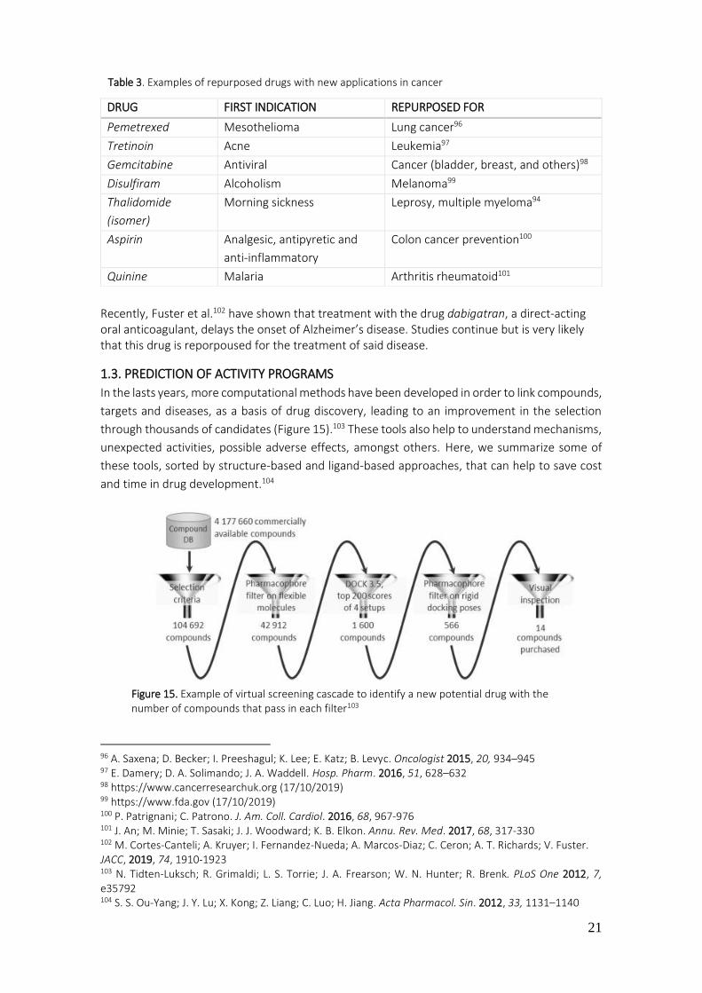
21
Table 3. Examples of repurposed drugs with new applications in cancer
Recently, Fuster et al.102 have shown that treatment with the drug dabigatran, a direct-acting oral anticoagulant, delays the onset of Alzheimer’s disease. Studies continue but is very likely that this drug is reporpoused for the treatment of said disease.
1.3. PREDICTION OF ACTIVITY PROGRAMS
In the lasts years, more computational methods have been developed in order to link compounds,
targets and diseases, as a basis of drug discovery, leading to an improvement in the selection
through thousands of candidates (Figure 15).103 These tools also help to understand mechanisms,
unexpected activities, possible adverse effects, amongst others. Here, we summarize some of
these tools, sorted by structure-based and ligand-based approaches, that can help to save cost
and time in drug development.104
96 A. Saxena; D. Becker; I. Preeshagul; K. Lee; E. Katz; B. Levyc. Oncologist 2015, 20, 934–945 97 E. Damery; D. A. Solimando; J. A. Waddell. Hosp. Pharm. 2016, 51, 628–632 98 https://www.cancerresearchuk.org (17/10/2019) 99 https://www.fda.gov (17/10/2019) 100 P. Patrignani; C. Patrono. J. Am. Coll. Cardiol. 2016, 68, 967-976 101 J. An; M. Minie; T. Sasaki; J. J. Woodward; K. B. Elkon. Annu. Rev. Med. 2017, 68, 317-330 102 M. Cortes-Canteli; A. Kruyer; I. Fernandez-Nueda; A. Marcos-Diaz; C. Ceron; A. T. Richards; V. Fuster. JACC, 2019, 74, 1910-1923 103 N. Tidten-Luksch; R. Grimaldi; L. S. Torrie; J. A. Frearson; W. N. Hunter; R. Brenk. PLoS One 2012, 7, e35792 104 S. S. Ou-Yang; J. Y. Lu; X. Kong; Z. Liang; C. Luo; H. Jiang. Acta Pharmacol. Sin. 2012, 33, 1131–1140
DRUG FIRST INDICATION REPURPOSED FOR
Pemetrexed Mesothelioma Lung cancer96
Tretinoin Acne Leukemia97
Gemcitabine Antiviral Cancer (bladder, breast, and others)98
Disulfiram Alcoholism Melanoma99
Thalidomide
(isomer)
Morning sickness Leprosy, multiple myeloma94
Aspirin Analgesic, antipyretic and
anti-inflammatory
Colon cancer prevention100
Quinine Malaria Arthritis rheumatoid101
Figure 15. Example of virtual screening cascade to identify a new potential drug with the number of compounds that pass in each filter103

22
STRUCTURE-BASED APPROACH (Interaction drug-target)
The target structure (macromolecule) has to be known through crystallization, NMR or/and
homology models.105
a) COMPUTATIONAL DOCKING
Computational docking is based on the structure in order to predict the best conformation and
orientation of a small molecule in a specific target (Figure 16).106 It can be used testing a variety
of ligands for one target or one ligand in front of different targets.107
In order to work with this tool a 3D structure of the molecule is needed and some proteins are
still not available, for example G protein-coupled receptors (GPCRs), which are the largest class
of membrane proteins in humans with vital roles in cells and approximately 475 approved drugs
by FDA target GPCRs.108 Also, some GPCRs are still considered orphan, probably crystallization of
these receptors to obtain the 3D structure will open a new world of opportunities (new targets,
new drugs).
LIGAND BASED APPROACH
These tools are applied when the targets 3D structure is not well known.
a) PHARMACOPHORE MODEL
Pharmacophore is the part of a molecule responsible for a specific biological or pharmacological
activity. The pharmacophore model is based on ligand approach, that is, superposing different
active compounds and extracting shared chemical features that confers the activity.
105 L. Chen; J. K. Morrow; H. T. Tran; S. S. Phatak; L. Du-Cuny; S. Zhang. Curr. Pharm. Des. 2012, 18, 1217–1239 106 C. H. M. M. P. Rao; R. P. Yejella; S. A. Rehman; C. Rajeswari; S. H. Basha. Journal of Peer Scientist 2018, 1, e1000008 107 L. Scotti; M. F. J. Junior; H. M. Ishiki; F. F. Ribeiro; R. K. Singla; J. M. Barbosa; M. S. Da Silva; M. T. Scotti. Curr. Drug Targets 2017, 18, 592-604 108 A. S. Hauser; S. Chavali; I. Masuho; L. J. Jahn; K. A. Martemyanov; D. E. Gloriam; M. M. Babu. Cell. 2018, 172, 41–54
Figure 16. Example of computational docking in anti-cancer drug development targeting EGFR106
23
In order to build a good model, it will be necessary the called ‘’training set of ligands’’, what
means, choosing a wide spectrum of molecules (some with activity and others without) to stablish
references for the approach, this step is the most important.
Then, a good conformation generation must be created, taking in consideration the bounds with
target, low energy conformation, and others. The lasts steps are superimposition or alignment of
molecules and finally the results of common chemical features which affords activity.109
This method can draw some mistakes depending on the library of compounds chosen for the
training set. Summarizing, this approach provides compounds information (hydrogen bonds,
hydrophobicity, aromaticity, and others) allowing to create a relation between structural
features-specific target.
b) SAR-QSAR
Quantitative Structure Activity Relationships (QSAR) is based on the idea that when a structure
of a molecule changes also his properties or activity change. It is based on mathematical and
statistical calculations of relations between psychochemical properties and biological activity.110
Since QSAR equations are created by descriptors (spatial, structure, thermodynamic,
conformational, and others), it is important to define them properly, because it is the mechanism
through which the molecule properties become numbers in the program. It is centered on
structure-activity relationship, therefore, when a compound is tested in a SAR model it means
that it is being compared whit a wide spectra of active and non-active compounds (references)
for a specific target. Finally, it gives as a result a mathematical formula correlating structure and
activity.111
Nowadays, some derivations exist, for example QSPR (quantitative structure-property
relationship) or QSRR (quantitative structure-reactivity relationship).
OPEN DATA BASES APPROACH
The number of potential targets, new mechanisms of action and molecular mechanism are
increasing and therefore, valuable information that has to be processed. Since this data is online
in open sources, tools to predict activities (between other properties) are increasing as well,
enabling the research community to optimize the process of drug discovery (Table 4). Some
computational tools, like machine learning or neural networks helps to maintain data bases in
constant actualization through algorithms that regulates its bases.112
In the following table some open sources are summarized, and the Prediction of Activity Spectra
for Substance (PASS) is developed as an example.
109 S. Y. Yang. Drug Discov Today 2010, 15, 444-450 110 G. Gini. Methods Mol. Biol. 2016, 1425, 1-20 111 J. Verma; V. M. Khedkar; E. C. Coutinho. Curr. Top. Med. Chem. 2010, 10, 95-115 112 J. Y. Trosset; C. Cave. Methods Mol. Biol. 2019, 1953, 189-193
24
· PASS
This open source allows to predict over 3500 different biological activities and only structural
formula is needed, then virtual drugs can be screened before synthetized.
In order to obtain the results, this platform is based on relationship activity-structure of 250.000
compounds with some biological activity.113 In the case of cancer, detecting cytotoxicity in
different cell lines by different compounds is a good example about how these tools can improve
research. Prediction of cytotoxicity for 24 breast cancer cell lines, 49 compounds were selected
between 1 million, based on cytotoxicity and interactions with cancer targets. Of this 49, only
eight showed a significative IC50.114
Combining these tools, it is afforded a valuable network of information with capacity to predict
activities of potential drugs, considering other parameters like interaction with transporters,
metabolic processes and signal pathways, which are related with tissue and cells.115 In cancer,
specifically in drug resistance, these tools can be really useful. For example, when tumor
heterogeneity is described through genetic tests, data can be collected in these platforms and
113 http://www.way2drug.com (18/10/2019) 114 V. Konova; A. Lagunin; P. Pogodin; E. Kolotova; A. Shtil; V. Poroikov. SAR QSAR Environ. Res. 2015, 26, 595-604 115 A. A. Lagunin; V. I. Dubovskaja; A. V. Rudik; P. V. Pogodin; D. S. Druzhilovskiy; T. A. Gloriozova; D. A. Filimonov; N. G. Sastry; V. V. Poroikov. PLoS One 2018, 13, e0191838
DATABASE DESCRIPTION SOURCE
ChEMBL Source of chemical, bioactivity and
genomic data to support transition of
genomic information to new drugs
https://www.ebi.ac.uk/chembl/
Chemspider
67 million structures from hundreds
of data sources
http://www.chemspider.com
Open Targets
Platform
Visualization of potential drug targets
associated with disease
https://www.targetvalidation.org
Human Protein
Atlas
Tissue atlas, distribution of proteins
in tissues.
Cell atlas, expression and distribution
of proteins within human cells.
Pathology atlas, mRNA and protein
expression data from 17 different
forms of cancer
https://www.proteinatlas.org
Genomics Data
Commons
Data repository that allows data
sharing in cancer genomic studies in
support of precision medicine
https://gdc.cancer.gov
PASS
Source to predict biological activity of
any compound. Predicts biological
activity (pharmacological effects,
mechanism of action, toxicity,
interactions, and others)
http://www.way2drug.com/PASSonline/
Table 4. Examples of open source data base

25
therefore new or already commercial drugs (repurposing drugs) can be tested affording new
results and enhancing a wide spectrum of drugs to treat cancer. With this example, stands out
the need to take all processes together, clinical and research, because clinic feedbacks will
improve research. Hence, open sources should collect data becoming for clinical trials, hospitals,
laboratories, and others. Unfortunately, there are still lots of sources of valuable information
without a software batching it, for example, treatments and outcomes of patients in hospitals
where, in most cases, data is collected but not processed.
1.4. TROPICAL DISEASES (TDs)
Tropical diseases englobe all those diseases mostly, or only, present in tropical countries. These
countries, positioned between Tropic of Cancer and Tropic of Capricorn, are approximately
occupied for 40% of worlds population and it is estimated a growth in the following years. 116
Tropical diseases are causing approximately 57 million deaths worldwide, affecting 149
countries.117 They are called tropical in reference to infectious diseases prospering in humid-
warm conditions. Some of them are chagas disease, malaria, dengue, dracunculiasis (guinea-
worm disease), leishmaniasis, lymphatic filariasis, neglected tropical diseases, onchocerciasis,
schistosomiasis, and others (Figure 17).118 Most of them can drive to death, while others can
produce malformation, injuries in cognitive development, disfigure, blind, and others.119
Since population continue growing and migration movements as well, the impact of TDs has
increased. New diseases like Ebola have appear and controlled diseases like dengue, has recurred.
Also, due to climate changes in temperate climate zones, and through migrations, some TDs have
thrived to new countries.
116 http://worldpopulationreview.com (22/10/2019) 117 https://www.cdc.gov (22/10/2019) 118 M. De Rycker; B. Baragaña; S. L. Duce; I. H. Gilbert. Nature 2018, 559, 498–506 119 https://www.who.int (22/10/2019)
Figure 17. Illustration of deaths and disability-adjusted life years (DALYs) due to tropical diseases118
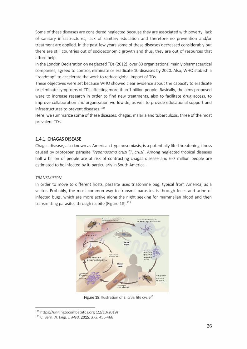
26
Some of these diseases are considered neglected because they are associated with poverty, lack
of sanitary infrastructures, lack of sanitary education and therefore no prevention and/or
treatment are applied. In the past few years some of these diseases decreased considerably but
there are still countries out of socioeconomic growth and thus, they are out of resources that
afford help.
In the London Declaration on neglected TDs (2012), over 80 organizations, mainly pharmaceutical
companies, agreed to control, eliminate or eradicate 10 diseases by 2020. Also, WHO stablish a
‘’roadmap’’ to accelerate the work to reduce global impact of TDs.
These objectives were set because WHO showed clear evidence about the capacity to eradicate
or eliminate symptoms of TDs affecting more than 1 billion people. Basically, the aims proposed
were to increase research in order to find new treatments, also to facilitate drug access, to
improve collaboration and organization worldwide, as well to provide educational support and
infrastructures to prevent diseases.120
Here, we summarize some of these diseases: chagas, malaria and tuberculosis, three of the most
prevalent TDs.
1.4.1. CHAGAS DISEASE
Chagas disease, also known as American trypanosomiasis, is a potentially life-threatening illness
caused by protozoan parasite Trypanosoma cruzi (T. cruzi). Among neglected tropical diseases
half a billion of people are at risk of contracting chagas disease and 6-7 million people are
estimated to be infected by it, particularly in South America.
TRANSMISION
In order to move to different hosts, parasite uses triatomine bug, typical from America, as a
vector. Probably, the most common way to transmit parasites is through feces and urine of
infected bugs, which are more active along the night seeking for mammalian blood and then
transmitting parasites through its bite (Figure 18).121
120 https://unitingtocombatntds.org (22/10/2019) 121 C. Bern. N. Engl. J. Med. 2015, 373, 456-466
Figure 18. Ilustration of T. cruzi life cycle121

27
DISEASE PHASES
The initial acute phase use to appear 2 months after infection. During acute phase, an elevated
quantity of parasites circulates in blood. Symptoms can be either absent or some fever, headache,
enlarged lymph glands, pallor, muscle pain, difficulty in breathing, swelling, and abdominal or
chest pain can appear. Along chronic phase, parasites are concealed in heart and digestive
muscles, so around 30% of patients suffer from cardiac alterations and up to 10% suffer from
digestive, neurological or a mix of them. After some years, due to parasite attacks in heart muscle
and nervous system, it can produce heart diseases, like heart failure or arrhythmias, drawing
patients to death.119
PREVENTION
Prevention in chagas disease embraces those practices that prevents contact with vector, such
as spraying the house with insecticide, good hygiene practices, screenings and blood testing to
detect infected people as soon as possible, the use of bed-nets, and others.119
TREATMENT
Nowadays there are only two drugs to treat chagas, benznidazole and nifurtimox (Figure 19).
Drugs developed 40 years ago. Both of them attacks DNA inhibiting its replication.
If treatment starts in acute phases the rate of cure is around 76%, much higher compared with
the 37% in chronic phases. For infants infected congenital, the cure rate of treatment is 96% when
it starts sooner and 62% when it starts in chronic phase.122
Figure 19. Chemical structure of Benznidazole and Nifurtimox
Chagas parasites are Kinetoplastid, a flagellated protozoan. They are defined by the presence of
DNA-containing region, the ‘kinetoplast’, in their single large mitochondrion. They have similar
genomic organization and cellular structures and undergo morphological changes during their life
cycles inside insects and vertebrate hosts. Approximately 50% of these genomes encode for
hypothetical proteins that do not resemble orthologs in human genome, plus several human
protein classes are not represented in kinetoplastid genomes. All these differences suggest that
may be there are essential proteins that can be exploited as selective targets for therapy.123
122 S. Meymandi; S. Hernandez; S. Park; D. R. Sanchez; C. Forsyth. Curr. Treat. Options Infect. Dis. 2018, 10, 373–388 123 J. A. Padilla; I. Cotillo; J. Presa; J. Cantizani; I. Peña; A. I. Bardera; J. J. Martín; A. Rodriguez. PLoS Negl. Trop. Dis. 2015, 9, e0003493
Benznidazole Nifurtimox
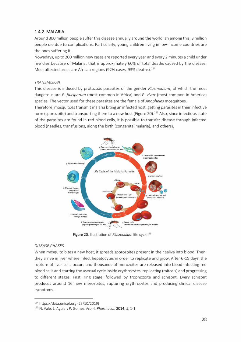
28
1.4.2. MALARIA
Around 300 million people suffer this disease annually around the world, an among this, 3 million
people die due to complications. Particularly, young children living in low-income countries are
the ones suffering it.
Nowadays, up to 200 million new cases are reported every year and every 2 minutes a child under
five dies because of Malaria, that is approximately 60% of total deaths caused by the disease.
Most affected areas are African regions (92% cases, 93% deaths).124
TRANSMISION
This disease is induced by protozoas parasites of the gender Plasmodium, of which the most
dangerous are P. falciparum (most common in Africa) and P. vivax (most common in America)
species. The vector used for these parasites are the female of Anopheles mosquitoes.
Therefore, mosquitoes transmit malaria biting an infected host, getting parasites in their infective
form (sporozoite) and transporting them to a new host (Figure 20).125 Also, since infectious state
of the parasites are found in red blood cells, it is possible to transfer disease through infected
blood (needles, transfusions, along the birth (congenital malaria), and others).
DISEASE PHASES
When mosquito bites a new host, it spreads sporozoites present in their saliva into blood. Then,
they arrive in liver where infect hepatocytes in order to replicate and grow. After 6-15 days, the
rupture of liver cells occurs and thousands of merozoites are released into blood infecting red
blood cells and starting the asexual cycle inside erythrocytes, replicating (mitosis) and progressing
to different stages. First, ring stage, followed by trophozoite and schizont. Every schizont
produces around 16 new merozoites, rupturing erythrocytes and producing clinical disease
symptoms.
124 https://data.unicef.org (23/10/2019) 125 N. Vale; L. Aguiar; P. Gomes. Front. Pharmacol. 2014, 5, 1-1
Figure 20. Illustration of Plasmodium life cycle125
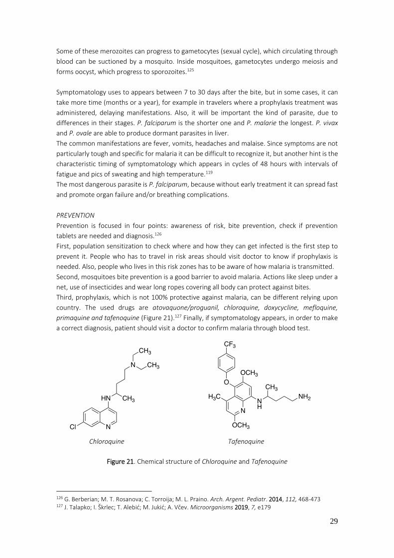
29
Some of these merozoites can progress to gametocytes (sexual cycle), which circulating through
blood can be suctioned by a mosquito. Inside mosquitoes, gametocytes undergo meiosis and
forms oocyst, which progress to sporozoites.125
Symptomatology uses to appears between 7 to 30 days after the bite, but in some cases, it can
take more time (months or a year), for example in travelers where a prophylaxis treatment was
administered, delaying manifestations. Also, it will be important the kind of parasite, due to
differences in their stages. P. falciparum is the shorter one and P. malarie the longest. P. vivax
and P. ovale are able to produce dormant parasites in liver.
The common manifestations are fever, vomits, headaches and malaise. Since symptoms are not
particularly tough and specific for malaria it can be difficult to recognize it, but another hint is the
characteristic timing of symptomatology which appears in cycles of 48 hours with intervals of
fatigue and pics of sweating and high temperature.119
The most dangerous parasite is P. falciparum, because without early treatment it can spread fast
and promote organ failure and/or breathing complications.
PREVENTION
Prevention is focused in four points: awareness of risk, bite prevention, check if prevention
tablets are needed and diagnosis.126
First, population sensitization to check where and how they can get infected is the first step to
prevent it. People who has to travel in risk areas should visit doctor to know if prophylaxis is
needed. Also, people who lives in this risk zones has to be aware of how malaria is transmitted.
Second, mosquitoes bite prevention is a good barrier to avoid malaria. Actions like sleep under a
net, use of insecticides and wear long ropes covering all body can protect against bites.
Third, prophylaxis, which is not 100% protective against malaria, can be different relying upon
country. The used drugs are atovaquone/proguanil, chloroquine, doxycycline, mefloquine,
primaquine and tafenoquine (Figure 21).127 Finally, if symptomatology appears, in order to make
a correct diagnosis, patient should visit a doctor to confirm malaria through blood test.
Figure 21. Chemical structure of Chloroquine and Tafenoquine
126 G. Berberian; M. T. Rosanova; C. Torroija; M. L. Praino. Arch. Argent. Pediatr. 2014, 112, 468-473 127 J. Talapko; I. Škrlec; T. Alebić; M. Jukić; A. Včev. Microorganisms 2019, 7, e179
Chloroquine Tafenoquine
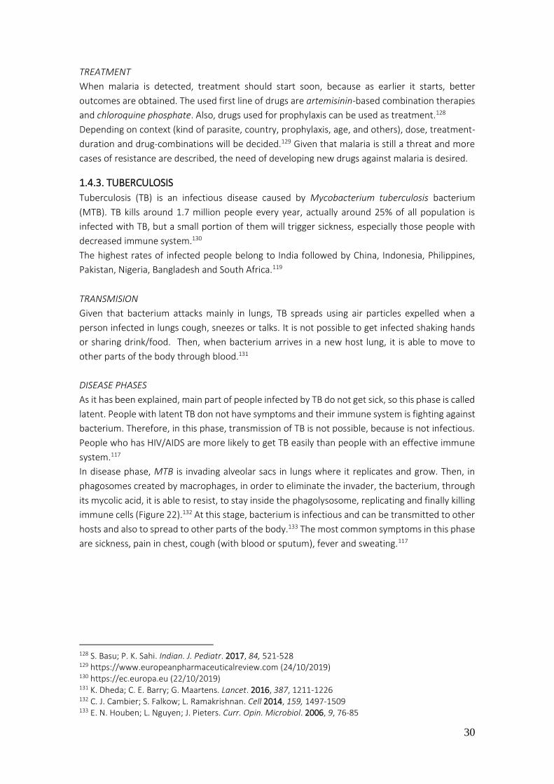
30
TREATMENT
When malaria is detected, treatment should start soon, because as earlier it starts, better
outcomes are obtained. The used first line of drugs are artemisinin-based combination therapies
and chloroquine phosphate. Also, drugs used for prophylaxis can be used as treatment.128
Depending on context (kind of parasite, country, prophylaxis, age, and others), dose, treatment-
duration and drug-combinations will be decided.129 Given that malaria is still a threat and more
cases of resistance are described, the need of developing new drugs against malaria is desired.
1.4.3. TUBERCULOSIS
Tuberculosis (TB) is an infectious disease caused by Mycobacterium tuberculosis bacterium
(MTB). TB kills around 1.7 million people every year, actually around 25% of all population is
infected with TB, but a small portion of them will trigger sickness, especially those people with
decreased immune system.130
The highest rates of infected people belong to India followed by China, Indonesia, Philippines,
Pakistan, Nigeria, Bangladesh and South Africa.119
TRANSMISION
Given that bacterium attacks mainly in lungs, TB spreads using air particles expelled when a
person infected in lungs cough, sneezes or talks. It is not possible to get infected shaking hands
or sharing drink/food. Then, when bacterium arrives in a new host lung, it is able to move to
other parts of the body through blood.131
DISEASE PHASES
As it has been explained, main part of people infected by TB do not get sick, so this phase is called
latent. People with latent TB don not have symptoms and their immune system is fighting against
bacterium. Therefore, in this phase, transmission of TB is not possible, because is not infectious.
People who has HIV/AIDS are more likely to get TB easily than people with an effective immune
system.117
In disease phase, MTB is invading alveolar sacs in lungs where it replicates and grow. Then, in
phagosomes created by macrophages, in order to eliminate the invader, the bacterium, through
its mycolic acid, it is able to resist, to stay inside the phagolysosome, replicating and finally killing
immune cells (Figure 22).132 At this stage, bacterium is infectious and can be transmitted to other
hosts and also to spread to other parts of the body.133 The most common symptoms in this phase
are sickness, pain in chest, cough (with blood or sputum), fever and sweating.117
128 S. Basu; P. K. Sahi. Indian. J. Pediatr. 2017, 84, 521-528 129 https://www.europeanpharmaceuticalreview.com (24/10/2019) 130 https://ec.europa.eu (22/10/2019) 131 K. Dheda; C. E. Barry; G. Maartens. Lancet. 2016, 387, 1211-1226 132 C. J. Cambier; S. Falkow; L. Ramakrishnan. Cell 2014, 159, 1497-1509 133 E. N. Houben; L. Nguyen; J. Pieters. Curr. Opin. Microbiol. 2006, 9, 76-85
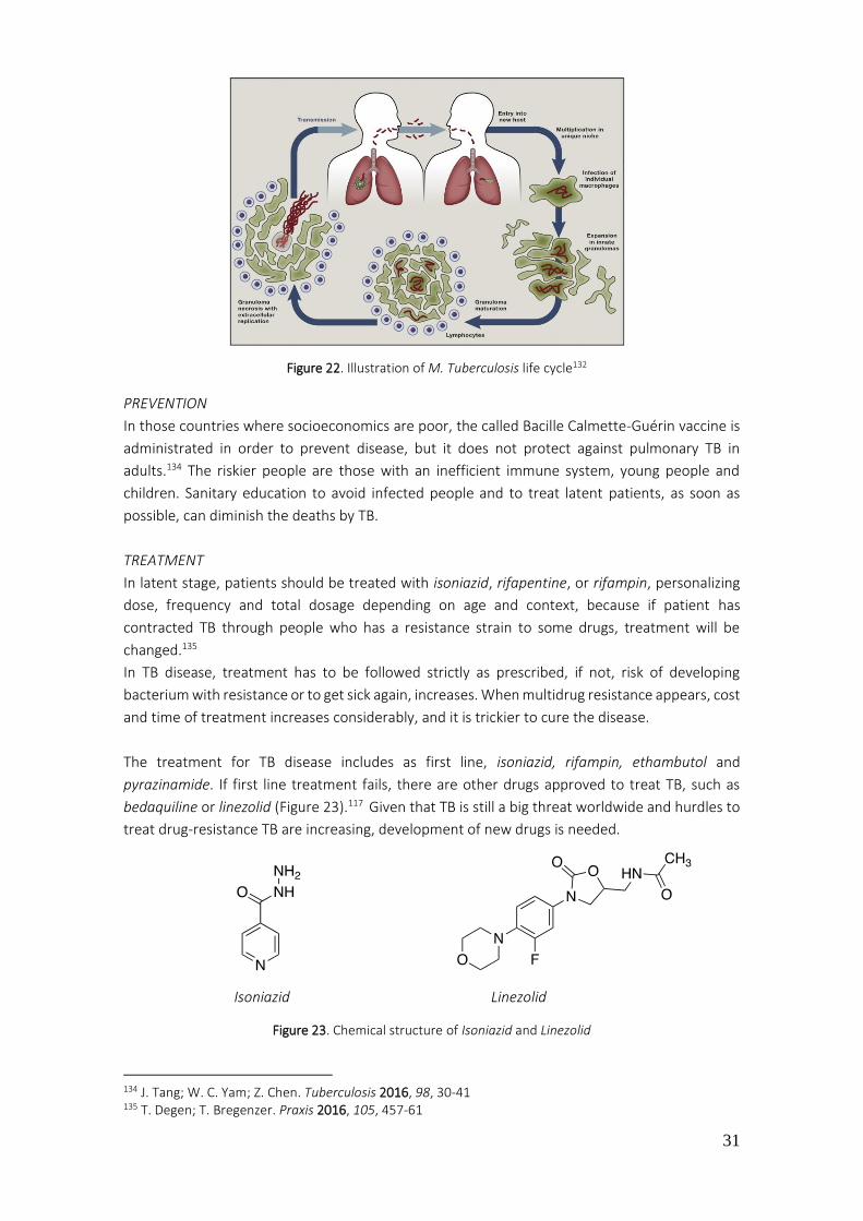
31
PREVENTION
In those countries where socioeconomics are poor, the called Bacille Calmette-Guérin vaccine is
administrated in order to prevent disease, but it does not protect against pulmonary TB in
adults.134 The riskier people are those with an inefficient immune system, young people and
children. Sanitary education to avoid infected people and to treat latent patients, as soon as
possible, can diminish the deaths by TB.
TREATMENT
In latent stage, patients should be treated with isoniazid, rifapentine, or rifampin, personalizing
dose, frequency and total dosage depending on age and context, because if patient has
contracted TB through people who has a resistance strain to some drugs, treatment will be
changed.135
In TB disease, treatment has to be followed strictly as prescribed, if not, risk of developing
bacterium with resistance or to get sick again, increases. When multidrug resistance appears, cost
and time of treatment increases considerably, and it is trickier to cure the disease.
The treatment for TB disease includes as first line, isoniazid, rifampin, ethambutol and
pyrazinamide. If first line treatment fails, there are other drugs approved to treat TB, such as
bedaquiline or linezolid (Figure 23).117 Given that TB is still a big threat worldwide and hurdles to
treat drug-resistance TB are increasing, development of new drugs is needed.
Figure 23. Chemical structure of Isoniazid and Linezolid
134 J. Tang; W. C. Yam; Z. Chen. Tuberculosis 2016, 98, 30-41 135 T. Degen; T. Bregenzer. Praxis 2016, 105, 457-61
Isoniazid Linezolid
Figure 22. Illustration of M. Tuberculosis life cycle132
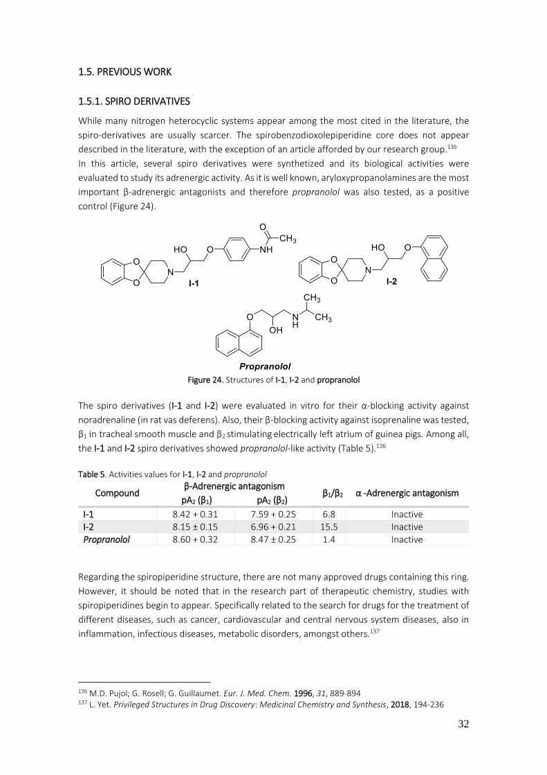
32
1.5. PREVIOUS WORK
1.5.1. SPIRO DERIVATIVES
While many nitrogen heterocyclic systems appear among the most cited in the literature, the
spiro-derivatives are usually scarcer. The spirobenzodioxolepiperidine core does not appear
described in the literature, with the exception of an article afforded by our research group.136
In this article, several spiro derivatives were synthetized and its biological activities were
evaluated to study its adrenergic activity. As it is well known, aryloxypropanolamines are the most
important β-adrenergic antagonists and therefore propranolol was also tested, as a positive
control (Figure 24).
Figure 24. Structures of I-1, I-2 and propranolol
The spiro derivatives (I-1 and I-2) were evaluated in vitro for their α-blocking activity against
noradrenaline (in rat vas deferens). Also, their β-blocking activity against isoprenaline was tested,
β1 in tracheal smooth muscle and β2 stimulating electrically left atrium of guinea pigs. Among all,
the I-1 and I-2 spiro derivatives showed propranolol-like activity (Table 5).136
Table 5. Activities values for I-1, I-2 and propranolol
Compound β-Adrenergic antagonism
β1/β2 α -Adrenergic antagonism pA2 (β1) pA2 (β2)
I-1 8.42 + 0.31 7.59 + 0.25 6.8 Inactive I-2 8.15 ± 0.15 6.96 + 0.21 15.5 Inactive Propranolol 8.60 + 0.32 8.47 ± 0.25 1.4 Inactive
Regarding the spiropiperidine structure, there are not many approved drugs containing this ring.
However, it should be noted that in the research part of therapeutic chemistry, studies with
spiropiperidines begin to appear. Specifically related to the search for drugs for the treatment of
different diseases, such as cancer, cardiovascular and central nervous system diseases, also in
inflammation, infectious diseases, metabolic disorders, amongst others.137
136 M.D. Pujol; G. Rosell; G. Guillaumet. Eur. J. Med. Chem. 1996, 31, 889-894 137 L. Yet. Privileged Structures in Drug Discovery: Medicinal Chemistry and Synthesis, 2018, 194-236

33
1.5.2. PIRROLOPYRAZINES
The 5H-pyrrolo[2,3-b]pyrazines nucleus, also known as aloisine, is described in the literature as
a potent scaffold structure for protein kinase inhibition, such as,
CDKs,138,139 GSK,138 Syk,140 ERK2,141 JAK,142 among others (Figure
25).143
The Aloisine A is one of the products to highlight due to its high
inhibitory activity in front of CDK1, CDK2, CDK5, GSK, JNK and less
inhibitory activity for CK1, CK2, CDK4, MAPKK, PKA, PKG, PKCs and
c-raf (Table 6). Therefore, inhibits the cell cycle in both G1 and G2
phases.138,139
Table 6. IC50 of Aloisine A in different targets
Target IC50 (ATP-competitive inhibitor)
CDK1/cyclin B 150 nM
CDK2/cyclin A, CDK2/cyclin E 120 nM, 400 nM
CDK5/p25, CDK5/p35 200 nM, 160 nM
GSK-3a, GSK-3β 500 nM, 650 nM
JNK 3-10 µM
CK1, CK2, CDK4/cyclin D1, MAPKK, PKA, PKG, PKCs and c-raf ≥ 100 µM
Another related product to stand out is a patented compound by Sanofi that has showed a potent
inhibitory activity for Syk kinase. Thus, can be useful in the treatment of inflammatory diseases
(arthritis, asthma), cardiovascular diseases, cancer, amongst others (Figure 26).144
Figure 26. Pyrrolopyrazine with inhibition activity for Syk kinase
138 Y. Mettey; M. Gompel. V. Thomas, M. Garnier; M. Leost; I. C. Picot; M. Noble; J. Endicott; J. Vierfond; L. Meijer. J. Med. Chem. 2003, 46, 222-236 139 C. Corbel; R. Haddoub; D. Guiffant; O. Lozach; D. Gueyrard; J. Lemoine; M. Ratin; L. Meijer; S. Bach; P. Goekjian. Bioorg. Med. Chem. 2009, 17, 5572–5582 140 F. Padilla; N. Bhagirath; S. Chen; E.Chiao; D. M. Goldstein; J. C. Hermann; J. Hsu; J. J. Kennedy-Smith; A. Kuglstatter; C. Liao; W. Liu; L. E. Lowrie; K. C. Luk; S. M. Lynch; J. Menke; L. Niu; T. D. Owens; C. O-Yang; A. Railkar; R. C. Schoenfeld; M. Slade; S. Steiner; Y. C. Tan; A. G. Villaseñor; C. Wang; J. Wanner; W. Xie; D. Xu; X. Zhang; M. Zhou; M. C. Lucas. J. Med. Chem. 2013, 56, 1677-1692 141 D. J. Burdick; S. Wang; C. Heise; B. Pan; J. Drummond; J. P. Yin; L. Goeser; S. Magnuson; J. Blaney; J. Moffat; W. Wang; H. Chen. Bioorg. Med. Chem. Lett. 2015, 25, 4728-4732 142 J. de Vicente; R. Lemoine; M. Bartlett; J. C. Hermann; M. Hekmat-Nejad; R. Henningsen; S. Jin; A. Kuglstatter; H. Li; A. J. Lovey; J. Menke; L. Niu, V. Patel; A. Petersen; L. Setti; A. Shao; P. Tivitmahaisoon; M. D. Vu, M. Soth. Bioorg. Med. Chem. Lett. 2014, 24, 4969-4975 143 G. C. Senadi; B. C. Guo; Yu. C. Chang; W.P. Hu; J. J. Wang. Adv. Synth. Catal. 2017, 360, 491-501 144 SANOFI. Una pirrolopirazina como inhibidor de Syk-quinasa. España; patente de invención; ES2437318; 23-10-2013
Figure 25. Aloisine A
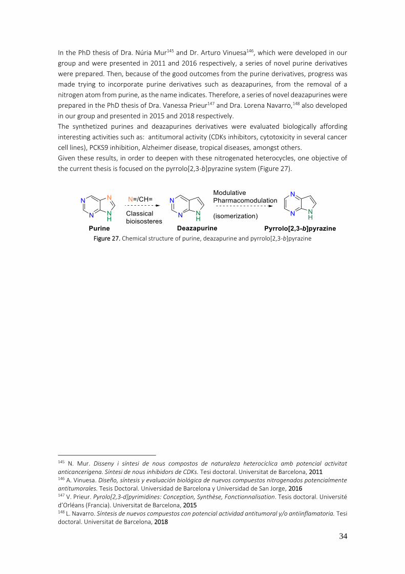
34
In the PhD thesis of Dra. Núria Mur145 and Dr. Arturo Vinuesa146, which were developed in our
group and were presented in 2011 and 2016 respectively, a series of novel purine derivatives
were prepared. Then, because of the good outcomes from the purine derivatives, progress was
made trying to incorporate purine derivatives such as deazapurines, from the removal of a
nitrogen atom from purine, as the name indicates. Therefore, a series of novel deazapurines were
prepared in the PhD thesis of Dra. Vanessa Prieur147 and Dra. Lorena Navarro,148 also developed
in our group and presented in 2015 and 2018 respectively.
The synthetized purines and deazapurines derivatives were evaluated biologically affording
interesting activities such as: antitumoral activity (CDKs inhibitors, cytotoxicity in several cancer
cell lines), PCKS9 inhibition, Alzheimer disease, tropical diseases, amongst others.
Given these results, in order to deepen with these nitrogenated heterocycles, one objective of
the current thesis is focused on the pyrrolo[2,3-b]pyrazine system (Figure 27).
Figure 27. Chemical structure of purine, deazapurine and pyrrolo[2,3-b]pyrazine
145 N. Mur. Disseny i síntesi de nous compostos de naturaleza heterocíclica amb potencial activitat anticancerígena. Síntesi de nous inhibidors de CDKs. Tesi doctoral. Universitat de Barcelona, 2011 146 A. Vinuesa. Diseño, síntesis y evaluación biológica de nuevos compuestos nitrogenados potencialmente antitumorales. Tesis Doctoral. Universidad de Barcelona y Universidad de San Jorge, 2016 147 V. Prieur. Pyrolo[2,3-d]pyrimidines: Conception, Synthèse, Fonctionnalisation. Tesis doctoral. Université d’Orléans (Francia). Universitat de Barcelona, 2015 148 L. Navarro. Síntesis de nuevos compuestos con potencial actividad antitumoral y/o antiinflamatoria. Tesi doctoral. Universitat de Barcelona, 2018

35
2. OBJECTIVES
Considering the bibliographic precedents described in the introduction and the chemistry
preparation and biological avaluation undertaken by our research group, hereunder, diferent
structures with potential multitarget activity are shown. The structure design of the following
compounds was prepared by means of an iteration process (Figure 28):
Figure 28. Iteration process followed for the design of new structures
2.1. FIRST OBJECTIVE
As described before, the spiroderivatives are an atractive systems with scant information in the
literature. For this reason, the preparation of compounds 1-42 with structure of
spirobenzodioxolepiperidine and 43, 44 with structure of spirobenzoxazolepiperidine forms the
first objective of this work (Table 7). From the active compounds 6a and 6c described in the
bibliography by M. D. Pujol and col. a modulative approximation is proposed, modifying the
substituents of the piperidine ring, adding substituents to the position 5 of the
spirobenzodioxolepiperidine and a bioisostere replacement in the dioxole ring (Figure 29).
Figure 29. Modulative aproximation for compounds 1-44

36
Table 7. The proposed 44 spiro derivative compounds
COMPOUNDS R1 R2 R3 X
1 -COCH3 -H - O
2 “ -COCH3 - “
3 -H “ - “
4 -COCH3 -CN(OH)-CH3 (E) - “
5 “ -CH(OH)-CH3 - “
6 “ -Br - “
7 “ -C6H4-CH3 (p) - “
8 “ -C6H4-NO2 (p) - “
9 “ -C6H4-NH2 (p) - “
10 “ -C6H4-CN (p) - “
11 “ -C6H4-O-C6H5 (m) - “
12 “ -NO2 - “
13 -H “ - “
14 -C6H4-NO2 (p) “ - “
15 -C6H4-NH2 (p) -NH2 - “
16 -C6H4-NO2 (p) -H - “
17 “ -COCH3 - “
18 “ -C=N(OH)-CH3 (E) - “
19 “ -NHCOCH3 - “
20 “ -NH2 “
21 “ -CH(OH)-CH3 - “
22 -C6H4-NH2 (p) “ - “
23 “ -COCH3 - “
24
‘’ - “
25 “ -CH(OH)-CH3 - “
26
-COCH3 - “
27 -SO2-C6H5 “ - “
28 -SO2-C6H5-CH3 (p) “ - “
29 -SO2-C6H5 -CH(OH)-CH3 - “
30 -SO2-C6H5-CH3 (p) “ - “
31 -SO2-C6H5 -CH(Cl)-CH3 - “
32 -SO2-C6H5-CH3 (p) “ - “
33 -SO2-C6H5 -CH(CH3)-O-(CH2)2-N(CH2CH3)2 - “
34 “ -CH(CH3)-NH-(CH2)2-N(CH3)2 - “
35 -SO2-C6H5-CH3 (p) -CH(CH3)-NH-(CH2)5-CH3 - “
36 -NH-(CH2)2-OH -CH(OH)-CH3 - “
37
-COCH3 - “
38 “ -CH(OH)-CH3 - “

37
2.2. SECOND OBJECTIVE
The pirrolo[2,3-b]pyrazine system, a derivative from the purines, has not studied as much as
purines, neither the synthesis or its biological activities. Therefore, the second objective is to set
up a synthesis in order to prepare the pirrolo[2,3-b]pyrazine system as optimally as possible
(Figure 30).
Figure 30. Pyrazine derivative 45a and its starting material 2-aminopyrazine
If the desired pirrolo[2,3-b]pyrazine system is obtained, a synthesis to prepare diarylates, triarylates and tetrarylates compounds consist the second part of this objective. Thus, starting from the good scaffolds 45a-c the preparation of products 47-58 is proposed (Table 8).
Table 8. The proposed 13 pirrolo-pyrazine derivative compounds
39 -C6H4-NH-CO-NH-Et “ - “
40
-NH2 - “
41 -C6H4-NH-CO-NH-Et -C6H4-NH-CO-NH-Et - “
42
-COCH3
- “
43 -COCH3 -H -H N
44 -COCH3 -COCH3 -COCH3 N
COMPOUNDS R1 R2 R3 R4 R5
47 -C6H5 -C6H5-CH3 (p) -H -H -H
48 “ -C6H5-OCH3 (p) “ “ “
49 “ -C6H5-CF3 (p) “ “ “
50 -C6H5-F (p) -C6H5-CH3 (p) “ “ “
51 “ -C6H5-CH3 (m) “ “ “
52 -C6H5-Cl (p) -C6H5-CH3 (p) “ “ “
53a -C6H5 “ -C6H5-CH3 (p) “ “
54 “ “ -Br “ -C6H5-CH3 (p)
55a -C6H5-F (p) “ -H -MO-Et -H
55b “ -C6H5-CH3 (m) “ “ “
56 “ -C6H5-CH3 (p) -C6H5-CH3 (p) -Bn “
57 “ “ “ -H “
58 -C6H5 -C6H5-OCH3 (p) -H -C6H5-NO2 (p) “
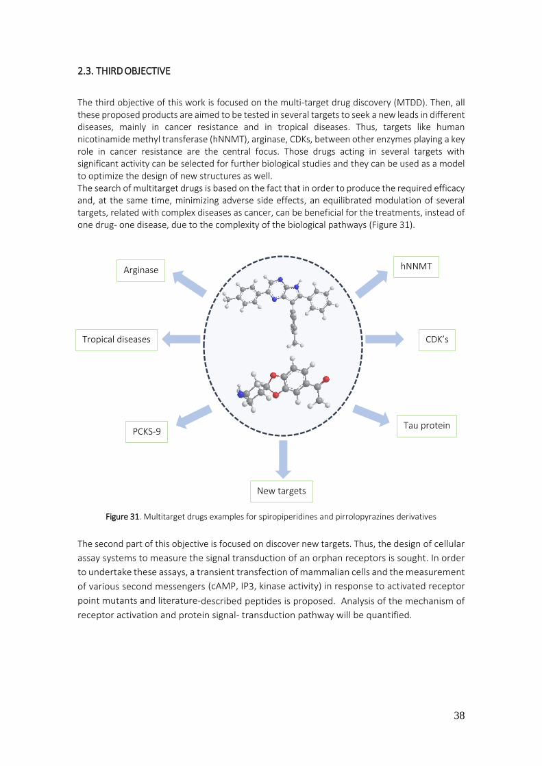
38
2.3. THIRD OBJECTIVE
The third objective of this work is focused on the multi-target drug discovery (MTDD). Then, all these proposed products are aimed to be tested in several targets to seek a new leads in different diseases, mainly in cancer resistance and in tropical diseases. Thus, targets like human nicotinamide methyl transferase (hNNMT), arginase, CDKs, between other enzymes playing a key role in cancer resistance are the central focus. Those drugs acting in several targets with significant activity can be selected for further biological studies and they can be used as a model to optimize the design of new structures as well. The search of multitarget drugs is based on the fact that in order to produce the required efficacy and, at the same time, minimizing adverse side effects, an equilibrated modulation of several targets, related with complex diseases as cancer, can be beneficial for the treatments, instead of one drug- one disease, due to the complexity of the biological pathways (Figure 31).
Figure 31. Multitarget drugs examples for spiropiperidines and pirrolopyrazines derivatives
The second part of this objective is focused on discover new targets. Thus, the design of cellular
assay systems to measure the signal transduction of an orphan receptors is sought. In order
to undertake these assays, a transient transfection of mammalian cells and the measurement
of various second messengers (cAMP, IP3, kinase activity) in response to activated receptor
point mutants and literature-described peptides is proposed. Analysis of the mechanism of
receptor activation and protein signal- transduction pathway will be quantified.
Arginase hNNMT
CDK’s Tropical diseases
PCKS-9 Tau protein
New targets

39
3. THEORICAL DISCUSSION
3.1. CHEMICAL DISCUSSION
3.1.1. Preparation of 1-spiro[benzodioxole-2,4’-piperidine]
3.1.1.1. Preparation of N-acetyl-spiro[1,3-benzodioxole-2,4'-piperidine] (1)
The spirobenzodioxolepiperidine system is not described in the bibliography, with the exception
of one reference afforded by our research group.136
In order to prepare this dioxygenated nucleus, the following retrosynthetic scheme was proposed
(Scheme 1):
Scheme 1
In the first assay, through direct condensation of the catechol with N-acetyl-4-piperidone, in
toluene and under acid conditions (APTS), the expected spiranic system was not obtained
(Scheme 2).
Scheme 2
These results, led to consider a different conditions of reaction, such as the transacetalization of
the acetal 60, starting from the acetone, for the catechol (Scheme 3).
Scheme 3
The treatment of 4-piperidone with methyl orthoformate, warming up until the distillation of
methyl formate (boiling point = 31.5 C), leads to acetal 60, which is stable under these conditions
and then it becomes the spiranic derivative 1 by treatment with catechol in toluene and through
acid catalysis (APTS). The global yield of the two steps was 70%.
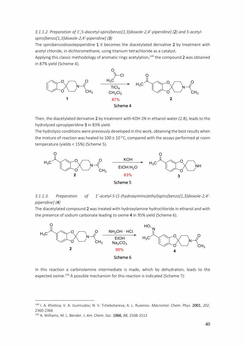
40
3.1.1.2. Preparation of 1',5-diacetyl-spiro[benzo[1,3]dioxole-2,4'-piperidine] (2) and 5-acetyl-
spiro[benzo[1,3]dioxole-2,4'-piperidine] (3)
The spirobenzodioxolepiperidine 1 it becomes the diacetylated derivative 2 by treatment with
acetyl chloride, in dichloromethane, using titanium tetrachloride as a catalyst.
Applying this classic methodology of aromatic rings acetylation,149 the compound 2 was obtained
in 87% yield (Scheme 4).
Scheme 4
Then, the diacetylated derivative 2 by treatment with KOH 2N in ethanol-water (2:8), leads to the
hydrolyzed spiropiperidine 3 in 83% yield.
The hydrolysis conditions were previously developed in this work, obtaining the best results when
the mixture of reaction was heated to 100 10 C, compared with the assays performed at room
temperature (yields < 15%) (Scheme 5).
Scheme 5
3.1.1.3. Preparation of 1’-acetyl-5-(1-(hydroxyimino)ethyl)spiro[benzo[1,3]dioxole-2,4'-
piperidine] (4)
The diacetylated compound 2 was treated with hydroxylamine hydrochloride in ethanol and with
the presence of sodium carbonate leading to oxime 4 in 95% yield (Scheme 6).
Scheme 6
In this reaction a carbinolamine intermediate is made, which by dehydration, leads to the
expected oxime.150 A possible mechanism for this reaction is indicated (Scheme 7):
149 I. A. Khotina; V. A. Izumrudov; N. V. Tchebotareva; A. L. Rusenov. Macromol. Chem. Phys. 2001, 202, 2360-2366 150 A. Williams; M. L. Bender. J. Am. Chem. Soc. 1966, 88, 2508-2513
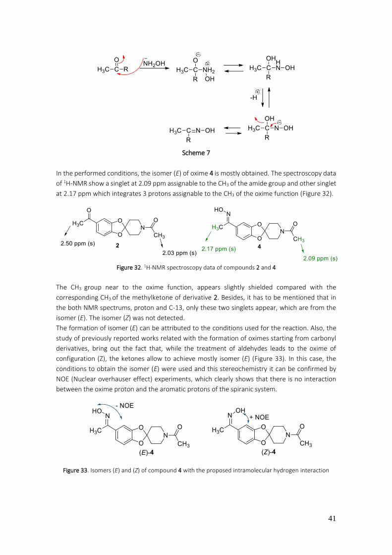
41
Scheme 7
In the performed conditions, the isomer (E) of oxime 4 is mostly obtained. The spectroscopy data
of 1H-NMR show a singlet at 2.09 ppm assignable to the CH3 of the amide group and other singlet
at 2.17 ppm which integrates 3 protons assignable to the CH3 of the oxime function (Figure 32).
Figure 32. 1H-NMR spectroscopy data of compounds 2 and 4
The CH3 group near to the oxime function, appears slightly shielded compared with the
corresponding CH3 of the methylketone of derivative 2. Besides, it has to be mentioned that in
the both NMR spectrums, proton and C-13, only these two singlets appear, which are from the
isomer (E). The isomer (Z) was not detected.
The formation of isomer (E) can be attributed to the conditions used for the reaction. Also, the
study of previously reported works related with the formation of oximes starting from carbonyl
derivatives, bring out the fact that, while the treatment of aldehydes leads to the oxime of
configuration (Z), the ketones allow to achieve mostly isomer (E) (Figure 33). In this case, the
conditions to obtain the isomer (E) were used and this stereochemistry it can be confirmed by
NOE (Nuclear overhauser effect) experiments, which clearly shows that there is no interaction
between the oxime proton and the aromatic protons of the spiranic system.
Figure 33. Isomers (E) and (Z) of compound 4 with the proposed intramolecular hydrogen interaction
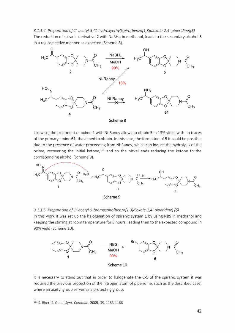
42
3.1.1.4. Preparation of 1’-acetyl-5-(1-hydroxyethyl)spiro[benzo[1,3]dioxole-2,4'-piperidine](5)
The reduction of spiranic derivative 2 with NaBH4, in methanol, leads to the secondary alcohol 5
in a regioselective manner as expected (Scheme 8).
Scheme 8
Likewise, the treatment of oxime 4 with Ni-Raney allows to obtain 5 in 13% yield, with no traces
of the primary amine 61, the aimed to obtain. In this case, the formation of 5 it could be possible
due to the presence of water proceeding from Ni-Raney, which can induce the hydrolysis of the
oxime, recovering the initial ketone,151 and so the nickel ends reducing the ketone to the
corresponding alcohol (Scheme 9).
Scheme 9
3.1.1.5. Preparation of 1’-acetyl-5-bromospiro[benzo[1,3]dioxole-2,4'-piperidine] (6)
In this work it was set up the halogenation of spiranic system 1 by using NBS in methanol and
keeping the stirring at room temperature for 3 hours, leading then to the expected compound in
90% yield (Scheme 10).
Scheme 10
It is necessary to stand out that in order to halogenate the C-5 of the spiranic system it was
required the previous protection of the nitrogen atom of piperidine, such as the described case,
where an acetyl group serves as a protecting group.
151 S. Bher; S. Guha. Synt. Commun. 2005, 35, 1183-1188

43
However, in the present case, a soft halogenation conditions are applied, because it is also
possible to brominate the methyl group, an -carbonyl position. Previously, in our research
group, the halogenation of the spiropiperidine hydrochloride system was made, as it is indicated
hereunder (Scheme 11).136
Scheme 11
3.1.1.6. Preparation of 7
From the bromo derivative 6 it is possible to access to the spiro-derivatives 7-11 through cross-
coupling reactions catalyzed by Pd-complex (Scheme 12).
Scheme 12
The formation of carbon-carbon (C-C) bonds is of vital importance for the preparation of different
chemical products, many of them with application in therapeutic chemistry.
The cross-coupling reactions are very versatile and allow to prepare a wide number of compounds
with different structural complexity, appearing as the most referenced reactions from the last
decade. The conditions of this kind of reactions are usually softly than other type of radical
reactions or in some cases of nucleophilic aromatic substitutions. Specifically in 2010, three
scientist working in this field were winners of Nobel prize, Richard F. Heck, Ei-ichi Negishi and
Akira Suzuki. Each one applied a methodology that allows to achieve a certain compounds. Thus,
the Heck reaction involves the use of an unsaturated halide in presence of a base and a Pd
catalyst, allowing to obtain alkenes and stilbenes above all, which are very useful for the
preparation of drugs and other organic compounds (Scheme 13).152
Scheme 13
E. Negishi also describes the preparation of compounds through the formation of C-C bonds
under cross-coupling conditions catalyzed by palladium or nickel derivatives.153 The reaction
152 R. F. Heck; J. P. Nolley. J. Org. Chem. 1972, 37, 2320-2322 153 A. O. King; N. Okukado; E. Negishi. J. Chem. Soc. Chem. Commun. 1977, 19, 683-684

44
involves the use of halides or organic triflate and organozincates as it is indicated in the following
scheme (Scheme 14):
Scheme 14
Finally, A. Suzuki, which also afforded its methodology to make C-C bonds, distinguishing himself
of the latter for the use of aryl or alkyl boronic acids which couple with organic halides, using
palladium (0) as a catalyst in presence of a base.154 Hereunder, a general scheme of this reaction
is showed (Scheme 15).
Scheme 15
In these reactions, solvents, ligands, bases and catalyst are known to have very significant role in
order to improve the effectiveness of these reactions. In this work it has been tried that the
amount of transition metals was minimal so that the process was compatible and respectful with
the environment. In this work, the arylation of the spiranic derivative 6 was carried out, using p-
tolylboronic acid and palladium-triphenylphosphine complex (tetrakis) as a catalyst, through the
Suzuki-Miyaura cross-coupling conditions (Scheme 16). Thus, the desired product 7 was obtained
in 87% yield.
Scheme 16
The presence of two doublets in the 1H-NMR spectrum, which integrate 2 protons each one at
7.11 and at 7.30 ppm assignable at protons of the positions 3’’, 5’’ and 2’’, 6’’, and as well as the
presence of a singlet at 2.28 ppm belonging to the CH3 group of the phenyl, incorporated in the
position 5 of the spiranic system, allow to confirm the proposed structure for compound 7 (Figure
34).
154 N. Miyaura; A. Suzuki. J. Chem. Soc. Chem. Commun. 1979, 19, 866-867
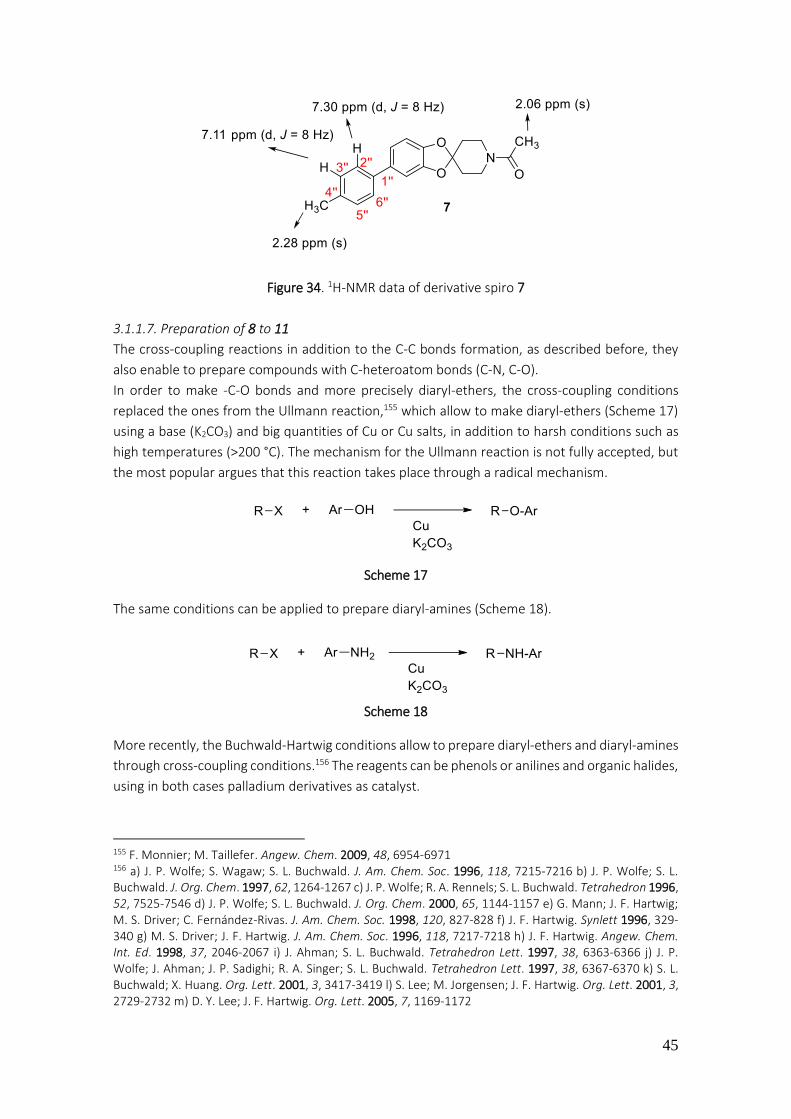
45
Figure 34. 1H-NMR data of derivative spiro 7
3.1.1.7. Preparation of 8 to 11
The cross-coupling reactions in addition to the C-C bonds formation, as described before, they
also enable to prepare compounds with C-heteroatom bonds (C-N, C-O).
In order to make -C-O bonds and more precisely diaryl-ethers, the cross-coupling conditions
replaced the ones from the Ullmann reaction,155 which allow to make diaryl-ethers (Scheme 17)
using a base (K2CO3) and big quantities of Cu or Cu salts, in addition to harsh conditions such as
high temperatures (>200 °C). The mechanism for the Ullmann reaction is not fully accepted, but
the most popular argues that this reaction takes place through a radical mechanism.
Scheme 17
The same conditions can be applied to prepare diaryl-amines (Scheme 18).
Scheme 18
More recently, the Buchwald-Hartwig conditions allow to prepare diaryl-ethers and diaryl-amines
through cross-coupling conditions.156 The reagents can be phenols or anilines and organic halides,
using in both cases palladium derivatives as catalyst.
155 F. Monnier; M. Taillefer. Angew. Chem. 2009, 48, 6954-6971 156 a) J. P. Wolfe; S. Wagaw; S. L. Buchwald. J. Am. Chem. Soc. 1996, 118, 7215-7216 b) J. P. Wolfe; S. L. Buchwald. J. Org. Chem. 1997, 62, 1264-1267 c) J. P. Wolfe; R. A. Rennels; S. L. Buchwald. Tetrahedron 1996, 52, 7525-7546 d) J. P. Wolfe; S. L. Buchwald. J. Org. Chem. 2000, 65, 1144-1157 e) G. Mann; J. F. Hartwig; M. S. Driver; C. Fernández-Rivas. J. Am. Chem. Soc. 1998, 120, 827-828 f) J. F. Hartwig. Synlett 1996, 329-340 g) M. S. Driver; J. F. Hartwig. J. Am. Chem. Soc. 1996, 118, 7217-7218 h) J. F. Hartwig. Angew. Chem. Int. Ed. 1998, 37, 2046-2067 i) J. Ahman; S. L. Buchwald. Tetrahedron Lett. 1997, 38, 6363-6366 j) J. P. Wolfe; J. Ahman; J. P. Sadighi; R. A. Singer; S. L. Buchwald. Tetrahedron Lett. 1997, 38, 6367-6370 k) S. L. Buchwald; X. Huang. Org. Lett. 2001, 3, 3417-3419 l) S. Lee; M. Jorgensen; J. F. Hartwig. Org. Lett. 2001, 3, 2729-2732 m) D. Y. Lee; J. F. Hartwig. Org. Lett. 2005, 7, 1169-1172

46
It is necessary to add ligands monophosphine-type such as triphenylphosphine or diphosphines,
like BINAP, and also it is required the presence of a base. The most appropriate solvent is usually
toluene or xylene, due to the high boiling point that enable, if it was necessary, to work at high
temperatures (Scheme 19).
Scheme 19
A proposed mechanism to explain this cross-coupling reaction is indicated in the following
example (Scheme 20).
Scheme 20
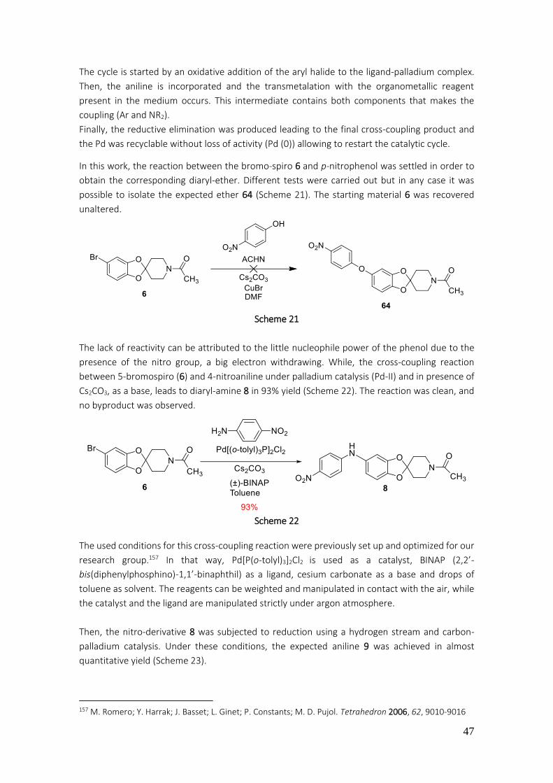
47
The cycle is started by an oxidative addition of the aryl halide to the ligand-palladium complex.
Then, the aniline is incorporated and the transmetalation with the organometallic reagent
present in the medium occurs. This intermediate contains both components that makes the
coupling (Ar and NR2).
Finally, the reductive elimination was produced leading to the final cross-coupling product and
the Pd was recyclable without loss of activity (Pd (0)) allowing to restart the catalytic cycle.
In this work, the reaction between the bromo-spiro 6 and p-nitrophenol was settled in order to
obtain the corresponding diaryl-ether. Different tests were carried out but in any case it was
possible to isolate the expected ether 64 (Scheme 21). The starting material 6 was recovered
unaltered.
Scheme 21
The lack of reactivity can be attributed to the little nucleophile power of the phenol due to the
presence of the nitro group, a big electron withdrawing. While, the cross-coupling reaction
between 5-bromospiro (6) and 4-nitroaniline under palladium catalysis (Pd-II) and in presence of
Cs2CO3, as a base, leads to diaryl-amine 8 in 93% yield (Scheme 22). The reaction was clean, and
no byproduct was observed.
Scheme 22
The used conditions for this cross-coupling reaction were previously set up and optimized for our
research group.157 In that way, Pd[P(o-tolyl)3]2Cl2 is used as a catalyst, BINAP (2,2’-
bis(diphenylphosphino)-1,1’-binaphthil) as a ligand, cesium carbonate as a base and drops of
toluene as solvent. The reagents can be weighted and manipulated in contact with the air, while
the catalyst and the ligand are manipulated strictly under argon atmosphere.
Then, the nitro-derivative 8 was subjected to reduction using a hydrogen stream and carbon-
palladium catalysis. Under these conditions, the expected aniline 9 was achieved in almost
quantitative yield (Scheme 23).
157 M. Romero; Y. Harrak; J. Basset; L. Ginet; P. Constants; M. D. Pujol. Tetrahedron 2006, 62, 9010-9016

48
Scheme 23
The diarylamine 10 was prepared by cross-coupling between bromo-spiro 6 and 4-
aminobenzonitrile (65), using the conditions, previously described, for the preparation of 8
(Scheme 24). Thus, the synthesis of diaryl-amine 10 was afforded in 90% yield.
Scheme 24
The shielding of the protons in the 1H-NMR spectrum of positions 4 (6.67 ppm) and 6 (6.56 ppm)
of the spiranic system of compound 10 in comparison with the bromo derivative 6 (CH-4 a 6.81
ppm and CH-6 a 6.82 ppm), indicate the substitution of the bromo atom for an electron donor
group. Besides, the presence of two new doublets that integrates two protons each one at 6.78
ppm and 7.37 ppm, belonging to the CH of the 4-cyanophenyl, allow to confirm the proposed
structure for the spiro derivative 10 (Figure 35).
Figure 35. 1H-NMR spectroscopy data of compound 10
Following the same methodology, the amination of the spiro derivative 6 it allows the preparation
of the amide 11 in 86% yield. In this case, it starts from bromo-spiro 6 which couples with 3-
phenoxyaniline (66) (Scheme 25).
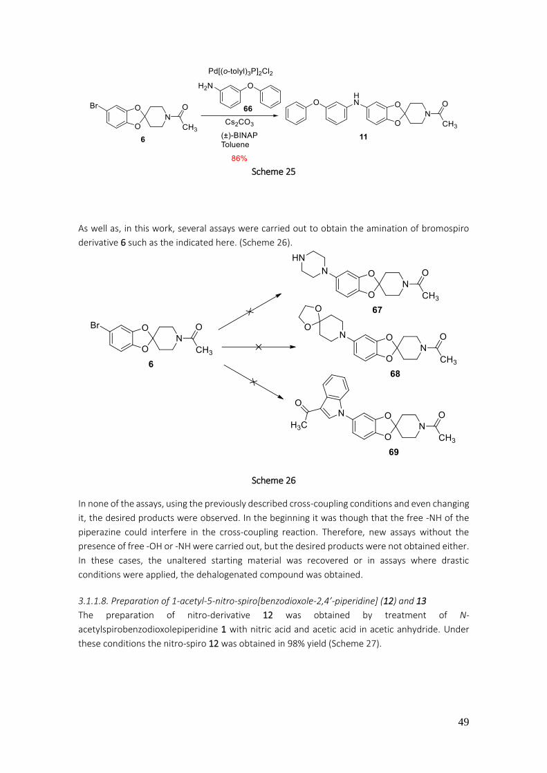
49
Scheme 25
As well as, in this work, several assays were carried out to obtain the amination of bromospiro
derivative 6 such as the indicated here. (Scheme 26).
Scheme 26
In none of the assays, using the previously described cross-coupling conditions and even changing
it, the desired products were observed. In the beginning it was though that the free -NH of the
piperazine could interfere in the cross-coupling reaction. Therefore, new assays without the
presence of free -OH or -NH were carried out, but the desired products were not obtained either.
In these cases, the unaltered starting material was recovered or in assays where drastic
conditions were applied, the dehalogenated compound was obtained.
3.1.1.8. Preparation of 1-acetyl-5-nitro-spiro[benzodioxole-2,4’-piperidine] (12) and 13
The preparation of nitro-derivative 12 was obtained by treatment of N-
acetylspirobenzodioxolepiperidine 1 with nitric acid and acetic acid in acetic anhydride. Under
these conditions the nitro-spiro 12 was obtained in 98% yield (Scheme 27).
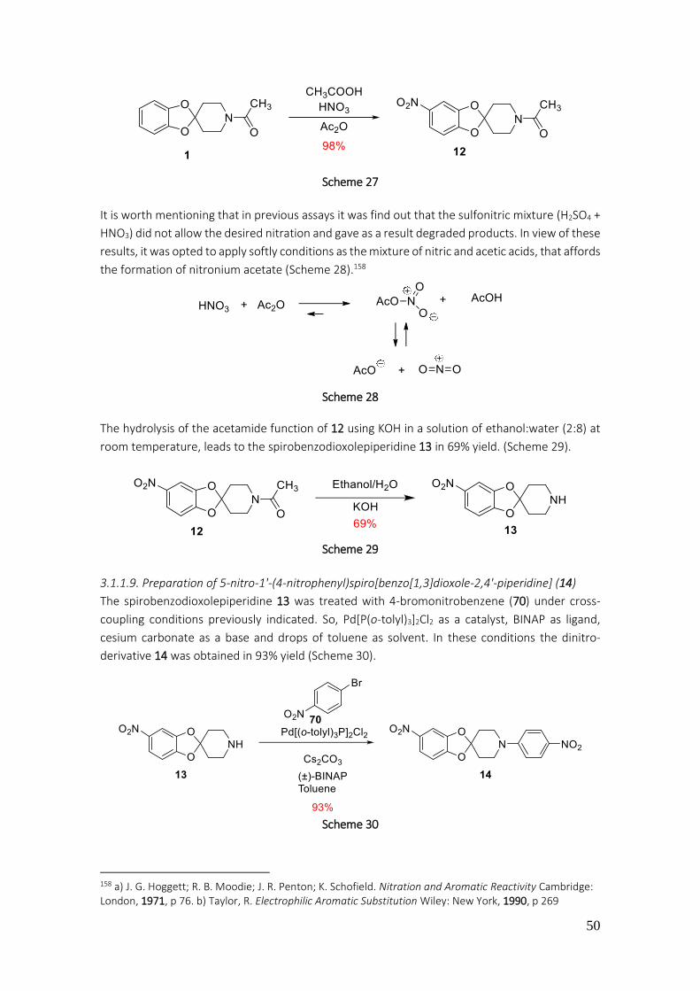
50
Scheme 27
It is worth mentioning that in previous assays it was find out that the sulfonitric mixture (H2SO4 +
HNO3) did not allow the desired nitration and gave as a result degraded products. In view of these
results, it was opted to apply softly conditions as the mixture of nitric and acetic acids, that affords
the formation of nitronium acetate (Scheme 28).158
Scheme 28
The hydrolysis of the acetamide function of 12 using KOH in a solution of ethanol:water (2:8) at
room temperature, leads to the spirobenzodioxolepiperidine 13 in 69% yield. (Scheme 29).
Scheme 29
3.1.1.9. Preparation of 5-nitro-1'-(4-nitrophenyl)spiro[benzo[1,3]dioxole-2,4'-piperidine] (14)
The spirobenzodioxolepiperidine 13 was treated with 4-bromonitrobenzene (70) under cross-
coupling conditions previously indicated. So, Pd[P(o-tolyl)3]2Cl2 as a catalyst, BINAP as ligand,
cesium carbonate as a base and drops of toluene as solvent. In these conditions the dinitro-
derivative 14 was obtained in 93% yield (Scheme 30).
Scheme 30
158 a) J. G. Hoggett; R. B. Moodie; J. R. Penton; K. Schofield. Nitration and Aromatic Reactivity Cambridge: London, 1971, p 76. b) Taylor, R. Electrophilic Aromatic Substitution Wiley: New York, 1990, p 269

51
The emergence in the 1H-NMR spectrum of two doublets at 6.83 and 8.08 ppm integrating two
protons each one, it allows us to confirm the proposed structure for the spiro-derivative 14
(Figure 36).
Figure 36. 1H-NMR spectroscopy data of compound 14
The reduction of both nitro groups through catalytic hydrogenation, leads to the double aniline
15 in 93% yield (Scheme 31).
Scheme 31
The shielding suffered by protons in the 1H-NMR spectrum, in the positions 3’’ and 5’’, and as well
the ones in the positions 4 and 6 in comparison with the spectrum signals of the nitrate starting
material, enable to confirm the proposed structure for the spiro-derivative 15 (Figure 37).
Figure 37. 1H-NMR spectroscopy data of compound 15
3.1.1.10. Preparation of spiro[benzo[1,3]dioxole-2,4'-piperidine] (16) and the 1'-(4-
nitrophenyl)spiro[benzo[1,3]dioxole-2,4'-piperidine] (17)
Hydrolysis of the amide 1 using KOH in ethanol and water leads to the piperidine 71 in a 70% yield
(Scheme 32).
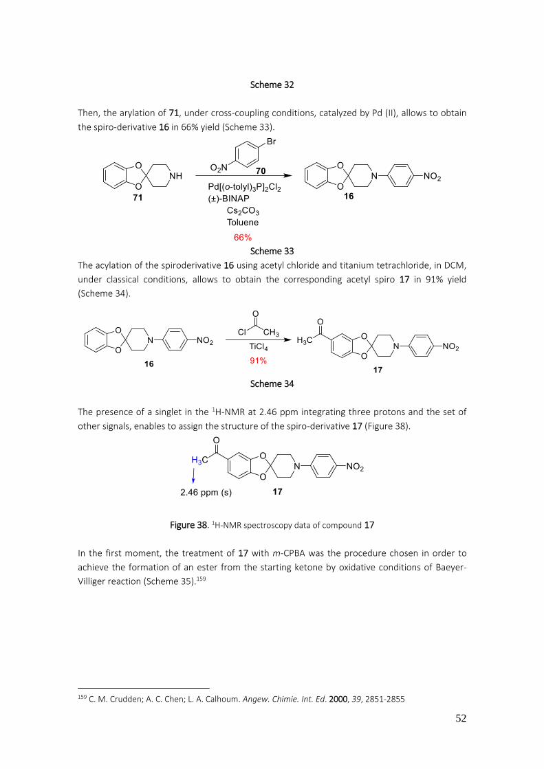
52
Scheme 32
Then, the arylation of 71, under cross-coupling conditions, catalyzed by Pd (II), allows to obtain
the spiro-derivative 16 in 66% yield (Scheme 33).
Scheme 33
The acylation of the spiroderivative 16 using acetyl chloride and titanium tetrachloride, in DCM,
under classical conditions, allows to obtain the corresponding acetyl spiro 17 in 91% yield
(Scheme 34).
Scheme 34
The presence of a singlet in the 1H-NMR at 2.46 ppm integrating three protons and the set of
other signals, enables to assign the structure of the spiro-derivative 17 (Figure 38).
Figure 38. 1H-NMR spectroscopy data of compound 17
In the first moment, the treatment of 17 with m-CPBA was the procedure chosen in order to
achieve the formation of an ester from the starting ketone by oxidative conditions of Baeyer-
Villiger reaction (Scheme 35).159
159 C. M. Crudden; A. C. Chen; L. A. Calhoum. Angew. Chimie. Int. Ed. 2000, 39, 2851-2855

53
Scheme 35
In the applied conditions it was not possible to prepare the expected ester 72, but the
corresponding N-oxide 73 was obtained. The m-CPBA produces oxidation of the tertiary amine160
and as a result the corresponding N-oxide is obtained which presents solubility problems and
would not allow the oxidation of the acylated position. The mass spectroscopy analysis with a
molecular signal at 372.1243 g/mol (expected 372.1200 g/mol) allows to confirm the proposed
structure for the N-oxide 73.
3.1.1.11. Preparation of 18 and 20
The treatment of the methylketone 17 with hydroxylamine hydrochloride in presence of sodium
carbonate, leads to the expected oxime 18 in 75% yield of isolated product (Scheme 36).
By NMR techniques it was confirmed only obtention of the isomer (E), that as it has been
mentioned previously, is due to the applied conditions of reaction.
Scheme 36
It is well known that oximes are synthetic intermediates that can be transformed into amides or
lactones through Beckmann transposition.161 In order for the transposition to take place it
requires presence of an acid as sulfuric acid or AlCl3·H2O.162
A proposal mechanism for this transposition is indicated below (Scheme 37), where the most
important characteristic lies in the stereochemistry disposition of the R and R’ groups regarding
the oxime. In the practice the migrating group is the one in anti disposition regarding the hydroxyl
group (here it is the R’ group).
160 N. K. Jana; J. G. Verkade. Org. Lett. 2003, 5, 3787-3790 161 a) R. E. Gawly. Org. React. 1988, 35, 39-40 b) M. B. Smith; J. March. In Advanced Organic Chemistry 5th ed., John Wiley & Sons, New York, 2001, 1415-1416 162 M. Boruah; D. Konwar. J. Org. Chem. 2002, 67, 7138-7139
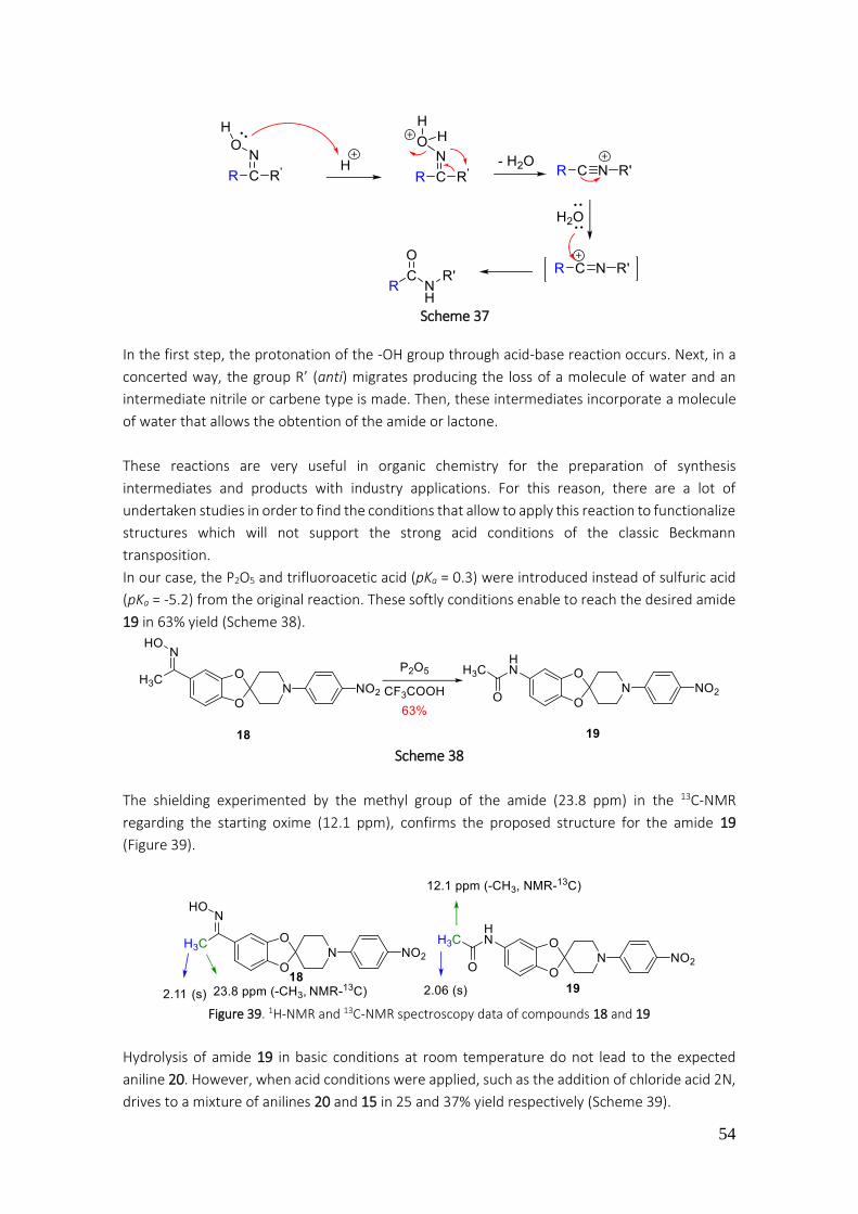
54
Scheme 37
In the first step, the protonation of the -OH group through acid-base reaction occurs. Next, in a
concerted way, the group R’ (anti) migrates producing the loss of a molecule of water and an
intermediate nitrile or carbene type is made. Then, these intermediates incorporate a molecule
of water that allows the obtention of the amide or lactone.
These reactions are very useful in organic chemistry for the preparation of synthesis
intermediates and products with industry applications. For this reason, there are a lot of
undertaken studies in order to find the conditions that allow to apply this reaction to functionalize
structures which will not support the strong acid conditions of the classic Beckmann
transposition.
In our case, the P2O5 and trifluoroacetic acid (pKa = 0.3) were introduced instead of sulfuric acid
(pKa = -5.2) from the original reaction. These softly conditions enable to reach the desired amide
19 in 63% yield (Scheme 38).
Scheme 38
The shielding experimented by the methyl group of the amide (23.8 ppm) in the 13C-NMR
regarding the starting oxime (12.1 ppm), confirms the proposed structure for the amide 19
(Figure 39).
Figure 39. 1H-NMR and 13C-NMR spectroscopy data of compounds 18 and 19
Hydrolysis of amide 19 in basic conditions at room temperature do not lead to the expected
aniline 20. However, when acid conditions were applied, such as the addition of chloride acid 2N,
drives to a mixture of anilines 20 and 15 in 25 and 37% yield respectively (Scheme 39).
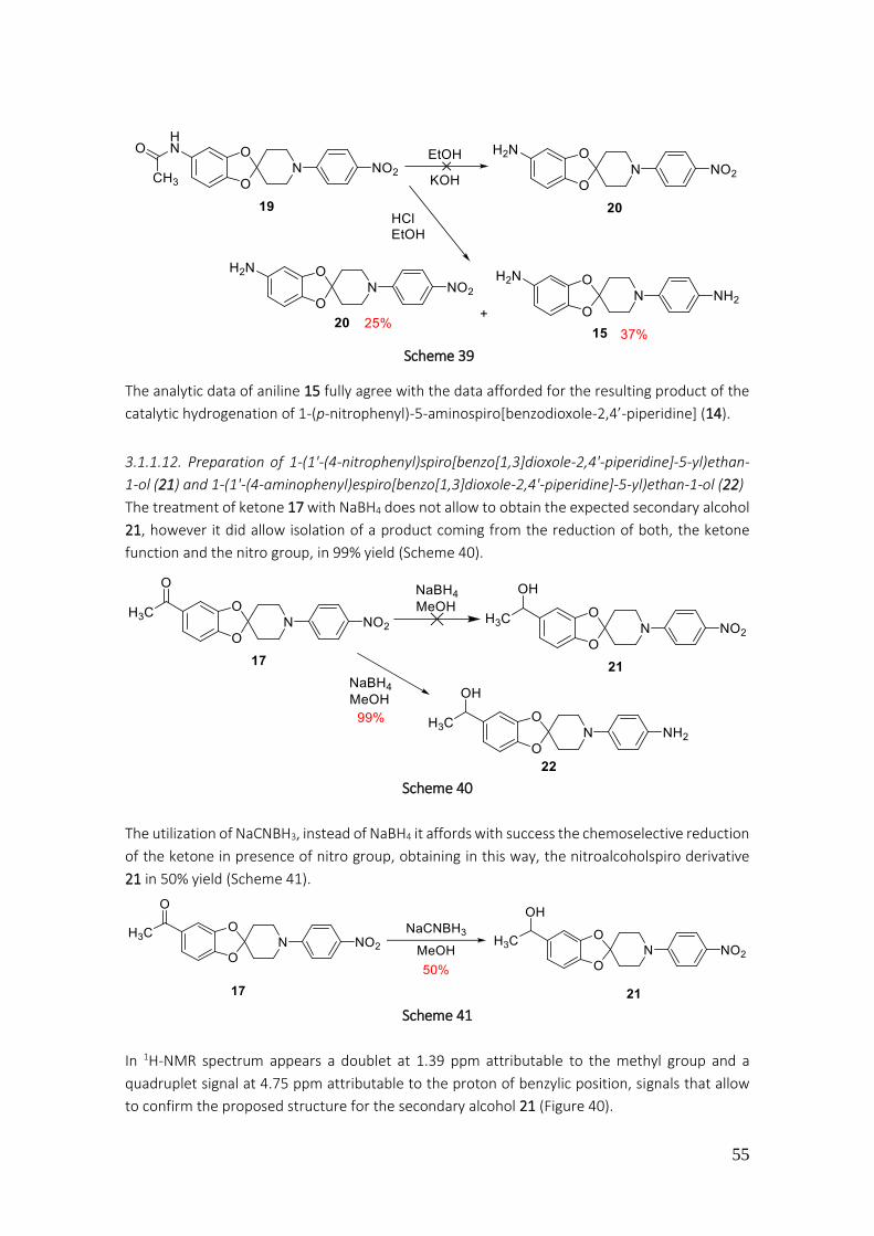
55
Scheme 39
The analytic data of aniline 15 fully agree with the data afforded for the resulting product of the
catalytic hydrogenation of 1-(p-nitrophenyl)-5-aminospiro[benzodioxole-2,4’-piperidine] (14).
3.1.1.12. Preparation of 1-(1'-(4-nitrophenyl)spiro[benzo[1,3]dioxole-2,4'-piperidine]-5-yl)ethan-
1-ol (21) and 1-(1'-(4-aminophenyl)espiro[benzo[1,3]dioxole-2,4'-piperidine]-5-yl)ethan-1-ol (22)
The treatment of ketone 17 with NaBH4 does not allow to obtain the expected secondary alcohol
21, however it did allow isolation of a product coming from the reduction of both, the ketone
function and the nitro group, in 99% yield (Scheme 40).
Scheme 40
The utilization of NaCNBH3, instead of NaBH4 it affords with success the chemoselective reduction
of the ketone in presence of nitro group, obtaining in this way, the nitroalcoholspiro derivative
21 in 50% yield (Scheme 41).
Scheme 41
In 1H-NMR spectrum appears a doublet at 1.39 ppm attributable to the methyl group and a
quadruplet signal at 4.75 ppm attributable to the proton of benzylic position, signals that allow
to confirm the proposed structure for the secondary alcohol 21 (Figure 40).

56
Figure 40. 1H-NMR spectroscopy data of compound 21
Alcohol 21 was undertaken by the action of NBS in DMF in order to substitute the hydroxyl group
for a bromo atom.163 Even trying to modify the conditions, it was not possible to obtain the
expected product 74. With the applied conditions, it was possible to isolate the bromovinyl
derivative 75 in 54% yield (Scheme 42).
Scheme 42
The following mechanism is proposed for the formation of 75 (Scheme 43):
Scheme 43
163 S. Zhao; Y. Wuo; Q. Sun; T. M. Cheng; R. T. Li. Synthesis 2015, 47, 1154-1162

57
3.1.1.13. Preparation of spiroderivatives 23 to 26
The treatment of nitro-derivative 17 with hydrogen stream under carbon-palladium catalysis,
drives chemoselectively to the expected aniline 23 in 93% yield (Scheme 44).
Scheme 44
The addition of the aniline 23 to the phthalic anhydride (76) allows to obtain the N-substituted
phthalimide 24 in 36% yield (Scheme 45).
Scheme 45
In the 1H-NMR spectrum of 24 are distinguish two double doublets at 7.78 and 7.94 ppm
integrating 2 protons each one, being attributable to the protons of positions and with
respect to the carbonyl groups. These data together with the C-13 NMR allow to confirm the
proposed structure for imide 24 (Figure 41).
Figure 41. 1H-NMR spectroscopy data of compound 24
The methylketone 24 is reduced to secondary alcohol 25 by treatment with NaBH4 in methanol
(Scheme 46).
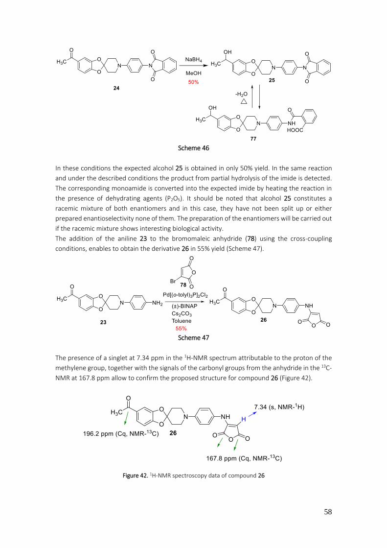
58
Scheme 46
In these conditions the expected alcohol 25 is obtained in only 50% yield. In the same reaction
and under the described conditions the product from partial hydrolysis of the imide is detected.
The corresponding monoamide is converted into the expected imide by heating the reaction in
the presence of dehydrating agents (P2O5). It should be noted that alcohol 25 constitutes a
racemic mixture of both enantiomers and in this case, they have not been split up or either
prepared enantioselectivity none of them. The preparation of the enantiomers will be carried out
if the racemic mixture shows interesting biological activity.
The addition of the aniline 23 to the bromomaleic anhydride (78) using the cross-coupling
conditions, enables to obtain the derivative 26 in 55% yield (Scheme 47).
Scheme 47
The presence of a singlet at 7.34 ppm in the 1H-NMR spectrum attributable to the proton of the
methylene group, together with the signals of the carbonyl groups from the anhydride in the 13C-
NMR at 167.8 ppm allow to confirm the proposed structure for compound 26 (Figure 42).
Figure 42. 1H-NMR spectroscopy data of compound 26
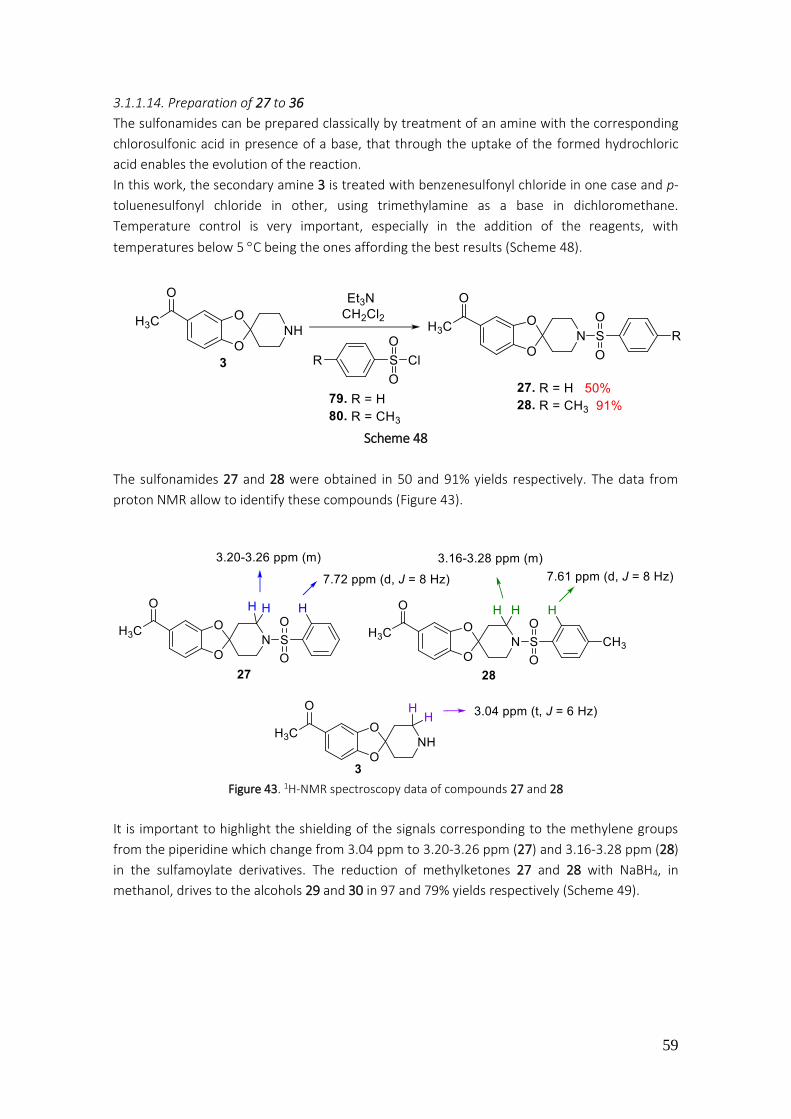
59
3.1.1.14. Preparation of 27 to 36
The sulfonamides can be prepared classically by treatment of an amine with the corresponding
chlorosulfonic acid in presence of a base, that through the uptake of the formed hydrochloric
acid enables the evolution of the reaction.
In this work, the secondary amine 3 is treated with benzenesulfonyl chloride in one case and p-
toluenesulfonyl chloride in other, using trimethylamine as a base in dichloromethane.
Temperature control is very important, especially in the addition of the reagents, with
temperatures below 5 C being the ones affording the best results (Scheme 48).
Scheme 48
The sulfonamides 27 and 28 were obtained in 50 and 91% yields respectively. The data from
proton NMR allow to identify these compounds (Figure 43).
Figure 43. 1H-NMR spectroscopy data of compounds 27 and 28
It is important to highlight the shielding of the signals corresponding to the methylene groups
from the piperidine which change from 3.04 ppm to 3.20-3.26 ppm (27) and 3.16-3.28 ppm (28)
in the sulfamoylate derivatives. The reduction of methylketones 27 and 28 with NaBH4, in
methanol, drives to the alcohols 29 and 30 in 97 and 79% yields respectively (Scheme 49).

60
Scheme 49
In both cases, the presence of quadruplet signals at 4.68 and 4.77 ppm in the 1H-NMR spectrum
of compounds 29 and 30 respectively, allows to confirm the proposed structures. With the
purpose to convert the hydroxyl group of alcohols 29 and 30 into a good leaving group, these
were treated with thionyl chloride using dichloromethane as a solvent (Scheme 50).
Scheme 50
The chlorinated derivatives 31 and 32 were obtained in high purity and were used without
purification in the following reactions.
The treatment of the chloro-derivative 31 with N,N-diethyl-2-hidroxyethylamine in presence of
trimethylamine dissolved in dichloromethane, leads to the expected aminoalcohol 33 in 45% yield
after purification (Scheme 51). The same conditions were applied, using N,N-diethyl-2-
hidroxyethylamine, allowing the obtention of diamine 34 in 16% yield of the purified product
(Scheme 51).
Scheme 51

61
Alternatively, the alkylation of alcohol 29 with N,N-diethiyl-2-chloroetylamine in DMF and with
the presence of potassium carbonate was attempted(Scheme 52).
Scheme 52
The characteristic signals in the 1H-NMR spectrums of 33 and 34 that enable the structural assign,
will be the next ones: for 33, the presence of a quadruplet signals at 4.23 ppm attributable to the
benzylic proton, the quadruplet at 2.51 ppm and the triplet at 0.97 ppm, both signals attributable
to the ethylene groups. While for the diamine 34, there is a quadruplet at 3.54 ppm attributable
to benzylic proton and a singlet at 2.08 ppm integrating six protons attributable to the two methyl
of the tertiary amine (Figure 44).
Figure 44. 1H-NMR spectroscopy data of compounds 33 and 34
In order to obtain a more lipophilic compound than diamine 34, the preparation of amine 35 was
carried out by treatment of chloro-derivative 32 with hexylamine, using the same conditions
previously indicated for the preparation of diamine 34 (Scheme 53).
Scheme 53
Thus, the corresponding alkylated hexylamine is obtained in 20% yield. In this case, the presence
of different signals in the 1H-NMR spectrum attributable to the lineal chain of hexylamine, allow
to confirm the proposed structure for 35 (Figure 45).
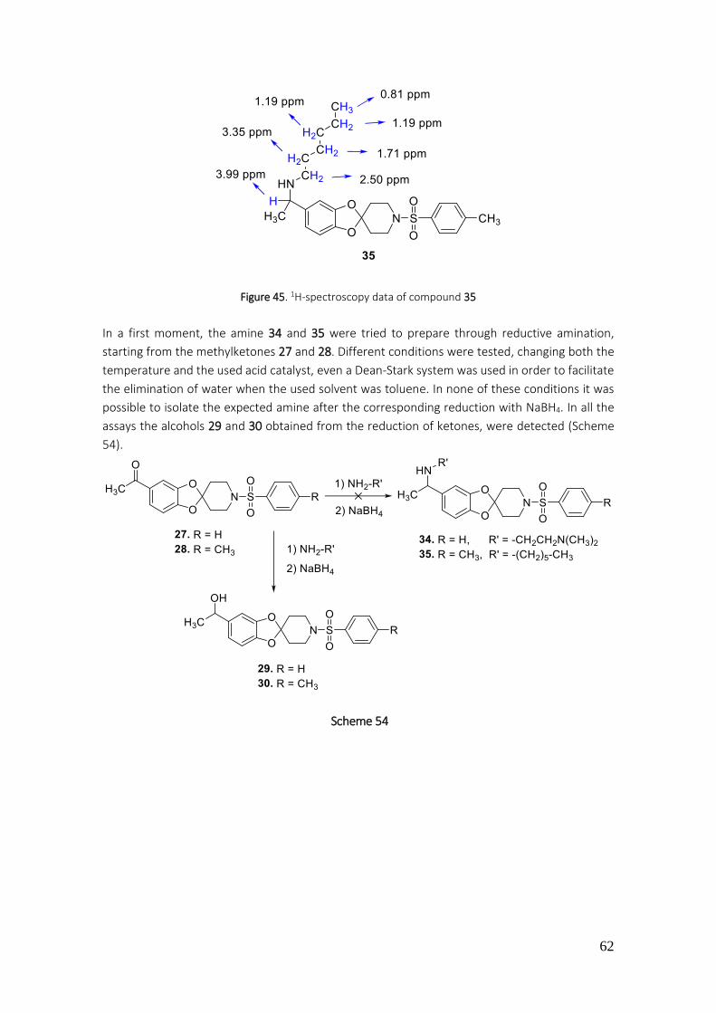
62
Figure 45. 1H-spectroscopy data of compound 35
In a first moment, the amine 34 and 35 were tried to prepare through reductive amination,
starting from the methylketones 27 and 28. Different conditions were tested, changing both the
temperature and the used acid catalyst, even a Dean-Stark system was used in order to facilitate
the elimination of water when the used solvent was toluene. In none of these conditions it was
possible to isolate the expected amine after the corresponding reduction with NaBH4. In all the
assays the alcohols 29 and 30 obtained from the reduction of ketones, were detected (Scheme
54).
Scheme 54
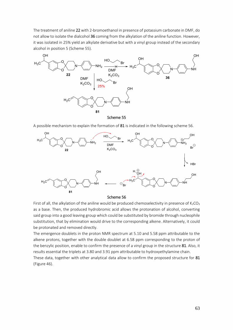
63
The treatment of aniline 22 with 2-bromoethanol in presence of potassium carbonate in DMF, do
not allow to isolate the dialcohol 36 coming from the alkylation of the aniline function. However,
it was isolated in 25% yield an alkylate derivative but with a vinyl group instead of the secondary
alcohol in position 5 (Scheme 55).
Scheme 55
A possible mechanism to explain the formation of 81 is indicated in the following scheme 56.
Scheme 56
First of all, the alkylation of the aniline would be produced chemoselectivity in presence of K2CO3
as a base. Then, the produced hydrobromic acid allows the protonation of alcohol, converting
said group into a good leaving group which could be substituted by bromide through nucleophile
substitution, that by elimination would drive to the corresponding alkene. Alternatively, it could
be protonated and removed directly.
The emergence doublets in the proton NMR spectrum at 5.10 and 5.58 ppm attributable to the
alkene protons, together with the double doublet at 6.58 ppm corresponding to the proton of
the benzylic position, enable to confirm the presence of a vinyl group in the structure 81. Also, it
results essential the triplets at 3.80 and 3.91 ppm attributable to hydroxyethylamine chain.
These data, together with other analytical data allow to confirm the proposed structure for 81
(Figure 46).

64
Figure 46. 1H-NMR spectroscopy data of compound 81
3.1.1.15. Preparation of ureas 37-42
Ureas are present in many compounds of both industrial chemistry and chemistry applied to drug
synthesis. Regarding the preparation of ureas it is possible to differentiate between symmetric
ureas and no symmetric. The methodology to prepare them is diverse, some of these methods
are explain hereunder:
·No symmetric ureas
These are some of the methods to obtain no symmetric ureas:
a) Mitsunobu reaction
This method allows to obtain ureas using PPh3 and DEAD (diethyl azodicarboxylate), at room
temperature and CO2 stream, softly conditions that leads to the ureas with good yields. As a result
of the treatment of urea with CO2, the carbamic acid is obtained and reacts with the zwitterion
derivative of PPh3, making a stable zwitterion species, which reacts with another amine molecule.
The resultant intermediate is restructured forming the desired substituted ureas (Scheme 57).164
Scheme 57
164 H. Wang; Z. Xim; Y. Li. Top. Curr. Chem. 2017, 375, 49-75

65
b) Carbonyldiimidazole salts165,166
The reaction of carbonyldiimidazole (CDI) with amines or anilines gives as a result the N-
(aminocarbonyl)imidazole, which through reaction with methyl iodide becomes
carbonylimidazole iodide, an intermediate salt, that treat it with another amine leads to the
corresponding urea (Scheme 58).
Scheme 58
c) Isocyanates167,168
When amines or anilines are added to isocyanates, asymmetric ureas are obtained (Scheme 59).
It is necessary to highlight that the transformation of ammonium isocyanate to urea, carried out
by Wöhler in 1824, constituted the first synthesis of an organic compound coming from an
inorganic precursor. This method affords the best results regarding the previous described
methods, allowing the synthesis of ureas in only one step and with softly conditions (room
temperature and short times of reaction). It uses solvents as DMSO,169 ethylic ether167,168 or
THF.167,168 Most of the isocyanates are commercially available, however its preparation is possible
in the laboratory.
Scheme 59
d) Chloroformates170
Through the use of chloroformates and amines or anilines it is possible to obtain asymmetric
ureas. To explain these reactions the mechanism would be: first the addition to the carbonyl
group from the amine or aniline and elimination of chloride, next, the reaction of a second amine
which adds again into the carbamate intermediate to generate the urea (Scheme 60).
Scheme 60
165 J. A. Grzyb; R. A. Batey. Tetrahedron Lett. 2008, 49, 5279-5282 166 M. N. Bertrand; J. P. Wolfe. Tetrahedron 2005, 61, 6447-6459 167 D. P. N. Satchel; R. S. Satchel. Chem. Soc. Rev. 1975, 4, 231-250 168 E. A. Castro; R. B. Moodle; P. J. Sansom. Chem. Soc. Perkin Trans. ɪ. 1985, 2, 737-742 169 Y. Zhang; M. Anderson; J. L. Weisman; M. Lu; C. J. Choy; V. A. Boyd; J. Price; M. Sigal; J. Clark; M. Connelly; F. Zhu; W. A. Guiguemde; C. Jeffries; L. Yang; A. Lemoff: A. P. Liou; T. R. Webb; J. L. DeRisi; R. K. Guy. ACS Med. Chem. Lett. 2010, 1, 460-465 170 C. Han; J. A. Porco. Org. Lett. 2007, 9, 1517-1520

66
Symmetric ureas171
These are some of the possible reactions to obtain symmetric ureas:
a) S,S-Dimethyldithiocarbamate with an amine or aniline (addition to the carbonyl
group plus elimination of the methyl methanethiol) (Scheme 61).
Scheme 61
b) Ammonia or amines in the presence of CO2
Through the addition of ammonia or amines to carbon dioxide the corresponding ureas are
afforded (Scheme 62). This synthesis of urea was carried out for first time at 1870 by A. Bazarov.
172
Scheme 62
Being a fast method and with simple reagents, it is considered a good option to prepare ureas at
industry scale and sustainable with natural environment. Thanks to the low acidity of CO2 and the
softly conditions of the reaction, the carbamic acid is made together with the carbamate salt. The
conversion between the carmate salt, the carbamic acid and CO2 is reversible, therefore the
efficiency of the synthesis lies in the dehydration of the carbamate salt which leads to the
corresponding urea.
This reaction can be carried out as well with secondary or tertiary amines as indicated in he
following scheme 63.164
Scheme 63
From the Bazarov, Li and col.173 methodology, it was proposed changes to improve the
dehydration (Scheme 64). It was used assays of radioactive labeling with DBU (1,8-
diazabicyclo[5.4.0]undec-7-ene) as a base, in presence of di-terc-butyl azodicarboxylate, in order
to label the ureas in only one step.
171 E. Artuso; I. Degani; R. Fochi; C. Magistris. Synthesis 2007, 3497-3506 172 A. I. Bazarov. J. Prakt. Chem. 1870, 2, 283-312 173 A. K. H. Dheere; S. Bongarzone; C. Taddei; R. Yan; A. D. Gee. Synlett 2015, 26, 2257-2260
67
Scheme 64
In our research group, the urea 37 was prepared by addition of the amine 23 to the corresponding
isocyanate under the conditions reported before (Scheme 65).
Scheme 65
The used solvent was THF, which under stirring at room temperature allows to achieve 37 in 80%
yield. The data corresponding to the 1H-NMR spectroscopy signals as the pics at 7.07, 7.18 and
8.54 ppm attributable to the protons of positions 4’’’, 3’’’ and 6’’’ and the signal at 154 ppm (d, J
= 247 Hz) that appears in the 13C-NMR spectroscopy, attributable to C-2’’’ and bringing out the C-
F coupling, allow to confirm the proposed structure for urea 37 (Figure 47).
The reduction with NaBH4 of urea 37 leads to secondary alcohol 38 in 99% yield (Scheme 66).
Scheme 66
Figure 47. 1H-NMR and 13C-NMR spectroscopy data of compound 37
68
The step from 37 to 38 was confirmed by the presence of a doublet signal at 1.23 ppm (CH3) and
a quadruplet at 4.63 ppm (CH-OH) (Figure 48).
Figure 48. 1H-NMR spectroscopy data of compound 38
Following the indicated methodology for the anilines addition to isocyanates, at room
temperature, in THF, it arrives to the obtention of urea 39 in 80% yield (Scheme 67).
Scheme 67
In this case, the presence of signals in the 1H-NMR spectrum corresponding to -CH2-CH3 group, it
allows to confirm the proposed structure for the arylalkylurea 39 (Figure 49).
Figure 49. 1H-NMR spectroscopy data of compound 39
The treatment of diamine 15 with pyridyl-3-isocyanate leads to the monourea 40 in only 7% yield.
(Scheme 68).
Scheme 68
The structure of monourea 40 was confirmed by mass spectroscopy (mass found: 418.1883
g/mol) being the theorical mass of 418.1834 g/mol for C23H23N5O3 applying ESI (+) method.
In this case, the urea from the reaction of amino group at C-4’’ position is obtained
regioselectively. The main reactivity of this NH2 group regarding the position C-5, it can be
69
attributed to the major nucleophile power of this group due to the higher electron donating
character of the amine function in para position. While the amino group of position 5 account
with an -O- group in para position presenting electron donor character and other -O- in meta
position, acting as electron withdrawing group through inductive effect.
Alternatively, the treatment of dianiline 15 with an excess of ethyl-isocyanate leads to the bis-
urea 41 in 79% yield (Scheme 69).
Scheme 69
The presence of signals in 1H-NMR spectrum attributable to the ethyl groups of the ureas 41, and
as well the integral corresponding to the exact number of protons, allow to confirm the proposed
structure for this compound (Figure 50).
Figure 50. 1H-NMR spectroscopy data of compound 41
The urea 42 was prepared by addition of aniline 9 to isocyanate 84, in THF and under stirring at
room temperature (Scheme 70).
Scheme 70
In the NMR proton spectrum are distinguishable signals at 2.08, at 7.52, at 7.72 and at 8.14 ppm,
attributable to protons of CH3 group of acetyl function and positions C-5’’’, C-6’’’ and C-2’’’
respectively (Figure 51).

70
Figure 51. 1H-NMR spectroscopy data of compound 42
3.1.1.16. Preparation of 1’-acetyl-1-(3H-spiro[benzoxazole-2,4’-piperidine]) (43) and 1’,3,6-
triacetyl-1-(spiro[benzoxazole-2,4’-piperidine]) (44)
The system spiro[benzoxazole-2,4’-piperidine] is not found described in the bibliography. This
nucleus is considered a bioisostere of the spiro[benzodioxole-2,4’-piperidine] nucleus, previously
commented in this memory, regarding the Grimm's hydride displacement law (Scheme 71).
The addition of hydrogen atom to an atom of atomic number ‘’n’’ gives as a result an specie with
the same properties than the atom with a superior atomic number n+1.174
Scheme 71
The condensation of aminoalcohols with aldehydes or ketones in acidic media and elimination of
the produced water is proposed for the preparation of 1,3-benzoxazoles.175
In this work, it was carried out the condensation of 2-aminophenol (85) with 4-piperidone in
toluene and in presence of APTS as a catalyst. In the reaction system the crystal device of Dean-
Stark was introduced to facilitate the elimination of water (Scheme 72).
Scheme 72
174 H. G. Grimm. Electrochem. 1925, 31, 474-480 175 A. I. Moskalenko; V. I. Boev. Russ. J. Org. Chem. 2009, 45, 1018-1023
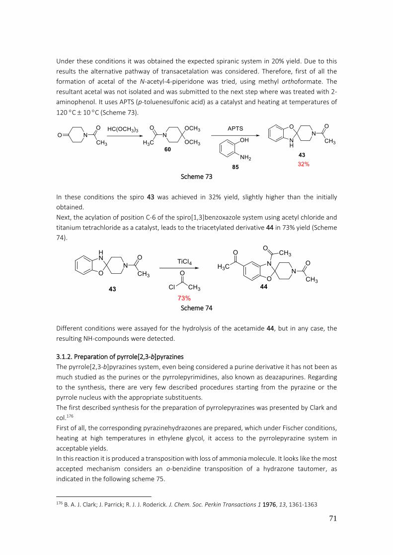
71
Under these conditions it was obtained the expected spiranic system in 20% yield. Due to this
results the alternative pathway of transacetalation was considered. Therefore, first of all the
formation of acetal of the N-acetyl-4-piperidone was tried, using methyl orthoformate. The
resultant acetal was not isolated and was submitted to the next step where was treated with 2-
aminophenol. It uses APTS (p-toluenesulfonic acid) as a catalyst and heating at temperatures of
120 C 10 C (Scheme 73).
Scheme 73
In these conditions the spiro 43 was achieved in 32% yield, slightly higher than the initially
obtained.
Next, the acylation of position C-6 of the spiro[1,3]benzoxazole system using acetyl chloride and
titanium tetrachloride as a catalyst, leads to the triacetylated derivative 44 in 73% yield (Scheme
74).
Scheme 74
Different conditions were assayed for the hydrolysis of the acetamide 44, but in any case, the
resulting NH-compounds were detected.
3.1.2. Preparation of pyrrole[2,3-b]pyrazines
The pyrrole[2,3-b]pyrazines system, even being considered a purine derivative it has not been as
much studied as the purines or the pyrrolepyrimidines, also known as deazapurines. Regarding
to the synthesis, there are very few described procedures starting from the pyrazine or the
pyrrole nucleus with the appropriate substituents.
The first described synthesis for the preparation of pyrrolepyrazines was presented by Clark and
col.176
First of all, the corresponding pyrazinehydrazones are prepared, which under Fischer conditions,
heating at high temperatures in ethylene glycol, it access to the pyrrolepyrazine system in
acceptable yields.
In this reaction it is produced a transposition with loss of ammonia molecule. It looks like the most
accepted mechanism considers an o-benzidine transposition of a hydrazone tautomer, as
indicated in the following scheme 75.
176 B. A. J. Clark; J. Parrick; R. J. J. Roderick. J. Chem. Soc. Perkin Transactions 1 1976, 13, 1361-1363
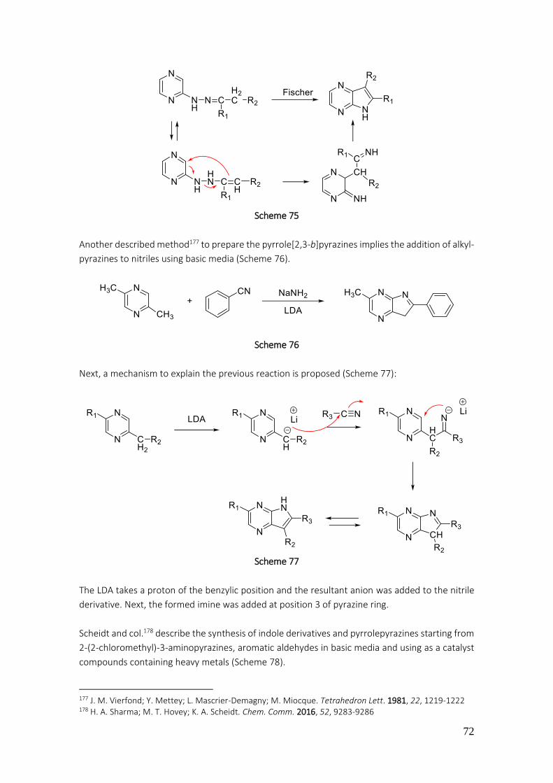
72
Scheme 75
Another described method177 to prepare the pyrrole[2,3-b]pyrazines implies the addition of alkyl-
pyrazines to nitriles using basic media (Scheme 76).
Scheme 76
Next, a mechanism to explain the previous reaction is proposed (Scheme 77):
Scheme 77
The LDA takes a proton of the benzylic position and the resultant anion was added to the nitrile
derivative. Next, the formed imine was added at position 3 of pyrazine ring.
Scheidt and col.178 describe the synthesis of indole derivatives and pyrrolepyrazines starting from
2-(2-chloromethyl)-3-aminopyrazines, aromatic aldehydes in basic media and using as a catalyst
compounds containing heavy metals (Scheme 78).
177 J. M. Vierfond; Y. Mettey; L. Mascrier-Demagny; M. Miocque. Tetrahedron Lett. 1981, 22, 1219-1222 178 H. A. Sharma; M. T. Hovey; K. A. Scheidt. Chem. Comm. 2016, 52, 9283-9286
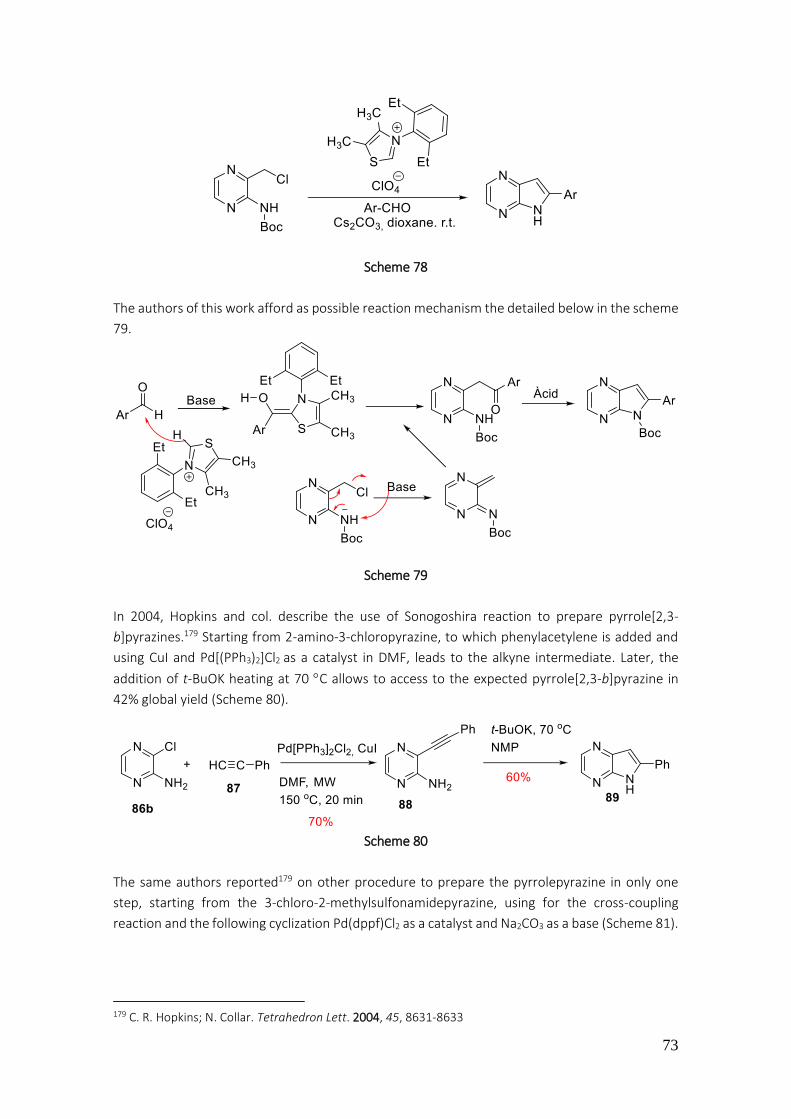
73
Scheme 78
The authors of this work afford as possible reaction mechanism the detailed below in the scheme
79.
Scheme 79
In 2004, Hopkins and col. describe the use of Sonogoshira reaction to prepare pyrrole[2,3-
b]pyrazines.179 Starting from 2-amino-3-chloropyrazine, to which phenylacetylene is added and
using CuI and Pd[(PPh3)2]Cl2 as a catalyst in DMF, leads to the alkyne intermediate. Later, the
addition of t-BuOK heating at 70 C allows to access to the expected pyrrole[2,3-b]pyrazine in
42% global yield (Scheme 80).
Scheme 80
The same authors reported179 on other procedure to prepare the pyrrolepyrazine in only one
step, starting from the 3-chloro-2-methylsulfonamidepyrazine, using for the cross-coupling
reaction and the following cyclization Pd(dppf)Cl2 as a catalyst and Na2CO3 as a base (Scheme 81).
179 C. R. Hopkins; N. Collar. Tetrahedron Lett. 2004, 45, 8631-8633
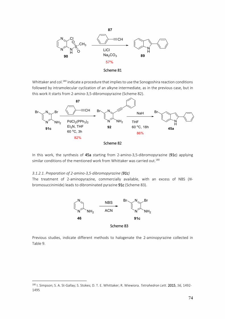
74
Scheme 81
Whittaker and col.180 indicate a procedure that implies to use the Sonogoshira reaction conditions
followed by intramolecular cyclization of an alkyne intermediate, as in the previous case, but in
this work it starts from 2-amino-3,5-dibromopyrazine (Scheme 82).
Scheme 82
In this work, the synthesis of 45a starting from 2-amino-3,5-dibromopyrazine (91c) applying
similar conditions of the mentioned work from Whittaker was carried out.180
3.1.2.1. Preparation of 2-amino-3,5-dibromopyrazine (91c)
The treatment of 2-aminopyrazine, commercially available, with an excess of NBS (N-
bromosuccinimide) leads to dibrominated pyrazine 91c (Scheme 83).
Scheme 83
Previous studies, indicate different methods to halogenate the 2-aminopyrazine collected in
Table 9.
180 I. Simpson; S. A. St-Gallay; S. Stokes; D. T. E. Whittaker; R. Wiewiora. Tetrahedron Lett. 2015, 56, 1492-1495

75
Table 9. Halogenation of 2-aminopyrazine. Bibliographic data
ENTRY CONDITIONS 93 (YIELD %) 91 (YIELD %) REFERENCES
1 NBS, DMF 0 °C, overnight 88 181
2 NIS, DMF 28 °C, overnight 61 181
3 NBS, CH2Cl2, r.t. 4 h 50 182
4 NBS, CH2Cl2, r.t. 1.5 h 52 183
5 DBH, DMF-MeCN, r.t. 0.5 h 65 184
6a Br2/HBr/H2O 5 °C 82 185
7 Br2, Piridina, CHCl3, r.t. 36 186
8 NBS, CH2Cl2 63 187
9 NBS, DMSO-H2O, 15 °C, 6 h 85 188
10 NBS, DMSO-H2O, 15 °C, 16 h 77 189
11 NBS, DMSO-H2O, r.t. 6 h 90 190 a) From 2-amino-3-bromopyrazine
In our research group, the halogenation conditions of 2-aminopyrazine (46) were studied in detail
with the intention to optimize the yields of the reaction. The obtained conditions and results are
reviewed on Table 10.191 It is necessary to highlight that it can be obtained in a regioselective
manner the compound 93, the dihalogenated 91 or a mixture of both.
181 C. M. Chou; Y. W. Tung; M. I. Ling; D. Chan; W. Phakhodee; M. Isobe. Heterocycles 2012, 86, 1323-1329 182 Y. Younis; F. Douelle; D. González-Cabrera; C. Le Manach; A. T. Nchinda; T. Paquet; L. Street; K. L. White; M. Zabiulla; J. T. Joseph; S. Bashyam; D. Waterson; M. J. Witty; S. Wittlin; S. A. Charman; K. J. Chibale. Med. Chem. 2013, 56, 8860-8871 183 C. Le Manach; A. T. Nchinda; T. Paquet; D. Gonzàlez-Cabrera; Y. Younis; Z. Han; S. Bashyam; M. Zabiulla; D. Taylor; N. Lawrence; K. L. White; S. A. Charman; D. Waterson; M. J. Witty; S. Wittlin; M. E. Botha; S. H. Nondaba; J. Reader; L. M. Birkholtz; M. B. jiménez-Díaz; M. Santos-Martínez; S. Ferrer; I. Angulo-Barturen; S. Meister; Y. Antonova-Koch; E. A. Winzeler; L. Street; J. K. Chibale. J. Med. Chem. 2016, 59, 9890-9905 184 T. Itho; S. Kato; N. Nonoyama; T. Wada; K. Maeda; T. Mase; M. M. Zhao; J. Z. Song; D. M. Tschaen; J. M. McNamara. Org. Process Res. Dev. 2006, 10, 822 185 H. J. Brachwitz. Prakt. Chem. 1969, 311, 40 186 S. Martínez-González; A. I. Hernández; C. Varela; S. Rodríguez- Arístegui; J. Pastor. Bioorg. Med. Chem. Lett. 2012, 22, 1874-1878 187 P. J. Zimmermann; C. Brehm; W. Buhr; A. M. Palmer; W. A. Simon. Bioorg. Med. Chem. 2008, 16, 536-541 188 B. Jiang; C. G. Yang; W. N. Xiong; J. Wang. Bioorg. Med. Chem. 2001, 9, 1149-1154 189 J. J. Caldwell; N. Veillard; I. Collins. Tetrahedron 2012, 68, 9713-9728 190 R. Goel; V. Luxami; K. Paul. R.S.C. Adv. 2014, 4, 9885-9892 191 E. Lizano; J. Grima; M. D. Pujol. Synlett 2019, 30, 2000-2003
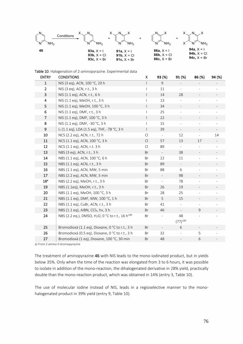
76
Table 10. Halogenation of 2-aminopyrazine. Experimental data
ENTRY CONDITIONS X 93 (%) 91 (%) 86 (%) 94 (%)
1 NIS (3 eq), ACN, 100 °C, 20 h I 9 - - -
2 NIS (3 eq), ACN, r.t., 3 h I 11 - - -
3 NIS (1.1 eq), ACN, r.t., 6 h I 14 28 - -
4 NIS (1.1 eq), MeOH, r.t., 3 h I 13 - - -
5 NIS (1.1 eq), MeOH, 100 °C, 3 h I 34 - - -
6 NIS (1.1 eq), DMF, r.t., 3 h I 25 - - -
7 NIS (1.1 eq), DMF, 100 °C, 3 h I 22 - - -
8 NIS (1.1 eq), DMF, -30 °C, 3 h I 15 - - -
9 I2 (1.1 eq), LDA (1.5 eq), THF, -78 °C, 3 h I 39 - - -
10 NCS (2.2 eq), ACN, r.t., 72 h Cl - 12 - 14
11 NCS (1.1 eq), ACN, 100 °C, 3 h Cl 57 13 17 -
12 NCS (1.1 eq), ACN, r.t. 3 h Cl 89 - - -
13 NBS (3 eq), ACN, r.t., 3 h Br - 38 - -
14 NBS (1.1 eq), ACN, 100 °C, 6 h Br 22 11 - -
15 NBS (1.1 eq), ACN, r.t., 3 h Br 89 - - -
16 NBS (1.1 eq), ACN, MW, 5 min Br 88 6 - -
17 NBS (2.2 eq), ACN, MW, 5 min Br - 98 - -
18a NBS (2.2 eq), MeOH, r.t., 3 h Br - 78 - -
19 NBS (1.1eq), MeOH, r.t., 3 h Br 26 19 - -
20 NBS (1.1 eq), MeOH, 100 °C, 3 h Br 28 25 - -
21 NBS (1.1 eq), DMF, MW, 100 °C, 1 h Br 5 15 - -
22 NBS (1.1 eq), CuBr, ACN, r.t., 3 h Br 41 - - -
23 NBS (1.1 eq), AIBN, CCl4, hv, 3 h Br 46 - 9 -
24 NBS (2.2 eq.), DMSO, H2O, 0 °C to r.t., 16 h189 Br - 48
(77)189
- -
25 Bromodioxà (1.1 eq), Dioxane, 0 °C to r.t., 3 h Br - 6 - -
26 Bromodioxà (0.5 eq), Dioxane, 0 °C to r.t., 3 h Br 32 - 5 -
27 Bromodioxà (1 eq), Dioxane, 100 °C, 30 min Br 48 - 6 - a) From 2-amino-5-bromopyrazine
The treatment of aminopyrazine 46 with NIS leads to the mono-iodinated product, but in yields
below 35%. Only when the time of the reaction was elongated from 3 to 6 hours, it was possible
to isolate in addition of the mono-reaction, the dihalogenated derivative in 28% yield, practically
double than the mono-reaction product, which was obtained in 14% (entry 3, Table 10).
The use of molecular iodine instead of NIS, leads in a regioselective manner to the mono-
halogenated product in 39% yield (entry 9, Table 10).
77
The treatment of 46 with NCS (N-chlorosuccinimide) at room temperature for 3 hours, leads to
mono-chlorinated pyrazine 93b in 57% yield, together with the dihalogenated product 91b (13%)
and the derivative selectively halogenated in C-3 86b (17%) (entry 11, Table 10).
The halogenation reaction with and excess of NCS (2.2 eq) leads to the dihalogenated product
91b (12%) along with the trihalogenated derivative 94b (14%) (entry 10, Table 10).
These unsatisfying results bring us to consider the bromination of aminopyrazines, thus the
treatment of 46 with NBS, in acetonitrile, leads to different products depending on the applied
conditions in each assay. With an excess of NBS (3 eq) it obtains selectively the dihalogenated 91c
in 38% yield (entry 13, Table 10).
Diminishing the quantity of NBS at 1.1 eq and heating to 100 C for 6 hours, it obtains a mixture
of 93c and 91c in 22 and 11% yields respectively (entry 14, Table 10).
When these mentioned conditions are applied but the crude of reaction stirs at room
temperature for 3 hours, it obtains exclusively 93c in 89% yield (entry 15, Table 10).
The application of assistance by microwave irradiation leads to 93c (88% yield) and 91c (6% yield),
using 1.1 eq of NBS (entry 16, Table 10), while when NBS quantities are increased double, it
achieves the preparation of 91c in 98% yield (entry 17, Table 10). These results are obtained after
heating for 5 minutes the crude of reaction at 100 °C.
It must be indicated, that the assistance by microwave irradiation allows to reduce considerably
the time of reaction and also it achieves a notably improvement in the outcomes.192
When instead of acetonitrile, methanol is used as a solvent and 1.1 eq of NBS at room
temperature or heating at 100 C, it obtains a mixture of mono-brominated and dibrominated
compounds 93c and 91c in yields of the same order (entry 19 and 20, Table 10). However, when
it works at room temperature but adding double quantity of halogenating agent (NBS), it obtains
selectively the dihalogenated derivative 91c in 78% yield (entry 18, Table 10). Other solvents as
DMF (entry 21, Table 10) lead to a mixture of 91c and 93c in low yields.
The addition of a catalyst as CuBr (entry 22, Table 10) leads to mono-brominated pyrazine 91c in
41% yield. The application of radical conditions in this reaction, using NBS as halogenating agent,
AIBN as radical indicator in carbon tetrachloride, keeping the reaction system under radiation of
a 100 W lamp, drives to mono-brominated derivative 93c (46%) in yields slightly higher regarding
the previous mentioned conditions. Along with it, it obtains derivative 86c brominated at position
3 (entry 23, Table 10).
192 a) D. Bogdal; M. Lukasiewicz; A. Pielichowski; A. Miciak; Sz. Bednarz. Tetrahedron 2003, 59, 649-653 b) E. Buxaderas; D. A. Alonso; C. Nájera. Eur. J. Org. Chem. 2013, 5864-5870 c) G. Broggini; V. Barbera; E. M. Beccalli; U. Chiacchio; A. Fasana; S. Galli; S. Gazzola. Adv. Synth. Catal. 2013, 355, 1640-1648 d) H. H. Nguyen; M. J. Kurth. Org. Lett. 2013, 15, 362-365 e) W. Qian; L. Zhang; H. Sun; H. Jiang; H. Liu. Adv. Synth. Catal. 2012, 354, 3231-3236 f) M. Baghbanzadeh; C. Pilger; C. O. Kappe. J. Org. Chem. 2011, 76, 8138-8142 g) J. F. Cívicos; D. A. Alonso; C. Nájera. Adv. Synth. Catal. 2011, 353, 1683-1687 h) E. M. Beccalli; A. Bernasconi; E. Borsini; G. Broggini; M. Rigamonti; G. Zecchi. J. Org. Chem. 2010, 75, 6923-6932 i) D. Liptrot; G. Alcaraz; B. Roberts. Adv. Synth. Catal. 2010, 352, 2183-2188

78
It is of interest the halogenation of 2-aminopyrazine 46 using NBS and DMSO-H2O as a solvent,
which are the most described conditions in the literature for the obtention of 3,5-dihalogenated
pyrazine, which is obtained in 48% yield (entry 24, Table 10).
The reaction was assayed several times but it was not possible to achieve the described yield of
77%.189
Also, in this work, the halogenation of 2-aminopirazyne 46 was assayed, using bromodioxane as
halogenating agent. The best results, of this reaction, were obtained by heating to 100 C for 30
minutes the mixture of reaction, which contains 2-aminopyrazine and bromodioxane (1 eq) (entry
25, 26 and 27, Table 10).
3.1.2.2. Preparation of pyrrole[2,3-b]pyrazine
The alkylation of 2-amino-3,5-dibromopyrazine (91c) with phenylacetylene, was achieved with
success using the conditions of Sonogoshira reaction catalyzed by palladium complexes in basic
media (Scheme 84).
Scheme 84
The Sonogoshira reaction is based in a cross-coupling reaction between bromoaryl and alkyne,
allowing the formation of C-C bonds, with high utility for the formation of aromatic systems with
potential industrial application.193 This reaction was presented by K. Sonogoshira and N. Hagihara
in 1975.194 The advantage lies on the fact that the cross-coupling is produced in softly conditions,
using palladium as a catalyst, CuI as a co-catalyst and an organic salt as triethylamine, which in
most cases acts as a solvent as well. The mechanism of this reaction can be represented through
the cycle of cross-coupling reaction catalyzed by palladium complex (Scheme 85).
193 a) M. Bakherad. Appl. Organomet. Chem. 2013, 27, 125-140 b) C. Torborg; M. Beller. Adv. Synth. Catal. 2009, 351, 3027-3043 c) J. W. B. Cooke; R. Bright; M. J. Coleman; K. P. Jenkins. Org. Process Res. Dev. 2001, 5, 383-386 194 K. Sonogoshira; Y. Tohda; N. Hagihara. Tetrahedron Lett. 1975, 16, 4467-4470
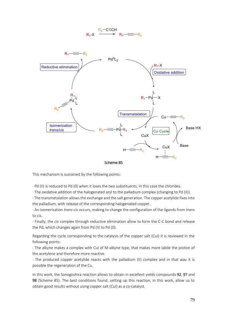
79
Scheme 85
This mechanism is sustained by the following points:
· Pd (II) is reduced to Pd (0) when it loses the two substituents, in this case the chlorides.
· The oxidative addition of the halogenated aryl to the palladium complex (changing to Pd (II)).
· The transmetalation allows the exchange and the salt generation. The copper acetylide fixes into
the palladium, with release of the corresponding halogenated copper.
· An isomerization trans-cis occurs, making to change the configuration of the ligands from trans
to cis.
· Finally, the cis complex through reductive elimination allow to form the C-C bond and release
the Pd, which changes again from Pd (II) to Pd (0).
Regarding the cycle corresponding to the catalysis of the copper salt (CuI) it is reviewed in the
following points:
· The alkyne makes a complex with CuI of M-alkyne type, that makes more labile the proton of
the acetylene and therefore more reactive.
· The produced copper acetylide reacts with the palladium (II) complex and in that way it is
possible the regeneration of the Cu.
In this work, the Sonogoshira reaction allows to obtain in excellent yields compounds 92, 97 and
98 (Scheme 85). The best conditions found, setting up this reaction, in this work, allow us to
obtain good results without using copper salt (CuI) as a co-catalyst.
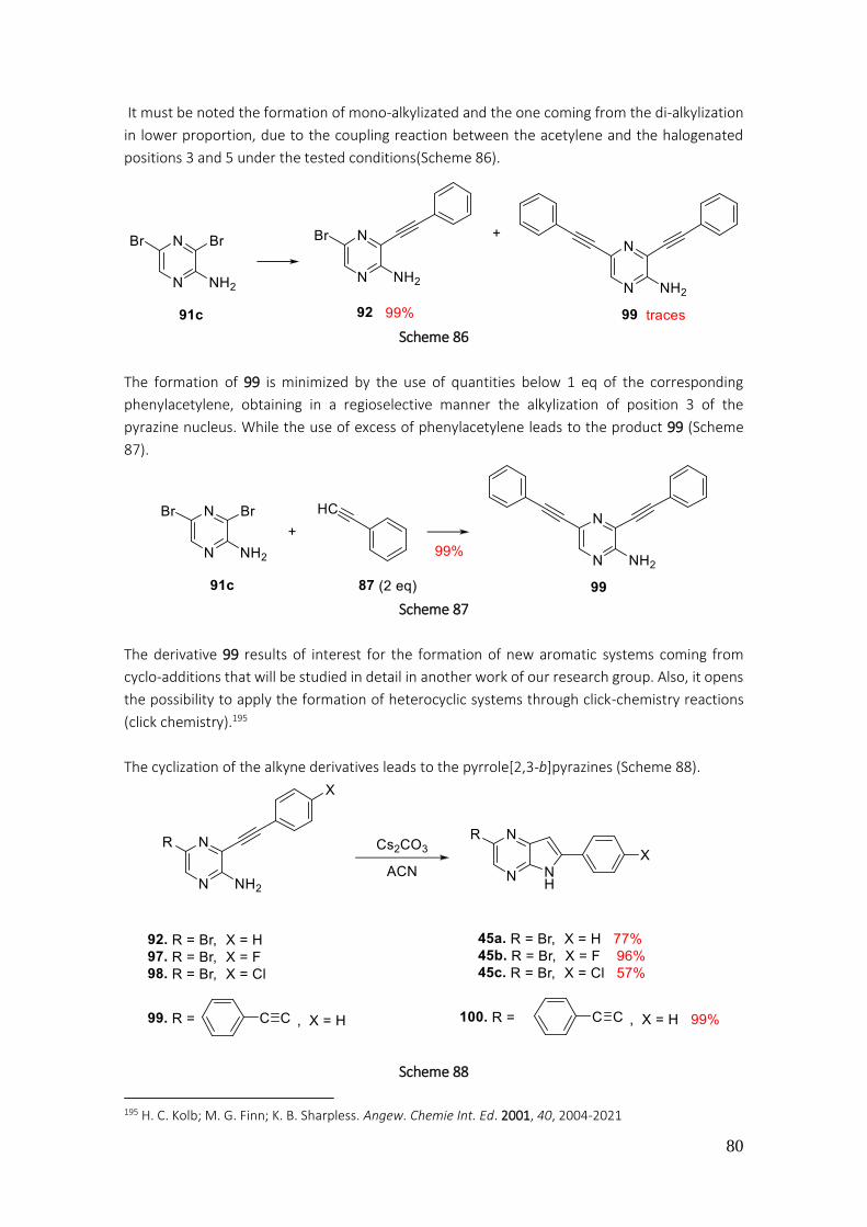
80
It must be noted the formation of mono-alkylizated and the one coming from the di-alkylization
in lower proportion, due to the coupling reaction between the acetylene and the halogenated
positions 3 and 5 under the tested conditions(Scheme 86).
Scheme 86
The formation of 99 is minimized by the use of quantities below 1 eq of the corresponding
phenylacetylene, obtaining in a regioselective manner the alkylization of position 3 of the
pyrazine nucleus. While the use of excess of phenylacetylene leads to the product 99 (Scheme
87).
Scheme 87
The derivative 99 results of interest for the formation of new aromatic systems coming from
cyclo-additions that will be studied in detail in another work of our research group. Also, it opens
the possibility to apply the formation of heterocyclic systems through click-chemistry reactions
(click chemistry).195
The cyclization of the alkyne derivatives leads to the pyrrole[2,3-b]pyrazines (Scheme 88).
Scheme 88
195 H. C. Kolb; M. G. Finn; K. B. Sharpless. Angew. Chemie Int. Ed. 2001, 40, 2004-2021

81
The cyclization of alkynes derivatives 92, 97-99 to the corresponding 45a-c and 100 was carried
out by treatment of the starting alkynes with Cs2CO3 in acetonitrile.
The cesium carbonate presents better solubility in organic solvents than sodium or potassium
carbonate.196 In similar products to pyrazines, as pyrimidines or pyridinic derivatives, the
cyclization is carried out in basic media and using CuI as a catalyst, affording interesting results.
(Scheme 89).197
Scheme 89
In this work, it is not used CuI as a catalyst, only the presence of a base as cesium carbonat at
reflux of acetonitrile.
This is a 5-endo-dig cyclisation following the Baldwin rules. The amino group is added to the
alkyne without expulsion of any leaving group (endo) and thus, affords a 5 atoms ring198 and
because it is and addition to an alkyne (Sp) it is called dig (diagonal) (Figure 52).
Figure 52. Cyclization 5-endo-dig
The emergence of two singlets in the 1H-NMR at 6.81 and at 7.93 ppm corresponding to the
protons of positions 7 and 3 respectively, confirms the proposed structure for pyrrole[2,3-
b]pyrazines (Figure 53).
Figure 53. 1H-NMR spectroscopy data of compound 45a
3.1.2.3. Preparation of derivatives 2,6-diphenyl-5H-pyrrole[2,3-b]pyrazines 47-52
The arylation of derivatives 2-bromopyrrolepyrazines was carried out by reaction of bromo-
derivatives with the corresponding boronic acids (Scheme 90).
196 R. Gujadhur; D. Ven Katarama; J. T. Kintight. Tetrahedron Lett. 2001, 42, 4791-4793 197 Z. Li; L. Hong; R. Lui; J. Shen; X. Zhou. Tetrahedron Lett. 2011, 52, 1343-1347 198 J. E. Baldwin J. Chem. Soc. Chem. Commun. 1976, 18, 734-736

82
Scheme 90
For this cross-coupling reaction it is required the Suzuki-Miyaura conditions, previously
mentioned in this work (page 44).
Thus, the pyrrole[2,3-b]pyrazines 47-52 are obtained in yields from acceptable to excellent (from
37% to 96%) (Scheme 90).
The proposed structure for this pyrrolepyrazines was confirmed by the presence of 1H-NMR
signals as the singlets at 7.00 ppm and at 8.50 ppm attributable to the protons of positions C-7
and C-3 respectively for 47 (Figure 54). Also, the presence of doublets at 7.27 and at 7.81 ppm
corresponding to the protons of positions C-3’’, C-5’’ and C-2’’, C-6’’ respectively, showed the
incorporation of the methylphenyl group at position 2 (Figure 54).
Figure 54. 1H-NMR spectroscopy data of compound 47
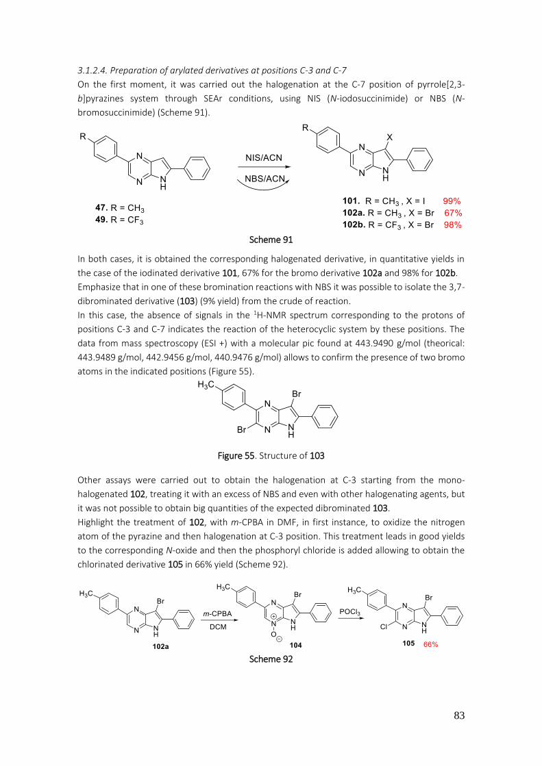
83
3.1.2.4. Preparation of arylated derivatives at positions C-3 and C-7
On the first moment, it was carried out the halogenation at the C-7 position of pyrrole[2,3-
b]pyrazines system through SEAr conditions, using NIS (N-iodosuccinimide) or NBS (N-
bromosuccinimide) (Scheme 91).
Scheme 91
In both cases, it is obtained the corresponding halogenated derivative, in quantitative yields in
the case of the iodinated derivative 101, 67% for the bromo derivative 102a and 98% for 102b.
Emphasize that in one of these bromination reactions with NBS it was possible to isolate the 3,7-
dibrominated derivative (103) (9% yield) from the crude of reaction.
In this case, the absence of signals in the 1H-NMR spectrum corresponding to the protons of
positions C-3 and C-7 indicates the reaction of the heterocyclic system by these positions. The
data from mass spectroscopy (ESI +) with a molecular pic found at 443.9490 g/mol (theorical:
443.9489 g/mol, 442.9456 g/mol, 440.9476 g/mol) allows to confirm the presence of two bromo
atoms in the indicated positions (Figure 55).
Figure 55. Structure of 103
Other assays were carried out to obtain the halogenation at C-3 starting from the mono-
halogenated 102, treating it with an excess of NBS and even with other halogenating agents, but
it was not possible to obtain big quantities of the expected dibrominated 103.
Highlight the treatment of 102, with m-CPBA in DMF, in first instance, to oxidize the nitrogen
atom of the pyrazine and then halogenation at C-3 position. This treatment leads in good yields
to the corresponding N-oxide and then the phosphoryl chloride is added allowing to obtain the
chlorinated derivative 105 in 66% yield (Scheme 92).
Scheme 92
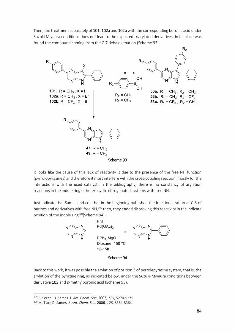
84
Then, the treatment separately of 101, 102a and 102b with the corresponding boronic acid under
Suzuki Miyaura conditions does not lead to the expected triarylated derivatives. In its place was
found the compound coming from the C-7 dehalogenation (Scheme 93).
Scheme 93
It looks like the cause of this lack of reactivity is due to the presence of the free NH function
(pyrrolopyrazines) and therefore it must interfere with the cross-coupling reaction, mostly for the
interactions with the used catalyst. In the bibliography, there is no constancy of arylation
reactions in the indole ring of heterocyclic nitrogenated systems with free NH.
Just indicate that Sames and col. that in the beginning published the functionalization at C-5 of
purines and derivatives with free NH,199 then, they ended disproving this reactivity in the indicate
position of the indole ring200(Scheme 94).
Scheme 94
Back to this work, it was possible the arylation of position 3 of pyrrolepyrazine system, that is, the
arylation of the pyrazine ring, as indicated below, under the Suzuki-Miyaura conditions between
derivative 103 and p-methylboronic acid (Scheme 95).
199 B. Sezen; D. Sames. J. Am. Chem. Soc. 2003, 125, 5274-5275 200 M. Tian; D. Sames. J. Am. Chem. Soc. 2006, 128, 8364-8364
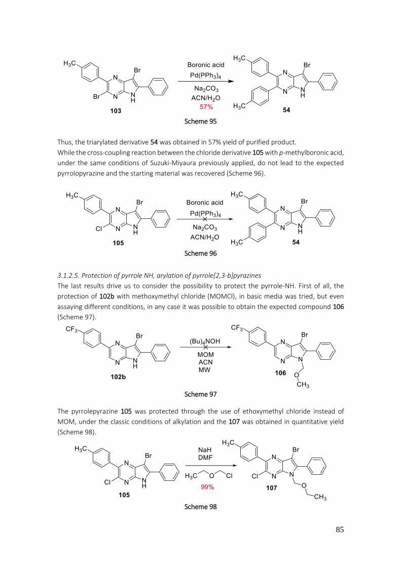
85
Scheme 95
Thus, the triarylated derivative 54 was obtained in 57% yield of purified product.
While the cross-coupling reaction between the chloride derivative 105 with p-methylboronic acid,
under the same conditions of Suzuki-Miyaura previously applied, do not lead to the expected
pyrrolopyrazine and the starting material was recovered (Scheme 96).
Scheme 96
3.1.2.5. Protection of pyrrole NH, arylation of pyrrole[2,3-b]pyrazines
The last results drive us to consider the possibility to protect the pyrrole-NH. First of all, the
protection of 102b with methoxymethyl chloride (MOMCl), in basic media was tried, but even
assaying different conditions, in any case it was possible to obtain the expected compound 106
(Scheme 97).
Scheme 97
The pyrrolepyrazine 105 was protected through the use of ethoxymethyl chloride instead of
MOM, under the classic conditions of alkylation and the 107 was obtained in quantitative yield
(Scheme 98).
Scheme 98
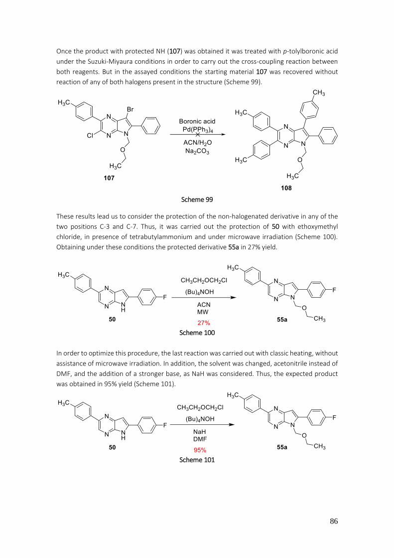
86
Once the product with protected NH (107) was obtained it was treated with p-tolylboronic acid
under the Suzuki-Miyaura conditions in order to carry out the cross-coupling reaction between
both reagents. But in the assayed conditions the starting material 107 was recovered without
reaction of any of both halogens present in the structure (Scheme 99).
Scheme 99
These results lead us to consider the protection of the non-halogenated derivative in any of the
two positions C-3 and C-7. Thus, it was carried out the protection of 50 with ethoxymethyl
chloride, in presence of tetrabutylammonium and under microwave irradiation (Scheme 100).
Obtaining under these conditions the protected derivative 55a in 27% yield.
Scheme 100
In order to optimize this procedure, the last reaction was carried out with classic heating, without
assistance of microwave irradiation. In addition, the solvent was changed, acetonitrile instead of
DMF, and the addition of a stronger base, as NaH was considered. Thus, the expected product
was obtained in 95% yield (Scheme 101).
Scheme 101
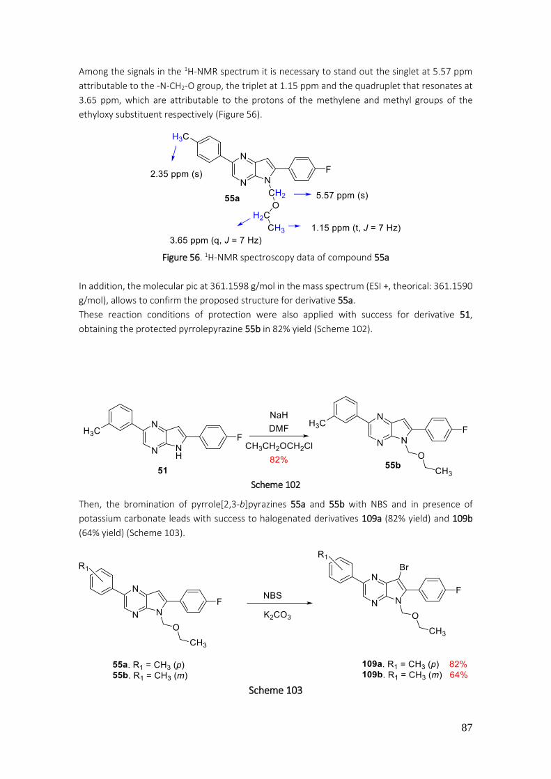
87
Among the signals in the 1H-NMR spectrum it is necessary to stand out the singlet at 5.57 ppm
attributable to the -N-CH2-O group, the triplet at 1.15 ppm and the quadruplet that resonates at
3.65 ppm, which are attributable to the protons of the methylene and methyl groups of the
ethyloxy substituent respectively (Figure 56).
Figure 56. 1H-NMR spectroscopy data of compound 55a
In addition, the molecular pic at 361.1598 g/mol in the mass spectrum (ESI +, theorical: 361.1590
g/mol), allows to confirm the proposed structure for derivative 55a.
These reaction conditions of protection were also applied with success for derivative 51,
obtaining the protected pyrrolepyrazine 55b in 82% yield (Scheme 102).
Scheme 102
Then, the bromination of pyrrole[2,3-b]pyrazines 55a and 55b with NBS and in presence of
potassium carbonate leads with success to halogenated derivatives 109a (82% yield) and 109b
(64% yield) (Scheme 103).
Scheme 103

88
The addition of p-methylphenylboronic acid to each 7-bromo-derivative 109a and 109b applying
the Suzuki-Miyaura conditions, allows to reach with moderated yields the 2,6,7-triarylated
derivatives 110a and 110b (Scheme 104).
Scheme 104
The signals appearing at 7.12-7.15 ppm and at 7.49-7.55 ppm in the 1H-NMR spectrum
attributable to the orto and meta positions corresponding to the tolyl from position 7 of
pyrrolepyrazine system, allow to confirm the proposed structure for 100a (Figure 57).
Figure 57. 1H-NMR spectroscopy data of compound 110a
In the mass spectrum (ESI +) appears a molecular pic at 452.2099 g/mol (theorical: 452.2093
g/mol) which supports the structure of 110a.
It is recorded that the alkyloxymethyl groups, considered acetalic derivatives, it can be hydrolyzed
in acidic conditions.201 In this work, even different conditions were tried, in any case it was
possible to isolate the product with free NH 57a (Scheme 105).
201 T. W. Greene; P. G. W. Wuts. Protective groups in orgànic synthesis. John Wiley and Sons, Inc. 2ona edició, Harvard 1980, pg 18
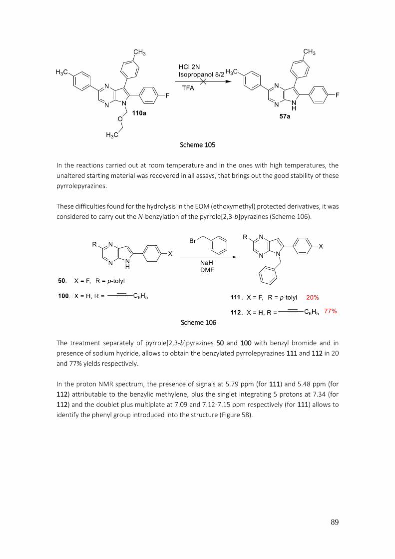
89
Scheme 105
In the reactions carried out at room temperature and in the ones with high temperatures, the
unaltered starting material was recovered in all assays, that brings out the good stability of these
pyrrolepyrazines.
These difficulties found for the hydrolysis in the EOM (ethoxymethyl) protected derivatives, it was
considered to carry out the N-benzylation of the pyrrole[2,3-b]pyrazines (Scheme 106).
Scheme 106
The treatment separately of pyrrole[2,3-b]pyrazines 50 and 100 with benzyl bromide and in
presence of sodium hydride, allows to obtain the benzylated pyrrolepyrazines 111 and 112 in 20
and 77% yields respectively.
In the proton NMR spectrum, the presence of signals at 5.79 ppm (for 111) and 5.48 ppm (for
112) attributable to the benzylic methylene, plus the singlet integrating 5 protons at 7.34 (for
112) and the doublet plus multiplate at 7.09 and 7.12-7.15 ppm respectively (for 111) allows to
identify the phenyl group introduced into the structure (Figure 58).
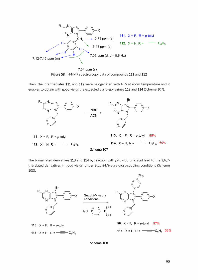
90
Figure 58. 1H-NMR spectroscopy data of compounds 111 and 112
Then, the intermediates 111 and 112 were halogenated with NBS at room temperature and it
enables to obtain with good yields the expected pyrrolepyrazines 113 and 114 (Scheme 107).
Scheme 107
The brominated derivatives 113 and 114 by reaction with p-tolylboronic acid lead to the 2,6,7-
triarylated derivatives in good yields, under Suzuki-Miyaura cross-coupling conditions (Scheme
108).
Scheme 108
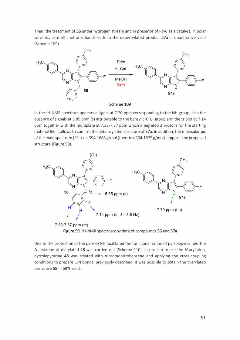
91
Then, the treatment of 56 under hydrogen stream and in presence of Pd-C as a catalyst, in polar
solvents, as methanol or ethanol leads to the debenzylated product 57a in quantitative yield
(Scheme 109).
Scheme 109
In the 1H-NMR spectrum appears a signal at 7.70 ppm corresponding to the NH group, also the
absence of signals at 5.85 ppm (s) attributable to the benzylic-CH2- group and the triplet at 7.14
ppm together with the multiplate at 7.32-7.37 ppm which integrated 3 protons for the starting
material 56, it allows to confirm the debenzylated structure of 57a. In addition, the molecular pic
of the mass spectrum (ESI +) at 394.1688 g/mol (theorical 394.1675 g/mol) supports the proposed
structure (Figure 59).
Figure 59. 1H-NMR spectroscopy data of compounds 56 and 57a
Due to the protection of the pyrrole NH facilitated the functionalization of pyrrolepyrazines, the
N-arylation of diarylated 48 was carried out (Scheme 110). In order to make the N-arylation,
pyrrolepyrazine 48 was treated with p-bromonitrobenzene and applying the cross-coupling
conditions to prepare C-N bonds, previously described, it was possible to obtain the triarylated
derivative 58 in 69% yield.

92
Scheme 110
The proton NMR spectrum of 58 shows two doublets at 6.99 ppm (orto position) and at 7.80 ppm
(meta position) which allow to confirm the introduction of p-nitrophenyl group into the
pyrrolepyrazine structure 58 (Figure 60).
Figure 60. 1H-NMR spectroscopy data of compound 58
Then, the halogenation of 58 with NBS in acetonitrile allows the obtention of 116 in 93% yield
(Scheme 111).
Scheme 111
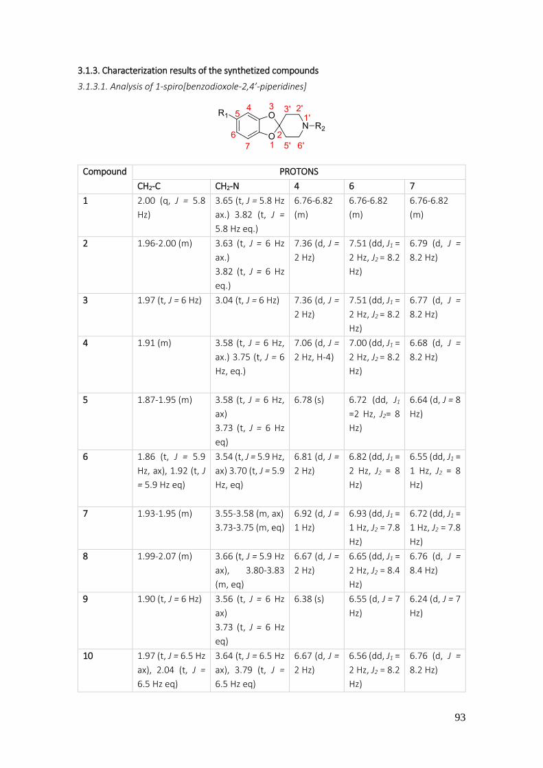
93
3.1.3. Characterization results of the synthetized compounds
3.1.3.1. Analysis of 1-spiro[benzodioxole-2,4’-piperidines]
Compound PROTONS
CH2-C CH2-N 4 6 7
1 2.00 (q, J = 5.8
Hz)
3.65 (t, J = 5.8 Hz
ax.) 3.82 (t, J =
5.8 Hz eq.)
6.76-6.82
(m)
6.76-6.82
(m)
6.76-6.82
(m)
2 1.96-2.00 (m) 3.63 (t, J = 6 Hz
ax.)
3.82 (t, J = 6 Hz
eq.)
7.36 (d, J =
2 Hz)
7.51 (dd, J1 =
2 Hz, J2 = 8.2
Hz)
6.79 (d, J =
8.2 Hz)
3 1.97 (t, J = 6 Hz) 3.04 (t, J = 6 Hz) 7.36 (d, J =
2 Hz)
7.51 (dd, J1 =
2 Hz, J2 = 8.2
Hz)
6.77 (d, J =
8.2 Hz)
4 1.91 (m)
3.58 (t, J = 6 Hz,
ax.) 3.75 (t, J = 6
Hz, eq.)
7.06 (d, J =
2 Hz, H-4)
7.00 (dd, J1 =
2 Hz, J2 = 8.2
Hz)
6.68 (d, J =
8.2 Hz)
5 1.87-1.95 (m) 3.58 (t, J = 6 Hz,
ax)
3.73 (t, J = 6 Hz
eq)
6.78 (s) 6.72 (dd, J1
=2 Hz, J2= 8
Hz)
6.64 (d, J = 8
Hz)
6 1.86 (t, J = 5.9
Hz, ax), 1.92 (t, J
= 5.9 Hz eq)
3.54 (t, J = 5.9 Hz,
ax) 3.70 (t, J = 5.9
Hz, eq)
6.81 (d, J =
2 Hz)
6.82 (dd, J1 =
2 Hz, J2 = 8
Hz)
6.55 (dd, J1 =
1 Hz, J2 = 8
Hz)
7 1.93-1.95 (m) 3.55-3.58 (m, ax)
3.73-3.75 (m, eq)
6.92 (d, J =
1 Hz)
6.93 (dd, J1 =
1 Hz, J2 = 7.8
Hz)
6.72 (dd, J1 =
1 Hz, J2 = 7.8
Hz)
8 1.99-2.07 (m) 3.66 (t, J = 5.9 Hz
ax), 3.80-3.83
(m, eq)
6.67 (d, J =
2 Hz)
6.65 (dd, J1 =
2 Hz, J2 = 8.4
Hz)
6.76 (d, J =
8.4 Hz)
9 1.90 (t, J = 6 Hz) 3.56 (t, J = 6 Hz
ax)
3.73 (t, J = 6 Hz
eq)
6.38 (s) 6.55 (d, J = 7
Hz)
6.24 (d, J = 7
Hz)
10 1.97 (t, J = 6.5 Hz
ax), 2.04 (t, J =
6.5 Hz eq)
3.64 (t, J = 6.5 Hz
ax), 3.79 (t, J =
6.5 Hz eq)
6.67 (d, J =
2 Hz)
6.56 (dd, J1 =
2 Hz, J2 = 8.2
Hz)
6.76 (d, J =
8.2 Hz)

94
11 1.91 (t, J = 5.8
Hz)
3.55 (t, J = 5.8 Hz
ax), 3.72 (t, J =
5.8 Hz eq)
6.51 (d, J =
2 Hz)
6.46 (dd, J1 =
2 Hz, J2 = 8
Hz)
6.36 (dd, J1 =
2 Hz, J2 = 8
Hz)
12 1.97 (t, J = 6 Hz
ax); 2.02 (t, J = 6
Hz eq)
3.62 (t, J = 6 Hz
ax)
3.75 (t, J = 6 Hz
eq)
7.54 (d, J =
2.3 Hz)
7.79 (dd, J1 =
2.3. J2 = 8.6
Hz)
6.77 (d, J =
8.6 Hz)
13 1.94 (t, J = 6 Hz) 2.99 (t, J = 6 Hz) 7.52 (d, J =
2.3 Hz)
7.77 (dd, J1 =
2.3. J2 = 8.6
Hz
6.72 (d, J =
8.6 Hz)
14 2.11 (t, J = 5.7
Hz)
3.64 (t, J = 5.7 Hz) 7.58 (d, J =
2.3 Hz)
7.82 (dd, J1 =
2.3. J2 = 8.6
Hz)
6.77 (d, J =
8.6 Hz)
15 2.13 (t, J = 6 Hz) 3.25 (t, J = 6 Hz) 6.24 (d, J =
2 Hz)
6.10 (dd, J1 =
2 Hz, J2 = 8
Hz)
6.56 (d, J = 8
Hz)
16 2.11 (t, J = 6 Hz) 3.68 (t, J = 5.7 Hz) 6.77-6.78
(m)
6.77-6.78
(m)
6.77-6.78
(m)
17 2.07 (t, J = 6 Hz) 3.63 (t, J = 6 Hz) 7.33 (d, J =
2 Hz)
7.47 (dd, J1 =
2 Hz, J2 = 8.2
Hz)
6.74 (d, J =
8.2 Hz)
73 2.10-2.12 (m,
ax.)
4.15-4.17 (m,
eq.)
3.25-3.37 (m) 7.40 (s)
7.51 (dd, J1 =
2 Hz, J2 = 8.2
Hz)
7.62 (d, J =
8.2 Hz)
18 2.03 (t, J = 5 Hz) 3.69 (q, J = 5 Hz) 7.14 (d, J =
2 Hz)
7.09 (dd, J1 =
2 Hz, J2 = 8.1
Hz)
6.86 (d, J =
8.1 Hz)
19 2.03 (t, J = 5.7
Hz)
3.61 (t, J = 5.7 Hz) 7.09 (d, J =
2 Hz)
7.18-7.19
(m)
6.63-6.64
(m)
20 1.99-2.12 (m) 3.59-3.66 (m) 6.21 (s) 6.08 (d, J = 7
Hz)
6.52 (d, J = 7
Hz)
21 2.04 (t, J = 5.7
Hz)
3.62 (t, J = 5.7 Hz) 7.79 (s) 6.74 (dd, J1 =
2 Hz, J2 = 8
Hz)
6.65 (d, J = 8
Hz)
22 2.08 (t, J = 5.5
Hz)
3.19 (t, J = 5.5 Hz) 6.77 (d, J =
2 Hz)
6.71 (dd, J1 =
2 Hz J2 = 8
Hz)
6.64 (d, J = 8
Hz)
75 2.12 (t, J = 6 Hz) 3.70 (t, J = 6 Hz) 6.93 (d, J =
2 Hz)
6.83 (dd, J1 =
2 Hz, J2 = 8
Hz)
6.72 (d, J = 8
Hz)

95
23 2.09 (t, J = 6 Hz) 3.19 (t, J = 6 Hz) 7.31 (d, J =
2 Hz)
7.45 (dd, J1 =
2 Hz, J2 = 8.1
Hz)
6.72 (d, J =
8.1 Hz)
24 2.17 (t, J = 5.8
Hz)
3.52 (t, J = 5.8 Hz) 7.41 (d, J =
2 Hz)
7.54 (dd, J1 =
2 Hz, J2 = 8.2
Hz)
6.81 (d, J =
8.2 Hz)
25 2.07-2.09 (m) 3.53 (t, J = 5.8 Hz) 6.78 (d, J =
2 Hz)
6.72 (dd, J1 =
2 Hz, J2 = 8
Hz)
6.65 (d, J = 8
Hz)
26 2.08 (t, J = 6 Hz) 3.44 (t, J = 6 Hz) 6.93 (d, J =
2 Hz)
7.47 (dd, J1 =
2 Hz, J2 = 8
Hz)
6.74 (d, 1H, J
= 8 Hz)
27 2.05 (t, J = 5.7
Hz)
3.20-3.26 (m) 7.24 (d, J =
1.8 Hz)
7.42 (dd, J1 =
1.8 Hz, J2 = 8
Hz)
6.66 (d, J = 8
Hz)
28 2.05 (t, J = 7 Hz) 3.16–3.28 (m) 7.26 (s) 7.43 (d, J = 8
Hz)
6.67 (d, J = 8
Hz)
29 2.02 (t, J = 5.5
Hz)
3.22 (t, J = 5.5 Hz) 6.71 (s) 6.69 (d, J = 8
Hz)
6.58 (d, J = 8
Hz)
30 2.00-2.03 (m) 3.27 (t, J = 5.8 Hz) 6.78 (d, J =
2 Hz)
6.75 (dd, J1 =
2Hz, J2 = 8
Hz)
6.64 (d, J = 8
Hz)
32 2.09 (t, J = 5.7
Hz)
3.27 (t, J = 5.7 Hz) 6.82 (d, J =
1.5 Hz)
6.79 (dd, J1 =
1.5 Hz, J2 = 8
Hz)
6.63 (d, J = 8
Hz)
33 2.08 (t, J = 5.3
Hz)
3.29 (t, J = 5.3 Hz) 6.70 (s) 6.67 (d, J =
7.9 Hz)
6.61 (d, J =
7.9 Hz)
34 2.02 (t, J = 5.7
Hz)
3.22 (t, J = 5.7 Hz) 6.65 (s) 6.62 (d, J =
7.9 Hz)
6.54 (d, J =
7.9 Hz)
35 2.08-2.10 (m) 3.07 (q, J = 7 Hz)
3.32 (q, J = 7 Hz)
7.08 (s) 6.62 (d, J = 8
Hz)
6.81 (d, J = 8
Hz)
81 2.14 (q, J = 6Hz) 2.14 (t, J = 6 Hz) 6.85 (s) 7.05 (d, J =
8.8 Hz)
6.97 (d, J =
8.8 Hz)
37 2.12 (t, J = 5.5
Hz)
3.52 (t, J = 5.5 Hz) 7.40 (d, J =
2 Hz)
7.54 (dd, J1 =
2 Hz, J2 = 8.2
Hz)
6.80 (d, J =
8.2 Hz)
38 1.98 (t, J = 5.7
Hz)
3.26 (t, J = 5.7 Hz) 6.74 (d, J =
2 Hz)
6.68 (dd, J1 =
2 Hz, J2 = 8
Hz)
6.59 (d, J = 8
Hz)
39 2.13-2.14 (m) 3.27-3.40 (m) 6.85 (d, J =
2 Hz)
6.79 (dd, J1 =
2 Hz, J2= 8
Hz)
6.71 (d, J = 8
Hz)

96
40 2.04 (t, J = 5.8
Hz)
3.34 (t, J = 5.8 Hz) 6.17 (d, J =
2 Hz)
6.05 (dd, J1 =
2 Hz, J2 = 8
Hz)
6.50 (d, J = 8
Hz)
41 2.04 (t, J = 5.4 Hz
ax)
3.28 (t, J = 5.4 Hz
eq)
3.15 (t, J = 5.4 Hz) 6.78 (d, J =
2 Hz)
6.52 (dd, J1 =
2 Hz, J2 = 8.2
Hz)
6.57 (d, J =
8.2 Hz)
42
0.90 (t, J = 5.8
Hz, ax), 1.93 (t, J
= 5.8 Hz eq)
3.70 (q, J = 5.8
Hz)
6.62 (d, J =
2 Hz)
6.53 (dd, J1 =
2 Hz, J2 = 8.3
Hz)
6.69 (d, J =
8.3 Hz)
Compound Carbons
CH2-C CH2-N 4 (CH) 6 (CH) 7 (CH) C-2
(Cq)
C-5
(Cq)
C-3a
(Cq)
C-7a
(Cq)
4 34.6
35.4
38.8
43.5
106.4 116.3 108.2 120.1 130.8 147.1 147.7
5 34.6
35.4
38.7
43.5
106.2 118.5 108.2 115.8 139.8 147.1 146.1
6 34.5
35.3
38.5
43.3
109.7 112.3 124.0
112.7 117.0 146.2 147.8
7 34.7
35.4
38.7
43.5
108.7 120.1 107.6 116.0 136.6 147.4 146.1
9 34.5
35.3
38.8
43.6
99.9 109.6 108.6 115.5 127.8 147.4 140.8
10
34.6
35.4
38.7
43.5
104.8 109.8 108.7 112.4 134.0 146.2 143.8
11 34.6
35.4
38.7
43.5
108.6 110.7 109.8 113.5 136.3 147.5 146.5
12 34.6
35.4
38.5
43.2
104.6 107.8 119.7 119.3 142.6 147.3 152.3
13 36.1 43.4 104.3 107.4 119.4 120.4 142.4 147.6 152.7
14 34.2 44.8 104.8 107.8 119.7 119.2 142.8 152.3 153.7
15 34.7 49.0 98.1 106.4 108.5 108.2 147.8 140.9 140.1
16 34.2 45.0 113.0 126.1 113.0 115.3 126.1 146.7 146.7
17 34.2 44.9 108.1 124.6 108.2 117.2 132.0 147.4 151.0
18 34.2 44.6 108.6 120.1 106.0 117.1 131.0 147.1 147.4
19 34.1 44.9 103.1 112.9 108.1 116.1 133.3 142.5 146.9
21 34.2 44.9 106.2 118.6 108.2 115.7 138.5 147.1 146.1
22 34.9 48.8 106.0 118.2 108.0 116.5 139.4 147.4 146.5
24 34.4 46.6 107.9 116.7 108.1 117.8 125.5 147.6 151.3
25 25.1 34.3 106.1 117.4 108.1 118.2 131.7 147.2 146.4
26 34.3 46.4 107.9 116.6 108.1 117.8 129.7 147.6 151.2
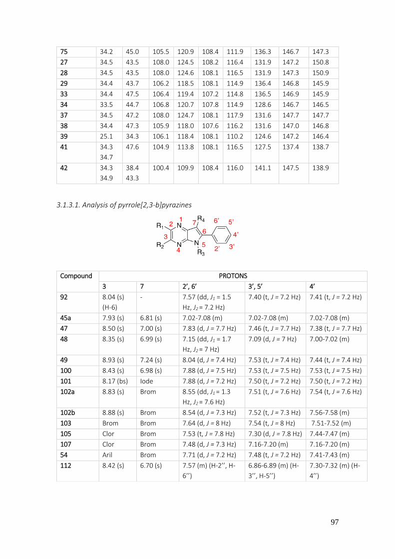
97
75 34.2 45.0 105.5 120.9 108.4 111.9 136.3 146.7 147.3
27 34.5 43.5 108.0 124.5 108.2 116.4 131.9 147.2 150.8
28 34.5 43.5 108.0 124.6 108.1 116.5 131.9 147.3 150.9
29 34.4 43.7 106.2 118.5 108.1 114.9 136.4 146.8 145.9
33 34.4 47.5 106.4 119.4 107.2 114.8 136.5 146.9 145.9
34 33.5 44.7 106.8 120.7 107.8 114.9 128.6 146.7 146.5
37 34.5 47.2 108.0 124.7 108.1 117.9 131.6 147.7 147.7
38 34.4 47.3 105.9 118.0 107.6 116.2 131.6 147.0 146.8
39 25.1 34.3 106.1 118.4 108.1 110.2 124.6 147.2 146.4
41 34.3
34.7
47.6 104.9 113.8 108.1 116.5 127.5 137.4 138.7
42 34.3
34.9
38.4
43.3
100.4 109.9 108.4 116.0 141.1 147.5 138.9
3.1.3.1. Analysis of pyrrole[2,3-b]pyrazines
Compound PROTONS
3 7 2’, 6’ 3’, 5’ 4’
92 8.04 (s)
(H-6)
- 7.57 (dd, J1 = 1.5
Hz, J2 = 7.2 Hz)
7.40 (t, J = 7.2 Hz) 7.41 (t, J = 7.2 Hz)
45a 7.93 (s) 6.81 (s) 7.02-7.08 (m) 7.02-7.08 (m) 7.02-7.08 (m)
47 8.50 (s) 7.00 (s) 7.83 (d, J = 7.7 Hz) 7.46 (t, J = 7.7 Hz) 7.38 (t, J = 7.7 Hz)
48 8.35 (s) 6.99 (s)
7.15 (dd, J1 = 1.7
Hz, J2 = 7 Hz)
7.09 (d, J = 7 Hz) 7.00-7.02 (m)
49 8.93 (s) 7.24 (s) 8.04 (d, J = 7.4 Hz) 7.53 (t, J = 7.4 Hz) 7.44 (t, J = 7.4 Hz)
100 8.43 (s) 6.98 (s) 7.88 (d, J = 7.5 Hz) 7.53 (t, J = 7.5 Hz) 7.53 (t, J = 7.5 Hz)
101 8.17 (bs) Iode 7.88 (d, J = 7.2 Hz) 7.50 (t, J = 7.2 Hz) 7.50 (t, J = 7.2 Hz)
102a 8.83 (s) Brom 8.55 (dd, J1 = 1.3
Hz, J2 = 7.6 Hz)
7.51 (t, J = 7.6 Hz) 7.54 (t, J = 7.6 Hz)
102b 8.88 (s) Brom 8.54 (d, J = 7.3 Hz) 7.52 (t, J = 7.3 Hz) 7.56-7.58 (m)
103 Brom Brom 7.64 (d, J = 8 Hz) 7.54 (t, J = 8 Hz) 7.51-7.52 (m)
105 Clor Brom 7.53 (t, J = 7.8 Hz) 7.30 (d, J = 7.8 Hz) 7.44-7.47 (m)
107 Clor Brom 7.48 (d, J = 7.3 Hz) 7.16-7.20 (m) 7.16-7.20 (m)
54 Aril Brom 7.71 (d, J = 7.2 Hz) 7.48 (t, J = 7.2 Hz) 7.41-7.43 (m)
112 8.42 (s) 6.70 (s) 7.57 (m) (H-2’’, H-
6’’)
6.86-6.89 (m) (H-
3’’, H-5’’)
7.30-7.32 (m) (H-
4’’)
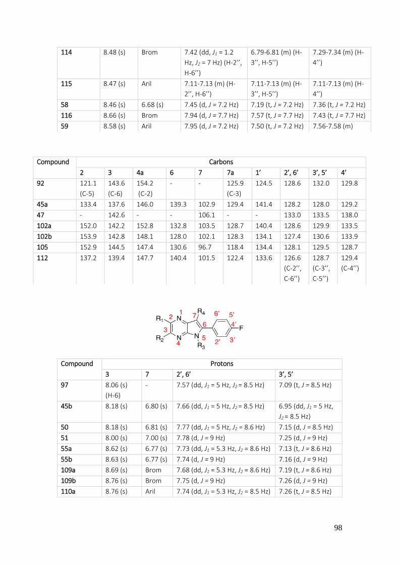
98
114 8.48 (s) Brom 7.42 (dd, J1 = 1.2
Hz, J2 = 7 Hz) (H-2’’,
H-6’’)
6.79-6.81 (m) (H-
3’’, H-5’’)
7.29-7.34 (m) (H-
4’’)
115 8.47 (s) Aril 7.11-7.13 (m) (H-
2’’, H-6’’)
7.11-7.13 (m) (H-
3’’, H-5’’)
7.11-7.13 (m) (H-
4’’)
58 8.46 (s) 6.68 (s) 7.45 (d, J = 7.2 Hz) 7.19 (t, J = 7.2 Hz) 7.36 (t, J = 7.2 Hz)
116 8.66 (s) Brom 7.94 (d, J = 7.7 Hz) 7.57 (t, J = 7.7 Hz) 7.43 (t, J = 7.7 Hz)
59 8.58 (s) Aril 7.95 (d, J = 7.2 Hz) 7.50 (t, J = 7.2 Hz) 7.56-7.58 (m)
Compound Carbons
2 3 4a 6 7 7a 1’ 2’, 6’ 3’, 5’ 4’
92 121.1
(C-5)
143.6
(C-6)
154.2
(C-2)
- - 125.9
(C-3)
124.5 128.6 132.0 129.8
45a 133.4 137.6 146.0 139.3 102.9 129.4 141.4 128.2 128.0 129.2
47 - 142.6 - - 106.1 - - 133.0 133.5 138.0
102a 152.0 142.2 152.8 132.8 103.5 128.7 140.4 128.6 129.9 133.5
102b 153.9 142.8 148.1 128.0 102.1 128.3 134.1 127.4 130.6 133.9
105 152.9 144.5 147.4 130.6 96.7 118.4 134.4 128.1 129.5 128.7
112 137.2 139.4 147.7 140.4 101.5 122.4 133.6 126.6
(C-2’’,
C-6’’)
128.7
(C-3’’,
C-5’’)
129.4
(C-4’’)
Compound Protons
3 7 2’, 6’ 3’, 5’
97 8.06 (s)
(H-6)
- 7.57 (dd, J1 = 5 Hz, J2 = 8.5 Hz) 7.09 (t, J = 8.5 Hz)
45b 8.18 (s) 6.80 (s) 7.66 (dd, J1 = 5 Hz, J2 = 8.5 Hz) 6.95 (dd, J1 = 5 Hz,
J2 = 8.5 Hz)
50 8.18 (s) 6.81 (s) 7.77 (dd, J1 = 5 Hz, J2 = 8.6 Hz) 7.15 (d, J = 8.5 Hz)
51 8.00 (s) 7.00 (s) 7.78 (d, J = 9 Hz) 7.25 (d, J = 9 Hz)
55a 8.62 (s) 6.77 (s) 7.73 (dd, J1 = 5.3 Hz, J2 = 8.6 Hz) 7.13 (t, J = 8.6 Hz)
55b 8.63 (s) 6.77 (s) 7.74 (d, J = 9 Hz) 7.16 (d, J = 9 Hz)
109a 8.69 (s) Brom 7.68 (dd, J1 = 5.3 Hz, J2 = 8.6 Hz) 7.19 (t, J = 8.6 Hz)
109b 8.76 (s) Brom 7.75 (d, J = 9 Hz) 7.26 (d, J = 9 Hz)
110a 8.76 (s) Aril 7.74 (dd, J1 = 5.3 Hz, J2 = 8.5 Hz) 7.26 (t, J = 8.5 Hz)

99
111 7.38 (s) 7.01 (s) 7.23 (d, J = 8 Hz rotamer), 7.64
(dd, J1 = 5 Hz, J2 = 8 Hz), 7.69 (d,
J = 8 Hz rotamer)
7.35 (t, J = 6 Hz)
113 7.49 (s) Brom 7.78 (bs), 7.81 (dd, J1 = 5 Hz, J2
= 8.4 Hz)
7.40 (sc)
56 7.71 (s) Aril 7.57 (d, J = 7 Hz) 7.56 (d, J = 7 Hz)
57 8.63 (bs) Aril 7.54 (dd, J1 = 5.5 Hz, J2 = 8.5
Hz), 7.76-7.80 (m)
7.21 (d, J = 8.5 Hz)
Compound Carbons
2 3 4a 6 7 7a 1’ 2’, 6’ 3’, 5’ 4’
55a 148.3 135.1 145.9 142.2 101.9 127.3 135.2 131.3
(J = 8.2
Hz)
115.9
(J =
21.5
Hz)
163
(J =
248.6
Hz)
109b 149.2 136.6 142.1 137.5 91.8 124.8 127.7 132.8
(J = 9
Hz)
115.7
(J =
22
Hz)
163.7
(J =
254
Hz)
110a 149.1 136.4 148.0 134.8 129.2 115.1 124.9
(J =
3.5
Hz)
132.8
(J = 8.5
Hz)
115.7
(J =
21.7
Hz)
163.0
(J =
249.9
Hz)
113 151.4 120.3 150.0 139.7 98.6 123.7 126.4 131.3
(J = 8.4
Hz)
115.4
(J =
21.5
Hz)
163.8
(J =
250
Hz)
Compound Protons
3 7 2’, 6’ 3’, 5’
98 8.06 (s) (H-6) - 7.50 (d, J = 8.5 Hz) 7.36 (d, J = 8.5 Hz)
45c 8.27 (s) 6.90 (s) 7.66 (d, J = 8.4 Hz) 7.49 (d, J = 8.4 Hz)
52 8.05 (s) 6.91 (s) 7.63 (d, J = 7.7 Hz) 7.22 (d, J = 7.7 Hz)
Compound Carbons
2 3 4a 6 7 7a 1’ 2’, 6’ 3’, 5’ 4’
52 143,7 137,6 141,1 128,5 97,9 120,1 133,0 129,4 129,4 135,6
100
3.2. BIOLOGICAL DISCUSSION Eli Lilly & Company laboratories (Indianapolis, USA) have carried out a biological assays for a
selected compounds described in this doctoral thesis. A high throughput screening for several
biological targets related with different diseases, such as cancer, tropical diseases, autoimmune
diseases and others, was made in order to acquire a potential drugs. Therefore, a review of the
tested compounds and their activities is described hereunder.
It is necessary to highlight that some assays were interrupted along this work due to Eli Lilly
interest. Hence, even one of the aims of this thesis was to evaluate synthetized compounds in
targets like hNNMT (nicotinamide N-methyl transferase), arginase or CD73 (all three with
important roles in MDR), as well as in malaria, chagas or tuberculosis, it was not possible to obtain
all the desired results.
3.2.1. ONCOLOGY
INHIBITION OF NMMT
As it has been described in the introduction of this work memory, the N-nicotinamide N-methyl
transferase (NNMT), cytosolic enzyme that is expressed in the adipose tissue and in the liver,
catalyzes the transfer of methyl groups from the methyl donor (S)-adenosyl-L-methionine (SAM)
to nicotinamide (vitamin B3), generating (S)-adenosyl-L-homocysteine (SAH) and N1-
methylnicotinamide (MNAM). Previous reports have connected the increased expression levels
of NNMT in cancer cells with chemotherapy and radiation resistance as well as with increased
tumor aggressiveness. Metabolic changes driven by activation of oncogenes, inactivation of
tumor suppressors, and protumorigenic mutations are considered a hallmark of cancer. So,
downregulation or silencing of NNMT has been shown to increase the sensitivity of carcinoma
cells to radiation therapy and the decrease of tumorigenicity, providing key support for the role
of NNMT in promoting treatment resistance and tumorigenesis in cancer cells. Thus, compounds
able to modulate the activity of NNMT in the setting oncology may have therapeutic utility.202
NNMT is also implicated in others diseases including obesity, diabetes and neurodegenerative
diseases.203
In order to identify possible NNMT inhibitors, the Open Innovation Drug Discovery (OIDD) from
Eli Lilly carry out an evaluation of the enzymatic modulation of the human NNMT (hNNMT)
through liquid chromatography and mass spectrometry (LC/MS), obtaining as a result the
percentage of inhibited enzyme (Table 11).
202 J. V. Haren; J. S. Toraño; D. Sartini; M. Emanuelli; R. B. Parsons; N. I. Martin. Biochemistry 2016, 55, 5307-5315 203 D. Kraus; Q. Yang; D. Kong; A. S. Banks; L. Zhang; J. T. Rodgers; E. Pirinen. Nature 2014, 508, 258-262

101
Table 11. Determination of hNNMT inhibition
hNNMT INHIBITORS
COMPOUNDS
Primary SP Primary CRC Secondary
NNMT MNAN SP
NNMT SAH SP
NNMT MNAN
CRC
NNMT SAH CRC
NV-NNMT (10 nM) MNAM 120min MS CRC
% Inh. @ 10 µM
% Inh. @ 10 µM
Rel IC50 (µM)
Rel IC50 (µM)
Rel IC50 (µM)
3
2.2 0 - - -
4
-10.6 25.5 -1.2
- - -
5
-11.5
- - - -
6
-1.6
- - - -
7
-3.5
- - - -
10
4.8
- - - -
11
12.5
- - - -
12
29.1
- - - -
15
6.5 24.7 -8.9
- - -

102
17
18.2 -7.9 - - -
18
19.6
27 - - -
19
20.5
0.7 - - -
20
10.5 47.3 8.9
170.1* 192* -
21
-16
3.5 - - -
22
33.8 - - - -
23
14.4
14.9 - - -
24
8.6 - - - -
25
22.4 - - - -
28
- - - - >20*
30
- - - - >20*
33
-5.1 1.6 - - -
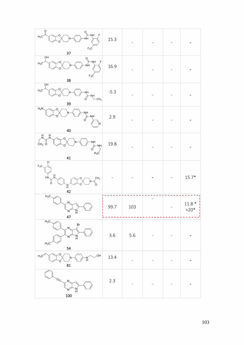
103
37
15.3
- - - -
38
16.9
- - - -
39
-5.3
- - - -
40
2.9
- - - -
41
19.8
- - - -
42
- - - - 15.7*
47
99.7 103
-
- 11.8 * >20*
54
3.6 5.6 - - -
81
13.4
- - - -
100
2.3
- - - -

104
115
- - - - 4.2*
SP: single point. CRC: concentration response curve. * The CRC is showed hereunder
Figure 61. Graphic of the most significant products with their hNNMT inhibitory percentage
Figure 62. Concentration response curve of product 20 in hNNMT inhibition
-20
0
20
40
60
80
100
120
4 11 12 15 18 19 20 22 23 25 47
% in
hib
itio
n
Compounds
NNMT MNAN
NNMT SAH
hNNMT MNAN CRC IC50 = 170 µM
hNNMT SAH CRC IC50 = 191 µM

105
NV-hNNMT (10nM) MNAN 120 min MS CRC IC50 > 20 µM
Figure 63. Concentration response curve of products 28 and 30 in hNNMT inhibition
Figure 64. Concentration response curve of product 42 and 115 in hNNMT inhibition
NV-hNNMT (10nM) MNAN 120 min MS CRC IC50 > 20 µM
NV-hNNMT (10nM) MNAN 120 min MS CRC IC50 = 15 µM
NV-hNNMT (10nM) MNAN 120 min MS CRC IC50 = 4.2 µM

106
NV-hNNMT (10nM) MNAN 120 min MS CRC IC50 > 20 µM
NV-hNNMT (10nM) MNAN 120 min MS CRC IC50 = 11 µM
Figure 65. Concentration response curve of product 47 in hNNMT inhibition
From the results obtained, it can be extracted that the pyrrolopyrazine nucleus allows to obtain
better NNMT inhibitors than the spiranic nucleus. The introduction of a sulfonyl substituent in
spiranic derivatives (compounds 28 and 30) on piperidine nitrogen is less effective than p-
nitrophenyl (20) or acetyl (42). In the other hand, the introduction of a urea function on C-5 is an
effective modification since it leads to compound 42, that has an IC50 = 15 µM. Regarding
pyrrolopyrazines, it seems clear that the phenylacetylene substituent favors the NNMT inhibitory
activity (compare 47 and 115) (Figure 66).
Figure 66. Activity relationships of compounds 20, 28, 30, 42, 47 and 115
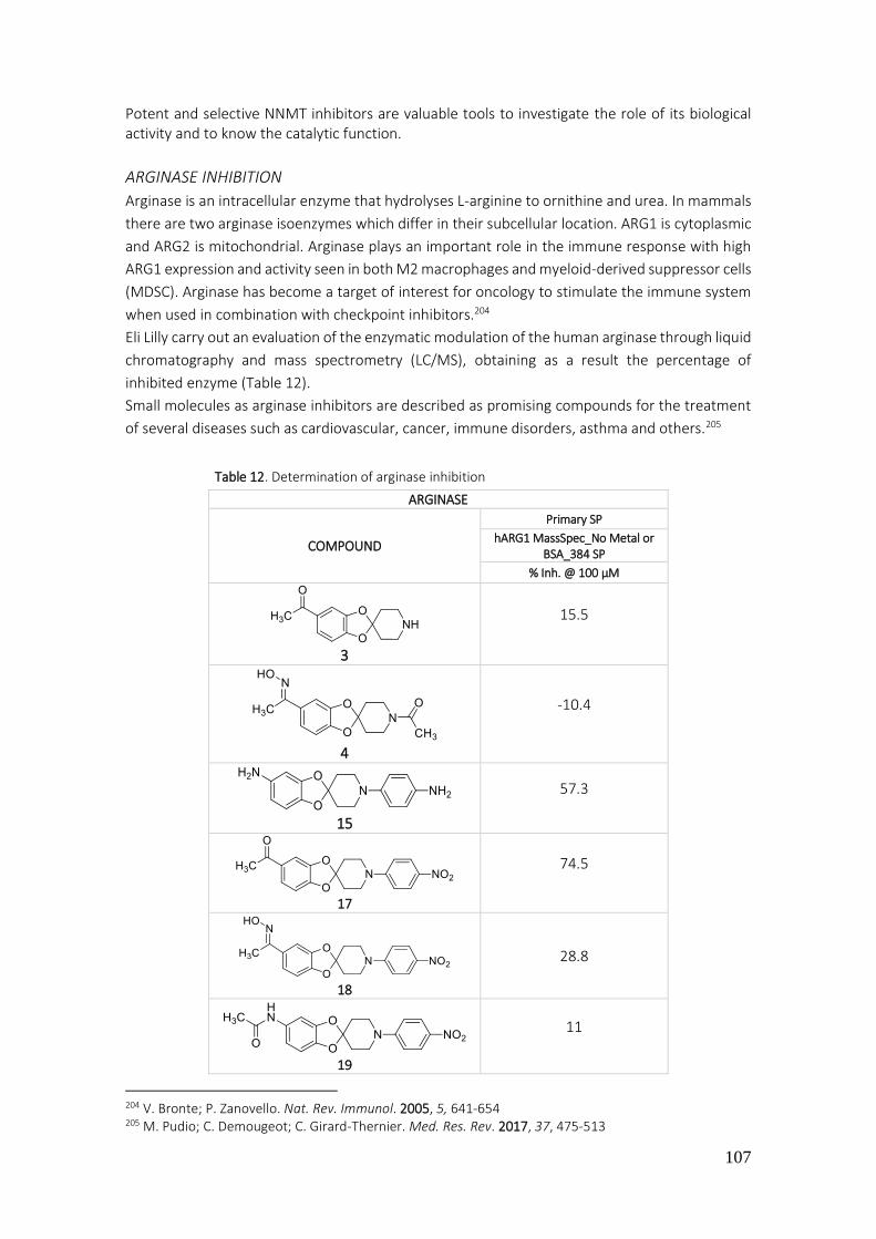
107
Table 12. Determination of arginase inhibition
Potent and selective NNMT inhibitors are valuable tools to investigate the role of its biological activity and to know the catalytic function.
ARGINASE INHIBITION
Arginase is an intracellular enzyme that hydrolyses L-arginine to ornithine and urea. In mammals
there are two arginase isoenzymes which differ in their subcellular location. ARG1 is cytoplasmic
and ARG2 is mitochondrial. Arginase plays an important role in the immune response with high
ARG1 expression and activity seen in both M2 macrophages and myeloid-derived suppressor cells
(MDSC). Arginase has become a target of interest for oncology to stimulate the immune system
when used in combination with checkpoint inhibitors.204
Eli Lilly carry out an evaluation of the enzymatic modulation of the human arginase through liquid
chromatography and mass spectrometry (LC/MS), obtaining as a result the percentage of
inhibited enzyme (Table 12).
Small molecules as arginase inhibitors are described as promising compounds for the treatment
of several diseases such as cardiovascular, cancer, immune disorders, asthma and others.205
204 V. Bronte; P. Zanovello. Nat. Rev. Immunol. 2005, 5, 641-654 205 M. Pudio; C. Demougeot; C. Girard-Thernier. Med. Res. Rev. 2017, 37, 475-513
ARGINASE
COMPOUND
Primary SP
hARG1 MassSpec_No Metal or BSA_384 SP
% Inh. @ 100 µM
3
15.5
4
-10.4
15
57.3
17
74.5
18
28.8
19
11

108
SP: single point
Figure 67. Graphic of the most significant products with their arginase inhibition percentage
The evaluated compounds show a slight arginase inhibitory activity at 100 µM. Compounds 15,
17 and 20 are those with the greatest inhibitory power of the enzyme. The spiranic derivative 17
turns out to be the most powerful in these tests. The simple reduction of the nitro group to amino
leads to a 50% reduction of inhibitory activity (Table 12, compare 17 and 23).
Comparing the results of 17 and 20 it is demonstrated that the change of acetyl group of C-5 by
an amino group leads to a decrease in the inhibitory capacity of arginase.
20
48.5
21
13
23
38.7
33
35.4
0
10
20
30
40
50
60
70
80
3 1 5 1 7 1 8 1 9 2 0 2 1 2 3 3 3
% In
hib
itio
n
Compounds
Arginase inhibition
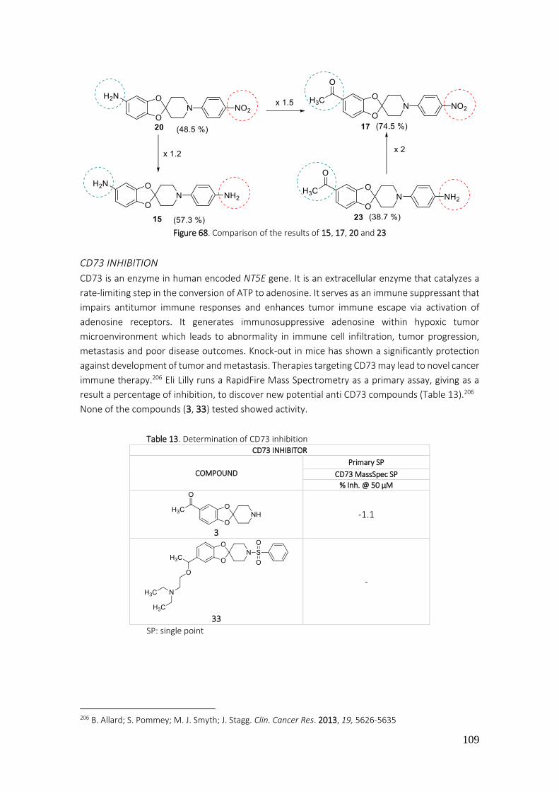
109
Figure 68. Comparison of the results of 15, 17, 20 and 23
CD73 INHIBITION
CD73 is an enzyme in human encoded NT5E gene. It is an extracellular enzyme that catalyzes a
rate-limiting step in the conversion of ATP to adenosine. It serves as an immune suppressant that
impairs antitumor immune responses and enhances tumor immune escape via activation of
adenosine receptors. It generates immunosuppressive adenosine within hypoxic tumor
microenvironment which leads to abnormality in immune cell infiltration, tumor progression,
metastasis and poor disease outcomes. Knock-out in mice has shown a significantly protection
against development of tumor and metastasis. Therapies targeting CD73 may lead to novel cancer
immune therapy.206 Eli Lilly runs a RapidFire Mass Spectrometry as a primary assay, giving as a
result a percentage of inhibition, to discover new potential anti CD73 compounds (Table 13).206
None of the compounds (3, 33) tested showed activity.
Table 13. Determination of CD73 inhibition
SP: single point
206 B. Allard; S. Pommey; M. J. Smyth; J. Stagg. Clin. Cancer Res. 2013, 19, 5626-5635
CD73 INHIBITOR
COMPOUND
Primary SP
CD73 MassSpec SP
% Inh. @ 50 µM
3
-1.1
33
-
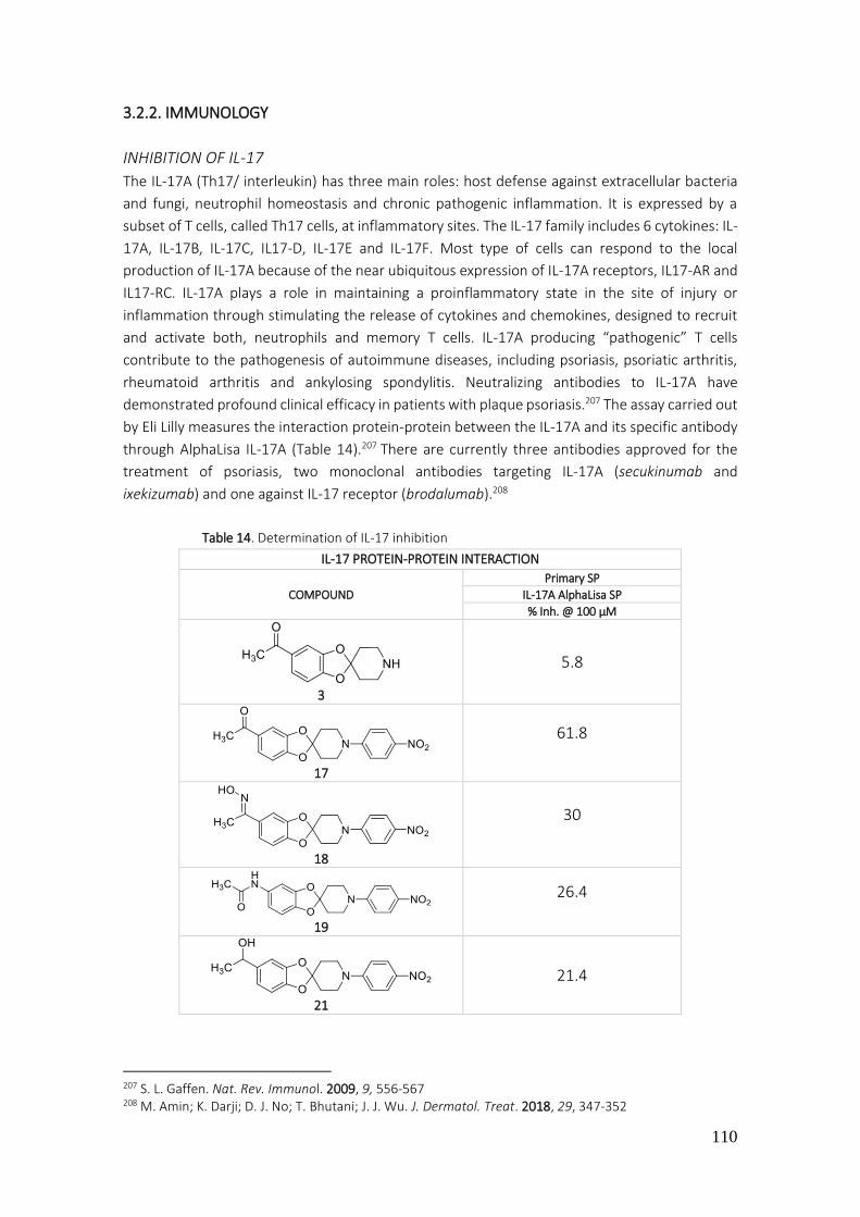
110
3.2.2. IMMUNOLOGY
INHIBITION OF IL-17
The IL-17A (Th17/ interleukin) has three main roles: host defense against extracellular bacteria
and fungi, neutrophil homeostasis and chronic pathogenic inflammation. It is expressed by a
subset of T cells, called Th17 cells, at inflammatory sites. The IL-17 family includes 6 cytokines: IL-
17A, IL-17B, IL-17C, IL17-D, IL-17E and IL-17F. Most type of cells can respond to the local
production of IL-17A because of the near ubiquitous expression of IL-17A receptors, IL17-AR and
IL17-RC. IL-17A plays a role in maintaining a proinflammatory state in the site of injury or
inflammation through stimulating the release of cytokines and chemokines, designed to recruit
and activate both, neutrophils and memory T cells. IL-17A producing “pathogenic” T cells
contribute to the pathogenesis of autoimmune diseases, including psoriasis, psoriatic arthritis,
rheumatoid arthritis and ankylosing spondylitis. Neutralizing antibodies to IL-17A have
demonstrated profound clinical efficacy in patients with plaque psoriasis.207 The assay carried out
by Eli Lilly measures the interaction protein-protein between the IL-17A and its specific antibody
through AlphaLisa IL-17A (Table 14).207 There are currently three antibodies approved for the
treatment of psoriasis, two monoclonal antibodies targeting IL-17A (secukinumab and
ixekizumab) and one against IL-17 receptor (brodalumab).208
Table 14. Determination of IL-17 inhibition
207 S. L. Gaffen. Nat. Rev. Immunol. 2009, 9, 556-567 208 M. Amin; K. Darji; D. J. No; T. Bhutani; J. J. Wu. J. Dermatol. Treat. 2018, 29, 347-352
IL-17 PROTEIN-PROTEIN INTERACTION
COMPOUND
Primary SP
IL-17A AlphaLisa SP
% Inh. @ 100 µM
3
5.8
17
61.8
18
30
19
26.4
21
21.4

111
SP: single point
These compounds 3, 17, 18, 19, 21, 23 and 33 were tested at a concentration of 100 µM and the results of interaction with the protein were moderate. The nitro derivative 17 was the most active with 62% inhibition. The reduction of the nitro to amino group and the reduction of the ketone to alcohol lead to a notable decrease in activity (compare 17 and 23). The other compounds (3, 18, 19, 21 and 33) showed poor activity on this target.
Figure 69. Graphic of the most significant products with their IL-17 inhibition percentage
IL-17 is produced and secreted by at least two classes of lymphocytes, CD4+ T cells (Th17) and .
The IL17 phenotypic drug discovery module seeks to identify inhibitors of IL-17 secretion from
human peripheral blood mononuclear cells (PBMCs) following stimulation of memory T cells by
IL-23, anti-CD3, and anti-CD28.209 Eli Lilly performed an assay to find inhibitor compounds of IL-
17 secretion without overt cytotoxicity and without affecting secretion of INF and IL-5, cytokine
markers for Th1 and Th2 T cell subset, respectively (Table 15).210
209 O. Ouyang; J. K. Kolls; Y. Zheng. Immunity 2008, 28, 454-467 210 https://openinnovation.lilly.com (20/12/2018)
23
39.6
33
6.7
0
10
20
30
40
50
60
70
3 1 7 1 8 1 9 2 1 2 3 3 3
% In
hib
itio
n
Compounds
IL-17 inhibition at 100 µM
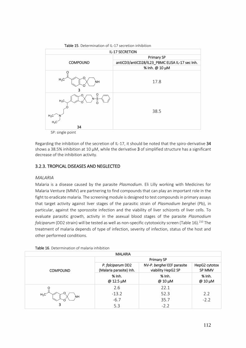
112
Table 15. Determination of IL-17 secretion inhibition
SP: single point
Regarding the inhibition of the secretion of IL-17, it should be noted that the spiro-derivative 34 shows a 38.5% inhibition at 10 µM, while the derivative 3 of simplified structure has a significant decrease of the inhibition activity.
3.2.3. TROPICAL DISEASES AND NEGLECTED
MALARIA
Malaria is a disease caused by the parasite Plasmodium. Eli Lilly working with Medicines for
Malaria Venture (MMV) are partnering to find compounds that can play an important role in the
fight to eradicate malaria. The screening module is designed to test compounds in primary assays
that target activity against liver stages of the parasitic strain of Plasmodium berghei (Pb), in
particular, against the sporozoite infection and the viability of liver schizonts of liver cells. To
evaluate parasitic growth, activity in the asexual blood stages of the parasite Plasmodium
falciparum (DD2 strain) will be tested as well as non-specific cytotoxicity screen (Table 16).210 The
treatment of malaria depends of type of infection, severity of infection, status of the host and
other performed conditions.
Table 16. Determination of malaria inhibition
IL-17 SECRETION
COMPOUND
Primary SP
antiCD3/antiCD28/IL23_PBMC ELISA IL-17 sec Inh.
% Inh. @ 10 µM
3
17.8
34
38.5
MALARIA
COMPOUND
Primary SP
P. falciparum DD2 (Malaria parasite) Inh.
NV-P. berghei EEF parasite viability HepG2 SP
HepG2 cytotox SP MMV
% Inh. @ 12.5 µM
% Inh. @ 10 µM
% Inh. @ 10 µM
3
2.6 -13.2 -6.7 5.3
22.1 52.3 35.7 -2.2
2.2 -2.2

113
SP: single point
Of the two products evaluated for the treatment of malaria, compound 33 shows better activity than derivative 3. Structural simplification, in this case, has reduced antimalarial activity. Regarding the obtained results, 33 reduces the viability of P. berghei more than that of P. falciparum. It should be noted that 33 has an interesting antimalarial activity and minimal toxicity. These results lead to 33 being taken as a model for the design of new compounds.
Figure 70. Graphic of the malaria average inhibitory percentage for products 3 and 33
CHAGAS
Eli Lilly performs a growth inhibition (in NIH-3T3 cells), a trypomastigote luciferin-based and a
cytotoxicity test as a primary assay. Then, T. cruzi CYP51 inhibition and T. cruzi intracellular
Imaging Rate of Kill (RoK) kinetic as a secondary assay (Table 17 and 18).211
211 J. A. Padilla; I. Cotillo; J. Presa; J. Cantizani; I. Peña; A. I. Bardera; J. J. Martín; A. Rodriguez. PLoS Negl. Trop. Dis. 2015, 9, e0003493
33
10.4 51.4 19.3 65.3
87.7 89.5 82
87.2
12.9 4.4
-20
0
20
40
60
80
100
3 33
% in
hib
itio
n
Compounds
P. falciparum
P. berghei
Hepatic cytotoxicity
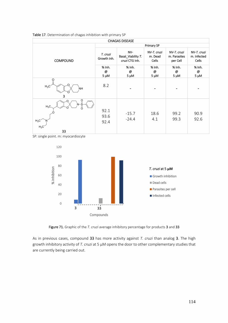
114
Table 17. Determination of chagas inhibition with primary SP
SP: single point. m: myocardiocyte
Figure 71. Graphic of the T. cruzi average inhibitory percentage for products 3 and 33
As in previous cases, compound 33 has more activity against T. cruzi than analog 3. The high
growth inhibitory activity of T. cruzi at 5 µM opens the door to other complementary studies that
are currently being carried out.
CHAGAS DISEASE
COMPOUND
Primary SP
T. cruzi Growth Inh.
NV-Basal_Viability T.
cruzi CTG Inh.
NV-T. cruzi m. Dead
Cells
NV-T. cruzi m. Parasites
per Cell
NV-T. cruzi m. Infected
Cells
% Inh. @
5 µM
% Inh. @
5 µM
% Inh. @
5 µM
% Inh. @
5 µM
% Inh. @
5 µM
3
8.2
- - - -
33
92.1 93.6 92.4
-15.7 -24.4
18.6 4.1
99.2 99.3
90.9 92.6
0
20
40
60
80
100
120
% in
hib
itio
n
Compounds
T. cruzi at 5 µM
Growth inhibition
Dead cells
Parasites per cell
Infected cells
3 33
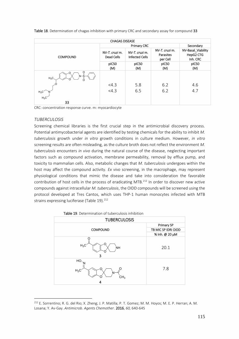
115
Table 18. Determination of chagas inhibition with primary CRC and secondary assay for compound 33
CRC: concentration response curve. m: myocardiocyte
TUBERCULOSIS
Screening chemical libraries is the first crucial step in the antimicrobial discovery process.
Potential antimycobacterial agents are identified by testing chemicals for the ability to inhibit M.
tuberculosis growth under in vitro growth conditions in culture medium. However, in vitro
screening results are often misleading, as the culture broth does not reflect the environment M.
tuberculosis encounters in vivo during the natural course of the disease, neglecting important
factors such as compound activation, membrane permeability, removal by efflux pump, and
toxicity to mammalian cells. Also, metabolic changes that M. tuberculosis undergoes within the
host may affect the compound activity. Ex vivo screening, in the macrophage, may represent
physiological conditions that mimic the disease and take into consideration the favorable
contribution of host cells in the process of eradicating MTB.212 In order to discover new active
compounds against intracellular M. tuberculosis, the OIDD compounds will be screened using the
protocol developed at Tres Cantos, which uses THP-1 human monocytes infected with MTB
strains expressing luciferase (Table 19).212
Table 19. Determination of tuberculosis inhibition
212 E. Sorrentino; R. G. del Rio; X. Zheng; J. P. Matilla; P. T. Gomez; M. M. Hoyos; M. E. P. Herran; A. M. Losana; Y. Av-Gay. Antimicrob. Agents Chemother. 2016, 60, 640-645
CHAGAS DISEASE
COMPOUND
Primary CRC Secondary
NV-T. cruzi m. Dead Cells
NV-T. cruzi m. Infected Cells
NV-T. cruzi m. Parasites per Cell
NV-Basal_Viability HepG2 CTG
Inh. CRC
pIC50 (M)
pIC50 (M)
pIC50 (M)
pIC50 (M)
33
<4.3 <4.3
5.8 6.5
6.2 6.2
4.6 4.7
TUBERCULOSIS
COMPOUND
Primary SP
TB MIC SP IDRI OIDD
% Inh. @ 20 µM
3
20.1
4
7.8

116
5
6.3
7
3.8
10
6.8
11
13.2
12
-2.7
15
-2.1
17
7.7 -1.8
18
6.6 1.7
19
23.6 5.1
20
1.6
21
18.3 12.7

117
22
-4.3
23
5.6 -1
24
2
25
5.9
33
32.4 19.2
37
10.2
38
6.3
39
2.7
41
1.7
46
-3.3

118
0
5
10
15
20
25
30
3 11 19 21 33 37
% in
hib
itio
n
Compounds
Tuberculosis inhibitionat 20 µM
SP: single point. MIC: minimum inhibitory concentration
Figure 72. Graphic of the most significant products with their tuberculosis inhibition percentage
The results of growth inhibitory M. tuberculosis at 20 µM of the evaluated compounds show a
low to moderate activity. In this case, compound 33 is again the one that shows the best activity.
The pyrrolopyrazines show almost no activity against M. tuberculosis.
47
6.5
54
-4.4
81
-8.3
100
2
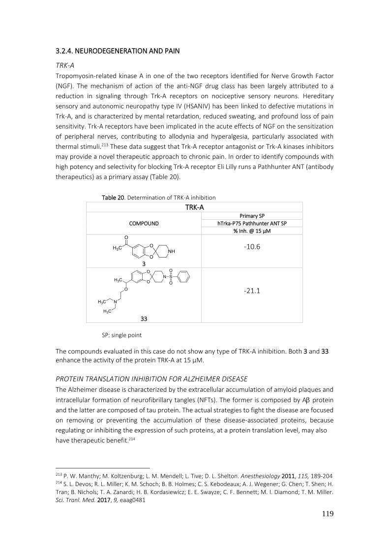
119
3.2.4. NEURODEGENERATION AND PAIN
TRK-A
Tropomyosin-related kinase A in one of the two receptors identified for Nerve Growth Factor
(NGF). The mechanism of action of the anti-NGF drug class has been largely attributed to a
reduction in signaling through Trk-A receptors on nociceptive sensory neurons. Hereditary
sensory and autonomic neuropathy type IV (HSANIV) has been linked to defective mutations in
Trk-A, and is characterized by mental retardation, reduced sweating, and profound loss of pain
sensitivity. Trk-A receptors have been implicated in the acute effects of NGF on the sensitization
of peripheral nerves, contributing to allodynia and hyperalgesia, particularly associated with
thermal stimuli.213 These data suggest that Trk-A receptor antagonist or Trk-A kinases inhibitors
may provide a novel therapeutic approach to chronic pain. In order to identify compounds with
high potency and selectivity for blocking Trk-A receptor Eli Lilly runs a Pathhunter ANT (antibody
therapeutics) as a primary assay (Table 20).
Table 20. Determination of TRK-A inhibition
SP: single point
The compounds evaluated in this case do not show any type of TRK-A inhibition. Both 3 and 33 enhance the activity of the protein TRK-A at 15 µM.
PROTEIN TRANSLATION INHIBITION FOR ALZHEIMER DISEASE
The Alzheimer disease is characterized by the extracellular accumulation of amyloid plaques and
intracellular formation of neurofibrillary tangles (NFTs). The former is composed by A protein
and the latter are composed of tau protein. The actual strategies to fight the disease are focused
on removing or preventing the accumulation of these disease-associated proteins, because
regulating or inhibiting the expression of such proteins, at a protein translation level, may also
have therapeutic benefit.214
213 P. W. Manthy; M. Koltzenburg; L. M. Mendell; L. Tive; D. L. Shelton. Anesthesiology 2011, 115, 189-204 214 S. L. Devos; R. L. Miller; K. M. Schoch; B. B. Holmes; C. S. Kebodeaux; A. J. Wegener; G. Chen; T. Shen; H. Tran; B. Nichols; T. A. Zanardi; H. B. Kordasiewicz; E. E. Swayze; C. F. Bennett; M. I. Diamond; T. M. Miller. Sci. Tranl. Med. 2017, 9, eaag0481
TRK-A
COMPOUND
Primary SP
hTrka-P75 Pathhunter ANT SP
% Inh. @ 15 µM
3
-10.6
33
-21.1
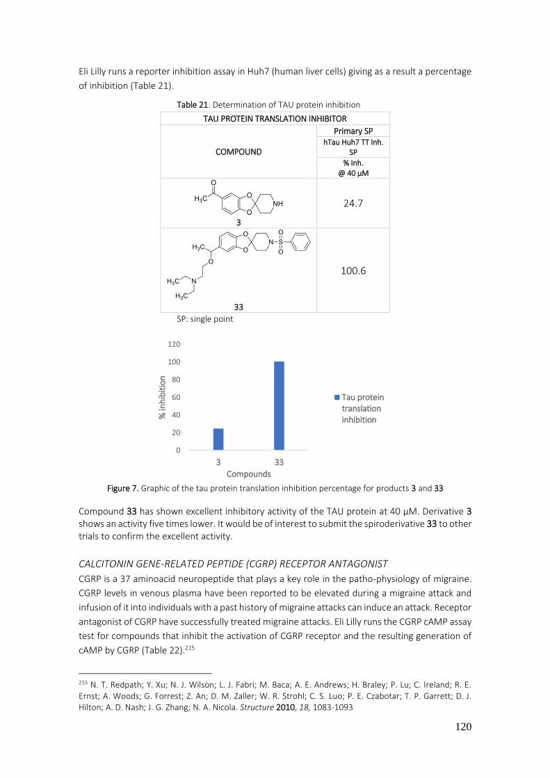
120
Eli Lilly runs a reporter inhibition assay in Huh7 (human liver cells) giving as a result a percentage
of inhibition (Table 21).
Table 21. Determination of TAU protein inhibition
TAU PROTEIN TRANSLATION INHIBITOR
COMPOUND
Primary SP hTau Huh7 TT Inh.
SP
% Inh. @ 40 µM
3
24.7
33
100.6
SP: single point
Figure 7. Graphic of the tau protein translation inhibition percentage for products 3 and 33
Compound 33 has shown excellent inhibitory activity of the TAU protein at 40 µM. Derivative 3 shows an activity five times lower. It would be of interest to submit the spiroderivative 33 to other trials to confirm the excellent activity.
CALCITONIN GENE-RELATED PEPTIDE (CGRP) RECEPTOR ANTAGONIST
CGRP is a 37 aminoacid neuropeptide that plays a key role in the patho-physiology of migraine.
CGRP levels in venous plasma have been reported to be elevated during a migraine attack and
infusion of it into individuals with a past history of migraine attacks can induce an attack. Receptor
antagonist of CGRP have successfully treated migraine attacks. Eli Lilly runs the CGRP cAMP assay
test for compounds that inhibit the activation of CGRP receptor and the resulting generation of
cAMP by CGRP (Table 22).215
215 N. T. Redpath; Y. Xu; N. J. Wilson; L. J. Fabri; M. Baca; A. E. Andrews; H. Braley; P. Lu; C. Ireland; R. E. Ernst; A. Woods; G. Forrest; Z. An; D. M. Zaller; W. R. Strohl; C. S. Luo; P. E. Czabotar; T. P. Garrett; D. J. Hilton; A. D. Nash; J. G. Zhang; N. A. Nicola. Structure 2010, 18, 1083-1093
0
20
40
60
80
100
120
3 33
% in
hib
itio
n
Compounds
Tau proteintranslationinhibition

121
Table 22. Determination of CGRP inhibition
CGRP ANTAGONIST
COMPOUND Primary SP Primary CRC
hCGRP1 cAMP hCGRP1 cAMP
% Inh. @ 30 µM IC50 (µM) Kb (µM)
3
-80.6
- -
33
76.3 >30
10.7* 7570.1 2690.7
SP: single point. CRC: concentration response curve. * The CRC is showed hereunder
Figure 74. Concentration response curve of product 33 in hCGRP1 inhibition
It is repeated again that compound 33 has an interesting activity profile, in this case it is an antagonist activity on the receptors of CGRP.
NAV1.7 ANTAGONIST
Nav1.7 is encoded by the SCN9A gene and is one of nine currently described voltage-gated
sodium ion channels. All sodium channels in this family are involved in action potential
generation, propagation, and/or neurotransmitter release in excitable tissues. Interest in Nav1.7
as a potential therapeutic target for pain comes from several converging lines of evidence.
Patients with activating or gain function mutations in Nav1.7 display varying degrees of chronic
and intermittent pain.216 Together, the efficacy of nonselective sodium channel inhibitors
combined with the wealth of genetic information strongly suggest that selective NAv1.7 inhibitors
may provide a novel therapeutic approach to chronic pain of different etiologies without
significant safety/tolerability side-effects. Hence, Eli Lilly performs an Ion Works Quattro
electrophoresis assay in HEK293 cell line (from human embryonic kidney) and a target based
216 S. D. Dib-Hajj; Y. Yang; J. A. Black; S. G. Waxman. Nat. Rev. Neurosci. 2013, 14, 49-62
hCGRP1 cAMP IC50 = 10.7 µM

122
inhibitory assay in Huh7 cell line (human hepatocyte-derived carcinoma), to identify compounds
with high potency and selectivity for blocking Nav1.7 channels (Table 23).210
Table 23. Determination of NAV 1.7. inhibition
SP: single point
Figure 75. Graphic of the Nav 1.7 inhibition percentage for products 3 and 33
KCNQ2/3 AGONIST KCNQ2/3 is a heteromeric voltage-gated potassium channel. Together with other members of
the Kv7 subfamily of voltage-gated potassium channels, KCNQ2/3 underlies the neuronal M-
current that modulates excitability in many neurons of the central and peripheral nervous system.
It is exclusively expressed in the nervous system, and it is the predominant subtype in pain sensing
neurons in the dorsal root ganglion (DRG) and spinal cord. Agonist of KCNQ2/3 hyperpolarize
sensory neurons, reduce excitability and show efficacy in rodent models of persist pain. The
fluorescence-based thallium flux FLIPR assay carried by Eli Lilly measures functional activity of
ligand and voltage-gated potassium channels in a HEK293 cell type (Table 24).217
217 X. Du; H. Hao; S. Gigout; D. Huang; Y. Yang; L. Li; C. Wang; D. Sundt; D. B. Jaffe; H. Zhang; N. Gamper. Pain 2014, 155, 2306-2322
NAV 1.7. ANTAGONIST
COMPOUND Primary SP
hNav1.7 IWQ hNav1.7 Huh7 TT Inh. SP
% Inh. @ 3 µM % Inh. @ 40 µM
3
9.7
25.6
33
40.6
98.2
0
20
40
60
80
100
120
3 33
% in
hib
itio
n
Compounds
Nav 1.7 inhibition IWQ
Nav 1.7 inhibition Huh7

123
Table 24. Determination of KCNQ2-3 stimulation
SP: single point
None of the evaluated compounds have shown stimulatory activity of KCNQ2/3. The results indicate, in some cases, a slight inhibitory activity.
3.2.5. ENDOCRINE/CARDIOVASCULAR
GLP-1 SECRETION
Glucagon like peptide 1 is derived from transcription of the proglucagon gene. Its secretion by
ileal L cells is dependent on the presence of nutrients in the lumen of the small intestine. It is a
potent anti-hyperglycemic hormone inducing glucose-dependent insulin secretion and
suppressing glucagon secretion. When the glucose in the plasma is in the fasting range stage, the
KCNQ2-3 AGONIST
COMPOUND Primary SP
hKCNQ2/3 FluxOR Ag SP
% Stim @ 10 µM
4
-6.7 5
15
-5.4 2
17
-3 -1.4
18
-1.1 6.5
19
-2.6 -2.5
20
-1.6 -8.2
21
-2.9 -5.9
23
-2.3 18.2

124
GLP-1 does not stimulate the secretion of insulin. Diabetes phenotypic module identifies
compounds that stimulate secretion of GLP-1 in mouse and human cell lines derived from
gastrointestinal tract tissue.210 GLP-1 secretion is measured by Eli Lilly using ELISA assay that was
specifically designed to detect the appropriate forms of GLP-1 secreted from these cells (Table
25).
Table 25. Determination of GLP-1 stimulation
SP: single point
PCSK9 INHIBITION
Proprotein convertase subtilisin kexin type 9 (PCSK9) belongs to the proteinase K subfamily of
secretory proteases. This protein plays a major regulatory role in cholesterol homeostasis. It
regulates plasma LDL-cholesterol (LDL-C) levels by directing LDL receptor (LDLR) to lysosomal
degradation, resulting in reduced LDL clearance and accumulation of LDL in circulation. So, gain
function mutations of PCSK9 lead to hyperlipidemia and premature coronary artery disease (CAD)
in humans, whereas loss of function mutations of PCSK9 are associated with lower levels of LDL
and protection from CAD. Therapeutic antibodies are demonstrating impressive efficacy in LDL
lowering either as a monotherapy or combination therapy with statins. After this strong clinical
validation, small molecule inhibitors of PCSK9 are being actively sought. PCSK9 protein expression
is actively regulated at transcription and post transcriptional levels. The PCSK9 phenotypic
module is designed to identify compounds that inhibit expression of PCSK9 protein multiple cell
systems, including human hepatoma cell lines (Huh7), HeLa cells, and primary human
hepatocytes. Eli Lilly runs an Alphalisa and a CellTiter-Glo® luminescent cell viability assay in Huh7
cell lines as a primary assay to identify possible inhibitors (Table 26 and 27).218
218 S. Crunkhorn. Nat. Rev. Drug Discov. 2012, 11, 189-189
GLP-1 SECRETION
COMPOUND
Primary SP
mSTC-1 GLP1 Sec SP hNCI-H716 GLP1 Sec SP
% Stim @ 20 µM
% Stim @ 2 µM
% Stim @ 20 µM
% Stim @ 2 µM
3
-0.8 -3.5 0.1 -0.4
33
-5.2 -0.9 3.5 0.2

125
Table 26. Determination of PCSK-9 inhibition
SP:
single point
Table 27. Determination of PCSK-9 inhibition
SP: single point
PCSK9 Inhibition
COMPOUND
Primary SP
PCSK9 AlphaLisa Huh7 SP
PCSK9 Huh7 Viability CellTiter-Glo SP
% Inh. @ 5 µM % Inh. @ 5 µM
17
-6.7 6
18
-1.2 18.4
19
9.2 7.7
20
1.2 17
23
-74.2 0.3
PCSK9 INHIBITOR (EFFORT-1)
COMPOUND
Primary SP Primary CRC Confirmatory
Basal_PCSK9 HepG2
SP
Basal_PCSK9 HepG2 Cell Health
Basal_PCSK9 HepG2 Cell Health
Metridia luc HepG2
Non Specific Secretion Inh.
PCSK9 ELISA RPH SP
% Inh. @ 5 µM
% Inh. @ 5 µM
IC50 (µM)
IC50 (µM)
% Inh. @ 100 µM
% Inh. @ 20 µM
3
42.3 -0.7 >50 >50 4.5
-22.1
-5.7 -0.8 -9.6 1.6
33
-80.7 0.79 - - - -

126
3.2.6. ELANCO ANIMAL HEALTH
Animal welfare is highly compromised by parasites. Because of the growing issue about the
resistance to existing antiparasitic treatments the options are limited. Synergic with human
welfare exist due to zoonotic potential and overlapping spectrums with human diseases. Eli Lilly
performs an assay with the whole organism motility phenotypic screening that enables the quick
and highly relevant assessment of antiparasitic potential (Table 28).210Error! No s'ha definit el marcador.
Table 28. Determination of antiparasitic potential
SP: single point
Finally, these last tests show, in most of the cases, results with a lower tan per cent of inhibition, so even they are not significant to continue its studies in such targets, the results have been included in order to highlight the importance of relationship between structure-activity, a preliminary activity that helps to continue our work.
ELANCO HEALTH ANIMAL
COMPOUND
Primary SP Primary CRC
NV-D. immitis Mobility Inh. SP Elanco
NV-H. contortus Mobility
Inh. SP Elanco
NV-A. aegypti Mobility Inh. SP Elanco
NV-A. aegypti
Mobility Inh. CRC Elanco
%Inh. @ 5 µM
%Inh. @ 5 µM
%Inh. @ 8 µM
%Inh. @ 10 µM
%Inh. @ 5 µM
Rel IC50 (µM)
4
-4 0
10 -1
- 2 -5
- -
15
-1 0
11 1
- 1 3
- -
20
0 0
7 1
- 14 56
- -
33
32 34
- 34 31
-4 -5
-11 69
>10 >10
127
3.3. DISCOVERING NEW TARGETS- Quantitative Biology in Eli Lilly & Company The work accomplished in Eli Lilly & Company was focused on a family of drug targets known as
G-protein coupled receptors (GPCRs). An untapped subfamily of GPCRs is exemplified by the
33 adhesion receptors that exist in mammals.219 This adhesion family of GPCRs is considered
‘dark’ due to the fact that very few small or large molecule ligands are known for these
proteins. This is primarily due to a lack of assay approaches to investigate their cellular
pharmacology as commented before in the introduction of this work.
GPCRs are found ubiquitously in the human body. GPCRs are important drug targets to attenuate
inflammation, cancer, cardiac dysfunction, diabetes, and others.220,221 Therefore, it is important
to describe and summarize their structure, mechanisms, roles and other important concepts as
a basis for understanding the GPCRs experiments conducted.
3.3.1. G-PROTEIN COUPLED RECEPTORS (GPCRs)
The GPCRs, also called seven transmembrane receptors, are proteins located in the cell
membrane, coupled to heterotrimeric G proteins.221 The GPCRs are the largest and most diverse
membrane receptors in eukaryotes. The exact size of the GPCR superfamily in humans is
unknown,222 but from genome sequence analysis is deduced that at least 810 human genes
encoding GPCRs exist.
The existence of GPCRs was demonstrated along the twentieth century by several scientist such
as Robert J. Lefkowitz, Martin Rodbell, Alfred G. Gilman, amongst others.223,224
STRUCTURE
GPCRs have a conserved tertiary structure: seven α-helices form transmembrane domains, acting
as a recognition and connection site and allowing extracellular ligands to modulate intracellular
physiology. GPCRs canonically interact with heterotrimeric G-proteins, which are composed by
three subunits: alpha, beta and gamma. Physiologically, several ligands bind these receptors:
peptide hormones, neurotransmitters, lipids, nucleotides, ions, varying from small molecules to
peptides and proteins. Also, the sensation of external stimuli as odor, light or taste is mediated
through them. Those receptors with unknown ligand are named as orphan.225
Once the receptor has been activated by an agonist ligand it acts as a guanine-nucleotide
exchange factor, producing a conformational change in the associated G protein, specifically in
α-subunit, leading to release of GDP subsequent GTP binding on this subunit. Thereafter the -
subunit-GTP is disassociated from the receptor and from the βγ-dimer, and both subunits are
able to modulate several intracellular signal transduction pathways.226
219 T. Xiao-long; W. Ying; L. Da-li; L. Jian; L. Ming-yao. Acta Pharmacol. Sin. 2012, 33, 363–371 220 J. Wang; G. Clarice; A. H. Rockman. Circ. Res. 2018, 123, 716–735 221 S. P. Alexander; A. Christopoulos. Br. J. Pharmacol. 2019, 176, 21–141 222 https://www.guidetopharmacology.org (20/05/2019) 223 https://www.britannica.com (20/05/2019) 224 M. Rodbell. Environ Health Perspect. 1995, 103, 338–345 225 V. V. Gurevich; E. V. Gurevich. Int. J. Mol. Sci. 2017, 18, e2519 226 C. J. Folts; S. Giera; T. Li; X. Piao. Trends Pharmacol. Sci. 2019, 40, 278-293
128
Based on these features, many GPCRs have been detected in the human genome and many of
them are well described in the literature (distribution in human body, ligands, functions and
others) and also their roles in diseases. For example, beta-adrenergic receptors227 and their
antagonist (i.e. bisoprolol) for the treatment of hypertension and cardiac diseases.228 GPCRs are
involved in Systems as diverse as neurotransmitter and hormonal signaling.
CLASSIFICATION
The GPCR superfamily has been sub-divided into classes based on evolutionary homology and
with common physiological ligands.
The G protein-coupled receptors (GPCRs) consist in five main families in mammal:229
· Rhodopsin family (class A): 284 members (plus 380 olfactory receptors)
· Secretin family (class B1): 15 members
· Adhesion GPCR (class B2): 33 members
· Glutamate family (class C): 22 members
· Frizzled family: 11 members
GPCRs IN DRUG DISCOVERY
GPCRs, due to their wide role in the human body regulating diverse physiological processes and
due to their accessible ligand binding sites in the cell membrane have been a point of interest as
pharmacological targets. In fact, the GPCRs are the most studied drug targets because of their
relevant role in human pathophysiology and their understood and reproduceable pharmacology.
It is claimed that 25-50% of approved drugs target GPCRs in different diseases such as cancer,
endocrinology, psychiatric conditions, cardiovascular pathologies, among others (Table 29).230
The untapped potential of the drug-discovery in the GPCR field is thought to be vast.
Table 29. Few examples of drugs in different diseases with GPCRs as a targets
R: receptor
227 T. Warne; R. Moukhametzianov; J. G. Baker; R. Nehmé; P. C. Edwards; A. G. Leslie; Nature 2011, 469, 241-244 228 J. P. Vilardaga; M. Búnemann; T. N. Feinstein; N. Lambert; V. O. Nikolaev; S. Engelhardt; M. J. Lohse; C. Hoffmann. Mol. Endocrinol. 2009, 23, 590-599 229 O. Civelli; R. K. Reinscheid; Y. Zhang; Z. Wang; R. Fredriksson; H. B. Schiöth. Annu. Rev. Pharmacol. Toxicol. 2013, 53, 127-146 230 A. S. Hauser; M. M. Attwood; M. Rask-Andersen; H. B. Schiöth; D. E. Gloriam. Nat. Rev. Drug. Discov. 2017, 6, 829-842
Substance Tradename Indications Targets Approval year
Propranolol Inderal Cardiovascular diseases β-adrenergic R 1965
Cimetidine Tagamet Heartburn Histamine H2 R 1977
Clozapine Clozaril Antipsychotic 5-HT2A 1972
Dulaglutide Trulicity Type 2 diabetes GLP1R 2014
Suvorexant Belsomra Insomnia OX2R and OX1R 2015
Aripiprazole Aristada Schizophrenia 5HT1A, 5HT2A 2015
Sonidegib Odomzo Basal cell carcinoma SMO 2015
Abaloparatide Tymlos Osteoporosis PTHR1 2017
Etelcalcetide Parsabiv Hyperparathyroidism CASR 2017
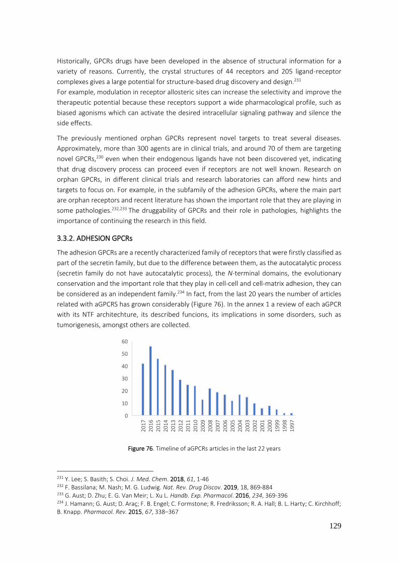
129
Historically, GPCRs drugs have been developed in the absence of structural information for a
variety of reasons. Currently, the crystal structures of 44 receptors and 205 ligand-receptor
complexes gives a large potential for structure-based drug discovery and design.231
For example, modulation in receptor allosteric sites can increase the selectivity and improve the
therapeutic potential because these receptors support a wide pharmacological profile, such as
biased agonisms which can activate the desired intracellular signaling pathway and silence the
side effects.
The previously mentioned orphan GPCRs represent novel targets to treat several diseases.
Approximately, more than 300 agents are in clinical trials, and around 70 of them are targeting
novel GPCRs,230 even when their endogenous ligands have not been discovered yet, indicating
that drug discovery process can proceed even if receptors are not well known. Research on
orphan GPCRs, in different clinical trials and research laboratories can afford new hints and
targets to focus on. For example, in the subfamily of the adhesion GPCRs, where the main part
are orphan receptors and recent literature has shown the important role that they are playing in
some pathologies.232,233 The druggability of GPCRs and their role in pathologies, highlights the
importance of continuing the research in this field.
3.3.2. ADHESION GPCRs
The adhesion GPCRs are a recently characterized family of receptors that were firstly classified as
part of the secretin family, but due to the difference between them, as the autocatalytic process
(secretin family do not have autocatalytic process), the N-terminal domains, the evolutionary
conservation and the important role that they play in cell-cell and cell-matrix adhesion, they can
be considered as an independent family.234 In fact, from the last 20 years the number of articles
related with aGPCRS has grown considerably (Figure 76). In the annex 1 a review of each aGPCR
with its NTF architechture, its described funcions, its implications in some disorders, such as
tumorigenesis, amongst others are collected.
Figure 76. Timeline of aGPCRs articles in the last 22 years
231 Y. Lee; S. Basith; S. Choi. J. Med. Chem. 2018, 61, 1-46 232 F. Bassilana; M. Nash; M. G. Ludwig. Nat. Rev. Drug Discov. 2019, 18, 869-884 233 G. Aust; D. Zhu; E. G. Van Meir; L. Xu L. Handb. Exp. Pharmacol. 2016, 234, 369-396 234 J. Hamann; G. Aust; D. Araç; F. B. Engel; C. Formstone; R. Fredriksson; R. A. Hall; B. L. Harty; C. Kirchhoff; B. Knapp. Pharmacol. Rev. 2015, 67, 338–367

130
The subfamilies of the adhesion GPCRs were decided based on previous phylogenetic
classifications according to their 7TM regions and their extracellular regions.235,236
TAXONOMY
Adhesion GPCRs are found in all vertebrates and high efforts to sequence the genomes of
invertebrates provided a wide dataset to classify aGPCRs. Earlier literature, that looks to
determine aGPCRs in these species, found homologs of select human aGPCR subfamilies.
Additionally, aGPCRs have been identified in fungi, which indicates that aGPCRS were developed
before the split of unikonts (members of a taxonomic group), from the common ancestor of
eukaryotes, about 1275 million years ago.237,238
Another argument to reinforce this idea, is the fact that they are the most complex among GPCRs
in terms of molecular genetics as they have: multiple introns, a complex genomic size, and
multiple functional domains present in other proteins.237,238
NOMENCLATURE
The initial names for the adhesion GPCRs were established by the pioneers of the research field
without a common harmonization, like BAI (brain-specific angiogenesis inhibitor), VLGR (very
large GPCR), and others. Then, with the collaboration of the Human Genome Organization
(HUGO) and the Gene Nomenclature Committee (HGNC), most of the aGPCRs were named after
GPR# names (where GPR stands for G-Protein coupled Receptor).239
All these names were kept as identifiers until a better nomenclature could be devised.
Subsequently, the field has expanded and aGPCRs are being studied in many areas and therefore
common names are needed for researchers.234
The Adhesion GPCR Consortium working with HGNC and the IUPHAR Committee on Receptor
Nomenclature and Drug Classification (NC-IUPHAR) established a prefix that identifies any aGPCR
homolog, independent of the family or even the species. That prefix is ADGR (Adhesion G protein-
coupled receptor). Then, for each family a letter to relate them with the previous names was
assigned (for example, B for the BAIs) and for the subfamilies with receptors called after GPR#,
were recalled with a letter (following alphabetic order, A, D, F, G). Before this agreed, the
subfamilies were indicated with Roman numbers I-IX. Finally, for each member, 33 receptors, a
number was given to each one in a similar order to the previous names (Table 30).
The HGNC as well as the NC-IUPHAR are agreement about the use of this new nomenclature to
promote easier research.240
235 T. K. Bjarnadóttir; K. Geirardsdóttir; M. Ingemansson; M. A. Mirza; R. Fredriksson; H. B. Schiöth. Gene 2007, 387, 38-48 236 M. C. Lagerström; H. B. Schiöth. Nat. Rev. Drug Discov. 2008, 7, 339-357 237 N. Kamesh; G. K. Aradhyam; N. Manoj. BMC Evol. Biol. 2008, 8, 129-129 238 A. Krishnan; M. S. Almén; R. Fredriksson; H. B. Schiöth. PLoS One 2012, 7, e29817 239 R. Fredriksson; M. C. Lagerström; L. G. Lundin; H. B. Schiöth. Mol. Pharmacol. 2003, 63, 1256-1272 240 https://www.adhesiongpcr.org (20/05/2019)

131
Taula 30. Classification of aGPCRs
Subfamilies Proposed names for
subfamilies
Previous gene names Currently accepted
nomenclature
I Latrophilin (L) LPHN1 ADGRL1
LPHN2 ADGRL2
LPHN3 ADGRL3
ELTD1 ADGRL4
II EGF-TM7
(E)
EMR1 ADGRE1
EMR2 ADGRE2
EMR3 ADGRE3
EMR4 ADGRE4
CD97 ADGRE5
III A GPR123 ADGRA1
GPR124 ADGRA2
GPR125 ADGRA3
IV CELSR
(C)
CELSR1 ADGRC1
CELSR2 ADGRC2
CELSR3 ADGRC3
V D GPR133 ADGRD1
GPR144 ADGRD2
VI F GPR110 ADGRF1
GPR111 ADGRF2
GPR113 ADGRF3
GPR115 ADGRF4
GPR116 ADGRF5
VII BAI (B) BAI1 ADGRB1
BAI2 ADGRB2
BAI3 ADGRB3
VIII G GPR56 ADGRG1
GPR64 ADGRG2
GPR97 ADGRG3
GPR112 ADGRG4
GPR114 ADGRG5
GPR126 ADGRG6
GPR128 ADGRG7
IX V GPR98 ADGRV1
BAI: Brain-specific angiogenesis inhibitor, CD: Cluster of differentiation, CELSR: Cadherin EGF LAG seven-pass G-type
receptor, EGF-7TM: epiderma growth factor-seven-span transmembrane, ELTD: EGF, latrophilin, and seven-
transmembrane domain-containing protein, EMR: EGF-like molecule-containing mucin-like hormone receptor.
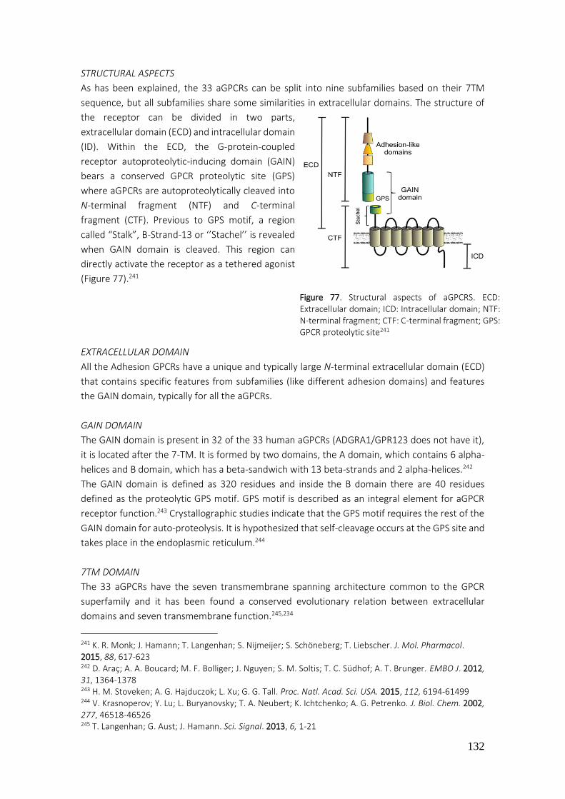
132
STRUCTURAL ASPECTS
As has been explained, the 33 aGPCRs can be split into nine subfamilies based on their 7TM
sequence, but all subfamilies share some similarities in extracellular domains. The structure of
the receptor can be divided in two parts,
extracellular domain (ECD) and intracellular domain
(ID). Within the ECD, the G-protein-coupled
receptor autoproteolytic-inducing domain (GAIN)
bears a conserved GPCR proteolytic site (GPS)
where aGPCRs are autoproteolytically cleaved into
N-terminal fragment (NTF) and C-terminal
fragment (CTF). Previous to GPS motif, a region
called “Stalk”, B-Strand-13 or ‘’Stachel’’ is revealed
when GAIN domain is cleaved. This region can
directly activate the receptor as a tethered agonist
(Figure 77).241
EXTRACELLULAR DOMAIN
All the Adhesion GPCRs have a unique and typically large N-terminal extracellular domain (ECD)
that contains specific features from subfamilies (like different adhesion domains) and features
the GAIN domain, typically for all the aGPCRs.
GAIN DOMAIN
The GAIN domain is present in 32 of the 33 human aGPCRs (ADGRA1/GPR123 does not have it),
it is located after the 7-TM. It is formed by two domains, the A domain, which contains 6 alpha-
helices and B domain, which has a beta-sandwich with 13 beta-strands and 2 alpha-helices.242
The GAIN domain is defined as 320 residues and inside the B domain there are 40 residues
defined as the proteolytic GPS motif. GPS motif is described as an integral element for aGPCR
receptor function.243 Crystallographic studies indicate that the GPS motif requires the rest of the
GAIN domain for auto-proteolysis. It is hypothesized that self-cleavage occurs at the GPS site and
takes place in the endoplasmic reticulum.244
7TM DOMAIN
The 33 aGPCRs have the seven transmembrane spanning architecture common to the GPCR
superfamily and it has been found a conserved evolutionary relation between extracellular
domains and seven transmembrane function.245,234
241 K. R. Monk; J. Hamann; T. Langenhan; S. Nijmeijer; S. Schöneberg; T. Liebscher. J. Mol. Pharmacol. 2015, 88, 617-623 242 D. Araç; A. A. Boucard; M. F. Bolliger; J. Nguyen; S. M. Soltis; T. C. Südhof; A. T. Brunger. EMBO J. 2012, 31, 1364-1378 243 H. M. Stoveken; A. G. Hajduczok; L. Xu; G. G. Tall. Proc. Natl. Acad. Sci. USA. 2015, 112, 6194-61499 244 V. Krasnoperov; Y. Lu; L. Buryanovsky; T. A. Neubert; K. Ichtchenko; A. G. Petrenko. J. Biol. Chem. 2002, 277, 46518-46526 245 T. Langenhan; G. Aust; J. Hamann. Sci. Signal. 2013, 6, 1-21
Figure 77. Structural aspects of aGPCRS. ECD: Extracellular domain; ICD: Intracellular domain; NTF: N-terminal fragment; CTF: C-terminal fragment; GPS: GPCR proteolytic site241

133
Recently, a mechanism by which aGPCRs are activated has been proposed that utilizes the N-
terminal of the 7-TM region. After proteolysis at the GPS site, this N-terminus (Stachel part) is
free to stimulate the 7TM as tethered agonist and therefore to induce activation. It is remarkable
that all aGPCRS keep this conserved motif. 243,246,247,248
Actually, it is similar to how thrombin receptors work (PAR1). When thrombin binds to its receptor
and cleaves a part of its exodomain a new N-terminus appears, which acts as a tethered ligand
activating the receptor.249
HETEROTRIMERIC G-PROTEINS
Another characteristic in aGPCRs is the associated G protein that mediates the intracellular
pathways. The heterotrimeric G protein complex, as has been described, consist of an -subunit
that binds and hydrolyzes GTP and also a -subunit that forms a non-dissociable complex.250
In one hand, complex usage does not appear to be significantly different among the 33
aGPCRs. In the other hand, the alpha subunit, can be, sorted in four types: Gs, Gi/o, Gq, G12.
In humans 16 genes encode for Gα-subunits, five genes encode for Gβ-subunits, and 12 genes
encode for Gγ-subunits.251 Most GPCRs are able to activate more than one G subunit, but each
receptor has selectivity towards particular G families. For example, a ligand binding in a receptor
can produces a specific conformation which can activate a specific G protein pathway. G-protein
coupling selectivity is known to depend on local physiological conditions and thus may change in
different cell lines, tissues, or experimental conditions.
G PROTEIN SIGNAL TRANSDUCTION MECHANISMS
The heterotrimeric G protein coupled to the aGPCRs undergoes a cycle of activation and
inactivation. When the receptor is in basal state, the -GDP, and -subunits form a tight trimeric
complex that does not signal. Once the GPCR has been activated, the conformational changes in
the receptor promote an exchange of the GDP to GTP by G, followed by a disassociation of -
GTP from both the receptor and the -complex. The two complexes are freed, so are able to
independently modulate effector proteins. The activity is terminated by GTP hydrolysis and
reformation of the G-protein heterotrimer. This can be enhanced by effector proteins or by
regulators of G protein signaling (RGS), promoting hydrolysis of GTP by the intrinsic GTPase
activity of G (Figure 78).252
246 G. S. Salzman; S. Zhang; A. Gupta; A. Koide; S. Koide; D. Araç. Proc. Natl. Acad. Sci. USA. 2017, 114, 10095-10100 247 L. M. Demberg; J. Winkler; C. Wilde; K. U. Simon; J. Schön; S. Rothemund; T. Schöneberg; S. Prömel; I. Liebscher. J. Biol. Chem. 2017, 292, 4383-4394 248 I. Liebscher; T. Schöneberg. Handb. Exp. Pharmacol. 2016, 234, 111-125 249 S.R. Coughlin. Proc. Natl. Acad. Sci. 1999, 96, 11023-11027 250 H. E. Hamm. J. Biol. Chem. 1998, 273, 669-672 251 G. B. Downes; N. Gautam. Genomics 1999, 62, 544-552 252 T. M. Cabrera-Vera; J. Vanhauwe; T. O. Thomas; M. Medkova; A. Preininger; M. R. Mazzoni; H. E. Hamm. Endocr. Rev. 2003, 24, 765-781
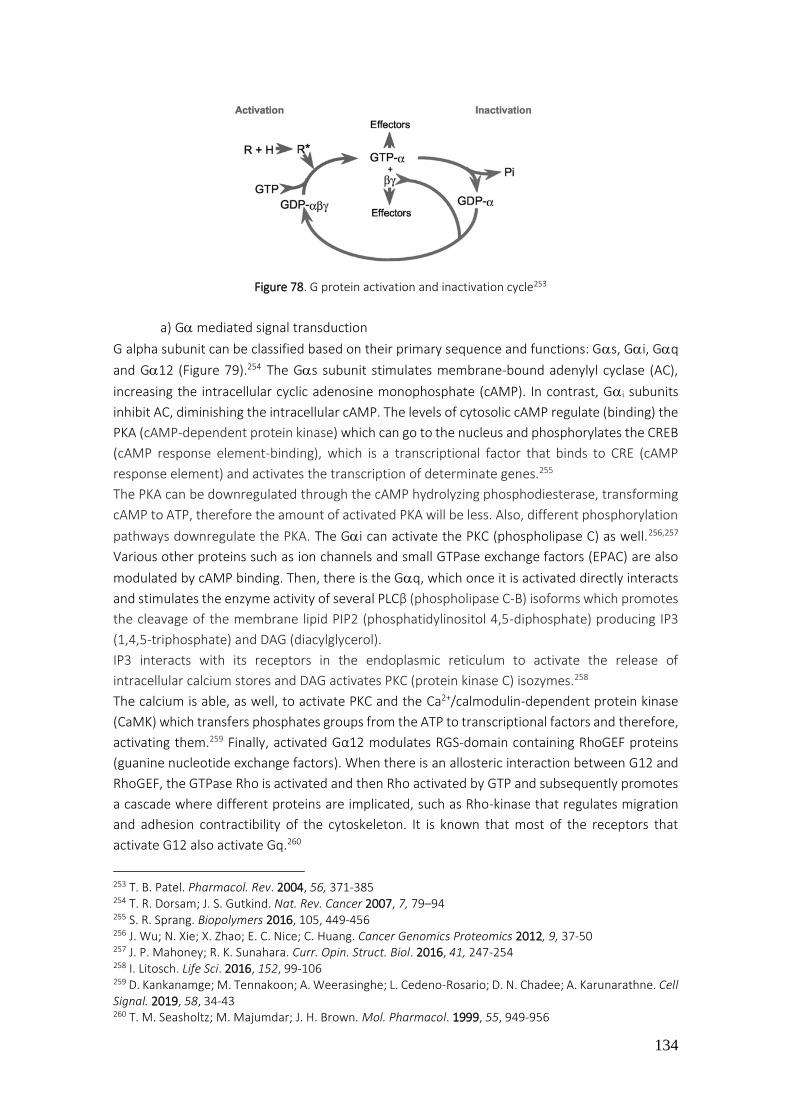
134
Figure 78. G protein activation and inactivation cycle253
a) G mediated signal transduction
G alpha subunit can be classified based on their primary sequence and functions: Gs, Gi, Gq
and G12 (Figure 79).254 The Gs subunit stimulates membrane-bound adenylyl cyclase (AC),
increasing the intracellular cyclic adenosine monophosphate (cAMP). In contrast, Gi subunits
inhibit AC, diminishing the intracellular cAMP. The levels of cytosolic cAMP regulate (binding) the
PKA (cAMP-dependent protein kinase) which can go to the nucleus and phosphorylates the CREB
(cAMP response element-binding), which is a transcriptional factor that binds to CRE (cAMP
response element) and activates the transcription of determinate genes.255
The PKA can be downregulated through the cAMP hydrolyzing phosphodiesterase, transforming
cAMP to ATP, therefore the amount of activated PKA will be less. Also, different phosphorylation
pathways downregulate the PKA. The Gi can activate the PKC (phospholipase C) as well.256,257
Various other proteins such as ion channels and small GTPase exchange factors (EPAC) are also
modulated by cAMP binding. Then, there is the Gq, which once it is activated directly interacts
and stimulates the enzyme activity of several PLCβ (phospholipase C-B) isoforms which promotes
the cleavage of the membrane lipid PIP2 (phosphatidylinositol 4,5-diphosphate) producing IP3
(1,4,5-triphosphate) and DAG (diacylglycerol).
IP3 interacts with its receptors in the endoplasmic reticulum to activate the release of
intracellular calcium stores and DAG activates PKC (protein kinase C) isozymes.258
The calcium is able, as well, to activate PKC and the Ca2+/calmodulin-dependent protein kinase
(CaMK) which transfers phosphates groups from the ATP to transcriptional factors and therefore,
activating them.259 Finally, activated Gα12 modulates RGS-domain containing RhoGEF proteins
(guanine nucleotide exchange factors). When there is an allosteric interaction between G12 and
RhoGEF, the GTPase Rho is activated and then Rho activated by GTP and subsequently promotes
a cascade where different proteins are implicated, such as Rho-kinase that regulates migration
and adhesion contractibility of the cytoskeleton. It is known that most of the receptors that
activate G12 also activate Gq.260
253 T. B. Patel. Pharmacol. Rev. 2004, 56, 371-385 254 T. R. Dorsam; J. S. Gutkind. Nat. Rev. Cancer 2007, 7, 79–94 255 S. R. Sprang. Biopolymers 2016, 105, 449-456 256 J. Wu; N. Xie; X. Zhao; E. C. Nice; C. Huang. Cancer Genomics Proteomics 2012, 9, 37-50 257 J. P. Mahoney; R. K. Sunahara. Curr. Opin. Struct. Biol. 2016, 41, 247-254 258 I. Litosch. Life Sci. 2016, 152, 99-106 259 D. Kankanamge; M. Tennakoon; A. Weerasinghe; L. Cedeno-Rosario; D. N. Chadee; A. Karunarathne. Cell Signal. 2019, 58, 34-43 260 T. M. Seasholtz; M. Majumdar; J. H. Brown. Mol. Pharmacol. 1999, 55, 949-956
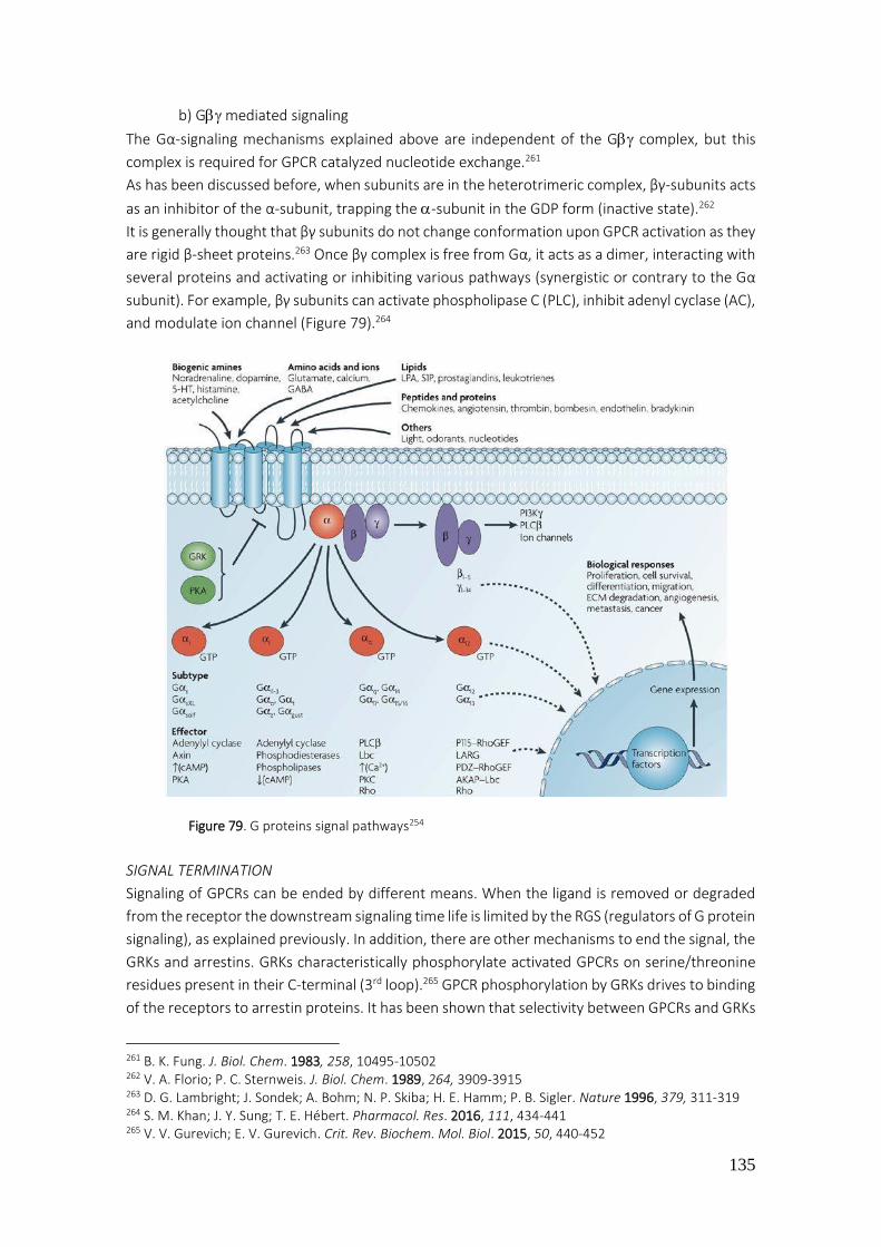
135
b) G mediated signaling
The Gα-signaling mechanisms explained above are independent of the G complex, but this
complex is required for GPCR catalyzed nucleotide exchange.261
As has been discussed before, when subunits are in the heterotrimeric complex, βγ-subunits acts
as an inhibitor of the α-subunit, trapping the -subunit in the GDP form (inactive state).262
It is generally thought that βγ subunits do not change conformation upon GPCR activation as they
are rigid β-sheet proteins.263 Once βγ complex is free from Gα, it acts as a dimer, interacting with
several proteins and activating or inhibiting various pathways (synergistic or contrary to the Gα
subunit). For example, βγ subunits can activate phospholipase C (PLC), inhibit adenyl cyclase (AC),
and modulate ion channel (Figure 79).264
Figure 79. G proteins signal pathways254
SIGNAL TERMINATION
Signaling of GPCRs can be ended by different means. When the ligand is removed or degraded
from the receptor the downstream signaling time life is limited by the RGS (regulators of G protein
signaling), as explained previously. In addition, there are other mechanisms to end the signal, the
GRKs and arrestins. GRKs characteristically phosphorylate activated GPCRs on serine/threonine
residues present in their C-terminal (3rd loop).265 GPCR phosphorylation by GRKs drives to binding
of the receptors to arrestin proteins. It has been shown that selectivity between GPCRs and GRKs
261 B. K. Fung. J. Biol. Chem. 1983, 258, 10495-10502 262 V. A. Florio; P. C. Sternweis. J. Biol. Chem. 1989, 264, 3909-3915 263 D. G. Lambright; J. Sondek; A. Bohm; N. P. Skiba; H. E. Hamm; P. B. Sigler. Nature 1996, 379, 311-319 264 S. M. Khan; J. Y. Sung; T. E. Hébert. Pharmacol. Res. 2016, 111, 434-441 265 V. V. Gurevich; E. V. Gurevich. Crit. Rev. Biochem. Mol. Biol. 2015, 50, 440-452
136
is more controlled by the activation and the situation of this activation (quantity of GRKs, quantity
of agonist, amongst others) rather than a specific phosphorylation site.266 Arrestins recognize
activated GPCRs and the affinity for them is increased by GRKs phosphorylation. Depending on
the pattern of the phosphorylation, arrestins act to terminate signal transduction signal by two
mechanisms, desensitization and receptor sequestration. One arrestin is able to regulate one
receptor, playing an important homeostasis role. 265
Activated arrestin-GPCR complexes recruit the proteins adaptin and chlatrin to endocytosis and
receptor internalization (vesicular traffic), diminishing the amount of cell surface receptors. The
endocytic process may occur in different ways based on the affinity of arrestins to GPCR
phosphorylation sites. Weak affinity interactions induce a state where arrestins disassociate fast
and receptors are recycled and back to the cell surface.267 It is important to remark on the role of
conformational changes in GPCRs in which the dynamics and the different states can result in
different functional outcomes. For example, some drugs (biased drugs) activate a conformational
state where arrestin pathway is activated but the G protein pathway is not.268
3.3.3. CARACTERIZATION of 33 ADHESION GPCRs
Given that the physiological basics of these receptors and their promise as drug targets for
human disease are established, the executed procedures to characterize them are explained
hereunder.
The project involved design of cellular assay systems to measure the signal transduction of
recombinant adhesion family receptors. It has involved transient transfection of mammalian
cells and the measurement of various second messengers (cAMP, IP3, kinase activity) in
response to activated receptor point mutants and literature-described peptides. Analysis of
the mechanism of receptor activation and G-protein signal- transduction pathway will be
quantified. The receptors were mutated in order to be cleavable by the bovine enterokinase
enzyme, generating an agonist neo-epitope through the proteolysis of their GPS sequence. Then,
the free Stachel sequence of the receptors will be available to bind the transmembrane domains
of the receptor and promote the conformational changes that causes activation (Figure 80). Also,
recent literature described how aGPCRs can be activated by peptides containing the aminoacid
sequence of the β-strand.269
Thus, a peptide sequenced by the aminoacids present in the GPS of GPR126 were tested as well.
Since they were able to be activated, the next step was to quantify the G-protein signal-
transduction pathways in every aGPCR. So, it was necessary to transfect each receptor in
HEK293 cells (human embryonic kidney cells), to incubate them, and finally, to measure
second messengers activated through G protein activity.
266 C. A. Moore; S. K. Milano; J. L. Benovic. Annu. Rev. Physiol. 2007, 69, 451-482 267 S. M. DeWire; S. Ahn; R. J. Lefkowitz; S. K. Shenoy. Annu. Rev. Physiol. 2007, 69, 483-510 268 Y. K. Peterson; L. M. Luttrell. Pharmacol. Rev. 2017, 69, 256-297 269 H. M. Stoveken; L. L. Bahr; M. W. Anders; A. P. Wojtovich; A. V. Smrcka; G. G. Tall. Mol. Pharmacol. 2016, 90, 214-222

137
Figure 80. Activation of the receptor through the free stachel part after proteolysis270
In that way, it was possible to quantify the preferred G protein associated in each receptor.
As a negative control, untransfected cells were used to assure that response was due to
receptor activation (Figure 81).
270 http://www.adhesiongpcr.de (09/01/2020)

138
For each Gα protein a different assay (using different procedures and devices) was
accomplished:
a) Gαs: cAMP was measured with luminescence using the Glosensor system (Promega)
b) Gαi: the decrease of Forskolin-induced cAMP271 was measured using the Glosensor
approach. Pertussis toxin,272,273 as an inhibitor of Gαi, was used to confirm the results.
c) Gαq: intracellular calcium was measured using calcium sensitive fluorescent dyes. YM-
254890274,275 was used as an inhibitor of Gαq to confirm the results.
d) Gα12: the intracellular calcium was measured after stimulation of RhoA (increases
intracellular calcium) through PLC-. YM-254890 was used as an inhibitor of Gαq to
characterized any false positive results.
e) Reporter assay: transcription factor response elements (CRE- Gαs, SRE- Gαi, NFAT-RE-
Gαq, SRF-RE-Gα12) were measured after receptor activation. These response elements
encoding DNAs coupled activate to the production of luciferase. If the signal transduction
pathway is activated through CRE, SRE, NFAT or SRF, luciferase expression increases and it
can be detected with luminescence (Figure 82).
271 J. Gilissen; P. Geubelle; N. Dupuis; C. Laschet; B. Pirotte; J. Hanson. Biochem. Pharmacol. 2015, 98, 381-391 272 S. Mangmool; H. Kurose. Toxins 2011, 3, 884-899 273 T. Katada. Biol. Pharm. Bull. 2012, 35, 2103-2111 274 H. Zhang; A. L. Nielsen; K. Strømgaard. Med. Res. Rev. 2020, 40, 135-157 275 A. Nishimura; K. Kitano; J. Takasaki; M. Taniguchi; N. Mizuno; K. Tago; T. Hakoshima; H. Itoh. Proc. Natl. Acad. Sci. USA 2010, 107, 13666-13671
Figure 81. cAMP formation before and after activation through enterokinase or peptide (containing
the sequence of GPR126 β-strand) in untransfected cells (negative control) and transfected cells
with GPR112

139
Figure 82. Pathways undertaken through G alpha subunit276
Very, little information exists as to the G-protein coupling specificities of aGPCRs.
Accordingly, most of the aGPCRs have the theoretical potential to be associated with several
diferent G proteins.
The used methods and materials are described in the experimental part.
After several assays and repetitions the obtained results for the 33 receptors are summarized
in the following table, where the number of independent repetitions of each experiment are
shown (Table 31).
Table 31. Number of independent repetitions of each experiment for each receptor.
The concluded coupled G protein for each receptor is
showed in different colors depending if the results were
significant, possible or null.
276 www.promega.es (25/05/2019)
G-protein Significant Possible
Gs
Gi
Gq
G12
Gq + Gs
Gi + Gq
Gs Gi Gq G12 cAMP CRE cAMP G.F. SRE Ca2+ NFAT plc-Ca2+ SRF
BAI 1 3 2 (1 PT) 3 (1 PT) 1 4 (1 PT, 1 YM) 5 (2 YM) - 1 3
BAI2 2 1 3 (1PT) 1 4 (1 PT, 1 YM) 5 (1 YM) - 1 3
BAI 3 1 1 2 (1PT) 1 3 (1YM) 5 (1 YM) - 1 2
CD97 2 2 (1PT) 3 (1PT) 1 4 (1 PT, 1 YM) 5 (2 YM) - 1 3
CELSR1 3 2 (1PT) 3 (1PT) 1 4 (1 PT, 1 YM) 5 (2 YM) - 1 3
CELSR2 2 1 3 (1PT) 1 4 (1 PT, 1 YM) 5 (2 YM) - 1 3
CELSR3 2 1 3 (1PT) 1 4 (1 PT, 1 YM) 5 (2 YM) - 1 3
ELTD1 2 1 4 (2 PT) 1 4 (1 PT, 1 YM) 5 (1 YM) - 1 3

140
PT: pertussis toxin, YM: YM-254890, GLO: Glosensor experiment, CRE: cAMP response element, G.F.: Gradual
forskolin, SRE: serum response element, NFAT: nuclear factor of activated T-cells, SRF: serum response factor
1) It is concluded that under the described assays conditions GPR64, GPR112, GPR114,
GPR126, GPR128, GPR133, GPR144 were coupled with Gs.
2) It is concluded that under the described assays conditions EMR2, GPR56, GPR110 and
GPR116 were coupled with Gi.
3) It is concluded that under the described assays conditions BAI2, GPR64 and GPR126 were
coupled with Gq.
4) It is concluded that under the described assays conditions GPR56, GPR64, GPR112,
GPR126 and GPR128 were coupled with G12.
The proposed and undertaken assays were reproducible and efficient to analyze the coupled G
proteins and the intracellular mechanisms related with adhesion GPCRs.
EMR1 2 2 (1 PT) 4 (2 PT) 1 4 (1 PT, 1 YM) 6 (2 YM) - 1 3
EMR2 2 2 (1 PT) 3 (1 PT) 4 (1 PT, 1 YM) 6 (2 YM) - 1 3
EMR3 2 1 3 (2 PT) 1 3 (1 YM) 5 (1 YM) - 1 3
EMR4 4 1 3 (1PT) 4 (1 PT, 1 YM) 4 (1 YM) - 1 3
GPR56 3 2 (1 PT) 4 (1 PT, 1 PEPTIDE) 1 4 (1 PT, 1 YM) 4 (2 YM, 1 PEPTIDE) - 1 3
GPR64 4 (1 PEPTIDE) 2 2 4 (1 PT, 1 YM) 5 (2 YM, 1 PEPTIDE) - 1 3
GPR97 3 2 (1 PT) 3 (1 PT) 1 2 4 (2 YM) - 1 2
GPR98 2 2 (1PT) 3 (1 PT) 3 (1 YM) 4 (1 YM) - 1 3
GPR110 4 2 (1PT) 3 (1 PT) 1 4 (1 PT, 1 YM) 5 (1 YM) - 1 3
GPR111 3 1 3 (1 PT) 3 (1 YM) 4 (1 YM) - 1 2
GPR112 4 (1 PEPTIDE) 1 3 4 (1 PT, 1 YM) 5 (1 YM, 1 PEPTIDE) - 1 3
GPR113 3 1 3 (1 PT) 4 (1 PT, 1 YM) 5 (2 YM) - 1 2
GPR114 4 (1 PEPTIDE) 1 2 2 5 (1 YM, 1 PEPTIDE) - 1 2
GPR115 3 2 (1PT) 3 (1PT) 1 3 (1 YM) 4 (1 YM) - 1 2
GPR116 3 2 (1PT) 3 (1PT) 1 4 (1 PT, 1 YM) 3 (2 YM) - 1 3
GPR123 2 1 3 (1PT) 4 (1 PT, 1 YM) 6 (2 YM) - 1 2
GPR124 2 2 (1PT) 2 (1PT) 3 (1 YM) 4 (1 YM) - 1 2
GPR125 3 2 (1 PT) 4 (2 PT) 1 4 (1 PT, 1 YM) 6 (1 YM) - 1 3
GPR126 4 (1 PEPTIDE) 2 2 4 (1 PT, 1 YM) 4 (2 YM, 1 PEPTIDE) - 3 3
GPR128 4 (1 PEPTIDE) 1 2 4 (1 PT, 1 YM) 4 (1 YM) - 1 3
GPR133 4 (1 PEPTIDE) 1 2 4 (1 PT, 1 YM) 4 (1 YM) - 1 2
GPR144 4 (1 PEPTIDE) 1 1 2 4 (1 YM) - 1 2
LPHN1 3 1 (1 PT) 4 (2 PT) 1 2 5 (1 YM) - 1 2
LPHN2 2 1 (1 PT) 3 (1 PT) 1 4 (1 PT, 1 YM) 5 (1 YM) - 1 2
LPHN3 2 2 (1 PT) 3 (1 PT) 1 4 (1 PT, 1 YM) 5 (1 YM) - 1 2
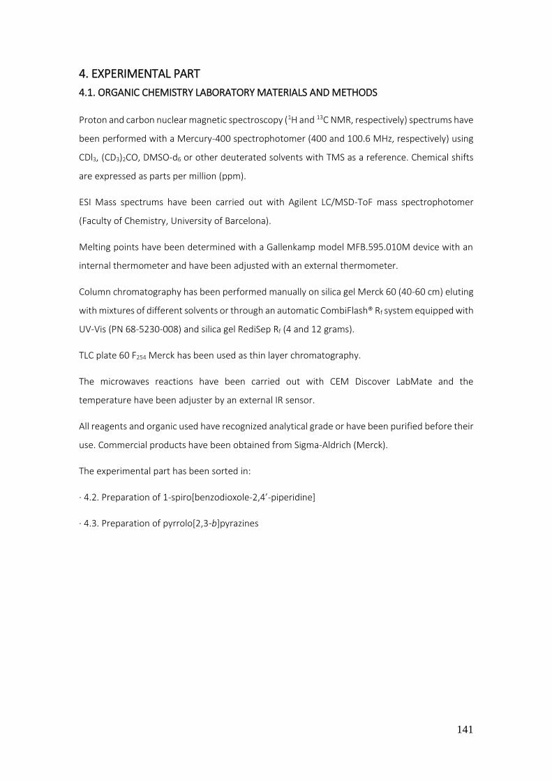
141
4. EXPERIMENTAL PART
4.1. ORGANIC CHEMISTRY LABORATORY MATERIALS AND METHODS
Proton and carbon nuclear magnetic spectroscopy (1H and 13C NMR, respectively) spectrums have
been performed with a Mercury-400 spectrophotomer (400 and 100.6 MHz, respectively) using
CDl3, (CD3)2CO, DMSO-d6 or other deuterated solvents with TMS as a reference. Chemical shifts
are expressed as parts per million (ppm).
ESI Mass spectrums have been carried out with Agilent LC/MSD-ToF mass spectrophotomer
(Faculty of Chemistry, University of Barcelona).
Melting points have been determined with a Gallenkamp model MFB.595.010M device with an
internal thermometer and have been adjusted with an external thermometer.
Column chromatography has been performed manually on silica gel Merck 60 (40-60 cm) eluting
with mixtures of different solvents or through an automatic CombiFlash® Rf system equipped with
UV-Vis (PN 68-5230-008) and silica gel RediSep Rf (4 and 12 grams).
TLC plate 60 F254 Merck has been used as thin layer chromatography.
The microwaves reactions have been carried out with CEM Discover LabMate and the
temperature have been adjuster by an external IR sensor.
All reagents and organic used have recognized analytical grade or have been purified before their
use. Commercial products have been obtained from Sigma-Aldrich (Merck).
The experimental part has been sorted in:
· 4.2. Preparation of 1-spiro[benzodioxole-2,4’-piperidine]
· 4.3. Preparation of pyrrolo[2,3-b]pyrazines

142
4.2. Preparation of 1-spiro[benzodioxole-2,4’-piperidine]
4.2.1. Preparation of N-acetyl-spiro[1,3-benzodioxole-2,4'-piperidine] (1)
· Procedure
In a 25 mL round bottom flask the N-acetyl-4-piperidone (0.78 mL, 6.37 mmol) was dissolved in
8 mL of methanol. The trimethyl orthoformate (0.83 mL, 7.6 mmol) and p-toluenesulfonic acid
(catalytic quantity) were also added.
The reaction was stirred at 34 C until distillation of methyl formate was observed. Then, the
reaction was stopped, cooled (0 C) and 5 mL of sodium methoxide 10% were added. Methanol
was evaporated to obtain the crude of reaction. The crude of reaction was extracted with
dichloromethane (3 x 20 mL) and water (1 x 20 mL) was made. The organic phases were collected,
dried over anhydrous Na2SO4, filtered and concentrated under reduced pressure to obtain the
acetal intermediate. Then, it was dissolved in a round bottom flask with toluene (10 mL) and
catechol (2.1 g, 19 mmol) was added. The mixture was stirred at 120 C until distillation of toluene
was observed. After this time, the reaction was cooled at 60 C in order to add APTS (catalytic
quantity). Soon after, the reaction crude was heated at 120 °C. The reaction control was carried
out by TLC (hexane/ethyl acetate 4:6).
· Work-Up
The residue of reaction obtained was cooled below 0 C and 3 mL of triethylamine were added.
The solvent was concentrated in vacuo giving as a result the crude of reaction, which was
dissolved in dichloromethane (20 mL) and washed with water (3 x 20 mL). The organic phase was
dried over anhydrous Na2SO4, filtered and concentrated under reduced pressure. A mixture of
expected compound and starting catechol was obtained.
· Purification
An automatic flash column chromatography was carried out to afford the expected compound.
The desired product eluted with a mixture of hexane/ethyl acetate 40:60.
· Analytical data
1H NMR (CDCl3, 400 MHz) δ(ppm), 2.00 (q, J = 5.8 Hz, 4H, CH2-C (x2)); 2.15 (s, 3H, CH3); 3.65 (t, J =
5.8 Hz, 2H, CH-N axial (x2)); 3.82 (t, J = 5.8 Hz, 2H, CH-N equatorial (x2)); 6.76 – 6.82 (m, 4H, Ar).
Product aspect Mass obtained Yield Rf (hexane/ethyl acetate 4:6) Melting point
White solid 1 g 70% 0.32 162-163 C (ethanol)

143
4.2.2. Preparation of 1',5-diacetyl-spiro[benzo[1,3]dioxole-2,4'-piperidine] (2)
· Procedure (general acetylation procedure)
In a 50 mL round bottom flask the spiro derivative 1 (0.4 g, 1.71 mmol) was dissolved in
dichloromethane (15 mL). Then, acetyl chloride (0.83 mL, 7.6 mmol) and titanium tetrachloride
were added (0.18 mL, 2.4 mmol) dropwise at 0 °C (keeping the reaction submerged in an ice
bath). The reaction mixture was stirred for 6 hours at 0 °C to room temperature.
· Work-up
A thin layer chromatography (TLC) of reaction (hexane/ethyl acetate 7:3) indicated a formation
of a new compound. Thus, 3 mL of water were added in order to hydrolyze the crude of reaction.
The mixture was washed with water (3 x 20 mL) and diluted with dichloromethane (20 mL). The
organic phase was dried over anhydrous sodium sulfate, filtered and concentrated in vacuo.
· Purification
No purification of the obtained residue by column chromatography was performed since the
product was obtained with enough purity.
· Analytical data
1H NMR (CDCl3, 400 MHz) δ(ppm), 1.96-2.00 (m, 4H, CH2-C (x2)); 2.03 (s, 3H, CH3-CO-N); 2.50 (s,
3H, CH3-CO-C); 3.63 (t, J = 6 Hz, 2H, CH-N axial (x2)); 3.82 (t, J = 6 Hz, 2H, CH-N equatorial (x2));
6.79 (d, J = 8.2 Hz, 1H, H-7); 7.36 (d, J = 2 Hz, 1H, H-4); 7.51 (dd, J1 = 2 Hz, J2 = 8.2 Hz, 1H, H-6).
Product aspect Mass obtained Yield Rf (hexane/ethyl acetate 7:3)
White solid 0.4 g 87% 0.18
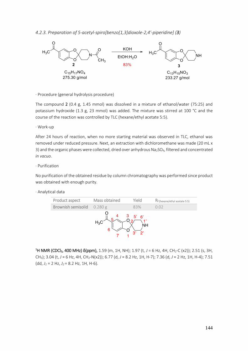
144
4.2.3. Preparation of 5-acetyl-spiro[benzo[1,3]dioxole-2,4'-piperidine] (3)
· Procedure (general hydrolysis procedure)
The compound 2 (0.4 g, 1.45 mmol) was dissolved in a mixture of ethanol/water (75:25) and
potassium hydroxide (1.3 g, 23 mmol) was added. The mixture was stirred at 100 °C and the
course of the reaction was controlled by TLC (hexane/ethyl acetate 5:5).
· Work-up
After 24 hours of reaction, when no more starting material was observed in TLC, ethanol was
removed under reduced pressure. Next, an extraction with dichloromethane was made (20 mL x
3) and the organic phases were collected, dried over anhydrous Na2SO4, filtered and concentrated
in vacuo.
· Purification
No purification of the obtained residue by column chromatography was performed since product
was obtained with enough purity.
· Analytical data
1H NMR (CDCl3, 400 MHz) δ(ppm), 1.59 (m, 1H, NH); 1.97 (t, J = 6 Hz, 4H, CH2-C (x2)); 2.51 (s, 3H,
CH3); 3.04 (t, J = 6 Hz, 4H, CH2-N(x2)); 6.77 (d, J = 8.2 Hz, 1H, H-7); 7.36 (d, J = 2 Hz, 1H, H-4); 7.51
(dd, J1 = 2 Hz, J2 = 8.2 Hz, 1H, H-6).
Product aspect Mass obtained Yield Rf (hexane/ethyl acetate 5:5)
Brownish semisolid 0.280 g 83% 0.02

145
4.2.4. Preparation of 1’-acetyl-5-(1-(hydroxyimino)ethyl)spiro[benzo[1,3]dioxole-2,4'-
piperidine] (4)
· Procedure In a 50 mL round bottom flask the compound 2 (0.2 g, 0.73 mmol) was dissolved in ethanol (10
mL). Then, hydroxylamine hydrochloride (0.065 g, 0.90 mmol) and sodium carbonate (0.06 g, 0.56
mmol), dissolved in 5 mL of water, were added. The reaction was stirred for 22 h at room
temperature. At this time, starting material was still present in TLC, therefore, reaction was
heated at 80 °C.
· Work-up
After 20 hours of reaction, a TLC of reaction indicated consumption of starting material
(hexane/ethyl acetate 5:5). The ethanol was removed under reduced pressure and the crude was
extracted with dichloromethane (20 mL x 3). The organic phases were collected, dried over
anhydrous Na2SO4, filtered and concentrated in vacuo.
· Purification
No purification of the obtained residue by column chromatography was performed since product
was obtained with enough purity.
· Analytical data
1H NMR (CDCl3, 400 MHz) δ(ppm), 1.91 (m, 4H, CH2-C (x2)); 2.09 (s, 3H, CH3-CO); 2.17 (s, 3H, CH3);
3.58 (t, J = 6 Hz, 2H, CH-N axial (x2)); 3.75 (t, J = 6 Hz, 2H, CH-N equatorial (x2)); 6.68 (d, J = 8.2
Hz, 1H, H-7); 7.00 (dd, J1 = 2 Hz, J2 = 8.2 Hz, 1H, H-6); 7.06 (d, J = 2 Hz, 1H, H-4).
13C NMR (CDCl3, 100.6 MHz) δ(ppm), 12.2 (CH3); 21.3 (CH3); 34.6 (CH-C); 35.4 (CH-C); 38.8 (CH-N);
43.5 (CH-N); 106.4 (CH, C-4); 108.2 (CH, C-7); 116.3 (CH, C-6); 120.1 (Cq, C-2); 130.8 (Cq, C-5);
147.1 (Cq, C-3a); 147.7 (Cq, C-7a); 151.1 (Cq, CN); 169.2 (Cq, C=O).
Product aspect Mass obtained Yield Rf (hexane/ethyl acetate 5:5) Melting point
Yellow solid 0.1 g 95% 0.25 180-182 °C (DCM)
146
4.2.5. Preparation of 1’-acetyl-5-(1-hydroxyethyl)spiro[benzo[1,3]dioxole-2,4'-
piperidine](5)
· Procedure
In a 100 mL round-bottomed flask, specific for catalytic hydrogenations, equipped with a
magnetic stirring bar, the oxime 4 (0.160 g, 0.55 mmol) and Ni-Raney (0.06 mmol) were added
suspended in methanol (10 mL) and ethyl acetate (3 mL). The reaction was stirred at room
temperature for 5 days under hydrogen stream.
· Work-up
The theoretical required volume for the reaction was 25 mL. Given that hydrogenation apparatus
was not completely hermetical, the consumed volume was 40 mL, higher than expected. The
analysis of TLC showed consumption of starting material (hexane/ethyl acetate 7:3). The reaction
crude was filtered by means of a pleated filter and washed with 20 mL of methanol and was then
collected in a 100 mL round-bottomed flask. Finally, the solvent was removed under reduced
pressure. The residue was dissolved in ethyl ether (15 mL x 1) and washed with a solution of HCl
3N (10 mL x 3). The aqueous phase was basified with NaOH 2N and extracted with
dichloromethane (3 x 20 mL). The last organic phases were collected, dried over anhydrous
sodium sulfate, filtered and concentrated in vacuo.
· Purification
No purification of the obtained residue by column chromatography was performed. The 1H NMR
and 13C NMR signals indicated formation of alcohol 5.
· Analytical data
Product aspect Mass obtained Yield Rf (hexane/ethyl acetate 7:3)
Brown oil 0.02 g 13% 0.05

147
1H NMR (CDCl3, 400 MHz) δ(ppm), 1.38 (d, J = 6.5 Hz, 3H, CH3-CH); 1.87-1.95 (m, 4H, CH2-C (x2));
2.08 (s, 3H, CH3); 3.58 (t, J = 6 Hz, 2H, CH-N axial (x2)); 3.73 (t, J = 6 Hz, 2H, CH-N equatorial (x2));
4.73 (q, J = 6.5 Hz, 1H, CH-O); 6.64 (d, J = 8 Hz, 1H, H-7); 6.72 (dd, J1 = 2 Hz, J2 = 8 Hz, 1H, H-6);
6.78 (s, 1H, H-4).
13C NMR (CDCl3, 100.6 MHz) δ(ppm), 21.4 (CH3-CH); 25.2 (CH3-CO); 34.6 (CH2, CH2-C); 35.4 (CH2,
CH2-C); 38.7 (CH2-N); 43.5 (CH2, CH2-N); 70.2 (CH, CH-O); 106.2 (CH, C-4); 108.2 (CH, C-7); 115.8
(Cq, C-2); 118.5 (CH, C-6); 139.8 (Cq, C-5); 146.1 (Cq, C-7a); 147.1 (Cq, C-3a); 168.9 (C=O).
4.2.6. Preparation of 1’-acetyl-5-bromospiro[benzo[1,3]dioxole-2,4'-piperidine] (6)
· Procedure
In a 50 mL round bottom flask, the spiro 1 (0.5 g, 2.1 mmol) was dissolved in methanol (12 mL)
and NBS (0.42 g, 2.3 mmol) was added slowly. The reaction was vigorously stirred at room
temperature.
· Work-up
After 3 hours of reaction, analysis of TLC showed a formation of a new product (hexane/ethyl
acetate 6:4). The solvent was removed in vacuo affording the crude mixture, which was dissolved
in dichloromethane (20 mL x 1) and washed with a basic solution of NaOH 2N (20 mL x 3). The
organic phase was dried over anhydrous sodium sulfate, filtered and concentrated under reduced
pressure.
· Purification
Finally, the crude of reaction was purified by a flash automatic column chromatography. The
desired product eluted with a mixture of hexane/ethyl acetate 60:40.
· Analytical data
1H NMR (CDCl3, 400 MHz) δ(ppm), 1.86 (t, J = 5.9 Hz, 2H, CH-C axial (x2)); 1.92 (t, J = 5.9 Hz, 2H,
CH-C equatorial (x2)); 3.54 (t, J = 5.9 Hz, 2H, CH-N axial (x2)), 3.70 (t, J = 5.9 Hz, 2H, CH-N equatorial
Product aspect Mass obtained Yield Rf (hexane/ethyl acetate 6:4) Melting point
White solid 0.6 g 90% 0.17 253-255 C (DCM)

148
(x2)); 6.55 (dd, J1 = 1 Hz, J2 = 8 Hz, 1H, H-7); 6.81 (d, J = 2 Hz, 1H, H-4); 6.82 (dd, J1 = 2 Hz, J2 = 8
Hz, 1H, H-6).
13C NMR (CDCl3, 100.6 MHz) δ(ppm), 21.3 (CH3); 34.5 (CH2-C); 35.3 (CH2, CH2-C); 38.5 (CH2, CH2-
N); 43.3 (CH2, CH2-N); 109.7 (CH, C-4); 112.3 (CH, C-6); 112.7 (Cq, C-2); 117.0 (Cq, C-5); 124.0 (CH,
C-7); 146.2 (Cq, C-3a); 147.8 (Cq, C-7a); 168.8 (C=O).
4.2.7. Preparation of 1’-acetyl-5-(p-tolyl)spiro[benzo[1,3]dioxole-2,4'-piperidine] (7)
· Procedure
In a Pyrex glass tube with screw cap, previously flame-dried under argon atmosphere, the bromo
spiro 6 (0.1 g, 0.32 mmol) was dissolved in acetonitrile (10 mL). Then, aryl boronic acid (0.05 g,
0.37 mmol), sodium carbonate (0.068 g, 0.61 mmol), tetrakis(triphenylphosphine)palladium (0.06
mmol) and water (0.6 mL) were added under argon atmosphere. The reaction was stirred at 150
°C for 24 hours.
· Work-up
A TLC of reaction mixture showed a formation of a new product and consumption of starting
material (hexane/ethyl acetate 7:3). The toluene was removed under reduced pressure to afford
the crude, which was dissolved in dichloromethane (20 mL x 1) and washed with water (20 mL x
3). The organic layer was dried over anhydrous Na2SO4, filtered and concentrated in vacuo.
· Purification
Finally, the crude of reaction was purified by a flash automatic column chromatography. The
desired product eluted with a mixture of hexane/ethyl acetate 65:45.
· Analytical data
Product aspect Mass obtained Yield Rf (hexane/ethyl acetate 7:3)
Grey oil 0.09 g 87% 0.11
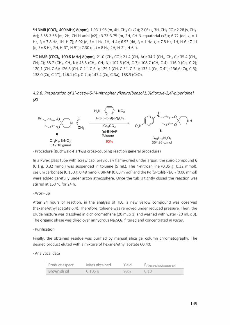
149
1H NMR (CDCl3, 400 MHz) δ(ppm), 1.93-1.95 (m, 4H, CH2-C (x2)); 2.06 (s, 3H, CH3-CO); 2.28 (s, CH3-
Ar); 3.55-3.58 (m, 2H, CH-N axial (x2)); 3.73-3.75 (m, 2H, CH-N equatorial (x2)); 6.72 (dd, J1 = 1
Hz, J2 = 7.8 Hz, 1H, H-7); 6.92 (d, J = 1 Hz, 1H, H-4); 6.93 (dd, J1 = 1 Hz, J2 = 7.8 Hz, 1H, H-6); 7.11
(d, J = 8 Hz, 2H, H-3’’, H-5’’); 7.30 (d, J = 8 Hz, 2H, H-2’’, H-6’’).
13C NMR (CDCl3, 100.6 MHz) δ(ppm), 21.0 (CH3-CO); 21.4 (CH3-Ar); 34.7 (CH2, CH2-C); 35.4 (CH2,
CH2-C); 38.7 (CH2, CH2-N); 43.5 (CH2, CH2-N); 107.6 (CH, C-7); 108.7 (CH, C-4); 116.0 (Cq, C-2);
120.1 (CH, C-6); 126.6 (CH, C-2’’, C-6’’); 129.1 (CH, C-3’’, C-5’’); 135.4 (Cq, C-4’’); 136.6 (Cq, C-5);
138.0 (Cq, C-1’’); 146.1 (Cq, C-7a); 147.4 (Cq, C-3a); 168.9 (C=O).
4.2.8. Preparation of 1’-acetyl-5-(4-nitrophenyl)spiro[benzo[1,3]dioxole-2,4'-piperidine]
(8)
· Procedure (Buchwald-Hartwig cross-coupling reaction general procedure)
In a Pyrex glass tube with screw cap, previously flame-dried under argon, the spiro compound 6
(0.1 g, 0.32 mmol) was suspended in toluene (5 mL). The 4-nitroaniline (0.05 g, 0.32 mmol),
cesium carbonate (0.150 g, 0.48 mmol), BINAP (0.06 mmol) and the Pd[(o-tolil)3P]2Cl2 (0.06 mmol)
were added carefully under argon atmosphere. Once the tub is tightly closed the reaction was
stirred at 150 °C for 24 h.
· Work-up
After 24 hours of reaction, in the analysis of TLC, a new yellow compound was observed
(hexane/ethyl acetate 6:4). Therefore, toluene was removed under reduced pressure. Then, the
crude mixture was dissolved in dichloromethane (20 mL x 1) and washed with water (20 mL x 3).
The organic phase was dried over anhydrous Na2SO4, filtered and concentrated in vacuo.
· Purification
Finally, the obtained residue was purified by manual silica gel column chromatography. The
desired product eluted with a mixture of hexane/ethyl acetate 60:40.
· Analytical data
Product aspect Mass obtained Yield Rf (hexane/ethyl acetate 6:4)
Brownish oil 0.105 g 93% 0.10
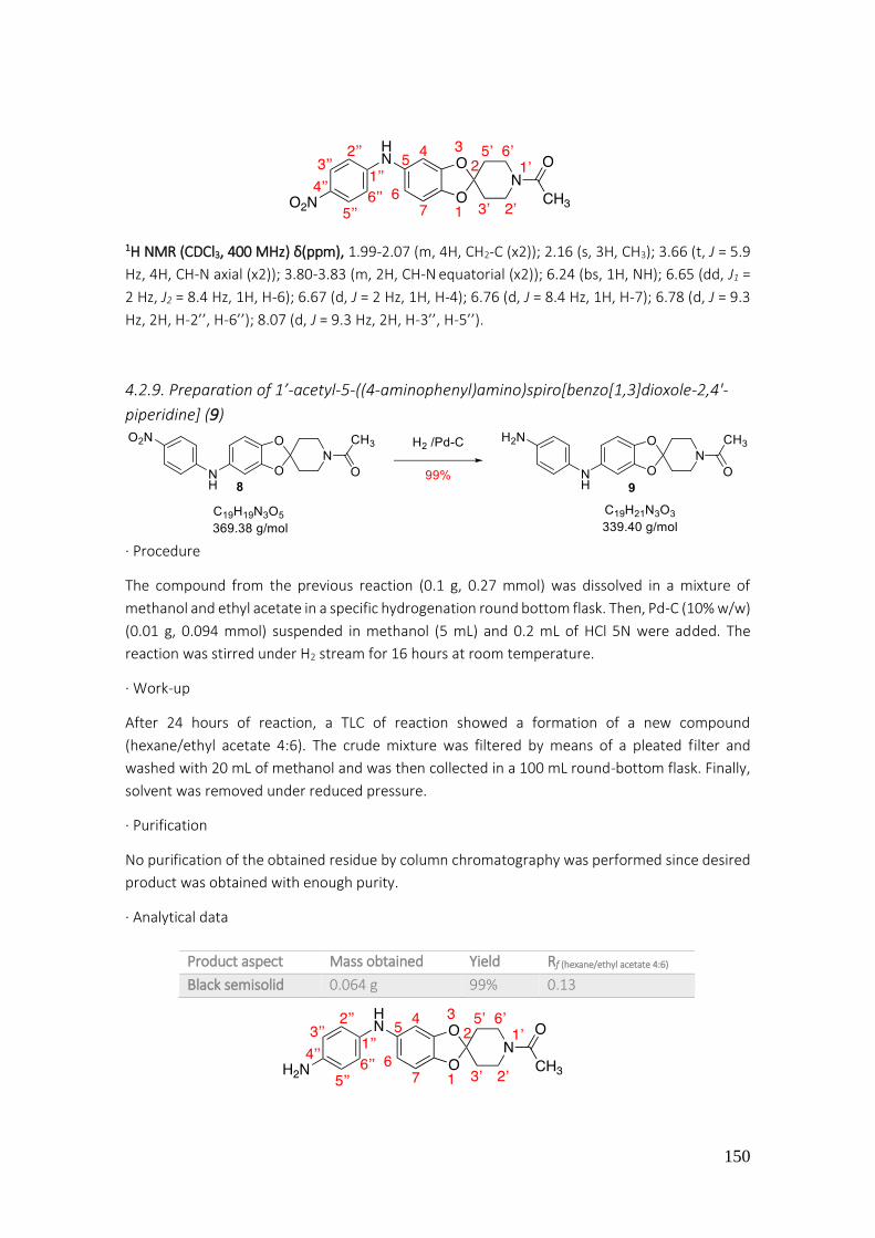
150
1H NMR (CDCl3, 400 MHz) δ(ppm), 1.99-2.07 (m, 4H, CH2-C (x2)); 2.16 (s, 3H, CH3); 3.66 (t, J = 5.9
Hz, 4H, CH-N axial (x2)); 3.80-3.83 (m, 2H, CH-N equatorial (x2)); 6.24 (bs, 1H, NH); 6.65 (dd, J1 =
2 Hz, J2 = 8.4 Hz, 1H, H-6); 6.67 (d, J = 2 Hz, 1H, H-4); 6.76 (d, J = 8.4 Hz, 1H, H-7); 6.78 (d, J = 9.3
Hz, 2H, H-2’’, H-6’’); 8.07 (d, J = 9.3 Hz, 2H, H-3’’, H-5’’).
4.2.9. Preparation of 1’-acetyl-5-((4-aminophenyl)amino)spiro[benzo[1,3]dioxole-2,4'-
piperidine] (9)
· Procedure
The compound from the previous reaction (0.1 g, 0.27 mmol) was dissolved in a mixture of
methanol and ethyl acetate in a specific hydrogenation round bottom flask. Then, Pd-C (10% w/w)
(0.01 g, 0.094 mmol) suspended in methanol (5 mL) and 0.2 mL of HCl 5N were added. The
reaction was stirred under H2 stream for 16 hours at room temperature.
· Work-up
After 24 hours of reaction, a TLC of reaction showed a formation of a new compound
(hexane/ethyl acetate 4:6). The crude mixture was filtered by means of a pleated filter and
washed with 20 mL of methanol and was then collected in a 100 mL round-bottom flask. Finally,
solvent was removed under reduced pressure.
· Purification
No purification of the obtained residue by column chromatography was performed since desired
product was obtained with enough purity.
· Analytical data
Product aspect Mass obtained Yield Rf (hexane/ethyl acetate 4:6)
Black semisolid 0.064 g 99% 0.13
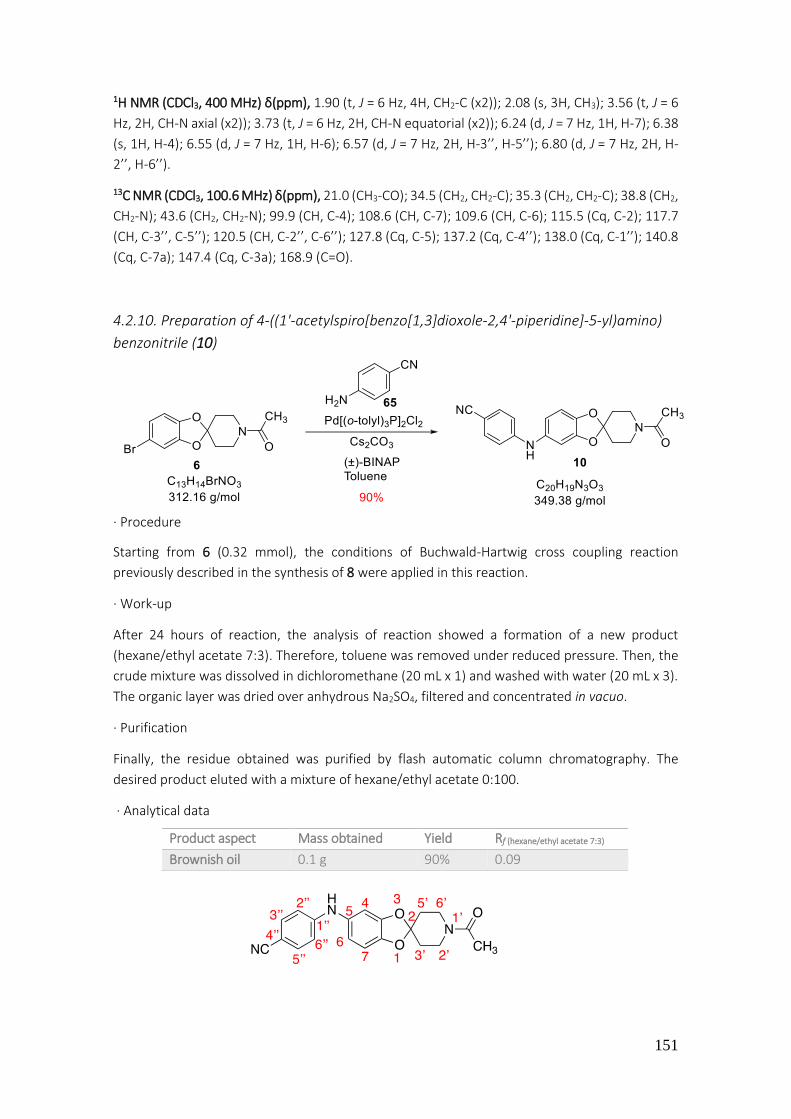
151
1H NMR (CDCl3, 400 MHz) δ(ppm), 1.90 (t, J = 6 Hz, 4H, CH2-C (x2)); 2.08 (s, 3H, CH3); 3.56 (t, J = 6
Hz, 2H, CH-N axial (x2)); 3.73 (t, J = 6 Hz, 2H, CH-N equatorial (x2)); 6.24 (d, J = 7 Hz, 1H, H-7); 6.38
(s, 1H, H-4); 6.55 (d, J = 7 Hz, 1H, H-6); 6.57 (d, J = 7 Hz, 2H, H-3’’, H-5’’); 6.80 (d, J = 7 Hz, 2H, H-
2’’, H-6’’).
13C NMR (CDCl3, 100.6 MHz) δ(ppm), 21.0 (CH3-CO); 34.5 (CH2, CH2-C); 35.3 (CH2, CH2-C); 38.8 (CH2,
CH2-N); 43.6 (CH2, CH2-N); 99.9 (CH, C-4); 108.6 (CH, C-7); 109.6 (CH, C-6); 115.5 (Cq, C-2); 117.7
(CH, C-3’’, C-5’’); 120.5 (CH, C-2’’, C-6’’); 127.8 (Cq, C-5); 137.2 (Cq, C-4’’); 138.0 (Cq, C-1’’); 140.8
(Cq, C-7a); 147.4 (Cq, C-3a); 168.9 (C=O).
4.2.10. Preparation of 4-((1'-acetylspiro[benzo[1,3]dioxole-2,4'-piperidine]-5-yl)amino)
benzonitrile (10)
· Procedure
Starting from 6 (0.32 mmol), the conditions of Buchwald-Hartwig cross coupling reaction
previously described in the synthesis of 8 were applied in this reaction.
· Work-up
After 24 hours of reaction, the analysis of reaction showed a formation of a new product
(hexane/ethyl acetate 7:3). Therefore, toluene was removed under reduced pressure. Then, the
crude mixture was dissolved in dichloromethane (20 mL x 1) and washed with water (20 mL x 3).
The organic layer was dried over anhydrous Na2SO4, filtered and concentrated in vacuo.
· Purification
Finally, the residue obtained was purified by flash automatic column chromatography. The
desired product eluted with a mixture of hexane/ethyl acetate 0:100.
· Analytical data
Product aspect Mass obtained Yield Rf (hexane/ethyl acetate 7:3)
Brownish oil 0.1 g 90% 0.09
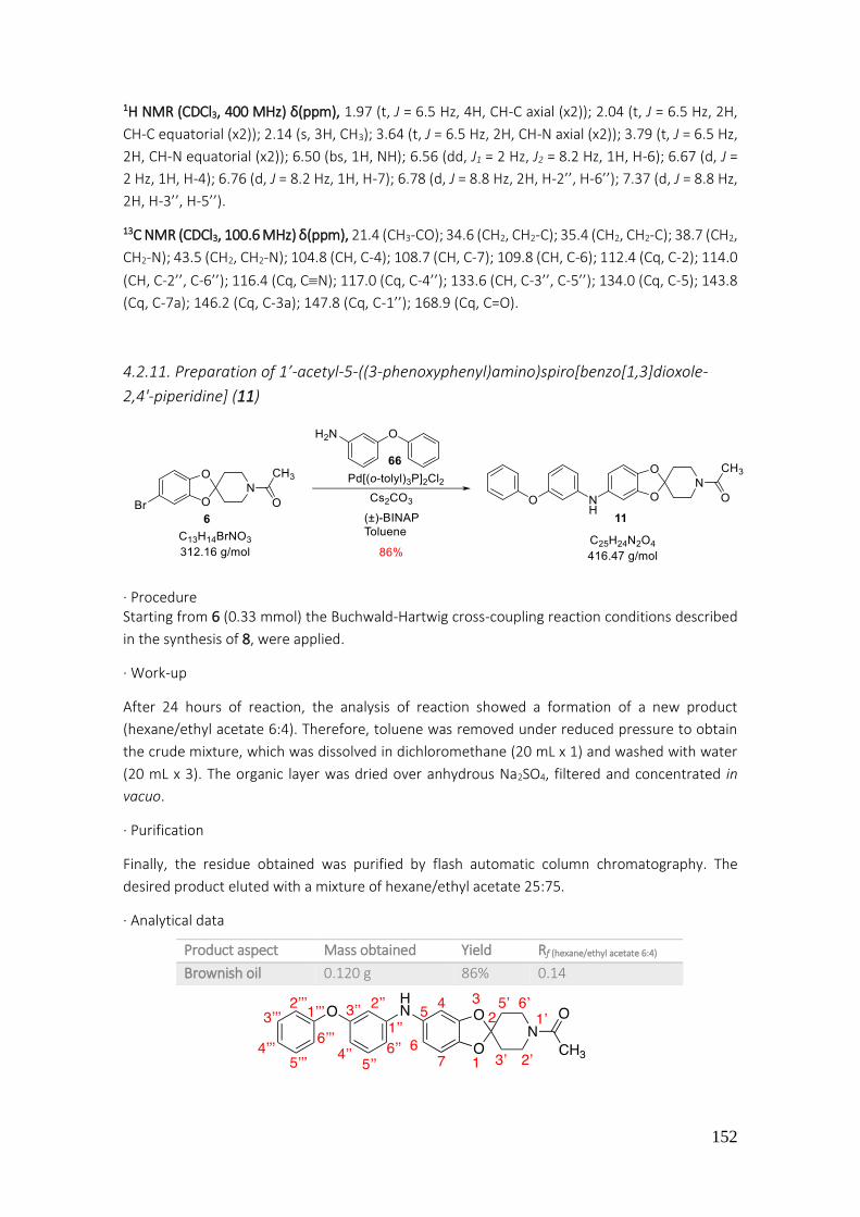
152
1H NMR (CDCl3, 400 MHz) δ(ppm), 1.97 (t, J = 6.5 Hz, 4H, CH-C axial (x2)); 2.04 (t, J = 6.5 Hz, 2H,
CH-C equatorial (x2)); 2.14 (s, 3H, CH3); 3.64 (t, J = 6.5 Hz, 2H, CH-N axial (x2)); 3.79 (t, J = 6.5 Hz,
2H, CH-N equatorial (x2)); 6.50 (bs, 1H, NH); 6.56 (dd, J1 = 2 Hz, J2 = 8.2 Hz, 1H, H-6); 6.67 (d, J =
2 Hz, 1H, H-4); 6.76 (d, J = 8.2 Hz, 1H, H-7); 6.78 (d, J = 8.8 Hz, 2H, H-2’’, H-6’’); 7.37 (d, J = 8.8 Hz,
2H, H-3’’, H-5’’).
13C NMR (CDCl3, 100.6 MHz) δ(ppm), 21.4 (CH3-CO); 34.6 (CH2, CH2-C); 35.4 (CH2, CH2-C); 38.7 (CH2,
CH2-N); 43.5 (CH2, CH2-N); 104.8 (CH, C-4); 108.7 (CH, C-7); 109.8 (CH, C-6); 112.4 (Cq, C-2); 114.0
(CH, C-2’’, C-6’’); 116.4 (Cq, CN); 117.0 (Cq, C-4’’); 133.6 (CH, C-3’’, C-5’’); 134.0 (Cq, C-5); 143.8
(Cq, C-7a); 146.2 (Cq, C-3a); 147.8 (Cq, C-1’’); 168.9 (Cq, C=O).
4.2.11. Preparation of 1’-acetyl-5-((3-phenoxyphenyl)amino)spiro[benzo[1,3]dioxole-
2,4'-piperidine] (11)
· Procedure Starting from 6 (0.33 mmol) the Buchwald-Hartwig cross-coupling reaction conditions described
in the synthesis of 8, were applied.
· Work-up
After 24 hours of reaction, the analysis of reaction showed a formation of a new product
(hexane/ethyl acetate 6:4). Therefore, toluene was removed under reduced pressure to obtain
the crude mixture, which was dissolved in dichloromethane (20 mL x 1) and washed with water
(20 mL x 3). The organic layer was dried over anhydrous Na2SO4, filtered and concentrated in
vacuo.
· Purification
Finally, the residue obtained was purified by flash automatic column chromatography. The
desired product eluted with a mixture of hexane/ethyl acetate 25:75.
· Analytical data
Product aspect Mass obtained Yield Rf (hexane/ethyl acetate 6:4)
Brownish oil 0.120 g 86% 0.14

153
1H NMR (CDCl3, 400 MHz) δ(ppm), 1.91 (t, J = 5.8 Hz, 4H, CH-CH2 (x2)); 2.06 (s, CH3, 3H); 3.55 (t, J
= 5.8 Hz, 2H, CH-N axial (x2)); 3.72 (t, J = 5.8 Hz, 2H, CH-N equatorial (x2)); 6.36 (dd, J1 = 2 Hz, J2 =
8 Hz, 1H, H-7); 6.46 (dd, J1 = 2 Hz, J2 = 8 Hz, 1H, H-6); 6.51 (d, J = 2 Hz, 1H, H-4); 6.55 (d, J = 2 Hz,
1H, H-2’’); 6.58 (d, J = 8.2 Hz, 2H, H-4’’, H-6’’); 6.92 (d, J = 7.6 Hz, 2H, H-2’’’, H-6’’’); 6.98 (t, J = 7.6
Hz, 1H, H-4’’’); 7.05 (t, J = 8.2 Hz, 1H, H-5’’); 7.23 (t, J = 7.6 Hz, 2H, H-3’’’, H-5’’’).
13C NMR (CDCl3, 100.6 MHz) δ(ppm), 21.4 (CH3); 34.6 (CH2, CH2-C); 35.4 (CH2, CH2-C); 38.7 (CH2,
CH2-N); 43.5 (CH2, CH2-N); 103.3 (CH, C-2’’); 106.2 (CH, C-4’’); 108.6 (CH, C-4); 109.8 (CH, C-7);
110.7 (CH, C-6); 113.5 (Cq, C-2); 116.1 (CH, C-6’’); 118.9 (CH, C-2’’’, C-6’’’); 123.1 (CH, C-4’’’); 129.6
(CH, C-3’’’, C-5’’’); 130.3 (CH, C-5’’); 136.3 (Cq, C-5); 142.6 (Cq, C-1’’); 146.5 (Cq, C-7a); 147.5 (Cq,
C-3a); 157.1 (Cq, C-1’’’); 158.3 (Cq, C-3’’); 168.9 (C=O).
4.2.12. Preparation of 1’-acetyl-5-nitrospiro[benzo[1,3]dioxole-2,4'-piperidine] (12)
· Procedure
In a 10 mL beaker, a mixture between acetic acid and nitric acid was prepared (1:1). Then, in a 25
mL round bottom flask the spiro compound 1 (0.1 g, 0.43 mmol) was dissolved in acetic anhydride
(2 mL). A bath with acetone and dry ice was introduced to keep the reaction at -15 °C. At the time
the temperature was reached, the mixture of acids was added dropwise into the reaction. Soon
after, the bath was removed, and the reaction was stirred for 24 hours at room temperature.
· Work-up
A TLC analysis of the crude of reaction showed a formation of a new product and the consumption
of the starting material (hexane/ethyl acetate 7:3). Thus, 5 mL of water were added into the
mixture. Next, an extraction with dichloromethane (20 mL x 3) and a wash with water (20 mL x 1)
was done. Organic layers were collected, dried over anhydrous sodium sulfate, filtered and
concentrated under reduced pressure. To remove acetic anhydride traces a co-evaporation with
toluene was accomplished.
· Purification
No purification of the obtained residue by column chromatography was performed since the
desired product was obtained with enough purity.
· Analytical data
Product aspect Mass obtained Yield Rf (hexane/ethyl acetate 7:3) Melting point
Yellow solid 0.098 g 98% 0.1 175-177°C (DCM)

154
1H NMR (CDCl3, 400 MHz) δ(ppm), 1.97 (t, J = 6 Hz, 2H, CH-C axial (x2)); 2.02 (t, J = 6 Hz, 2H, CH-C
equatorial (x2)); 2.03 (s, 3H, CH3); 3.62 (t, J = 6 Hz, 2H, CH-N axial (x2)); 3.75 (t, J = 6 Hz, 2H, CH-N
equatorial (x2)); 6.77 (d, J = 8.6 Hz, H-7); 7.54 (d, J = 2.3 Hz, 1H, H-4); 7.79 (dd, J1 = 2.3 Hz, J2 = 8.6
Hz, 1H, H-6).
13C NMR (CDCl3, 100.6 MHz) δ(ppm), 21.3 (CH3); 34.6 (CH, CH-C); 35.4 (CH, CH-C); 38.5 (CH, CH-
N); 43.2 (CH, CH-N); 104.6 (CH, C-4); 107.8 (CH, C-6); 119.3 (Cq, C-2); 119.7 (CH, C-7); 142.6 (Cq,
C-5); 147.3 (Cq, C-3a); 152.3 (Cq, C-7a); 169.0 (Cq, C=O).
4.2.13. Preparation of 5-nitrospiro[benzo[1,3]dioxole-2,4'-piperidine] (13)
· Procedure
Starting from 12 (0.43 mmol), the general hydrolysis procedure described in the synthesis of 3
was applied in this reaction. After 24 hours at room temperature, in TLC of reaction (ethyl acetate
100%) starting material was observed. In order to accelerate the hydrolysis, reaction was stirred
for 5 hours at 100 °C.
· Work-up
After this time, ethanol was removed under reduced pressure. The obtained crude mixture was
dissolved in dichloromethane (20 mL x 1) and washed with water (20 mL x 3). The organic layer
was dried over anhydrous sodium sulfate, filtered and concentrated under reduced pressure.
· Purification
No purification of the obtained residue by column chromatography was performed since desired
spiro-compound was obtained with enough purity.
· Analytical data
Product aspect Mass obtained Yield Rf (hexane/ethyl acetate 0:1)
Yellow solid 0.070 g 69% 0.05

155
1H NMR (CDCl3, 400 MHz) δ(ppm), 1.94 (t, J = 6 Hz, 4H, CH2-C (x2)); 2.99 (t, J = 6 Hz, 4H, CH2-N
(x2)); 6.72 (d, J = 8.6 Hz, H-7); 7.52 (d, J = 2.3 Hz, 1H, H-4); 7.77 (dd, J1 = 2.3 Hz, J2 = 8.6 Hz, 1H, H-
6).
13C NMR (CDCl3, 100.6 MHz) δ(ppm), 36.1 (CH2-C (x2)); 43.4 (CH2-N (x2)); 104.3 (CH, C-4); 107.4
(CH, C-6); 119.4 (CH, C-7); 120.4 (Cq, C-2); 142.4 (Cq, C-5); 147.6 (Cq, C-3a); 152.7 (Cq, C-7a).
4.2.14. Preparation of 5-nitro-1'-(4-nitrophenyl)spiro[benzo[1,3]dioxole-2,4'-piperidine]
(14)
· Procedure
Starting from 13 (0.027 mmol), the conditions of Buchwald-Hartwig cross-coupling reaction
described in synthesis of the spiro derivative 8, were applied in this reaction.
· Work-up
A TLC analysis showed a formation of a new product and consumption of starting material
(hexane/ethyl acetate 7:3). The toluene was removed in vacuo to afford a crude mixture, which
was dissolved in dichloromethane (20 mL x 1) and washed with water (20 mL x 3). The organic
layer was dried over anhydrous Na2SO4, filtered and concentrated under reduced pressure.
· Purification
The crude of reaction was purified by flash automatic column chromatography to afford the
expected compound. The desired spiro-compound eluted with a mixture of hexane/ethyl acetate
70:30.
· Analytical data
Product aspect Mass obtained Yield Rf (hexane/ethyl acetate 7:3) Melting point
Yellowish solid 0.097 g 93% 0.67 210-212 C (DCM)

156
1H NMR (CDCl3, 400 MHz) δ(ppm), 2.11 (t, J = 5.7 Hz, 4H, CH2-C (x2)); 3.64 (t, J = 5.7 Hz, 4H, CH2-N
(x2)); 6.77 (d, J = 8.6 Hz, 1H, H-7); 6.83 (d, J = 9.3 Hz, 2H, H-2’’, H-6’’); 7.58 (d, J = 2.3 Hz, 1H, H-4);
7.82 (dd, J1 = 2.3 Hz, J2 = 8.6 Hz, 1H, H-6); 8.08 (d, J = 9.3 Hz, 2H, H-3’’, H-5’’).
13C NMR (CDCl3, 100.6 MHz) δ(ppm), 34.2 (CH2, CH2-C (x2)); 44.8 (CH2, CH2-N (x2)); 104.8 (CH, C-
4); 107.8 (CH, C-6); 113.2 (CH, C-2’’, C-6’’); 119.2 (Cq, C-2); 119.7 (CH, C-7); 126.1 (CH, C-3’’, C-
5’’); 138.9 (Cq, C-4’’); 142.8 (Cq, C-5); 147.3 (Cq, C-1’’); 152.3 (Cq, C-3a); 153.7 (Cq, C-7a).
4.2.15. Preparation of 1'-(4-aminophenyl)spiro[benzo[1,3]dioxole-2,4'-piperidine]-5-
amine (15)
· Procedure
Starting from 14 (0.77 mmol) the catalytic hydrogenation conditions described in synthesis of the
spiro derivative 9 were also applied in this case.
· Work-up
After 24 hours, a TLC confirmed consumption of starting material (hexane/ethyl acetate 6:4). The
crude reaction was filtered by means of a pleated filter and washed with 20 mL of methanol and
was then collected in a 100 mL round-bottomed flask. Finally, solvent was removed under
reduced pressure.
· Purification
The crude of reaction was purified by flash automatic column chromatography to afford the
expected compound. The desired product eluted with a mixture of hexane/ethyl acetate 30:70.
· Analytical data
1H NMR (CDCl3, 400 MHz) δ(ppm), 2.13 (t, J = 6 Hz, 4H, CH2-C (x2)); 3.25 (t, J = 6 Hz, 4H, CH2-N
(x2)); 3.46 (bs, 4H, NH2 (x2)); 6.10 (dd, J1 = 2 Hz, J2 = 8 Hz, 1H, H-6); 6.24 (d, J = 2 Hz, 1H, H-4); 6.56
(d, J = 8 Hz, 1H, H-7); 6.65 (d, J = 8.7 Hz, 2H, H-3’’, H-5’’); 6.86 (d, J = 8.7 Hz, 2H, H-2’’, H-6’’).
Product aspect Mass obtained Yield Rf (hexane/ethyl acetate 6:4)
Greenish semisolid 0.230 g 93% 0.06

157
13C NMR (CDCl3, 100.6 MHz) δ(ppm), 34.7 (CH2-C (x2)); 49.0 (CH2-N (x2)); 98.1 (CH, C-4); 106.4
(CH, C-6); 108.2 (Cq, C-2); 108.5 (CH, C-7); 116.1 (CH, C-2’’, C-6’’); 119.5 (CH, C-3’’, C-5’’); 140.1
(Cq, C-7a); 140.9 (Cq, C-3a); 141.0 (Cq, C-4’’); 144.1 (CH, C-1’’); 147.8 (Cq, C-5).
4.2.16. Preparation of 1'-(4-nitrophenyl)spiro[benzo[1,3]dioxole-2,4'-piperidine] (16)
· Procedure
Starting from 71 (1.79 mmol), the conditions of Buchwald-Hartwig cross-coupling reaction
described in the synthesis of 8, were applied in this reaction.
· Work-up
A TLC of reaction (hexane/ethyl acetate 8:2) showed formation of a characteristic yellowish
product. The toluene was removed in vacuo. The crude mixture was dissolved in dichloromethane
and washed with water (20 mL x 3). The organic phase was dried over anhydrous Na2SO4, filtered
and concentrated in vacuo.
· Purification
Finally, the crude reaction was purified by flash automatic column chromatography. The desired
product eluted with a mixture of hexane/ethyl acetate 80:20.
· Analytical data
1H NMR (CDCl3, 400 MHz) δ(ppm), 2.11 (t, J = 6 Hz, 4H, CH2-C (x2)); 3.68 (t, J = 5.7 Hz, 4H, CH2-
N(x2)); 6.77-6.78 (m, 4H, H-4, H-5, H-6, H-7); 6.87 (d, J = 9.4 Hz, 2H, H-2’’, H-6’’); 8.12 (d, J = 9.4
Hz, 2H, H-3’’, H-5’’).
Product aspect Mass obtained Yield Rf (hexane/ethyl acetate 8:2)
Yellow solid 0.560 g 66% 0.68

158
4.2.17. Preparation of 5-acetyl-1'-(4-nitrophenyl)spiro[benzo[1,3]dioxole-2,4'-piperidine]
(17)
· Procedure
Starting from 16 (1.6 mmol), the general acetylation procedure described previously by us, was
applied in this case.
· Work-up
In TLC analysis (hexane/ethyl acetate 7:3) a new yellow compound was observed. The mixture
was washed with water (3 x 20 mL) and the organic phase was dried over anhydrous Na2SO4,
filtered and concentrated in vacuo.
· Purification
No purification of the obtained residue by column chromatography was performed since the
spiro-compound was obtained with enough purity.
· Analytical data
1H NMR (CDCl3, 400 MHz) δ(ppm), 2.07 (t, J = 6 Hz, 4H, CH2-C (x2)); 2.46 (s, 3H, CH3); 3.63 (t, J = 6
Hz, 4H, CH2-N(x2)); 6.74 (d, J = 8.2 Hz, 1H, H-7); 6.81 (d, J = 9.4 Hz, 2H, H-2’’, H-6’’); 7.33 (d, J = 2
Hz, 1H, H-4); 7.47 (dd, J1 = 2 Hz, J2 = 8.2 Hz, 1H, H-6); 8.05 (d, J = 9.4 Hz, 2H, H-3’’, H-5’’).
13C NMR (CDCl3, 100.6 MHz) δ(ppm), 26.4 (CH3); 34.2 (CH2, CH2-C (x2)); 44.9 (CH2, CH2-N (x2));
108.1 (CH, C-4); 108.2 (CH, C-7); 113.1 (CH, C-2’’, C-6’’); 117.2 (Cq, C-2); 124.6 (CH, C-6); 126.1
(CH, C-3’’, C-5’’); 132.0 (Cq, C-5); 138.6 (Cq, C-4’’); 147.4 (Cq, C-3a); 151.0 (Cq, C-7a); 153.0 (Cq,
C-1’’).
Product aspect Mass obtained Yield Rf (hexane/ethyl acetate 7:3) Melting point
Yellow solid 0.550 g 91% 0.63 153-155 C (DCM)

159
4.2.18. Preparation of 5-acetyl-1'-(4-nitrophenyl)spiro[benzo[1,3]dioxole-2,4'-piperidine]
(17)
· Procedure
Starting from 3 (0.3 mmol), the general procedure of Buchwald-Hartwig cross-coupling reaction
for the formation of C-N bond was applied.
· Work-up
In TLC analysis (hexane/ethyl acetate 7:3) the described yellow product 17 was observed. The
toluene was removed under reduced pressure to afford the crude, which was dissolved in
dichloromethane (10 mL) and washed with water (20 mL x 3). Organic phase was dried over
anhydrous sodium sulfate, filtered and concentrated in vacuo.
· Purification
The crude of the reaction was purified by flash automatic column chromatography. The desired
spiro-compound eluted with a mixture of hexane/ethyl acetate 62:38.
· Analytical data
The analytical data (product aspect, 1H NMR, Rf, and others) was coincident with those previously
described for 17.
4.2.19. Preparation of 1-(1'-(4-nitrophenyl)spiro[benzo[1,3]dioxole-2,4'-piperidine]-5-
yl)ethanone oxime (18)
· Procedure
In a 50 mL round bottom flask, the nitro derivative 17 (0.2 g, 0.56 mmol) was dissolved in ethanol
(10 mL). Then, hydroxylamine hydrochloride (0.05 g, 0.71 mmol) and sodium carbonate (0.03 g,
0.28 mmol), dissolved in 5 mL of water, were added. The reaction was stirred for 22 h at room
temperature. At this time, the starting material was still present on the TLC, therefore, reaction
was heated to 80 °C during 20 h.

160
· Work-up
Once this time has passed, a TLC of crude reaction indicated consumption of starting material
(hexane/ethyl acetate 6:4). The ethanol was removed under reduced pressure and the obtained
crude mixture was extracted with dichloromethane (20 mL x 3). The organic phases were
collected, dried over anhydrous Na2SO4, filtered and concentrated in vacuo.
· Purification
The crude of reaction was purified by manual silica gel flash column chromatography. The desired
product eluted with a mixture of hexane/ethyl acetate 80:20.
· Analytical data
1H NMR (Acetone-d6, 400 MHz) δ(ppm), 2.03 (t, J = 5 Hz, 4H, CH2-C (x2)); 2.06 (s, 3H, CH3); 3.69 (q,
J = 5 Hz, 4H, CH2-N (x2)); 6.86 (d, J = 8.1 Hz, 1H, H-7); 7.08 (d, J = 9.4 Hz, 2H, H-2’’, H-6’’); 7.09 (dd,
J1 = 2 Hz, J2 = 8.1 Hz, 1H, H-6); 7.14 (d, J = 2 Hz, 1H, H-4); 8.03 (d, J = 9.4 Hz, 2H, H-3’’, H-5’’); 10.99
(s, 1H, -OH).
13C NMR (Acetone-d6, 100.6 MHz) δ(ppm), 12.1 (CH3); 34.2 (CH2, CH2-C (x2)); 44.6 (CH2, CH2-N (x2));
106.0 (CH, C-7); 108.6 (CH, C-4); 113.5 (CH, C-2’’, C-6’’); 117.1 (Cq, C-2); 120.1 (CH, C-6); 126.3
(CH, C-3’’, C-5’’); 131.0 (Cq, C-5); 137.1 (Cq, C-4’’); 147.1 (Cq, C-3a); 147,4 (Cq, C-7a); 152.9 (Cq,
C-1’’); 154.2 (Cq, CN).
4.2.20. Preparation of N-(1'-(4-nitrophenyl)spiro[benzo[1,3]dioxole-2,4'-piperidine]-5-
yl)acetamide (19)
· Procedure
In a 5 mL round bottom flask, the oxime 18 (0.08 g, 0.22 mmol) was added. The diphosphorus
pentoxide (0.061 g, 0.43 mmol) and TFA (1 mL) were also added. Reaction was stirred under reflux
(80 C) for 12 h.
Product aspect Mass obtained Yield Rf (hexane/ethyl acetate 6:4) Melting point
Yellow solid 0.156 g 75% 0.88 160-162 C (DCM)
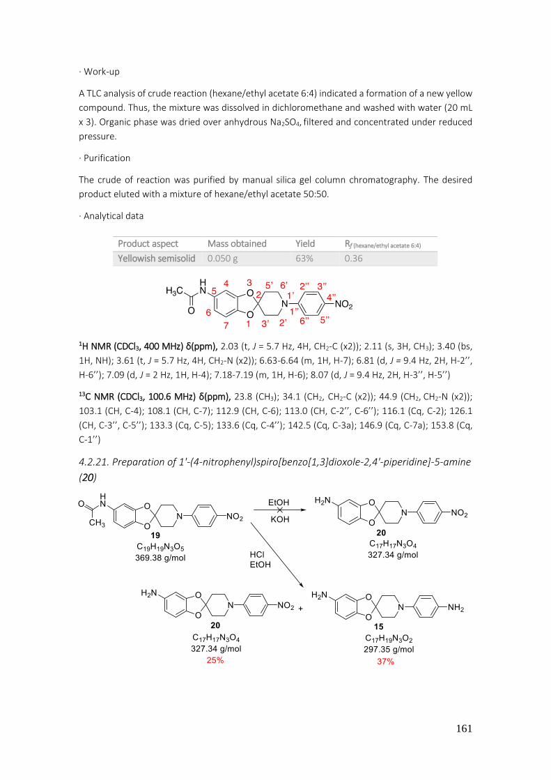
161
· Work-up
A TLC analysis of crude reaction (hexane/ethyl acetate 6:4) indicated a formation of a new yellow
compound. Thus, the mixture was dissolved in dichloromethane and washed with water (20 mL
x 3). Organic phase was dried over anhydrous Na2SO4, filtered and concentrated under reduced
pressure.
· Purification
The crude of reaction was purified by manual silica gel column chromatography. The desired
product eluted with a mixture of hexane/ethyl acetate 50:50.
· Analytical data
1H NMR (CDCl3, 400 MHz) δ(ppm), 2.03 (t, J = 5.7 Hz, 4H, CH2-C (x2)); 2.11 (s, 3H, CH3); 3.40 (bs,
1H, NH); 3.61 (t, J = 5.7 Hz, 4H, CH2-N (x2)); 6.63-6.64 (m, 1H, H-7); 6.81 (d, J = 9.4 Hz, 2H, H-2’’,
H-6’’); 7.09 (d, J = 2 Hz, 1H, H-4); 7.18-7.19 (m, 1H, H-6); 8.07 (d, J = 9.4 Hz, 2H, H-3’’, H-5’’)
13C NMR (CDCl3, 100.6 MHz) δ(ppm), 23.8 (CH3); 34.1 (CH2, CH2-C (x2)); 44.9 (CH2, CH2-N (x2));
103.1 (CH, C-4); 108.1 (CH, C-7); 112.9 (CH, C-6); 113.0 (CH, C-2’’, C-6’’); 116.1 (Cq, C-2); 126.1
(CH, C-3’’, C-5’’); 133.3 (Cq, C-5); 133.6 (Cq, C-4’’); 142.5 (Cq, C-3a); 146.9 (Cq, C-7a); 153.8 (Cq,
C-1’’)
4.2.21. Preparation of 1'-(4-nitrophenyl)spiro[benzo[1,3]dioxole-2,4'-piperidine]-5-amine
(20)
Product aspect Mass obtained Yield Rf (hexane/ethyl acetate 6:4)
Yellowish semisolid 0.050 g 63% 0.36
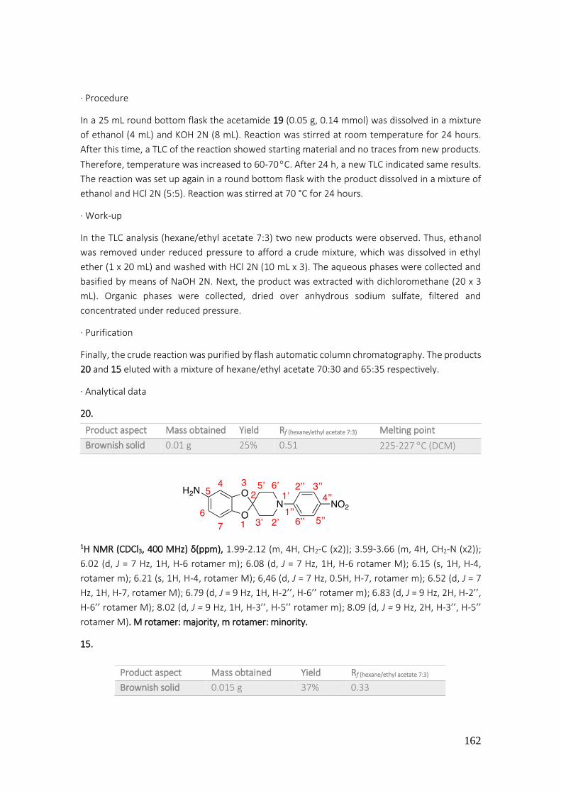
162
· Procedure
In a 25 mL round bottom flask the acetamide 19 (0.05 g, 0.14 mmol) was dissolved in a mixture
of ethanol (4 mL) and KOH 2N (8 mL). Reaction was stirred at room temperature for 24 hours.
After this time, a TLC of the reaction showed starting material and no traces from new products.
Therefore, temperature was increased to 60-70 C. After 24 h, a new TLC indicated same results.
The reaction was set up again in a round bottom flask with the product dissolved in a mixture of
ethanol and HCl 2N (5:5). Reaction was stirred at 70 °C for 24 hours.
· Work-up
In the TLC analysis (hexane/ethyl acetate 7:3) two new products were observed. Thus, ethanol
was removed under reduced pressure to afford a crude mixture, which was dissolved in ethyl
ether (1 x 20 mL) and washed with HCl 2N (10 mL x 3). The aqueous phases were collected and
basified by means of NaOH 2N. Next, the product was extracted with dichloromethane (20 x 3
mL). Organic phases were collected, dried over anhydrous sodium sulfate, filtered and
concentrated under reduced pressure.
· Purification
Finally, the crude reaction was purified by flash automatic column chromatography. The products
20 and 15 eluted with a mixture of hexane/ethyl acetate 70:30 and 65:35 respectively.
· Analytical data
20.
1H NMR (CDCl3, 400 MHz) δ(ppm), 1.99-2.12 (m, 4H, CH2-C (x2)); 3.59-3.66 (m, 4H, CH2-N (x2));
6.02 (d, J = 7 Hz, 1H, H-6 rotamer m); 6.08 (d, J = 7 Hz, 1H, H-6 rotamer M); 6.15 (s, 1H, H-4,
rotamer m); 6.21 (s, 1H, H-4, rotamer M); 6,46 (d, J = 7 Hz, 0.5H, H-7, rotamer m); 6.52 (d, J = 7
Hz, 1H, H-7, rotamer M); 6.79 (d, J = 9 Hz, 1H, H-2’’, H-6’’ rotamer m); 6.83 (d, J = 9 Hz, 2H, H-2’’,
H-6’’ rotamer M); 8.02 (d, J = 9 Hz, 1H, H-3’’, H-5’’ rotamer m); 8.09 (d, J = 9 Hz, 2H, H-3’’, H-5’’
rotamer M). M rotamer: majority, m rotamer: minority.
15.
Product aspect Mass obtained Yield Rf (hexane/ethyl acetate 7:3) Melting point
Brownish solid 0.01 g 25% 0.51 225-227 C (DCM)
Product aspect Mass obtained Yield Rf (hexane/ethyl acetate 7:3)
Brownish solid 0.015 g 37% 0.33

163
The analytical data (product aspect, 1H NMR, Rf, and others) was coincident with those previously
described for 15.
4.2.22. Preparation of 1-(1'-(4-nitrophenyl)spiro[benzo[1,3]dioxole-2,4'-piperidine]-5-
yl)ethanol (21)
· Procedure
In a 25 mL round bottom flask, the spiro-derivative 17 (0.06 g, 0.22 mmol) was dissolved in
methanol (10 mL) and sodium borohydride (0.01 g, 0.22 mmol) was slowly added. The reaction
was stirred for 2 hours at room temperature. After this time, a TLC of reaction (hexane/ethyl
acetate 6:4) showed a formation of two new products. As well, a remaining quantity of starting
material was observed. Therefore, one more equivalent of NaBH4 was added and the color of
reaction turned from yellow to brownish.
· Work-up
A new TLC indicated a presence of one compound and complete consumption of starting
material. To proceed with hydrolysis of the reaction, 2 mL of water were added. Finally, methanol
was evaporated in vacuo.
The crude mixture was dissolved in ethyl ether (10 mL) and washed with HCl 2N (10 mL x 3).
Aqueous phases were collected and basified by means of NaOH 2N. Then, the product was
extracted with dichloromethane (20 mL x 3). Organic phases were collected, dried over
anhydrous sodium sulfate, filtered and concentrated in vacuo.
· Purification
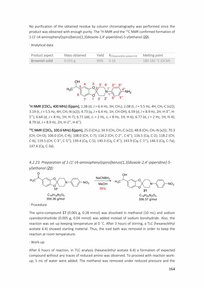
164
No purification of the obtained residue by column chromatography was performed since the
product was obtained with enough purity. The 1H NMR and the 13C NMR confirmed formation of
1-(1'-(4-aminophenyl)spiro[benzo[1,3]dioxole-2,4'-piperidine]-5-yl)ethanol (22).
· Analytical data
1H NMR (CDCl3, 400 MHz) δ(ppm), 1.38 (d, J = 6.4 Hz, 3H, CH3); 2.08 (t, J = 5.5 Hz, 4H, CH2-C (x2));
3.19 (t, J = 5.5 Hz, 4H, CH2-N (x2)); 4.73 (q, J = 6.4 Hz, 1H, CH-OH); 6.59 (d, J = 8.9 Hz, 2H, H-3’’, H-
5’’); 6.64 (d, J = 8 Hz, 1H, H-7); 6.71 (dd, J1 = 2 Hz, J2 = 8 Hz, 1H, H-6); 6.77 (d, J = 2 Hz, 1H, H-4);
8.79 (d, J = 8.9 Hz, 2H, H-2’’, H-6’’).
13C NMR (CDCl3, 100.6 MHz) δ(ppm), 25.0 (CH3); 34.9 (CH2, CH2-C (x2)); 48.8 (CH2, CH2-N (x2)); 70.3
(CH, CH-O); 106.0 (CH, C-4); 108.0 (CH, C-7); 116.2 (CH, C-2’’, C-6’’); 116.5 (Cq, C-2); 118.2 (CH,
C-6); 119.5 (CH, C-3’’, C-5’’); 139.4 (Cq, C-5); 140.3 (Cq, C-4’’); 143.9 (Cq, C-1’’); 146.5 (Cq, C-7a);
147.4 (Cq, C-3a).
4.2.23. Preparation of 1-(1'-(4-aminophenyl)spiro[benzo[1,3]dioxole-2,4'-piperidine]-5-
yl)ethanol (21)
· Procedure
The spiro-compound 17 (0.065 g, 0.28 mmol) was dissolved in methanol (10 mL) and sodium
cyanoborohydride (0.005 g, 0.04 mmol) was added instead of sodium borohydride. Also, the
reaction was set up keeping temperature at 0 C. After 3 hours of stirring, a TLC (hexane/ethyl
acetate 6:4) showed starting material. Thus, the iced bath was removed in order to keep the
reaction at room temperature.
· Work-up
After 6 hours of reaction, in TLC analysis (hexane/ethyl acetate 6:4) a formation of expected
compound without any traces of reduced amino was observed. To proceed with reaction work-
up, 5 mL of water were added. The methanol was removed under reduced pressure and the
Product aspect Mass obtained Yield Rf (hexane/ethyl acetate 6:4) Melting point
Brownish solid 0.055 g 99% 0.16 180-182 °C (DCM)

165
resultant crude mixture was extracted with dichloromethane (20 mL x 3). Organic phases were
collected, dried over anhydrous sodium sulfate, filtered and concentrated in vacuo.
· Purification
Finally, the crude reaction was purified by flash automatic column chromatography. The desired
product (21) eluted with a mixture of hexane/ethyl acetate 60:40.
· Analytical data
1H NMR (CDCl3, 400 MHz) δ(ppm), 1.39 (d, J = 6.4 Hz, 3H, CH3); 2.04 (t, J = 5.7 Hz, 4H, CH2-C (x2));
2.10 (s, 1H, OH); 3.62 (t, J = 5.7 Hz, 4H, CH2-N (x2)); 4.75 (q, J = 6.4 Hz, 1H, CH); 6.65 (d, J = 8 Hz,
1H, H-7); 6.74 (dd, J1 = 2 Hz, J2 = 8 Hz, 1H, H-6); 6.80 (d, J = 9.5 Hz, 2H, H-2’’, H-6’’); 7.79 (s, 1H, H-
4); 8.06 (d, J = 9.5 Hz, 2H, H-3’’, H-5’’).
13C NMR (CDCl3, 100.6 MHz) δ(ppm), 25.1 (CH3); 34.2 (CH2, CH2-C (x2)); 44.9 (CH2, CH2-N (x2)); 70.2
(CH, CH-O); 106.2 (CH, C-4); 108.2 (CH, C-7); 113.0 (CH, C-2’’, C-6’’); 115.7 (Cq, C-2); 118.6 (CH,
C-6); 126.1 (CH, C-3’’, C-5’’); 138.5 (Cq, C-5); 139.8 (Cq, C-4’’); 146.1 (Cq, C-7a); 147.1 (Cq, C-3a)
153.9 (Cq, C-1’’).
4.2.24. Preparation of 5-acetyl-1'-(4-aminophenyl)spiro[benzo[1,3]dioxole-2,4'-
piperidine] (23)
· Procedure
The spiro derivative 17 (0.1 g, 0.26 mmol) was added, dissolved in a mixture of methanol and
ethyl acetate, in a specific hydrogenation round bottom flask. Then, Pd-C (10%) (0.01 g, 0.094
mmol) in methanol and 0.2 mL of HCl 5N were added. The mixture of reaction was stirred
vigorously under H2 stream at atmospheric pressure for 16 hours.
· Work-up
The theoretical required volume for the reaction was 10 mL. Given that hydrogenation apparatus
was not completely hermetical, the consumed volume was 30 mL, higher than expected. TLC
(hexane/ethyl acetate 7:3) of reaction indicated a formation of a new compound. The mixture
Product aspect Mass obtained Yield Rf (hexane/ethyl acetate 6:4)
Yellow semisolid 0.030 g 50% 0.47

166
was filtered by means of a pleated filter and washed with 20 mL of methanol and was then
collected in a 100 mL round-bottomed flask. The solvent was removed under reduced pressure
to afford a residue that was dissolved in dichloromethane (10 mL) and washed with a solution of
NaOH 8N (20 mL x 3). Organic phase was dried over anhydrous Na2SO4, filtered and concentrated
under reduced pressure.
· Purification
Finally, the crude reaction was purified by flash automatic column chromatography. The desired
product eluted with a mixture of hexane/ethyl acetate 80:20.
· Analytical data
1H NMR (CDCl3, 400 MHz) δ(ppm), 2.09 (t, J = 6 Hz, 4H, CH2-C (x2)); 2.45 (s, 3H, CH3); 3.19 (t, J = 6
Hz, 4H, CH2-N (x2)); 3.40 (bs, 2H, NH2); 6.58 (d, J = 8.9 Hz, 2H, H-3’’, H-5’’); 6.72 (d, J = 8.1 Hz, 1H,
H-7); 6.78 (d, J = 8.9 Hz, 2H, H-2’’, H-6’’); 7.31 (d, J = 2 Hz, 1H, H-4); 7.45 (dd, J1 = 2 Hz, J2 = 8.1 Hz,
1H, H-6).
4.2.25. Preparation of 2-(4-(5-acetylspiro[benzo[1,3]dioxole-2,4'-piperidine]-1'-
yl)phenyl)-isoindoline-1,3-dione (24)
· Procedure In a Pyrex glass tube, previously flame-dried under argon, aniline 23 (0.08 g, 0.462 mmol)
dissolved in toluene (6 mL) and phthalic anhydride (0.05 g, 0.35 mmol) were added under argon
atmosphere. After 6 hours of reaction, a catalytic quantity of APTS (0.06 mmol) was added. The
tube closes tightly and the mixture was stirred overnight at 120-130 °C.
· Work-up
After 18 hours of reaction, a TLC analysis showed consumption of starting material (hexane/ethyl
acetate 7:3). Thus, toluene was removed in vacuo and the resultant crude mixture was diluted
Product aspect Mass obtained Yield Rf (hexane/ethyl acetate 7:3) Melting point
Brownish solid 0.085 g 93% 0.29 200-202 C (DCM)

167
with water (20 mL) and extracted with ethyl acetate (3 x 20 mL). The combined organic phases
were dried over anhydrous Na2SO4, filtered and concentrated under reduced pressure.
· Purification
Finally, the crude reaction was purified by flash automatic column chromatography. The desired
product (24) eluted with a mixture of hexane/ethyl acetate 30:70.
· Analytical data
1H NMR (CDCl3, 400 MHz) δ(ppm), 2.17 (t, J = 5.8 Hz, 4H, CH2-C (x2)); 2.54 (s, 3H, CH3); 3.52 (t, J =
5.8 Hz, 4H, CH2-N (x2)); 6.81 (d, J = 8.2 Hz, 1H, H-7); 7.06 (d, J = 8.9 Hz, 2H, H-2'', H-6''); 7.32 (d, J
= 8.9 Hz, 2H, H-3'', H-5''); 7.41 (d, J = 2 Hz, 1H, H-4); 7.54 (dd, J1 = 2 Hz, J2 = 8.2 Hz, 1H, H-6); 7.78
(dd, J1 = 3 Hz, J2 = 5.5 Hz, 2H, H-4’’’, H-5’’’); 7.94 (dd, J1 = 3 Hz, J2 = 5.5 Hz, 2H, H-3’’’, H-6’’’).
13C NMR (CDCl3, 100.6 MHz) δ(ppm), 26.4 (CH3); 34.4 (CH2, CH2-C (x2)); 46.6 (CH2, CH2-N (x2));
107.9 (CH, C-4); 108.1 (CH, C-7); 116.7 (CH, C-6); 117.8 (Cq, C-2); 123.6 (CH, C-2’’, C-6’’); 123.8
(Cq, C-4’’); 124.4 (CH, C-3’’’, C-6’’’); 125.5 (Cq, C-5); 127.6 (CH, C-3’’, C-5’’); 131.8 (Cq, C-C=O (x2));
134.3 ( CH, C-4’’’, C-5’’’); 147.6 (Cq, C-3a); 149.8 (Cq, C-1’’); 151.3 (Cq, C-7a); 167.6 (Cq, C=O (x2));
196.3 (Cq, C=O).
4.2.26. Preparation of 2-(4-(5-(1-hydroxyethyl)spiro[benzo[1,3]dioxole-2,4'-piperidine]-
1'-yl)phenyl)isoindoline-1,3-dione (25)
· Procedure
In a 50 mL round-bottom flask, the compound 24 (0.03 g, 0.07 mmol) and sodium borohydride
(0.005 g, 0.07 mmol) were added. Reaction was stirred for 3 hours at room temperature. Along
this time, a color change was observed, white to colorless.
· Work-up
Analysis of TLC showed a formation of a new product (hexane/ethyl acetate 7:3). Therefore, 5 mL
of water were added. Methanol was removed under reduced pressure and the obtained crude
Product aspect Mass obtained Yield Rf (hexane/ethyl acetate 3:7) Melting point
Whitish solid 0.036 g 36% 0.78 223-225 C (DCM)

168
mixture was extracted with dichloromethane (20 mL x 3). The combined organic layers were dried
over anhydrous Na2SO4, filtered and concentrated under reduced pressure.
· Purification
Finally, the reaction mixture was purified by flash automatic column chromatography. The
desired product eluted with a mixture of hexane/ethyl acetate 70:30. A co-evaporation with
toluene was done in order to dry the product and to close phthalimide ring.
· Analytical data
1H NMR (CDCl3, 400 MHz) δ(ppm), 1.39 (d, J = 6.4 Hz, 3H, CH3); 2.07-2.09 (m, 4H, CH2-C (x2)); 3.53
(t, J = 5.8 Hz, 4H, CH2-N (x2)); 4.74 (q, J = 6.4 Hz, 1H, CH-O); 6.65 (d, J = 8 Hz, 1H, H-7); 6.72 (dd, J1
= 2 Hz, J2 = 8 Hz, 1H, H-6); 6.78 (d, J = 2 Hz, 1H, H-4); 6.93 (d, J = 8 Hz, 2H, H-2'', H-6''); 7.44 (dt, J1
= 1.5 Hz, J2 = 7.2 Hz, 1H, C-H phthalimide); 7.50 (d, J = 8 Hz, 2H, H-3'', H-5''); 7.54 (dt, J1 = 1.5 Hz,
J2 = 7.2 Hz, 1H, C-H phthalimide); 7.58 (d, J = 7.2 Hz, 1H, C-H phthalimide); 7.73 (d, J = 7.2 Hz, 1H,
C-H phthalimide); (Due to the multiplicity observed in phthalimide protons it has been concluded
that imide ring was open during the time of spectrum acquisition).
13C NMR (CDCl3, 100.6 MHz) δ(ppm), 22.7 (CH3); 25.1 (CH2, CH2-C (x2)); 34.3 (CH2, CH2-N (x2)); 70.3
(CH-O); 106.1 (CH, C-4); 108.1 (CH, C-7); 117.4 (CH, C-6); 118.2 (Cq, C-2); 118.4 (CH, C-2’’, C-6’’);
122.2 (CH, phthalimide); 123.8 (CH, phthalimide); 124.2 (CH, C-3’’,C-5’’); 130.1 (CH; phthalimide);
131.7 (Cq, C-5); 132.6 (CH, phthalimide); 134.7 (Cq, phthalimide); 136.2 (Cq, phthalimide); 139.5
(Cq, C-4’’); 142.8 (Cq, C-1’’); 146.4 (Cq, C-7a); 147.2 (Cq, C-3a); 166.2 (Cq, C=O (x2)).
4.2.27. Preparation of 3-((4-(5-acetylspiro[benzo[1,3]dioxole-2,4'-piperidine]-1'-
yl)phenyl)amino)furan-2,5-dione (26)
Product aspect Mass obtained Yield Rf (hexane/ethyl acetate 7:3) Melting point
White solid 0.030 g 50% 0.16 193-195 C (DCM)

169
· Procedure
The general procedure of Buchwald-Hartwig cross-coupling reaction previously described, was
applied to spiro-compound 23 (0.15 mmol)
· Work-up
In TLC analysis (hexane/ethyl acetate 7:3) a new yellow product was observed. Thus, toluene was
removed under reduced pressure to afford a crude mixture which was dissolved in
dichloromethane (20 mL x 1) and washed with water (20 mL x 3). Organic phase was dried over
anhydrous sodium sulfate, filtered and concentrated in vacuo.
· Purification
Finally, the crude of reaction was purified by flash automatic column chromatography. The
desired product eluted with a mixture of hexane/ethyl acetate 70:30.
· Analytical data
1H NMR (CDCl3, 400 MHz) δ(ppm), 2.08 (t, J = 6 Hz, 4H, CH2-C (x2)); 2.47 (s, 3H, CH3); 3.44 (t, J = 6
Hz, 4H, CH2-N (x2)); 6.74 (d, J = 8 Hz, 1H, H-7); 6.93 (d, J = 2 Hz, 1H, H-4); 6.94 (d, J = 9 Hz, 2H, H-
2’’, H-6’’); 7.12 (d, J = 9 Hz, 2H, H-3’’, H-5’’); 7.34 (s, 1H, CH=); 7.47 (dd, J1 = 2 Hz, J2 = 8 Hz, 1H, H-
6).
13C NMR (CDCl3, 100.6 MHz) δ(ppm), 26.4 (CH3); 34.3 (CH2, CH2-C (x2)); 46.4 (CH2, CH2-N (x2));
107.9 (CH, C-4); 108.1 (CH, C-7); 116.6 (CH, C-6); 117.8 (Cq, C-2); 124.4 (CH, C-4’’’); 127.2 (CH, C-
2’’, C-6’’); 129.7 (Cq, C-5); 131.80 (Cq, C-4’’); 131.82 (CH, C-3’’, C-5’’); 147.6 (Cq, C-3a); 150.0 (Cq,
C-1’’); 151.2 (Cq, C-7a); 164.5 (Cq, C-3’’); 167.8 (Cq, C=O (x2)); 196.2 (Cq, C=O).
4.2.28. Preparation of 5-acetyl-1'-(phenylsulfonyl)spiro[benzo[1,3]dioxole-2,4'-
piperidine] (27)
Product aspect Mass obtained Yield Rf (hexane/ethyl acetate 7:3) Melting point
White solid 0.050 g 55% 0.40 225-227 C (DCM)
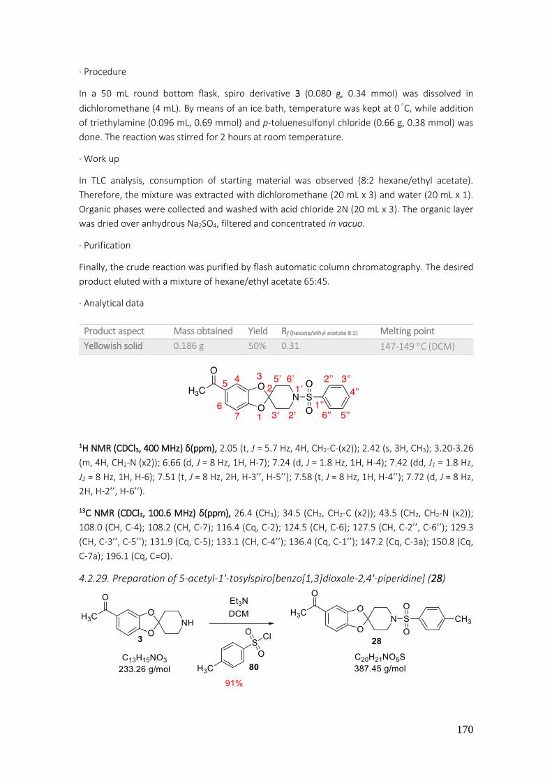
170
· Procedure
In a 50 mL round bottom flask, spiro derivative 3 (0.080 g, 0.34 mmol) was dissolved in
dichloromethane (4 mL). By means of an ice bath, temperature was kept at 0 C, while addition
of triethylamine (0.096 mL, 0.69 mmol) and p-toluenesulfonyl chloride (0.66 g, 0.38 mmol) was
done. The reaction was stirred for 2 hours at room temperature.
· Work up
In TLC analysis, consumption of starting material was observed (8:2 hexane/ethyl acetate).
Therefore, the mixture was extracted with dichloromethane (20 mL x 3) and water (20 mL x 1).
Organic phases were collected and washed with acid chloride 2N (20 mL x 3). The organic layer
was dried over anhydrous Na2SO4, filtered and concentrated in vacuo.
· Purification
Finally, the crude reaction was purified by flash automatic column chromatography. The desired
product eluted with a mixture of hexane/ethyl acetate 65:45.
· Analytical data
1H NMR (CDCl3, 400 MHz) δ(ppm), 2.05 (t, J = 5.7 Hz, 4H, CH2-C-(x2)); 2.42 (s, 3H, CH3); 3.20-3.26
(m, 4H, CH2-N (x2)); 6.66 (d, J = 8 Hz, 1H, H-7); 7.24 (d, J = 1.8 Hz, 1H, H-4); 7.42 (dd, J1 = 1.8 Hz,
J2 = 8 Hz, 1H, H-6); 7.51 (t, J = 8 Hz, 2H, H-3’’, H-5’’); 7.58 (t, J = 8 Hz, 1H, H-4’’); 7.72 (d, J = 8 Hz,
2H, H-2’’, H-6’’).
13C NMR (CDCl3, 100.6 MHz) δ(ppm), 26.4 (CH3); 34.5 (CH2, CH2-C (x2)); 43.5 (CH2, CH2-N (x2));
108.0 (CH, C-4); 108.2 (CH, C-7); 116.4 (Cq, C-2); 124.5 (CH, C-6); 127.5 (CH, C-2’’, C-6’’); 129.3
(CH, C-3’’, C-5’’); 131.9 (Cq, C-5); 133.1 (CH, C-4’’); 136.4 (Cq, C-1’’); 147.2 (Cq, C-3a); 150.8 (Cq,
C-7a); 196.1 (Cq, C=O).
4.2.29. Preparation of 5-acetyl-1'-tosylspiro[benzo[1,3]dioxole-2,4'-piperidine] (28)
Product aspect Mass obtained Yield Rf (hexane/ethyl acetate 8:2) Melting point
Yellowish solid 0.186 g 50% 0.31 147-149 C (DCM)

171
· Procedure
The conditions described in synthesis of spiro-derivative 27 were also applied to the reaction of
3 (0.30 mmol).
· Purification
The crude reaction obtained was purified by flash automatic column chromatography. The
desired product eluted with a mixture of hexane/ethyl acetate 65:45.
· Analytical data
1H NMR (CDCl3, 400 MHz) δ(ppm), 2.05 (t, J = 7 Hz, 4H, CH2-C-(x2)); 2.39 (s, 3H, CH3-Ar); 2.43 (s,
3H, CH3-C-O-); 3.16–3.28 (m, 4H, CH2-N (x2)); 6.67 (d, J = 8 Hz, 1H, H-7); 7.26 (s, 1H, H-4); 7.29 (d,
J = 8 Hz, 2H, H-3’’, H-5’’); 7.43 (d, J = 8 Hz, 1H, H-6); 7.61 (d, J = 8 Hz, 2H, H-2’’, H-6’’).
13C NMR (CDCl3, 100.6 MHz) δ(ppm), 21.6 (CH3-Ar); 26.4 (CH3-CO); 34.5 (CH2, CH2-C (x2)); 43.5 (CH2,
CH2-N (x2)); 108.0 (CH, C-4); 108.1 (CH, C-7); 116.5 (Cq, C-2); 124.6 (CH, C-6); 127.6 (CH, C-2’’, C-
6’’); 129.9 (CH, C-3’’, C-5’’); 131.9 (Cq, C-5); 133.6 (Cq, C-4’’); 143.9 (Cq, C-1’’); 147.3 (Cq, C-3a);
150.9 (Cq, C-7a); 196.1 (Cq, CO).
4.2.30. Preparation of 1-(1'-(phenylsulfonyl)spiro[benzo[1,3]dioxole-2,4'-piperidine]-5-
yl)ethan-1-ol (29)
· Procedure
in a 25 mL round bottom flask the sulfonyl derivative (27) (0.186 g, 0.5 mmol) was dissolved in
methanol (5 mL). Next, NaBH4 (0.03 mmol) was gently added along 5-10 minutes. The crude
reaction was stirred at room temperature.
· Work up
After 2 hours of reaction, a TLC showed a formation of a new product and consumption of starting
material (8:2 hexane/ethyl acetate). Hence, methanol was concentrated in vacuo to afford a
residue, which was dissolved in ethyl acetate (20 mL x 1) and washed with water (20 mL x 3). The
Product aspect Mass obtained Yield Rf (hexane/ethyl acetate 8:2) Melting point
Yellowish solid 0.117 g 91% 0.29 155-157 C (DCM)
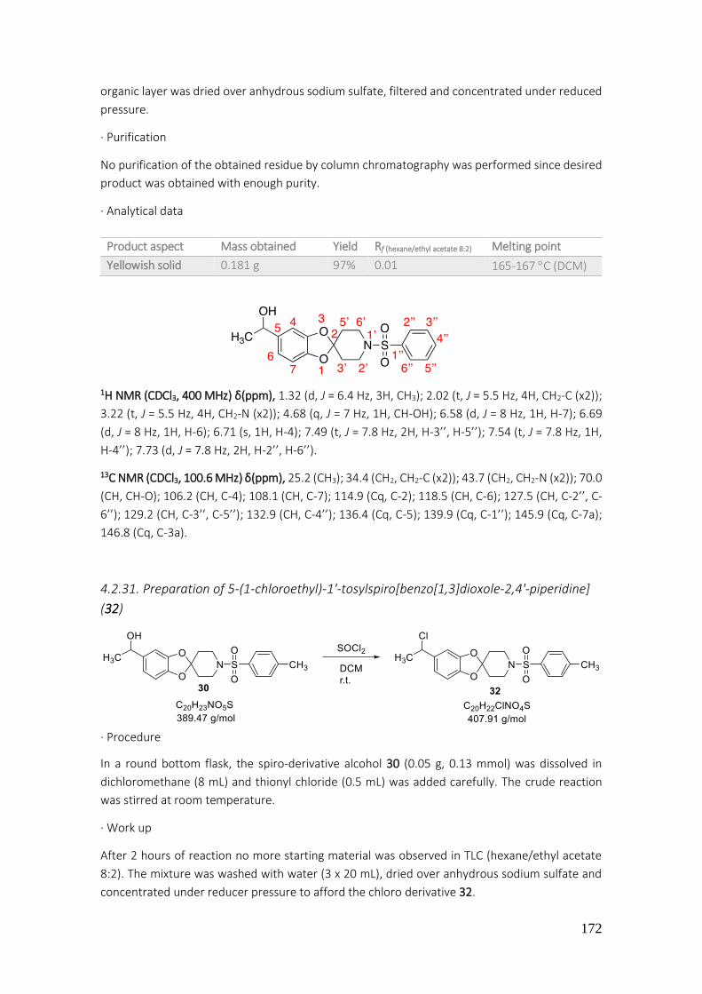
172
organic layer was dried over anhydrous sodium sulfate, filtered and concentrated under reduced
pressure.
· Purification
No purification of the obtained residue by column chromatography was performed since desired
product was obtained with enough purity.
· Analytical data
1H NMR (CDCl3, 400 MHz) δ(ppm), 1.32 (d, J = 6.4 Hz, 3H, CH3); 2.02 (t, J = 5.5 Hz, 4H, CH2-C (x2));
3.22 (t, J = 5.5 Hz, 4H, CH2-N (x2)); 4.68 (q, J = 7 Hz, 1H, CH-OH); 6.58 (d, J = 8 Hz, 1H, H-7); 6.69
(d, J = 8 Hz, 1H, H-6); 6.71 (s, 1H, H-4); 7.49 (t, J = 7.8 Hz, 2H, H-3’’, H-5’’); 7.54 (t, J = 7.8 Hz, 1H,
H-4’’); 7.73 (d, J = 7.8 Hz, 2H, H-2’’, H-6’’).
13C NMR (CDCl3, 100.6 MHz) δ(ppm), 25.2 (CH3); 34.4 (CH2, CH2-C (x2)); 43.7 (CH2, CH2-N (x2)); 70.0
(CH, CH-O); 106.2 (CH, C-4); 108.1 (CH, C-7); 114.9 (Cq, C-2); 118.5 (CH, C-6); 127.5 (CH, C-2’’, C-
6’’); 129.2 (CH, C-3’’, C-5’’); 132.9 (CH, C-4’’); 136.4 (Cq, C-5); 139.9 (Cq, C-1’’); 145.9 (Cq, C-7a);
146.8 (Cq, C-3a).
4.2.31. Preparation of 5-(1-chloroethyl)-1'-tosylspiro[benzo[1,3]dioxole-2,4'-piperidine]
(32)
· Procedure
In a round bottom flask, the spiro-derivative alcohol 30 (0.05 g, 0.13 mmol) was dissolved in
dichloromethane (8 mL) and thionyl chloride (0.5 mL) was added carefully. The crude reaction
was stirred at room temperature.
· Work up
After 2 hours of reaction no more starting material was observed in TLC (hexane/ethyl acetate
8:2). The mixture was washed with water (3 x 20 mL), dried over anhydrous sodium sulfate and
concentrated under reducer pressure to afford the chloro derivative 32.
Product aspect Mass obtained Yield Rf (hexane/ethyl acetate 8:2) Melting point
Yellowish solid 0.181 g 97% 0.01 165-167 C (DCM)
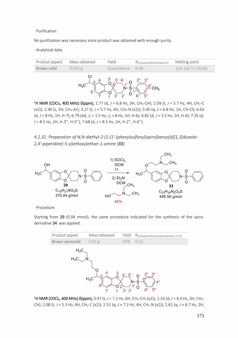
173
· Purification
No purification was necessary since product was obtained with enough purity.
· Analytical data
1H NMR (CDCl3, 400 MHz) δ(ppm), 1.77 (d, J = 6.8 Hz, 3H, CH3-CH); 2.09 (t, J = 5.7 Hz, 4H, CH2-C
(x2)); 2.46 (s, 3H, CH3-Ar); 3.27 (t, J = 5.7 Hz, 4H, CH2-N (x2)); 5.00 (q, J = 6.8 Hz, 1H, CH-Cl); 6.63
(d, J = 8 Hz, 1H, H-7); 6.79 (dd, J1 = 1.5 Hz, J2 = 8 Hz, 1H, H-6); 6.82 (d, J = 1.5 Hz, 1H, H-4); 7.35 (d,
J = 8.5 Hz, 2H, H-3’’, H-5’’); 7.68 (d, J = 8.5 Hz, 2H, H-2’’, H-6’’).
4.2.32. Preparation of N,N-diethyl-2-(1-(1'-(phenylsulfonyl)spiro[benzo[d][1,3]dioxole-
2,4'-piperidine]-5-yl)ethoxy)ethan-1-amine (33)
· Procedure
Starting from 29 (0.04 mmol), the same procedure indicated for the synthesis of the spiro-
derivative 34 was applied.
1H NMR (CDCl3, 400 MHz) δ(ppm), 0.97 (t, J = 7.2 Hz, 6H, CH3-CH2 (x2)); 1.33 (d, J = 6.4 Hz, 3H, CH3-
CH); 2.08 (t, J = 5.3 Hz, 4H, CH2-C (x2)); 2.51 (q, J = 7.2 Hz, 4H, CH2-N (x2)); 2.61 (q, J = 6.7 Hz, 2H,
Product aspect Mass obtained Yield Rf (hexane/ethyl acetate 8:2) Melting point
Brown solid 0.052 g Quantitative 0.68 154-156 C (DCM)
Product aspect Mass obtained Yield Rf (hexane/ethyl acetate/methanol 7:2:1)
Brown semisolid 0.02 g 45% 0.15

174
CH2-N); 3.29 (t, J = 5.3 Hz, 4H, CH2-N piperidine (x2)); 3.33 (t, J = 6.7 Hz, 2H, CH2-O); 4.27 (q, J =
6.4 Hz, 1H, CH-CH3); 6.61 (d, J = 7.9 Hz, 1H, H-7); 6.67 (d, J = 7.9 Hz, 1H, H-6); 6.70 (s, 1H, H-4);
7.56 (t, J = 7.7 Hz, 2H, H-3’’, H-5’’); 7.63 (t, J = 7.7 Hz, 1H, H-4’’); 7.78 (d, J = 7.7 Hz, 2H, H-2’’, H-
6’’).
13C NMR (CDCl3, 100.6 MHz) δ(ppm), 11.4 (CH3, CH3-CH2); 11.6 (CH3, CH3-CH2); 24.1 (CH3, CH3-CH);
34.4 (CH2-C (x2)); 43.7 (CH2, CH2-N); 47.5 (CH2-N piperidine (x2)); 52.4 (CH2, CH2-N (x2)); 66.6 (CH2,
CH2-O); 78.0 (CH, CH-O); 106.4 (CH, C-4); 107.2 (Cq, C-7); 114.8 (Cq, C-2); 119.4 (CH, C-6); 127.5
(CH, C-4’’); 129.2 (CH, C-2’’, C-6’’); 132.9 (CH, C-3’’, C-5’’); 136.5 (Cq, C-5); 137.8 (Cq, C-1’’); 145.9
(Cq, C-7a); 146.9 (Cq, C-3a).
4.2.33. Preparation of N,N-dimethyl-N-(1-(1'-tosylspiro[benzo[1,3]dioxole-2,4'-
piperidine]-5-yl)ethyl)ethane-1,2-diamine (34)
· Procedure
In a round bottom flask the spiro-compound 27 (0.065 g, 0.17 mmol) was dissolved in toluene (8
mL). Then N,N-dimethylethylenediamine (0.027 mL, 0.25 mmol) and p-toluenesulfonic acid were
added. A Dean-Stark system was used in order to remove the resultant water from reaction. The
mixture was stirred overnight at 140 C.
After 30 hours of reaction, toluene was removed under reduced pressure and the mixture was
extracted with ethyl acetate (20 mL x 3) and water (20 mL x 1). Organic phases were collected,
filtered and concentrated under reduced pressure, giving an intermediate product as a result.
That intermediary product (0.074 g, 0.02 mmol) was dissolved in methanol (8 mL) and NaBH4
(0.002 g, 0.05 mmol) was slowly added. Reaction was stirred at room temperature during 1 hour.

175
· Work up
After this time, a TLC of reaction showed a formation of a new compound (8:2 hexane/ethyl
acetate). Hence, methanol was removed under reduced pressure and the obtained crude mixture
was extracted with ethyl acetate (20 mL x 3) and water (20 mL x 1). Organic phases were collected,
dried over anhydrous sodium sulfate, filtered and concentrated in vacuo.
· Purification
The crude of reaction was purified by flash automatic column chromatography. The product
eluted with a mixture of hexane/ethyl acetate 50:50. The 1H NMR signals showed the alcohol 1-
(1'-tosylspiro[benzo[1,3]dioxole-2,4'-piperidine]-5-yl)ethanol (29).
· Analytical data
1H NMR (CDCl3, 400 MHz) δ(ppm), 1.42 (d, J = 6.4 Hz, 3H, CH3-CH); 2.00-2.03 (m, 4H, CH2-C (x2));
2.46 (s, 3H, CH3-Ar); 3.27 (t, J = 5.8 Hz, 4H, CH2-N (x2)); 4.77 (q, J = 6.4 Hz, 1H, CH-OH); 6.64 (d, J
= 8 Hz, 1H, H-7); 6.75 (dd, J1 = 2 Hz, J2 = 8 Hz, 1H, H-6); 6.78 (d, J = 2 Hz, 1H, H-4); 7.35 (d, J = 8
Hz, 2H, H-3’’, H-5’’); 7.68 (d, J = 8 Hz, 2H, H-2’’, H-6’’).
4.2.34. Preparation of N,N-dimethyl-N-(1-(1'-(phenylsulfonyl)spiro[benzo[d][1,3]dioxole-
2,4'-piperidine]-5-yl)ethyl)ethane-1,2-diamine (34)
· Procedure
In a round bottom flask, alcohol 29 (0.05 g, 0.13 mmol) was dissolved in dichloromethane (8 mL)
and thionyl chloride (0.5 mL). The reaction was stirred at room temperature for 2 hours. At this
time no more starting material was observed in TLC (hexane/ethyl acetate 8:2). The resulting
mixture was washed with water (3 x 20 mL), dried over anhydrous sodium sulfate and
concentrated under reducer pressure to afford the chloro derivative 31, which was diluted with
Product aspect Mass obtained Yield Rf (hexane/ethyl acetate 5:5) Melting point
White solid 0.059 g 79% 0.30 150-152 C (DCM)

176
dichloromethane (7 mL) in a 25 mL round bottom flask. Then, triethylamine (0.03 mL, 0.2 mmol)
and N,N-dimethylethylenediamine (0.02 mL, 0.15 mmol) were added. The crude reaction was
stirred at 100 °C.
· Work up
The reaction was stirred for 4 days, when no more starting material was observed in TLC
(hexane/ethyl acetate/methanol 7:2:1). Therefore, the mixture was washed with water (20 mL x
3), dried over anhydrous sodium sulfate, filtered off and concentrated in vacuo.
· Purification
The crude of the reaction was purified by flash automatic column chromatography. The product
eluted with a mixture of ethyl acetate/methanol 0:100.
· Analytical data
1H NMR (CDCl3, 400 MHz) δ(ppm), 1.21 (d, J = 6.4 Hz, 3H, CH3-CH); 2.02 (t, J = 5.7 Hz, 4H, CH2-C
(x2)); 2.08 (s, 3H, CH3-N (x2)); 2.22-2.29 (m, 2H, CH2-N(CH3)2); 2.39-2.42 (m, 2H, CH2-NH); 3.22 (t,
J = 5.7 Hz, 4H, CH2-N (x2)); 3.54 (q, J = 6.4 Hz, 1H, CH-NH); 6.54 (d, J = 7.9 Hz, 1H, H-7); 6.62 (d, J
= 7.9 Hz, 1H, H-6); 6.65 (s, 1H, H-4); 7.50 (t, J = 7.8 Hz, 2H, H-3’’, H-5’’); 7.48 (t, J = 7.8 Hz, 1H, H-
4’’); 7.73 (d, J = 7.8 Hz, 2H, H-2’’, H-6’’).
4.2.35. Preparation of N-(1-(1'-tosylspiro[benzo[d][1,3]dioxole-2,4'-piperidine]-5-
yl)ethyl)hexan-1-amine (35)
· Procedure Starting from 30 (0.04 mmol), the same procedure indicated for the synthesis of the spiro-
compound 34 was applied.
Product aspect Mass obtained Yield Rf (hexane/ethyl acetate/methanol 7:2:1)
Brown semisolid 0.01 g 16% 0.1
Product aspect Mass obtained Yield Rf (hexane/ethyl acetate/methanol 7:2:1)
Brown semisolid 0.02 g 20% 0.1

177
1H NMR (CDCl3, 400 MHz) δ(ppm), 0.74 (t, J = 6.4 Hz, 3H, CH3-CH2); 1.12-1.19 (m, 4H, CH2- (x2));
1.55 (q, J = 6.4 Hz, 2H, CH2-(x2)); 1.72 (d, J = 7 Hz, 3H, CH3-CH); 2.08-2.10 (m, 4H, CH2-C (x2)); 2.40
(s, 3H, CH3-Ar); 2.51-2.54 (m, 2H, CH2-); 3.07 (q, J = 7 Hz, 2H, CH2-N); 3.23 (q, J = 6.4 Hz, 2H, CH2-
); 3.32 (q, J = 7 Hz, 2H, CH2-N); 3.98-3.99 (m, 1H, CH-CH3); 6.62 (d, J = 8 Hz, 1H, H-6); 6.81 (d, J =
8 Hz, 1H, H-7); 7.08 (s, 1H, H-4); 7.29 (d, J = 8.2 Hz, 2H, H-3’’, H-5’’); 7.60 (d, J = 8.2 Hz, 2H, H-2’’,
H-6’’).
13C NMR (CDCl3, 100.6 MHz) δ(ppm), 12.9 (CH3, CH3-CH2); 20.0 (CH3, CH3-CH); 20.6 (CH3, CH3-Ar);
21.4 (CH2, CH2-CH3); 25.5 (CH2, CH2-CH2-CH3); 30.1 (CH2, CH2-CH2- CH2-CH3); 33.5 (CH2-C (x2));
42.7 (CH2, CH2-CH2-CH2- CH2-CH3); 44.7 (CH2-N (x2)); 46.8 (CH2, CH2-NH); 57.8 (CH, CH-NH); 106.8
(CH, C-4); 107.8 (Cq, C-7); 114.9 (Cq, C-2); 120.7 (CH, C-6); 126.5 (CH, C-2’’, C-6’’); 128.6 (Cq, C-
5); 128.9 (CH, C-3’’, C-5’’); 132.1 (Cq, C-4’’); 142.9 (Cq, C-1’’); 146.5 (Cq, C-7a); 146.7 (Cq, C-3a).
4.2.36. Preparation of 2-((4-(5-(1-(2-hydroxyethoxy)ethyl)spiro[benzo[1,3]dioxole-2,4'-
piperidine]-1'-yl)phenyl)amino)ethanol (36)
· Procedure
In a 25 mL round-bottom flask equipped with magnetic stirring, the aniline 22 (0.06 g, 0.19 mmol)
was dissolved in DMF (10 mL). Then, bromoethanol (0.07 g, 0.56 mmol) and potassium carbonate
(0.08 g, 0.56 mmol) were added. The crude reaction was stirred at 120 °C.
· Work-up
After 4 days under these conditions following the course of the reaction by TLC, its analysis
confirmed consumption of starting material (hexane/ethyl acetate 7:3). The mixture was

178
dissolved in ethyl ether (20 mL x 1) and washed with water (20 mL x 3). The organic phase was
dried over anhydrous sodium sulfate, filtered and concentrated under reduced pressure.
· Purification
Finally, the crude mixture was purified by flash automatic column chromatography. The product
eluted with a mixture of hexane/ethyl acetate 0:100. The 1H NMR signals indicated formation of
the non-desired 2-((4-(5-vinylspiro[benzo[1,3]dioxole-2,4'-piperidine]-1'-yl)phenyl)amino)-
ethanol (81).
· Analytical data
1H NMR (CDCl3, 400 MHz) δ(ppm), 2.14 (q, J = 6 Hz, 8H, CH2-C (x2), CH2-N (x2)); 3.40 (t, J = 5.5 Hz,
2H, CH2-N chain); 3.80 (m, 2H, CH2-O); 5.04 (d, J = 10.6 Hz, 1H, C=CH cis); 5.48 (d, J = 17.6 Hz, 1H,
C=CH trans); 6.54 (dd, J1 = 10.6 Hz, J2 = 17.6 Hz, 1H, CH=C); 6.64 (d, J = 8 Hz, 2H, H-3’’, H-5’’); 6.74
(d, J = 8 Hz, 2H, H-2’’, H-6’’); 6.85 (s, 1H, H-4); 6.97 (d, J = 8.8 Hz, 1H, H-7); 7.05 (d, J = 8.8 Hz, 1H,
H-6).
4.2.37. Preparation of 1-(4-(5-acetylspiro[benzo[1,3]dioxole-2,4'-piperidine]-1'-
yl)phenyl)-3-(2-fluoro-5-(trifluoromethyl)phenyl)urea (37)
· Procedure (general urea synthesis procedure) In a 50 mL round bottom flask equipped with magnetic stirring, the compound 23 (0.07 g, 0.22
mmol) was dissolved in THF (8 mL) and 2-fluoro-5-(trifluoromethyl)phenyl isocyanate (0.031 mL,
0.22 mmol) was added. The mixture reaction was stirred during 48 hours at room temperature.
· Work-up
A control of the reaction by TLC (hexane/ethyl acetate 7:3) showed consumption of starting
material. Thus, solvent was removed under reduced pressure to afford a crude mixture.
· Purification
The crude product obtained was purified by flash automatic column chromatography. The
desired product eluted with a mixture of hexane/ethyl acetate 58:42.
Product aspect Mass obtained Yield Rf (hexane/ethyl acetate 7:3)
Brown oil 0.015 g 25% 0.14
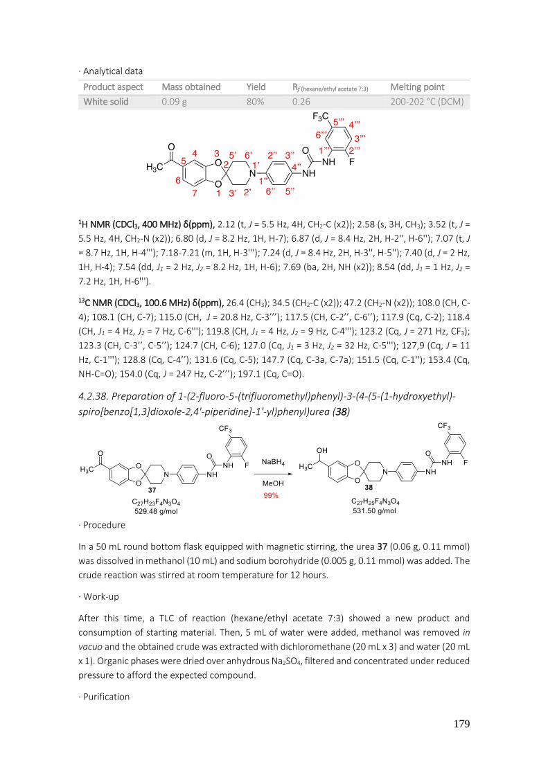
179
· Analytical data
1H NMR (CDCl3, 400 MHz) δ(ppm), 2.12 (t, J = 5.5 Hz, 4H, CH2-C (x2)); 2.58 (s, 3H, CH3); 3.52 (t, J =
5.5 Hz, 4H, CH2-N (x2)); 6.80 (d, J = 8.2 Hz, 1H, H-7); 6.87 (d, J = 8.4 Hz, 2H, H-2'', H-6''); 7.07 (t, J
= 8.7 Hz, 1H, H-4'''); 7.18-7.21 (m, 1H, H-3'''); 7.24 (d, J = 8.4 Hz, 2H, H-3'', H-5''); 7.40 (d, J = 2 Hz,
1H, H-4); 7.54 (dd, J1 = 2 Hz, J2 = 8.2 Hz, 1H, H-6); 7.69 (ba, 2H, NH (x2)); 8.54 (dd, J1 = 1 Hz, J2 =
7.2 Hz, 1H, H-6''').
13C NMR (CDCl3, 100.6 MHz) δ(ppm), 26.4 (CH3); 34.5 (CH2-C (x2)); 47.2 (CH2-N (x2)); 108.0 (CH, C-
4); 108.1 (CH, C-7); 115.0 (CH, J = 20.8 Hz, C-3’’’); 117.5 (CH, C-2’’, C-6’’); 117.9 (Cq, C-2); 118.4
(CH, J1 = 4 Hz, J2 = 7 Hz, C-6'''); 119.8 (CH, J1 = 4 Hz, J2 = 9 Hz, C-4'''); 123.2 (Cq, J = 271 Hz, CF3);
123.3 (CH, C-3’’, C-5’’); 124.7 (CH, C-6); 127.0 (Cq, J1 = 3 Hz, J2 = 32 Hz, C-5'''); 127,9 (Cq, J = 11
Hz, C-1'''); 128.8 (Cq, C-4’’); 131.6 (Cq, C-5); 147.7 (Cq, C-3a, C-7a); 151.5 (Cq, C-1''); 153.4 (Cq,
NH-C=O); 154.0 (Cq, J = 247 Hz, C-2’’’); 197.1 (Cq, C=O).
4.2.38. Preparation of 1-(2-fluoro-5-(trifluoromethyl)phenyl)-3-(4-(5-(1-hydroxyethyl)-
spiro[benzo[1,3]dioxole-2,4'-piperidine]-1'-yl)phenyl)urea (38)
· Procedure
In a 50 mL round bottom flask equipped with magnetic stirring, the urea 37 (0.06 g, 0.11 mmol)
was dissolved in methanol (10 mL) and sodium borohydride (0.005 g, 0.11 mmol) was added. The
crude reaction was stirred at room temperature for 12 hours.
· Work-up
After this time, a TLC of reaction (hexane/ethyl acetate 7:3) showed a new product and
consumption of starting material. Then, 5 mL of water were added, methanol was removed in
vacuo and the obtained crude was extracted with dichloromethane (20 mL x 3) and water (20 mL
x 1). Organic phases were dried over anhydrous Na2SO4, filtered and concentrated under reduced
pressure to afford the expected compound.
· Purification
Product aspect Mass obtained Yield Rf (hexane/ethyl acetate 7:3) Melting point
White solid 0.09 g 80% 0.26 200-202 °C (DCM)

180
No purification of the obtained residue by column chromatography was performed since product
was obtained with enough purity.
· Analytical data
1H NMR (Acetone-d6, 400 MHz) δ(ppm), 1.23 (d, J = 6.4 Hz, 3H, CH3); 1.98 (t, J = 5.7 Hz, 4H, CH2-C
(x2)); 3.26 (t, J = 5.7 Hz, 4H, CH2-N (x2)); 3.96 (d, J = 4 Hz, 1H, OH); 4.63 (q, J = 6.4 Hz, 1H, CH-O);
6.59 (d, J = 8 Hz, 1H, H-7); 6.68 (dd, J1 = 2 Hz, J2 = 8 Hz, 1H, H-6); 6.74 (d, J = 2 Hz, 1H, H-4); 6.88
(d, J = 9 Hz, 2H, H-2’’, H-6’’); 7.30 (d, J = 9 Hz, 2H, H-3'', H-5''); 8.13 (d, J = 3 Hz, 1H, H-3'''); 8.31 (s,
1H, H-6’’’); 8.67 (dd, J1 = 3 Hz, J2 = 7.6 Hz, 1H, H-4’’’).
13C NMR (Acetone-d6, 100.6 MHz) δ(ppm), 25.4 (CH3); 34.4 (CH2, CH2-C (x2)); 47.3 (CH2, CH2-N (x2));
68.9 (CH-OH); 105.9 (CH, C-4); 107.6 (CH, C-7); 115.4 (CH, J = 20.8 Hz, C-3’’’); 116.2 (Cq, C-2);
117.0 (CH, J1 = 4 Hz, J2 = 9 Hz, C-6'''); 117.2 (CH, C-2’’, C-6’’); 118.0 (CH, C-6); 118.9 (CH, J1 = 4 Hz,
J2 = 7 Hz, C-4’’’); 120.2 (CH, C-3’’, C-5’’); 124.2 (Cq, J = 271 Hz, CF3); 126.4 (Cq, J1 = 1.3 Hz, J2 = 32.5
Hz, C-5’’’); 128.8 (Cq, C-4’’); 129.3 (Cq, J = 11 Hz, C-1'''); 131.6 (Cq, C-5); 145.9 (Cq, C-1’’); 146.8
(Cq, C-7a); 147.0 (Cq, C-3a); 152.1 (Cq, C=O); 153.5 (Cq, J1 = 1.3 Hz, J2 = 246 Hz, C-2’’’).
4.2.39. Preparation of 1-ethyl-3-(4-(5-(1-hydroxyethyl)spiro[benzo[1,3]dioxole-2,4'-
piperidine]-1'-yl)phenyl)urea (39)
· Procedure In a 25 mL round-bottom flask equipped with magnetic stirring, the spiro derivative 22 (0.1 g,
0.31 mmol) was dissolved in THF and then ethyl isocyanate (0.02 mL, 0.31 mmol) was added. The
crude reaction was stirred at room temperature for 24 hours.
· Work-up
Product aspect Mass obtained Yield Rf (hexane/ethyl acetate 7:3) Melting point
White solid 0.06 g 99% 0.23 195-197 °C (DCM)

181
Analysis by TLC (hexane/ ethyl acetate 5:5) showed consumption of starting material. Therefore,
THF was evaporated under reduced pressure to afford the crude mixture.
· Purification
The crude was purified by flash automatic column chromatography. The desired product eluted
with a mixture of hexane/ethyl acetate 20:80.
· Analytical data
1H NMR (CDCl3, 400 MHz) δ(ppm), 1.12 (t, J = 6.4 Hz, 3H, CH3); 1.46 (d, J = 6.4 Hz, 3H, CH3-CH)
2.13-2.14 (m, 4H, CH2-C (x2)); 3.20-3.26 (m, 2H, CH2-NH); 3.27-3.40 (m, 4H, CH2-N (x2)); 4.74 (bs,
1H, OH); 4.82 (q, J = 6.4 Hz, 1H, CH-O); 6.71 (d, J = 8 Hz, 1H, H-7); 6.79 (dd, J1 = 2 Hz, J2 = 8 Hz, 1H,
H-6); 6.85 (d, J = 2 Hz, 1H, H-4); 6.91-6.94 (m, 2H, H-2’’, H-6’’); 7.15 (d, J = 8 Hz, 2H, H-3’’, H-5’’).
13C NMR (CDCl3, 100.6 MHz) δ(ppm), 14.01 (CH3-CH2); 22.7 (CH3, CH3-CH); 25.1 (CH2, CH2-C (x2));
34.3 (CH2, CH2-N (x2)); 70.3 (CH-O); 106.1 (CH, C-4); 108.1 (CH, C-7); 110.2 (Cq, C-2); 118.4 (CH,
C-6); 124.6 (Cq, C-5); 127.1 (Cq, C-4’’); 139.6 (Cq, C-1’’); 146.4 (Cq, C-7a); 147.2 (Cq, C-3a); 154.8
(Cq, CO); (The C-2’’, C-6’’ and C-3’’, C-5’’ carbons were not detected).
4.2.40. Preparation of 1-(pyridin-3-yl)-3-(4-(5-(3-(pyridin-3-yl)ureido)spiro[benzo[1,3]-
dioxole-2,4'-piperidine]-1'-yl)phenyl)urea (40)
· Procedure
Starting from 15 (3 mmol), the general procedure for urea synthesis described previously in this
work was applied in this case.
· Work-up
At 76 hours of reaction, a TLC of reaction showed the formation of a new product (ethyl acetate
100%). THF was removed in vacuo.
· Purification
Product aspect Mass obtained Yield Rf (hexane/ethyl acetate 5:5) Melting point
Whitish solid 0.1 g 80% 0.13 175-177 °C (DCM)

182
The crude mixture was purified by flash automatic column chromatography to afford the product.
The desired product eluted with a mixture of hexane/ethyl acetate 0:100.
· Analytical data
1H NMR (CDCl3, 400 MHz) δ(ppm), 2.04 (t, J = 5.8 Hz, 4H, CH2-C (x2) rotamer M); 2.25 (t, 1H, CH2-
C rotamer m); 2.48 (t, 1.7H, CH2-C rotamer m); 3.18 (t, 1.3H, CH2-N, rotamer m); 3.34 (t, J = 5.8
Hz, 4H, CH2-N (x2) rotamer M); 3.50 (t, 1.5H, CH2-N, rotamer m); 6.05 (dd, J1 = 2 Hz, J2 = 8 Hz, 1H,
H-6); 6.17 (d, J = 2 Hz, 1H, H-4); 6.50 (d, J = 8 Hz, 1H, H-7); 6.60 (t, J = 8.8 Hz, 1H, H-5’’’); 6.79 (d,
J = 8.8 Hz, 1H, H-4’’’); 6.88 (d, J = 9 Hz, 2H, H-2’’, H-6’’); 7.14 (d, J = 9 Hz, 2H, H-3’’, H-5’’); 7.96 (d
width, J = 8 Hz, 2H, H-6’’’, NH); 8.20 (bs, 2H, H-2’’’, NH); 8.28 (bs, 2H, NH2). M rotamer: majority,
m rotamer: minority.
HRMS (ESI +): Calculated for C23H23N5O3 [M+H]+: 418.1834, found 418.1883.
4.2.41. Preparation of 1-ethyl-3-(4-(5-(3-ethylureido)spiro[benzo[1,3]dioxole-2,4'-
piperidine]-1'-yl)phenyl)urea (41)
· Procedure
Starting from 15 (0.18 mmol), the general procedure for urea synthesis described previously in
this work was applied in this case. An excess of ethyl isocyanate was used (2 eq).
· Work-up
After 24 hours of stirring at room temperature, a TLC of reaction confirmed consumption of
starting material (ethyl acetate 100%). THF was removed in vacuo.
· Purification
The crude mixture obtained was purified by flash automatic column chromatography to afford
the expected compound. The desired product eluted with a mixture of hexane/ethyl acetate
0:100.
· Analytical data
Product aspect Mass obtained Yield Rf (hexane/ethyl acetate 0:1)
Grey semisolid 0.01 g 7% 0.15

183
1H NMR (CDCl3, 400 MHz) δ(ppm), 1.03 (t, J = 7.2 Hz, 6H, CH3 (x2)); 2.04 (t, J = 5.4 Hz, 2H, CH-C
axial (x2)); 3.08 (t, J = 7.4 Hz, 4H, CH2-NH (x2)); 3.15 (t, J = 5.4 Hz, 4H, CH2-N (x2)); 3.28 (t, J = 5.4
Hz, 2H, CH-C equatorial (x2)); 6.52 (dd, J1 = 2 Hz, J2 = 8.2 Hz, 1H, H-6); 6.57 (d, J = 8.2 Hz, 1H, H-
7); 6.78 (d, J = 2 Hz, 1H, H-4); 6.83 (d, J = 8.6 Hz, 2H, H-2'', H-6''); 7.09 (d, J = 8.6 Hz, 2H, H-3'', H-
5'').
13C NMR (CDCl3, 100.6 MHz) δ(ppm), 15.2 (CH3, (x2)); 34.3 (CH2, CH2-C); 34.5 (CH2, CH2-NH); 34.7
(CH2, CH2-C); 47.5 (CH2, CH2-NH); 47.6 (CH2, CH2-N (x2)); 104.9 (CH, C-4); 108.1 (CH, C-7); 113.8
(CH, C-6); 116.5 (Cq, C-2); 117.7 (CH, C-2’’, C-6’’); 122.7 (CH, C-3’’, C-5’’); 127.5 (Cq, C-5); 128.4
(Cq, C-4’’); 137.4 (Cq, C-3a); 138.7 (Cq, C-7a); 147.3 (Cq, C-1’’); 155.3 (Cq, C=O); 156.9 (Cq, C=O).
4.2.42. Preparation of 1-(4-((1'-acetylspiro[benzo[1,3]dioxole-2,4'-piperidine]-5-yl)-
amino)phenyl)-3-(4-chloro-3-(trifluoromethyl)phenyl)urea (42)
·Procedure
Starting from 9 (0.18 mmol), the general procedure for urea synthesis described previously in this
work was applied in this case.
· Work-up
At 24 hours of stirring, a precipitate in the mixture and analysis by TLC confirmed a formation of
a new product (hexane/ethyl acetate 5:5).
· Purification
The crude reaction was purified by flash automatic column chromatography. The desired product
eluted with a mixture of hexane/ethyl acetate 20:80.
· Analytical data
Product aspect Mass obtained Yield Rf (hexane/ethyl acetate 0:1) Melting point
White solid 0.074 g 79% 0.15 205-207 °C (DCM)
184
1H NMR (Acetone-d6, 400 MHz) δ(ppm), 0.90 (t, J = 5.8 Hz, 2H, CH-C axial (x2)); 1.93 (t, J = 5.8 Hz,
2H, CH-C equatorial (x2)); 2.08 (s, 3H, CH3); 3.70 (q, J = 5.8 Hz, 4H, CH2-N(x2)); 6.53 (dd, J1 = 2 Hz,
J2 = 8.3 Hz, 1H, H-6); 6.62 (d, J = 2 Hz, 1H, H-4); 6.69 (d, J = 8.3 Hz, 1H, H-7); 6.97 (d, J = 8.7 Hz, 2H,
H-2’’, H-6’’); 7.36 (dd, J1 = 1 Hz, J2 = 8.7 Hz, 2H, H-3’’, H-5’’); 7.52 (d, J = 8.8 Hz, 1H, H-5’’’); 7.72
(dd, J1 = 2.6 Hz, J2 = 8.8 Hz, 1H, H-6’’’); 8.07 (bs, 1H, NH); 8.14 (d, J = 2.6 Hz, 1H, H-2’’’); 8.49 (bs,
1H, NH).
13C NMR (Acetone-d6, 100.6 MHz) δ(ppm), 20.5 (CH3); 34.3 (CH2, CH-C); 34.9 (CH2, CH-C); 38.4
(CH2, CH-N); 43.3 (CH2, CH-N); 100.4 (CH, C-4); 108.4 (CH, C-7); 109.9 (CH, C-6); 116.0 (Cq, C-2);
117.3 (CH, C-2’’, C-6’’); 120.7 (CH, C-3’’, C-5’’); 122.6 (CH, C-2’’’); 122.8 (Cq, C-4’’’); 123.2 (Cq, J =
272 Hz, CF3); 125.9 (Cq, C-4’’); 127.2 (Cq, J = 33.6 Hz, C-3’’’); 131.7 (CH, C-5’’’, C-6’’’); 138.9 (Cq,
C-7a); 139.9 (Cq, C-1’’); 141.0 (Cq, C-1’’’); 141.1 (Cq, C-5); 147.5 (Cq, C-3a); 151.7 (Cq, N-C=O);
170.0 (Cq, NH-CO).
4.2.43. Preparation of 1’-acetyl-3H-spiro[benzoxazole-2,4'-piperidine] (43)
· Procedure
In a 50 mL round bottom-flask the 1-acetylpiperidine-4-one (0.17 mL, 0.92 mmol) was dissolved
in 10 mL of toluene and o-aminophenol (0.14 g, 0.91 mmol) was added in the mixture. A Dean-
Stark system was used to remove resultant water. The reaction was stirred for 24 hours at 130-
140 C.
· Work-up
In a TLC analysis a new product was observed (hexane/ethyl acetate 6:4). Reaction was cooled
down and toluene was removed to afford the crude mixture, which was dissolved in ethyl acetate
(20 mL x 1) and washed with water (20 mL x 3). Organic phase was dried over anhydrous Na2SO4,
filtered and concentrated under reduced pressure.
· Purification
Product aspect Mass obtained Yield Rf (hexane/ethyl acetate 5:5)
Greenish semisolid 0.065 g 67% 0.5

185
Finally, the crude residue obtained was purified by flash automatic column chromatography. The
obtained product eluted with a mixture of hexane/ethyl acetate 0:100.
· Analytical data
1H NMR (CDCl3, 400 MHz) δ(ppm), 2.10 (s, 3H, CH3); 2.47 (dd, J1 = 6.3 Hz, J2 = 15.8 Hz, 4H, CH2-C
(x2)); 3.74 (t, J = 6.3 Hz, 2H, CH-N axial (x2)); 3.87 (t, J = 6.3 Hz, 2H, CH-N equatorial (x2)); 6.57 (dt,
J1 = 1.5 Hz, J2 = 8 Hz, 1H, H-6); 6.63 (dd, J1 = 1.5 Hz, J2 = 8 Hz, 1H, H-4); 6.77 (dt, J1 = 1.5 Hz, J2 = 8
Hz, 1H, H-5); 6.79 (d, J = 8 Hz, 1H, H-7).
4.2.44. Preparation of 1’-acetyl-3H-spiro[benzoxazole-2,4'-piperidine] (43)
· Procedure
The general procedure of transacetylation described previously in this work to the synthesis of
the spiro derivative 1 was applied (0.09 mmol of 1-acetylpiperidine-4-one).
· Purification
Finally, the crude mixture obtained was purified by flash automatic column chromatography. The
desired product eluted with a mixture of hexane/ethyl acetate 0:100.
4.2.45. Preparation of 1,1'-(3H-spiro[benzoxazole-2,4'-piperidine]-1',5-diyl)bis(ethanone)
(44)
· Procedure
General acetylation procedure described previously in this work was applied in this case (0.43
mmol of 43).
Product aspect Mass obtained Yield Rf (hexane/ethyl acetate 6:4)
Brown oil 0.05 g 20% 0.14
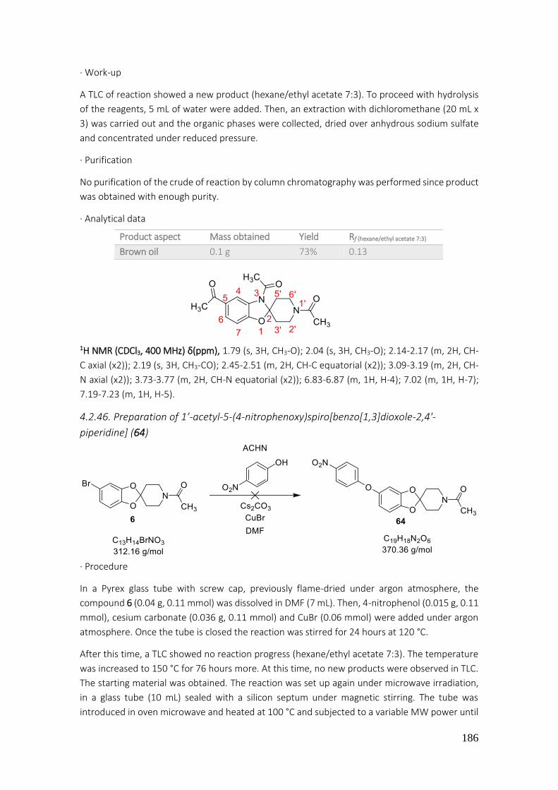
186
· Work-up
A TLC of reaction showed a new product (hexane/ethyl acetate 7:3). To proceed with hydrolysis
of the reagents, 5 mL of water were added. Then, an extraction with dichloromethane (20 mL x
3) was carried out and the organic phases were collected, dried over anhydrous sodium sulfate
and concentrated under reduced pressure.
· Purification
No purification of the crude of reaction by column chromatography was performed since product
was obtained with enough purity.
· Analytical data
1H NMR (CDCl3, 400 MHz) δ(ppm), 1.79 (s, 3H, CH3-O); 2.04 (s, 3H, CH3-O); 2.14-2.17 (m, 2H, CH-
C axial (x2)); 2.19 (s, 3H, CH3-CO); 2.45-2.51 (m, 2H, CH-C equatorial (x2)); 3.09-3.19 (m, 2H, CH-
N axial (x2)); 3.73-3.77 (m, 2H, CH-N equatorial (x2)); 6.83-6.87 (m, 1H, H-4); 7.02 (m, 1H, H-7);
7.19-7.23 (m, 1H, H-5).
4.2.46. Preparation of 1’-acetyl-5-(4-nitrophenoxy)spiro[benzo[1,3]dioxole-2,4'-
piperidine] (64)
· Procedure
In a Pyrex glass tube with screw cap, previously flame-dried under argon atmosphere, the
compound 6 (0.04 g, 0.11 mmol) was dissolved in DMF (7 mL). Then, 4-nitrophenol (0.015 g, 0.11
mmol), cesium carbonate (0.036 g, 0.11 mmol) and CuBr (0.06 mmol) were added under argon
atmosphere. Once the tube is closed the reaction was stirred for 24 hours at 120 °C.
After this time, a TLC showed no reaction progress (hexane/ethyl acetate 7:3). The temperature
was increased to 150 °C for 76 hours more. At this time, no new products were observed in TLC.
The starting material was obtained. The reaction was set up again under microwave irradiation,
in a glass tube (10 mL) sealed with a silicon septum under magnetic stirring. The tube was
introduced in oven microwave and heated at 100 °C and subjected to a variable MW power until
Product aspect Mass obtained Yield Rf (hexane/ethyl acetate 7:3)
Brown oil 0.1 g 73% 0.13

187
300 W (an IR sensor measured the temperature of glass tube surface). The same results were
obtained.
4.2.47. Preparation of 1’-acetyl-5-(piperazin-1-yl)spiro[benzod[1,3]dioxole-2,4'-
piperidine] (67)
· Procedure
The Buchwald-Hartwig cross-coupling reaction conditions described in the synthesis of 8, were
applied in this reaction (0.033 mmol of 6).
· Work-up
After 48 hours of reaction, a TLC of reaction showed consumption of starting material
(hexane/ethyl acetate 5:5). The toluene was evaporated under reduced pressure. Then, the crude
resdiue was dissolved in dichloromethane (20 mL x 1) and washed with water (20 mL x 3). The
organic phase was dried over anhydrous sodium sulfate, filtered and concentrated in vacuo.
· Purification
Finally, the obtained mixture was purified by flash automatic column chromatography. The main
product eluted with a mixture of hexane/ethyl acetate 45:55. The 1H NMR signals showed
formation of 6.
Preparation of 1’-acetyl-5-(1,4-dioxa-8-azaspiro[4.5]decan-8-yl)spiro[benzo[1,3]dioxole-2,4'-
piperidine] (68)
· Procedure
The conditions of Buchwald-Hartwig cross-coupling reaction were applied to 6 (0.033 mmol) in
this reaction.
· Work-up
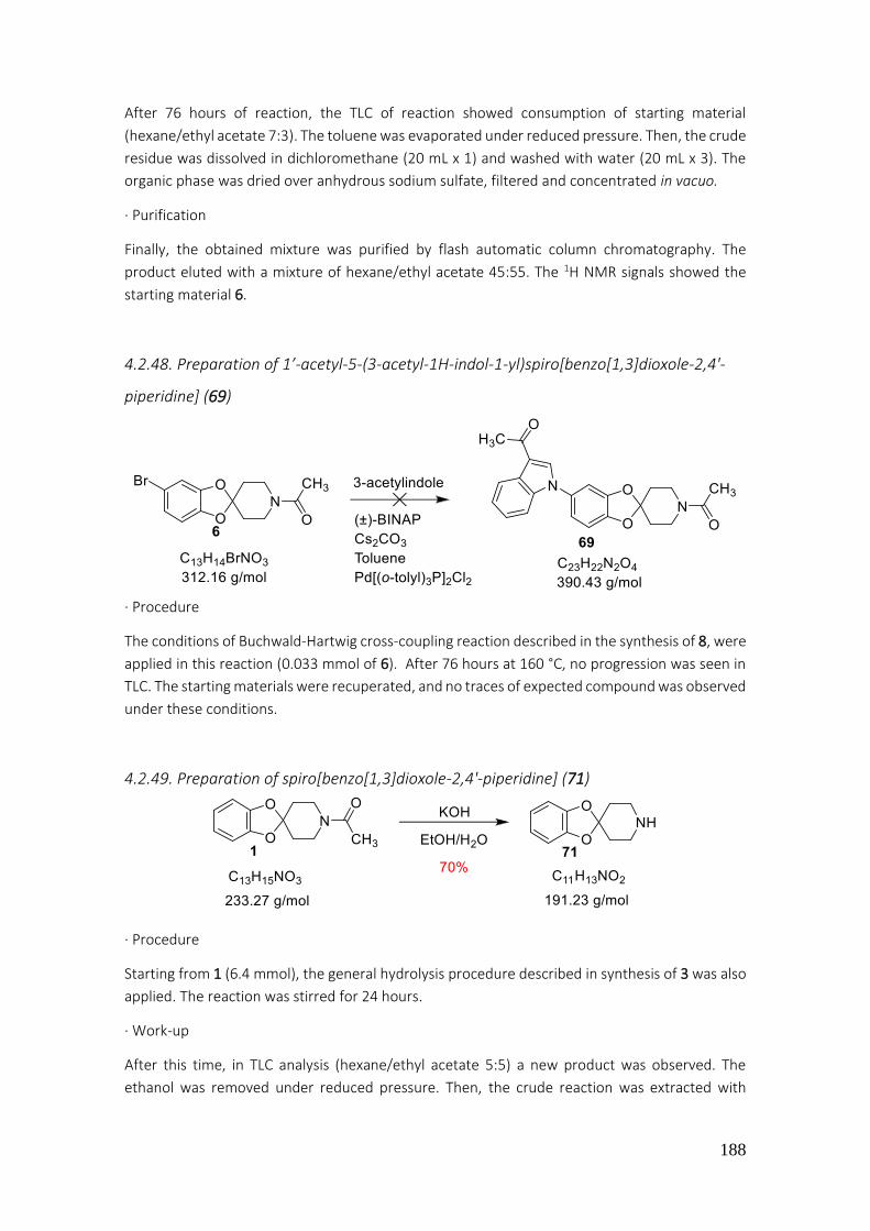
188
After 76 hours of reaction, the TLC of reaction showed consumption of starting material
(hexane/ethyl acetate 7:3). The toluene was evaporated under reduced pressure. Then, the crude
residue was dissolved in dichloromethane (20 mL x 1) and washed with water (20 mL x 3). The
organic phase was dried over anhydrous sodium sulfate, filtered and concentrated in vacuo.
· Purification
Finally, the obtained mixture was purified by flash automatic column chromatography. The
product eluted with a mixture of hexane/ethyl acetate 45:55. The 1H NMR signals showed the
starting material 6.
4.2.48. Preparation of 1’-acetyl-5-(3-acetyl-1H-indol-1-yl)spiro[benzo[1,3]dioxole-2,4'-
piperidine] (69)
· Procedure
The conditions of Buchwald-Hartwig cross-coupling reaction described in the synthesis of 8, were
applied in this reaction (0.033 mmol of 6). After 76 hours at 160 °C, no progression was seen in
TLC. The starting materials were recuperated, and no traces of expected compound was observed
under these conditions.
4.2.49. Preparation of spiro[benzo[1,3]dioxole-2,4'-piperidine] (71)
· Procedure
Starting from 1 (6.4 mmol), the general hydrolysis procedure described in synthesis of 3 was also
applied. The reaction was stirred for 24 hours.
· Work-up
After this time, in TLC analysis (hexane/ethyl acetate 5:5) a new product was observed. The
ethanol was removed under reduced pressure. Then, the crude reaction was extracted with
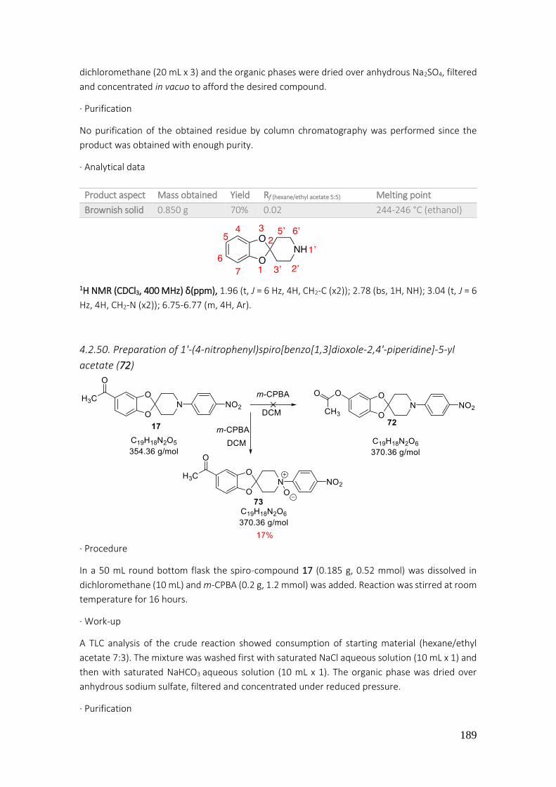
189
dichloromethane (20 mL x 3) and the organic phases were dried over anhydrous Na2SO4, filtered
and concentrated in vacuo to afford the desired compound.
· Purification
No purification of the obtained residue by column chromatography was performed since the
product was obtained with enough purity.
· Analytical data
1H NMR (CDCl3, 400 MHz) δ(ppm), 1.96 (t, J = 6 Hz, 4H, CH2-C (x2)); 2.78 (bs, 1H, NH); 3.04 (t, J = 6
Hz, 4H, CH2-N (x2)); 6.75-6.77 (m, 4H, Ar).
4.2.50. Preparation of 1'-(4-nitrophenyl)spiro[benzo[1,3]dioxole-2,4'-piperidine]-5-yl
acetate (72)
· Procedure
In a 50 mL round bottom flask the spiro-compound 17 (0.185 g, 0.52 mmol) was dissolved in
dichloromethane (10 mL) and m-CPBA (0.2 g, 1.2 mmol) was added. Reaction was stirred at room
temperature for 16 hours.
· Work-up
A TLC analysis of the crude reaction showed consumption of starting material (hexane/ethyl
acetate 7:3). The mixture was washed first with saturated NaCl aqueous solution (10 mL x 1) and
then with saturated NaHCO3 aqueous solution (10 mL x 1). The organic phase was dried over
anhydrous sodium sulfate, filtered and concentrated under reduced pressure.
· Purification
Product aspect Mass obtained Yield Rf (hexane/ethyl acetate 5:5) Melting point
Brownish solid 0.850 g 70% 0.02 244-246 °C (ethanol)
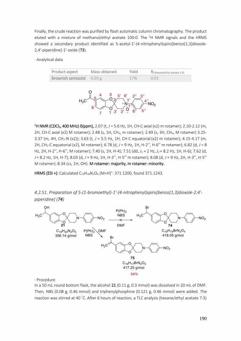
190
Finally, the crude reaction was purified by flash automatic column chromatography. The product
eluted with a mixture of methanol/ethyl acetate 100:0. The 1H NMR signals and the HRMS
showed a secondary product identified as 5-acetyl-1'-(4-nitrophenyl)spiro[benzo[1,3]dioxole-
2,4'-piperidine]-1'-oxide (73).
· Analytical data
1H NMR (CDCl3, 400 MHz) δ(ppm), 2.07 (t, J = 5.6 Hz, 1H, CH-C axial (x2) m rotamer); 2.10-2.12 (m,
2H, CH-C axial (x2) M rotamer); 2.48 (s, 1H, CH3, m rotamer); 2.49 (s, 3H, CH3, M rotamer) 3.25-
3.37 (m, 4H, CH2-N (x2)); 3.63 (t, J = 5.5 Hz, 1H, CH-C equatorial (x2) m rotamer); 4.15-4.17 (m,
2H, CH2-C equatorial (x2), M rotamer); 6.78 (d, J = 9 Hz, 1H, H-2’’, H-6’’ m rotamer); 6.82 (d, J = 8
Hz, 2H, H-2’’, H-6’’, M rotamer); 7.40 (s, 1H, H-4); 7.51 (dd, J1 = 2 Hz, J2 = 8.2 Hz, 1H, H-6); 7.62 (d,
J = 8.2 Hz, 1H, H-7); 8.03 (d, J = 9 Hz, 1H, H-3’’, H-5’’ m rotamer); 8.08 (d, J = 9 Hz, 2H, H-3’’, H-5’’
M rotamer); 8.34 (cs, 1H, OH). M rotamer: majority, m rotamer: minority.
HRMS (ESI +): Calculated C19H18N2O6 [M+H]+: 371.1200, found 371.1243.
4.2.51. Preparation of 5-(1-bromoethyl)-1'-(4-nitrophenyl)spiro[benzo[1,3]dioxole-2,4'-
piperidine] (74)
· Procedure In a 50 mL round bottom flask, the alcohol 21 (0.11 g, 0.3 mmol) was dissolved in 20 mL of DMF.
Then, NBS (0.08 g, 0.46 mmol) and triphenylphosphine (0.121 g, 0.46 mmol) were added. The
reaction was stirred at 40 C. After 6 hours of reaction, a TLC analysis (hexane/ethyl acetate 7:3)
Product aspect Mass obtained Yield Rf (hexane/ethyl acetate 1:9)
Brownish semisolid 0.03 g 17% 0.01

191
showed a mixture between starting material and new product. The reaction mixture was stirred
for 24 hours.
· Work-up
After this time, an extraction with ethyl ether (20 mL x 3) and water (20 mL x 1) was carried out
in order to remove DMF from organic phase. Organic phases were collected, dried over
anhydrous Na2SO4, filtered and concentrated under reduced pressure.
· Purification
Finally, the obtained residue was purified by flash automatic column chromatography. The
product eluted with a mixture of hexane/ethyl acetate 80:20. The 1H NMR and 13C NMR confirmed
that 5-(1-bromovinyl)-1'-(4-nitrophenyl)spiro[benzo[1,3]dioxole-2,4'-piperidine] (75) was
obtained.
· Analytical data
1H NMR (CDCl3, 400 MHz) δ(ppm), 2.12 (t, J = 6 Hz, 4H, CH2-C (x2)); 3.70 (t, J = 6 Hz, 4H, CH2-N
(x2)); 5.12 (d, J = 10.8 Hz, 1H, H cis-Br); 5.57 (d, J = 17.5 Hz, 1H, H trans-Br); 6.72 (d, J = 8 Hz, 1H,
H-7); 6.83 (dd, J1 = 2 Hz, J2 = 8 Hz, 1H, H-6); 6.88 (d, J = 9.4 Hz, 2H, H-2’’, H-6’’); 6.93 (d, J = 2 Hz,
1H, H-4); 8.14 (d, J = 9.4 Hz, 2H, H-3’’, H-5’’).
13C NMR (CDCl3, 100.6 MHz) δ(ppm), 34.2 (CH2, CH2-C (x2)); 45.0 (CH2, CH2-N (x2)); 105.5 (CH, C-
4); 108.4 (CH, C-7); 111.9 (Cq, C-2); 113.0 (CH, C-2’’, C-6’’); 115.9 (Cq, CH-Br); 120.9 (CH, C-6);
126.1 (CH, C-3’’, C-5’’); 132.0 (CH2, CH2=Br); 136.3 (Cq, C-5); 138.5 (Cq, C-4’’); 146.7 (Cq, C-3a);
147.3 (Cq, C-7a); 153.9 (Cq, C-1’’).
Product aspect Mass obtained Yield Rf (hexane/ethyl acetate 7:3)
Brown semisolid 0.07 g 54% 0.80

192
4.3. Preparation of pyrrolo[2,3-b]pyrazines
General procedure A (Sonogoshira coupling)
In a Pyrex glass tube equipped with a magnetic stirring, previously flame-dried under argon
atmosphere, 2-amino-3,5-dibromopyrazine (1.0 mmol), the appropriate aryl acetylene (0.95
mmol), Pd(dba)2 (0.03 mmol) and tri(2-furyl)phosphine (0.06 mmol) were added. Then, THF (3
mL) and triethylamine (0.45 mL), previously outgassed were added as well. The reaction mixture
was stirred for 16 hours. When control of reaction through TLC confirmed consumption of
starting material, 20 mL of NH4Cl solution were added and the obtained crude was extracted with
ethyl acetate (15 mL x 3). Organic phases were collected, dried over anhydrous sodium sulfate,
filtered and concentrated under reduced pressure to afford a solid which was purified by means
of a flash automatic silica gel column chromatography, using as an eluent an ascending polarity
from hexane to ethyl acetate.
General procedure B (Cyclization)
In a Pyrex glass tube equipped with a magnetic stirring, previously flame-dried under argon
atmosphere, the pyrazine obtained previously from Sonogoshira reaction (1.0 mmol), 6-8 mL of
acetonitrile and cesium carbonate (1.0 mmol) were added. The mixture was stirred at 130-140 °C
for 16 hours. When control of reaction through TLC confirmed consumption of starting material,
solvent was evaporated in vacuo, 20 mL of water were added, and the obtained crude of reaction
was extracted with ethyl acetate (15 mL x 3). If needed a warm extraction was carried out to
facilitate layers separation. Organic phases were collected, dried over anhydrous sodium sulfate,
filtered and concentrated under reduced pressure affording the desired product.
General procedure C (Suzuki-Miyaura coupling)
In a Pyrex glass tube equipped with a magnetic stirring, previously flame-dried under argon
atmosphere, the appropriate 5H-pyrrolo[2,3-b]pyrazine (1.0 mmol), acetonitrile (4 mL),
previously outgassed under argon, appropriate boronic acid (1.0 mmol), dried sodium carbonate
(2.0 mmol), Pd(P(C6H5)3)4 (0.02 mmol) and 0.6 mL of distillated water were added. The reaction
was stirred at 140 °C until no more starting material was observed in TLC. Then, a saturated NaCl
aqueous solution was added (20 mL) and the obtained crude was extracted with ethyl acetate
(20 mL x 3). Organic phases were collected, dried over anhydrous sodium sulfate, filtered and
concentrated under reduced pressure affording a solid, which was purified by means of a flash
automatic silica gel column chromatography, using as an eluent an ascending polarity from
hexane to ethyl acetate.
General procedure D (halogenation of C-7)
In a Pyrex glass tube equipped with a magnetic stirring, previously flame-dried under argon
atmosphere, the appropriate 5H-pyrrolo[2,3-b]pyrazine in acetonitrile and N-bromosuccinimide
(NBS) were added under argon atmosphere. The reaction mixture was stirred at room
temperature until no starting material could be detected by TLC. Solvent was removed under
reduced pressure and the obtained crude extracted with ethyl acetate (20 mL x 1) and water (20
mL x 3). Organic phases were dried over sodium sulfate, filtered and solvent removed to dryness
affording the desired characteristic yellow brominated product.
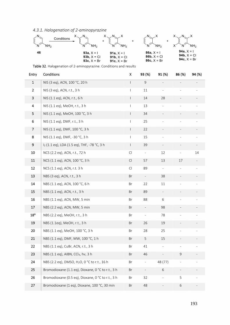
193
4.3.1. Halogenation of 2-aminopyrazine
Table 32. Halogenation of 2-aminopyrazine. Conditions and results
Entry Conditions X 93 (%) 91 (%) 86 (%) 94 (%)
1 NIS (3 eq), ACN, 100 °C, 20 h I 9 - - -
2 NIS (3 eq), ACN, r.t., 3 h I 11 - - -
3 NIS (1.1 eq), ACN, r.t., 6 h I 14 28 - -
4 NIS (1.1 eq), MeOH, r.t., 3 h I 13 - - -
5 NIS (1.1 eq), MeOH, 100 °C, 3 h I 34 - - -
6 NIS (1.1 eq), DMF, r.t., 3 h I 25 - - -
7 NIS (1.1 eq), DMF, 100 °C, 3 h I 22 - - -
8 NIS (1.1 eq), DMF, -30 °C, 3 h I 15 - - -
9 I2 (1.1 eq), LDA (1.5 eq), THF, -78 °C, 3 h I 39 - - -
10 NCS (2.2 eq), ACN, r.t., 72 h Cl - 12 - 14
11 NCS (1.1 eq), ACN, 100 °C, 3 h Cl 57 13 17 -
12 NCS (1.1 eq), ACN, r.t. 3 h Cl 89 - - -
13 NBS (3 eq), ACN, r.t., 3 h Br - 38 - -
14 NBS (1.1 eq), ACN, 100 °C, 6 h Br 22 11 - -
15 NBS (1.1 eq), ACN, r.t., 3 h Br 89 - - -
16 NBS (1.1 eq), ACN, MW, 5 min Br 88 6 - -
17 NBS (2.2 eq), ACN, MW, 5 min Br - 98 - -
18A NBS (2.2 eq), MeOH, r.t., 3 h Br - 78 - -
19 NBS (1.1eq), MeOH, r.t., 3 h Br 26 19 - -
20 NBS (1.1 eq), MeOH, 100 °C, 3 h Br 28 25 - -
21 NBS (1.1 eq), DMF, MW, 100 °C, 1 h Br 5 15 - -
22 NBS (1.1 eq), CuBr, ACN, r.t., 3 h Br 41 - - -
23 NBS (1.1 eq), AIBN, CCl4, hv, 3 h Br 46 - 9 -
24 NBS (2.2 eq), DMSO, H2O, 0 °C to r.t., 16 h Br - 48 (77) - -
25 Bromodioxane (1.1 eq), Dioxane, 0 °C to r.t., 3 h Br - 6 - -
26 Bromodioxane (0.5 eq), Dioxane, 0 °C to r.t., 3 h Br 32 - 5 -
27 Bromodioxane (1 eq), Dioxane, 100 °C, 30 min Br 48 - 6 -

194
· General procedure
General procedure using conventional heating: To a solution of pyrazine-2-amine (1.0 mmol) in
methanol, acetonitrile, dimethylformamide or lithium diisopropylethylamine (5-10 mL) the N-
iodosuccinimide (NIS) (procedure E) (1.1 mmol – 3.0 mmol) or I2 (1.1 mmol) or N-
chlorosuccinimide (NCS) (procedure F) or N-bromosuccinimide (NBS) (procedure G) were added
(in the reaction with I2 also LDA (1.5 mmol) was added). The obtained reaction mixture was
stirred until no starting material could be detected by thin-layer chromatography. Then solvent
was removed under reduced pressure to give the crude product, which was purified by silica gel
column chromatography.
General procedure with microwaves radiation assistance: To a solution of the pyrazine-2-amine
(1.0 mmol) in acetonitrile (5 mL) the N-iodosuccinimide (NIS) (procedure E) (1.1 mmol – 3.0
mmol) or I2 (1.1 mmol) or N-chlorosuccinimide (NCS) (procedure F) or N-bromosuccinimide (NBS)
(procedure G) (1.1 mmol – 2.2 mmol) were added to the glass tube (10 mL) sealed with a silicon
septum equipped with magnetic stirring. The tube was introduced in the oven microwave and
was heated to 100 °C (external temperature) and subjected to a variable MW power until 300 W
(an IR sensor measured the temperature of glass tube surface) until total consumption of starting
material could be detected by thin-layer chromatography. Then the solvent was removed under
reduced pressure to give the crude product, which was purified by silica gel column
chromatography. Alternatively, the residue of reaction can be extracted with ethyl ether and
water (3 x 15 mL), dried over sodium sulfate, filtered and the solvent removed to dryness.
· Analytical data
a) 2-Amino-5-iodopyrazine
1H NMR (CDCl3, 400 MHz) δ(ppm), 7.83 (s, 1H, H-3); 8.21 (s, 1H, H-6).
Mp: 110-113 °C (DCM)
b) 2-Amino-3.5-diiodopyrazine
1H NMR (CDCl3, 400 MHz) δ(ppm), 7.99 (s, 1H, H-3); 8.21 (s, 1H, H-6).
c) 2-Amino-5-chloropyrazine
1H NMR (CDCl3, 400 MHz) δ(ppm), 7.77 (s, 1H, H-3); 8.00 (s, 1H, H-6).
Mp: 122-124 °C (DCM)
d) 2-Amino-3.5-dichloropyrazine
1H NMR (CDCl3, 400 MHz) δ(ppm), 5.12 (bs, 2H, NH2); 7.97 (s, 1H, H-6).
Mp: 115-118 °C (DCM)
e) 2-Amino-5-bromopyrazine
1H NMR (CDCl3, 400 MHz) δ(ppm), 7.76 (s, 1H, H-3); 8.10 (s, 1H, H-6).
Mp: 113-115 °C (DCM)
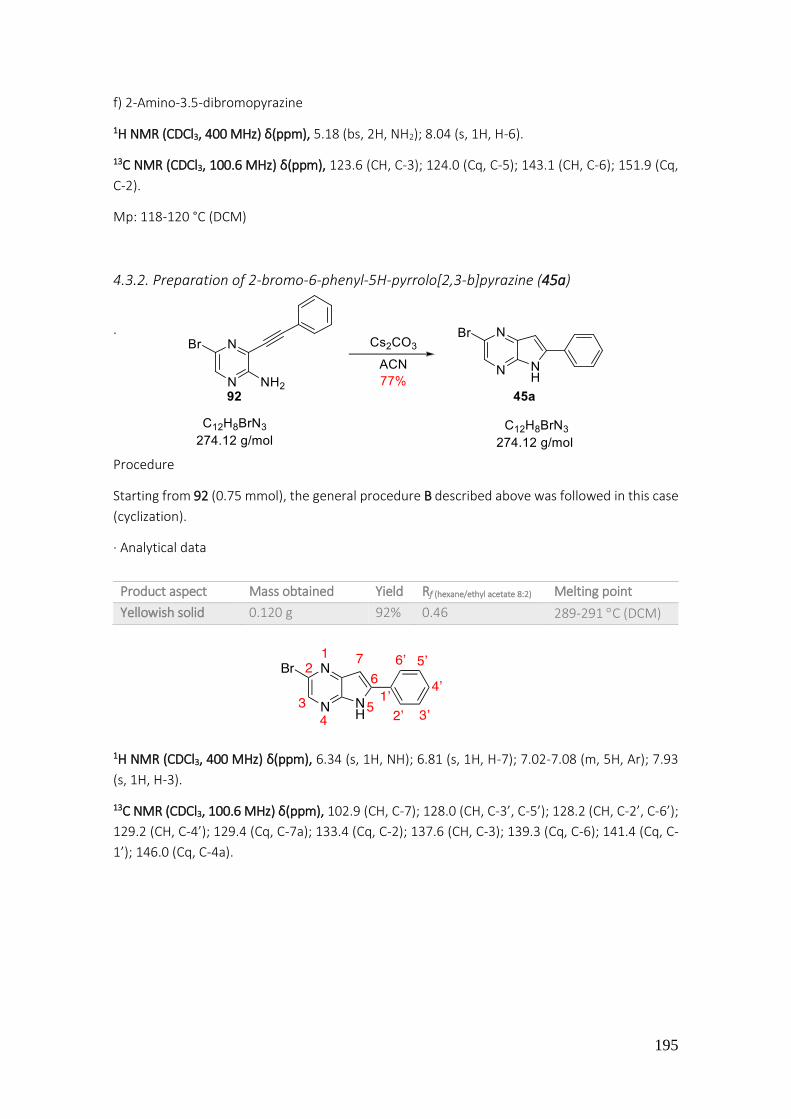
195
f) 2-Amino-3.5-dibromopyrazine
1H NMR (CDCl3, 400 MHz) δ(ppm), 5.18 (bs, 2H, NH2); 8.04 (s, 1H, H-6).
13C NMR (CDCl3, 100.6 MHz) δ(ppm), 123.6 (CH, C-3); 124.0 (Cq, C-5); 143.1 (CH, C-6); 151.9 (Cq,
C-2).
Mp: 118-120 °C (DCM)
4.3.2. Preparation of 2-bromo-6-phenyl-5H-pyrrolo[2,3-b]pyrazine (45a)
·
Procedure
Starting from 92 (0.75 mmol), the general procedure B described above was followed in this case
(cyclization).
· Analytical data
1H NMR (CDCl3, 400 MHz) δ(ppm), 6.34 (s, 1H, NH); 6.81 (s, 1H, H-7); 7.02-7.08 (m, 5H, Ar); 7.93
(s, 1H, H-3).
13C NMR (CDCl3, 100.6 MHz) δ(ppm), 102.9 (CH, C-7); 128.0 (CH, C-3’, C-5’); 128.2 (CH, C-2’, C-6’);
129.2 (CH, C-4’); 129.4 (Cq, C-7a); 133.4 (Cq, C-2); 137.6 (CH, C-3); 139.3 (Cq, C-6); 141.4 (Cq, C-
1’); 146.0 (Cq, C-4a).
Product aspect Mass obtained Yield Rf (hexane/ethyl acetate 8:2) Melting point
Yellowish solid 0.120 g 92% 0.46 289-291 C (DCM)

196
4.3.3. Preparation of 2-bromo-6-(4-fluorophenyl)-5H-pyrrolo[2,3-b]pyrazine (45b)
· Procedure
Starting from 97 (1 mmol), the general procedure B described above was followed in this case
(cyclization).
· Purification
The crude mixture obtained was purified by flash automatic silica gel column chromatography.
The desired product eluted with a mixture of hexane/ethyl acetate 80:20.
· Analytical data
1H NMR (CDCl3, 400 MHz) δ(ppm), 6.80 (s, 1H, H-7); 6.95 (dd, J1 = 5 Hz, J2 = 8.5 Hz, 2H, H-3’, H-5’);
7.66 (dd, J1 = 5 Hz, J2 = 8.5 Hz, 2H, H-2’, H-6’); 8.18 (s, 1H, H-3).
4.3.4. Preparation of 2-bromo-6-(4-chlorophenyl)-5H-pyrrolo[2,3-b]pyrazine (45c)
· Procedure
Starting from 98 (1.28 mmol), the general procedure B described above was followed in this case
(cyclization).
· Purification
Product aspect Mass obtained Yield Rf (hexane/ethyl acetate 8:2) Melting point
Yellowish solid 0.280 g 96% 0.46 > 300 C (DCM)

197
No purification was performed since the product was obtained with enough purity.
· Analytical data
1H NMR (CDCl3 + Acetone-D6 drop, 400 MHz) δ(ppm), 6.90 (s, 1H, H-7); 7.49 (d, J = 8.4 Hz, 2H, H-
3’, H-5’); 7.66 (d, J = 8.4 Hz, 2H, H-2’, H-6’); 8.27 (s, 1H, H-3).
4.3.5. Preparation of 6-phenyl-2-(p-tolyl)-5H-pyrrolo[2,3-b]pyrazine (47)
· Procedure
Starting from 45a (0.33 mmol), the general procedure C described previously in this work was
followed in this case (Suzuki-Miyaura coupling reaction).
· Analytical data
1H NMR (CDCl3, 400 MHz) δ(ppm), 2.38 (s, 3H, CH3); 7.00 (s, 1H, H-7); 7.27 (d, J = 8 Hz, 2H, H-3’’,
H-5’’); 7.38 (t, J = 7.7 Hz, 1H, H-4’); 7.46 (t, J = 7.7 Hz, 2H, H-3’, H-5’); 7.81 (d, J = 8 Hz, 2H, H-2’’,
H-6’’); 7.83 (d, J = 7.7 Hz, 2H, H-2’, H-6’); 8.50 (s, 1H, H-3).
13C NMR (CDCl3, 100.6 MHz) δ(ppm), 33.5 (CH3); 106.1 (CH, C-7); 129.7 (CH, C-2’’, C-6’’); 130.7
(CH, C-3’’, C-5’’); 133.0 (CH, C-2’, C-6’); 133.5 (CH, C-3’, C-5’); 138.0 (CH, C-4’); 142.6 (CH, C-3);
Quaternary carbons were not observed, in this spectrum.
Product aspect Mass obtained Yield Rf (hexane/ethyl acetate 8:2) Melting point
Yellow solid 0.227 g 57% 0.66 > 300 C (DCM)
Product aspect Mass obtained Yield Rf (hexane/ethyl acetate 8:2) Melting point
Yellow solid 0.090 g 96% 0.47 150-152C (DCM)
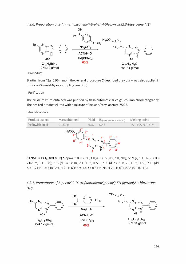
198
4.3.6. Preparation of 2-(4-methoxyphenyl)-6-phenyl-5H-pyrrolo[2,3-b]pyrazine (48)
· Procedure
Starting from 45a (0.96 mmol), the general procedure C described previously was also applied in
this case (Suzuki-Miyaura coupling reaction).
· Purification
The crude mixture obtained was purified by flash automatic silica gel column chromatography.
The desired product eluted with a mixture of hexane/ethyl acetate 75:25.
· Analytical data
1H NMR (CDCl3, 400 MHz) δ(ppm), 3.89 (s, 3H, CH3-O); 6.53 (bs, 1H, NH); 6.99 (s, 1H, H-7); 7.00-
7.02 (m, 1H, H-4’); 7.05 (d, J = 8.8 Hz, 2H, H-3’’, H-5’’); 7.09 (d, J = 7 Hz, 2H, H-3’, H-5’); 7.15 (dd,
J1 = 1.7 Hz, J2 = 7 Hz, 2H, H-2’, H-6’); 7.91 (d, J = 8.8 Hz, 2H, H-2’’, H-6’’); 8.35 (s, 1H, H-3).
4.3.7. Preparation of 6-phenyl-2-(4-(trifluoromethyl)phenyl)-5H-pyrrolo[2,3-b]pyrazine
(49)
Product aspect Mass obtained Yield Rf (hexane/ethyl acetate 8:2) Melting point
Yellowish solid 0.182 g 63% 0.46 153-155 C (DCM)

199
· Procedure
Starting from 45a (0.68 mmol), the general procedure C described previously in this work was
applied in this case (Suzuki-Miyaura coupling reaction).
· Purification
The crude mixture obtained was purified by flash automatic silica gel column chromatography.
The desired product eluted with a mixture of hexane/ethyl acetate 85:15.
· Analytical data
1H NMR (DMSO-d6, 400 MHz) δ(ppm), 7.24 (d, J = 2 Hz, 1H, H-7); 7.44 (t, J = 7.4 Hz, 1H, H-4’); 7.53
(t, J = 7.4 Hz, 2H, H-3’, H-5’); 7.85 (d, J = 8.2 Hz, 2H, H-3’’, H-5’’); 8.04 (d, J = 7.4 Hz, 2H, H-2’, H-
6’); 8.36 (d, J = 8.2 Hz, 2H, H-2’’, H-6’’); 8.93 (s, 1H, H-3).
4.3.8. Preparation of 6-(4-fluorophenyl)-2-(p-tolyl)-5H-pyrrolo[2,3-b]pyrazine (50)
· Procedure
Starting from 45b (1.02 mmol), the general procedure C described above was followed in this
case (Suzuki-Miyaura coupling reaction).
· Purification
The crude reaction was purified by flash automatic silica gel column chromatography. The desired
product eluted with a mixture of hexane/ethyl acetate 80:20.
· Analytical data
Product aspect Mass obtained Yield Rf (hexane/ethyl acetate 8:2)
Yellowish solid 0.153 g 66% 0.37
Product aspect Mass obtained Yield Rf (hexane/ethyl acetate 8:2) Melting point
Yellowish solid 0.257 g 80% 0.20 293-295 C (DCM)

200
1H NMR (CDCl3, 400 MHz) δ(ppm), 2.37 (s, 3H, CH3); 6.81 (s, 1H, H-7); 7.15 (d, J = 8.5 Hz, 2H, H-3',
H-5'); 7.28 (d, J = 7.8 Hz, 2H, H-3", H-5"); 7.77 (dd, J1 = 5 Hz, J2 = 8.6 Hz, 2H, H-2’, H-6’); 7.86 (d, J
= 7.8 Hz, 2H, H-2’’, H-6’’); 8.18 (s, 1H, H-3); 9.88 (bs, 1H, NH).
4.3.9. Preparation of 6-(4-fluorophenyl)-2-(m-tolyl)-5H-pyrrolo[2,3-b]pyrazine (51)
· Procedure
Starting from 45b (1.03 mmol), the general procedure C described above was followed in this
case (Suzuki-Miyaura coupling reaction).
· Purification
A purification of the residue obtained to remove excess of boronic acid was carried out by flash
automatic silica gel column chromatography. The desired product eluted with a mixture of
hexane/ethyl acetate 80:20.
· Analytical data
1H NMR (CDCl3, 400 MHz) δ(ppm), 2.46 (s, 3H, CH3); 4.88 (s, 1H, NH); 7.00 (s, 1H, H-7); 7.25 (d, J =
9 Hz, 2H, H-3’, H-5’); 7.29-7.31 (m, 1H, H-4”); 7.38-7.40 (m, 1H, H-5”); 7.78 (d, J = 9 Hz, 2H, H-2’,
H-6’); 8.00 (s, 1H, H-3); 7.98-8.03 (m, 2H, H-2”, H-6”); 10.4 (bs, 1H, NH).
Product aspect Mass obtained Yield Rf (hexane/ethyl acetate 8:2) Melting point
Yellowish solid 0.131 g 42% 0.31 292-294 C (DCM)
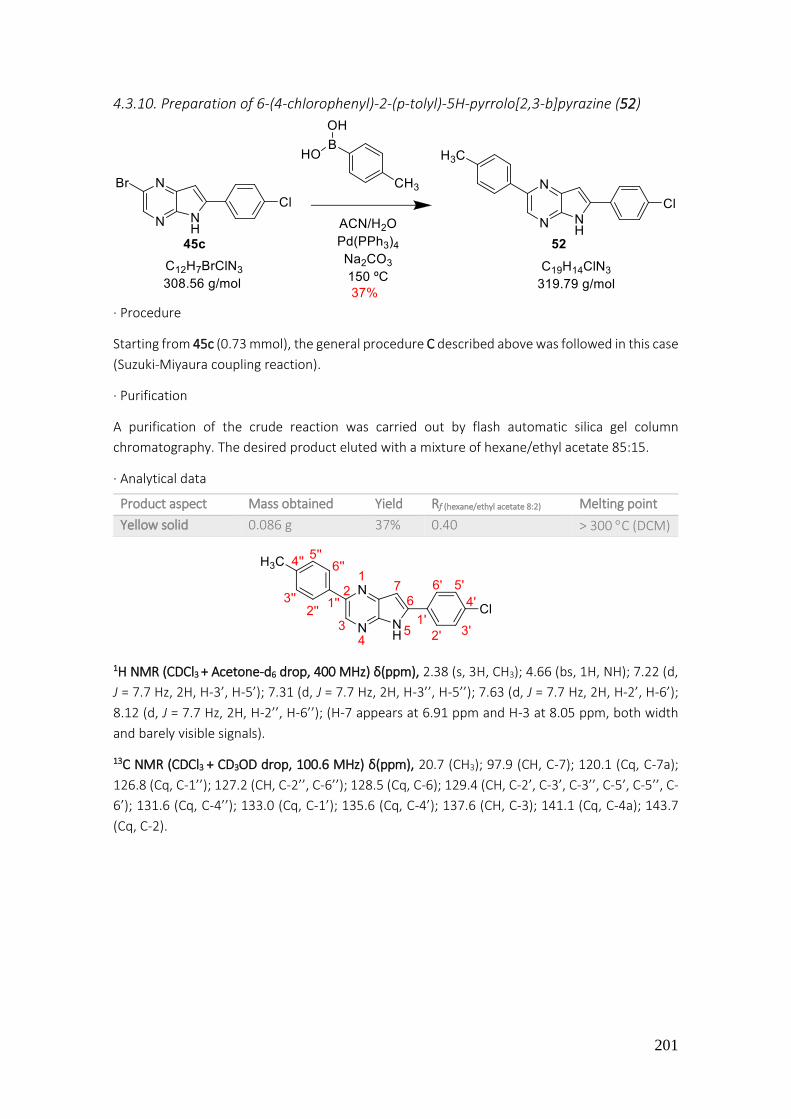
201
4.3.10. Preparation of 6-(4-chlorophenyl)-2-(p-tolyl)-5H-pyrrolo[2,3-b]pyrazine (52)
· Procedure
Starting from 45c (0.73 mmol), the general procedure C described above was followed in this case
(Suzuki-Miyaura coupling reaction).
· Purification
A purification of the crude reaction was carried out by flash automatic silica gel column
chromatography. The desired product eluted with a mixture of hexane/ethyl acetate 85:15.
· Analytical data
1H NMR (CDCl3 + Acetone-d6 drop, 400 MHz) δ(ppm), 2.38 (s, 3H, CH3); 4.66 (bs, 1H, NH); 7.22 (d,
J = 7.7 Hz, 2H, H-3’, H-5’); 7.31 (d, J = 7.7 Hz, 2H, H-3’’, H-5’’); 7.63 (d, J = 7.7 Hz, 2H, H-2’, H-6’);
8.12 (d, J = 7.7 Hz, 2H, H-2’’, H-6’’); (H-7 appears at 6.91 ppm and H-3 at 8.05 ppm, both width
and barely visible signals).
13C NMR (CDCl3 + CD3OD drop, 100.6 MHz) δ(ppm), 20.7 (CH3); 97.9 (CH, C-7); 120.1 (Cq, C-7a);
126.8 (Cq, C-1’’); 127.2 (CH, C-2’’, C-6’’); 128.5 (Cq, C-6); 129.4 (CH, C-2’, C-3’, C-3’’, C-5’, C-5’’, C-
6’); 131.6 (Cq, C-4’’); 133.0 (Cq, C-1’); 135.6 (Cq, C-4’); 137.6 (CH, C-3); 141.1 (Cq, C-4a); 143.7
(Cq, C-2).
Product aspect Mass obtained Yield Rf (hexane/ethyl acetate 8:2) Melting point
Yellow solid 0.086 g 37% 0.40 > 300 C (DCM)

202
4.3.11. Preparation of 6-phenyl-7-(m-tolyl)-2-(p-tolyl)-5H-pyrrolo[2,3-b]pyrazine (53a)
· Procedure
Starting from 101 (0.5 mmol), the general procedure C described previously was followed (Suzuki-
Miyaura coupling reaction). The 1H NMR signals of crude mixture showed a formation of the
described diarylated product 47.
The analytical data (product aspect, 1H NMR, Rf, and others) was coincident with those previously
described for 47.
HRMS, ESI(+) m/z: Calculated for C19H15N3 [M+H]+: 286.1300, found 286.1342.
4.3.12. Preparation of 6-phenyl-2,7-di-p-tolyl-5H-pyrrolo[2,3-b]pyrazine (53b)

203
· Procedure
The general procedure C described previously was followed in this case (Suzuki-Miyaura coupling
reaction). The 1H NMR signals and the HRMS of the crude mixture showed a formation of 47 and
the bromo derivative 102a.
The analytical data (product aspect, 1H NMR, Rf, and others) match completely with those
previously described for 47 and 102a.
HRMS, ESI(+) m/z: Calculated for C19H15N3 [M+H]+: 286.1300, major found 286.1340 and 364.0457
(bromo derivative).
4.3.13. Preparation of 7-bromo-6-phenyl-2,3-di-p-tolyl-5H-pyrrolo[2,3-b]pyrazine (54)
· Procedure Starting from 103 (0.04 mmol), the general procedure C described above was also followed in this
case (Suzuki-Miyaura coupling reaction).
· Purification
The residue mixture was purified by flash automatic silica gel column chromatography. The
product eluted with a mixture of hexane/ethyl acetate 90:10.
· Analytical data
1H NMR (CDCl3, 400 MHz) δ(ppm), 2.32 (s, 3H, CH3-Ar); 2.34 (s, 3H, CH3-Ar); 7.05 (d, J = 8.2 Hz, 2H,
H-3’’’, H-5’’’); 7.10 (d, J = 8.2 Hz, 2H, H-3’’, H-5’’); 7.30 (d, J = 8.2 Hz, 2H, H-2’’, H-6’’); 7.34 (d, J =
8.2 Hz, 2H, H-2’’’, H-6’’’); 7.41-7.43 (m, 1H, H-4’); 7.48 (t, J = 7.2 Hz, H-3’, H-5’); 7.71 (d, J = 7.2 Hz,
2H, H-2’, H-6’).
Product aspect Mass obtained Yield Rf (hexane/ethyl acetate 7:3) Melting point
Yellow solid 0.01 g 57% 0.75 > 300 C (DCM)

204
4.3.14. Preparation of 7-bromo-6-phenyl-2,3-di-p-tolyl-5H-pyrrolo[2,3-b]pyrazine (54)
· Procedure
Starting from 105 (0.1 mmol), the general procedure C described previously was followed in this
case (Suzuki-Miyaura coupling). The expected compound was not obtained and starting material
was recuperated under these conditions.
4.3.15. Preparation of 5-(ethoxymethyl)-6-(4-fluorophenyl)-2-(p-tolyl)-5H-pyrrolo[2,3-
b]pyrazine (55a)
· General procedure In a microwave tub, the pyrrolo-pyrazine 50 (0.1 g, 0.33 mmol) was diluted in acetonitrile (3 mL)
and chloromethyl ethyl ether (0.033 mL, 0.40 mmol) was added. Finally, a catalytic amount of
tetrabutylammonium hydroxide (0.06 mmol) was also added.
· Work up
The reaction was heated under MW radiation and stirred for 60 minutes, when no more starting
material was observed in TLC (hexane/ ethyl acetate 8:2). The color of reaction changed from
orange to greenish. Due to the acid pH of the mixture, 5 mL of NaOH 2N were added in order to
neutralize it. Then, an extraction with ethyl acetate (20 mL x 3) and water (10 mL x 1) was done.
The organic phases were collected, dried over Na2SO4, filtered and concentrated under reduced
pressure.
· Purification
The residue obtained was purified by flash automatic silica gel column chromatography. The
desired product eluted with a mixture of hexane/ethyl acetate 80:20.

205
· Analytical data
1H NMR (CDCl3, 400 MHz) δ(ppm), 1.15 (t, J = 7 Hz, 3H, CH3-CH2-O); 2.35 (s, 3H, CH3-Ar); 3.65 (q, J
= 7 Hz, 2H, CH2-O); 5.57 (s, 2H, N-CH2-O); 6.77 (s, 1H, H-7); 7.13 (t, J = 8.6 Hz, 2H, H-3', H-5'); 7.24
(d, J = 8 Hz, 2H, H-3", H-5"); 7.73 (dd, J1 = 5.3 Hz, J2 = 8.6 Hz, 2H, H-2’, H-6’); 7.86 (d, J = 8 Hz, 2H,
H-2’', H-6’'); 8.62 (s, 1H, H-3).
13C NMR (CDCl3, 100.6 MHz) δ(ppm), 15.1 (CH3, CH3-CH2); 21.2 (CH3, CH3-Ar); 64.9 (CH2, CH2-O);
71.1 (CH2, N-CH2-O); 101.9 (CH, C-7); 115.9 (CH, J = 21.5 Hz, C-3', C-5'); 126.9 (CH, C-2", C-6");
127.3 (Cq, C-7a); 129.6 (CH,C-3",C-5"); 131.3 (CH, J = 8.2 Hz, C-2', C-6'); 135.1 (CH, C-3); 135.2
(Cq, C-1'); 138.7 (Cq, C-1"); 138.9 (Cq, C-4"); 142.2 (Cq, C-6); 145.9 (Cq, C-4a); 148.3 (Cq, C-2);
163.0 (Cq, J = 248.6 Hz, C-4').
HRMS, ESI(+) m/z: Mass calculated for C22H20FN3O: 362.1600 g/mol [M+H]+. Mass found: 362.1598
g/mol.
4.3.16. Preparation of 5-(ethoxymethyl)-6-(4-fluorophenyl)-2-(p-tolyl)-5H-pyrrolo[2,3-
b]pyrazine (55a)
· Procedure (general procedure for 5H-pyrrolo[2,3-b]pyrazine protection)
In a 50 mL round bottom flask, NaH (60% vaseline) (0.015 g, 0.59 mmol) was washed with hexane
in order to remove vaseline. The compound 50 (0.150 g, 0.50 mmol) was suspended in DMF and
chloromethyl ethyl ether (0.07 mL, 0.67 mmol) was added. Finally, a catalytic amount of
tetrabutylammonium hydroxide (0.01 mL, 0.1 mmol) was added as well.
Product aspect Mass obtained Yield Rf (hexane/ethyl acetate 8:2)
Yellowish semisolid 0.03 g 27% 0.10

206
· Work up
The reaction was stirred at room temperature until no more starting material was observed in
TLC (hexane/ethyl acetate 8:2). The mixture of reaction was diluted with ethyl acetate (15 mL)
and washed with water (3 x 20 mL). The organic phase was dried over sodium sulfate, filtered and
concentrated under reduced pressure. When ethyl acetate was removed, a few amount of
toluene was added to co-evaporate and facilitate the removal of DMF residues.
· Purification
The desired product was obtained with enough purity.
· Analytical data
4.3.17. Preparation of 5-(ethoxymethyl)-6-(4-fluorophenyl)-2-(m-tolyl)-5H-pyrrolo[2,3-
b]pyrazine (55b)
· Procedure
Starting from 51 (0.36 mmol), the general procedure described previously for 5H-pyrrolo[2,3-
b]pyrazine protection was followed.
· Purification
The desired product was obtained with enough purity.
· Analytical data
Product aspect Mass obtained Yield Rf (hexane/ethyl acetate 8:2)
Yellowish semisolid 0.170 g 95% 0.10
Product aspect Mass obtained Yield Rf (hexane/ethyl acetate 8:2)
Greenish semisolid 0.107 g 82% 0.59
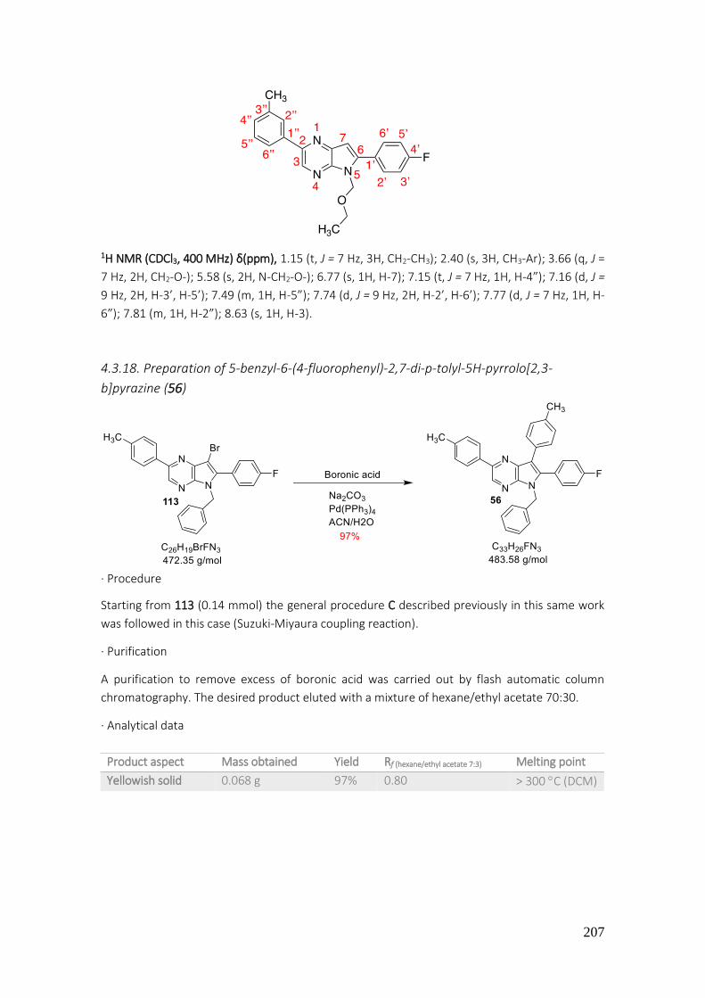
207
1H NMR (CDCl3, 400 MHz) δ(ppm), 1.15 (t, J = 7 Hz, 3H, CH2-CH3); 2.40 (s, 3H, CH3-Ar); 3.66 (q, J =
7 Hz, 2H, CH2-O-); 5.58 (s, 2H, N-CH2-O-); 6.77 (s, 1H, H-7); 7.15 (t, J = 7 Hz, 1H, H-4”); 7.16 (d, J =
9 Hz, 2H, H-3’, H-5’); 7.49 (m, 1H, H-5”); 7.74 (d, J = 9 Hz, 2H, H-2’, H-6’); 7.77 (d, J = 7 Hz, 1H, H-
6”); 7.81 (m, 1H, H-2”); 8.63 (s, 1H, H-3).
4.3.18. Preparation of 5-benzyl-6-(4-fluorophenyl)-2,7-di-p-tolyl-5H-pyrrolo[2,3-
b]pyrazine (56)
· Procedure
Starting from 113 (0.14 mmol) the general procedure C described previously in this same work
was followed in this case (Suzuki-Miyaura coupling reaction).
· Purification
A purification to remove excess of boronic acid was carried out by flash automatic column
chromatography. The desired product eluted with a mixture of hexane/ethyl acetate 70:30.
· Analytical data
Product aspect Mass obtained Yield Rf (hexane/ethyl acetate 7:3) Melting point
Yellowish solid 0.068 g 97% 0.80 > 300 C (DCM)

208
1H NMR (CDCl3, 400 MHz) δ(ppm), 2.32 (s, 3H, CH3-Ar (x2)); 5.85 (s, 2H, CH2-N); 7.14 (t, J = 8.8 Hz,
2H, H-2’’’, H-6’’’); 7.32-7.37 (m, 3H, H-3’’’, H-4’’’, H-5’’’); 7.39 (dd, J1 = 2 Hz, J2 = 7.8 Hz, 2H, H-3IV, H-
5IV); 7.46 (dd, J1 = 2 Hz, J2 = 7.8 Hz, 2H, H-2IV, H-6IV); 7.56 (d, J = 7 Hz, 2H, H-3’, H-5’); 7.57 (d, J = 7
Hz, 2H, H-2’, H-6’); 7.71 (s, 1H, H-3); 7.74 (t, J = 8 Hz, 2H, H-3’’, H-5’’); 8.41 (dd, J1 = 5.5 Hz, J2 = 8
Hz, 2H, H-2’’, H-6’’).
4.3.19. Preparation of 6-(4-fluorophenyl)-2,7-di-p-tolyl-5H-pyrrolo[2,3-b]pyrazine (57a)
· Procedure
In order to remove the protecting group, different conditions were tried. In a round bottom flask
the product 110a was dissolved in methanol or THF and HCl 2N was added. After 24 hours at
room temperature, TLC of reaction showed the presence of starting material. Therefore, the
reaction was heated at 60 C. After 48 hours of reaction, no new product was observed in TLC.
Different conditions were tried as TFA at room temperature, heating at 60 C or using a mixture
of HCl 2N and isopropanol (8:2), but same results were obtained in all reactions, starting material.

209
4.3.20. Preparation of 6-(4-fluorophenyl)-2,7-di-p-tolyl-5H-pyrrolo[2,3-b]pyrazine (57a)
· Procedure
In a specific catalytic hydrogenation round bottom flask equipped with a magnetic stirring, the
compound 56 (0.068 g, 0.14 mmol) was added with a mixture of methanol and ethyl acetate (2:1).
Then Pd-C 10% (0.01 g, 0.094 mmol) and 0.3 mL of HCl 5N were added. The reaction was stirred
under H2 stream for 16 hours.
· Work-up
Given that the hydrogenation apparatus was not completely hermetical, the consumed volume
was higher than expected. A TLC indicated consumption of starting material (hexane/ethyl
acetate 7:3). The crude reaction was filtered by means of a pleated filter and washed with 20 mL
of methanol and was then collected in a 100 mL round-bottomed flask. The solvent was removed
under reduced pressure. The crude was dissolved in dichloromethane (10 mL) and washed with
a solution of NaOH 8N (20 mL x 3). The organic phase was dried over anhydrous Na2SO4, filtered
and concentrated under reduced pressure.
· Purification
The desired product was obtained with enough purity.
· Analytical data
1H NMR (CDCl3, 400 MHz) δ(ppm), 2.07 (s, 3H, CH3-Ar); 2.37 (s, 3H, CH3-Ar); 7.12-7.17 (m, 2H, H-
3’’, H-5’’); 7.18 (d, J = 7.2 Hz, 2H, H-3’’’, H-5’’’); 7.21 (d, J = 8.5 Hz, 2H, H-3’, H-5’); 7.27 (d, J = 7.2
Hz, 2H, H-2’’’, H-6’’’); 7.54 (dd, J1 = 5.5 Hz, J2 = 8.5 Hz, 1H, H-2’); 7.76-7.80 (m, 1H, H-6’); 7.95 (q,
J = 7.7 Hz, 3H, H-2’’, H-6’’, NH); 8.63 (bs, 1H, H-3); (H-2’ and H-6’ are interchangeable protons).
Product aspect Mass obtained Yield Rf (hexane/ethyl acetate 7:3) Melting point
Yellowish solid 0.04 g Quantitative 0.45 233-235 C (DCM)

210
HRMS, ESI(+) m/z: Mass calculated for C26H20FN3: 394.1675 g/mol [M+H]+. Mass found: 394.1688
g/mol.
4.3.21. Preparation of 6-(4-fluorophenyl)-2-(m-tolyl)-7-(p-tolyl)-5H-pyrrolo[2,3-
b]pyrazine (57b)
· Procedure
In a 25 mL round bottom flask the triarylated 110b (0.025 g, 0.06 mmol) was dissolved in
isopropanol (2 mL) and HCl 2N (8 mL). The reaction was stirred at room temperature for two days.
The control of reaction by TLC (a sample of reaction was neutralized with NaOH 2N and extracted
with ether) showed no consumption of starting material (hexane/ethyl acetate 8:2). Then, 5 mL
of methanol were added, and the reaction was stirred for 24 hours more at reflux.
· Work up
At this time, no more starting material was observed in TLC. Therefore, 5 mL of NaOH 2N were
added in the mixture until basic pH was reached. Then, methanol and isopropanol were
evaporated under reduced pressure. The obtained crude mixture was diluted with water (15 mL)
and extracted with dichloromethane (3 x 20 mL). The organic layers were dried over anhydrous
sodium sulfate, filtered and concentrated under reduced pressure.
· Purification
No purification was necessary since product was obtained with enough purity. The 1H NMR of the
crude showed the 6-(4-fluorophenyl)-2-(m-tolyl)-5H-pyrrolo[2,3-b]pyrazine, the diarylated 50
instead of the expected compound.
· Analytical data
The analytical data (product aspect, 1H NMR, Rf, and others) was completely coincident with the
previously described for 50.

211
4.3.22. Preparation of 2-(4-methoxyphenyl)-5-(4-nitrophenyl)-6-phenyl-5H-pyrrolo[2,3-
b]pyrazine (58)
· Procedure
In a Pyrex glass tube, previously flame-dried under argon atmosphere, the diarylated pyrazine 48
(0.120 g, 0.4 mmol) was dissolved in toluene (7 mL). Then, p-bromonitrobenzene (0.08 g, 0.04
mmol), cesium carbonate (0.155 g, 0.048 mmol), palladium (0.06 mmol) and BINAP (0.06 mmol)
were added under argon atmosphere. The reaction mixture was stirred at 140 °C ± 5 °C.
· Work up
After 24 hours of reaction no more starting material was observed in a TLC of reaction
(hexane/ethyl acetate 8:2). Therefore, the mixture was diluted with ethyl acetate and washed
with water (3 x 20 mL) and brine (3 x 5 mL). The organic phase was dried over anhydrous sodium
sulfate, filtered and concentrated under reduced pressure.
· Purification
No purification was carried out since the product was obtained with enough purity.
· Analytical data
1H NMR (CDCl3, 400 MHz) δ(ppm), 3.83 (s, 3H, O-CH3); 6.68 (s, 1H, H-7); 6.97 (d, J = 9.0 Hz, 2H, H-
3’’, H-5’’); 6.99 (d, J = 7.0 Hz, 2H, H-2’’’, H-6’’’); 7.19 (t, J = 7.2 Hz, 2H, H-3’, H-5’); 7.36 (t, J = 7.2
Hz, 1H, H-4’); 7.45 (d, J = 7.2 Hz, 2H, H-2’, H-6’); 7.80 (d, J = 7.0 Hz, 2H, H-3’’’, H-5’’’); 7.87 (d, J =
9.0 Hz, 2H, H-2’’, H-6’’); 8.46 (s, 1H, 3-H).
Product aspect Mass obtained Yield Rf (hexane/ethyl acetate 8:2) Melting point
Yellow solid 0.116 g 69% 0.25 230-235 C (DCM)

212
4.3.23. Preparation of 5-bromo-3-(phenylethynyl)pyrazin-2-amine (92)
· Procedure
Starting from 91c (0.52 mmol) the general procedure A described above was followed
(Sonogoshira coupling reaction). The 1H NMR showed a subproduct, the 3,5-
bis(phenylethynyl)pyrazin-2-amine (99).
· Analytical data of 92
1H NMR (CDCl3, 400 MHz) δ(ppm), 5.18 (bs, 2H, NH2); 7.40 (t, J = 7.2 Hz, 2H, H-3’, H-5’); 7.41 (t, J
= 7.2 Hz, 1H, H-4’); 7.57 (dd, J1 = 1.5 Hz, J2 = 7.2 Hz, 2H, H-2’, H-6’); 8.04 (s, 1H, H-6).
13C NMR (CDCl3, 100.6 MHz) δ(ppm), 83.2 (Cq, C-≡-Bn); 97.4 (Cq, C-≡-Pyrazine); 121.1 (Cq, C-5);
124.5 (Cq, C-1’); 125.9 (Cq, C-3); 128.6 (CH, C-2’, C-6’); 129.8 (CH, C-4’); 132.0 (CH, C-3’, C-5’);
143.6 (CH, C-6); 154.2 (Cq, C-2).
· Analytical data of 99
1H NMR (CDCl3, 400 MHz) δ(ppm), 5.30 (bs, 2H, NH2); 7.40 (m, 6H, H-3’, H-4’, H-5’, H-3’’, H-4’’, H-
5’’); 7.60 (m, 4H, H-2’, H-6’, H-2’’, H-6’’); 8.20 (s, 1H, H-6).
Product aspect Mass obtained Yield Rf (hexane/ethyl acetate 8:2) Melting point
Yellowish solid 0.140 g Quantitative 0.40 140-142 C (DCM)
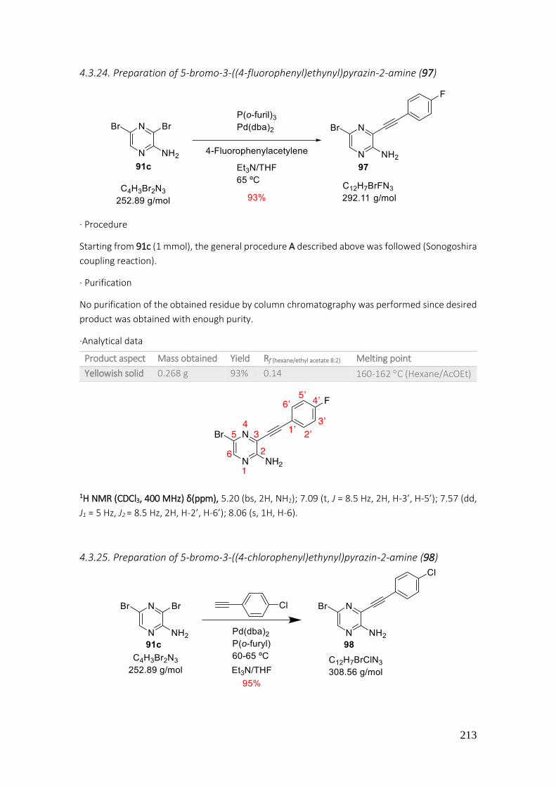
213
4.3.24. Preparation of 5-bromo-3-((4-fluorophenyl)ethynyl)pyrazin-2-amine (97)
· Procedure
Starting from 91c (1 mmol), the general procedure A described above was followed (Sonogoshira
coupling reaction).
· Purification
No purification of the obtained residue by column chromatography was performed since desired
product was obtained with enough purity.
·Analytical data
1H NMR (CDCl3, 400 MHz) δ(ppm), 5.20 (bs, 2H, NH2); 7.09 (t, J = 8.5 Hz, 2H, H-3’, H-5’); 7.57 (dd,
J1 = 5 Hz, J2 = 8.5 Hz, 2H, H-2’, H-6’); 8.06 (s, 1H, H-6).
4.3.25. Preparation of 5-bromo-3-((4-chlorophenyl)ethynyl)pyrazin-2-amine (98)
Product aspect Mass obtained Yield Rf (hexane/ethyl acetate 8:2) Melting point
Yellowish solid 0.268 g 93% 0.14 160-162 C (Hexane/AcOEt)
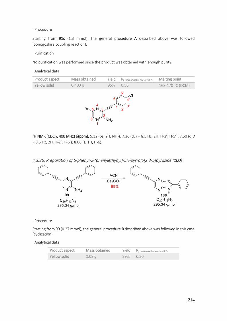
214
· Procedure
Starting from 91c (1.3 mmol), the general procedure A described above was followed
(Sonogoshira coupling reaction).
· Purification
No purification was performed since the product was obtained with enough purity.
· Analytical data
1H NMR (CDCl3, 400 MHz) δ(ppm), 5.12 (bs, 2H, NH2); 7.36 (d, J = 8.5 Hz, 2H, H-3’, H-5’); 7.50 (d, J
= 8.5 Hz, 2H, H-2’, H-6’); 8.06 (s, 1H, H-6).
4.3.26. Preparation of 6-phenyl-2-(phenylethynyl)-5H-pyrrolo[2,3-b]pyrazine (100)
· Procedure
Starting from 99 (0.27 mmol), the general procedure B described above was followed in this case (cyclization).
· Analytical data
Product aspect Mass obtained Yield Rf (hexane/ethyl acetate 8:2) Melting point
Yellow solid 0.400 g 95% 0.50 168-170 C (DCM)
Product aspect Mass obtained Yield Rf (hexane/ethyl acetate 8:2)
Yellow solid 0.08 g 99% 0.30
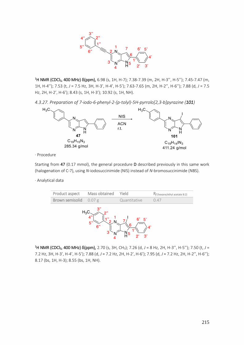
215
1H NMR (CDCl3, 400 MHz) δ(ppm), 6.98 (s, 1H, H-7); 7.38-7.39 (m, 2H, H-3’’, H-5’’); 7.45-7.47 (m,
1H, H-4’’); 7.53 (t, J = 7.5 Hz, 3H, H-3’, H-4’, H-5’); 7.63-7.65 (m, 2H, H-2’’, H-6’’); 7.88 (d, J = 7.5
Hz, 2H, H-2’, H-6’); 8.43 (s, 1H, H-3’); 10.92 (s, 1H, NH).
4.3.27. Preparation of 7-iodo-6-phenyl-2-(p-tolyl)-5H-pyrrolo[2,3-b]pyrazine (101)
· Procedure
Starting from 47 (0.17 mmol), the general procedure D described previously in this same work
(halogenation of C-7), using N-iodosuccinimide (NIS) instead of N-bromosuccinimide (NBS).
· Analytical data
1H NMR (CDCl3, 400 MHz) δ(ppm), 2.70 (s, 3H, CH3); 7.26 (d, J = 8 Hz, 2H, H-3’’, H-5’’); 7.50 (t, J =
7.2 Hz, 3H, H-3’, H-4’, H-5’); 7.88 (d, J = 7.2 Hz, 2H, H-2’, H-6’); 7.95 (d, J = 7.2 Hz, 2H, H-2’’, H-6’’);
8.17 (bs, 1H, H-3); 8.55 (bs, 1H, NH).
Product aspect Mass obtained Yield Rf (hexane/ethyl acetate 8:2)
Brown semisolid 0.07 g Quantitative 0.47
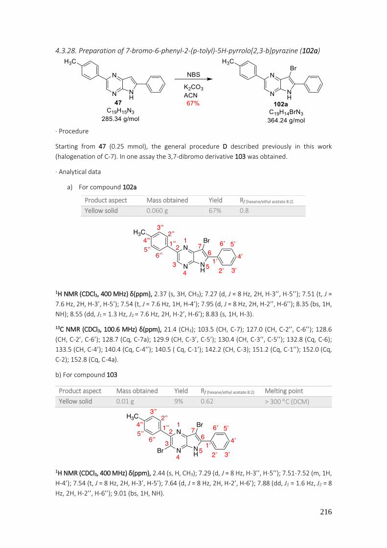
216
4.3.28. Preparation of 7-bromo-6-phenyl-2-(p-tolyl)-5H-pyrrolo[2,3-b]pyrazine (102a)
· Procedure
Starting from 47 (0.25 mmol), the general procedure D described previously in this work
(halogenation of C-7). In one assay the 3,7-dibromo derivative 103 was obtained.
· Analytical data
a) For compound 102a
1H NMR (CDCl3, 400 MHz) δ(ppm), 2.37 (s, 3H, CH3); 7.27 (d, J = 8 Hz, 2H, H-3’’, H-5’’); 7.51 (t, J =
7.6 Hz, 2H, H-3’, H-5’); 7.54 (t, J = 7.6 Hz, 1H, H-4’); 7.95 (d, J = 8 Hz, 2H, H-2’’, H-6’’); 8.35 (bs, 1H,
NH); 8.55 (dd, J1 = 1.3 Hz, J2 = 7.6 Hz, 2H, H-2’, H-6’); 8.83 (s, 1H, H-3).
13C NMR (CDCl3, 100.6 MHz) δ(ppm), 21.4 (CH3); 103.5 (CH, C-7); 127.0 (CH, C-2’’, C-6’’); 128.6
(CH, C-2’, C-6’); 128.7 (Cq, C-7a); 129.9 (CH, C-3’, C-5’); 130.4 (CH, C-3’’, C-5’’); 132.8 (Cq, C-6);
133.5 (CH, C-4’); 140.4 (Cq, C-4’’); 140.5 ( Cq, C-1’); 142.2 (CH, C-3); 151.2 (Cq, C-1’’); 152.0 (Cq,
C-2); 152.8 (Cq, C-4a).
b) For compound 103
1H NMR (CDCl3, 400 MHz) δ(ppm), 2.44 (s, H, CH3); 7.29 (d, J = 8 Hz, H-3’’, H-5’’); 7.51-7.52 (m, 1H,
H-4’); 7.54 (t, J = 8 Hz, 2H, H-3’, H-5’); 7.64 (d, J = 8 Hz, 2H, H-2’, H-6’); 7.88 (dd, J1 = 1.6 Hz, J2 = 8
Hz, 2H, H-2’’, H-6’’); 9.01 (bs, 1H, NH).
Product aspect Mass obtained Yield Rf (hexane/ethyl acetate 8:2)
Yellow solid 0.060 g 67% 0.8
Product aspect Mass obtained Yield Rf (hexane/ethyl acetate 8:2) Melting point
Yellow solid 0.01 g 9% 0.62 > 300 C (DCM)

217
HRMS, ESI(+) m/z: Calculated for C19H13Br2N3 [M+H]+: 443.9489 , found 443.9490.
4.3.29. Preparation of 7-bromo-6-phenyl-2-(4-(trifluoromethyl)phenyl)-5H-pyrrolo[2,3-
b]pyrazine (102b)
· Procedure
Starting from 49 (0.29 mmol), the general procedure D described above was followed in this case
(halogenation of C-7).
· Purification
A manual column chromatography was made. The desired product eluted with a mixture of
hexane/ethyl acetate 80:20.
· Analytical data
1H NMR (CDCl3, 400 MHz) δ(ppm), 7.52 (t, J = 7.3 Hz, 2H, H-3’, H-5’); 7.56-7.58 (m, 1H, H-4’); 7.72
(d, J = 8.2 Hz, 2H, H-3’’, H-5’’); 8.17 (d, J = 8.2 Hz, 2H, H-2’’, H-6’’); 8.54 (d, J = 7.3 Hz, 2H, H-2’, H-
6’); 8.88 (s, 1H, H-3).
13C NMR (CDCl3, 100.6 MHz) δ(ppm), 102.1 (Cq, C-7); 123.2 (Cq, J = 270.7 Hz, CF3); 126.0 (CH, J =
7.5 Hz, C-3’’, C-5’’); 127.4 (CH, C-2’, C-6’); 128.0 (Cq, C-6); 128.3 (Cq, C-7a); 128.7 (CH, J = 3 Hz, C-
2’’, C-6’’); 130.6 (CH, C-3’, C-5’); 131.8 (Cq, J = 32.5 Hz, C-4’’); 133.9 (CH, C-4’); 134.1 (Cq, C-1’);
138.9 (Cq, J = 1.2 Hz, C-1’’); 142.8 (CH, C-3); 148.1 (Cq, C-4a); 153.9 (Cq, C-2).
Product aspect Mass obtained Yield Rf (hexane/ethyl acetate 8:2) Melting point
Yellowish solid 0.080 g 65% 0.47 288-290 C (DCM)
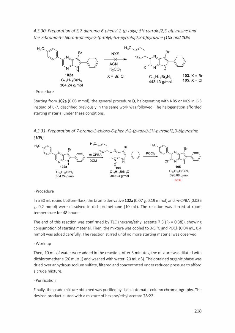
218
4.3.30. Preparation of 3,7-dibromo-6-phenyl-2-(p-tolyl)-5H-pyrrolo[2,3-b]pyrazine and
the 7-bromo-3-chloro-6-phenyl-2-(p-tolyl)-5H-pyrrolo[2,3-b]pyrazine (103 and 105)
· Procedure
Starting from 102a (0.03 mmol), the general procedure D, halogenating with NBS or NCS in C-3
instead of C-7, described previously in the same work was followed. The halogenation afforded
starting material under these conditions.
4.3.31. Preparation of 7-bromo-3-chloro-6-phenyl-2-(p-tolyl)-5H-pyrrolo[2,3-b]pyrazine
(105)
· Procedure
In a 50 mL round bottom-flask, the bromo derivative 102a (0.07 g, 0.19 mmol) and m-CPBA (0.036
g, 0.2 mmol) were dissolved in dichloromethane (10 mL). The reaction was stirred at room
temperature for 48 hours.
The end of this reaction was confirmed by TLC (hexane/ethyl acetate 7:3 (Rf = 0.38)), showing
consumption of starting material. Then, the mixture was cooled to 0-5 °C and POCl3 (0.04 mL, 0.4
mmol) was added carefully. The reaction stirred until no more starting material was observed.
· Work-up
Then, 10 mL of water were added in the reaction. After 5 minutes, the mixture was diluted with
dichloromethane (20 mL x 1) and washed with water (20 mL x 3). The obtained organic phase was
dried over anhydrous sodium sulfate, filtered and concentrated under reduced pressure to afford
a crude mixture.
· Purification
Finally, the crude mixture obtained was purified by flash automatic column chromatography. The
desired product eluted with a mixture of hexane/ethyl acetate 78:22.

219
· Analytical data
1H NMR (CDCl3, 400 MHz) δ(ppm), 2.42 (s, 3H, CH3); 7.30 (d, J = 7.8 Hz, 2H, H-3’, H-5’); 7.44-7.47
(m, 1H, H-4’); 7.53 (t, J = 7.8 Hz, 2H, H-2’, H-6’); 7.95 (d, J = 8 Hz, 2H, H-2’’, H-6’’); 7.97 (d, J = 8
Hz, 2H, H-3’’, H-5’’); 8.61 (bs, 1H, NH).
13C NMR (CDCl3, 100.6 MHz) δ(ppm), 21.2 (CH3); 96.7 (CH, C-7); 118.4 (Cq, C-7a); 126.9 (CH, C-2’’,
C-6’’); 128.1 (CH, C-2’, C-6’); 128.7 (CH, C-4’); 129.5 (CH, C-3’, C-5’); 129.6 (CH, C-3’’, C-5’’); 130.6
(Cq, C-6); 130.9 (Cq, C-1’’); 132.6 (Cq, C-4’’); 134.4 (Cq, C-1’); 144.5 (Cq, C-3); 147.4 (Cq, C-4a);
152.9 (Cq, C-2).
4.3.32. Preparation of 7-bromo-5-(methoxymethyl)-6-phenyl-2-(4-(trifluoromethyl)-
phenyl)-5H-pyrrolo[2,3-b]pyrazine (106)
· Procedure
In a microwave glass tube (10 mL) sealed with a silicon septum under magnetic stirring, the bromo
derivative 102b (0.09 g, 0.22 mmol) diluted in acetonitrile (3 mL) and methoxymethyl acetal
(MOM) (0.033 mL, 0.44 mmol) were added. Finally, a catalytic quantity of tetrabutylammonium
hydroxide (0.06 mmol) was added in the mixture. The tube was introduced in oven microwave
and heated to 100 °C (external temperature) and subjected to a variable MW power until 300 W
(an IR sensor measured the temperature of glass tube surface).
· Work up
A TLC of the reaction showed consumption of starting material (hexane/ethyl acetate 6:4). The
mixture was extracted with ethyl acetate (20 mL x 3) and water (20 mL x 1). The organic phases
were collected, dried over sodium sulfate, filtered and concentrated under reduced pressure to
afford the crude mixture. The 1H NMR of the obtained residue showed the diarylated 49.
Product aspect Mass obtained Yield Rf (hexane/ethyl acetate 7:3)
Yellowish solid 0.04 g 66% 0.66

220
4.3.33. Preparation of 7-bromo-3-chloro-5-(ethoxymethyl)-6-phenyl-2-(p-tolyl)-5H-
pyrrolo[2,3-b]pyrazine (107)
· Procedure In a 50 mL round bottom flask the NaH (60% in vaseline) (0.015 g, 0.59 mmol) was washed with
hexane to remove vaseline. The dihalogenated product 105 (0.150 g, 0.50 mmol) was dissolved
in DMF and chloromethyl ethyl ether (0.07 mL, 0.67 mmol) was added. Then, a catalytic quantity
of tetrabutylammonium hydroxide (0.01 mL, 0.1 mmol) was added. The reaction was stirred for
24 hours.
· Work up
A TLC analysis showed consumption of starting material (hexane/ethyl acetate 8:2). The mixture
of reaction was diluted with ethyl acetate (10 mL) and washed with water (20 mL x 3). The organic
layer was dried over anhydrous sodium sulfate, filtered and concentrated under reduced
pressure. When ethyl acetate was removed, a few quantity of toluene was added to co-evaporate
and facilitate the removal of DMF residues.
· Purification
The desired product was obtained with enough purity to characterize and continue the following
steps.
· Analytical data
1H NMR (CDCl3, 400 MHz) δ(ppm), 1.11 (t, J = 7.3 Hz, CH3-CH2-O); 2.36 (s, 3H, CH3-Ar); 3.52 (q, J =
7.3 Hz, 2H, CH2-O); 5.52 (s, 2H, N-CH2-O); 7.08-7.10 (m, 2H, H-3’’, H-5’’); 7.16-7.20 (m, 3H, H-3’,
H-5’, H-4’); 7.48 (d, J = 7.3 Hz, 2H, H-2’, H-6’); 7.94 (d, J = 8.0 Hz, 2H, H-2’’, H-6’’).
Product aspect Mass obtained Yield Rf (hexane/ethyl acetate 8:2)
Yellowish semisolid 0.04 g Quantitative 0.80

221
4.3.34. Preparation of 7-bromo-5-(ethoxymethyl)-6-phenyl-2,3-di-p-tolyl-5H-pyrrolo[2,3-
b]pyrazine (108)
· Procedure
Starting from 107 (0.05 mmol), the general procedure C described previously in this same work
was followed in this case (Suzuki-Miyaura coupling reaction). The expected compound was not
obtained and starting material was recuperated under these conditions.
4.3.35. Preparation of 7-bromo-5-(ethoxymethyl)-6-(4-fluorophenyl)-2-(p-tolyl)-5H-
pyrrolo[2,3-b]pyrazine (109a)
· Procedure
Starting from 55a (0.49 mmol) the general procedure D described above was followed in this case
(halogenation of C-7).
· Purification
The desired product was obtained with enough purity.
· Analytical data
Product aspect Mass obtained Yield Rf (hexane/ethyl acetate 8:2)
Yellow semisolid 0.178 g 82% 0.70
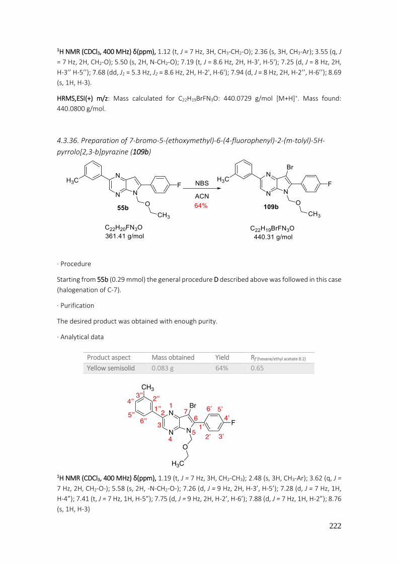
222
1H NMR (CDCl3, 400 MHz) δ(ppm), 1.12 (t, J = 7 Hz, 3H, CH3-CH2-O); 2.36 (s, 3H, CH3-Ar); 3.55 (q, J
= 7 Hz, 2H, CH2-O); 5.50 (s, 2H, N-CH2-O); 7.19 (t, J = 8.6 Hz, 2H, H-3', H-5'); 7.25 (d, J = 8 Hz, 2H,
H-3’’ H-5’’); 7.68 (dd, J1 = 5.3 Hz, J2 = 8.6 Hz, 2H, H-2', H-6'); 7.94 (d, J = 8 Hz, 2H, H-2’’, H-6’’); 8.69
(s, 1H, H-3).
HRMS,ESI(+) m/z: Mass calculated for C22H19BrFN3O: 440.0729 g/mol [M+H]+. Mass found:
440.0800 g/mol.
4.3.36. Preparation of 7-bromo-5-(ethoxymethyl)-6-(4-fluorophenyl)-2-(m-tolyl)-5H-
pyrrolo[2,3-b]pyrazine (109b)
· Procedure
Starting from 55b (0.29 mmol) the general procedure D described above was followed in this case
(halogenation of C-7).
· Purification
The desired product was obtained with enough purity.
· Analytical data
1H NMR (CDCl3, 400 MHz) δ(ppm), 1.19 (t, J = 7 Hz, 3H, CH2-CH3); 2.48 (s, 3H, CH3-Ar); 3.62 (q, J =
7 Hz, 2H, CH2-O-); 5.58 (s, 2H, -N-CH2-O-); 7.26 (d, J = 9 Hz, 2H, H-3’, H-5’); 7.28 (d, J = 7 Hz, 1H,
H-4”); 7.41 (t, J = 7 Hz, 1H, H-5”); 7.75 (d, J = 9 Hz, 2H, H-2’, H-6’); 7.88 (d, J = 7 Hz, 1H, H-2”); 8.76
(s, 1H, H-3)
Product aspect Mass obtained Yield Rf (hexane/ethyl acetate 8:2)
Yellow semisolid 0.083 g 64% 0.65

223
13C NMR (CDCl3, 100.6 MHz) δ(ppm), 15.0 (CH3, CH3-CH2); 21.6 (CH3, CH3-Ar); 65.0 (CH2, CH2-O-);
71.5 (CH2,-O-CH2-O-); 91.8 (Cq, C-7); 115.7 (CH, J = 22 Hz, C-3’, C-5’); 124.2 (CH, C-6”); 124.8 (Cq,
C-7a); 127.7 (Cq, C-1’); 127.9 (CH, C-4”); 128.8 (CH, C-5”); 129.7 (CH, C-2”); 132.8 (CH, J = 9 Hz, C-
2’, C-6’); 136.6 (CH, C-3); 137.5 (Cq, C-6); 138.6 (Cq, C-1”); 140.7 (Cq, C-3’’); 142.1 (Cq, C-4a);
149.2 (Cq, C-2); 163.7 (Cq, J = 254 Hz, C-4’)
4.3.37. Preparation of 5-(ethoxymethyl)-6-(4-fluorophenyl)-2,7-di-p-tolyl-5H-pyrrolo[2,3-
b]pyrazine (110a)
· Procedure
Starting from 109a (0.41 mmol) the general procedure C described previously in the same work was followed in this case (Suzuki-Miyaura coupling reaction).
· Purification
A purification to remove excess of boronic acid was done by flash automatic silica gel column
chromatography. The desired product eluted with a mixture of hexane/ethyl acetate 70:30.
· Analytical data
1H NMR (CDCl3, 400 MHz) δ(ppm), 1.20 (t, J = 7 Hz, 3H, CH3-CH2-O); 2.43 (s, 3H, CH3-Ar (x2)); 3.63
(q, J = 7 Hz, 2H, CH2-O); 5.57 (s, 2H, N-CH2-O); 7.12 (d, J = 8.4 Hz, 2H, H-3’’’, H-5’’’, rotamer m);
7.15 (d, J = 8.4 Hz, 2H, H-3’’’, H-5’’’, rotamer M); 7.26 (t, J = 8.5 Hz, 2H, H-3', H-5'); 7.32 (d, J = 8
Hz, 2H, H-3’’ H-5’’); 7.49 (d, J = 8.2 Hz, 2H, H-2’’’, H-6’’’, rotamer m); 7.55 (d, J = 8.2 Hz, 2H, H-2’’’,
H-6’’’ rotamer M); 7.74 (d, J = 8.5 Hz, 2H, H-2', H-6' rotamer M); 7.76 (d, J = 8.4 Hz, 2H, H-2', H-6',
rotamer m); 8.01 (d, J = 8 Hz, 2H, H-2’’, H-6’’); 8.76 (s, 1H, H-3).
Product aspect Mass obtained Yield Rf (hexane/ethyl acetate 7:3)
Yellowish semisolid 0.075 g 41% 0.57
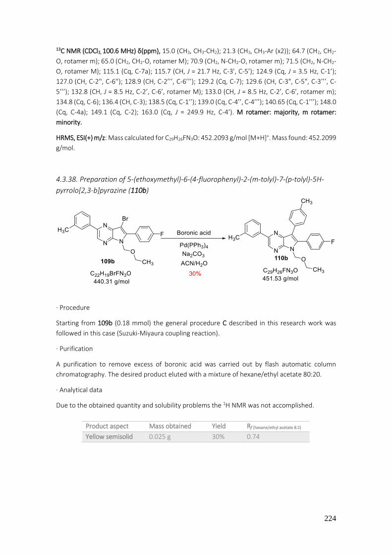
224
13C NMR (CDCl3, 100.6 MHz) δ(ppm), 15.0 (CH3, CH3-CH2); 21.3 (CH3, CH3-Ar (x2)); 64.7 (CH2, CH2-
O, rotamer m); 65.0 (CH2, CH2-O, rotamer M); 70.9 (CH2, N-CH2-O, rotamer m); 71.5 (CH2, N-CH2-
O, rotamer M); 115.1 (Cq, C-7a); 115.7 (CH, J = 21.7 Hz, C-3', C-5’); 124.9 (Cq, J = 3.5 Hz, C-1’);
127.0 (CH, C-2'', C-6''); 128.9 (CH, C-2’’’, C-6’’’); 129.2 (Cq, C-7); 129.6 (CH, C-3", C-5", C-3’’’, C-
5’’’); 132.8 (CH, J = 8.5 Hz, C-2’, C-6’, rotamer M); 133.0 (CH, J = 8.5 Hz, C-2’, C-6’, rotamer m);
134.8 (Cq, C-6); 136.4 (CH, C-3); 138.5 (Cq, C-1’’); 139.0 (Cq, C-4’’, C-4’’’); 140.65 (Cq, C-1’’’); 148.0
(Cq, C-4a); 149.1 (Cq, C-2); 163.0 (Cq, J = 249.9 Hz, C-4’). M rotamer: majority, m rotamer:
minority.
HRMS, ESI(+) m/z: Mass calculated for C29H26FN3O: 452.2093 g/mol [M+H]+. Mass found: 452.2099
g/mol.
4.3.38. Preparation of 5-(ethoxymethyl)-6-(4-fluorophenyl)-2-(m-tolyl)-7-(p-tolyl)-5H-
pyrrolo[2,3-b]pyrazine (110b)
· Procedure
Starting from 109b (0.18 mmol) the general procedure C described in this research work was
followed in this case (Suzuki-Miyaura coupling reaction).
· Purification
A purification to remove excess of boronic acid was carried out by flash automatic column
chromatography. The desired product eluted with a mixture of hexane/ethyl acetate 80:20.
· Analytical data
Due to the obtained quantity and solubility problems the 1H NMR was not accomplished.
Product aspect Mass obtained Yield Rf (hexane/ethyl acetate 8:2)
Yellow semisolid 0.025 g 30% 0.74

225
4.3.39. Preparation of 5-benzyl-6-(4-fluorophenyl)-2-(p-tolyl)-5H-pyrrolo[2,3-b]pyrazine
(111)
· Procedure
In a round bottom flask the diarylated pyrrolopyrazine 50 (0.070 g, 0.23 mmol) was dissolved in
DMF (6 mL). NaH (60% in vaseline) (0.012 g, 0.35 mmol) was washed with hexane in order to
remove vaseline and was then added into the mixture. The reaction was stirred at room
temperature for 30 minutes. After this time the benzyl bromide was also added (0.042 mL, 0.43
mmol).
· Work up
The reaction was stirred until no more starting material was observed in TLC (hexane/ethyl
acetate 7:3). The mixture was diluted with ethyl ether (15 mL) and washed with water (3 x 20
mL). The organic phases were collected, dried over sodium sulfate, filtered off and concentrated
under reduced pressure.
· Purification
A purification to remove excess of boronic acid was done by flash automatic column
chromatography. The desired product eluted with a mixture of hexane/ethyl acetate 70:30.
· Analytical data
1H NMR (CDCl3, 400 MHz) δ(ppm), 2.34 (s, 3H, CH3); 5.79 (s, 2H, CH2-Ar); 7.01 (s, 1H, H-7); 7.09 (d,
J = 8.6 Hz, 2H, H-2’’’, H-6’’’); 7.12-7.15 (m, 3H, H-3’’’, H-4’’’, H-5’’’); 7.23 (d, J = 8 Hz, 1H, H-2’
rotamer); 7.35 (t, J = 6 Hz, 2H, H-3’, H-5’); 7.38 (s, 1H, H-3); 7.42-7.44 (m, 2H, H-3’’, H-5’’) ; 7.64
Product aspect Mass obtained Yield Rf (hexane/ethyl acetate 7:3)
Yellow solid 0.041 g 20% 0.45

226
(dd, J1 = 5 Hz, J2 = 8 Hz, 1H, H-6’); 7.69 (d, J = 8 Hz, 1H, H-2’ rotamer); 8.18 (dd, J1 = 5.5 Hz, J2 = 8.9
Hz, 2H, H-2’’, H-6’’); (H-2’ and H-6’ are interchangeable protons).
4.3.40. Preparation of 5-benzyl-6-phenyl-2-(phenylethynyl)-5H-pyrrolo[2,3-b]pyrazine
(112)
· Procedure
Starting from 100 (0.2 mmol) the general procedure described in the previous reaction of this
work was applied for preparation of 112.
· Purification
The crude of reaction was purified by flash automatic column chromatography to remove excess
of benzyl bromide. The desired product eluted with a mixture of hexane/ethyl acetate 60:40.
· Analytical data
1H NMR (CDCl3, 400 MHz) δ(ppm), 5.48 (s, 2H, CH2-Ar); 6.70 (s, 1H, H-7); 6.86-6.89 (m, 2H, H-3’’,
H-5’’); 7.15 (dd, J1 = 2 Hz, J2 = 5 Hz, 2H, H-3’’’, H-5’’’); 7.30-7.32 (m, 4H, H-2’’’, H-4’’’, H-4’’, H-6’’’);
7.34 (s, 5H, phenyl); 7.57 (m, 2H, H-2’’, H-6’’); 8.42 (s, 1H, H-3).
13C NMR (CDCl3, 100.6 MHz) δ(ppm), 46.1 (CH2, CH2-N); 87.2 (Cq, Ar-C≡); 90.6 (Cq, ≡C-Pyrazine);
101.5 (CH, C-7); 122.4 (Cq, C-7a); 126.6 (CH, C-2’’, C-6’’); 127.5 (CH, C-4’’’); 127.7 (Cq, C-1’’’);
128.4 (CH, C-2’, C-6’); 128.5 (Cq, C-1’’); 128.6 (CH, C-3’’’, C-5’’’); 128.7 (CH, C-3’’, C-5’’); 128.8 (CH,
C-4’); 129.2 (CH, C-3’, C-5’); 129.4 (CH, C-4’’); 131.9 (CH, C-2’’’, C-6’’’); 133.6 (Cq, C-1’); 137.2 (Cq,
C-2); 139.4 (CH, C-3); 140.4 (Cq, C-6); 147.7 (Cq, C-4a).
Product aspect Mass obtained Yield Rf (hexane/ethyl acetate 7:3)
Yellow solid 0.060 g 77% 0.83

227
4.3.41. Preparation of 5-benzyl-7-bromo-6-(4-fluorophenyl)-2-(p-tolyl)-5H-pyrrolo[2,3-
b]pyrazine (113)
· Procedure
Starting from 111 (0.11 mmol) the general procedure D described previously in this work was
followed in this case (halogenation of C-7).
· Purification
The desired product was obtained with enough purity.
· Analytical data
1H NMR (CDCl3, 400 MHz) δ(ppm), 2.70 (s, 3H, CH3-Ar) ; 5.78 (s, 2H, CH2-N); 7.13 (t, J = 8.7 Hz, 2H,
H-2’’’, H-6’’’); 7.15-7.21 (m, 3H, H-3’’’, H-5’’’, H-4’’’); 7.36 (sc, 2H, H-3’’, H-5’’); 7.40 (sc, H-3’, H-
5’); 7.49 (s, 1H, H-3); 7.78 (bs, 1H, H-2’); 7.81 (dd, J1 = 5 Hz, J2 = 8.4 Hz, 1H, H-6’); 8.42 (dd, J1 = 5.6
Hz, J2 = 8.8 Hz, 2H, H-2’’, H-6’’); (H-2’ and H-6’ are interchangeable protons).
13C NMR (CDCl3, 100.6 MHz) δ(ppm), 21.2 (CH3); 55.4 (CH2, CH2-N) ; 115.4 (CH, J = 21.5 Hz, C-3’, C-
5’); 98.6 (Cq, C-7); 120.3 (CH, C-3); 123.7 (Cq, C-7a); 126.4 (Cq, C-1’); 129.0 (CH, C-2’’); 129.2 (CH,
C-2’’’, C-6’’’); 129.3 (CH, C-6’’); 129.5 (CH, C-3’’’, C-5’’’); 129.7 (CH, C-4’’’); 130.1 (CH, C-3’’); 130.2
(CH, C-5’’); 131.0 (Cq, C-1’’); 131.3 (CH, J = 8.4 Hz, C-2’, C-6’); 132.9 (Cq, C-4’’); 138.7 (Cq, C-1’’’);
139.7 (Cq, C-6); 150.0 (Cq, C-4a); 151.4 (Cq, C-2); 163.8 (CH, J = 250 Hz, C-4’); (C-2’’ and C-6 ‘’ are
interchangeable carbons; C-3’’ and C-5’’ are interchangeable carbons).
Product aspect Mass obtained Yield Rf (hexane/ethyl acetate 8:2) Melting point
Yellow solid 0.048 g 95% 0.40 > 300 C (DCM)

228
4.3.42. Preparation of 5-benzyl-7-bromo-6-phenyl-2-(phenylethynyl)-5H-pyrrolo[2,3-
b]pyrazine (114)
· Procedure
Starting from 112 (0.16 mmol) he general procedure D described previously was followed in this
case (halogenation of C-7).
· Purification
The desired product was obtained with enough purity.
· Analytical data
1H NMR (CDCl3, 400 MHz) δ(ppm), 5.41 (s, 2H, CH2-Ar); 6.79-6.81 (m, 2H, H-3’’, H-5’’); 7.12 (dd, J1
= 2 Hz, J2 = 5 Hz, 2H, H-3’’’, H-5’’’); 7.29-7.34 (m, 6H, phenyl + H-4’’) 7.42 (dd, J1 = 1.2 Hz, J2 = 7
Hz, 3H, H-2’’, H-4’’’, H-6’’); 7.57-7.59 (m, 2H, H-2’’’, H-6’’’); 8.48 (s, 1H, H-3).
Product aspect Mass obtained Yield Rf (hexane/ethyl acetate 7:3)
Yellow solid 0.050 g 69% 0.83

229
4.3.43. Preparation of 5-benzyl-6-phenyl-2-(phenylethynyl)-7-(p-tolyl)-5H-pyrrolo[2,3-
b]pyrazine (115)
· Procedure
Starting from 114 (0.06 mmol)The general procedure C described previously in this same work
was followed in this case (Suzuki-Miyaura coupling reaction).
· Purification
The crude of reaction was purified by flash automatic silica gel column chromatography. The
desired product eluted with a mixture of hexane/ethyl acetate 90:10.
· Analytical data
1H NMR (CDCl3, 400 MHz) δ(ppm), 2.29 (s, 3H, CH3); 5.38 (s, 2H, CH2-Ar); 6.83-6.85 (m, 2H, H-3’’’,
H-5’’’); 7.00 (d, J = 8 Hz, 2H, H-3IV, H-5IV); 7.11-7.13 (m, 5H, H-2’’, H-3’’, H-4’’, H-5’’, H-6’’); 7.29-
7.30 (m, 6H, phenyl + H-4’’’); 7.33 (d, J = 8 Hz, 2H, H-2IV, H-6IV); 7.56-7.58 (m, 2H, H-2’’’, H-6’’’);
8.47 (s, 1H, H-3).
HRMS, ESI (+) m/z: Mass calculated for C27H18BrN3: 476.2082 g/mol [M+H]+. Mass found: 476.2091
g/mol.
Product aspect Mass obtained Yield Rf (hexane/ethyl acetate 9:1) Melting point
Yellowish solid 0.01 g 33% 0.58 >300 C (DCM)

230
4.3.44. Preparation of 7-bromo-2-(4-methoxyphenyl)-5-(4-nitrophenyl)-6-phenyl-5H-
pyrrolo[2,3-b]pyrazine (116)
· Procedure
Starting from 58 (0.28 mmol) he general procedure D described previously in this work was
followed in this case (halogenation of C-7).
· Purification
No purification was made since de product was obtained with enough purity.
· Analytical data
1H NMR (CDCl3, 400 MHz) δ(ppm), 3.89 (s, 3H, O-CH3); 7.04 (d, J = 8.7 Hz, 2H, H-3’’, H-5’’); 7.43 (t,
J = 7.7 Hz, 1H, H-4’); 7.57 (t, J = 7.7 Hz, 2H, H-3’, H-5’); 7.69 (d, J = 8.9 Hz, 2H, H-2’’’, H-6’’’); 7.94
(d, J = 7.7 Hz, 2H, H-2’, H-6’); 8.06 (d, J = 8.7 Hz, 2H, H-2’’, H-6’’); 8.10 (d, J = 8.9 Hz, 2H, H-3’’’, H-
5’’’); 8.66 (s, 1H, H-3).
Product aspect Mass obtained Yield Rf (hexane/ethyl acetate 8:2) Melting point
Yellow solid 0.128 g 93% 0.75 > 300 C (DCM)
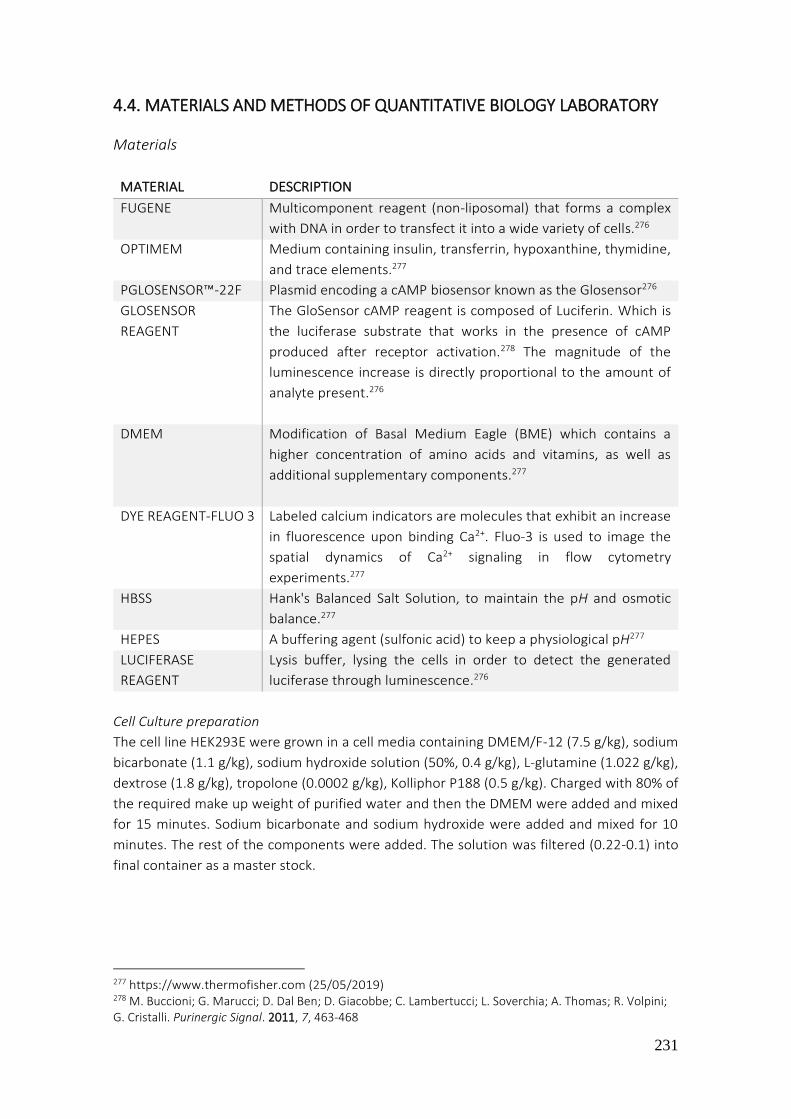
231
4.4. MATERIALS AND METHODS OF QUANTITATIVE BIOLOGY LABORATORY
Materials
MATERIAL DESCRIPTION
FUGENE Multicomponent reagent (non-liposomal) that forms a complex
with DNA in order to transfect it into a wide variety of cells.276
OPTIMEM Medium containing insulin, transferrin, hypoxanthine, thymidine,
and trace elements.277
PGLOSENSOR™-22F Plasmid encoding a cAMP biosensor known as the Glosensor276
GLOSENSOR
REAGENT
The GloSensor cAMP reagent is composed of Luciferin. Which is
the luciferase substrate that works in the presence of cAMP
produced after receptor activation.278 The magnitude of the
luminescence increase is directly proportional to the amount of
analyte present.276
DMEM Modification of Basal Medium Eagle (BME) which contains a
higher concentration of amino acids and vitamins, as well as
additional supplementary components.277
DYE REAGENT-FLUO 3 Labeled calcium indicators are molecules that exhibit an increase
in fluorescence upon binding Ca2+. Fluo-3 is used to image the
spatial dynamics of Ca2+ signaling in flow cytometry
experiments.277
HBSS Hank's Balanced Salt Solution, to maintain the pH and osmotic
balance.277
HEPES A buffering agent (sulfonic acid) to keep a physiological pH277
LUCIFERASE
REAGENT
Lysis buffer, lysing the cells in order to detect the generated
luciferase through luminescence.276
Cell Culture preparation
The cell line HEK293E were grown in a cell media containing DMEM/F-12 (7.5 g/kg), sodium
bicarbonate (1.1 g/kg), sodium hydroxide solution (50%, 0.4 g/kg), L-glutamine (1.022 g/kg),
dextrose (1.8 g/kg), tropolone (0.0002 g/kg), Kolliphor P188 (0.5 g/kg). Charged with 80% of
the required make up weight of purified water and then the DMEM were added and mixed
for 15 minutes. Sodium bicarbonate and sodium hydroxide were added and mixed for 10
minutes. The rest of the components were added. The solution was filtered (0.22-0.1) into
final container as a master stock.
277 https://www.thermofisher.com (25/05/2019) 278 M. Buccioni; G. Marucci; D. Dal Ben; D. Giacobbe; C. Lambertucci; L. Soverchia; A. Thomas; R. Volpini; G. Cristalli. Purinergic Signal. 2011, 7, 463-468

232
Transfection (general procedure)
The FuGENE was mixed with OptiMEM and incubated at room temperature for 5 minutes.
When the ratio FuGENE:DNA was established (6:1), the DNA of each receptor plus reporter
vectors or pGlosensor-22F or PLC-, depending on the assay, were added and mixed with 0.2
mL of FuGENE-OptiMEM. These mixtures were incubated for 20 minutes at room
temperature.
At this time, 1 mL aliquot from the master HEK293 cells stock was quantified through Vicell
(counting cells device). The viability of the cells oscillates between 95-100%. Dilutions were
made to obtain the desired quantity of cells/mL.
Finally, 0.2 mL of the DNA complexes with a total amount of 1.5 µg of DNA
(FuGENE:OptiMEM containing aGPCRs DNA (0.75 µg) + reporter vectors DNA/ pGlosensor-
22F/ PLC- (0.75 µg) were added into a solution of 250,000 cells/mL in a 50 mL culture tubs.
The tubes were left incubating in a shaker at 150 rpm.
In all assays, a culture tube with untransfected cells was used as a negative control. As well,
in each assay an appropriate positive control was conducted.
a) cAMP assay - Gs quantification- pGlosensor-22F
After 48h in suspension in a rotating shaker, according to standard protocols, HEK293
transfected cells, with aGPCRs DNA and pGlosensor-22F, were pelleted by gentle
centrifugation (1500 rpm/ 5 min) and resuspended by tituration in 5 mL of assay media (CO2
Independent Media + 0.1% casein + 2% Glosensor reagent). 1 mL of each sample was
quantified by Vicell to control the viability and the number of cells/mL.
Samples were plated into a 96 well assay plate (costar 3917, 200 µL cells/well), and
luminescence was measured to stablish a baseline (Envision Luminescence mode). Then a
series of enterokinase dilutions were made in order to create a dose-response curve. 20 µL
of enterokinase light chain stock (2 µg/mL, 7.6 nM) were added to 200 µL assay buffer and
dilutions 1:2 were created with assay buffer. 20 µL were added in each well, obtaining a
starting concentration of 76 pM.
For the custom peptide GPR126, (peptide with the GPS sequence of GPR126) a gradient of
concentrations was created as well. 20 µL from the stock (1 mM) were added to 200 µL assay
buffer. 1:2 dilutions were created with assay buffer. The starting concentration was 0.1 mM.
Luminescence was measured in real time for 45 minutes (Figure 83).

233
Figure 83. Example of cAMP assay for Gs quantification
b) cAMP assay – Gi/o quantification- pGlosensor-22F
In these assays, pertussis toxin (from Bordetella pertussis) was used and the experiment was
doubled in size to allow the comparison of samples with and without pertussis toxin.
At 24h after transfection every sample of cells was split in 2 tubes and 10 µL of pertussis
toxin (100 ng/mL) was added in one of them. Therefore, 2 samples were created: one of
transfected cells with receptor and pGlosensor and other sample with the same plus
pertussis toxin. The samples were incubated in the shaker at 150 rpm for a further 24 hours.
Subsequently, the samples were pelleted by gentle centrifugation (1500 rpm/ 5 min) and
resuspended by tituration in 5 mL of assay media (CO2 independent media + 0.1% casein +
2% Glosensor reagent). 1 mL of each sample was quantified with a Vicell cytometer to
determine the viability of the cells/mL.
Using an assay plate (costar 3917), cells were placed (200 µL cells/well), and luminescence
was measured to stablish a baseline (Envision Luminescence mode).
Because the Gi is an inhibitor of adenylate cyclase, an increase of intracellular cAMP has to
be induced prior to receptor activation in order to be able to observe this effect. If a decrease
of cAMP is observed after receptor activation through enterokinase, it is probable that the
receptor is coupled to Gi. Hence, 20 µL Forskolin, a natural diterpene that activates
adenylate cyclase (cAMP rises)271 was added with the same concentration (2 µM) in each
well.
Luminescence was measured in real time for 25 minutes (cAMP levels upswing to 25.000-
200.000 RLU).
Then, a series of enterokinase dilutions were made in order to create a dose-response curve
(same dilutions applied for Gs assay). Luminescence was measured in real time for 45
minutes (Figure 84).
a)
-15 -14 -13 -12 -11 -10 -90
2000
4000
6000
CRC data calculated from t=50 min
log[EK], pM
cA
MP
fo
rmati
on
(R
LU
) GPR128
Ec50
= 2.11 x 10⁻³
µM

234
-11 -10 -9 -8 -7-20000
-15000
-10000
-5000
0
CRC data calculated from t=70 min
log[EK], M
cA
MP
fo
rmati
on
(R
LU
) emr2
PT
b)
Figure 84. a) Example of cAMP assay for Gi quantification b) IC50 calculated with and without PT.
c) Calcium mobilization assay
After 24 hours post-transfection (general procedure), assay plates were coated with 20 µL of
poly-L-lysine for 2 hours at 37 oC. Then, the buffer was removed and with 50 µL of DMEM,
plates were rinsed.
The HEK293E transfected cells with the aGPCRs DNA (and PLC- for G12 assays) were
pelleted by gentle centrifugation (1500 rpm/ 5 min) and resuspended by tituration in 5 mL
assay media (DMEM 500mL + 10% FBS + Glutamax 6 mL + MEM 6 mL + Ampicilin 6 mL +
Hepes 12 mL). One mL of each sample was quantified by Vicell to quantify viability and
concentration.
Cells were plated in an assay plate (corning 354663, 40 µL/well, that is 12000 to 16000
cells/well) and incubated for 24 hours at 37oC in the tissue culture incubator. Every sample
was plated twice in order to compare the sample with YM-254890 with the one without it.
After this time, a dye reagent Fluo-3 was diluted with HBSS/Hepes. 20 µL of this mixture was
added in each well and the cells were incubated at room temperature for two hours. The
Gq inhibitor YM-254890, was added (1.04 µM) 30 minutes before running the assay in one
of the rows of each sample. Meanwhile, in an assay plate, dilutions of enterokinase were
created with assay buffer (same dilutions applied for Gs assay). For the custom peptide (with
GPS sequence of GPR126), dilutions were created as well.
Through the FLIPR (Fluorescent Imaging Plate Reader), the fluorescence baseline was
measured (10 seconds). Then, enterokinase dilutions were added automatically into the
samples and the fluorescence was measured for 300 seconds (Figure 85 and Figure 86).
a)
1200,00
1400,00
1600,00
1800,00
2000,00
2200,00
2400,00
2600,00
2800,00
3000,00
1 11
21
31
41
51
61
71
81
91
101
111
121
131
141
151
161
171
181
191
201
211
221
231
Cal
ciu
m f
orm
atio
n (
RLU
)
Time
GPR126- Gq
Enterokinase
[EK] (pM)
YM + [EK] (pM)
IC50
= 3.42 x 10⁻³
µM
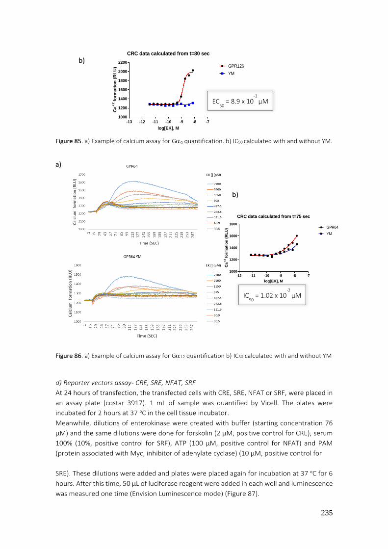
235
-12 -11 -10 -9 -8 -71000
1200
1400
1600
1800
CRC data calculated from t=75 sec
log[EK], M
Ca
+2
form
ati
on
(R
LU
) GPR64
YM
b)
Figure 85. a) Example of calcium assay for Gq quantification. b) IC50 calculated with and without YM.
a)
b)
Figure 86. a) Example of calcium assay for G12 quantification b) IC50 calculated with and without YM
d) Reporter vectors assay- CRE, SRE, NFAT, SRF
At 24 hours of transfection, the transfected cells with CRE, SRE, NFAT or SRF, were placed in
an assay plate (costar 3917). 1 mL of sample was quantified by Vicell. The plates were
incubated for 2 hours at 37 oC in the cell tissue incubator.
Meanwhile, dilutions of enterokinase were created with buffer (starting concentration 76
µM) and the same dilutions were done for forskolin (2 µM, positive control for CRE), serum
100% (10%, positive control for SRF), ATP (100 µM, positive control for NFAT) and PAM
(protein associated with Myc, inhibitor of adenylate cyclase) (10 µM, positive control for
SRE). These dilutions were added and plates were placed again for incubation at 37 oC for 6
hours. After this time, 50 µL of luciferase reagent were added in each well and luminescence
was measured one time (Envision Luminescence mode) (Figure 87).
-13 -12 -11 -10 -9 -8 -71000
1200
1400
1600
1800
2000
2200
CRC data calculated from t=80 sec
log[EK], M
Ca
+2
form
ati
on
(R
LU
) GPR126
YM
EC50
= 8.9 x 10⁻³
µM
IC50
= 1.02 x 10⁻²
µM

236
a) b)
Figure 87. a) Example of reporter vector CRE with and without PT for GPR110. b) Example of
reporter vector SRE with and without PT and YM for GPR112.
IC50
= 9.1 x 10⁻⁴
µM IC50
= 4.1 x 10⁻³
µM
CRC data calculated from t = 6h CRC data calculated from t = 6h

237
5. CONCLUSIONS The most relevant conclusions of this work are indicated below:
1. The transacetylation reaction between N-methyl-4-piperidone and catechol or 2-aminophenol affords the best yields to obtain the spirobenzodioxole (1) and spirobenzoxazole (43) systems respectively. Between them, the preparation of 1 provides better yield (70%) than that which leads 43 (32%), probably due to lower reactivity of 2-aminophenol compared with catechol. Since the main nucleus is obtained, the formation of the spiro-piperidines 2, 3, 4, 5, 6 and 12 are prepared with high yields (80-98%) through classical reactions: alkylation, hydrolysis of amida, oxime formation with hydroxylamine, reduction, halogenation and nitration, respectively.
2. The 1’-acetyl-5-bromospiro[benzo[1,3]dioxole-2,4'-piperidine] (6) is a good scaffold to add aryl groups into position 5 of spiro derivatives through coupling reactions (Suzuki-Miyaura and Buchwald–Hartwig), affording compounds 7, 8, 9, 10 and 11 with high yields (86-93%). Also, from the nitrospiro derivative 12, the consequent basic hydrolysis, followed by a coupling reaction using Pd[(o-tolil)3P]2Cl2, cesium carbonate, BINAP and toluene affords the compound 14 which is reduced to the diamino 15, a desirable compound to synthetize the corresponding diaryl or arylalkyl ureas.
3. The 5-acetyl-1'-(4-nitrophenyl)spiro[benzo[1,3]dioxole-2,4'-piperidine] (17) is a good intermediate for the synthesis of the following spiro derivatives: 1-(1'-(4-nitrophenyl)spiro[benzo[1,3]dioxole-2,4'-piperidine]-5-yl)ethanone oxime (18) with a Z conformation confirmed by NOE experiments, the 1-(1'-(4-nitrophenyl)spiro[benzo[1,3]dioxole-2,4'-piperidine]-5-yl)ethanol (21) and the 5-acetyl-1'-(4-aminophenyl)spiro[benzo[1,3]dioxole-2,4'-piperidine] (23). The amide 19 was obtained with 63% of yield from the oxime 18 by Beckmann rearrangement. Then, the amide 19 was reduced with chloride acid and ethanol to amine 20 with 25% of yield. When the amino 23 was treated with phthalic anhydride in toluene, the imide 24 was obtained with 24% of yield. Then, when 23 was treated with bromomaleic anhydride and coupling reaction conditions (Buchwald-Hartwig), the anhydride 26 was obtained with 55% of yield.
4. The activation of hydroxyl group by treatment with thyonil chloride is a good method for functionalization of spiropiperidines 29 and 30 allowing the nucleophile substitution reaction with N,N-diethyl-2-aminoethanol, N,N-diethylethylenediamine and hexylamina given as a result the final compounds 33, 34 and 35 respectively. The reductive aminations of the methylcetones 27 and 28 were tried as a first pathway to obtain these final spiro derivatives (33, 34 and 35), but in all assays, with different conditions, the alcohol derivatives 29 and 30 were obtained.
5. The addition of isocyanate to the anilines dissolved in THF constitutes an excellent method for the preparation of ureas. Thus, the ureas 37, 39, 40, 41 and 42 were sucessfully prepared. The addition of aniline 22 to ethylisocianate allows the formation of urea 39 with 50% yield. The treatment of diamina 15 with the same isocyanate afforded the diurea 41 with 79% yield. By contrast, the treatment of 15 with pyridine-3-isocyanate afforded the monourea 40 with only 7% of yield, due to the lower reactivity of the isocyanate. The treatment of amina 23 with 2-fluoro-5-(trifluoromethyl)phenyl isocyanate afforded as a result the urea 37 with 80% yield and the treatment of 9 with 4-chloro-3-(trifluoromethyl)phenyl isocyanate afforded the urea 42 with 62% yield.
238
6. A new method for the preparation of 2-amino-5-bromopyrazine (93c) and 2-amino-3,5-
dibromopyrazine (91c) has developed. The yields obtained are in function of the amount of halogenating agent added. Acetonitrile has proved to be the ideal solvent and the assistance of microwave irradiation is essential to obtain halogenated 2-aminopyrazine. 3,5-Dibromo-2-aminopyrazine (91c) is an excellent functionalized starting material for the synthesis of nitrogen heterocycles.
7. The Sonogoshira reaction conditions between 2-amino-3,5-dibromopyrazine (91c) and different phenylacetylenes, using a proportion of base (Et3N)/solvent (THF) 1:7 and without CuI, afforded the pyrazine derivatives 92, 97 and 98 with the best yields 99%, 93% and 95% respectively. These derivatives were cyclized through an endo-dig cyclization and followed by Suzuki Miyaura cross-coupling reaction in order to arylate at position 2 using different boronic acids and given as a result the diarylated pyrazines 47-52 with considerable yields (36%-96%).
8. The halogenation at the position 7 of the pyrrolo[2,3-b]pyrazine system was a success, using NBS and acetonitrile, but the followed Suzuki Miyaura reaction to arylate the same position was never achieved due to the interactions of catalyst with unprotected NH of the pyrrolo ring. Then, protecting this position with MOM or benzyl groups the halogenation and the Suzuki Miyaura reaction were accomplished given as a result the triarylated compounds, at (positions 2, 6 and 7) 110a (41%), 110b (30%), both protected with MOM and 56 (95%) and 115 (69%) both protected with benzyl group.
9. The halogenation of 6-(4-fluorophenyl)-2-(p-tolyl)-5H-pyrrolo[2,3-b]pyrazine in carbon 3 was achieved through oxidation of the pyrazine ring followed by halogenation with POCl3 affording the product 105 with 66% yield. When the halogenation of this position was tried directly with NBS and NCS was not obtained, only once, the dibromide derivative was achieved with 9% yield (103). For the following arylation of position 3, only whit the bromo derivative, the triarylated pyrrolo[2,3-b]pyrazine 54 was found with 56% yield.
10. The hydrolysis of the NH- protecting group of the pyrrolopyrazine system was only possible through catalytic hydrogenation of 54, given as a result the 6-(4-fluorophenyl)-2,7-di-p-tolyl-5H-pyrrolo[2,3-b]pyrazine with quantitative yields. The hydrolysis of acetal group of compounds 110a and 110b were not possible, even trying different reaction conditions.
11. Of the biological assays performed at Eli Lilly & Company in oncology field, it is interesting to note that compounds 20, 28, 30, 42 show the best therapeutic profile in hNNMT inhibition, compared with its spiro analogues. While compounds 47 and 115 are the best ones for the pyrrolo[2,3-b]pyrazine family. In fact, compound 47 is the most potent among all tested compounds with an IC50 of 4.2 µM. For arginase inhibition, a change in the amino group in the product 20 for an acetyl group, compound 17, increases the activity x 1.5. Also, when the nitro group of 17 is reduced to amino, compound 23, the activity is reduced 2 times.
12. The results of preliminary tests on other targets also reveal interesting information. In the field of tropical diseases, comparing products 3 and 33 stands out the importance of the substituents groups, N,N-diethyl-2-aminoethoxy chain and sulfonamide, because a 65% of inhibition of P. falciparum at 12 µM of concentration and 90% of inhibition of P. Bergheri at 10 µM, with low hepatic cytotoxicity, shows the potential of compound 33 in malaria treatment, not as 5-acetyl-spiro[benzo[1,3]dioxole-2,4'-piperidine] (3) that
239
shows low inhibitory activity for both parasites. Concerning chagas disease, compound 33 shows an outstanding inhibitory activity in front of the grow of T. cruzi with 93% of inhibition and 99% when the number of parasites per cell is evaluated, both at 5 µM. Again, compound 3 showed no activity.
13. In neurodegeneration and pain, spiro-sulfonamide 33 is highlighted due to an inhibitory activity in front Tau protein of 100% at 40 µM, with interest in Alzheimer disease and 76% of inhibitory activity at 30 µM in front of calcitonin gene-related peptide (CGRP) related with migraine attacks.
14. The project undertaken at Eli Lilly & Company, designing cellular assay systems to
measure the signal transduction of recombinant adhesion family receptors involved
transient transfection of mammalian cells and the measurement of various second
messengers (cAMP, IP3, kinase activity) in response to activated receptor point mutants
and literature-described peptides. The analysis of the mechanism of receptor activation
and G-protein signal- transduction pathway has been quantified and the proposed and
accomplished assays are reproducible and efficient to analyze the coupled G proteins and
the intracellular mechanisms related with adhesion GPCRs.

240
6. REFERENCES AND NOTES
1 https://www.cancer.gov (07/08/2019) 2 C. Alibert; B. Goud; J. B. Manneville. Biol. Cell. 2017, 109, 167-189 3 https://www.who.int (07/08/2019) 4 https://www.cancer.org (07/10/2019) 5 R. D. Wood; M. Mitchell; J. Sgouros; T. Lindahl. Science 2001, 291, 1284-1289 6 S. I. Hajdu. Cancer 2011, 117, 1097-1102 7 https://gco.iarc.fr (08/10/2019) 8 https://www.iqvia.com (08/10/2019) 9 https://www.aecc.es (08/10/2019) 10 https://seom.org (08/10/2019) 11 F. T. Kolligs. Visc. Med. 2016, 32, 158–164 12 https://ghr.nlm.nih.gov (09/10/2019) 13 F. Zijl; G. Krupitza; W. Mikulitsa. Mutat. Res. 2011, 728, 23–34 14 T. N. Seyfried; L. C. Huysentruyt. Crit. Rev. Oncog. 2013, 18, 43–73 15 J. Rueff; A. S. Rodrigues. Methods Mol. Biol. 2016, 1395, 1-18 16 S. Turajlic; A. Sottoriva; T. Graham; C. Swanton. Nat. Rev. Genet. 2019, 20, 404-416 17 M. Najafi; B. Farhood; K. Mortezaee. J. Cell. Physiol. 2019, 234, 8381-8395 18 D. Huang; H. Duan; H. Huang; X. Tong; Y. Han; G. Ru; L. Qu; C. Shou; Z. Zhao. Sci. Rep. 2016, 6,
1-12 19 M. Kartal-Yandim; A. Adan-Gokbulut; Y. Baran. Crit. Rev. Biotechnol. 2016, 36, 716-726 20 C. Holohan; S. Van Schaeybroeck; D. B. Longley; P.G. Johnston. Nat. Rev. Cancer 2013, 13, 714-
726 21 M. Akada; T. Crnogorac-Jurcevic; S. Lattimore; P. Mahon; R. Lopes; M. Sunamura; S. Matsuno;
N.R. Lemoine. Clin. Cancer. Res. 2005, 11, 3094-3101 22 J. R. Bertino; E. Göker; R. Gorlick; W. W. Li; D. Banerjee. Stem Cells 1996, 14, 5-9 23 W. Guo; J. H. Healey; P. A. Meyers; M. Ladanyi; A. G. Huvos; J. R. Bertino; R. Gorlick. Clin. Cancer
Res. 1999, 51, 621-627 24 D. S. Hsu; W. L. H. Wang; C. H. Yuh; C. H. Chu; Y. H. Ho; P. B. Chen; H. S. Lin; H. K. Lin; S. P. Wu;
C. Y. Lin; W. H. Hsu; H. Y. Lan; H. J. Wang; S. K. Tai; M. C. Hung; M. H. Yang. Clin. Cancer Res. 2017,
23, 4388-4401 25 H. J. Choi; H. S. Joo; H. Y. Won; K. W. Min; H. Y. Kim; T. Son; Y. H. Oh; J. Y. Lee; G. Kong. J. Nat.
Cancer Inst. 2018, 110, 400-410 26 D. Yan; J. Kowal; L. Akkari; A. J. Schuhmacher; J. T. Huse; B. L. West; J. A. Joyce. Oncogene 2017,
36, 6049-6058 27 R. R. Begicevic; M. Falasca. Int. J. Mol. Sci. 2017, 18, 2362-2386 28 Y. H. Choi; A. M. Yu. Curr. Pharm. Des. 2014, 20, 793–807 29 A. Lucci; W. I. Cho; T. Y. Han; A. E. Giuliano; D. L. Morton; M. C. Cabot. Anticancer Res. 1998, 18,
475-480 30 L. H. Matherly; M. R. Wilson; Z. Hou. Drug Metab. Dispos. 2014, 42, 632-649 31 D. T. Manallack. Perspect. Medicin. Chem. 2007, 17, 25-38 32 F. J. Sharom. Pharmacogenomics 2008, 9, 105-127 33 A. Adamska; M. Falasca. World J. Gastroenterol. 2018, 24, 3222–3238 34 X. X. Peng; K. T. Amit; H. C. Wu; Z. S. Chen. Chin. J. Cancer 2012, 31, 110–118

241
35 Z. Lin; M. Chen; X. Yang; Z. Cui; X. Zhang; L. Yuan; Q. Zhang. Int. J. Nanomedicine 2014, 9, 3425-
3437 36 J. Gao; H. R. Li; C. Jin; J. H. Jiang; J. Y. Ding. Clin. Transl. Oncol. 2019, 21, 1287-1301 37 M. Kacevska; M. Ivanov; M. Ingelman-Sundberg. Pharmacogenomics 2012, 13, 1373–1385 38 M. D. Wyatt; D. M. Wilson. Cell. Mol. Life Sci. 2009, 66, 788–799 39 G. Sharma; S. Mirza; R. Parshad; A. Srivastava; S. D. Gupta; P. Pandya; R. Ralhan. Clin. Biochem.
2010, 43, 373–379 40 A. A. Muggerud; J. A. Rønneberg; F. Wärnberg; J. Botling; F. Busato; J. Jovanovic; H. Solvang; I.
Bukholm; A. L. Børresen-Dale; V. N. Kristensen; T. Sørlie; J. Tost. Breast Cancer Res. 2010, 12, 1-
10 41 J. Jung; L. J. Kim; X. Wang; Q. Wu; T. Sanvoranart; C. G. Hubert; B. C. Prager; L. C. Wallace; X.
Jin; S. C. Mack; J. N. Rich; J.C.I. Insight 2017, 2, e90019 42 F. P. Andrea; A. Safwat; M. Kassem; L. Gautier; J. Overgaard; M. R. Horsman. Radiother. Oncol.
2011, 99, 373-378 43 Y. Wang; J. Zeng; W. Wu; S. Xie; H. Yu; G. Li; T. Zhu; F. Li; J. Lu; G. Y. Wang; X. Xie; J. Zhang. Breast
Cancer Res. 2019, 21, 1-17 44 https://www.proteinatlas.org (11/10/2019) 45 X. Xinyou; L. Huixing; W. Yanzhong; Z. Yanwen; Y. Haitao; L. Guiling; R. Zhi; L. Fengying; W.
Xiuhong; Z. Jun. Oncotarget 2016, 7, 45837–45848 46 M. A. Eckert; F. Coscia; A. Chryplewicz; J. W. Chang; K. M. Hernandez; S. Pan; S. M. Tienda; D.
A. Nahotko; G. Li; I. Blaženović; R. R. Lastra; M. Curtis; S. D. Yamada; R. Perets; S. M. McGregor; J.
Andrade; O. Fiehn; R. E. Moellering; M. Mann; E. Lengyel. Nature 2019, 569, 723-728 47 S. Qu; Y. Zhong; R. Shang; X. Zhang; W. Song; J. Kjems; H. Li. RNA Biol. 2017, 14, 992-999 48J. S. Mattick. EMBO Rep. 2001, 2, 986-991 49 E. Anastasiadou; L. S. Jacob; F. J. Slack. Nature Reviews Cancer 2018, 18, 5–18 50 W. Z. Hu; C. L. Tan; Y. J. He; G. Q. Zhang; Y. Xu; J. H. Tang. Onco. Targets Ther. 2018, 11, 1529-
1541 51 I. Berindan-Neagoe; P. Monroig; B. Pasculli; G. A. Calin. C.A. Cancer J. Clin. 2014, 64, 311–336 52 H. Lan; H. Lu; X. Wang; H. Jin. Biomed. Res. Int. 2015, v2015, 1-17 53 M. Carlo; M. D. Croce. Nat. Rev. Genet. 2009, 10, 704–714 54 H. Hu; S. Li; X. Cui; X. Lv; Y. Jiao; F. Yu; H. Yao; E. Song; Y. Chen; M. Wang; L. Lin. J. Biol. Chem.
2013, 288, 10973-10985 55 S. J. Shi; L. J. Wang; B. Yu; Y. H. Li; Y. Jin; X. Z. Bai. Oncotarget 2015, 6, 11652-11663 56 H. Zhang; J. Yan; X. Lang; Y. Zhuang. Oncol. Lett. 2018, 16, 5856-5862 57 https://www.nature.com (14/10/2019) 58 M. W. Pickup; J. K. Mouw; V. M. Weaver. EMBO Rep. 2014, 15, 1243-1253 59 A. B. Hanker; M. V. Estrada; G. Bianchini; P. D. Moore; J. Zhao; F. Cheng; J. P. Koch; L. Gianni; D.
R. Tyson; V. Sánchez; B. N. Rexer; M. E. Sanders; Z. Zhao; T. P. Stricker; C. L. Arteaga. Cancer Res.
2017, 77, 3280-3292 60 Z. N. Lu; B. Tian; X. L. Guo. Cancer Chemother. Pharmacol. 2017, 80, 925-937 61 M. Wang; J. Zhao; L. Zhang; F. Wei; Y. Lian; Y. Wu; Z. Gong; S. Zhang; J. Zhou; K. Cao; X. Li; W.
Xiong; G. Li; Z. Zeng; C. Guo. J. Cancer 2017, 8, 761–773 62 X. Qin; H. Guo; X. Wang; X. Zhu; M. Yan; X. Wang; Q. Xu; J. Shi; E. Lu; W. Chen; J. Zhang. Genome
Biol. 2019, 20, 1-21 63 Q. Zhang; J. Yang; J. Bai; J. Ren. Cancer Sci. 2018, 109, 944-955

242
64 A. J. Petty; Y. Yang. Immunotherapy 2017, 9, 289–302 65 X. T. Fu; K. Song; J. Zhou; Y. H. Shi; W. R. Liu; G. M. Shi; Q. Gao; X. Y. Wang; Z. B. Ding; J. Fan.
Cancer Cell Int. 2019, 19, 1-11 66 Y. Zhou; F. Tozzi; J. Chen; F. Fan; L. Xia; J. Wang; G. Gao; A. Zhang; X. Xia; H. Brasher; W. Widger;
L. M. Ellis; Z. Weihua. Cancer Res. 2012, 72, 304-314 67 Y. Qian; X. Wang; Y. Liu; Y. Li; R. A. Colvin; L. Tong; S. Wu; X. Chen. Cancer Lett. 2014, 351, 242-
251 68 X. Wang; Y. Li; Y. Qian; Y. Cao; P. Shriwas; H. Zhang; X. Chen. Oncotarget 2017, 8, 87860-87877 69 F. D. Virgilio; A. C. Sarti; S. Falzoni; E. De Marchi; E. Adinolfi. Nat. Rev. Cancer 2018, 10, 601-618 70 I. Dagogo-Jack; A. T. Shaw. Nat. Rev. Clin. Oncol. 2018, 15, 81-94 71 N. A. Yousafzai; H. Wang; Z. Wang; Y. Zhu; L. Zhu; H. Jin; X. Wang. Am. J. Cancer Res. 2018, 8,
2210–2226 72 K. Fernald; M. Kurokawa. Trends. Cell. Biol. 2013, 23, 620–633 73 N. Mizushima; M. Komatsu. Cell. 2011, 147, 728-741 74 Z. J. Yang; C. E. Chee; S. Huang; F. A. Sinicrope. Mol. Cancer. Ther. 2011, 10, 1533–1541 75 Y. J. Li; Y. H. Lei; N. Yao; C. R. Wang; N. Hu; W. C. Ye; D. M. Zhang; Z. S. Chen. Chin. J. Cancer
2017, 36, 1-10 76 Y. Cao; Y. Luo; J. Zou. J. Ouyang; Z. Cai; X. Zeng; H. Ling; T. Zeng. Clin. Chim. 2019, 489, 10-20 77 N. Xu; J. Zhang; C. Shen; Y. Luo; L. Xia; F. Xue; Q. Xia. Biochem. Biophys. Res. Commun. 2012,
423, 826-831 78 B. Y. K. Law; V. W. K. Wai. Intechopen 2016, 20, 435-454 79 F. Huang; B. R. Wang; Y. G. Wang. World J. Gastroenterol. 2018, 24, 4643-4651 80 X. Liu; D. Fan. Curr. Pharm. Des. 2015, 21, 1279-1291 81 Z. S. Jiang; Y. Z. Sun; S. M. Wang; J. S. Ruan. J. Cancer 2017, 8, 2319-2327 82 Y. T. Tang; Y. Y. Huang; J. H. Li; S. H. Qin; Y. Xu; T. X. An; C. C. Liu; Q. Wang; L. Zheng. BMC
Genomics 2018, 19, 1-14 83 M. Mutlu; U. Raza; Ö. Saatci; E. Eyüpoğlu; E. Yurdusev; Ö. Şahin. J. Mol. Med. 2016, 94, 629-644 84 N. E. Bhola; J. M. Balko; T. C. Dugger; M. G. Kuba; V. Sánchez; M. Sanders; J. Stanford; R. S. Cook;
C. L. Arteaga. J. Clin. Invest. 2013, 123, 1348-1358 85 T. B. Steinbichler; J. Dudás; S. Skvortsov; U. Ganswindt; H. Riechelmann; I. I. Skvortsova. Semin.
Cancer Biol. 2018, 53, 156-167 86 E. Batlle; H. Clevers. Nat. Med. 2017, 23, 1124-1134 87 S. Skvortsov; P. Debbage; P. Lukas; I. Skvortsova. Semin. Cancer Biol. 2015, 31, 36-42 88 X. Xu; S. Chai; P. Wang; C. Zhang; Y. Yang; Y. Yang; K. Wang. Cancer Lett. 2015, 369, 50-57 89 L. Ponnusamy; P. K. S. Mahalingaiah; Y. W. Chang; K. P. Singh. Cancer Drug Resist. 2019, 2, 297-
312 90 X. Wang; H. Zhang; X. Chen. Cancer Drug Resist. 2019, 2, 141-160 91 J. A. DiMasi; H. G. Grabowski; R. W. Hansen. J. Health Econ. 2016, 47, 20-33 92 N. Nosengo. Nature 2016, 534, 314-316 93 S. Pushpakom; F. Iorio; P. A. Eyers; K. J. Escott; S. H. A. Wells; A. Doig; T. Guilliams; J. Latimer; C.
McNamee; A. Norris; P. Sanseau; D. Cavalla; M. Pirmohamed. Drug Discov. 2019, 18, 41-58 94 C. M. Telleria. J. Cancer Sci. Ther. 2012, 4, 9-11 95 H. X. J. Li; H. Xie; Y. Wang. Int. J. Biol. Sci. 2018, 14, 1232-1244 96 A. Saxena; D. Becker; I. Preeshagul; K. Lee; E. Katz; B. Levyc. Oncologist 2015, 20, 934–945 97 E. Damery; D. A. Solimando; J. A. Waddell. Hosp. Pharm. 2016, 51, 628–632

243
98 https://www.cancerresearchuk.org (17/10/2019) 99 https://www.fda.gov (17/10/2019) 100 P. Patrignani; C. Patrono. J. Am. Coll. Cardiol. 2016, 68, 967-976 101 J. An; M. Minie; T. Sasaki; J. J. Woodward; K. B. Elkon. Annu. Rev. Med. 2017, 68, 317-330 102 N. Tidten-Luksch; R. Grimaldi; L. S. Torrie; J. A. Frearson; W. N. Hunter; R. Brenk. PLoS One
2012, 7, e35792 103 M. Cortes-Canteli; A. Kruyer; I. Fernandez-Nueda; A. Marcos-Diaz; C. Ceron; A. T. Richards; V.
Fuster. JACC, 2019, 74, 1910-1923 104 S. S. Ou-Yang; J. Y. Lu; X. Kong; Z. Liang; C. Luo; H. Jiang. Acta Pharmacol. Sin. 2012, 33, 1131–
1140 105 L. Chen; J. K. Morrow; H. T. Tran; S. S. Phatak; L. Du-Cuny; S. Zhang. Curr. Pharm. Des. 2012, 18,
1217–1239 106 C. H. M. M. P. Rao; R. P. Yejella; S. A. Rehman; C. Rajeswari; S. H. Basha. J. Peer Sci. 2018, 1,
e1000008 107 L. Scotti; M. F. J. Junior; H. M. Ishiki; F. F. Ribeiro; R. K. Singla; J. M. Barbosa; M. S. Da Silva; M.
T. Scotti. Curr. Drug Targets 2017, 18, 592-604 108 A. S. Hauser; S. Chavali; I. Masuho; L. J. Jahn; K. A. Martemyanov; D. E. Gloriam; M. M. Babu.
Cell. 2018, 172, 41–54 109 S. Y. Yang. Drug Discov. Today 2010, 15, 444-450 110 G. Gini. Methods Mol. Biol. 2016, 1425, 1-20 111 J. Verma; V. M. Khedkar; E. C. Coutinho. Curr. Top. Med. Chem. 2010, 10, 95-115 112 J. Y. Trosset; C. Cave. Methods Mol. Biol. 2019, 1953, 189-103 113 http://www.way2drug.com (18/10/2019) 114 V. Konova; A. Lagunin; P. Pogodin; E. Kolotova; A. Shtil; V. Poroikov. SAR QSAR Environ. Res.
2015, 26, 595-604 115 A. A. Lagunin; V. I. Dubovskaja; A. V. Rudik; P. V. Pogodin; D. S. Druzhilovskiy; T. A. Gloriozova;
D. A. Filimonov; N. G. Sastry; V. V. Poroikov. PLoS One 2018, 13, e0191838 116 http://worldpopulationreview.com (22/10/2019) 117 https://www.cdc.gov (22/10/2019) 118 M. De Rycker; B. Baragaña; S. L. Duce; I. H. Gilbert. Nature 2018, 559, 498–506 119 https://www.who.int (22/10/2019) 120 https://unitingtocombatntds.org (22/10/2019) 121 C. Bern. N. Engl. J. Med. 2015, 373, 456-466 122 S. Meymandi; S. Hernandez; S. Park; D. R. Sanchez; C. Forsyth. Curr. Treat. Opt. Infect. Dis.
2018, 10, 373–388 123 J. A. Padilla; I. Cotillo; J. Presa; J. Cantizani; I. Peña; A. I. Bardera; J. J. Martín; A. Rodriguez. PLoS
Negl. Trop. Dis. 2015, 9, e0003493 124 https://data.unicef.org (23/10/2019) 125 N. Vale; L. Aguiar; P. Gomes. Front. Pharmacol. 2014, 5, 1-1 126 G. Berberian; M. T. Rosanova; C. Torroija; M. L. Praino. Arch. Argent. Pediatr. 2014, 112, 468-
473 127 J. Talapko; I. Škrlec; T. Alebić; M. Jukić; A. Včev. Microorganisms 2019, 7, e179 128 S. Basu; P. K. Sahi. Indian. J. Pediatr. 2017, 84, 521-528 129 https://www.europeanpharmaceuticalreview.com (24/10/2019) 130 https://ec.europa.eu (22/10/2019)

244
131 K. Dheda; C. E. Barry; G. Maartens. Lancet. 2016, 387, 1211-1226 132 C. J. Cambier; S. Falkow; L. Ramakrishnan. Cell 2014, 159, 1497-1509 133 E. N. Houben; L. Nguyen; J. Pieters. Curr. Opin. Microbiol. 2006, 9, 76-85 134 J. Tang; W. C. Yam; Z. Chen. Tuberculosis 2016, 98, 30-41 135 T. Degen; T. Bregenzer. Praxis 2016, 105, 457-61 136 M. D. Pujol; G Rosell; G. Guillaumet. Eur. J. Med. Chem. 1996, 31, 889-894 137 L. Yet. Privileged Structures in Drug Discovery: Medicinal Chemistry and Synthesis 2018, 194-
236 138 Y. Mettey; M. Gompel. V. Thomas, M. Garnier; M. Leost; I. C. Picot; M. Noble; J. Endicott; J.
Vierfond; L. Meijer. J. Med. Chem. 2003, 46, 222-236 139 C. Corbel; R. Haddoub; D. Guiffant; O. Lozach; D. Gueyrard; J. Lemoine; M. Ratin; L. Meijer; S.
Bach; P. Goekjian. Bioorg. Med. Chem. 2009, 17, 5572–5582 140 F. Padilla; N. Bhagirath; S. Chen; E. Chiao; D. M. Goldstein; J. C. Hermann; J. Hsu; J. J. Kennedy-
Smith; A. Kuglstatter; C. Liao; W. Liu; L. E. Lowrie; K. C. Luk; S. M. Lynch; J. Menke; L. Niu; T. D.
Owens; C. O-Yang; A. Railkar; R. C. Schoenfeld; M. Slade; S. Steiner; Y. C. Tan; A. G. Villaseñor; C.
Wang; J. Wanner; W. Xie; D. Xu; X. Zhang; M. Zhou; M. C. Lucas. J. Med. Chem. 2013, 56, 1677-
1692 141 D. J. Burdick; S. Wang; C. Heise; B. Pan; J. Drummond; J. P. Yin; L. Goeser; S. Magnuson; J.
Blaney; J. Moffat; W. Wang; H. Chen. Bioorg. Med. Chem. Lett. 2015, 25, 4728-4732 142 J. de Vicente; R. Lemoine; M. Bartlett; J. C. Hermann; M. Hekmat-Nejad; R. Henningsen; S. Jin;
A. Kuglstatter; H. Li; A. J. Lovey; J. Menke; L. Niu, V. Patel; A. Petersen; L. Setti; A. Shao; P.
Tivitmahaisoon; M. D. Vu, M. Soth. Bioorg. Med. Chem. Lett. 2014, 24, 4969-4975 143 G. C. Senadi; B. C. Guo; Yu. C. Chang; W.P. Hu; J. J. Wang. Adv. Synth. Catal. 2017, 360, 491-
501 144 SANOFI. Una pirrolopirazina como inhibidor de Syk-quinasa. España; patente de invención;
ES2437318; 23-10-2013 145 N. Mur. Disseny i síntesi de nous compostos de naturaleza heterocíclica amb potencial activitat
anticancerígena. Síntesi de nous inhibidors de CDKs. Tesis doctoral. Universitat de Barcelona, 2011 146 A. Vinuesa. Diseño, síntesis y evaluación biológica de nuevos compuestos nitrogenados
potencialmente antitumorales. Tesis Doctoral. Universidad de Barcelona y Universidad de San
Jorge, 2016 147 V. Prieur. Pyrolo[2,3-d]pyrimidines: Conception, Synthèse, Fonctionnalisation. Tesis doctoral.
Université d’Orléans (Francia). Universitat de Barcelona, 2015 148 L. Navarro. Síntesis de nuevos compuestos con potencial actividad antitumoral y/o
antiinflamatoria. Tesis doctoral. Universitat de Barcelona, 2018 149 I. A. Khotina; V. A. Izumrudov; N. V. Tchebotareva; A. L. Rusenov. Macromol. Chem. Phys. 2001,
202, 2360-2366 150 A. Williams; M. L. Bender. J. Am. Chem. Soc. 1966, 88, 2508-2513 151 S. Bher; S. Guha. Synth. Commun. 2005, 35, 1183-1188 152 R. F. Heck; J. P. Nolley. J. Org. Chem. 1972, 37, 2320-2322 153 A. O. King; N. Okukado; E. Negishi. J. Chem. Soc. Chem. Commun. 1977, 19, 683-684 154 N. Miyaura; A. Suzuki. J. Chem. Soc. Chem. Commun. 1979, 19, 866-867 155 F. Monnier; M. Taillefer. Augew. Chem. 2009, 48, 6954-6971 156 a) J. P. Wolfe; S. Wagaw; S. L. Buchwald. J. Am. Chem. Soc. 1996, 118, 7215-7216 b) J. P. Wolfe;
S. L. Buchwald. J. Org. Chem. 1997, 62, 1264-1267 c) J. P. Wolfe; R. A. Rennels; S. L. Buchwald.

245
Tetrahedron 1996, 52, 7525-7546 d) J. P. Wolfe; S. L. Buchwald. J. Org. Chem. 2000, 65, 1144-
1157 e) G. Mann; J. F. Hartwig; M. S. Driver; C. Fernández-Rivas. J. Am. Chem. Soc. 1998, 120, 827-
828 f) J. F. Hartwig. Synlett 1996, 329-340 g) M. S. Driver; J. F. Hartwig. J. Am. Chem. Soc. 1996,
118, 7217-7218 h) J. F. Hartwig. Angew. Chem. Int. Ed. 1998, 37, 2046-2067 i) J. Ahman; S. L.
Buchwald. Tetrahedron Lett. 1997, 38, 6363-6366 j) J. P. Wolfe; J. Ahman; J. P. Sadighi; R. A.
Singer; S. L. Buchwald. Tetrahedron Lett. 1997, 38, 6367-6370 k) S. L. Buchwald; X. Huang. Org.
Lett. 2001, 3, 3417-3419 l) S. Lee; M. Jorgensen; J. F. Hartwig. Org. Lett. 2001, 3, 2729-2732 m) D.
Y. Lee; J. F. Hartwig. Org. Lett. 2005, 7, 1169-1172 157 M. Romero; Y. Harrak; J. Basset; L. Ginet; P. Constants; M. D. Pujol. Tetrahedron 2006, 62, 9010-
9016 158 a) J. G. Hoggett; R. B. Moodie; J. R. Penton; K. Schofield. Nitration and Aromatic Reactivity
Cambridge: London, 1971, p 76. b) Taylor, R. Electrophilic Aromatic Substitution Wiley: New York,
1990, p 269 159 C. M. Crudden; A. C. Chen; L. A. Calhoum. Augew. Chimie. Int. Ed. 2000, 39, 2851-2855 160 N. K. Jana; J. G. Verkade. Org. Lett. 2003, 5, 3787-3790 161 a) R. E. Gawly. Org. React. 1988, 35, 39-40 b) M. B. Smith; J. March. In Advanced Organic
Chemistry 5th ed., John Wiley & Sons, New York, 2001, 1415-1416 162 M. Boruah; D. Konwar. J. Org. Chem. 2002, 67, 7138-7139 163 S. Zhao; Y. Wuo; Q. Sun; T. M. Cheng; R. T. Li. Synthesis 2015, 47, 1154-1162 164 H. Wang; Z. Xim; Y. Li. Top. Curr. Chem. 2017, 375, 49-75 165 J. A. Grzyb; R. A. Batey. Tetrahedron Lett. 2008, 49, 5279-5282 166 M. N. Bertrand; J. P. Wolfe. Tetrahedron 2005, 61, 6447-6459 167 D. P. N. Satchel; R. S. Satchel. Chem. Soc. Rev. 1975, 4, 231-250 168 E. A. Castro; R. B. Moodle; P. J. Sansom. Chem. Soc. Perkin Trans. 1985, 2, 737-742 169 Y. Zhang; M. Anderson; J. L. Weisman; M. Lu; C. J. Choy; V. A. Boyd; J. Price; M. Sigal; J. Clark;
M. Connelly; F. Zhu; W. A. Guiguemde; C. Jeffries; L. Yang; A. Lemoff: A. P. Liou; T. R. Webb; J. L.
DeRisi; R. K. Guy. ACS Med. Chem. Lett. 2010, 1, 460-465 170 C. Han; J. A. Porco. Org. Lett. 2007, 9, 1517-1520 171 E. Artuso; I. Degani; R. Fochi; C. Magistris. Synthesis 2007, 3497-3506 172 A. I. Bazarov. J. Prakt. Chem. 1870, 2, 283-312 173 A. K. H. Dheere; S. Bongarzone; C. Taddei; R. Yan; A. D. Gee. Synlett 2015, 26, 2257-2260 174 H. G. Grimm. Electrochem. 1925, 31, 474-480 175 A. I. Moskalenko; V. I. Boev. Russ. J. Org. Chem. 2009, 45, 1018-1023 176 B. A. J. Clark; J. Parrick; R. J. J. Roderick. J. Chem. Soc. Perkin Transactions 1 1976, 13, 1361-
1363 177 J. M. Vierfond; Y. Mettey; L. Mascrier-Demagny; M. Miocque. Tetrahedron Lett. 1981, 22, 1219-
1222 178 H. A. Sharma; M. T. Hovey; K. A. Scheidt. Chem. Comm. 2016, 52, 9283-9286 179 C. R. Hopkins; N. Collar. Tetrahedron Lett. 2004, 45, 8631-8633 180 I. Simpson; S. A. St-Gallay; S. Stokes; D. T. E. Whittaker; R. Wiewiora. Tetrahedron Lett. 2015,
56, 1492-1495 181 C. M. Chou; Y. W. Tung; M. I. Ling; D. Chan; W. Phakhodee; M. Isobe. Heterocycles 2012, 86,
1323-1329

246
182 Y. Younis; F. Douelle; D. González-Cabrera; C. Le Manach; A. T. Nchinda; T. Paquet; L. Street; K.
L. White; M. Zabiulla; J. T. Joseph; S. Bashyam; D. Waterson; M. J. Witty; S. Wittlin; S. A. Charman;
K. J. Chibale. Med. Chem. 2013, 56, 8860-8871 183 C. Le Manach; A. T. Nchinda; T. Paquet; D. Gonzàlez-Cabrera; Y. Younis; Z. Han; S. Bashyam; M.
Zabiulla; D. Taylor; N. Lawrence; K. L. White; S. A. Charman; D. Waterson; M. J. Witty; S. Wittlin;
M. E. Botha; S. H. Nondaba; J. Reader; L. M. Birkholtz; M. B. jiménez-Díaz; M. Santos-Martínez; S.
Ferrer; I. Angulo-Barturen; S. Meister; Y. Antonova-Koch; E. A. Winzeler; L. Street; J. K. Chibale. J.
Med. Chem. 2016, 59, 9890-9905 184 T. Itho; S. Kato; N. Nonoyama; T. Wada; K. Maeda; T. Mase; M. M. Zhao; J. Z. Song; D. M.
Tschaen; J. M. McNamara. Org. Process Res. Dev. 2006, 10, 822 185 H. J. Brachwitz. Prakt. Chem. 1969, 311, 40 186 S. Martínez-González; A. I. Hernández; C. Varela; S. Rodríguez- Arístegui; J. Pastor. Bioorg. Med.
Chem. Lett. 2012, 22, 1874-1878 187 P. J. Zimmermann; C. Brehm; W. Buhr; A. M. Palmer; W. A. Simon. Bioorg. Med. Chem. 2008,
16, 536-541 188 B. Jiang; C. G. Yang; W. N. Xiong; J. Wang. Bioorg. Med. Chem. 2001, 9, 1149-1154 189 J. J. Caldwell; N. Veillard; I. Collins. Tetrahedron 2012, 68, 9713-9728 190 R. Goel; V. Luxami; K. Paul. R.S.C. Adv. 2014, 4, 9885-9892 191 E. Lizano; J. Grima; M. D. Pujol. Synlett 2019, 30, 2000-2003 192 a) D. Bogdal; M. Lukasiewicz; A. Pielichowski; A. Miciak; Sz. Bednarz. Tetrahedron 2003, 59,
649-653 b) E. Buxaderas; D. A. Alonso; C. Nájera. Eur. J. Org. Chem. 2013, 5864-5870 c) G.
Broggini; V. Barbera; E. M. Beccalli; U. Chiacchio; A. Fasana; S. Galli; S. Gazzola. Adv. Synth.
Catal. 2013, 355, 1640-1648 d) H. H. Nguyen; M. J. Kurth. Org. Lett. 2013, 15, 362-365 e) W. Qian;
L. Zhang; H. Sun; H. Jiang; H. Liu. Adv. Synth. Catal. 2012, 354, 3231-3236 f) M. Baghbanzadeh; C.
Pilger; C. O. Kappe. J. Org. Chem. 2011, 76, 8138-8142 g) J. F. Cívicos; D. A. Alonso; C. Nájera. Adv.
Synth. Catal. 2011, 353, 1683-1687 h) E. M. Beccalli; A. Bernasconi; E. Borsini; G. Broggini; M.
Rigamonti; G. Zecchi. J. Org. Chem. 2010, 75, 6923-6932 i) D. Liptrot; G. Alcaraz; B. Roberts. Adv.
Synth. Catal. 2010, 352, 2183-2188 193 a) M. Bakherad. Appl. Organomet. Chem. 2013, 27, 125-140 b) C. Torborg; M. Beller. Adv.
Synth. Catal. 2009, 351, 3027-3043 c) J. W. B. Cooke; R. Bright; M. J. Coleman; K. P. Jenkins. Org.
Process Res. Dev. 2001, 5, 383-386 194 K. Sonogoshira; Y. Tohda; N. Hagihara. Tetrahedron Lett. 1975, 16, 4467-4470 195 H. C. Kolb; M. G. Finn; K. B. Sharpless. Angew. Chemie Int. Ed. 2001, 40, 2004-2021 196 R. Gujadhur; D. Ven Katarama; J. T. Kintight. Tetrahedron Lett. 2001, 42, 4791-4793 197 Z. Li; L. Hong; R. Lui; J. Shen; X. Zhou. Tetrahedron Lett. 2011, 52, 1343-1347 198 J. E. Baldwin J. Chem. Soc. Chem. Commun. 1976, 18, 734-736 199 B. Sezen; D. Sames. J. Am. Chem. Soc. 2003, 125, 5274-5275 200 M. Tian; D. Sames. J. Am. Chem. Soc. 2006, 128, 8364-8364 201 T. W. Greene; P. G. W. Wuts. Protective groups in orgànic synthesis. John Wiley and Sons, Inc.
2nd edition, Harvard 1980, pg 18 202 J. V. Haren; J. S. Toraño; D. Sartini; M. Emanuelli; R. B. Parsons; N. I. Martin. Biochemistry 2016,
55, 5307-5315 203 D. Kraus; Q. Yang; D. Kong; A. S. Banks; L. Zhang; J. T. Rodgers; E. Pirinen. Nature 2014, 508,
258-262 204 V. Bronte; P. Zanovello. Nat. Rev. Immunol. 2005, 5, 641-654

247
205 M. Pudio; C. Demougeot; C. Girard-Thernier. Med. Res. Rev. 2017, 37, 475-513 206 B. Allard; S. Pommey; M. J. Smyth; J. Stagg. Clin. Cancer Res. 2013, 19, 5626-5635 207 S. L. Gaffen. Nat. Rev. Immunol. 2009, 9, 556-567 208 M. Amin; K. Darji; D. J. No; T. Bhutani; J. J. Wu. J. Dermatol. Treat. 2018, 29, 347-352 209 O. Ouyang; J. K. Kolls; Y. Zheng. Immunity 2008, 28, 454-467 210 https://openinnovation.lilly.com (20/12/2018) 211 J. A. Padilla; I. Cotillo; J. Presa; J. Cantizani; I. Peña; A. I. Bardera; J. J. Martín; A. Rodriguez. PLoS
Negl. Trop. Dis. 2015, 9, e0003493 212 E. Sorrentino; R. G. del Rio; X. Zheng; J. P. Matilla; P. T. Gomez; M. M. Hoyos; M. E. P. Herran;
A. M. Losana; Y. Av-Gay. Antimicrob. Agents Chemother. 2016, 60, 640-645 213 P. W. Manthy; M. Koltzenburg; L. M. Mendell; L. Tive; D. L. Shelton. Anesthesiology 2011, 115,
189-204 214 S. L. Devos; R. L. Miller; K. M. Schoch; B. B. Holmes; C. S. Kebodeaux; A. J. Wegener; G. Chen;
T. Shen; H. Tran; B. Nichols; T. A. Zanardi; H. B. Kordasiewicz; E. E. Swayze; C. F. Bennett; M. I.
Diamond; T. M. Miller. Sci. Tranl. Med. 2017, 9, eaag0481 215 N. T. Redpath; Y. Xu; N. J. Wilson; L. J. Fabri; M. Baca; A. E. Andrews; H. Braley; P. Lu; C. Ireland;
R. E. Ernst; A. Woods; G. Forrest; Z. An; D. M. Zaller; W. R. Strohl; C. S. Luo; P. E. Czabotar; T. P.
Garrett; D. J. Hilton; A. D. Nash; J. G. Zhang; N. A. Nicola. Structure 2010, 18, 1083-1093 216 S. D. Dib-Hajj; Y. Yang; J. A. Black; S. G. Waxman. Nat. Rev. Neurosci. 2013, 14, 49-62 217 X. Du; H. Hao; S. Gigout; D. Huang; Y. Yang; L. Li; C. Wang; D. Sundt; D. B. Jaffe; H. Zhang; N.
Gamper. Pain 2014, 155, 2306-2322 218 S. Crunkhorn. Nat. Rev. Drug Discov. 2012, 11, 189-189 219 T. Xiao-long; W. Ying; L. Da-li; L. Jian; L. Ming-yao. Acta Pharmacol. Sin. 2012, 33, 363–371 220 J. Wang; G. Clarice; A. H. Rockman. Circ. Res. 2018, 123, 716–735 221 S. P. Alexander; A. Christopoulos. Br. J. Pharmacol. 2019, 176, 21–141 222 https://www.guidetopharmacology.org (20/05/2019) 223 https://www.britannica.com (20/05/2019) 224 M. Rodbell. Environ Health Perspect. 1995, 103, 338–345 225 V. V. Gurevich; E. V. Gurevich. Int. J. Mol. Sci. 2017, 18, e2519 226 C. J. Folts; S. Giera; T. Li; X. Piao. Trends Pharmacol. Sci. 2019, 40, 278-293 227 T. Warne; R. Moukhametzianov; J. G. Baker; R. Nehmé; P. C. Edwards; A. G. Leslie. Nature
2011, 469, 241-244 228 J. P. Vilardaga; M. Búnemann; T. N. Feinstein; N. Lambert; V. O. Nikolaev; S. Engelhardt; M. J.
Lohse; C. Hoffmann. Mol. Endocrinol. 2009, 23, 590-599 229 O. Civelli; R. K. Reinscheid; Y. Zhang; Z. Wang; R. Fredriksson; H. B. Schiöth. Annu. Rev.
Pharmacol. Toxicol. 2013, 53, 127-146 230 A.S. Hauser; M. M. Attwood; M. Rask-Andersen; H. B. Schiöth; D. E. Gloriam. Nat. Rev. Drug.
Discov. 2017, 6, 829-842 231 Y. Lee; S. Basith; S. Choi. J. Med. Chem. 2018, 61, 1-46 232 F. Bassilana; M. Nash; M. G. Ludwig. Nat. Rev. Drug Discov. 2019, 18, 869-884 233 G. Aust; D. Zhu; E. G. Van Meir; L. Xu. Handb. Exp. Pharmacol. 2016, 234, 369-396 234 J. Hamann; G. Aust; D. Araç; F. B. Engel; C. Formstone; R. Fredriksson; R. A. Hall; B. L. Harty; C.
Kirchhoff; B. Knapp. Pharmacol. Rev. 2015, 67, 338–367 235 T. K. Bjarnadóttir; K. Geirardsdóttir; M. Ingemansson; M. A. Mirza; R. Fredriksson; H. B. Schiöth.
Gene 2007, 387, 38-48
248
236 M. C. Lagerström; H. B. Schiöth. Nat. Rev. Drug Discov. 2008, 7, 339-357 237 N. Kamesh; G. K. Aradhyam; N. Manoj. BMC Evol. Biol. 2008, 8, 129-129 238 A. Krishnan; M. S. Almén; R. Fredriksson; H. B. Schiöth. PLoS One 2012, 7, e29817 239 R. Fredriksson; M. C. Lagerström; L. G. Lundin; H. B. Schiöth. Mol. Pharmacol. 2003, 63, 1256-
1272 240 https://www.adhesiongpcr.org (20/05/2019) 241 K. R. Monk; J. Hamann; T. Langenhan; S. Nijmeijer; S. Schöneberg; T. Liebscher. Mol.
Pharmacol. 2015, 88, 617-623 242 D. Araç; A. A. Boucard; M. F. Bolliger; J. Nguyen; S. M. Soltis; T. C. Südhof; A. T. Brunger. EMBO
J. 2012, 31, 1364-1378 243 H. M. Stoveken; A. G. Hajduczok; L. Xu; G. G. Tall. Proc. Natl. Acad. Sci. USA. 2015, 112, 6194-
61499 244 V. Krasnoperov; Y. Lu; L. Buryanovsky; T. A. Neubert; K. Ichtchenko; A. G. Petrenko. J. Biol.
Chem. 2002, 277, 46518-46526 245 T. Langenhan; G. Aust; J. Hamann. Sci. Signal. 2013, 6,1-21 246 G. S. Salzman; S. Zhang; A. Gupta; A. Koide; S. Koide; D. Araç. Proc. Natl. Acad. Sci. USA. 2017,
114, 10095-10100 247 L. M. Demberg; J. Winkler; C. Wilde; K. U. Simon; J. Schön; S. Rothemund; T. Schöneberg; S.
Prömel; I. Liebscher. J. Biol. Chem. 2017, 292, 4383-4394 248 I. Liebscher; T. Schöneberg. Handb. Exp. Pharmacol. 2016, 234, 111-125 249 S.R. Coughlin. Proc. Natl. Acad. Sci. 1999, 96, 11023-11027 250 H. E. Hamm. J. Biol. Chem. 1998, 273, 669-672 251 G. B. Downes; N. Gautam. Genomics 1999, 62, 544-552 252 T. M. Cabrera-Vera; J. Vanhauwe; T. O. Thomas; M. Medkova; A. Preininger; M. R. Mazzoni; H.
E. Hamm. Endocr. Rev. 2003, 24, 765-781 253 T. B. Patel. Pharmacol. Rev. 2004, 56, 371-385 254 T. R. Dorsam; J. S. Gutkind. Nat. Rev. Cancer 2007, 7, 79–94 255 S. R. Sprang. Biopolymers 2016, 105, 449-456 256 J. Wu; N. Xie; X. Zhao; E. C. Nice; C. Huang. Cancer Genomics Proteomics 2012, 9, 37-50 257 J. P. Mahoney; R. K. Sunahara. Curr. Opin. Struct. Biol. 2016, 41, 247-254 258 I. Litosch. Life Sci. 2016, 152, 99-106 259 D. Kankanamge; M. Tennakoon; A. Weerasinghe; L. Cedeno-Rosario; D. N. Chadee; A.
Karunarathne. Cell Signal. 2019, 58, 34-43 260 T. M. Seasholtz; M. Majumdar; J. H. Brown. Mol. Pharmacol. 1999, 55, 949-956 261 B. K. Fung. J. Biol. Chem. 1983, 258, 10495-10502 262 V. A. Florio; P. C. Sternweis. J. Biol. Chem. 1989, 264, 3909-3915 263 D. G. Lambright; J. Sondek; A. Bohm; N. P. Skiba; H. E. Hamm; P. B. Sigler. Nature 1996, 379,311-
319 264 S. M. Khan; J. Y. Sung; T. E. Hébert. Pharmacol. Res. 2016, 111, 434-441 265 V. V. Gurevich; E. V. Gurevich. Crit. Rev. Biochem. Mol. Biol. 2015, 50, 440-452 266 C. A. Moore; S. K. Milano; J. L. Benovic. Annu. Rev. Physiol. 2007, 69, 451-482 267 S. M. DeWire; S. Ahn; R. J. Lefkowitz; S. K. Shenoy. Annu. Rev. Physiol. 2007, 69, 483-510 268 Y. K. Peterson; L. M. Luttrell. Pharmacol. Rev. 2017, 69, 256-297 269 H. M. Stoveken; L. L. Bahr; M. W. Anders; A. P. Wojtovich; A. V. Smrcka; G. G. Tall. Mol.
Pharmacol. 2016, 90, 214-222
249
270 http://www.adhesiongpcr.de (09/01/2020) 271 J. Gilissen; P. Geubelle; N. Dupuis; C. Laschet; B. Pirotte; J. Hanson. Biochem. Pharmacol. 2015,
98, 381-391 272 S. Mangmool; H. Kurose. Toxins 2011, 3, 884-899 273 T. Katada. Biol. Pharm. Bull. 2012, 35, 2103-2111 274 H. Zhang; A. L. Nielsen; K. Strømgaard. Med. Res. Rev. 2020, 40, 135-157 275 A. Nishimura; K. Kitano; J. Takasaki; M. Taniguchi; N. Mizuno; K. Tago; T. Hakoshima; H. Itoh.
Proc. Natl. Acad. Sci. USA 2010, 107, 13666-13671 276 www.promega.es (25/05/2019) 277 https://www.thermofisher.com (25/05/2019) 278 M. Buccioni; G. Marucci; D. Dal Ben; D. Giacobbe; C. Lambertucci; L. Soverchia; A. Thomas; R.
Volpini; G. Cristalli. Purinergic Signal. 2011, 7, 463-468

250
7. ANNEX 1
7.1. A REVIEW OF THE ADHESION GPCRS FROM THE AVAILABLE LITERATURE UNTIL NOW A review of 33 adhesion GPCRs has carried out in order to collect in tables the available
information of these receptors.
Abbreviations: Calx: Calnexin CUB: Cs1 and Csr/Uegf/BMP1 EGF_Ca: Calcium-binding EGF EPTP: Epitempin HRM: Hormone receptor motif I-set: immunoglobulin I-set domain L_EGF: laminin_EGF LRR: leucine-rich repeat OLFM: olfactomedin PTX: pentraxin RBL: rhamnose-binding lectin SEA: sperm protein, enterokinase, agrin module TSP: trombospondin AM: adhesion and migration CC: Cell cycle, death and differentiation CD: Cell development CNS: Central nervous system CVS: Cardiovascular system DS: Digestive system ESM: Endocrine system and metabolism HIS: Hematopoietic system and immunity MS: Musculoskeletal system MSM: Musculoskeletal system and metabolism NS: Nervous system PCP: Planar cell polarity RO: Reproductive system RS: Respiratory system S: Skin SO: Sensory organs US: Urinary system
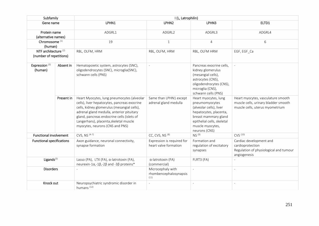
251
Subfamily I (L, Latrophilin)
Gene name
LPHN1 LPHN2 LPHN3 ELTD1
Protein name (alternative names)
ADGRL1 ADGRL2 ADGRL3 ADGRL4
Chromosome (1) (human)
19 1 4 6
NTF architecture (2) (number of repetitions)
RBL, OLFM, HRM RBL, OLFM, HRM RBL, OLFM HRM EGF, EGF_Ca
Expression (3) (human)
Absent in Hematopoietic system, astrocytes (SNC), oligodendrocytes (SNC), microglia(SNC), schwann cells (PNS)
- Pancreas exocrine cells, kidney glomerulus (mesangial cells), astrocytes (CNS), oligodendrocytes (CNS), microglia (CNS), schwann cells (PNS)
-
Present in Heart Myocytes, lung pneumocytes (alveolar cells), liver hepatocytes, pancreas exocrine cells, kidney glomerulus (mesangial cells), adrenal gland medulla, anterior pituitary gland, pancreas endocrine cells (islets of Langerhans), placenta,skeletal muscle myocytes, neurons (CNS and PNS)
Same than LPHN1 except adrenal gland medulla
Heart myocytes, lung pneumomyocytes (alveolar cells), liver hepatocytes, placenta, breast mammary gland epithelial cells, skeletal muscle myocytes, neurons (CNS)
Heart myocytes, vasculature smooth muscle cells, urinary bladder smooth muscle cells, uterus myometrium
Functional involvement CVS, NS (4-7) CC, CVS, NS (8) NS (9) CVS (10)
Functional specifications Axon guidance, neuronal connectivity, synapse formation
Expression is required for heart valve formation
Formation and regulation of excitatory synapses
Cardiac development and cardioprotection Regulation of physiological and tumour angiogenesis
Ligands(3) Lasso (PA), LTX (FA), α-latrotoxin (FA), neurexin-1α,-1β,-2β and -3β proteins*
α-latrotoxin (FA) (commercial)
FLRT3 (FA) -
Disorders - Microcephaly with rhombencephalosynapsis (11)
- -
Knock out Neuropsychiatric syndromic disorder in humans (12)
- - -

252
*Neurexins 1β, 2β and 1α have been reported to bind ADGRL1 but have not been shown to stimulate it or cause any effect, except limited increase in adhesion between ADGRL1-transfected and NRXN-transfected HEK293 cells.
SNP - - Risk factor for attention deficit/hyperactivity disorder (13-15)
-
Tumorigenesis Expressed in human monocytic leukemia cell lines and in primary human acute myeloid leukemia (AML) cells(16)
Cancer cells in GI cancer with lower LPHN2 expression show higher sensitivity to cisplatin(17)
Mutation of missense in lung cancer have been predicted to result in loss of function of the receptor – Tumor suppressor (18)
ELTD1 protein levels are significantly higher in human gliomas(19) High endothelial cell ELTD1 expression was found to correlate with improved overall survival in a number of cancer types(4)
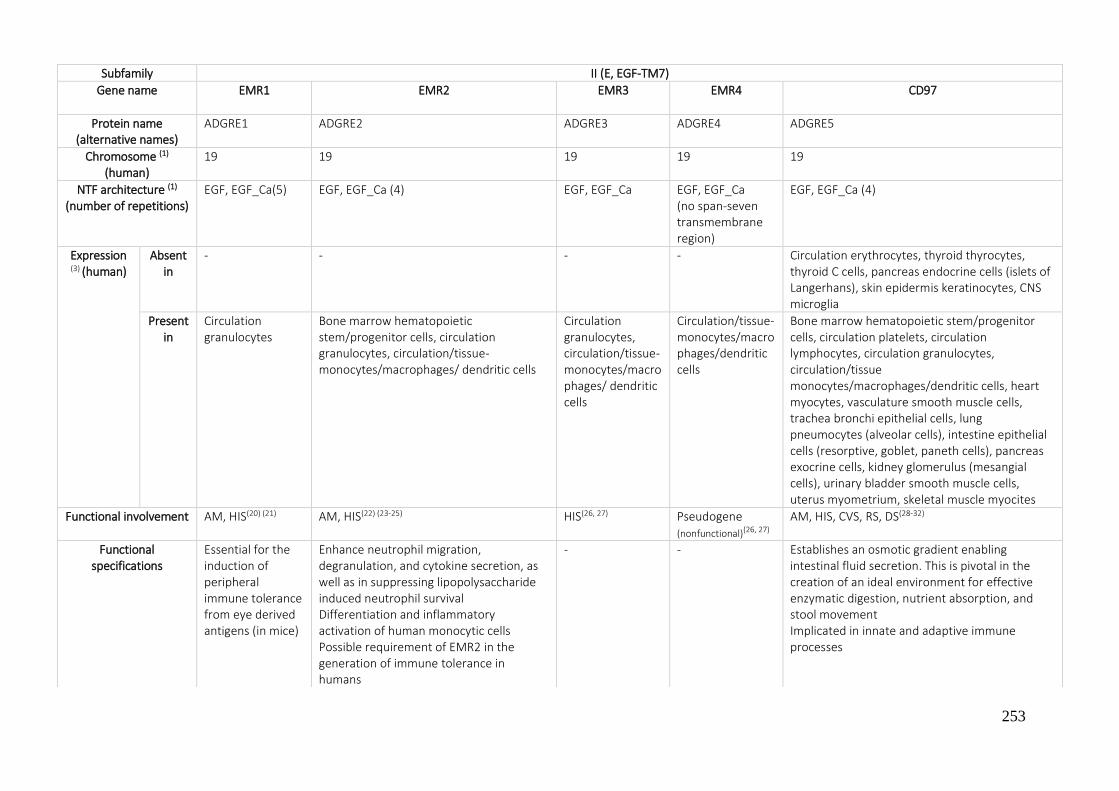
253
Subfamily II (E, EGF-TM7)
Gene name
EMR1 EMR2 EMR3 EMR4 CD97
Protein name (alternative names)
ADGRE1 ADGRE2 ADGRE3 ADGRE4 ADGRE5
Chromosome (1)
(human) 19 19 19 19 19
NTF architecture (1)
(number of repetitions) EGF, EGF_Ca(5) EGF, EGF_Ca (4) EGF, EGF_Ca EGF, EGF_Ca
(no span-seven transmembrane region)
EGF, EGF_Ca (4)
Expression (3) (human)
Absent in
- - - - Circulation erythrocytes, thyroid thyrocytes, thyroid C cells, pancreas endocrine cells (islets of Langerhans), skin epidermis keratinocytes, CNS microglia
Present in
Circulation granulocytes
Bone marrow hematopoietic stem/progenitor cells, circulation granulocytes, circulation/tissue-monocytes/macrophages/ dendritic cells
Circulation granulocytes, circulation/tissue-monocytes/macrophages/ dendritic cells
Circulation/tissue-monocytes/macrophages/dendritic cells
Bone marrow hematopoietic stem/progenitor cells, circulation platelets, circulation lymphocytes, circulation granulocytes, circulation/tissue monocytes/macrophages/dendritic cells, heart myocytes, vasculature smooth muscle cells, trachea bronchi epithelial cells, lung pneumocytes (alveolar cells), intestine epithelial cells (resorptive, goblet, paneth cells), pancreas exocrine cells, kidney glomerulus (mesangial cells), urinary bladder smooth muscle cells, uterus myometrium, skeletal muscle myocites
Functional involvement AM, HIS(20) (21) AM, HIS(22) (23-25) HIS(26, 27) Pseudogene (nonfunctional)(26, 27)
AM, HIS, CVS, RS, DS(28-32)
Functional specifications
Essential for the induction of peripheral immune tolerance from eye derived antigens (in mice)
Enhance neutrophil migration, degranulation, and cytokine secretion, as well as in suppressing lipopolysaccharide induced neutrophil survival Differentiation and inflammatory activation of human monocytic cells Possible requirement of EMR2 in the generation of immune tolerance in humans
- - Establishes an osmotic gradient enabling intestinal fluid secretion. This is pivotal in the creation of an ideal environment for effective enzymatic digestion, nutrient absorption, and stool movement Implicated in innate and adaptive immune processes

254
* Each was able to bind to the receptor as a ligand but none were found to have agonist activity at the receptor
Ligands 3 - - - - CD55, α5β1 integrin, chrondroitin sulfate B and CD90 (Thy1) *
Disorders Eosinophilic disorders (33)
Vibratory urticarial(25) - - -
Knock out - - - - -
SNP - - - - -
Tumorigenesis - Immune suppressing function in tumor cells (breast cancer)(34)
Upregulation of EMR2 in GBM is associated with poor survival(35)
A significant but low number of colorectal carcinomas are positive for EMR2(36) Lung cancer (adenocarcinomas)(37)
Upregulation of EMR3 in GBM is associated with poor survival(169)
- CD97 and CD55 are involved in the dedifferentiation, migration, invasiveness and metastasis of tumors, examples: In thyroid cancer, expression levels correlate with dedifferentiation.(38) In colorectal cancer is overexpressed in the invasion front.(39) (40) In Gall bladder carcinoma and glioblastoma expression is inversely related to overall survival.(41) (42) Has been identified as a leukemic stem cell marker in acute myeloid leukemia.(41) Marker for minimal residual disease in acute lymphoblastic leukemia(43)
CD97 promotes gastric cancer cell proliferation and invasion in vitro (44)

255
* Vascular overexpression results in CNS specific hyperproliferative vascular malformations, suggesting relevance for GPR124 in various CNS related vascular pathologies. + Gpr124 physically interact to synergistically stimulate Wnt7-specific responses in the brain.
Subfamily III (A)
Gene name
GPR123 GPR124 GPR125
Protein name (alternative names)
ADGRA1 ADGRA2 ADGRA3
Chromosome (1)
(human) 10 8 4
NTF architecture (1)
(number of repetitions) No specific domain (No GPS)
LRR-8, HRM LRR-8, I-set, HRM
Expression(3) (human)
Absent in
- - -
Present in
Vasculature endothelial cells, CNS
Eye retina, CNS, placenta, prostate, ovary, heart, skeletal muscle, small intestine, colon, lung, liver, kidney, pancreas, spleen, testis
Thyroid thyrocytes, testis spermatocytes, spermatogonial stem cell, colon, skeletal muscle, prostate, testis, colon, heart, placenta, pancreas, small intestine, brain, spleen, thymus
Functional involvement Unknown CVS, RS ,CNS (5, 39, 41, 42, 45) RS (46)
Functional specifications
- In CNS endothelial cells to regulate sprouting and migration Controls CNS angiogenesis and blood-brain barrier integrity
Role in repopulating spermatogonial stem cells (SSCs) in the adult human testes
Ligands (3) - Several GAGs + -
Disorders - CNS related vascular pathologies* (5, 41, 45) -
Knock out - Embryonic lethality (mouse)(5, 45)
Reduced lung size (mouse) (5, 45)
-
SNP - - -
Tumorigenesis - Highly expressed in tumor vasculature: GPR124 is required for VEGF induced tumor angiogenesis(45)
Overexpression suppress colorectal cancer formation by interacting with the Wnt pathway(47)
Up-regulation of GPR125 promoted cell adhesion and contributed to myeloid sarcoma formation(48)
Highly expressed in human brain cancer tissues (detected as oncogene)(41)

256
Subfamily IV (C,CELSR)
Gene name
CELSR1 CELSR2 CELSR3
Protein name (alternative names)
ADGRC1 ADGRC2 ADGRC3
Chromosome (1)
(human) 22 1 3
NTF architecture (2)
(number of repetitions)
C (9),EGF_Ca (2), L_G/Ptx, EGF_Ca, L_G/Ptx, EGF_Ca, L_EGF (No proteolysis in GPS)
C (9),EGF_Ca (2), L_G/Ptx, EGF_Ca, L_G/Ptx, EGF_Ca, L_EGF
C (9),EGF_Ca (2), L_G/Ptx, EGF_Ca, L_G/Ptx, EGF_Ca, L_EGF
Expression (3) (human)
Absent in
Neural stem cells
Present in
Neural stem cell CNS neurons, eye retina (CNS), neural stem cell, intermediate progenitors (IP)
Neurons
Functional involvement
CD (Cell development), PCP, CVS, RS, US, S, NS, SO (49) (48, 50-63)
CD, PCP, CC, CVS, ESM,NS (50-53),(48, 60-67) (68)
CD, PCP, CC, ESM, NS, RS (49) (50-52),(48, 61-67) (68-73)
Functional specifications *
Regulates epithelial planar cell polarity (PCP) in several organs (abates postnatally in parallel to decreasing numbers of NSC) Ex: plays a critical role for PCP establishment in the developing skin and hair follicles Facilitates neural tube closure via core PCP signaling Role in the planar polarity of vestibular hair cells during inner ear development Important in mammalian kidney development and role in rostrocaudal patterning during organogenesis
Development and maintenance of the nervous system Regulate dendrite morphogenesis Regulate axonal navigation in the forebrain and in the limbs (witch Fzd3) Ciliogenesis and neural development, especially neuron migration and axon guidance in the central, peripheral and enteric nervous systems
Neuronal maturation Regulate dendrite morphogenesis Regulate axonal navigation in the forebrain and in the limbs (witch Fzd3) Ciliogenesis and neural development, especially neuron migration and axon guidance in the central, peripheral and enteric nervous systems Is required for the fine mapping of sensory fibers in the spinal cord
Ligands(3) Vangl2 (core PCP protein), VhaPRR (accessory unit of the proton pump V-ATPase) via HRM domain of CELSR1
Unknown+ Unknown+
Disorders Neural tube defects: caudal agenesis type II, chiari II malformation, craniorachischisis, hydrocephalus, hydromyelia, lipoma, lipomyelocele, lipomyelomeningocele, myelomeningocele (74) ischemic stroke, possible risk factor for chronic obstructive pulmonary disease risk among women (75)
Idiopathic scoliosis in the Swedish-Danish population(76)
May be related with Joubert Syndrome (77)
Associated with myocardial infraction(42, 64)
Associated with Tourette disorder (78) Hirschsprung disease (79)

257
* Subfamily ADGRC (CELSR) receptors recruit Rho- GEFs during PCP signaling to control the size and shape of asymmetric epithelial membrane domains. It is nearly believed that ADGRC (CELSR) proteins form homodimeric molecular bridges between neighboring cells. ADGRCs (CELSRs) are therefore fundamental to local PCP transmission, and their asymmetric enrichment at both interfaces of opposing cell membranes is unique (Strutt et al., 2002; Devenport et al., 2011). + CELSR/FZD receptors are probably activated by WNT ligands but no ligand receptor-binding interaction was clearly identified.
Knock out Defects in the orientation of hair outgrowth in the adult (mice) (80)
Defects in hindbrain neuron migration (mouse) (81)
Defects in vestibular hair cells during inner ear development (mice) (59)
Induced missense mutations develop the severe neural tube defect (82)
Induced missense mutation develop lung branching defects (57)
Simplification of dendritic arbors of cortical pyramidal neurons and Purkinje neurons (rat brain)(83)
Defects in the migration of hindbrain neurons (mouse)(84)
Reduced quiescence of haemopoetic stem cells (mice)(85)
Defects in the development and planar organization of ependymal cilia are compromised, leading to a fatal hydrocephalus (mice)(86)
Mice deficient for ADGRC 2/3 had severe defects in pancreatic beta cell differentiation, resulting in decreased glucose clearance (66)
Mice deficient for ADGRC 2/3 had severe defects in pancreatic beta cell differentiation, resulting in decreased glucose clearance(66) Defects in tangential cortical interneuron migration (mouse)(87)
Dysfunction of hippocampal connectivity and maturation and forebrain wiring (mouse)(88) Defects in forebrain connections such as the anterior commissure and thalamocortical and corticospinal tracts (mouse)(89) Impaired axonal development (mouse) Marked malformation in the forebrain, absence of anterior commissure, and abnormalities in major axonal tracts (mouse) (90)
SNP -
Correlated with decreased low density lipoprotein levels (risk for coronary artery disease) (91)
-
Tumorigenesis Associated with glioma and glioblastoma (up-regulated)(92) (93) May act as an invasion suppressor in ductal carcinoma (94)
mRNA expressed in gastrointestinal tumor (95)
Cytoplasmic expression significantly stronger in breast cancer (96)
Associated with colorectal cancer metastasis (97) In pancreatic, bladder, renal, hepatic and oral carcinogenesis CELSR3 was identified as a hypermethylated marker(98) (99) (84) (100) (101)

258
Subfamily V (D)
Gene name
GPR133 GPR144
Protein name (alternative names)
ADGRD1 ADGRD2
Chromosome (1)
(human) 12 9
NTF architecture (2)
(number of repetitions)
Laminin_G/Ptx Laminin_G/Ptx
Expression (3)
(human) Absent in Pons, hippocampus, thalamus, medulla, midbrain, hypothalamus, amygdala
and frontal cortex -
Present in Anterior pituitary gland CNS neurons
Functional involvement CVS(102), MS (103-106) -
Functional specifications Heart rate control, associated locus with human height and weight -
Ligands 3 Tethered peptide from its ECD (Stachel sequence) -
Disorders - -
Knock out - -
SNP May cause clinically relevant phenotypes (approximately 9000 single nucleotide polymorphisms (SNPs) in the human ADGRD1)(103) SNPs associated with RR duration leading to an increase of one beat per second in heart rate(102) Association of ADGRD1 with the postprandial levels of very low density lipoproteins (VLDL) (107)
-
Tumorigenesis Is expressed within hypoxic regions of GBM, promotes tumor growth In Vitro and In Vivo and It’s expression correlates with poor prognosis in GBM (108)
Up-regulated in a murine model of gastrointestinal stromal tumors(109)
-

259
Subfamily VI (F)
Gene name
GPR110 GPR111 GPR113 GPR115 GPR116
Protein name (alternative names)
ADGRF1 ADGRF2 ADGRF3 ADGRF4 ADGRF5
Chromosome (1)
(human) 6 6 2 6 6
NTF architecture (2)
(number of repetitions) SAE No specific
domain (No GPS) HRM No specific domain
(No GPS) SEA, I-set, Ig_2
Expression(3)
(human) Absent
in
Present in
Kidney glomerulus (mesangial cells), kidney tubules (proximal, distal, collecting), prostate epithelial cells, trachea, mouth and lungs
Kidney (cortex and medulla), small bowel and pancreas
Skin epidermis keratinocytes, mouth, heart, uterus, vascular
Thyroid, placenta, uterus, spleen, stomach, lung (alveolar cells), kidney, brain, connective tissue, heart, eye, liver, muscle, pancreas, pharynx, testis, vasculature endothelial cells, breast mammary gland epithelial cells
Functional involvement US S (mouse) RS, S
Functional specifications
- Suggesting hypothesis that play a function in skin specialization that is essential for life in land(110)
- Suggesting hypothesis that play a function in skin specialization that is essential for life in land (110)
Pulmonary surfactant production, essential for surfactant homeostasis(111-114)
Ligands (3) Tethered peptide from its ECD (Stachel sequence)(115)
Synaptamide * (mouse) (116, 117)
Surfactant protein D (113, 118)
Disorders
Knock out Decelerates carcinogen-induced hepatocarcinogenesis via activation of the IL-6/STAT3 pathway (mouse) (119)
Loss of Gpr110, Gpr111, or Gpr115 function did not result in detectable defects (110)
Loss of Gpr110, Gpr111, or Gpr115 function did not result in detectable defects (110)
Loss of Gpr110, Gpr111, or Gpr115 function did not result in detectable defects (110)
Local inflammation in the lung (emphysema symptoms) (mice)(42)
Glucose intolerance and insulin resistance (mice) (120)
Loss of the both GPR116 and ELTD1 results in malformation of the cardiac outflow tract and the aortic arch arteries as well in the postnatal development of a thrombotic microangiopathy (mice)+(121)
SNP - - - - -

260
* Metabolite of the omega-3 fatty acid docosahexanoic (N-docosahexaenoylethanolamine, synaptamide), potently promotes neurogenesis, neuritogenesis and synaptogenesis. Mediating synaptamide-induced bioactivity in a cAMP-dependent manner. + Knockout alone of the receptors had no obvious effect in the CV or renal system.
Tumorigenesis Overexpression of GPR110 was correlated with the advanced stage of osteosarcoma.(122) Correlated with cell malignant transformation and tumor metastasis in glioma.(122, 123) Oncogene in prostate and lung cancer.(124)
Potential role of GPR110 in tumorigenicity and in tumor cell dissemination in HER2+ breast cancer.(125) Overexpressed in papillary thyroid cancer while underexpressed in anaplastic thyroid and follicular thyroid cancer. (126) High expression predicts a poor prognosis of gastric cancer patients(127)
- Its expression could serve as a useful marker for distinguishing gastric duodenal neuroendocrine tumors (down regulated) from small bowel neuroendocrine tumor and pancreatic neuroendocrine tumor.(128) (33) (33)
Potential prognostic biomarker in lung squamous cell carcinoma(129)
High expression has been significantly associated with poor overall survival of colorectal carcinoma patients(130)
Recurrent mutations in early gastric cancer so it may be involved in early carcinogenesis(131)
Associated with progression, metastasis, and poor prognosis in breast cancer patients. In breast cancer cell lines modulates motility and morphology(132)
Overexpressed in lung adenocarcinoma(133)

261
Subfamily VII (B,BAI)
Gene name BAI1 BAI2 BAI3
Protein name (alternative names)
ADGRB1 ADGRB2 ADGRB3
Chromosome (1)
(human) 8 1 6
NTF architecture (2)
(number of repetitions) TSP1(5), HRM TSP1 (4), HRM CUB, TSP1(4), HRM
Expression (3)
(human) Absent in
Placenta, lung, liver, skeletal muscle, kidney, pancreas, spleen, thymus, prostate, testis, ovary, intestines, leukocytes
Present in
Circulation/tissue monocytes/macrophages/DCs, brain tissues (hippocampus, basal ganglia, cerebral cortex, and olfactory bulb), neurons and glia, skeletal muscle myoblasts, peritoneal exudate cells, bone marrow, the spleen, the colon, and testes
Brain (adult and neonatal), different embryonically cells types in brain, heart, skeletal muscle, intestine, thymus
Brain (adult and neonatal π) (abundance in the postsynaptic neuron part), in various adult human tissues: the heart, testis, and small intestinal tissues
Functional involvement CD, CC, HIS, CVS, MSM, NS (100, 134-148) (145, 149, 150) CC, MS, NS (151) (152) (153)
(154) (155) (144)
CVS, NS (156-159) (140, 160, 161)
Functional specifications Inhibition of angiogenesis/tumorigenesis ¤ Synaptogenesis* Phagocytosis (Engulfment receptor) (especially involved in the clearance of dying neurons by promoting phagosome formation in microglia) x. Pattern recognition receptor, interacting with bacterial (Gram -) lipopolysaccharide (LPS) for innate immunity and inflammation in host cells (macrophages) (Pattern recognition receptor) (bacterial cell clearance). Promotes phagosomal reactive oxygen species (ROS) production. α Mediator of myoblast function (myogenesis)+. Role for BAI1 in facilitating macrophage anti-viral responses (135)
Inhibition of angiogenesis
Myoblast fusion µ Regulation of dendrite morphogenesis β
Regulation of synaptogenesis Ω Inhibition of angiogenesis Involved in the inhibition of Insulin secretion from pancreatic β-cells ∑
Ligands (3) p53-binding site # , CD36 (inhibition angiogenesis), αvβ5 integrin (inhibition of proliferation of endothelial cells), phosphatidylserine (clearance apoptotic cells). BAI1-associated protein 1 (Magi 1) (involved in cell adhesion and signal transduction in brain), BAI1-associated protein 2 (link between membrane and cytoskeleton in the process of neuronal growth), BAI1-associated protein 3 (involved in neuronal function (regulating release of neurotransmitters)), BAI1-associated protein 4. Its C terminus can interact with several PDZ domain–containing synaptic proteins (MAGI-1, MAGI-3, and PSD-95), which can regulate various signaling cascades downstream
GA-binding protein gamma (GABPγ), GABPα/γ or GABPα/β¥ Cytoplasmic region binds to glutaminase interacting protein (GIP)
Complement component 1, q subcomponent-like 1 (C1ql1), C1ql2, C1ql3 and Clql4
Disorders Decreased in Parkinson disease(162)
Involved in neurological diseases(163)
Expression decreases in the ischemic cerebral
Downregulated in the early phase of ischemia-induced brain injury(157)
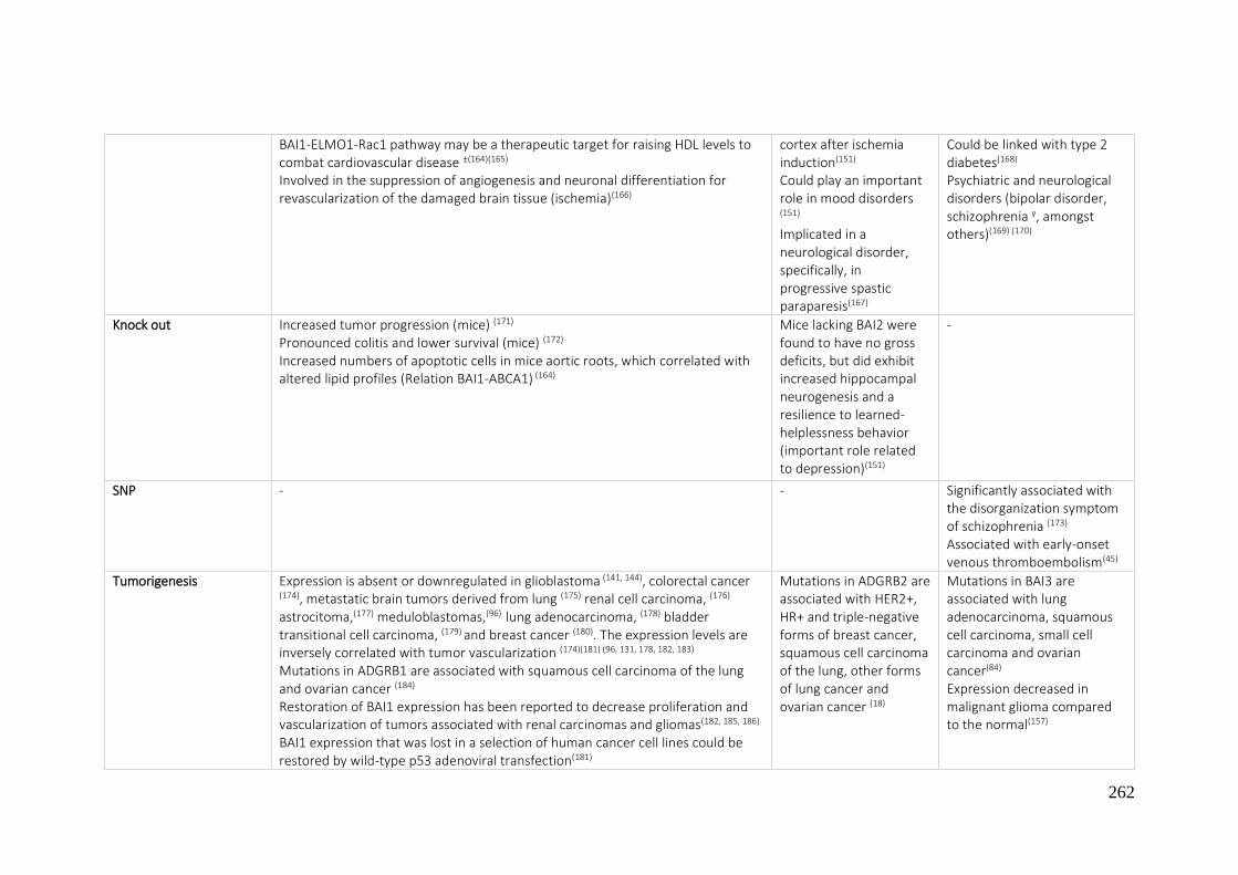
262
BAI1-ELMO1-Rac1 pathway may be a therapeutic target for raising HDL levels to combat cardiovascular disease ±(164)(165)
Involved in the suppression of angiogenesis and neuronal differentiation for revascularization of the damaged brain tissue (ischemia)(166)
cortex after ischemia induction(151)
Could play an important role in mood disorders (151)
Implicated in a neurological disorder, specifically, in progressive spastic paraparesis(167)
Could be linked with type 2 diabetes(168) Psychiatric and neurological disorders (bipolar disorder, schizophrenia γ, amongst others)(169) (170)
Knock out Increased tumor progression (mice) (171)
Pronounced colitis and lower survival (mice) (172)
Increased numbers of apoptotic cells in mice aortic roots, which correlated with altered lipid profiles (Relation BAI1-ABCA1) (164)
Mice lacking BAI2 were found to have no gross deficits, but did exhibit increased hippocampal neurogenesis and a resilience to learned-helplessness behavior (important role related to depression)(151)
-
SNP - - Significantly associated with the disorganization symptom of schizophrenia (173)
Associated with early-onset venous thromboembolism(45)
Tumorigenesis Expression is absent or downregulated in glioblastoma (141, 144), colorectal cancer
(174), metastatic brain tumors derived from lung (175) renal cell carcinoma, (176)
astrocitoma,(177) meduloblastomas,(96) lung adenocarcinoma, (178) bladder transitional cell carcinoma, (179) and breast cancer (180). The expression levels are inversely correlated with tumor vascularization (174)(181) (96, 131, 178, 182, 183) Mutations in ADGRB1 are associated with squamous cell carcinoma of the lung and ovarian cancer (184) Restoration of BAI1 expression has been reported to decrease proliferation and vascularization of tumors associated with renal carcinomas and gliomas(182, 185, 186)
BAI1 expression that was lost in a selection of human cancer cell lines could be restored by wild-type p53 adenoviral transfection(181)
Mutations in ADGRB2 are associated with HER2+, HR+ and triple-negative forms of breast cancer, squamous cell carcinoma of the lung, other forms of lung cancer and ovarian cancer (18)
Mutations in BAI3 are associated with lung adenocarcinoma, squamous cell carcinoma, small cell carcinoma and ovarian cancer(84) Expression decreased in malignant glioma compared to the normal(157)

263
¤ BAI1 contains 5 TSP that can be cleaved by matrix metalloproteinase 14 (MMP14) to release Vstat40 (vasculostatin 40), which inhibits angiogenesis * Through PAR3/TIAM1/RAC1 ± BAI1-ELMO1-Rac1 signaling pathway was found to contribute to the upregulation of ATP-binding cassette transporter 1 (ABCA1), which is known to be a critical cholesterol transporter that is essential for high-density lipoprotein (HDL). Higher HDL levels strongly correlate with a lower incidence of cardiovascular disease + Apoptotic myoblasts expose phosphatidylserine which activates BAI 1 signaling on healthy myoblasts to promote fusion with myoblast and myotubes. α Via activation of the Rho family guanosine triphosphate (GTPase) Rac1, improving the microbicidal activity of the macrophages. BAI 1 is necessary not only clearance via macrophages, but also for activation of the phagosomal nicotinamide adenine dinucleotide phosphate (NADPH) oxidase complex. X Binding phosphatidylserine mediate the uptake of apoptotic cells by numerous cell types, including macrophages and microglia, operating with the upstream of the signaling comprised of ELMO/Dock180/Rac proteins, facilitating the clearance. # The regulatory role of p53 in BAI1 is not clear. Some studies show that BAI1 is transcriptionally regulated by p53(144, 187) meanwhile others have shown that the level of BAI1 messenger RNA (mRNA) does not correlate with the p53 status.(141) ¥ Expression of BAI2 inversely correlates with the vascular endothelial growth factor (VEGF) expression, which is a key mediator of angiogenesis. GA-binding protein gamma (GABPgamma) associates with the cytoplasmic domain of BAI2, and GABPalpha/gamma or GABPalpha/beta works as a transcriptional repressor of VEGF. The C-terminal fragment cleaved at the GPS domain specifically activated the nuclear factor of activated T-cells (NFAT) pathway. Furin prohormone convertase cleaved BAI2 at a different site of the GPS in the ECR.(188) Π During embryonic stages, the expression of BAI3 mRNA is limited to the central nervous system unlike the other members of BAI family. Β The C-terminal region binds to ELMO, resulting in the regulation of dendrite morphogenesis via the activation of small GTPase Rac1. µ Regulated by C1qL4 and stabillin 2, ligand of BAI3 and its binding partner, respectively. BAI1 and BAI3 can promote myoblast fusion via binding to ELMO-Dock1 in muscle development and repair. Ω BAI3 binds to the C1ql protein that is expressed and secreted in the brain, and this interaction is important for the regulation of synapse formation and maintenance. It has been demonstrated that the expression of BAI3 is enhanced in Purkinje cells, and C1ql1, as a ligand of BAI3, regulates the spinogenesis of Purkinje cells in a BAI3-dependent manner(159) ∑ As a receptor of Clql3. γ Its expression is changed by treatment with lithium carbonate, an effective drug for bipolar disorder.
Antitumor effects of Vstat120 (cleaved and secreted extracellular fragment of BAI1) delivery into established tumors and supports the further development of Rapid Antiangiogenesis Mediated By Oncolytic virus as a possible cancer therapy(183)
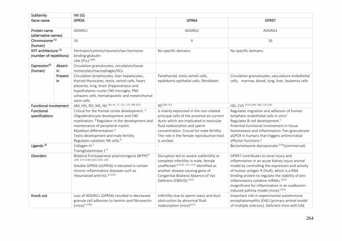
264
Subfamily VIII (G)
Gene name
GPR56 GPR64 GPR97
Protein name (alternative names)
ADGRG1 ADGRG2 ADGRG3
Chromosome (1)
(human) 16 X 16
NTF architecture (2)
(number of repetitions)
Pentraxin/Laminin/neurexin/sex-hormone-binding-globulin- Like (PLL) (189)
No specific domains No specific domains
Expression(3)
(human) Absent in
Circulation granulocytes, circulation/tissue monocytes/macrophages/DCs
Present in
Circulation lymphocytes, liver hepatocytes, thyroid thyrocytes, testis, sertoli cells, heart, placenta, lung, brain (hippocampus and hypothalamic nuclei CNS microglia, PNS schwann cells, hematopoietic and mesenchymal stem cells
Parathyroid, testis sertoli cells, epididymis epithelial cells, fibroblasts
Circulation granulocytes, vasculature endothelial cells, marrow, blood, lung, liver, leukemia cells
Functional involvement AM, HSI, RO, MS, NS (39, 41, 77, 115, 173, 189-207) RS(208-212) HSI, CVS (213) (200, 208, 214-216)
Functional specifications
Critical for the frontal cortex development. α
Oligodendrocyte development and CNS myelination. β Regulator in the development and maintenance of peripheral myelin. Myoblast differentiation.γ Testis development and male fertility Regulates cytotoxic NK cells.Ω
Is mainly expressed in the non-ciliated principal cells of the proximal ex-current ducts which are implicated in testicular fluid reabsorption and sperm concentration. Crucial for male fertility. The role in the female reproductive tract is unclear.
Regulates migration and adhesion of human lymphatic endothelial cells in vitroƮ Regulates B-cell development.ƪ
Potential functional involvement in tissue homeostasis and inflammation. Pan-granulocyte aGPCR in humans that triggers antimicrobial effector functions.ƻ
Ligands (3) Collagen III ∑
Transglutaminase 2 δ - Beclometasone dipropionate (214)(commercial)
Disorders Bilateral frontoparietal polymicrogyria (BFPP)Ʊ (198, 217) (218) (201) (219, 220)
Soluble GPR56 (sGPR56) is elevated in certain chronic inflammatory diseases such as rheumatoid arthritis.ψ (221)
Disruption led to severe subfertility or complete infertility in male, female unaffected.Ɯ (191, 211, 212) Identified as another disease‐causing gene of Congenital Bilateral Absence of Vas Deferens (CBAVD).(222)
GPR97 contributes to renal injury and inflammation in an acute kidney injury animal model by controlling the expression and activity of human antigen R (HuR), which is a RNA binding protein to regulate the stability of pro-inflammatory cytokine mRNAs (223)
Insignificant for inflammation in an ovalbumin-induced asthma model (mice).(224)
Knock out Loss of ADGRG1 (GPR56) resulted in decreased granule cell adhesion to laminin and fibronectin (mice)π (196)
Infertility due to sperm stasis and duct obstruction by abnormal fluid reabsorption (mice)(211)
Important role in experimental autoimmune encephalomyelitis (EAE) (primary animal model of multiple sclerosis). Deficient mice with EAE

265
Decreased cellular adhesion of acute myeloid leukemia (AML) cells to fibronectin, laminin-1, and collagen III, corresponding to a significant decrease in the number of hematopoietic stem cells (HSCs) in the bone marrow and, conversely, an increase in HSC number in spleen, liver, and peripheral blood (mice).μ (202)
Cerebellar malformation (mouse)(196, 197)
Male-specific sterility, malformation of male gonad (mouse)(191)
exhibited increases in peak severity and the cumulative disease score compared with the control, followed by a notable increase of leukocyte infiltration and more extensive demyelination (216)
SNP - - -
Tumorigenesis The function of ADGRG1 (GPR56) in cancer seems to be cell/context specific. Expression inversely correlated with the metastatic potential of melanoma cell lines(225,
226) and the re-expression of the receptor led to a reduction in melanoma growth and metastasis.κ (227)Found to be upregulated in various cancer types and in tumor cell lines. (228-
230) Inhibits mesenchymal differentiation and radioresistance in Glioblastoma.(231) Promotes proliferation of colorectal cancer cells and enhances metastasis. ν (232, 233) Prognostic factor in epithelial ovarian cancer and could promote the progression and invasion.φ (234)Associated with tumor progression in non-small-cell lung carcinoma.(235) Regulates the proliferation and invasion capacity of osteosarcoma cells.(192) Highly expressed in acute myeloid leukemia with poor outcome (significantly associated with high-risk genetic subgroups and poor outcome).(236) GPR56 as a novel marker in acute myeloid leukemia (AML) with high ecotropic viral integration site-1 (EVI1) expression.ƿ (203)
The levels are significantly overexpressed in the Wnt signaling-dependent subgroup of medulloblastoma (238) and higher in Ewing sarcomasƝ (239) Overexpressed in parathyroid tumors from patients with primary hyperparathyroidism (PHPT).Ƒ
Involved in the adhesion and migration of certain breast cancer cellsƟ(240)
Also has been found to be upregulated in various carcinomas, including prostate, kidney, breast and non-small cell lung cancer(239)
Increases in acute and chronic inflamed and cancerous tissues. Adgrg3 is repressed by the transcription factor Pax5 but activated by the Pax5-Etv6 oncoprotein in B-cell acute lymphoblastic leukemia (241) (242) More infiltrating macrophages and higher tumor necrosis factor levels were identified in the liver and kidney of Adgrg3-deficient mice, compared to wild-type animals, in a high-fat diet-induced experimental obesity model(243)
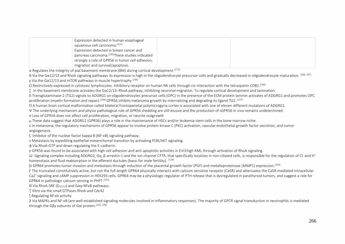
266
α Regulates the integrity of pial basement membrane (BM) during cortical development (173) Β Via the Gα12/13 and RhoA signaling pathways its expression is high in the oligodendrocyte precursor cells and gradually decreased in oligodendrocyte maturation. (206, 207)
γ Via the Gα12/13 and mTOR pathways in muscle hypertrophy (244)
Ω Restrictively expressed in cytotoxic lymphocytes. Inhibitory receptor on human NK cells through cis-interaction with the tetraspanin CD81.(190)
∑ In the basement membrane activates the Gα12/13–RhoA pathway, inhibiting neuronal migration. To regulate cortical development and lamination. δ Transglutaminase-2 (TG2) signals to ADGRG1 on oligodendrocytes precursor cells (OPC) in the presence of the ECM protein laminin activates of ADGRG1 and promotes OPC proliferation (myelin formation and repair).(206) GPR56 inhibits melanoma growth by internalizing and degrading its ligand TG2. (227)
Ʊ A human brain cortical malformation called bilateral frontoparietal polymicrogyria cortex is associated with one of eleven different mutations of ADGRG1. Ψ The underlying mechanism and physio pathological role of GPR56 shedding are still elusive and the production of sGPR56 in vivo remains undetermined. π Loss of GPR56 does not affect cell proliferation, migration, or neurite outgrowth µ These data suggest that ADGRG1 (GPR56) plays a role in the maintenance of HSCs and/or leukemia stem cells in the bone marrow niche. κ In melanoma, the regulatory mechanisms of GPR56 appear to involve protein kinase C (PKC) activation, vascular endothelial growth factor secretion, and tumor angiogenesis. ξ Inhibitor of the nuclear factor kappa B (NF-κB) signaling pathway. ν Metastasis by expediting epithelial-mesenchymal transition by activating PI3K/AKT signaling. φ Via RhoA-GTP and down regulating the E-cadherin. ƿ GPR56 was found to be associated with high cell adhesion and anti-apoptotic activities in EVI1high AML through activation of RhoA signaling. Ɯ Signaling complex including ADGRG2, Gq, β-arrestin-1 and the ion channel CFTR, that specifically localizes in non-ciliated cells, is responsible for the regulation of Cl- and H+ homeostasis and fluid reabsorption in the efferent ductules (basic for male fertility). (129)
Ɲ GPR64 promotes tumor invasion and metastasis through induction of the placental growth factor (PGF) and metalloproteinase (MMP1) expression.(239) Ƒ The truncated constitutively active, but not the full-length GPR64 physically interacts with calcium sensitive receptor (CaSR) and attenuates the CaSR-mediated intracellular Ca2+ signaling and cAMP suppression in HEK293 cells. GPR64 may be a physiologic regulator of PTH release that is dysregulated in parathyroid tumors, and suggest a role for GPR64 in pathologic calcium sensing in PHPT.(245)
Ɵ Via RhoA-SRE (G12/13) and Gαq-NFκB pathways. Ʈ Vitro via the small GTPases RhoA and Cdc42 ƪ Regulating Nf-κb activity ƻ Via MAPKs and NF-κB (are well-established signaling molecules involved in inflammatory responses). The majority of GPCR signal transduction in neutrophils is mediated through the Gβγ subunits of Gαi protein.(215, 246)
Expression detected in human esophageal squamous cell carcinoma.(237)
Expression detected in breast cancer and pancreas carcinoma.(230)These studies indicated strongly a role of GPR56 in tumor cell adhesion, migration and survival/apoptosis.

267
Subfamily VIII (G)
Gene name
GPR112 GPR114 GPR126 GPR128
Protein name (alternative names)
ADGRG4 ADGRG5 ADGRG6 ADGRG7
Chromosome (1) (human)
X 16 6 3
NTF architecture (2) (number of repetitions)
Laminin_G/Ptx No specific domains Laminin_G/Ptx, CUB No specific domains
Expression(3) (human)
Absent in - - - -
Present in Mammary gland, kidney, blood vessel, retina, testis
Circulation granulocytes, colon, lung, spleen, thymus, pancreas islets of Langerhans, leukocytes, tonsil
Bone marrow hematopoietic stem/progenitor cells, vasculature endothelial cells, liver hepatocytes, pancreas endocrine cells (islets of Langerhans), placenta, skin epidermis keratinocytes, cartilage chondrocytes, PNS Schwann cells
Liver hepatocytes, small intestine, cervix, skin
Functional involvement - HSI CC,CVS, RS, MS, NSB, SO (247-254) -
Functional specifications Potential roles in the regulation of secretion, immunity, and epithelial homeostasis (mice)(255)
Immune-related functions(256)
PNS myelinitation (important in development)Ʊ, adult height (trunk height), pulmonary function, modulates both physiological and pathological angiogenesis ψ, regeneration of axons following injuries φ
-
Ligands (3) - Custom peptide GPR126
Colagen IV
Disorders - - Adolescent idiopathic scoliosisƮ Arthrogryposis multiplex congenita Intellectual disability(257)
Can contribute to the cellular events in adolescent idiopathic scoliosis ƻ (258)
Knock out - - Conditional loss of it in Schwann cells causes increased proliferation of this cell type(251) ,essential in neural, cardiac, and ear development (mice)
Reduced body weight and increased intestinal contraction frequency (mouse) (259)
SNP - Differences in the Vitamin D metabolism(260)
Impaired pulmonary function in humansα(261)
Variation in Height(262)
Candidate genetic risk factor for aggressive periodontitis in the Japanese population(263)
-

268
ƻ The estrogens induced auto-proteolytic mechanism of the GAIN domain and activate via modulated by ESRα (estrogen receptor) and SP1 (specificity protein 1) complex. Ʊ GPR126 drives the differentiation of Schwann cells through inducing cAMP levels, which causes Oct6 transcriptional activities to promote myelin gene activity. Domain-specific functions of GPR126 during Schwann cells development: the NTF is necessary and sufficient for axon sorting, while the CTF promotes wrapping through cAMP induction to regulate early and late stages of Schwann cells development.(268)
Ʈ Mouse models have shown GPR126 deletion to affect cartilage biology and spinal column development.(269)
Ψ GPR126 stimulates VEGF signaling and angiogenesis by modulating VEGF receptor 2 (VEGFR2) expression through STAT5 and GATA2 in endothelial cells.(247) α Measured as the ratio of forced expiratory volume in the first second/forced vital capacity (FEV1/FVC), in a meta-analysis of 28,890 participants. May contribute to the physiological adaptations to hypobaric hypoxia, possibly by altering lung function.(270)
φ GPR126 is not necessary for normal myelin maintenance; knocking out Gpr126 without injuring nerves produced no change in myelination for up to 4 months.(252)
Tumorigenesis Upregulated in related primary neuroendocrine carcinoma and liver metastasis(264) High expression in ileal carcinoids(265)
- High Prevalence of a Hotspot of Noncoding Somatic Mutations in Intron 6 of GPR126 in Bladder Cancer(266) Associated with reduced survival in breast cancer(267)
-
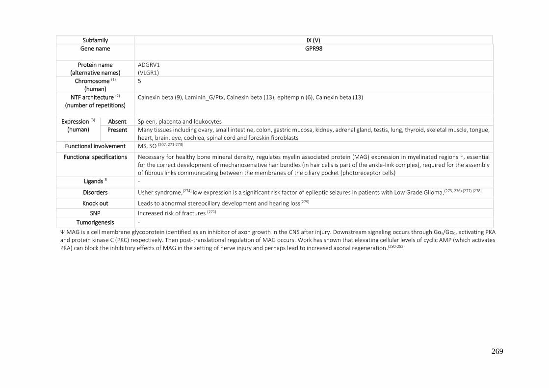
269
Ψ MAG is a cell membrane glycoprotein identified as an inhibitor of axon growth in the CNS after injury. Downstream signaling occurs through Gαs/Gαq, activating PKA and protein kinase C (PKC) respectively. Then post-translational regulation of MAG occurs. Work has shown that elevating cellular levels of cyclic AMP (which activates PKA) can block the inhibitory effects of MAG in the setting of nerve injury and perhaps lead to increased axonal regeneration.(280-282)
Subfamily IX (V)
Gene name
GPR98
Protein name (alternative names)
ADGRV1 (VLGR1)
Chromosome (1) (human)
5
NTF architecture (2) (number of repetitions)
Calnexin beta (9), Laminin_G/Ptx, Calnexin beta (13), epitempin (6), Calnexin beta (13)
Expression (3) (human)
Absent Spleen, placenta and leukocytes
Present Many tissues including ovary, small intestine, colon, gastric mucosa, kidney, adrenal gland, testis, lung, thyroid, skeletal muscle, tongue, heart, brain, eye, cochlea, spinal cord and foreskin fibroblasts
Functional involvement MS, SO (207, 271-273)
Functional specifications Necessary for healthy bone mineral density, regulates myelin associated protein (MAG) expression in myelinated regions ψ, essential for the correct development of mechanosensitive hair bundles (in hair cells is part of the ankle-link complex), required for the assembly of fibrous links communicating between the membranes of the ciliary pocket (photoreceptor cells)
Ligands 3 -
Disorders Usher syndrome,(274) low expression is a significant risk factor of epileptic seizures in patients with Low Grade Glioma,(275, 276) (277) (278)
Knock out Leads to abnormal stereociliary development and hearing loss(279)
SNP Increased risk of fractures (271)
Tumorigenesis -
270
7.2. BIBLIOGRAPHY
1 https://www.ensembl.org (10/01/2019) 2 T. Langenhan; G. Aust; J. Hamann. Science Signal. 2013, 6, 1-22 3 https://www.guidetopharmacology.org - Adhesion Class GPCRs (10/01/2019) 4 B. Davletov; F. Meunier; A. Ashton; H. Matsushita; W. Hirst; V. Lelianova. EMBO J. 1998, 17, 3909-3920 5 A. C. Ashton; V. Lelianova; E. Orlova; C. Van Renterghem; M. Canepari; M. Seagar. J. Biol. Chem. 2001, 276, 44695-44703 6 M. Capogna; K. Volynski; Y. Ushkaryov. J. Neurosci. 2003, 23, 4044-4053 7 K. Volynski; J. L. Silva; M. A. Rahman; C. Hopkins; Y. Ushkaryov. EMBO J. 2004, 23, 4423-4433 8 S. Doyle; M. Scholz; K. Greer; A. Hubbard; D. Darnell; P. Antin. Dev. Dyn. 2006, 235, 3213-3221 9 M. O'Sullivan; J. de Wit; J. Savas; D. Comoletti; S. Otto-Hitt; J. Yates. Neuron. 2012, 73, 903-910 10 D. Favara; A. Banham; A. Harris. Biochem Soc Trans. 2014, 42, 1658-1664 11 M. Vezain; M. Lecuyer; M. Rubio; V. Dupé; L. Ratié; V. David. Acta Neuropathol. Commun. 2018, 6, 1-23 12 M. Bonaglia; S. Marelli; F. Novara; S. Commodaro; R. Borgatti; G. Minardo. Eur. J. Hum. Genet. 2010, 18, 1302-139 13 M. Arcos-Burgos; D. Wallis. Mol. Psychiatry. 2010, 15, 1053-1066 14 S. Domené; H. Stanescu; D. Wallis; B. Tinloy; D. Pineda; R. Kleta. Am. J. Med. Genet. B. Neuropsychiatt. Genet. 2011, 156, 11-8 15 M. Ribasés; J. Ramos-Quiroga; C. Sánchez-Mora; R. Bosch; V. Richarte; G. Palomar. Genes Brain Behav. 2011, 10, 149-157 16 Z. Kocibalova; M. Guzyova; D. Imrichova; Z. Sulova; A. Breier. Gen. Physiol. Biophys. 2018, 37, 353-357 17 M. Jeon; J. Yun; H. Kim; S. Han. Cancer Res. Treat. 2016, 48, 676-686 18 Z. Kan; B. Jaiswal; J. Stinson; V. Janakiraman; D. Bhatt; H. Stern. Nature, 2010, 466, 869-873 19 J. Ziegler; R. Pody; P. Coutinho; B. Evans; D. Saunders; N. Smith. Neuro. Oncol. 2017, 19, 175-185 20 H. Lin; D. Faunce; M. Stacey, A. Terajewicz; T. Nakamura; J. Zhang-Hoover. J. Exp. Med. 2005, 201, 1615-1625 21 H. Warschkau; A. Kiderlen. J. immunol. 1999, 163, 3409-3416 22 S. Yona; H. Lin; P. Dri; J. Davies; R. Hayhoe; S. Lewis. FASEB J. 2008, 22, 741-751 23 S. Yona; H. Lin; W. Siu; S. Gordon; M. Stacey. Trends Biochem. Sci. 2008, 33, 491-500 24 Y. Huang; N. Chiang; C. Hu; C. Hsiao; K. Cheng; W. Tsai. Mol. Cell. Biol. 2012, 32, 1408-1420 25 Y. Huang; C. Hu; W. Tseng; C. Cheng; M. Stacey. Front. Immunol. 2017, 373, 1-13 26 J. Hamann; M. Kwakkenbos; E. de Jong; H. Heus; A. Olsen; R. van Lier. Eur. J. Immunol. 2003, 33, 1365-1371 27 I. Caminschi; S. Vandenabeele; M. Sofi; A. McKnight; N. Ward; T. Brodnicki. DNA seq. 2006, 17, 8-14 28 J. Hamann; E. Hartmann; R. van Lier. Genomics. 1996, 32, 144-147 29 J. Leemans; A. te Velde; S. Florquin; R. Bennink; K. de Bruin; R. van Lier. J. immunol. 2004, 172, 1125-1131 30 M. Capasso; L. Durrant; M. Stacey; S. Gordon; J. Ramage; I. Spendlove. J. Immunol. 2006, 177, 1070-1077 31 M. van Pel; H. Hagoort; M. Kwakkenbos. J. Hamann; W. Fibbe. Haematologica. 2008, 93, 601-604 32 G. Aust; C. Kerner; S. Gonsior; D. Sittig; H. Schneider; P. Buske. Mol. Bio. Cell. 2013, 24, 2256-2268 33 J. C. Carr; S. K. Sherman; D. Wang; F. S. Dahdaleh; A. M. Bellizzi; M. S. O'Dorisio. Annals of surgical oncology 2013, 20, S739-s46
271
34 J. Davies; H. Lin; M. Stacey; S. Yona; G. Chang; S. Gordon. Oncol. Rep. 2011, 25, 619-627 35 M. Rutkowski; M. Sughrue; A. Kane; J. Kim; O. Bloch; A. Parsa. J. Neurooncol. 2011, 105, 165-171 36 G. Aust; J. Hamann; N. Schilling; M. Wobus. Virchows Arch. 2003, 443, 32-37 37 A. Feliciano; Y. Garcia-Mayea; L. Jubierre; C. Mir; M. Hummel; J. Castellvi. Cell Death Dis. 2017, 8, e3141 38 G. E. Aust; S. Laue; I. Lehmann; N. Heldin; O. Lotz; W. Scherbaum. Cancer Res. 1997, 57, 1798-1806 39 S. D. Ackerman; R. Luo; Y. Poitelon; A. Mogha; B. L. Harty; M. D'Rozario. J. Exp. Med. 2018, 215, 941-961 40 J. Galle; D. Sittig; I. Hanisch; M. Wobus; E. Wandel; M. Loeffler. Am. J. Pathol. 2006, 169, 1802-1811 41 S. D. Ackerman; C. Garcia; X. Piao; D. H. Gutmann; K. R. Monk. Nature 2015, 6, 1-4 42 D. M. Ariestanti; H. Ando; S. Hirose; N. Nakamura. J. Biol. Chem. 2015, 290, 11032-1140 43 E. Coustan-Smith; G. Song; C. Clark; L. Key; P. Liu; M. Mehrpooya. Blood 2011, 117, 6267-6276 44 C. Li; D. Liu; G. Li; H. Wang; X. Li; W. Zhang. World J. Gastroenterol. 2015, 21, 6215-6228 45 G. Antoni; P. E. Morange; Y. Luo; N. Saut; G. Burgos; S. Heath. JTH. 2010, 8, 2671-2679 46 F. Izadyar; J. Wong; C. Maki; J. Pacchiarotti; T. Ramos; K. Howerton. Hum. Reprod. 2011, 26, 1296-1306 47 Y. Wu; W. Chen; L. Gong; C. Ke; H. Wang; Y. Cai. Med. Sci. Monit. 2018, 24, 6608-6616 48 S. Berger-Müller; T. Suzuki. Mol. Neurobiol. 2011, 44, 313-320 49 A. Goffinet; F. Tissir. Semin. Cell. Dev. Biol. 2017, 69, 102-110 50 D. Strutt; R. Johnson; K. Cooper. Curr. Biol. 2002, 12, 813-824 51 D. Devenport; D. Oristian; E. Heller; E. Fuchs. Nat. Cell. Biol. 2011, 13, 893-902 52 S. J. Warrington; H. Strutt; D. Strutt. Development 2013, 5, 1045-1554 53 T. Nishimura; H. Honda; M. Takeichi. Cell. 2012, 149, 1084-1097 54 Y. Zhan; Q. Luo; X. Zhang; N. Xiao; C. Lu; C. Yue. Biochemistry 2016, 81, 591-599 55 Y. Yamada; N. Fuku; M. Tanaka; Y. Aoyagi; M. Sawabe; N. Metoki. Atherosclerosis 2009, 207, 144-149 56 L. Yates; C. Schnatwinkel; J. Murdoch; D. Bogani; C. Formstone; S. Townsend. Hum. Mol. Genet. 2010, 19, 2251-2267 57 L. Yates; J. Papakrivopoulou; D. Long; P. Goggolidou; J. Connolly; A. Woolf. Hum. Mol. Genet. 2010, 19, 4663-4676 58 M. Cetera; L. Leybova; F. Woo; M. Deans; D. Devenport. Dev. Biol. 2017, 428, 188-203 59 J. Duncan; M. Stoller; A. Francl; F. Tissir; D. Devenport; M. Deans. Dev. Biol. 2017, 423, 126-137 60 T. S. Usui; Y. Shimada; S. Hirano: R. Burgess; T. Schwarz; M. Takeichi; T. Uemura. Cell. 1999, 98, 585-595 61 D. Matsubara; S. Horiuchi; K. Shimono; T. Usui; T. Uemura. Genes Dev. 2011, 25, 1982-1996 62 S. Hakeda; T. Suzuki. Genes Cells. 2013, 18, 960-973 63 E. H. Najarro; B. Ackley. Dev. Biol. 2013, 377, 224-235 64 S. Kathiresan; B. Voight; S. Purcell; K. Musunuru; D. Ardissino; P. Mannucci. Nat. Genet. 2009, 41, 334-341 65 L. Qi; J. Ma; Q. Qi; J. Hartiala; H. Allayee; H. Campos. Circulation. 2011, 123, 374-380 66 C. Cortijo; M. Gouzi; F. Tissir; A. Grapin-Botton. Cell. Rep. 2012, 2, 1593-1606 67 Y. Shima; S. Kawaguchi; K. Kosaka; M. Nakayama; M. Hoshino; Y. Nabeshima. Nat. Neurosci. 2007, 10, 963-969 68 Y. Qu; Y. Huang; J. Feng; G. Alvarez-Bolado; E. Grove; Y. Yang. Proc. Natl. Acad. Sci. USA. 2014, 111, e2996-3004 69 G. Chai; L. Zhou; M. Manto; F. Helmbacher; F. Clotman; A. Goffinet. Nat. Neurosci. 2014, 17, 1171-1179 70 F. Wang; Q. Wang. C. Li; P. Yu; Y. Qu; L. Zhou. Neuroscience 2017, 359, 267-276 71 F. Tissir; A. Goffinet. Nat. Rev. Neurosci. 2013, 14, 525-535
272
72 V. Sasselli; W. Boesmans; P. V. Berghe; F. Tissir; A. Goffinet; V. Pachnis. J. Clin. Investig. 2013, 123, 1763-7172 73 J. Feng; Q. Xian; T. Guan; J. Hu; M. Wang; Y. Huang. Cereb. Cortex. 2016, 26, 3323-3334 74 R. Allache; P. De Marco; E. Merello; V. Capra; Z. Kibar. Birth Defects Res. A Clin. Mol. Teratol. 2012, 94, 176-181 75 M. Cho; S. Sharma; K. Glass; P. Castaldi; M. McDonald. Am. J. Respir. Cell. Mol. Biol. 2017, 56, 332-341 76 E. Einarsdottir; A. Grauers; J. Wang; H. Jiao; S. Escher; A. Danielsson. PLoS One. 2017, 12, e0189591 77 B. I. Bae; I. Tietjen; K. D. Atabay; G. D. Evrony; M. B. Johnson; E. Asare. Science 2014, 343, 764-768 78 A. J. Willsey; T. V. Fernandez; D. Yu; R. A. King; A. Dietrich; J. Xing. Neuron. 2017, 94, 486-499 79 L. Su; Z. Zhang; L. Gan; Q. Jiang; P. Xiao; J. Zou. Exp. mol. pathol. 2016, 101, 241-248 80 A. Ravni; Y. Qu; A. M. Goffinet; F. Tissir. J. inv. dermatol. 2009, 129, 2507-2509 81 Y. Qu; D. M. Glasco; L. Zhou; A. Sawant; A. Ravni; B. Fritzsch. J. Neurosci. 2010, 30, 9392-9401 82 J. A. Curtin; E. Quint; V. Tsipouri; R. M. Arkell; B. Cattanach; A. J. Copp. Curr. Biol. 2003, 13, 1129-1133 83 Y. Shima; M. Kengaku; T. Hirano; M. Takeichi; T. Uemura. Developmental cell. 2004, 7, 205-216 84 M. Erkan; N. Weis; Z. Pan; C. Schwager; T. Samkharadze; X. Jiang. Molecular cancer 2010, 9, 88 85 R. Sugimura; X. C. He; A. Venkatraman; F. Arai; A. Box; C. Semerad C. Cell. 2012, 150, 351-365 86 F. Tissir; Y. Qu; M. Montcouquiol; L. Zhou; K. Komatsu; D. Shi. Nat. Neurosci. 2010, 13, 700-707 87 G. Ying; S. Wu; R. Hou; W. Huang; M. R. Capecchi; Q. Wu. Mol. Cell. Biol. 2009, 29, 3045-3061 88 J. Feng; Y. Xu; M. Wang; Y. Ruan; K. F. So; F. Tissir. J. Neurosci. 2012, 32, 13729-13743 89 L. Zhou; Y. Qu; F. Tissir; A. M. Goffinet. Cereb. Cortex. 2009, 19, 114-119 90 F. Tissir; I. Bar; Y. Jossin; O. De Backer; A. M. Goffinet. Nat. Neurosci. 2005, 8, 451-457 91 S. Kathiresan; O. Melander; C. Guiducci; A. Surti; N. P. Burtt; M. J. Rieder. Nat. Genet. 2008, 40, 189-197 92 B. Vastrad; C. Vastrad; A. Godavarthi; R. Chandrashekar. Medical oncology 2017, 34, 182 93 M. Feve; J. M. Saliou; M. Zeniou; S. Lennon; C. Carapito; J. Dong. PLoS One. 2014, 9, e91519 94 J. Geradts; J. Groth; Y. Wu; G. Jin. Breast cancer research and treatment 2016, 157, 447-459 95 M. Katoh. Int. J. Mol. Med. 2007 20, 405-409 96 L. Jiang; X. Zhang; C. Xiang; J. Geradts; Q. Wei; Y. Liang. Histol. and histopathol. 2018, 33, 835-842 97 K. Goryca; M. Kulecka; A. Paziewska; M. Dabrowska; M. Grzelak; M. Skrzypczak. BMC genetics 2018, 19, 85 98 G. H. Khor; G. R. Froemming; R. B. Zain; T. M. Abraham; T. K. Lin. APJCP. 2016, 17, 219-223 99 M. R. Morris; C. J. Ricketts; D. Gentle; F. McRonald; N. Carli; H. Khalili. Oncogene 2011, 30, 1390-1401 100 P. Jeong; Y. S. Ha; I. C. Cho; S. J. Yun; E. S. Yoo; I. Y. Kim. Oncology letters 2011, 2, 679-684 101 J. Shen; S. Wang; Y. J. Zhang; M. Kappil; H. C. Wu; M. G. Kibriya. Hepatology 2012, 55, 1799-1808 102 F. Marroni; A. Pfeufer; Y. S. Aulchenko; C. S. Franklin; A. Isaacs; I. Pichler. Cir. Cardiovascular genet. 2009, 2, 322-328 103 L. Fischer; C. Wilde; T. Schoneberg; I. Liebscher. BMC genomics 2016, 17, 609 104 Y. F. Chan; F. C. Jones; E. McConnell; J. Bryk; L. Bunger; D. Tautz. Curr. Biol. 2012, 22, 794-800 105 J. J. Kim; Y. M. Park; K. H. Baik; H. Y. Choi; G. S. Yang; I. Koh. Human genetics 2012, 131, 471-478 106 A. Tonjes; M. Koriath; D. Schleinitz; K. Dietrich; Y. Bottcher; N. W. Rayner. Hum. Mol. Genet. 2009, 18, 4662-4668 107 A. T. Kraja; I. B. Borecki; M. Y. Tsai; J. M. Ordovas; P. N. Hopkins; C. Q. Lai. Lipids 2013, 48, 155-165
273
108 J. D. Frenster; J. F. Inocencio; Z. Xu; J. Dhaliwal; A. Alghamdi; D. Zagzag. Neurosurgery 2017, 64, 177-181 109 P. Gromova; S. Ralea; A. Lefort; F. Libert; B. P. Rubin; C. Erneux. J. Cell. Mol. Med. 2009, 13, 1536-1548 110 S. Promel; H. Waller-Evans; J. Dixon; D. Zahn; W. H. Colledge; J. Doran. Dev. Dyn. 2012, 241, 1591-1602 111 J. P. Bridges; M. G. Ludwig; M. Mueller; B. Kinzel; A. Sato; Y. Xu. Am. J. Respir. Cell. Mol. Biol. 2013, 49, 348-357 112 K. Brown; A. Filuta; M. G. Ludwig; K. Seuwen; J. Jaros; S. Vidal. JCI insight 2017, 2, e93700 113 T. Fukuzawa; J. Ishida; A. Kato; T. Ichinose; D. M. Ariestanti; T. Takahashi. PLoS One. 2013, 8, e69451 114 M. Y. Yang; M. B. Hilton; S. Seaman; D. C. Haines; K. Nagashima; C. M. Burks. Cell Rep. 2013, 3, 1457-1464 115 H. M. Stoveken; A. G. Hajduczok; L. Xu; G. G. Tall. Proc. Natl. Acad. Sci. USA. 2015, 112, 6194-6199 116 H. Y. Kim; A. A. Spector. Mol. Asp. Med. 2018, 64, 34-44 117 J. W. Lee; B. X. Huang; H. Kwon; M. A. Rashid; G. Kharebava; A. Desai. Nature commun. 2016, 7, 13123 118 D. Schneberger; J. M. DeVasure; S. A. Kirychuk; T. A. Wyatt. PLoS One 2018, 13, e0208597 119 B. Ma; J. Zhu; J. Tan; Y. Mao; L. Tang; C. Shen. A. J. Cancer Res. 2017, 7, 433-447 120 T. Nie; X. Hui; X. Gao; K. Li; W. Lin; X. Xiang. FEBS letters 2012, 586, 3618-3625 121 S. Lu; S. Liu; A. Wietelmann; B. Kojonazarov; A. Atzberger; C. Tang. PLoS One. 2017, 12, e0183166 122 Z. Liu; G. Zhang; C. Zhao; J. Li. Med. Sci. Monit. 2018, 24, 5216-5224 123 H. Shi; S. Zhang. Biochem. Biophys. Res. Commun. 2017, 491, 349-354 124 A. M. Lum; B. B. Wang; G. B. Beck-Engeser; L. Li; N. Channa; M. Wabl. BMC cancer 2010, 10, 40 125 R. R. Bhat; P. Yadav; D. Sahay; D. K. Bhargava; C. J. Creighton; S. Yazdanfard. Breast Cancer Res. Treat. 2018, 170, 279-292 126 J. Espinal-Enriquez; S. Munoz-Montero; I. Imaz-Rosshandler; A. Huerta-Verde; C. Mejia; E. Hernandez-Lemus. BMC genomics 2015, 16, 207 127 X. Zhu; G. Huang; P. Jin. Path. Res. Pract. 2018, 215, 539-545 128 S. K. Sherman; J. E. Maxwell; J. C. Carr; D. Wang; M. S. O'Dorisio; T. M. O'Dorisio. J. Surg. Res. 2014, 190, 587-593 129 W. Zhang; Q. Cui; W. Qu; X. Ding; D. Jiang; H. Liu. Oncol. Rep. 2018, 40, 206-216 130 L. Yang; X. L. Lin; W. Liang; S. W. Fu; W. F. Lin; X. Q. Tian. Oncotarget. 2017, 8, 47943-47956 131 G. Kang; W. C. Hwang; I. G. Do; K. Wang; S. Y. Kang; J. Lee. PLoS One 2013, 8, e82770 132 X. Tang; R. Jin; G. Qu; X. Wang; Z. Li; Z. Yuan; Cancer Res. 2013, 73, 6206-6218 133 B. Davidson; H. T. Stavnes; B. Risberg; J. M. Nesland; J. Wohlschlaeger; Y. Yang. Hum. Pathol. 2012, 43, 684-694 134 E. A. Billings; C. S. Lee; K. A. Owen; R. S. D'Souza; K. S. Ravichandran; J. E. Casanova. Sci. Signaling 2016, 9, ra14 135 C. Bolyard; W. H. Meisen; Y. Banasavadi-Siddegowda; J. Hardcastle; J. Y. Yoo; E. S. Wohleb. Cli. Cancer Res. : an official journal of the American Association for Cancer Research 2017, 23, 1809-1819 136 S. M. Cork; B. Kaur; N. S. Devi; L. Cooper; J. H. Saltz; E. M. Sandberg. Oncogene. 2012, 31, 5144-5152 137 S. Das; K. A. Owen; K. T. Ly; D. Park; S. G. Black; J. M. Wilson. Proc. Natl. Acad. Sci. USA. 2011, 108, 2136-2141 138 S. Das; A. Sarkar; K. A. Ryan; S. Fox; A. H. Berger; I. J. Juncadella. Faseb j. 2014, 28, 2214-2224 139 J. G. Duman; C. P. Tzeng; Y. K. Tu; T. Munjal; B. Schwechter; T. S. Ho. J. Neurosci. 2013, 33, 6964-6978

274
140 A. E. Hochreiter-Hufford; C. S. Lee; J. M. Kinchen; J. D. Sokolowski; S. Arandjelovic; J. A. Call Nature 2013, 497, 263-267 141 B. Kaur; D. J. Brat; C.C. Calkins; E. G. Van Meir. Am. J. Pathol. 2003, 162, 19-27 142 J. T. Koh; H. Kook; H. J. Kee; Y. W. Seo; B. C. Jeong; J. H. Lee. Exp. Cell Res. 2004, 294, 172-184 143 K. Mori; Y. Kanemura; H. Fujikawa; A. Nakano; H. Ikemoto; I. Ozaki. Neurosc. Res. 2002, 43, 69-74 144 H. Nishimori; T. hiratsuchi; T. Urano; Y. Kimura; K. Kiyono; K. Tatsumi. Oncogene. 1997, 15, 2145-2150 145 D. Park; A. C. Tosello-Trampont; M. R. Elliott; M. Lu; L. B. Haney; Z. Ma. Nature 2007, 450, 430-434 146 J. D. Sokolowski; S. L. Nobles; D. S. Heffron; D. Park; K. S. Ravichandran; J. W. Mandell. Brain, behavior, and immunity. 2011, 25, 915-921 147 J. R. Stephenson; K. J. Paavola; S. A. Schaefer; B. Kaur; E. G. Van Meir; R. A. Hall. J. Biol. Chem. 2013, 288, 22248-22256 148 Z. Weng; C. Situ; L. Lin; Z. Wu; J. Zhu; R. Zhang. Nature commun. 2019, 10, 51 149 M. R. Elliott; S. Zheng; D. Park; R. I. Woodson; M. A. Reardon; I. J. Juncadella. Nature 2010, 467, 333-337 150 F. Mazaheri; O. Breus; S. Durdu; P. Haas; J. Wittbrodt; D. Gilmour. Nature commun. 2014, 5, 4046 151 D. Okajima; G. Kudo; H. Yokota. JPS, 2011, 61, 47-54 152 S. Y. Moon; S. A. Shin; Y. S. Oh; H. H. Park; C. S. Lee. Genes 2018, 9, 597 153 H. J. Kee; J. T. Koh; M. Y. Kim; K. Y. Ahn; J. K. Kim; C. S. Bae. J. Cerebral blood flow and metabolism : official journal of the International Society of Cerebral Blood Flow and Metabolism 2002, 22, 1054-1067 154 J. R. Stephenson; R. H. Purcell; R. A. Hall. Trends Pharmacol. Sci. 2014, 35, 208-215 155 B. C. Jeong; M. Y. Kim; J. H. Lee; H. J. Kee; D. H. Kho; K. E. Han. FEBS letters 2006, 580, 669-676 156 W. Kakegawa; N. Mitakidis; E. Miura; M. Abe; K. Matsuda; Y. H. Takeo. Neuron. 2015, 85, 316-329 157 H. J. Kee; K. Y. Ahn; K. C. Choi; J. W. Song; T. Heo; S. Jung. FEBS letters 2004, 569, 307-316 158 V. Lanoue; A. Usardi; S. M. Sigoillot; M. Talleur; K. Iyer; J. Mariani. Mol. Psychiatry. 2013, 18, 943-950 159 S. M. Sigoillot; K. Iyer; F. Binda; I. Gonzalez-Calvo; M. Talleur; G. Vodjdani. Cell Rep. 2015, 10, 820-832 160 N. Hamoud; V. Tran; T. Aimi; W. Kakegawa; S. Lahaie; M. P. Thibault. Nature commun. 2018, 9, 4470 161 N. Hamoud; V. Tran; L. P. Croteau; A. Kania; J. F. Cote. Proc. Natl. Acad. Sci. U S A. 2014, 111, 3745-3750 162 J. S. hoi; W. Y. Bae; S. Nam; J. W. Jeong. Omics: J. Integr. Biol. 2018, 22, 493-501 163 D. Zhu; E. G. Van Meir. Oncotarget 2016, 7, 17288-17289 164 A. M. Fond; C. S. Lee; I. G. Schulman; R. S. Kiss; K. S. Ravichandran. J. Clin. Invest. 2015, 125, 2748-2758 165 J. G. Duman; Y. K. Tu; K. F. Tolias. Neural plasticity 2016, 2016, 8301737 166 J. T. Koh; Z. H. Lee; K. Y. Ahn; J. K. Kim; C. S. Bae; H. H. Kim. Brain Res. Mol. Brain Res. 2001, 87, 223-237 167 R. H. Purcell; C. Toro; W. A. Gahl; R. A. Hall. Hum. Mut. 2017, 38, 1751-1760 168 R. Gupta; D. C. Nguyen; M. D. Schaid; X. Lei; A. N. Balamurugan; G. W. Wong. J. Biol. Chem. 2018, 293, 18086-18098 169 M. J. McCarthy; D. K. Welsh. J. Biol. Rhyth. 2012, 27, 339-352 170 A. McQuillin; M. Rizig; H. M. Gurling. Pharmacogene. Genomics 2007, 17, 605-617 171 D. Zhu; S. Osuka; Z. Zhang; Z. R. Reichert; L. Yang; Y. Kanemura. Cancer cell 2018, 33, 1004-1016

275
172 C. S. Lee; K. K. Penberthy; K. M. Wheeler; I. J. Juncadella; P. Vandenabeele; J. J. Lysiak. Immunity 2016, 44, 807-820 173 P. DeRosse; T. Lencz; K. E: Burdick; S. G. Siris; J. M. Kane; A. K. Malhotra. Schizophrenia Bull. 2008, 34, 1047-1053 174 Y. Fukushima; Y. Oshika; T. Tsuchida; T. Tokunaga; H. Hatanaka; H. Kijima. Int. J. Oncol. 1998, 13, 967-970 175 V. M. Zohrabian; H. Nandu; N. Gulati; G. Khitrov; C. Zhao; A. Mohan. Oncol. Rep. 2007, 18, 321-328 176 T. Izutsu; R. Konda; J. Sugimura; K. Iwasaki; T. Fujioka. J. Urol. 2011, 185, 2353-2358 177 W. Wang; R. Da; M. Wang; T. Wang; L. Qi; H. Jiang. Oncol. Lett. 2013, 5, 1513-1518 178 H. Hatanaka; Y. Oshika; Y. Abe; Y. Yoshida; T. Hashimoto; A. Handa. Int. J.Mol. Med. 2000, 5, 181-183 179 X. Tian; Q. Wang; Y. Li; J. Hu; L. Wu; Q. Ding. POR. 2015, 21, 195-201 180 W. H. Meisen; S. Dubin; S. T. Sizemore; H. Mathsyaraja; K. Thies; N. L. Lehman. Mol. Cancer Therap. 2015, 14, 307-314 181 D. G. Duda; M. Sunamura; L. Lozonschi; T. Yokoyama; T. Yatsuoka; F. Motoi. British J. Cancer 2002, 86, 490-496 182 S. Kudo; R. Sonda; W. Obara; D. Kudo; K. Tani; Y. Nakamura. Oncol. Rep. 2007,18, 785-791 183 J. Hardcastle; K. Kurozumi; N. Dmitrieva; M. P. Sayers; S. Ahmad; P. Waterman. Mol. Therap. : J. Am. Soc. Gene Therapy 2010, 18, 285-294 184 Z. Kan; B. S. Jaiswal; J. Stinson; V. Janakiraman; D. Bhatt; H. M. Stern. Nature 2010, 466, 869-873 185 D. H. Nam; K. Park; Y. L. Suh; J. H. Kim. Oncol. Rep. 2004, 11, 863-869 186 V. B. Mathema; K. Na-Bangchang. Critical reviews in oncology/hematology 2017, 111, 81-86 187 T. Shiratsuchi; K. Oda; H. Nishimori; M. Suzuki; E. Takahashi; T. Tokino. Biochem. Biophys. Res. Commun. 1998, 251,158-165 188 D. Okajima; G. Kudo; H. Yokota. J. Recept. Signal Transd. Res. 2010, 30, 143-153 189 G. S. Salzman; S. D. Ackerman; C. Ding; A. Koide; K. Leon; R. Luo. Neuron. 2016, 91, 1292-1304 190 G. W. Chang; C. C. Hsiao; Y. M. Peng; F. A. Vieira; N. A. Kragten; E. B. Remmerswaal. Cell Rep. 2016, 15, 1757-1770 191 G. Chen; L. Yang; S. Begum; L. Xu. Dev. Dyn. 2010, 239, 3358-3367 192 S. R. Chen; Y. X. Liu. Biol. Reprod. 2016, 94, 42 193 M. Della Chiesa; M. Falco; S. Parolini; F. Bellora; A. Petretto; E. Romeo. Int. immunology 2010, 22, 91-100 194 K. Y. Huang; H. H. Lin. Front. Oncol. 2018, 8, 304 195 S. J. Jeong; R. Luo; K. Singer; S. Giera; J. Kreidberg; D. Kiyozumi. PLoS One 2013, 8, e68781 196 S. Koirala; Z. Jin; X. Piao; G. Corfas. J. Neurosci. 2009, 29, 7439-7449 197 S. Li; Z. Jin; S. Koirala; L. Bu; L. Xu; R. O. Hynes. J. Neurosci. 2008, 28, 5817-5826 198 R. Luo; S. J. Jeong; Z. Jin; N. Strokes; S. Li; X. Piao. Proc. Natl. Acad. Sci. U S A. 2011, 108, 12925-12930 199 R. Luo; S. J. Jeong; A. Yang; M. Wen; D. E. Saslowsky; W. I. Lencer. PLoS One 2014, 9, e100043 200 Y. M. Peng; M. D. van de Garde; K. F. Cheng; P. A. Baars; E. B. Remmerswaal. J.Leukoc. Biol. 2011, 90, 735-740 201 X. Piao; R. S. Hill; A. Bodell; B. S. Chang; L. Basel-Vanagaite; R. Straussberg. Science 2004, 303, 2033-2036 202 Y. Saito; K. Kaneda; A. Suekane; E. Ichihara; S. Nakahata; N. Yamakawa. Leukemia 2013, 27, 1637-1649 203 Y. Saito; K. Morishita. Jap. J. Clin. Hematol. 2015, 56, 375-383 204 K. Singer; R. Luo; S. J. Jeong; X. Piao. Mol. Neurobiol. 2013, 47, 186-196 205. M. P. Wu; J. R. Doyle; B. Barry; A. Beauvais; A. Rozkalne; X. Piao. The FEBS J. 2013, 280, 6097-6113 206 S. Giera; Y. Deng; R. Luo; S. D. Ackerman; A. Mogha; K. R. Monk. Nature Commun. 2015, 6, 6121

276
207 P. Mehta; X. Piao. Dev. Dyn. 2017, 246, 275-284 208 J. Hamann; G. Aust; D. Arac; F. B. Engel; C. Formstone; R. Fredriksson. Pharmacol. Rev. 2015, 67, 338-367 209 C. Kirchhoff; C. Osterhoff; A. Samalecos. Reproduction 2008, 136, 235-245 210 J. Y. Yoo; J. I. Ahn; T. H. Kim; S. Yu; J. Y. Ahn; J. M. Lim. Scient. Rep. 2017, 7, 5021 211 B. Davies; C. Baumann; C. Kirchhoff; R. Ivell; R. Nubbemeyer; U. F. Habenicht. Mol. Cell. Biol. 2004, 24, 8642-8648 212 D. L. Zhang; Y. J. Sun; M. L. Ma; Y. J. Wang; H. Lin; R. R. Li. Elife 2018, 7, e33432 213 N. Valtcheva; A. Primorac; G. Jurisic; M. Hollmén; M. Detmar. J. Biol. Chem. 2013, 288, 35736-35748 214 J. Gupte; G. Swaminath; J. Danao; H. Tian; Y. Li; X. Wu. FEBS Lett. 2012, 586, 1214-1219 215 C. C. Hsiao; T. Y. Chu; C. J. Wu; M. van den Biggelaar; C. Pabst; J. Hébert. Front. Immunol. 2018, 9, 2830 216 J. J. Wang; L. L. Zhang; H. X. Zhang; C. L. Shen; S. Lu; Y. Kuang. Cell Death & Disease 2013, 4, e853 217 N. Bahi-Buisson; K. Poirier; N. Boddaert; C. Fallet-Bianco; N. Specchio; E. Bertini. Brain 2010, 133, 3194-3209 218 E. Parrini; A. R. Ferrari; T. Dorn; C. A. Walsh; R. Guerrini. Epilepsia 2009, 50, 1344-1353 219 C. C. Quattrocchi; G. Zanni; A. Napolitano; D. Longo; D. M. Cordelli; S. Barresi. Neurogenet. 2013, 14, 77-83 220 R. Santos-Silva; A. Passas; C. Rocha; R. Figueiredo; J. Mendes-Ribeiro; S. Fernandes. Neuropediatrics 2015, 46, 134-138 221 W. Y. Tseng; Y. J. Wu; T. Y. Yang; N. Y. Chiang; W. P. Tsai; S. Gordon. J. Microbiol. Immunol. Infect. 2018, 51, 485-491 222 O. Patat; A. Pagin; A. Siegfried; V. Mitchell; N. Chassaing; S. Faguer. Am. J. Hum. Genet. 2016, 99, 437-442 223 W. Fang; Z. Wang; Q. Li; X. Wang; Y. Zhang; Y. Sun. J. Am. Soc. Nephrol. 2018, 29, 1475-1489 224 J. P. Shi; X. N. Li; X. Y. Zhang; B. Du; W. Z. Jiang; M. Y. Liu. PloS one 2015, 10, e0131461 225 M. W. Millar; N. Corson; L. Xu. Front. Oncol. 2018, 8, 8 226 A. J. Zendman; I. M. Cornelissen; U. H. Weidle; D. J. Ruiter; G. N. van Muijen. FEBS Lett. 1999, 446, 292-298 227 L. Yang; S. Friedland; N. Corson; L. Xu. Cancer Res. 2014, 74, 1022-1031 228 S. Shashidhar; G. Lorente; U. Nagavarapu; A. Nelson; J. Kuo; J. Cummins. Oncogene 2005, 24, 1673-1682 229 T. Kausar; R. Sharma; M. R. Hasan; S. C. Tripathi; A. Saraya; T. K. Chattopadhyay. Cancer Invest. 2011, 29, 42-48 230 N. Ke; H. Ma; G. Diedrich; J. Chionis; G. Liu; D. H. Yu. Biochem. Biophys Res. Commun. 2008, 366, 314-320 231 M. Moreno; L. Pedrosa; L. Paré; E. Pineda; L. Bejarano; J. Martínez. Cell Rep. 2017, 21, 2183-2197 232 B. Ji; Y. Feng; Y. Sun; D. Ji; W. Qian; Z. Zhang. Oncol. Rep. 2018, 40, 1885-1896 233 G. Jin; K. Sakitani; H. Wang; Y. Jin; A. Dubeykovskiy; D. L. Worthley. Oncotarget 2017, 8, 40606-40619 234 Z. Liu; Z. Huang; W. Yang; Z. Li; S. Xing; H. Li. Neoplasma 2017, 64, 32-39 235 Y. Song; A. Li; L. Zhang; L. Duan. Onco. Targets Ther. 2016, 9, 4105-4112 236 H. R. Saha; K. Kaneda-Nakashima; S. Shimosaki; A. Suekane; B. Sarkar; Y. Saito. Sci. Rep. 2018, 8, 13741 237 N. Sud; R. Sharma; R. Ray; T. K. Chattopadhyay; R. Ralhan. Cancer Lett. 2006, 233, 265-270 238 K. L. Whittier; E. A. Boese; K. N. Gibson-Corley; P. A. Kirby; B. W. Darbro; Q. Qian. Acta. Neuropathol. Commun. 2013, 1, 66 239 G. H. S. Richter; A. Fasan; K. Hauer; T. G. P. Grunewald; C. Berns; S. Rössler. J. Pathol. 2013, 230, 70-81
277
240 M. C. Peeters; M. Fokkelman; B. Boogaard; K. L. Egerod; B. van de Water; A. P. Ijzerman. Cell Signal. 2015, 27, 579-88 241 A. Delogu; A. Schebesta; Q. Sun; K. Aschenbrenner; T. Perlot; M. Busslinger. Immunity 2006, 24, 269-281 242 L. Smeenk; M. Fischer; S. Jurado; M. Jaritz; A. Azaryan; B. Werner. EMBO J. 2017, 36, 718-735 243 J. Shi; X. Zhang; S. Wang; J. Wang; B. Du; Z. Wang. Sci. Rep. 2016, 6, 24649 244 J. P. White; C. D. Wrann; R. R. Rao; S. K. Nair; M. P. Jedrychowski; J. S. You. Proc. Natl. Acad. Sci. USA. 2014, 111, 15756-15761 245 N. Balenga; P. Azimzadeh; J. A. Hogue; P. N. Staats; Y. Shi; J. Koh. J. Bone Min. Res. 2017, 32, 654-666 246 K. Futosi; S. Fodor; A. Mócsai. Int. Immunopharmacol. 2013, 17, 638-650 247 H. Cui; Y. Wang; H. Huang; W. Yu; M. Bai; L. Zhang. J. Biol. Chem. 2014, 289, 34871-34885 248 F. S. Geng; L. Abbas; S. Baxendale; C. J. Holdsworth; A. G. Swanson; K. Slanchev. Development 2013, 140, 4362-4374 249 T. D. Glenn; W. S. Talbot. Development 2013, 140, 3167-3175 250 D. A. Lyons; W. S. Talbot. Spring. Harb. Perspect. Biol. 2014, 7, a020586 251 A. Mogha; A. E. Benesh; C. Patra; F. B. Engel; T. Schöneberg; I. Liebscher. J. Neurosci.: Off. J. Soc. Neurosci. 2013, 33, 17976-17985 252 A. Mogha; B. L. Harty; D. Carlin; J. Joseph; N. E. Sanchez; U. Suter. J. Neurosci.: Off. J. Soc. Neurosci. 2016, 36, 12351-12367 253 K. R. Monk; K. Oshima; S. Jörs; S. Heller; W. S. Talbot. Development 2011, 138, 2673-2680 254 C. Patra; K. R. Monk; F. B. Engel. Recep. Clin. Invest. 2014, 1, 79 255 J. Ito; M. Ito; H. Nambu; T. Fujikawa; K. Tanaka; H. Iwaasa. Cell. Tissue. Res. 2009, 338, 257-269 256 Y. M. Peng; M. D. B. van de Garde; K. F. Cheng; P. A. Baars; E. B. M. Remmerswaal; R. A. W. van Lier. J. Leukoc. Biol. 2011, 90, 735-740 257 M. Hosseini; Z. Fattahi; S. S. Abedini; H. Hu; H. H. Ropers; V. M. Kalscheuer. Am. J. Med. Gen. Part A 2019, 179, 13-9 258 A. Hassan; E. T. Bagu; M. Levesque; S. A. Patten; S. Benhadjeba; L. Edjekouane. Biol. Open 2019, 8, bio037390 259 Y. Y. Ni; Y. Chen; S. Y. Lu; B. Y. Sun; F. Wang; X. L. Wu. World J. Gastroenterol. 2014, 20, 498-508 260 C. D. Engelman; K. J. Meyers; J. T. Ziegler; K. D. Taylor; N. D. Palmer; S. M. Haffner. J. Steroid. Biochem. Mol. Biol. 2010, 122, 186-192 261 D. B. Hancock; M. Eijgelsheim; J. B. Wilk; S. A. Gharib; L. R. Loehr; K. D. Marciante. Nature genetics 2010, 42, 45-52 262 G. Lettre; A. U. Jackson; C. Gieger; F. R. Schumacher; S. I. Berndt; S. Sanna. Nature genetics 2008, 40, 584-591 263 J. Kitagaki; S. Miyauchi; Y. Asano; A. Imai; S. Kawai; I. Michikami. PloS One 2016, 11, e0160765 264 J. Leja; A. Essaghir; M. Essand; K. Wester; K. Oberg; T. H. Tötterman. Mod. Pathol. 2009, 22, 261-272 265 O. Nilsson. Neuroendocrinology 2013, 97, 7-18 266 S. Garinet; G. Pignot; S. Vacher; C. Le Goux; A. Schnitzler; W. Chemlali. Mol. Cancer Res. 2019, 17, 469-475 267 A. Fatima; F. Tariq; M. F. A. Malik; M. Qasim; F. Haq. J. Breast Cancer. 2017, 20, 246-253 268 S. C. Petersen; R. Luo; I. Liebscher; S. Giera; S. J. Jeong; A. Mogha. Neuron. 2015, 85, 755-769 269 C. M. Karner; F. Long; L. Solnica-Krezel; K. R. Monk; R. S. Gray. Hum. Mol. Genet. 2015, 24, 4365-4373 270 C. A. Eichstaedt; L. Pagani; T. Antao; C. E. Inchley; A. Cardona; A. Mörseburg. Sci. Rep. 2017, 7, 13042 271 T. Urano; M. Shiraki; H. Yagi; M. Ito; N. Sasaki; M. Sato. J. Clin. Endocrinol. Metab. 2012, 97, E565-E574

278
272 J. Yang; X. F. Huang; Y. Tong; Z. B. Jin. Clinical & Experimental Ophthalmology 2016, 44, 197-199 273 D. Shin; S. T. Lin; Y. H. Fu; L. J. Ptácek. Pro. Nat. Acad. Sci. USA. 2013, 110, 19101-19106 274 Q. X. Hu; J. H. Dong; H. B. Du; D. L. Zhang; H. Z. Ren; M. L. Ma. J. Biol. Chem. 2014, 289, 24215-24225 275 Y. Wang; X. Fan; W. Zhang; C. Zhang; J. Wang; T. Jiang. J. Neuro-oncol. 2015, 121, 609-616 276 T. Maerker; E. van Wijk; N. Overlack; F. F. J. Kersten; J. McGee; T. Goldmann. Hum. Mol. Genet. 2008, 17, 71-86 277 J. Reiners; E. van Wijk; T. Märker; U. Zimmermann; K. Jürgens; H. te Brinke. Hum. Mol. Genet. 2005, 14, 3933-3943 278 M. Zallocchi; D. Delimont; D. T. Meehan; D. Cosgrove. J. Neurosci. : Off. J. Soc. Neurosci. 2012, 32, 13841-13859 279 J. P. Sun; R. Li; H. Z. Ren; A. T. Xu; X. Yu; Z. G. Xu. J. Mol. Neurosci. 2013, 50, 204-214 280 M. E. DeBellard; S. Tang; G. Mukhopadhyay; Y. J. Shen; M. T. Filbin. Mol. Cell. Neurosci. 1996, 7, 89-101 281 D. D. Li; S. A. Li. J. Finance 1996, 51, 691-709 282 L. McKerracher; S. David; D. L. Jackson; V. Kottis; R. J. Dunn; P. E. Braun. Neuron. 1994, 13, 805-811