Hypertension favors the inflammatory process in rats with experimentally induced periodontitis

Ehp
Oa
b
c
a
ARRA
KDHHNRa
1
o
ST
0h
Int. J. Devl Neuroscience 30 (2012) 517–537
Contents lists available at SciVerse ScienceDirect
International Journal of Developmental Neuroscience
j our na l ho me p age: www.elsev ier .com/ locate / i jdevneu
ffects of experimentally induced maternal hypothyroidism andyperthyroidism on the development of rat offspring: II—The developmentalattern of neurons in relation to oxidative stress and antioxidant defense system
.M. Ahmeda,∗, R.G. Ahmedb, A.W. El-Gareibc, A.M. El-Bakryb, S.M. Abd El-Tawaba
Physiology Division, Zoology Department, Faculty of Science, Beni-Suef University, Beni-Suef, EgyptComparative Anatomy and Embryology Division, Zoology Department, Faculty of Science, Beni-Suef University, Beni-Suef, EgyptComparative Anatomy and Embryology Division, Zoology Department, Faculty of Science, Cairo University, Cairo, Egypt
r t i c l e i n f o
rticle history:eceived 18 November 2011eceived in revised form 30 April 2012ccepted 30 April 2012
eywords:evelopmentypothyroidismyperthyroidismeuronseactive oxygen species generation andntioxidant defense system
a b s t r a c t
Excessive concentrations of free radicals in the developing brain may lead to neurons maldevelopmentand neurons damage and death. Thyroid hormones (THs) states play an important role in affecting themodulation of oxidative stress and antioxidant defense system. Thus, the objective of this study was toclarify the effect of hypothyroidism and hyperthyroidism in rat dams on the neurons development ofdifferent brain regions of their offspring at several postnatal weeks in relation to changes in the oxidativestress and antioxidant defense system. The adult female rats were administered methimazole (MMI) indrinking water (0.02% w/v) from gestation day 1 to lactation day 21 to induce hypothyroidism and exoge-nous thyroxine (T4) in drinking water (0.002% w/v) beside intragastric incubation of 50––200 T4 �g/kgbody weight (b. wt.) to induce hyperthyroidism. In normal female rats, the sera total thyroxine (TT4) andtotal triiodothyronine (TT3) levels were detectably increased at day 10 post-partum than those at day 10of pregnancy. Free thyroxine (FT4), free triiodothyronine (FT3), thyrotropin (TSH) and growth hormone(GH) concentrations in normal offspring were elevated at first, second and third postnatal weeks in anage-dependent manner. In hypothyroid group, a marked depression was observed in sera of dam TT3and TT4 as well as offspring FT3, FT4 and GH, while there was a significant increase in TSH level with theage progress. The reverse pattern to latter state was recorded in hyperthyroid group. Concomitantly, incontrol offspring, the rate of neuron development in both cerebellar and cerebral cortex was increased inits density and complexity with age progress. This development may depend, largely, on THs state. Bothmaternal hypothyroidism and hyperthyroidism caused severe growth retardation in neurons of theseregions of their offspring from the first to third weeks. Additionally, in normal offspring, seven antiox-idant enzymes, four non-enzymatic antioxidants and one oxidative stress marker (lipid peroxidation,LPO) followed a synchronized course of alterations in cerebrum, cerebellum and medulla oblongata. In
both thyroid states, the oxidative damage has been demonstrated by the increased LPO and inhibition ofenzymatic and non-enzymatic antioxidants in most examined ages and brain regions. These disturbancesin the antioxidant defense system led to deterioration in the neuronal maturation and development. Inconclusion, it can be suggested that the maldevelopment of neurons and dendrites in different brainregions of offspring of hypothyroid and hyperthyroid mother rat dams may be attributed, at least in part,to the excess oxidative stress and deteriorated antioxidant defense system in such conditions.. Introduction
Thyroid hormones (THs) are essential for the proper devel-pment of numerous tissues, notably the brain (Carageorgiou
∗ Corresponding author at: Physiology Division, Zoology Department, Faculty ofcience, Salah Salem Street, Beni-Suef University, Beni-Suef, Egypt.el.: +20 822317607; fax: +20 822334551.
E-mail address: [email protected] (O.M. Ahmed).
736-5748/$36.00 Published by Elsevier Ltd on behalf of ISDN.ttp://dx.doi.org/10.1016/j.ijdevneu.2012.04.005
Published by Elsevier Ltd on behalf of ISDN.
et al., 2007; Ahmed et al., 2008; Visser et al., 2008; Zoeller, 2008;Ahmed et al., 2010). Also, several reports are listed on the harmfuleffect of TH deficiency during the development (Koibuchi, 2006;Carageorgiou et al., 2007; Stoica et al., 2007; Ahmed et al., 2008;Hasebe et al., 2008). A number of other authors (Higuchi et al.,2001; Jaeggi and Roman, 2006; Ahmed et al., 2008, 2010) recorded
that hyperthyroidism is accompanied by THs increment duringthe development. Neonatal hyperthyroidism in rats (Varma andCrawford, 1979) results in permanent decrease in pituitary reserveof thyrotropin (TSH) secretion and permanent imprinting regarding
5 l Neur
gct(relIdod2
aAteshr(oehsttidbi
2
2
mowmiwvrat(twifiTt
2
tg
-
-
-
18 O.M. Ahmed et al. / Int. J. Dev
rowth and thyroidal development and thus, neonatal period isritical for thyroidal development. Furthermore, hypo- or hyper-hyroidism affects the maturation of the central nervous systemCNS) and causes irreversible dysfunction of the brain if not cor-ected shortly after the birth (Wong and Leung, 2001). This lateffect of neonatal hypo- or hyperthyroidism on the CNS is probablyeading to defective neuronal circuit formation (Ahmed et al., 2008).n addition, both hypo- and hyperthyroidism are known to affectirectly or indirectly the proliferation, apoptosis and differentiationf several neuronal and glial cell types during the postnatal brainevelopment (Oppenheimer and Schwartz, 1997; Ahmed et al.,008).
Generally, hyperthyroid animals appear to have a shorter lifend, at advanced age, show a myelin deficiency (Pasquini anddamo, 1994). They suggested that these changes may be due
o the damage produced by the oxidative stress generated by anxcess of THs. Furthermore, Yavuz et al. (2008) reported that TSHuppression therapy with l-thyroxine (l-T4) leading to subclinicalyperthyroidism may cause increased oxidative stress in euthy-oid nodular goiter patients. On the other hand, Abalovich et al.2003) and Suchetha Kumari et al. (2011) indicated an increase inxidative stress and a decrease in the antioxidant system mark-rs in hyperthyroid patients with Graves’ disease. In addition, ratypothyroidism was found to be associated with marked oxidativetress, one of the earliest manifestations of which was a decline inhe level of glutathione (Rahaman et al., 2001). Therefore, the objec-ive of this study was designed to assess the probable alterationsnduced by maternal hypo- and hyperthyroidism on the neuronalevelopment of cerebellar and cerebral cortex and on the balanceetween antioxidant defense systems and oxidative stress markers
n distinct developmental stages of rat offspring.
. Materials and methods
.1. Experimental animals
The present study was carried out on white albino rat (Rattus norvegicus); 46ature virgin females weighting about 170–190 g and 11 mature males. They were
btained from the National Institute of Ophthalmology, Giza, Egypt. The adult ratsere kept under observation in the department animal house (Zoology Depart-ent, Faculty of Science, Beni-Suef University, Egypt) for 2 weeks to exclude any
ntercurrent infection and to acclimatize the new conditions. The culled animalsere marked, housed in metal (stainless steel) separate bottom cages with well
entilation at normal atmospheric temperature (23 ±± 2 ◦C) and fed on standardodent pellet diet manufactured by the Egyptian Company for oil and soap as wells some vegetables as a source of vitamins. Tap water was used for drinking ad libi-um and these animals were exposed to constant daily light/dark periods of 12 hh) each (lights on at 06:00 h) and 50 ±± 5% relative humidity. Generally, the pro-ocol follows the general guidelines of animal care (Ernest et al., 1993). All effortsere made to minimize the number of animals used and their suffering. Mating was
nduced by housing proesterous females with male in separate cage at ratio of twoemales and one male overnight for one or two consecutive days. In the next morn-ng, the presence of sperm in vaginal smear determined the first day of gestation.hen, the pregnant females were transferred into separate cages from males to starthe experiment.
.2. Experimental schedule
The adult female rats from the first day of pregnancy [gestation day (GD) 1] tohe first 3 weeks of lactation period [lactation day (LD) 21] were divided into threeroups as follows:
Hypothyroid group: 15 rats were rendered hypothyroid by administration ofantithyroid agent, methimazole (MMI) (Sigma Chemical Company, USA), aninhibitor of triiodothyronine (T3) and thyroxine (T4) synthesis (Ornellas et al.,2003; Hasebe et al., 2008), in drinking water at concentration of 0.02% (weightper volume; w/v) (Venditti et al., 1997) directly after mating (GD 1–LD 21).
Hyperthyroid group: Further 15 rats were rendered hyperthyroid by exogenousthyroxine (T4) (Eltroxine tablets; GlaxoWellcome, Germany) intragastric admin-istration in increasing doses beginning from 50 �g to reach 200 �g/Kg body weight(b. wt.) beside adding 0.002% (w/v) T4 to the drinking water (Guerrero et al., 1999;Ahmed, 2006) directly after mating (GD 1–LD 21).
Control group: 16 control rats received tap water.
oscience 30 (2012) 517–537
The mother sera (six per group) were taken during the pregnancy at day 10and after pregnancy at day 10 to estimate the total triiodothyronine (TT3) and totalthyroxine (TT4) in normal, hypothyroid and hyperthyroid states. The blood sampleswere taken from optic vein and centrifuged at 3000 round per minute (r.p.m.) for30 min (min). The clear, non-hemolyzed supernatant sera were quickly removed,divided into three portions for each individual animal, and kept at −30 ◦C until use.
After the pregnancy, the decapitation of normal, hypothyroid and hyperthyroidoffspring was done at the end of the first, second and third postnatal weeks undermild diethyl ether anaesthesia. The blood samples were taken from jugular vein,centrifuged and kept at −30 ◦C until use. For investigation of the neurons develop-ment and dendrites arborization, cerebella and cerebra were rapidly excised andfixed in 4:1 mixture of 5% potassium dichromate and 40% formalin before furtherprocessing and staining with Golgi-Copsch stain. On the other hand, the cerebrum,cerebellum and medulla oblongata (MO) of these offsprings were quickly removed,separated and homogenized by using a Teflon homogenizer (Glas-Col, Terre Hautein USA) and kept in deep freezer at −30 ◦C until use.
2.3. The radioimmunoassay examinations
Estimation of total thyroxine (TT4), total triiodothyronine (TT3) in sera of moth-ers and free thyroxine (FT4), free triiodothyronine (FT3), thyrotropin (TSH) andgrowth hormone (GH) in sera of their offspring were determined (quantitative mea-surement) in Diabetic Endocrine Metabolic Pediatric Unit, Center for Social andPreventive Medicine, New Children Hospital, Faculty of Medicine, Cairo University.The kit was obtained from Calbiotech INC (CBI), USA. The procedures were estimatedaccording to the method of Thakur et al. (1997), Maes et al. (1997), Larsen (1982),Smals et al. (1981), Mandel et al. (1993) and Reutens (1995), respectively.
2.4. The biochemical examinations
Determination of oxidative stress markers and antioxidant defense system wereestimated in our laboratory. The ascorbic acid (vitamin C), �-tocopherol (vitaminE), total thiol (t-SH), glutathione (GSH) and lipid peroxidation (LPO) process weremeasured according to the method of Kyaw (1978), Hawk et al. (1954), Kosteret al. (1986), Beutler et al. (1963) and Preuss et al. (1998), respectively. Also, theglutathione reductase (GSSGR), glutathione-S-transferase (GST), glutathione per-oxidase (GPx), superoxide dismutase (SOD), catalase (CAT), peroxidase (PO) andpolyphenol oxidase (PPO) activities were estimated according to the method ofGoldberg and Spooner (1983), Mannervik and Guthenberg (1981), Pinto and Bartley(1989), Marklund and Marklund (1974), Cohen et al. (1970), Kar and Mishra (1976)and Kar and Mishra (1976), respectively.
2.5. Golgi-Copsch stain
The Golgi-Copsch staining was performed according to Tömböl (1967) andAhmed et al. (2007). The cerebella and cerebra fixed in 4:1 mixture of 5% potas-sium dichromate and 40% formalin for 4 days were transferred to 3.6% potassiumdichromate for another 4 days. The tissues were washed with 0.75% silver nitrateand then placed in the same solution for 4 days. The last two steps were repeatedonce. The slices were dehydrated then placed in xylene for 20 min. Embedding pro-cess was made in paraffin wax. Thereafter, serial sections were made at 50 �m toshow the cortex neurons of both regions. The dewaxed process was carried out byusing xylene. The sections were mounted in Canada balsm.
2.6. Statistical analysis
The data are analyzed using one-way analysis of variance (ANOVA) (PC-STAT,1985) followed by LSD analysis to discern the main effects and compare variousgroups with each other. F-probability for each variable expresses the general effectbetween groups. The data are presented as mean ±standard error (SE) and val-ues of P > 0.05 are considered statistically non-significant while those of P < 0.05,P < 0.01 and P < 0.001 are considered statistically significant, highly significant andvery highly significant, respectively.
3. Results
3.1. Serum-hormonal levels (Table 1 and Figs. 1–3)
The present work comprised the disturbance induced in serum-hormonal system of pregnant rats and their offspring in responseto administrations of MMI and thyroxine (T4) to mothers from ges-tation day (GD) 1 to lactation day (LD) 21.
3.1.1. Maternal total thyroxine (TT4) and total triiodothyronine(TT3) concentrations (Fig. 1)
Fig. 1 showed higher serum TT4 and TT3 of adult female ratsat day 10 post-partum than those at gestational day 10 in control

O.M
. A
hmed
et al.
/ Int.
J. D
evl N
euroscience 30
(2012) 517–537
519
Table 1Effect of thyroid status on ascorbic acid (vitamin C, �g/100 mg), �-tocopherol (Vitamin E, nmol/100 mg), total thiol (t-SH, nmol/100 mg), glutathione (GSH, nmol/100 mg) and lipid peroxidation process (LPO, nmol MDA/100 mg/h)in different brain regions at various postnatal ages of rat newborns.
Periods Status Ascorbic acid �-Tocopherol t-SH
CR CB MO CR CB MO CR CB MO
1 Week Control 37.654 ± 0.559c 30.809± 0.581c 35.655±0.336c 184.408 ± 0.893c 202.355 ± 3.109c 189.554 ± 0.603c 80.691±2.137c 61.071±2.119c 89.955±2.004c
Hypothyroid 26.101 ± 0.850e 20.051± 0.424d−e 24.104±1.029d 169.150 ± 0.423e 155.011 ± 1.208d 159.850 ± 0.648d 66.824± 1.641d 44.468±2.558d 68.883 ± 3.627e
Hyperthyroid 31.300 ± 0.269d 21.006± 0.537d 22.156±0.201e 173.551 ± 1.051d 157.003 ± 1.655d 160.901 ± 0.850d 69.380±2.447d 38.462±2.439d−e 82.141 ± 2.220d
2 Week Control 45.459 ± 1.811b 42.057± 0.559b 47.357±0.828b 220.453 ± 0.962b 247.550 ± 1.186b 218.855 ± 0.872b 97.258±3.062b 78.655±2.432b 117.057 ± 1.929b
Hypothyroid 19.152 ± 0.067f 17.805± 0.402f 16.406±0.223f 155.054 ± 1.453g 143.351 ± 1.140e 137.804 ± 1.029f 46.893±2.377e 33.209±2.194e 49.208 ± 2.163g
Hyperthyroid 18.854 ± 0.111f 18.850± 0.022e−f 15.055±0.111f−g 165.157 ± 0.774f 119.502 ± 0.367g 149.053 ± 0.961e 51.714±1.743e 24.313±0.823f 59.424 ± 2.416f
3 Week Control 68.055 ± 0.201a 56.554± 1.006a 50.054±0.917a 298.759 ± 0.960a 282.600 ± 0.759a 262.504 ± 0.849a 124.507 ± 2.090a 102.533 ± 3.306a 146.463 ± 3.778a
Hypothyroid 14.600 ± 0.626g 11.351± 0.559h 13.751±0.469g 131.754 ± 0.648i 133.907 ± 1.654f 122.359 ± 0.782g 29.722±1.293f 21.062±2.029f−g 30.778 ± 1.758i
Hyperthyroid 16.056 ± 0.247g 13.652± 0.558g 11.950±0.111h 151.705 ± 0.403h 110.150 ± 1.140h 106.406 ± 1.744h 34.544±2.092f 17.496±0.950g 40.132 ± 2.548h
LSD at 5% level 2.130 1.645 1.676 2.567 4.443 2.827 6.218 6.409 7.467LSD at 1% level 2.869 2.215 2.257 3.457 5.984 3.807 8.374 8.631 10.056
GSH LPO
CR CB MO CR CB MO
1 Week Control 26.699±1.431c 20.802±0.403c 29.267±1.552c 26.503±0.773e 28.611±0.050f 37.459±0.251g
Hypothyroid 18.438±0.640d 17.346±1.047d 22.141±0.933d 30.947±0.483d 37.244±0.953e 43.275±1.063f
Hyperthyroid 16.111±0.379d 12.422±1.032e 22.472±2.284d 38.297±2.074c 40.908±1.830e 58.670±2.403d
2 Week Control 39.065±1.879b 28.345±1.560b 41.859±2.088b 30.586±0.341d 31.564±0.453f 45.015±0.759f
Hypothyroid 12.871±0.093e 11.411±0.728e 16.988±0.437e 38.330±1.194c 51.974±2.220d 56.690±1.978d
Hyperthyroid 12.579±0.803e 11.639±0.686e 15.310±0.598e−f 52.942±1.849b 60.950±2.736c 81.804±1.340b
3 Week Control 54.946±1.644a 42.038±1.324a 55.808±2.072a 35.076±0.059c 40.177±0.393e 50.144±0.079e
Hypothyroid 8.140±0.439f 6.638±0.791f 11.963±0.608f 51.817±1.179b 66.339±1.930b 76.988±0.914c
Hyperthyroid 8.052±0.940f 6.703±0.467f 6.885±0.813g 71.938±1.618a 77.990±2.139a 99.532±1.600a
LSD at 5% level 3.129 2.782 4.166 3.615 4.844 3.928LSD at 1% level 4.213 3.747 5.611 4.869 6.524 5.289
CR is Cerebrum, CB is Cerebellum and MO is Medulla Oblongata. Data are expressed as mean ±SE. Number of animals in each group is six.For each variable, values which share the same superscript symbols are not significantly different. F-probability expresses the effect between groups, where P < 0.001 is very highly significant.

520 O.M. Ahmed et al. / Int. J. Devl Neuroscience 30 (2012) 517–537
Fig. 1. Effect of thyroid status on total thyroxine (TT4) (A) and total triiodothy-ronine (TT3) (B) concentrations in serum of pregnant and lactating female rats.TF
gtatPaorsb
et
3to
osFo
Fig. 2. Effect of thyroid status on free thyroxine (FT4) (A), free triiodothyronine (FT3)(B) and thyrotropin (TSH) (C) concentrations in serum of rat newborns. (W; week).F-probability: P < 0.001. LSD at the 5% and 1% levels are 0.148 and 0.198 for FT4;
T4: F-probability: P < 0.001; LSD at the 5% level: 0.289; LSD at the 1%: 0.389. TT3:-probability: P < 0.001; LSD at the 5% level: 1.900; LSD at the 1%: 2.560.
roup. Generally, administration of MMI to female rats from GD 1o LD 21 resulted in a marked decrease (LSD; P < 0.01) of serum TT4nd TT3 levels (characteristic of hypothyroidism); at day 10 duringhe pregnancy, TT4 and TT3 levels were significantly lower (LSD;
< 0.01) in hypothyroid rats than in controls and remained lowert day 10 after the birth (Table 1). Conversely, the administrationf exogenous T4 during the same previous periods exhibited theeverse pattern of changes; serum TT4 and TT3 levels increasedignificantly (LSD; P < 0.01) at day 10 during pregnancy and afterirth.
Considering one-way ANOVA analysis of TT4 and TT3, the gen-ral effect between groups was very highly significant (P < 0.001)hroughout the experiment.
.1.2. Free thyroxine (FT4), free triiodothyronine (FT3),hyrotropin (TSH) and growth hormone (GH) concentrations inffspring (Figs. 2 and 3)
The effects of thyroid dysfunction, hypo- and hyperthyroidism,
n serum FT4, FT3, TSH and GH levels at the end of the first,econd and third weeks after birth of rat offspring, were allotted inigs. 2 and 3 and Table 1. In normal rat offspring, the concentrationsf these parameters were increased with the age progress in all2.492 and 3.355 for FT3; 0.134 and 0.181 for TSH, respectively.
investigated periods. At all testing periods, the baseline levelsof serum FT4, FT3 and GH were decreased significantly (LSD;P < 0.01) below normal values in offspring of hypothyroid motherswhose serum TSH levels were significantly elevated (LSD; P < 0.01).However, FT4, FT3 and GH levels in the offspring of hyperthyroidmothers were increased significantly (LSD; P < 0.01); their serumTSH levels were significantly lower (LSD; P < 0.01) as the ageprogressed from the first to third postnatal weeks as comparedwith the corresponding controls (Figs. 2 and 3). Moreover, at theend of the third week, TSH levels in hyperthyroid group were verylow as compared to the levels in the age-matched normal controls
(0.150 ± 0.022 vs. 1.750 ± 0.021).With regards one-way ANOVA of FT4, FT3, TSH and GH, the gen-eral effect between groups was found to be very highly significant(P < 0.001) throughout the experiment.

O.M. Ahmed et al. / Int. J. Devl Neur
Frt
3d
3t
eatwaccgtidhAnGcPar
G(
3
Ttncpthtos
ig. 3. Effect of thyroid status on growth hormone (GH) concentration in serum ofat newborns. (W; week). F-probability: P < 0.001; LSD at the 5% level: 0.103; LSD athe 1% level: 0139.
.2. Oxidative stress markers and antioxidant defense system inifferent brain regions of offspring (Tables 1 and 2)
.2.1. Ascorbic acid (vitamin C), ˛-tocopherol (vitamin E), totalhiol (t-SH) and glutathione (GSH) concentrations (Table 1)
It is clear from Table 2 that the normal values of these param-ters were markedly increased in an age-dependent manner inll tested brain regions to reach maximum values at the end ofhe third postnatal week. The concentrations of these parametersere affected by both treatments in respect to control group. In
ll studied regions and periods of both treated groups, the con-entrations of vitamin C and E were decreased (LSD; P < 0.01) inomparison with their corresponding controls. Also, in both treatedroups, there were a highly significant decreases (LSD; P < 0.01) inhe t-SH and GSH concentrations at the end of the first week in allnvestigated regions except in MO of hyperthyroid group where theecrease in t-SH was significant (LSD; P < 0.05) and in cerebellum ofypothyroid group where the decrease in GSH was also significant.s the age progressed to second week in both treated groups, a sig-ificant reduction (LSD; P < 0.01) in the concentrations of t-SH andSH in all investigated regions was observed as compared with theorresponding controls. Generally, a highly significant impact (LSD;
< 0.01) in preserving the level of these parameters was observedt the end of the third week in both treated groups in all testedegions (Table 1).
Considering one-way ANOVA of vitamin C and E, t-SH andSH the general effect between groups was very highly significant
P < 0.001) (Table 1) in all tested regions.
.2.2. Lipid peroxidation (LPO) (Table 1)The results of LPO (thiobarbituric acid reacting substances;
BARS) obtained under the conditions described in the experimen-al part were represented in Table 1. Additionally, the data of theormal rat offspring indicated a gradual increase in TBARS con-entration in all investigated regions of rat offspring with the agerogress. As expected, along the duration of the experiment fromhe first to third weeks, in both hypo- and hyperthyroid groups, a
ighly significant increase (LSD; P < 0.01) in the TBARS concentra-ion was found in all tested regions in comparison with the controlne except in cerebrum of hypothyroid group, this increase wasignificant only (LSD; P < 0.05) at the end of the first week (Table 1).oscience 30 (2012) 517–537 521
Interestingly, hyperthyroid offspring had a higher TBARS level thanhypothyroid values. Notably, in both treated groups, the level ofTBARS showed its highest profile (LSD; P < 0.01) at the end of thethird postnatal week in all studied brain regions as compared to thelevels in the age-matched normal controls (Table 1).
Regarding one-way ANOVA, the general effect between groupswas very highly significant (P < 0.001) in all tested regions (Table 1).
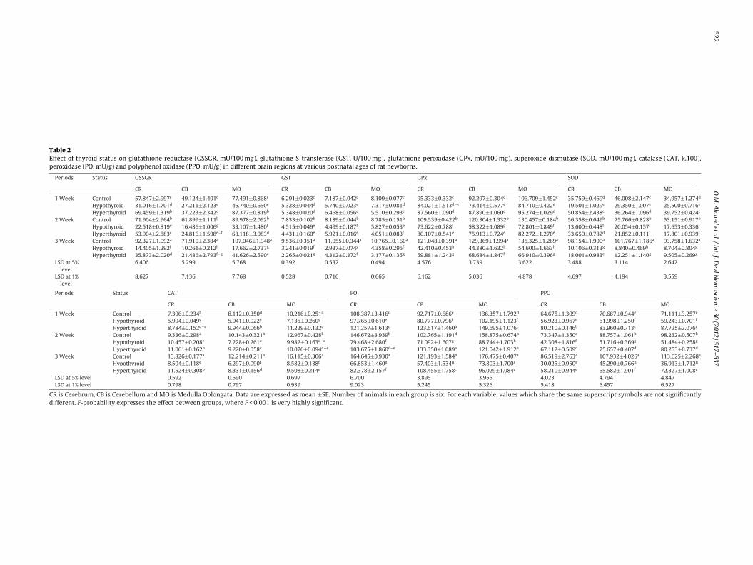
3.2.3. Glutathione reductase (GSSGR), glutathione-S-transferase(GST), glutathione peroxidase (GPx) and superoxide dismutase(SOD) activities (Table 2)
Table 2 showed that the activities of GSSGR, GST, GPx and SOD ofnormal rat offspring exhibited a stepwisely increased with the ageprogress. On the other hand, this behavioral pattern was disruptedin both treated groups which led to a significant suppression ofthese activities in all tested regions and all ages except at the endof the first week, in cerebrum and MO, for the activity of GSSGRand SOD in hyperthyroid group only, which showed an increaseas compared with the control group. In both treated groups, themost potent decrease (LSD; P < 0.01) of these enzymes activities wasrecorded in all tested regions at the end of the third week (Table 2).With regard to the one-way ANOVA of these parameters, the gen-eral effect between groups was very highly significant (P < 0.001)(Table 2) in all investigated regions throughout the experiment.
3.2.4. Catalase (CAT), peroxidase (PO) and polyphenol oxidase(PPO) activities (Table 2)
Similar to the previous enzymes, Table 2 depicted that CAT, POand PPO activities in normal offspring were markedly increasedin an age-dependent manner in all tested regions. Also, in bothtreated groups, the enzyme activities showed fluctuated changes.In cerebellum and MO, the CAT activity profoundly decreased (LSD;P < 0.01) in both treated groups at all tested ages except at the endof the first week of hyperthyroid group only. Though the CAT activ-ity, in cerebrum of hypothyroid group, was enormously increased(LSD; P < 0.01) at the end of the second week, it was detectablydiminished (LSD; P < 0.01) at other ages in comparison with thevalues of the corresponding controls. Furthermore, the CAT activ-ity was clearly increased (LSD; P < 0.01) at the end of the first andsecond weeks and depressed (LSD; P < 0.01) at the end of the exper-iment in cerebrum of hyperthyroid group (Table 2). Moreover, bothtreated offsprings exhibited an obvious decrease (LSD; P < 0.01) ofthe PO activity in all investigated regions during the studied devel-opmental ages except in hyperthyroid group at the end of the firstweek in all tested regions and at the end of the second week in cere-bellum only when compared to the corresponding normal controls(Table 2). On the other hand, compared to controls, hypothyroidgroup was characterized by a highly significant depletion (LSD;P < 0.01) of the PPO activity in all investigated regions at the firsttwo tested weeks. On the other hand, in all studied regions, eventhough the activity of PPO in hyperthyroid group was increasedsignificantly (LSD; P < 0.01) at the end of the first postnatal week,this activity was profoundly depressed (LSD; P < 0.01) at the endof the second week. With the age progress to the end of the thirdpostnatal week, the recorded activity of PPO, in all tested regions,was more deteriorated (LSD; P < 0.01) in both treated groups thanthe control activity (Table 2).
The analysis of one-way ANOVA of CAT, PO and PPO recordedthat the general effect between groups was very highly significant(P < 0.001) throughout the experiment (Table 2).
3.3. The cerebellar neurons of offspring as shown by Golgi-Copsch
stain (Figs. 4–6)In normal and both hypo- and hyperthyroid groups, five types ofneurons in layers of cerebellar cortex (gray matter) were recorded.
522
O.M
. A
hmed
et al.
/ Int.
J. D
evl N
euroscience 30
(2012) 517–537
Table 2Effect of thyroid status on glutathione reductase (GSSGR, mU/100 mg), glutathione-S-transferase (GST, U/100 mg), glutathione peroxidase (GPx, mU/100 mg), superoxide dismutase (SOD, mU/100 mg), catalase (CAT, k.100),peroxidase (PO, mU/g) and polyphenol oxidase (PPO, mU/g) in different brain regions at various postnatal ages of rat newborns.
Periods Status GSSGR GST GPx SOD
CR CB MO CR CB MO CR CB MO CR CB MO
1 Week Control 57.847±2.997c 49.124±1.401c 77.491±0.868c 6.291±0.023c 7.187±0.042c 8.109±0.077c 95.333±0.332c 92.297±0.304c 106.709±1.452c 35.759±0.469d 46.008±2.147c 34.957±1.274d
Hypothyroid 31.016±1.701d 27.211±2.123e 46.740±0.650e 5.328±0.044d 5.740±0.023e 7.317±0.081d 84.021±1.513d−e 73.414±0.577e 84.710±0.422e 19.501±1.029e 29.350±1.007e 25.500±0.716e
Hyperthyroid 69.459±1.319b 37.223±2.342d 87.377±0.819b 5.348±0.020d 6.468±0.056d 5.510±0.293e 87.560±1.090d 87.890±1.060d 95.274±1.029d 50.854±2.438c 36.264±1.096d 39.752±0.424c
2 Week Control 71.904±2.964b 61.899±1.111b 89.978±2.092b 7.833±0.102b 8.189±0.044b 8.785±0.151b 109.539±0.422b 120.304±1.332b 130.457±0.184b 56.358±0.649b 75.766±0.828b 53.151±0.917b
Hypothyroid 22.518±0.819e 16.486±1.006g 33.107±1.480f 4.515±0.049e 4.499±0.187f 5.827±0.053e 73.622±0.788f 58.322±1.089g 72.801±0.849f 13.600±0.448f 20.054±0.157f 17.653±0.336f
Hyperthyroid 53.904±2.883c 24.816±1.598e−f 68.118±3.083d 4.431±0.160e 5.921±0.016e 4.051±0.083f 80.107±0.541e 75.913±0.724e 82.272±1.270e 33.650±0.782d 21.852±0.111f 17.801±0.939f
3 Week Control 92.327±1.092a 71.910±2.384a 107.046±1.948a 9.536±0.351a 11.055±0.344a 10.765±0.160a 121.048±0.391a 129.369±1.994a 135.325±1.269a 98.154±1.900a 101.767±1.186a 93.758±1.632a
Hypothyroid 14.405±1.292f 10.261±0.212h 17.662±2.737g 3.241±0.019f 2.937±0.074g 4.358±0.295f 42.410±0.453h 44.380±1.632h 54.600±1.663h 10.106±0.313g 8.840±0.469h 8.704±0.804g
Hyperthyroid 35.873±2.020d 21.486±2.793f−g 41.626±2.590e 2.265±0.021g 4.312±0.372f 3.177±0.135g 59.881±1.243g 68.684±1.847f 66.910±0.396g 18.001±0.983e 12.251±1.140g 9.505±0.269g
LSD at 5%level
6.406 5.299 5.768 0.392 0.532 0.494 4.576 3.739 3.622 3.488 3.114 2.642
LSD at 1%level
8.627 7.136 7.768 0.528 0.716 0.665 6.162 5.036 4.878 4.697 4.194 3.559
Periods Status CAT PO PPO
CR CB MO CR CB MO CR CB MO
1 Week Control 7.396±0.234f 8.112±0.350d 10.216±0.251d 108.387±3.416d 92.717±0.686e 136.357±1.792d 64.675±1.309d 70.687±0.944e 71.111±3.257e
Hypothyroid 5.904±0.049g 5.041±0.022g 7.135±0.260g 97.765±0.610e 80.777±0.796f 102.195±1.123f 56.923±0.967e 61.998±1.250f 59.243±0.701f
Hyperthyroid 8.784±0.152d−e 9.944±0.066b 11.229±0.132c 121.257±1.613c 123.617±1.460b 149.695±1.076c 80.210±0.146b 83.960±0.713c 87.725±2.076c
2 Week Control 9.336±0.298d 10.143±0.321b 12.967±0.428b 146.672±3.939b 102.765±1.191d 158.875±0.674b 73.347±1.350c 88.757±1.061b 98.232±0.507b
Hypothyroid 10.457±0.208c 7.228±0.261e 9.982±0.163d−e 79.468±2.680f 71.092±1.607g 88.744±1.703h 42.308±1.816f 51.716±0.369g 51.484±0.258g
Hyperthyroid 11.061±0.162b 9.220±0.058c 10.076±0.094d−e 103.675±1.860d−e 133.350±1.089a 121.042±1.912e 67.112±0.509d 75.657±0.407d 80.253±0.737d
3 Week Control 13.826±0.177a 12.214±0.211a 16.115±0.306a 164.645±0.930a 121.193±1.584b 176.475±0.407a 86.519±2.763a 107.932±4.026a 113.625±2.268a
Hypothyroid 8.504±0.118e 6.297±0.090f 8.582±0.138f 66.853±1.460g 57.403±1.534h 73.803±1.700i 30.025±0.950g 45.290±0.766h 36.913±1.712h
Hyperthyroid 11.524±0.308b 8.331±0.156d 9.508±0.214e 82.378±2.157f 108.455±1.758c 96.029±1.084g 58.210±0.944e 65.582±1.901f 72.327±1.008e
LSD at 5% level 0.592 0.590 0.697 6.700 3.895 3.955 4.023 4.794 4.847LSD at 1% level 0.798 0.797 0.939 9.023 5.245 5.326 5.418 6.457 6.527
CR is Cerebrum, CB is Cerebellum and MO is Medulla Oblongata. Data are expressed as mean ±SE. Number of animals in each group is six. For each variable, values which share the same superscript symbols are not significantlydifferent. F-probability expresses the effect between groups, where P < 0.001 is very highly significant.
O.M. Ahmed et al. / Int. J. Devl Neuroscience 30 (2012) 517–537 523
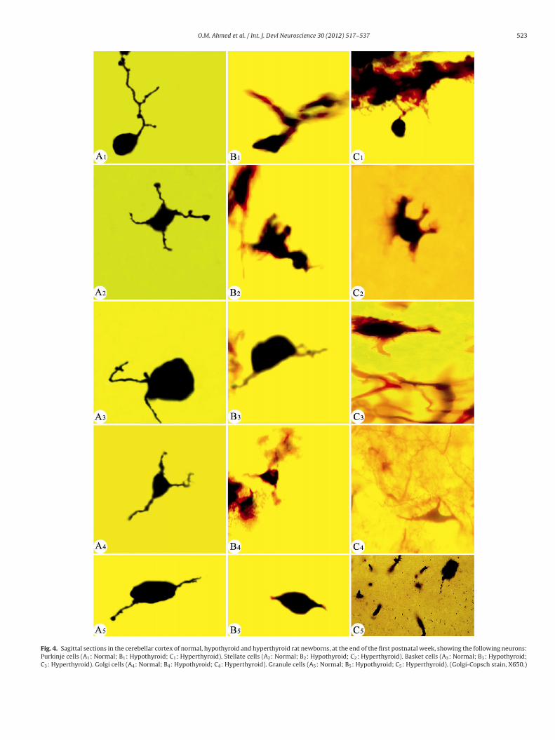
Fig. 4. Sagittal sections in the cerebellar cortex of normal, hypothyroid and hyperthyroid rat newborns, at the end of the first postnatal week, showing the following neurons:Purkinje cells (A1: Normal; B1: Hypothyroid; C1: Hyperthyroid). Stellate cells (A2: Normal; B2: Hypothyroid; C2: Hyperthyroid). Basket cells (A3: Normal; B3: Hypothyroid;C3: Hyperthyroid). Golgi cells (A4: Normal; B4: Hypothyroid; C4: Hyperthyroid). Granule cells (A5: Normal; B5: Hypothyroid; C5: Hyperthyroid). (Golgi-Copsch stain, X650.)

524 O.M. Ahmed et al. / Int. J. Devl Neuroscience 30 (2012) 517–537
Fig. 5. Sagittal sections in the cerebellar cortex of normal, hypothyroid and hyperthyroid rat newborns, at the end of the second postnatal week, showing the followingneurons: Purkinje cells (A1: Normal; B1: Hypothyroid; C1: Hyperthyroid). Stellate cells (A2: Normal; B2: Hypothyroid; C2: Hyperthyroid). Basket cells (A3: Normal; B3:Hypothyroid; C3: Hyperthyroid). Golgi cells (A4: Normal; B4: Hypothyroid; C4: Hyperthyroid). Granule cells (A5: Normal; B5: Hypothyroid; C5: Hyperthyroid). (Golgi-Copschstain, X650.)

O.M. Ahmed et al. / Int. J. Devl Neuroscience 30 (2012) 517–537 525
Fig. 6. Sagittal sections in the cerebellar cortex of normal, hypothyroid and hyperthyroid rat newborns, at the end of the third postnatal week, showing the following neurons:Purkinje cells (A1: Normal; B1: Hypothyroid; C1: Hyperthyroid). Stellate cells (A2: Normal; B2: Hypothyroid; C2: Hyperthyroid). Basket cells (A3: Normal; B3: Hypothyroid;C3: Hyperthyroid). Golgi cells (A4: Normal; B4: Hypothyroid; C4: Hyperthyroid). Granule cells (A5: Normal; B5: Hypothyroid; C5: Hyperthyroid). (Golgi-Copsch stain, X650.)

5 l Neur
3
(scntdpam(wid(tiidtp(ntidiiw
3C
b
3ttbahthtdi(bC
3atbspbawiniwes
26 O.M. Ahmed et al. / Int. J. Dev
.3.1. The Purkinje layer (PL) (Figs. 4A1–C1 to 6A–C1)This layer consists of one type of neurons; the Purkinje cells
PCs). These cells in normal and both treated groups appeared pos-essing one main dendrite, which arise from the upper end of theell body and extending through the ML at the end of the first post-atal week (4A1–C1). These primary dendrites branch repeatedlyhrough the ML into secondary and tertiary dendrites giving a richendritic tree with innumerable tiny spicules at the end of the thirdostnatal week in normal offspring (Fig. 6A1). In addition, theserborizations were confined to a plane perpendicular to the piaatter in normal offspring at the end of the second postnatal week
Fig. 5A1) and reached their maximum length at the end of the thirdeek (Fig. 6A1). At the end of the first and second postnatal weeks,
n normal offspring, these cells were large in size and had long den-rites when compared to those of hypo- and hyperthyroid groupsFigs. 4A1–C1 and 5A1–C1). Moreover, the latter figures showed thathe dendrites of PCs had spicules and nodules in normal offspringn respect to hypo- and hyperthyroid groups. On the other hand,n both treated groups, there was some delay in the growth of theendrites of these cells at the end of the first and second postna-al weeks (Figs. 4B1 and C1 and 5B1 and C1). Also, some importantoints, at the end of the third postnatal week, can be summarized6A1–C1) as the following: (1) PCs of normal offspring increased inumber and in their dendrites as compared to both treated ones, (2)here were few spicules on the dendrites of both treated offspringn comparison with normal ones and (3) the density of the den-ritic network in both treated offsprings was lesser than that seen
n the normal ones. Furthermore, some degeneration was noticedn hypo- and hyperthyroid groups at the end of the third postnatal
eek (6B1 and C1).
.3.2. The molecular layer (ML) (Figs. 4 A2, A3, B2, B3, C2 and3–6A2, A3, B2, B3, C2 and C3)
This layer consists of two types of neurons; the stellate andasket cells.
.3.2.1. The stellate cells. The stellate cells were shown occupyinghe upper half of the ML in normal and both treated groups. Athe end of the first postnatal week, these cells had thick dendrites’ranches in normal offspring with the presence of some spiculesnd nodules on their branches when compared to their respectiveypo- and hyperthyroid groups (4A2–C2). At other studied ages,he dendrites appeared long and thick in normal offspring andaving some spicules and nodules in relation to their respectivereated groups (Figs. 5A2–C2 and 6A2–C2). The branching of theseendrites form-interlacing network in normal offspring in compar-
son with both treated ones at the end of the third postnatal week6A2–C2). Some degeneration and deformations were recorded inoth treated groups during all tested periods (4B2 and C2, 5B2 and2 and 6B2 and C2).
.3.2.2. The basket cells. The basket cells in normal and both hypo-nd hyperthyroid groups were found to occupy the deep region inhe ML at different examined ages. They appeared rounded or oval,ipolar perikaryon and their branches moving parallel to the pialurface in normal and both treated groups at the end of the firstostnatal week (4A3–C3). However, the dendrites of these cells, inoth treated groups, were short when compared to normal onest the latter age. Also, at the end of the second and third postnataleeks, these cells appeared having shorter dendrites with miss-
ng the spicules or nodules in both treated groups as compared toormal ones (Figs. 5A3–C3 and 6A3–C3). Furthermore, these cells,
n both treated groups, were shown with small size in comparisonith their corresponding normal group along the duration of the
xperiment (Figs. 4A3–C3, 5A3–C3 and 6A3–C3). As well, there wereome degeneration in these cells and their dendrites in hypo- and
oscience 30 (2012) 517–537
hyperthyroid groups during the examined periods (Figs.4B3 andC3, 5B3 and C3 and 6B3 and C3). The degeneration of neuron bod-ies and dendrites as well as delay in the development of dendritearborizations are more deteriorated as a result of hyperthyroidismthan hypothyroidism.
3.3.3. The internal granular layer (IGL) (Figs. 4A4, A5, B4, B5, C4and C5–6A4, A5, B4, B5, C4 and C5)
This layer consists of two types of neurons; the Golgi and granulecells.
3.3.3.1. The Golgi cells. The Golgi cells were shown occupying theupper region of the IGL in normal and both treated groups as wellas these cells appeared triangular in shape from the end of thefirst to third postnatal weeks (Figs. 4A4–C4, 5A4–C4 and 6A4–C4).Also, the dendrites of these cells were found to be branching andextending in all directions in normal and both treated groups. Atthe end of the first postnatal week, these cells appeared to havethick and long branches of dendrites which were produced fromthe different parts of the cell body in normal offspring when com-pared to their respective hypo- and hyperthyroid groups (4A4–C4).These dendrites move at different angles with the surface of thepia matter and the cell size was in the following order: nor-mal > hyperthyroid > hypothyroid groups at the latter age. Indeed,in normal offspring, these cells had long dendrites with the pres-ence of nodules and spicules at the end of the second and thirdpostnatal weeks as compared to the previous age (Figs. 4A4, 5A4and 6A4). At the end of the second and third postnatal weeks, thesecells increased in the number and size, and their dendritic branchesbecame longer and thicker in normal offspring in relation to bothtreated ones (Figs. 5A4–C4, 6A4–C4). The degeneration, disorgani-zations and deformations were observed in both treated groupsduring the investigated ages in both treated groups (Figs. 4B4 andC4, 5B4 and C4 and 6B4 and C4).
3.3.3.2. The granule cells. The granule cells in normal and bothhypo- and hyperthyroid groups were shown occupying the deepregion of the IGL. These cells appeared rounded to ovoid in shapeand were characterized by the presence of 1 or 2 dendrites fromthe end of the first to third postnatal weeks in normal and bothtreated groups (Figs. 4A5–C5, 5A5–C5 and 6A5–C5). The main den-drites branched repeatedly into secondary dendrites during theexperimental period in normal offspring (Figs. 4A5, 5A5 and 6A5).In addition, in normal offspring, these cells were found to havelong dendrites as compared to both treated ones in all tested ages(Figs. 4A5–C5, 5A5–C5 and 6A5–C5). Also, the latter figures clearlydepicted that the spicules and nodules in both hypo- and hyper-thyroid groups were missing in relation to normal ones. These cellsincreased in the number and in their dendrites in normal offspringin comparison with both treated ones at the end of the third week(6A5–C5). Additionally, there were some degeneration in the den-drites, deformations and reduction in the population of these cellsduring the employed ages in both treated groups (Figs. 4B5 and C5,5B5 and C5 and 6B5 and C5).
3.4. The cerebral neurons of offspring as shown by Golgi-Copschstain (Figs. 7–10)
In normal and both treated groups, the layers of the cere-bral cortex were characterized by the presence of the PYCs whichwere characterized by a pyramid-shaped perikaryon with an api-
cal dendrite directed toward the surface of the brain and an axonleaving the base of the perikaryon to course into the white matter(Figs. 7–10). Also, the sections showed four types of nerve cells inlayers III, IV, V and VI of cerebral cortex (Figs. 7–10).
O.M. Ahmed et al. / Int. J. Devl Neuroscience 30 (2012) 517–537 527
Fig. 7. Sagittal sections in the cerebral cortex showing the pyramidal and polymorphous cells in different layers at the end of the first postnatal week of normal, hypothyroidand hyperthyroid rat newborns. Pyramidal cells in layer III (A1: Normal; B1: Hypothyroid; C1: Hyperthyroid). Pyramidal cells in layer IV (A2: Normal; B2: Hypothyroid; C2:Hyperthyroid). Pyramidal cells in layer V (A3: Normal; B3: Hypothyroid; C3: Hyperthyroid). Polymorphous cells in layer VI (A4: Normal; B4: Hypothyroid; C4: Hyperthyroid).(Golgi-Copsch stain, X650) Note: The arrows refer to the apical dendrites. The arrows heads refer to lateral dendrites. AX refers to the axon.

528 O.M. Ahmed et al. / Int. J. Devl Neuroscience 30 (2012) 517–537
Fig. 8. Sagittal sections in the cerebral cortex showing the pyramidal and polymorphous cells in different layers at the end of the second postnatal week of normal, hypothyroidand hyperthyroid rat newborns. Pyramidal cells in layer III (A1: Normal; B1: Hypothyroid; C1: Hyperthyroid). Pyramidal cells in layer IV (A2: Normal; B2: Hypothyroid; C2:Hyperthyroid). Pyramidal cells in layer V (A3: Normal; B3: Hypothyroid; C3: Hyperthyroid). Polymorphous cells in layer VI (A4: Normal; B4: Hypothyroid; C4: Hyperthyroid).(Golgi-Copsch stain, X650). Note: The arrows refer to the apical dendrites. The arrows heads refer to lateral dendrites. AX refers to the axon.

O.M. Ahmed et al. / Int. J. Devl Neuroscience 30 (2012) 517–537 529
Fig. 9. Sagittal sections in the cerebral cortex showing the pyramidal and polymorphous cells in different layers at the end of the third postnatal week of normal, hypothyroidand hyperthyroid rat newborns. Pyramidal cells in layer III (A1: Normal; B1: Hypothyroid; C1: Hyperthyroid). Pyramidal cells in layer IV (A2: Normal; B2: Hypothyroid; C2:Hyperthyroid). Pyramidal cells in layer V (A3: Normal; B3: Hypothyroid; C3: Hyperthyroid). Polymorphous cells in layer VI (A4: Normal; B4: Hypothyroid; C4: Hyperthyroid).(Golgi-Copsch stain, X650). Note: The arrows refer to the apical dendrites. The arrows heads refer to lateral dendrites. AX refers to the axon.

530 O.M. Ahmed et al. / Int. J. Devl Neuroscience 30 (2012) 517–537
F ) and pp (C1–C
stt
ig. 10. General sagittal sections in the cerebral cortex showing the pyramidal (PYCostnatal week of normal (A), hypothyroid (B1–B4) and hyperthyroid rat newborns
In normal rat offspring, the length of the apical dendrites showedteady increase with the development in all layers of cerebral cor-ex. The pyramidal neurons of layer V had longer apical dendriteshan those of layers III and IV in normal group and this is due to
olymorphous cells (PMC) in different layers (III, IV, V and VI) at the end of the third4). (Golgi-Copsch stain, X650). TM: Thickening Meninges.
the difference in their location throughout the experimental period(Figs. 7A1–A3, 8A1–C3, 9A1–A3 and 10A). The spicules appeared onthe apical dendrites as projections on its shaft. Also, the distributionof the spine density over the different segments of apical dendrites

l Neur
sFf
eaccrsSptitttcctchiCmCiltap(aa
lipInthdoogd(rVh(st
w(ew9ttnPda
O.M. Ahmed et al. / Int. J. Dev
howed different patterns in layers III, IV and V for all tested ages.or these reasons, the following description was based on findingsrom the first to third postnatal weeks in these neurons.
At the end of the first postnatal week, the neurons, in the differ-nt layers of the cerebral cortex, were clearly visualized in normalnd both treated groups (Fig. 7). In normal rat offspring, the api-al dendrites of PYCs in layer III appeared short and thick whenompared to hyperthyroid treated ones (7A1 and C1) and long inelation to hypothyroid ones (7A1 and B1). However, there wasome degeneration in PYCs in both treated groups (7B1 and C1).ome lateral dendrites which originate from the lateral side of theyramidal perikaryon of layer III were noticed in normal and hyper-hyroid groups and lost in hypothyroid group (7A1–C1). Moreover,n normal and both treated groups, the latter figures clearly showedhat the axon of these cells in layer III was thick; it originates fromhe base of the perikaryon and moves far from the pial surface. Onhe other hand, in layers IV and V of normal offspring, the typi-al PYCs appeared with their multipolar shape (7A2–A3) and theseells became more complex in their dendritic fields as comparedo the cells of the third layer in normal ones (7A1–A3). The api-al dendrites of the PYCs in layers IV and V were thick, long andaving some nodules and spicules in normal offspring in compar-
son with the respective treated groups (7A2, A3, B2, B3, C2 and3). Also, the axons of PYCs in layers IV and V were short in nor-al offspring as compared with hyperthyroid ones (7A2, A3, C2 and
3). However, in hypothyroid offspring, PYCs was lost their axonsn layers IV and V (7B2 and B3). The lateral dendrites of PYCs, inayers IV and V, were long in normal offspring in comparison withhe corresponding hypo- or hyperthyroid ones (7A2, A3, B2, B3, C2nd C3). In both treated groups, layer VI was observed owning fewolymorphic cells with short dendrites in respect to normal ones7A4–C4). Some deformations and degeneration in the pyramidalnd polymorphic cells of both treated groups were noticed at thisge (7B1–B4 and C1–C4).
Moreover, at the end of the second postnatal week, the PYCs ofayer III had long apical and lateral dendrites in normal offspringn comparison with both treated ones (8A1–C1). Furthermore, therevious figures also revealed that the axons of these cells in layer
II were short in both treated groups in relation to normal ones. Inormal offspring, the dendrites and axons of the PYCs increased inheir lengths in layers IV and V in comparison with their respectiveypo- and hyperthyroid groups (8A2, A3, B2, B3, C2 and C3). Thisevelopment leads to clear pyramidal shape as in layer V of normalffspring (Fig. 8A3). Also, in layers IV and V, the spicules and nodulesf PYCs were noticed in normal offspring in respect to both treatedroups (8A2, A3, B2, B3, C2 and C3). On the other hand, the lateralendrites of PYCs of layers IV and V were lost in hypothyroid group8B2 and B3) while the axon of PYCs of layer V was lost in hyperthy-oid group (Fig. 8C3). Compared with the normal offspring, in layerI, the polymorphic cells appeared with thin and short dendritesaving few spicules and nodules in hypo- and hyperthyroid groups8A4–C4). Furthermore, both hypo- and hyperthyroidism causedome degenerated symptoms in the neurons and their dendrites athis stage (8B4 and C4).
As development proceeded to the end of the third postnataleek, the neurons increased in the number in normal rat offspring
Fig. 10A). The PYCs in different layers attained long apical and lat-ral dendrites in normal rat offspring; the neurons were matureith respect to the extension of their dendritic complexity (Figs.
A1–A4 and 10A). At this age, the collaterals of dendrites arboriza-ions increased in their density and complexity much more thanhe collaterals of the comparable layers at the previous ages in
ormal offspring (Figs. 7A1–A3, 8A1–A3, 9A1–A3 and 10A). TheYCs of layer III appeared clearly in a typical form with a pyrami-al perikaryon and lateral dendrites in normal offspring (Fig. 9A1)nd these dendrites were complex in normal group than thoseoscience 30 (2012) 517–537 531
dendrites of both treated ones in the layer III (9A1–C1). Also, thesecells appeared with short and thick apical and lateral dendrites inboth treated groups when compared to the normal ones (9A1–C1).In addition, the axons of PYCs cells, in layer III, were long and thickin normal offspring as compared to hyperthyroid ones (9A1 andC1). However, the axon of PYCs was missing in layer III of hypothy-roid group (Fig. 9B1). Moreover, as indicated in 9A2–C2, the apicaland lateral dendrites of the PYCs, in layer IV of normal offspring,were long when compared to both treated ones. Also, the latterfigures illustrated that the spicules and nodules were observed innormal and both treated groups. The PYCs of layer V, in normal off-spring, reach their typical form; they had an ideal multipolar formwith densely arborized apical and lateral dendrites whose collat-erals were long and rich in the spicules (Fig. 9A3). Also, 9A3–C3showed that the PYCs had network tree of long dendrites in thelayer V of normal offspring in respect to both treated ones. Onthe other hand, in hypo- and hyperthyroid groups, PYCs of layerV had thin axon and short dendrites in comparison with normalones, however their dendrites had number of spicules in normaland both treated groups (9A3–C3). The polymorphic cells of layerVI had long and thin dendrites with spicules and nodules in normaloffspring in comparison with their corresponding treated groups(9A4–C4).
4. Discussion
THs are essential both for the physiological course of pregnancy,the optimal differentiation of the embryonic tissues, normal devel-opment of body organs and integrated fetal brain development(Jiskra et al., 2007; Suchetha Kumari et al., 2011). Thus, the currentstudy highlights the effects of maternal hypo- and hyperthyroidismon the thyroid function, neuronal development of cerebellar andcerebral cortex, and reactive oxygen species (ROS)/antioxidantdefense system in different brain regions (cerebrum, cerebellumand MO) of rat offspring at the end of the first, second and thirdpostnatal weeks.
With regards the normal maternal THs, in the present study,their serum levels [total thyroxine (TT4) and total triiodothyro-nine (TT3)] were lower during the pregnancy at gestation day (GD)10 than those at day 10 post-partum. This state may reflect thehigher transfer of THs from pregnant females to their fetuses dur-ing pregnancy and/or more efficiency of thyroid gland to secreteTHs after birth (Ahmed et al., 2010). In normal rat offspring, the cur-rent study revealed gradual increases of serum free thyroxine (FT4),free triiodothyronine (FT3), thyrotropin (TSH) and GH levels at theend of the first, second and third postnatal weeks. As suggestedby Thilly et al. (1978), Momotani et al. (1986) and Porterfield andHendrich (1992), there is a positive relationship between mater-nal and offspring thyroid functions. This elucidation is concomitantwith the observations of the present study. Interestingly, Walkeret al. (1980) elucidated that free TH concentrations in rats followessentially the same developmental profile as do total TH concen-trations. Moreover, the gradual increase of TSH herein is necessaryfor the development and growth of thyroid gland during this sen-sitive period because it increases the size of the follicles and itincreases the rate of synthesis, secretion and iodination of glyco-protein into colloid, the rate of breakdown of thyroglobulin and theliberation of THs into circulation (Nosseir et al., 1991; Ahmed et al.,2008, 2010). In addition, GH is a key factor controlling postnatalgrowth and development (Zhou et al., 2005; Wong et al., 2006).THs are also a necessary component for the physiological growth
of a young organism, stimulating the secretion of GH and insulin-like growth factor (IGF) (Wasniewska et al., 2003). In turn, theseobservations imply that these hormones may regulate the growthand development, in general.
5 l Neur
dnaFcs22ertrT
roiTTedfiaaalsrtmtd
ittrpMTt(rmAt
thats
cpTnpegweb(
32 O.M. Ahmed et al. / Int. J. Dev
The present study revealed that administration of MMI inrinking water (0.02% w/v) to adult female rats during preg-ancy and weaning periods induced hypothyroidism in mothersnd their offspring as indicated by decrease in serum TT4, TT3,T4 and FT3 levels, and increase TSH. From the literature, itould be observed that the current work is in harmony witheveral publications using MMI-treated rats (MacNabb et al.,000; Mookadam et al., 2004; Hasebe et al., 2008; Ahmed et al.,010). The mechanism by which MMI exerts hypothyroidism wasxplained by Awad (2002) and Ahmed et al. (2008, 2010) whoeported that MMI interferes with incorporation of iodine intoyrosyl residues of TG and inhibits the coupling of iodotyrosylesidues to form iodothyronine, thus inhibiting the synthesis ofHs.
On the other hand, the administration of T4 to adult femaleats in drinking water at 0.002% w/v beside gastric incubationf 50–200 �g/Kg b. wt. during pregnancy and weaning periodsnduced a marked hyperthyroidism in mothers and their offspring.his hyperthyroidism was ensured by elevated levels of serum TT3,T4, FT3 and FT4 in these animals. Higuchi et al. (2001) and Ahmedt al. (2008, 2010) hypothesized that maternal hyperthyroidismuring pregnancy and lactation leads to a hyperthyroid state inetus and neonates. They attributed the state of hyperthyroidismn fetuses or early neonates to passive transfer of maternal T4 from
mother with hyperthyroidism or thyrotoxicosis through placentand in mother’s milk. Moreover, the decrease in serum TSH level isttributed to negative feedback effect of the excess circulating THsevels on the anterior lobe of pituitary gland. These suggestions areupported by Fisher et al. (2000) and Higuchi et al. (2005) whoeported that the exposure of the fetal hypothalamic-pituitary-hyroid system to a higher-than-normal TH (T4) concentration
ight impair its physiologic maturation, because there is a con-inuous significant decrease in the TSH/fetal T4 ratio during theevelopment.
Furthermore, while a depression in level of GH was observedn offspring of hypothyroid group, the opposite occurs in hyper-hyroid group at all ages employed if compared to the levels inhe age-matched normal group. Concomitant with the presentesults, Morreale de Escobar et al. (1993) emphasized that bothlasma and pituitary GH decreased in hypothyroid fetuses fromMI-treated pregnant rats while their plasma TSH was elevated.
hus, it is worth mentioning that maternal THs deficiency may dis-urb the secretion of other pituitary hormones in their offspringWong et al., 1980; Tamasy et al., 1984). Furthermore, neonatalat’s hyperthyroidism (Varma and Crawford, 1979) results in per-anent imprinting regarding growth and thyroidal development.lso, Segni and Gorman (2001) speculated that untreated childhood
hyrotoxicosis causes accelerated growth.To sum, it is observed from the above mentioned results that a
ransient and moderate deficiency or increase of maternal THs canave deleterious consequences on thyroid function of both mothersnd their offspring. The results indicated that the thyroid func-ions of offspring are indirectly affected by both maternal thyroidtatuses.
On the other hand, the rate of neuron development in botherebellar and cerebral cortex was increased in its density and com-lexity with the age progress in the present normal rat offspring.hese results are in association with the increase in the levels ofeurotransmitters and activities of ATPases and deiodinase in ourrevious study with age progress in these brain regions (Ahmedt al., 2010). THs regulate the neuronal cytoarchitecture, neuronalrowth, myelination and synaptogenesis, where their receptors are
idely distributed in the CNS (Zoeller, 2004; Bernal, 2007; Ahmedt al., 2008; Leonard, 2008). This is particularly apparent in the cere-ral and cerebellar cortex (Süha and C alikoglu, 1999). Ahmed et al.2008) reported that the maternal THs play a critical role in CNS
oscience 30 (2012) 517–537
development and differentiation depending on the brain region andthe developmental stage of their offspring.
As indicated in the current study, five types of neurons werenoticed in cerebellar cortex; stellate and basket cells in molecu-lar layer, PC in PL, and Golgi and granule cells in IGL. This resultgoes parallel with the work of Dalia (1998, 2002), Ahmed (2004)and Ahmed et al. (2007) in normal rat offspring. These cells wereincreased in number with age progress in the present normal off-spring. This observation was recorded by several authors (Salehet al., 1993; Krause and Cutts, 1994; Dalia, 2002; Ahmed, 2004;Ahmed et al., 2007). On the other hand, several authors reportedthat THs regulate neuronal outgrowth, proliferation and sur-vival, and synapse formation (Oppenheimer and Schwartz, 1997;Rodriguez-Pena, 1999; Durand and Raff, 2000; Koibuchi and Chin,2000; Thompson and Potter, 2000; Ahmed et al., 2008). In fact,Patel et al. (1987) indicated that in rats, the effect of TH on neuralmaturation is cell-type specific.
On the other hand, there were some degeneration and defor-mation in cells of molecular layer (ML) of hypo- and hyperthyroidgroups during all studied ages. These observations are in accor-dance with those of Nicholson and Altman (1972a, b) who depictedthat in rat, hypothyroidism caused retardation in cell differentia-tion of the ML. Moreover, Lauder (1978) recorded that the delay incell acquisition and development results in abnormal proportionsof basket and stellate cells in rat. On the other hand, hyper-thyroidism (Nicholson and Altman, 1972c) in rat caused somemalformations as terminal decrease in basket cells. Regarding thestudy herein, PCs of offspring of both hypothyroid and hyperthy-roid mothers decreased in number and in their dendrites lengthand density as compared to normal group in all investigated ages aswell as some degeneration were observed in hypo- and hyperthy-roid groups. Previous results in rats showed that hypothyroidismcan cause a retardation in the morphological maturation of PCs(Dussault and Ruel, 1987; Ahmed et al., 2008) as a reductionin the dendritic arborization (Nicholson and Altman, 1972a, b;Legrand, 1984), spine number (Nicholson and Altman, 1972a, b),cell size (Lauder, 1978; Vincent et al., 1983) and proliferation caus-ing hypoplasia (Nicholson and Altman, 1972b,c; Legrand, 1984;Potter et al., 2001; Ahmed et al., 2008) during the development.Moreover, excessive doses of T4 or T3 led to an early stimulationof cell acquisition followed by a permanent deficit of cells in thecerebellum of rat (Legrand et al., 1976). It is obvious from the afore-mentioned results that the both treatments may retard the growthand development of PC. Concurrently, the Golgi and granule cells,in the current work, had shorter dendrites in hypo- and hyperthy-roid offspring in comparison with normal ones between the first3 weeks after birth. Also, there were some degenerated dendritesin both treated groups with the age progress. A number of inves-tigators showed that TH affects cerebellar granule proliferation,migration, and apoptosis (Muller et al., 1995; Pasquini et al., 2000;Singh et al., 2003). The lack of TH during the critical period of neu-ronal migration leads to a multitude of irreversible morphologicalabnormalities (Porterfield and Hendrich, 1993; Bernal and Nunez,1995) as defected in granule cell migration and its increased death(Dubuis et al., 1992; Lee et al., 2003), as well as a reduction in itsnumber (Anderson, 2001) and parallel fiber growth (Lauder, 1978;Anderson, 2001). Furthermore, hypothyroidism causes a decreasein number and density of synaptic contacts between granule cellsand PCs in rats (Legrand, 1979). Overall, a deficiency of THs inthe neonatal rat has been shown to cause disorganization of thecerebellar cortex (Nicholson and Altman, 1972a, b; Nunez, 1984).On the other hand, Nicholson and Altman (1972c) reported that
hyperthyroidism caused some malformations as terminal decreasein granule cells of rat. Ahmed et al. (2008) postulated that dur-ing the critical periods of the development, increase or decreasein the thyroid secretions may retard the neurogenesis and CNS
l Neur
ghorfitCheatws
tttmpiTtaioAtToWdardaiHpparbttdtep
sstties(pccnee(lo
O.M. Ahmed et al. / Int. J. Dev
rowth. According to the above results, it is worth mentioning thatypo- or hyperthyroidism may affect the growth and maturationf neurons through its effect on the vital processes of these neu-ons. The delayed growth and degeneration of neurons and theirbers in cerebellar cortex of hypothyroid rats are associated withhe stepwise decrease in the monoamines levels and ATPase andhE activity in our previous study (Ahmed et al., 2010). However, inyperthyroid rats of the same study, the delayed growth and degen-ration of cerebellar neurons and their fibers are associated withn increase in these variables. This controversy may be attributedo the differences in THs levels, in hypo- and hyperthyroid groups,hich may directly or indirectly, affected the synthesis and expres-
ion of these neurotransmitters and enzymes.The maturation process of pyramidal neurons of cerebral cor-
ex involves many aspects. In normal rat offspring, the increase inhe dendrites arborization with the presence of spicules is one ofhe maturation features. The rate of pyramidal neuron develop-
ent increased herein in its density and complexity with the agerogress in normal offspring. These observations are also noticed
n normal rat offspring by Ahmed (2004) and Ahmed et al. (2007).he neurons of layer V in the present normal offspring precedehose of layers III and IV in the maturity of all investigated ageslong the duration of the experiment. This finding may show thenside outside gradient of neuron migration in white albino ratffspring. These observations are concomitant with the results ofhmed and Gabr (2000) in normal rat offspring, and go parallel with
he results of Angevine and Sidman (1961) in mice. Concurrently,Hs control dendritic development and number of dendritic spinesf pyramidal cells of neocortex and hippocampus (Bernal, 2003).ith respect to these results, it must be mentioned that our evi-
ences concur with the observations of Süha and C alikoglu (1999)nd Ahmed et al. (2008) who concluded that THs play an importantole in cerebral neurogenesis. The interpretation of changes in theensity of spicules and nodules during the development in normalnd both treated groups had to be considered in view of changesn the length of the shaft of the apical dendrites as well as its size.owever, at the end of the first postnatal week, the present normalyramidal cells of layer III had short and thick dendrites as com-ared to hyperthyroid group. Also, the axons of PYCs in layers IVnd V were short in normal offspring as compared with hyperthy-oid ones at the same period. These observations can be explainedy Mussa et al. (1990) who revealed that hyperthyroidism may ini-ially induce an acceleration of the maturation processes, includinghe migration and differentiation of cells, the extension of the den-ritic processes and synaptogenesis. Also, Miller (1981) suggestedhat in the course of rat maturation, some dendrites might degen-rate or fuse with adjacent ones, thus reducing the total number ofrimary processes.
The dendrites of the pyramidal cells of layers III, IV and V werehort in the present hypo- and hyperthyroid groups in compari-on with normal ones in all investigated ages except at the end ofhe first postnatal week in layer III of hyperthyroid group only. Fur-her, in both treated groups, some deformations and degenerationsn the pyramidal and polymorphic cells were noted throughout thexperiment with lost axons or lateral dendrites of pyramidal cells inome ages. Several authors discuss these findings. Mirabella et al.2005) reported that in infants, a lack of TH during fetal or earlyostnatal life is associated with specific brain damage. Deficientellular maturation in the cerebral cortex of hypothyroid rats isharacterized by (Balázs, 1973; Schwartz et al., 1997): (1) smallereuronal cell bodies that are more tightly packed than those inuthyroid animals, (2) diminished axonal and dendritic outgrowth,
longation and branching, (3) reduced numbers of dendritic spines,4) diminished myelination of neuronal axons (5) changes in cal-osally projecting neurons which may be due to the maintenancef a juvenile pattern of projections (Berbel et al., 1993, 1994, 1996,oscience 30 (2012) 517–537 533
2001; Zoeller and Rovet, 2004) and (6) alterations in dendritic mor-phology and structure in several cell types, including pyramidalcells in the cortex (decrease in dendritic spine number) (Schwartz,1983). Similarly, Lavado-Autric et al. (2003) pointed out that someneurons in the neocortex of rats do not reach their normal positionsin the absence of TH. These investigations led to the suggestionthat TH is necessary for normal brain development in the fetusas it is during the early postnatal period (Obregon et al., 1984;Pérez-Castillo et al., 1985). In other instance, the abnormal braindevelopment observed during hypothyroidism, in rats, may par-tially result from a deficiency of GH (Savard et al., 1984).
Oppenheimer and Schwartz (1997) and Ahmed et al. (2008)reported that hyperthyroidism, during the brain development,results in cytoarchitecture abnormalities and disorganization ofseveral neuronal cells. Moreover, Sala-Roca et al. (2008) recordedthat thyroxine reduces apical and basal tree of pyramidal neuronsin CA3 hippocampal region. In 2008, the innovative review in theinteractions between thyroid and brain development was carriedout by our group has led to the conclusion that the dramatic neuro-logical abnormalities associated with hyperthyroidism during thedevelopment testify to the importance and effect of the THs on theCNS development. Thus, worth noting in the present study is thatTHs dysfunctions during the critical period of development maydisrupt interneuronal connections and neuronal integration.
In view of the recent studies, the current results depict impor-tant finding that the development of the investigated brain regionsin rat offspring are extremely sensitive to maternal hypo- andhyperthyroidism during the pregnancy period and the first 3 weeksof lactation period. Thus, both THs defects may be responsible forthe loss of neurons vital functions and decrease in their dendriticarborization. Introducing the same mode of though, the experi-mental research and clinical studies have partially clarified thecorrelation between the maturation of the nervous system and thy-roid function during the early stages of development; both a deficitand excess of THs may lead to permanent anatomo-functionaldamage to the CNS (Mussa et al., 1990). By the way, adult-onset thy-roid disorders in humans impair several important CNS functions(Sarkar, 2008).
ROS play an essential role in the regulation of cell prolifera-tion, e.g. within the central and peripheral nervous system becauseROS initiate and promote the establishment of neuronal patternsand subsequent neurogenesis (Verity, 1994). It was also reportedthat ROS play an important role in physiological processes, but –when being in excess – ROS cause oxidative damage to molecules(Karbownik and Lewinski, 2003). Antioxidant enzymes (AOEs) arean important protective mechanism against excess ROS and likemany other biochemical systems, their effectiveness may varywith the stage of development and other physiological aspects ofthe organism (Halliwell and Gutteridge, 1999; Livingstone, 2001;Ahmed, 2004; Ahmed, 2005). Several reports have been conductedto demonstrate the modulation of the antioxidant defense systemof tissues by the thyroid state of the body (Venditti et al., 1997;Pamplona et al., 1999; Tapia et al., 1999). Thus, it appears moremeaningful to determine the pattern levels of the AOEs and theirrelationships with each other and with the oxidative markers.
The enzymatic and non-enzymatic antioxidant defense vari-ables [glutathione reductase (GSSGR), GST, GPx, SOD, CAT, PO, PPO,vitamin C (ascorbic acid), vitamin E (�-Tocopherol), total thiol (t-SH) and glutathione (GSH)], in the present normal rat offspring,were substantially and gradually increased with the age progressfrom the first to third week old in all investigated regions. Previ-ous studies reported that the overall antioxidant enzyme activities
tend to increase in different brain regions with the age progressin rat (L’vova and Abaeva, 1996; Agarwal et al., 1999; Ahmed,2004, 2005) and mice (Hussain et al., 1995). Also, the levels ofnon-enzymatic antioxidants were regularly increased in different
5 l Neur
bAoabtp
pdhtd
tSSrcegbiLso2rshietcmppwcami
(edadGlFtirei(pAriAtopM
34 O.M. Ahmed et al. / Int. J. Dev
rain regions with the age progress in rat (Shivakumar et al., 1991;hmed, 2004, 2005) and mice (Hussain et al., 1995). Thus, basedn these evidences, the increase in the activities of the tested AOEsnd levels of antioxidant substrates in various brain regions (cere-rum, cerebellum and MO), in the present study, potentially reflectshe enhancement in the antioxidant defense system with the agerogress from PND 7 to PND 21.
Thus, based on the previous publications and results of theresent study, it can be suggested that the degenerations andeformations of neurons and their dendrites in MMI-inducedypothyroidism may be due at least in part to the increase in oxida-ive stress and LPO as well as the deterioration in the anti-oxidantefense system.
The oxidative damage has been demonstrated, in our study, byhe increased LPO and inhibition of enzymatic (GSSGR, GST, GPx,OD, CAT, PO and PPO) and non-enzymatic (vitamin C, vitamin E, t-H and GSH) antioxidant defense systems in most tested ages andegions of both hypo- and hyperthyroidism groups in respect toontrol offspring. These alterations were more pronounced at thend of the experiment. As reported by Rahaman et al. (2001), pro-ressive hypothyroidism during the first 4 weeks of postnatal ratrain development led to a decline in the level of GSH and increase
n the level of OH along with enhanced protein carbonylation andPO; these changes are in concordance with those in the presenttudy. The LPO is known to be involved in the damaging mechanismf several acute and chronic brain disorders of rats (Garcia et al.,005). It was reported by Dasgupta et al. (2005) that in developingat brain, hypothyroidism is associated with augmented oxidativetress and a decline in the level of GSH. Propylthiouracil-inducedypothyroidism can affect GST activity by depleting GSH levels that
n turn affect other GSH-dependent enzymes such as GPx (Kirsteint al., 1991). Oxidative stress in MMI-induced hypothyroidism, inhe present study, may initiate a cascade of events that result inellular ionic imbalance, signal transduction and enzyme activityodifications in mammalian CNS (Janaky et al., 1999). Another
ossibility for the harmful effects of the antithyroid drugs likeropylthiouracil and metimazole on the normal fetal developmentas illustrated by Awad (2002) who ascribed the high fetal sus-
eptibility to some drugs to the decreased antioxidant enzymaticctivity of the pregnant rats, which is essential for the detoxificationechanism and to the lack of development of enzymatic systems
n the embryo.Furthermore, in hyperthyroid state, several studies
Komosinska-Vessev et al., 2000; Abalovich et al., 2003; Bednarekt al., 2004a,b) have indicated an imbalance between oxi-ant/antioxidant status and enhanced oxidative stress in plasmand/or other tissues. Even more interesting is the elegant studiesiscussed by Goswami et al. (2003), Cetinkaya et al. (2005) anduerra et al. (2005) who proposed that oxidative stress corre-
ates with signs and symptoms of hyperthyroidism. Previously,ernández et al. (1985) and Asayama et al. (1987) speculated thathe hypermetabolic state in hyperthyroidism is associated withncreases in free radicals production and lipid peroxide levels inats. In addition, increased oxidative stress (Komosinska-Vessevt al., 2000; Abalovich et al., 2003; Reid and Wheeler, 2005) andmpairment of cellular and extracellular antioxidant systemsimbalance of antioxidant/oxidant status) may play a role in theathogenesis of Graves’ disease (Komosinska-Vessev et al., 2000;balovich et al., 2003). Administration of L-T4 (100 �g/kg) toabbits for 21 days caused an increase in LPO and a decreasen concentration of vitamins C and E (Kowalczyk et al., 2003;l-Rubae’i and Al-Musawi, 2011). Mogulkoc et al. (2005) recorded
hat high-dose of T4 administration in hypothyroid rats increasedxidative damage in cerebrum and that this damage could not berevented despite the increase in the antioxidant system activity.oreover, in human, oxidative stress plays an important role in
oscience 30 (2012) 517–537
hyperthyroidism-induced tissue damage (Bednarek et al., 2004a).Also, hyperthyroidism in man is characterized by significantchanges in circulating parameters related to oxidative stress as (1)increased levels of lipid peroxides (Yavuz et al., 2004; Al-Rubae’iand Al-Musawi, 2011), (2) elevated levels of H2O2 and lipidhydroperoxides (Bednarek et al., 2004b) and (3) reduced levels ofthiols (Adali et al., 1999; Komosinska-Vessev et al., 2000), ascorbicacid (Ademoglu et al., 1998; Seven et al., 1998), �-tocopherol(Ademoglu et al., 1998; Bianchi et al., 1999) and GPx (Ademogluet al., 1998).
The depletion in GSH content as a result of hypo- and hyper-thyroidism in the current study can be explained by: (1) thehigher levels of free radicals that convert more reduced glu-tathione (GSH) to its oxidized form (GSSG) (Ou et al., 1996) and(2) decreased activity of GSSGR (Costagliola, 1991) regeneratesreduced glutathione in a nicotinamide adenine dinucleotide phos-phate hydrogen (NADPH)-dependent reaction. The present resultssupport this attribution since the decrease in GSH levels in differ-ent brain regions was associated with the increase of free radicalsand lipid peroxides as well as decrease in the GSSGR. In addition,Ahmed (2004) reported that the increase in the oxidative stressmay be a reason for such decrease and exhaustion of antioxidantdefense system, as a result of increased endogenous production ofthe free radicals. In general, Morozova et al. (2007) observed thatGSH depletion has been implicated in neurodegenerative disorders.Furthermore, Chattopadhyay et al. (2007) speculated that reducedand oxidized glutathione were depleted in hyperthyroid rats. Pre-vious experimental studies have commented that hyperthyroidismtended to increase the LPO products contents in all tissues (Sevenet al., 1996). In conclusion, the intensification of LPO process con-comitant with impairment of antioxidant potential may confirmthe presence of oxidative stress in both thyroid states and dis-turbances in the antioxidant systems might be useful indicatorsof mediated oxidative stress damage. Worth noting in the presentstudy is that low GSH content also be correlated with low GSSGR,GST and GPx activities and this association may produce increasedoxidative stress propensity and depletion of the antioxidant capac-ities as a result of MMI-induced hypothyroidism and exogenousT4-induced hyperthyroidism. Similarly, Adali et al. (1999) reportedthat in human, the GSH consumption may also increase because itis used as an antioxidant against oxidant stress in hyperthyroidism.
Hence, the decreased levels of antioxidants should create animbalance between pro-oxidant and anti-oxidant systems in bothhypo- and hyperthyroid groups, thus stimulating the harmfuleffects in all tested regions through a deleterious release of thefree radicals. Thus, our study may provide an evidence for theadverse correlation between the changes of LPO and the alterationin antioxidant defense system in all examined regions. The com-pensatory increase in some enzymatic antioxidant system (GSSGR,SOD, CAT, PPO and PO), in offspring of hypo- or hyperthyroidgroups, in the present study, during the first two weeks in somestudied regions, may be a trial from the antioxidant defense systemto acclimatize the new condition in which excess free radicals andlipid peroxides are produced. However, the continuation of exac-erbated levels of lipid peroxides and ROS for longer periods (thirdweek) may lead to exhaustion and reduction in the levels of someAOEs. This assumption was supported well with various publica-tions. Ademoglu et al. (2006) emphasized that the increased activityof the enzymes of the antioxidant defense system (GPx and SOD)and the increased t-SH concentrations in thyroid tissue indicatethe reactive response to increased FR generation as well as thissuggests that the total peroxidation capacity is influenced by the
thyroid state of the body.From the pre-said studies, it is also worth mentioning thatthe thyroid-antioxidant system interactions could protect the cellsand tissues, in general, during the normal development from the

l Neur
hiiabteasF
tMtddbi
R
A
A
A
A
A
A
A
A
A
A
A
A
A
A
A
A
A
A
B
O.M. Ahmed et al. / Int. J. Dev
armful effect of ROS. Altogether, the results are suggestive of thembalance of generation and scavenging of free radicals and ROS,n both thyroid states, may play an important role in determining
partial retardation in neuron development. These investigationsear certain resemblance to several publications, which revealedhat the oxidative stress and damage play a role in the pathogen-sis of a number of diseases associated with neurodegenerationnd accelerated cell death by means of either apoptosis or necro-is (Bednarek et al., 2004a; Ferriero, 2004; Ahmed et al., 2006;ernández et al., 2006; Kolosova et al., 2006).
In conclusion, THs, in normal state, play a crucial role inhe development and physiological functioning of the CNS. The
MI-induced hypothyroidism and exogenous T4-induced hyper-hyroidism may cause a number of injurious anomalies in theevelopment including degeneration, damage, disorganization andeformation of neurons and dendrites. These deteriorations maye attributed, at least in part, to the increased oxidative stress and
mpaired anti-oxidant defense system in both thyroid states.
eferences
balovich, M., Llesuy, S., Gutierrez, S., Repetto, M., 2003. Peripheral parameters ofoxidative stress in Graves’ disease: The effects of methimazole and 131 iodinetreatments. Clinical Endocrinology (Oxford) 59 (3), 321–327.
dali, M., Inal-Erden, M., Akalin, A., Efe, B., 1999. Effects of propylthiouracil, propra-nolol, and vitamin E on lipid peroxidation and antioxidant status in hyperthyroidpatients. Clinical Biochemistry 32, 363–367.
demoglu, E., Gokkusu, C., Yarman, S., Azizlerli, H., 1998. The effect of methima-zole on the oxidant and antioxidant system in patients with hyperthyroidism.Pharmacological Research 38 (2), 93–96.
demoglu, E., Özbey, N., Erbil, Y., Tanrikulu, S., Barbaros, U., Yanik, B.T., Bozbora,A., Özarmagan, S., 2006. Determination of oxidative stress in thyroid tissue andplasma of patients with Graves’ disease. European Journal of Internal Medicine17, 545–550.
garwal, R., Gupta, A., Shukla, G.S., 1999. Developmental pattern of reactive oxy-gen species generation and antioxidative defense machinery in rat cerebralmicrovessels. International Journal of Developmental Neuroscience 17 (7),673–679.
hmed, R.G., 2004. Effect of heat Stress on the development of the nervous system inalbino rats. M.Sc. Thesis, Faculty of Science, Beni-Suef Branch, Cairo University,Egypt.
hmed, R.G., 2005. Is there a balance between oxidative stress and antioxidantdefense system during development? Medical Journal of Islamic Academy ofScience 15 (2), 55–63.
hmed, O.M., 2006. Evaluation of the antihyperglycemic, antihyperlipidemic andmyocardial enhancing properties of pioglitazone in diabetic and hyperthyroidrats. Journal of the Egyptian German Society of Zoology, Comparative Physiology51A, 253–278.
hmed, M.G.E., Gabr, M.A., 2000. Postnatal development of the visual cortex of albinorat. Medical Journal (Assiut, Egypt) 24 (1), 161–181.
hmed, R.G., Ma, Y.Y., Lee, S.H., 2006. Peroxiredoxins and neurodegeneration. Inter-national Journal of Zoological Research 2 (3), 226–241.
hmed, O.M., Nada, M.M., Ahmed, R.G., 2007. Does the heat stress deteriorate theneurons development in albino rat offspring? Asian Journal of Animal and Vet-erinary Science 2 (3), 86–103.
hmed, O.M., El-Gareib, A.W., El-bakry, A.M., Abd El-Tawab, S.M., Ahmed, R.G., 2008.Thyroid hormones states and brain development interactions. InternationalJournal of Developmental Neuroscience 26 (2), 147–209.
hmed, O.M., Abd El-Tawab, S.M., Ahmed, R.G., 2010. Effects of experimentallyinduced maternal hypothyroidism and hyperthyroidism on the development ofrat offspring: I. The development of the thyroid hormones–neurotransmittersand adenosinergic system interactions. International Journal of DevelopmentalNeuroscience 28, 437–454.
l-Rubae’i, S.H.N., Al-Musawi, A.K., 2011. An evaluation of antioxidants and oxida-tive stress in Iraqi patients with thyroid gland dysfunction. African Journal ofBiochemistry Research 5 (7), 188–196.
nderson, G.W., 2001. Thyroid hormones and the brain. Frontiers in Neuroen-docrinology 22, 1–17.
ngevine, J.B., Sidman, R.L., 1961. Autoradiographic study of cell migration duringhistogenesis of cerebral cortex in the mouse. Nature 192, 766–768.
sayama, K., Dobashi, K., Hayashibe, H., Megata, Y., Kato, K., 1987. Lipid peroxidationand free radical scavengers in thyroid dysfunction in the rat: a possible mecha-nism of injury to heart and skeletal muscle in hyperthyroidism. Endocrinology121, 2112–2118.
wad, M.F.I., 2002. Increased risk of fetal anomalies following maternally inducedhypothyroidism in female albino rats. Ph.D. Thesis, Faculty of Science, CairoUniversity, Egypt.
alázs, R., 1973. Hormonal influences on brain development. In: Michael, R.P. (Ed.),Biochemistry of the Developing Brain. Dekker, New York, pp. 39–63.
oscience 30 (2012) 517–537 535
Bednarek, J., Wysocki, H., Sowinski, J., 2004a. Oxidation products and antioxidantmarkers in plasma of patients with Graves’ disease and toxic multinodular goi-ter: effect of methimazole treatment. Free Radical Research 38 (6), 659–664.
Bednarek, J., Wysocki, H., Sowinski, J., 2004b. Peripheral parameters of oxidativestress in patients with infiltrative Graves’ ophthalmopathy treated with corti-costeroids. Immunology Letters 15, 227–232.
Berbel, P., Guadano-Ferraz, A., Martinez, M., Quiles, J., Balboa, R., Innocenti, G., 1993.Organization of auditory callosal connections in hypothyroid adult rats. Euro-pean Journal of Neuroscience 5, 1465–1478.
Berbel, P., Guadano-Ferraz, A., Angulo, A., Ramon, C.J., 1994. Role of thyroid hormonesin the maturation of interhemispheric connections in rats. Behavioural BrainResearch 64, 9–14.
Berbel, P., Marco, P., Cerezo, J.R., DeFelipe, J., 1996. Distribution of parvalbuminimmunoreactivity in the neocortex of hypothyroid adult rats. Neuroscience Let-ters 204, 65–68.
Berbel, P., Ausó, E., Garcia-Velasco, J.V., Molina, M.L., Camacho, M., 2001. Role ofthyroid hormones in the maturation and organisation of rat barrel cortex. Neu-roscience 107, 383–394.
Bernal, J., 2003. Thyroid hormone and the brain: target cells, role of receptors, andtiming of action. Hot Thyroidology (www.hotthyroidology.com), October, No. 2.
Bernal, J., 2007. Thyroid hormone receptors in brain development and function.Nature Clinical Practice Endocrinology & Metabolism 3 (3), 249–259.
Bernal, J., Nunez, J., 1995. Thyroid hormones and brain development. EuropeanJournal of Endocrinology 133, 390–398.
Beutler, E., Duron, O., Kelly, B.M., 1963. Improved method for the determina-tion of blood glutathione. Journal of Laboratory and Clinical Medicine 61 (5),882–888.
Bianchi, G., Solaroli, E., Zaccheroni, V., Grossi, G., Bargossi, A.M., Melchionda, N.,Marchesini, G., 1999. Oxidative stress and antioxidant metabolites in patientswith hyperthyroidism: effect of treatment. Hormone and Metabolic Research31, 620–624.
Carageorgiou, H., Pantos, C., Zarros, A., Stolakis, V., Mourouzis, I., Cokkinos, D.,Tsakiris, S., 2007. Changes in acetylcholinesterase, Na+,K+-ATPase, and Mg2+-ATPase activities in the frontal cortex and the hippocampus of hyper- andhypothyroid adult rats. Metabolism 56 (8), 1104–1110.
Cetinkaya, A., Kurutas, E.B., Buyukbese, M.A., Kantarceken, B., Bulbuloglu, E., 2005.Levels of malondialdehyde and superoxide dismutase in subclinical hyperthy-roidism. Mediators of Inflammation 2005 (1), 57–59.
Chattopadhyay, S., Sahoo, D.K., Subudhi, U., Chainy, G.B., 2007. Differentialexpression profiles of antioxidant enzymes and glutathione redox status inhyperthyroid rats: a temporal analysis. Comparative Biochemistry and Physi-ology Part C: Toxicology & Pharmacology 46 (3), 383–391.
Cohen, C., Dembiec, D., Marcus, J., 1970. Measurement of catalase activity in tissueextracts. Journal of Analytical Biochemistry 34, 30–38.
Costagliola, C., 1991. Oxidative state of glutathione in red blood cells and plasma ofdiabetic patients: in vivo and vitro study. Clinical Physiology and Biochemistry8, 204–210.
Dalia, M.S., 1998. Comparative neuroanatomical studies on the brain developmentin small mammals under the effect of high temperature. M. Sc. Thesis, Facultyof Science, Mansura University, Egypt.
Dalia, M.S., 2002. Comparative studies on the ontogeny of sensorimotor reflexes andlocomotive activity in small mammals and their applications on infants. Ph.D.Thesis, Faculty of Science, Mansura University, Egypt.
Dasgupta, A., Das, S., Sarkar, P.K., 2005. Thyroid hormone stimulates gamma-glutamyl transpeptidase in the developing rat cerebra and in astroglial cultures.Neuroscience Research 82, 851–857.
Dubuis, J.M., Sanchez-Mengay, C., Burger, A.G., 1992. Effects of thyroxine, triiodothy-ronine and reverse triiodothyronine on the neonatal rat cerebellum. Acta MedicaAustriaca 1 (Suppl.), 106–109.
Durand, B., Raff, M., 2000. A cell-intrinsic timer that operates during oligodendrocytedevelopment. BioEssays 22, 64–71.
Dussault, J.H., Ruel, J., 1987. Thyroid hormone and brain development. AnnualReview of Physiology 49, 321–334.
Ernestt, D., Cross, Brenda M., McWilliam, Ann, 1993. Guide To the Care and Use ofExperimental Animals, vol. 1. CCAC, Canada, pp. 1–298.
Fernández, V., Barrientos, X., Kipreos, K., Valenzuela, A., Videla, L.A., 1985. Superoxideradical generation, NADPH oxidase activity, and cytochrome P-450 content ofrat liver microsomal fractions in an experimental hyperthyroid state: Relationto lipid peroxidation. Endocrinology 117, 496–501.
Fernández, V., Tapia, G., Varela, P., Romanque, P., Cartier-Ugarte, D., Videla, L.A., 2006.Thyroid hormone-induced oxidative stress in rodents and humans: a compar-ative view and relation to redox regulation of gene expression. ComparativeBiochemistry and Physiology Part C: Toxicology & Pharmacology 142 (3–4),231–239.
Ferriero, D.M., 2004. Neonatal brain injury. The New England Journal of Medicine351, 1985–1995.
Fisher, D.A., Nelson, J.C., Carlton, E.I., Wilcox, R.B., 2000. Maturation of humanhypothalamic–pituitary–thyroid function and control. Thyroid 10, 229–234.
Garcia, Y.J., Rodríguez-Malaver, A.J., Penaloza, N., 2005. Lipid peroxidation measure-ment by thiobarbituric acid assay in rat cerebellar slices. Neuroscience Methods144 (1), 127–135.
Goldberg, D.M., Spooner, R.J., 1983. In: Bergmeyer, H.V. (Ed.), Methods of EnzymaticAnalysis, 3, third ed. Verlag Chemie, Deerfield Beach, FL, USA, pp. 258–265.
Goswami, K., Nandakumar, D.N., Koner, B.C., Bobby, Z., Sen, S.K., 2003. Oxidativechanges and desialylation of serum proteins in hyperthyroidism. Clinica ChimicaActa 337 (1-2), 163–168.

5 l Neur
G
G
H
H
H
H
H
H
J
J
J
K
K
K
K
K
K
K
K
K
K
K
L
L
L
L
L
L
36 O.M. Ahmed et al. / Int. J. Dev
uerra, L.N., Rios de Molina Mdel, C., Miler, E.A., Moiguer, S., Karner, M., Burd-man, J.A., 2005. Antioxidants and methimazole in the treatment of Graves’disease: effect on urinary malondialdehyde levels. Clinica Chimica Acta 352(1–2), 115–120.
uerrero, A., Palmpona, R., Portero-Otín, M., Baja, G., López-Torres, M., 1999. Effectof thyroid status on lipid composition and peroxidation in the mouse liver. FreeRadical Biology & Medicine 26 (1-2), 73–80.
alliwell, B., Gutteridge, J.M.C., 1999. Free Radicals in Biology and Medicine. OxfordUniversity, Oxford.
asebe, M., Matsumoto, I., Imagawa, T., Uehara, M., 2008. Effects of an anti-thyroiddrug, methimazole, administration to rat dams on the cerebellar cortex devel-opment in their pups. International Journal of Developmental Neuroscience 26(5), 409–414.
awk, P.D., Oser, B.L., Summerson, W.H., 1954. Vitamins and deficiency diseases.In: Practical Physiological Chemistry. Blakiston Comp. Inc., New York, pp.1194–1296.
iguchi, R., Kumagai, T., Kobayashi, M., Minami, T., Koyama, H., Ishii,Y., 2001. Short-term hyperthyroidism followed by transient pituitaryhypothyroidism in a very low birth weight infant born to a motherwith uncontrolled Graves’ disease. Pediatrics 107 (4), 1–3 (available at:www.pediatrics.org/cgi/content/full/107/4/e57).
iguchi, R., Miyawaki, M., Kumagai, T., Okutani, T., Shima, Y., Yoshiyama, M., Ban,H., Yoshikawa, N., 2005. Central hypothyroidism in infants who were born tomothers with thyrotoxicosis before 32 weeks gestation: 3 cases. Pediatrics 115(5), e623–e625.
ussain, S., Slikker Jr., W., Ali, S.F., 1995. Age-related changes in antioxidant enzymes,superoxide dismutase, catalase, glutathione peroxidase and glutathione indifferent regions of mouse brain. International Journal of Developmental Neu-roscience 13 (8), 811–817.
aeggi, E.T., Roman, K.S., 2006. Maternal autoimmune disease and its impact on thefetal heart: Management and prognosis. Progress in Pediatric Cardiology 22,85–93.
anaky, R., Ogita, K., Pasqualotto, B., Bains, J., Oja, S., Yoneda, Y., Shaw, C., 1999. Glu-tathione and signal transduction in the mammalian CNS. Neurochemistry 73,889–902.
iskra, J., Límanová, Z., Potluková, E., Antosová, M., 2007. The importance of screeningfor thyroid dysfunction during pregnancy: pathophysiological background andpractical implications. Casopis Lekaru Ceskych 146 (11), 827–833.
ar, M., Mishra, D., 1976. Catalase, peroxidase and polyphenoloxidase activitiesduring rice leaf senescence. Plant physiology 57, 315–319.
arbownik, M., Lewinski, A., 2003. The role of oxidative stress in physiological andpathological processes in the thyroid gland; possible involvement in pineal-thyroid interactions. Neuroendocrinology Letters 24 (5), 293–303.
irstein, C., Coopersmith, R., Bridges, R., Leon, M., 1991. Glutathione levels in olfac-tory and non-olfactory neural structures of rats. Brain Research 543, 341–346.
oibuchi, N., 2006. Thyroid hormone action in developing brain and its modulationby polyhalogenated aromatic hydrocarbons. International Congress Series 1287,190–194.
oibuchi, N., Chin, W.W., 2000. Thyroid hormone action and brain development.Trends in Endocrinology & Metabolism 11, 123–128.
olosova, N.G., Shcheglova, T.V., Sergeeva, S.V., Loskutova, L.V., 2006. Long-termantioxidant supplementation attenuates oxidative stress markers and cogni-tive deficits in senescent-accelerated OXYS rats. Neurobiology of Aging 27 (9),1289–1297.
omosinska-Vessev, K., Olczyk, K., Kucharz, E.J., Marcisz, C., Winsz-Szczotka, K.,Kotulska, A., 2000. Free radical activity and antioxidant defense mechanismsin patients with hyperthyroidism due to Graves’ disease during therapy. ClinicaChimica Acta 300, 107–117.
oster, J.F., Biermond, P., Swaak, A.J.G., 1986. Intracellular and extracellular sulph-hydryl levels in rheumatoid arthritis. Annals of the Rheumatic Diseases 45,44–46.
owalczyk, E., Kopff, M., Kopff, A., Rudnicka, M., Blaszczyk, J., 2003. The influenceof hyperthyroidism on selected parameters of oxidant-antioxidant balance onanimal model. Polskie Archiwum Medycyny Wewnetrznej 110 (2), 837–841.
rause, W.J., Cutts, J.H., 1994. Essentials of Histology. Little, Brown and Company,London.
yaw, A., 1978. A simple colorimetric method for ascorpic acid determination inplasma. Clinica Chimica Acta 86 (2), 153–157.
arsen, P.R., 1982. Thyroid–pituitary interaction; feedback regulation of thyrotropinsecretion by thyroid hormones. The New England Journal of Medicine 306,23–32.
auder, J.M., 1978. Effects of early hypo- and hyperthyroidism on development ofrat cerebellar cortex. IV. The parallel fibers. Brain Research 142, 25–39.
avado-Autric, R., Ausó, E., Garcia-Velasco, J.V., Arufe Mdel, C., Escobar del Rey, F.,Berbel, P., Morreale de Escobar, G., 2003. Early maternal hypothyroxinemia altershistogenesis and cerebral cortex cytoarchitecture of the progeny. The Journal ofClinical Investigation 111 (7), 1073–1082.
ee, P.R., Brady, D., Koenig, J.I., 2003. Thyroid hormone regulation of N-Methyl-d-aspartic acid receptor subunit mRNA expression in adult brain.Neuroendocrinology 15 (1), 87–92.
egrand, J., 1979. Morphogenetic actions of thyroid hormones. Trends in Neuro-
science 2, 234–236.egrand, J., 1984. Effects of thyroid hormones on central nervous system. In: Yanai,J. (Ed.), Neurobehavioural Teratology. Elsevier/North Holland, Amsterdam, pp.331–363.
oscience 30 (2012) 517–537
Legrand, J., Selme-Matrat, M., Rabie, A., Clos, J., Legrand, C., 1976. Thyroid hormoneand cell formation in the developing rat cerebellum. Biology of the Neonate 29(5-6), 368–380.
Leonard, J.L., 2008. Non-genomic actions of thyroid hormone in brain development.Steroids 73 (9-10), 1008–1012.
Livingstone, D.R., 2001. Contaminated-stimulated reactive oxygen species produc-tion and oxidative damage in aquatic organisms. Marine Pollution Bulletin 42,656–666.
L’vova, S.P., Abaeva, E.M., 1996. The tissue antioxidant system in the early postnataldevelopment of rats. Journal of Ontogenez 27 (3), 204–207.
MacNabb, C., O’Hare, E., Cleary, J., Georgopoulos, A.P., 2000. Varied duration ofcongenital hypothyroidism potentiates preservation in a response alterationdiscrimination task. Neuroscience Research 36 (2), 121–127.
Maes, M., Mommen, K., Hendrickx, D., Peeters, D., D’Hondt, P., Ranjan, R., De Meyer,F., Scharpé, S., 1997. Components of biological variation, including seasonality, inblood concentrations of TSH, TT3, FT4, PRL, cortisol and testosterone in healthyvolunteers. Clinical Endocrinology (Oxford) 46 (5), 587–598.
Mandel, S.J., Brent, G.A., Larsen, P.R., 1993. Levothyroxine therapy in patientswith thyroid disease. Ann. Intern. Med. 119, 492–502, also, available athttp://www.nacb.org/lmpg/thyroid LMPG Word.stm (accessed January 2005).
Mannervik, B., Guthenberg, C., 1981. Glutathione transferase (human placenta).Methods in Enzymology 77, 231–235.
Marklund, S., Marklund, G., 1974. Involvement of the superoxide anion radical inthe autoxidation of pyrogallol and a convenient assay for superoxide dismutase.European Journal of Biochemistry 47, 469–474.
Miller, M., 1981. Maturation of rat visual cortex. I. A quantitive study of Golgi-impregnated pyramidal neurons. Journal of Neurocytology 10, 859–878.
Mirabella, G., Westall, C.A., Asztalos, E., Perlman, K., Koren Oren, G., Rovet, J., 2005.Development of contrast sensitivity in infants with prenatal and neonatal thy-roid hormone insufficiencies. Pediatric Research 57, 902–907.
Mogulkoc, R., Baltaci, A.K., Aydin, L., Oztekin, E., Sivrikaya, A., 2005. The effect ofthyroxine administration on lipid peroxidation in different tissues of rats withhypothyroidism. Acta Physiologica Hungarica 92 (1), 39–46.
Momotani, N., Noh, J., Oyanagi, H., Ishikawa, N., Ito, K., 1986. Antithyroid drug ther-apy for Graves’ disease during pregnancy: Optimal regimen for fetal thyroidstatus. The New England Journal of Medicine 315 (1), 24–28.
Mookadam, M., Leske, D.A., Fautsch, M.P., Lanier, W.L., Holmes, J.M., 2004. Theanti-thyroid drug methimazole induces neo-vascularization in the neona-tal rat analogous to ROP. Investigative Ophthalmology & Visual Science 45,4145–4150.
Morozova, N., Khrapko, K., Panee, J., Liu, W., Harney, J.W., Berry, M.J., 2007. Glu-tathione depletion in hippocampal cells increases levels of H and L ferritin andglutathione S-transferase mRNAs. Genes Cells 12 (5), 561–567.
Morreale de Escobar, G., Calvo, R., Escobar Del Rey, F., Obregón, M.J., 1993. Differentialeffects of thyroid hormones on growth and thyrotrophic hormones in rat fetusesnear term. Endocrinology 132 (5), 2056–2064.
Muller, Y., Rocchi, E., Lazaro, J.B., Clos, J., 1995. Thyroid hormone promotes BCL-2expression and prevents apoptosis of early differentiating cerebellar granuleneurons. International Journal of Developmental Neuroscience 13 (8), 871–885.
Mussa, G.C., Zaffaroni, M., Mussa, F., 1990. Thyroid hormones and the developmentof the nervous system. Minerva Pediatrica 42 (9), 321–329.
Nicholson, J.L., Altman, J., 1972a. Synaptogenesis in the rat cerebellum: effects ofearly hypo- and hyperthyroidism. Science 176, 530–532.
Nicholson, J.L., Altman, J., 1972b. The effects of early hypo- and hyperthyroidism onthe development of the rat cerebellar cortex. II. Synaptogenesis in the molecularlayer. Brain Research 44, 25–36.
Nicholson, J.L., Altman, J., 1972c. The effects of early hypo- and hyperthyroidism onthe development of rat cerebellar cortex. I. Cell proliferation and differentiation.Brain Research 44, 13–23.
Nosseir, D.A., El-Mohandes, E.A., Sayed, N.M., El-Hadidy, A.R., 1991. Histological andhistochemical changes in the thyroid gland after exposure to burn and scalding.Egyptian Journal of Histology 14 (1), 9–23.
Nunez, J., 1984. Effects of thyroid hormones during brain differentiation. Molecularand Cellular Endocrinology 37, 125–132.
Obregon, M.J., Mallol, J., Pastor, R., Morreale de Escobar, G., Escobar del Rey, F., 1984.L-thyroxine and 353’-triiodo-l-thyronine in rat embryos before onset of fetalthyroid function. Endocrinology 114, 305–307.
Oppenheimer, J.H., Schwartz, H.L., 1997. Molecular basis of thyroid hormone-dependent brain development. Endocrine Reviews 18, 462–475.
Ornellas, D.S., Grozovsky, R., Goldenberg, R.C., Carvalho, D.P., Fong, P., Guggino, W.B.,Morales, M., 2003. Thyroid hormone modulates ClC-2 chloride channel geneexpression in rat renal proximal tubules. Endocrinology 178, 503–511.
Ou, P., Nourooz-Zadeh, J., Tritschler, H.J., Wolff, S., 1996. Activation of aldose reduc-tase in rat lens and metal-ion chelation by aldose reducdase inhibitors and lipoicacid. Free Radical Research 25, 337–346.
Pamplona, R., Portero-Otin, M., Ruiz, C., Bellmunt, M.J., Requena, J.R., Thrope, S.R.,Baynes, J.W., Romero, M., Lopez-Torres, M., Barja, G., 1999. Thyroid status modu-lates glycoxidative and lipoxidative modification of tissue proteins. Free RadicalBiology & Medicine 27, 901–910.
Pasquini, J.M., Adamo, A.M., 1994. Thyroid hormones and the central nervous system.Developmental Neuroscience 16 (1-2), 1–8.
Pasquini, L.A., Marta, C.B., Adamo, A.M., Pasquini, J.M., Soto, E.F., 2000. Relationshipbetween the ubiquitin-dependent pathway and apoptosis in different cells of thecentral nervous system: effect of thyroid hormones. Neurochemical Research 25,627–635.
l Neur
P
P
P
P
P
P
P
P
R
R
R
R
S
S
S
S
S
S
S
S
S
S
S
S
S
O.M. Ahmed et al. / Int. J. Dev
atel, A.J., Hayashi, M., Hunt, A., 1987. Selective persistent reduction in cholineacetyltransferase activity in basal forebrain of the rat after thyroid deficiencyduring early life. Brain Research 422, 182–185.
C-STAT (1985). One way analysis of variance. Version 1A(c) copyright. Programscoded by Roa, M., Blane, K., Zonneberg, M. University of Georgia, USA.
érez-Castillo, A., Bernal, J., Ferreiro, B., Pans, T., 1985. The early ontogenesis ofthyroid hormone receptor in the rat fetus. Endocrinology 117, 2457–2461.
into, R.E., Bartley, W., 1989. The effect of age and sex on glutathione reductaseand glutathione peroxidase activities and on aerobic glutathione oxidation inrat liver homogenates. Biochemical Journal 112, 109–115.
orterfield, S.P., Hendrich, C.E., 1992. Tissue iodothyronine levels in fetuses of con-trol and hypothyroid rats at 13 and 16 days gestation. Endocrinology 131 (1),195–200.
orterfield, S.P., Hendrich, C.E., 1993. The role of thyroid hormones in prenatal andneonatal neurological development-current perspectives. Endocrine Reviews14, 94–106.
otter, G.B., Facchinetti, F., Beaudoin III, G.M.J., Thompson, C.C., 2001. Neuronalexpression of synaptotagmin-related gene 1 is regulated by thyroid hormoneduring cerebellar development. Neuroscience 21 (12), 4373–4380.
reuss, H.G., Jarrel, S.T., Seheckenbach, R., Lieberman, S., Anderson, R.A., 1998.Comparative effects of chromium, vanadium and gymnema sylvestre on sugar-induced blood pressure elevations in SHR. Journal of the American College ofNutrition 17 (2), 116–123.
ahaman, S.O., Ghosh, S., Mohanakumar, K.P., Das, S., Sarkar, P.K., 2001. Hypothy-roidism in the developing rat brain is associated with marked oxidative stressand aberrant intraneuronal accumulation of neurofilaments. NeuroscienceResearch 4, 273–279.
eid, J.R., Wheeler, S.F., 2005. Hyperthyroidism: diagnosis and treatment. AmericanFamily Physician 72 (4), 623–630.
eutens, A.T., 1995. Evaluation and application of a highly sensitive assay for serumgrowth hormone (GH) in the study of adult GH deficiency. Journal of ClinicalEndocrinology Metabolism 80 (2), 480–485.
odriguez-Pena, A., 1999. Oligodendrocyte development and thyroid hormone.Journal of Neurobiology 40, 497–512.
ala-Roca, J., Estebanez-Perpina, E., Balada, F., Garau, A., Martí-Carbonell, M.A., 2008.Effects of adult dysthyroidism on the morphology of hippocampal neurons.Behavioural Brain Research 188 (2), 348–354.
aleh, M.N., Saleh, M.M., Desouky, M.A., Sayed, S.R., Saber, E.A., 1993. Effect of alcoholon the postnatal development of cerebellar cortex in rat. I. Histological and mor-phometric studies. Veterinary Medical Journal (Assiut, Egypt) 30 (59), 11–13.
arkar, P.K., 2008. L-triiodothyronine differentially and nongenomically regulatessynaptosomal protein phosphorylation in adult rat brain cerebral cortex: role ofcalcium and calmodulin. Life Science 82 (17-18), 920–927.
avard, P., Blanchard, L.M., Mérand, Y., Dupont, A., 1984. Serotonin, 5-hydroxyindoleacetic acid and substance P content of discrete brain nuclei in ratsmade hypo- or hyperthyroid in the neonatal period: Effect of growth hormonetreatment. Developmental Brain Research 15 (2), 239–245.
chwartz, H.L., 1983. Effect of thyroid hormone on growth and development. In:Oppenheimer, J.H., Samuels, H.H. (Eds.), Molecular Basis of Thyroid HormoneAction. Academic Press, New York, pp. 413–444.
chwartz, H.L., Ross, M., Oppenheimer, J.H., 1997. Lack of effects of thyroid hormoneon late fetal rat brain development. Endocrinology 138, 3119–3124.
egni, M., Gorman, C.A., 2001. The aftermath of childhood hyperthyroidism. Journalof Pediatric Endocrinology & Metabolism 14 (Suppl. 5), 1277–12782.
even, A., Seymen, O., Hatemi, S., Hatemi, H., Yigit, G., Candan, G., 1996. Antioxidantstatus in experimental hyperthyrodism: effect of vitamin E supplementation.Clinica Chimica Acta 256, 65–74.
even, A., Tasan, E., Inci, F., Hatemi, H., Burcak, G., 1998. Biochemical evaluationof oxidative stress in propylthiouracil treated hyperthyroid patients, Effects ofvitamin C supplementation. Clinical Chemistry and Laboratory Medicine 36,767–770.
hivakumar, B.R., Anandatheerthavarada, H.K., Ravindranath, V., 1991. Free radicalscavengig systems in developing rat brain. International Journal of Developmen-tal Neuroscience 9 (2), 181–185.
ingh, R., Upadhyay, G., Kumar, S., Kapoor, A., Kumar, A., Tiwari, M., Godbole, M.M.,2003. Hypothyroidism alters the expression of Bcl-2 family genes to induceenhanced apoptosis in the developing cerebellum. Endocrinology 176, 39–46.
mals, A.G.H., Ross, A.H., Kloppenborg, P.W.C., 1981. Dichotomy between serum
free triiodothyronine and free thyroxine concentrations in familial thyroxine-binding globulin deficiency. Journal of Clinical Endocrinology Metabolism 53,917–922.toica, G., Lungu, G., Xie, X., Abbott, L.C., Stoica, H.M., Jaques, J.T., 2007. Inheritedtertiary hypothyroidism in Sprague–Dawley rats. Brain Research 1148, 205–216.
oscience 30 (2012) 517–537 537
Suchetha Kumari, N., Sandhya, Damodara Gowda, K.M., 2011. Oxidative stress inhypo and hyperthyroidism. Al Ameen Journal of Medical Science 4 (1), 49–53.
Süha, A., C alikoglu, M.D., 1999. Effects of thyroid hormones on central nervous sys-tem development. Gazi Medical Journal 10, 3–10.
Tamasy, V., Du, J-Z., Vallerga, A., Meisami, E., Timiras, P.S., 1984. Suckling abilityand maternal prolactine levels in hypothyroid rats. Hormones and Behavior 18,457–464.
Tapia, G., Cornejo, P., Fernandez, V., Videla, L.A., 1999. Protein oxidation in thyroidhormone-induced liver oxidative stress: relation to lipid peroxidation. Toxicol-ogy Letters 106, 209–214.
Thakur, C., Saikia, T.C., Yadav, R.N., 1997. Total serum levels of triiodothyronine(T3), thyroxine (T4) and thyrotropin (TSH) in school going children of Dibru-garh district: An endemic goiter region of Assam. Indian Journal of Physiologyand Pharmacology 41 (2), 167–170.
Thilly, C.H., Delange, F., Lagasse, R., Bourdoux, P., Ramioul, L., Berquist, H., Ermans,A.M., 1978. Fetal hypothyroidism and maternal thyroid status in severe endemicgoiter. Journal of Clinical Endocrinology Metabolism 47 (2), 354–360.
Thompson, C.C., Potter, G.B., 2000. Thyroid hormone action in neural development.Cerebral Cortex 10 (10), 939–945.
Tömböl, T., 1967. Short neurons and their synaptic relations in the specific thalamicnuclei. Brain Research 3 (4), 309–326.
Varma, S.K., Crawford, J.D., 1979. Long-term perspectives of thyroxine administra-tion in neonatal rats. Hormone Research 10 (6), 327–335.
Venditti, P., Balestrieri, M., Di Meo, S., De Leo, T.J., 1997. Effect of thyroid state onlipid peroxidation, antioxidant defences and susceptibility to oxidative stress inrat tissues. Endocrinology 155, 151–157.
Verity, M.A., 1994. Oxidative damage and repair in the developing nervous system.Neurotoxicology 15 (1), 81–91.
Vincent, J., Legrand, C., Rabie, A., Legrand, J., 1983. Effects of thyroid hormone onsynaptogenesis in the molecular layer of the developing rat cerebellum. Journalof Physiology (Paris) 78 (8), 729–738.
Visser, W.E., Friesema, E.C.H., Jansen, J., Visser, T.J., 2008. Thyroid hormone transportin and out of cells. Trends in Endocrinology and Metabolism 19 (2), 50–56.
Walker, P., Dubois, J.D., Dussault, J.H., 1980. Free thyroid hormone concentra-tions during postnatal development in the rat. Pediatric Research 14 (3),247–249.
Wasniewska, M., De Luca, F., Cassio, A., Oggiaro, N., Gianino, P., Delvecchio, M., Aiazzi,R., Stoppioni, V., Lombardo, F., Messina, M.F., Valenzise, M., Arrigo, T., 2003. Incongenital hypothyroidism bone maturation at birth may be a predictive fac-tor of psychomotor development during the first year of life irrespective ofother variables related to treatment. European Journal of Endocrinology 149,1–6.
Wong, C.C., Leung, M.S., 2001. Effects of neonatal hypothyroidism on the expres-sions of growth cone proteins and axon guidance molecules related genes in thehippocampus. Molecular and Cellular Endocrinology 184 (1-2), 143–150.
Wong, C.C., Dohler, K.D., Muhlern, A., 1980. Effects of triiodothyronine, thyrox-ine and isopropyl-di-iodothyronine on thyroid stimulating hormone in serumand pituitary gland on pituitary concentrations of prolactine, growth hor-mone, luteinzing hormone and follicle stimulating hormone in hypothyroid rats.Endocrinology 87, 255–263.
Wong, A.O., Zhou, H., Jiang, Y., Ko, W.K., 2006. Feedback regulation of growth hor-mone synthesis and secretion in fish and the emerging concept of intrapituitaryfeedback loop. Comparative Biochemistry and Physiology Part A: Molecular &Integrative Physiology 144 (3), 284–305.
Yavuz, D.G., Yuksel, M., Deyneli, O., Ozen, Y., Aydin, H., Akalin, S., 2004. Associationof serum paraoxonase activity with insulin sensitivity and oxidative stress inhyperthyroid and TSH-suppressed nodular goiter patients. Clinical Endocrinol-ogy 61, 515–521.
Yavuz, D.G., Yazici, D., Toprak, A., Deyneli, O., Aydin, H., Yüksel, M., Akalin, S., 2008.Exogenous subclinical hyperthyroidism impairs endothelial function in nodulargoiter patients. Thyroid 18 (4), 395–400.
Zhou, Y., Wang, X., Hadley, J., Corey, S.J., Vasilatos-Younken, R., 2005. Regulationof JAK2 protein expression by chronic, pulsatile GH administration in vivo: apossible mechanism for ligand enhancement of signal transduction. General andComparative Endocrinology 144, 128–139.
Zoeller, R.T., 2004. Editorial: Local control of the timing of thyroid hormone actionin the developing human brain. Journal of Clinical Endocrinology Metabolism89 (7), 3114–3116.
Zoeller, R.T., 2008. Environmental neuroendocrine and thyroid disruption: Rele-vance for reproductive medicine? Fertility and Sterility 89, e99–e100.
Zoeller, R.T., Rovet, J., 2004. Timing of thyroid hormone action in the developingbrain: Clinical observations and experimental findings. Neuroendocrinology 16,809–818.
Copyright © 2022 FDOKUMEN