Hypothyroidism and Its Rapid Correction Alter Cardiac ... - PLOS

Perchlorate Exposure Induces Hypothyroidism and AffectsThyroid-Responsive Genes in Liver But Not Brain of Quail Chicks
Yu Chen Æ F. M. Anne McNabb Æ Jill C. Sible
Received: 23 October 2008 / Accepted: 23 February 2009
� Springer Science+Business Media, LLC 2009
Abstract Ground-dwelling birds in perchlorate-contami-
nated areas are exposed to perchlorate ion, a known
thyroid disruptor, and might be vulnerable to the develop-
mental effects of perchlorate-induced hypothyroidism. We
hypothesized that perchlorate-induced hypothyroidism
would alter the expression of thyroid-responsive genes
involved in thyroid hormone (TH) regulation and in the
development of target organ function. Japanese quail chicks
were exposed to 2000 mg/L ammonium perchlorate in
drinking water for 7.5 weeks beginning on day 5 posthatch.
Hypothyroidism was evident after 2 weeks of exposure as
lower plasma THs and lower TH content in exposed chicks
than in controls. The degree of hypothyroidism was
increased at 7.5 weeks, as indicated by significant thyroid
gland hypertrophy and sustained changes in thyroid func-
tion. After 2 weeks of exposure, hypothyroidism increased
type 2 50-deiodinase (D2) mRNA level and decreased Spot
14 (SP14) mRNA level in the liver, whereas D2 mRNA and
RC3 mRNA levels in brain were not affected. After
7.5 weeks of exposure, mRNA levels in the exposed group
did not differ from those in controls in either the liver or
brain, suggesting the responsiveness of these genes to THs
decreased during development. These results suggest that
the brain, but not the liver, was protected from the effects of
hypothyroidism, probably by changes in D2 activity at the
protein level and/or regulation of TH entry and exit from the
brain. We concluded that perchlorate exposure caused
hypothyroidism in young Japanese quail and affected the
expression of thyroid-responsive genes during early pos-
thatch development.
Perchlorate ion is a thyroid disruptor that inhibits iodine
uptake by the thyroid gland and reduces the production of
thyroid hormones (THs). The manufacture and use of
ammonium perchlorate as an oxidizer in solid rocket fuels
has led to the contamination of groundwater, rivers, and
lakes in at least 11 states in the United States (Jackson et al.
2006; Mayer et al. 2006). Wildlife species such as ground-
dwelling birds in perchlorate-contaminated areas might
develop hypothyroidism at young ages. Perchlorate-
induced hypothyroidism might alter the expression of thy-
roid-responsive genes in young animals and thereby
interfere with their growth and development. Recently, we
have shown that perchlorate deposited in eggs by quail hens
can decrease thyroid function and alter gene expression in
embryos (Chen et al. 2008); the present article addresses the
effects of posthatch perchlorate exposure in quail chicks.
Thyroid hormones play essential roles in vertebrate
development. THs control the expression of thyroid-
responsive genes during critical developmental windows
and are thus required for tissue or organ development.
Hypothyroidism during these times might result in irre-
versible defects (McNabb 2007).
Of the two forms of THs, T3 is the physiologically
active, receptor-binding form. The conversion of T4, the
most abundant hormone, to T3 by 50-deiodination is cata-
lyzed by type 1 and 2 deiodinases (D1 and D2). D2 is the
major 50 deiodinase in the central nervous system and D2
activity is influenced by TH concentrations at both mRNA
Y. Chen � F. M. A. McNabb (&) � J. C. Sible
Department of Biological Sciences, Virginia Polytechnic
Institute and State University, Blacksburg, VA 24061-0406,
USA
e-mail: [email protected]
123
Arch Environ Contam Toxicol
DOI 10.1007/s00244-009-9304-0

and protein levels (Burmeister et al. 1997). D2 activity is
enhanced by low TH concentrations but inhibited by high
TH concentrations. Studies in chickens have demonstrated
that brain T3 concentration is relatively stable despite
changes in plasma T4 concentration, and D2 is one of the
key contributors to this stability (Rudas et al. 2005). D2
activity also is found in several other organs such as brown
adipose tissue, thyroid gland, skeletal muscle in mammals,
and liver in both birds and fish (Bianco et al. 2002; Gere-
ben et al. 1999). Thyroid status regulates brain D2
inversely, a phenomenon that has been most studied in
mammals and birds (Bianco et al. 2002).
In mammals, T3 regulates the expression of many thy-
roid-responsive genes such as RC3/neurogranin (RC3) in
both developing and adult animals. RC3 is a neuron-spe-
cific, calmodulin-binding protein that regulates calcium
availability in neurons (Gerendasy and Sutcliffe 1997). A
significant increase in the RC3 expression during devel-
opment coincides with the timing of synaptogenesis, which
has led to the assumption that RC3 plays an important role
during brain development. In mammals, hypothyroidism
reduces RC3 mRNA and protein levels in a number of
brain regions and such reductions of RC3 expression dur-
ing brain development are potentially related to some
irreversible mental deficits associated with hypothyroidism
(Bernal 2002).
Thyroid hormones also are involved in the control of
metabolic processes in the liver. The mRNA level of
Spot 14 (SP14), a protein involved in the lipogenic
pathway in the liver, is directly and rapidly upregulated
by T3. The control of SP14 in relation to thyroid status
has been well studied in both mammals and birds.
Hypothyroid animals characteristically have lower SP14
mRNA levels than euthyroid animals and their lipogenic
function might be disrupted (Brown et al. 1997; Wang
et al. 2004).
In this study, we hypothesized that perchlorate-induced
hypothyroidism would alter the expression of thyroid-
responsive genes in the brain and liver in birds. We used
Japanese quail as a model for ground-dwelling galliform
birds, in general, and high levels of perchlorate exposure to
produce overt hypothyroidism in the chicks. Hypothy-
roidism was evaluated by measurements of plasma TH
concentrations, thyroid gland weight [to assess activation
of the hypothalamic–pituitary–thyroid axis (HPT)] and TH
storage in the thyroid gland. The steady-state D2 mRNA
levels were measured in the brain and liver, RC3 mRNA
levels were measured in the brain, and SP14 mRNA levels
were measured in the liver to evaluate tissue-specific
responses of thyroid-responsive gene expression. Our
results demonstrated that perchlorate-induced hypothy-
roidism altered the mRNA level of thyroid-responsive
genes in the liver but not in the brain.
Materials and Methods
Animal Maintenance, Treatment, and Sampling
Japanese quail (Coturnix japonica) eggs were collected
from a breeding colony in the animal care facilities in the
Department of Biological Sciences at Virginia Tech. Eggs
were incubated and hatched at 39 ± 1�C and[90% relative
humidity in a forced-air incubator (Humidaire Hatchette
Incubator; New Madison, OH). Newly hatched chicks were
divided into two treatment groups, banded, and kept in
separate shelves in a brooder. Game bird feed (Big Spring
Mills, Ellison, VA) and drinking solutions were provided ad
libitum. Birds were moved to taller cages from 4 weeks of
age until the end of the experiment. All maintenance, han-
dling, and sacrifice procedures of the animals were
approved by the Virginia Tech Animal Care Committee
(IACUC) in accordance with federal guidelines.
Chicks, 4–5 days old, were divided into two groups and
housed in separate cages. One group of 17 chicks was given
2000 mg/L ammonium perchlorate (AP; Fluka Chimka,
Steinheim, Germany) solution in tap water as drinking
water. Previous work in our laboratory on Bobwhite quail
chicks showed that this concentration of AP was sufficient
to cause decreased thyroid function after a 2-week exposure
period (McNabb et al. 2004a). The control group of 13
chicks was given tap water. After 2 weeks, five chicks,
randomly selected from each group, were sacrificed to
evaluate the short-term effects of perchlorate exposure. The
remaining chicks were continued in the experiment and
blood samples were drawn from the brachial vein at
6 weeks of exposure to determine their plasma T4 concen-
trations. After it was shown that at 6 weeks the mean
plasma T4 concentration was significantly lower in the
perchlorate-exposed group than the control group, these
chicks were sacrificed at 7.5 weeks of exposure. Chicks
were sacrificed by decapitation; trunk blood was collected
in heparinized capillary tubes and plasma was stored at -
20�C until analysis. Brains and livers were dissected
immediately after sacrifice, flash-frozen in liquid nitrogen,
and stored at -80�C until RNA isolation. Thyroid glands
were removed, weighed, frozen, and stored at -20�C in
snap-cap tubes.
Thyroid Assays
Plasma TH concentrations were determined by a double
antibody radioimmunoassay (RIA) described by Wilson
and McNabb (1997). Hormone standards were prepared in
charcoal-stripped chicken plasma. Duplicate aliquots for
each plasma sample were used and sample volumes were
12.5 lL for T4 and 25 lL for T3. Primary antibodies were
purchased from Fitzgerald (Fitzgerald Industries
Arch Environ Contam Toxicol
123
International, Inc, Concord, MA). 125I-labeled hormones
(high specific activity; 1200 lCi/lg) were purchased from
Perkin-Elmer Life Sci (Boston, MA). Three levels of
Randox Immunoassay Control serum (Randox Laborato-
ries, San Diego, CA) were included in each assay to
evaluate assay performance. The lowest sensitivity of the
RIA is 0.125 ng/mL for T3 and 1.25 ng/mL for T4. The
intra-assay precision of RIA, ± 2 SE, was 3.1% of the
mean for T4 (n = 6) and 2.6% of the mean for T3 (n = 6;
McNabb et al. 2004b).
The plasma of quail at 6 weeks contained high lipid
content that interfered with T4 antibody binding in the
RIAs. We addressed this by extracting plasma by mixing
equal volumes of plasma with 100% ethanol for the 6-week
samples for the T4 assay only. After centrifugation at
12,000g to remove insoluble material, hormones in the
supernatant were measured by RIA using standards pre-
pared in 50% ethanol. This method was validated by
demonstrating that dilutions of plasma extract paralleled the
standard curve. This strategy did not allow us to measure T4
in the 7.5-week samples. The lipid content had much less
interference with T3 antibody binding and plasma T3 assays
were performed without ethanol extraction on all samples.
Activation of the HPT axis was evaluated by comparing
mean thyroid gland weights from the control and per-
chlorate-exposed groups. The pair of thyroid glands from
each bird was weighed to the nearest 0.01 mg.
Thyroidal hormone content of the glands was measured
using the method described by McNabb and Cheng (1985).
Thyroid gland tissue (10 mg or less) was digested in 350
lL of digestion medium containing 25 mg of Pronase
(Sigma-Aldrich, St. Louis, MO) at 37�C for 24 h. When the
combined weight of the gland pair exceeded 10 mg, each
gland was digested separately. The digestion was stopped
by the addition of 1.0 mL of absolute ethanol and the tubes
were stored at -20�C for 24 h to extract the THs, tubes
were centrifuged at 13,500g for 5 min, and the supernatant
was stored at –20�C until analysis. Dilutions of the
supernatant in 75% ethanol were analyzed for T4 and T3 by
RIA as described earlier, using standards prepared in 75%
ethanol.
Thyroid hormone concentrations in samples within each
dataset (e.g., plasma T4 at 2 weeks) were measured in a
single assay. Therefore, for each thyroid variable, com-
parisons among the different treatment groups were made
with data determined in the same assay.
Total RNA Isolation
Total RNA was isolated from brain and liver tissues using
Tri-Reagent (Sigma-Aldrich, St. Louis, MO) following the
protocol provided by the manufacturer. Total RNA from
each brain or liver tissue was isolated individually. Tissues
were homogenized in Tri-Reagent with a Brinkmann PT
10/35 homogenizer (Brinkmann Instruments, Inc., West-
bury, NY). For each 50 mg of tissue, 1 mL of the Tri-
Reagent was added. Insoluble material in the homogenate
was eliminated by centrifuging at 12,000g for 10 min. The
supernatant was mixed with chloroform (0.2 mL for each
milliliter of Tri-Reagent) and centrifuged at 12,000g for
15 min for phase separation. The top aqueous phase con-
taining RNA was transferred to a new tube and mixed with
isopropanol (0.5 mL for each milliliter of Tri-Reagent) to
precipitate the RNA for 15 min at room temperature. After
centrifugation, RNA pellets were washed in 75% ethanol,
dried, and resuspended in RNase-free water. The RNA
samples were then stored at -80�C for further analysis.
Northern Blotting
The mRNA levels of thyroid-responsive genes were
determined by Northern blot following the protocol
described by Sible et al. (1997). Before Northern blotting,
total RNA samples within the same group were pooled. For
electrophoresis, 20 lg from each pooled total RNA sample
was loaded on a 1% denaturing agarose gel containing
formaldehyde. After electrophoresis, the RNA was trans-
ferred to a nylon membrane and cross-linked by ultraviolet
(UV) irradiation. 32P-labeled probes were prepared using
the Random Primed DNA labeling kit (Roche Applied
Science, Indianapolis, IN) and 32P-labeled dCTP (Amer-
sham Life Sci, Pittsburgh, PA). The cDNA probes of
Japanese quail D2, RC3, SP14, and b-actin were obtained
from young Japanese quail brain and liver total RNA by
reverse transcription–polymerase chain reaction (RT-
PCR). Sequence information of the cDNAs is available in
the NCBI nucleotide database (NCBI accession Nos: RC3,
EU558133; DII, EU558134; and SP14, EU558135).
Membranes were hybridized with 20 9 106 cpm/mL
denatured probes in QuikHyb solution (Stratagene, La
Jolla, CA) and washed following the manufacturer’s
instruction. Brain total RNA was hybridized with D2 and
RC3 probes and liver total RNA was hybridized with D2
and SP14 probes. The experiments were repeated three
times for the same samples to confirm the results. In
addition, b-actin, which is constitutionally expressed in
most tissues, was used as the housekeeping gene to test the
quantity and integrity of RNA.
Statistical Analyses
Multivariate analysis of variance (MANOVA) was used to
analyze the thyroid variables measured. For chicks sacri-
ficed at 2 weeks, plasma T4 and T3 concentration, thyroid
gland weight, and thyroidal T4 and T3 storages were inclu-
ded as responses. For chicks sacrificed at 7.5 weeks, thyroid
Arch Environ Contam Toxicol
123
gland weight and thyroidal T4 and T3 storage were included
as responses. For post hoc analysis, univariate analysis of
variance (ANOVA) was used to investigate the data for each
individual response. Body weight at 2 and 7.5 weeks and
plasma T4 and T3 concentrations at 6 weeks were analyzed
using the Student’s t-test. Statistically significant differ-
ences were defined as probabilities of p B 0.05. Statistical
analyses were performed using Minitab 15 (Minitab, Inc.,
State College, PA). Birds sacrificed at 2 weeks of exposure
were randomly selected, and the sex of the birds could not be
distinguished by appearance at that age. As a result, all five
chicks in the control group at 2 weeks turned out to be
females and three out of the five chicks in the perchlorate-
exposed group were females. Of the birds sacrificed at
7.5 weeks, 3 out of 8 birds in the control group were
females, whereas 6 of 12 birds in the exposed group were
females. Therefore, the effects of perchlorate exposure on
difference sexes were not analyzed statistically.
Results
Growth and Development
The body weights of the control group and perchlorate-
exposed groups were not significantly different at either
2 weeks (p = 0.168, meanctrl = 33.49 ± 3.7, meanAP =
27.07 ± 1.0) or 7.5 weeks (p = 0.657, meanctrl =
105.25 ± 2.3, meanAP = 103.3 ± 3.6) of treatment. In
males of the perchlorate-exposed group, the change of their
plumage from the dull immature color to a brighter
adultlike color was delayed for about 1 week compared
with those of the control group. When sacrificed at
7.5 weeks, four of the six males in the perchlorate-exposed
group were sexually mature and the other two had under-
developed testes; all control males had testicular
development characteristic of sexually mature adults. Thus,
perchlorate exposure did not affect the overall growth of
the birds, but there was evidence that the development of
the male reproductive system might have been delayed.
Thyroid Function
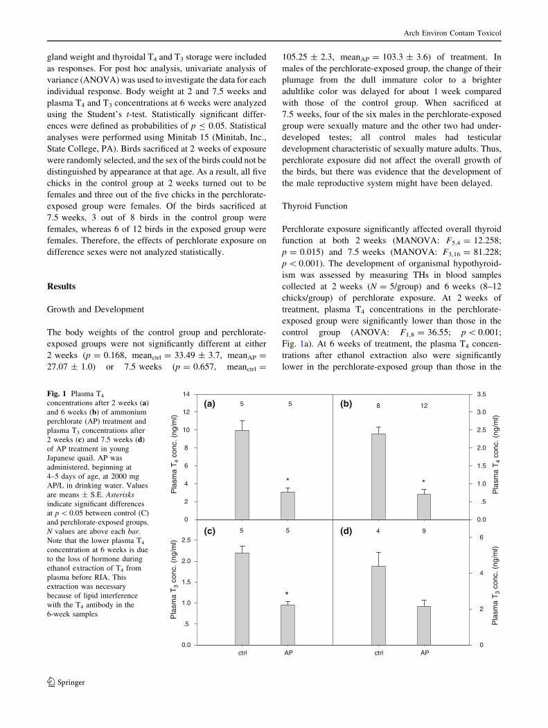
Perchlorate exposure significantly affected overall thyroid
function at both 2 weeks (MANOVA: F5,4 = 12.258;
p = 0.015) and 7.5 weeks (MANOVA: F3,16 = 81.228;
p \ 0.001). The development of organismal hypothyroid-
ism was assessed by measuring THs in blood samples
collected at 2 weeks (N = 5/group) and 6 weeks (8–12
chicks/group) of perchlorate exposure. At 2 weeks of
treatment, plasma T4 concentrations in the perchlorate-
exposed group were significantly lower than those in the
control group (ANOVA: F1,8 = 36.55; p \ 0.001;
Fig. 1a). At 6 weeks of treatment, the plasma T4 concen-
trations after ethanol extraction also were significantly
lower in the perchlorate-exposed group than those in the
(c)
ctrl AP
Pla
sma
T3
conc
. (ng
/ml)
0.0
.5
1.0
1.5
2.0
2.5(d)
ctrl AP
Pla
sma
T3
conc
. (ng
/ml)
0
2
4
65 5
*
4 9
(a)
Pla
sma
T4
conc
. (ng
/ml)
0
2
4
6
8
10
12
14
(b)
Pla
sma
T4
conc
. (ng
/ml)
0.0
.5
1.0
1.5
2.0
2.5
3.0
3.5
5 5
*
8 12
*
Fig. 1 Plasma T4
concentrations after 2 weeks (a)
and 6 weeks (b) of ammonium
perchlorate (AP) treatment and
plasma T3 concentrations after
2 weeks (c) and 7.5 weeks (d)
of AP treatment in young
Japanese quail. AP was
administered, beginning at
4–5 days of age, at 2000 mg
AP/L in drinking water. Values
are means ± S.E. Asterisksindicate significant differences
at p \ 0.05 between control (C)
and perchlorate-exposed groups.
N values are above each bar.
Note that the lower plasma T4
concentration at 6 weeks is due
to the loss of hormone during
ethanol extraction of T4 from
plasma before RIA. This
extraction was necessary
because of lipid interference
with the T4 antibody in the
6-week samples
Arch Environ Contam Toxicol
123
control group (t-test: p \ 0.0001; Fig. 1b). The propor-
tional difference in plasma T4 concentration (perchlorate
group/control group) was very similar at 2 weeks (*0.31)
and 6 weeks (*0.29) of treatment. Plasma T3 concentra-
tions were significantly lower in the perchlorate-exposed
group than in the control group at 2 weeks (ANOVA:
F1,8 = 49.31; p \ 0.001; Fig. 1c), but the groups did not
differ significantly at 7.5 weeks (t-test: p = 0.057; Fig. 1d)
due to greater variability at the later time. Note that T3
concentrations in a few samples in both groups were not
measurable due to the lipid interference. Most of the birds
were sexually mature at the time of sacrifice and the lipid
content in the plasma increased dramatically between 6 and
7.5 weeks of perchlorate exposure. Ethanol extraction was
not effective in eliminating lipid interference with antibody
binding in either T4 or T3 RIAs at this age.
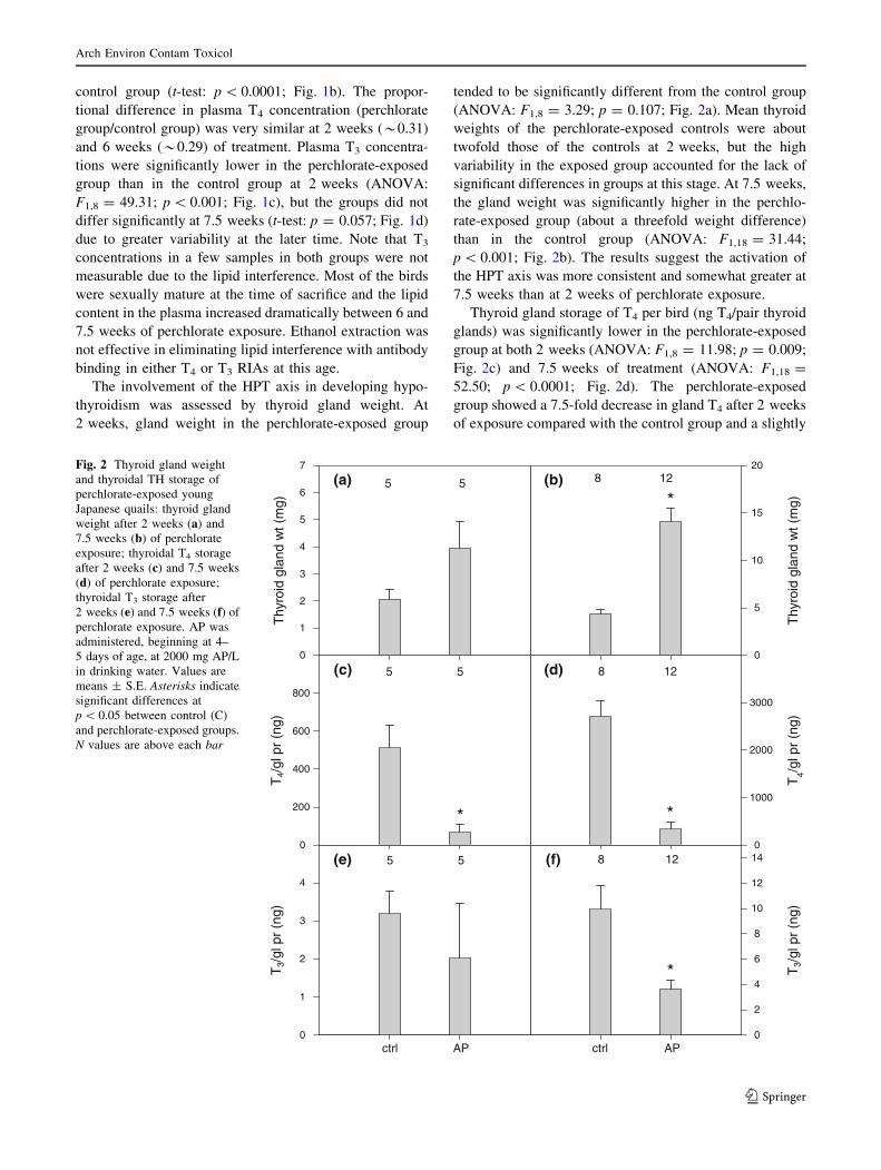
The involvement of the HPT axis in developing hypo-
thyroidism was assessed by thyroid gland weight. At
2 weeks, gland weight in the perchlorate-exposed group
tended to be significantly different from the control group
(ANOVA: F1,8 = 3.29; p = 0.107; Fig. 2a). Mean thyroid
weights of the perchlorate-exposed controls were about
twofold those of the controls at 2 weeks, but the high
variability in the exposed group accounted for the lack of
significant differences in groups at this stage. At 7.5 weeks,
the gland weight was significantly higher in the perchlo-
rate-exposed group (about a threefold weight difference)
than in the control group (ANOVA: F1,18 = 31.44;
p \ 0.001; Fig. 2b). The results suggest the activation of
the HPT axis was more consistent and somewhat greater at
7.5 weeks than at 2 weeks of perchlorate exposure.
Thyroid gland storage of T4 per bird (ng T4/pair thyroid
glands) was significantly lower in the perchlorate-exposed
group at both 2 weeks (ANOVA: F1,8 = 11.98; p = 0.009;
Fig. 2c) and 7.5 weeks of treatment (ANOVA: F1,18 =
52.50; p \ 0.0001; Fig. 2d). The perchlorate-exposed
group showed a 7.5-fold decrease in gland T4 after 2 weeks
of exposure compared with the control group and a slightly
(a)
Thy
roid
gla
nd w
t (m
g)
0
1
2
3
4
5
6
7
(b)
Thy
roid
gla
nd w
t (m
g)
0
5
10
15
20
(c)
T4/
gl p
r (n
g)
0
200
400
600
800
(d)
T4/g
l pr
(ng)
0
1000
2000
3000
5 5 8 12
*
5 5
*
8 12
*
(e)
ctrl AP
T3/
gl p
r (n
g)
0
1
2
3
4
(f)
ctrl AP
T3/
gl p
r (n
g)
0
2
4
6
8
10
12
145 5 8 12
*
Fig. 2 Thyroid gland weight
and thyroidal TH storage of
perchlorate-exposed young
Japanese quails: thyroid gland
weight after 2 weeks (a) and
7.5 weeks (b) of perchlorate
exposure; thyroidal T4 storage
after 2 weeks (c) and 7.5 weeks
(d) of perchlorate exposure;
thyroidal T3 storage after
2 weeks (e) and 7.5 weeks (f) of
perchlorate exposure. AP was
administered, beginning at 4–
5 days of age, at 2000 mg AP/L
in drinking water. Values are
means ± S.E. Asterisks indicate
significant differences at
p \ 0.05 between control (C)
and perchlorate-exposed groups.
N values are above each bar
Arch Environ Contam Toxicol
123
greater 8.1-fold decrease after 7.5 weeks. Thyroidal T3
content, which accounts for a very small proportion of
gland hormones, was not significantly different in the
perchlorate-exposed group from controls at 2 weeks of
exposure (ANOVA: F1,8 = 0.57; p = 0.472; Fig. 2e) but
was significantly lower at 7.5 weeks of exposure
(ANOVA: F1,18 = 175.37; p \ 0.001; Fig. 2f). Thyroidal
hormones decreased quickly after perchlorate exposure
started (by 2 weeks of exposure) and remained low through
the exposure period.
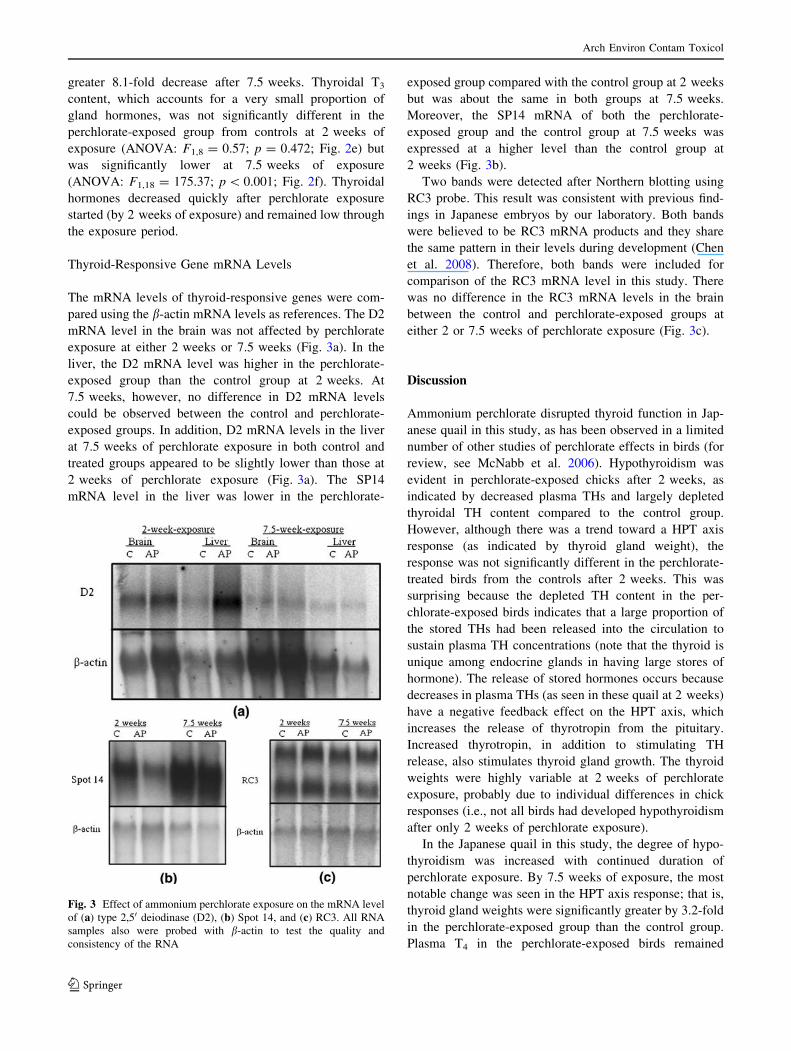
Thyroid-Responsive Gene mRNA Levels
The mRNA levels of thyroid-responsive genes were com-
pared using the b-actin mRNA levels as references. The D2
mRNA level in the brain was not affected by perchlorate
exposure at either 2 weeks or 7.5 weeks (Fig. 3a). In the
liver, the D2 mRNA level was higher in the perchlorate-
exposed group than the control group at 2 weeks. At
7.5 weeks, however, no difference in D2 mRNA levels
could be observed between the control and perchlorate-
exposed groups. In addition, D2 mRNA levels in the liver
at 7.5 weeks of perchlorate exposure in both control and
treated groups appeared to be slightly lower than those at
2 weeks of perchlorate exposure (Fig. 3a). The SP14
mRNA level in the liver was lower in the perchlorate-
exposed group compared with the control group at 2 weeks
but was about the same in both groups at 7.5 weeks.
Moreover, the SP14 mRNA of both the perchlorate-
exposed group and the control group at 7.5 weeks was
expressed at a higher level than the control group at
2 weeks (Fig. 3b).
Two bands were detected after Northern blotting using
RC3 probe. This result was consistent with previous find-
ings in Japanese embryos by our laboratory. Both bands
were believed to be RC3 mRNA products and they share
the same pattern in their levels during development (Chen
et al. 2008). Therefore, both bands were included for
comparison of the RC3 mRNA level in this study. There
was no difference in the RC3 mRNA levels in the brain
between the control and perchlorate-exposed groups at
either 2 or 7.5 weeks of perchlorate exposure (Fig. 3c).
Discussion
Ammonium perchlorate disrupted thyroid function in Jap-
anese quail in this study, as has been observed in a limited
number of other studies of perchlorate effects in birds (for
review, see McNabb et al. 2006). Hypothyroidism was
evident in perchlorate-exposed chicks after 2 weeks, as
indicated by decreased plasma THs and largely depleted
thyroidal TH content compared to the control group.
However, although there was a trend toward a HPT axis
response (as indicated by thyroid gland weight), the
response was not significantly different in the perchlorate-
treated birds from the controls after 2 weeks. This was
surprising because the depleted TH content in the per-
chlorate-exposed birds indicates that a large proportion of
the stored THs had been released into the circulation to
sustain plasma TH concentrations (note that the thyroid is
unique among endocrine glands in having large stores of
hormone). The release of stored hormones occurs because
decreases in plasma THs (as seen in these quail at 2 weeks)
have a negative feedback effect on the HPT axis, which
increases the release of thyrotropin from the pituitary.
Increased thyrotropin, in addition to stimulating TH
release, also stimulates thyroid gland growth. The thyroid
weights were highly variable at 2 weeks of perchlorate
exposure, probably due to individual differences in chick
responses (i.e., not all birds had developed hypothyroidism
after only 2 weeks of perchlorate exposure).
In the Japanese quail in this study, the degree of hypo-
thyroidism was increased with continued duration of
perchlorate exposure. By 7.5 weeks of exposure, the most
notable change was seen in the HPT axis response; that is,
thyroid gland weights were significantly greater by 3.2-fold
in the perchlorate-exposed group than the control group.
Plasma T4 in the perchlorate-exposed birds remained
Fig. 3 Effect of ammonium perchlorate exposure on the mRNA level
of (a) type 2,50 deiodinase (D2), (b) Spot 14, and (c) RC3. All RNA
samples also were probed with b-actin to test the quality and
consistency of the RNA
Arch Environ Contam Toxicol
123
significantly lower than in control birds (ratio perchlorate
group/control group = 0.31 at 2 weeks and 0.29 at
6 weeks). Thyroidal T4 stores also continued to decrease
slightly from 2 weeks to 7.5 weeks of treatment (perchlo-
rate group/control group; 0.13 vs. 0.12, respectively).
The thyroid-responsive gene for D2 is important in the
central nervous system enzymatic regulation of T3, the
most active TH. We predicted that the brain mRNA level of
D2 would increase in hypothyroid chicks, reflecting a
compensatory D2 response that would protect brain T3
concentrations. However, the mRNA level of D2 was not
affected in the brain of Japanese quail chicks after 2 or
7.5 weeks of perchlorate exposure. The brain is known to be
protected from short-term hypothyroidism by several
mechanisms: (1) increases in D2 activity, which increase T3
production, (2) increases in the uptake of THs into the brain,
and (3) decreases in the loss of THs from the brain [rats
(Bernal 2002); young chickens (Gereben et al. 1999; Rudas
et al. 2005)]. The third of these mechanisms results from
decreased T3 deactivation by inner-ring deiodination (Type
3, a 5-deiodinase) and consequent decreased loss of T3 to
the circulation (Rudas et al. 2005). Our measurements
focused only on whether decreases in circulating TH
affected D2 expression at the level of mRNA, as a mech-
anism for increasing T3 generation. However, if the second
and third mechanisms listed protected brain T3 concentra-
tions in these quail chicks, then increases in D2 gene
expression or enzyme activity would not be ‘‘needed.’’
Thus, the lack of D2 mRNA increase in hypothyroid chicks
in this study argues that brain T3 concentrations were
maintained by the second and third mechanisms listed
within the exposure times and perchlorate concentrations
we used. In addition, levels of mRNA might not be a direct
reflection of the D2 activity in specific tissues. D2 activity is
regulated by both T3 and T4 through different mechanisms.
Studies in mammals have revealed that T4 alters D2 activity
at a posttranslational level; that is, high T4 concentrations
facilitate the degradation of the enzyme (Kim et al. 1998;
Steinsapir et al. 1998). In contrast, T3 regulates at the level
of D2 mRNA, which also ultimately changes the activity of
the enzyme. High T3 concentrations decrease and low T3
concentrations increase steady-state D2 mRNA levels. The
effect of T4 is usually fast, whereas that of T3 is slow
(Burmeister et al. 1997). Moreover, studies have shown that
D2 protein stability and activity were also regulated by
complex mechanisms at the posttranscriptional level
(Gereben et al. 2002; Zeold et al. 2006). Therefore,
Northern blotting might not detect the effects of all these
mechanisms. Furthermore, D2 might not be affected by
hypothyroidism to the same degree in all brain areas
(Gereben et al. 2004; Verhoelst et al. 2004). Subtle changes
in regions more sensitive to hypothyroidism might not be
detected by measuring mRNA levels in brain total RNA.
Our study also demonstrated that D2 mRNA is present
in Japanese quail liver, a peripheral tissue exposed to cir-
culating TH concentrations; that is, the liver does not have
‘‘protective’’ mechanisms for controlling tissue hormones.
This is in contrast to the central nervous system, where the
entry and exit of THs is regulated (see section above). In
avian species, the presence of hepatic D2 has been con-
firmed by both detection of PTU insensitive 5’-deiodinase
activity in Japanese quail liver (Hughes and McNabb 1986;
McNabb et al. 1986) and the presence of hepatic D2
mRNA in chickens (Gereben et al. 1999). Liver is one of
the tissues responsible for the production of a major frac-
tion of circulating T3 from T4, and, historically, D1 was
believed to be the sole source of T3 from peripheral deio-
dination in mammals (Chopra 1991). Nonetheless, some
studies in rats indicate that D2 in brown adipose tissue
(BAT), and perhaps in other tissues, might account for as
much as 50% of the T3 production for release to the cir-
culation. The ratio might be even higher in hypothyroid
rats, where D1 activity is reduced by low circulating T4
(Nguyen et al. 1998; Silva and Larsen 1985). In birds,
although D1 predominates in the liver, enzymatic assays
have indicated that D2 activity accounts for some T3 pro-
duction (probably \ 10% of the total) during posthatch
development (Hughes and McNabb 1986). In adult chick-
ens, the level of D2 mRNA in the liver is comparable to
that in the brain (Gereben et al. 1999). However, D2
activity is much lower in the liver than in the brain. This
might be a result of alternative splicing of D2 mRNA in the
liver, which can result in higher proportions of inactive D2
(Gereben et al. 2002).
The presence of D2 in avian liver raises the question of
whether there is a hepatic D2 response to changes in cir-
culating THs and whether D2 might be elevated in the liver
of hypothyroid birds, thus contributing to circulating T3. In
the present study, hypothyroidism increased D2 mRNA
level in the liver of Japanese quail chicks after 2 weeks of
perchlorate exposure (although this was not the case at
7.5 weeks of exposure). Similar results were reported by
our laboratory in an earlier study on perchlorate-exposed
Japanese embryos, in which D2mRNA was detected in
perchlorate-exposed embryos but not in the control embryos
(Chen et al. 2008). Unlike the brain, for which hormone
entry and exit are regulated, liver tissue is not protected
against hypothyroid conditions. In the present study,
decreased plasma THs increased the expression of D2 in the
livers of perchlorate-exposed quail chicks, which suggests
that the liver initially responded to hypothyroidism by
increasing T3 production. In contrast, at 7.5 weeks,
although the perchlorate-exposed birds remained hypothy-
roid, the liver D2 mRNA level was not different from that of
the control group. There is evidence that D2 expression
changes during the course of development. In rats, the D2/
Arch Environ Contam Toxicol
123
D1 activity ratio decreases substantially from the neonatal
period to that in adults in tissues such as thyroid gland and
pituitary (Bates et al. 1999). Hence, developmentally, D2
function and regulation might be different in quail chicks
than adults. Furthermore, D2 might be regulated differently
in the liver than in the brain in adult chickens (Gereben et al.
2002). Our results in both Japanese embryos and young
chicks might indicate a more active role of D2 in TH reg-
ulation during early development.
In this study, RC3 was investigated as an example of a
thyroid-responsive gene with developmental effects on a
target tissue (brain). We predicted that perchlorate-induced
hypothyroidism would result in decreased brain RC3
mRNA levels. However, the mRNA level of RC3 in the
brain was not affected by hypothyroidism (in the body) at
either 2 or 7.5 weeks of perchlorate exposure. Because the
brain D2 mRNA level also was not affected in the per-
chlorate-exposed group, these two congruent results argue
that T3 concentrations in the brain did not change appre-
ciably (see discussion above about other hormone
regulatory mechanisms that protect the brain). However, it
is possible that there were changes in RC3 mRNA in some
brain regions that were not detectable in our studies of total
brain mRNA. Many of the previous studies on RC3 were
done by in situ hybridization of RC3 mRNA in brain tis-
sues and they have found that RC3 mRNA level did not
change the same way in all brain areas (Ishitobi et al. 2007;
Lein et al. 2007; Piosik et al. 1995, 1996; Wilcoxon et al.
2007; Zoeller et al. 2000).
In the liver, hypothyroidism lowered the SP14 mRNA
level (another thyroid-responsive target tissue gene) at
2 weeks of perchlorate exposure. SP14 codes for a protein
that regulates enzymes in the lipogenic pathway in the liver
and its mRNA level is rapidly upregulated by THs (Jump
et al. 1984; Narayan et al. 1984). As predicted, this result
indicates that some liver functions might have been affected
by perchlorate-induced hypothyroidism. At 7.5 weeks, no
significant difference was seen between the perchlorate-
exposed and control groups. With increasing maturity, SP14
mRNA levels were increased considerably at 7.5 weeks
compared to those at 2 weeks in both control and treated
birds. SP14 is known to be responsive to factors other than
plasma TH concentrations (e.g., circulating insulin con-
centrations as well as carbohydrate content of the diet; Jump
et al. 2001; LaFave et al. 2006). At 7.5 weeks of treatment,
quail are entering breeding age and we observed plasma
lipids increased markedly, indicating that lipogenesis was
highly active at that time. In such conditions, SP14
expression and lipogenesis might be regulated by multiple
mechanisms and the influence of THs might not be as
significant as at 2 weeks of perchlorate exposure.
In summary, perchlorate exposure causes organismal
hypothyroidism in young Japanese quail and thyroid
function indicators suggest the degree of hypothyroidism
increased as the time of exposure lengthened. The effects
of perchlorate exposure on the expression of thyroid-
responsive genes were more complicated. Genes in dif-
ferent tissues responded differently. The brain showed no
effects of organismal hypothyroidism on the mRNA level
of the hormone regulatory gene, D2, or the developmen-
tally relevant gene, RC3, suggesting that other mechanisms
provided protection of the brain from the decreased TH
concentrations in the body. In the liver, a hormone regu-
latory gene, D2, and a developmentally relevant gene,
SP14, were influenced by hypothyroidism early in per-
chlorate exposure (2 weeks) but not after sustained
exposure (7.5 weeks). This study suggests that wild birds
in perchlorate-contaminated areas might develop hypo-
thyroidism from perchlorate exposure during their
posthatch life. The effects of hypothyroidism on thyroid-
responsive genes might differ in different target tissues and
with exposure time and age. Thus, thyroid disruption by
perchlorate exposure might affect development and reduce
the overall fitness of both young and adult birds living in
contaminated habitats. However, it should be noted that we
used higher perchlorate exposures than commonly occur
environmentally because our purpose was to address the
effects of hypothyroidism on thyroid-responsive genes in
different target tissues.
Acknowledgments The study was supported by the Strategic Envi-
ronmental Research and Development Program (SERDP), grants from
Graduate Research and Development Program (GRDP), grants from
Sigma-Xi grants-in-aids, and a Waste Policy Institute (WPI) summer
fellowship. We thank Dr. Michael Denbow, Dr. Bradley Klein, and Dr.
Ignacio Moore for useful advice during the study. Laila Queral-Kirk-
patrick, Catherine Webb, and Eric Weigel participated in animal
sampling. Bambi Jarrett helped with animal care and maintenance.
References
Bates JM, St. Germain DL, Galton VA (1999) Expression profiles of
the three iodothyronine deiodinases, D1, D2, and D3, in the
developing rat. Endocrinology 140:844–851. doi:10.1210/en.
140.2.844
Bernal J (2002) Thyroid hormones and brain development. In: Pfaff
D, Arnold A, Etgen A, Fahrbach S, Rubin R (eds) Hormones,
brain and behavior. Academic Press, Burlington, pp 543–587
Bianco AC, Salvatore D, Gereben B, Berry MJ, Larsen PR (2002)
Biochemistry, cellular and molecular biology, and physiological
roles of the iodothyronine selenodeiodinases. Endocr Rev 23:38–
89. doi:10.1210/er.23.1.38
Brown S, Maloney M, Kinlaw W (1997) ‘‘Spot 14’’ protein functions
at the pretranslational level in the regulation of hepatic
metabolism by TH and glucose. J Biol Chem 272:2163–2166.
doi:10.1074/jbc.272.4.2163
Burmeister LA, Pachucki J, St. Germain DL (1997) Thyroid
hormones inhibit type 2 iodothyronine deiodinase in the rat
cerebral cortex by both pre- and post-translational mechanisms.
Endocrinology 138:5231–5237. doi:10.1210/en.138.12.5231
Arch Environ Contam Toxicol
123

Chen Y, Sible JC, McNabb FMA (2008) Effects of maternal exposure
to ammonium perchlorate on thyroid function and the expression
of thyroid-responsive genes in Japanese quail embryos. Gen
Comp Endocrinol 159:196–207. doi:10.1016/j.ygcen.2008.
08.014
Chopra IJ (1991) Nature, sources, and relative biologic significance of
circulating THs. In: Braverman LE, Utiger RD (eds) Werner and
Ingbar’s the thyroid: a fundamental and clinical text, 6th edn. J.
B. Lippincott, Philadelphia, pp 126–143
Gereben B, Bartha T, Tu HM, Harney JW, Rudas P, Larsen PR (1999)
Cloning and expression of the chicken type 2 iodothyronine 50-deiodinase. J Biol Chem 274:13,768–13,776. doi:10.1074/jbc.
274.20.13768
Gereben B, Kollar A, Harney JW, Larsen PR (2002) The mRNA
structure has potent regulatory effects on type 2 iodothyronine
deiodinase expression. Mol Endocrinol 16:1667–1679. doi:
10.1210/me.16.7.1667
Gereben B, Pachucki J, Kollar A, Liposits Z, Fekete C (2004)
Ontogenic redistribution of type 2 deiodinase messenger ribo-
nucleic acid in the brain of chicken. Endocrinology 145:3619–
3625. doi:10.1210/en.2004-0229
Gerendasy DD, Sutcliffe JG (1997) RC3/neurogranin, a postsynaptic
calpacitin for setting the response threshold to calcium influxes.
Mol Neurobiol 15:131–163. doi:10.1007/BF02740632
Hughes TE, McNabb FMA (1986) Avian hepatic T-3 production by
two pathways of 50-monodeiodination: effects of fasting and
patterns during development. J Exp Zool 238:393–399. doi:
10.1002/jez.1402380312
Ishitobi H, Mori K, Yoshida K, Watanabe C (2007) Effects of
perinatal exposure to low-dose cadmium on thyroid hormone-
related and sex hormone receptor gene expressions in brain of
offspring. Neurotoxicology 28:790–797. doi:10.1016/j.neuro.
2007.02.007
Jackson WA, Anderson TA, Canas JE, Snyder SA, Tan K (2006)
Environmental fate of perchlorate. In: Kendall RJ, Smith PN
(eds) Perchlorate ecotoxicology. SETAC Press, Pensacola, pp
21–45
Jump DB, Narayan P, Towle H, Oppenheimer JH (1984) Rapid
effects of triiodothyronine on hepatic gene expression. Hybrid-
ization analysis of tissue-specific triiodothyronine regulation of
mRNAS14. J Biol Chem 259:2789–2797
Jump DB, Thelen AP, Mater MK (2001) Functional interaction
between sterol regulatory element-binding protein-1c, nuclear
factor Y, and 3,5,30-triiodothyronine nuclear receptors. J Biol
Chem 276:34,419–34,427 doi:10.1074/jbc.M105471200
Kim SW, Harney JW, Larsen PR (1998) Studies of the hormonal
regulation of type 2 50-iodothyronine deiodinase messenger
ribonucleic acid in pituitary tumor cells using semiquantitative
reverse transcription-polymerase chain reaction. Endocrinology
139:4895–4905. doi:10.1210/en.139.12.4895
LaFave LT, Augustin LB, Mariash CN (2006) S14: insights from
knockout mice. Endocrinology 147:4044–4047. doi:10.1210/en.
2006-0473
Lein PJ, Yang D, Bachstetter AD, Tilson HA, Harry GJ, Mervis RF,
Kodavanti PRS (2007) Ontogenetic alterations in molecular and
structural correlates of dendritic growth after developmental
exposure to polychlorinated biphenyls. Environ Health Perspect
115:556–558
Mayer KP, Jackson WA, Snyder SA, Smith PN, Anderson TA (2006)
State of the science: background, history, and occurrence. In:
Kendall RJ, Smith PN (eds) Perchlorate ecotoxicology. SETAC
Press, Pensacola, pp 1–21
McNabb FMA (2007) The hypothalamic-pituitary-thyroid (HPT) axis
in birds and its role in bird development and reproduction. Crit
Rev Toxicol 37:163–193. doi:10.1080/10408440601123552
McNabb FMA, Cheng M-F (1985) Thyroid development in ring
doves, Streptopelia risoria. Gen Comp Endocrinol 58:243–251.
doi:10.1016/0016-6480(85)90340-5
McNabb FMA, Lyons LJ, Hughes TE (1986) Avian hepatic T3
generation by 50-monodeiodination: characterization of two
enzymatic pathways and the effects of goitrogens. Comp
Biochem Physiol A 85:249–255. doi:10.1016/0300-9629
(86)90247-1
McNabb FMA, Larsen CT, Pooler PS (2004a) Ammonium perchlorate
effects on thyroid function and growth in bobwhite quail chicks.
Environ Toxicol Chem 23:997–1003. doi:10.1897/03-362
McNabb FMA, Jang DA, Larsen CT (2004b) Does thyroid function in
developing birds adapt to sustained ammonium perchlorate
exposure? Toxicol Sci 82:106–113. doi:10.1093/toxsci/kfh247
McNabb FMA, Hooper MJ, Smith EE, Scott M, Gentles BA (2006)
Perchlorate effects on birds. In: Kendall RJ, Smith PN (eds)
Perchlorate ecotoxicology. SETAC Press, Pensacola, FL, pp 99–
127
Narayan P, Liaw CW, Towle HC (1984) Rapid induction of a specific
nuclear mRNA precursor by thyroid hormone. Proc Natl Acad
Sci USA 81:4687–4691. doi:10.1073/pnas.81.15.4687
Nguyen TT, Chapa F, DiStefano JJ III (1998) Direct measurement of
the contributions of type I and type II 50-deiodinases to whole
body steady state 3,5,30-triiodothyronine production from thy-
roxine in the rat. Endocrinology 139:4626–4633. doi:10.1210/
en.139.11.4626
Piosik PA, van Groenigen M, Ponne NJ, Bolhuis PA, Baas F (1995)
RC3/neurogranin structure and expression in the caprine brain in
relation to congenital hypothyroidism. Mol Brain Res 29:119–
130. doi:10.1016/0169-328X(94)00237-9
Piosik PA, van Groenigen M, Baas F (1996) Effect of thyroid
hormone deficiency on RC3/neurogranin mRNA expression in
the prenatal and adult caprine brain. Mol Brain Res 42:227–235.
doi:10.1016/S0169-328X(96)00126-X
Rudas P, Ronai Z, Bartha T (2005) Thyroid hormone metabolism in
the brain of domestic animals. Domest Anim Endocrinol 29:88–
96. doi:10.1016/j.domaniend.2005.02.032
Sible JC, Anderson JA, Lewellyn AL, Maller JL (1997) Zygotic
transcription is required to block a maternal program of
apoptosis in Xenopus embryos. Dev Biol 189:335–346 doi:
10.1006/dbio.1997.8683
Silva JE, Larsen PR (1985) Potential of brown adipose tissue Type II
thyroxine 50-deiodinase as a local and systemic source of
triiodothyronine in rats. J Clin Invest 76:2296–2305. doi:
10.1172/JCI112239
Steinsapir J, Harney J, Larsen PR (1998) Type 2 iodothyronine
deiodinase in rat pituitary tumor cells is inactivated in protea-
somes. J Clin Invest 102:1895–1899. doi:10.1172/JCI4672
Verhoelst CHJ, Darras VM, Roelens SA, Artykbaeva GM, Van der
Geyten S (2004) Type II iodothyronine deiodinase protein in
chicken choroid plexus: additional perspectives on T3 supply in
the avian brain. J Endocrinol 183:235–241. doi:10.1677/joe.
1.05743
Wang X, Carre W, Zhou H, Lamont SJ, Cogburn LA (2004)
Duplicated Spot 14 genes in the chicken: characterization and
identification of polymorphisms associated with abdominal fat
traits. Gene 332:79–88. doi:10.1016/j.gene.2004.02.021
Wilcoxon JS, Nadolski GJ, Samarut J, Chassande O, Redei EE (2007)
Behavioral inhibition and impaired spatial learning and memory
in hypothyroid mice lacking TH receptor [alpha]. Behav Brain
Res 177:109–116. doi:10.1016/j.bbr.2006.10.030
Wilson CM, McNabb FMA (1997) Maternal thyroid hormones in
Japanese quail egg and their influence on embryonic develop-
ment. Gen Comp Endocrinol 107:153–165. doi:10.1006/gcen.
1997.6906
Arch Environ Contam Toxicol
123

Zeold A, Pormuller L, Dentice M, Harney JW, Curcio-Morelli C,
Tente SM, Bianco AC, Gereben B (2006) Metabolic instability
of type 2 deiodinase is transferable to stable proteins indepen-
dently of subcellular localization. J Biol Chem 281:31,538–
31,543. doi:10.1074/jbc.M604728200
Zoeller RT, Dowling ALS, Vas AA (2000) Developmental exposure
to polychlorinated biphenyls exerts thyroid hormone-like effects
on the expression of RC3/Neurogranin and myelin basic protein
messenger ribonucleic acids in the developing rat brain.
Endocrinology 141:181–189. doi:10.1210/en.141.1.181
Arch Environ Contam Toxicol
123
Copyright © 2022 FDOKUMEN