Arrest of mammalian fibroblasts in G1 in response to actin ...

Effect of malathion on apoptosis of murine L929 fibroblasts: apossible mechanism for toxicity in low dose exposure.
L. Masoud a,1, C. Vijayasarathy a, M. Fernandez-Cabezudo a, G. Petroianu b,A.M. Saleh a,�
a Department of Biochemistry, Faculty of Medicine & Health Sciences, United Arab Emirates University, P.O. Box 17666, Al Ain,
United Arab Emiratesb Department of Pharmacology, Faculty of Medicine & Health Sciences, United Arab Emirates University, P.O. Box 17666, Al Ain,
United Arab Emirates
Received 5 July 2002; received in revised form 21 October 2002; accepted 21 October 2002
Abstract
While acute organophosphorous compound poisoning due to inhibition of acetylcholinesterase is a well-established
clinical entity, the existence of chronic poisoning due to exposure to low levels of organophosphorous compounds
(below the threshold required for cholinergic clinical symptoms) is a hotly debated issue. In this study, we have
evaluated the effects of noncholinergic doses of malathion (0.01�/20 mM) on apoptosis of murine L929 fibroblasts.
Employing flow cytometric and caspase activation analyses we demonstrate that malathion induces apoptosis in L929
cells in a dose- and time-dependent manner. The initiator caspases (caspase-8 and caspase-9) as well as the effector
caspase (caspase-3) were activated by the treatment of L929 cells with malathion. Exposure of L929 cells to malathion
in the presence of a general inhibitor of caspase, z-VAD-FMK abolished the apoptotic effect of the compound. In
addition, malathion induced an increase in the expression of the pro-apoptotic protein p53. However, the induction of
p53 expression was subsequent to activation of the caspase cascades. The present findings suggest, that the cytotoxicity
of malathion at noncholinergic doses is mediated through caspase-dependent apoptosis.
# 2002 Elsevier Science Ireland Ltd. All rights reserved.
Keywords: Apoptosis; Caspases; p53; Organophosphorus compounds; Malathion
Abbreviations: AChE, acetylcholinesterase; Caspase, cysteine specific aspartate protease; IC50, inhibitory concentration 50
(concentration of a chemical required to reduce the enzyme activity by 50%); OPCs, organophosphorus compounds; PARP,
poly(ADP-ribose) polymerase; PI, propidium iodide; z-VAD-FMK, benzyloxy-valine-alanine-aspartate-O -methyl-
fluoromethylketone.
� Corresponding author. Fax: �/971-3-767-2033
E-mail address: [email protected] (A.M. Saleh).1 This author and A.M. Saleh contributed equally to the work.
Toxicology 185 (2003) 89�/102
www.elsevier.com/locate/toxicol
0300-483X/02/$ - see front matter # 2002 Elsevier Science Ireland Ltd. All rights reserved.
PII: S 0 3 0 0 - 4 8 3 X ( 0 2 ) 0 0 5 9 6 - 6

1. Introduction
Malathion [S -1,2-bis(ethoxycarbonyl)ethyl
O ,O -dimethyl phosphorodithioate; CAS registry
no. 121-75-5] is one of the most widely used
organophosphate compounds (OPCs) applied in
agriculture as a pesticide, in veterinary practice as
an ectoparasiticide (reviewed by Flessel et al.,
1993), in eradication of human body lice (Elston,2002; Roberts, 2002) and in food preparation and
processing areas (Savage et al., 1981). Acute toxic
effects induced by malathion pesticides are mainly
caused by inhibition of acetylcholinesterase
(AChE) in the nervous tissue with a consequent
increase in the levels of the neurotransmitter
acetylcholine (reviewed in Nigg and Knaak,
2000; Overstreet and Djuric, 2001; Kwong,2002). Malathion can be metabolized to non-toxic
intermediates by carboxyesterases enzymes (Gupta
et al., 1983; Nigg and Knaak, 2000). However, its
widespread use in agriculture and household
practices has raised concern over its potential to
cause adverse health effects in humans, animals,
wildlife and fish (Flessel et al., 1993; Wolfe and
Seiber, 1993). Occupational exposure to malathionis a cause for concern. Dermal exposure to
malathion following aerosol application has been
estimated to be as high as �/5 mg/kg per day
(Wolfe et al., 1967, 1978).
Recent studies have indicated that at noncholi-
nergic (sub-lethal acute) doses, malathion affects
several biochemical pathways that do not involve
modulation of AChE activity. Both under in vitroand in vivo conditions, malathion (B/10 mg/kg
body weight:/30 mM) has been shown to induce
DNA damage, chromosomal aberrations (Blasiak
et al., 1999; Giri et al., 2002) and malignant
transformation (Cabello et al., 2001). Malathion
has also been shown to modulate oxidative stress
and immune response in experimental animals
(John et al., 2001; Johnson et al., 2002).Cells undergo apoptosis (programmed cell
death) in response to various stimuli including
chemical stress. Apoptosis, which is distinct from
necrosis, another form of cell death (reviewed by
Raffray and Cohen, 1997; Pallardy et al., 1999;
Bratton and Cohen, 2001) is mediated by the
intracellular aspartate specific cysteine proteases
designated as caspases (Alnemri, 1997). Caspasesare synthesized and stored as inactive precursors
(procaspases) and activated by proteolysis upon
induction by various apoptotic stimuli (Pallardy et
al., 1999).
Of the two pathways that activate caspases, the
receptor-mediated pathway involves the ligation of
death receptors resulting in recruitment and acti-
vation of caspase-8 (Scaffidi et al., 1998), while thestress induced mitochondria dependent pathway
leads to activation of caspase-9 (Saleh et al., 1999;
Zou et al., 1999). Following activation of the
initiator caspase-8 or -9, the two pathways con-
verge on the activation of the effector caspase-3,
which finally cleaves various vital substrates in the
cell (Alnemri, 1997). Apart from these two path-
ways, p53, a molecule involved in cell surveillance,also activates the mitochondrial pathway of apop-
tosis in response to DNA damage (Luu et al.,
2002; Trinei et al., 2002). Although these pathways
appear to be distinct from each other, a cross talk
between them ensures effective elimination of
injured or damaged cells.
Although the cytotoxicity of malathion has been
shown to involve several biochemical pathways,studies on apoptosis following exposure to this
compound have not received much attention as
yet. In the present study, we assessed the cytotoxic
effect of very low non-cholinergic doses of ma-
lathion (concentrations that do not inhibit AChE
activity: 0.01�/20 mM) on apoptosis of cultured
mouse L929 fibroblasts using flow cytometry,
caspase activation and DNA fragmentation tech-niques. Our results demonstrate that malathion
induces a caspase-dependent apoptosis in a time
and dose-dependent manner in cultured mouse
L929 fibroblasts.
2. Materials and methods
2.1. Chemicals
Malathion with 95% purity was obtained from
PolyScience (Division of Preston Industries Inc.,
Niles, IL). Stock solution of malathion (100 mM)
was prepared in dimethyl sulfoxide (DMSO) and
stored at �/80 8C. The working dilutions in
L. Masoud et al. / Toxicology 185 (2003) 89�/10290

phosphate buffered saline (PBS) were prepared
just before use. Protease inhibitors, [PMSF, pep-
statin A, leupeptin and aprotinin] were acquired
form Sigma Chemical Company (Sigma Aldrich
Chemie GmbH Steinheim, Germany). The colori-
metric tetrapeptide substrates for caspase-8 (Ac-
IETD-pNA), caspase-9 (Ac-LEHD-pNA) and
caspase-3 (Ac-DEVD-pNA) were purchased from
Calbiochem (San Diego, CA). Caspase-8 inhibitor
(Ac-IETD-CHO), caspase-9 inhibitor (z-LEHD-
FMK), caspase-3 inhibitor (Ac-DEVD-CHO) and
the general caspase inhibitor (z-VAD-FMK) were
also obtained from Calbiochem. The following
polyclonal antibodies were obtained from various
sources as indicated: anti-caspase-8, anti-p53, anti
a-tubulin and anti-PARP (Santa Cruz Biotechnol-
ogy, CA), anti-caspase-9 and anti-caspase-3
(Stressgen Biotechnolgies, Victoria, BC, Canada).
2.2. Cell culture
L929 mouse fibroblast cells were obtained from
American Type Culture Collection (ATCC; Rock-
ville, MD). Cells were cultured in L-glutamine
containing RPMI 1640 medium (Sigma, St. Lois,
MO) supplemented with 10% heat-inactivated
fetal bovine serum (GIBCO BRL, Grand Island,
NY), 100 U/ml penicillin and 100 mg/ml strepto-
mycin, and were kept at 37 8C in humidified 5%
CO2/95% air. Prior to confluence, the cells were
harvested and seeded onto plastic 6-well culture
plates or 100-mm culture dishes at 5�/105 cells/ml.
The cells were then allowed to grow for 2�/3 days
and exposed to varying concentrations of ma-
lathion (0.01�/20 mM) for 16 h. Following treat-
ment, the cells were harvested using 0.25% trypsin/
EDTA (GIBCO BRL), centrifuged at 1000 rpm
for 5 min, washed with PBS and subsequently used
for various biochemical investigations. The speci-
ficity of caspase-mediated apoptosis was deter-
mined by including z-VAD-FMK, a general
inhibitor of caspases in the medium. In those
experiments where the inhibitors were used, the
cells were pretreated with 25 or 50 mM z-VAD-
FMK for 6 h, followed by treatment with
malathion for 16 h prior to harvesting.
2.3. Analysis of apoptosis by flow cytometry
Loss of plasma membrane asymmetry is one of
the earliest features of apoptosis. In apoptotic
cells, the membrane phospholipid, phosphatidyl-
serine (PS) is exposed to the external cellular
environment as a result of translocation from the
inner to the outer leaflet of the plasma membrane
(Okamoto et al., 2002). To identify early apoptoticcells, we used the Annexin V-FITC (fluorescein
isothiocyanate) staining kit from BD Biosciences
(Franklin Lakes, NJ). Annexin V has a high
affinity for PS and binds to cells with exposed
PS (Vermes et al., 1995). Propidium Iodide (PI)
was used to differentiate apoptotic cells with
preserved membrane integrity (Annexin�, PI�)
from necrotic cells that lost membrane integrity(Annexin�, PI�). The assay was performed fol-
lowing the manufacturer’s procedure. Briefly,
malathion-treated cells (1�/106/ml) were harvested
and washed twice with ice-cold PBS. About 1�/
105 cells were stained with Annexin V-FITC and
PI as per the manufacturer’s protocol. 10,000 cells
were analyzed by flow cytometry (Facs Vantage,
Becton, Dickinson, USA). Cells with positiveAnnexin V stain (Annexin�, PI�) were counted
as apoptotic and their number expressed as a
percentage of the total cells.
2.4. Western blot analysis
Malathion-treated cells were harvested, washed
twice with PBS and suspended in a lysis buffer
containing 100 mM HEPES, pH 7.5, 10% sucrose,10 mM DTT, 0.1% CHAPS, 150 mM NaCl and
protease inhibitors (1 mM PMSF, and 1 mg/ml
leupeptin, aprotinin and pepstain A). Cells were
lysed by four repeated cycles of freeze thawing and
centrifuged at 4 8C for 30 min at 14,000�/g . The
supernatant was collected and stored at �/80 8Cor used immediately. The samples were analyzed
for total protein by a protein assay kit based onBradford (1976) colorimetric reaction (BioRad,
USA). Cell lysates (50 mg protein per lane) were
separated on 8 and 12% SDS�/polyacrylamide gels
and electroblotted onto a PVDF membrane (Milli-
pore, USA) using standard techniques (Laemmli,
1970; Towbin et al., 1979). After electrophoretic
L. Masoud et al. / Toxicology 185 (2003) 89�/102 91

transfer, the membranes were blocked by incuba-tion for 1 h with 5% non-fat dry milk in PBS
containing 0.1% Tween-20. The blots were then
incubated for 2 h at room temperature or over-
night at 4 8C with one of the following antibodies
diluted in PBS containing 0.1% Tween-20 and 2%
non-fat dry milk: anti-caspase-8 (1:1000), anti-
caspase-9 (1:3000), anti-caspase-3 (1:1000), anti-
PARP (1: 1000) and anti-p53 (1: 500). The blotswere then incubated with a horseradish perox-
idase-conjugated secondary antibody against rab-
bit IgG (1:2000 dilution; Sigma). The antigen�/
antibody complexes on the blots were detected by
SuperSignal chemiluminescence kit as described in
the manufacturer’s protocol (Pierce Biotechnol-
ogy, Rockford, IL) and visualized by autoradio-
graphy. To confirm equal loading of proteins, theblots were also immunoprobed with a rabbit
polyclonal antibody against the cytoskeletal pro-
tein a-tubulin (1:2500 dilution). The consistent
equal signals of a-tubulin from the different
extracts also indicate that malathion does not
interfere with protein synthesis in L929 cells.
2.5. Assay of caspase-8, -9 and -3 activities
Caspase-3, one of the principal caspases in
apoptotic cells, is activated during the apoptotic
signaling by upstream caspases including caspase-
8 and -9 (Bratton and Cohen, 2001). The activities
of caspase-8, -9 and -3 were determined using the
colorimetric tetrapeptide substrates Ac-IETD-
pNA, Ac-LEHD-pNA and Ac-DEVD-pNA re-
spectively, as previously described by Srinivasulaet al. (2001). Lysates (30 mg protein) from
malathion treated cells were incubated with 0.2
mM of caspase-3, -8 or -9 colorimetric peptide
substrates in a 1/2 volume microtiter plate at
30 8C. The assays were performed both in the
presence and absence of the caspase-8 (Ac-IETD-
CHO), caspase-9 (z-LEHD-FMK) or caspase-3
(Ac-DEVD-CHO) specific inhibitors (20 mM) toeliminate non-specific activities in the lysates.
Caspase activity was monitored spectrophotome-
tricaly, using microtiter plate reader by measuring
the increase in absorbance at 405 nm, which
corresponds to the amount of p-Nitroaniline
(pNA), liberated from the peptide substrates.
The absorbance change was converted into unitsof enzyme activity using a standard curve gener-
ated with free pNA. One unit of caspase-3, -8, or -
9 activity corresponds to the amount of enzyme
that will release 1 pmol of pNA from 0.2 mM
DEVD-pNA, IETD-pNA or Ac-LEHD-pNA per
min, respectively.
2.6. DNA fragmentation assay
DNA fragmentation was analysed using agarose
gel electrophoresis. Genomic DNA was prepared
using the method of Gong et al. (1994) with slight
modification. After treatment of L929 fibroblasts
with malathion, about 2�/106 cells were washed in
ice-cold PBS and prefixed in ice-cold 70% ethanol.
The partially degraded oligonucleosomal DNAwas selectively extracted with 100 ml of 0.2 M
phosphate�/citrate buffer at pH 7.8. The extracts
were then treated with NP-40 (0.015%) RNase A
(0.06 mg/ml) and Proteinase K (0.06 mg/ml). The
DNA was extracted and purified sequentially with
phenol: chloroform (1:1) and chloroform followed
by precipitation in 100% ethanol. The samples
were then air dried and resuspended in TE buffer(10 mM Tris HCl, 1 mM EDTA, pH 7.5). DNA
samples (5 mg each) were resolved by electrophor-
esis for 4 h on a 1.5% agarose gel at 40 V. The gel
was stained with ethidium bromide and the band-
ing patterns were visualized with the Foto/Eclipse
UV transilluminator and photographed.
2.7. RBC-AChE activity
The effect of malathion (0�/50 mM) on AChE
activity was measured in diluted whole blood
samples in the presence of the selective butyryl-
cholinesterase inhibitor ethoproprazine as pre-
viously described (Worek et al., 1999). The assay,
which is based on Ellman’s method, measures the
reduction of dithiobis�/nitrobenzoic acid (DTNB)
to nitrobenzoate (TNB�) by thiocholine, theproduct of acetylthiocholine (ASch) hydrolysis.
Freshly drawn venous blood samples from three
male mice were diluted in 0.1 M phosphate buffer
(pH 7.4) and incubated with DTNB (10 mM) and
ethopropazine (6 mM) for 10 min at 37 8C prior
to addition of ASch. The change in the absorbance
L. Masoud et al. / Toxicology 185 (2003) 89�/10292

of DTNB was measured at 436 nm. The AChEactivity was calculated using an absorption coeffi-
cient of TNB� at 436 nm (o�/10.6 mM�1 cm1).
The values were normalized to the hemoglobin
(Hb) content (determined as cyanmethemoglobin)
and expressed as mU/mmol/Hb. IC50 values were
derived from a plot that indicates percent AChE
activity as a function of malathion concentration.
3. Results
3.1. Evaluation of apoptosis by flow cytometry
First, we investigated whether malathion in-
duces apoptosis. To assess this effect, we incubated
L929 cells with varying concentrations of ma-
lathion for overnight and measured apoptosis bycounting the Annexin V stain positive and PI
negative cells (Annexin�, PI�). As shown in Fig.
1A, malathion induced apoptosis in L929 cells in a
dose-dependent manner. As compared to un-
treated cells, apoptosis became apparent (An-
nexin�, PI�; 10% above the background
apoptosis seen in the control) at doses of ma-
lathion as low as 10 nM and reached maximum(30%) in cells treated with 1 mM of malathion.
Higher concentrations of malathion up to 20 mM
did not cause any further increase in the percen-
tage of apoptotic cells. At all concentrations of
malathion tested, the number of necrotic cells that
lost their plasma membrane as determined by PI
stain (Annexin�, PI�) were not significant. How-
ever there was a dose-dependent increase in thenumber of cells with positive Annexin V and PI
signal (Annexin�, PI�). Since there is a possibility
that the necrotic stage cells also acquire Annexin V
signal and could not be distinguished from apop-
totic cells, the Annexin�, PI� cells were not
included in the analysis (Fig. 1A, B). To further
assess the effect of malathion-induced apoptosis,
we determined the percentage of apoptotic cellsafter incubation with 1 mM of malathion at
different intervals of time (Fig. 1B). Malathion
induced apoptosis in a time-dependent manner.
Apoptosis (Annexin�, PI�) was apparent as early
as 2 h of incubation with malathion (�/10%) and
reached maximum (�/45%) between 16 and 20 h
of treatment. These results indicate that malathionis a potent apoptosis-inducing agent at concentra-
tions as low as 5/1 mM.
To relate the potential of malathion to induce
apoptosis at such low concentrations to it’s
cholinergic effect, we determined the IC50 value
for malathion. As shown in Fig. 2, we report IC50
value of 24 mM for malathion as an inhibitor of
mouse RBC-AcChE. These results indicate thatthe toxicity of malathion at low noncholinergic
doses is mediated through it’s effect on apoptosis.
3.2. Processing and activation of caspases
Activation of caspases is the major signal to
initiate apoptosis. We, therefore, tested processing
(activation) of caspase-8, -9 and -3 to complement
the flow cytometry results. Fig. 3A, shows theimmunoblot analysis for detection of caspase
processing in L929 fibroblasts treated with ma-
lathion. Cell lysates (50 mg protein) were separated
by SDS�/PAGE and immunoblotted with the
respective antibodies. Treatment of L929 murine
cells for 16 h with malathion was effectively
associated with processing of procaspase-8, -9,
and -3 as detected by the formation of the smallersubunits (p20, p35 and p19, respectively) of their
active enzyme complexes. While there was a
gradual increase in caspase-8, -9 and -3 processing
in a dose-dependent manner, the maximum pro-
cessing of these proenzymes was detected after
treatment with 1 mM of malathion. Consistent
with the apoptosis percentages observed by An-
nexin V staining assay, increasing the malathionconcentrations up to 20 mM did not induce further
processing of any one of these caspases. Taken
together, these results suggest that malathion
induces apoptosis in a caspase-dependent fashion.
We directly assayed caspase-8, -9 and -3 activ-
ities by measuring the hydrolysis of colorimetric
tetrapeptide substrates of these enzymes (Fig. 3B).
As expected, there was a dose-dependent increasein caspase-8, -9 and -3 activities, reaching peak
levels in lysates obtained from 1 mM malathion-
treated cells. Thus, the processing of caspases, as
shown by immunoblot analysis, is associated with
their activation as demonstrated in this experi-
ment.
L. Masoud et al. / Toxicology 185 (2003) 89�/102 93

Fig. 1
L. Masoud et al. / Toxicology 185 (2003) 89�/10294

3.3. PARP cleavage and DNA fragmentation
Activation of the effector caspase-3, under in
vivo conditions, leads to cleavage (inactivation) of
essential target proteins required for cell viability.
One of the targets of active caspase-3, is the DNA
repair enzyme PARP (Lazebnik et al., 1994). To
demonstrate the effect of caspase-3 activation on
PARP, we subjected the lysates from malathion-
treated cells to immunoblot analysis with a poly-
clonal antibody against PARP. As shown in Fig.
4A, the 116 kDa PARP was effectively cleaved
into a 89-kDa fragment in a dose-dependent
manner with maximal processing occurring in cells
treated with 1 mM of malathion. Thus, the ability
of malathion to induce apoptosis at low but
effective noncholinergic concentrations is asso-
ciated with elevated caspase-3 activity and clea-
vage of PARP by proteolysis.
The effect of malathion-induced caspase-3 acti-vation on DNA fragmentation was viewed by
agarose gel electrophoresis of DNA, isolated
from malathion treated cells. Fig. 4B, shows
DNA fragmentation, a characteristic ladder pat-
tern in malathion treated cells that is consistent
with the classical apoptotic features. Cells treated
with 1 mM malathion showed intense banding
pattern as compared to the cells treated with lowerconcentrations of malathion (data not shown). In
tune with the earlier results discussed above, there
were no significant differences between the inten-
sities and the banding patterns of DNA obtained
from cells treated with 1, 10 and 20 mM of
malathion.
3.4. Specificity of caspase activation and apoptosis
The above results suggest that the ability of
malathion to induce apoptosis is dependent on
triggering the activation of caspase cascades. To
investigate this possibility, we compared the ability
of malathion to induce apoptosis in the presence
and absence of the broad-range caspase inhibitor
z-VAD-FMK (Fig. 5A). L929 cells were treatedwith 1 mM malathion alone or in combination with
25 or 50 mM z-VAD-FMK and apoptosis was
assessed by the flow cytometry method described
earlier. As compared to untreated control and
malathion alone treated cells, 25 mM z-VAD-FMK
decreased the ability of malathion to induce
apoptosis by �/40% while the presence of 50 mM
z-VAD-FMK completely prevented malathion in-duced apoptosis. Inhibition of the malathion-
mediated apoptosis in the presence of z-VAD-
FMK correlated well with a substantial decrease in
DNA fragmentation (Fig. 5B). Under these con-
ditions, the cleavage of full-length PARP into a 89-
kDa product was also inhibited in the presence of
Fig. 1. Malathion induces apoptosis in a dose- and time-dependent manner. (A) L929 cells were treated with varying concentrations of
malathion (0�/20 mM) overnight. After treatment, cells were harvested, stained with Annexin V-FITC and PI and analyzed by flow
cytometry. The percentages of cells in early (Annexin�, PI�; lower right quadrant) and late apoptotic-necrotic stages (Annexin�,
PI�; upper right quadrant) are indicated. The results are representative of two independent experiments. Cells with positive Annexin
stain (Annexin�, PI�) were counted as apoptotic and their number expressed as a percentage of the total cells. (B) L929 cells were
incubated with 1 mM malathion for different periods of time (0�/24 h) and apoptosis was analyzed by flow cytometry as described in
(A). The control (0) represents cells incubated under similar conditions in the presence of the carrier DMSO. The results represent the
average of two independent experiments.
Fig. 2. IC50 value for malathion as inhibitor of mouse RBC-
AchE. The effect of malathion (0�/50 mM) on AChE activity
was measured in mouse blood samples as described in the
Materials and Methods and the IC50 values were calculated
from a plot that indicates percent AChE activity as a function
of malathion concentration. The results are representative of
two independent experiments.
L. Masoud et al. / Toxicology 185 (2003) 89�/102 95
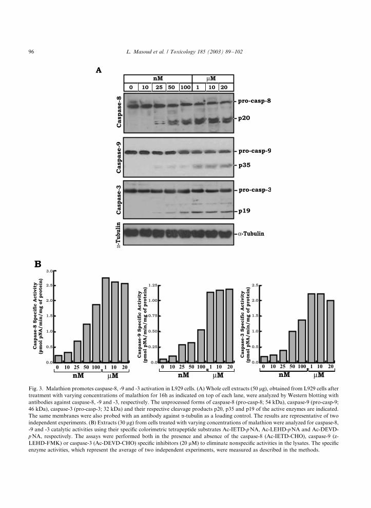
Fig. 3. Malathion promotes caspase-8, -9 and -3 activation in L929 cells. (A) Whole cell extracts (50 mg), obtained from L929 cells after
treatment with varying concentrations of malathion for 16h as indicated on top of each lane, were analyzed by Western blotting with
antibodies against caspase-8, -9 and -3, respectively. The unprocessed forms of caspase-8 (pro-casp-8; 54 kDa), caspase-9 (pro-casp-9;
46 kDa), caspase-3 (pro-casp-3; 32 kDa) and their respective cleavage products p20, p35 and p19 of the active enzymes are indicated.
The same membranes were also probed with an antibody against a-tubulin as a loading control. The results are representative of two
independent experiments. (B) Extracts (30 mg) from cells treated with varying concentrations of malathion were analyzed for caspase-8,
-9 and -3 catalytic activities using their specific colorimetric tetrapeptide substrates Ac-IETD-p NA, Ac-LEHD-pNA and Ac-DEVD-
p NA, respectively. The assays were performed both in the presence and absence of the caspase-8 (Ac-IETD-CHO), caspase-9 (z-
LEHD-FMK) or caspase-3 (Ac-DEVD-CHO) specific inhibitors (20 mM) to eliminate nonspecific activities in the lysates. The specific
enzyme activities, which represent the average of two independent experiments, were measured as described in the methods.
L. Masoud et al. / Toxicology 185 (2003) 89�/10296

z-VAD-FMK (data not shown). These observa-
tions indicate that malathion-induced apoptosis is
mediated by the activation of the caspase cascades
in L929 fibroblasts.
3.5. p53 expression and caspase activation
It was reported that malathion is capable ofinducing DNA damage in human peripheral blood
lymphocytes as well as in purified bacterial plas-
mids (Griffin and Hill, 1978; Richardson and
Imamura, 1985; Pluth et al., 1996; Blasiak et al.,
1999). This effect was attributed to its ability to act
as a strong positive alkylating agent. Cells respond
to DNA damage by increasing the production ofseveral DNA repair enzymes/proteins including
p53. The protein molecule p53, is known to
induces apoptosis through its effect on mitochon-
drial pathway (Robles et al., 1999; Chao et al.,
2000). To examine the possible relationship be-
tween malathion-induced apoptosis and p53, we
examined the kinetics of processing of caspase-3
and expression of p53 by immunoblotting (Fig.6A). While the appearance of the cleaved p19
product of caspase-3 was evident as early as 2 h of
incubation with malathion, the increase in p53
expression became apparent only at 12 h of
treatment. These results, in addition to the inhibi-
tion of malathion-induced DNA fragmentation in
the presence of z-VAD-FMK, suggest that ma-
lathion induces a caspase-dependent DNA frag-mentation which subsequently promotes higher
p53 expression. In turn, p53 can amplify the
apoptotic signal through its effect on caspase
activation.
To further confirm whether p53 induction is a
pre or post apoptotic event, the cells were first
preincubated with z-VAD-FMK, the general in-
hibitor of caspase, and later exposed to malathionfor 16 h. As shown in Fig. 6B, inhibition of
caspase mediated DNA fragmentation by z-
VAD-FMK suppressed the induction of p53 by
malathion. These results thus confirm that p53
induction is subsequent to caspase-mediated DNA
fragmentation.
4. Discussion
Recent studies on OPC toxicity have focused on
chronic intoxication, environmental contamina-
tion and diseases not immediately related to their
toxic potential on AChE such as Parkinson’s
disease, skin, lung and immune diseases. Most of
these diseases appear as long term and delayed
health effects in agricultural workers and inpopulations exposed to environmental sources. In
an effort to understand the pathophysiology of
these non-cholinergic effects, both in vitro and in
vivo investigations have been carried out, using
concentrations of OPCs comparable to their
chronic exposure levels (Blasiak et al., 1999;
Fig. 4. Malathion induces PARP cleavage and DNA fragmen-
tation in L929 cells. (A) 50 mg protein of whole cell extracts,
obtained from cells treated with varying concentrations of
malathion, as indicated on top of each lane, were loaded onto a
8% SDS�/PAGE, followed by Western blotting with a poly-
clonal antibody against PARP. The 116 kDa full-length (FL)
and the 89 kDa cleavage product (CP) immunoreactive bands
of PARP are labeled. (B) Oligonucleosomal DNA was selec-
tively extracted from cells treated with different concentrations
of malathion as described in the methods. 5 mg of the extracted
DNA were resolved onto 1.5% agarose gels and the DNA
bands were visualized on a UV transilluminator after staining
with ethidium bromide. A DNA size marker (marker) was run
along with the samples as indicated. The results are representa-
tive of two independent experiments.
L. Masoud et al. / Toxicology 185 (2003) 89�/102 97

Fig. 5
L. Masoud et al. / Toxicology 185 (2003) 89�/10298

Samimi and Last 2001; Saleh et al., 2002). In these
studies, the dosage schedules (B/0.001�/125 mM)
were selected to model the environmental and
occupational exposure levels and were kept far
below the levels found in the blood of individuals
(530�/1560 mM) who were dead following an
overdose of malathion (Jadhav et al., 1992; Blasiak
et al., 1999). These studies have provided insights
into the mechanisms of action of OPCs that are
independent of AChE inhibition. Samimi and Last
(2001) reported a decrease in hydroxylation of
collagen lysine residues in fetal lung fibroblasts
exposed to malathion or malaoxon (0�/125 mM).
Inhibition of lysyl hydroxylase activity will affect
the maturation of collagen that might lead to skin
diseases and teratogenic effects of malathion.
Fig. 5. Malathion induces a caspase-dependent apoptosis in L929 cells. (A) L929 cells were cultured for 16h without (control) or with 1
mM malathion alone, or in combination with 25 mM or 50 mM z-VAD-FMK for 6h. After staining with Annexin V-FITC and PI,
apoptotic cells were counted by flow cytometry as described above (Fig. 1A). The results are representative of three independent
experiments. (B) Oligonucleosomal DNA was extracted from L929 cells treated with malathion alone or in combination with z-VAD-
FMK as indicated on top of each lane. DNA fragmentation was analyzed by agarose gel electrophoresis as described in Fig. 4B. The
results are representative of two independent experiments.
Fig. 6. Malathion-induced expression of p53 is subsequent to caspase activation. (A) 50 mg protein of whole cell extracts, obtained
from cells treated with 1 mM or 10 mM malathion at different time points, as indicated on top of each lane, were loaded onto a 12%
SDS�/PAGE, followed by Western blotting analysis with polyclonal antibodies against caspase-3 and p53. The immunoreactive bands
of the unprocessed form of caspase-3 (pro-casp-3), p19 cleavage product of the active caspase-3 enzyme and p53 are labeled. (B) 50 mg
protein of whole cell extracts, obtained from cells treated with 50 mM of z-VAD-FMK for 6h prior to incubation with 1 mM malathion
at different time points, were loaded onto a 12% SDS�/PAGE and the expression level of p53 was visualized by Western blotting with a
polyclonal antibody against the protein. The results are representative of two independent experiments.
L. Masoud et al. / Toxicology 185 (2003) 89�/102 99

The observations made in the present studydemonstrate that at non-cholinergic doses (0.01�/
20 mM) malathion induces apoptosis in cultured
L929 mouse fibroblasts. The cells exposed to
malathion, exhibited both time- and dose-depen-
dent morphological and biochemical changes that
are characteristic of classical apoptosis. This find-
ing is of great physiological relevance as OPCs,
including malathion, are readily absorbed throughthe skin, respiratory or gastrointestinal tract and
via eyes (Marty et al., 1994). Dermal exposure to
pesticide applicators has been estimated to be as
high as �/5 mg malathion per kg per day (Wolfe et
al., 1967). Immune cells such as T-lymphocytes are
also widely used as a sensitive tool to examine the
subclinical effects of chemical exposures (Luster et
al., 1988). At low, non-cholinergic doses, ma-lathion also induced apoptosis in EL4 murine T-
lymphocytes (data not shown). In a previous study
we have also reported that at doses below IC50
(12.5 nM for mouse RBC-AChE), paraoxon is a
potent inducer of apoptosis both in cultured EL4
T-lymphocytes and mouse fibroblasts (Saleh et al.,
2002). These results, as well as a growing body of
recent evidence (Carlson and Ehrich, 2001) indi-cate that apoptosis might be one of the mechan-
isms by which the cells respond to chronic
exposure to low doses of OPCs.
The acute toxicity of OPCs is mediated by the
inhibition of AChE. However, the inhibition
appears to decrease as the OPC concentration
increases (Kardos and Sultatos, 2000). The mole-
cular basis of the biphasic dose response is poorlyunderstood. In our study, the response to non-
cholinergic doses of malathion (10 nM�/20 mM)
exhibited a hyperbolic curve reflecting saturation
kinetics with a maximal percentage of apoptosis
occurring at 1 mM level, a value much less than the
observed IC50 value of 24 mM for malathion as an
inhibitor of mouse RBC-AChE.
The chemical properties of the OPCs includingmalathion, support their potential to cause cell
injury. The cells respond to such an injury by
undergoing apoptosis. Recent studies have demon-
strated the ability of malathion to act as a strong
positive alkylating and cause genotoxic effects
(Griffin and Hill, 1978; Richardson and Imamura,
1985; Pluth et al., 1996; Blasiak et al., 1999; Amer
et al., 2002; Giri et al., 2002). Further, the
lipophilic nature of OPCs facilitates their interac-
tion with the membrane and lead to perturbations
of the phospholipid bilayer structure (Videira et
al., 2001). Such a phenomenon occurring in
mitochondrial inner membrane might affect elec-
tron delivery and signal apoptosis as a result of
mitochondrial injury (Moreno and Madeira,
1990).
Caspase activation is a hallmark of classical
apoptosis and our results show the activation of
both the initiator caspases, namely caspase-8 and
caspase-9. Furthermore, we demonstrate that
there is a coordinated induction of apoptotic
signals in malathion treated cells. The time courses
of caspase-3 activation and p53 induction in
malathion treated cells indicate that during the
initial phase of malathion-mediated cell injury,
there is an activation of caspase cascade that will
execute apoptosis by DNA fragmentation. Cas-
pase-mediated DNA fragmentation leads to the
induction of p53 expression, which can amplify the
apoptotic processes through its effect on mito-
chondria.
In summary, the results of this study indicate
that at non-cholinergic doses, malathion induces a
caspase-mediated apoptosis. By using caspase
specific inhibitors we are currently investigating
the molecular mechanisms by which malathion
and other OPCs trigger apoptosis. Such studies
will provide further insights into the molecular
mechanisms behind OPCs poisoning, and might
suggest new diagnostic and therapeutic ap-
proaches for their toxicity.
Acknowledgements
We sincerely thank Professor Miodrag L. Lukic,
Mr. Allen Shahin and Dr. Basel K. al-Ramadi for
the help in FACS analysis and Mrs. Melita
Kosanavic for the technical help. This project
was supported by a research grants awarded to
A.M.S. by the Faculty of Medicine and Health
Sciences, UAE University, Al Ain, UAE.
L. Masoud et al. / Toxicology 185 (2003) 89�/102100

References
Alnemri, E.S., 1997. Mammalian cell death proteases: a family
of highly conserved aspartate specific cysteine proteases. J.
Cell Biochem. 64, 33�/42.
Amer, S.M., Fahmy, M.A., Aly, F.A., Farghaly, A.A., 2002.
Cytogenetic studies on the effect of feeding mice with stored
wheat grains treated with malathion. Mutat. Res. 513, 1�/
10.
Blasiak, J., Jaloszynski, P., Trzeciak, A., Szyfter, K., 1999. In
vitro studies on the genotoxicity of the organophosphorus
insecticide malathion and its two analogues. Mutat. Res.
445, 275�/283.
Bradford, M.M., 1976. A rapid and sensitive method for the
quantitation of microgram quantities of protein utilizing the
principle of protein�/dye binding. Anal. Biochem. 72, 248�/
254.
Bratton, S.B., Cohen, G.M., 2001. Apoptotic death sensor: an
organelle’s alter ego? Trends Pharmacol. Sci. 22, 306�/315.
Cabello, G., Valenzuela, M., Vilaxa, A., Duran, V., Rudolph,
I., Hrepic, N., Calaf, G., 2001. A rat mammary tumor
model induced by the organophosphorous pesticides para-
thion and malathion, possibly through acetylcholinesterase
inhibition. Environ. Health Perspect. 109, 471�/479.
Carlson, K., Ehrich, M., 2001. Organophosphorus compounds
alter intracellular F-actin content in SH-SY5Y human
neuroblastoma cells. Neurotoxicology. 6, 819�/827.
Chao, C., Saito, S., Kang, J., Anderson, C.W., Appella, E., Xu,
Y., 2000. p53 transcriptional activity is essential for p53-
dependent apoptosis following DNA damage. EMBO J. 19,
4967�/4975.
Elston, D.M., 2002. Controversies concerning the treatment of
lice and scabies. J. Am. Acad. Dermatol. 46, 794�/796.
Flessel, P., Quintana, P.J., Hooper, K., 1993. Genetic toxicity
of malathion. Environ. Mol. Mutagen. 22, 7�/17.
Giri, S., Prasad, S.B., Giri, A., Sharma, G.D., 2002. Genotoxic
effects of malathion: an organophosphorus insecticide,
using three mammalian bioassays in vivo. Mutat. Res.
514, 223�/231.
Gong, J., Traganos, F., Darzynkiewicz, Z., 1994. A selective
procedure for DNA extraction from apoptotic cells applic-
able for gel electrophoresis and flow cytometry. Anal.
Biochem. 218, 314�/319.
Griffin, D.E., III, Hill, W.E., 1978. In vitro breakage of plasmid
DNA by mutagens and pesticides. Mutat. Res. 52, 161�/169.
Gupta, R.C., Welsch, F., Thornburg, J.E., Paul, B.S., 1983.
Effect of chloramphenicol pretreatment on malathion-
induced acute toxicity in the rat. J. Toxicol. Environ. Health
11, 897�/905.
Jadhav, R.K., Sharma, V.K., Rao, G.J., Saraf, A.K., Chandra,
H., 1992. Distribution of malathion in body tissues and
fluids. Forensic Sci. Int. 52, 223�/229.
John, S., Kale, M., Rathore, N., Bhatnagar, D., 2001.
Protective effect of vitamin E in dimethoate and malathion
induced oxidative stress in rat erythrocytes. J. Nutr.
Biochem. 12, 500�/504.
Johnson, V.J., Rosenberg, A.M., Lee, K., Blakley, B.R., 2002.
Increased T-lymphocyte dependent antibody production in
female SJL/mice following exposure to commercial grade
malathion. Toxicology 170, 119�/129.
Kardos, S.A., Sultatos, L.G., 2000. Interactions of the organo-
phosphates paraoxon and methyl paraoxon with mouse
brain acetylcholinesterase. Toxicol. Sci. 58, 118�/126.
Kwong, T.C., 2002. Organophosphate pesticides: biochemistry
and clinical toxicology. Ther. Drug Monit. 24, 144�/149.
Laemmli, U.K., 1970. Cleavage of structural proteins during
the assembly of the head of the bacteriophage T4. Nature
227, 680�/685.
Lazebnik, Y.A., Kaufmann, S.H., Desnoyers, S., Poirier, G.G.,
Earnshaw, W.C., 1994. Cleavage of poly(ADP-ribose)
polymerase by a proteinase with properties like ICE. Nature
371, 346�/347.
Luster, M.I., Munson, A.E., Thomas, P.T., Holsapple, M.P.,
Fenters, J.D., White, K.L., Jr, Lauer, L.D., Germolec,
D.R., Rosenthal, G.J., Dean, J.H., 1988. Development of a
testing battery to assess chemical-induced immunotoxicity:
National Toxicology Program’s guidelines for immunotoxi-
city evaluation in mice. Fundam. Appl. Toxicol. 10, 2�/19.
Luu, Y., Bush, J., Cheung, K.J., Jr, Li, G., 2002. The p53
stabilizing compound CP-31398 induces apoptosis by acti-
vating the intrinsic bax/mitochondrial/caspase-9 pathway.
Exp. Cell. Res. 276, 214�/222.
Marty, M.A., Dawson, S.V., Bradman, M.A., Harnly, M.E.,
Dibartolomeis, M.J., 1994. Assessment of exposure to
malathion and malaoxon due to aerial application over
urban areas of southern California. J. Expo. Anal. Environ.
Epidemiol. 4, 65�/81.
Moreno, A.J., Madeira, V.M., 1990. Interference of parathion
with mitochondrial bioenergetics. Biochim. Biophys. Acta
1015, 361�/367.
Nigg, H.N., Knaak, J.B., 2000. Blood cholinesterases as human
biomarkers of organophosphorus pesticide exposure. Rev.
Environ. Contam. Toxicol. 163, 29�/111.
Okamoto, K., Mizuno, M., Nakahara, N., Natsume, A.,
Yoshida, J., Mori, T., Hori, S., Kobayashi, H., 2002.
Process of apoptosis induced by TNF-alpha in murine
fibroblast Ltk-cells: continuous observation with video
enhanced contrast microscopy. Apoptosis. 7, 77�/86.
Overstreet, D.H., Djuric, V., 2001. A genetic rat model of
cholinergic hypersensitivity: implications for chemical intol-
erance, chronic fatigue, and asthma. Ann. N. Y. Acad. Sci.
933, 92�/102.
Pallardy, M., Biola, A., Lebrec, H., Breard, J., 1999. Assess-
ment of apoptosis in xenobiotic-induced immunotoxicity.
Methods. 19, 36�/47.
Pluth, J.M., Nicklas, J.A., O’Neill, J.P., Albertini, R.J., 1996.
Increased frequency of specific genomic deletions resulting
from in vitro malathion exposure. Cancer Res. 56, 2393�/
2399.
Raffray, M., Cohen, G.M., 1997. Apoptosis and necrosis in
toxicology: a continuum or distinct modes of cell death?
Pharmacol. Ther. 75, 153�/177.
L. Masoud et al. / Toxicology 185 (2003) 89�/102 101

Richardson, R.J., II, Imamura, T., 1985. Interaction of O ,O ,S -
trimethyl phosphorothioate and O ,O ,S -trimethyl phos-
phorodithioate, the impurities of malathion with super-
coiled PM2 DNA. Biochem. Biophys. Res. Commun. 126,
1251�/1258.
Roberts, R.J., 2002. Clinical practice. Head lice. N. Engl. J.
Med. 346, 645�/1650.
Robles, A.I., Wang, X.W., Harris, C.C., 1999. Drug-induced
Apoptosis is delayed and reduced in XPD lymphoblastoid
cell lines: possible role of TFIIH in p53-mediated apoptotic
cell death. Oncogene 18, 4681�/4688.
Saleh, A., Srinivasula, S.M., Acharya, S., Fishel, R., Alnemri,
E.S., 1999. Cytochrome c and dATP-mediated oligomeriza-
tion of Apaf-1 is a prerequisite for procaspase-9 activation.
J. Biol. Chem. 274, 17941�/17945.
Saleh, A.M., Vijayasarathy, C., Fernandez-Cabezudo, M.,
Taleb, M., Petroianu, G., 2002. Influence of paraoxon
(POX) and parathion (PAT) on apoptosis: a possible
mechanism for toxicity in low dose exposure. J. Appl.
Toxicol., in press.
Samimi, A., Last, J.A., 2001. Inhibition of lysyl hydroxylase by
malathion and malaoxon. Toxicol. Appl. Pharmacol. 172,
203�/209.
Savage, E.P., Keefe, T.J., Wheeler, H.W., Mounce, L., Helwic,
L., Applehans, F., Goes, E., Goes, T., Mihlan, G., Rench,
J., Taylor, D.K., 1981. Household pesticide usage in the
United States. Arch. Environ. Health 36, 304�/309.
Scaffidi, C., Fulda, S., Srinivasan, A., Friesen, C.L.i.F.,
Tomaselli, K.J., Debatin, K.M., Krammer, P.H., Peter,
M.E., 1998. Two CD95 (APO-1/Fas) signaling pathways.
EMBO J. 17, 1675�/1687.
Srinivasula, S.M., Saleh, A., Ahmad, M., Fernandes-Alnemri,
T., Alnemri, E.S., 2001. Isolation and assay of caspases.
Methods Cell Biol. 66, 1�/27.
Towbin, H., Staehelin, T., Gordon, J., 1979. Electrophoretic
transfer of proteins from polyacrylamide gels to nitrocellu-
lose sheets: procedure and some applications. Biotechnology
24, 145�/149.
Trinei, M., Giorgio, M., Cicalese, A., Barozzi, S., Ventura, A.,
Migliaccio, E., Milia, E., Padura, I.M., Raker, V.A.,
Maccarana, M., Petronilli, V., Minucci, S., Bernardi, P.,
Lanfrancone, L., Pelicci, P.G., 2002. A p53-p66Shc signal-
ling pathway controls intracellular redox status, levels of
oxidation-damaged DNA and oxidative stress-induced
apoptosis. Oncogene 21, 3872�/3878.
Vermes, I., Haanen, C., Steffens-Nakken, H., Reutelingsperger,
C., 1995. A novel assay for apoptosis. Flow cytometric
detection of phosphatidylserine expression on early apopto-
tic cells using fluorescein labelled Annexin V. J. Immunol.
Methods 184, 39�/51.
Videira, R.A., Antunes-Madeira, M.C., Lopes, V.I., Madeira,
V.M., 2001. Changes induced by malathion, methylpar-
athion and parathion on membrane lipid physicochemical
properties correlate with their toxicity. Biochim. Biophys.
Acta 1511, 360�/368.
Wolfe, H.R., Durham, W.F., Armstrong, J.F., 1967. Exposure
of workers to pesticides. Arch. Environ. Health 14, 622�/
633.
Wolfe, H.R., Staiff, D.C., Armstrong, J.F., 1978. Exposure of
formulating plant workers to ethion and malathion. Bull.
Environ. Contam. Toxicol. 20, 778�/781.
Wolfe, M.F., Seiber, J.N., 1993. Environmental activation of
pesticides. Occup. Med. 8, 561�/573.
Worek, F., Mast, U., Kiderlen, D., Diepold, C., Eyer, P., 1999.
Improved determination of acetylcholinesterase act-
ivity in human whole blood. Clin. Chim. Acta 288,
73�/90.
Zou, H., Li, Y., Liu, X., Wang, X., 1999. An APAF-
1cytochrome c multimeric complex is a functional apopto-
some that activates procaspase-9. J. Biol. Chem. 274,
11549�/11556.
L. Masoud et al. / Toxicology 185 (2003) 89�/102102
Copyright © 2022 FDOKUMEN