Mechano-insensitive Nociceptors are Sufficient to Induce Histamine-induced Itch
Upload
irccs-sanraffelleCategory
view
1download
0
Partial dysferlin reconstitution by adult murinemesoangioblasts is sufficient for full functionalrecovery in a murine model of dysferlinopathy
J Dıaz-Manera1,2,3, T Touvier1, A Dellavalle1, R Tonlorenzi1, FS Tedesco1, G Messina1,4, M Meregalli5, C Navarro5, L Perani1,
C Bonfanti1, I Illa2,3, Y Torrente5 and G Cossu*,1,4
Dysferlin deficiency leads to a peculiar form of muscular dystrophy due to a defect in sarcolemma repair and currently lacksa therapy. We developed a cell therapy protocol with wild-type adult murine mesoangioblasts. These cells differentiate with highefficiency into skeletal muscle in vitro but differ from satellite cells because they do not express Pax7. After intramuscularor intra-arterial administration to SCID/BlAJ mice, a novel model of dysferlinopathy, wild-type mesoangioblasts efficientlycolonized dystrophic muscles and partially restored dysferlin expression. Nevertheless, functional assays performed on isolatedsingle fibers from transplanted muscles showed a normal repairing ability of the membrane after laser-induced lesions; thisresult, which reflects gene correction of an enzymatic rather than a structural deficit, suggests that this myopathy may be easierto treat with cell or gene therapy than other forms of muscular dystrophies.Cell Death and Disease (2010) 1, e61; doi:10.1038/cddis.2010.35; published online 5 August 2010Subject Category: Neuroscience
Mutations in the human dysferlin gene (DYSF) cause anautosomal recessive muscular dystrophy with different clinicalphenotypes: limb girdle muscular dystrophy (LGMD-2B),distal posterior myopathy or Miyoshi’s myopathy, distalanterior myopathy, asymptomatic hyperckemia and therecently described, congenital muscular dystrophy.1–3
Generally, the first symptoms appear in the late teens orearly in adulthood.4 The rate of progression is variable,although the majority of patients develop a severe clinicalsituation characterized by inability to walk without support orconfinement to a wheelchair within 10–20 years from theonset of symptoms.5
Two different naturally occurring murine models withmutations in the dysferlin gene have been described: theA/J and SJL lines.6 The genetic modification of the A/J strainconsists in a unique ETn retrotransposon insertion near the50 end (intron 4) of the dysferlin gene producing the completeloss of the protein.6 A/J mice develop a late-onset and slowlyprogressive muscular disease. The first dystrophic featuresappear at 4–5 months, affecting both lumbar and proximalmuscles of the lower limbs. By 9 months of age, dystrophicmuscles present variation in fiber size, moderate fattyinfiltration and sparse necrotic fibers surrounded by macro-phages infiltrates.7
The function of dysferlin in skeletal muscle is related tomembrane repair. It has been shown that dysferlin is requiredfor the fusion of intracellular vesicles to the membraneand consequent resealing of the sarcolemma after externaldamage.8 In support of this hypothesis, nonfused intracellularvesicles near the surface of the muscular fibers have beendescribed in biopsies from affected patients.9 Moreover, norecovery of sarcolemma integrity was observed after laser-induced lesions in the membrane of isolated single fibers fromdysferlin-deficient mice.10,11
Mesoangioblasts (MABs) are vessel-associated progeni-tors12 that can be isolated from different embryonic and adulttissues, expanded in vitro, easily transduced with lenti-viral vectors and have the ability to cross the vessel wallwhen injected into the bloodstream.13 Intra-arterial deliveryof murine and canine MABs, respectively, ameliorated thedystrophic phenotype of Sgca null mice (a murine modelof LGMD-2D) and of Golden Retriever dogs affected bya dystrophin deficit (a natural occurring model of Duchennemuscular dystrophy (DMD)).14,15 Similar cells isolated fromhuman postnatal skeletal muscle were shown to represent asubset of pericytes and were able to give rise to dystrophin-positive muscle fibers when transplanted into scid/mdxmice.16 Based on these studies, a phase I clinical trial with
Received 14.4.10; revised 25.5.10; accepted 10.6.10; Edited by G Melino
1Division of Regenerative Medicine, San Raffaele Scientific Institute, 58 via Olgettina, Milan 20132, Italy; 2Neuromuscular Diseases Unit, Neurology Department,Hospital Santa Creu i Sant Pau, Universitat Autonoma de Barcelona, 167 Sant Antoni Maria Claret, Barcelona 08025, Spain; 3Centro de Investigacion Biomedica en Redsobre Enfermedades Neurodegenerativas, Madrid 28031, Spain; 4Department of Biology, University of Milan, 26 via Celoria, Milano 20133, Italy and 5Stem CellLaboratory, Department of Neurological Sciences, Fondazione IRCCS Ospedale Maggiore Policlinico, Centro Dino Ferrari, Universita di Milano, via F. Sforza 35, Milano20122, Italy*Corresponding author: G Cossu, Division of Regenerative Medicine, San Raffaele Scientific Institute, 58 via Olgettina, Milan 20132, Italy.Tel: þ 39 02 2643 4954; Fax: þ 39 02 2643 4621; E-mail: [email protected]: mesoangioblasts; stem cells; dysferlin; therapy; A/J miceAbbreviations: MABs, mesoangioblasts; DMD, Duchenne muscular dystrophy; PM, proliferation medium; DMEM, Dulbecco’s modified Eagle’s medium;IF, immunofluorescence; WB, western blot; MHC, myosin heavy chain; TGF-b, transforming growth factor b; BMP-2, bone morphogenic protein-2;AP, alkaline phosphatase
Citation: Cell Death and Disease (2010) 1, e61; doi:10.1038/cddis.2010.35& 2010 Macmillan Publishers Limited All rights reserved 2041-4889/10
www.nature.com/cddis
MAB allo-transplantation in DMD patients is at this moment inthe recruitment phase.
In this study, we isolated and characterized MABs frommuscular biopsies of adult C57BL/6 wild-type mice (C57-J1cells). These cells shared several characteristics with previouslydescribed embryonic mouse MABs (e.g., the D16 cells), such asthe ability to differentiate into other mesenchymal tissues or thecapacity to cross the vessel wall and colonize dystrophicmuscles after intra-arterial injection. However, at variance withD16 cells, and similar to human postnatal MABs, C57-J1 cellsspontaneously differentiate into skeletal myotubes with highefficiency; yet they differ from bona fide satellite cells (SCs)for the absence of Pax7 expression. Here we show thatafter transplantation into the dysferlin-deficient murine modelSCID/BlAJ, C57-J1 cells were able to fuse with muscle fibers,restoring the expression of dysferlin and causing normalizationof the resealing capacity of the plasma membrane.
Results
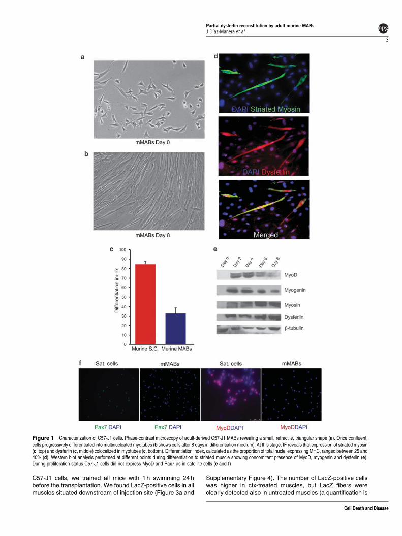
Characterization of adult murine-derived MABs (C57-J1).Adult-derived MABs were isolated from the tibialis anterior ofa 2-month-old C57BL/6 female mouse.17 When cultured inproliferation medium (PM), they showed a small, refractilemorphology and proliferated rapidly, with a doubling time ofapproximately 24h (Figure 1a). During proliferation, virtually allcells expressed at high level the surface markers Sca-1 andCD44 (Supplementary Figure 1) but, in contrast to SCs, they didnot express detectable levels of MyoD or Pax7 (Figure 1e and f).Unexpectedly, they did not express alkaline phosphatase (AP)(Supplementary Figure 2a) similar to their human counterparts.18
Once confluent, they progressively differentiated intostriated muscle (Figure 1b). On average, 25–40% of cellsfused into striated, myosin-positive, multinucleated myotubes(Figure 1c and d). Myogenic differentiation was not influencedby the substratum (Matrigel versus plastic: SupplementaryFigure 2d). The myogenic transcription factors MyoD andMyogenin were robustly expressed only after 48 h indifferentiation medium, at the onset of myotube formation,and underwent a progressive reduction in the expression levelafter day 4 (Figure 1e). The expression of dysferlin was clearlydetectable in myotubes concomitant with MyoD expression(Figure 1e). Its concentration increased progressively peakingup at days 6 and 8, concomitant with the maximum fusion ofmononucleated cells into myotubes (Figure 1c and e).
Despite their spontaneous myogenic differentiationpotency, C57-J1 cells were also able to differentiate in vitrointo other mesoderm tissues upon exposure to proper induc-tive signals, as previously described for other MAB lines. Aftertreatment with bone morphogenic protein-2 (BMP-2), theyexpressed AP whereas they expressed smooth muscle actinwhen treated with transforming growth factor-b (TGF-b);moreover, after 6 days of culture into adipogenic medium,rounded intracellular oil-red-positive vesicles were detected.(Supplementary Figures 2a–c).
Engraftment failure in A/J mice and generation of A/JSCID strain. To investigate the ability of adult-derived murineMABs to restore the expression of dysferlin in dystrophic
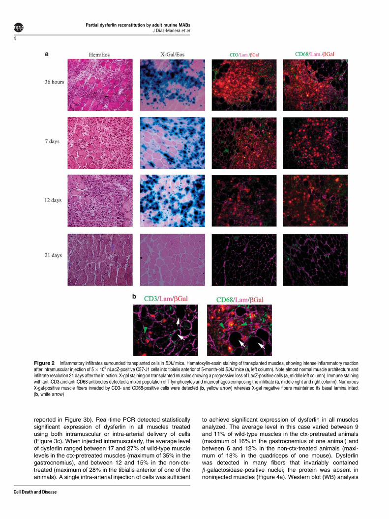
muscles, we injected 5� 105 C57-J1 cells, previously trans-duced with lentiviral vector expressing nuclear LacZ, into thetibialis anterior and quadriceps of 5-month-old Bl/AJ mice(n¼ 7) (generous gift from I Richard, Genethon). At 36 h afterthe injection, numerous nuclear LacZ (nLacZ)-positive cellswere detected in the muscles, but they appeared alreadysurrounded by an inflammatory infiltrate composed byneutrophils, CD68þ macrophages and CD3þ T lymphocytes(Figure 2a). The inflammatory infiltrate increased progressivelyuntil day 7, when numerous CD68þ cells were localizedin the injection area, but also in distant areas of the muscle andin the vessel walls (data not shown). By day 12 we detectedsome degenerating muscle fibers with LacZ-positive nucleisurrounded by CD68þ and CD3þ cells (Figure 2b). At3 weeks after the injection muscle regeneration wascompleted, probably at the expense of resident SCsbecause no LacZ-positive cells could be detected eitherinside or outside fibers, whereas some CD68 and CD3 cellsremained in the perymysium (Figure 2a). This result maydepend upon the altered immune response of Bl/AJ micecharacterized by a more aggressive phagocytic activity ofmacrophages and a reduced expression of decay-accele-rating factor (DAF) in muscles.19,20 Because of these results,we decided to generate a dystrophic, immune-deficient mouseby crossing Bl/AJ mice onto an SCID mice background (SCIDBL/AJ). The F4 generation used in this study showed absenceof mature T and B lymphocytes by FACS analyses (datanot shown) and the presence of retrotransposon insertionin dysferlin gene, as originally described in the A/J strain byHo et al.6 At 5 months of age, A/J SCID and Bl/AJ miceshowed dystrophic changes, such as central nuclei, fibersplitting, variation on fiber size and presence of regeneratingfibers (Supplementary Figure 3a). Azan Mallory stainingshowed a similar degree of fibrosis in both AJ/SCID andBl/AJ model compared to the control (Supplementary Figure 3a).As expected, immunofluorescence (IF) analysis and reversetranscription (RT)-PCR staining confirmed the absence ofdysferlin expression (Supplementary Figure 3b and c).
Restoration of dysferlin expression by injection ofC57-J1 cells into the new SCID/BlAJ line. We tested theability of C57-J1 cells to restore the expression of dysferlin inmuscles from the SCID/BlAJ mice. We performed a singleintramuscular injection of 5� 105 nLacZ labeled C57-J1 cellsin untreated or cardiotoxin (ctx)-pretreated tibialis anterior,quadriceps and gastrocnemius of 5-month-old SCID/BlAJmice (n¼ 6 mice, 3 pretreated with ctx). ctx was used toexacerbate the pathology of these mice that at 5 monthsshow a less severe phenotype than other strains such asthe a-sarcoglycan null mice, previously used for MAB trans-plantation.13 At 1 month after the injection, numerous nLacZ-positive fibers were detected in transverse muscle sectionsat different craniocaudal levels (Figure 3a); although nLacz-positive fibers were diffuse in the ctx-treated muscles,they were more concentrated near the injection area in theuntreated muscles. In parallel, we performed a single intra-arterial injection of 5� 105 nLacZ labeled C57-J1 cellsinto the right femoral artery of untreated and ctx-treated,5-month-old SCID/BlAJ mice (n¼ 10, 5 pretreated with ctx).To increase susceptibility of muscles to engraftment by
Partial dysferlin reconstitution by adult murine MABsJ Dıaz-Manera et al
2
Cell Death and Disease
C57-J1 cells, we trained all mice with 1 h swimming 24 hbefore the transplantation. We found LacZ-positive cells in allmuscles situated downstream of injection site (Figure 3a and
Supplementary Figure 4). The number of LacZ-positive cellswas higher in ctx-treated muscles, but LacZ fibers wereclearly detected also in untreated muscles (a quantification is
Figure 1 Characterization of C57-J1 cells. Phase-contrast microscopy of adult-derived C57-J1 MABs revealing a small, refractile, triangular shape (a). Once confluent,cells progressively differentiated into multinucleated myotubes (b shows cells after 8 days in differentiation medium). At this stage, IF reveals that expression of striated myosin(c, top) and dysferlin (c, middle) colocalized in myotubes (c, bottom). Differentiation index, calculated as the proportion of total nuclei expressing MHC, ranged between 25 and40% (d). Western blot analysis performed at different points during differentiation to striated muscle showing concomitant presence of MyoD, myogenin and dysferlin (e).During proliferation status C57-J1 cells did not express MyoD and Pax7 as in satellite cells (e and f)
Partial dysferlin reconstitution by adult murine MABsJ Dıaz-Manera et al
3
Cell Death and Disease
reported in Figure 3b). Real-time PCR detected statisticallysignificant expression of dysferlin in all muscles treatedusing both intramuscular or intra-arterial delivery of cells(Figure 3c). When injected intramuscularly, the average levelof dysferlin ranged between 17 and 27% of wild-type musclelevels in the ctx-pretreated muscles (maximum of 35% in thegastrocnemius), and between 12 and 15% in the non-ctx-treated (maximum of 28% in the tibialis anterior of one of theanimals). A single intra-arterial injection of cells was sufficient
to achieve significant expression of dysferlin in all musclesanalyzed. The average level in this case varied between 9and 11% of wild-type muscles in the ctx-pretreated animals(maximum of 16% in the gastrocnemius of one animal) andbetween 6 and 12% in the non-ctx-treated animals (maxi-mum of 18% in the quadriceps of one mouse). Dysferlinwas detected in many fibers that invariably containedb-galactosidase-positive nuclei; the protein was absent innoninjected muscles (Figure 4a). Western blot (WB) analysis
Figure 2 Inflammatory infiltrates surrounded transplanted cells in BlAJ mice. Hematoxylin-eosin staining of transplanted muscles, showing intense inflammatory reactionafter intramuscular injection of 5� 105 nLacZ-positive C57-J1 cells into tibialis anterior of 5-month-old BlAJ mice (a, left column). Note almost normal muscle architecture andinfiltrate resolution 21 days after the injection. X-gal staining on transplanted muscles showing a progressive loss of LacZ-positive cells (a, middle left column). Immune stainingwith anti-CD3 and anti-CD68 antibodies detected a mixed population of T lymphocytes and macrophages composing the infiltrate (a, middle right and right column). NumerousX-gal-positive muscle fibers invaded by CD3- and CD68-positive cells were detected (b, yellow arrow) whereas X-gal negative fibers maintained its basal lamina intact(b, white arrow)
Partial dysferlin reconstitution by adult murine MABsJ Dıaz-Manera et al
4
Cell Death and Disease
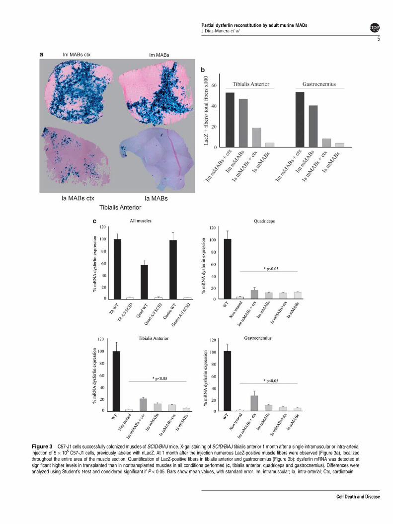
Figure 3 C57-J1 cells successfully colonized muscles of SCID/BlAJ mice. X-gal staining of SCID/BlAJ tibialis anterior 1 month after a single intramuscular or intra-arterialinjection of 5� 105 C57-J1 cells, previously labeled with nLacZ. At 1 month after the injection numerous LacZ-positive muscle fibers were observed (Figure 3a), localizedthroughout the entire area of the muscle section. Quantification of LacZ-positive fibers in tibialis anterior and gastrocnemius (Figure 3b): dysferlin mRNA was detected atsignificant higher levels in transplanted than in nontransplanted muscles in all conditions performed (c, tibialis anterior, quadriceps and gastrocnemius). Differences wereanalyzed using Student’s t-test and considered significant if Po0.05. Bars show mean values, with standard error. Im, intramuscular; Ia, intra-arterial; Ctx, cardiotoxin
Partial dysferlin reconstitution by adult murine MABsJ Dıaz-Manera et al
5
Cell Death and Disease
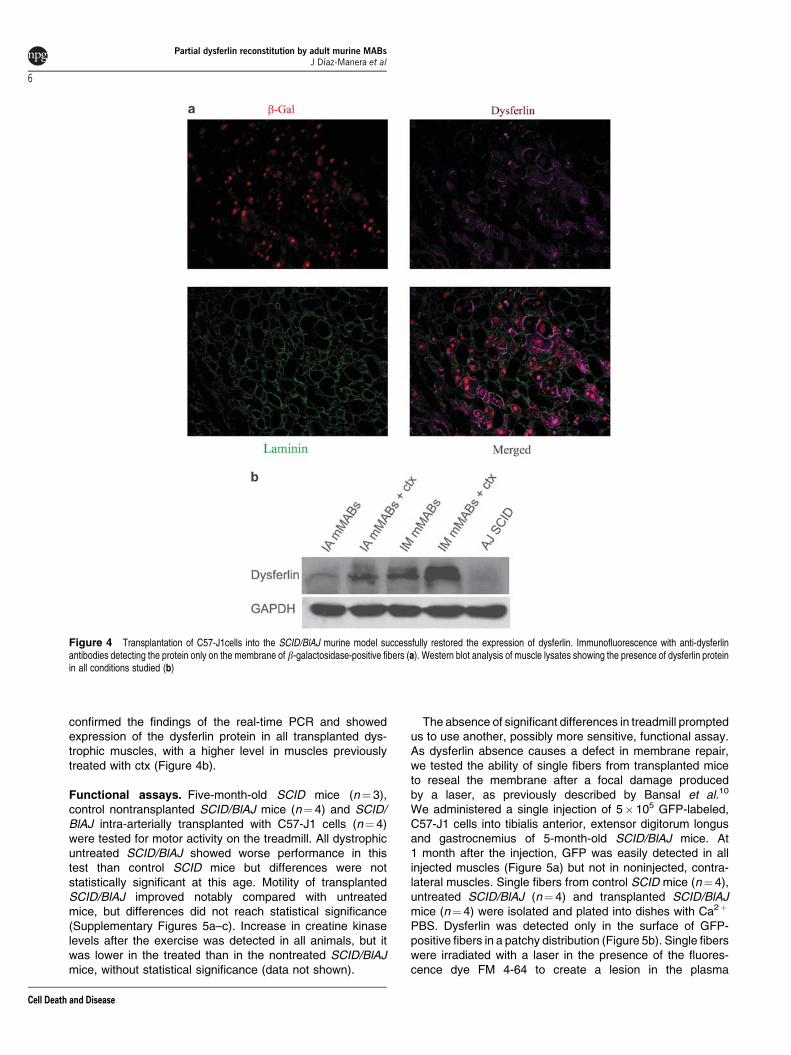
confirmed the findings of the real-time PCR and showedexpression of the dysferlin protein in all transplanted dys-trophic muscles, with a higher level in muscles previouslytreated with ctx (Figure 4b).
Functional assays. Five-month-old SCID mice (n¼ 3),control nontransplanted SCID/BlAJ mice (n¼ 4) and SCID/BlAJ intra-arterially transplanted with C57-J1 cells (n¼ 4)were tested for motor activity on the treadmill. All dystrophicuntreated SCID/BlAJ showed worse performance in thistest than control SCID mice but differences were notstatistically significant at this age. Motility of transplantedSCID/BlAJ improved notably compared with untreatedmice, but differences did not reach statistical significance(Supplementary Figures 5a–c). Increase in creatine kinaselevels after the exercise was detected in all animals, but itwas lower in the treated than in the nontreated SCID/BlAJmice, without statistical significance (data not shown).
The absence of significant differences in treadmill promptedus to use another, possibly more sensitive, functional assay.As dysferlin absence causes a defect in membrane repair,we tested the ability of single fibers from transplanted miceto reseal the membrane after a focal damage producedby a laser, as previously described by Bansal et al.10
We administered a single injection of 5� 105 GFP-labeled,C57-J1 cells into tibialis anterior, extensor digitorum longusand gastrocnemius of 5-month-old SCID/BlAJ mice. At1 month after the injection, GFP was easily detected in allinjected muscles (Figure 5a) but not in noninjected, contra-lateral muscles. Single fibers from control SCID mice (n¼ 4),untreated SCID/BlAJ (n¼ 4) and transplanted SCID/BlAJmice (n¼ 4) were isolated and plated into dishes with Ca2þ
PBS. Dysferlin was detected only in the surface of GFP-positive fibers in a patchy distribution (Figure 5b). Single fiberswere irradiated with a laser in the presence of the fluores-cence dye FM 4-64 to create a lesion in the plasma
Figure 4 Transplantation of C57-J1cells into the SCID/BlAJ murine model successfully restored the expression of dysferlin. Immunofluorescence with anti-dysferlinantibodies detecting the protein only on the membrane of b-galactosidase-positive fibers (a). Western blot analysis of muscle lysates showing the presence of dysferlin proteinin all conditions studied (b)
Partial dysferlin reconstitution by adult murine MABsJ Dıaz-Manera et al
6
Cell Death and Disease

membrane. In this experimental model, increase in thefluorescence signal in the area adjacent to the sarcolemmallesion inversely correlates with the resealing activity ofthe membrane; in fact, the increase in signal intensityrapidly stops in wild-type, dysferlin-positive fibers whereasit continues to augment in the dysferlin negative fibers.10
The increase in the signal was much lower in the MABs-transplanted fibers from the SCID/BlAJ mice than from thenontreated mice (Po0.0001) and, importantly, this increasewas not different from the one detected in single fibersfrom wild-type control mice (Figure 5c and d).
Discussion
New therapies for muscular dystrophies. Muscular dys-trophies still lack an efficacious therapy. In preclinicalmodels, cell transplantation, gene therapy, exon skippingand molecules inducing muscle hypertrophy all producedencouraging results but await the results of controlled clinicalexperimentation.
Our laboratory pioneered progenitor cell transplantation in amurine (a-sarcoglycan deficiency) and in a canine (dystrophindeficiency) model of muscular dystrophy. The success of the
transplantation depends upon a number of parameters amongwhich the histopathology of the transplanted muscle, thepresence of a resident myogenic population and the engraft-ment and myogenic potency of the donor cells appear themost important. For example, in the mdx mouse, which hasvigorous endogenous regeneration, embryonic D16 MABtransplantation produced modest engraftment and dystrophinproduction (M Sampaolesi and G Cossu, unpublished data). Itthus becomes very important to test the cell therapy protocolin other forms of muscular dystrophy, possibly using arobustly myogenic MAB population, given the fact that theD16 embryonic MABs have a modest intrinsic myogenicpotency.
Adult MABs have robust myogenic potency. To this aim,we have isolated and characterized MABs from skeletalmuscles of adult mice. Although these cells have the ability todifferentiate into diverse types of mesoderm cell types, theyshow a robust myogenic differentiation both in vitro and, mostimportantly, when injected intramuscularly or intra-arteriallyin dystrophic muscle. The MABs obtained from murine adultskeletal muscle biopsies are distinguishable from SCs asthey do not express CD34, MyoD or Pax7 during proli-feration, although they activate MyoD when myogenesis is
Figure 5 Functional assays. GFP expression pattern of dystrophic tibialis anterior of 5-month-old SCID/BlAJ 1 month after the intramuscular injection of 5� 105
GFP-positive C57-J1 cells (a). Immunofluorescence analysis showing dysferlin expression on the membrane of GFP-positive single fibers in a patchy distribution (b, whitearrows). Membrane repair assay was performed in isolated single fibers from WT, SCID (c, top panel), nontransplanted SCID/BlAJ (c, middle panel) and transplanted SCID/BlAJ (c, bottom panel) mice. Note a larger area of dye staining in the nontreated SCID/BlAJ fibers after laser-induced lesions (c). Measurement of fluorescence intensityversus time is shown in d for WT (blue line), nontransplanted SCID/BlAJ (red line) and transplanted SCID/BlAJ (green line) showing significant higher levels in thenontransplanted SCID/BlAJ. There were no differences between transplanted SCID/BlAJ and WT mice. Data are mean±S.E. (WT n:10 fibers, nontreated SCID/BlAJ n:15fibers, transplanted SCID/BlAJ n:20 fibers); statistical analysis was carried out with Student’s t-test and ANOVA test for repeated measurements. Po0.05 was consideredsignificant
Partial dysferlin reconstitution by adult murine MABsJ Dıaz-Manera et al
7
Cell Death and Disease
induced.21 Surprisingly, AP is not expressed by these cells inculture, at variance with human MABs. In murine muscles,AP is expressed in vivo by pericytes and by endothelial cells,but this expression is lost after serial passages in culture.Dysferlin was expressed by C57-J1 cells and also by human-derived MABs when differentiated into skeletal muscle(J Dıaz-Manera and G Cossu, unpublished data).
Adult MABs repair the dysferlinopathy in the mouse. Weshowed expression of dysferlin in b-galactosidase-positivefibers after nLacZ-positive MAB transplantation in the SCID/BlAJ mice, and because revertant fibers have not beenreported in the A/J mice, dysferlin detected by IF or WBshould have been produced from transplanted cells oncefused with dystrophic fibers. The A/J mutation results in adrastic reduction of dysferlin mRNA (o2–4% of wild-typemuscle). After transplantation, dysferlin mRNA was detectedin muscles at much higher levels, up to 35% of wild-typelevels after a single intramuscular injection, and 18% after asingle intra-arterial injection. It has been previously reportedthat the levels of dysferlin can be reduced in asymptomatic ormildly symptomatic carriers of the disease.22 WB of musclesfrom heterozygous familiars of affected patients showeddysferlin expression of 40–50% compared to control levels.23
Moreover, a recent analysis of the dysferlin protein levels inCD14þ monocytes from peripheral blood, which correlateswith levels in skeletal muscle, showed reduced expression in60% of asymptomatic carriers, than in some cases reachedup to 80% of reduction compared to normal controls.24 Thusa partial reconstitution of dysferlin, to the levels detected inthis study after a single injection, should be sufficient toreduce significantly or even eliminate symptoms in affectedpatients.
Although myoblast transplantation had previously resultedin dysferlin expression in the tibialis anterior of SJL dystrophicmice,25 no analysis of functional improvement was reportedand, moreover, myoblast transplantation is limited to topicintramuscular delivery. In our study, we observed functionaldifferences with the treadmill test, but they did not reachstatistical significance likely because of the modest differencebetween dystrophic and control mice in this specific model.To conclusively show a functional recovery in transplantedmice, we used and assay previously described by Bansalet al.10 that is based upon laser-induced membrane lesion.This method has recently been used to analyze responseto different gene therapy strategies and is very sensitivein detecting functional recovery of dysferlin null mice treatedwith gene or stem cell therapy.26 In our case, transplantationwas followed by normal resealing activity of isolated singlefibers from MAB-transplanted SCID/BlAJ mice, comparedto the nontransplanted fibers. These results, in agreementwith the reduced expression of dysferlin in asymptomaticcarriers, indicate that normal levels are not necessary toobtain a complete resealing activity of the plasma membrane.Dysferlin has not a structural role in the muscle fiber asdystrophin or sarcoglycans; its activity is related to repairingof the membrane and, thus, much as it happens with proteinsendowed with an enzymatic function in metabolic diseases,a partial restoration of expression would be sufficient for avirtually complete functional recovery.
The immune response obtained in the nonimmuno-suppressed Bl/AJ muscles after transplantation of C57-J1cells has been also described after myoblast transplantationin nonimmunosuppressed mice,27 and might be exacerbatedin this case by the expression of b-galactosidase by the cellsand by the altered immune system of the dysferlin-deficientmice. It is known that dysferlin-deficient monocytes andmacrophages show abnormally high phagocytic activity.20
Moreover, the expression of DAF (or CD55), a molecule thathas been related with suppression of complement activationand inhibition of the interaction of antigen-presenting cells withT lymphocytes has been shown to be reduced in dysferlin-deficient muscles.19 Accordingly to this, a lower expressionof DAF by grafts has been related with accelerated T-cell-mediated rejection in renal or heart transplants.28,29 There-fore, a permanent immune suppression would be necessary ina hypothetical transplantation of human MABs in patientsaffected by this form of muscular dystrophy.
Cell transplantation at an early stage of the disease. Tooptimize therapeutic results, we decided to treat dystrophicmice in a paucisymptomatic stage, before the presenceof abundant fibrous tissue or fatty infiltration of muscles.Our previous studies using MAB injection in advanced stagea-sarcoglycan mice showed poor results that improvedremarkably when host muscles were pretreated with cellssecreting metalloproteinase-9 and placenta-derived growthfactor.30 We selected 5-month-old SCID/BlAJ because thefirst dystrophic changes are already recognizable, but fattyinfiltration or fibrous tissue is not present yet. One of the mostimportant characteristics of MABs is their ability to cross thevessel walls and colonize affected muscles in response todifferent inflammatory signals.31 It has been reported thatdystrophic changes in the A/J muscles appear quite late inlife and remain confined to a minority of muscles, suggestingthat few inflammatory signals should be released.7 Never-theless, C57-J1 cells were able to cross the vessel wallsand massively colonize dystrophic muscles; this colonizationwas further enhanced by ctx-induced muscle damageand subsequent regeneration, even though this could notpossibly apply to a clinical situation. Nevertheless, the higheramount of nLacZ-positive fibers in ctx-pretreated musclessuggests that inflammatory signals and chemoattractantsare released at higher levels after an acute damage andconsequent inflammation. Interestingly, we observed similarresults with adult MABs in mdx dystrophic mice (G Messina,unpublished results). The ability to cross the vessel is ofoutstanding importance to design a therapeutic strategyin humans, as it would allow the distribution of donor cellsall over the body muscles after intra-arterial injection.32
Moreover, significant differences exist between skeletalmuscles from dysferlinopathy human patients and SCID/BlAJ mice. Strong inflammatory infiltrates,33 degeneratingand regenerating fibers34,35 are frequently recognized,especially during the first stage of the disease in humans,when some cases has been misdiagnosed as inflammatorymyopathies.36 This inflammatory activity would hypotheticallyenhance the migration of MABs from the vessel to muscles inpatients but an equilibrium should be found to prevent an
Partial dysferlin reconstitution by adult murine MABsJ Dıaz-Manera et al
8
Cell Death and Disease
exacerbated tissue reaction against donor cells even in thepresence of immune suppression.
In conclusion, we have shown that adult murine MABs,when transplanted into the SCID/BlAJ mice, partially restoredthe expression of dysferlin to levels that allowed a normalmembrane resealing activity of single fibers. These resultsmay lead to the first therapeutic assays in patients affected bymuscular dystrophies secondary to dysferlin deficiency.
Materials and MethodsIsolation of MABs from mouse muscular biopsies. Murine MABswere isolated from a 2-month-old C57BL/6 female as previously described.17
Briefly, both tibialis anterior were dissected and minced in 1–2 mm pieces.Fragments were selected, transferred onto collagen type I (Sigma, St. Louis, MO,USA) coated dishes and incubated in PM consisting of Dulbecco’s modified Eagle’smedium (DMEM) (Sigma) supplemented with 20% fetal bovine serum (Lonza,Verviers, Belgium), 2 mM L-glutamine (Sigma), 1 mM sodium pyruvate (Sigma),100 IU ml�1 penicillin and 100 mg ml�1 streptomycin (Sigma), at 371C, 5% CO2
and 5% O2. After 5–7 days, the mixed cell population deriving from primaryculture outgrowth was enzymatically dissociated by collagenase/dispase treatment.Cells obtained by dissociation were counted by Trypan blue exclusion, using ahemocytometer, and immediately used for cloning. The cell suspension was clonedin PM by limiting dilution in 96-multiwell dishes and incubated at 371C, 5% CO2 and5% O2. After 7–10 days first clones of cells were distinguishable. Once the cellscovered almost 50% of the well surface, the clones were passed and propagated inPM. The majority of clones adopted after a few passages a large, flat morphologyand underwent proliferative senescence. A few clones however maintained a small,refractile, poorly adhering morphology and continued to proliferate for at least40 passages. One of these clones, C57-J1 has been used for the experimentshere described.
In vitro differentiation assay. Spontaneous skeletal myogenic differentiationof C57-J1 cells was induced by plating 75� 103 cells onto reduced growthfactor Matrigel-coated dishes (Becton Dickinson Biosciences, San Jose, CA, USA).Differentiation medium consisted of DMEM supplemented with 2% horse serum(Sigma), 2 mM L-glutamine, 1 mM sodium pyruvate and 100 IU ml�1 penicillin and100 mg ml�1 streptomycin. Cultures were incubated at 371C, 5% CO2 for differentperiods and then processed for IF and WB analysis. The differentiation indexwas calculated as the ratio between nuclei expressing myosin heavy chain (MHC)and the total number of nuclei.
Differentiation in smooth muscle cells and osteoblasts was induced by treatmentwith TGF-b (Sigma) and BMP-2 (PeproTech, Rocky Hill, NJ, USA) respectively,and analyzed as previously described.21 To induce differentiation in adipose cells,we grew MABs in adipogenic-inducting medium (Lonza) for approximately 5 days,as described previously.17
Flow cytometry. C57-J1 cells in culture were analyzed by flow cytometryas previously reported.16 The antibodies used for this analysis were the following:anti-CD-44 FITC, anti-CD-34 FITC, anti-CD-45 PE, anti-CD 117-PE, anti-Flk-1-PE,anti-Sca-1-PE (all from Becton Dickinson Biosciences) and anti-CD 31 FITC(ID Laboratories, London, Canada).
Satellite cell cultures. SCs were isolated from skeletal muscles of 10-day-oldC57BL/6 pups. Muscle fragments were mechanically minced until a fine mixture wasobtained. This mixture was then digested with collagenase (Sigma) and dispase(Gibco, Paisley, UK) for 20 min at 371C with gentle agitation. After the incubation,isolated cells were collected and fragments were incubated again until the wholetissue was digested (usually three times). Isolated cells were pooled, centrifugedand resuspended in DMEM supplemented with 20% fetal bovine serum,2 mM L-glutamine, 1 mM sodium pyruvate, 100 IU ml�1 penicillin, 100 mg ml�1
streptomycin, 3% chicken embryo extract, 1% gentamicin and 50 nM basic FGF(PeproTech). Contamination by nonmyogenic cell was reduced by preplating thecell suspension onto plastic dishes where fibroblasts tend to adhere more rapidly.Differentiation was induced shifting the medium to DMEM supplemented with5% horse serum.
Generation of scid /blAJ mice. BlAJ mice (gift from I Richard, Genethon)were bred onto the scid/scid (severe combined immune deficient) strain,homozygous for the mutation located on chromosome 16. Resulting F1heterozygous siblings were intercrossed to obtain homozygotes for both loci:dysf and scid.
Cell transduction with lentiviral vectors. Cells were transduced, aspreviously described,14 with third-generation lentiviral vectors expressing nLacZ.
Intramuscular delivery of MABs. All animal studies presented in thispaper were approved by the San Raffaelle Institutional Review Board. Five-month-old BlAJ (n¼ 7) and SCID/BlAJ (n¼ 6) mice were used for intramuscular injectionof cells. When used, ctx was injected into both tibialis anterior, gastrocnemius(25ml, 100mM) and quadriceps (50 ml, 100mM), 24 h before the transplantation ofcells. Previous to the transplantation, mice were anesthetized with an intraperitonealinjection of Avertin, containing tert-amyl Alcohol (2.5 ml; Sigma) and 2,2,2-tribromoethanol (2.5 g; Sigma) in 100 ml 0.9% saline serum. Intramusculardelivery was performed by injection of approximately 5� 105 C57-J1 cells,previously transduced with lentiviral vector expressing nLacZ, into tibialis anterior,gastrocnemius and quadriceps using a 30 gauge needle. Animals were killed1 month after the transplantation, muscles were collected and the expression ofdysferlin was analyzed by IF, real-time PCR and WB.
Intra-arterial delivery of MABs. Five-month-old SCID/BlAJ mice (n¼ 10)were used for the intra-arterial delivery of the cells. All mice were trained by 1 hswimming 24 h before the transplantation. When used, ctx was injected into bothtibialis anterior, gastrocnemius (25 ml, 100mM) and quadriceps (50 ml, 100mM),just after the exercise. Previous to the transplantation, mice were anesthetizedwith Avertin (see receipt above). For intra-arterial delivery, approximately 5� 105
nLacZ C57-J1 cells were injected into the right femoral artery. A limited incision onthe medial upper side of the leg was performed; femoral artery was identified andcarefully isolated from femoral nerve and vein. Cells were injected by a 30 gaugeneedle inserted into the artery. After injection, body wall muscles and skinwere closed with sutures. Animals were killed 1 month after the injection, muscleswere collected and the expression of dysferlin was analyzed by IF, real-time PCRand WB.
Immunoblotting. WB analysis of cells and tissues was performed asdescribed.18 Antibodies used were mouse anti-dysferlin Hamlet (Novacastra,Newcastle upon Tyne, UK) at 1 : 100 dilution, mouse anti-MyoD (Dako, Carpentaria,CA, USA) at 1 : 100 dilution, mouse anti-myogenin (Hybridoma Bank, Iowa City, IA,USA) at 1 : 2 dilution, mouse anti-MHC (Hybridoma Bank) at 1 : 2 dilution, mouseanti-GAPDH at1 : 5000 (Sigma) and mouse anti-b-tubulin (Covance, Princeton,NJ, USA) at 1 : 5000 dilution.
Immunofluorescence. Cell cultures were washed three times with PBS andfixed with 4% paraformaldehyde at 41C for 10 min. Muscle samples were frozen inliquid nitrogen cooled isopentane and serial 8-mm-thick sections were cut with aLeica cryostat (Leica Microsystems GmbH, Wetzlar, Germany). Cells and tissuesections were processed for IF microscopy as previously described.12 Theantibodies used were mouse anti-dysferlin Hamlet (Novacastra) at 1 : 20 dilution,rabbit anti-dysferlin (GenWay, San Diego, CA, USA) at 1 : 50 dilution, mouse anti-MHC (Hybridoma Bank) at 1 : 2 dilution, mouse anti-Pax 7 (Hybridoma Bank) at 1 : 2dilution, mouse anti-MyoD (Dako) at 1 : 100 dilution, rabbit anti-GFP (ChemiconInternational, Temecula, CA, USA) at 1 : 300 dilution, rabbit anti-laminin (Sigma)at 1 : 300 dilution, goat anti-b-galactosidase (Biogenesis, Poole, UK) at 1 : 300, ratanti-CD68 (AbD Serotec, Raleigh, NC, USA) at 1 : 100 and rat anti-CD3 (Santa CruzBiotechnology, Santa Cruz, CA, USA) at 1 : 100. The staining for X-Gal wasperformed as previously described.37
Real-time PCR. Total RNA from muscles was isolated using TRIzol protocol(Invitrogen, Carlsbad, CA, USA). RNA (1mg) was reverse transcribed into cDNAusing the cDNA synthesis Thermoscript RT-PCR System kit (Invitrogen). Primersused for amplify cDNA were the following: dysferlin primer: forward 50-TCCGAAGCTAGA GGATCCAA-30; reverse 50-AGCTTCAATGGCAGCGTAGTT-30 andmurine cyclophilin: forward 50-CATACGGGTCCTGGCATCTTGTCC-30; reverse50-TGGTGATCTTCTTGCTGGTCTTGC-30. Each cDNA sample was amplified intriplicate using the SYBR Green supermix (Invitrogen). Student’s t-test was used
Partial dysferlin reconstitution by adult murine MABsJ Dıaz-Manera et al
9
Cell Death and Disease
to analyze the differences between muscles and conditions of treatment, settingPo0.05 as significant.
Exercise protocols. Control 6-month-old SCID mice (n¼ 3), nontransplantedSCID/BlAJ mice (n¼ 4) and SCID/BlAJ mice transplanted with C57-J1 cellsby intra-arterial injection (n¼ 4) were tested for functional recovery with thetreadmill test (Columbus Instruments, Columbus, OH, USA). Transplantedmice were tested at 21 days after the injection. Mice were adapted to theprocedure (10 min every other day; 3 m/min at 01 inclination) for 1 week beforebeginning the exercise training protocol. For the exercise test, mice were putinto a 101 inclined treadmill at 10 m/min, then the speed was increased by2 m/min every 2 min until the mouse reached exhaustion, which was definedas a prolonged spending time (10 s) on the shocker plate without attemptingto reengage the treadmill, or when a fixed speed of 46 m/min was reached.Total distance, running time and work was registered for every mouse. Work wascalculated as the body weight (kg), gravity (9.81 m/s2), vertical speed (m/s� angle)and time (s).
Creatine kinase blood levels were analyzed 5 days before the exercise test and6 h after the exercise following manufacturer’s instruction (CK-MB detection kit;Randox Laboratories, Crumlin, UK).
Membrane repair assay on skeletal muscle single fibers. Controldysferlin-positive SCID mice (n¼ 4), dystrophic, nontransplanted SCID/BlAJ (n¼ 4)mice and transplanted SCID/BlAJ mice (n¼ 4) were used for this experiment. SCID/BlAJ mice were used 1 month after the intramuscular injection of GFP-labeled C57-J1cells. Single fibers were extracted from gastrocnemius, extensor digitorum longus andtibialis anterior as previously described.38 Fibers were washed, resuspended inDulbecco’s PBS containing 1 mM Ca2þ (Invitrogen, Eugene, OR, USA) and mountedon glass bottom culture dishes (MatTek, Ashland, MA, USA) in the presence of 10mmof the fluorescent dye FM 4-64 (Invitrogen). To induce the damage, we irradiated a7� 7mm area of the membrane of muscle fibers with the lasers 488 at 50% of powerand 405 at 100% AOTF for 45 s. Images were captured every 30 s for 6 min after theirradiation. For every image taken, the fluorescence intensity variation at the site of thedamage was measured with the ImageJ imaging software (National Institute ofHealth, Bethesda, MD, USA; http://rsbweb.nih.gov/ij/index.html). ANOVA test forrepeated measurements and Student’s t-test for every time acquisition were used toidentify significant differences in the increase of light intensity. Po0.05 wasconsidered to be significant.
Conflict of Interest
The authors declare no conflict of interest.
Acknowledgements. This work was supported by a Scientific Fellowship fromthe European Federation of Neurological Societies, a grant from the SpanishMinistry of Health (BAE) and the special collaboration of Fundacion Isabel Gemio toJDM, and by grants from Telethon, AFM, Duchenne Parent Project, EC (Optistem,Angioscaff), ERC and the Italian Ministry of Research and Health to GC. We thankD Covarello for technical assistance; C Covino for support with the confocalmicroscope; P Pessina, S Brunelli, E Gallardo and N De Luna for critical commentson the paper and I Richard for the BlAJ mice.
1. Liu J, Aoki M, Illa I, Wu C, Fardeau M, Angelini C et al. Dysferlin, a novel skeletal musclegene, is mutated in Miyoshi myopathy and limb girdle muscular dystrophy. Nat Genet(1998); 20: 31–36.
2. Illa I, Serrano-Munuera C, Gallardo E, Lasa A, Rojas-Garcia R, Palmer J et al. Distalanterior compartment myopathy: a dysferlin mutation causing a new muscular dystrophyphenotype. Ann Neurol (2001); 49: 130–134.
3. Paradas C, Gonzalez-Quereda L, De Luna N, Gallardo E, Garcia-Consuegra I, Gomez Het al. A new phenotype of dysferlinopathy with congenital onset. Neuromuscul Disord(2009); 19: 21–25.
4. Bushby KM. Making sense of the limb-girdle muscular dystrophies. Brain (1999); 122(Pt 8): 1403–1420.
5. Zatz M, de Paula F, Starling A, Vainzof M. The 10 autosomal recessive limb-girdlemuscular dystrophies. Neuromuscul Disord (2003); 13: 532–544.
6. Ho M, Post CM, Donahue LR, Lidov HG, Bronson RT, Goolsby H et al. Disruption of musclemembrane and phenotype divergence in two novel mouse models of dysferlin deficiency.Hum Mol Genet (2004); 13: 1999–2010.
7. Kobayashi K, Izawa T, Kuwamura M, Yamate J. The distribution and characterization ofskeletal muscle lesions in dysferlin-deficient SJL and A/J mice. Exp Toxicol Pathol 2009,in press.
8. Bansal D, Campbell KP. Dysferlin and the plasma membrane repair in muscular dystrophy.Trends Cell Biol (2004); 14: 206–213.
9. Selcen D, Stilling G, Engel AG. The earliest pathologic alterations in dysferlinopathy.Neurology (2001); 56: 1472–1481.
10. Bansal D, Miyake K, Vogel SS, Groh S, Chen CC, Williamson R et al. Defectivemembrane repair in dysferlin-deficient muscular dystrophy. Nature (2003); 423:168–172.
11. Cai C, Weisleder N, Ko JK, Komazaki S, Sunada Y, Nishi M et al. Membrane repair defectsin muscular dystrophy are linked to altered interaction between MG53, caveolin-3, anddysferlin. J Biol Chem (2009); 284: 15894–15902.
12. Minasi MG, Riminucci M, De Angelis L, Borello U, Berarducci B, Innocenzi A et al.The meso-angioblast: a multipotent, self-renewing cell that originates from thedorsal aorta and differentiates into most mesodermal tissues. Development (2002); 129:2773–2783.
13. Cossu G, Sampaolesi M. New therapies for Duchenne muscular dystrophy: challenges,prospects and clinical trials. Trends Mol Med (2007); 13: 520–526.
14. Sampaolesi M, Torrente Y, Innocenzi A, Tonlorenzi R, D’Antona G, Pellegrino MA et al.Cell therapy of alpha-sarcoglycan null dystrophic mice through intra-arterial delivery ofmesoangioblasts. Science (2003); 301: 487–492.
15. Sampaolesi M, Blot S, D’Antona G, Granger N, Tonlorenzi R, Innocenzi A et al.Mesoangioblast stem cells ameliorate muscle function in dystrophic dogs. Nature (2006);444: 574–579.
16. Dellavalle A, Sampaolesi M, Tonlorenzi R, Tagliafico E, Sacchetti B, Perani L et al.Pericytes of human skeletal muscle are myogenic precursors distinct from satellite cells.Nat Cell Biol (2007); 9: 255–267.
17. Tonlorenzi R, Dellavalle A, Schnapp E, Cossu G, Sampaolesi M. Isolation andcharacterization of mesoangioblasts from mouse, dog, and human tissues. Curr ProtocStem Cell Biol 2007; (Supp 3), Chapter 2: Unit 2B 1.
18. Messina G, Blasi C, La Rocca SA, Pompili M, Calconi A, Grossi M. p27Kip1 actsdownstream of N-cadherin-mediated cell adhesion to promote myogenesis beyond cellcycle regulation. Mol Biol Cell (2005); 16: 1469–1480.
19. Wenzel K, Zabojszcza J, Carl M, Taubert S, Lass A, Harris CL et al. Increasedsusceptibility to complement attack due to down-regulation of decay-acceleratingfactor/CD55 in dysferlin-deficient muscular dystrophy. J Immunol (2005); 175:6219–6225.
20. Nagaraju K, Rawat R, Veszelovszky E, Thapliyal R, Kesari A, Sparks S et al. Dysferlindeficiency enhances monocyte phagocytosis: a model for the inflammatory onset of limb-girdle muscular dystrophy 2B. Am J Pathol (2008); 172: 774–785.
21. Messina G, Sirabella D, Monteverde S, Galvez BG, Tonlorenzi R, Schnapp E et al. Skeletalmuscle differentiation of embryonic mesoangioblasts requires pax3 activity. Stem Cells(2009); 27: 157–164.
22. Illa I, De Luna N, Dominguez-Perles R, Rojas-Garcia R, Paradas C, Palmer J et al.Symptomatic dysferlin gene mutation carriers: characterization of two cases. Neurology(2007); 68: 1284–1289.
23. Fanin M, Nascimbeni AC, Angelini C. Muscle protein analysis in the detection ofheterozygotes for recessive limb girdle muscular dystrophy type 2B and 2E. NeuromusculDisord (2006); 16: 792–799.
24. De Luna N, Gallardo E, Rojas-Garcia R, Dominguez-Perles R, Dıaz-Manera J,De La Torre C et al. Quantification of dysferlin in monocytes: a useful tool forthe detection of patients and carriers of dysferlinopathy. Neuromuscul Disord (2009);18: 790–791.
25. Leriche-Guerin K, Anderson LV, Wrogemann K, Roy B, Goulet M, Tremblay JP. Dysferlinexpression after normal myoblast transplantation in SCID and in SJL mice. NeuromusculDisord (2002); 12: 167–173.
26. Lostal W, Bartoli M, Bourg N, Roudaut C, Bentaib A, Miyake K et al. Efficient recovery ofdysferlin deficiency by dual adeno-associated vector-mediated gene transfer. Hum MolGenet (2010); 19: 1897–1907.
27. Skuk D, Tremblay JP. Progress in myoblast transplantation: a potential treatment ofdystrophies. Microsc Res Tech (2000); 48: 213–222.
28. Pavlov V, Raedler H, Yuan S, Leisman S, Kwan WH, Lalli PN et al. Donor deficiency ofdecay-accelerating factor accelerates murine T cell-mediated cardiac allograft rejection.J Immunol (2008); 181: 4580–4589.
29. Brodsky SV, Nadasdy GM, Pelletier R, Satoskar A, Birmingham DJ, Hadley GA et al.Expression of the decay-accelerating factor (CD55) in renal transplants – a possibleprediction marker of allograft survival. Transplantation (2009); 88: 457–464.
30. Gargioli C, Coletta M, De Grandis F, Cannata SM, Cossu G. PlGF-MMP-9-expressing cellsrestore microcirculation and efficacy of cell therapy in aged dystrophic muscle. Nat Med(2008); 14: 973–978.
31. Palumbo R, Sampaolesi M, De Marchis F, Tonlorenzi R, Colombetti S, Mondino A et al.Extracellular HMGB1, a signal of tissue damage, induces mesoangioblast migration andproliferation. J Cell Biol (2004); 164: 441–449.
32. Peault B, Rudnicki M, Torrente Y, Cossu G, Tremblay JP, Partridge T et al. Stem andprogenitor cells in skeletal muscle development, maintenance, and therapy. Mol Ther(2007); 15: 867–877.
Partial dysferlin reconstitution by adult murine MABsJ Dıaz-Manera et al
10
Cell Death and Disease

33. Gallardo E, Rojas-Garcia R, de Luna N, Pou A, Brown Jr RH, Illa I. Inflammation in dysferlin
myopathy: immunohistochemical characterization of 13 patients. Neurology (2001); 57:
2136–2138.34. Fanin M, Angelini C. Muscle pathology in dysferlin deficiency. Neuropathol Appl Neurobiol
(2002); 28: 461–470.35. Linssen WH, Notermans NC, Van der Graaf Y, Wokke JH, Van Doorn PA, Howeler CJ et al.
Miyoshi-type distal muscular dystrophy. Clinical spectrum in 24 Dutch patients. Brain
(1997); 120 (Pt 11): 1989–1996.36. Nguyen K, Bassez G, Krahn M, Bernard R, Laforet P, Labelle V et al. Phenotypic study
in 40 patients with dysferlin gene mutations: high frequency of atypical phenotypes.
Arch Neurol (2007); 64: 1176–1182.
37. Tajbakhsh S, Vivarelli E, Cusella-De Angelis G, Rocancourt D, Buckingham M, Cossu G.A population of myogenic cells derived from the mouse neural tube. Neuron (1994); 13: 813–821.
38. Rosenblatt JD, Lunt AI, Parry DJ, Partridge TA. Culturing satellite cells from living singlemuscle fiber explants. In Vitro Cell Dev Biol Anim (1995); 31: 773–779.
Cell Death and Disease is an open-access journalpublished by Nature Publishing Group. This work is
licensed under the Creative Commons Attribution-Noncommercial-NoDerivative Works 3.0 Unported License. To view a copy of this license,visit http://creativecommons.org/licenses/by-nc-nd/3.0/
Supplementary Information accompanies the paper on Cell Death and Disease website (http://www.nature.com/cddis)
Partial dysferlin reconstitution by adult murine MABsJ Dıaz-Manera et al
11
Cell Death and Disease
Copyright © 2022 FDOKUMEN