Metabolomic characterization of myocardial ischemia ... - Nature
Upload
independentCategory
view
3download
0
A Functional Murine Model of Hind Limb Demand Ischemia
Michael A. Peck, MD , Robert S. Crawford, MD , Christopher J. Abularrage, MD , Virendra I.Patel, MD , Mark F. Conrad, MD , Jin Hyung Yoo, MA , Michael T. Watkins, MD , and HassanAlbadawi, M.D. *
Department of Surgery, Division of Vascular and Endovascular Surgery, Massachusetts GeneralHospital, Harvard Medical School, Boston, MA 02114
Abstract
Introduction— To date murine models of treadmill exercise have been used to study generalexercise physiology and angiogenesis in ischemic hind limbs. The purpose of these experimentswas to develop a murine model of demand ischemia in an ischemic limb to mimic claudication inhumans. The primary goal was to determine whether treadmill exercise reflected a hemodynamicpicture which might be consistent with the hyperemic response observed in humans.
Methods— Aged hypercholesterolemic ApoE null mice ( ApoE−/−, n=13) were subjected toFemoral Artery Ligation (FAL), and allowed to recover from the acute ischemic response.Peripheral perfusion of the hind limbs at rest was determined by serial evaluation using laserDoppler imaging (LDI) on days 0, 7, and 14 following FAL. During the duration of theexperiments, the mice were also assessed on an established 5 point clinical ischemic score whichassessed the degree of digital amputation, necrosis, and cyanosis as compared to the non ischemiccontralateral limb. After stabilization of the LDI ratio (ischemic limb flux/contralateral nonischemic limb flux) and clinical ischemic score, mice underwent two days of treadmill training (10min @ 10 m/min, incline of 10°) followed by 60 minutes daily treadmill exercise (13 m/min,incline of 10°) through day 25. An evaluation of pre-exercise and post exercise perfusion usingLDI was performed on two separate occasions following the onset of daily exercise. During theimmediate 15 minute post exercise evaluation, LDI scanning was obtained in quadruplicate, toallow identification of peak flux ratios. Statistical analysis included unpaired t-tests and ANOVA.
Results— After FAL, the LDI Flux ratio reached a nadir between days one and two, thenstabilized by day 14 and remained stable through day 25. The clinical ischemic score stabilized atday 7, and remained stable throughout the rest of the experiment. Based on stabilization of boththe clinical ischemic score and LDI ratio, exercise training began on day 15. The peak 15 minutepost exercise LDI ratio increased significantly as compared to pre-exercise ratio on day 17(0.48+0.04 vs. 0.34± 0.04, p<0.05) and day 25 (0.37±0.03 vs. 0.27±0.03, p<0.01). Within 2 hoursof exercise, the LDI ratio returned to pre-exercise levels on both day 17 and 25.
Conclusions— Clinical and hemodynamic stabilization of limb perfusion is evident by 14 daysafter FAL. FAL followed by demand ischemia results in a reversible relative hyperemic responsesimilar to those observed in exercising human claudicants. A murine model of FAL associated
To Whom Correspondence is addressed: Hassan Albadawi, M.D., Division of Vascular and Endovascular Surgery, MassachusettsGeneral Hospital, 80 Blossom Street, Edwards 301, Boston, MA 02114, Phone: 617-724-7818, Fax: 617-726-2560,[email protected].
Publisher's Disclaimer: This is a PDF file of an unedited manuscript that has been accepted for publication. As a service to ourcustomers we are providing this early version of the manuscript. The manuscript will undergo copyediting, typesetting, and review ofthe resulting proof before it is published in its final citable form. Please note that during the production process errors may bediscovered which could affect the content, and all legal disclaimers that apply to the journal pertain.
NIH Public AccessAuthor ManuscriptAnn Vasc Surg. Author manuscript; available in PMC 2011 May 1.
Published in final edited form as:Ann Vasc Surg. 2010 May ; 24(4): 532–537. doi:10.1016/j.avsg.2009.12.003.
NIH
-PA
Author M
anuscriptN
IH-P
A A
uthor Manuscript
NIH
-PA
Author M
anuscript

with demand ischemia may be a useful model to evaluate the metabolic, inflammatory and flowrelated changes associated with claudication in humans.
Introduction
Studies of lower extremity claudication in humans have revealed alterations in blood flowand energy metabolism associated with exercise. These alterations are believed to be relatedto fundamental changes in the mitochondrial activity, chronic inflammation and intermittentlocal ischemia reperfusion (IR) injury1–4. Furthermore, there is evidence of significantcoincident coronary artery disease5–7 and cerebrovascular morbidity8, 9 in patients withclaudication. In recognition of this association between claudication and cardiovascularmortality the National Institute of Health has funded the CLEVER (Claudication: ExerciseVs. Endoluminal Revascularization) study, which is a prospective, multicenter, randomized,controlled clinical trial evaluating the relative efficacy, safety, and health economic impactof four treatment strategies for people with aortoiliac peripheral arterial disease andclaudication10. While there are many laboratory models developed to evaluate the effect ofischemia on muscle reperfusion injury, and angiogenesis, there is little literature to evaluatethe effect of hind limb demand ischemia, i.e. claudication on murine skeletal muscle. Thepurpose of these experiments was to develop a murine model of hind limb demand ischemiawhich mimics parameters of claudication in humans.
Methods
Animal Protocol
Experiments with ApoE null(ApoE−/−) mice were approved by the Massachusetts GeneralHospital’s subcommittee on research animal care in accordance with, the “Principles ofLaboratory Animal Care” (Guide for the Care and Use of Laboratory Animals, NationalInstitutes of Health Publication No. 86-23, Revised 1996). The ApoE−/− mice weresubjected to femoral artery ligation on the left and sham ischemia (femoral cut down but noarterial ligation) on the right. The mice were fed normal chow during the course of theseexperiments.
Hind limb Femoral Artery Ligation
8–10 month-old female ApoE−/− mice (n=13, Jackson Laboratory, Bar Harbor, Maine)were fed a normal diet. Mice were anesthetized by IP administration of 40–50mg/kgPentobarbital, and placed on a heating pad to maintain their core temperature at 37±0.5°C bymonitoring rectal temperature. A 0.5–1 cm skin incision was made longitudinally on theanterior thigh of the left hind limb. The femoral artery was exposed under a surgicalmicroscope and dissected from the femoral vein and nerve. The femoral artery and visiblebranches were ligated proximal to the superficial epigastric artery and above the bifurcationof the saphena and popliteal arteries with 8.0 silk ligatures (FAL). The arterial segmentbetween the ligatures was excised11. Incision was closed with a 5.0 polypropylene suture. Inthe right leg, there was a surgical incision and dissection but no ligation. The incision wasclosed with 5.0 polypropylene sutures. Mice received 0.05–0.1mg/kg Buprenorphine HCLsc for pain relief. Hind limb blood flow was assessed immediately before and after ligationusing Laser Doppler Imaging.
Laser Doppler Imaging
A laser Doppler imager (LDI, Moor Instruments, Wilmington, DE) was used to assess limbperfusion12, 13 by acquiring flow images of the ischemic and the contralateral foot at 37–38.0°C core temperature under Isoflurane anesthesia. Baseline LDI images were obtainedbefore and immediately after the femoral cut downs were completed in both limbs as
Peck et al. Page 2
Ann Vasc Surg. Author manuscript; available in PMC 2011 May 1.
NIH
-PA
Author M
anuscriptN
IH-P
A A
uthor Manuscript
NIH
-PA
Author M
anuscript

described above. The LDI source was mounted on a movable track that was fixed exactly 10cm above the mice limbs when the animals were restrained on a warming table. The laserbeam (780 nm), reflected from moving red blood cells in nutritional arterioles, capillariesand venules, was detected and processed to provide a computerized, color-coded image.Image analysis software (Laser Doppler Perfusion Measure, V3.08, Moor Instruments) wasused to calculate the limb mean flux units, which represents a quantitative analysis of tissueperfusion on a scale of 0 to 1000. Limb perfusion was expressed as the ratio of the Fluxvalue of the ischemic limb relative to the value of the contralateral limb (Flux Ratio).
Exercise Protocol
Aged hypercholesterolemic ApoE null mice (n=10) were subjected to Femoral ArteryLigation (FAL), and allowed to recover for 15 days. Peripheral perfusion of the hind limbsat rest was determined by serial evaluation using laser Doppler imaging (LDI) on days 0, 7,and 14 following FAL. After stabilization of the LDI ratio (ischemic limb flux/contralateralnon ischemic limb flux) at day 14, mice underwent two days of treadmill training (10 min at10 m/min, incline of 10°) followed by 60 minutes daily treadmill exercise (13 m/min, inclineof 10°) through day 26. On days 17 and 25 after FAL mice underwent pre-exercise, 15minute post exercise and 2 hours post exercise evaluation of the ischemic and contralaterallimb perfusion using LDI. During the immediate 15 minute post exercise evaluation, LDIscanning was obtained 4 times during this interval allowing measurement of the initial andpeak flux ratios.
Clinical Ischemic Score
A five point scoring system described by Stabile et al.14 were utilized to characterizeischemic tissue changes in the limb after FAL. Any evidence of post FAL digital amputationwas rated as 5. Frank tissue necrosis was scored as a 4. Mice with a clinical ischemic scoreof 4 or 5 within the course of the experiment were eliminated from subsequent DopplerImaging and exercise analysis. Severe cyanotic discoloration was scored as a 3 and milddiscoloration (pale appearance) was scored as 2. A completely normal appearance of thelimb as compared to the non FAL limb was rated 1. Mice were examined at day1immediately prior to ligation, 6 hours after ligation, then 2, 7,14,21 and 25 days afterligation.
Statistical Analysis
All data was expressed as mean ± SEM and all analyses were performed using InStat 3.0software (GraphPad Software, San Diego, CA). Comparisons along the time course of theLDI monitoring, the Clinical Score, and exercises was done with an ANOVA, and Tukey-Kramer post tests.
Results
Temporal Profile of Limb Perfusion after FAL
There was a significant difference over time in the LDI ratio after FAL (p<0.0001,ANOVA). There was a sharp immediate decrease in LDI ratio after FAL between days 1and 2 (1.0087±0.039 vs. 0.1862±0.0128, p<0.001), days 1 and 7 (1.0087±0.0128 vs.0.1651±0.0103. p<0.001) and 1 and 14 days (1.0087±0.0128 vs. 0.3429±0.0381, p<0.001).There was a stabilization of the LDI ratio between days 14 and 21 (0.3248±0.0381 vs.0.3248±0.0262, p>0.05) and days 21 and 25 (0.3248±0.0262 vs. 0.2646±0.0328, p>0.05).
Peck et al. Page 3
Ann Vasc Surg. Author manuscript; available in PMC 2011 May 1.
NIH
-PA
Author M
anuscriptN
IH-P
A A
uthor Manuscript
NIH
-PA
Author M
anuscript
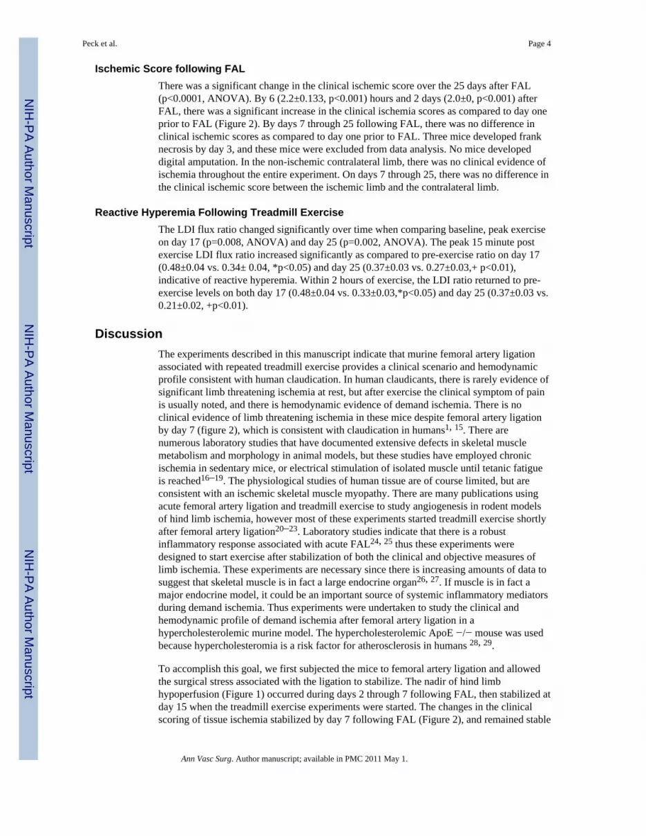
Ischemic Score following FAL
There was a significant change in the clinical ischemic score over the 25 days after FAL(p<0.0001, ANOVA). By 6 (2.2±0.133, p<0.001) hours and 2 days (2.0±0, p<0.001) afterFAL, there was a significant increase in the clinical ischemia scores as compared to day oneprior to FAL (Figure 2). By days 7 through 25 following FAL, there was no difference inclinical ischemic scores as compared to day one prior to FAL. Three mice developed franknecrosis by day 3, and these mice were excluded from data analysis. No mice developeddigital amputation. In the non-ischemic contralateral limb, there was no clinical evidence ofischemia throughout the entire experiment. On days 7 through 25, there was no difference inthe clinical ischemic score between the ischemic limb and the contralateral limb.
Reactive Hyperemia Following Treadmill Exercise
The LDI flux ratio changed significantly over time when comparing baseline, peak exerciseon day 17 (p=0.008, ANOVA) and day 25 (p=0.002, ANOVA). The peak 15 minute postexercise LDI flux ratio increased significantly as compared to pre-exercise ratio on day 17(0.48±0.04 vs. 0.34± 0.04, *p<0.05) and day 25 (0.37±0.03 vs. 0.27±0.03,+ p<0.01),indicative of reactive hyperemia. Within 2 hours of exercise, the LDI ratio returned to pre-exercise levels on both day 17 (0.48±0.04 vs. 0.33±0.03,*p<0.05) and day 25 (0.37±0.03 vs.0.21±0.02, +p<0.01).
Discussion
The experiments described in this manuscript indicate that murine femoral artery ligationassociated with repeated treadmill exercise provides a clinical scenario and hemodynamicprofile consistent with human claudication. In human claudicants, there is rarely evidence ofsignificant limb threatening ischemia at rest, but after exercise the clinical symptom of painis usually noted, and there is hemodynamic evidence of demand ischemia. There is noclinical evidence of limb threatening ischemia in these mice despite femoral artery ligationby day 7 (figure 2), which is consistent with claudication in humans1, 15. There arenumerous laboratory studies that have documented extensive defects in skeletal musclemetabolism and morphology in animal models, but these studies have employed chronicischemia in sedentary mice, or electrical stimulation of isolated muscle until tetanic fatigueis reached16–19. The physiological studies of human tissue are of course limited, but areconsistent with an ischemic skeletal muscle myopathy. There are many publications usingacute femoral artery ligation and treadmill exercise to study angiogenesis in rodent modelsof hind limb ischemia, however most of these experiments started treadmill exercise shortlyafter femoral artery ligation20–23. Laboratory studies indicate that there is a robustinflammatory response associated with acute FAL24, 25 thus these experiments weredesigned to start exercise after stabilization of both the clinical and objective measures oflimb ischemia. These experiments are necessary since there is increasing amounts of data tosuggest that skeletal muscle is in fact a large endocrine organ26, 27. If muscle is in fact amajor endocrine model, it could be an important source of systemic inflammatory mediatorsduring demand ischemia. Thus experiments were undertaken to study the clinical andhemodynamic profile of demand ischemia after femoral artery ligation in ahypercholesterolemic murine model. The hypercholesterolemic ApoE −/− mouse was usedbecause hypercholesteromia is a risk factor for atherosclerosis in humans 28, 29.
To accomplish this goal, we first subjected the mice to femoral artery ligation and allowedthe surgical stress associated with the ligation to stabilize. The nadir of hind limbhypoperfusion (Figure 1) occurred during days 2 through 7 following FAL, then stabilized atday 15 when the treadmill exercise experiments were started. The changes in the clinicalscoring of tissue ischemia stabilized by day 7 following FAL (Figure 2), and remained stable
Peck et al. Page 4
Ann Vasc Surg. Author manuscript; available in PMC 2011 May 1.
NIH
-PA
Author M
anuscriptN
IH-P
A A
uthor Manuscript
NIH
-PA
Author M
anuscript

during the entire 26 days of the experiments. The stabilization of the flow to a mouse hind-limbs subjected to FAL has been described by other investigators 11, 12, 25. The gradual postischemic stabilization of blood flow is thought to be related to increased collateral andcompensatory flow due to angiogenesis and arteriogenesis, which are known to be abnormalin the setting of hypercholesterolemia12, 30. Because of these factors, the treadmillexperiments began during an interval where objective and subjective measurement of theFAL limb function and perfusion was stable.
In the clinical scenario of patient care, documentation of significant resting ischemia can bemade by a combination of physical exam, imaging studies and non invasive testing. Inclaudicants, there is usually no evidence of significant resting ischemia thus the clinicalexam may be unreliable in documenting the presence or absence of ischemia which could becontributing to pain with ambulation. Most non invasive tests document a decreasedperfusion associated with treadmill exercise31. Laser Doppler imaging in humans32, 33 andanimals30, 34 subjected to demand ischemia demonstrate a hyperemic response associatedwith exercise. In humans with critical limb ischemia, the hyperemic response to exercise isabsent35. Thus experiments were performed to determine whether the FAL mice developeda hyperemic response to demand ischemia caused by treadmill exercise. As Figure 3 clearlyshows, on both day 17 and 25, the mice exhibited a vigorous hyperemic response totreadmill exercise. This is entirely consistent with demand ischemia. This response isequivalent to a decreased ankle brachial index or decreased PVR and is caused by extensivevasodilation. Based on this observation, ongoing biochemical and molecular analysis ofmoderately ischemic murine hind limb muscle subjected to sedentary and demand ischemiais needed to understand to what extent claudication might contribute to a systemicinflammatory response in humans.
Conclusion
In the setting of the known cardiovascular morbidity associated with claudication36 and limbischemia37, 38, a hemodynamically relevant model is needed to test pathologic muscledysfunction, regeneration, cytokine production, experimental therapeutics and the capacityfor muscle subjected to demand ischemia to affect other organ systems. This model is a stepin the direction needed to perform laboratory studies to address some of these issues.
AcknowledgmentsThis paper was presented as a “quick shot” oral presentation at the 4th Academic Surgical Congress in Fort Myers,Florida, February, 2009. The authors acknowledge funding from the National Institutes of Health(1R01AR055843), the Foundation for Advanced Vascular Research (Wylie Scholar Award), and the Division ofVascular and Endovascular Surgery, Massachusetts General Hospital (The Geneen Fund). Dr Watkins is the RonnieIsenberg Fellow in Academic Surgery at the Massachusetts General Hospital.
References
1. Hiatt WR, Regensteiner JG, Wolfel EE, Carry MR, Brass EP. Effect of exercise training on skeletalmuscle histology and metabolism in peripheral arterial disease. J Appl Physiol 1996;81(2):780–8.[PubMed: 8872646]
2. Bhat HK, Hiatt WR, Hoppel CL, Brass EP. Skeletal muscle mitochondrial DNA injury in patientswith unilateral peripheral arterial disease. Circulation 1999;99(6):807–12. [PubMed: 9989967]
3. Brass EP, Hiatt WR. Acquired skeletal muscle metabolic myopathy in atherosclerotic peripheralarterial disease. Vasc Med 2000;5(1):55–9. [PubMed: 10737157]
4. Brass EP, Hiatt WR, Gardner AW, Hoppel CL. Decreased NADH dehydrogenase and ubiquinol-cytochrome c oxidoreductase in peripheral arterial disease. Am J Physiol Heart Circ Physiol2001;280(2):H603–9. [PubMed: 11158957]
Peck et al. Page 5
Ann Vasc Surg. Author manuscript; available in PMC 2011 May 1.
NIH
-PA
Author M
anuscriptN
IH-P
A A
uthor Manuscript
NIH
-PA
Author M
anuscript

5. Criqui MH, Langer RD, Fronek A, Feigelson HS, Klauber MR, McCann TJ, et al. Mortality over aperiod of 10 years in patients with peripheral arterial disease. N Engl J Med 1992;326(6):381–6.[PubMed: 1729621]
6. Stoica A, Ginghina C. Cardiovascular risk in patients with peripheral vascular diseases. Rom JIntern Med 2008;46(4):275–83. [PubMed: 19480293]
7. Smith GD, Shipley MJ, Rose G. Intermittent claudication, heart disease risk factors, and mortality.The Whitehall Study. Circulation 1990;82(6):1925–31. [PubMed: 2242518]
8. Bowlin SJ, Medalie JH, Flocke SA, Zyzanski SJ, Yaari S, Goldbourt U. Intermittent claudication in8343 men and 21-year specific mortality follow-up. Ann Epidemiol 1997;7(3):180–7. [PubMed:9141640]
9. Manolio TA, Kronmal RA, Burke GL, O’Leary DH, Price TR. Short-term predictors of incidentstroke in older adults. The Cardiovascular Health Study. Stroke 1996;27(9):1479–86. [PubMed:8784116]
10. Murphy TP, Hirsch AT, Ricotta JJ, Cutlip DE, Mohler E, Regensteiner JG, et al. The Claudication:Exercise Vs. Endoluminal Revascularization (CLEVER) study: rationale and methods. J VascSurg 2008;47(6):1356–63. [PubMed: 18440181]
11. Shireman PK, Quinones MP. Differential necrosis despite similar perfusion in mouse strains afterischemia. J Surg Res 2005;129(2):242–50. [PubMed: 16051277]
12. Couffinhal T, Silver M, Zheng L, Witzenbichler B, Isner JM. Mouse Model of Angiogenesis. Am JPathol 1998;152:1667. [PubMed: 9626071]
13. Bonheur JA, Albadawi H, Patton GM, Watkins MT. A noninvasive murine model of hind limbischemia-reperfusion injury. J Surg Res 2004;116(1):55–63. [PubMed: 14732349]
14. Stabile E, Burnett MS, Watkins C, Kinnaird T, Bachis A, la Sala A, et al. Impaired arteriogenicresponse to acute hindlimb ischemia in CD4-knockout mice. Circulation 2003;108(2):205–10.[PubMed: 12821542]
15. Hiatt WR, Nehler MR. Peripheral arterial disease. Adv Intern Med 2001;47:89–110. [PubMed:11795081]
16. Makris KI, Nella AA, Zhu Z, Swanson SA, Casale GP, Gutti TL, et al. Mitochondriopathy ofperipheral arterial disease. Vascular 2007;15(6):336–43. [PubMed: 18053417]
17. Pipinos, Judge AR, Selsby JT, Zhu Z, Swanson SA, Nella AA, et al. The myopathy of peripheralarterial occlusive disease: part 1. Functional and histomorphological changes and evidence formitochondrial dysfunction. Vasc Endovascular Surg 2007;41(6):481–9. [PubMed: 18166628]
18. Pipinos, Judge AR, Selsby JT, Zhu Z, Swanson SA, Nella AA, et al. The myopathy of peripheralarterial occlusive disease: Part 2. Oxidative stress, neuropathy, and shift in muscle fiber type. VascEndovascular Surg 2008;42(2):101–12. [PubMed: 18390972]
19. Pipinos, Swanson SA, Zhu Z, Nella AA, Weiss DJ, Gutti TL, et al. Chronically ischemic mouseskeletal muscle exhibits myopathy in association with mitochondrial dysfunction and oxidativedamage. Am J Physiol Regul Integr Comp Physiol 2008;295(1):R290–6. [PubMed: 18480238]
20. Ikenaga S, Hamano K, Nishida M, Kobayashi T, Li TS, Kobayashi S, et al. Autologous bonemarrow implantation induced angiogenesis and improved deteriorated exercise capacity in a ratischemic hindlimb model. J Surg Res 2001;96(2):277–83. [PubMed: 11266284]
21. Lloyd PG, Yang HT, Terjung RL. Arteriogenesis and angiogenesis in rat ischemic hindlimb: roleof nitric oxide. Am J Physiol Heart Circ Physiol 2001;281(6):H2528–38. [PubMed: 11709420]
22. Olfert IM, Breen EC, Mathieu-Costello O, Wagner PD. Skeletal muscle capillarity and angiogenicmRNA levels after exercise training in normoxia and chronic hypoxia. J Appl Physiol 2001;91(3):1176–84. [PubMed: 11509513]
23. Lloyd PG, Prior BM, Yang HT, Terjung RL. Angiogenic growth factor expression in rat skeletalmuscle in response to exercise training. Am J Physiol Heart Circ Physiol 2003;284(5):H1668–78.[PubMed: 12543634]
24. Yang Y, Tang G, Yan J, Park B, Hoffman A, Tie G, et al. Cellular and molecular mechanismregulating blood flow recovery in acute versus gradual femoral artery occlusion are distinct in themouse. J Vasc Surg 2008;48(6):1546–58. [PubMed: 19118738]
Peck et al. Page 6
Ann Vasc Surg. Author manuscript; available in PMC 2011 May 1.
NIH
-PA
Author M
anuscriptN
IH-P
A A
uthor Manuscript
NIH
-PA
Author M
anuscript

25. Paoni NF, Peale F, Wang F, Errett-Baroncini C, Steinmetz H, Toy K, et al. Time course of skeletalmuscle repair and gene expression following acute hind limb ischemia in mice. Physiol Genomics2002;11(3):263–72. [PubMed: 12399448]
26. Pedersen BK, Edward F. Adolph Distinguished Lecture: Muscle as an Endocrine Organ: IL-6 andother myokines. J Appl Physiol. 2009
27. Scheele C, Nielsen S, Pedersen BK. ROS and myokines promote muscle adaptation to exercise.Trends Endocrinol Metab 2009;20(3):95–9. [PubMed: 19269849]
28. Morrison KM, Dyal L, Conner W, Helden E, Newkirk L, Yusuf S, et al. Cardiovascular riskfactors and non-invasive assessment of subclinical atherosclerosis in youth. Atherosclerosis. 2009
29. Drexel H, Aczel S, Marte T, Vonbank A, Saely CH. Factors predicting cardiovascular events instatin-treated diabetic and non-diabetic patients with coronary atherosclerosis. Atherosclerosis.2009
30. Tirziu D, Moodie KL, Zhuang ZW, Singer K, Helisch A, Dunn JF, et al. Delayed arteriogenesis inhypercholesterolemic mice. Circulation 2005;112(16):2501–9. [PubMed: 16230502]
31. Hiatt WR, Marshall JA, Baxter J, Sandoval R, Hildebrandt W, Kahn LR, et al. Diagnostic methodsfor peripheral arterial disease in the San Luis Valley Diabetes Study. J Clin Epidemiol 1990;43(6):597–606. [PubMed: 2189949]
32. Rossi M, Cupisti A, Perrone L, Mariani S, Santoro G. Acute effect of exercise-induced legischemia on cutaneous vasoreactivity in patients with stage II peripheral artery disease. MicrovascRes 2002;64(1):14–20. [PubMed: 12074626]
33. Ludyga T, Kuczmik WB, Kazibudzki M, Nowakowski P, Orawczyk T, Glanowski M, et al. Ankle-brachial pressure index estimated by laser Doppler in patients suffering from peripheral arterialobstructive disease. Ann Vasc Surg 2007;21(4):452–7. [PubMed: 17379477]
34. Corcoran HA, Smith BE, Mathers P, Pisacreta D, Hershey JC. Laser Doppler imaging of reactivehyperemia exposes blood flow deficits in a rat model of experimental limb ischemia. J CardiovascPharmacol 2009;53(6):446–51. [PubMed: 19433986]
35. Kvernebo K, Slasgsvold CE, Stranden E. Laser Doppler flowmetry in evaluation of skin post-ischaemic reactive hyperaemia. A study in healthy volunteers and atherosclerotic patients. JCardiovasc Surg (Torino) 1989;30(1):70–5.
36. Brass EP, Hiatt WR. Review of mortality and cardiovascular event rates in patients enrolled inclinical trials for claudication therapies. Vasc Med 2006;11(3):141–5. [PubMed: 17288119]
37. Flu HC, Lardenoye JH, Veen EJ, Aquarius AE, Van Berge Henegouwen DP, Hamming JF.Morbidity and mortality caused by cardiac adverse events after revascularization for critical limbischemia. Ann Vasc Surg 2009;23(5):583–97. [PubMed: 19747609]
38. Nguyen LL, Hevelone N, Rogers SO, Bandyk DF, Clowes AW, Moneta GL, et al. Disparity inoutcomes of surgical revascularization for limb salvage: race and gender are synergisticdeterminants of vein graft failure and limb loss. Circulation 2009;119(1):123–30. [PubMed:19103988]
Peck et al. Page 7
Ann Vasc Surg. Author manuscript; available in PMC 2011 May 1.
NIH
-PA
Author M
anuscriptN
IH-P
A A
uthor Manuscript
NIH
-PA
Author M
anuscript

Figure 1. LDI Flux Ratio following Femoral Artery LigationOn day one, the flux ratios were identical. By day 2, the LDI flux ratio fell significantlybetween days 2 through day 7. By day seven, the LDI increased significantly and remainedstable throughout the experiment.
Peck et al. Page 8
Ann Vasc Surg. Author manuscript; available in PMC 2011 May 1.
NIH
-PA
Author M
anuscriptN
IH-P
A A
uthor Manuscript
NIH
-PA
Author M
anuscript

Figure 2. Clinical Ischemia ScoreThere was evidence of significant ischemia in the FAL limb at 6 hours and day2 afterligation. By day 7, the clinical scores stabilized in the ischemic limb throughout the rest ofthe experimental intervals. In contrast, the clinical ischemic score in the non ischemiccontralateral limb was stable throughout the entire experiment.
Peck et al. Page 9
Ann Vasc Surg. Author manuscript; available in PMC 2011 May 1.
NIH
-PA
Author M
anuscriptN
IH-P
A A
uthor Manuscript
NIH
-PA
Author M
anuscript

Figure 3. Hyperemia Induced by Demand IschemiaOn days 17 and 25 the mice subjected to LDI prior to and after exercise showed evidence ofa significant hyperemic response which is characteristic of demand ischemia.
Peck et al. Page 10
Ann Vasc Surg. Author manuscript; available in PMC 2011 May 1.
NIH
-PA
Author M
anuscriptN
IH-P
A A
uthor Manuscript
NIH
-PA
Author M
anuscript
Copyright © 2022 FDOKUMEN