Arrest of mammalian fibroblasts in G1 in response to actin ...
11
The Journal of Cell Biology The Rockefeller University Press, 0021-9525/2003/04/67/11 $8.00 The Journal of Cell Biology, Volume 161, Number 1, April 14, 2003 67–77 http://www.jcb.org/cgi/doi/10.1083/jcb.200208140 JCB Article 67 Arrest of mammalian fibroblasts in G1 in response to actin inhibition is dependent on retinoblastoma pocket proteins but not on p53 Olivier D. Lohez, Caroline Reynaud, Franck Borel, Paul R. Andreassen, and Robert L. Margolis Institut de Biologie Structurale Jean Pierre Ebel (Commissariat à l'Energie Atomique–Centre National de la Recherche Scientifique–Université Joseph Fourier), 38027 Grenoble cedex 1, France 53 and the retinoblastoma (RB) pocket proteins are central to the control of progression through the G1 phase of the cell cycle. The RB pocket protein family is downstream of p53 and controls S-phase entry. Disruption of actin assembly arrests nontransformed mammalian fibro- blasts in G1. We show that this arrest requires intact RB pocket protein function, but surprisingly does not require p53. Thus, mammalian fibroblasts with normal pocket protein function reversibly arrest in G1 on exposure to actin inhibitors regardless of their p53 status. By contrast, pocket protein triple knockout mouse embryo fibroblasts and T antigen– transformed rat embryo fibroblasts lacking both p53 and p RB pocket protein function do not arrest in G1. Fibroblasts are very sensitive to actin inhibition in G1 and arrest at drug concentrations that do not affect cell adhesion or cell cleavage. Interestingly, G1 arrest is accompanied by inhibi- tion of surface ruffling and by induction of NF2/merlin. The combination of failure of G1 control and of tetraploid checkpoint control can cause RB pocket protein–suppressed cells to rapidly become aneuploid and die after exposure to actin inhibitors, whereas pocket protein–competent cells are spared. Our results thus establish that RB pocket proteins can be uniquely targeted for tumor chemotherapy. Introduction Both p53 and the retinoblastoma (RB)* pocket protein family are central to the control of G1 progression in mammalian cells. Between them, they can impose G1 arrest in response to a multitude of stresses or signals, including DNA damage (Kastan et al., 1991; Di Leonardo et al., 1994), polyploidization (Cross et al., 1995; Minn et al., 1996; Lanni and Jacks, 1998; Andreassen et al., 2001b), depletion of nucleotide pools (Linke et al., 1996), and TGF- (Polyak et al., 1994a; Zhang et al., 1999). During G1 progression, p53 mediates cell cycle arrest by transactivating specific targets. Among these, the Cdk inhibitor (CKI) p21 WAF1 acts to arrest the cell cycle by inhibiting Cdk2/cyclin E and Cdk2/cyclin A (Dulic et al., 1994). Suppression of Cdk activity blocks G1 progression by regulating the phosphorylation status of the RB pocket proteins (RB, p107, and p130), tumor suppressors of central importance to the control of S-phase entry (Weinberg, 1995; Mittnacht, 1998; Mulligan and Jacks, 1998). Pocket proteins are substrates for phosphorylation by G1 Cdks, and once fully phosphorylated, they release E2F transcription factors that permit progression from G1 to S phase (Dyson, 1998). The clear link between p53- and RB-dependent sup- pression of the cell cycle in G1 is underlined by the fact that abrogation of either RB pocket protein function or p53 function permits the cell cycle to continue after DNA damage (Levine, 1997; Dannenberg et al., 2000; Sage et al., 2000). RB pocket proteins appear to act downstream of p53 because a normal p53 and p21 WAF1 response occurs in cells deficient for RB pocket protein function (Dannenberg et al., 2000; Sage et al., 2000). In addition, p53 and the RB pocket protein family cooperate in mediating G1 arrest in response to polyploidiza- tion or exposure to inhibitors of mitotic spindle assembly (Di Leonardo et al., 1997; Andreassen et al., 2001b; Borel et al., 2002b). In contrast, as exemplified by G1 arrest of fibro- blasts in response to the growth factor TGF-, RB pocket proteins can act independently of p53 (Zhang et al., 1999). In addition to the intersection of the p53 and RB path- ways, pocket protein suppression of G1 progression can be Address correspondence to Robert L. Margolis, Institut de Biologie Structurale, 41 rue Jules Horowitz, 38027 Grenoble cedex 1, France. Tel.: (33) 4-38-78-9616. Fax: (33) 4-38-78-5494. E-mail: [email protected] P.R. Andreassen’s present address is Dana-Farber Cancer Institute, M640, 44 Binney St., Boston, MA 02115. *Abbreviations used in this paper: ARF, alternative reading frame; CKI, Cdk inhibitor; DCB, dihydrocytochalasin B; ERK, extracellular signal– regulated kinase; HU, hydroxyurea; MEF, mouse embryo fibroblast; RB, retinoblastoma; REF, rat embryo fibroblast. Key words: aneuploidy; tetraploidy; cytoskeleton; dihydrocytochalasin B; cell cycle
-
Upload
khangminh22 -
Category
Documents
-
view
4 -
download
0
Transcript of Arrest of mammalian fibroblasts in G1 in response to actin ...
The
Jour
nal o
f Cel
l Bio
logy
The Rockefeller University Press, 0021-9525/2003/04/67/11 $8.00The Journal of Cell Biology, Volume 161, Number 1, April 14, 2003 67–77http://www.jcb.org/cgi/doi/10.1083/jcb.200208140
JCB
Article
67
Arrest of mammalian fibroblasts in G1 in response to actin inhibition is dependent on retinoblastoma pocket proteins but not on p53
Olivier D. Lohez, Caroline Reynaud, Franck Borel, Paul R. Andreassen, and Robert L. Margolis
Institut de Biologie Structurale Jean Pierre Ebel (Commissariat à l'Energie Atomique–Centre National de la Recherche Scientifique–Université Joseph Fourier), 38027 Grenoble cedex 1, France
53 and the retinoblastoma (RB) pocket proteins arecentral to the control of progression through the G1phase of the cell cycle. The RB pocket protein family
is downstream of p53 and controls S-phase entry. Disruptionof actin assembly arrests nontransformed mammalian fibro-blasts in G1. We show that this arrest requires intact RBpocket protein function, but surprisingly does not requirep53. Thus, mammalian fibroblasts with normal pocket proteinfunction reversibly arrest in G1 on exposure to actin inhibitorsregardless of their p53 status. By contrast, pocket proteintriple knockout mouse embryo fibroblasts and T antigen–transformed rat embryo fibroblasts lacking both p53 and
p
RB pocket protein function do not arrest in G1. Fibroblastsare very sensitive to actin inhibition in G1 and arrest atdrug concentrations that do not affect cell adhesion or cellcleavage. Interestingly, G1 arrest is accompanied by inhibi-tion of surface ruffling and by induction of NF2/merlin. Thecombination of failure of G1 control and of tetraploidcheckpoint control can cause RB pocket protein–suppressedcells to rapidly become aneuploid and die after exposureto actin inhibitors, whereas pocket protein–competent cellsare spared. Our results thus establish that RB pocket proteinscan be uniquely targeted for tumor chemotherapy.
Introduction
Both p53 and the retinoblastoma (RB)* pocket protein familyare central to the control of G1 progression in mammaliancells. Between them, they can impose G1 arrest in responseto a multitude of stresses or signals, including DNA damage(Kastan et al., 1991; Di Leonardo et al., 1994), polyploidization(Cross et al., 1995; Minn et al., 1996; Lanni and Jacks,1998; Andreassen et al., 2001b), depletion of nucleotidepools (Linke et al., 1996), and TGF-
�
(Polyak et al., 1994a;Zhang et al., 1999). During G1 progression, p53 mediatescell cycle arrest by transactivating specific targets. Amongthese, the Cdk inhibitor (CKI) p21
WAF1
acts to arrest the cellcycle by inhibiting Cdk2/cyclin E and Cdk2/cyclin A (Dulicet al., 1994). Suppression of Cdk activity blocks G1 progression
by regulating the phosphorylation status of the RB pocketproteins (RB, p107, and p130), tumor suppressors of centralimportance to the control of S-phase entry (Weinberg,1995; Mittnacht, 1998; Mulligan and Jacks, 1998). Pocketproteins are substrates for phosphorylation by G1 Cdks, andonce fully phosphorylated, they release E2F transcriptionfactors that permit progression from G1 to S phase (Dyson,1998). The clear link between p53- and RB-dependent sup-pression of the cell cycle in G1 is underlined by the fact thatabrogation of either RB pocket protein function or p53function permits the cell cycle to continue after DNA damage(Levine, 1997; Dannenberg et al., 2000; Sage et al., 2000).RB pocket proteins appear to act downstream of p53 becausea normal p53 and p21
WAF1
response occurs in cells deficientfor RB pocket protein function (Dannenberg et al., 2000; Sageet al., 2000). In addition, p53 and the RB pocket protein familycooperate in mediating G1 arrest in response to polyploidiza-tion or exposure to inhibitors of mitotic spindle assembly (DiLeonardo et al., 1997; Andreassen et al., 2001b; Borel et al.,2002b). In contrast, as exemplified by G1 arrest of fibro-blasts in response to the growth factor TGF-
�
, RB pocketproteins can act independently of p53 (Zhang et al., 1999).
In addition to the intersection of the p53 and RB path-ways, pocket protein suppression of G1 progression can be
Address correspondence to Robert L. Margolis, Institut de BiologieStructurale, 41 rue Jules Horowitz, 38027 Grenoble cedex 1, France.Tel.: (33) 4-38-78-9616. Fax: (33) 4-38-78-5494. E-mail: [email protected]
P.R. Andreassen’s present address is Dana-Farber Cancer Institute,M640, 44 Binney St., Boston, MA 02115.*Abbreviations used in this paper: ARF, alternative reading frame; CKI,Cdk inhibitor; DCB, dihydrocytochalasin B; ERK, extracellular signal–regulated kinase; HU, hydroxyurea; MEF, mouse embryo fibroblast; RB,retinoblastoma; REF, rat embryo fibroblast.Key words: aneuploidy; tetraploidy; cytoskeleton; dihydrocytochalasin B;cell cycle
The
Jour
nal o
f Cel
l Bio
logy
68 The Journal of Cell Biology
|
Volume 161, Number 1, 2003
activated by CKIs, such as p27
Kip1
and p16
INK4A
, that are in-dependent of p53 control (Sherr and Roberts, 1995, 1999).Given that the RB pocket protein pathway is believed to bedefective in nearly all human tumors (Weinberg, 1995;Sherr, 1996; Hanahan and Weinberg, 2000), whereas p53mutations are present in approximately half of human tu-mors (Vogelstein et al., 2000), it is important to distinguishRB-dependent suppression of the cell cycle that is clearly in-dependent of the p53 pathway. The recent development ofmouse embryo fibroblast (MEF) cells with a knockout for allthree RB pocket proteins (RB, p107, and p130) in a back-ground of normal p53 response now permits dissection ofsuch pocket protein–specific events.
Among the metabolic signals that the cell reads to enableG1 progression is the status of the cytoskeleton. We have es-tablished that stabilization of the microtubule array by taxol,a drug inhibitor of microtubule dynamics, arrests nontrans-formed fibroblasts in G1 in a p53-dependent manner (Tri-elli et al., 1996; unpublished data). In addition, numerousstudies have shown that inhibition of actin assembly bydrugs of the cytochalasin family also suppresses G1 progres-sion (Maness and Walsh, 1982; Iwig et al., 1995; Bohmer etal., 1996; Huang et al., 1998; Reshetnikova et al., 2000),and that arrest is abrogated by SV-40 transformation (Ma-ness and Walsh, 1982).
As the SV-40 T antigen suppresses both p53 and RBpocket protein function (Bargonetti et al., 1992; Zalvide etal., 1998), we have here addressed whether the actin inhibi-tion–dependent suppression of G1 progression is due top53- or RB pocket protein–specific signaling events. Forthis, we have used mammalian cells containing either spe-cific suppression of p53 or triple knockout of the RB pocketproteins. We have also tested MEFs with a knockout of RBalone or knockout of the alternative reading frame (ARF) tu-mor suppressor that regulates p53 accumulation (Sherr andWeber, 2000).
Strikingly, we demonstrate here that suppression of G1progression in response to dihydrocytochalasin B (DCB) ex-posure is dependent on the function of the RB pocket pro-tein family, but not on p53 status. Moreover, RB pocketprotein–deficient cells also fail to undergo tetraploid check-point arrest at concentrations of DCB that induce cleavagefailure (Andreassen et al., 2001b; Borel et al., 2002b). As aresult, RB pocket protein–deficient cells become highlyaneuploid after transient DCB exposure and rapidly die,whereas p53-deficient cells efficiently arrest in G1. Ourwork thus demonstrates that the RB pocket protein controlpathway has a unique capacity to control G1 progression af-ter actin disruption that is clearly independent of the p53pathway. Because there are functions specific to RB pocketproteins that are independent of p53, our results show itshould be possible to selectively kill cells with RB pathwaydefects. This may be important for therapeutic consider-ations, given the nearly universal alterations of function ofthe RB family pathway in tumor cells.
In addition, we have found in this work that pocket pro-tein–competent G1 cells are extraordinarily sensitive to actininhibitors, arresting at concentrations that do not affect sub-strate adhesion, cell spreading, or cell cleavage, but leave theactin array largely undisturbed. This was a surprising result,
as the effect of actin assembly inhibition on G1 progressionhad been linked to loss of cytoskeletal integrity and substrateadhesion (Bohmer et al., 1996; Assoian and Zhu, 1997;Reshetnikova et al., 2000). The question arises concerningthe control system in G1 that might be directly affected byactin status. We have found that cell surface ruffling is spe-cifically suppressed in cells arrested with low DCB concen-trations. As the actin-associated ERM (ezrin, radixin, andmoesin) protein NF2/merlin has been implicated in bothcell surface ruffling (Bashour et al., 2002) and in suppressionof cell cycle progression in G1 (Shaw et al., 1998; Morrisonet al., 2001), we tested its status in arrested cells and foundthat NF2/merlin is up-regulated in DCB-induced G1 arrest.This result suggests that subtle cues from the actin cytoskele-ton induce NF2/merlin-dependent arrest.
Results
DCB reversibly arrests mammalian fibroblasts in G1 at concentrations that do not suppress cleavage
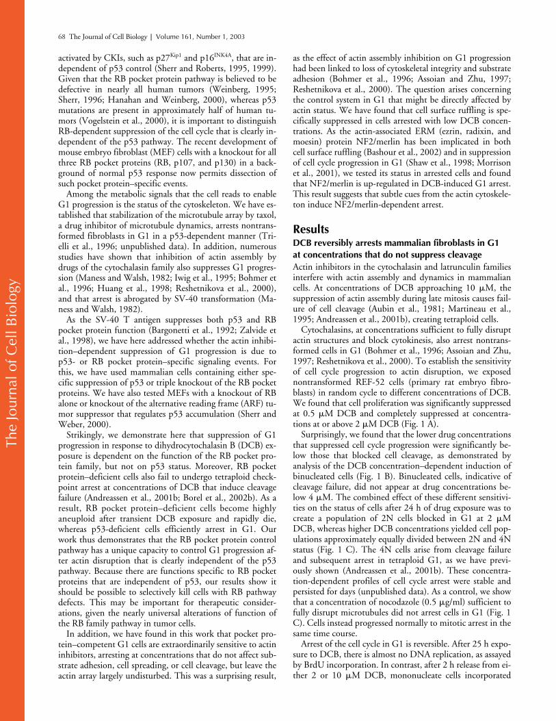
Actin inhibitors in the cytochalasin and latrunculin familiesinterfere with actin assembly and dynamics in mammaliancells. At concentrations of DCB approaching 10
�
M, thesuppression of actin assembly during late mitosis causes fail-ure of cell cleavage (Aubin et al., 1981; Martineau et al.,1995; Andreassen et al., 2001b), creating tetraploid cells.
Cytochalasins, at concentrations sufficient to fully disruptactin structures and block cytokinesis, also arrest nontrans-formed cells in G1 (Bohmer et al., 1996; Assoian and Zhu,1997; Reshetnikova et al., 2000). To establish the sensitivityof cell cycle progression to actin disruption, we exposednontransformed REF-52 cells (primary rat embryo fibro-blasts) in random cycle to different concentrations of DCB.We found that cell proliferation was significantly suppressedat 0.5
�
M DCB and completely suppressed at concentra-tions at or above 2
�
M DCB (Fig. 1 A).Surprisingly, we found that the lower drug concentrations
that suppressed cell cycle progression were significantly be-low those that blocked cell cleavage, as demonstrated byanalysis of the DCB concentration–dependent induction ofbinucleated cells (Fig. 1 B). Binucleated cells, indicative ofcleavage failure, did not appear at drug concentrations be-low 4
�
M. The combined effect of these different sensitivi-ties on the status of cells after 24 h of drug exposure was tocreate a population of 2N cells blocked in G1 at 2
�
MDCB, whereas higher DCB concentrations yielded cell pop-ulations approximately equally divided between 2N and 4Nstatus (Fig. 1 C). The 4N cells arise from cleavage failureand subsequent arrest in tetraploid G1, as we have previ-ously shown (Andreassen et al., 2001b). These concentra-tion-dependent profiles of cell cycle arrest were stable andpersisted for days (unpublished data). As a control, we showthat a concentration of nocodazole (0.5
�
g/ml) sufficient tofully disrupt microtubules did not arrest cells in G1 (Fig. 1C). Cells instead progressed normally to mitotic arrest in thesame time course.
Arrest of the cell cycle in G1 is reversible. After 25 h expo-sure to DCB, there is almost no DNA replication, as assayedby BrdU incorporation. In contrast, after 2 h release from ei-ther 2 or 10
�
M DCB, mononucleate cells incorporated
The
Jour
nal o
f Cel
l Bio
logy
Actin-dependent G1 arrest requires RB but not p53 |
Lohez et al. 69
BrdU at levels comparable to controls that had not been ex-posed to DCB (Fig. 1 D). Reentry into the cell cycle is re-stricted to the mononucleate cells. As we have previouslyshown (Andreassen et al., 2001b), nontransformed cells donot recover from tetraploid G1 arrest. Thus, mononucleatecells recover from 25 h exposure to 10
�
M DCB and incor-porate BrdU, whereas binucleate cells do not (Fig. 1 E).
Arrest of randomly cycling cells in G1 by low concentra-tions of DCB is not restricted to REF-52 cells. Human non-transformed IMR-90 fibroblasts show the same concentra-tion-dependent sensitivity to DCB and arrest in 2N G1 whenexposed to 2
�
M DCB, but arrest with approximately equal2N and 4N status at higher DCB concentrations (Fig. 2 A). Itis important to note that the arrest in 2N G1 can be distin-guished from the arrest of tetraploid cells with 4N DNA con-tent after DCB exposure (Andreassen et al., 2001b).
Loss of p53 function does not affect G1 arrest
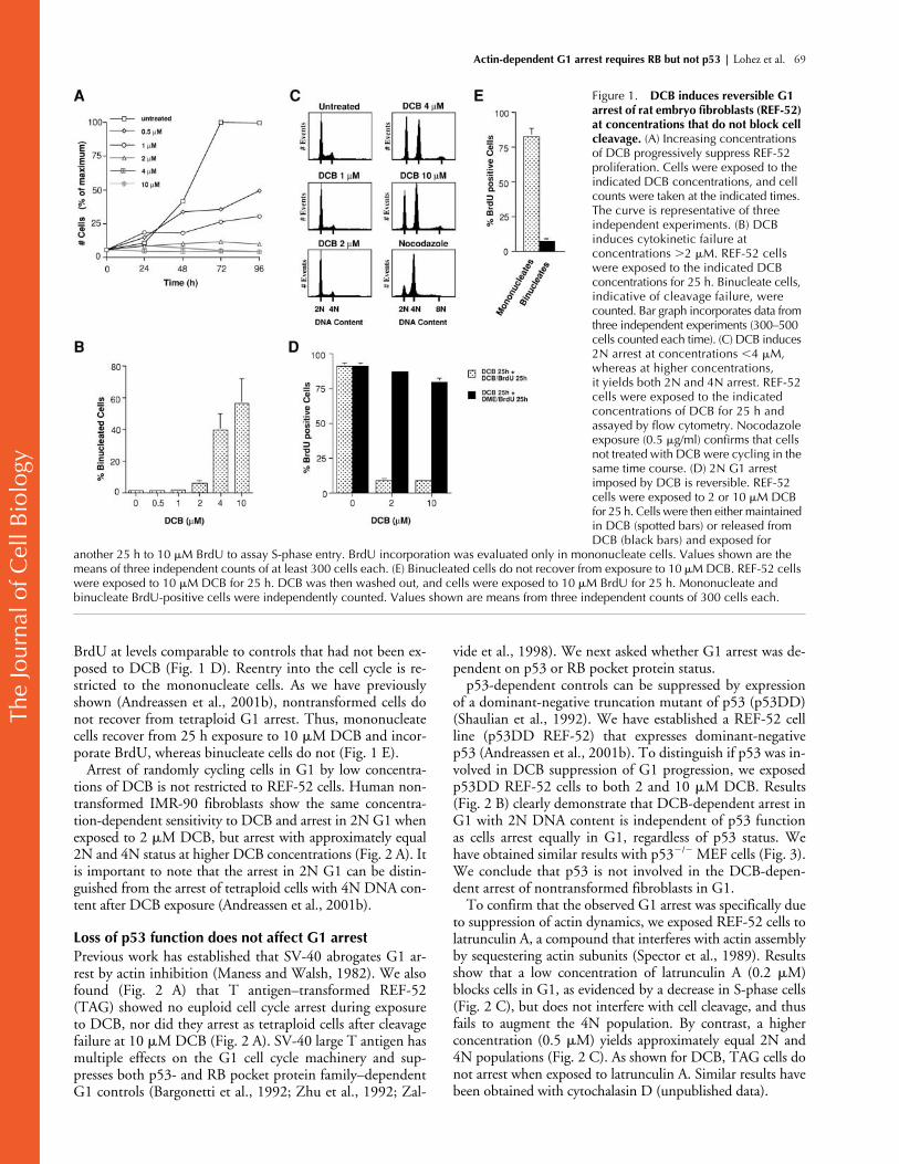
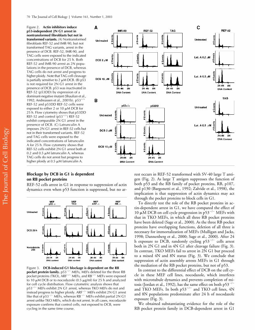
Previous work has established that SV-40 abrogates G1 ar-rest by actin inhibition (Maness and Walsh, 1982). We alsofound (Fig. 2 A) that T antigen–transformed REF-52(TAG) showed no euploid cell cycle arrest during exposureto DCB, nor did they arrest as tetraploid cells after cleavagefailure at 10
�
M DCB (Fig. 2 A). SV-40 large T antigen hasmultiple effects on the G1 cell cycle machinery and sup-presses both p53- and RB pocket protein family–dependentG1 controls (Bargonetti et al., 1992; Zhu et al., 1992; Zal-
vide et al., 1998). We next asked whether G1 arrest was de-pendent on p53 or RB pocket protein status.
p53-dependent controls can be suppressed by expressionof a dominant-negative truncation mutant of p53 (p53DD)(Shaulian et al., 1992). We have established a REF-52 cellline (p53DD REF-52) that expresses dominant-negativep53 (Andreassen et al., 2001b). To distinguish if p53 was in-volved in DCB suppression of G1 progression, we exposedp53DD REF-52 cells to both 2 and 10
�
M DCB. Results(Fig. 2 B) clearly demonstrate that DCB-dependent arrest inG1 with 2N DNA content is independent of p53 functionas cells arrest equally in G1, regardless of p53 status. Wehave obtained similar results with p53
�
/
�
MEF cells (Fig. 3).We conclude that p53 is not involved in the DCB-depen-dent arrest of nontransformed fibroblasts in G1.
To confirm that the observed G1 arrest was specifically dueto suppression of actin dynamics, we exposed REF-52 cells tolatrunculin A, a compound that interferes with actin assemblyby sequestering actin subunits (Spector et al., 1989). Resultsshow that a low concentration of latrunculin A (0.2
�
M)blocks cells in G1, as evidenced by a decrease in S-phase cells(Fig. 2 C), but does not interfere with cell cleavage, and thusfails to augment the 4N population. By contrast, a higherconcentration (0.5
�
M) yields approximately equal 2N and4N populations (Fig. 2 C). As shown for DCB, TAG cells donot arrest when exposed to latrunculin A. Similar results havebeen obtained with cytochalasin D (unpublished data).
Figure 1. DCB induces reversible G1 arrest of rat embryo fibroblasts (REF-52) at concentrations that do not block cell cleavage. (A) Increasing concentrations of DCB progressively suppress REF-52 proliferation. Cells were exposed to the indicated DCB concentrations, and cell counts were taken at the indicated times. The curve is representative of three independent experiments. (B) DCB induces cytokinetic failure at concentrations �2 �M. REF-52 cells were exposed to the indicated DCB concentrations for 25 h. Binucleate cells, indicative of cleavage failure, were counted. Bar graph incorporates data from three independent experiments (300–500 cells counted each time). (C) DCB induces 2N arrest at concentrations �4 �M, whereas at higher concentrations, it yields both 2N and 4N arrest. REF-52 cells were exposed to the indicated concentrations of DCB for 25 h and assayed by flow cytometry. Nocodazole exposure (0.5 �g/ml) confirms that cells not treated with DCB were cycling in the same time course. (D) 2N G1 arrest imposed by DCB is reversible. REF-52 cells were exposed to 2 or 10 �M DCB for 25 h. Cells were then either maintained in DCB (spotted bars) or released from DCB (black bars) and exposed for
another 25 h to 10 �M BrdU to assay S-phase entry. BrdU incorporation was evaluated only in mononucleate cells. Values shown are the means of three independent counts of at least 300 cells each. (E) Binucleated cells do not recover from exposure to 10 �M DCB. REF-52 cells were exposed to 10 �M DCB for 25 h. DCB was then washed out, and cells were exposed to 10 �M BrdU for 25 h. Mononucleate and binucleate BrdU-positive cells were independently counted. Values shown are means from three independent counts of 300 cells each.
The
Jour
nal o
f Cel
l Bio
logy
70 The Journal of Cell Biology
|
Volume 161, Number 1, 2003
Blockage by DCB in G1 is dependent on RB pocket proteins
REF-52 cells arrest in G1 in response to suppression of actindynamics even when p53 function is suppressed, but no ar-
rest occurs in REF-52 transformed with SV-40 large T anti-gen (Fig. 2). As large T antigen suppresses the function ofboth p53 and the RB family of pocket proteins, RB, p107,and p130 (Bargonetti et al., 1992; Zalvide et al., 1998), theimplication is that suppression of actin dynamics may actthrough the pocket proteins to block cells in G1.
To directly test the role of the RB pocket proteins in ac-tin-dependent arrest in G1, we have compared the effect of10
�
M DCB on cell cycle progression in p53
�
/
�
MEFs withthat in TKO MEFs, in which all three RB pocket proteinshave been deleted (Sage et al., 2000). As the three RB pocketproteins have overlapping functions, deletion of all three isnecessary for immortalization of MEFs (Mulligan and Jacks,1998; Dannenberg et al., 2000; Sage et al., 2000). After 24h exposure to DCB, randomly cycling p53
�
/
�
cells arrestboth in 2N G1 and in 4N G1 after cleavage failure (Fig. 3).In contrast, TKO MEFs fail to arrest in 2N G1 but proceedto a mixed 4N and 8N status (Fig. 3). We conclude thatsuppression of actin assembly arrests MEFs in G1 throughthe mediation of the RB pocket proteins, but not of p53.
In contrast to the differential effect of DCB on the cell cy-cle in these MEF cell lines, nocodazole, which interfereswith microtubule dynamics and prevents completion of mi-tosis (Jordan et al., 1992), has the same effect on both p53
�
/
�
and TKO MEFs. In both p53
�
/
�
and TKO cell lines, 4Nand 8N populations predominate after 24 h of nocodazoleexposure (Fig. 3).
We obtained substantiating evidence for the role of theRB pocket protein family in DCB-dependent arrest in G1
Figure 2. Actin inhibitors induce p53-independent 2N G1 arrest in nontransformed fibroblasts but not in transformed variants. (A) Nontransformed fibroblasts REF-52 and IMR-90, but not transformed TAG variants, arrest in the presence of DCB. REF-52, IMR-90, and TAG cells were exposed to the indicated concentrations of DCB for 25 h. Both REF-52 and IMR-90 arrest as 2N popu-lations in the presence of DCB, whereas TAG cells do not arrest and progress to higher ploidy. Note that TAG cell cleavage is partially sensitive to 2 �M DCB. (B) p53 is not required for 2N G1 arrest in the presence of DCB. p53 was inactivated in REF-52 (p53DD) by expression of a dominant-negative mutant (Shaulian et al., 1992; Andreassen et al., 2001b). p53�/� REF-52 and p53DD REF-52 cells were exposed to either 2 or 10 �M DCB for 25 h. Flow cytometry shows that p53DD REF-52 and control (p53�/�) REF-52 exhibit comparable 2N G1 arrest in the presence of DCB. (C) Latrunculin A imposes 2N G1 arrest in REF-52 cells but not in their transformed variants. REF-52 and TAG cells were exposed to the indicated concentrations of latrunculin A for 25 h. Flow cytometry shows that REF-52 cells exhibit 2N G1 arrest both at 0.2 and 0.5 �M latrunculin A, whereas TAG cells do not arrest but progress to higher ploidy at 0.5 �M latrunculin A.
Figure 3. DCB-induced G1 blockage is dependent on the RB pocket protein family. p53�/� MEFs, MEFs deleted for the three RB pocket proteins (TKO), ARF�/� MEFs, and RB�/� MEFs were exposed to 10 �M DCB or to nocodazole (0.5 �g/ml) for 25 h and analyzed for cell cycle distribution. Flow cytometric analysis shows that p53�/� MEFs exhibit 2N G1 arrest, whereas TKO MEFs do not and instead progress to higher ploidy. ARF�/� MEFs exhibit 2N G1 arrest like that of p53�/� MEFs, whereas RB�/� MEFs exhibit partial 2N G1 arrest unlike TKO MEFs, which do not arrest. In all cases, nocodazole exposure confirms that control cells, not exposed to DCB, were cycling in the same time course.

The
Jour
nal o
f Cel
l Bio
logy
Actin-dependent G1 arrest requires RB but not p53 |
Lohez et al. 71
by also examining the response of RB
�
/
�
and ARF
�
/
�
MEFs. Knockout of RB alone does not immortalize cells orblock contact inhibition arrest (Dannenberg et al., 2000;Sage et al., 2000), as the RB protein family has overlappingand redundant functions. RB
�
/
�
cells, however, showed apartial loss of arrest in G1 in response to DCB (Fig. 3). ARFstabilizes p53 and is required for p53-dependent cell cyclearrest, but is not required for the RB pocket protein controlin G1 (Sherr and Weber, 2000). As expected for a proteinthat stabilizes p53 but does not influence the RB pathway,knockout of ARF had no effect on the capacity of MEFs toarrest in G1 when exposed to DCB (Fig. 3).
G1-arrested fibroblasts remain adherent but suppress ruffling and up-regulate NF2/merlin in 2
�
M DCB
There is substantial evidence that the capacity of nontrans-formed fibroblasts to progress in the cell cycle is coupled to sub-stratum adherence (Stoker et al., 1968; Assoian, 1997; Assoianand Schwartz, 2001). However, even at 10
�
M DCB, we havefound that cells that are no longer capable of undergoing cleav-age remain adherent (unpublished data). At 2
�
M DCB, thereis little overt evidence of disturbance of the actin cytoskeleton inrandomly cycling interphase cells and no evidence of distur-bance of adhesion (Fig. 4). Compared with controls, cells retainabundant actin cables, and focal adhesion plaques appear intact,as assayed by both vinculin antibody (Fig. 4 A) and antiphos-photyrosine antibody (Fig. 4 B). One notable change during ar-rest is that, in comparison to controls, cells exposed to 2
�
MDCB have smooth margins and do not appear to undergo ruf-fling (Fig. 4 C). The absence of ruffling is rapidly reversible onrelease from DCB, and ruffling is equally evident at 30 min anda full day after DCB release (Fig. 4 C).
The absence of ruffling (Fig. 4) and the RB pocket proteindependence of DCB G1 arrest induced by DCB (Fig. 3) sug-gested a possible involvement of NF2/merlin as a mediator ofDCB-dependent arrest. Knockout of the RB pocket proteinfamily abolishes contact inhibition–dependent arrest (Dan-nenberg et al., 2000; Sage et al., 2000). The tumor suppressorNF2/merlin, an actin-binding protein (Gautreau et al., 2002)that suppresses cell surface ruffling (Bashour et al., 2002), isspecifically up-regulated during, and is required for, contactinhibition arrest in G1 (Shaw et al., 1998; Morrison et al.,2001). We therefore assayed for the status of NF2/merlin inG1 arrest induced by 2
�
M DCB. Strikingly, we found thatNF2/merlin is strongly up-regulated and dephosphorylated inDCB-dependent arrest in comparison to randomly cyclingcells (Fig. 4 D). This result suggests that NF2/merlin up-reg-ulation may mediate the actin-dependent arrest.
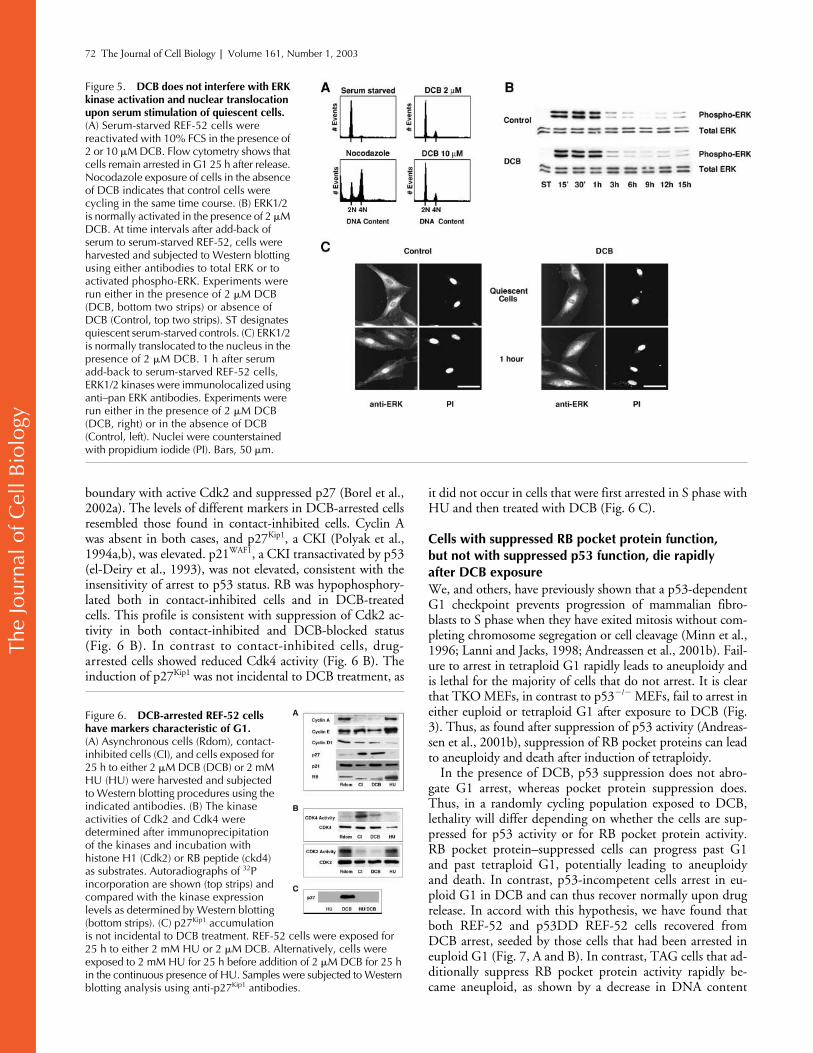
Adherence to the substratum is intact in 2
�
M DCB-treated cells by the criterion of the activation of extracellularsignal–regulated kinase (ERK) on serum stimulation in thepresence of 2
�
M DCB. Fibroblast cell lines maintained insuspension fail to activate the ERK pathway in response togrowth factor stimulation (Lin et al., 1997; Renshaw et al.,1997; Aplin et al., 1999). REF-52 cells arrest in G0 after se-rum starvation, and 2
�
M or 10
�
M DCB, but not nocoda-zole, inhibits progression past G1 upon serum add-back(Fig. 5 A). In accord with previous findings (Reshetnikova etal., 2000), we have found that REF-52 cells released from
G0 serum starvation in the presence of low concentrationsof DCB showed time-dependent ERK activation after serumadd-back that is indistinguishable from controls (Fig. 5 B).Further, after serum stimulation, ERK migrates to the nucleiof DCB-blocked cells (Fig. 5 C).
Our results indicating up-regulation of NF2/merlin duringDCB-induced arrest and the requirement for RB proteinsboth suggested that arrest would be in G1. Further, Bohmeret al. (1996) found, in timed experiments with synchronouscells, that arrest of fibroblasts with high concentrations of cy-tochalasin occurred in mid to late G1. To assess the physio-logical status of randomly cycling cells arrested in G1 by 25 hexposure to 2
�
M DCB, we assayed for the presence and ac-tivity of different cell cycle markers, compared with the pres-ence of these markers in contact-inhibited cells in G0/G1, incontrol cycling cells, or in cycling cells arrested in S phase(Fig. 6). S-phase cells were obtained by exposure for 25 h to 2mM hydroxyurea (HU), which blocks cells at the G1/S
Figure 4. Actin stress fibers and substratum adherence are not perturbed but NF2/merlin is up-regulated after exposure of REF-52 cells to 2 �M DCB. REF-52 cells were exposed to 2 �M DCB for 25 h. Cells were then analyzed for actin distribution with TRITC-phalloidin (A and B, left), and vinculin (A, right) and phosphotyrosine (B, right) distribution were determined by immunolabeling. For each pair (untreated/DCB) in both A and B, the intensity settings used for confocal microscopy were kept constant. Bars, 40 �m. (C) Higher magnification images show that ruffling was suppressed in cells exposed to 2 �M DCB for 25 h (DCB), but reappeared rapidly (arrowheads) in cells released from DCB (30 min) and persisted at 25 h of release. Cells were imaged for actin with TRITC-phalloidin. Bar, 40 �m. (D) NF2/merlin is up-regulated and dephosphorylated in DCB-arrested cells. REF-52 cells, either random cycling (Rdom), contact inhibited (CI), or random cycling while exposed to 2 �M DCB for 25 h (DCB), were harvested and subjected to Western blotting procedures using anti-NF2/merlin antibodies. An equivalent amount of protein was loaded for each sample.
The
Jour
nal o
f Cel
l Bio
logy
72 The Journal of Cell Biology
|
Volume 161, Number 1, 2003
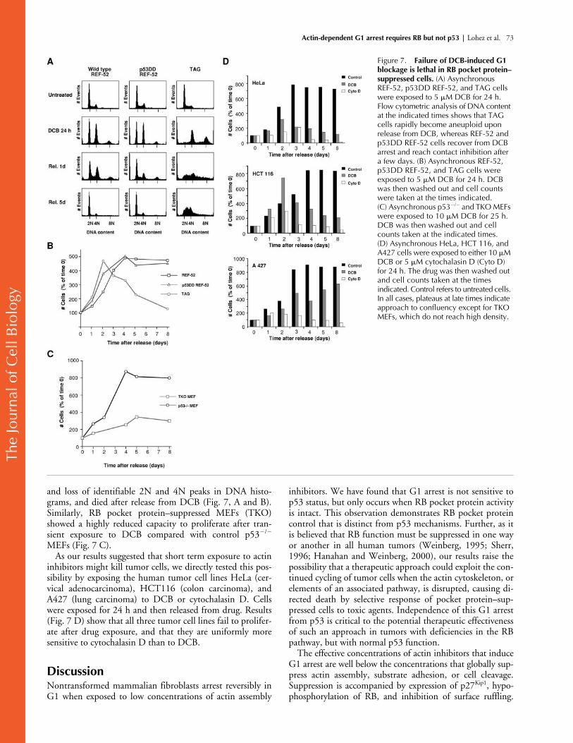
boundary with active Cdk2 and suppressed p27 (Borel et al.,2002a). The levels of different markers in DCB-arrested cellsresembled those found in contact-inhibited cells. Cyclin Awas absent in both cases, and p27
Kip1
, a CKI (Polyak et al.,1994a,b), was elevated. p21
WAF1
, a CKI transactivated by p53(el-Deiry et al., 1993), was not elevated, consistent with theinsensitivity of arrest to p53 status. RB was hypophosphory-lated both in contact-inhibited cells and in DCB-treatedcells. This profile is consistent with suppression of Cdk2 ac-tivity in both contact-inhibited and DCB-blocked status(Fig. 6 B). In contrast to contact-inhibited cells, drug-arrested cells showed reduced Cdk4 activity (Fig. 6 B). Theinduction of p27
Kip1
was not incidental to DCB treatment, as
it did not occur in cells that were first arrested in S phase withHU and then treated with DCB (Fig. 6 C).
Cells with suppressed RB pocket protein function, but not with suppressed p53 function, die rapidly after DCB exposure
We, and others, have previously shown that a p53-dependentG1 checkpoint prevents progression of mammalian fibro-blasts to S phase when they have exited mitosis without com-pleting chromosome segregation or cell cleavage (Minn et al.,1996; Lanni and Jacks, 1998; Andreassen et al., 2001b). Fail-ure to arrest in tetraploid G1 rapidly leads to aneuploidy andis lethal for the majority of cells that do not arrest. It is clearthat TKO MEFs, in contrast to p53
�
/
�
MEFs, fail to arrest ineither euploid or tetraploid G1 after exposure to DCB (Fig.3). Thus, as found after suppression of p53 activity (Andreas-sen et al., 2001b), suppression of RB pocket proteins can leadto aneuploidy and death after induction of tetraploidy.
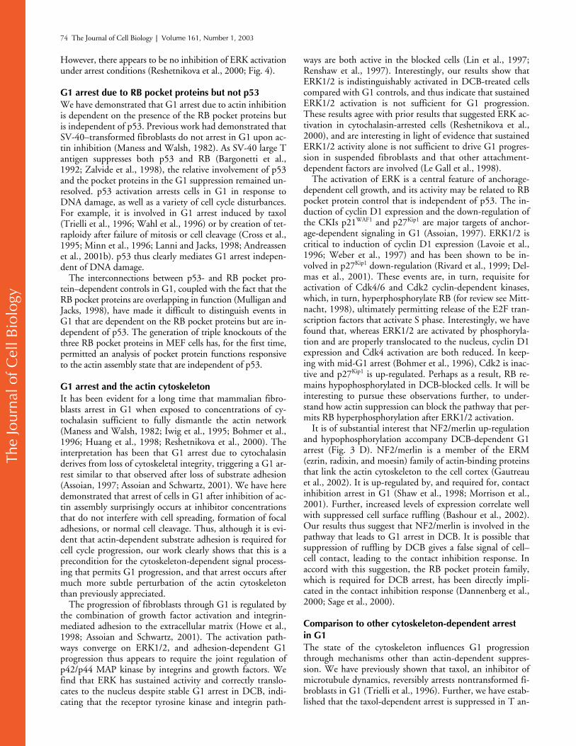
In the presence of DCB, p53 suppression does not abro-gate G1 arrest, whereas pocket protein suppression does.Thus, in a randomly cycling population exposed to DCB,lethality will differ depending on whether the cells are sup-pressed for p53 activity or for RB pocket protein activity.RB pocket protein–suppressed cells can progress past G1and past tetraploid G1, potentially leading to aneuploidyand death. In contrast, p53-incompetent cells arrest in eu-ploid G1 in DCB and can thus recover normally upon drugrelease. In accord with this hypothesis, we have found thatboth REF-52 and p53DD REF-52 cells recovered fromDCB arrest, seeded by those cells that had been arrested ineuploid G1 (Fig. 7, A and B). In contrast, TAG cells that ad-ditionally suppress RB pocket protein activity rapidly be-came aneuploid, as shown by a decrease in DNA content
Figure 5. DCB does not interfere with ERK kinase activation and nuclear translocation upon serum stimulation of quiescent cells. (A) Serum-starved REF-52 cells were reactivated with 10% FCS in the presence of 2 or 10 �M DCB. Flow cytometry shows that cells remain arrested in G1 25 h after release. Nocodazole exposure of cells in the absence of DCB indicates that control cells were cycling in the same time course. (B) ERK1/2 is normally activated in the presence of 2 �M DCB. At time intervals after add-back of serum to serum-starved REF-52, cells were harvested and subjected to Western blotting using either antibodies to total ERK or to activated phospho-ERK. Experiments were run either in the presence of 2 �M DCB (DCB, bottom two strips) or absence of DCB (Control, top two strips). ST designates quiescent serum-starved controls. (C) ERK1/2 is normally translocated to the nucleus in the presence of 2 �M DCB. 1 h after serum add-back to serum-starved REF-52 cells, ERK1/2 kinases were immunolocalized using anti–pan ERK antibodies. Experiments were run either in the presence of 2 �M DCB (DCB, right) or in the absence of DCB (Control, left). Nuclei were counterstained with propidium iodide (PI). Bars, 50 �m.
Figure 6. DCB-arrested REF-52 cells have markers characteristic of G1. (A) Asynchronous cells (Rdom), contact-inhibited cells (CI), and cells exposed for 25 h to either 2 �M DCB (DCB) or 2 mM HU (HU) were harvested and subjected to Western blotting procedures using the indicated antibodies. (B) The kinase activities of Cdk2 and Cdk4 were determined after immunoprecipitation of the kinases and incubation with histone H1 (Cdk2) or RB peptide (ckd4) as substrates. Autoradiographs of 32P incorporation are shown (top strips) and compared with the kinase expression levels as determined by Western blotting (bottom strips). (C) p27Kip1 accumulation is not incidental to DCB treatment. REF-52 cells were exposed for 25 h to either 2 mM HU or 2 �M DCB. Alternatively, cells were exposed to 2 mM HU for 25 h before addition of 2 �M DCB for 25 h in the continuous presence of HU. Samples were subjected to Western blotting analysis using anti-p27Kip1 antibodies.
The
Jour
nal o
f Cel
l Bio
logy
Actin-dependent G1 arrest requires RB but not p53 |
Lohez et al. 73
and loss of identifiable 2N and 4N peaks in DNA histo-grams, and died after release from DCB (Fig. 7, A and B)
.
Similarly, RB pocket protein–suppressed MEFs (TKO)showed a highly reduced capacity to proliferate after tran-sient exposure to DCB compared with control p53
�
/�
MEFs (Fig. 7 C).As our results suggested that short term exposure to actin
inhibitors might kill tumor cells, we directly tested this pos-sibility by exposing the human tumor cell lines HeLa (cer-vical adenocarcinoma), HCT116 (colon carcinoma), andA427 (lung carcinoma) to DCB or cytochalasin D. Cellswere exposed for 24 h and then released from drug. Results(Fig. 7 D) show that all three tumor cell lines fail to prolifer-ate after drug exposure, and that they are uniformly moresensitive to cytochalasin D than to DCB.
DiscussionNontransformed mammalian fibroblasts arrest reversibly inG1 when exposed to low concentrations of actin assembly
inhibitors. We have found that G1 arrest is not sensitive top53 status, but only occurs when RB pocket protein activityis intact. This observation demonstrates RB pocket proteincontrol that is distinct from p53 mechanisms. Further, as itis believed that RB function must be suppressed in one wayor another in all human tumors (Weinberg, 1995; Sherr,1996; Hanahan and Weinberg, 2000), our results raise thepossibility that a therapeutic approach could exploit the con-tinued cycling of tumor cells when the actin cytoskeleton, orelements of an associated pathway, is disrupted, causing di-rected death by selective response of pocket protein–sup-pressed cells to toxic agents. Independence of this G1 arrestfrom p53 is critical to the potential therapeutic effectivenessof such an approach in tumors with deficiencies in the RBpathway, but with normal p53 function.
The effective concentrations of actin inhibitors that induceG1 arrest are well below the concentrations that globally sup-press actin assembly, substrate adhesion, or cell cleavage.Suppression is accompanied by expression of p27Kip1, hypo-phosphorylation of RB, and inhibition of surface ruffling.
Figure 7. Failure of DCB-induced G1 blockage is lethal in RB pocket protein–suppressed cells. (A) Asynchronous REF-52, p53DD REF-52, and TAG cells were exposed to 5 �M DCB for 24 h. Flow cytometric analysis of DNA content at the indicated times shows that TAG cells rapidly become aneuploid upon release from DCB, whereas REF-52 and p53DD REF-52 cells recover from DCB arrest and reach contact inhibition after a few days. (B) Asynchronous REF-52, p53DD REF-52, and TAG cells were exposed to 5 �M DCB for 24 h. DCB was then washed out and cell counts were taken at the times indicated. (C) Asynchronous p53�/� and TKO MEFs were exposed to 10 �M DCB for 25 h. DCB was then washed out and cell counts taken at the indicated times. (D) Asynchronous HeLa, HCT 116, and A427 cells were exposed to either 10 �M DCB or 5 �M cytochalasin D (Cyto D) for 24 h. The drug was then washed out and cell counts taken at the times indicated. Control refers to untreated cells. In all cases, plateaus at late times indicate approach to confluency except for TKO MEFs, which do not reach high density.
The
Jour
nal o
f Cel
l Bio
logy
74 The Journal of Cell Biology | Volume 161, Number 1, 2003
However, there appears to be no inhibition of ERK activationunder arrest conditions (Reshetnikova et al., 2000; Fig. 4).
G1 arrest due to RB pocket proteins but not p53We have demonstrated that G1 arrest due to actin inhibitionis dependent on the presence of the RB pocket proteins butis independent of p53. Previous work had demonstrated thatSV-40–transformed fibroblasts do not arrest in G1 upon ac-tin inhibition (Maness and Walsh, 1982). As SV-40 large Tantigen suppresses both p53 and RB (Bargonetti et al.,1992; Zalvide et al., 1998), the relative involvement of p53and the pocket proteins in the G1 suppression remained un-resolved. p53 activation arrests cells in G1 in response toDNA damage, as well as a variety of cell cycle disturbances.For example, it is involved in G1 arrest induced by taxol(Trielli et al., 1996; Wahl et al., 1996) or by creation of tet-raploidy after failure of mitosis or cell cleavage (Cross et al.,1995; Minn et al., 1996; Lanni and Jacks, 1998; Andreassenet al., 2001b). p53 thus clearly mediates G1 arrest indepen-dent of DNA damage.
The interconnections between p53- and RB pocket pro-tein–dependent controls in G1, coupled with the fact that theRB pocket proteins are overlapping in function (Mulligan andJacks, 1998), have made it difficult to distinguish events inG1 that are dependent on the RB pocket proteins but are in-dependent of p53. The generation of triple knockouts of thethree RB pocket proteins in MEF cells has, for the first time,permitted an analysis of pocket protein functions responsiveto the actin assembly state that are independent of p53.
G1 arrest and the actin cytoskeletonIt has been evident for a long time that mammalian fibro-blasts arrest in G1 when exposed to concentrations of cy-tochalasin sufficient to fully dismantle the actin network(Maness and Walsh, 1982; Iwig et al., 1995; Bohmer et al.,1996; Huang et al., 1998; Reshetnikova et al., 2000). Theinterpretation has been that G1 arrest due to cytochalasinderives from loss of cytoskeletal integrity, triggering a G1 ar-rest similar to that observed after loss of substrate adhesion(Assoian, 1997; Assoian and Schwartz, 2001). We have heredemonstrated that arrest of cells in G1 after inhibition of ac-tin assembly surprisingly occurs at inhibitor concentrationsthat do not interfere with cell spreading, formation of focaladhesions, or normal cell cleavage. Thus, although it is evi-dent that actin-dependent substrate adhesion is required forcell cycle progression, our work clearly shows that this is aprecondition for the cytoskeleton-dependent signal process-ing that permits G1 progression, and that arrest occurs aftermuch more subtle perturbation of the actin cytoskeletonthan previously appreciated.
The progression of fibroblasts through G1 is regulated bythe combination of growth factor activation and integrin-mediated adhesion to the extracellular matrix (Howe et al.,1998; Assoian and Schwartz, 2001). The activation path-ways converge on ERK1/2, and adhesion-dependent G1progression thus appears to require the joint regulation ofp42/p44 MAP kinase by integrins and growth factors. Wefind that ERK has sustained activity and correctly translo-cates to the nucleus despite stable G1 arrest in DCB, indi-cating that the receptor tyrosine kinase and integrin path-
ways are both active in the blocked cells (Lin et al., 1997;Renshaw et al., 1997). Interestingly, our results show thatERK1/2 is indistinguishably activated in DCB-treated cellscompared with G1 controls, and thus indicate that sustainedERK1/2 activation is not sufficient for G1 progression.These results agree with prior results that suggested ERK ac-tivation in cytochalasin-arrested cells (Reshetnikova et al.,2000), and are interesting in light of evidence that sustainedERK1/2 activity alone is not sufficient to drive G1 progres-sion in suspended fibroblasts and that other attachment-dependent factors are involved (Le Gall et al., 1998).
The activation of ERK is a central feature of anchorage-dependent cell growth, and its activity may be related to RBpocket protein control that is independent of p53. The in-duction of cyclin D1 expression and the down-regulation ofthe CKIs p21WAF1 and p27Kip1 are major targets of anchor-age-dependent signaling in G1 (Assoian, 1997). ERK1/2 iscritical to induction of cyclin D1 expression (Lavoie et al.,1996; Weber et al., 1997) and has been shown to be in-volved in p27Kip1 down-regulation (Rivard et al., 1999; Del-mas et al., 2001). These events are, in turn, requisite foractivation of Cdk4/6 and Cdk2 cyclin-dependent kinases,which, in turn, hyperphosphorylate RB (for review see Mitt-nacht, 1998), ultimately permitting release of the E2F tran-scription factors that activate S phase. Interestingly, we havefound that, whereas ERK1/2 are activated by phosphoryla-tion and are properly translocated to the nucleus, cyclin D1expression and Cdk4 activation are both reduced. In keep-ing with mid-G1 arrest (Bohmer et al., 1996), Cdk2 is inac-tive and p27Kip1 is up-regulated. Perhaps as a result, RB re-mains hypophosphorylated in DCB-blocked cells. It will beinteresting to pursue these observations further, to under-stand how actin suppression can block the pathway that per-mits RB hyperphosphorylation after ERK1/2 activation.
It is of substantial interest that NF2/merlin up-regulationand hypophosphorylation accompany DCB-dependent G1arrest (Fig. 3 D). NF2/merlin is a member of the ERM(ezrin, radixin, and moesin) family of actin-binding proteinsthat link the actin cytoskeleton to the cell cortex (Gautreauet al., 2002). It is up-regulated by, and required for, contactinhibition arrest in G1 (Shaw et al., 1998; Morrison et al.,2001). Further, increased levels of expression correlate wellwith suppressed cell surface ruffling (Bashour et al., 2002).Our results thus suggest that NF2/merlin is involved in thepathway that leads to G1 arrest in DCB. It is possible thatsuppression of ruffling by DCB gives a false signal of cell–cell contact, leading to the contact inhibition response. Inaccord with this suggestion, the RB pocket protein family,which is required for DCB arrest, has been directly impli-cated in the contact inhibition response (Dannenberg et al.,2000; Sage et al., 2000).
Comparison to other cytoskeleton-dependent arrest in G1The state of the cytoskeleton influences G1 progressionthrough mechanisms other than actin-dependent suppres-sion. We have previously shown that taxol, an inhibitor ofmicrotubule dynamics, reversibly arrests nontransformed fi-broblasts in G1 (Trielli et al., 1996). Further, we have estab-lished that the taxol-dependent arrest is suppressed in T an-
The
Jour
nal o
f Cel
l Bio
logy
Actin-dependent G1 arrest requires RB but not p53 | Lohez et al. 75
tigen–transformed cells (Trielli et al., 1996). Unlike DCB-dependent G1 arrest, taxol-dependent arrest in G1 is p53dependent (unpublished data).
Arrest with DCB in G1 can also be distinguished from theG1 arrest that arises from creation of a tetraploid state byDCB-dependent suppression of cell cleavage. Tetraploid G1arrest is permanent, persisting after removal of DCB (An-dreassen et al., 2001b), and unlike the DCB G1 block, is de-pendent on p53 status (Andreassen et al., 2001b). Tetra-ploid G1 arrest occurs regardless of the cause of tetraploidy(Andreassen et al., 2001b) and appears to be the predomi-nant cause of p53-dependent 4N arrest in colon carcinomacells after DNA damage (Andreassen et al., 2001a).
Implications for tumor therapyA remarkable aspect of the DCB-dependent G1 arrest re-ported here is that it is insensitive to p53 status, but sensitiveto the status of the RB pocket proteins. Only MEF cells thathave a triple knockout for the three RB pocket proteins failto arrest in G1 in response to DCB exposure. The result isduplicated in TAG cells, which are large T antigen–trans-formed REF-52. We have shown that p53DD REF-52 cellsrespond like wild-type cells to DCB, but that TAG cells failto arrest. The major effects of large T antigen are to suppressp53 and the RB pocket protein functions (Bargonetti et al.,1992; Zalvide et al., 1998). It is therefore likely that TAGcells fail to arrest, as in the case of TKO MEF cells, becausethe RB pocket proteins are suppressed.
To our knowledge, this is the first evidence that a pharma-cological approach can both selectively block cells with intactRB function and permit induction of death in cells withcompromised function. The result has been striking. The fail-ure of RB pocket protein–suppressed cells to arrest in G1 inthe presence of high concentrations of DCB causes them toselectively continue to cycle to aneuploid status and die. Wehave further demonstrated that DCB and cytochalasin D effi-ciently kill human tumor cells of three different tissue origins.Latrunculin and other actin inhibitors are being used in clini-cal chemotherapy trials (Jordan and Wilson, 1998). It willnow be of primary importance to establish whether latruncu-lin or other drugs will have a selective lethal effect on RB-sup-pressed cells. As the RB pocket protein pathway is suppressedin virtually all human tumors, this approach may hold sub-stantial promise for selective tumor therapy.
We have demonstrated the effect of RB suppression onprogression to cell death after exposure to DCB alone. Analternative approach that might prove highly effective wouldbe to use low doses of actin inhibitors accompanied by expo-sure to ionizing radiation or taxol, to which cycling cells areparticularly sensitive (Iliakis, 1991; Trielli et al., 1996;Blajeski et al., 2001). In this case, only RB-suppressed cellswould bypass G1 arrest and would be selectively vulnerableto death induced by conventional tumor therapy.
Materials and methodsCell culture and infectionREF-52 cells and their SV-40 large T antigen–transformed derivatives(TAG) (Perry et al., 1992) were a gift from G.R. Stark (Cleveland Clinic,Cleveland, OH). REF-52 primary cells were used at �35 passages. IMR-90cells were obtained from Coriell Cell Repositories. p53DD REF-52 were
prepared as previously described (Andreassen et al., 2001b). p53�/� MEFswere generated as described by Borel et al. (2002b). TKO and RB�/� MEFswere a gift from J. Sage and T. Jacks (Massachusetts Institute of Technol-ogy, Cambridge, MA). ARF�/� MEFs were obtained from C. Sherr and M.Roussel (St. Jude’s Children’s Hospital, Memphis, TN). MEFs were used atno more than four passages. HeLa and A427 cells were obtained from theAmerican Type Culture Collection. HCT 116 cells were the gift of B. Vo-gelstein (Johns Hopkins University, Baltimore, MD). Cell doubling times atmid-log phase were 24 h for REF-52, p53DD REF-52, IMR-90, TKO MEFs,p53�/� MEFs, RB�/� MEFs, and Arf�/� MEFs and 18 h for TAG. All cellswere cultured as monolayers in DME (GIBCO BRL) supplemented with10% FCS (Biological Industries). Cells were maintained in a humid incuba-tor at 37�C in a 5% CO2 environment.
Cell treatment and synchronizationRandomly cycling cells were exposed to the indicated doses of DCB for24 h. To assay for cell cycle progression during or after exposure to DCB,cells were incubated with 10 �M BrdU for 24 h. To synchronize REF-52cells in G0, cells were cultured in medium containing 0.1% FCS for 24 h.Serum-starved cells were then released from G0 by the addition of 10%FCS, and DCB, as appropriate, was added 2 h before serum addition. Toinduce contact inhibition, REF-52 cells were kept at confluency for 36 h.
To synchronize REF-52 cells in early S phase, randomly cycling cellswere incubated with 2 mM HU for 24 h. In all cases, drugs were added torandomly cycling cells no earlier than 36 h after replating to allow for fullspreading before drug treatment. HU, DCB, and nocodazole were ob-tained from Sigma-Aldrich. Latrunculin A was obtained from MolecularProbes. HU was prepared as a 200 mM stock in DME containing 10% FCS.DCB, latrunculin A, and nocodazole were prepared in DMSO as 10 mM, 2mM, and 1 mg/ml stocks respectively. BrdU was prepared from a 10 mMstock in DME.
Cell counts and flow cytometryRandomly cycling REF-52 cells were incubated with the indicated doses ofDCB. Every 24 h, cells were harvested by trypsinization and resuspendedin 1 PBS, and cell counts were taken using a Neubauer hemacytometer.To assess the impact of DCB or cytochalasin D on cell survival, cells wereexposed to drug for 24 h and then released, and counts were then takenevery 24 h. For flow cytometry, cells were trypsinized, collected by centrif-ugation, and then fixed, stored, and analyzed as previously described (An-dreassen et al., 2001b).
Assay of Cdk activityTo assay for Cdk2 and Cdk4 activities, cell lysates were prepared from ran-dom cycling REF-52, contact-inhibited REF-52, or random cycling REF-52exposed to 2 �M DCB or 2 mM HU for 24 h. Cells were collected bytrypsinization, and preparation of lysates, immunoprecipitation, and radio-immune assay for Cdk4 and Cdk2 were as described by Trielli et al. (1996)and Andreassen and Margolis (1994), respectively.
ImmunoblottingCell lysates were prepared for Western blots as described above for Cdkactivity assay. 10 �g of whole cell lysates (25 �g for cyclin D1 and RB)was then resolved by PAGE, transferred to nitrocellulose or Immobilon(RB) sheets, blocked with 5% nonfat milk in TNT buffer (25 mM Tris, pH7.5, 150 mM NaCl, and 0.05% Tween 20), incubated overnight with pri-mary antibodies, and then incubated with HRP-conjugated anti–rabbit oranti–mouse IgG secondary antibodies for 1 h. Protein–antibody complexeswere detected by ECL (Pierce Chemical Co.). The primary antibodies usedwere anti–cyclin A (1:5,000), anti–cyclin E (1:3,000), anti-Cdk2 (1:3,000)(Brenot-Bosc et al., 1995), anti–cyclin D1 (HD11; 1:200), anti-Cdk4(1:1,000), anti-p21WAF1 (C-19; 1:2,000), anti-NF2/merlin (C-18; 1:200) (allfrom Santa Cruz Biotechnology, Inc.), anti-p27Kip1 (1:2,500; TransductionLaboratories), anti-RB (1:500; BD Biosciences), anti–pan ERK (1:12,000;Zymed Laboratories), and anti–dual phosphorylated ERK (V8031; 1:2,000;New England Biolabs, Inc.). All primary antibodies, except for p21WAF1,were diluted in TNT buffer containing nonfat milk (1% for RB; 2% forp27Kip1 and ERK antibodies; 2.5% for cyclin A, cyclin E, cyclin D1, andCdk2; 5% for Cdk4; and 10% for NF2/merlin). All secondary antibodieswere diluted 1:5,000 (except for detection of RB [1:300] and cyclin D1[1:2,500]) in TNT buffer (also containing 2.5% nonfat milk for detection ofcyclin A, cyclin E, cyclin D1, and ERK).
Immunofluorescence microscopyFor immunofluorescence microscopy, cells were grown on poly-D-lysine–coated glass coverslips. To assay for cytokinetic failure in the presence of
The
Jour
nal o
f Cel
l Bio
logy
76 The Journal of Cell Biology | Volume 161, Number 1, 2003
DCB, cells were fixed with methanol at �20�C for 10 min, microtubuleswere labeled as previously described (Trielli et al., 1996), and nuclei werestained by the addition of 0.5 �g/ml propidium iodide. For quantificationof BrdU incorporation, cells were fixed and prepared with FITC-conjugatedanti-BrdU antibody as previously described (Andreassen et al., 2001b).
To assay for actin assembly and cell adhesion, randomly cycling cellswere exposed to 2 �M DCB for 24 h as described above, fixed with 2%paraformaldehyde in PBS, and incubated with antibodies as previously de-scribed (Andreassen and Margolis, 1994). Monoclonal anti-vinculin andanti-phosphotyrosine antibodies (Sigma-Aldrich) were diluted 500-fold.F-actin was detected by a 30-min incubation at 37�C with TRITC-conju-gated phalloidin (1:1,000) (Sigma-Aldrich). To assay for ERK localization,cells were fixed and prepared as above, except for pretreatment for 1 hwith PBS containing 5% FCS and 1% BSA and then incubation with anti–pan ERK for 1 h at 37�C.
After appropriate staining and washes in PBS, cells were observed usingan Optiphot II microscope (Nikon) attached to an MRC-600 laser scanningconfocal apparatus (Bio-Rad Laboratories). Images were treated withAdobe Photoshop®.
We thank G.R. Stark for providing REF-52 and TAG cell lines, J. Sage andT. Jacks for TKO and RB�/� MEFs, C. Sherr and M. Roussel for ARF�/�
MEFs, M. Oren (Weizmann Institute of Science, Rehovot, Israel) forp53DD retrovirus–producing cells, and B. Vogelstein for HCT 116 cells.
This work was supported by grants to R.L. Margolis from La Ligue Natio-nale Contre le Cancer (Equipe Labellisée) and l’Association pour la Re-cherche sur le Cancer (ARC). O.D. Lohez was supported by the Commis-sariat à l’Energie Atomique, ARC, and Centre National de la RechercheScientifique.
Submitted: 22 August 2002Revised: 28 February 2003Accepted: 28 February 2003
ReferencesAndreassen, P.R., and R.L. Margolis. 1994. Microtubule dependency of p34cdc2
inactivation and mitotic exit in mammalian cells. J. Cell Biol. 127:789–802.Andreassen, P.R., F.B. Lacroix, O.D. Lohez, and R.L. Margolis. 2001a. Neither
p21WAF1 nor 14-3-3 prevents G2 progression to mitotic catastrophe inhuman colon carcinoma cells after DNA damage, but p21WAF1 inducesstable G1 arrest in resulting tetraploid cells. Cancer Res. 61:7660–7668.
Andreassen, P.R., O.D. Lohez, F.B. Lacroix, and R.L. Margolis. 2001b. Tetraploidstate induces p53-dependent arrest of nontransformed mammalian cells inG1. Mol. Biol. Cell. 12:1315–1328.
Aplin, A.E., S.M. Short, and R.L. Juliano. 1999. Anchorage-dependent regulationof the mitogen-activated protein kinase cascade by growth factors is sup-ported by a variety of integrin � chains. J. Biol. Chem. 274:31223–31228.
Assoian, R.K. 1997. Anchorage-dependent cell cycle progression. J. Cell Biol. 136:1–4.
Assoian, R.K., and M.A. Schwartz. 2001. Coordinate signaling by integrins and re-ceptor tyrosine kinases in the regulation of G1 phase cell-cycle progression.Curr. Opin. Genet. Dev. 11:48–53.
Assoian, R.K., and X. Zhu. 1997. Cell anchorage and the cytoskeleton as partnersin growth factor dependent cell cycle progression. Curr. Opin. Cell Biol.9:93–98.
Aubin, J.E., M. Osborn, and K. Weber. 1981. Inhibition of cytokinesis and alteredcontractile ring morphology induced by cytochalasins in synchronized PtK2cells. Exp. Cell Res. 136:63–79.
Bargonetti, J., I. Reynisdottir, P.N. Friedman, and C. Prives. 1992. Site-specificbinding of wild-type p53 to cellular DNA is inhibited by SV40 T antigenand mutant p53. Genes Dev. 6:1886–1898.
Bashour, A.M., J.J. Meng, W. Ip, M. MacCollin, and N. Ratner. 2002. The neu-rofibromatosis type 2 gene product, merlin, reverses the F-actin cytoskeletaldefects in primary human Schwannoma cells. Mol. Cell. Biol. 22:1150–1157.
Blajeski, A.L., T.J. Kottke, and S.H. Kaufmann. 2001. A multistep model forpaclitaxel-induced apoptosis in human breast cancer cell lines. Exp. Cell Res.270:277–288.
Bohmer, R.M., E. Scharf, and R.K. Assoian. 1996. Cytoskeletal integrity is re-quired throughout the mitogen stimulation phase of the cell cycle and medi-ates the anchorage-dependent expression of cyclin D1. Mol. Biol. Cell.7:101–111.
Borel, F., F.B. Lacroix, and R.L. Margolis. 2002a. Prolonged arrest of mammalian
cells at the G1/S boundary results in permanent S phase stasis. J. Cell Sci.115:2829–2838.
Borel, F., O.D. Lohez, F.B. Lacroix, and R.L. Margolis. 2002b. Multiple cen-trosomes arise from tetraploidy checkpoint failure and mitotic centrosomeclusters in p53 and RB pocket protein-compromised cells. Proc. Natl. Acad.Sci. USA. 99:9819–9824.
Brenot-Bosc, F., S. Gupta, R.L. Margolis, and R. Fotedar. 1995. Changes in thesubcellular localization of replication initiation proteins and cell cycle pro-teins during G1- to S-phase transition in mammalian cells. Chromosoma.103:517–527.
Cross, S.M., C.A. Sanchez, C.A. Morgan, M.K. Schimke, S. Ramel, R.L. Idzerda,W.H. Raskind, and B.J. Reid. 1995. A p53-dependent mouse spindle check-point. Science. 267:1353–1356.
Dannenberg, J.H., A. van Rossum, L. Schuijff, and H. te Riele. 2000. Ablation ofthe retinoblastoma gene family deregulates G(1) control causing immortal-ization and increased cell turnover under growth-restricting conditions.Genes Dev. 14:3051–3064.
Delmas, C., S. Manenti, A. Boudjelal, C. Peyssonnaux, A. Eychene, and J.M. Dar-bon. 2001. The p42/p44 mitogen-activated protein kinase activation trig-gers p27Kip1 degradation independently of CDK2/cyclin E in NIH 3T3cells. J. Biol. Chem. 276:34958–34965.
Di Leonardo, A., S.P. Linke, K. Clarkin, and G.M. Wahl. 1994. DNA damagetriggers a prolonged p53-dependent G1 arrest and long-term induction ofCip1 in normal human fibroblasts. Genes Dev. 8:2540–2551.
Di Leonardo, A., S.H. Khan, S.P. Linke, V. Greco, G. Seidita, and G.M. Wahl.1997. DNA rereplication in the presence of mitotic spindle inhibitors in hu-man and mouse fibroblasts lacking either p53 or pRb function. Cancer Res.57:1013–1019.
Dulic, V., W.K. Kaufmann, S.J. Wilson, T.D. Tlsty, E. Lees, J.W. Harper, S.J.Elledge, and S.I. Reed. 1994. p53-dependent inhibition of cyclin-dependentkinase activities in human fibroblasts during radiation-induced G1 arrest.Cell. 76:1013–1023.
Dyson, N. 1998. The regulation of E2F by pRB-family proteins. Genes Dev. 12:2245–2262.
el-Deiry, W.S., T. Tokino, V.E. Velculescu, D.B. Levy, R. Parsons, J.M. Trent, D.Lin, W.E. Mercer, K.W. Kinzler, and B. Vogelstein. 1993. WAF1, a poten-tial mediator of p53 tumor suppression. Cell. 75:817–825.
Gautreau, A., D. Louvard, and M. Arpin. 2002. ERM proteins and NF2 tumorsuppressor: the Yin and Yang of cortical actin organization and cell growthsignaling. Curr. Opin. Cell Biol. 14:104–109.
Hanahan, D., and R.A. Weinberg. 2000. The hallmarks of cancer. Cell. 100:57–70.Howe, A., A.E. Aplin, S.K. Alahari, and R.L. Juliano. 1998. Integrin signaling and
cell growth control. Curr. Opin. Cell Biol. 10:220–231.Huang, S., C.S. Chen, and D.E. Ingber. 1998. Control of cyclin D1, p27(Kip1),
and cell cycle progression in human capillary endothelial cells by cell shapeand cytoskeletal tension. Mol. Biol. Cell. 9:3179–3193.
Iliakis, G. 1991. The role of DNA double strand breaks in ionizing radiation-induced killing of eukaryotic cells. Bioessays. 13:641–648.
Iwig, M., E. Czeslick, A. Muller, M. Gruner, M. Spindler, and D. Glaesser. 1995.Growth regulation by cell shape alteration and organization of the cytoskele-ton. Eur. J. Cell Biol. 67:145–157.
Jordan, M.A., and L. Wilson. 1998. Microtubules and actin filaments: dynamictargets for cancer chemotherapy. Curr. Opin. Cell Biol. 10:123–130.
Jordan, M.A., D. Thrower, and L. Wilson. 1992. Effects of vinblastine, podophyl-lotoxin and nocodazole on mitotic spindles. Implications for the role of mi-crotubule dynamics in mitosis. J. Cell Sci. 102:401–416.
Kastan, M.B., O. Onyekwere, D. Sidransky, B. Vogelstein, and R.W. Craig. 1991.Participation of p53 protein in the cellular response to DNA damage. CancerRes. 51:6304–6311.
Lanni, J.S., and T. Jacks. 1998. Characterization of the p53-dependent postmitoticcheckpoint following spindle disruption. Mol. Cell. Biol. 18:1055–1064.
Lavoie, J.N., G. L’Allemain, A. Brunet, R. Muller, and J. Pouyssegur. 1996. CyclinD1 expression is regulated positively by the p42/p44MAPK and negativelyby the p38/HOGMAPK pathway. J. Biol. Chem. 271:20608–20616.
Le Gall, M., D. Grall, J.C. Chambard, J. Pouyssegur, and E. Van Obberghen-Schill-ing. 1998. An anchorage-dependent signal distinct from p42/44 MAP kinaseactivation is required for cell cycle progression. Oncogene. 17:1271–1277.
Levine, A.J. 1997. p53, the cellular gatekeeper for growth and division. Cell. 88:323–331.
Lin, T.H., Q. Chen, A. Howe, and R.L. Juliano. 1997. Cell anchorage permits ef-ficient signal transduction between ras and its downstream kinases. J. Biol.Chem. 272:8849–8852.

The
Jour
nal o
f Cel
l Bio
logy
Actin-dependent G1 arrest requires RB but not p53 | Lohez et al. 77
Linke, S.P., K.C. Clarkin, A. Di Leonardo, A. Tsou, and G.M. Wahl. 1996. A re-versible, p53-dependent G0/G1 cell cycle arrest induced by ribonucleotide de-pletion in the absence of detectable DNA damage. Genes Dev. 10:934–947.
Maness, P.F., and R.C. Walsh, Jr. 1982. Dihydrocytochalasin B disorganizes actincytoarchitecture and inhibits initiation of DNA synthesis in 3T3 cells. Cell.30:253–262.
Martineau, S.N., P.R. Andreassen, and R.L. Margolis. 1995. Delay of HeLa cellcleavage into interphase using dihydrocytochalasin B: retention of a postmi-totic spindle and telophase disc correlates with synchronous cleavage recov-ery. J. Cell Biol. 131:191–205.
Minn, A.J., L.H. Boise, and C.B. Thompson. 1996. Expression of Bcl-xL and lossof p53 can cooperate to overcome a cell cycle checkpoint induced by mitoticspindle damage. Genes Dev. 10:2621–2631.
Mittnacht, S. 1998. Control of pRB phosphorylation. Curr. Opin. Genet. Dev.8:21–27.
Morrison, H., L.S. Sherman, J. Legg, F. Banine, C. Isacke, C.A. Haipek, D.H.Gutmann, H. Ponta, and P. Herrlich. 2001. The NF2 tumor suppressorgene product, merlin, mediates contact inhibition of growth through inter-actions with CD44. Genes Dev. 15:968–980.
Mulligan, G., and T. Jacks. 1998. The retinoblastoma gene family: cousins withoverlapping interests. Trends Genet. 14:223–229.
Perry, M.E., M. Commane, and G.R. Stark. 1992. Simian virus 40 large tumor an-tigen alone or two cooperating oncogenes convert REF52 cells to a state per-missive for gene amplification. Proc. Natl. Acad. Sci. USA. 89:8112–8116.
Polyak, K., J.Y. Kato, M.J. Solomon, C.J. Sherr, J. Massague, J.M. Roberts, and A.Koff. 1994a. p27Kip1, a cyclin-Cdk inhibitor, links transforming growthfactor-� and contact inhibition to cell cycle arrest. Genes Dev. 8:9–22.
Polyak, K., M.H. Lee, H. Erdjument-Bromage, A. Koff, J.M. Roberts, P. Tempst,and J. Massague. 1994b. Cloning of p27Kip1, a cyclin-dependent kinase in-hibitor and a potential mediator of extracellular antimitogenic signals. Cell.78:59–66.
Renshaw, M.W., X.D. Ren, and M.A. Schwartz. 1997. Growth factor activation ofMAP kinase requires cell adhesion. EMBO J. 16:5592–5599.
Reshetnikova, G., R. Barkan, B. Popov, N. Nikolsky, and L.S. Chang. 2000. Dis-ruption of the actin cytoskeleton leads to inhibition of mitogen-induced cy-clin E expression, Cdk2 phosphorylation, and nuclear accumulation of theretinoblastoma protein-related p107 protein. Exp. Cell Res. 259:35–53.
Rivard, N., M.J. Boucher, C. Asselin, and G. L’Allemain. 1999. MAP kinase cas-cade is required for p27 downregulation and S phase entry in fibroblasts andepithelial cells. Am. J. Physiol. 277:C652–C664.
Sage, J., G.J. Mulligan, L.D. Attardi, A. Miller, S. Chen, B. Williams, E. The-odorou, and T. Jacks. 2000. Targeted disruption of the three Rb-relatedgenes leads to loss of G(1) control and immortalization. Genes Dev. 14:3037–3050.
Shaulian, E., A. Zauberman, D. Ginsberg, and M. Oren. 1992. Identification of aminimal transforming domain of p53: negative dominance through abroga-tion of sequence-specific DNA binding. Mol. Cell. Biol. 12:5581–5592.
Shaw, R.J., A.I. McClatchey, and T. Jacks. 1998. Regulation of the neurofibroma-tosis type 2 tumor suppressor protein, merlin, by adhesion and growth arreststimuli. J. Biol. Chem. 273:7757–7764.
Sherr, C.J. 1996. Cancer cell cycles. Science. 274:1672–1677.Sherr, C.J., and J.M. Roberts. 1995. Inhibitors of mammalian G1 cyclin-depen-
dent kinases. Genes Dev. 9:1149–1163.Sherr, C.J., and J.M. Roberts. 1999. CDK inhibitors: positive and negative regula-
tors of G1-phase progression. Genes Dev. 13:1501–1512.Sherr, C.J., and J.D. Weber. 2000. The ARF/p53 pathway. Curr. Opin. Genet.
Dev. 10:94–99.Spector, I., N.R. Shochet, D. Blasberger, and Y. Kashman. 1989. Latrunculins–
novel marine macrolides that disrupt microfilament organization and affectcell growth: I. Comparison with cytochalasin D. Cell Motil. Cytoskeleton. 13:127–144.
Stoker, M., C. O’Neill, S. Berryman, and V. Waxman. 1968. Anchorage andgrowth regulation in normal and virus-transformed cells. Int. J. Cancer.3:683–693.
Trielli, M.O., P.R. Andreassen, F.B. Lacroix, and R.L. Margolis. 1996. DifferentialTaxol-dependent arrest of transformed and nontransformed cells in the G1phase of the cell cycle, and specific-related mortality of transformed cells. J.Cell Biol. 135:689–700.
Vogelstein, B., D. Lane, and A.J. Levine. 2000. Surfing the p53 network. Nature.408:307–310.
Wahl, A.F., K.L. Donaldson, C. Fairchild, F.Y. Lee, S.A. Foster, G.W. Demers,and D.A. Galloway. 1996. Loss of normal p53 function confers sensitizationto Taxol by increasing G2/M arrest and apoptosis. Nat. Med. 2:72–79.
Weber, J.D., D.M. Raben, P.J. Phillips, and J.J. Baldassare. 1997. Sustained activa-tion of extracellular-signal-regulated kinase 1 (ERK1) is required for the con-tinued expression of cyclin D1 in G1 phase. Biochem. J. 326:61–68.
Weinberg, R.A. 1995. The retinoblastoma protein and cell cycle control. Cell. 81:323–330.
Zalvide, J., H. Stubdal, and J.A. DeCaprio. 1998. The J domain of simian virus 40large T antigen is required to functionally inactivate RB family proteins.Mol. Cell. Biol. 18:1408–1415.
Zhang, H.S., A.A. Postigo, and D.C. Dean. 1999. Active transcriptional repressionby the Rb-E2F complex mediates G1 arrest triggered by p16INK4a, TGF�,and contact inhibition. Cell. 97:53–61.
Zhu, J., P.W. Rice, L. Gorsch, M. Abate, and C.N. Cole. 1992. Transformation ofa continuous rat embryo fibroblast cell line requires three separate domainsof simian virus 40 large T antigen. J. Virol. 66:2780–2791.