Effect of Cholesterol on Electrostatics in Lipid− Protein Films of a Pulmonary Surfactant
7
DOI: 10.1021/la904335m 1929 Langmuir 2010, 26(3), 1929–1935 Published on Web 01/05/2010 pubs.acs.org/Langmuir © 2010 American Chemical Society Effect of Cholesterol on Electrostatics in Lipid-Protein Films of a Pulmonary Surfactant Eric Finot, † Yuri Leonenko, ‡ Brad Moores, ‡ Lukas Eng, ) Matthias Amrein, ^ and Zoya Leonenko* ,‡,§ † Institut Carnot de Bourgogne, University of Burgundy, Dijon, France, ‡ Department of Physics and Astronomy, and § Department of Biology, University of Waterloo, Canada, ) Institute of Applied Photophysics, Technical University Dresden, Germany, and ^ Department of Cell Biology and Anatomy, University of Calgary, Canada Received April 17, 2009. Revised Manuscript Received December 8, 2009 We report the changes in the electrical properties of the lipid-protein film of pulmonary surfactant produced by excess cholesterol. Pulmonary surfactant (PS) is a complex lipid-protein mixture that forms a molecular film at the interface of the lung’s epithelia. The defined molecular arrangement of the lipids and proteins of the surfactant film gives rise to the locally highly variable electrical surface potential of the interface, which becomes considerably altered in the presence of cholesterol. With frequency modulation Kelvin probe force microscopy (FM-KPFM) and force measure- ments, complemented by theoretical analysis, we showed that excess cholesterol significantly changes the electric field around a PS film because of the presence of nanometer-sized electrostatic domains and affects the electrostatic interaction of an AFM probe with a PS film. These changes in the local electrical field would greatly alter the interaction of the surfactant film with charged species and would immediately impact the manner in which inhaled (often charged) airborne nanoparticles and fibers might interact with the lung interface. Introduction The air-lung interface is covered with a molecular film of pulmonary surfactant (PS). The PS film is a complex lipid-pro- tein mixture primarily composed of lipids (75-80%) with small amounts of proteins (10%) and cholesterol (5-10% by mass). The major function of the film is to reduce the surface tension of the lung’s air-liquid interface, providing stability to the alveolar structure and reducing the work of breathing. The PS film is also the first barrier in the body that nanosized particles (NSPs) in breathed air encounter. NSPs in the ambient (polluted) air pose a major health threat 1,2 and have been associated with substantial respiratory and cardiovascular morbidity and mortality 1-3 as well as cancer. 4,5 However, NSPs are prospective systems for aerosolized drug delivery to the lung. Both man-made and natural nanoparticles may be invariably charged 6 and may be affected strongly by electrostatic forces at the air-lung interface. For these reasons, understanding the electrostatic interactions of NSPs with PS films is of great importance. In this work, we investigated how the composition of a surfactant film and, in particular, excess cholesterol may affect these interactions through changes in the surface potential of the film. The apex of the AFM probe may serve as a model for the nanoparticle and therefore may provide important input into the interaction of nanoparticles with PS films. Cholesterol plays an important role in PS structure and function. We showed earlier that the function of pulmonary surfactant is greatly suppressed by excess cholesterol (20%), which has been associated with surfactant inhibition in adult respiratory distress syndrome. 7,8 We demonstrated earlier that excess cholesterol in PS films impairs surfactant function because of the prevention of multilayer formation, characteristic of normal functional surfactant 7 as a result of the loss of mechanical stability and reduced adhesion. In this work, we show that, in addition, excess cholesterol changes the local surface potential distribution of the PS film and alters the electrical potential in close proximity to the film, which affects the electrostatic inter- action of the PS film with the AFM probe. On a broader scale, the local electrical surface potential in a complex biomolecular monolayer such as PS is a direct reflection of its molecular-level structure and provides insight into the function of these films. Lipid monolayers are also widely used as models for studying biological membranes; therefore, our work may be helpful in elucidating the effect of cholesterol on the structure and function of biological membranes. Cholesterol plays an important role in controlling the fluidity, permeability, and mechanical strength of biological and model lipid mem- branes. 9-13 On the molecular scale, the incorporation of chole- sterol into the membrane leads to an increase in lipid hydrocarbon *Corresponding author. E-mail: [email protected]. Tel: (519) 888-4567, ext 38273. Fax: (519) 746-8115. (1) Schwartz, J. F.; Laden, F.; Zanobetti, A. Environ. Health Perspect. 2002, 110, 1025–1029. (2) Boldo, E.; Medina, S.; LeTertre, A.; Hurley, F.; Mucke, H. G.; Ballester, F.; Aguilera, I.; Eilstein, D. Eur. J. Epidemiol. 2006, 21, 449–458. (3) Li, Z.; Carter, J. D.; Dailey, L. A.; Huang, Y. C. Environ. Health Perspect. 2005, 113, 1009–1014. (4) Knaapen, A. M.; Borm, P. J. A.; Albrecht, C.; Schins, R. P. F. Int. J. Cancer 2004, 109, 799–809. (5) Forastiere, F. Occup. Environ. Med. 2004, 61, 797–798. (6) Bailey, A. G. J. Electrost. 1998, 44,3–10. (7) Leonenko, Z.; Gill, S.; Baoukina, S.; Monticelli, L.; Doehner, J.; Gunasekara, L.; Felderer, F.; Rodenstein, M.; Eng, L. M.; Amrein, M. Biophys. J. 2007, 93, 674–683. (8) Gunasekara, L.; Schurch, S.; Schoel, W. M.; Nag, K.; Leonenko, Z.; Haufs, M.; Amrein, M. Biochim. Biophys. Acta 2005, 1737, 27–35. (9) Demel, R. A.; Kruyff, B. D. Biochim. Biophys. Acta 1976, 457, 109–132. (10) Ohvo-Rekil€ a, H.; Ramstedt, B.; Leppim€ aki, P.; Slotte, J. P. Prog. Lipid Res. 2002, 41, 66–97. (11) McMullen, T. P. W.; Lewis, R. N. A.; McElhaney, R. N. Curr. Opin. Colloid Interface Sci. 2004, 8, 459–468. (12) Bonn, M.; Roke, S.; Berg, O.; Juurlink, L. B. F.; Stamouli, A.; Muller, M. J. Phys. Chem. B 2004, 108, 19083–19085. (13) Cadenhead, G.A. Structure and Properties of Cell Membranes; CRC Press: Boca Raton, FL, 1985.
-
Upload
independent -
Category
Documents
-
view
0 -
download
0
Transcript of Effect of Cholesterol on Electrostatics in Lipid− Protein Films of a Pulmonary Surfactant
DOI: 10.1021/la904335m 1929Langmuir 2010, 26(3), 1929–1935 Published on Web 01/05/2010
pubs.acs.org/Langmuir
© 2010 American Chemical Society
Effect of Cholesterol on Electrostatics in Lipid-Protein Films of a
Pulmonary Surfactant
Eric Finot,† Yuri Leonenko,‡ Brad Moores,‡ Lukas Eng, ) Matthias Amrein,^ andZoya Leonenko*,‡,§
†Institut Carnot de Bourgogne, University of Burgundy, Dijon, France, ‡Department of Physics and Astronomy,and §Department of Biology, University of Waterloo, Canada, )Institute of Applied Photophysics, TechnicalUniversity Dresden, Germany, and ^Department of Cell Biology and Anatomy, University of Calgary, Canada
Received April 17, 2009. Revised Manuscript Received December 8, 2009
We report the changes in the electrical properties of the lipid-protein film of pulmonary surfactant produced byexcess cholesterol. Pulmonary surfactant (PS) is a complex lipid-protein mixture that forms a molecular film at theinterface of the lung’s epithelia. The definedmolecular arrangement of the lipids and proteins of the surfactant film givesrise to the locally highly variable electrical surface potential of the interface, which becomes considerably altered in thepresence of cholesterol. With frequency modulation Kelvin probe force microscopy (FM-KPFM) and force measure-ments, complemented by theoretical analysis, we showed that excess cholesterol significantly changes the electric fieldaround a PS film because of the presence of nanometer-sized electrostatic domains and affects the electrostaticinteraction of an AFMprobe with a PS film. These changes in the local electrical field would greatly alter the interactionof the surfactant film with charged species and would immediately impact the manner in which inhaled (often charged)airborne nanoparticles and fibers might interact with the lung interface.
Introduction
The air-lung interface is covered with a molecular film ofpulmonary surfactant (PS). The PS film is a complex lipid-pro-tein mixture primarily composed of lipids (75-80%) with smallamounts of proteins (10%) and cholesterol (5-10% by mass).The major function of the film is to reduce the surface tension ofthe lung’s air-liquid interface, providing stability to the alveolarstructure and reducing the work of breathing. The PS film is alsothe first barrier in the body that nanosized particles (NSPs) inbreathed air encounter. NSPs in the ambient (polluted) air pose amajor health threat1,2 and have been associated with substantialrespiratory and cardiovascular morbidity and mortality1-3 aswell as cancer.4,5 However, NSPs are prospective systems foraerosolized drugdelivery to the lung. Bothman-made and naturalnanoparticles may be invariably charged6 and may be affectedstrongly by electrostatic forces at the air-lung interface. For thesereasons, understanding the electrostatic interactions ofNSPswithPS films is of great importance. In this work, we investigated howthe composition of a surfactant film and, in particular, excesscholesterol may affect these interactions through changes in thesurface potential of the film. The apex of the AFM probe mayserve as a model for the nanoparticle and therefore may provideimportant input into the interaction of nanoparticles with PSfilms. Cholesterol plays an important role in PS structure and
function. We showed earlier that the function of pulmonarysurfactant is greatly suppressed by excess cholesterol (20%),which has been associated with surfactant inhibition in adultrespiratory distress syndrome.7,8 We demonstrated earlier thatexcess cholesterol in PS films impairs surfactant function becauseof the prevention of multilayer formation, characteristic ofnormal functional surfactant7 as a result of the loss of mechanicalstability and reduced adhesion. In this work, we show that, inaddition, excess cholesterol changes the local surface potentialdistribution of the PS film and alters the electrical potential inclose proximity to the film, which affects the electrostatic inter-action of the PS film with the AFM probe.
On a broader scale, the local electrical surface potential in acomplex biomolecular monolayer such as PS is a direct reflectionof its molecular-level structure and provides insight into thefunction of these films. Lipid monolayers are also widely usedasmodels for studying biologicalmembranes; therefore, ourworkmay be helpful in elucidating the effect of cholesterol on thestructure and function of biological membranes. Cholesterolplays an important role in controlling the fluidity, permeability,and mechanical strength of biological and model lipid mem-branes.9-13 On the molecular scale, the incorporation of chole-sterol into themembrane leads to an increase in lipid hydrocarbon
*Corresponding author.E-mail: [email protected]. Tel: (519) 888-4567,ext 38273. Fax: (519) 746-8115.(1) Schwartz, J. F.; Laden, F.; Zanobetti, A.Environ. Health Perspect. 2002, 110,
1025–1029.(2) Boldo, E.; Medina, S.; LeTertre, A.; Hurley, F.; Mucke, H. G.; Ballester, F.;
Aguilera, I.; Eilstein, D. Eur. J. Epidemiol. 2006, 21, 449–458.(3) Li, Z.; Carter, J. D.; Dailey, L. A.; Huang, Y. C. Environ. Health Perspect.
2005, 113, 1009–1014.(4) Knaapen, A. M.; Borm, P. J. A.; Albrecht, C.; Schins, R. P. F. Int. J. Cancer
2004, 109, 799–809.(5) Forastiere, F. Occup. Environ. Med. 2004, 61, 797–798.(6) Bailey, A. G. J. Electrost. 1998, 44, 3–10.
(7) Leonenko, Z.; Gill, S.; Baoukina, S.; Monticelli, L.; Doehner, J.;Gunasekara, L.; Felderer, F.; Rodenstein, M.; Eng, L. M.; Amrein, M. Biophys.J. 2007, 93, 674–683.
(8) Gunasekara, L.; Schurch, S.; Schoel, W. M.; Nag, K.; Leonenko, Z.; Haufs,M.; Amrein, M. Biochim. Biophys. Acta 2005, 1737, 27–35.
(9) Demel, R. A.; Kruyff, B. D. Biochim. Biophys. Acta 1976, 457, 109–132.(10) Ohvo-Rekil€a,H.; Ramstedt, B.; Leppim€aki, P.; Slotte, J. P.Prog. Lipid Res.
2002, 41, 66–97.(11) McMullen, T. P.W.; Lewis, R.N.A.;McElhaney, R.N.Curr. Opin. Colloid
Interface Sci. 2004, 8, 459–468.(12) Bonn, M.; Roke, S.; Berg, O.; Juurlink, L. B. F.; Stamouli, A.; Muller, M.
J. Phys. Chem. B 2004, 108, 19083–19085.(13) Cadenhead, G.A. Structure and Properties of Cell Membranes; CRC Press:
Boca Raton, FL, 1985.
1930 DOI: 10.1021/la904335m Langmuir 2010, 26(3), 1929–1935
Article Finot et al.
chain order, the so-called condensation effect.14-18 On themacroscale, cholesterol has a dual effect on membrane fluidityby broadening and eventually eliminating the liquid-to-solidphase transition of phospholipid membranes. Phase separationof phospholipid monolayers in the presence of cholesterol hasbeen visualized by atomic force microscopy.19 We hope that ourfindings on the effect of cholesterol in pulmonary surfactant mayaid in the understanding of the role of cholesterol in the electricalproperties of lipidmonolayers andmembranes. To the best of ourknowledge, therewere no experimental data reported so far on thelocal nanoscale electrostatic effect of cholesterol on the electro-static interactions between an AFM probe and PS film.
Materials and Methods
Sample Preparation. In this work, we used pulmonarysurfactant BLES (bovine lipid extract surfactant), which is ahydrophobic extract of bovine lung lavage that differs fromnatural surfactant in the lack of surfactant-specific proteins SP-A and SP-D and cholesterol. Pulmonary surfactant BLES is alipid-proteinmixturewhere phosphatidylcholines (PC) represent80%of itsmasswith half of the PCbeing disaturated dipalmitoyl-phosphatidylcholine (DPPC). Five to ten mass % is the concen-tration of negatively charged phosphatidylglycerol (PG) and twohydrophobic surfactant-associated proteins (SP-B, SP-C). BLESin nonbuffered normal saline (pH 5 to 6) with a phospholipidconcentration of 27mg/mLwas a kindgift from themanufacturer(BLES Biochemical Inc. of London, Ontario, Canada). Chole-sterol was purchased from Sigma Chemicals (St. Louis, MO). Toadd cholesterol, a solution of 1:1:1 methanol/chloroform/BLESby volume was first vortex mixed and then spun in a centrifuge at100Gfor 5min.Themethanol/water phasewasdiscarded, and theBLES in chloroformwas retained and either 0 or 20% cholesterol(bymass) with respect to phospholipids in chloroformwas added.Each solution was then dried under N2 and resuspended withGoerke’s buffer (140 mM NaCl, 10 mM Hepes, and 2.5 mMCaCl2; pH 6.9) or chloroform to obtain a solution of BLES at aconcentration of 27 mg/mL phospholipids containing 0 or 20%cholesterol.
Atomic Force Microscopy and Force Measurements. Forimaging and force measurements, BLES films were deposited ontoa mica substrate by means of a Langmuir-Blodgett techniquefrom buffer or chloroform solution. Freshly cleaved ASTMV-2quality, scratch-free rubymica (Asheville-SchoonmakerMica Co.,Newport News, VA) was used as a substrate. AFM morphologyimages of supported BLES films were collected in air withatomic force microscopy (NanoWizard AFM, JPK InstrumentsAG, Berlin) prior to force measurements. Silicon cantilevers fromMikroMasch with a cantilever spring constant of 0.6 to0.7 N/m (determined by a thermofluctuation method using JPKSPM software) and a resonance frequency of 41 kHz were used forimaging and following force measurements. To measure forceversus distance plots, the atomic force microscope was used as aforce apparatus.Weperformed forcemeasurements onBLES filmssupportedonmica inair. InanAFMforcemeasurement, cantileverdeflectionZc is measured as a function of the distance between thescanner and the sample Zp. Raw data (Zc vs Zp) were thenconverted into force F versus surface-tip separation D usingHooke’s law, F = ksZc, where ks is the spring constant of thecantilever and the geometric relationship D = Zc - Zp is for
incremental changes.20,21 All measurements were performed at25 �C at five different velocities, collecting 10 force curves eachtime. To address the electrostatic interactions, we analyzed theapproach part of the curve where the probe was brought into closeproximity to the sample surface. The force plots were averaged andfit with theoretical models.
Frequency Modulation Kelvin Probe Force Microscopy.Surface potential images were obtained by frequency modulationKelvin probe forcemicroscopy (FM-KPFM22).Kelvin probe forcemicroscopy (KPFM) is an AFM-based microscopy technique thatis used to measure the local distribution of contact potentialdifference (CPD). The CPD is in turn a measure of the electricalsurface potential or work function of the sample. KPFM is carriedout by applying a voltage to a conducting AFM probe. Scanningthe probe across the sample surface produces a map of theelectrostatic interactions at each point on the sample. Commonlyused KPFM methods, such as “lift mode”,22-24 have significantdisadvantages. These include low resolution, low sensitivity, and aninability to perform AFM and KPFM imaging simultaneously.Moreover, the resolution and performance of KPFMmethods aregreatly affected by the environment.22 As a result, for a long timeKPFM methods had limited or no applications in biologicalresearch, which requires high resolution and high sensitivity be-cause of very low electrostatic signals and an ambient environ-ment. Frequency-modulated KPFM (FM-KPFM) overcomesthese deficiencies22,25 and is realized by applying ac voltage Amod
at themodulation frequency fmod to a conducting AFMprobe thatis kept oscillating at its mechanical resonance frequency fo close tothe sample surface in noncontact mode. The principle of FM-KPFMused in thiswork and its capabilitieswere described by us indetail previously.22,26 Briefly, the cantilever is forced to oscillate atfo and fmod, which superpose to produce side bands at fo ( fmod.FM-KPFM overcomes all of the disadvantages of other Kelvinsetups by tracking the electrostatic signal of interest at these sidebands fo ( fmod.
22 These side bands in FM-KPFM are verysensitive to the electrostatic forces because the carrier frequencyfo can be chosen out of noise bands or other disturbing resonances.Moreover, because FM-KPFM records the force gradient ratherthan the electrostatic force,22,23,25 the largest sensitivity stems fromthe tip apex itself, avoiding any contribution from the tip shaft andcantilever beam that reflect the dc components in theKelvin signal,commonly used in a lift-mode KPFM.24 FM-KPFM allows thesimultaneous recording of topographic and local surface potentialimages with very high resolution and sensitivity, reaching quanti-tative values even below the 1 nm resolution limit,22 and is lessaffected by humidity. We demonstrated earlier its superior resolu-tion of ∼1 nm and sensitivity of ∼1 mV in vacuum over otherKPFM methods in imaging the work function in metallic andinorganic surfaces in vacuum.22We adapted aFM-KPFMmethodto map the local electric surface potential of pulmonary surfactant(PS) films in ambient air and demonstrated its superior resolutionwhen compared to that of other modes.26
FM-KPFM mode images were collected using a homebuiltsetup developed atDresdenUniversity of Technology, which wasdescribed previously by Zerweck et al.22 It comprises a homebuiltAFM, operated in noncontact mode using a homebuilt PLL,with the addition of Scientific Instruments SR830 and SR844lock-in models. Conductive chromium/platinum tips were usedfor imaging. Constant tip-sample separation was controlledusing adigital phase-locked loop (PLL) bymaintaining a constantfrequency shift during scanning and, constant phase difference
(14) Moy, V. T.; Keller, D. J.; Gaub, H. E.; McConnell, H. M. J. Phys. Chem.1986, 90, 3198–3202.(15) Weis, R. M.; McConnell, H. M. J. Phys. Chem. 1985, 89, 4453–4459.(16) Subramaniam, S.; McConnell, H. M. J. Phys. Chem. 1987, 91, 1715–1718.(17) Slotte, J. P. Biochim. Biophys. Acta 1995, 1238, 118–126.(18) Worthman, L. A.; Nag, K.; Davis, P. J.; Keough, K. M. Biophys. J. 1997,
72, 2569–2580.(19) Yuan, C.; Johnston, L. J. Microsc. 2002, 205, 136–146.(20) Cappella, B.; Dietler, G. Surf. Sci. Rep. 1999, 1, 1–104.(21) Butt, H. J.; Cappella, B.; Kappl, M. Surf. Sci. Rep. 2005, 59, 1–152.
(22) Zerweck, U.; Loppacher, C.; Otto, T.; Grafstr€om, S.; Eng, L.M. Phys. Rev. B2005, 71, 125424.
(23) Jacobs, H. O.; Leuchtmann, P.; Homan, O. J.; Stemmer, A. Appl. Phys.Lett. 1998, 84, 1168–1173.
(24) Charrier, D. S. H.; Kemerink, M.; Smalbrugge, B. E.; de Vries, T.; Janssen,R. A. J. Nano 2008, 2, 622–626.
(25) Kitamura, S.; Iwatsuki, M. Appl. Phys. Lett. 1998, 72, 3154–3156.(26) Moores, B.; Eng, L. M.; Leonenko, Z. Ultramicrosc. J., accepted for
publication.
DOI: 10.1021/la904335m 1931Langmuir 2010, 26(3), 1929–1935
Finot et al. Article
between tip excitationanddetected resonance. Image analysiswascarried out using unfiltered FM-KPFM images, and potentialdifferences were determined using cross-section analysis by aver-aging at least 50 measurements.
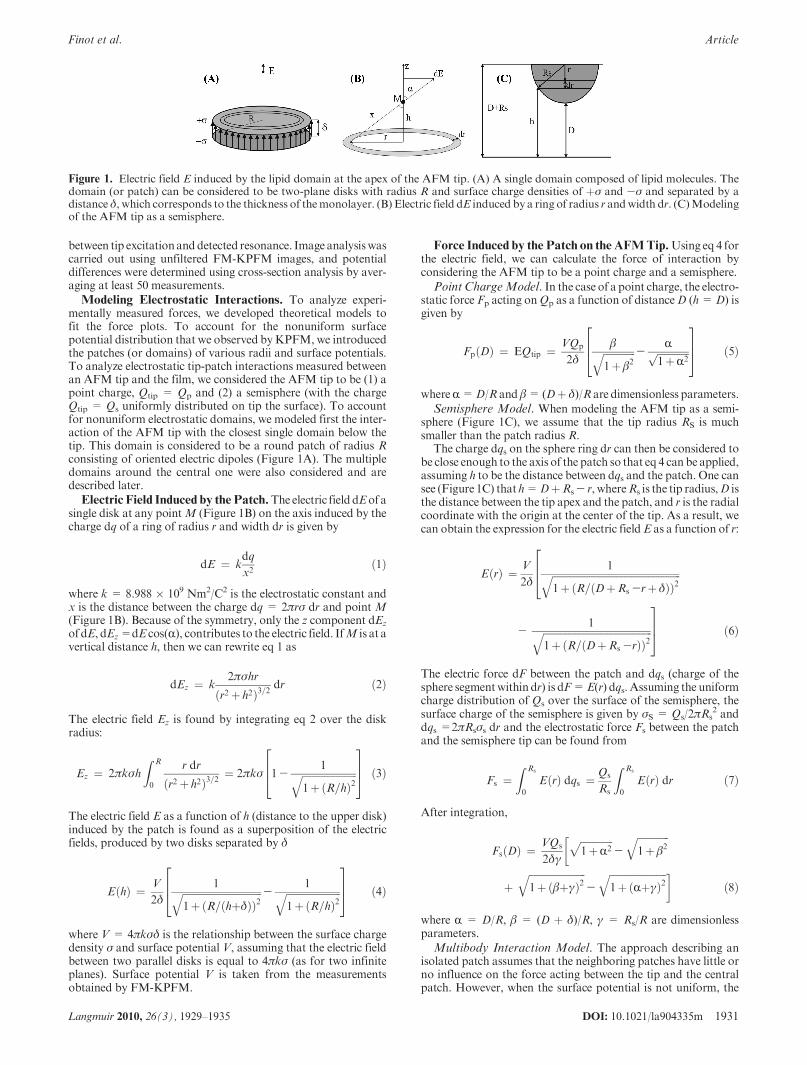
Modeling Electrostatic Interactions. To analyze experi-mentally measured forces, we developed theoretical models tofit the force plots. To account for the nonuniform surfacepotential distribution that we observed byKPFM, we introducedthe patches (or domains) of various radii and surface potentials.To analyze electrostatic tip-patch interactions measured betweenan AFM tip and the film, we considered the AFM tip to be (1) apoint charge, Qtip = Qp and (2) a semisphere (with the chargeQtip = Qs uniformly distributed on tip the surface). To accountfor nonuniform electrostatic domains, we modeled first the inter-action of the AFM tip with the closest single domain below thetip. This domain is considered to be a round patch of radius Rconsisting of oriented electric dipoles (Figure 1A). The multipledomains around the central one were also considered and aredescribed later.
Electric Field Induced by thePatch.The electric field dEof asingle disk at any pointM (Figure 1B) on the axis induced by thecharge dq of a ring of radius r and width dr is given by
dE ¼ kdq
x2ð1Þ
where k = 8.988 � 109 Nm2/C2 is the electrostatic constant andx is the distance between the charge dq = 2πrσ dr and point M(Figure 1B). Because of the symmetry, only the z component dEz
of dE, dEz=dE cos(R), contributes to the electric field. IfM is at avertical distance h, then we can rewrite eq 1 as
dEz ¼ k2πσhr
ðr2 þ h2Þ3=2dr ð2Þ
The electric field Ez is found by integrating eq 2 over the diskradius:
Ez ¼ 2πkσh
Z R
0
r dr
ðr2 þ h2Þ3=2¼ 2πkσ 1-
1ffiffiffiffiffiffiffiffiffiffiffiffiffiffiffiffiffiffiffiffiffiffi1þðR=hÞ2
q264
375 ð3Þ
The electric field E as a function of h (distance to the upper disk)induced by the patch is found as a superposition of the electricfields, produced by two disks separated by δ
EðhÞ ¼ V
2δ
1ffiffiffiffiffiffiffiffiffiffiffiffiffiffiffiffiffiffiffiffiffiffiffiffiffiffiffiffiffiffiffiffi1þðR=ðhþδÞÞ2
q -1ffiffiffiffiffiffiffiffiffiffiffiffiffiffiffiffiffiffiffiffiffiffi
1þðR=hÞ2q
264
375 ð4Þ
where V = 4πkσδ is the relationship between the surface chargedensity σ and surface potential V, assuming that the electric fieldbetween two parallel disks is equal to 4πkσ (as for two infiniteplanes). Surface potential V is taken from the measurementsobtained by FM-KPFM.
Force Induced by the Patch on theAFMTip.Using eq 4 forthe electric field, we can calculate the force of interaction byconsidering the AFM tip to be a point charge and a semisphere.
Point ChargeModel. In the case of a point charge, the electro-static force Fp acting onQp as a function of distanceD (h=D) isgiven by
FpðDÞ ¼ EQtip ¼ VQp
2δ
βffiffiffiffiffiffiffiffiffiffiffiffiffi1þβ2
q -Rffiffiffiffiffiffiffiffiffiffiffiffiffi
1þR2p
264
375 ð5Þ
whereR=D/R and β= (Dþ δ)/R are dimensionless parameters.
Semisphere Model. When modeling the AFM tip as a semi-sphere (Figure 1C), we assume that the tip radius RS is muchsmaller than the patch radius R.
The charge dqs on the sphere ring dr can then be considered tobe close enough to the axis of the patch so that eq 4 can be applied,assuming h to be the distance between dqs and the patch. One cansee (Figure 1C) that h=DþRs- r, whereRs is the tip radius,D isthe distance between the tip apex and the patch, and r is the radialcoordinate with the origin at the center of the tip. As a result, wecan obtain the expression for the electric fieldE as a function of r:
EðrÞ ¼ V
2δ
1ffiffiffiffiffiffiffiffiffiffiffiffiffiffiffiffiffiffiffiffiffiffiffiffiffiffiffiffiffiffiffiffiffiffiffiffiffiffiffiffiffiffiffiffiffiffiffiffiffiffiffiffi1þðR=ðDþRs -rþδÞÞ2
q264
-1ffiffiffiffiffiffiffiffiffiffiffiffiffiffiffiffiffiffiffiffiffiffiffiffiffiffiffiffiffiffiffiffiffiffiffiffiffiffiffiffiffiffiffiffi
1þðR=ðDþRs -rÞÞ2q
375 ð6Þ
The electric force dF between the patch and dqs (charge of thesphere segmentwithin dr) is dF=E(r) dqs. Assuming the uniformcharge distribution of Qs over the surface of the semisphere, thesurface charge of the semisphere is given by σS = Qs/2πRs
2 anddqs =2πRsσs dr and the electrostatic force Fs between the patchand the semisphere tip can be found from
Fs ¼Z Rs
0
EðrÞ dqs ¼ Qs
Rs
Z Rs
0
EðrÞ dr ð7Þ
After integration,
FsðDÞ ¼ VQs
2δγ
ffiffiffiffiffiffiffiffiffiffiffiffiffi1þR2
p-
ffiffiffiffiffiffiffiffiffiffiffiffiffi1þβ2
q�
þffiffiffiffiffiffiffiffiffiffiffiffiffiffiffiffiffiffiffiffiffiffiffi1þðβþγÞ2
q-
ffiffiffiffiffiffiffiffiffiffiffiffiffiffiffiffiffiffiffiffiffiffiffi1þðRþγÞ2
q �ð8Þ
where R = D/R, β = (D þ δ)/R, γ = Rs/R are dimensionlessparameters.
Multibody Interaction Model. The approach describing anisolated patch assumes that the neighboring patches have little orno influence on the force acting between the tip and the centralpatch. However, when the surface potential is not uniform, the
Figure 1. Electric field E induced by the lipid domain at the apex of the AFM tip. (A) A single domain composed of lipid molecules. Thedomain (or patch) can be considered to be two-plane disks with radius R and surface charge densities of þσ and -σ and separated by adistanceδ, which corresponds to the thickness of themonolayer. (B) Electric field dE induced by a ring of radius r andwidth dr. (C)Modelingof the AFM tip as a semisphere.
1932 DOI: 10.1021/la904335m Langmuir 2010, 26(3), 1929–1935
Article Finot et al.
interaction between the tip and multiple domains should beconsidered. To take this into account, we developed a multibodyinteractionmodelwhere the tipwas considered to be a point charge(or a semisphere) and the surface was viewed as an organizedstructure composed of N circular patches having two differentsurface potentials as obtained experimentally by KPFMmeasure-ments (Figure 2E). The tip is assumed to be above the center of onepatch, interacting withN patches, each separated from the tip by adistance Dj, all having the same radius R. Blue (B) and gray (G)disks (Figure 2E) represent electrostatic domains having twodifferent electric surface potentials, (-0.35 and-0.45 V), observedby FM-KPFM. The first disk (gray) is positioned in the center,right below the tip, and other disks are placed around and theirpositions are described by the radius from the center Rc. Fourcircles were considered at distances of Rcj = 2R, 3.33R, 4R, and5.9R from the central disk. It was shown that other disks placed atRcj=5.9R do not significantly alter the interaction (less than 1%).Each circle is composedof three gray and three bluedisks.The totalforce Fmb acting on the tip is then the sum of the individual forcesFcj (or Fsj depending on the modeling of the tip, point charge orsemisphere) acting between one patch and the tip:
Fmbc, sðDÞ ¼XNj¼1
Fcj, sjðDjÞ, where Dj ¼ffiffiffiffiffiffiffiffiffiffiffiffiffiffiffiffiffiffiffiffiffiffiffiffiðRcj
2 þ D2Þq
ð9Þ
Fitting Procedure. The best theoretical fit of the experimentalphysical plots was obtained by minimizing the nonlinear error χ2
χ2 ¼ minXni¼1
ðFexp - FcalcÞi2ΓF, i
" #ð10Þ
where n is the number of points in the data set, ΓFj is the error in themeasurement estimated to be around 0.1 nN, Fexp represents theexperimental forces, and Fcalc is obtained from the theoretical model.ThemethodofLevenberg-Marquardt27,28 is thenused tominimizeχ2.
There were three variable parameters for bothmodels:R- thedomain radius of the lipid patch and Qp or Qs for the charge ofthe tip and the tip radius. Therefore, the nonlinear error χ shouldbe minimized with consideration for these parameters, wherethe domain radius, tip radius, and tip charge corresponding tothe minimum nonlinear error χ should be chosen. The radius ofthe AFM tip was varied between 2 and 30 nm, the surface chargewas varied between 10-20 and 10 -16 C, and the radius of theelectrostatic domainswas variedbetween 1 and 50nm.The best fitwas obtained by minimizing the nonlinear error χ, when threeparameters were varied simultaneously as a 3 � 3 matrix.Although we varied the radius of the probe, the best fit was foundat a radius of 10 nm, as expected for these tips. We used a silicontip without any modification or applying additional voltage.
Results and Discussion
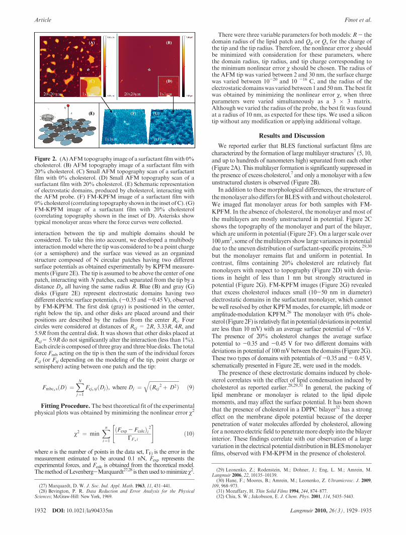
We reported earlier that BLES functional surfactant films arecharacterized by the formation of large multilayer structures7 (5, 10,and up to hundreds of nanometers high) separated from each other(Figure 2A). This multilayer formation is significantly suppressed inthe presence of excess cholesterol,7 and only amonolayer with a fewunstructured clusters is observed (Figure 2B).
In addition to these morphological differences, the structure ofthemonolayer also differs for BLESwith andwithout cholesterol.We imaged flat monolayer areas for both samples with FM-KPFM. In the absence of cholesterol, the monolayer andmost ofthe multilayers are mostly unstructured in potential. Figure 2Cshows the topography of the monolayer and part of the bilayer,which are uniform in potential (Figure 2F). On a larger scale over100 μm2, some of themultilayers show large variances in potentialdue to the uneven distribution of surfactant-specific proteins,29,30
but the monolayer remains flat and uniform in potential. Incontrast, films containing 20% cholesterol are relatively flatmonolayers with respect to topography (Figure 2D) with devia-tions in height of less than 1 nm but strongly structured inpotential (Figure 2G). FM-KPFM images (Figure 2G) revealedthat excess cholesterol induces small (10-50 nm in diameter)electrostatic domains in the surfactant monolayer, which cannotbe well resolved by other KPFMmodes, for example, lift mode oramplitude-modulation KPFM.26 The monolayer with 0% chole-sterol (Figure 2F) is relatively flat inpotential (deviations inpotentialare less than 10 mV) with an average surface potential of -0.6 V.The presence of 20% cholesterol changes the average surfacepotential to -0.35 and -0.45 V for two different domains withdeviations in potential of 100mVbetween the domains (Figure 2G).These two types of domains with potentials of-0.35 and- 0.45 V,schematically presented in Figure 2E, were used in the models.
The presence of these electrostatic domains induced by chole-sterol correlates with the effect of lipid condensation induced bycholesterol as reported earlier.28,29,31 In general, the packing oflipid membrane or monolayer is related to the lipid dipolemoments, and may affect the surface potential. It has been shownthat the presence of cholesterol in a DPPC bilayer32 has a strongeffect on the membrane dipole potential because of the deeperpenetration of water molecules afforded by cholesterol, allowingfor a nonzero electric field to penetratemore deeply into the bilayerinterior. These findings correlate with our observation of a largevariation in the electrical potential distribution inBLESmonolayerfilms, observed with FM-KPFM in the presence of cholesterol.
Figure 2. (A) AFMtopography image of a surfactant filmwith 0%cholesterol. (B) AFM topography image of a surfactant film with20% cholesterol. (C) Small AFM topography scan of a surfactantfilm with 0% cholesterol. (D) Small AFM topography scan of asurfactant film with 20% cholesterol. (E) Schematic representationof electrostatic domains, produced by cholesterol, interacting withthe AFM probe. (F) FM-KPFM image of a surfactant film with0%cholesterol (correlating topography shown in the inset ofC). (G)FM-KPFM image of a surfactant film with 20% cholesterol(correlating topography shown in the inset of D). Asterisks showtypical monolayer areas where the force curves were collected.
(27) Marquardt, D. W. J. Soc. Ind. Appl. Math. 1963, 11, 431–441.(28) Bevington, P. R. Data Reduction and Error Analysis for the Physical
Sciences; McGraw-Hill: New York, 1969.
(29) Leonenko, Z.; Rodenstein, M.; Dohner, J.; Eng, L. M.; Amrein, M.Langmuir 2006, 22, 10135–10139.
(30) Hane, F.; Moores, B.; Amrein, M.; Leonenko, Z. Ultramicrosc. J. 2009,109, 968–973.
(31) Mozaffary, H. Thin Solid Films 1994, 244, 874–877.(32) Chiu, S. W.; Jakobsson, E. J. Chem. Phys. 2001, 114, 5435–5443.
DOI: 10.1021/la904335m 1933Langmuir 2010, 26(3), 1929–1935
Finot et al. Article
When it comes to interactions with charged species, the localchanges in surface potential may play an important role becausethey may regulate the distinctive affinity of some regions over therest of the film surface. In this work, we focused on the electricalsurface potential of the PS film and how it may affect theinteraction of the AFM probe with the film’s surface. WithAFM force spectroscopy, we measured the interaction forcesbetween the AFM probe and the supported BLES monolayerwith andwithout 20%cholesterol. The approachpart of the curvewas analyzed for electrostatic interaction. In this work, wefocused only on the electrostatic force; the elastic deformationof the lipid has not been considered. The van der Waals’ forcesnormally are considerably lower than the electrostatic forces20,21
(more than 10 times lower as measured in this work); therefore,the van der Waals interactions have been neglected in this study.With atomic force spectroscopy, we showed that the interactionof the AFM probe with the electrical field, arising from the PSmonolayer, is affected by the presence of electrostatic domainsinduced by cholesterol, producing longer-ranged, stronger elec-trostatic forces. The apex of theAFMprobemay serve as amodelnanoparticle; therefore, this approach may help to investigate theinteraction of nanoparticles with PS films. Figure 2E shows aschematic representation of the electrostatic interaction of thenanoparticle (AFM probe) with electrostatic domains observedby FM-KPFM (Figure 2G).
To analyze the electrostatic part of the interaction, experimentalforce curves were fit with theoretical models. To account for thenonuniform surface potential distribution that we observed by FM-KPFM, we introduced patches (or domains) of various radii andsurface potentials. We considered the AFM probe to interact withthis film as (1) a point charge,Qtip, and (2) a semisphere. The forceinduced on the AFM tip modeled as a point charge is given by eq 5,and the force inducedon theAFMtipmodeledasapoint semisphereis given by eq 8. The experimentally measured electrical surfacepotential V was used as a parameter in a fitting procedure. Thesample with 0% cholesterol was characterized by a surface potentialof-0.6V,determined fromthe cross-sectionanalysisofFM-KPFMimages. For the samples with 20% cholesterol, two potentials wereexperimentally determined for two different domains: -0.35 and-0.45V.The domain sizeR and theAFMtip chargeQs orQtip wereused as variable parameters to obtain the best fit. We used a silicontip without any modification or applying additional voltage. Fromthe experimental force plots observed, it was clear that the tip wascharged and the electrical field produced by the surface potential ofsurfactant had a charge of the same sign as the charge on the tip,which leads to repulsion. For 0% cholesterol, the best fit wasobtained at a tip charge of 10-17C, and for 20% cholesterol, thebest fit was obtained at a tip charge of 10-16 C. The tip radius foundto give the best fit was at 10 nm in all models. The important findingis that the repulsionbetween the tip and the film increasedbecause ofthe presence of 20% cholesterol in the film and because of thepresence of a nonuniform potential distribution or nanoscaleelectrostatic domains induced by cholesterol.
When comparing the point charge to the semispheremodel, wefound experimentally a ratio of∼10 in the surface charge given bythe sphere and the point charge. To understand this, we shouldcompare charges using the effective distanceDeff to the patch.Oneshould consider the fact that the semisphere charge is not localizedat the very end of the tip but rather in the center of mass of thesemisphere, which is located at around 3/4 tip radius away fromthe tip end (Figure 1C). The effective distance in this case shouldbe enlarged by approximatelyΔ= 3Rs/4. The force acting on thetip is roughly the ratio of the charge Qtip and the effective Deff
2.Because the effective distance is larger than D for the semisphere
model, the semisphere charge Qs should be larger than the pointcharge Qp to fit the experimental force curve.
Qs
Qp¼ ðDþ ΔÞ2
D2ð11Þ
Figure 3 shows the average of experimental force plots andtheoretical fits (Figure 3a,c) and the standard deviations(Figure 3b,d). We observed that electrostatic forces were short-range forces (<5 nm) in the case of a cholesterol-free layer(Figure 3a), whereas the presence of cholesterol (20%) induceda long-range interaction (>10 nm) (Figure 3c).
Experimental force plots and the theoretical fit for the pointchargemodel and the semispheremodel (eqs 5 and 8) are shown inFigure 3a for the BLES sample with 0% cholesterol. Bothmodelsfit well with the experimental data and give a small domain radiusof around 1.2-3.7 nm. This small domain size corresponds to apatch composedof only a few lipids heads (assuming 2 nm2 for thegyration area of one lipid). This also corresponds to observationsby FM-KPFM of a relatively uniform distribution of surfacepotential (Figure 2F). Figure 3b shows the dependence ofstandard deviation (STD) on distance between the tip and thesample. For both models, the standard deviation was small overthe distances between 3 and 14 nm. The standard deviation ishigher when the tip is close to the contact point.
When using the single domain model for the sample with0% cholesterol, the best fit was obtained at R = 2.7 nm (Qtip=-1.0 � 10-17 C, V = -0.6 V, and χ = 3.9 when consideringthe AFM tip to be a point charge), R = 0.74 nm (Qtip= -7 �10-17 C,V=-0.6 V, and χ=2.2 when considering the AFM tipto be a semisphere), and Rtip = 10 nm. When using the singledomain model for a sample with 20% cholesterol, the best fit wasobtained at R = 7.4 nm (Qtip= -1.8 � 10-16 C, V = -0.35 to0.45 V, and χ = 19.8 for a point charge), and with semispheremodel, the best fit was obtained at R = 7.4 nm (Qtip= -3.8 �10-16 C, V = -0.35 to 0.45 V, and χ = 22.4).
Electrostatic forces measured on BLESwith a 20% cholesterolsample extend to a wider range of up to 15-20 nm (Figure 3c),compared to 5 nm measured on the sample with 0% cholesterol.In the case of 20% cholesterol, the analysis of the theoretical fitshows an increase in the domain radius of up to 10 times (from0.74 to 7.4 nm, point charge, single domain model) as well as anincrease in the effective charge of the tip when compared to BLESwith 0% cholesterol. The larger domain size is consistent with thenonuniform surface potential distribution shown on the KPFMimage (Figure 2G), which shows the presence of electrostaticdomains with variable radii between 10 and 50 nm. Taking intoaccount the domain size, the radius of the tip, and the long-rangenature of the interaction in the case where 20% cholesterol ispresent in the film, the point chargemodel generally gives a betterfit than the semisphere model (plot C, Figure 3c). We consideredthe interaction of the tipwith the closest single domain right belowthe tip and assumed that the presence of the AFM tip does notinfluence the uniform charge distribution on the patch surface.Because of these assumptions, one should expect a discrepancybetween model predictions and experimental results over shortranges (where the tip comes into contact and violates the surfacecharge distribution) as well as over wide ranges (where thepresence of neighboring patches should be taken into account).Direct comparison with experimental results showed that withthe exception of these two extreme limits (D < 3 nm and D >14 nm) both proposed models fit the experimental data well,although the range is slightly different for the case with no
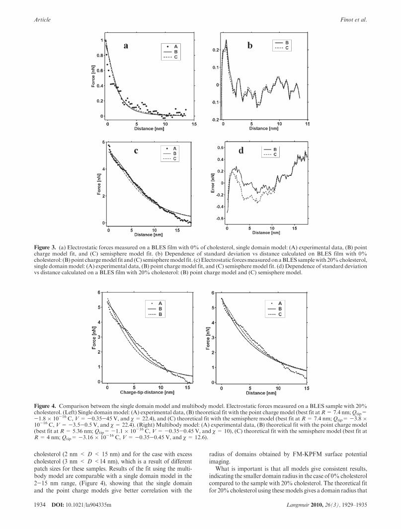
1934 DOI: 10.1021/la904335m Langmuir 2010, 26(3), 1929–1935
Article Finot et al.
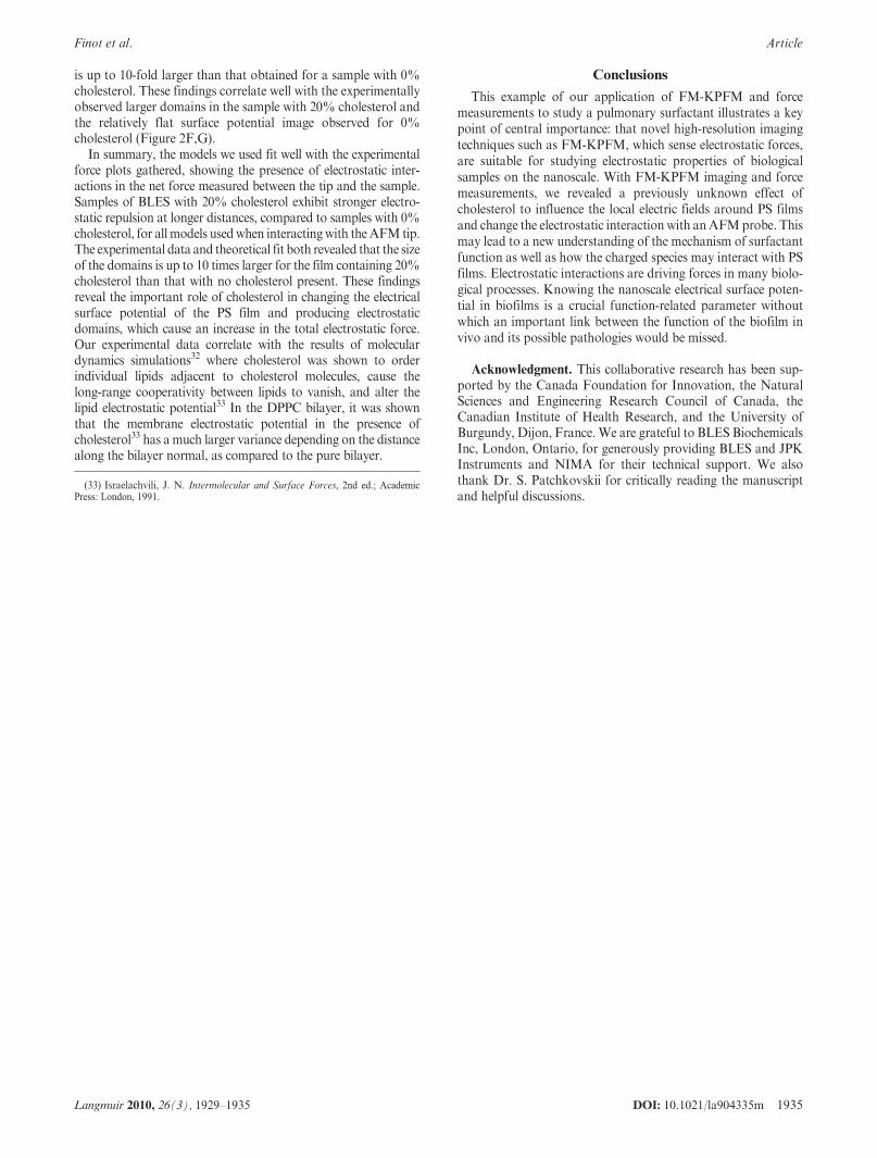
cholesterol (2 nm < D < 15 nm) and for the case with excesscholesterol (3 nm < D <14 nm), which is a result of differentpatch sizes for these samples. Results of the fit using the multi-body model are comparable with a single domain model in the2-15 nm range, (Figure 4), showing that the single domainand the point charge models give better correlation with the
radius of domains obtained by FM-KPFM surface potentialimaging.
What is important is that all models give consistent results,indicating the smaller domain radius in the case of 0%cholesterolcompared to the sample with 20% cholesterol. The theoretical fitfor 20%cholesterol using thesemodels gives a domain radius that
Figure 4. Comparison between the single domain model and multibody model. Electrostatic forces measured on a BLES sample with 20%cholesterol. (Left) Single domainmodel: (A) experimental data, (B) theoretical fit with the point chargemodel (best fit atR=7.4 nm;Qtip=-1.8� 10-16 C, V=-0.35-45 V, and χ= 22.4), and (C) theoretical fit with the semisphere model (best fit at R= 7.4 nm;Qtip=-3.8�10-16 C, V=-3.5-0.5 V, and χ= 22.4). (Right) Multibody model: (A) experimental data, (B) theoretical fit with the point charge model(best fit at R= 5.36 nm; Qtip=-1.1 � 10-16 C, V= -0.35-0.45 V, and χ= 10), (C) theoretical fit with the semisphere model (best fit atR= 4 nm; Qtip= -3.16 � 10-16 C, V= -0.35-0.45 V, and χ= 12.6).
Figure 3. (a) Electrostatic forces measured on a BLES film with 0% of cholesterol, single domain model: (A) experimental data, (B) pointcharge model fit, and (C) semisphere model fit. (b) Dependence of standard deviation vs distance calculated on BLES film with 0%cholesterol: (B) point chargemodel fit and (C) semispheremodel fit. (c)Electrostatic forcesmeasuredonaBLESsamplewith20%cholesterol,single domainmodel: (A) experimental data, (B) point chargemodel fit, and (C) semispheremodel fit. (d) Dependence of standard deviationvs distance calculated on a BLES film with 20% cholesterol: (B) point charge model and (C) semisphere model.
DOI: 10.1021/la904335m 1935Langmuir 2010, 26(3), 1929–1935
Finot et al. Article
is up to 10-fold larger than that obtained for a sample with 0%cholesterol. These findings correlate well with the experimentallyobserved larger domains in the sample with 20% cholesterol andthe relatively flat surface potential image observed for 0%cholesterol (Figure 2F,G).
In summary, the models we used fit well with the experimentalforce plots gathered, showing the presence of electrostatic inter-actions in the net force measured between the tip and the sample.Samples of BLES with 20% cholesterol exhibit stronger electro-static repulsion at longer distances, compared to samples with 0%cholesterol, for allmodels usedwhen interactingwith theAFM tip.The experimental data and theoretical fit both revealed that the sizeof the domains is up to 10 times larger for the film containing 20%cholesterol than that with no cholesterol present. These findingsreveal the important role of cholesterol in changing the electricalsurface potential of the PS film and producing electrostaticdomains, which cause an increase in the total electrostatic force.Our experimental data correlate with the results of moleculardynamics simulations32 where cholesterol was shown to orderindividual lipids adjacent to cholesterol molecules, cause thelong-range cooperativity between lipids to vanish, and alter thelipid electrostatic potential33 In the DPPC bilayer, it was shownthat the membrane electrostatic potential in the presence ofcholesterol33 has amuch larger variance depending on the distancealong the bilayer normal, as compared to the pure bilayer.
Conclusions
This example of our application of FM-KPFM and forcemeasurements to study a pulmonary surfactant illustrates a keypoint of central importance: that novel high-resolution imagingtechniques such as FM-KPFM, which sense electrostatic forces,are suitable for studying electrostatic properties of biologicalsamples on the nanoscale. With FM-KPFM imaging and forcemeasurements, we revealed a previously unknown effect ofcholesterol to influence the local electric fields around PS filmsand change the electrostatic interaction with anAFMprobe. Thismay lead to a new understanding of the mechanism of surfactantfunction as well as how the charged species may interact with PSfilms. Electrostatic interactions are driving forces in many biolo-gical processes. Knowing the nanoscale electrical surface poten-tial in biofilms is a crucial function-related parameter withoutwhich an important link between the function of the biofilm invivo and its possible pathologies would be missed.
Acknowledgment. This collaborative research has been sup-ported by the Canada Foundation for Innovation, the NaturalSciences and Engineering Research Council of Canada, theCanadian Institute of Health Research, and the University ofBurgundy, Dijon, France. We are grateful to BLES BiochemicalsInc, London, Ontario, for generously providing BLES and JPKInstruments and NIMA for their technical support. We alsothank Dr. S. Patchkovskii for critically reading the manuscriptand helpful discussions.
(33) Israelachvili, J. N. Intermolecular and Surface Forces, 2nd ed.; AcademicPress: London, 1991.