Influence of cholesterol on phospholipid bilayers phase domains as detected by Laurdan fluorescence
Upload
independentCategory
view
3download
0
Atomic-Scale Structure and Electrostatics of AnionicPalmitoyloleoylphosphatidylglycerol Lipid Bilayers with Na1 Counterions
Wei Zhao,* Tomasz Rog,*z Andrey A. Gurtovenko,§ Ilpo Vattulainen,y{k and Mikko Karttunen***Biophysics and Statistical Mechanics Group, Laboratory of Computational Engineering; yLaboratory of Physics and Helsinki Instituteof Physics, Helsinki University of Technology, Espoo, Finland; zDepartment of Biophysics, Jagiellonian University, Krakow, Poland;§Computational Laboratory, Institute of Pharmaceutical Innovation, University of Bradford, Bradford, West Yorkshire, United Kingdom;{Institute of Physics, Tampere University of Technology, Tampere, Finland; kMEMPHYS–Center for Biomembrane Physics, Universityof Southern Denmark, Odense, Denmark; and **Department of Applied Mathematics, The University of Western Ontario, London,Ontario, Canada
ABSTRACT Anionic palmitoyloleoylphosphatidylglycerol (POPG) is one of the most abundant lipids in nature, yet its atomic-scale properties have not received significant attention. Here we report extensive 150-ns molecular dynamics simulations of apure POPG lipid membrane with sodium counterions. It turns out that the average area per lipid of the POPG bilayer underphysiological conditions is ;19% smaller than that of a bilayer built from its zwitterionic phosphatidylcholine analog,palmitoyloleoylphosphatidylcholine. This suggests that there are strong attractive interactions between anionic POPG lipids,which overcome the electrostatic repulsion between negative charges of PG headgroups. We demonstrate that interlipidcounterion bridges and strong intra- and intermolecular hydrogen bonding play a key role in this seemingly counterintuitivebehavior. In particular, the substantial strength and stability of ion-mediated binding between anionic lipid headgroups leadsto complexation of PG molecules and ions and formation of large PG-ion clusters that act in a concerted manner. The ion-mediated binding seems to provide a possible molecular-level explanation for the low permeability of PG-containing bacterialmembranes to organic solvents: highly polar interactions at the water/membrane interface are able to create a high free energybarrier for hydrophobic molecules such as benzene.
INTRODUCTION
Phosphatidylglycerols (PGs) are among the most abundant
lipids in nature. In higher plants, they constitute ;20%–30%
of lipids (1). They are also one of the major constituents of
bacterial membranes (2); mixed PG/phosphatidylethanola-
mine (PE) lipid bilayers have been used as models for
bacterial membranes (3–6). In contrast, the concentration of
PG lipids in animal cell membranes is low. In red blood cells
PGs constitute ;2% of all phospholipids (7). Though, whereas
being less common in animal membranes, they are present in
mitochondria and some tissues. From the physical point of
view, PGs, together with phosphatidylserine (POPS) and
cardiolipin, are anionic, the former two carrying a unit
positive charge and the latter being divalent under physio-
logical conditions.
The nonzero net charge of PGs makes them different from
commonly studied zwitterionic (neutral) lipids. Since direct
electrostatic interactions between charged species are rather
strong, PGs, as well as other charged lipids, are considered
membrane stabilizers and destabilizers (8) and are believed to
play an important role in controlling membrane-peptide/
protein interactions. For this reason, PGs have been suggested
as one of the possible components in immunoliposomes,
generic drug delivery vehicles (9). It is also noteworthy that
PGs constitute up to 80% of membrane lipids of Staphylo-
coccus aureus, a common human pathogen with its methi-
cillin-resistant strain (MRSA)—the so-called hospital
bacteria—being a serious problem in hospitals around the
world. Hence, it has been suggested that electrostatics may
play an important role in membrane selectivity of bacteria.
Atomic-scale details of these electrostatic interactions would,
therefore, be very helpful and important for a better under-
standing of how such a selectivity can be controlled to make
drugs more efficient.
Furthermore, studies of toxic effects of organic solvents on
bacteria have revealed that relative concentrations of PG and
PE lipids in their membranes could change depending on the
chemical character of solvent molecules (10,11). Such an
alteration in headgroup composition seems to be a means to
change permeability of a membrane and to preserve its
stability. Precise mechanisms responsible for solvent-induced
effects are not known, due to the lack of detailed microscopic
information.
Molecular dynamics (MD) simulations provide a detailed
microscopic picture of atomic-scale interactions and pro-
cesses, which are not easily accessible by experimental
methods. Systems having an abundance of charged compo-
nents, such as anionic or cationic membranes, and systems
with salt ions are computationally demanding—first reliable
MD studies in the field were reported only recently: anionic
POPS lipid bilayers (12,13), PG monolayers (14), mixed
cationic/zwitterionic lipid membranes (15–17), phosphati-
dylcholine lipid membranes with salt (18–21), and ion
leakage through water pores in PC membranes (22).
Submitted March 31, 2006, and accepted for publication September 5, 2006.
Address reprint requests to Mikko Karttunen, E-mail: [email protected];
Web: www.softsimu.org.
� 2007 by the Biophysical Society
0006-3495/07/02/1114/11 $2.00 doi: 10.1529/biophysj.106.086272
1114 Biophysical Journal Volume 92 February 2007 1114–1124

Given the abundance of PGs in nature, it is somewhat
surprising that, to the best of our knowledge, there has been
only one atomistic computational study of pure PG lipid
bilayers (23) focusing on the hydrogen bonding between the
lipids. All related simulation studies to date have dealt with
mixtures of PGs with other lipids (3,6) or with PG monolayers
at the air/water interface (14). On the experimental side, pure
POPG bilayers, or POPG mixed with PC at high concentra-
tions, have been studied (to our knowledge) only by Cowley
et al. (24) in the 1970s and more recently by Pozo-Navas et al.
(25,26). The phase behavior (27,28) and various aspects of the
structure-function relationship of POPGs in PE bilayers
(8,29–32) have been experimentally characterized, though.
In this work we employ atomistic MD simulations to gain
a thorough understanding of the properties of PG-rich
membranes through a systematic comparison with corre-
sponding PC bilayers. This is needed to establish the fun-
damental molecular and atomistic level physical origin of the
observed phenomena. This knowledge should be useful also
in interpreting experimental findings, and it offers a possible
explanation as to why nature has chosen to use charged lipids
structures such as bacterial membranes.
We focus on the structural and electrostatic properties
of single-component palmitoyloleoylphosphatidylglycerol
(POPG) lipid membranes. Zwitterionic palmitoyloleoylphos-
phatidylcholine (POPC) was used as a control as its properties
are well known and, importantly, since PCs are the most
abundant lipids in biological membranes. As they have a dipole
in their headgroup, their behavior is expected to be quite
different from that of anionic PGs. Indeed, when compared to a
POPC bilayer, the average area per lipid in POPG bilayer
simulations turns out to be much smaller. That seems to be
characteristic for charged bilayer as similar compression has
also been seen in other anionic bilayers (see, e.g., Petrache et al.
(33)). Our simulations reveal the physical mechanism respon-
sible for that, and the key results of this work are the following:
there are strong attractive interactions between anionic POPG
lipids, which overcome the electrostatic repulsion between
negative charges of PG headgroups. We show that interlipid
counterion bridges and strong intra- and intermolecular hy-
drogen bonding lead to this seemingly counterintuitive behav-
ior. In particular, the strength and stability of ion-mediated
binding between anionic lipid headgroups leads to complex-
ation of PG molecules and ions and formation of large PG-ion
clusters that act in a concerted manner. The ion-mediated
binding may provide a possible molecular-level mechanism for
the low permeability of PG-containing bacterial membranes to
organic solvents: highly polar interactions at the water/
membrane interface are able to create a high free energy
barrier for hydrophobic molecules such as benzene.
The rest of this work is organized as follows: in the next
section we present the model and simulation details. Then,
we first show the results for the basic structural quantities
such as the area per lipid, density profiles, and order param-
eters. After that we come to the main results: hydrogen, ion
bonding, and their consequences such as clustering. Finally,
we finish with conclusions and a discussion.
MODEL AND SIMULATION DETAILS
We have studied a lipid bilayer system consisting of 128
anionic POPGs and 3527 water molecules. A total of 128 Na1
counterions was included to ensure electroneutrality. The force
field parameters for POPG (Fig. 1) were taken from the united
atom force field of Berger et al. (34). They are based on the
GROMOS force field for lipid headgroups, the Ryckaert-
Bellemans potential functions (35,36) for hydrocarbon chains,
and the optimized parameters for liquid simulations parameters
(34) for the Lennard-Jones interactions between united CHn
groups of nonpolar lipid chains. Partial charges for the glycol
group (H16-O16-C13-C12-O15-H15 in the headgroup of
POPG; see Fig. 1) were taken from 1,2-propanediol (D. Warren,
unpublished). The force field parameters for POPG are available
online at http://www.softsimu.org/downloads.shtml. The SPC
model (38) was used for water, and standard GROMACS force
field parameters were employed for sodium ions. For subtleties
related to modeling of Na1, please see Patra and Karttunen (39).
All Lennard-Jones interactions were cut off at 1.0 nm
without shift or switch functions. Lipid bonds were con-
strained using the LINCS algorithm (40), and the SETTLE
algorithm (41) was used for water molecules. The electrostatic
interactions were handled by the particle mesh Ewald (PME)
method (42,43), which has been shown to perform well in
membrane simulations (44–46).
The simulations were performed in the NpT ensemble with
a time step of 2 fs, using the GROMACS simulation package
(47). The temperature was kept constant at T ¼ 310 K using
the Berendsen thermostat (48) with a coupling time constant
of 0.1 ps. Lipid molecules and solvent (water molecules and
sodium ions) were separately coupled to a heat bath. The
Berendsen barostat (48) with a coupling constant of 1.0 ps was
employed to keep pressure constant (1 bar). The pressure cou-
pling was applied semiisotropically: the box size in the direc-
tion of the bilayer normal (z axis) and the cross sectional area
(xy-plane) were allowed to vary independently.
The starting structure of a POPG bilayer was prepared using
an equilibrated POPC bilayer (49): the choline moieties of
POPC lipids were replaced with the glycerol groups such that
equal numbers of D-POPG and L-POPG lipids were created,
leading to a lipid bilayer system with neutral chirality
(racemic). The two cases differ with respect to the chirality of
the C12 site (50), and hence this choice guarantees that our lipid
bilayer system is characterized by neutral chirality. After initial
energy minimization, a short 10-ps run in the NVT ensemble
was performed to remove unphysical voids due to structural
modifications. The resulting POPG system was simulated for
150 ns. By analyzing the time evolution of the area per lipid and
the coordination of Na1 counterions with lipids and water, we
concluded that the system had equilibrated within 70 ns (see
Fig. 2). The time range from 70 to 150 ns was therefore used for
POPG Bilayers 1115
Biophysical Journal 92(4) 1114–1124

analysis. In total, the MD simulations took;12,000 CPU hours
on AMD Opteron 2.2 GHz computers (AMD, Sunnyvale, CA).
Below, the properties of POPG are compared with those of
POPC. Hence, for the purpose of comparison, a comple-
mentary simulation of a POPC lipid bilayer containing 128
POPC and 3655 water molecules was also conducted at a
temperature of 310 K for 30 ns. Our choices for the force
field and the initial (and already equilibrated) configuration
are the same as in our previous simulations (49). For the
given temperature and hydration, both studied systems,
POPG and POPC, are in the fluid (liquid-disordered) phase.
The experimenatlly determined main transition temperatures
are below 273 K for both POPG (� 273 K) (26,27) and
POPC (268 K) (51). Our data (in the following sections) are
also consistent with the liquid-disordered phase.
RESULTS
Area per lipid
The average area per lipid, ÆAæ, is perhaps the most widely
used quantity to characterize lipid bilayer systems, since it
affects a variety of physical processes such as lateral
diffusion, membrane elastic properties, and permeation. The
time evolution of the area per lipid is shown in Fig. 2. The long
equilibration time visible in the figure is mainly due to the
slow kinetics associated with the binding of counterions with
the charged POPG lipids and in part due to hydrogen-bond
formation among the lipids. Similar findings have also been
made in simulations of anionic POPS bilayers (13) and in
bilayer simulations under the influence of salt (17,18,20,21).
For the average area per lipid in a POPG bilayer we obtain
ÆAPOPGæ ¼ 0.530 6 0.006 nm2. This is a remarkably small
FIGURE 1 Chemical structures and the num-
bering of atoms of POPG and POPC molecules
used in this study.
FIGURE 2 Time evolution of the area per lipid for a POPG bilayer.
Dashed line shows the average value in equilibrium, ÆAæ.
1116 Zhao et al.
Biophysical Journal 92(4) 1114–1124

number compared to typical values in corresponding zwit-
terionic lipid systems. For example, for a POPC bilayer
studied in this work we found ÆAPOPCæ¼ 0.658 6 0.009 nm2
at the same temperature. Sphingomyelin bilayers in turn have
yielded average areas per lipid of the order of 0.52 nm2, and
the lipid hydrocarbon chains in these systems are known to
be highly ordered. Similar enhanced ordering is therefore
expected for POPG too (see below).
Comparison of our results to experimental data is difficult
since, to our knowledge, there are no experimental estimates
for the average area per lipid in single-component POPG
bilayers. However, comparison to previous related simula-
tions is possible. Murzyn et al. (6) used the Voronoi
tessellation technique to extract the average area per lipid
from simulations of a POPE/POPG mixture. They reported a
value of 0.628 6 0.003 nm2 for the average area per POPG,
which is considerably larger than the value found in our work.
One has to note, however, that the result by Murzyn et al.
cannot be directly compared to our findings. This stems from
the fact that they applied the Voronoi tessellation technique to
a binary system, and then there is no unique way to decom-
pose the free area (volume) among the different lipid com-
ponents. Hence the areas of two-dimensional Wigner-Seiz
cells in binary systems do not correspond to the area per lipid
measured in single-component lipid bilayers. This issue has
been extensively discussed in computational studies of
cholesterol in phospholipid bilayers (52–55) and also in
experiments on mixtures of nonsterol natural lipids (56).
A better, but also indirect comparison can be provided by
MD simulations of pure POPS bilayers (13). Although head-
groups of POPG and POPS lipids have different chemical
structures, both of these lipids are anionic under physiological
conditions. Mukhopadhyay et al. obtained 0.55 6 0.01 nm2 for
the average area per POPS under simulation conditions similar
to those employed in this study—close to 0.53 nm2 found here
for a POPG bilayer. These findings are also in agreement with
Cowley et al. (24), who studied PC/POPG bilayers at different
concentrations under varying hydration conditions.
We also computed the autocorrelation function to measure the
characteristic timescales for area fluctuations of POPGs (data not
shown). As expected on the basis of the above discussion, they
turned out to be much longer than those observed for POPC
bilayers (49). The above observation indicates that an anionic
POPG bilayer is less fluid than one would naıvely expect (27): its
densely packed structure, which seems to be counterintuitive at
first sight, is caused by tight binding of anionic lipids through
relatively stable ion bridges (see the section Interactions of Na1
ions with water/membrane interface).
Density profiles
Next, we consider the density profiles of various bilayer
constituents (see Fig. 3).
The maxima of the (mass or electron) density profiles are
often used to estimate the thickness of the bilayer (6,44,57).
As in previous studies, we considered the distance between
the average positions of phosphate atoms in the two leaflets
of a lipid bilayer. That distance computed from the electron
density profile yielded 4.39 6 0.02 nm for the phosphate-
phosphate distance dP-P. The result is in line with the
previous simulation study of a POPG bilayer at 310 K (23).
The maximum of the mass density profile of POPG lipids
turns out to be located at the average position of the ester groups
instead of the phosphate groups as has been found for pho-
sphatidylcholine bilayers (49). The most remarkable feature is
the distribution of Na1 ions characterized by two peaks in the
interface region (see Fig. 4). The first (main) peak is located at
�1.9 nm from the bilayer center and overlaps with the peak due
to the POPG carbonyl ester groups. Elmore has recently made a
similar observation (23). The second, much smaller peak is
at�2.65 nm. Further analysis shows that this peak corresponds
to Na1 ions mainly located in the water phase, close to the
membrane but beyond the phosphate groups, although the
FIGURE 3 Component-wise density profiles across the bilayer as a func-
tion of distance from the membrane center (z¼ 0). Gly: the glycol group (H16,
O16, C13, C12, O15, H15); Phos: the phosphate group (O11, O12, O13, O13,
P); and Carb: the carbonyl ester group (C21, O21, O22, C31, O31, O32).
FIGURE 4 Scaled number densities for various constituents of the POPG
system. The number density of lipids is not scaled, whereas the densities of
water, phosphorus, O22, O32, and Na1 ions are scaled by dividing them by
70, 10, 10, 10, and 7, respectively. The bilayer center is at z ¼ 0.
POPG Bilayers 1117
Biophysical Journal 92(4) 1114–1124
distribution of Na1 is strongly affected by the surface potential
of the negatively charged surface of a POPG bilayer. Hence the
positively charged sodium ions tend to avoid being in close
contact with the negatively charged phosphate groups and
instead prefer interactions with the ester groups.
The peaks of the density profiles of phosphodiester oxygens
(O13, O14), hydroxyl oxygens O16 and O15 (see Fig. 1) are
located at�2.2 nm, 2.05 nm, and 2.0 nm, respectively (data not
shown). When these numbers are compared to the distribution
of Na1 ions, they suggest that sodium ions interact more
favorably with ester groups rather than phosphodiester moieties.
Meanwhile, sodium ions corresponding to the second, smaller
peak of the distribution (�2.65 nm) are not bound to lipids (see
the section Interactions of Na1 ions with water/membrane
interface for a detailed discussion). Due to strong electrostatic
interactions, all the Na1 ions stay near lipid headgroups and
interact with various oxygen atoms. Yet the mutual electrostatic
repulsion between the Na1 ions together with their thermal
motion and energy compensation from hydration competes
with the tendency of sodium ions to reside close to the
headgroups. The two effects together lead to the existence of the
broad second peak in the distribution of the Na1 ions.
Headgroup orientation
Due to the glycerol group, it is not straightforward to
characterize the orientation of PG headgroups as compared
to that of phosphatidylcholine headgroups defined by their
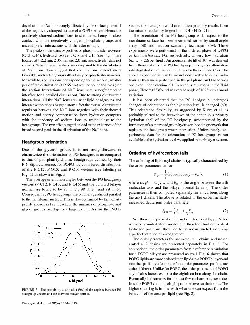
P-N dipoles. Hence, for POPG we considered distributions
of the P-C12, P-O15, and P-O16 vectors (see labeling in
Fig. 1) as shown in Fig. 5.
The average orientation angles between the PG headgroup
vectors (P-C12, P-O15, and P-O16) and the outward bilayer
normal are found to be 85 6 2�, 98 6 3�, and 89 6 6�.
Consequently, PG headgroups are on average almost parallel
to the membrane surface. This is also confirmed by the density
profile shown in Fig. 3, where the maxima of phosphate and
glycol groups overlap to a large extent. As for the P-O15
vector, the average inward orientation possibly results from
the intramolecular hydrogen bond O15-H15-O12.
The orientation of the PG headgroup with respect to the
membrane surface has been examined earlier by small angle
x-ray (58) and neutron scattering techniques (59). These
experiments were performed in the ordered phase of DPPG
or Escherichia coli PG, respectively, at very low hydration
(nwater ; 2.6 per lipid). An approximate tilt of 30� was derived
from these data for the PG headgroup, though an alternative
interdigitated structure could not be strictly excluded (59). The
above experimental results are not comparable to our simula-
tions as they were performed in the gel phase, and the former
one even under varying pH. In recent simulations in the fluid
phase, Elmore (23) found an average angle of 102� with a broad
variation.
It has been observed that the PG headgroup undergoes
changes of orientation as the hydration level is changed (60).
This orientation flexibility, as suggested by Kurze et al., is
probably related to the breakdown of the continuous primary
hydration shell of the PG headgroup, accompanied by the
formation of an interheadgroup hydrogen-bonding network that
replaces the headgroup-water interaction. Unfortunately, ex-
perimental data for the orientation of PG headgroup are not
available at the hydration level we applied in our bilayer system.
Ordering of hydrocarbon tails
The ordering of lipid acyl chains is typically characterized by
the order parameter tensor
Sab ¼1
2Æ3cosua cosub � dabæ; (1)
where a, b ¼ x, y, z, and ua is the angle between the ath
molecular axis and the bilayer normal (z axis). The order
parameter is then computed separately for all carbons along
the acyl chains. The above is related to the experimentally
measured deuterium order parameter
SCD ¼2
3Sxx 1
1
3Syy: (2)
We therefore present our results in terms of |SCD|. Since
we used a united atom model and therefore had no explicit
hydrogen positions, they had to be reconstructed assuming
a perfect tetrahedral arrangement.
The order parameters for saturated sn-1 chains and unsat-
urated sn-2 chains are presented separately in Fig. 6. For
comparison, the order parameters from a reference simulation
for a POPC bilayer are presented as well. Fig. 6 shows that
POPG lipids are more ordered than lipids in a POPC bilayer and
that the qualitative features of the order parameter profiles are
quite different. Unlike for POPC, the order parameter of POPG
acyl chains increases up to the eighth carbon along the chain.
Eventually it decreases for the last few carbons but, neverthe-
less, the POPG chains are highly ordered even at their ends. The
higher ordering is in line with what one can expect from the
behavior of the area per lipid (see Fig. 2).FIGURE 5 The probability distribution P(a) of the angle a between PG
headgroup vectors and the outward bilayer normal.
1118 Zhao et al.
Biophysical Journal 92(4) 1114–1124

Hydrogen bonding
Experimental observations have suggested that the hydroxyl
group of a PG lipid has the potential to form both intra- and
intermolecular hydrogen bonds (61). To determine whether
these hydrogen bonds exist, we follow the generally
accepted approach and define two geometrical parameters:
the distance r between the hydrogen and the acceptor, and
the donor-hydrogen-acceptor angle (zero extended) a. The
conditions r # 0.25 nm and a # 60� were then used as
criteria for a hydrogen bond (H-bond) to exist.
On the average, we found that O16 forms ;0.15 intra-
molecular and ;0.47 intermolecular hydrogen bonds with
various lipid oxygen atoms per POPG molecule. The inter-
molecular hydrogen bonds between the O16-H16 groups and
carbonyl groups are the most abundant hydrogen bonds
found in our study. We will discuss this issue in more detail
below. Table 1 summarizes the average numbers of intra-
and intermolecular hydrogen bonds.
Interactions of Na1 ions withwater/membrane interface
Radial distribution functions and coordination numbers
Fig. 7 shows the radial distribution functions (RDFs) of
various oxygen atoms (including intermolecular and intra-
molecular parts) with the hydrogen atom H16 in the POPG
headgroup. The results for H15 are not shown since it forms
intramolecular hydrogen bonds with the phosphoether
oxygens. In Fig. 7, the first minima in the RDF for all the
carbonyl and phosphodiester oxygen atoms are located at
;0.24 nm, indicating hydrogen bonding with H16 hydrogen
atoms. This confirms intermolecular hydrogen bonding in
the POPG bilayer.
The distribution of Na1 in Fig. 8 suggests that a large
amount of sodium ions are located in the ester region of the
water/membrane interface, rather than in the phosphate
region. Clearly, there are prominent peaks with oxygen atoms
O22 and O32 associated with the ester bonds, followed by
O15 in the glycerol part of the headgroup. The remaining
peaks related to O16 (also part of the glycerol group) and
oxygen atoms O13 and O14 in the phosphate group are con-
siderably less significant.
The structure and population of oxygen atoms around the
Na1 ions can also be characterized by coordination numbers
(39) computed by integration of the corresponding RDF over
the first coordination shell,
nNa
1
coord ¼4p
VNa
1
Z rmin
0
drr2gðrÞ; (3)
FIGURE 6 Deuterium order parameters SCD of saturated sn-1 (top) and
unsaturated sn-2 (bottom) chains in POPG and POPC bilayers.
TABLE 1 Average number of intermolecular and
intramolecular hydrogen bonds in a POPG lipid molecule.
The errors given are standard error estimates. See Fig. 1
for atom labeling.
H-donor H-acceptor Intermolecular Intramolecular
O15 O12 – 1.00 6 0.01
O16 O11 0.06 6 0.02 –
O16 O12 0.01 6 0.01 0.02 6 0.01
O16 O13, O14 0.07 6 0.02 –
O16 O15 0.05 6 0.02 0.05 6 0.02
O16 O16 0.02 6 0.01 –
O16 O21 0.07 6 0.01 0.04 6 0.01
O16 O22 0.14 6 0.02 0.02 6 0.01
O16 O31 0.01 6 0.01 0.01 6 0.01
O16 O32 0.05 6 0.02 0.01 6 0.01
FIGURE 7 RDFs of various oxygen atoms with the hydroxyl hydrogen
H16; see Fig. 1 for atom labeling.
POPG Bilayers 1119
Biophysical Journal 92(4) 1114–1124

where VNa1 is the van der Waals volume of a sodium ion and
rmin is the position of the first minimum of the RDF. This
coordination number nNa1coord describes the number of atoms of
interest in the first coordination shell of a Na1 ion.
From Eq. 3 one finds, on average, 3.36, 0.91, 0.60, 0.40,
0.19, and 0.04 oxygen atoms from water, O22, O32, O15,
O16, and from phosphodiester oxygen atoms, respectively,
around the Na1 ion.
To get a more detailed view on how Na1 ions interact with
different charged groups, in Fig. 9 we show the coordination
numbers of Na1 ions with various oxygen atoms as a func-
tion of the distance from the bilayer center.
The total coordination number shows a small increase,
from ;5.0 deep inside the lipid bilayer to ;5.7 in the
aqueous phase. The coordination numbers of Na1 ions with
lipid oxygen atoms show a maximum in the ester region of
the water/membrane interface, indicating strong interactions
between Na1 ions and carbonyl groups. Due to binding of
sodium ions to the membrane, water molecules are squeezed
out from the first coordination shell.
In the section Density profiles we found that the density
profile of sodium ions was characterized by two peaks in the
membrane-water interfacial region. Fig. 10 elucidates the
origin of this behavior: the first peak in the density profile of
sodium ions around z � 1.9 nm is due to ions bound to lipid
oxygens and especially those bound to the oxygens in the
ester groups. The second peak at z ; 2.65 nm, in turn, arises
from those sodium ions that are not bound to any lipid
oxygens but are rather confined to water molecules in the
vicinity of the membrane-water interface.
Ion bridges and ion-lipid clusters
The term ‘‘ion bridge’’ refers to the case in which two lipids
bind to each other via a Na1 ion. To characterize the existence
of ion bridges, we define an ion-lipid bond to occur when the
carbonyl oxygen is within a distance of 0.33 nm from a Na1
ion, which is the radius of the first shell of Na1 ions.
Here, we only define lipid carbonyl oxygens (O22, O32)
instead of all lipid oxygens as binding sites available for ion
bridges (19). This is justified since the bonding between Na1
ions and carbonyl oxygens is much stronger compared to
interactions between Na1 ions and other lipid oxygens.
These ion bridges constitute the majority of all interactions
between Na1 ions and lipid, as seen above.
When analyzing the trajectory we found, on average, 193 6 5
ion-lipid bonds (in the model of 128 PGs and 128 sodium ions),
involving 63% of Na1 ions and 97% of lipids. The percentages
for Na1 ions binding to one, two, three, and four lipids are,
respectively, 9%, 24%, 24%, and 6%. Fig. 11 illustrates some of
the cases. As for lipids, the percentages binding to one, two, and
three Na1 ions are, respectively, 45%, 51%, and 1%.
As we can see, the majority of Na1 ions bind to two or
three lipids, whereas most of the lipids bind to one or two
Na1 ions with almost equal probabilities. It is observed that,
although each lipid has two binding sites for ion bridging, the
cases with an individual Na1 ion binding to two carbonyl
oxygens within the same lipid are rare.
As seen above, an ion often binds to more than one lipid.
That makes it possible for the lipids to form larger connected
FIGURE 8 RDFs of various oxygen atoms with Na1 ions.
FIGURE 9 Average coordination numbers of Na1 ions with various lipid
oxygens, water oxygens, as well as the total coordination numbers
(including contributions from all oxygen atoms in POPG) as a function of
the distance from the bilayer center.
FIGURE 10 Distributions of Na1 bound and unbound to lipid oxygens
as functions of distance from the bilayer center.
1120 Zhao et al.
Biophysical Journal 92(4) 1114–1124

clusters in which the lipids are linked to each other by ion
bonds. We define such a continuous and connected network as
an ion-lipid cluster. Fig. 12 a shows a typical configuration of
an ion-lipid cluster. We also studied the distribution of the
cluster sizes as shown in the inset in Fig. 13. Although smaller
clusters appear frequently, most of the lipids belong to larger
clusters. As seen in the inset in Fig. 13, some very large clusters
also appear. An example of such a cluster is shown in Fig. 12 b.
It can be easily imagined that the existence of clusters has
an effect on the dynamics of the whole bilayer since the
robust ion bridges force the lipids to move collectively. This
suggestion is supported by the strong correlation between the
evolution of the area per lipid and the number of clusters
(Figs. 2 and 13). The slow process of the formation and
breaking of ion-lipid bonds is likely to be the key factor that
makes the equilibration of the POPG bilayer slow.
Water orientation
Ordering of water molecules in the vicinity of the water/
membrane interface can be characterized by the time-
averaged projection of the water dipole unit vector ~mðzÞonto the bilayer normal n~,
PðzÞ ¼ Æ~mðzÞ � n~æ ¼ Æcosfæ; (4)
where z is the z-component of the center of mass of a water
molecule, and f is the angle which the water dipole vector
makes with the bilayer normal.
The orientation of water dipoles as a function of the
distance from the bilayer center is presented in Fig. 14. Due to
the negative surface charge of the POPG bilayer, interfacial
water is highly polarized and distinctly different from the
POPC case. Whereas in POPC there is a single minimum in
the interfacial region, the ordering of water within the POPG
bilayer is characterized by a rather deep minimum around
2.4 nm and a broad maximum around 1.9 nm. The water
orientation profile reflects the total charge density profile of
the POPG/water/ion system shown in Fig. 15.
FIGURE 11 Snapshots of lipids binding to Na1 ions. The cases shown
here correspond to the binding of (a) three lipids and two waters with a
Na1 ion, and (b) four lipids with a Na1 ion.
FIGURE 12 A snapshot of a typical configuration (one leaflet) demon-
strating the existence of clusters. Each lipid (red) is represented by the
average position of its two carbonyl oxygens. An ion-lipid bond (light blue
dotted line; an ion can be bound to more than one lipid) appears between
a lipid and a Na1 ion (blue). b) A snapshot of a giant cluster consisting of
51 lipids, 32 Na1 ions, and 86 ion-lipid bonds.
FIGURE 13 Time evolution of the number of clusters during the whole
simulation. Inset: Size distribution of clusters. The cluster size is defined
by the number of lipids it contains, e.g., a cluster of size 1 might or might
not contain a Na1 ion, whereas clusters of size more than 1 always contain
Na1 ions.
FIGURE 14 The average cosine of the angle f between the water dipoles
and the outward bilayer normal.
POPG Bilayers 1121
Biophysical Journal 92(4) 1114–1124
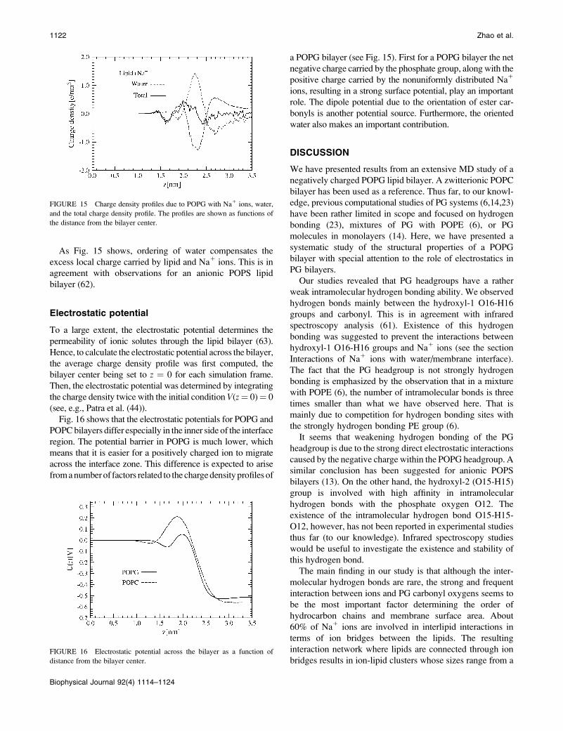
As Fig. 15 shows, ordering of water compensates the
excess local charge carried by lipid and Na1 ions. This is in
agreement with observations for an anionic POPS lipid
bilayer (62).
Electrostatic potential
To a large extent, the electrostatic potential determines the
permeability of ionic solutes through the lipid bilayer (63).
Hence, to calculate the electrostatic potential across the bilayer,
the average charge density profile was first computed, the
bilayer center being set to z ¼ 0 for each simulation frame.
Then, the electrostatic potential was determined by integrating
the charge density twice with the initial condition V(z¼ 0)¼ 0
(see, e.g., Patra et al. (44)).
Fig. 16 shows that the electrostatic potentials for POPG and
POPC bilayers differ especially in the inner side of the interface
region. The potential barrier in POPG is much lower, which
means that it is easier for a positively charged ion to migrate
across the interface zone. This difference is expected to arise
from a number of factors related to the charge density profiles of
a POPG bilayer (see Fig. 15). First for a POPG bilayer the net
negative charge carried by the phosphate group, along with the
positive charge carried by the nonuniformly distributed Na1
ions, resulting in a strong surface potential, play an important
role. The dipole potential due to the orientation of ester car-
bonyls is another potential source. Furthermore, the oriented
water also makes an important contribution.
DISCUSSION
We have presented results from an extensive MD study of a
negatively charged POPG lipid bilayer. A zwitterionic POPC
bilayer has been used as a reference. Thus far, to our knowl-
edge, previous computational studies of PG systems (6,14,23)
have been rather limited in scope and focused on hydrogen
bonding (23), mixtures of PG with POPE (6), or PG
molecules in monolayers (14). Here, we have presented a
systematic study of the structural properties of a POPG
bilayer with special attention to the role of electrostatics in
PG bilayers.
Our studies revealed that PG headgroups have a rather
weak intramolecular hydrogen bonding ability. We observed
hydrogen bonds mainly between the hydroxyl-1 O16-H16
groups and carbonyl. This is in agreement with infrared
spectroscopy analysis (61). Existence of this hydrogen
bonding was suggested to prevent the interactions between
hydroxyl-1 O16-H16 groups and Na1 ions (see the section
Interactions of Na1 ions with water/membrane interface).
The fact that the PG headgroup is not strongly hydrogen
bonding is emphasized by the observation that in a mixture
with POPE (6), the number of intramolecular bonds is three
times smaller than what we have observed here. That is
mainly due to competition for hydrogen bonding sites with
the strongly hydrogen bonding PE group (6).
It seems that weakening hydrogen bonding of the PG
headgroup is due to the strong direct electrostatic interactions
caused by the negative charge within the POPG headgroup. A
similar conclusion has been suggested for anionic POPS
bilayers (13). On the other hand, the hydroxyl-2 (O15-H15)
group is involved with high affinity in intramolecular
hydrogen bonds with the phosphate oxygen O12. The
existence of the intramolecular hydrogen bond O15-H15-
O12, however, has not been reported in experimental studies
thus far (to our knowledge). Infrared spectroscopy studies
would be useful to investigate the existence and stability of
this hydrogen bond.
The main finding in our study is that although the inter-
molecular hydrogen bonds are rare, the strong and frequent
interaction between ions and PG carbonyl oxygens seems to
be the most important factor determining the order of
hydrocarbon chains and membrane surface area. About
60% of Na1 ions are involved in interlipid interactions in
terms of ion bridges between the lipids. The resulting
interaction network where lipids are connected through ion
bridges results in ion-lipid clusters whose sizes range from a
FIGURE 15 Charge density profiles due to POPG with Na1 ions, water,
and the total charge density profile. The profiles are shown as functions of
the distance from the bilayer center.
FIGURE 16 Electrostatic potential across the bilayer as a function of
distance from the bilayer center.
1122 Zhao et al.
Biophysical Journal 92(4) 1114–1124

few to ;100 particles. These interlipid interactions are stable,
some of them persisting over the whole simulation run.
Consequently, the ion-lipid clusters are also rather stable,
implying that the lipids involved in a cluster form entities that
act in a concerted fashion. It is likely that this plays a role in the
structural as well as the dynamical properties of the bilayer,
including, e.g., the movement of lipids in the plane of the
bilayer and the possible formation of small-scale domains of
PG-ion rich regions in many component membranes. Further,
the strength and stability of positively charged ions and
negatively charged headgroups provide a possible molecular
level explanation for the action of PG in bacterial membranes
under the influence of organic solvents. These highly polar
interactions create a high free energy barrier for hydrophobic
molecules such as benzene, thus possibly giving one reason
nature uses charged lipids in these structures that have to
adjust quickly to external changes.
PG headgroups also strongly modify the orientation of
water near the bilayer. Two peaks of opposite sign in the
interfacial region of the POPG bilayer related to the local
water dipole order were observed (Fig. 14). Correlation of the
position of these peaks with the position of the two peaks of
opposite sign on the charge density (Fig. 15) along the bilayer
normal suggests that the ordering of water molecules orig-
inates from the shielding of local charges by water dipoles.
The snapshot of a Na1 ion coordinated with PG oxygen atoms
and water molecules, which is additionally hydrogen bonded
with a phosphate oxygen (Fig. 11 a), illustrates well the origin
of the first peak. The second peak is due to water molecules
hydrogen bonded with phosphate and glycerol oxygens from
the side of the water phase. In the case of PC, water is ordered
by the phosphate groups in a manner similar to PG, but the
positively charged choline group does not act the same way as
PG (which has no positive charge around) and Na1 ions
around the choline group create a clathrates-like structure,
thus the lack of ordering relative to the bilayer normal. The
long-range polarization of the water molecules throughout the
simulation box suggests that the amount of interlayer water in
our model is too low for comparisons with unilamellar
vesicles. That means that there is an additional repulsive
interaction through the water phase between headgroups of
opposite layers which potentially can influence the structure
and dynamics of simulated bilayers. This is in agreement with
experimental studies of charged lipids, which show contin-
uous swelling with water added between lamellae up to a
certain threshold at which two phases are formed, a fully
hydrated unilamellar vesicle with bulk water (64). However,
the exact amount of interlamellar water for POPG is not
known. Despite that, we can exclude the possibility that the
increase of the order and decrease of the surface area observed
in our studies are additionally facilitated by a too small
number of interbilayer water molecules. A similar increase for
the order of PG acyl chains was observed in mixed PE-PG
bilayers (6) and PG monolayers (14), and thus it is unlikely
that it can influence our conclusions.
We thank the Finnish IT Center for Science (CSC) and the HorseShoe
(DCSC) supercluster computing facility at the University of Southern
Denmark for computer resources.
This work has been supported by the Emil Aaltonen foundation, the
Academy of Finland through its Center of Excellence Program (I.V.), and
the Academy of Finland projects.
REFERENCES
1. Mead, J. F., R. B. Alfin-Slater, D. R. Howton, and G. Popjak. 1986.Lipids—Chemistry, Biochemistry and Nutrition. Plenum Press, New York.
2. Dowhan, W. 1996. Molecular basis for membrane phospholipid diversity:why are there so many lipids? Annu. Rev. Biochem. 66:199–232.
3. Pasenkiewicz-Gierula, M., K. Murzyn, T. Rog, and C. Czaplewski.2000. Molecular dynamics simulation studies of lipid bilayer systems.Acta Biochim. Pol. 47:601–611.
4. Hallock, K. J., D.-K. Lee, J. Omnaas, H. I. Mosberg, and A.Ramamoorthy. 2002. Membrane composition determines pardaxin’smechanism of lipid bilayer disruption. Biophys. J. 83:1004–1013.
5. Lee, S.-Y., and R. MacKinnon. 2004. A membrane-access mechanismof ion channel inhibition by voltage sensor toxins from spider venom.Nature. 430:232–235.
6. Murzyn, K., T. Rog, and M. Pasenkiewicz-Gierula. 2005. Phosphati-dylethanolamine-phosphatidylglycerol bilayer as a model of the innerbacterial membrane. Biophys. J. 88:1091–1103.
7. Uran, S., A. Larsen, P. B. Jacobsen, and T. Skotland. 2001. Analysis ofphospholipid species in human blood using normal-phase liquidchromatography coupled with electrospray ionization ion-trap tandemmass spectrometry. J. Chrom. B. 758:265–275.
8. Tari, A., and L. Huang. 1989. Structure and function relationship ofphosphatidylglycerol in the stabilization of phosphatidylethanolaminebilayer. Biochemistry. 28:7708–7712.
9. Maruyama, K. 2002. Peg-immunoliposomes. Biosci. Rep. 22:251–266.
10. Isken, S., and J. A. M. de Bont. 1998. Bacteria tolerant to organicsolvents. Extremophiles. 2:229–238.
11. Weber, F. J., and J. A. M. de Bont. 1996. Adaptation mechanisms ofmicroorganisms to the toxic effects of organic solvents on membranes.Biochim. Biophys. Acta. 1286:225–245.
12. Pandit, S. A., and M. L. Berkowitz. 2002. Molecular dynamicssimulation of dipalmitoylphosphatidylserine bilayer with Na1 coun-terions. Biophys. J. 82:1818–1827.
13. Mukhopadhyay, P., L. Monticelli, and D. P. Tieleman. 2004. Moleculardynamics simulation of a palmitoyl-oleoyl phosphatidylserine bilayerwith Na1 counterions and NaCl. Biophys. J. 86:1601–1609.
14. Kaznessis, Y. N., S. Kim, and R. G. Larson. 2002. Simulations ofzwitterionic and anionic phospholipid monolayers. Biophys. J. 82:1731–1742.
15. Bandyopadhyay, S., M. Tarek, and M. L. Klein. 1999. Moleculardynamics study of a lipid-DNA complex. J. Phys. Chem. B. 103:10075–10080.
16. Gurtovenko, A. A., M. Patra, M. Karttunen, and I. Vattulainen. 2004.Cationic DMPC/DMTAP lipid bilayers: molecular dynamics study.Biophys. J. 86:3461–3472.
17. Gurtovenko, A. A., M. Miettinen, M. Karttunen, and I. Vattulainen. 2005.Effect of monovalent salt on cationic lipid membranes as revealed bymolecular dynamics simulations. J. Phys. Chem. B. 109:21126–21134.
18. Bockmann, R. A., A. Hac, T. Heimburg, and H. Grubmuller. 2003.Effect of sodium chloride on a lipid bilayer. Biophys. J. 85:1647–1655.
19. Pandit, S. A., D. Bostick, and M. L. Berkowitz. 2003. Moleculardynamics simulation of a dipalmitoylphosphatidylcholine bilayer withNaCl. Biophys. J. 84:3743–3750.
20. Bockmann, R. A., and H. Grubmuller. 2004. Multistep binding ofdivalent cations to phospholipid bilayers: a molecular dynamics study.Angew. Chem. Int. Edn. 43:1021–1024.
POPG Bilayers 1123
Biophysical Journal 92(4) 1114–1124

21. Gurtovenko, A. A. 2005. Asymmetry of lipid bilayers induced bymonovalent salt: atomistic molecular dynamics study. J. Chem. Phys.122:244902.
22. Gurtovenko, A. A., and I. Vattulainen. 2005. Pore formation coupled toion transport through lipid membranes as induced by transmembraneionic charge imbalance: atomistic molecular dynamics study. J. Am.Chem. Soc. 127:17570–17571.
23. Elmore, D. E. 2006. Molecular dynamics simulation of a phosphat-idylglycerol membrane. FEBS Lett. 580:144–148.
24. Cowley, A. C., N. L. Fuller, R. P. Rand, and V. A. Parsegian. 1978.Measurement of repulsive forces between charged phospholipidbilayers. Biochemistry. 17:3163–3168.
25. Pozo-Navas, B., V. A. Raghunathan, J. Katsaras, M. Rappolt, K.Lohner, and G. Pabst. 2003. Discontinuous unbinding of lipidmultibilayers. Phys. Rev. Lett. 91:028101.
26. Pozo-Navas, B., K. Lohner, G. Deutsch, E. Sevcsik, K. A. Riske, R.Dimova, P. Garidel, and G. Pabst. 2004. Composition dependence ofvesicle morphology and mixing properties in a bacterial modelmembrane system. Biochim. Biophys. Acta. 1716:40–48.
27. Wiedmann, T., A. Salmon, and V. Wong. 1993. Phase behavior ofmixtures of DPPC and POPG. Biochim. Biophys. Acta. 1167:14–20.
28. Koynova, R. 1997. Liquid crystalline phase metastability of phosphat-idylglycerols. Chem. Phys. Lipids. 89:67–73.
29. Wohlgemuth, R., N. Waespe-Sarcevic, and J. Seelig. 1980. Bilayers ofphosphatidylglycerol. A deuterium and phosphorus nuclear magneticresonance study of the head-group region. Biochemistry. 19:3315–3321.
30. Pascher, I., M. Lundmark, P.-G. Nyholm, and S. Sundell. 1992. Crystalstructures of membrane lipids. Biochim. Biophys. Acta. 1113:339–373.
31. Garidel, P., and A. Blume. 2000. Miscibility of phosphatidylethanol-amine-phosphatidylglycerol mixtures as a function of pH and acylchain length. Eur. Biophys. J. 28:629–638.
32. Lohner, K., A. Latal, G. Degovics, and P. Garidel. 2001. Packingcharacteristics of a model system mimicking cytoplasmic bacterialmembranes. Chem. Phys. Lipids. 111:177–192.
33. Petrache, H. I., S. Tristram-Nagle, K. Gawrisch, D. Harries, V. A. Parsegian,and J. F. Nagle. 2004. Structure and fluctuations of charged phosphatidyl-serine bilayers in the absence of salt. Biophys. J. 86:1574–1586.
34. Berger, O., O. Edholm, and F. Jahnig. 1997. Molecular dynamicssimulations of a fluid bilayer of dipalmitoylphosphatidylcholine at fullhydration, constant pressure, and constant temperature. Biophys. J. 72:2002–2013.
35. Ryckaert, J. P., and A. Bellemans. 1975. Molecular dynamics of liquidn-butane near its boiling point. Chem. Phys. Lett. 30:123–125.
36. Ryckaert, J. P., and A. Bellemans. 1975. Molecular dynamics of liquidalkanes. Faraday Discuss Chem Soc. 66:95–106.
37. Reference deleted in proof.
38. Berendsen, H. J. C., J. P. M. Postma, W. F. van Gunsteren, and J. Hermans.1981. Interaction models for water in relation to protein hydration. InIntermolecular Forces. B. Pullman, editor. Reidel, Dordrecht. 331–342.
39. Patra, M., and M. Karttunen. 2004. Systematic comparison of forcefields for microscopic simulations of NaCl in aqueous solutions:diffusion, free energy of hydration and structural properties. J. Comp.Chem. 25:678–689.
40. Hess, B., H. Bekker, H. J. C. Berendsen, and J. G. E. M. Fraaije. 1997.LINCS: a linear constraint solver for molecular simulations. J. Comp.Chem. 18:1463–1472.
41. Miyamoto, S., and P. A. Kollman. 1992. SETTLE: an analyticalversion of the SHAKE and RATTLE algorithms for rigid watermodels. J. Comput. Chem. 13:952–962.
42. Darden, T., D. York, and L. Pedersen. 1993. Particle mesh Ewald: an Nlog(N) method for Ewald sums in large systems. J. Chem. Phys. 98:10089–10092.
43. Essman, U., L. Perela, M. L. Berkowitz, H. L. T. Darden, and L. G.Pedersen. 1995. A smooth particle mesh Ewald method. J. Chem. Phys.103:8577–8592.
44. Patra, M., M. Karttunen, M. T. Hyvonen, P. Lindqvist, E. Falck, andI. Vattulainen. 2003. Molecular dynamics simulations of lipid bilayers:major artifacts due to truncating electrostatic interaction. Biophys. J.84:3636–3645.
45. Anezo, C., A. H. de Vries, H.-D. Holtje, D. P. Tieleman, and S.-J.Marrink. 2003. Methodological issues in lipid bilayer simulations.J. Phys. Chem. B. 107:9424–9433.
46. Patra, M., M. Karttunen, M. T. Hyvonen, E. Falck, and I. Vattulainen.2004. Lipid bilayers driven to a wrong lane in molecular dynamicssimulations by truncation of long-range electrostatic interactions.J. Phys. Chem. B. 108:4485–4494.
47. Lindahl, E., B. Hess, and D. van der Spoel. 2001. GROMACS 3.0: apackage for molecular simulation and trajectory analysis. J. Mol.Model. (Online). 7:306–317.
48. Berendsen, H. J. C., J. P. M. Postma, W. F. van Gunsteren, A. DiNola,and J. R. Haak. 1984. Molecular dynamics with coupling to an externalbath. J. Chem. Phys. 81:3684–3690.
49. Patra, M., E. Salonen, E. Terama, I. Vattulainen, R. Faller, B. W. Lee,J. Holopainen, and M. Karttunen. 2006. Under the influence of alcohol:the effect of ethanol and methanol on lipid bilayers. Biophys. J.90:1121–1135.
50. Pascher, I., S. Sundell, K. Harlos, and H. Eibl. 1987. Conformation andpacking properties of membrane lipids: the crystal structure of sodiumdimyristoylphosphatidylglycerol. Biochem. Biophys. Acta. 896:77–88.
51. Seelig, J., and N. Waespe-Sarcevic. 1978. Molecular order in cis andtrans unsaturated phospholipid bilayers. Biochemistry. 17:3310–3315.
52. Chiu, S. W., E. Jacobsson, R. J. Mashl, and H. L. Scott. 2002.Cholesterol-induced modifications in lipid bilayers: a simulation study.Biophys. J. 83:1842–1853.
53. Hofsass, C., E. Lindahl, and O. Edholm. 2003. Molecular dynamicssimulations of phospholipid bilayers with cholesterol. Biophys. J. 84:2192–2206.
54. Falck, E., M. Patra, M. Karttunen, M. T. Hyvonen, and I. Vattulainen.2004. Lessons of slicing membranes: interplay of packing, free area,and lateral diffusion in phospholipid/cholesterol bilayers. Biophys. J.87:1076–1091.
55. Edholm, O., and J. F. Nagle. 2005. Areas of molecules in membranesconsisting of mixtures. Biophys. J. 89:1827–1832.
56. Andersson, A.-S., R. A. Demel, L. Rilfors, and G. Lindblom. 1998.Lipids in total extracts from Acholeplasma laidlawii A pack moreclosely than the individual lipids. Monolayers studied at the air-waterinterface. Biochim. Biophys. Acta. 1369:94–102.
57. Armen, R. S., O. D. Uitto, and S. E. Feller. 1998. Phospholipidcomponent volumes: determination and application to bilayer structurecalculations. Biophys. J. 75:734–744.
58. Watts, A., K. Harlos, and D. Marsh. 1981. Charge-induced tilt inordered-phase phosphatidylglycerol bilayers evidence from x-ray dif-fraction. Biochim. Biophys. Acta. 645:91–96.
59. Mischel, M., J. Seelig, L. F. Braganza, and G. Buldt. 1987. A neutrondiffraction study of the headgroup conformation of phosphatidylglycerolfrom Escherichia coli membranes. Chem. Phys. Lipids. 43:237–246.
60. Kurze, V., B. Steinbauer, T. Huber, and K. Beyer. 2000. A 2H NMRstudy of macroscopically aligned bilayer membranes containinginterfacial hydroxyl residues. Biophys. J. 78:2441–2451.
61. Dicko, A., H. Bourque, and M. Pezolet. 1998. Study by infraredspectroscopy of the conformation of dipalmitoylphosphatidylglycerolmonolayers at the air–water interface and transferred on solidsubstrates. Chem. Phys. Lipids. 96:125–139.
62. Cascales, J. J. L., J. D. L. Torre, S. Marrink, and H. Berendsen. 1996.Molecular dynamics simulation of a charged biological membrane.J. Chem. Phys. 104:2713–2720.
63. Gennis, R. B. 1989. Biomembranes. Molecular Structure and Function.Springer Advanced Texts in Chemistry, Springer, New York.
64. Boggs, J. M. 1987. Lipid intermolecular hydrogen bonding: influenceon structural organization and membrane function. Biochim. Biophys.Acta. 906:353–404.
1124 Zhao et al.
Biophysical Journal 92(4) 1114–1124
Copyright © 2022 FDOKUMEN