Interactions of arbutin with dry and hydrated bilayers
11
Ž . Biochimica et Biophysica Acta 1370 1998 87–97 Interactions of arbutin with dry and hydrated bilayers Ann E. Oliver a, ) , Dirk K. Hincha b , Lois M. Crowe a , John H. Crowe a a Section of Molecular and Cellular Biology, UniÕersity of California, DaÕis, CA 95616, USA b Institut fur Pflanzenphysiologie und Mikrobiologie, Freie UniÕersitat, Konigin Luise-Str. 12-16, D-14195 Berlin, Germany ¨ ¨ ¨ Received 12 August 1997; revised 30 September 1997; accepted 3 October 1997 Abstract Ž . The glycosylated hydroquinone arbutin 4-hydroxyphenyl-b-D-glucopyranoside is abundant in certain resurrection plants, which can survive almost complete dehydration for prolonged periods. Little is known about the role of arbutin in vivo, but it is thought to contribute toward survival of the plants in the dry state. We have investigated the interactions of arbutin with model membranes under conditions of high and low hydration, as well as the possible participation of arbutin in carbohydrate glasses formed at low water contents. Retention of a trapped soluble marker inside large unilamellar vesicles and fusion of vesicles was monitored by fluorescence spectroscopy. Effects of arbutin on glass-transition temperatures and hydrated membrane phase-transition temperatures were measured by differential scanning calorimetry. The possible insertion of arbutin into membrane bilayers was estimated by following arbutin auto-fluorescence. Evidence is presented that arbutin does not change the glass-transition temperature of a sucrosertrehalose glass, but that arbutin does interact with hydrated membranes by insertion of the phenol moiety into the lipid bilayer. This interaction causes increased membrane leakage during air-drying by a mechanism other than vesicle–vesicle fusion. Implications of these effects on the dehydrated plant cells, as well as possible methods of obviating the damage, are discussed. q 1998 Elsevier Science B.V. Keywords: Hydroquinone; Membrane bilayer; Liposome; Dehydration; Carbohydrate glass 1. Introduction Ž . Arbutin 4-hydroxyphenyl-b-D-glucopyranoside is a glycosylated hydroquinone, which consists of a phenol molecule with a glucose moiety in the para- Ž . position see Fig. 1 . Arbutin is well known as a wx substrate of the enzyme b-glucosidase 1 and also for its diuretic and urinary anti-infective properties. It has been found in the leaves of several plant species, wx such as Vaccinium sp. 2 , but its physiological role in these plants is unknown. Arbutin constitutes as ) Corresponding author. Fax: q1-916-752 5305; E-mail: [email protected] much as 25% of the dry weight in the leaves of w x Myrothamnus flabellifolia Welw. 2,3 , a desiccation tolerant angiosperm that is found in regions of south- ern Africa subject to prolonged drought conditions. It is thought to play an important role in resistance to environmental stress, as it is present in several di- verse plant taxa capable of withstanding such stresses as extreme low temperature and extended drought ww x x 2 , and references therein . Although the possible contribution of arbutin to freezing-tolerance is also under current investigation, it is outside the scope of a single paper and will be reported separately. In general, resurrection plants are able to survive nearly complete dehydration for extended periods of 0005-2736r98r$19.00 q 1998 Elsevier Science B.V. All rights reserved. Ž . PII S0005-2736 97 00246-0
-
Upload
independent -
Category
Documents
-
view
0 -
download
0
Transcript of Interactions of arbutin with dry and hydrated bilayers
Ž .Biochimica et Biophysica Acta 1370 1998 87–97
Interactions of arbutin with dry and hydrated bilayers
Ann E. Oliver a,), Dirk K. Hincha b, Lois M. Crowe a, John H. Crowe a
a Section of Molecular and Cellular Biology, UniÕersity of California, DaÕis, CA 95616, USAb Institut fur Pflanzenphysiologie und Mikrobiologie, Freie UniÕersitat, Konigin Luise-Str. 12-16, D-14195 Berlin, Germany¨ ¨ ¨
Received 12 August 1997; revised 30 September 1997; accepted 3 October 1997
Abstract
Ž .The glycosylated hydroquinone arbutin 4-hydroxyphenyl-b-D-glucopyranoside is abundant in certain resurrectionplants, which can survive almost complete dehydration for prolonged periods. Little is known about the role of arbutin invivo, but it is thought to contribute toward survival of the plants in the dry state. We have investigated the interactions ofarbutin with model membranes under conditions of high and low hydration, as well as the possible participation of arbutinin carbohydrate glasses formed at low water contents. Retention of a trapped soluble marker inside large unilamellarvesicles and fusion of vesicles was monitored by fluorescence spectroscopy. Effects of arbutin on glass-transitiontemperatures and hydrated membrane phase-transition temperatures were measured by differential scanning calorimetry.The possible insertion of arbutin into membrane bilayers was estimated by following arbutin auto-fluorescence. Evidence ispresented that arbutin does not change the glass-transition temperature of a sucrosertrehalose glass, but that arbutin doesinteract with hydrated membranes by insertion of the phenol moiety into the lipid bilayer. This interaction causes increasedmembrane leakage during air-drying by a mechanism other than vesicle–vesicle fusion. Implications of these effects on thedehydrated plant cells, as well as possible methods of obviating the damage, are discussed. q 1998 Elsevier Science B.V.
Keywords: Hydroquinone; Membrane bilayer; Liposome; Dehydration; Carbohydrate glass
1. Introduction
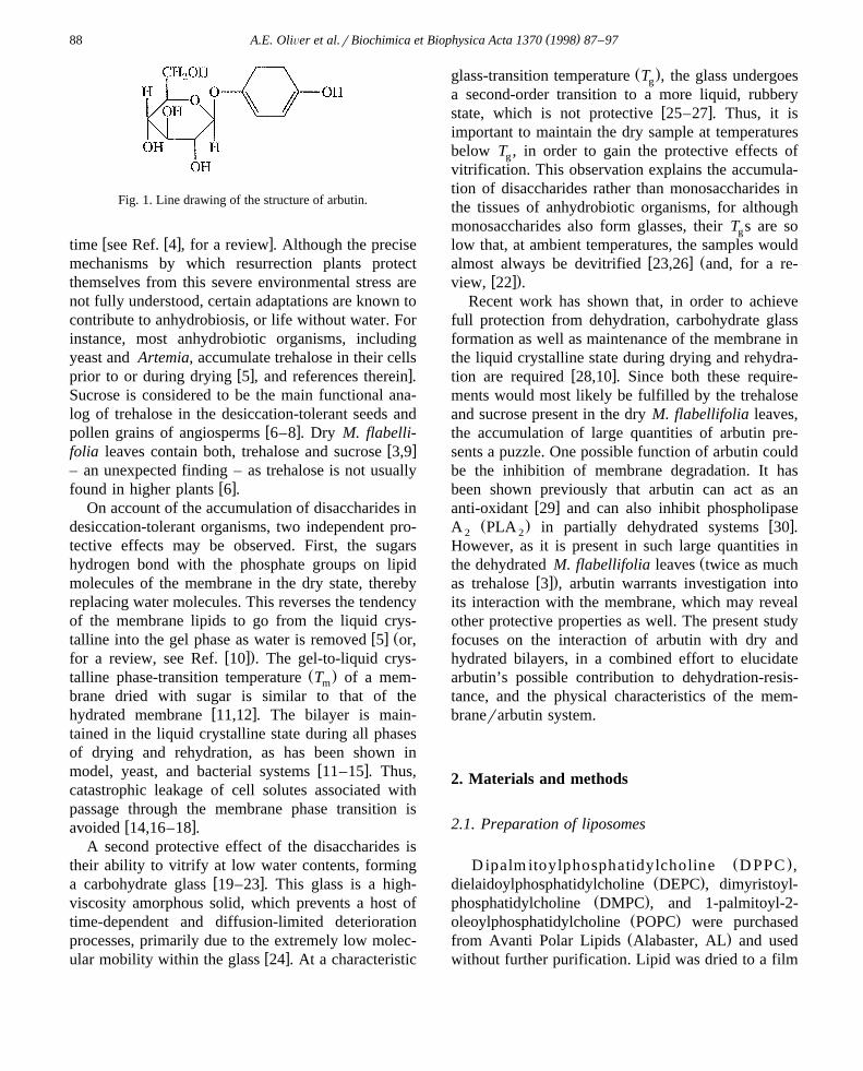
Ž .Arbutin 4-hydroxyphenyl-b-D-glucopyranoside isa glycosylated hydroquinone, which consists of aphenol molecule with a glucose moiety in the para-
Ž .position see Fig. 1 . Arbutin is well known as aw xsubstrate of the enzyme b-glucosidase 1 and also
for its diuretic and urinary anti-infective properties. Ithas been found in the leaves of several plant species,
w xsuch as Vaccinium sp. 2 , but its physiological rolein these plants is unknown. Arbutin constitutes as
) Corresponding author. Fax: q1-916-752 5305; E-mail:[email protected]
much as 25% of the dry weight in the leaves ofw xMyrothamnus flabellifolia Welw. 2,3 , a desiccation
tolerant angiosperm that is found in regions of south-ern Africa subject to prolonged drought conditions. Itis thought to play an important role in resistance toenvironmental stress, as it is present in several di-verse plant taxa capable of withstanding such stressesas extreme low temperature and extended droughtww x x2 , and references therein . Although the possiblecontribution of arbutin to freezing-tolerance is alsounder current investigation, it is outside the scope ofa single paper and will be reported separately.
In general, resurrection plants are able to survivenearly complete dehydration for extended periods of
0005-2736r98r$19.00 q 1998 Elsevier Science B.V. All rights reserved.Ž .PII S0005-2736 97 00246-0
( )A.E. OliÕer et al.rBiochimica et Biophysica Acta 1370 1998 87–9788
Fig. 1. Line drawing of the structure of arbutin.
w w x xtime see Ref. 4 , for a review . Although the precisemechanisms by which resurrection plants protectthemselves from this severe environmental stress arenot fully understood, certain adaptations are known tocontribute to anhydrobiosis, or life without water. Forinstance, most anhydrobiotic organisms, includingyeast and Artemia, accumulate trehalose in their cells
w x xprior to or during drying 5 , and references therein .Sucrose is considered to be the main functional ana-log of trehalose in the desiccation-tolerant seeds and
w xpollen grains of angiosperms 6–8 . Dry M. flabelli-w xfolia leaves contain both, trehalose and sucrose 3,9
– an unexpected finding – as trehalose is not usuallyw xfound in higher plants 6 .
On account of the accumulation of disaccharides indesiccation-tolerant organisms, two independent pro-tective effects may be observed. First, the sugarshydrogen bond with the phosphate groups on lipidmolecules of the membrane in the dry state, therebyreplacing water molecules. This reverses the tendencyof the membrane lipids to go from the liquid crys-
w x Žtalline into the gel phase as water is removed 5 or,w x.for a review, see Ref. 10 . The gel-to-liquid crys-
Ž .talline phase-transition temperature T of a mem-m
brane dried with sugar is similar to that of thew xhydrated membrane 11,12 . The bilayer is main-
tained in the liquid crystalline state during all phasesof drying and rehydration, as has been shown in
w xmodel, yeast, and bacterial systems 11–15 . Thus,catastrophic leakage of cell solutes associated withpassage through the membrane phase transition is
w xavoided 14,16–18 .A second protective effect of the disaccharides is
their ability to vitrify at low water contents, formingw xa carbohydrate glass 19–23 . This glass is a high-
viscosity amorphous solid, which prevents a host oftime-dependent and diffusion-limited deteriorationprocesses, primarily due to the extremely low molec-
w xular mobility within the glass 24 . At a characteristic
Ž .glass-transition temperature T , the glass undergoesg
a second-order transition to a more liquid, rubberyw xstate, which is not protective 25–27 . Thus, it is
important to maintain the dry sample at temperaturesbelow T , in order to gain the protective effects ofg
vitrification. This observation explains the accumula-tion of disaccharides rather than monosaccharides inthe tissues of anhydrobiotic organisms, for althoughmonosaccharides also form glasses, their T s are sog
low that, at ambient temperatures, the samples wouldw x Žalmost always be devitrified 23,26 and, for a re-
w x.view, 22 .Recent work has shown that, in order to achieve
full protection from dehydration, carbohydrate glassformation as well as maintenance of the membrane inthe liquid crystalline state during drying and rehydra-
w xtion are required 28,10 . Since both these require-ments would most likely be fulfilled by the trehaloseand sucrose present in the dry M. flabellifolia leaves,the accumulation of large quantities of arbutin pre-sents a puzzle. One possible function of arbutin couldbe the inhibition of membrane degradation. It hasbeen shown previously that arbutin can act as an
w xanti-oxidant 29 and can also inhibit phospholipaseŽ . w xA PLA in partially dehydrated systems 30 .2 2
However, as it is present in such large quantities inŽthe dehydrated M. flabellifolia leaves twice as much
w x.as trehalose 3 , arbutin warrants investigation intoits interaction with the membrane, which may revealother protective properties as well. The present studyfocuses on the interaction of arbutin with dry andhydrated bilayers, in a combined effort to elucidatearbutin’s possible contribution to dehydration-resis-tance, and the physical characteristics of the mem-branerarbutin system.
2. Materials and methods
2.1. Preparation of liposomes
Ž .D ipalm itoylphosphatidylcholine D PPC ,Ž .dielaidoylphosphatidylcholine DEPC , dimyristoyl-
Ž .phosphatidylcholine DMPC , and 1-palmitoyl-2-Ž .oleoylphosphatidylcholine POPC were purchasedŽ .from Avanti Polar Lipids Alabaster, AL and used
without further purification. Lipid was dried to a film
( )A.E. OliÕer et al.rBiochimica et Biophysica Acta 1370 1998 87–97 89
under nitrogen gas and placed under vacuumovernight to remove residual chloroform. Deionizedwater was added to the lipid, which was warmed andagitated gently to produce multilamellar vesicles.
Ž .Large unilamellar vesicles LUVs were formed byŽextrusion with a hand-held extruder Avestin; Ottawa,
w x.ON, Canada; 31 , using 0.1 mm pore polycarbonateŽ .filters Poretics; Livermore, CA . Alternatively, soni-
cated vesicles were prepared by bath sonicationŽ .Laboratory Supplies, Hicksville, NY of multi-lamellar vesicles until a clear suspension was ob-tained.
2.2. Differential scanning calorimetry
Glass transitions were measured using a Perkin–Ž .Elmer differential scanning calorimeter DSC-7 ,
cooled by a liquid-nitrogen reservoir. CarbohydrateŽ .samples ; 2–5 mg from stock solutions of
100 mgrml were dried for several hours in smallaluminum pans, in a dry box maintained at 0%
Ž .relative humidity RH . The pans were then eitherpressure-sealed inside the dry box or transferred toRH chambers, maintained at specific relative humidi-
w xties over saturated salt solutions 32 , overnight be-fore sealing. The DSC-7 head was flushed with He,and the box with dry N . Carbohydrate samples were2
scanned between y1008 and 2008C at 208Crmin onupscans and 1008Crmin on downscans. Sample panswere weighed on a Cahn C-33 electrobalance before,and after DSC scanning to ensure that no water waslost during the procedure. Sample pans were thenpunctured and incubated at 1208C for at least 24 hand re-weighed. This incubation and re-weighing stepwas repeated until the sample weight remained con-stant for two consecutive days, thereby allowing ac-curate calculation of sample hydration during theDSC scan.
Gel-to-liquid crystalline phase transitions of hy-drated membranes were measured with a second highsensitivity DSC from Calorimetry Sciences, tempera-ture controlled by a Peltier device. Aqueous suspen-
Žsions of DPPC, DMPC, or DEPC LUVs 0.5 ml of.40 mgrml in different arbutin concentrations were
scanned in sealed ampules between 58 and 508C at arate of 208Crh, and T s detected as the maxima ofm
sharp endothermic peaks on thermal upscans.
2.3. Measurement of retention
Ž .Large unilamellar vesicles LUVs were preparedas described by extrusion in the presence of 100 mM
Žcarboxyfluorescein CF, Molecular Probes, Eugene,w x.OR; purified according to Weinstein 33 . External
ŽCF was removed by column chromatography Sep-. Žhadex G 50–80, 1=20 cm . Trehalose, sucrose high
purity disaccharides obtained from Pfanstiehl Labora-. Žtories, Waukegan, IL , arbutin Fluka, Buchs,
.Switzerland: biochemistry grade , or a combinationof excipients were added to the liposome suspensionat various concentrations, as stated in the text. Lipo-
Ž .some samples 10 ml were placed into the lids of1.5 ml microfuge tubes, and air-dried for at least 5 hin a dry box, maintained at 0% RH. The sampleswere then transferred to a Virtis Freezemobile 25SL
Ž .freeze dryer Gardiner, NY and stored overnightunder a 10 mtorr vacuum. Retention of carboxyfluo-rescein was either measured immediately, or afterincubation at 228C and 58% RH over a saturated
w xsolution of NaBr 32 for a specified length of time.After re-hydration with water, a small volume of
Ž .LUVs 5ml was transferred to a cuvette containing2.5 ml 10 mM TES, pH 7.5. Samples were excited at460 nm, and emission was measured at 550 nm on aPerkin-Elmer LS-5 fluorescence spectrophotometer.Intravesicular CF concentrations are self-quenching,so the emission at 550 nm was due only to externalCF. After an initial fluorescence measurement wastaken, 50 ml of 1% Triton X-100 was added to each
Ž .sample to cause total 100% leakage. A final fluores-cence measurement was taken at this point. Percentretention was calculated from the following twoequations:
% Trapped
final fluorescencey initial fluorescences
final fluorescence
=100 1Ž .% trapped after drying
% Retentions =100 2Ž .trapped before drying
2.4. Measurement of fusion
Fusion during air-drying was measured accordingw xto the method of Struck et al. 34 . Briefly, liposomes
( )A.E. OliÕer et al.rBiochimica et Biophysica Acta 1370 1998 87–9790
were prepared, as already described, at 20 mgrml inŽthe absence, and presence of 0.5 mol% each of N- 7-
.nitro-2,1,3-benzoxadiazol-4-yl phosphatidylethanola-Ž . Žmine NBD-PE and N- lissamine Rhodamine B. Ž .sulfonyl dioleoylphosphatidylethanolamine Rh-PE ,
which were purchased from Molecular ProbesŽ .Eugene, OR . Following extrusion, liposomes were
Ž .combined at a ratio of 1 : 9 labeled : unlabeled , air-dried, and stored as described. In order to measurefusion, dried samples were re-hydrated and mixedinto 2.5 ml 10 mM TES, pH 7.5. Fluorescence mea-
Ž .surements excitation 450 nm, emission 530 nm wereŽ . Ž .made in the absence F , and in the presence F of0
50 ml 1% Triton X-100. The energy transfer effi-Ž .ciency E was calculated as follows:
Es1yFrF 3Ž .0
This energy-transfer efficiency was calculated forŽ .both unfused control vesicles E , and for the dried0
Ž .experimental samples E . Percent fusion was calcu-s
lated as follows:
% Fusions100y E rE =100 4� 4Ž . Ž .s 0
Thus, fusion is detected as the decrease in energytransfer between the two fluorophores, as fused vesi-cles constitute a dilution of the probes by unlabeled
w xlipid molecules 34 .
2.5. Arbutin auto-fluorescence
Certain aromatic amino acids, such as tryptophanor tyrosine, will emit fluorescence when excited at
w x280 nm 35 . The fluorescence emission maximumwill shift when these amino acids enter a hydropho-bic environment. This phenomenon has been utilizedin many studies to investigate the insertion of pep-tides or proteins containing these residues, into a lipid
w xmembrane 36,37 . Using aqueous quenchers such asNaNO , it is also possible to estimate the relative3
depth of insertion of a protein into a membrane, bymeasuring the protection from quenching afforded bythe hydrophobic environment of the lipid phasew x37,38 . Since arbutin also contains an aromatic ringstructure, we conducted analogous experiments withthis hydroquinone. To an aqueous solution of 4 mM
Ž .arbutin in 10 mM TES pH 7.5 , either in the pres-Ž .ence or absence of liposomes 0.4 mM , 5ml aliquots
Ž . Ž .of either buffer 10 mM TES or NaNO 5 M were3
Žadded stepwise, and the fluorescence excitation.280 nm, emission 340 nm was measured at each
point. An emission wavelength of 340 nm was usedin order to preferentially observe arbutin in the hy-drophobic environment of the membrane. Fluores-
Ž .cence in the presence of buffer F and fluorescence0Ž .in the presence of NaNO F were used to measure3
the extent of quenching by the following calculation:
QuenchingsF rFy1 5Ž .0
Stern–Volmer plots were constructed by plottingw xthe concentration of NaNO against F rF-1 36,38 .3 0
The slope of this plot yields the quenching constantŽ .K , which provides information about the extent ofsv
protection from quenching afforded arbutin by theliposomes and, thus, about insertion of the arbutin
w xmolecule into the membrane bilayer 37,38 .
3. Results and discussion
3.1. Arbutin does not change T of the carbohydrateg
glass
As a first hypothesis, we considered that arbutinmight participate in the carbohydrate glass formed at
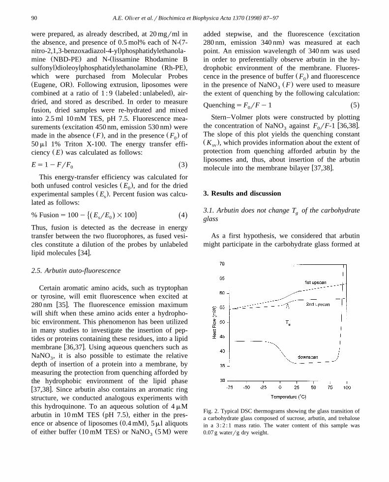
Fig. 2. Typical DSC thermograms showing the glass transition ofa carbohydrate glass composed of sucrose, arbutin, and trehalosein a 3 : 2 : 1 mass ratio. The water content of this sample was0.07 g waterrg dry weight.
( )A.E. OliÕer et al.rBiochimica et Biophysica Acta 1370 1998 87–97 91
low water contents. If arbutin significantly raised theglass-transition temperature of this glass, the plantwould be better protected during re-hydration, asmore water could be adsorbed by the plant tissuesbefore T dropped below the ambient temperature,g
w xresulting in devitrification of the glass 39–41 .Glass-transition temperatures were measured by
differential scanning calorimetry in carbohydrateglasses composed of sucrose, arbutin and trehalose inthe mass ratio of 3 : 2 : 1, which mimics the sugar
w xcomposition in dry M. flabellifolia leaves 3 . TypicalDSC data are shown in Fig. 2. The T of thisg
combination–carbohydrate glass can be easily de-tected by a change in heat flow on both, up- anddownscans, and T was taken as the midpoint of theg
shift. Crystallization of arbutin was prevented in thiscarbohydrate mixture as indicated by the disappear-
ance of the characteristic arbutin crystalline melt at1998C, which is clearly visible in thermal scans of
Ž .pure arbutin samples data not shown . This findingsuggests that arbutin may participate in the carbo-hydrate glass.
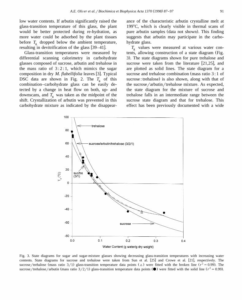
T values were measured at various water con-gŽtents, allowing construction of a state diagram Fig.
.3 . The state diagrams shown for pure trehalose andw xsucrose were taken from the literature 21,25 , and
are plotted as solid lines. The state diagram for aŽsucrose and trehalose combination mass ratio 3 : 1 of
.sucrose : trehalose is also shown, along with that ofthe sucroserarbutinrtrehalose mixture. As expected,the state diagram for the mixture of sucrose andtrehalose falls in an intermediate range between thesucrose state diagram and that for trehalose. Thiseffect has been previously documented with a wide
Fig. 3. State diagrams for sugar and sugar-mixture glasses showing decreasing glass-transition temperatures with increasing waterw x w xcontents. State diagrams for sucrose and trehalose were taken from Sun et al. 25 and Crowe et al. 21 , respectively. The
Ž . Ž . Ž 2 .sucrosertrehalose mass ratio 3r1 glass-transition temperature data points ^ were fitted with the broken line r s0.99 . TheŽ . Ž . Ž 2 .sucrosertrehaloserarbutin mass ratio 3r2r1 glass-transition temperature data points v were fitted with the solid line r s0.99 .
( )A.E. OliÕer et al.rBiochimica et Biophysica Acta 1370 1998 87–9792
w xvariety of other sugar mixtures 42,43 . The interest-ing finding, however, is that the state diagram for thesucroserarbutinrtrehalose combination is nearlyidentical to that of the sucrosertrehalose mixture.This result indicates that, although arbutin may par-ticipate in the glass, it did not change the T of theg
sucrosertrehalose combination–carbohydrate glass.
3.2. Arbutin causes membrane leakage during air-drying
Since arbutin was found not to change T of theg
carbohydrate glass, we investigated the possibilitythat it might protect the dry membranes from leaking
w xin a manner similar to that of the disaccharides 44 .This hypothesis was not unlikely, as arbutin hadalready been shown to depress the phase-transition
Ž .temperature T of dry phospholipid bilayers as domw xsucrose and trehalose 30 . POPC LUVs were air-dried
in the presence of various combinations of disaccha-rides and arbutin, and retention was measured imme-
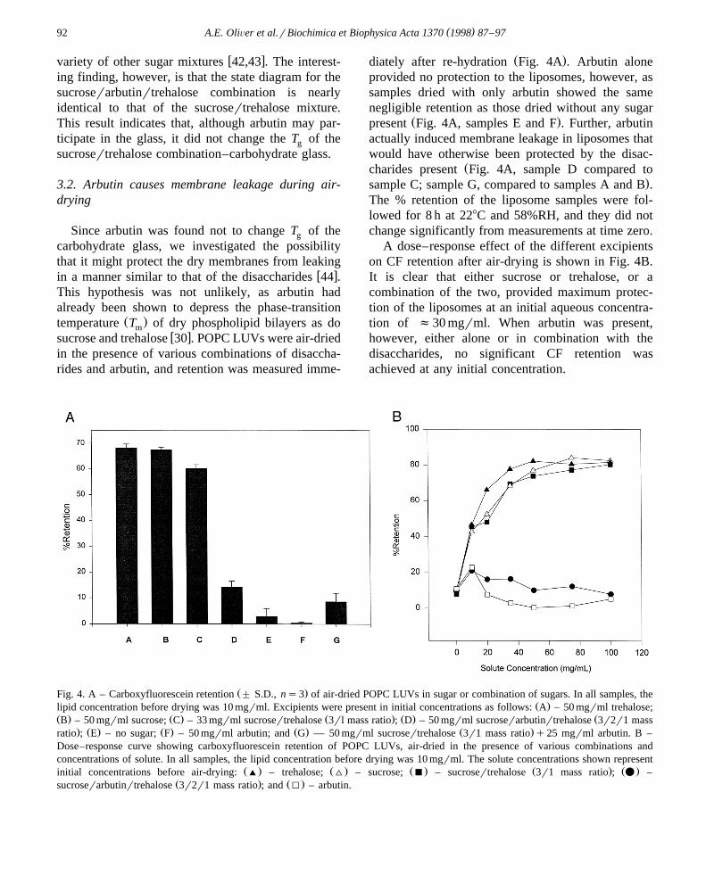
Ž .diately after re-hydration Fig. 4A . Arbutin aloneprovided no protection to the liposomes, however, assamples dried with only arbutin showed the samenegligible retention as those dried without any sugar
Ž .present Fig. 4A, samples E and F . Further, arbutinactually induced membrane leakage in liposomes thatwould have otherwise been protected by the disac-
Žcharides present Fig. 4A, sample D compared to.sample C; sample G, compared to samples A and B .
The % retention of the liposome samples were fol-lowed for 8 h at 228C and 58%RH, and they did notchange significantly from measurements at time zero.
A dose–response effect of the different excipientson CF retention after air-drying is shown in Fig. 4B.It is clear that either sucrose or trehalose, or acombination of the two, provided maximum protec-tion of the liposomes at an initial aqueous concentra-tion of f30 mgrml. When arbutin was present,however, either alone or in combination with thedisaccharides, no significant CF retention wasachieved at any initial concentration.
Ž .Fig. 4. A – Carboxyfluorescein retention " S.D., ns3 of air-dried POPC LUVs in sugar or combination of sugars. In all samples, theŽ .lipid concentration before drying was 10 mgrml. Excipients were present in initial concentrations as follows: A – 50 mgrml trehalose;
Ž . Ž . Ž . Ž . ŽB – 50 mgrml sucrose; C – 33 mgrml sucrosertrehalose 3rl mass ratio ; D – 50 mgrml sucroserarbutinrtrehalose 3r2r1 mass. Ž . Ž . Ž . Ž .ratio ; E – no sugar; F – 50 mgrml arbutin; and G — 50 mgrml sucrosertrehalose 3r1 mass ratio q25 mgrml arbutin. B –
Dose–response curve showing carboxyfluorescein retention of POPC LUVs, air-dried in the presence of various combinations andconcentrations of solute. In all samples, the lipid concentration before drying was 10 mgrml. The solute concentrations shown represent
Ž . Ž . Ž . Ž . Ž .initial concentrations before air-drying: ' – trehalose; ^ – sucrose; B – sucrosertrehalose 3r1 mass ratio ; v –Ž . Ž .sucroserarbutinrtrehalose 3r2r1 mass ratio ; and I – arbutin.

( )A.E. OliÕer et al.rBiochimica et Biophysica Acta 1370 1998 87–97 93
3.3. Arbutin is non-fusogenic
The negligible retention seen in liposomes air-driedin the presence of arbutin raised the possibility thatarbutin might cause fusion, inducing nearly completeleakage in the liposome samples. Fusion was mea-sured, using a resonance energy-transfer assay, ac-
w xcording to the method of Struck et al. 34 as de-scribed in Section 2. POPC LUVs were air-dried inthe presence of sucrose, trehalose, arbutin, or a com-bination of all three, and fusion was measured imme-
Ž .diately after re-hydration Fig. 5 . Although arbutin,by itself, did not prevent fusion, its presence in thecombination with sucrose and trehalose also did notcause fusion in the liposome samples.
The percentages of fusion of the liposome sampleswere also followed for 8 h at 228C and 58% RH, andthey did not change significantly from the measure-ments at time zero. The finding that neither CFleakage nor vesicle–vesicle fusion changed signifi-cantly over time at 58% RH, provides additionalevidence that the effects of arbutin are unrelated to its
Fig. 5. Fusion in POPC LUVs, air-dried in the presence ofdifferent excipients. In all samples, the concentration of lipidbefore drying was 10 mgrml. The initial solute concentrationbefore air-drying was 50 mgrml for each. In the control sample,no sugar was present. Experimental samples contained trehalose,sucrose, arbutin, or the combination of sucroserarbutinrtreha-
Ž .lose 3r2r1 mass ratio .
Fig. 6. Dose–response curve showing the effects of increasingŽ . Ž .arbutin concentration on ^ fusion and v CF retention of
air-dried POPC LUVs. In all samples, initial lipid concentrationbefore air-drying was 10 mgrml, and the initial trehalose concen-tration was 50 mgrml. The concentration of arbutin before air-drying was varied as shown. Inset: lack of correlation betweenfusion and retention in this study. This plot of CF retention as afunction of fusion shows the presence of arbutin in the carbo-hydrate glasses caused a decrease in CF retention that was notdue to vesicle–vesicle fusion.
effect on the carbohydrate glass-transition tempera-ture. If arbutin were compromising the glass in someway, then, as water was adsorbed by the liposomesamples during incubation at elevated relative humid-ity, the glass would devitrify, and the CF retentionandror fusion values would be expected to change.
A summary of the effects of arbutin on liposomeCF retention and fusion during air-drying is presented
Ž .in an arbutin dose–response curve Fig. 6 . Trehalosewas present in all samples at an initial aqueousconcentration of 50 mgrml, and the arbutin concen-tration was subsequently varied. The inset shows thetotal lack of correlation between fusion and decreas-ing retention in these experiments. In fact, the sam-ples showing the highest level of fusion also had thehighest retention. Although we do not have a satisfac-tory explanation for this effect, it is clear evidencethat arbutin caused leakage by some mechanism otherthan vesicle–vesicle fusion.
It is interesting to note that arbutin, in combinationwith the disaccharides, caused a moderate decrease in
( )A.E. OliÕer et al.rBiochimica et Biophysica Acta 1370 1998 87–9794
fusion compared to samples with only trehalose orŽ .sucrose Figs. 5 and 6 . Although it could be argued
that this is a protective effect, the principal benefit ofreducing fusion is the improvement of membraneintegrity, leading to an increase in % retention. Sincethe CF retention was decreased so drastically, evenby the lowest concentrations of arbutin, this smallreduction in fusion seems unlikely to provide anysignificant protection to dry membranes. In addition,the possibility that arbutin might interfere with thefusion assay was eliminated by control experimentsshowing that arbutin had no fluorescent properties in
Žthe range of either fluorescent probe used data not.shown .
3.4. Arbutin decreases T of hydrated lipid mem-m
branes
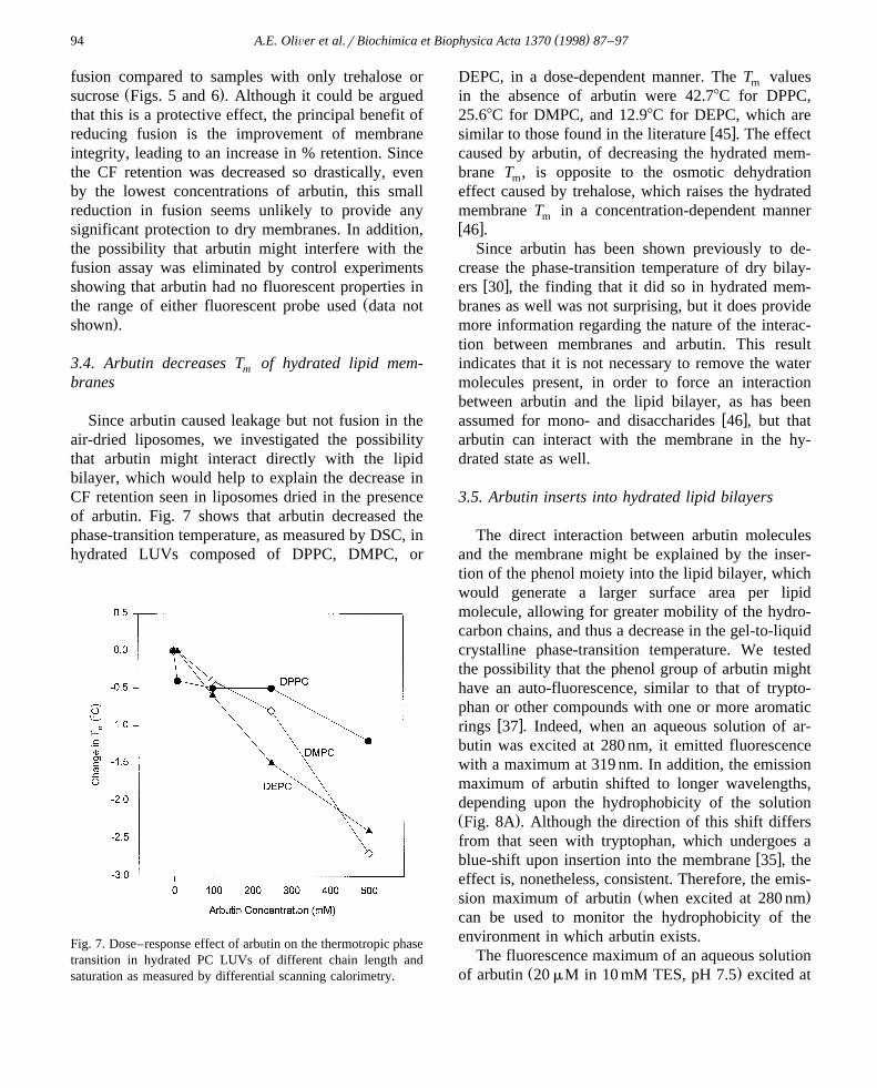
Since arbutin caused leakage but not fusion in theair-dried liposomes, we investigated the possibilitythat arbutin might interact directly with the lipidbilayer, which would help to explain the decrease inCF retention seen in liposomes dried in the presenceof arbutin. Fig. 7 shows that arbutin decreased thephase-transition temperature, as measured by DSC, inhydrated LUVs composed of DPPC, DMPC, or
Fig. 7. Dose–response effect of arbutin on the thermotropic phasetransition in hydrated PC LUVs of different chain length andsaturation as measured by differential scanning calorimetry.
DEPC, in a dose-dependent manner. The T valuesm
in the absence of arbutin were 42.78C for DPPC,25.68C for DMPC, and 12.98C for DEPC, which are
w xsimilar to those found in the literature 45 . The effectcaused by arbutin, of decreasing the hydrated mem-brane T , is opposite to the osmotic dehydrationm
effect caused by trehalose, which raises the hydratedmembrane T in a concentration-dependent mannermw x46 .
Since arbutin has been shown previously to de-crease the phase-transition temperature of dry bilay-
w xers 30 , the finding that it did so in hydrated mem-branes as well was not surprising, but it does providemore information regarding the nature of the interac-tion between membranes and arbutin. This resultindicates that it is not necessary to remove the watermolecules present, in order to force an interactionbetween arbutin and the lipid bilayer, as has been
w xassumed for mono- and disaccharides 46 , but thatarbutin can interact with the membrane in the hy-drated state as well.
3.5. Arbutin inserts into hydrated lipid bilayers
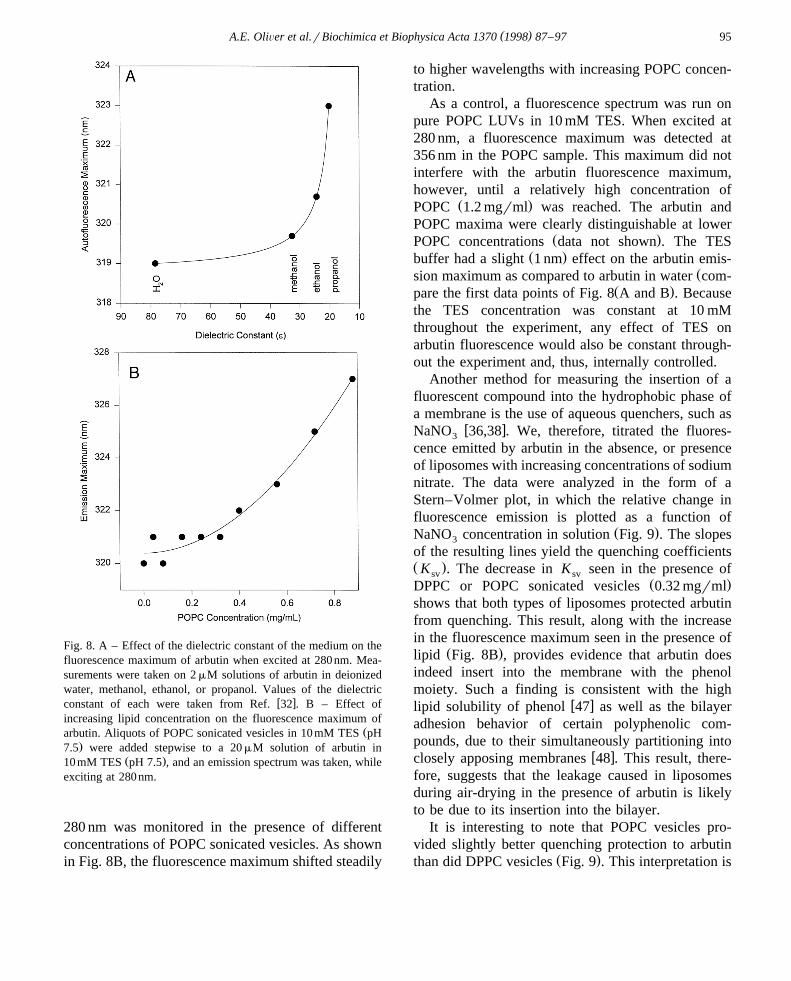
The direct interaction between arbutin moleculesand the membrane might be explained by the inser-tion of the phenol moiety into the lipid bilayer, whichwould generate a larger surface area per lipidmolecule, allowing for greater mobility of the hydro-carbon chains, and thus a decrease in the gel-to-liquidcrystalline phase-transition temperature. We testedthe possibility that the phenol group of arbutin mighthave an auto-fluorescence, similar to that of trypto-phan or other compounds with one or more aromatic
w xrings 37 . Indeed, when an aqueous solution of ar-butin was excited at 280 nm, it emitted fluorescencewith a maximum at 319 nm. In addition, the emissionmaximum of arbutin shifted to longer wavelengths,depending upon the hydrophobicity of the solutionŽ .Fig. 8A . Although the direction of this shift differsfrom that seen with tryptophan, which undergoes a
w xblue-shift upon insertion into the membrane 35 , theeffect is, nonetheless, consistent. Therefore, the emis-
Ž .sion maximum of arbutin when excited at 280 nmcan be used to monitor the hydrophobicity of theenvironment in which arbutin exists.
The fluorescence maximum of an aqueous solutionŽ .of arbutin 20 mM in 10 mM TES, pH 7.5 excited at
( )A.E. OliÕer et al.rBiochimica et Biophysica Acta 1370 1998 87–97 95
Fig. 8. A – Effect of the dielectric constant of the medium on thefluorescence maximum of arbutin when excited at 280nm. Mea-surements were taken on 2 mM solutions of arbutin in deionizedwater, methanol, ethanol, or propanol. Values of the dielectric
w xconstant of each were taken from Ref. 32 . B – Effect ofincreasing lipid concentration on the fluorescence maximum of
Žarbutin. Aliquots of POPC sonicated vesicles in 10 mM TES pH.7.5 were added stepwise to a 20 mM solution of arbutin in
Ž .10 mM TES pH 7.5 , and an emission spectrum was taken, whileexciting at 280nm.
280 nm was monitored in the presence of differentconcentrations of POPC sonicated vesicles. As shownin Fig. 8B, the fluorescence maximum shifted steadily
to higher wavelengths with increasing POPC concen-tration.
As a control, a fluorescence spectrum was run onpure POPC LUVs in 10 mM TES. When excited at280 nm, a fluorescence maximum was detected at356 nm in the POPC sample. This maximum did notinterfere with the arbutin fluorescence maximum,however, until a relatively high concentration of
Ž .POPC 1.2 mgrml was reached. The arbutin andPOPC maxima were clearly distinguishable at lower
Ž .POPC concentrations data not shown . The TESŽ .buffer had a slight 1 nm effect on the arbutin emis-
Žsion maximum as compared to arbutin in water com-Ž .pare the first data points of Fig. 8 A and B . Because
the TES concentration was constant at 10 mMthroughout the experiment, any effect of TES onarbutin fluorescence would also be constant through-out the experiment and, thus, internally controlled.
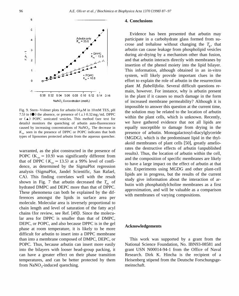
Another method for measuring the insertion of afluorescent compound into the hydrophobic phase ofa membrane is the use of aqueous quenchers, such as
w xNaNO 36,38 . We, therefore, titrated the fluores-3
cence emitted by arbutin in the absence, or presenceof liposomes with increasing concentrations of sodiumnitrate. The data were analyzed in the form of aStern–Volmer plot, in which the relative change influorescence emission is plotted as a function of
Ž .NaNO concentration in solution Fig. 9 . The slopes3
of the resulting lines yield the quenching coefficientsŽ .K . The decrease in K seen in the presence ofsv sv
Ž .DPPC or POPC sonicated vesicles 0.32 mgrmlshows that both types of liposomes protected arbutinfrom quenching. This result, along with the increasein the fluorescence maximum seen in the presence of
Ž .lipid Fig. 8B , provides evidence that arbutin doesindeed insert into the membrane with the phenolmoiety. Such a finding is consistent with the high
w xlipid solubility of phenol 47 as well as the bilayeradhesion behavior of certain polyphenolic com-pounds, due to their simultaneously partitioning into
w xclosely apposing membranes 48 . This result, there-fore, suggests that the leakage caused in liposomesduring air-drying in the presence of arbutin is likelyto be due to its insertion into the bilayer.
It is interesting to note that POPC vesicles pro-vided slightly better quenching protection to arbutin
Ž .than did DPPC vesicles Fig. 9 . This interpretation is
( )A.E. OliÕer et al.rBiochimica et Biophysica Acta 1370 1998 87–9796
ŽFig. 9. Stern–Volmer plots for arbutin 4 mM in 10mM TES, pH. Ž . Ž .7.5 in v the absence, or presence of ^ 0.32 mgrmL DPPCŽ . Žor ' POPC sonicated vesicles. This method see text for
.details monitors the quenching of arbutin auto-fluorescencecaused by increasing concentrations of NaNO . The decrease in3
K seen in the presence of DPPC or POPC indicates that bothsv
types of liposomes protected arbutin from the aqueous quencher.
warranted, as the plot constructed in the presence ofŽ .POPC K s10.9 was significantly different fromsv
Ž .that of DPPC K s13.5 at a 99% level of confi-sv
dence, as determined by the SigmaPlot regressionŽanalysis SigmaPlot, Jandel Scientific, San Rafael,
.CA . This finding correlates well with the resultshown in Fig. 7 that arbutin decreased the T ofm
hydrated DMPC and DEPC more than that of DPPC.These phenomena can both be explained by the dif-ferences amongst the lipids in surface area permolecule. Molecular area is inversely proportional tochain length and level of saturation of the fatty acyl
Ž w x.chains for review, see Ref. 49 . Since the molecu-lar area for DPPC is smaller than that of DMPC,DEPC, or POPC, and also because DPPC is in the gelphase at room temperature, it is likely to be moredifficult for arbutin to insert into a DPPC membranethan into a membrane composed of DMPC, DEPC, orPOPC. Thus, because arbutin can insert more easilyinto the bilayers with looser head-group packing, itcan have a greater effect on their phase transitiontemperatures, and can be better protected by themfrom NaNO -induced quenching.3
4. Conclusions
Evidence has been presented that arbutin mayparticipate in a carbohydrate glass formed from su-crose and trehalose without changing the T , thatg
arbutin can cause leakage from phospholipid vesiclesduring air-drying by a mechanism other than fusion,and that arbutin interacts directly with membranes byinsertion of the phenol moiety into the lipid bilayer.This information, although obtained in an in-vitrosystem, will likely provide important clues in theeffort to explain the role of arbutin in the resurrectionplant M. flabellifolia. Several difficult questions re-main, however. For instance, why is arbutin presentin the plant if it causes so much damage in the formof increased membrane permeability? Although it isimpossible to answer this question at the current time,the solution may be related to the location of arbutinwithin the plant cells, which is unknown. Recently,we have gathered evidence that not all lipids areequally susceptible to damage from drying in thepresence of arbutin. Monogalactosyl-diacylglycerideŽ .MGDG , which is the predominant lipid in the thyl-
w xakoid membranes of plant cells 50 , greatly amelio-Žrates the destructive effects of arbutin unpublished
.results . Thus, the location of arbutin within the cell,and the composition of specific membranes are likelyto have a large impact on the effect of arbutin at thatsite. Experiments using MGDG and other plant-celllipids are in progress, but the results of the currentstudy give information about the interaction of ar-butin with phosphatidylcholine membranes as a firstapproximation, and will be valuable as a comparisonwith membranes of varying compositions.
Acknowledgements
This work was supported by a grant from theNational Science Foundation, No. IBN93-08581 andgrant USN N00014-94-1 from the Office of NavalResearch. Dirk K. Hincha is the recipient of aHeisenberg stipend from the Deutsche Forschungsge-meinschaft.
( )A.E. OliÕer et al.rBiochimica et Biophysica Acta 1370 1998 87–97 97
References
w x Ž .1 H. Friedrich, Arch. Pharm. 228 1955 583.w x2 R. Suau, A. Cuevas, V. Valpuesta, M.S. Reid, Phytochem.
Ž .30 1991 2555–2556.w x3 G. Bianchi, A. Gamba, R. Limiroli, N. Pozzi, R. Elster, F.
Ž .Salamini, D. Bartels, Physiol. Plant. 87 1993 223–226.w x4 J. Ingram, D. Bartels, Ann. Rev. Plant Physiol. Plant Mol.
Ž .Biol. 47 1996 377–403.w x Ž .5 J.H. Crowe, L.M. Crowe, D. Chapman, Science 223 1984
701–703.w x6 F.A. Hoekstra, J.H. Crowe, L.M. Crowe, in: R.B. Taylorson
Ž .Ed. , Recent Advances in the Development and Germina-tion of Seeds, Plenum Press, New York, 1989, pp. 71–88.
w x Ž .7 K.L. Koster, A.C. Leopold, Plant Physiol. 88 1988 829–832.
w x Ž .8 F.A. Hoekstra, in: A.C. Leopold Ed. , Membranes,Metabolism and Dry Organisms, Comstock Pub. Assoc.,Ithaca, NY, 1986, pp. 102–122.
w x9 P.M. Drennan, M.T. Smith, D. Goldsworthy, J. van Staden,Ž .J. Plant Physiol. 142 1993 493–496.
w x10 J.H. Crowe, L.M. Crowe, J.F. Carpenter, S. Prestrelski, F.A.Ž .Hoekstra, P. de Araujo, A.D. Panek, in: W.H. Danzer Ed. ,
Handbook of Physiology, Section 13: Comparative Physiol-ogy, Vol. II, Oxford University Press, New York, 1996, pp.1145–1477.
w x11 L.M. Crowe, J.H. Crowe, Biochim. Biophys. Acta 946Ž .1988 193–201.
w x12 L.M. Crowe, C. Womersley, J.H. Crowe, D. Reid, L. Appel,Ž .A. Rudolf, Biochim. Biophys. Acta 861 1986 131–140.
w x13 S.B. Leslie, E. Israeli, B. Lighthart, J.H. Crowe, L.M.Ž .Crowe, Appl. Environ. Microbiol. 61 1995 3592–3597.
w x14 S.B. Leslie, S.A. Teter, L.M. Crowe, J.H. Crowe, Biochim.Ž .Biophys. Acta 1192 1994 7–13.
w x Ž .15 E. Israeli, B.T. Shaffer, B. Lighthart, Cryobiology 30 1993519–523.
w x16 F.A. Hoekstra, J.H. Crowe, L.M. Crowe, Physiol. Plant. 84Ž .1992 29–34.
w x17 P.R. Harrigan, T.D. Madden, P.R. Cullis, Chem. Phys.Ž .Lipids 52 1990 139–149.
w x18 L.M. Crowe, J.H. Crowe, Biochim. Biophys. Acta 939Ž .1988 327–334.
w x Ž .19 R.J. Williams, A.C. Leopold, Plant Physiol. 89 1989 977–981.
w x Ž .20 F. Bruni, A.C. Leopold, Seed Sci. Res. 2 1992 251–253.w x Ž .21 L.M. Crowe, D.S. Reid, J.H. Crowe, Biophys. J. 71 1996
2087–2093.w x Ž .22 L. Slade, H. Levine, Adv. Food Nutr. Res. 38 1995
103–269.w x Ž .23 J.L. Green, C.A. Angell, J. Phys. Chem. 93 1989 2880–
2882.w x Ž .24 F. Franks, Pure Appl. Chem. 65 1993 2527–2537.w x25 W.Q. Sun, A.C. Leopold, L.M. Crowe, J.H. Crowe, Bio-
Ž .phys. J. 70 1996 1769–1776.
w x Ž .26 Y. Roos, Carbohydr. Res. 238 1993 39–48.w x Ž .27 L. Slade, H. Levine, Crit. Rev. Food Sci. Nutr. 30 1991
115–360.w x Ž .28 J.H. Crowe, S.B. Leslie, L.M. Crowe, Cryobiology 31 1994
355–366.w x29 K. Ioku, J. Terao, N. Nakatani, Biosci. Biotech. Biochem.
Ž .56 1992 1658–1659.w x30 A.E. Oliver, L.M. Crowe, P.S. de Araujo, E. Fisk, J.H.
Ž .Crowe, Biochim. Biophys. Acta 1302 1996 69–78.w x31 R.C. MacDonald, R.I. MacDonald, B.P.M. Menco, K.
Takeshita, N.K. Subbarao, L.-R. Hu, Biochim. Biophys.Ž .Acta 1061 1991 297–303.
w x Ž .32 R.C. Weast, Ed. , CRC Handbook of Chemistry andPhysics, 64th edn., CRC Press, Boca Raton, 1983–1984, p.E-42.
w x33 J.N. Weinstein, E. Relston, L.D. Leserman, R.D. Klausner,P. Dragsten, P. Henkart, R. Blumenthal, in: G. GregoriadisŽ .Ed. , Liposome Technology, CRC Press, Boca Raton, 1993,pp. 183–204.
w x Ž .34 D.K. Struck, D. Hoekstra, R.E. Pagano, Biochem. 20 19814093–4099.
w x35 J. Dufourcq, J.-F. Faucon, Biochim. Biophys. Acta 467Ž .1977 1–11.
w x36 D.K. Hincha, J.H. Crowe, Biochim. Biophys. Acta 1284Ž .1996 162–170.
w x Ž .37 M.R. Eftink, C.A. Ghiron, Anal. Biochem. 114 1981199–227.
w x Ž .38 H. Vogel, FEBS Lett. 134 1981 37–42.w x Ž .39 K.L. Koster, Plant Physiol. 96 1991 302–304.w x Ž .40 Y. Roos, M. Karel, Int. J. Food Sci. Tech. 26 1991
553–566.w x Ž .41 Y. Roos, M. Karel, Biotechnol. Prog. 6 1990 159–163.w x42 P.D. Orford, R. Parker, S.G. Ring, Carbohydr. Res. 196
Ž .1990 11–18.w x43 L. Finegold, F. Franks, R.H.M. Hatley, J. Chem. Soc.
Ž .Faraday Trans. 85 1989 2945–2951.w x44 J.H. Crowe, L.M. Crowe, J.F. Carpenter, C. Aurell Wistrom,
Ž .Biochem. J. 342 1987 1–10.w x Ž .45 D.M. Small, Handbook of Lipid Research Appendix VI 4
Ž .1986 627–635.w x46 L.M. Crowe, J.H. Crowe, Biochim. Biophys. Acta 1064
Ž .1991 267–274.w x Ž .47 M. Windholz Ed. , Merck Index, 10th edn., Merck and Co.,
Inc., Rahway, NJ., 1983, p. 1043.w x48 N.-W. Huh, N.A. Porter, T.J. McIntosh, S.A. Simon, Bio-
Ž .phys. J. 71 1996 3261–3277.w x Ž .49 M.K. Jain, in: M.K. Jain, R.C. Wagner Eds. , Introduction
to Biological Membranes, Wiley, New York, 1980, pp.53–86.
w x50 M.S. Webb, B.R. Green, Biochim. Biophys. Acta 1060Ž .1991 133–158.