Maximum solubility of cholesterol in phosphatidylcholine and phosphatidylethanolamine bilayers
Upload
independentCategory
view
0download
0
CREATED USING THE RSC ARTICLE TEMPLATE (VER. 3.1) - SEE WWW.RSC.ORG/ELECTRONICFILES FOR DETAILS
ARTICLE TYPE www.rsc.org/xxxxxx | XXXXXXXX
This journal is © The Royal Society of Chemistry [year] Lab on a chip, 2008, [vol], 00–00 | 1
Formation of Supported Lipid Bilayers on Indium Tin Oxide for Dynamically Patterned Membrane-Functionalized Microelectrode Arrays
Karthik Kumara, Clarence Tanga,b, Fernanda Rossettia, Marcus Textora, Beat Kellerb, Janos Vörösa, Erik Reimhulta* 5
Received (in XXX, XXX) Xth XXXXXXXXX 200X, Accepted Xth XXXXXXXXX 200X First published on the web Xth XXXXXXXXX 200X DOI: 10.1039/b000000x
Supported lipid bilayers (SLBs) are ideal platforms for the study of membrane proteins and function. Assembly of functional SLBs in an array format would lead to a breakthrough in high-10
throughput screening of membrane-associated processes, e.g., drugs targeted towards transmembrane proteins. We report the formation of SLBs from the rupture of anionic vesicles in the presence of Ca2+ ions on ITO-coated surfaces and characterise the assembly and SLB properties. Furthermore, the formation, manipulation and regeneration of SLBs adsorbed on ITO microelectrode array spots using an electric potential switch are demonstrated. This platform 15
enables addressable assembly and the study of electrochemically mediated membrane processes in a microarray format which can be regenerated in situ.
Introduction
Membrane proteins are vital for a cell’s function and survival. These proteins, which are responsible for the transport of 20
material and signals, are the prime targets of many pharmacologically and toxicologically active substances. Consequently, there is great interest in the pharmaceutical community to develop membrane protein sensing platforms to measure their function.1, 2 Purified membrane proteins 25
denature outside of a cell membrane-like environment.3 Hence, a cell membrane-mimicking sensor platform into which membrane proteins can easily be reconstituted is required. One such platform is the supported lipid bilayer (SLB), which has received much attention in recent years as a 30
robust model for cell membranes. These are two-dimensional self-assembled lipid bilayer structures supported by a substrate, typically a sensor, allowing access to surface sensitive analytical techniques and integration with chip-based platforms.4-6 Consequently, there has been a tremendous effort 35
to control the self-assembly of SLBs on various sensor substrates of interest. Typically, electrode materials for direct sensing of membrane integrity and membrane protein function have been the most challenging substrates; in particular, there has been a strong push toward approaches to pattern 40
membranes on the nano and microscale to make high-throughput multiplexed array-based readouts possible. 7-14 Fusion of lipid vesicles (liposomes; spherical lipid bilayer shells encasing aqueous solution) to form SLBs on certain 45
suitably prepared surfaces is a simple and reproducible method to form almost defect-free SLBs. It has been studied using surface-sensitive techniques such as quartz crystal microbalance with dissipation monitoring (QCM-D),15, 16 surface plasmon resonance (SPR)17, 18 and dual polarization 50
interferometry (DPI),19 electrochemical techniques,20 and
imaged directly using atomic force microscopy (AFM)15, 18, 21 and fluorescence microscopy.22 The surfaces that can be used to form SLBs through spontaneous rupture of liposomes include various oxide surfaces such as glass,23 silica,16 mica24 55
and titania.25 For incorporation of SLBs into a commercial sensor or microarray, a responsive surface that allows switching between states to achieve a certain surface functionality would 60
be ideal. Electronic switching schemes are advantageous in that they are fast, can be applied in a dynamic way and are compatible with a microarray format without the need for microfluidic channels. While many membrane patterning schemes have been proposed, they mostly rely on pre-65
patterning of the substrate,9, 14, 26 µ-contact printing13 or “shaving”.7, 12 These approaches are work-intensive and do not allow for in situ re-patterning and variation of individual array spot composition. The latter can be done by advanced microfluidic channel patterning,10, 11 but this puts practical 70
limits on spot and array size and necessitates the use of detergents. A method whereby each spot functions as a conductive working electrode allowing individual control of adsorption, SLB formation and desorption conditions has the potential to make dynamic control of heterogenous arrays 75
possible. Furthermore, it would enable electrochemical sensing and readout of membrane processes, which is the method of choice to study membrane protein functionality.4, 5,
27 80
Exposure of liposomes directly to electrode materials such as gold or platinum has been shown typically to only result in formation of vesicular layers instead of SLBs,14, 16, 28 however, and none of the above-mentioned SLB-forming substrates are electrically conductive. Indium tin oxide (ITO) 85
on the other hand is one of the few electrically conducting

2 | Lab on a chip, 2008, [vol], 00–00 This journal is © The Royal Society of Chemistry [year]
oxides and hence a promising material to obtain direct SLB formation from liposomes on an electrode substrate. ITO is desirable for creation of sensor substrates as it is conductive as a thin film as well as optically transparent, which makes it suitable for use with conventional optical characterization 5
methods – including waveguide spectroscopy – in combination with electrochemistry.29, 30 Krysinski et al. and Salamon et al. formed SLBs by transferring monolayers at the air-water interface onto ITO.31,
10
32 Gritsch et al.33 and Merz et al.34 are the only ones to our knowledge who have attempted formation of SLBs on ITO through direct rupturing of liposomes to obtain SLBs with few defects and no residual solvents. Gritsch et al. achieved SLBs using a cationic lipid starting mixture. Merz et al. showed 15
preliminary QCM-D results for SLB formation on ITO from POPC and POPC:POPS mixtures with improved results in the presence of Ca2+ ions. While these previous studies have reported successful formation of SLBs on ITO as characterized with optical and electrochemical techniques, 20
they have not investigated the possibility of controlling their adsorption or desorption by means of electric potentials. Moreover, none of the previous studies have scaled down their sensor substrates, or attempted to present a format for a viable SLB microarray sensor. 25
This paper reports the conditions for formation of SLBs from anionic liposomes using Ca2+ ions on ITO electrodes and characterization of their properties by QCM-D, cyclic voltammetry (CV) and confocal laser scanning microscopy 30
(CLSM) using fluorescently-labelled lipids. These conditions were adapted to enable the formation of SLBs on a microelectrode array substrate, with ITO spots embedded in a polymer (SU8) background. Dynamic external control of the adsorption and desorption of lipid bilayers at the surface is 35
also demonstrated allowing the microarray to be patterned as required. These achievements herald the creation of a new type of SLB sensor with the potential to combine dynamic control of the membrane interface with multiple sensor formats on a miniaturized chip. 40
Materials and Methods
Buffer solutions
“HEPES” buffer was prepared by dissolving 10 mM of 4-(2-Hydroxyethyl) piperazine-1-ethane-sulfonic acid (Fluka, 45
Switzerland) and 150 mM of NaCl (Fluka, Switzerland) in ultrapure (Millipore, USA) water adjusting pH to 7.4. “HEPES Ca” was prepared as “HEPES”, but with the addition of 2 mM CaCl2 (Sigma Aldrich, Switzerland). For the cyclic voltammetry (CV) measurements 2 mg ml-1 of potassium 50
ferrocyanide (4.73 10-3 mM) K4Fe(CN)6●3H2O (Sigma Aldrich, Switzerland) was added to the HEPES and HEPES Ca buffers.
Lipid solutions 55
1,2-Dioleoyl-sn-glycero-3-phosphocholine (DOPC), 1,2-Dioleoyl-sn-glycero-3-[phospho-L-serine] (DOPS) and 1,2-Dioleoyl-sn-Glycero-3-ethylphosphocholine (EDOPC) and fluorescently labelled 1-Oleoyl-2-[6-[(7-nitro-2-1,3-benzoxadiazol-4-yl)amino]hexanoyl]-sn-Glycero-3-60
Phosphocholine (NBDPC) were purchased from Avanti Polar Lipids, USA. All vesicle solutions were prepared by extrusion to a nominal diameter of 50 nm in accordance with formerly established 65
protocols25 and diluted with HEPES or HEPES Ca buffers to a final concentration of 0.5 mg ml-1. Anionic vesicles had a composition of 80% DOPC and 20% DOPS (w/w). Cationic vesicles were prepared by using 80% DOPC and 20% EDOPC (w/w). For the fluorescence imaging experiments, 2% DOPC 70
lipids were substituted with 2% NBDPC lipids
Fluorescently-labelled PLL-g-PEG/633
Poly(L-Lysine)-graft-Poly(Ethylene Glycol) labelled by Alexa Fluor-633 (PLL-g-PEG/633) was prepared in-house as 75
described by Tang et al.35
Substrates
Quartz crystals for QCM-D measurements (Q-Sense, Sweden), glass slides (Braun, Germany) used for the CV 80
experiments and glass cover slips (Menzel, Germany) for CLSM experiments were coated with a 20 nm thick ITO layer by radio frequency Ar sputtering, with a 90:10 ratio of In2O3:SnO2 (w/w). The substrates were cleaned by first soaking in 2% sodium dodecyl sulphate (SDS) for at least 30 85
minutes before rinsing with ultra-pure water, dried with filtered nitrogen and exposed to UV-ozone for 30 minutes before usage. The ITO microelectrode arrays, (MEA) (Ayanda Biosystems, 90
Switzerland) were cleaned by soaking in 2% SDS for at least 30 minutes before rinsing with ultra-pure water (Millipore, USA), dried with filtered nitrogen and exposed to oxygen plasma for 2 minutes before usage.
95
Quartz Crystal Microbalance with Dissipation monitoring (QCM-D)
QCM is a surface sensitive technique based on the change in resonance frequency of a piezoelectric quartz crystal driven by an applied AC voltage when material adsorbs to it. 100
Additionally, the quartz crystal microbalance with dissipation monitoring allows for following the change in dissipation of stored energy in the oscillator, which is affected by the viscoelastic properties of the adsorbed film.36 The QCM-D measures the water coupled with the adsorbed molecules, 105
making it possible to distinguish between adsorption of liposomes and SLB to the surface due to their different amounts of coupled water and viscosity.16, 18 The discrimination between liposomes and SLB is possible both due to the higher mass load resulting in a larger frequency 110

This journal is © The Royal Society of Chemistry [year] Lab on a chip, 2008, [vol], 00–00 | 3
shift through the sensitivity to coupled water and through the much higher dissipation for liposomes. The latter is due to their mechanical softness compared to the rigidity of SLBs to the shear oscillations, which leads to higher dissipation of energy from the freely oscillating crystal. A partially formed 5
SLB contains non-ruptured liposomes and can thus be distinguished from a complete SLB in the same way as vesicular layers are distinguished from SLBs. The sensitivity to adsorbed lipospomes is very high, on the order of less than 1% of the surface covered.37 The adsorption of a single from a 10
multiple SLB can also be distinguished easily, since the final mass of an SLB is known, corresponding to -25 Hz and 0 dissipation. The measurements were conducted with the QE301 (electronics unit) and QAFC301 (axial flow chamber) (Q-Sense, Sweden). Vesicles were incubated for 70 minutes 15
on ITO-coated crystals at 25°C. The 3rd, 5th and 7th overtones were recorded, but only the frequency (f) and dissipation (D) shifts of the representative 3rd overtone are presented. Viscoelastic modelling of the overtone responses, although typically required to obtain the correct mass for viscoelastic 20
films,38 is not required for our measurements since only a 20% deviation in mass is observed for a layer of liposomes on the sensor surface and the deviation is 0% for an SLB and 0-10% for a partial SLB.37 Thus, a direct comparison of the frequency and dissipation response at a single overtone is 25
sufficient for analysing the formation of vesicular layers or SLBs due to their characteristic f and D signatures, as well as for comparing the mass of partial and full SLBs.
Fluorescence Recovery after Photobleaching (FRAP) 30
FRAP is used to test for the mobility of fluorescently labelled molecules.39 The experiments were conducted using a Zeiss Axiowert 100M microscope, coupled to an LSM 510 confocal scanning module (Carl Zeiss, Germany). Fluorescent liposomes were incubated on ITO-coated glass cover slips and 35
the excess removed with pure buffer. The fluorescence was bleached in a small area and the recovery of the bleached area observed. The mobile lipid fractions were recorded and the diffusion coefficients were calculated according to Axelrod et al.39 40
Cyclic Voltammetry (CV)
CV is an electrochemical measurement technique where a voltage range is scanned at a given rate while the current is recorded. It can be used to study processes occurring at the 45
electrode/electrolyte surface.40 The redox couple Fe(CN)64-
/Fe(CN)63- (ferrocyanide/ferricyanide) is a well-established
additive to amplify the current signal for an electrochemical response.41, 42 For this purpose, HEPES and HEPES Ca buffers with 4.73 10-3 mM K4Fe(CN)6●3H2O were used for 50
the experiments. CV was also employed to clean the electrode surfaces.43 Scans on ITO were only conducted between 0 mV and +1500 mV as the working electrode was damaged by applying 55
negative potentials. X-ray photoelectron spectroscopy
measurements suggest that ITO is reduced to metallic indium and tin at negative voltages, which is also supported by results by Huang et al.44 Thus, determination of the reduction peak and changes in the redox peak separation were not possible. 60
The quality of an SLB is best understood as its coverage of the surface, which limits diffusion of charged species through defects in the layer. Thus, the SLBs were electrochemically characterised by the change in oxidation peak height and potential. Vesicles were incubated on the electrode for 70 min 65
and the excess removed. CV scans (0 to +1500 mV, 50 mV s-
1, 5 cycles) were conducted before and after the incubation. SLB patterned microelectrode arrays (MEA). The experiments on the microelectrode arrays were conducted on 70
an Ayanda Biochip using a 60 channel pre-amplifier / filter amplifier MEA 1060-AMP (Ayanda Biosystems, Switzerland) multiplexing unit that was connected to an integrated potentiostat/galvanostat and a frequency analyzer, PGSTAT12 (Ecochemie, The Netherlands). PLL-g-PEG/633 was allowed 75
to incubate for 20 minutes on the MEA and excess was rinsed away. PLL-g-PEG/633 was specifically desorbed from electrode spots using a potential of +1800 mV according to the protocol by Tang et al.35 Vesicles were incubated for 70 min. Desorption of adsorbed lipids was performed by 80
applying +1800 mV to selected ITO electrodes.
Results
Determination of lipid structures on the ITO surface
Fig. 1A – C show the responses obtained for cationic vesicles in HEPES Ca buffer, anionic vesicles in HEPES Ca buffer and 85
anionic vesicles in HEPES buffer respectively. In Fig. 1A, a large decrease in the frequency with a corresponding high dissipation stabilising at high asymptotic values can be observed upon injection of cationic vesicles in HEPES Ca buffer on ITO. This is indicative of a vesicle layer on the 90
surface.16, 25, 28 FRAP measurements confirmed this result as no fluorescence recovery was detected for the cationic vesicles in HEPES Ca buffer on ITO, demonstrating that the lipids were not mobile, as expected for a vesicular layer.14 95
On the other hand, in Fig. 1B we observe that upon addition of anionic vesicles in HEPES Ca buffer to ITO the typical QCM-D response for formation of supported lipid bilayer is obtained,16, 25, 28 i.e. a sharp decrease in frequency followed by a relaxation of the frequency to -25 Hz. The corresponding 100
dissipation curve goes through a transient maximum and has a low final dissipation change of 0.2 10-6 indicating a rather rigid final layer adsorbed on the sensor surface. Corresponding FRAP data confirmed that adsorbed anionic vesicles in HEPES Ca buffer on ITO formed a film with 105
laterally mobile lipids, with a diffusion coefficient of 2.4 ± 0.5 × 10⁻⁸ cm2 s-1 and a mobile fraction of 91.3 ± 1.8 %, indicating a good and fluid lipid bilayer (Fig. 2 circles).23, 25, 45 Anionic vesicles in HEPES buffer (without Ca) displayed 110
intermediate behaviour between a complete vesicle layer and a complete lipid bilayer (Fig. 1C). Upon addition of anionic
4 | Lab on a chip, 2008, [vol], 00–00 This journal is © The Royal Society of Chemistry [year]
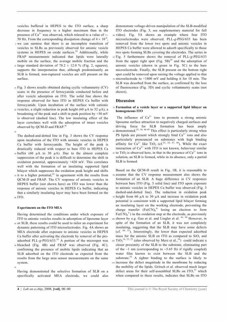
vesicles buffered in HEPES to the ITO surface, a sharp decrease in frequency to a higher maximum than in the presence of Ca2+ was observed, which relaxed to a value of -50 Hz. From the corresponding dissipation change of 4 10-6, we can surmise that there is an incomplete transition of 5
vesicles to SLBs as previously observed for anionic vesicle systems in HEPES on oxide surfaces.34 Additionally, while FRAP measurements indicated that lipids were laterally mobile on the surface, the average mobile fraction and the large standard deviation of 78.2 ± 12.0 % (Fig. 2, squares), 10
supports the interpretation that, although predominantly an SLB is formed, non-ruptured vesicles are still present on the surface. Fig. 3 shows results obtained during cyclic voltammetry (CV) 15
scans in the presence of ferrocyanide conducted before and after vesicle adsorption on ITO. The solid line shows the response observed for bare ITO in HEPES Ca buffer with ferrocyanide. Upon incubation of the surface with cationic vesicles, a slight reduction in peak height (60 µA to 52 µA), a 20
broadening of the peak and a shift in peak position by +30 mV is observed (dashed line). The low insulating effect of the layer correlates well with the adsorption of intact vesicles observed by QCM-D and FRAP.41 25
The dashed-and-dotted line in Fig. 3 shows the CV response upon incubation of the ITO with anionic vesicles in HEPES Ca buffer with ferrocyanide. The height of the peak is drastically reduced with respect to bare ITO in HEPES Ca buffer (60 µA to 30 µA). Due to the almost complete 30
suppression of the peak it is difficult to determine the shift in oxidation potential, approximately +420 mV. This correlates well with the formation of an insulating supported lipid bilayer which suppresses the oxidation peak height and shifts it to a higher potential,41 in agreement with the results from 35
QCM-D and FRAP. The CV response of anionic vesicles in HEPES buffer (not shown here) on ITO was lower than the response of anionic vesicles in HEPES Ca buffer, indicating that a similarly insulating layer may have been formed on the ITO. 40
Experiments on the ITO MEA
Having determined the conditions under which exposure of ITO to anionic vesicles results in adsorption of liposome layer or SLB, these results could be used to tailor an experiment for 45
dynamic patterning of ITO microelectrodes. Fig. 4A shows an MEA electrode after exposure to anionic vesicles in HEPES Ca buffer after activating the electrode by removal of the pre-adsorbed PLL-g-PEG/633.35 A portion of the microspot was bleached (Fig. 4B) and FRAP was observed (Fig. 4C), 50
confirming the presence of mobile lipids indicating that an SLB adsorbed on the ITO electrode as expected from the results from the large area sensor measurements on the same system. 55
Having demonstrated the selective formation of SLB on a specifically activated MEA electrode, we could also
demonstrate voltage-driven manipulation of the SLB-modified ITO electrodes (Fig. 5, see supplementary material for full video). Fig. 5A shows an example where four ITO 60
microelectrodes were observed. PLL-g-PEG/633 has been removed from the lower two spots and anionic vesicles in HEPES Ca buffer were allowed to adsorb specifically to those two spots forming SLBs covering the electrodes. The series in Fig. 5 furthermore shows the removal of PLL-g-PEG/633 65
from the upper right spot (Fig. 5B)35 and the adsorption of anionic vesicles (shown in green in Fig. 5C) to the bare microelectrode. Finally, the SLB present on the bottom right spot could be removed upon raising the voltage applied to that microelectrode to +1800 mV and holding it for 10 min. The 70
SLB was desorbed from the surface, as confirmed by the loss of fluorescence (Fig. 5D) and cyclic voltammetry scans (not shown).
Discussion
Formation of a vesicle layer or a supported lipid bilayer on 75
homogeneous ITO
The influence of Ca2+ ions to promote a strong anionic liposome surface attraction to negatively charged surfaces and driving force for SLB formation has been amply demonstrated.25, 34, 46-49 This effect is particularly strong when 80
PS lipids are present which strongly bind Ca2+ ions and also particularly pronounced on substrates with high binding affinity for Ca2+ like TiO2 (cf.25, 34, 47, 48). While the exact interaction of Ca2+ with ITO is not known, behaviour similar to TiO2 is observed here, in that in the presence of Ca2+ ions in 85
solution, an SLB is formed, while in its absence, only a partial SLB is formed. Based on the QCM-D result in Fig. 1B, it is reasonable to assume that the CV response measurement also shows the 90
formation of an SLB. A huge difference in CV responses between bare ITO (Fig. 3 solid line) and ITO upon exposure to anionic vesicles in HEPES Ca buffer was observed (Fig. 3 dashed-and-dotted line). The reduction in oxidation peak height from 60 µA to 30 µA and increase in oxidation peak 95
potential is consistent with a supported lipid bilayer forming an insulating layer on the working electrode, preventing the charge transfer (Fe(CN)6
4- losing an electron to form Fe(CN)6
3-) in the oxidation step at the electrode, as previously shown by e.g. Gao et al. and Lingler et al. 41, 50 However, in 100
spite of the formation of an SLB, it was not completely insulating, suggesting that the SLB may have some defects (cf. 50, 51). Interestingly, the lower than expected adsorbed mass for the anionic SLB on ITO as compared to SiO2 and TiO2
16, 25, 52 (also observed by Merz et al.,34) could indicate a 105
closer proximity of the SLB to the substrate, eliminating part of the ~1 nm (corresponding to 5.65 Hz if rigidly coupled) water film known to exist between the SLB and the substrate.53 A tighter binding to the surface is likely to increase the defect magnitude in the membrane by reducing 110
the mobility of the lipids. Gritsch et al. observed much larger defect areas for their self-assembled SLBs on ITO,33 which when compared to these results, indicates that SLBs on ITO
This journal is © The Royal Society of Chemistry [year] Lab on a chip, 2008, [vol], 00–00 | 5
presumably could be optimized to be fully insulating. Cationic vesicles in HEPES Ca buffer on ITO do not seem to form an SLB according to the QCM-D and CV data. A layer of non-ruptured vesicles is formed instead. This, at first 5
glance, seems to be in direct contradiction to an observation by Gritsch et al.,33 who claimed that SLBs were predominantly formed using cationic vesicles on ITO. The most probable explanation for the observed difference for cationic liposome rupture is that Gritsch et al. reported that 10
formation of SLBs only occurred for cholesterol concentrations of 40 mol% or higher and got their best results for a composition of 42% DMPC, 49% cholesterol and 9% positively charged DHADAB, while we did not include any cholesterol in our liposomes. As liposome rupture will be 15
decided primarily by the liposome-surface interaction together with the mechanical properties of the liposome membrane, the variation in lipid composition is likely to contribute to the different outcomes. Cholesterol is known to strongly affect the membrane elastic modulus and has a strong effect on 20
liposome fusion as well as being able to induce lateral segregation of lipid phases,54, 55 which is likely to have led to the increased incidence of rupture but reduced coverage observed by Gritsch et al. 25
Anionic vesicles on ITO in the absence of Ca in the buffer seem to form incompletely fused vesicle layers. In spite of the fact that only partial fusion of vesicles to bilayers occurred in this case, the recovered fractions in the FRAP experiments was 78% with a large standard deviation of 12%. Assuming a 30
similar mass response per adsorbed vesicle for the cationic and anionic liposomes1 the QCM-D data in Fig. 1 can be used to estimate the SLB coverage obtained in the QCM-D experiments. The mass of the liposome film is reasonably well approximated by the Sauerbrey relation,56 according to which 35
the frequency change is linearly related to the adsorbed mass. Comparing the frequency shift for the partial SLB formed without Ca (f = -50 Hz) to the full SLB (f = -25 Hz) and the complete liposome layer (f = -100 Hz) yields an SLB coverage of 70%. This coverage is high enough to assume a 40
high degree of inter-connectedness of the SLB, which agrees with the FRAP result of a partial SLB. The corresponding CV response from anionic vesicles in HEPES buffer on ITO indicated that the mixed vesicle layer 45
and SLB film showed a higher insulating quality than that of the supported lipid bilayer. This is counter-intuitive, as one would expect an SLB to have better insulating quality than an incompletely fused SLB. While it is possible that the organisation of SLB patches completely surrounds the larger 50
1This assumption ignores that the anionic vesicles being closer to rupture are likely to be more deformed46 and thus higher vesicle coverage per frequency change is expected for the anionic vesicles. However, this will be offset by that vesicles at lower coverage also are likely to couple more water mass and thus give a lower vesicle coverage per frequency change.17, 37
vesicles sitting on the surface or even form membrane junctions integrating the outer monolayer of the liposomes with the SLB to explain the discrepancy between the QCM-D/FRAP and CV data sets, it is more likely that small variations in experimental conditions prevent reliable 55
quantitative conclusions from being drawn. For this system both QCM-D and FRAP results indicated a large variability in the results, sometimes indicating close to a full SLB being formed, similar to what has previously been observed for POPC liposomes adsorbing on ITO.34 This indicates a system 60
striking a close balance between liposome adsorption and SLB formation and it is possible that small differences in preparations of the substrates resulted in more reproducible SLB formation on the CV electrodes. 65
Formation and desorption of supported lipid bilayers by electrochemical addressing on the ITO microelectrode array
Lipid vesicles are known to adhere to most types of surfaces, including all tested electrode materials. Hence it normally presents a challenge to be able to isolate their adhesion only 70
to certain parts of a sensor surface creating a functional pattern. Non-specific binding of SLBs can be prevented by exploiting the non-fouling properties of PLL-g-PEG on the microelectrode array background35. 75
The measurements on large area samples using QCM-D, FRAP and CV resulted in a parameter space for liposome adsorption and SLB formation. It also provided insight into the mechanism of adsorption that controls the respective outcome. It seems likely that attraction of Ca2+ to the ITO 80
surface is necessary to fuse the anionic liposomes to an SLB, through the specific interaction of Ca2+ with PS lipids. This led us to postulate that we could remove an adsorbed SLB by applying a positive bias that would displace the Ca2+ ions from the interface in analogy to the displacement of 85
electrostatically adhering PLL-g-PEG by a +1800 mV bias.35 As shown in Fig. 5 this approach was successful and an anionic PS-containing membrane in HEPES Ca buffer was shown to desorb at a potential of +1800 mV applied to the ITO microelectrode. A possible scenario for the induced 90
desorption is thus that SLB patches associated with Ca2+ ions adsorbed to the interface are desorbed along with the Ca2+ ions. It is likely that the local oversaturation of positive charges at the DOPC:DOPS membrane interface due to the strong interaction of Ca2+ ions with the PS headgroups makes 95
the entire membrane susceptible to expulsion from the interface when the ions are displaced. Similar desorption experiments were conducted using cationic vesicles instead of anionic vesicles. The assembly of cationic 100
vesicles were also successfully desorbed at a potential of +1800 mV. In this context it is important to point out that the application of a high positive bias to the ITO electrode might not produce a pure electrostatic repulsion effect. Tang et al. describes that water dissociation effects are known to occur at 105
an electrode potential of +1800 mV, which is used to explain the mechanism by which PLL-g-PEG desorbs.35 The

6 | Lab on a chip, 2008, [vol], 00–00 This journal is © The Royal Society of Chemistry [year]
accumulation of positively charged hydrogen ions at the surface could cause a local drop in the pH on the ITO surface, which could contribute to a change to a more positive charge of the lipid headgroups and lead to destabilisation and repulsion of the SLB. 5
An application of potential-induced SLB desorption was demonstrated in Fig. 5 where one micro-SLB spot was deposited and another removed on a standard electrochemical microarray chip. With this platform, each electrode can be 10
patterned, addressed and regenerated individually by means of controlling the electrode potential. The external and reproducible nature of this switch makes it possible for these processes to be automated. In principle, there are no obstacles to further miniaturisation and densification of the 15
demonstrated membrane array technology down to spots in the few micron range typical for, e.g., DNA arrays. The technology for fabrication of such chips is available and the self assembly of supported lipid bilayers from liposomes has been demonstrated down to the few hundred nm spot size.7, 9,
20
26
Conclusions
In summary, the essential elements have been demonstrated by this and previous work35 to realise an array platform as the 25
one visualised in Fig. 6, where different membrane and polymer functionalities can be individually given to each sensor spot and that functionality can selectively be released or regenerated by an external switch. We have specifically shown that supported lipid bilayers can be formed on ITO 30
with retained lipid fluidity using a mixture of PC and PS lipids by adding a small amount of Ca2+ to the buffering solution. It was further demonstrated that these SLBs can be patterned in the micrometer size range on an ITO micrelectrode array, which is likely to be possible to further 35
increase the spot density. Both liposomes and SLB on ITO electrodes could be desorbed from the surface by a simple DC voltage bias or readsorbed on a bare ITO electrode at zero bias. This novel micro-patterning technique has equipped us with a toolbox for possible applications in areas such as 40
triggered release for lab-on-a-chip applications and selective patterning for high-throughput screening platforms.
Acknowledgements
The authors thank Prof. Jen-Sue Chen (National Cheng Kung 45
University, Taiwan, ROC) for providing the ITO coatings; Dr. Gabor Csucs (Light Microscopy Centre, ETH Zurich, Switzerland) for support with the CLSM measurements; Ayanda Biosystems SA, Switzerland for providing the microelectrode arrays. The authors further acknowledge the 50
financial support from the Agency for Science, Technology and Research, Singapore; Swiss Federal Laboratories for Materials Testing and Research, Dübendorf, Switzerland; Competence Centre for Materials Science and Technology, Switzerland. 55
Notes a BioInterface Group, Laboratory of Surface Science and Technology, Swiss Federal Institute of Technology (ETH), Zürich, CH-8093 Zurich, Switzerland. E-mail: [email protected] b Swiss Federal Laboratories for Materials Testing and Research 60
(EMPA), CH-8600 Dübendorf. † Electronic Supplementary Information (ESI) available: A video where supported lipid bilayers are sequentially adsorbed and desorbed from different ITO microelectrodes in an array. See DOI: 10.1039/b000000x/ 65
References
1. Y. Fang, Y. L. Hong, B. Webb and J. Lahiri, Mrs Bulletin, 2006, 31, 541-545.
2. A. D. Howard, G. McAllister, S. D. Feighner, Q. Y. Liu, R. P. Nargund, L. H. T. Van der Ploeg and A. A. Patchett, Trends 70
Pharmacol. Sci., 2001, 22, 132-140. 3. A. Engel, Supramolecular Structure and Function, 2004, 8, 119 -
134. 4. E. Reimhult and K. Kumar, Trends in Biotechnology, 2008, 26, 82-
89. 75
5. A. Janshoff and C. Steinem, Analytical and Bioanalytical Chemistry, 2006, 385, 433-451.
6. E. T. Castellana and P. S. Cremer, Surface Science Reports, 2006, 61, 429-444.
7. J. J. Shi, J. X. Chen and P. S. Cremer, Journal of the American 80
Chemical Society, 2008, 130, 2718-+. 8. A. E. Oliver, E. L. Kendall, M. C. Howland, B. Sanii, A. P. Shreve
and A. N. Parikh, Lab on a Chip, 2008, 8, 892-897. 9. K. Morigaki, Biointerphases, 2008, 3, FA85-FA89. 10. J. D. Taylor, K. S. Phillips and Q. Cheng, Lab on a Chip, 2007, 7, 85
927-930. 11. J. M. Moran-Mirabal, J. B. Edel, G. D. Meyer, D. Throckmorton, A.
K. Singh and H. G. Craighead, Biophys J, 2005, 89, 296-305. 12. B. L. Jackson and J. T. Groves, Journal of the American Chemical
Society, 2004, 126, 13878-13879. 90
13. J. S. Hovis and S. G. Boxer, Langmuir, 2000, 16, 894-897. 14. J. T. Groves, N. Ulman and S. G. Boxer, Science, 1997, 275, 651-
653. 15. R. Richter, A. Mukhopadhyay and A. Brisson, Biophys J, 2003, 85,
3035-3047. 95
16. C. A. Keller and B. Kasemo, Biophys J, 1998, 75, 1397-1402. 17. E. Reimhult, C. Larsson, B. Kasemo and F. Hook, Analytical
Chemistry, 2004, 76, 7211-7220. 18. E. Reimhult, M. Zach, F. Hook and B. Kasemo, Langmuir, 2006, 22,
3313-3319. 100
19. A. Mashaghi, B. Swann, J. Popplewell, M. Textor and E. Reimhult, Analytical Chemistry (ASAP), 2008.
20. O. Purrucker, H. Hillebrandt, K. Adlkofer and M. Tanaka, Electrochim. Acta, 2001, 47, 791-798.
21. H. Schonherr, J. M. Johnson, P. Lenz, C. W. Frank and S. G. Boxer, 105
Langmuir, 2004, 20, 11600-11606. 22. J. M. Johnson, T. Ha, S. Chu and S. G. Boxer, Biophys J, 2002, 83,
3371-3379. 23. P. S. Cremer and S. G. Boxer, J. Phys. Chem. B, 1999, 103, 2554-
2559. 110
24. R. P. Richter and A. R. Brisson, Biophys J, 2005, 88, 3422-3433. 25. F. F. Rossetti, M. Bally, R. Michel, M. Textor and I. Reviakine,
Langmuir, 2005, 21, 6443-6450. 26. A. Dahlin, M. Zach, T. Rindzevicius, M. Kall, D. S. Sutherland and
F. Hook, Journal of the American Chemical Society, 2005, 127, 115
5043-5048. 27. F. Giess, M. G. Friedrich, J. Heberle, R. L. Naumann and W. Knoll,
Biophys J, 2004, 87, 3213-3220. 28. E. Reimhult, F. Hook and B. Kasemo, Langmuir, 2003, 19, 1681-
1691. 120
29. Z. Salamon, G. Lindblom and G. Tollin, Biophys J, 2003, 84, 1796-1807.

This journal is © The Royal Society of Chemistry [year] Lab on a chip, 2008, [vol], 00–00 | 7
30. J. P. Bearinger, J. Voros, J. A. Hubbell and M. Textor, Biotechnol. Bioeng., 2003, 82, 465-473.
31. P. Krysinski and G. J. Blanchard, Langmuir, 2003, 19, 3875-3882. 32. Z. Salamon, J. T. Hazzard and G. Tollin, Proc. Natl. Acad. Sci. U. S.
A., 1993, 90, 6420-6423. 5
33. S. Gritsch, P. Nollert, F. Jahnig and E. Sackmann, Langmuir, 1998, 14, 3118-3125.
34. C. Merz, W. Knoll, M. Textor and E. Reimhult, Biointerphases, 2008.
35. C. S. Tang, M. Dusseiller, S. Makohliso, M. Heuschkel, S. Sharma, 10
B. Keller and J. Voros, Anal. Chem., 2006, 78, 711-717. 36. M. Rodahl, F. Hook, C. Fredriksson, C. A. Keller, A. Krozer, P.
Brzezinski, M. Voinova and B. Kasemo, Faraday Discussions, 1997, 229-246.
37. E. Reimhult, F. Hook and B. Kasemo, J. Chem. Phys., 2002, 117, 15
7401-7404. 38. R. Etchenique and A. D. Weisz, J Appl Phys, 1999, 86, 1994-2000. 39. D. Axelrod, D. E. Koppel, J. Schlessinger, E. Elson and W. W.
Webb, Biophys J, 1976, 16, 1055-1069. 40. A. J. Bard, Electrochemical Methods - Fundamentals and 20
Application, John Wiley and Sons, Inc., 2001. 41. H. Gao, G. A. Luo, J. Feng, A. L. Ottova and H. T. Tien, J.
Photochem. Photobiol. B-Biol., 2000, 59, 87-91. 42. H.-G. Hong and M. D. Porter, Journal of Electroanalytical
Chemistry, 2005, 578, 113-119. 25
43. Z. P. Aguilar, W. R. Vandaveer and I. Fritsch, Analytical Chemistry, 2002, 74, 3321-3329.
44. C. A. Huang, K. C. Li, G. C. Tu and W. S. Wang, Electrochim. Acta, 2003, 48, 3599-3605.
45. T. E. Starr and N. L. Thompson, Langmuir, 2000, 16, 10301-10308. 30
46. T. R. Khan, H. M. Grandin, A. Mashaghi, M. Textor, E. Reimhult and I. Reviakine, Biointerphases, 2008.
47. F. F. Rossetti, M. Textor and I. Reviakine, Langmuir, 2006, 22, 3467-3473.
48. R. P. Richter, N. Maury and A. R. Brisson, Langmuir, 2005, 21, 299-35
304. 49. J. Ekeroth, P. Konradsson and F. Hook, Langmuir, 2002, 18, 7923-
7929. 50. S. Lingler, I. Rubinstein, W. Knoll and A. Offenhausser, Langmuir,
1997, 13, 7085-7091. 40
51. S. E. Creager, L. A. Hockett and G. K. Rowe, Langmuir, 1992, 8, 854-861.
52. R. P. Richter, R. Berat and A. R. Brisson, Langmuir, 2006, 22, 3497-3505.
53. T. M. Bayerl and M. Bloom, Biophys. J., 1990, 58, 357-362. 45
54. M. E. Haque, T. J. McIntosh and B. R. Lentz, Biochemistry, 2001, 40, 4340-4348.
55. D. Needham, T. J. McIntosh and E. Evans, Biochemistry, 1988, 27, 4668-4673.
56. G. Sauerbrey, Zeitschrift Fur Physik, 1959, 155, 206-222. 50

8 | Lab on a chip, 2008, [vol], 00–00 This journal is © The Royal Society of Chemistry [year]
Figure Captions
Figure 1A-C. Results for adsorption of liposomes on ITO (injected at t = 0 min) obtained by quartz crystal microbalance with dissipation monitoring. A sharp decrease in frequency (solid line) and increase in dissipation (dotted line) were 5
observed upon addition of cationic vesicles in HEPES Ca buffer on ITO in A. In B, upon addition of anionic vesicles on ITO in HEPES Ca buffer, after an initial sharp decrease in frequency, the frequency recovered to a stable asymptotic value of -25 Hz with a corresponding low dissipation. Upon 10
addition of anionic vesicles on ITO in HEPES buffer (without Ca), after an initial sharp decrease in frequency, the frequency recovered to -52 Hz with the dissipation following a reciprocal trajectory stabilizing at a rather high dissipation of 4 10-6. 15
Figure 2. Representative fluorescence recovery after photobleaching curves for the experiments where FRAP was observed. Anionic vesicles in HEPES Ca buffer show an almost complete recovery of fluorescence (red circles, 95 %), 20
while anionic vesicles in HEPES buffer showed a poorer recovery of fluorescence (80 %) after 400 s. Figure 3. Cyclic voltammetry experiments conducted in Ca buffer with ferrocyanide (Fe(CN)6
4-/Fe(CN)63-) to amplify the 25
electrochemical response scan. A total of 5 scans were conducted for each experiment, but only the first scan is shown. A CV scan on a bare ITO substrate in HEPES Ca buffer (solid) was observed to have an oxidation peak of about 60 µA, which indicated a low electrical interfacial resistance 30
of a bare ITO surface. The CV scan of an ITO surface which has been exposed to the cationic vesicles in HEPES Ca buffer (dotted) displayed a slightly lower current signal of 52 µA, indicating layer only partially covering the surface. This corresponds to the behaviour of an adsorbed vesicle layer. The 35
CV scan of an ITO surface which has been exposed to the anionic vesicles in HEPES Ca buffer (dashed-and-dotted) has a relatively constant peak current of 30 µA, demonstrating a high electrical resistance corresponding to the assembly of a supported lipid bilayers on the ITO surface. 40
Figure 4A-C. A series of CLSM images depicting the localised assembly of anionic vesicles in HEPES Ca buffer (green) on an ITO microelectrode, surrounded by fluorescently-labelled polycationic protein-resistant polymer, 45
poly(L-lysine)-grafted-poly(ethylene glycol) (red). Figure 3A shows that before photobleaching, similar fluorescence intensities were observed on both points 1 and 2. Figure 3B shows that photobleaching conducted on the surrounding region of point 1. After 10 minutes, figure 3C shows similar 50
relative fluorescence intensities on both points 1 and 2. This demonstrates free lateral diffusion of the phospholipids and formation of a fluid supported phospholipid bilayer on the ITO microelectrode. 55
Figure 5A-D. The fluorescence microscopy images show (see Supplementary material for video): a) An ITO microelectrode
array (MEA) was uniformly backfilled with fluorescently-labelled polycationic protein-resistant polymer, poly(L-lysine)-grafted-poly(ethylene glycol) (PLL-g-PEG/633) (red). 60
Two selected electrodes (bottom left and right) were biased at +1800 mV to remove the protein-resistant adlayer, followed by exposure to fluorescently labelled anionic vesicles (green). b) A selected ITO microelectrode (top right) was biased at +1800 mV to remove the PLL-g-PEG still adsorbed to this 65
electrode. Subsequently the ITO MEA was exposed to fluorescently-labelled anionic vesicles (green) in HEPES Ca buffer, which adsorbed specifically onto the previously electrically addressed microspot at the top right. c) An electric field of +1800 mV was applied to the SLB-covered ITO 70
microelectrode (bottom right) which desorbed the SLB rapidly from its surface. d) The final configuration showing the SLB on the top right corner electrode. Figure 6. Schematic depiction of the ITO microelectrode 75
array functionality applied to the systems for which adsorption and desorption have been shown controllable by an applied potential bias: electrostatically adsorbed functional non-fouling polymers (PLL-g-PEG), supported lipid membranes and liposomes. Also depicted is the possibility to 80
include membrane functionality through integration of membrane proteins into liposomes and SLB.

This journal is © The Royal Society of Chemistry [year] Lab on a chip, 2008, [vol], 00–00 | 9
Figure 1
5

10 | Lab on a chip, 2008, [vol], 00–00 This journal is © The Royal Society of Chemistry [year]
Figure 2

This journal is © The Royal Society of Chemistry [year] Lab on a chip, 2008, [vol], 00–00 | 11
Figure 3

12 | Lab on a chip, 2008, [vol], 00–00 This journal is © The Royal Society of Chemistry [year]
Figure 4
5

This journal is © The Royal Society of Chemistry [year] Lab on a chip, 2008, [vol], 00–00 | 13
Figure 5

14 | Lab on a chip, 2008, [vol], 00–00 This journal is © The Royal Society of Chemistry [year]
Figure 6
5
Copyright © 2022 FDOKUMEN