Evaluation of cholesterol metabolism in cerebrotendinous xanthomatosis
9
ORIGINAL ARTICLE Evaluation of cholesterol metabolism in cerebrotendinous xanthomatosis Andrea Mignarri 1 & Alessandro Magni 2 & Marina Del Puppo 2 & Gian Nicola Gallus 1 & Ingemar Björkhem 3 & Antonio Federico 1 & Maria Teresa Dotti 1 Received: 23 March 2015 /Revised: 2 June 2015 /Accepted: 5 June 2015 # SSIEM 2015 Abstract Background Cerebrotendinous xanthomatosis (CTX) is a treatable bile acid disorder caused by mutations of CYP27A1. The pathogenesis of neurological damage has not been completely explained. Oral chenodeoxycholic acid (CDCA) can lead to clinical stabilization, but in a subgroup of patients the disease progresses despite treatment. In the present study, we aimed at clarifying cholesterol metabolism abnormalities and their response to CDCA treatment, in order to identify reliable diagnostic and prognostic markers and understand if differences exist between stable patients and those with neu- rological progression. Methods We enrolled 19 untreated CTX patients and assessed serum profile of bile acids intermediates, oxysterols, choles- terol, lathosterol, and plant sterols. Then we performed a long- term follow up during CDCA therapy, and compared bio- chemical data with neurological outcome. Results We observed increase of cholestanol, 7α-hydroxy-4- cholesten-3-one (7αC4), lathosterol, and plant sterols, where- as 27-hydroxycholesterol (27-OHC) was extremely low or absent. CDCA treatment at a daily dose of 750 mg normalized all biochemical parameters except for 7αC4 which persisted slightly higher than normal in most patients, and 27-OHC which was not modified by therapy. Biochemical evaluation did not reveal significant differences between stable and wors- ening patients. Discussion Cholestanol and 7αC4 represent important markers for CTX diagnosis and monitoring of therapy. Treat- ment with CDCA should aim at normalizing serum 7αC4 as well as cholestanol, since 7 α C4 better mirrors 7 α- hydroxylation rate and is thought to be correlated with cholestanol accumulation in the brain. Assessment of serum 27-OHC is a very good tool for biochemical diagnosis at any stage of disease. Lathosterol and plant sterols should be con- sidered as additional markers for diagnosis and monitoring of therapy. Further studies including long-term assessment of bile acid intermediates in cerebrospinal fluid are needed in patients who show clinical progression despite treatment. Introduction Cerebrotendinous xanthomatosis (CTX) is an autosomal re- cessive lipid storage disease caused by mutations of CYP27A1. The biochemical defect is due to the deficiency of sterol-27-hydroxylase, a mitochondrial enzyme catalyzing the first step of degradation of the sterol side chain of bile acid biosynthesis (Cali et al 1991). CTX is characterized by high plasma and tissue concentration of cholestanol and other bile acid precursors, decreased levels of chenodeoxycholic acid (CDCA) and 27-hydroxycholesterol (27-OHC), markedly in- creased excretion of 25-hydroxylated bile alcohols in urine, bile, and faeces (Björkhem 2013). Also, serum levels of cho- lesterol precursors and plant sterols have been found to be elevated (Kuriyama et al 1991; Wolthers et al 1991; de Sain-van der Velden et al 2008). Communicated by: Jutta Gaertner * Andrea Mignarri [email protected] 1 Unit of Neurology and Neurometabolic Disorders, Department of Medicine, Surgery and Neurosciences, University of Siena, Siena, Italy 2 Department of Health Sciences, Medical School, University of Milano-Bicocca, Milano, Italy 3 Department of Laboratory Medicine, Division of Clinical Chemistry, Karolinska Institute, Karolinska University Hospital, Huddinge, Sweden J Inherit Metab Dis DOI 10.1007/s10545-015-9873-1
-
Upload
independent -
Category
Documents
-
view
2 -
download
0
Transcript of Evaluation of cholesterol metabolism in cerebrotendinous xanthomatosis

ORIGINAL ARTICLE
Evaluation of cholesterol metabolismin cerebrotendinous xanthomatosis
Andrea Mignarri1 & Alessandro Magni2 & Marina Del Puppo2 & Gian Nicola Gallus1 &
Ingemar Björkhem3& Antonio Federico1 & Maria Teresa Dotti1
Received: 23 March 2015 /Revised: 2 June 2015 /Accepted: 5 June 2015# SSIEM 2015
AbstractBackground Cerebrotendinous xanthomatosis (CTX) is atreatable bile acid disorder caused by mutations of CYP27A1.The pathogenesis of neurological damage has not beencompletely explained. Oral chenodeoxycholic acid (CDCA)can lead to clinical stabilization, but in a subgroup of patientsthe disease progresses despite treatment. In the present study,we aimed at clarifying cholesterol metabolism abnormalitiesand their response to CDCA treatment, in order to identifyreliable diagnostic and prognostic markers and understand ifdifferences exist between stable patients and those with neu-rological progression.Methods We enrolled 19 untreated CTX patients and assessedserum profile of bile acids intermediates, oxysterols, choles-terol, lathosterol, and plant sterols. Then we performed a long-term follow up during CDCA therapy, and compared bio-chemical data with neurological outcome.Results We observed increase of cholestanol, 7α-hydroxy-4-cholesten-3-one (7αC4), lathosterol, and plant sterols, where-as 27-hydroxycholesterol (27-OHC) was extremely low orabsent. CDCA treatment at a daily dose of 750 mg normalized
all biochemical parameters except for 7αC4 which persistedslightly higher than normal in most patients, and 27-OHCwhich was not modified by therapy. Biochemical evaluationdid not reveal significant differences between stable and wors-ening patients.Discussion Cholestanol and 7αC4 represent importantmarkers for CTX diagnosis and monitoring of therapy. Treat-ment with CDCA should aim at normalizing serum 7αC4 aswell as cholestanol, since 7αC4 better mirrors 7α-hydroxylation rate and is thought to be correlated withcholestanol accumulation in the brain. Assessment of serum27-OHC is a very good tool for biochemical diagnosis at anystage of disease. Lathosterol and plant sterols should be con-sidered as additional markers for diagnosis and monitoring oftherapy. Further studies including long-term assessment ofbile acid intermediates in cerebrospinal fluid are needed inpatients who show clinical progression despite treatment.
Introduction
Cerebrotendinous xanthomatosis (CTX) is an autosomal re-cessive lipid storage disease caused by mutations ofCYP27A1. The biochemical defect is due to the deficiencyof sterol-27-hydroxylase, a mitochondrial enzyme catalyzingthe first step of degradation of the sterol side chain of bile acidbiosynthesis (Cali et al 1991). CTX is characterized by highplasma and tissue concentration of cholestanol and other bileacid precursors, decreased levels of chenodeoxycholic acid(CDCA) and 27-hydroxycholesterol (27-OHC), markedly in-creased excretion of 25-hydroxylated bile alcohols in urine,bile, and faeces (Björkhem 2013). Also, serum levels of cho-lesterol precursors and plant sterols have been found to beelevated (Kuriyama et al 1991; Wolthers et al 1991; deSain-van der Velden et al 2008).
Communicated by: Jutta Gaertner
* Andrea [email protected]
1 Unit of Neurology and Neurometabolic Disorders, Department ofMedicine, Surgery and Neurosciences, University of Siena,Siena, Italy
2 Department of Health Sciences, Medical School, University ofMilano-Bicocca, Milano, Italy
3 Department of Laboratory Medicine, Division of Clinical Chemistry,Karolinska Institute, Karolinska University Hospital,Huddinge, Sweden
J Inherit Metab DisDOI 10.1007/s10545-015-9873-1
The clinical picture is extremely heterogeneous evenamong members of the same family. The main systemic man-ifestations are tendon xanthomas, juvenile cataract, chronicdiarrhoea, and premature osteoporosis, while neurologicaldysfunction includes spastic paraparesis, cerebellar ataxia,cognitive impairment, psychiatric disturbances, peripheralneuropathy, epilepsy, and parkinsonism (Mignarri et al2014). Brain magnetic resonance imaging (MRI) typicallyshows dentate nuclei signal abnormalities (Barkhof et al2000; De Stefano et al 2001), and cerebellar vacuolation canbe observed as disease progresses (Mignarri et al 2012). Plas-ma cholestanol assessment is useful for CTX diagnosis, show-ing elevated concentrations in all untreated CTX patients(Mignarri et al 2014). Definitive diagnosis is obtained by mo-lecular analysis of the CYP27A1 gene.
The pathogenetic mechanisms underlying neurologicaldamage in CTX have not been completely clarified. Largeamounts of cholestanol are known to induce apoptosis, withparticular involvement of cerebellar neuronal cells (Inoue et al1999). However, the origin of brain cholestanol is not obvi-ous: it has been suggested that cerebral storage of cholestanolmay be a consequence of the flux of its precursor 7α-hydroxy-4-cholesten-3-one (7αC4) across the blood-brain barrier(Panzenboeck et al 2007; Bavner et al 2010). The lack of27-OHC could also play a role: indeed, cerebral 27-OHCoriginates from the circulation and represents an importantregulator of cholesterol metabolism in the brain (Heverinet al 2005; Ali et al 2013).
CTX is a chronic disorder leading to progressive motor andcognitive impairment. If recognized early, many patients canhave a remarkable clinical response to treatment (Van Heijstet al 1998; Berginer et al 2009; Yahalom et al 2013). Oralreplacement therapy with bile acids decreases the productionof toxic intermediates by inhibiting CYP7A1, the key enzymefor the Bclassic^ pathway of bile acid synthesis. Among bileacids, CDCA is considered the strongest inhibitor of CYP7A1(Ellis et al 2003; Liu et al 2014). Long-term oral CDCA intakenormalizes bile acid synthesis in CTX patients, leading toclinical stabilization (Berginer et al 1984). Moreover, CDCAtherapy has been reported to improve neurophysiological pa-rameters including motor evoked potentials (Mignarri et al2011) and nerve conduction velocities (Ginanneschi et al2013), and to increase bone mineral density (Martini et al2013), while no improvement of MRI appearances has beenobserved (Berginer et al 1994). Patients with neurologicalprogression despite CDCA treatment have been largely re-ported (Pilo de la Fuente et al 2011a; Mignarri et al 2012;Rubio-Agusti et al 2012; Yahalom et al 2013). Notably,cholestanol assessment is important for diagnosis but doesnot seem to have a prognostic value, since normalization ofserum cholestanol levels following CDCA therapy is not al-ways associated with clinical stabilization (Pilo de la Fuenteet al 2011b).
In the present study, we evaluated the serum profile of bileacids intermediates (cholestanol and its precursor 7αC4, 27-OHC, 24S-hydroxycholesterol), cholesterol, lathosterol, andplant sterols (campesterol, sitosterol) in a significantly largeCTX population: we performed a baseline assessment and along-term follow up during therapy with oral CDCA, compar-ing biochemical data with neurological outcome. We aimed at(a) clarifying the biochemical abnormalities and their responseto CDCA treatment, (b) identifying reliable diagnostic andprognostic markers, and (c) understanding if a distinctive bio-chemical pattern exists in patients with neurological progres-sion despite treatment.
Materials and methods
Patient population and study design
We studied 19 CTX patients (11 males, 8 females) aged 13-54years (median age 32 years, mean±standard deviation 32.5±10.4 years), belonging to 15 unrelated Italian families. In allcases the diagnosis was confirmed by point mutations or de-letions in the CYP27A1 gene. Demographic, clinical and mo-lecular details of patients are summarized in Table 1.
On enrolment in the study, all patients were untreated. Afterbaseline (t0) clinical examination and biochemical analyses,they started oral CDCA therapy at a daily dose of 750 mg(250 mg three times a day). Then a long-term clinical andbiochemical follow up was performed (t1=0-1.5 years; t2=1.5-2.5 years; t3=2.5-3.5 years; t4=>4 years).
All patients underwent neurological examination includingRankin Scale (van Swieten et al 1988) and Expanded Disabil-ity Status Scale (Kurtzke 1983) administration at baseline andduring the follow up period: subjects with unchanged RankinScale (RS) and Expanded Disability Status Scale (EDSS)scores over time were considered Bneurologically stable^,while those with increased RS and/or EDSS scores at followup were classified as Bneurologically worsening^.
We analyzed cholesterol metabolism both at baseline andduring CDCA treatment. Moreover, we compared biochemi-cal data of Bneurologically stable^ patients with those ofBneurologically worsening^ patients.
The study was approved by the Local Ethics Committee,Faculty of Medicine, University of Siena, and written in-formed consent was obtained from all patients.
Solvents and reagents
All solvents were obtained from Merck (Darmstadt, Germa-ny) and were of analytical grade. BHT, piperidine, 2,3,4,5,6-pentafluorobenzoyl chloride and trimethylsilylimidazole werepurchased from Sigma-Aldrich (St Louis, MO, USA). Silicacartridge columns (Supelclean LC-Si, size 1 ml) were
J Inherit Metab Dis
obtained from Supelco Inc. (Bellefonte, PA, USA). Total Cho-lesterol Assay Kit (Colorimetric) was purchased from CellBiolabs Inc. (San Diego, CA, USA).
Standards
All deuterated sterols used as internal standards (cholestanol-d4, 27-hydroxycholesterol-d9, lathosterol-d4) were synthe-sized in our laboratory as described previously (Galli Kienleet al 1980; Alessandrini et al 2004). Cholestanewas purchasedfrom Sigma-Aldrich (St Louis, MO, USA) and 19-hydroxycholesterol was obtained from Steraloids (Newport,RI, USA). Cholestanol, lathosterol, campesterol, β-sitosterolwere purchased from Sigma-Aldrich (St Louis, MO, USA),7αC4, 27-OHC and 24S-hydroxycholesterol (24-OHC) wereobtained from Steraloids (Newport, RI, USA).
Sample preparation for the determination of plasma levelsof cholestanol
Deuterated cholestanol was added to 0.2-ml plasma samplesas solutions of 0.2 μg/μl (5 μl) in ethyl acetate. Alkalinehydrolysis was carried out with 1 ml 1 N NaOH in 90 %
ethanol at 60 °C for 90 minutes under nitrogen; physiologicsaline solution (1 ml) was then added, and sterols were ex-tracted with 2 ml of petroleum ether and taken to drynessunder a stream of nitrogen. HPLC separation was performedin order to remove excess cholesterol from lipid extract, aspreviously described (Kuriyama et al 1991). The extractedsterols were converted into their benzoyl derivatives by addi-tion of 0.2 ml of a dichloromethane reaction mixture contain-ing 0.2 % triethylamine (TEA) and 0.5 % 2,3,4,5,6-pentafluoro-benzoyl chloride (PFB). After incubation (20 mi-nutes at room temperature) reaction mixture was evaporatedunder nitrogen. HPLC separation was performed with a JascoHPLC system (880-PU, 801-SC, 880-02) equipped with aJasco 875-UV detector (Jasco, Tokyo, Japan). Analyseswere carried out with an Inertsil® ODS-2 (4.6 mm×150 mm) column with guard column (GL SciencesInc., Tokyo, Japan) using a mobile phase of acetoni-trile/water/acetic acid (100:3:0.2, v/v/v) at a flow rateof 1 ml/min. Absorbance was monitored at λ 228 nm.Samples were injected dissolved in 0.2 ml of eluent andthe retention time of PFB derivatives of cholesterol andcholestanol was verified by injection of pure derivatizedstandards. The PFB-cholestanol fraction of each sample
Table 1 Demographic information, clinical picture (including neurological disability scores), and molecular analysis of the 19 CTX patients enrolledfor the study
Demographic info Clinical picture and disability scores CYP27A1 gene analysis
Fm Pt S Age Catar Xant Cogn Psych Spast Ataxia RS/EDSS First mutation Second mutationimpair disturb
a 1CP M 43 yes yes yes no yes yes 3/6 (=) c.646 G>C c.646 G>C
b 2CG M 21 yes yes yes yes no no 1/ 3 (=) c.752 C>A c.1263+5 G>T
3CR F 18 yes yes no yes no no 0/2 (=) c.752 C>A c.1263+5 G>T
c 4DGS M 30 yes yes yes no yes yes 2/4 (=) c.776 A>G c.776 A>G
d 5DL F 34 yes no yes no yes yes 3/5 (↑) c.1263+1 G>A c.1263+1 G>A
6DL M 36 no yes yes no yes yes 3/7 (↑) c.1263+1 G>A c.1263+1 G>A
e 7FG M 45 yes yes yes yes yes yes 3/5 (↑) c.752 C>A c.752 C>A
f 8FP M 37 yes yes yes no yes yes 3/4 (↑) c.1184+1 G>A c.1184+1 G>A
g 9GL M 25 yes yes no no no no 1/1.5 (=) c.863 delA c.1183 C>T
10GM M 13 no no no no no no 0/0 (=) c.863 delA c.1183 C>T
h 11IS M 31 yes yes yes yes yes yes 2/3 (=) c.647-1 G>T c.1183 C>T
i 12PC F 33 yes no yes no yes no 2/3 (↑) c.752 C>A c.752 C>A
j 13RS F 54 no yes no yes yes no 2/3.5 (=) c.646 G>C c.1538 G>A
k 14RA F 31 yes yes yes yes yes yes 2/3.5 (=) c.1263+81_1596+?del c.1263+81_1596+?del
l 15RM F 22 yes yes no yes no no 0/2 (=) c.1183 C>T c.646 G>C
m 16SD M 32 yes yes yes yes yes yes 2/3.5 (↑) c.1184+1 G>A c.1184+1 G>A
n 17SR F 36 yes yes yes no yes no 3/3.5 (=) c.646 G>C c.1184+1 G>A
18SV M 29 yes yes yes yes yes no 2/3.5 (↑) c.646 G>C c.1184+1 G>A
o 19VS F 48 yes no yes no no no 1/1.5 (=) c.1016 C>T c.1016 C>T
Catar: Cataracts; Cogn impair: Cognitive impairment; del: deletion; EDSS: Expanded Disability Status Scale; F: Female; Fm: Family; M: Male; Pt:Patient; Psych disturb: Psychiatric disturbances; RS: Rankin Scale; S: sex; Spast: Spastic paraparesis; Xant: Xanthomas of tendons; (=): unchanged RSand EDSS scores at follow up (neurologically stable patients); (↑): increased RS and/or EDSS scores at follow up (neurologically worsening patients)
J Inherit Metab Dis

was collected and, after solvent evaporation, dissolvedin 25 μl of toluene for GC-MS analysis.
Sample preparation for analysis of circulating oxysterols,lathosterol and plant sterols
Extraction and purification of plasma 7αC4, 27-OHC, 24-OHC, lathosterol and plant sterols (campesterol and β-sitosterol) was performed as previously described (Kuriyamaet al 1991; Alessandrini et al 2004). As internal standards, weused 19-hydroxycholesterol for 7αC4 and 24-OHC, deuterat-ed 27-OHC for 27-OHC, deuterated lathosterol for lathosteroland cholestane for plant sterols. Before GC-MS analysis allsterols were converted into their trimethylsilyl ethers (TMS)with a mixture of trimethylsilylimidazole:piperidine (1:1) (byvolume), as already reported (Del Puppo et al 1998; Bertolottiet al 2008).
GC-MS analysis of plasma sterols
Analysis of sterols was carried out using a Thermo FinniganGC-Q instrument (Waltham, MA, USA). The spectrometerwas set at 70 eV ion energy, 0.1 mA emission current, and300 °C transfer line temperature. Separation of sterols wasachieved by a J&W HP5 capillary column (Agilent technolo-gies, Santa Clara, CA, USA) 0.32 mm i.d., 0.25 mm filmthickness, 30 m long, operating at 1 ml/min helium flow rate.Column temperature was programmed from 180 °C to 300 °C.For sterols quantification, we focused on specific ions: m/z215 and 427 for PFB-cholestanol, m/z 219 and 431 for deu-terated PFB-cholestanol (IS), m/z 382 and 472 for 7αC4-TMS, m/z 353 for 19-hydroxycholesterol-TMS (IS), m/z456 for 27-OHC-TMS, m/z 413 for 24-OHC -TMS, m/z 465for deuterated 27-OHC-TMS (IS), m/z 255 for lathosterol-TMS, m/z 382 for campesterol-TMS, m/z 396 for β-sitoster-ol-TMS, 259 for deuterated lathosterol-TMS (IS) and 372 forcholestane (IS). Calibration curves were prepared by spikingplasma with a fixed amount of each internal standard andincreasing amounts of the above-mentioned sterols. Thesesamples were treated and analyzed as the experimental sam-ples. Concentrations were calculated on the basis of the slopeof the standard curve as well as of the peak area ratio (sterol/IS) found in the sample. The assay results were linear (r>0.98)in the tested ranges.
Quantification of total plasma cholesterol
To measure total plasma cholesterol levels, a commercial as-say kit (colorimetric) was used according to the manufac-turer's instructions (Cell Biolabs Inc., San Diego, CA, USA).Absorbance at 570 nm was read with a BGM Labtech spec-trophotometric microplate reader (Cary, NC, USA). Sample
cholesterol concentrations were determined by interpolationfrom a standard curve.
Statistical analysis
We tested data for normal distribution with the Anderson–Darling test. In the case of normally distributed data samples,we used parametric tests for comparisons (Student’s t-test tocompare two groups, Duncan's new multiple range parametricto compare three or more groups). In the case of not normallydistributed data samples, we used non parametric tests forcomparisons (Mann-Whitney U test to compare two groups,Kruskal-Wallis test with Dunn’s post hoc test to compare threeor more groups). We also performed analysis of correlationsbetween variables by calculation of Spearman coefficient andrelative p-value. Statistical significance was assumed forp<0.05.
Results
Plasma concentrations of cholestanol, 7αC4, 27-OHC, 24-OHC, lathosterol, and plant sterols of our 19 CTX patientsbefore and during CDCA therapy are reported in Table 2and Figs. 1 and 2.
Serum cholestanol levels at baseline were on average ten-fold higher than normal, and a statistical significance wasfound between CTX patients and control subjects (p<0.01).Once treatment was started, plasma cholestanol sharply de-creased until normalization after 18 months of CDCA intake,and no loss of efficacy was detectable in the long-term. Un-treated CTX patients had significantly elevated (from 100-foldto 200-fold increased) plasma concentration of 7αC4 com-pared to controls (p<0.01), indicating marked hyperstimula-tion of the first part of the Bclassic^ pathway of bile acidsynthesis. Serum levels of 7αC4 consistently decreased afterCDCA treatment; however, 7αC4 never normalized in half ofsubjects, and some degree of accumulation appeared to persistin most patients. Notably, a significant positive correlationwas found between plasma values of 7αC4 and cholestanol(ρ=0.78; p<<0.01). As expected, serum 27-OHC was belowthe limit of detection in almost all CTX plasma samples, andno changes were observed under CDCA treatment. Serum 24-OHC was slightly but significantly (p<0.05) increased in un-treated CTX patients, and mildly but not significantly de-creased during long-term treatment.
In order to complete our investigation of cholesterol me-tabolism, we also assayed plasma levels of cholesterol,lathosterol (as biomarker of hepatic and whole body choles-terol Bde-novo^ synthesis) and phytosterols (as biomarkers ofcholesterol intestinal absorption). Serum cholesterol was nor-mal in most patients (only few patients showed mild hyper-cholesterolemia), and did not substantially change during
J Inherit Metab Dis

CDCA treatment. Plasma lathosterol levels were significantlyincreased in untreated CTX patients (p<0.01), indicating thatcholesterol synthesis was incremented, and normalized in thecourse of therapy. Moreover, plasma levels of lathosterol and7αC4 resulted to be strictly correlated (ρ=0.8; p<<0.01). Sim-ilar to lathosterol, baseline serum levels of the plant sterolscampesterol and sitosterol were high in CTX patients, indicat-ing increased cholesterol intestinal absorption; however, onlysitosterol increase was statistically significant (p<0.05).CDCA treatment strongly reduced plasma concentration ofplant sterols.
We performed neurological examination and disability as-sessment with RS and EDSS at baseline and during the entirefollow up period: on the basis of unchanged disability scoresover time, 12 patients were classified as Bneurologicallystable^; on the other hand, seven patients showed in-creased RS and/or EDSS scores at follow up and wereconsidered as Bneurologically worsening^. Age at diag-nosis was not significantly different between the two
groups. Disability at diagnosis was higher in theBneurologically worsening^ group; in particular a statis-tically significant difference was found between baselineRS of the two samples (p<0.05). When we comparedbiochemical data of Bneurologically stable^ patients withthose of Bneurologically worsening^ subjects, we didnot find significant differences between the two groups,both before therapy and during long-term follow up.Notably, serum concentration of cholestanol, 7αC4,and lathosterol at baseline tended to be higher in thosepatients who later presented neurological worsening de-spite therapy, but no statistical significance was ob-served. Given the small number of biochemical dataavailable at long-term follow up on Bneurologicallyworsening^ patients, it was difficult to perform a statis-tical comparison between the two groups.
Finally, no correlation was observed between genotype andbiochemical pattern, as well as between genotype and pheno-typical characteristics or response to therapy.
Table 2 Determination of plasma levels of cholestanol, oxysterols(7αC4, 27-OHC, 24-OHC), cholesterol, lathosterol, and plant sterols(campesterol, sitosterol) at baseline and during CDCA treatment. Data
were reported as mean±sd and interquartile range (IQR) with median.Evaluation times: t0=baseline; t1=0-1.5 years; t2=1.5-2.5 years; t3=2.5-3.5 years; t4=>4 years
t0(n=19)
t1(n=13)
t2(n=11)
t3(n=11)
t4(n=12)
Normal values
Cholestanol(mg/dl)
mean±sdIQR(median)
3.42±1.282.46-4.01(3.72)
0.81±0.590.55-0.84(0.72)
0.69±0.600.28-0.84(0.48)
0.53±0.300.33-0.68(0.40)
0.38±0.150.30-0.42(0.38)
0.34±0.16(Kuriyama et al 1991)
7αC4(μg/dl)
mean±sdIQR(median)
368.0±221.0200.0-527.0(297.0)
34.9±51.112.7-33.4(21.0)
25.9±53.62.4-12.9(8.2)
20.0±18.07.6-28.0(13.7)
22.9±29.03.1-44.4(7.3)
2.2±2.0(Camilleri et al 2009)
27-OHC(μg/dl)
mean±sdIQR(median)
1.0±1.20.0-1.2(0.8)
1.1±0.80.8-1.4(1.2)
0.7±0.60.4-1.0(0.6)
0.7±0.70.0-1.3(0.5)
0.6±0.70.0-1.2(0.3)
15.4±4.3(Dzeletovic et al 1995)
24-OHC(μg/dl)
mean±sdIQR(median)
8.6±4.55.5-10.4(7.4)
7.8±3.84.9-12.3(6.5)
7.2±1.76.5-8.4(7.4)
7.3±3.25.2-8.6(6.6)
7.2±2.44.9-9.1(7.2)
6.4±2.4(Dzeletovic et al 1995)
Cholesterol(mg/dl)
mean±sdIQR(median)
187±67149-215(160)
196±97149-191(166)
176±37150-186(176)
187±41172-209(191)
165±42132-187(172)
166±32(Kempen et al 1988)
Lathosterol(μg/dl)
mean±sdIQR(median)
934±347677-1200(842)
178±102123-480(141)
158±82118-176(124)
168±92124-213(133)
136±7685-159(113)
206±79(Kempen et al 1988)
(μg/100 mg cholesterol) mean±sdIQR(median)
526±160458-615(499)
105±15965-114(86)
78±2170-83(79)
86±3668-98(80)
83±4852-94(70)
96±34(Kempen et al 1988)
Campesterol(μg/dl)
mean±sdIQR(median)
561±286346-734(525)
226±76161-249(233)
182±86115-256(166)
186±62149-209(178)
169±77111-210(154)
399±218(Kuriyama et al 1991)
(μg/100 mg cholesterol) mean±sdIQR(median)
334±203209-328(283)
141±9589-141(105)
104±6152-127(89)
107±5777-134(83)
108±6474-111(81)
240±120(Kuriyama et al 1991)
Sitosterol(μg/dl)
mean±sdIQR(median)
1119±595651-1628(938)
491±148413-599(468)
418±202245-533(435)
430±184330-492(370)
402±223245-445(355)
629±171(Kuriyama et al 1991)
(μg/100 mg cholesterol) mean±sdIQR(median)
637±293434-682(552)
301±159226-361(278)
244±140124-348(218)
253±163152-333(201)
256±170160-254(183)
410±90(Kuriyama et al 1991)
J Inherit Metab Dis
Discussion
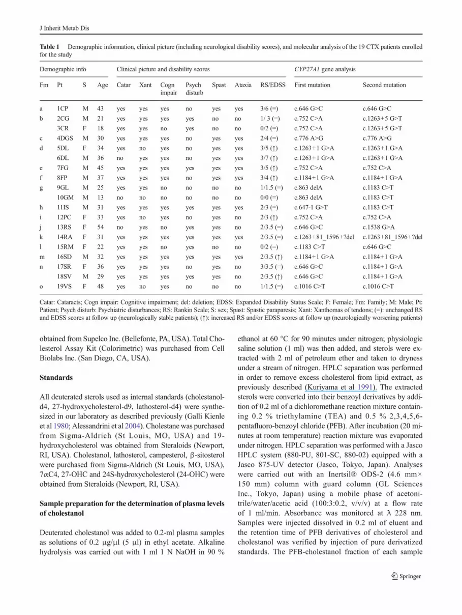
In spite of the undoubted improvement in the diagnosisand therapy and the better understanding of pathogenet-ic mechanisms, several points of criticism on CTX path-ogenesis, diagnosis, and treatment still remain to besolved (Björkhem and Hansson 2010). In this respect,our long-term study of cholesterol metabolism may helpto clarify some relevant aspects. As previously observedin small series of untreated CTX patients, this studyconfirmed pre-treatment increase of cholestanol,lathosterol, and plant sterols. Moreover, 7αC4 wasmarkedly increased, whereas 27-OHC was generally ab-sent or extremely low. Total cholesterol levels were sub-stantially normal. After CDCA treatment normalizationof all biochemical parameters was observed with theexception of serum 7αC4, whose level was sharply reducedbut still higher than normal in most patients, and plasma 27-OHC which was not modified. Each of the above reportedbiochemical findings suggests some considerations both initself and in relation to clinical follow up.
As expected, plasma cholestanol was elevated in all un-treated patients, thus confirming its utility as diagnostic, easyto dose marker. However, cholestanol may be reduced byseveral drugs (bile acids, statins, and steroids) whereas in-creased levels can be found in sitosterolemia (Salen et al1985). Treatment with CDCA normalized serum cholestanol,and we did not observe any loss of efficacy at follow up. Inthis respect, our data differ from previous observation ofcholestanol increase (after initial normalization) during long-term treatment in two children reported by de Sain-van derVelden et al (2008), which could be due, for instance, to toolow dose of CDCA.
Serum 7αC4 has been proposed as a very important markerfor both diagnosis and monitoring of replacement therapy inCTX (DeBarber et al 2010; Björkhem et al 2014). Circulating7αC4 closely mirrors cholesterol 7α-hydroxylation rate, thusreflecting the activity of the Bclassic^ pathway of bile acidsynthesis (Bertolotti et al 2008). Furthermore, 7αC4 is strictlycorrelated with accumulation of cholestanol in the brain inboth CTX subjects (Panzenboeck et al 2007) and mice withdisruption of sterol 27-hydroxylase (Bavner et al 2010).
Fig. 1 Box plots representing plasma levels of cholestanol, 7αC4, 27-OHC, and 24-OHC before therapy and during CDCA treatment. Data areexpressed as minimum, 25th percentile, median, 75th percentile, and
maximum; mean values are also indicated. Evaluation times: t0=baseline; t1=0-1.5 years; t2=1.5-2.5 years; t3=2.5-3.5 years;t4=>4 years
J Inherit Metab Dis
However, so far data on 7αC4 in CTX patients are very few.We found extremely high plasma 7αC4 at baseline, whileunder CDCA 7αC4 consistently decreased; however, a mild7αC4 increase appeared to persist in most patients. Probably,CDCA dose may be incremented until normal 7αC4 valuesare reached. In this respect, a pilot study in CTX patientsshould be performed, increasing CDCA and monitoring the7αC4 levels in serum as well as in the cerebrospinal fluid.Finally, the positive correlation between plasma values of7αC4 and cholestanol strengthens the value of 7αC4 as anadjunctive diagnostic biomarker.
Serum 27-OHCwas very low or absent in all CTX patients,regardless of CDCA treatment confirming the substantial in-activity of sterol 27-hydroxylase, and the inability of CDCAto correct deficiencies of intermediates in the "alternative"pathway of bile acid synthesis. Assessment of serum 27-OHC may be considered the best analysis for CTX diagnosisbeing reliable at any stage of disease, irrespective of treatment.Furthermore, dosage of plasma 27-OHC could be consideredof choice in the view of a possible neonatal screening. Ac-cording to a recent paper the preferred nomenclature for 27-
hydroxylation and 27-hydroxycholesterol should be (25R)26-hydroxylation and (25R)26-hydroxycholesterol, respectively(Fakheri and Javitt 2012).
Under in vitro conditions 27-OHC is an activator of liver Xreceptor (LXR). The role of this activation under in vivo con-ditions is controversial, however. The activation of someLXR-regulated genes as a consequence of feeding mice withhigh dietary cholesterol has been demonstrated to be mediatedby side-chain oxidized cholesterol. If the oxysterol responsi-ble for the activation is 24-, 25- or 27-hydroxycholesterol hasnot been shown, however (Chen et al 2007). A knockout ofCYP27 in mice does not change the expression of a number ofLXR-target genes in the brain. The situation is similar in micewith an overexpression of the enzyme. We must con-clude that 27-OHC is of little importance for LXR sig-naling in the brain, at least under normal conditions,while it may be of some importance for regulation ofcholesterol synthesis by mechanisms not involvingLXR. Indeed, the flux of 27-OHC from the circulationinto the brain is of some importance for cholesterolhomeostasis in the brain (Ali et al 2013).
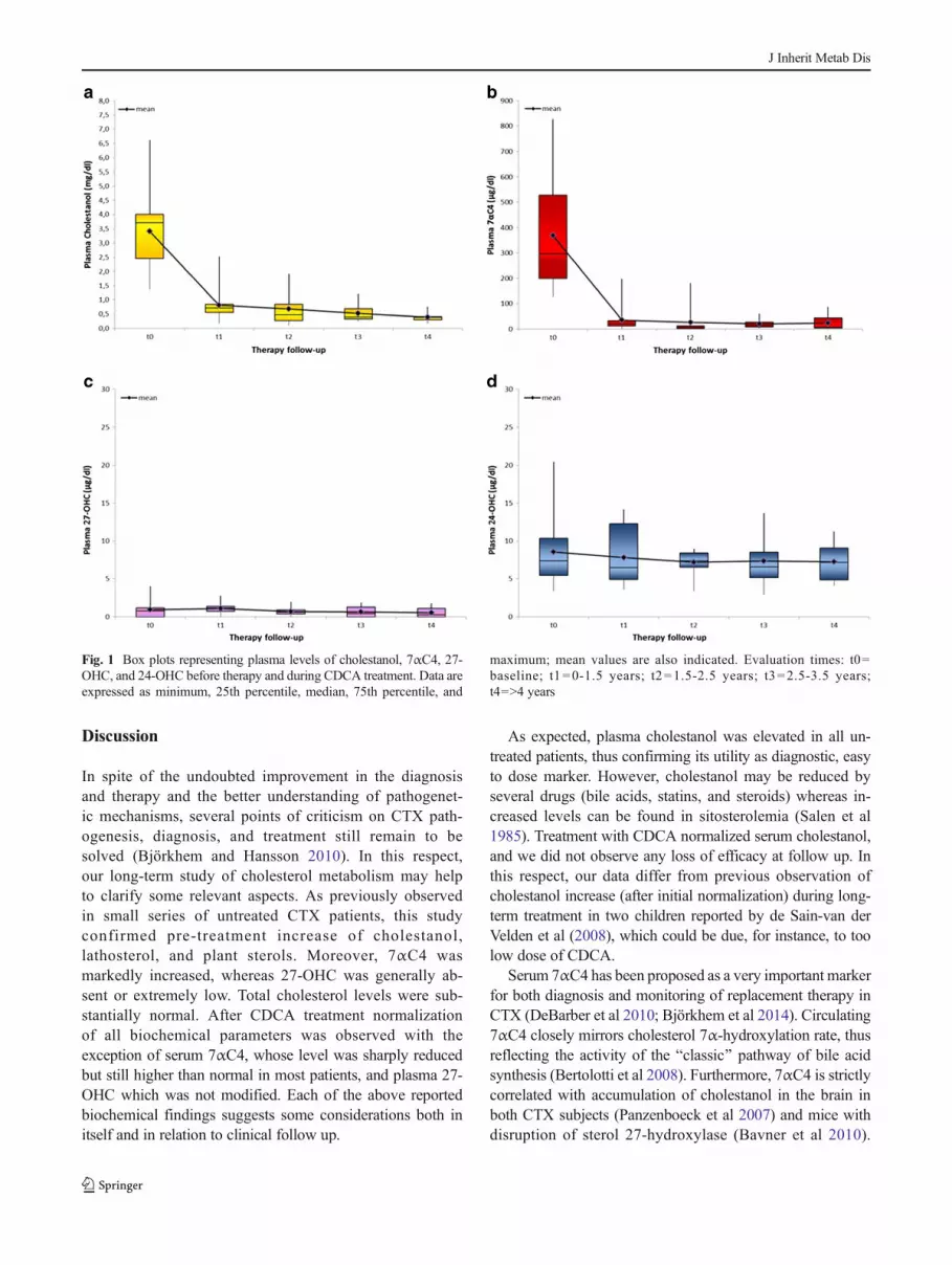
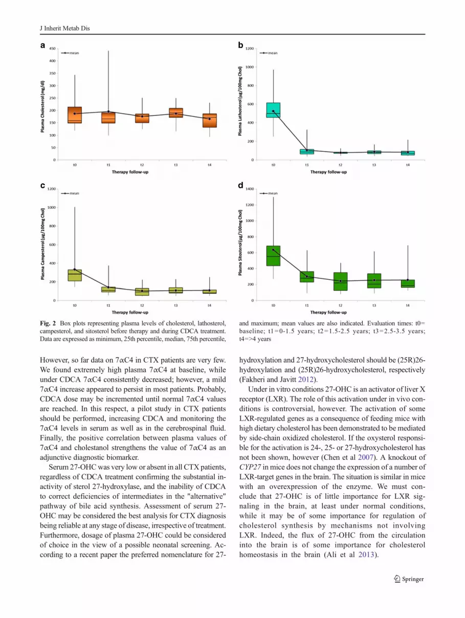
Fig. 2 Box plots representing plasma levels of cholesterol, lathosterol,campesterol, and sitosterol before therapy and during CDCA treatment.Data are expressed as minimum, 25th percentile, median, 75th percentile,
and maximum; mean values are also indicated. Evaluation times: t0=baseline; t1=0-1.5 years; t2=1.5-2.5 years; t3=2.5-3.5 years;t4=>4 years
J Inherit Metab Dis

Decreased serum 24-OHC levels have been found to berelated to the rate of neuronal degeneration in Huntington’sdisease and Alzheimer’s disease (Leoni and Caccia 2011).Unexpectedly, 24-OHC was normal or slightly increased inour patients and poorly influenced by therapy. Moreover, thelevels of 24-OHC were not significantly changed whencorrected for cholesterol levels. Therefore, in CTX it doesnot seem to have an important role on pathogenesis or moni-toring of neurodegeneration over time.
Lathosterol is a well-known marker of cholesterol Bde-novo^ synthesis. In our patients, serum lathosterol was mark-edly increased at baseline, and normalized during CDCAtreatment. Moreover, lathosterol and 7αC4 levels were strictlycorrelated. Most likely, the hyperactivation of the Bclassic^pathway of bile acid synthesis due to the lack of CDCA causesa very high consumption of cholesterol, which is compensatedby incrementing Bde-novo^ synthesis. CDCA therapy inacti-vates 7α-hydroxylation strongly reducing cholesterol con-sumption. As a result, cholesterol Bde-novo^ synthesis de-creases with corresponding lathosterol normalization. Increaseof lathosterol may be considered a valuable diagnostic markerwhich parallels elevation of 7αC4 and cholestanol levels inuntreated CTX subjects. Moreover, the assessment of serumlathosterol may help the clinician in deciding whether to add astatin to CDCA or not.
Plant sterols are considered markers of cholesterol intesti-nal absorption. Sitosterol, and to a lesser extent campesterol,were increased at baseline and decreased during treatmentwith CDCA. Since cholesterol absorption and synthesis areusually inversely correlated, one could expect that the valuesof plant sterols were reduced. The elevated concentration ofplant sterols in CTX subjects may be linked to the percentageincrease of cholic acid (CA), produced via the Balternative^pathway, in bile: indeed, CA is much more effective in stim-ulating absorption of cholesterol and plant sterols than CDCA.If that was the case, the decrease of plant sterols occurringduring treatment would be explained by reduced absorptiondue to replacement of CA in bile with CDCA as the dominat-ing bile acid.
With the exception of a few patients showing mild hyper-cholesterolemia, serum cholesterol was substantially normal,and no changes were observed under CDCA treatment. Thesedata may mean that, in absence of altered dietary intake,cholesterol synthesis is equivalent to cholesterol con-sumption in CTX.
Biochemical evaluation did not evidence statistically rele-vant differences between Bneurologically stable^ andBneurologically worsening^ patients. Nevertheless, some po-tentially interesting aspects emerged. Serum levels ofcholestanol, 7αC4, and lathosterol at baseline tended to behigher in those patients who later presented neurologicalworsening suggesting that higher levels of toxic bile acid in-termediates can predict a poor response to therapy. Regarding
follow up, one could expect that 7αC4 decrease to a lesserextent during CDCA treatment inworsening patients: wewerenot able to confirm this hypothesis, but the small number ofbiochemical data available at long-term follow up onBneurologically worsening^ patients may have led us to un-derestimate possible differences. In this respect assessment of7αC4 in cerebrospinal fluid may be very helpful. Moreover,since 27-OHC was absent or very low in all of our patientsboth at baseline and under CDCA treatment, we do not havedata pointing to a pathogenetic role of 27-OHC deficiency inneurological evolution. Finally, it is worth noting the higherbaseline disability in the Bneurologically worsening^ groupsuggesting that the presence of significant neurological im-pairment before treatment may predict poorer response totherapy.
In conclusion, our metabolic evaluation of CTX patientsallowed us to clarify some pathogenetic aspects of the disease,to specify the role of the various biochemical parameters in thediagnostic setting, and to better assess the effects of CDCAtreatment on cholesterol metabolism, also in the light of clin-ical evolution.
Compliance with Ethic Guidelines All procedures followed were inaccordance with the ethical standards of the responsible committee onhuman experimentation (institutional and national) and with the HelsinkiDeclaration of 1975, as revised in 2000 (5). Informed consent was ob-tained from all patients for being included in the study.
Conflict of interest None.
Funding Andrea Mignarri, Alessandro Magni, Marina Del Puppo,Gian Nicola Gallus, Ingemar Björkhem, Antonio Federico, and MariaTeresa Dotti declare no funding.
References
Alessandrini L, Ciuffreda P, Santaniello E, Terraneo G (2004)Clemmensen reduction of diosgenin and kryptogenin: synthesis of[16,16,22,22,23,23-(2)H(6)]-(25R)-26-hydroxycholesterol. Steroids69:789–794
Ali Z, HeverinM, OlinM et al (2013) On the regulatory role of side-chainhydroxylated oxysterols in the brain. Lessons from CYP27A1 trans-genic and Cyp27a1(-/-) mice. J Lipid Res 54:1033–1043
Barkhof F, Verrips A, Wesseling P et al (2000) Cerebrotendinousxanthomatosis: the spectrum of imaging findings and the correlationwith neuropathologic findings. Radiology 217:869–876
Båvner A, Shafaati M, Hansson M et al (2010) On the mechanism ofaccumulation of cholestanol in the brain of mice with a disruption ofsterol 27-hydroxylase. J Lipid Res 51:2722–2730
Berginer VM, Berginer J, Korczyn AD, Tadmor R (1994) Magnetic res-onance imaging in cerebrotendinous xanthomatosis: a prospectiveclinical and neuroradiological study. J Neurol Sci 122:102–108
Berginer VM, Gross B, Morad K et al (2009) Chronic diarrhea and juve-nile cataracts: think cerebrotendinous xanthomatosis and treat.Pediatrics 123:143–147
J Inherit Metab Dis
Berginer VM, Salen G, Shefer S (1984) Long-term treatment ofcerebrotendinous xanthomatosis with chenodeoxycholic acid. NEngl J Med 311:1649–1652
Bertolotti M, Del Puppo M, Gabbi C et al (2008) Correlation betweenplasma levels of 7alpha-hydroxy-4-cholesten-3-one and cholesterol7alpha-hydroxylation rates in vivo in hyperlipidemic patients.Steroids 73:1197–1202
Björkhem I (2013) Cerebrotendinous xanthomatosis. Curr Opin Lipidol24:283–287
Björkhem I, Diczfalusy U, Lövgren-Sandblom A et al (2014) On theformation of 7-ketocholesterol from 7-dehydrocholesterol in pa-tients with CTX and SLO. J Lipid Res 55:1165–1172
Björkhem I, Hansson M (2010) Cerebrotendinous xanthomatosis: an in-born error in bile acid synthesis with defined mutations but still achallenge. Biochem Biophys Res Commun 396:46–49
Cali JJ, Hsieh CL, Francke U et al (1991) Mutations in the bile acidbiosynthet ic enzyme sterol 27-hydroxylase under l iecerebrotendinous xanthomatosis. J Biol Chem 266:7779–7783
Camilleri M,NadeauA, TremaineWJ et al (2009)Measurement of serum7alpha-hydroxy-4-cholesten-3-one (or 7alphaC4), a surrogate testfor bile acid malabsorption in health, ileal disease and irritable bowelsyndrome using liquid chromatography-tandem mass spectrometry.Neurogastroenterol Motil 21:734–e743
Chen W, Chen G, Head DL, Mangelsdorf DJ, Russell DW (2007)Enzymatic reduction of oxysterols impairs LXR signaling in cul-tured cells and the livers of mice. Cell Metab 5:73–79
DeBarber AE, Connor WE, Pappu AS, Merkens LS, Steiner RD (2010)ESI-MS/MS quantification of 7alpha-hydroxy-4-cholesten-3-onefacilitates rapid, convenient diagnostic testing for cerebrotendinousxanthomatosis. Clin Chim Acta 411:43–48
de Sain-van der Velden MG, Verrips A, Prinsen BH, de Barse M, BergerR, Visser G (2008) Elevated cholesterol precursors other thancholestanol can also be a hallmark for CTX. J Inherit Metab Dis31:S387–S393
De Stefano N, Dotti MT, Mortilla M, Federico A (2001) Magnetic reso-nance imaging and spectroscopic changes in brains of patients withcerebrotendinous xanthomatosis. Brain 124:121–131
Del Puppo M, Kienle MG, Petroni ML, Crosignani A, Podda M (1998)Serum 27-hydroxycholesterol in patients with primary biliary cir-rhosis suggests alteration of cholesterol catabolism to bile acids viathe acidic pathway. J Lipid Res 39:2477–2482
Dzeletovic S, Breuer O, Lund E, Diczfalusy U (1995) Determination ofcholesterol oxidation products in human plasma by isotope dilution-mass spectrometry. Anal Biochem 225:73–80
Ellis E, Axelson M, Abrahamsson A et al (2003) Feedback regulation ofbile acid synthesis in primary human hepatocytes: evidence thatCDCA is the strongest inhibitor. Hepatology 38:930–938
Fakheri RJ, Javitt NB (2012) 27-Hydroxycholesterol, does it exist? Onthe nomenclature and stereochemistry of 26-hydroxylated sterols.Steroids 77:575–577
Galli Kienle M, Anastasia M, Cighetti G, Galli G, Fiecchi A (1980)Studies on the 14 alpha-demethylation mechanism in cholesterolbiosynthesis. Eur J Biochem 110:93–105
Ginanneschi F, Mignarri A, Mondelli M et al (2013) Polyneuropathy incerebrotendinous xanthomatosis and response to treatment withchenodeoxycholic acid. J Neurol 260:268–274
HeverinM,Meaney S, Lütjohann D, DiczfalusyU,Wahren J, Björkhem I(2005) Crossing the barrier: net flux of 27-hydroxycholesterol intothe human brain. J Lipid Res 46:1047–1052
Inoue K, Kubota S, Seyama Y (1999) Cholestanol induces apoptosis ofcerebellar neuronal cells. Biochem Biophys Res Commun256:198–203
Kempen HJ, Glatz JF, Gevers Leuven JA, van der Voort HA,Katan MB (1988) Serum lathosterol concentration is an indi-cator of whole-body cholesterol synthesis in humans. J LipidRes 29:1149–1155
Kuriyama M, Fujiyama J, Kasama T, Osame M (1991) High levels ofplant sterols and cholesterol precursors in cerebrotendinousxanthomatosis. J Lipid Res 32:223–229
Kurtzke JF (1983) Rating neurologic impairment in multiple sclerosis: anexpanded disability status scale (EDSS). Neurology 33:1444–1452
Leoni V, Caccia C (2011) Oxysterols as biomarkers in neurodegenerativediseases. Chem Phys Lipids 164:515–524
Liu J, Lu H, Lu YF et al (2014) Potency of individual bile acids toregulate bile acid synthesis and transport genes in primary humanhepatocyte cultures. Toxicol Sci 141:538–546
Martini G, Mignarri A, Ruvio M et al (2013) Long-term bone densityevaluation in cerebrotendinous xanthomatosis: evidence of im-provement after chenodeoxycholic acid treatment. Calcif TissueInt 92:282–286
Mignarri A, Dotti MT, Del Puppo M et al (2012) Cerebrotendinousxanthomatosis with progressive cerebellar vacuolation: six-yearMRI follow-up. Neuroradiology 54:649–651
Mignarri A, Gallus GN, Dotti MT, Federico A (2014) A suspicion indexfor early diagnosis and treatment of cerebrotendinousxanthomatosis. J Inherit Metab Dis 37:421–429
Mignarri A, Rossi S, Ballerini M et al (2011) Clinical relevance andneurophysiological correlates of spasticity in cerebrotendinousxanthomatosis. J Neurol 258:783–790
Panzenboeck U, Andersson U, Hansson M, Sattler W, Meaney S,Björkhem I (2007) On the mechanism of cerebral accumulation ofcholestanol in patients with cerebrotendinous xanthomatosis. J LipidRes 48:1167–1174
Pilo de la Fuente B, Jimenez-Escrig A, Lorenzo JR et al (2011a)Cerebrotendinous xanthomatosis in Spain: clinical, prognostic, andgenetic survey. Eur J Neurol 18:1203–1211
Pilo de la Fuente B, Sobrido MJ, Girós M et al (2011b) Usefulnessof cholestanol levels in the diagnosis and follow-up of pa-tients with cerebrotendinous xanthomatosis. Neurologia 26:397–404
Rubio-Agusti I, Kojovic M, Edwards MJ et al (2012) Atypical parkin-sonism and cerebrotendinous xanthomatosis: report of a family withcorticobasal syndrome and a literature review. Mov Disord 27:1769–1774
Salen G, Kwiterovich PO Jr, Shefer S et al (1985) Increased plasmacholestanol and 5 alpha-saturated plant sterol derivatives in subjectswith sitosterolemia and xanthomatosis. J Lipid Res 26:203–209
Van Heijst AF, Verrips A, Wevers RA, Cruysberg JR, Renier WO,Tolboom JJ (1998) Treatment and follow-up of children withcerebrotendinous xanthomatosis. Eur J Pediatr 157:313–316
van Swieten JC, Koudstaal PJ, Visser MC et al (1988) Interobserveragreement for the assessment of handicap in stroke patients.Stroke 19:604–607
Wolthers BG, Walrecht HT, van der Molen JC, Nagel GT, Van DoormaalJJ, Wijnandts PN (1991) Use of determinations of 7-lathosterol (5alpha-cholest-7-en-3 beta-ol) and other cholesterol precursors in se-rum in the study and treatment of disturbances of sterol metabolism,particularly cerebrotendinous xanthomatosis. J Lipid Res 32:603–612
Yahalom G, Tsabari R, Molshatzki N, Ephraty L, Cohen H, Hassin-BaerS (2013) Neurological outcome in cerebrotendinous xanthomatosistreated with chenodeoxycholic acid: early versus late diagnosis. ClinNeuropharmacol 36:78–83
J Inherit Metab Dis