Distinction between true acrosome reaction and degenerative acrosome loss by a one-step staining...
9
Distinction between true acrosome reaction and degenerative acrosome loss by a one-step staining method using Pisum sativum agglutinin C. Mendoza, A. Carreras, J. Moos and J. Tesarik 'Department of Biochemistry and Molecular Biology, Faculty of Sciences, University of Granada, Campus Universitario 'Fuentenueva', 18071 Granada, Spain; 2 Institute of Molecular Genetics, Czechoslovak Academy of Sciences, Prague, Czechoslovakia; and3Institute of Health and Medical Research (INSERM), Unit 355, Clamart, France and American Hospital of Paris, Neuilly sur Seine, France Summary. When western blots of human sperm proteins solubilized by acid extraction (presumably mainly acrosomal proteins) or by sodium dodecyl sulfate (SDS) were probed with biotin-conjugated Pisum sativum agglutinin (PSA), distinct sets of proteins were labelled in both preparations. When smears of human spermatozoa were treated with methanol either for 30 s or for 15 min and then exposed to FITC-conjugated PSA, the resulting fluorescence pattern essentially depended on the time of methanol treatment. With the longer treatment, fewer spermatozoa showed selective acrosomal labelling and more were labelled uniformly throughout, without a clear predilection for a single sperm region. With the shorter time of methanol treatment, the poorly topographically differentiated, whole-cell labelling was typical of dead spermatozoa as confirmed by a close correlation between the percentages of spermatozoa showing this type of labelling and of those stained supravitally with Hoechst 33258. The preferential whole-cell labelling of dead spermatozoa with PSA is considered to be due to increased availability of the nonacrosomal set of PSA-reactive sites in dead spermatozoa after a short treatment with methanol, whereas this treatment is probably not sufficient to expose most of these sites when applied to living spermatozoa. The simplicity of the staining protocol makes this method feasible in routine work in a number of clinical and research applications. Keywords: acrosomal staining; acrosome reaction; sperm viability; human Introduction The acrosome reaction of mammalian spermatozoa is a calcium-dependent exocytotic process that is necessary for fertilization (reviewed by Yanagimachi, 1988). A better knowledge of the phenomena occurring during the human acrosome reaction is important for understanding both the mechanisms controlling normal human fertilization and the causes of its defects. Accordingly, the frequency with which spermatozoa from a given sample undergo the acrosome reaction represents an important parameter in the clinical evaluation of sperm function (Tesarik & Testart, 1989). A major problem in the study of the acrosome reaction of human spermatozoa is that the acrosome loss cannot be observed on living spermatozoa by phase contrast or differential interference contrast microscopy as it can be in spermatozoa of some other mammalian species 'Address for correspondence: American Hospital of Paris, 63 boulevard Victor Hugo, F-92202 Neuilly sur Seine, France.
Transcript of Distinction between true acrosome reaction and degenerative acrosome loss by a one-step staining...
Distinction between true acrosome reaction anddegenerative acrosome loss by a one-step staining method
using Pisum sativum agglutininC. Mendoza, A. Carreras, J. Moos and J. Tesarik
'Department of Biochemistry and Molecular Biology, Faculty of Sciences, University of Granada,Campus Universitario 'Fuentenueva', 18071 Granada, Spain; 2 Institute of Molecular Genetics,
Czechoslovak Academy of Sciences, Prague, Czechoslovakia; and3Institute of Health and MedicalResearch (INSERM), Unit 355, Clamart, France and American Hospital of Paris,
Neuilly sur Seine, France
Summary. When western blots of human sperm proteins solubilized by acid extraction(presumably mainly acrosomal proteins) or by sodium dodecyl sulfate (SDS) were
probed with biotin-conjugated Pisum sativum agglutinin (PSA), distinct sets of proteinswere labelled in both preparations. When smears of human spermatozoa were treatedwith methanol either for 30 s or for 15 min and then exposed to FITC-conjugatedPSA, the resulting fluorescence pattern essentially depended on the time of methanoltreatment. With the longer treatment, fewer spermatozoa showed selective acrosomallabelling and more were labelled uniformly throughout, without a clear predilectionfor a single sperm region. With the shorter time of methanol treatment, the poorlytopographically differentiated, whole-cell labelling was typical of dead spermatozoa asconfirmed by a close correlation between the percentages of spermatozoa showing thistype of labelling and of those stained supravitally with Hoechst 33258. The preferentialwhole-cell labelling of dead spermatozoa with PSA is considered to be due to increasedavailability of the nonacrosomal set of PSA-reactive sites in dead spermatozoa after ashort treatment with methanol, whereas this treatment is probably not sufficient toexpose most of these sites when applied to living spermatozoa. The simplicity of thestaining protocol makes this method feasible in routine work in a number of clinicaland research applications.Keywords: acrosomal staining; acrosome reaction; sperm viability; human
Introduction
The acrosome reaction of mammalian spermatozoa is a calcium-dependent exocytotic processthat is necessary for fertilization (reviewed by Yanagimachi, 1988). A better knowledge of thephenomena occurring during the human acrosome reaction is important for understanding both themechanisms controlling normal human fertilization and the causes of its defects. Accordingly,the frequency with which spermatozoa from a given sample undergo the acrosome reaction representsan important parameter in the clinical evaluation of sperm function (Tesarik & Testart, 1989).
A major problem in the study of the acrosome reaction of human spermatozoa is that theacrosome loss cannot be observed on living spermatozoa by phase contrast or differentialinterference contrast microscopy as it can be in spermatozoa of some other mammalian species
'Address for correspondence: American Hospital of Paris, 63 boulevard Victor Hugo, F-92202 Neuilly sur Seine,France.
possessing large acrosomes, such as guinea-pigs or hamsters. Electron microscopy may be used toassess the acrosomal status of human spermatozoa (e.g. Tesarik, 1985; Stock & Fraser, 1987;Yudin et al, 1988), but the technique is too laborious to be applied in routine examination ofspermatozoa. Consequently, various light microscope techniques (bright-field or fluorescence)using acrosomal staining with histological dyes (Talbot & Chacon, 1981), lectins (Talbot &Chacon, 1980; Cross et al, 1986; Mortimer et al, 1987) and monoclonal antibodies (Byrd & Wolf,1986; Kallajoki et al, 1986; Moore et al, 1987) have been developed. However, all these techniquesare burdened by the problem of distinction between degenerative acrosome loss accompanyingsperm cell death and the true acrosome reaction. This problem has been dealt with by combiningthe acrosome staining with other methods that claim to distinguish between living and deadspermatozoa (for recent reviews, see Cross & Meizel, 1989; Wolf, 1989). Even though such com¬bined protocols apparently improve the correlation of estimates of acrosome reaction with thoseobtained by electron microscopy (Talbot & Chacon, 1981; Thomas & Meizel, 1989), the inclusionof an additional stain makes the procedure longer and more complicated. Moreover, the additionof centrifugation steps required for the supravital staining reduces the yield of spermatozoa forsmear preparation, which is inconvenient in cases where only a limited number of spermatozoa are
available for this kind of examination.In this study we describe a method for distinguishing between the true acrosome reaction and a
degenerative acrosome loss in human spermatozoa using a one-step staining with fluorescein-labelled Pisum sativum agglutinin (PSA). Sperm compounds showing an affinity for this lectin were
analysed by western blotting and the ability of the PSA stain to mark selectively dead spermatozoaunder certain conditions was assessed by examining the correlation between the estimates ofpercentages of dead cells using PSA and those obtained using supravital staining with Hoechst33258.
Materials and MethodsSource and preparation of spermatozoa
Human semen samples were obtained from 25 healthy donors with normal parameters of sperm density, motilityand morphology. The samples were left to stand for 30 min at room temperature to liquefy. Aliquots of semen
samples were diluted with an equal volume of Biggers, Whitten and Whittingham (BWW) medium (Biggers et ai,1971) and centrifuged at 500 g for 5 min. The supernatant was discarded and the sperm pellet was resuspended in 5 mlBWW and centrifuged again under the same conditions. The supernatant was discarded and the pellet of the washedspermatozoa was overlayered with 1 ml B2 medium (Api System, Montalieu-Vercieu, France). Spermatozoa were
allowed to swim up to this medium during a 30 min incubation at 37°C in an atmosphere of 5% C02 in air. Themigrated spermatozoa were then recovered from the tubes with the upper 0-5 ml of medium and incubated under thesame conditions for an additional 4 h.
Induction of the acrosome reactionThe acrosome reaction was induced by ionophore A23187 (Sigma, La Verpillière, France) after incubation of
spermatozoa for 4 h in B2 medium. A stock solution of 20 mmol ionophore A231871" ' dimethylsulfoxide (Sigma)was prepared and added to sperm suspensions to give a final concentration of lOpmoll-1. Spermatozoa were thenincubated with the ionophore at 37°C under 5% C02 in air for 30 min.
Extraction of sperm proteinsFor quantitative protein extraction, pellets of washed spermatozoa were incubated for 15 min at room tempera¬
ture in 300 pi of a mixture consisting of 2% (w/v) SDS (Sigma), 10% (v/v) glycerol and 01 mol Tris HCl buffer l"1(pH 6-8). Samples were then centrifuged at lOOOOj» for 10 min, supernatants were decanted and used in individualexperiments.
Acid extraction was used to obtain preparations enriched in acrosomal proteins. It was performed using a slightmodification of the method used for the extraction of boar acrosomal proteins (Zelezna & Cechova, 1982; Cechovaet ai, 1984). Briefly, pellets of washed spermatozoa were incubated for 16 h at 4°C in a solution containing 3% (v/v)acetic acid, 10% (v/v) glycerol and 10 mmol benzamidine 1_1, followed by high-speed centrifugation (4°C) and thesupernatants containing solubilized acrosomal proteins were decanted.
Protein concentration in sperm extracts was determined using the method of Bradford (1976). It ranged fromO-Sragmr1 to 2-7mg ml-1 in individual preparations.
Electrophoresis and western blottingSDS-polyacrylamide gel electrophoresis (SDS-PAGE) was performed on 12% slab gels according to the method
of Laemmli (1970). Each sample was electrophoresed as duplicate aliquots on two adjacent gels. After SDS-PAGE,one of these gels was used for blotting, while the other was stained with Coomassie brilliant blue G. The apparentmolecular masses of proteins from individual samples were estimated by comparing with the protein standards thatwere run in parallel. Western blotting was carried out as described by Towbin et ai (1979). Proteins were electro-transferred from polyacrylamide gels into Hybond C nitrocellulose membranes (Amersham, UK). The membranesheets were incubated with biotin-conjugated PSA (3 pg ml" ') (Sigma) in phosphate-buffered saline (PBS) followedby streptavidin-peroxidase complex (Amersham) and bound peroxidase was visualized using diaminobenzidine(Sigma) as chromogen.
Sperm staining with FITC-labelled PSA
Aliqots of sperm suspensions were used to prepare smears on microscope slides. Care was taken to remove theoriginal culture medium from the suspensions used for smear preparation (at least two cycles of centrifugation andresuspension in protein-free PBS of BWW) because traces of proteins on smears react with fluorescein isothiocyanate(FITC) labelled PSA and produce a strongly fluorescent background which makes the evaluation of sperm stainingimpossible. After air-drying, sperm smears were dipped in absolute methanol for either 30 s or 15 min and allowed todry rapidly. Such smears were either processed immediately or kept in a dry place at room temperature for up to 1week without any disturbance of the results of subsequent staining. Methanol-treated smears were incubated for30 min at room temperature in a moisture chamber with a solution of FITC-conjugated PSA (50pgml_1) (Sigma) inPBS. The slides were then washed in distilled water to remove unbound probe. This washing represents anothercritical step in the protocol because insufficiently washed preparations are difficult to assess. For good results, slidesmust be rinsed in a stream of water and then dipped in excess water for at least 15 min. After drying, smears were
examined immediately, without mounting, in an epifluorescence microscope. For good staining results, preparationsmust be examined on the day of staining, since unmounted slides rapidly lose the staining specificity.
Supravital staining with Hoechst 33258
Suspensions of washed spermatozoa were incubated for 5 min at room temperature with the Hoechst 33258 dye(bis benzimide; Sigma) diluted in PBS at a concentration of 1 pg ml " '. The free dye was washed from sperm suspen¬sions by two cycles of centrifugation (500 # for 10 min) and the pellet resuspended in fresh PBS. These spermatozoawere then used immediately for smear preparation.
Quantitative analysisThe proportions of spermatozoa showing different topographical patterns of fluorescent staining (see below) were
determined in smears. Each slide was examined by two independent observers and 200 spermatozoa were counted byeach observer. The mean value from the two examinations was calculated.
Statistical analysisPercentages of spermatozoa showing different staining patterns in individual experiments were compared, after
arcsine transformation, by t test. Correlation between staining patterns was evaluated by linear regression analysis.
Results
Reactivity of sperm proteins with PSA in western blots
When western blots of sperm proteins were probed with PSA, the reactivity pattern stronglydepended on the method of protein extraction used. In blots of acid-extracted sperm proteins(preferentially acrosomal proteins), most proteins reactive with the probe had an apparentmolecular mass greater than 50 kDa, with the strongest labelling in a protein of 53-55 kDa (Fig. 1,lane a). When spermatozoa previously subjected to acid extraction were re-extracted with SDS, a
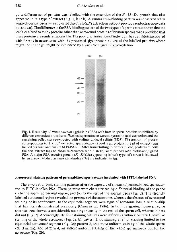
quite different set of proteins was labelled, with the exception of the 53-55 kDa protein that alsoappeared in this type of extract (Fig. 1, lane b). A similar PSA-binding pattern was observed whenwashed spermatozoa were subjected directly to SDS extraction without previous acid extraction (datanot shown). The difference in the PSA-binding pattern of the two types ofsperm extract shows that thelectin can bind to many proteins other than acrosomal proteins ofhuman spermatozoa provided thatthese proteins are rendered accessible. The poor discrimination of individual bands in blots incubatedwith PSA is in accordance with the presumed glycoprotein nature of the labelled proteins whosemigration in the gel might be influenced by a variable degree of glycosylation.
Fig. 1. Reactivity oí Pisum sativum agglutinin (PSA) with human sperm proteins solubilized bydifferent extraction procedures. Washed spermatozoa were subjected to acid extraction and theremaining pellet was re-extracted with sodium dodecyl sulfate (SDS). The amount of proteincorresponding to 1 IO6 extracted spermatozoa (about 5 pg protein in 8 µ of extract) wasloaded per lane and run on SDS-PAGE. After transblotting to nitrocellulose, proteins of boththe acid extract (a) and those re-extracted with SDS (b) were probed with biotin-conjugatedPSA. A major PSA-reactive protein (53-55 kDa) appearing in both types of extract is indicatedby an arrow. Molecular mass standards (kDa) are indicated in (a).
Fluorescent staining patterns of permeabilized spermatozoa incubated with FITC-labelled PSA
There were four basic staining patterns after the exposure of smears of permeabilized spermato¬zoa to FITC-labelled PSA. These patterns were characterized by differential binding of the probe(i) to the sperm acrosomal region, and (ii) to the rest of the spermatozoa (Fig. 2). The stronglylabelled acrosomal region revealed the presence of the acrosome, whereas the absence of acrosomalstaining or its confinement to the equatorial segment were signs of acrosome loss, a relationshipthat has been demonstrated previously (Cross et al, 1986). In both categories, however, some
spermatozoa showed a considerable staining intensity in the rest of the sperm cell, whereas othersdid not (Fig. 2). Accordingly, the four staining patterns were defined as follows: pattern 1, selectivestaining of the whole acrosome (Fig. 2a, b); pattern 2, no staining at all or staining limited to theequatorial acrosomal segment (Fig. 2c); pattern 3, an almost uniform staining of the whole spermcell (Fig. 2a); and pattern 4, an almost uniform staining of the whole spermatozoa but for theacrosome (Fig. 2b).
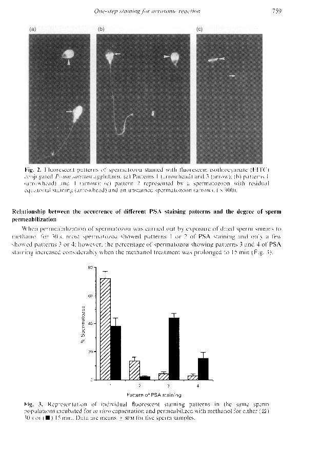
Fig. 2. Fluorescent patterns of spermatozoa stained with fluorescein isothiocyanate (FITC)conjugated Pisum sativum agglutinin. (a) Patterns 1 (arrowhead) and 3 (arrow); (b) patterns 1(arrowhead) and 4 (arrow); (c) pattern 2 represented by a spermatozoon with residualequatorial staining (arrowhead) and an unstained spermatozoon (arrow). ( 900).
Relationship between the occurrence of different PSA staining patterns and the degree of spermpermeabilization
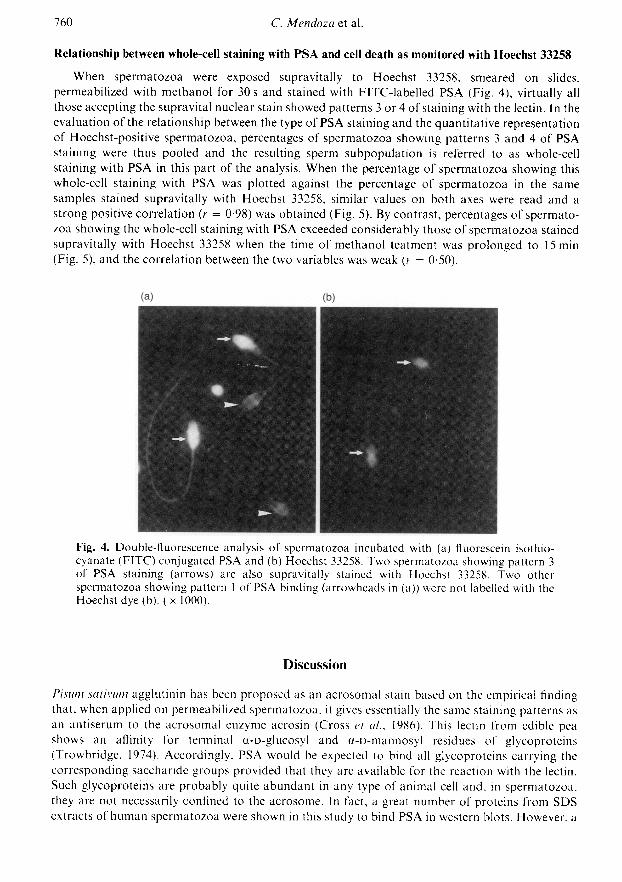
When permeabilization of spermatozoa was carried out by exposure of dried sperm smears tomethanol for 30 s, most spermatozoa showed patterns 1 or 2 of PSA staining and only a fewshowed patterns 3 or 4; however, the percentage of spermatozoa showing patterns 3 and 4 of PSAstaining increased considerably when the methanol treatment was prolonged to 15 min (Fig. 3).
Pattern of PSA stainingFig. 3. Representation of individual fluorescent staining patterns in the same spermpopulations incubated for in vitro capacitation and permeabilized with methanol for either (E3)30 s or ( ) 15 min. Data are means + sem for five sperm samples.
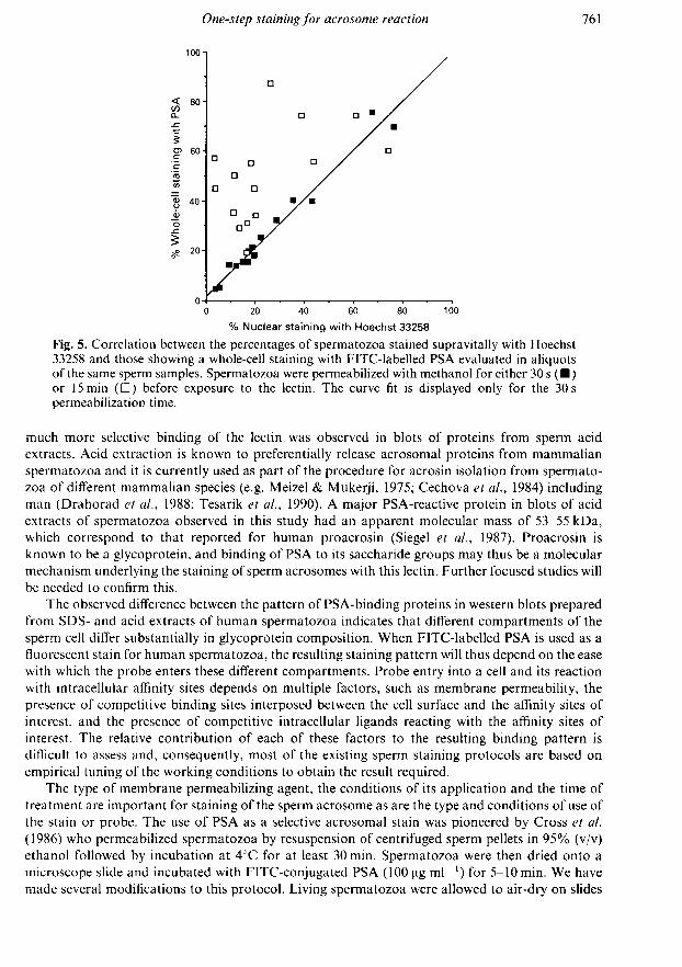
Relationship between whole-cell staining with PSA and cell death as monitored with Hoechst 33258When spermatozoa were exposed supravitally to Hoechst 33258, smeared on slides,
permeabilized with methanol for 30 s and stained with FITC-labelled PSA (Fig. 4), virtually allthose accepting the supravital nuclear stain showed patterns 3 or 4 of staining with the lectin. In theevaluation of the relationship between the type of PSA staining and the quantitative representationof Hoechst-positive spermatozoa, percentages of spermatozoa showing patterns 3 and 4 of PSAstaining were thus pooled and the resulting sperm subpopulation is referred to as whole-cellstaining with PSA in this part of the analysis. When the percentage of spermatozoa showing thiswhole-cell staining with PSA was plotted against the percentage of spermatozoa in the same
samples stained supravitally with Hoechst 33258, similar values on both axes were read and a
strong positive correlation (r = 0-98) was obtained (Fig. 5). By contrast, percentages of spermato¬zoa showing the whole-cell staining with PSA exceeded considerably those of spermatozoa stainedsupravitally with Hoechst 33258 when the time of methanol teatment was prolonged to 15 min(Fig. 5), and the correlation between the two variables was weak (r = 0-50).
Fig. 4. Double-fluorescence analysis of spermatozoa incubated with (a) fluorescein isothio¬cyanate (FITC) conjugated PSA and (b) Hoechst 33258. Two spermatozoa showing pattern 3of PSA staining (arrows) are also supravitally stained with Hoechst 33258. Two otherspermatozoa showing pattern 1 of PSA binding (arrowheads in (a)) were not labelled with theHoechst dye (b). ( 1000).
Discussion
Pisum sativum agglutinin has been proposed as an acrosomal stain based on the empirical findingthat, when applied on permeabilized spermatozoa, it gives essentially the same staining patterns asan antiserum to the acrosomal enzyme acrosin (Cross et al, 1986). This lectin from edible peashows an affinity for terminal a-D-glucosyl and a-D-mannosyl residues of glycoproteins(Trowbridge, 1974). Accordingly, PSA would be expected to bind all glycoproteins carrying thecorresponding saccharide groups provided that they are available for the reaction with the lectin.Such glycoproteins are probably quite abundant in any type of animal cell and, in spermatozoa,they are not necessarily confined to the acrosome. In fact, a great number of proteins from SDSextracts of human spermatozoa were shown in this study to bind PSA in western blots. However, a
% Nuclear staining with Hoechst 33258
Fig. 5. Correlation between the percentages of spermatozoa stained supravitally with Hoechst33258 and those showing a whole-cell staining with FITC-labelled PSA evaluated in aliquotsof the same sperm samples. Spermatozoa were permeabilized with methanol for either 30 s ( )or 15 min (D) before exposure to the lectin. The curve fit is displayed only for the 30 s
permeabilization time.
much more selective binding of the lectin was observed in blots of proteins from sperm acidextracts. Acid extraction is known to preferentially release acrosomal proteins from mammalianspermatozoa and it is currently used as part of the procedure for acrosin isolation from spermato¬zoa of different mammalian species (e.g. Meizel & Mukerji, 1975; Cechova et al, 1984) includingman (Drahorad et al, 1988; Tesarik et al, 1990). A major PSA-reactive protein in blots of acidextracts of spermatozoa observed in this study had an apparent molecular mass of 53-55 kDa,which correspond to that reported for human proacrosin (Siegel et al, 1987). Proacrosin isknown to be a glycoprotein, and binding of PSA to its saccharide groups may thus be a molecularmechanism underlying the staining of sperm acrosomes with this lectin. Further focused studies willbe needed to confirm this.
The observed difference between the pattern of PSA-binding proteins in western blots preparedfrom SDS- and acid extracts of human spermatozoa indicates that different compartments of thesperm cell differ substantially in glycoprotein composition. When FITC-labelled PSA is used as afluorescent stain for human spermatozoa, the resulting staining pattern will thus depend on the easewith which the probe enters these different compartments. Probe entry into a cell and its reactionwith intracellular affinity sites depends on multiple factors, such as membrane permeability, thepresence of competitive binding sites interposed between the cell surface and the affinity sites ofinterest, and the presence of competitive intracellular ligands reacting with the affinity sites ofinterest. The relative contribution of each of these factors to the resulting binding pattern isdifficult to assess and, consequently, most of the existing sperm staining protocols are based on
empirical tuning of the working conditions to obtain the result required.The type of membrane permeabilizing agent, the conditions of its application and the time of
treatment are important for staining of the sperm acrosome as are the type and conditions of use ofthe stain or probe. The use of PSA as a selective acrosomal stain was pioneered by Cross et al(1986) who permeabilized spermatozoa by resuspension of centrifuged sperm pellets in 95% (v/v)ethanol followed by incubation at 4°C for at least 30 min. Spermatozoa were then dried onto a
microscope slide and incubated with FITC-conjugated PSA (100 pg mP1) for 5-10min. We havemade several modifications to this protocol. Living spermatozoa were allowed to air-dry on slides
and only then exposed to the permeabilizing agent. Methanol was used as the permeabilizingagent instead of ethanol, and the time was either 30 s or 15 min. The incubation of slides with theFITC-labelled lectin was increased to 30 min. With a time of methanol treatment of 30 s, we haveconfirmed a strong correlation of values for acrosome reaction frequency with those obtainedby electron microscopy (Tesarik et al, 1990). In this study we have shown that prolonging themethanol fixation has a considerable effect on the pattern of staining with PSA, mainly by augmen¬tation of the percentage of spermatozoa that show a poorly differentiated, whole-cell staining withthe lectin. However, slight variation of the fixation time (20 s to 1 min) did not influence the stainingresults. This is important for the feasibility of the proposed staining method because variationsoccur, owing to the effect of atmospheric conditions on the rate of methanol evaporation, which are
difficult to avoid.The change in the staining pattern after prolonging the methanol treatment may be due to a
higher degree of membrane permeabilization, removal of competitive ligands occupying thebinding sites for PSA, or both. The short methanol treatment (30 s) appears to be just sufficientto allow a limited interaction of PSA with its intracellular binding sites, with a clear predilectionfor acrosomal components. However, many new sites become reactive with the lectin after theprolonged methanol treatment, which leads to the loss of selectivity and to the appearance of a
whole-cell staining in many spermatozoa.With the short permeabilization time, spermatozoa showing any of the whole-cell staining
patterns with PSA were essentially those that were stained supravitally with Hoechst 33258, andthe percentages of spermatozoa showing the whole-cell staining with PSA and those acceptingthe Hoechst nuclear stain were both strongly correlated and similar in all sperm samples tested.In view of the above reasoning, this finding can be explained by the breakdown of the barrierfunction of sperm membranes in dead cells (stained supravitally with Hoechst 33258) leading to
exposure of lectin-binding sites after a short methanol treatment that is not efficient enough tomake these sites available when the spermatozoon has been living at the time of smear prep¬aration. Alternatively, dead cells may lack ligands that, in living cells, competitively block thelectin-binding sites.
Whatever the exact mechanism underlying the preferential whole-cell staining of dead sperma¬tozoa with PSA, this relationship has a great practical significance because it enables considerablesimplification of the evaluation of acrosome reaction of human spermatozoa. This can now be doneafter a one-step staining with a commercially available FITC-labelled PSA conjugate and scoringboth the true acrosome reaction (pattern 1 versus pattern 2 of PSA staining) and sperm cell death(patterns 3 & 4) by fluorescent microscopy using the same filter adjustment. Alternatively, stainingwith PSA (FITC or TRITC conjugate) can be used as part of a double-labelling protocol withwhich the acrosome reaction is evaluated conjointly with other sperm properties, reflected by thereactivity with a second stain or probe. Finally, the use of peroxidase-labelled PSA will bring thistechnique within the reach of those who prefer to use bright-field light microscopy rather thanfluorescence microscopy for this type of examination. The simplicity of the staining protocol, therelatively low cost of reagents and the wide range of applications warrant the use of this method forboth clinical and research purposes.
References
Biggers, J.D., Whitten, W.K. & Whittingham, D.G.(1971) The culture of mouse embryos in vitro. InMethods in Mammalian Embryology, pp. 86-116. Ed.J. C. Daniel Jr. Freeman, San Francisco.
Bradford, M.M. (1976) A rapid and sensitive methodfor the quantitation of microgram quantities ofprotein utilizing the principle of protein-dye binding.Analytical Biochemistry 72, 248-254.
Byrd, W. & Wolf, D.P. (1986) Acrosomal status in fresh
and capacitated human ejaculated sperm. Biology ofReproduction 34, 859-869.
Cechova, D., Zelezna, B., Petelikova, J. & Pavlok, A.(1984) Boar proacrosin. Correlation between totalproacrosin content and sperm fertilizing capacity.Andrologia 16, 169-174.
Cross, N.L. & Meizel, S. (1989) Methods for evaluatingthe acrosomal status of mammalian sperm. Biologyof Reproduction 41, 635-641.
Cross, N.L., Morales, P., Overstreet, J.W. & Hanson,F.W. (1986) Two simple methods for detectingacrosome-reacted human sperm. Gamete Research15,213-226.
Drahorad, J., Cechova, D. & Tesarik, J. (1988)Activation of proacrosin by a locally producedcomponent of human follicular fluid. Journal ofReproduction and Fertility 83, 599-603.
Kallajoki, M., Virtanen, I. & Suominen, J. (1986) The fateof acrosomal staining during the acrosome reactionof human spermatozoa as revealed by a monoclonalantibody and PNA-lectin. International Journal ofAndrology 9, 181-194.
Laemmli, U.K. (1970) Cleavage of structural proteinsduring the assembly of the head of bacteriophage T4.Nature 227, 680-685.
Meizel, S. & Mukerji, S.K. (1975) Proacrosin from rabbitepididymal spermatozoa: partial purification andinitial biochemical characterization. Biology ofReproduction 13, 83-93.
Moore, H.D.M., Smith, CA., Hartman, T.D. & Bye,A.P. (1987) Visualization and characterization ofthe acrosome reaction of human spermatozoa byimmunolocalization with monoclonal antibody.Gamete Research 17, 245-259.
Mortimer, D., Curtis, E.F. & Miller, R.G. (1987) Specificlabelling by peanut agglutinin of the outer acrosomalmembrane of the human spermatozoon. Journal ofReproduction and Fertility 81, 127-135.
Siegel, M.S., Bechtold, D.S.i Willand, J.L. & Polakoski,K.L. (1987) Biochemical and immunochemical com¬
parisons between the human and boar proacrosin-acrosin proteinase systems. Journal of ReproductiveImmunology 11, 307-319.
Stock, CE. & Fraser, L.R. (1987) The acrosome reactionin human sperm from men of proven fertility. HumanReproduction 2, 109-119.
Talbot, P. & Chacon, R.S. (1981) A triple-stain techniquefor evaluating normal acrosome reactions of humansperm. Journal of Experimental Zoology 215,201-208.
Tesarik, J. (1985) Comparison of acrosome reaction-inducing activities of human cumulus oophorus,follicular fluid and ionophore A23187 in humansperm populations of proven fertilizing ability in vitro.Journal of Reproduction and Fertility 74, 383-388.
Tesarik, J. & Testart, J. (1989) Human sperm-egginteractions and their disorders: implications in themanagement of infertility. Human Reproduction 4,729-741.
Tesarik, J., Drahorad, J., Testart, J. & Mendoza, C(1990) Acrosin activation follows its surface exposureand precedes membrane fusion in human sperm acro¬some reaction. Development 110, 391^100.
Thomas, P. & Meizel, S. (1989) Effects of metalloendo-protease substrates on the human sperm acrosomereaction. Journal of Reproduction and Fertility 85,241-249.
Towbin, H., Staehelin, T.H. & Gordon, J. (1979) Electro¬phoretic transfer of proteins from polyacrylamidegels to nitrocellulose sheets: procedure and someapplications. Proceedings of the National Academy ofSciences USA 76, 4350-4354.
Trowbridge, I.S. (1974) Isolation and chemical character¬ization of a mitogenic lectin from Pisum sativum.Journal of Biological Chemistry 249, 6004-6012.
Wolf, D.P. (1989) Acrosomal status quantitation inhuman sperm. American Journal of ReproductiveImmunology 20, 106-113.
Yanagimachi, R. (1988) Mammalian fertilization. In ThePhysiology of Reproduction, Vol. 1, pp. 135-185. EdsE. Knobil & J. Neill. Raven Press, New York.
Yudin, A.I., Gottlieb, W. & Meizel, S. (1988) Ultra-structural studies of the early events of the humansperm acrosome reaction as initiated by humanfollicular fluid. Gamete Research 20, 11-24.
Zelezna, B. & Cechova, D. (1982) Boar acrosin. Isolationof two active forms from boar ejaculated spermato¬zoa. Hoppe-Seyler's Zeitschrift fur physiologischeChemie 363, 757-766.
Received 11 June 1991