Surface-Based and Mass Spectrometric Approaches to Deciphering Sugar–Protein Interactions in a...
6
Surface-Based and Mass Spectrometric Approaches to Deciphering Sugar-Protein Interactions in a Galactose-Specific Agglutinin Carmen Jime ́ nez-Castells, † Sira Defaus, † Adrian Moise, ‡ Michael Przbylski, ‡, * David Andreu,* ,† and Ricardo Gutie ́ rrez-Gallego* ,†,§ † Department of Experimental and Health Sciences, Pompeu Fabra University, Barcelona Biomedical Research Park, 08003 Barcelona, Spain ‡ Department of Chemistry, University of Konstanz, 78457 Konstanz, Germany § Bio-analysis group, Neuroscience Research Program, IMIM-Parc Salut Mar, Barcelona Biomedical Research Park, 08003 Barcelona, Spain ABSTRACT: Interest in powerful, nanosized tools to analyze in detail glycan-protein interactions has increased significantly over recent years. Here, we report two complementary approaches to characterize such interactions with high sensitivity, low sample consumption, and without the need for sample labeling, namely, surface plasmon resonance (SPR) and an approach that combines limited proteolysis and mass spectrometry. Combination of these two approaches to investigate glycan-protein interactions allows (1) to character- ize interactions through kinetic and thermodynamic parame- ters, (2) to capture efficiently the carbohydrate-binding protein, and (3) to identify the interacted protein and its carbohydrate binding site by mass spectrometry. As a proof of principle, the interaction of the galactose-specific legume lectin Erythrina cristagalli agglutinin with several sugars has been characterized in-depth by means of these two approaches. I n recent years the interest in exploring carbohydrate- protein interactions has soared as their decisive role in many biological processes, for example, pathogen-host cell adhe- sion, 1 metastasis, 2 or fertilization, 3 became evident. Although it is estimated that over 50% of all proteins are glycosylated, 4 and that these glycans play key roles in all sorts of communication processes, little is known about their binding partners. Several techniques for screening interaction partners, mostly focused on protein-protein binding, have been developed, including tandem affinity purification 5 or two-hybrid screening. 6 More recently, surface plasmon resonance (SPR) has also been employed to capture new binding partners prior to character- ization by mass spectrometry 7 or HPLC profiling. 8 In this approach one of two interacting entities is immobilized onto the surface of a sensor chip and its partner is flown over. Unlike other techniques, SPR provides both quantitative and qualitative interaction data, in real time and under conditions closely mimicking physiological ones. Although originally designed for protein-protein studies, 9 soon it was applied in other contexts, for example, DNA-protein, 10 sugar-protein, 11 and lipid-protein interactions. 12 While SPR-based biosensors are mostly used to determine kinetic rate constants, thermodynamic data can also be obtained by determining equilibrium constants within a given temperature range. 13 Moreover, some instrumental designs incorporate sample recovery features that allow to combine interaction analysis with MS identification. 14 In the particular case of sugar-protein interactions, we have devised a reliable method for immobiliz- ing glycan-displaying probes on SPR chips. 15 In this approach, the sugar moiety is immobilized through a tailor-made peptide module on the sensor surface and the interacting lectin is passed over. The oxime ligation chemistry is used to attach the glycan via the aminooxy functionality (Aoa) to the peptide module, Aoa-GFKKG-amide, 16 whereas the methylated version, N[Me]-O-Aoa-GFKKG-amide ensures correct exposure of the carbohydrate on the chip surface as well as the conformational integrity (e.g., as a pyranose ring) of the monosaccharide unit proximal to the surface, a particularly relevant point for short (mono- and disaccharide) epitopes. 17 The two Lys residues in the peptide module guide coupling to the carboxyl-function- alized sensor surface, previously activated as N-hydroxysucci- nimide ester. For detailed structural information on lectin-glycan interactions, well-established techniques, such as X-ray crystallography 18 and NMR spectroscopy, 19 require amounts of both partners in the milligram range and purity levels not often easily achievable. Recently, a novel approach combining proteolytic digestion of protein-glycan complexes and mass spectrometry (CREDEX-MS, carbohydrate recognition domain excision mass spectrometry) 20,21 has demonstrated its efficiency Received: March 19, 2012 Accepted: July 3, 2012 Article pubs.acs.org/ac © XXXX American Chemical Society A dx.doi.org/10.1021/ac300766z | Anal. Chem. XXXX, XXX, XXX-XXX
Transcript of Surface-Based and Mass Spectrometric Approaches to Deciphering Sugar–Protein Interactions in a...

Surface-Based and Mass Spectrometric Approaches to DecipheringSugar−Protein Interactions in a Galactose-Specific Agglutinin
Carmen Jimenez-Castells,† Sira Defaus,† Adrian Moise,‡ Michael Przbylski,‡,* David Andreu,*,†
and Ricardo Gutierrez-Gallego*,†,§
†Department of Experimental and Health Sciences, Pompeu Fabra University, Barcelona Biomedical Research Park, 08003 Barcelona,Spain‡Department of Chemistry, University of Konstanz, 78457 Konstanz, Germany§Bio-analysis group, Neuroscience Research Program, IMIM-Parc Salut Mar, Barcelona Biomedical Research Park, 08003 Barcelona,Spain
ABSTRACT: Interest in powerful, nanosized tools to analyzein detail glycan−protein interactions has increased significantlyover recent years. Here, we report two complementaryapproaches to characterize such interactions with highsensitivity, low sample consumption, and without the needfor sample labeling, namely, surface plasmon resonance (SPR)and an approach that combines limited proteolysis and massspectrometry. Combination of these two approaches toinvestigate glycan−protein interactions allows (1) to character-ize interactions through kinetic and thermodynamic parame-ters, (2) to capture efficiently the carbohydrate-bindingprotein, and (3) to identify the interacted protein and itscarbohydrate binding site by mass spectrometry. As a proof of principle, the interaction of the galactose-specific legume lectinErythrina cristagalli agglutinin with several sugars has been characterized in-depth by means of these two approaches.
I n recent years the interest in exploring carbohydrate−protein interactions has soared as their decisive role in many
biological processes, for example, pathogen−host cell adhe-sion,1 metastasis,2 or fertilization,3 became evident. Although itis estimated that over 50% of all proteins are glycosylated,4 andthat these glycans play key roles in all sorts of communicationprocesses, little is known about their binding partners. Severaltechniques for screening interaction partners, mostly focusedon protein−protein binding, have been developed, includingtandem affinity purification5 or two-hybrid screening.6 Morerecently, surface plasmon resonance (SPR) has also beenemployed to capture new binding partners prior to character-ization by mass spectrometry7 or HPLC profiling.8 In thisapproach one of two interacting entities is immobilized ontothe surface of a sensor chip and its partner is flown over. Unlikeother techniques, SPR provides both quantitative andqualitative interaction data, in real time and under conditionsclosely mimicking physiological ones. Although originallydesigned for protein−protein studies,9 soon it was applied inother contexts, for example, DNA−protein,10 sugar−protein,11
and lipid−protein interactions.12 While SPR-based biosensorsare mostly used to determine kinetic rate constants,thermodynamic data can also be obtained by determiningequilibrium constants within a given temperature range.13
Moreover, some instrumental designs incorporate samplerecovery features that allow to combine interaction analysiswith MS identification.14 In the particular case of sugar−protein
interactions, we have devised a reliable method for immobiliz-ing glycan-displaying probes on SPR chips.15 In this approach,the sugar moiety is immobilized through a tailor-made peptidemodule on the sensor surface and the interacting lectin ispassed over. The oxime ligation chemistry is used to attach theglycan via the aminooxy functionality (Aoa) to the peptidemodule, Aoa-GFKKG-amide,16 whereas the methylated version,N[Me]-O-Aoa-GFKKG-amide ensures correct exposure of thecarbohydrate on the chip surface as well as the conformationalintegrity (e.g., as a pyranose ring) of the monosaccharide unitproximal to the surface, a particularly relevant point for short(mono- and disaccharide) epitopes.17 The two Lys residues inthe peptide module guide coupling to the carboxyl-function-alized sensor surface, previously activated as N-hydroxysucci-nimide ester.For detailed structural information on lectin−glycan
interactions, well-established techniques, such as X-raycrystallography18 and NMR spectroscopy,19 require amountsof both partners in the milligram range and purity levels notoften easily achievable. Recently, a novel approach combiningproteolytic digestion of protein−glycan complexes and massspectrometry (CREDEX-MS, carbohydrate recognition domainexcision mass spectrometry)20,21 has demonstrated its efficiency
Received: March 19, 2012Accepted: July 3, 2012
Article
pubs.acs.org/ac
© XXXX American Chemical Society A dx.doi.org/10.1021/ac300766z | Anal. Chem. XXXX, XXX, XXX−XXX

in the identification and structural definition of carbohydratebinding sites. In this approach, the carbohydrate is coupled to adivinylsulfone-activated Sepharose support and the lectin-containing specimen is bound to it under controlled conditions.The carbohydrate-lectin complex is then digested with specificproteases and, after washing-off non-interacting fragments, thebinding peptides are eluted and subsequently identified by MS.Like in all MS techniques the sample amount requirements aresubstantially lower (micrograms vs milligrams) than for X-raycrystallography or NMR.Here we describe how the combined use of SPR and
CREDEX-MS, two nanosized complementary analyticalmethodologies, can provide detailed information on carbohy-drate-lectin interactions. As an example, the interactionbetween the β-galactose-specific legume lectin Erythrinacristagalli agglutinin (ECA) and a series of related β-galactosides [Gal(β1−4)GlcNAc, Gal(β1−4)Glc, Gal(β1−3)-GlcNAc, and Gal(β1−6)GlcNAc) has been studied by bothSPR and MS, and the carbohydrate-binding site of the lectinhas been identified.
■ MATERIALS AND METHODS
Chemicals. Fmoc [Nα-(9-fluorenylmethyloxycarbonyl)]-protected amino acids were purchased from Senn Chemicals(Dielsdorf, Switzerland). The dicyclohexylamine (DCHA) saltof Boc (tert-butyloxycarbonyl)-methylaminooxyacetic acid wasfrom NeoMPS (Strasbourg, France). 2-(1H-Benzotriazol-1-yl)-1,1,3,3-tetramethyluronium hexafluorophosphate (HBTU) wasobtained from Iris Biotech (Marktredwitz, Germany). Rinkamide MBHA resin was from Novabiochem (Laufelfingen,Switzerland). N,N-Diisopropylethylamine (DIEA) was fromMerck Biosciences (Darmstadt, Germany), and triisopropylsi-lane was from Sigma-Aldrich (Madrid, Spain). HPLC-gradeacetonitrile (ACN), N,N-dimethylformamide (DMF), trifluoro-acetic acid (TFA), and diethyl ether were from SDS (Peypin,France). Disaccharides (Gal(β1−3,4,6)GlcNAc) were pur-chased from Dextra (Reading, United Kingdom). Lactose(Gal(β1−4)Glc) and lectin from Erythrina cristagalli (ECA)were from Sigma-Aldrich (Madrid, Spain). CM5 sensor chips,1-ethyl-3-(3-diethylaminopropyl)-carbodiimide (EDC), N-hy-droxysuccimide (NHS), and ethanolamine hydrochloride pH8.5, were from BIAcore (GE Healthcare, Uppsala, Sweden),Sepharose-4B and divinylsulfone were purchased from Sigma-Aldrich (Madrid, Spain). Microcolumn and 35-μm pore sizefilters were from MoBiTec (Gottingen, Germany). Sequencing-grade modified porcine trypsin was from Promega (Madison,USA). Sequencing-grade chymotrypsin and Glu-C were fromRoche Diagnostics GmbH (Penzberg, Germany). Poros 20 R2was obtained from Applied BioSystems (Foster City, USA).Peptide and Glycopeptide Synthesis. N[Me]-O-Aoa-
GFKKG-amide17 was synthesized by Fmoc-based solid-phasesynthesis on a Rink MBHA resin (0.70 mmol/g). Boc-methylaminooxyacetic acid·DCHA (500 mg) was convertedto the free carboxyl form by acid extraction with 0.1 M HCl andethyl acetate (50 mL each). Manual couplings with 3 equiv eachof Boc-amino acid and HBTU and 6 equivalent of DIEA wereused for 1 h, r.t., in DMF. Resin cleavage and full deprotectionwere done with TFA−water−triisopropylsilane (95:2.5:2.5, v/v,90 min, r.t.). Peptides were isolated by precipitation with coldtert-butyl-methyl ether and centrifugation, then solubilized inwater and lyophilized. The synthetic product was >95% pure byanalytical HPLC and had the correct mass by MALDI-TOFMS.
Conjugation of N[Me]-O-Aoa-GFKKG-amide to disacchar-ides was done at 20 and 25 mM, respectively in 0.1 M sodiumacetate, at pH 3.5 for NAc-disaccharides and pH 4.6 for lactose.After 72 h at 37 °C, the glycopeptides were purified bysemipreparative HPLC on SphereClone C18 (Phenomenex,250 × 10 mm; 5 μm) using a 10−20% linear gradient ofacetonitrile into water (both eluents with 0.1% TFA).Glycopeptide-containing fractions were neutralized with 10mM ammonium bicarbonate to prevent acid degradation andlyophilized. All disaccharide-N[Me]-O-GFKKG-amide glyco-peptides had the expected mass by MALDI-TOF MS.
SPR Measurements. Experiments were performed oncarboxymethyl-functionalized CM5 sensor chips in a Biacore3000 instrument (Biacore SA, Uppsala, Sweden). In allexperimental procedures the running buffer was HBS-P buffersupplemented with 5 mM CaCl2 and 1 mM MnCl2. ForN[Me]-O-Aoa-GFKKG-amide immobilization, the surface wasactivated with 60 μL of a mixture of EDC (0.2 M) and NHS(0.05 M) in water, at 5 μL/min, then glycopeptides werepassed at 1 mg/mL in 10 mM sodium acetate, pH 6, for 12min, followed by a blocking step with 1 M ethanolamine·HCl,pH 8.5. As a reference surface, N[Me]-O-Aoa-GFKKG-amidewas immobilized on a different flow channel.For determination of kinetic parameters, several concen-
trations in the 66 nM−2.6 μM range were prepared by a set of2/3-fold dilutions of the most concentrated sample in runningbuffer (10 mM HEPES, 25 mM sodium chloride, 5 mMcalcium chloride, 1 mM manganese(II) chloride,22 pH 7.4).Binding experiments were performed at 25 °C at two flow rates(10 and 50 μL/min). After lectin injection (3-min pulse),sample solution was replaced by running buffer and thedissociation phase was monitored for 6 min. Sensor surface wasregenerated with a 50 μL-injection of 10 mM lactose. Tworeplicates were performed for each injection.For thermodynamic experiments, five ECA concentrations
(100, 250, 400, 550, and 700 nM) in running buffer wereexplored in the 10 to 25 °C interval, with 2.5 °C incrementscontrolled by a Peltier device. Two replicates of each solutionwere injected (3-min pulse) over the N-acetyllactosamine-(lacNAc)-funcionalized and the reference surface at 50 μL/min.As in the kinetic experiments, after lectin injection sampleinjection was replaced by running buffer and the dissociationphase was monitored for 6 min. After each cycle, the sensorsurface was regenerated with 50-μL injections of 25 mMlactose. In both kinetic and thermodynamic experiments, thespecific binding response was obtained by subtracting formeach channel the reference channel response. Curve fitting ofthe sensorgram curves was done with the BIAevaluation 4.0.1software package.For recovery experiments, a 1 μM solution of ECA was
injected for 3 min at 10 μL/min over the lacNAc surface at 25°C. By means of the MS recover function, captured materialwas eluted in a 2-μL volume of 10 mM lacNAc, concentratedby vacuum centrifugation and analyzed by MALDI-TOF MS.
CREDEX-MS. For disaccharide immobilization, 50 μg of drydivinylsulfonyl-activated Sepharose were treated with a solutionof 5 mg of lacNAc in 50 μL of 0.5 M potassium carbonate, pH11, overnight at r.t. The reaction mixture was poured into amicrocolumn and washed sequentially with 50 mM ammoniumacetate, pH 4, and 50 mM and ammonium bicarbonate, pH 8.The microcolumn was equilibrated with SPR running bufferand stored at 4 °C. For excision experiments, 20 μg of ECA inrunning buffer (75 μL) were loaded onto the lacNAc-Sepharose
Analytical Chemistry Article
dx.doi.org/10.1021/ac300766z | Anal. Chem. XXXX, XXX, XXX−XXXB

microcolumn and incubated for 24 h at 37 °C, followed bywashes with binding buffer. The sugar-lectin complex was thendigested overnight with 1 μg trypsin in 75 μL of 25 mMammonium bicarbonate, pH 8.5, 37 °C. Unbound digestionproducts were eluted and the column washed with runningbuffer. For chymotrypsin digestion, 1 μg enzyme in 75 μL of100 mM Tris·HCl, 10 mM calcium chloride, pH 7.8, was addedto the microcolumn and incubated for 24 h at 35 °C. Afterwashes with running buffer, specific-bound peptides wereeluted with 600 μL of acetonitrile−water (6:4; with 0.1% TFA),concentrated and lyophilized. Prior to analysis, digestionpeptides were desalted on Poros R2 mini-columns packed inGeloader tips. MALDI-TOF MS measurements were carriedout on a Voyager-DE STR workstation (Applied Biosystems)operating in the reflectron, positive polarity mode, with dataprocessing by the Data Explorer Software (Applied Biosys-tems), or in a Bruker Biflex linear TOF mass spectrometer(Bruker Daltonik, Bremen, Germany) equipped with a nitrogenUV laser (λmax 337 nm) and a XMASS data system for spectraacquisition and instrument control.
■ RESULTS AND DISCUSSION
Structural Insights from SPR Studies. The medium-throughput screening capacity of our Biacore 3000 instrumentallowed simultaneous analysis of ECA binding to four β-galactoside-containing epitopes: Gal(β1−4)GlcNAc, Gal(β1−4)Glc, Gal(β1−3)GlcNAc, and Gal(β1−6)GlcNAc. Kineticrate constants (ka, kd) for both association and dissociationphases, and the derived affinity constants (KA) could bedetermined for the first three disaccharides (Figure 1); forGal(β1−6)GlcNAc the response was too low for reliablequantitative data to be derived. In addition to bindingparameters, SPR results provided helpful structural insightsinto the binding events, particularly about the functional groupsinvolved in each interaction. Thus, binding to ECA wasstrongly influenced by the type of glycosidic linkage, as well asby the nature of the monosaccharide at the reducing end, with aclear preference for Gal(β1−4)GlcNAc, in agreement withprevious studies.23 For this most favorable epitope, the 15-foldhigher affinity over Gal(β1−4)Glc underlines the significantrole of the N-acetyl group at position C2 in lectin binding. Also,by comparing the responses of the three N-acetyl-disaccharides,
Figure 1. SPR analysis of ECA binding to four β-galactosides. Glycopeptide probes displaying the epitopes were coupled to the sensor surface atsimilar immobilization levels. The sensorgrams show the differential curves after subtracting a reference channel with no epitope immobilized.Gal(β1−4)GlcNAc functional groups crucially involved in ECA interaction are marked with an arrow. In the other disaccharide structures, functionalgroups whose modification causes loss of ECA affinity are shaded. The kinetic rate constants (ka, kd) and the derived affinity constant (KA) of ECA tothe four different glycoprobes exposing terminal β-galactosyl-disaccharides are provided in the boxes.
Figure 2. Left: SPR sensorgrams demonstrating the effect of temperature on the interaction between ECA and Gal-β1,4-GlcNAc. Top right: Van'tHoff plot for the binding of ECA to lacNAc. Dots showing affinity constants determined at 2.5 °C intervals in the 10−25 °C range could be linearlyfitted to derive ΔH values. B. Eyring plots for the same interaction. Dots and squares correspond to association and dissociation rate constants,respectively; both series could be linearly fitted to derive ΔH values. Bottom right: Thermodynamic parameters for ECA-lacNAc interactionsdetermined using SPR, ITC and NMR. In the SPR block the thermodynamic parameters derived from the (a) Van't Hoff or (b) Eyring equation.
Analytical Chemistry Article
dx.doi.org/10.1021/ac300766z | Anal. Chem. XXXX, XXX, XXX−XXXC
the relative role of the hydroxyls of the nonterminal sugar in theinteraction can be ascertained. Thus, the decreased binding ofthe β1−3 isomer (8% relative to the β1−4), or the even loweraffinity of the β1−6-linked disaccharide suggest that both C6and C3 hydroxyls are significantly involved in the canonicbinding of Gal(β1−4)GlcNAc to ECA, so that when either ofthese hydroxyls is obliged to engage in glycosidic bondformation impaired affinity ensues. In summary, straightforwardinspection of SPR data highlights a key role of the N-acetylgroup and, to a lesser extent, of the C3 and C6 hydroxyls, insugar−ECA recognition, in good agreement with X-ray datashowing the O3, N2, O6, and ONAc atoms to be directlyinvolved in the interaction.24
SPR-Derived Thermodynamic Parameters. In additionto the kinetic and structural information discussed above,thermodynamic parameters for the preferential Gal(β1−4)GlcNAc (lacNAc)−ECA interaction could also be deter-mined in real time by monitoring the SPR response at varioustemperatures. Figure 2 (left) shows ECA binding profiles in the10 to 25 °C range, and how temperature rise affected (i.e.,accelerated) both association and dissociation steps. Both kaand kd rate constants were determined at each temperature bylocally fitting the sensorgrams to the five concentrations used.The derived association constants (KA) at each temperaturewere then used to calculate thermodynamic parameters bymeans of the Van't Hoff equation:
= −Δ ° + Δ °K H RT S Rln / /A
where R = 8.314 J K−1 mol−1.Alternatively, thermodynamic parameters could also be
determined from each rate constant (ka, kd), independently,by means of the Eyring equation
= −Δ + −Δ + ′k T H RT S R k hln( / ) / / ln( / )
where k is the appropriate rate constant and k′ and h are theBoltzmann (k′ = 1.380 × 10⁻²³ J K−1) and the Planck (h =6.626 × 10⁻³⁴ J s) constants, respectively.Both Van't Hoff and Eyring plots (Figure 2 Top right A, B)
could be fitted to a linear model. The ΔH values derived fromeach analysis (−41.9 and −42.3 kJ mol−1, respectively) were ingood agreement with the −45.6 and −54.5 kJ mol−1 valuesfrom ITC and NMR studies, respectively.25,26
In all these approaches, entropic values are calculated fromthe Gibbs free energy equation employing the equilibriumassociation constant and experimental enthalpy as recommen-ded.27 The SPR-derived equilibrium constant for lacNAc (KA ≈
106 M−1 at 25 °C, Figure 2) is about 2 orders of magnitudehigher than previously reported ITC- and NMR-derived valuesfor the same disaccharide in free form (KA ≈ 104 M−1, Figure 2bottom right). Similar differences between surface- andsolution-based methods have been already observed for othercarbohydrate−lectin interactions,28 probably because lectinmultivalency in addition to the rather high glycan surfacedensity required for SPR experiments may increase theapparent affinity through secondary interactions. In any event,such differences in equilibrium constants, compounded withthe slightly different ΔH values from SPR and ITC/NMRmethods, can explain the discrepancy in entropic values foundin Figure 2 (bottom right).SPR Lectin Capture and MALDI-TOF MS Identification.
In addition to quantitative kinetic data, SPR can serve as anaffinity capture/purification platform allowing subsequent MSidentification. This is shown here for the interaction of ECA
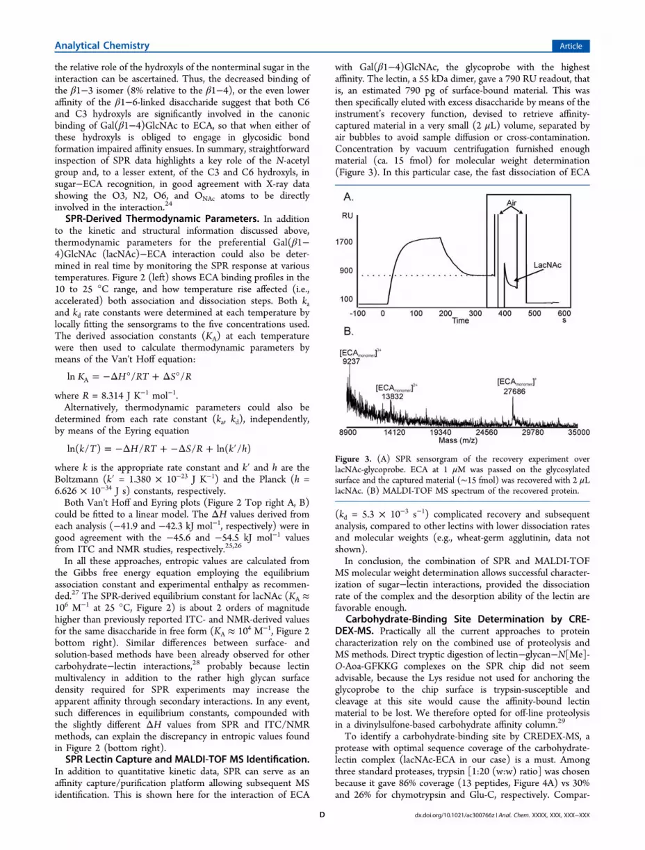
with Gal(β1−4)GlcNAc, the glycoprobe with the highestaffinity. The lectin, a 55 kDa dimer, gave a 790 RU readout, thatis, an estimated 790 pg of surface-bound material. This wasthen specifically eluted with excess disaccharide by means of theinstrument’s recovery function, devised to retrieve affinity-captured material in a very small (2 μL) volume, separated byair bubbles to avoid sample diffusion or cross-contamination.Concentration by vacuum centrifugation furnished enoughmaterial (ca. 15 fmol) for molecular weight determination(Figure 3). In this particular case, the fast dissociation of ECA
(kd = 5.3 × 10⁻³ s−1) complicated recovery and subsequentanalysis, compared to other lectins with lower dissociation ratesand molecular weights (e.g., wheat-germ agglutinin, data notshown).In conclusion, the combination of SPR and MALDI-TOF
MS molecular weight determination allows successful character-ization of sugar−lectin interactions, provided the dissociationrate of the complex and the desorption ability of the lectin arefavorable enough.
Carbohydrate-Binding Site Determination by CRE-DEX-MS. Practically all the current approaches to proteincharacterization rely on the combined use of proteolysis andMS methods. Direct tryptic digestion of lectin−glycan−N[Me]-O-Aoa-GFKKG complexes on the SPR chip did not seemadvisable, because the Lys residue not used for anchoring theglycoprobe to the chip surface is trypsin-susceptible andcleavage at this site would cause the affinity-bound lectinmaterial to be lost. We therefore opted for off-line proteolysisin a divinylsulfone-based carbohydrate affinity column.29
To identify a carbohydrate-binding site by CREDEX-MS, aprotease with optimal sequence coverage of the carbohydrate-lectin complex (lacNAc-ECA in our case) is a must. Amongthree standard proteases, trypsin [1:20 (w:w) ratio] was chosenbecause it gave 86% coverage (13 peptides, Figure 4A) vs 30%and 26% for chymotrypsin and Glu-C, respectively. Compar-
Figure 3. (A) SPR sensorgram of the recovery experiment overlacNAc-glycoprobe. ECA at 1 μM was passed on the glycosylatedsurface and the captured material (∼15 fmol) was recovered with 2 μLlacNAc. (B) MALDI-TOF MS spectrum of the recovered protein.
Analytical Chemistry Article
dx.doi.org/10.1021/ac300766z | Anal. Chem. XXXX, XXX, XXX−XXXD

ison of the standard (Figure 4A) with the flow-through fromon-column digest (Figure 4B) revealed essentially identicalmass fingerprint peaks with only minor differences in intensity.The column was next washed until no peptide signals wereobserved (Figure 4C) and then the glycan-interacting peptideswere eluted with 60% acetonitrile in water and analyzed (Figure4D). In this fraction, several peptides {[74−99], [75−94],[83−94], [100−116], [117−151], and [202−220]} (Figure4D) that contain amino acids displaying direct contact with thecarbohydrate in the X-ray structure of the Lac-ECA complex(Figure 4E) could be unequivocally assigned to the binding site.In addition to these specific peptides, three peptides found inlow abundance {[37−50], [51−73], and [74−84]} showed nocontacts with the sugar in the X-ray structure (Figure 4F);hence no relation to the binding site. Their presence in theelution fraction was explained as a case of “riding” (via β-strandinteraction) with the spatially close, glycan-binding peptide[202−220].To confirm the binding site identification, an additional
chymotrypsin digestion subsequent to the trypsin excision wasperformed. Figure 4 (right panel H−L) shows that, aftersequential trypsin-chymotrypsin digestions, the longest peptide
[202−220] is split into shorter fragments and the “ridingpeptides”, [37−50], [51−73], and [74−84], are no longerobserved in the elution fraction, while the specific bindingpeptides [74−99], [117−151], as well as nondigested [202−220] remain present (Figure 4L). This result proves that theGal(β1−4)GlcNAc-ECA interaction can withstand prolonged,sequential digestion with two proteases, despite its relativelylow affinity (see Figure 1), hence making possible accuratemolecular definition of the interaction partners and removal ofunspecific peptides. This is valuable when proteolytic excisionresults in long peptides enhancing the likelihood of peptide−peptide interactions that may hamper the identification of thecarbohydrate binding site.Additional unequivocal identification of the binding site came
from an extraction MS experiment,20 where ECA was firstdigested with trypsin, the digest was passed through the affinitycolumn, and only peptides [202−220] and [74−99] represent-ing the binding site were observed (data not shown).
■ CONCLUSION
Detailed molecular description of carbohydrate−proteininteractions is feasible by the combination of analytical
Figure 4. (A) Peptide mass fingerprint of ECA (digestion in solution). (B−D) MALDI-TOF MS spectra corresponding to different fractions ofexcision experiment. (B) On column digestion with trypsin of the complex lacNAc-ECA. (C) Supernatant after washing. (D) Elution fraction. Peaksin gray show the peptides with no direct contact with the sugar. (Center panel E−G) X-ray crystal structure (PDB 1GZC) of ECA in complex withlactose (in yellow). (E) Peptides identified in the elution fraction of an excision experiment with trypsin are shown in green (sugar-peptideinteraction) and red (peptide−peptide interaction) in the ribbon representation. (F) Peptides [37−50], [51−73] and [74−84] (in red),noncovalently bound with spatially close [202−220] (in green), “ride” with this peptide in the elution fraction. (G) In an excision experiment withtwo consecutive digestions, only peptides involved in sugar-peptide interactions (in green) are detected.22 (Right panel H−L) MALDI-TOF MSspectra corresponding to different fractions of excision experiment with two consecutive proteolytic digestions. (H) First on column digestion withtrypsin of the complex lacNAc-ECA. (I) Supernatant after washing. (J) Second on column digestion with chymotrypsin. (K) Supernatant afterwashing. (L) Elution fraction.
Analytical Chemistry Article
dx.doi.org/10.1021/ac300766z | Anal. Chem. XXXX, XXX, XXX−XXXE

techniques described here, which use low amounts of bothlectin and carbohydrate compatible with extraction from naturalsources, in contrast with more sample-demanding techniquessuch as NMR or X-ray crystallography. The agglutinin ECA hasbeen chosen as a case study to test the applicability of thesetechniques. SPR-based experiments showed a higher affinity ofECA for lacNAc relative to other β-galactosides. Additionalstructural information on the interaction was also provided bySPR, by comparing the differential binding responses betweenepitopes with subtle differences (i.e., glycosidic linkage or N-acetyl group at position C2). This analysis showed thehydroxyls at C3 and C6, as well as the N-acetyl at C2, beingcritical for interaction with ECA. Thermodynamic data on theinteraction were also derived by SPR. While enthalpy valueswere equivalent to those obtained by ITC or NMR, the higheraffinity constants determined by SPR translated into largerdifferences in entropy relative to ITC or NMR. Finally, SPRtechnology was successfully applied as a lectin capture platformfor subsequent MS analysis. SPR-based results are shown to bean efficient combination with CREDEX-MS, which provides amolecular definition of the carbohydrate-binding site. CRE-DEX-MS in conjunction with the SPR approach described hereconstitutes a valuable set of tools for decrypting carbohydrate−protein interaction details.
■ AUTHOR INFORMATION
Corresponding Author
*E-mail: [email protected] (R.G.-G.); [email protected](D.A); [email protected] (M.P.).
Notes
The authors declare no competing financial interest.
■ ACKNOWLEDGMENTS
This work was supported by the Spanish Ministry of Scienceand Innovation (projects BIO2008-04487-CO3-02 andHA2007-0021 to D.A. and BIO2009-08983 to R.G.G., andpredoctoral fellowship BES-2006-12879 to C.J.C.), by theSpanish Ministry of Economy and Competitiveness (projectSAF2011-24899 to D.A.), and by the Deutsche Forschungsge-meinschaft (PR-175/14-1) and the EU (MSLife).
■ REFERENCES
(1) Imberty, A.; Varrot, A. Curr. Opin. Struct. Biol. 2008, 18, 567−576.(2) Rambaruth, N. D.; Dwek, M. V. Acta Histochem. 2011, 113, 591−600.(3) Velasquez, J. G.; Canovas, S.; Barajas, P.; Marcos, J.; Jimenez-Movilla, M.; Gutierrez-Gallego, R.; Ballesta, J.; Aviles, M.; Coy, P. Mol.Reprod. Dev. 2007, 74, 617−628.(4) Apweiler, R.; Hermjakob, H.; Sharon, N. Biochim. Biophys. Acta1999, 1473, 4−8.(5) Puig, O.; Caspary, F.; Rigaut, G.; Rutz, B.; Bouveret, E.; Bragado-Nilsson, E.; Wilm, M.; Seraphin, B. Methods 2001, 24, 218−229.(6) Bruckner, A.; Polge, C.; Lentze, N.; Auerbach, D.; Schlattner, U.Int. J. Mol. Sci. 2009, 10, 2763−2788.(7) Madeira, A.; Ohman, E.; Nilsson, A.; Sjogren, B.; Andren, P. E.;Svenningsson, P. Nat. Protoc. 2009, 4, 1023−1037.(8) Gutierrez-Gallego, R.; Haseley, S. R.; van Miegem, V. F.;Vliegenthart, J. F.; Kamerling, J. P. Glycobiology 2004, 14, 373−386.(9) Slepak, V. Z. J. Mol. Recognit. 2000, 13, 20−26.(10) Ahmed, F. E.; Wiley, J. E.; Weidner, D. A.; Bonnerup, C.; Mota,H. Cancer Genomics Proteomics 2010, 7, 303−309.(11) Duverger, E.; Frison, N.; Roche, A. C.; Monsigny, M. Biochimie2003, 85, 167−79.
(12) Besenicar, M.; Macek, P.; Lakey, J. H.; Anderluh, G. Chem. Phys.Lipids 2006, 141, 169−178.(13) Roos, H.; Karlsson, R.; Nilshans, H.; Persson, A. J. Mol. Recognit.1998, 11, 204−210.(14) Zhukov, A.; Schurenberg, M.; Jansson, O.; Areskoug, D.; Buijs, J.J. Biomol. Tech. 2004, 15, 112−119.(15) Vila-Perello, M.; Gutierrez-Gallego, R.; Andreu, D. Chem-BioChem 2005, 6, 1831−1838.(16) Jimenez-Castells, C.; de la Torre, B. G.; Gutierrez-Gallego, R.;Andreu, D. Bioorg. Med. Chem. Lett. 2007, 17, 5155−5158.(17) Jimenez-Castells, C.; de la Torre, B. G.; Andreu, D.; Gutierrez-Gallego, R. Glycoconj. J. 2008, 25, 879−887.(18) Weis, W. I.; Drickamer, K. Annu. Rev. Biochem. 1996, 65, 441−473.(19) Asensio, J. L.; Canada, F. J.; Bruix, M.; Gonzalez, C.; Khiar, N.;Rodriguez-Romero, A.; Jimenez-Barbero, J. Glycobiology 1998, 8, 569−577.(20) Moise, A.; Andre, S.; Eggers, F.; Krzeminski, M.; Przybylski, M.;Gabius, H. J. J. Am. Chem. Soc. 2011, 133, 14844−14847.(21) Przybylski, M., Moise, A., Gabius, H. J. Identification Of LigandRecognition Domains. Patent [PCT/EP2009/003495], 2012, 2010.(22) Svensson, C.; Teneberg, S.; Nilsson, C. L.; Kjellberg, A.;Schwarz, F. P.; Sharon, N.; Krengel, U. J. Mol. Biol. 2002, 321, 69−83.(23) Itakura, Y.; Nakamura-Tsuruta, S.; Kominami, J.; Sharon, N.;Kasai, K.; Hirabayashi, J. J. Biochem. 2007, 142, 459−469.(24) Elgavish, S.; Shaanan, B. J. Mol. Biol. 1998, 277, 917−932.(25) Gupta, D.; Cho, M.; Cummings, R. D.; Brewer, C. F.Biochemistry 1996, 35, 15236−15243.(26) De Boeck, H.; Loontiens, F. G.; Lis, H.; Sharon, N. Arch.Biochem. Biophys. 1984, 234, 297−304.(27) Roos, H.; Karlsson, R.; Nilshans, H.; Persson, A. J. Mol. Recognit.1998, 11, 204−210.(28) Critchley, P.; Clarkson, G. J. Org. Biomol. Chem. 2003, 1, 4148−4159.(29) Stefanescu, R.; Born, R.; Moise, A.; Ernst, B.; Przybylski, M. J.Am. Soc. Mass Spectrom. 2011, 22, 148−157.
Analytical Chemistry Article
dx.doi.org/10.1021/ac300766z | Anal. Chem. XXXX, XXX, XXX−XXXF