Deciphering mycorrhizal fungi in cultivated Phalaenopsis microbiome with next-generation sequencing...
12
Deciphering mycorrhizal fungi in cultivated Phalaenopsis microbiome with next-generation sequencing of multiple barcodes Chao-Li Huang & Feng-Yin Jian & Hao-Jen Huang & Wen-Chi Chang & Wen-Luan Wu & Chi-Chuan Hwang & Ruey-Hua Lee & Tzen-Yuh Chiang Received: 5 July 2013 /Accepted: 6 February 2014 /Published online: 25 February 2014 # The Author(s) 2014. This article is published with open access at Springerlink.com Abstract Identifying the species composition of a microbial ecosystem is often hampered by difficulties in culturing the organisms and in the low sequencing depth of traditional DNA barcoding. Metagenomic analysis, a huge-scale nucleotide-sequence-based tool, can overcome such difficul- ties. In this study, Sanger sequencing of 500 nrITS clones uncovered 29 taxa of 19 fungal genera, whereas metagenomics with next-generation sequencing identified 512 operational taxonomic units (OTUs) for ITS1/2 and 364 for ITS3/4. Nevertheless, high throughput sequencing of PCR amplicons of ITS1/2, ITS3/4, nrLSU-LR, nrLSU-U, mtLSU, and mtATP6, all with at least 1,300× coverage and about 21 million reads in total, yielded a very diverse fungal composi- tion. The fact that 74 % of the OTUs were exclusively uncov- ered with single barcodes indicated that each marker provided its own insights into the fungal flora. To deal with the high heterogeneity in the data and to integrate the information on species composition across barcodes, a rank-scoring strategy was developed. Accordingly, 205 genera among 64 orders of fungi were identified in healthy Phalaenopsis roots. Of the barcodes utilized, ITS1/2, ITS3/4, and nrLSU-U were the most competent in uncovering the fungal diversity. These barcodes, though detecting different compositions likely due to primer preference, provided complementary and compre- hensive power in deciphering the microbial diversity, espe- cially in revealing rare species. Keywords DNA barcode . Metagenomics . Mycorrhizal fungi . Next-generation sequencing . Phalaenopsis orchid . Rank scoring Introduction The Orchidaceae (orchids) is one of the largest families of angiosperms (Pridgeon et al. 2005). A great number of orchid species have been developed commercially as potted flowering crops with an annual market growth rate of 30 % (Wang 2004). Among these, the monopodial epiphytic Phalaenopsis, one of the most popular orchids, is only avail- able in the retail markets when in bloom. Over the past decades, a large pool of cultivars with new traits and pheno- typic variation has been generated via traditional breeding. Great advances in tissue culture techniques have also allowed mass production of disease-free orchid plantlets from seeds or vegetative tissues. One of the major problems in orchid production is that 1-year-old tissue-culture plantlets require at least 16–24 months of vegetative growth for the leaf span to reach a minimum diameter of 25 cm (Konow and Wang 2001; Runkle et al. 2007). The ability of Phalaenopsis to spike and bloom under inducive conditions, e.g., low tem- peratures, is highly correlated with the size of the plant; however, fungal infection can greatly reduce plant size. In addition, common pathogens such as Fusarium oxysporum Chao-Li Huang, Feng-Yin Jian, Hao-Jen Huang, Wen-Chi Chang and Wen-Luan Wu contributed equally. Electronic supplementary material The online version of this article (doi:10.1007/s13225-014-0281-x) contains supplementary material, which is available to authorized users. C.<L. Huang : F.<Y. Jian : H.<J. Huang : W.<L. Wu : T.<Y. Chiang (*) Department of Life Sciences, National Cheng Kung University, Tainan, Taiwan 701 e-mail: [email protected] W.<C. Chang : R.<H. Lee (*) Institute of Tropical Plant Sciences, National Cheng Kung University, Tainan, Taiwan 701 e-mail: [email protected] C.<C. Hwang (*) Department of Engineering Science and Supercomputing Research Center, National Cheng Kung University, Tainan, Taiwan 701 e-mail: [email protected] Fungal Diversity (2014) 66:77–88 DOI 10.1007/s13225-014-0281-x
Transcript of Deciphering mycorrhizal fungi in cultivated Phalaenopsis microbiome with next-generation sequencing...
Deciphering mycorrhizal fungi in cultivated Phalaenopsismicrobiome with next-generation sequencing of multiple barcodes
Chao-Li Huang & Feng-Yin Jian & Hao-Jen Huang &
Wen-Chi Chang & Wen-Luan Wu & Chi-Chuan Hwang &
Ruey-Hua Lee & Tzen-Yuh Chiang
Received: 5 July 2013 /Accepted: 6 February 2014 /Published online: 25 February 2014# The Author(s) 2014. This article is published with open access at Springerlink.com
Abstract Identifying the species composition of a microbialecosystem is often hampered by difficulties in culturing theorganisms and in the low sequencing depth of traditionalDNA barcoding. Metagenomic analysis, a huge-scalenucleotide-sequence-based tool, can overcome such difficul-ties. In this study, Sanger sequencing of 500 nrITS clonesuncovered 29 taxa of 19 fungal genera, whereasmetagenomics with next-generation sequencing identified512 operational taxonomic units (OTUs) for ITS1/2 and 364for ITS3/4. Nevertheless, high throughput sequencing of PCRamplicons of ITS1/2, ITS3/4, nrLSU-LR, nrLSU-U, mtLSU,and mtATP6, all with at least 1,300× coverage and about 21million reads in total, yielded a very diverse fungal composi-tion. The fact that 74 % of the OTUs were exclusively uncov-ered with single barcodes indicated that each marker providedits own insights into the fungal flora. To deal with the highheterogeneity in the data and to integrate the information onspecies composition across barcodes, a rank-scoring strategy
was developed. Accordingly, 205 genera among 64 orders offungi were identified in healthy Phalaenopsis roots. Of thebarcodes utilized, ITS1/2, ITS3/4, and nrLSU-U were themost competent in uncovering the fungal diversity. Thesebarcodes, though detecting different compositions likely dueto primer preference, provided complementary and compre-hensive power in deciphering the microbial diversity, espe-cially in revealing rare species.
Keywords DNAbarcode .Metagenomics .Mycorrhizalfungi . Next-generation sequencing .Phalaenopsis orchid .
Rank scoring
Introduction
The Orchidaceae (orchids) is one of the largest families ofangiosperms (Pridgeon et al. 2005). A great number of orchidspecies have been developed commercially as pottedflowering crops with an annual market growth rate of 30 %(Wang 2004). Among these, the monopodial epiphyticPhalaenopsis, one of the most popular orchids, is only avail-able in the retail markets when in bloom. Over the pastdecades, a large pool of cultivars with new traits and pheno-typic variation has been generated via traditional breeding.
Great advances in tissue culture techniques have alsoallowed mass production of disease-free orchid plantlets fromseeds or vegetative tissues. One of the major problems inorchid production is that 1-year-old tissue-culture plantletsrequire at least 16–24 months of vegetative growth for theleaf span to reach a minimum diameter of 25 cm (Konow andWang 2001; Runkle et al. 2007). The ability of Phalaenopsisto spike and bloom under inducive conditions, e.g., low tem-peratures, is highly correlated with the size of the plant;however, fungal infection can greatly reduce plant size. Inaddition, common pathogens such as Fusarium oxysporum
Chao-Li Huang, Feng-Yin Jian, Hao-Jen Huang, Wen-Chi Chang andWen-Luan Wu contributed equally.
Electronic supplementary material The online version of this article(doi:10.1007/s13225-014-0281-x) contains supplementary material,which is available to authorized users.
C.<L. Huang : F.<Y. Jian :H.<J. Huang :W.<L. Wu :T.<Y. Chiang (*)Department of Life Sciences, National Cheng Kung University,Tainan, Taiwan 701e-mail: [email protected]
W.<C. Chang : R.<H. Lee (*)Institute of Tropical Plant Sciences, National Cheng KungUniversity, Tainan, Taiwan 701e-mail: [email protected]
C.<C. Hwang (*)Department of Engineering Science and Supercomputing ResearchCenter, National Cheng Kung University, Tainan, Taiwan 701e-mail: [email protected]
Fungal Diversity (2014) 66:77–88DOI 10.1007/s13225-014-0281-x
(Beckman 1987), Sclerotium rolfsii (Cating et al. 2009), andBotrytis cinerea (Wey 1988) cause various unsightly symp-toms on leaves and roots that, even if the orchid survives thedisease, the quality and growth of orchids are irrevocablydamaged and ruined for the commercial market.
Plant roots are surrounded by the rhizosphere, which con-tains compounds that are secreted by roots and microbes,which in turn influences the growth and survival of the organ-isms (Hinsinger et al. 2009). The interplay between plants andrhizosphere microorganisms can therefore affect plant growthand health (Bisseling et al. 2009; Berendsen et al. 2012). Inreturn, photosynthetic plants secrete up to 21 % of their fixedcarbon to the rhizosphere as nutrients, feeding the microfloraand influencing their metabolic activity and diversity (Mendeset al. 2011). For all or part of their life cycle, orchids areobligatorily dependent on their mycorrhizal partners in nature.For example, orchid seed is less likely to germinate in theabsence of mycorrhizal fungi under natural conditions(Burgeff 1959), and orchid plants depend on the symbiontsto gain access to organic and mineral nutrients by increasingnutrient absorption and translocation to plants via extraradicalhyphae (Arditti 1992; Rasmussen 1995; Smith and Read2008). Studying the microbiome of orchid roots enables oneto understand the complexity of plant–microbe interactionsassociated with plant health and growth, thus opening newavenues to increase orchid quality and productivity.
Although scientists have traditionally depended on in vitroand in vivo culturing to explore fungal communities, mostspecies remain unculturable, and rare strains can be easilyunexploited in culture (Kaeberlein 2002). Technically opti-mizing culture conditions for individual species, especiallywhen the species composition of a community remains un-known, can be time-consuming and difficult, especially toinduce sporulation. In addition, direct observation of fungalmorphotypes via isolation of a single peloton in roots requiresexpertise for accurate interpretation and is very time-consuming. DNA barcodes, biochemical markers, and analy-sis of acyl chain composition in membrane-phospholipids alsoprovide powerful tools for studyingmicrobial ecology withoutconventional culture (Alef and Nannipieri 1995). Of thesemethods, DNA barcoding is a powerful tool to identify speciesusing sequences for gene regions that are conserved acrossgreatly diverse taxonomic groups (Hebert and Gregory 2005;Schoch et al. 2012). Nuclear ribosomal RNA (nrRNA) is themost abundant RNA encoded by ribosomal RNA (rRNA)genes. High conservation in the genes thus provides a frame-work for assigning sequences to genera and species for inves-tigations of microbial community diversity (Rosselló-Moraand Amann 2001; Hirsch et al. 2010). Eukaryotic nrRNAbarcodes include large subunit 28S rRNA (nrLSU) gene,small subunit 18S rRNA (nrSSU) gene, and the internaltranscribed spacer (nrITS) rDNA plus the 5.85S gene(Druzhinina et al. 2005; Kõljalg et al. 2005). Among these
regions, nrITS is the most effective discriminator of fungalspecies, and the nrLSU is also very effective. The nrSSU,despite its prominence in assessing bacterial community, istoo conserved for fungi (Tringe and Hugenholtz 2008; Schochet al. 2012).
With the invention of next-generation sequencing (NGS),fungus-specific barcoding primers can be used withmetagenomics, a huge-scale nucleotide-sequence-based tool,to analyze microbial communities regardless of an organism’sculturability (Cowan et al. 2005). The tool provides highthroughput sequencing of PCR amplicons from a singleDNA extraction and estimates of the relative abundance ofthe organisms detected (Hirsch et al. 2010). However, becausea single barcode is limited in representing the panorama of amicrobial community, combinations of multiple barcodeshave thus been recommended (DeSalle et al. 2008). Basedon the evaluation of Schoch et al. (2012), we selected fournuclear ribosomal markers, two nrITS regions (ITS1/2 andITS3/4) and two in the nrLSU region (nrLSU-LR and nrLSU-U) (Vilgalys and Hester 1990; Wu et al. 2002). The largesubunit of the mitochondria ribosomal region (mtLSU) andthe sixth subunit of mitochondrial ATPase (mtATP6) (Zenget al. 2004; Grubisha et al. 2012) have also been adopted asmarkers.
In this study, we deciphered the microbiome of cultivatedorchid roots based on amplicon-based metagenomics. Usingmultiple barcodes, we investigated the taxon diversity of thefungal community and examined the consistency amongbarcodes in uncovering the composition of the fungal floraand the ecological interactions between fungal endophytesand orchids. We also compared traditional Sanger sequencingof full-length nrITS with NGS techniques. A rank-scoringstrategy was also developed to integrate the information onspecies composition across barcodes.
Materials and methods
Plant materials and DNA extraction
Phalaenopsis KC1111 (Phalaenopsis Taisuco Snow ×Doritaenopsis White Wonder) was obtained from the TaiwanSugar Corporation (Taisuco) and grown in the greenhouse ofNational Cheng Kung University in Tainan, Taiwan. Plantswere watered once a week without any pesticide or fertilizer.Microbial contamination from the potting media was elimi-nated by sterilizing the roots from five individuals ofPhalaenopsis KC1111 in 2 % NaOCl for 15 min with fivesubsequent washes with water (Zelmer et al. 1996). Thesetissues were ground into powder with liquid nitrogen. Totalgenomic DNAs were extracted by using a modifiedcetyltrimethylammonium bromide (CTAB) method (Doyleand Doyle 1987).
78 Fungal Diversity (2014) 66:77–88
Gene cloning and Sanger sequencing
Full-length nrITS genomic DNA region, a marker often usedfor identifying fungi (Nilsson et al. 2008), was PCR amplifiedusing the ITS1/ITS4 primer pairs (Wu et al. 2002) in a 50 μLreaction mixture containing 25 μL Taq DNA Polymerase 2×Master Mix Red (Ampliqon, Denmark), 5 μL forward andreverse primers (ITS1 and ITS4, 2 ng/μL, Table S1) each, and5 μL genomic DNA (2 ng/μL). The PCR cycling schemeconsisted of one cycle of 94 °C/3 min; 35 cycles of 94 °C/30 s,55 °C/37s, 72 °C/30 s; and a final extension at 72 °C/10 min.PCR products of ~650 bp were purified with a Gel/PCR DNAFragment Extraction Kit (Geneaid, Taiwan), ligated into thepGEM-T Easy Vector (Promega, USA), and used to transformDH5α competent cells (Yeastern Biotech, Taiwan). Cloneswere sequenced with an ABI PRISM 3730 DNA Sequencer(ABI Big Dye Terminator Cycle Sequencing Kit, Perkin-Elmer). The obtained sequences were used in a BLASTsearchagainst the NCBI (http://blast.ncbi.nlm.nih.gov/Blast.cgi)database with default blastn settings and assigned to specifictaxa using MEtaGenome Analyzer (MEGAN) software(Huson et al. 2011). With MEGAN software, the lowestcommon ancestor (LCA) algorithm was used for taxonomicclassification, with the required parameters of the LCA as-signment set as minimum support=1, minimum score=500,top percentage=1.
Metagenomic barcoding of the fungal community in orchidroots
Six DNA fragments derived from four DNA regions, namely,nrITS (ITS1/2 and ITS3/4), nrLSU (LR and U), mitochondriallarge subunit rDNA (mtLSU), and mitochondrial ATPasesubunit 6 (mtATP6), were PCR-amplified using genomicDNA isolated from roots of cultivated PhalaenopsisKC1111. PCR primers and annealing temperatures are listedin Table S1. Amplification was conducted as described in thegene cloning section. All PCR products of ca. 250–300 bpwere purified, pooled, and sequenced with Illumina GAIIxhigh-throughput paired-end sequencing to survey the compo-sition of fungal community. Raw reads were sorted into sixcategories according to the primer sequences, and the readswith an N residue in the sequences were discarded. Sortedsequences were merged to haplotypes for computing the copynumbers, and single-copy haplotypes were removed to lessenthe effect of sequencing errors. These haplotypes were furtherclustered into operational taxonomic units (OTUs) using theBLASTClust program in the standalone BLAST v2.2.26package of the NCBI. Because the average minimal diver-gence between fungal species is around 2.5–3 % (Seena et al.2010; Stockinger et al. 2010), the stringency of clustering wasset with two parameters at 97 % similarity and 80 % coveragebetween sequences and referred to as the average minimal
divergence of species between fungi. From reads sorting,singleton removal, to OTU generation, all steps were conduct-edwith our own Perl scripts. BLASTanalyses were performedon all reads against the NCBI nucleotide database, and theresults were further processed for taxonomic assignationsusing MEGAN. An optional score adjustment was used whenpaired reads matched the same species. The required param-eters of the LCA assignment were set as minimum support=2,minimum score=80, top percentage=1 (Murray et al. 2011;Montaña et al. 2012). Classification results were manuallychecked to correct the ambiguous assignation caused by syn-onyms for fungal species or an ambiguous annotation in theNCBI database.
Evaluating biodiversity based on metagenomic data
As recommended by Haegeman et al. (2013), Shannon’s andGini-Simpson’s diversity indices (Shannon 1948; Rao 1982)were adopted to estimate the alpha diversity in the fungalcommunity. Shannon’s index is affected by the species num-ber and their equitability, or evenness. A greater number ofspecies and an even distribution of abundances result in anelevated Shannon’s diversity index. The maximum Shannon’sdiversity index for a sample indicates that all species arenearly equally abundant. The Gini-Simpson’s diversity indexis measured as the probability that two individuals randomlyselected from a sample belong to the same species, with arange from 0 to 1. Value of 0 indicates lack of diversity, i.e.,one dominant species or taxon in the community, and 1suggests that the community contains an infinite number oftaxa with all taxa present equally. Before alpha-diversity indi-ces were calculated, multiple rarefactions were performedwith our own Perl scripts. All fungal reads from each markerwere resampled starting at the depth of 1,000 reads, steppingup to 385,000 reads with increments of 1,000, and ten repli-cates were done at each sampling depth. For illustrating fungaldiversities, taxonomic relationships of all detected fungalgenera were converted to the Newick format and uploadedto the web-based tool Interactive Tree Of Life v2.2 (Letunicand Bork 2011), and the taxonomic trees for each barcode andfor all barcodes combined were generated.
Estimation of the taxon abundance based on copy numbersof PCR-amplified DNA reads for a mixture of homologousgenes in a multi-template PCR can be biased due to thedifferences in the primer binding energy to the target(Kanagawa 2003). Consequently, the taxon diversity and pro-portion of any given operational taxonomic unit (OTU) in thefungal community are expected to differ when using differentsets of DNA barcodes. In this study, the percentage of readsfor a taxon was calculated by dividing the total reads of fungigenerated by individual barcodes (Table S3). Because of thebias in the taxonomic assignations of mtATP6, that was re-stricted to the class Agaricomycetes except for six reads, we
Fungal Diversity (2014) 66:77–88 79
excluded mtATP6 from estimating species abundance withmultiple barcodes. The percentage of reads for each of thegenera generated from five barcodes (ITS1/2, ITS3/4, nrLSU-LR, nrLSU-U and mtLSU) was then transformed to a rankscore based on the abundance of each genus in the communityusing the formula 20−19 (rank−1)/(N−1). The ranks (1, 2,3…to N) represent the order of abundance (percentage ofreads) for all taxa; thus, a taxon with rank 1 is most abundantand receives the highest rank score (20). When several taxahave the same abundance, the highest rank of these taxa wasused as representative. The highest rank score was set to 20 fora given taxon having the highest number of reads (rank=1),and the lowest rank score was set to 1 for a given taxon havinglowest number of reads (rank=N).
Results
Phylogenetic diversity of fungal community based on PCRcloning and Sanger sequencing of ITS1/ITS4
NrITS was amplified using the ITS1/ITS4 barcode for ana-lyzing the composition of the endophytic fungal communityin the orchid roots. Of the 500 nrITS sequences obtained andanalyzed, a BLAST search assigned 76.4 % of the sequencesto fungi, of which only 19 genera (29 taxa) were identified(Table 1). The top 10 most abundant fungal taxa werePenicillium sp. (20.0 %), Trechispora farinacea (17.2 %),Leotiomyceta (12.0 %), Exophiala (6.6 %), Fusarium solani(4.4 %), Cladosporium sp. (3.6 %), Epulorhiza sp. Van44(2.4 %), Alternaria sp. (2.0 %), Leucocoprinus birnbaumii(2.0 %), and Sporothrix inflata (1.2 %).
Efficiency of six barcoding markers in fungal identificationby metagenomics
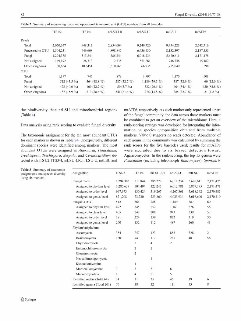
In total, 27,099,433 PE reads were obtained and sorted ac-cording to the six markers from the raw sequencing data. Aftersingle-copy haplotypes were removed, 21,009,068 (77.5 %)PE reads remained and were further clustered into OTUs.Among these markers, nrLSU-U yielded the most readsassigned to fungi (90.7 % of 6,636,430), followed by mtLSU(69.7 % of 8,132,397), mtATP6 (99.3 % of 2,187,555),ITS1/2 (86.1 % of 1,504,231), ITS3/4 (79.1 % of 649,608),and nrLSU-LR (20.3 % of 1,898,847). No correlation existedbetween the read numbers and the number of assigned fungalOTUs. The coverage (number of reads/number of OTUs) ofmarkers ranged from 1,338× of nrLSU-LR to 36,191× ofmtATP6. Taxon assignation using aMEGAN analysis showedthat 32.8–59.5 % of OTUs could be assigned to fungi, exceptfor mtATP6 (12.0 %) (Table 2). In contrast, 40.6 % of OTUsamplified with ITS1/ITS2, 22.7 % with ITS3/ITS4, 5.7 %with nrLSU-LR, 26.6 % with nrLSU-U, 34.4 % with mtLSU
and 83.8 % with mtATP6 were not assignable to any organ-isms based on the BLAST searches (Table 2). Although mostreads for mtATP6 were assigned to fungi, 96.2 % of thesereads belonged to one OTU (Ceratobasidium sp. CBS189.90).
Fungal diversity in orchid roots detected with six barcodingmarkers
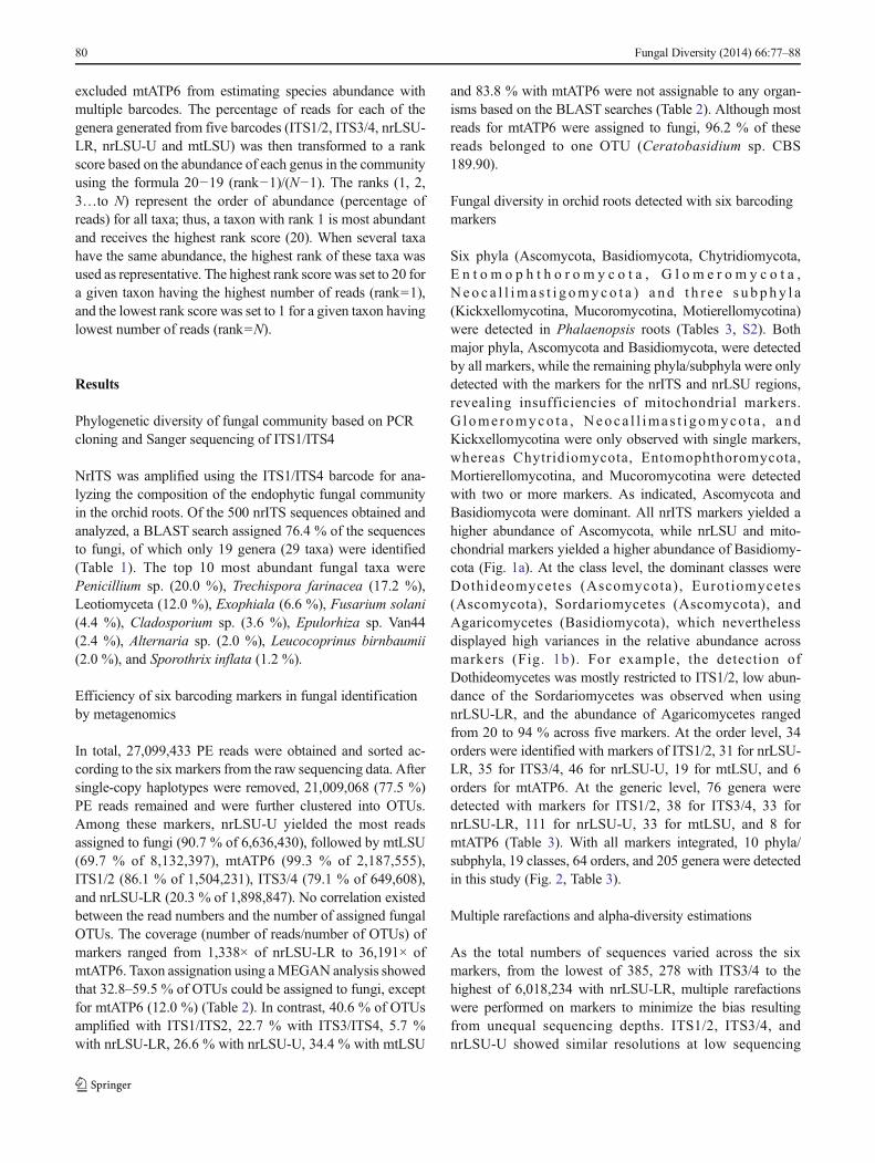
Six phyla (Ascomycota, Basidiomycota, Chytridiomycota,E n t o m o p h t h o r o m y c o t a , G l o m e r o m y c o t a ,N e o c a l l im a s t i g omyco t a ) a n d t h r e e s u b p hy l a(Kickxellomycotina, Mucoromycotina, Motierellomycotina)were detected in Phalaenopsis roots (Tables 3, S2). Bothmajor phyla, Ascomycota and Basidiomycota, were detectedby all markers, while the remaining phyla/subphyla were onlydetected with the markers for the nrITS and nrLSU regions,revealing insufficiencies of mitochondrial markers.G lome romyco t a , Neoca l l ima s t i gomyco t a , andKickxellomycotina were only observed with single markers,whereas Chytridiomycota, Entomophthoromycota,Mortierellomycotina, and Mucoromycotina were detectedwith two or more markers. As indicated, Ascomycota andBasidiomycota were dominant. All nrITS markers yielded ahigher abundance of Ascomycota, while nrLSU and mito-chondrial markers yielded a higher abundance of Basidiomy-cota (Fig. 1a). At the class level, the dominant classes wereDothideomycetes (Ascomycota) , Eurotiomycetes(Ascomycota), Sordariomycetes (Ascomycota), andAgaricomycetes (Basidiomycota), which neverthelessdisplayed high variances in the relative abundance acrossmarkers (Fig. 1b). For example, the detection ofDothideomycetes was mostly restricted to ITS1/2, low abun-dance of the Sordariomycetes was observed when usingnrLSU-LR, and the abundance of Agaricomycetes rangedfrom 20 to 94 % across five markers. At the order level, 34orders were identified with markers of ITS1/2, 31 for nrLSU-LR, 35 for ITS3/4, 46 for nrLSU-U, 19 for mtLSU, and 6orders for mtATP6. At the generic level, 76 genera weredetected with markers for ITS1/2, 38 for ITS3/4, 33 fornrLSU-LR, 111 for nrLSU-U, 33 for mtLSU, and 8 formtATP6 (Table 3). With all markers integrated, 10 phyla/subphyla, 19 classes, 64 orders, and 205 genera were detectedin this study (Fig. 2, Table 3).
Multiple rarefactions and alpha-diversity estimations
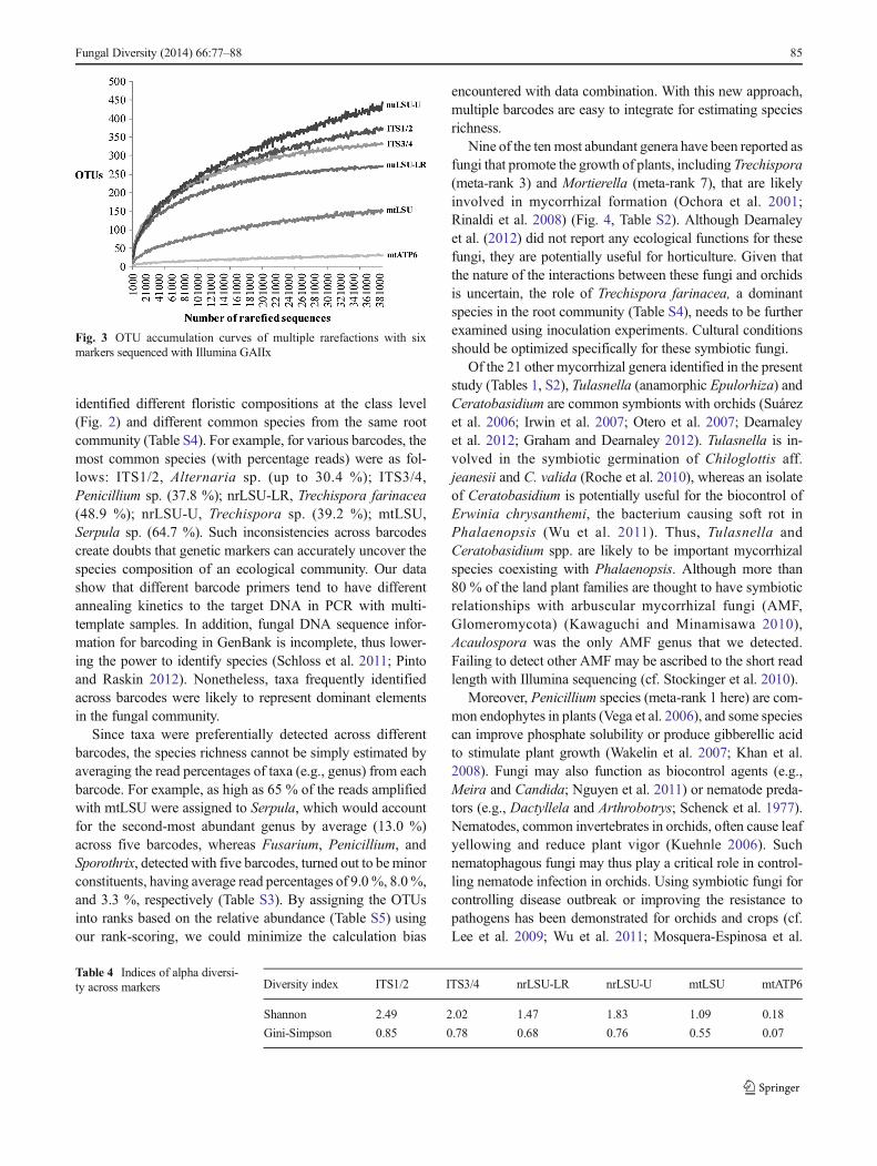
As the total numbers of sequences varied across the sixmarkers, from the lowest of 385, 278 with ITS3/4 to thehighest of 6,018,234 with nrLSU-LR, multiple rarefactionswere performed on markers to minimize the bias resultingfrom unequal sequencing depths. ITS1/2, ITS3/4, andnrLSU-U showed similar resolutions at low sequencing
80 Fungal Diversity (2014) 66:77–88

depths, as indicated by the curves of these markers that over-lapped when the rarefied number was less than 100,000(Fig. 3). Nevertheless, as the number of sequences increased,nrLSU-U demonstrated the best resolution (442.4 OTUs of385,000 sequences) compared with other markers, followedby ITS1/2 (371.4 OTUs) and ITS3/4 (333.8 OTUs). Wefurther estimated the alpha diversity of the fungal communitywith the rarefied data set. The two alpha diversity indicators,Shannon’s and Gini-Simpson’s indices, were adopted due to
their stability and robustness in metagenomic analyses(Haegeman et al. 2013). Table 4 shows the rarefied Shannon’sand Gini-Simpson’s indices for floras uncovered by markers,in which ITS1/2 (2.49 and 0.85 for Shannon’s and Gini-Simpson’s indices, respectively) displayed higher specie rich-ness than ITS3/4 (2.02 and 0.78), nrLSU-U (1.83 and 0.76),nrLSU-LR (1.47 and 0.68), mtLSU (1.09 and 0.58), andmtATP6 (0.18 and 0.07). Both indices showed that the nrITSregions had better resolution in width and depth in uncovering
Table 1 Taxonomic assignations and counts of endophytic species in PhalaenopsisKC1111 identified by gene cloning and Sanger sequencing ofITS1/4 regions
Phylum Class Order Genus Taxonomic assignation Counts
Ascomycota Leotiomyceta 60
Ascomycota 2
Dothideomycetes Capnodiales Cladosporium Cladosporium 18
Devriesia Devriesia strelitziicola 1
Pleosporales Thyridaria Thyridaria 1
Alternaria Alternaria 10
Eurotiomycetes Eurotiomycetes 3
Chaetothyriales Cladophialophora Cladophialophora bantiana 1
Exophiala Exophiala 32
Exophiala moniliae 1
Eurotiales Penicillium Penicillium 100
Saccharomycetes Saccharomycetales Saccharomycetales 2
Sordariomycetes Hypocreales Sarocladium Sarocladium strictum 1
Trichoderma Trichoderma 2
Fusarium Fusarium solani 22
Fusarium 2
Ophiostomatales Sporothrix Sporothrix inflata 6
Basidiomycota Erythrobasidiales Occultifur Occultifur aff. externus IMUFRJ 52019 1
Occultifur externus 1
Rhodotorula Rhodotorula calyptogenae 1
Sporidiobolales Sporidiobolales 1
Agaricomycetes Agaricales Leucocoprinus Leucocoprinus Birnbaumii 10
Cantharellales Epulorhiza Epulorhiza sp. Van44 12
Polyporales Nigroporus Nigroporus vinosus 1
Trechisporales Trechispora Trechispora farinacea 86
Trechispora 2
Agaricostilbomycetes Rhodotorula Rhodotorula bloemfonteinensis 2
Tremellomycetes Tremellales Cryptococcus Cryptococcus podzolicus 1
Other organisms Alveolata 5
Bacteria 1
Eukaryota 6
Metazoa 5
Viridiplantae 40
Not assigned 61
Total 500
Fungal Diversity (2014) 66:77–88 81
the biodiversity than nrLSU and mitochondrial regions(Table 4).
Data analysis using rank scoring to evaluate fungal diversity
The taxonomic assignment for the ten most abundant OTUsfor each marker is shown in Table S4. Unexpectedly, differentdominant species were identified among markers. The mostabundant OTUs were assigned as Alternaria, Penicillium,Trechispora, Trechispora, Serpula, and Ceratobasidium de-tectedwith ITS1/2, ITS3/4, nrLSU-LR, nrLSU-U, mtLSU and
mtATP6, respectively. As each marker only represented a partof the fungal community, the data across these markers mustbe combined to get an overview of the microbiome. Here, arank-scoring strategy was developed for integrating the infor-mation on species composition obtained from multiplemarkers. Value 0 suggests no reads detected. Abundance ofeach genus in the community was calculated by summing therank scores for the five barcodes used; results for mtATP6were excluded due to its biased detection towardAgaricomycetes. In the rank-scoring, the top 15 genera werePenicillium (including teleomorph Talaromyces), Sporothrix
Table 2 Summary of sequencing reads and operational taxonomic unit (OTU) numbers from all barcodes
ITS1/2 ITS3/4 nrLSU-LR nrLSU-U mtLSU mtATP6
Reads
Total 2,050,657 948,313 2,854,004 9,249,520 9,454,223 2,542,716
Processed to OTU 1,504,231 649,608 1,898,847 6,636,430 8,132,397 2,187,555
Fungi 1,294,385 513,844 385,244 6,018,234 5,670,611 2,171,475
Not assigned 149,192 26,313 2,735 551,261 746,746 15,482
Other kingdoms 60,654 109,451 1,510,868 66,935 1,715,040 598
OTU
Total 1,177 746 878 1,997 1,176 501
Fungi 512 (43.5 %) 364 (48.8 %) 287 (32.7 %) 1,189 (59.5 %) 387 (32.9 %) 60 (12.0 %)
Not assigned 478 (40.6 %) 169 (22.7 %) 50 (5.7 %) 532 (26.6 %) 404 (34.4 %) 420 (83.8 %)
Other kingdoms 187 (15.9 %) 213 (28.6 %) 541 (61.6 %) 276 (13.8 %) 385 (32.7 %) 21 (4.2 %)
Table 3 Summary of taxonomicassignations and species diversityusing six markers
Assignation ITS1/2 ITS3/4 nrLSU-LR nrLSU-U mtLSU mtATP6
Fungal reads 1,294,385 513,844 385,278 6,018,234 5,670,611 2,171,475
Assigned to phylum level 1,285,639 504,494 322,245 6,012,781 5,867,195 2,171,471
Assigned to order level 967,973 130,424 319,267 4,267,361 5,618,342 2,170,485
Assigned to genus level 871,208 73,730 283,860 4,025,934 5,616,600 2,170,410
Fungal OTUs 512 364 288 1,189 387 60
Assigned to phylum level 492 345 252 1,163 376 58
Assigned to class level 405 248 208 943 339 57
Assigned to order level 381 224 159 822 319 50
Assigned to genus level 260 132 112 487 260 43
Phylum/subphylum
Ascomycota 354 257 123 883 328 2
Basidiomycota 130 74 117 267 48 56
Chytridiomycota 2 4 2
Entomophthoromycota 2 2
Glomeromycota 2
Neocallimastigomycota 1
Kickxellomycotina 1
Mortierellomycotina 7 3 3 6
Mucoromycotina 1 4 2 5
Identified orders (Total 64) 34 31 35 46 19 6
Identified genera (Total 201) 76 38 32 111 33 8
82 Fungal Diversity (2014) 66:77–88
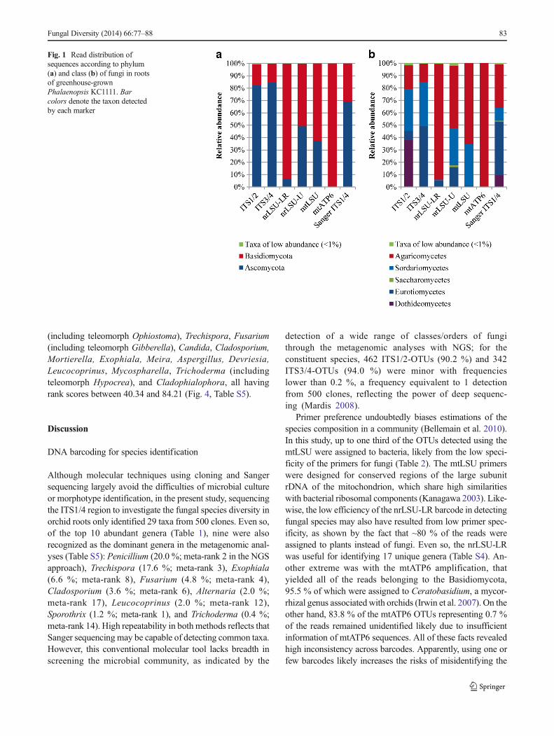
(including teleomorph Ophiostoma), Trechispora, Fusarium(including teleomorph Gibberella), Candida, Cladosporium,Mortierella, Exophiala, Meira, Aspergillus, Devriesia,Leucocoprinus, Mycospharella, Trichoderma (includingteleomorph Hypocrea), and Cladophialophora, all havingrank scores between 40.34 and 84.21 (Fig. 4, Table S5).
Discussion
DNA barcoding for species identification
Although molecular techniques using cloning and Sangersequencing largely avoid the difficulties of microbial cultureor morphotype identification, in the present study, sequencingthe ITS1/4 region to investigate the fungal species diversity inorchid roots only identified 29 taxa from 500 clones. Even so,of the top 10 abundant genera (Table 1), nine were alsorecognized as the dominant genera in the metagenomic anal-yses (Table S5): Penicillium (20.0 %; meta-rank 2 in the NGSapproach), Trechispora (17.6 %; meta-rank 3), Exophiala(6.6 %; meta-rank 8), Fusarium (4.8 %; meta-rank 4),Cladosporium (3.6 %; meta-rank 6), Alternaria (2.0 %;meta-rank 17), Leucocoprinus (2.0 %; meta-rank 12),Sporothrix (1.2 %; meta-rank 1), and Trichoderma (0.4 %;meta-rank 14). High repeatability in both methods reflects thatSanger sequencing may be capable of detecting common taxa.However, this conventional molecular tool lacks breadth inscreening the microbial community, as indicated by the
detection of a wide range of classes/orders of fungithrough the metagenomic analyses with NGS; for theconstituent species, 462 ITS1/2-OTUs (90.2 %) and 342ITS3/4-OTUs (94.0 %) were minor with frequencieslower than 0.2 %, a frequency equivalent to 1 detectionfrom 500 clones, reflecting the power of deep sequenc-ing (Mardis 2008).
Primer preference undoubtedly biases estimations of thespecies composition in a community (Bellemain et al. 2010).In this study, up to one third of the OTUs detected using themtLSU were assigned to bacteria, likely from the low speci-ficity of the primers for fungi (Table 2). The mtLSU primerswere designed for conserved regions of the large subunitrDNA of the mitochondrion, which share high similaritieswith bacterial ribosomal components (Kanagawa 2003). Like-wise, the low efficiency of the nrLSU-LR barcode in detectingfungal species may also have resulted from low primer spec-ificity, as shown by the fact that ~80 % of the reads wereassigned to plants instead of fungi. Even so, the nrLSU-LRwas useful for identifying 17 unique genera (Table S4). An-other extreme was with the mtATP6 amplification, thatyielded all of the reads belonging to the Basidiomycota,95.5 % of which were assigned to Ceratobasidium, a mycor-rhizal genus associated with orchids (Irwin et al. 2007). On theother hand, 83.8 % of the mtATP6 OTUs representing 0.7 %of the reads remained unidentified likely due to insufficientinformation of mtATP6 sequences. All of these facts revealedhigh inconsistency across barcodes. Apparently, using one orfew barcodes likely increases the risks of misidentifying the
Fig. 1 Read distribution ofsequences according to phylum(a) and class (b) of fungi in rootsof greenhouse-grownPhalaenopsis KC1111. Barcolors denote the taxon detectedby each marker
Fungal Diversity (2014) 66:77–88 83
species composition in a microbial community, althoughnrITS is one of the best barcodes for fungal species discrim-ination (Schoch et al. 2012). Using multiple barcodes is there-fore necessary and has been strongly recommended (Nilssonet al. 2008; Gazis et al. 2011). Among the barcodes utilized inthis study, ITS1/2, ITS3/4, and nrLSU-U were the most com-petent in uncovering the diversity of the fungal community inPhalaenopsis roots (Fig. 1), while mitochondrial markers
(mtLSU and mtATP6) yielded a low alpha diversity withrarely detected genera (Tables 3 and 4).
Species composition and ecological roles of constituent fungiwithin orchid roots
Orchid roots represent an ecosystem that fosters a high diver-sity of microbial species. Noticeably, genetic barcodes
Fig. 2 Hierarchical tree representing taxonomic relationships of fungalgenera detected in roots of greenhouse-grown Phalaenopsis. Branchcolors indicate the classes (in boxes) of the OTUs. The height of the bars
in the circle outside the branch tips corresponds to the number of OTUswithin genera. The key to bar color for the markers is at the top right
84 Fungal Diversity (2014) 66:77–88
identified different floristic compositions at the class level(Fig. 2) and different common species from the same rootcommunity (Table S4). For example, for various barcodes, themost common species (with percentage reads) were as fol-lows: ITS1/2, Alternaria sp. (up to 30.4 %); ITS3/4,Penicillium sp. (37.8 %); nrLSU-LR, Trechispora farinacea(48.9 %); nrLSU-U, Trechispora sp. (39.2 %); mtLSU,Serpula sp. (64.7 %). Such inconsistencies across barcodescreate doubts that genetic markers can accurately uncover thespecies composition of an ecological community. Our datashow that different barcode primers tend to have differentannealing kinetics to the target DNA in PCR with multi-template samples. In addition, fungal DNA sequence infor-mation for barcoding in GenBank is incomplete, thus lower-ing the power to identify species (Schloss et al. 2011; Pintoand Raskin 2012). Nonetheless, taxa frequently identifiedacross barcodes were likely to represent dominant elementsin the fungal community.
Since taxa were preferentially detected across differentbarcodes, the species richness cannot be simply estimated byaveraging the read percentages of taxa (e.g., genus) from eachbarcode. For example, as high as 65 % of the reads amplifiedwith mtLSU were assigned to Serpula, which would accountfor the second-most abundant genus by average (13.0 %)across five barcodes, whereas Fusarium, Penicillium, andSporothrix, detected with five barcodes, turned out to be minorconstituents, having average read percentages of 9.0 %, 8.0 %,and 3.3 %, respectively (Table S3). By assigning the OTUsinto ranks based on the relative abundance (Table S5) usingour rank-scoring, we could minimize the calculation bias
encountered with data combination. With this new approach,multiple barcodes are easy to integrate for estimating speciesrichness.
Nine of the ten most abundant genera have been reported asfungi that promote the growth of plants, including Trechispora(meta-rank 3) and Mortierella (meta-rank 7), that are likelyinvolved in mycorrhizal formation (Ochora et al. 2001;Rinaldi et al. 2008) (Fig. 4, Table S2). Although Dearnaleyet al. (2012) did not report any ecological functions for thesefungi, they are potentially useful for horticulture. Given thatthe nature of the interactions between these fungi and orchidsis uncertain, the role of Trechispora farinacea, a dominantspecies in the root community (Table S4), needs to be furtherexamined using inoculation experiments. Cultural conditionsshould be optimized specifically for these symbiotic fungi.
Of the 21 other mycorrhizal genera identified in the presentstudy (Tables 1, S2), Tulasnella (anamorphic Epulorhiza) andCeratobasidium are common symbionts with orchids (Suárezet al. 2006; Irwin et al. 2007; Otero et al. 2007; Dearnaleyet al. 2012; Graham and Dearnaley 2012). Tulasnella is in-volved in the symbiotic germination of Chiloglottis aff.jeanesii and C. valida (Roche et al. 2010), whereas an isolateof Ceratobasidium is potentially useful for the biocontrol ofErwinia chrysanthemi, the bacterium causing soft rot inPhalaenopsis (Wu et al. 2011). Thus, Tulasnella andCeratobasidium spp. are likely to be important mycorrhizalspecies coexisting with Phalaenopsis. Although more than80 % of the land plant families are thought to have symbioticrelationships with arbuscular mycorrhizal fungi (AMF,Glomeromycota) (Kawaguchi and Minamisawa 2010),Acaulospora was the only AMF genus that we detected.Failing to detect other AMF may be ascribed to the short readlength with Illumina sequencing (cf. Stockinger et al. 2010).
Moreover, Penicillium species (meta-rank 1 here) are com-mon endophytes in plants (Vega et al. 2006), and some speciescan improve phosphate solubility or produce gibberellic acidto stimulate plant growth (Wakelin et al. 2007; Khan et al.2008). Fungi may also function as biocontrol agents (e.g.,Meira and Candida; Nguyen et al. 2011) or nematode preda-tors (e.g., Dactyllela and Arthrobotrys; Schenck et al. 1977).Nematodes, common invertebrates in orchids, often cause leafyellowing and reduce plant vigor (Kuehnle 2006). Suchnematophagous fungi may thus play a critical role in control-ling nematode infection in orchids. Using symbiotic fungi forcontrolling disease outbreak or improving the resistance topathogens has been demonstrated for orchids and crops (cf.Lee et al. 2009; Wu et al. 2011; Mosquera-Espinosa et al.
Fig. 3 OTU accumulation curves of multiple rarefactions with sixmarkers sequenced with Illumina GAIIx
Table 4 Indices of alpha diversi-ty across markers Diversity index ITS1/2 ITS3/4 nrLSU-LR nrLSU-U mtLSU mtATP6
Shannon 2.49 2.02 1.47 1.83 1.09 0.18
Gini-Simpson 0.85 0.78 0.68 0.76 0.55 0.07
Fungal Diversity (2014) 66:77–88 85
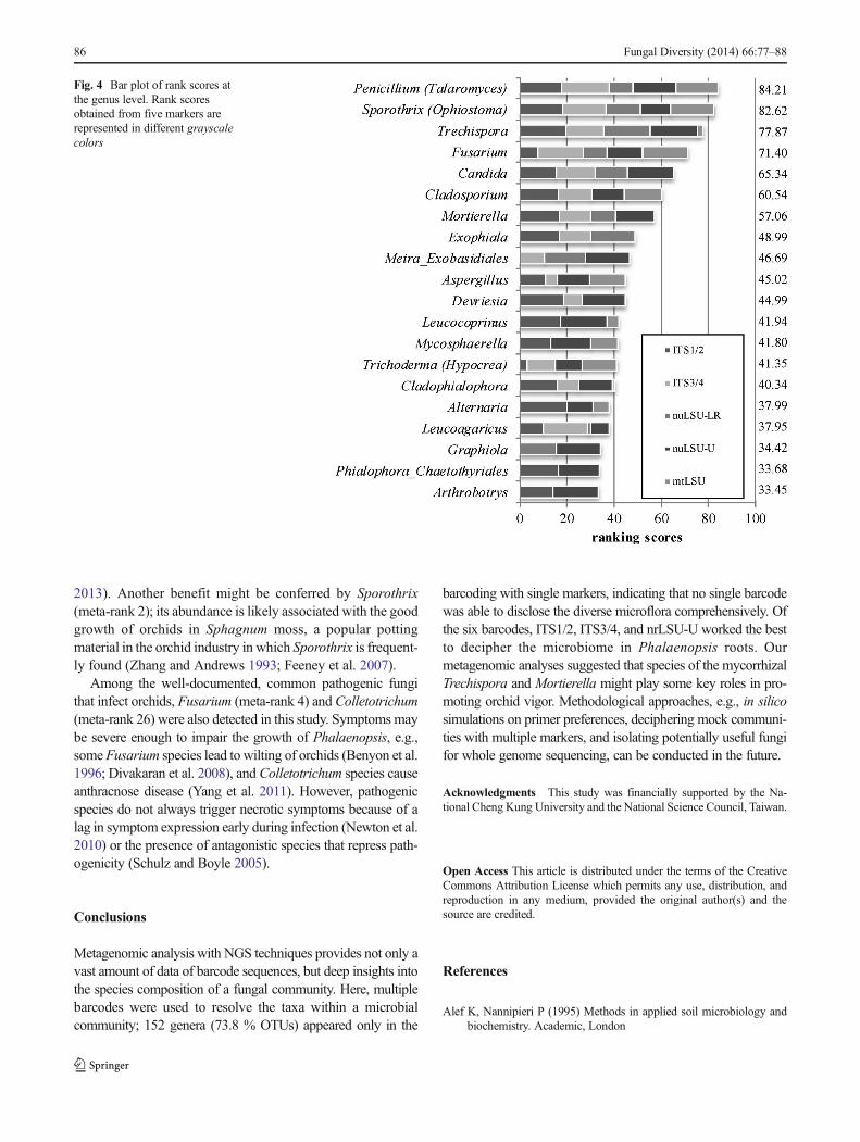
2013). Another benefit might be conferred by Sporothrix(meta-rank 2); its abundance is likely associated with the goodgrowth of orchids in Sphagnum moss, a popular pottingmaterial in the orchid industry in which Sporothrix is frequent-ly found (Zhang and Andrews 1993; Feeney et al. 2007).
Among the well-documented, common pathogenic fungithat infect orchids, Fusarium (meta-rank 4) and Colletotrichum(meta-rank 26) were also detected in this study. Symptoms maybe severe enough to impair the growth of Phalaenopsis, e.g.,some Fusarium species lead to wilting of orchids (Benyon et al.1996; Divakaran et al. 2008), and Colletotrichum species causeanthracnose disease (Yang et al. 2011). However, pathogenicspecies do not always trigger necrotic symptoms because of alag in symptom expression early during infection (Newton et al.2010) or the presence of antagonistic species that repress path-ogenicity (Schulz and Boyle 2005).
Conclusions
Metagenomic analysis with NGS techniques provides not only avast amount of data of barcode sequences, but deep insights intothe species composition of a fungal community. Here, multiplebarcodes were used to resolve the taxa within a microbialcommunity; 152 genera (73.8 % OTUs) appeared only in the
barcoding with single markers, indicating that no single barcodewas able to disclose the diverse microflora comprehensively. Ofthe six barcodes, ITS1/2, ITS3/4, and nrLSU-U worked the bestto decipher the microbiome in Phalaenopsis roots. Ourmetagenomic analyses suggested that species of the mycorrhizalTrechispora and Mortierella might play some key roles in pro-moting orchid vigor. Methodological approaches, e.g., in silicosimulations on primer preferences, deciphering mock communi-ties with multiple markers, and isolating potentially useful fungifor whole genome sequencing, can be conducted in the future.
Acknowledgments This study was financially supported by the Na-tional Cheng Kung University and the National Science Council, Taiwan.
Open Access This article is distributed under the terms of the CreativeCommons Attribution License which permits any use, distribution, andreproduction in any medium, provided the original author(s) and thesource are credited.
References
Alef K, Nannipieri P (1995) Methods in applied soil microbiology andbiochemistry. Academic, London
Fig. 4 Bar plot of rank scores atthe genus level. Rank scoresobtained from five markers arerepresented in different grayscalecolors
86 Fungal Diversity (2014) 66:77–88

Arditti J (1992) Fundamentals of orchid biology. Wiley, New YorkBeckman CH (1987) The nature of wilt diseases of plants. APS Press,
CaliforniaBellemain E, Carlsen T, Brochmann C, Coissac E, Taberlet P, KauserudH
(2010) ITS as an environmental DNA barcode for fungi: an in silicoapproach reveals potential PCR biases. BMC Microbiol 10:189
Benyon F, Summerell B, Burgess L (1996) Association of Fusariumspecies with root rot of Cymbidium orchids. Australas Plant Pathol25:226–228
Berendsen RL, Pieterse CMJ, Bakker PAHM (2012) The rhizospheremicrobiome and plant health. Trends Plant Sci 17:478–486
Bisseling T, Dangl JL, Schulze-Lefert P (2009) Next-generation commu-nication. Science 324:691
Burgeff H (1959) Mycorrhiza of orchids. In: Withner C (ed) The orchids.Ronald, New York, pp 361–395
Cating R, Palmateer A, McMillan R Jr (2009) First report of Sclerotiumrolfsii on Ascocentrum and Ascocenda orchids in Florida. Plant Dis93:963
Cowan D, Meyer Q, Stafford W, Muyanga S, Cameron R, Wittwer P(2005) Metagenomic gene discovery: past, present and future.Trends Biotechnol 23:321–329
Dearnaley J, Martos F, Selosse M-A (2012) Orchid mycorrhizas: molec-ular ecology, physiology, evolution and conservation aspects. In:Hock B (ed) Fungal associations. Springer, Berlin, pp 207–230
DeSalle R, Graham SW, Fazekas AJ, Burgess KS, KesanakurtiPR, Newmaster SG, Husband BC, Percy DM, Hajibabaei M,Barrett SCH (2008) Multiple multilocus DNA barcodes fromthe plastid genome discriminate plant species equally well.PLoS ONE 3:e2802
Divakaran M, Geetha S, Nirmal Babu K, Peter K (2008) Isolation andfusion of protoplasts in Vanilla species. Curr Sci 94:115–120
Doyle J, Doyle J (1987) Genomic plant DNA preparation from freshtissue-CTAB method. Phytochem Bull 19:11–15
Druzhinina IS, Kopchinskiy AG, Komoń M, Bissett J, Szakacs G,Kubicek CP (2005) An oligonucleotide barcode for species identifi-cation in Trichoderma andHypocrea. Fungal Genet Biol 42:813–828
Feeney KT, Arthur IH, Whittle AJ, Altman SA, Speers DJ (2007)Outbreak of sporotrichosis, Western Australia. Emerg Infect Dis13:1228
Gazis R, Rehner S, Chaverri P (2011) Species delimitation in fungalendophyte diversity studies and its implications in ecological andbiogeographic inferences. Mol Ecol 20:3001–3013
Graham RR, Dearnaley JDW (2012) The rare Australian epiphytic orchidSarcochilus weinthalii associates with a single species ofCeratobasidium. Fungal Divers 54:31–37
Grubisha LC, Levsen N, OlsonMS, Lee Taylor D (2012) Intercontinentaldivergence in the Populus-associated ectomycorrhizal fungus,Tricholoma populinum. New Phytol 194:548–560
Haegeman B, Hamelin J, Moriarty J, Neal P, Dushoff J, Weitz JS (2013)Robust estimation of microbial diversity in theory and in practice.ISME J 7:1092–1101
Hebert PD, Gregory TR (2005) The promise of DNA barcoding fortaxonomy. Syst Biol 54:852–859
Hinsinger P, Bengough AG, Vetterlein D, Young IM (2009) Rhizosphere:biophysics, biogeochemistry and ecological relevance. Plant Soil321:117–152
Hirsch PR, Mauchline TH, Clark IM (2010) Culture-independent molec-ular techniques for soil microbial ecology. Soil Biol Biochem 42:878–887
Huson DH, Mitra S, Ruscheweyh H-J, Weber N, Schuster SC (2011)Integrative analysis of environmental sequences using MEGAN4.Genome Res 21:1552–1560
Irwin MJ, Bougoure JJ, Dearnaley JDW (2007) Pterostylis nutans(Orchidaceae) has a specific association with two Ceratobasidiumroot-associated fungi across its range in eastern Australia.Mycoscience 48:231–239
Kaeberlein T (2002) Isolating “uncultivable” microorganisms in pureculture in a simulated natural environment. Science 296:1127–1129
Kanagawa T (2003) Bias and artifacts in multitemplate polymerase chainreactions (PCR). J Biosci Bioeng 96:317–323
Kawaguchi M, Minamisawa K (2010) Plant–microbe communicationsfor symbiosis. Plant Cell Physiol 51:1377–1380
Khan S, HamayunM, YoonH, KimH-Y, Suh S-J, Hwang S-K, Kim J-M,Lee I-J, Choo Y-S, Yoon U-H, Kong W-S, Lee B-M, Kim J-G(2008) Plant growth promotion and Penicillium citrinum. BMCMicrobiol 8:231
Kõljalg U, Larsson KH, Abarenkov K, Nilsson RH, Alexander IJ,Eberhardt U, Erland S, Høiland K, Kjøller R, Larsson E (2005)UNITE: a database providing web‐based methods for the molecularidentification of ectomycorrhizal fungi. New Phytol 166:1063–1068
Konow EA, Wang Y-T (2001) Irradiance levels affect in vitro andgreenhouse growth, flowering, and photosynthetic behavior of ahybrid Phalaenopsis orchid. J Am Soc Hortic Sci 126:531–536
Kuehnle AR (2006) Orchids. In: Anderson NO (ed) Flower breeding andgenetics. Springer, Dordrecht, pp 539–560
Lee SO, Kim HY, Choi GJ, Lee HB, Jang KS, Choi YH, Kim JC (2009)Mycofumigation withOxyporus latemarginatusEF069 for control ofpostharvest apple decay and Rhizoctonia root rot on moth orchid. JAppl Microbiol 106:1213–1219
Letunic I, Bork P (2011) Interactive tree of life v2: online annotation anddisplay of phylogenetic trees made easy. Nucleic Acids Res 39:W475–W478
Mardis ER (2008) The impact of next-generation sequencing technologyon genetics. Trends Genet 24:133–141
Mendes R, Kruijt M, de Bruijn I, Dekkers E, van der Voort M, SchneiderJH, Piceno YM, DeSantis TZ, Andersen GL, Bakker PA (2011)Deciphering the rhizosphere microbiome for disease-suppressivebacteria. Science 332:1097–1100
Montaña JS, Jiménez DJ, Hernández M, Ángel T, Baena S (2012)Taxonomic and functional assignment of cloned sequences fromhigh Andean forest soil metagenome. Antonie Van Leeuwenhoek101:205–215
Mosquera-Espinosa AT, Bayman P, Prado GA, Gómez-Carabalí A, OteroJT (2013) The double life of Ceratobasidium: orchid mycorrhizalfungi and their potential for biocontrol of Rhizoctonia solani sheathblight of rice. Mycologia 105:141–150
Murray DC, Bunce M, Cannell BL, Oliver R, Houston J, White NE,Barrero RA, Bellgard MI, Haile J (2011) DNA-based faecal dietaryanalysis: a comparison of qPCR and high throughput sequencingapproaches. PLoS ONE 6:e25776
Newton AC, Fitt BDL, Atkins SD, Walters DR, Daniell TJ (2010)Pathogenesis, parasitism and mutualism in the trophic space ofmicrobe–plant interactions. Trends Microbiol 18:365–373
Nguyen MT, Ranamukhaarachchi DB, Senaratne L (2011) Efficacy ofantagonist strains of Bacillus megaterium, Enterobacter cloacae,Pichia guilliermondii and Candida ethanolica against bacterial wiltdisease of tomato. J Phytol 3:01–10
Nilsson RH, Kristiansson E, Ryberg M, Hallenberg N, Larsson KH(2008) Intraspecific ITS variability in the kingdom fungi asexpressed in the international sequence databases and its implica-tions for molecular species identification. Evol BioinformaticsOnline 4:193–201
Ochora J, Stock W, Linder H, Newton L (2001) Symbiotic seed germi-nation in twelve Kenyan orchid species. Syst Geogr Plants 71:585–596
Otero JT, Flanagan NS, Herre EA, Ackerman JD, Bayman P (2007)Widespread mycorrhizal specificity correlates to mycorrhizal func-tion in the neotropical, epiphytic orchid Ionopsis utricularioides(Orchidaceae). Am J Bot 94:1944–1950
Pinto AJ, Raskin L (2012) PCR biases distort bacterial and archaealcommunity structure in pyrosequencing datasets. PLoS ONE 7:e43093
Fungal Diversity (2014) 66:77–88 87

Pridgeon A, Cribb P, Chase M, Rasmussen F (2005) Generaorchidacearum: epidendroideae (Part one). Oxford UniversityPress, Oxford
Rao CR (1982) Gini-Simpson index of diversity: a characterization,generalization and applications. Util Math 21:273–282
Rasmussen HN (1995) Terrestrial orchids: from seed to mycotrophicplant. Cambridge University Press, New York
Rinaldi A, Comandini O, Kuyper TW (2008) Ectomycorrhizal fungaldiversity: seperating thewheat from the chaff. Fungal Divers 33:1–45
Roche SA, Carter RJ, Peakall R, Smith LM, Whitehead MR, Linde CC(2010) A narrow group ofmonophyletic Tulasnella (Tulasnellaceae)symbiont lineages are associated with multiple species ofChiloglottis (Orchidaceae): implications for orchid diversity. Am JBot 97:1313–1327
Rosselló-Mora R, Amann R (2001) The species concept for prokaryotes.FEMS Microbiol Rev 25:39–67
Runkle E, Wang Y, Blanchard M, Lopez R (2007) Growing the bestPhalaenopsis, part 1: an introduction to potted Phalaenopsis or-chids. Orchids 76:24–28
Schenck S, Kendrick W, Pramer D (1977) A new nematode-trappinghyphomycete and a reevaluation of Dactylaria and Arthrobotrys.Can J Bot 55:977–985
Schloss PD, Gevers D, Westcott SL (2011) Reducing the effects of PCRamplification and sequencing artifacts on 16S rRNA-based studies.PLoS ONE 6:e27310
Schoch CL, Seifert KA, Huhndorf S, Robert V, Spouge JL, Levesque CA,Chen W, Bolchacova E, Voigt K, Crous PW, Miller AN, WingfieldMJ, Aime MC, An KD, Bai FY, Barreto RW, Begerow D, BergeronMJ, Blackwell M, Boekhout T, Bogale M, Boonyuen N, BurgazAR, Buyck B, Cai L, Cai Q, Cardinali G, Chaverri P, Coppins BJ,Crespo A, Cubas P, Cummings C, Damm U, de Beer ZW, de HoogGS, Del-Prado R, Dentinger B, Dieguez-Uribeondo J, Divakar PK,Douglas B, Duenas M, Duong TA, Eberhardt U, Edwards JE,Elshahed MS, Fliegerova K, Furtado M, Garcia MA, Ge ZW,Griffith GW, Griffiths K, Groenewald JZ, Groenewald M, GrubeM, Gryzenhout M, Guo LD, Hagen F, Hambleton S, Hamelin RC,Hansen K, Harrold P, Heller G, Herrera C, Hirayama K, Hirooka Y,Ho HM, Hoffmann K, Hofstetter V, Hognabba F, HollingsworthPM, Hong SB, Hosaka K, Houbraken J, Hughes K, Huhtinen S,Hyde KD, James T, Johnson EM, Johnson JE, Johnston PR, JonesEBG, Kelly LJ, Kirk PM, Knapp DG, Koljalg U, Kovacs GM,Kurtzman CP, Landvik S, Leavitt SD, Liggenstoffer AS,Liimatainen K, Lombard L, Luangsa-ard JJ, Lumbsch HT,Maganti H, Maharachchikumbura SSN, Martin MP, May TW,McTaggart AR, Methven AS, Meyer W, Moncalvo JM,Mongkolsamrit S, Nagy LG, Nilsson RH, Niskanen T, Nyilasi I,Okada G, Okane I, Olariaga I, Otte J, Papp T, Park D, Petkovits T,Pino-Bodas R, Quaedvlieg W, Raja HA, Redecker D, Rintoul TL,Ruibal C, Sarmiento-Ramirez JM, Schmitt I, Schussler A, ShearerC, Sotome K, Stefani FOP, Stenroos S, Stielow B, Stockinger H,Suetrong S, Suh SO, Sung GH, Suzuki M, Tanaka K, Tedersoo L,Telleria MT, Tretter E, Untereiner WA, Urbina H, Vagvolgyi C,
Vialle A, Vu TD, Walther G, Wang QM, Wang Y, Weir BS, WeissM, White MM, Xu J, Yahr R, Yang ZL, Yurkov A, Zamora JC,Zhang N, Zhuang WY, Schindel D (2012) From the cover: nuclearribosomal internal transcribed spacer (ITS) region as a universalDNA barcodemarker for Fungi. Proc Natl Acad Sci 109:6241–6246
Schulz B, Boyle C (2005) The endophytic continuum. Mycol Res 109:661–686
Seena S, Pascoal C, Marvanová L, Cássio F (2010) DNA barcoding offungi: a case study using ITS sequences for identifying aquatichyphomycete species. Fungal Divers 44:77–87
Shannon C (1948) A mathematical theory of communication. AT&TTech J 27:623–656
Smith SE, Read DJ (2008) Mycorrhizal symbiosis, 3rd edn. Academic,Amsterdam
Stockinger H, Krüger M, Schüßler A (2010) DNA barcoding ofarbuscular mycorrhizal fungi. New Phytol 187:461–474
Suárez JP, Weiß M, Abele A, Garnica S, Oberwinkler F, Kottke I (2006)Diverse tulasnelloid fungi form mycorrhizas with epiphytic orchidsin an Andean cloud forest. Mycol Res 110:1257–1270
Tringe SG, Hugenholtz P (2008) A renaissance for the pioneering 16SrRNA gene. Curr Opin Microbiol 11:442–446
Vega FE, Posada F, Peterson SW, Gianfagna TJ, Chaves F (2006)Penicillium species endophytic in coffee plants and ochratoxin Aproduction. Mycologia 98:31–42
Vilgalys R, Hester M (1990) Rapid genetic identification and mapping ofenzymatically amplified ribosomal DNA from severalCryptococcusspecies. J Bacteriol 172:4238–4246
Wakelin S, Gupta VV, Harvey P, Ryder M (2007) The effect ofPenicillium fungi on plant growth and phosphorus mobilization inneutral to alkaline soils from southern Australia. Can JMicrobiol 53:106–115
WangY-T (2004) Flourishingmarket for potted orchids. FlowerTech 7:2–5
Wey G (1988) Occurrence and investigation of important diseases onPhalaenopsis in Taiwan. Rep Taiwan Sugar Res Inst 122:31–41
Wu Z, Wang X-R, Blomquist G (2002) Evaluation of PCR primers andPCR conditions for specific detection of common airborne fungi. JEnviron Monitor 4:377–382
Wu P-H, Huang D-D, Chang DCN (2011) Mycorrhizal symbiosis en-hances Phalaenopsis orchid’s growth and resistence to Erwiniachrysanthemi. Afr J Biotechnol 10:10095–10100
Yang Y, Cai L, Yu Z, Liu Z, Hyde KD (2011) Colletotrichum species onOrchidaceae in southwest China. Cryptogam Mycol 32:229–253
Zelmer CD, Cuthbertson L, Currah RS (1996) Fungi associated withterrestrial orchid mycorrhizas, seeds and protocorms. Mycoscience37:439–448
Zeng QY, Rasmuson-Lestander Å, Wang XR (2004) Extensive set ofmitochondrial LSU rDNA‐based oligonucleotide probes for thedetection of common airborne fungi. FEMSMicrob Lett 237:79–87
Zhang X, Andrews JH (1993) Evidence for growth of Sporothrixschenckii on dead but not on living Sphagnum moss.Mycopathologia 123:87–94
88 Fungal Diversity (2014) 66:77–88