Deciphering the chromatin landscape induced around DNA double strand breaks

Q1
123456789
101112131415161718192021222324252627282930313233343536373839404142434445464748495051525354555657585960616263646566
Neuropsychologia ] (]]]]) ]]]–]]]
Contents lists available at SciVerse ScienceDirect
Neuropsychologia
0028-39
http://d
n Corrnn Cor
E-m
m.jahan
Pleasvaria
journal homepage: www.elsevier.com/locate/neuropsychologia
Deciphering the impact of cerebellar and basal ganglia dysfunctionin accuracy and variability of motor timing
Daniel O. Claassen a,n, Catherine R.G. Jones b, Minhong Yu c, Georg Dirnberger d, Tim Malone e,Michael Parkinson d, Paola Giunti d, Michael Kubovy c, Marjan Jahanshahi d,nn
a Department of Neurology, Vanderbilt University, Nashville TN, USAb Department of Psychology, University of Essex, Essex, C04 3SQ, UKc Department of Psychology, University of Virginia, Charlottesville, VA, USAd Sobell Department of Motor Neuroscience and Movement Disorders, University College London Institute of Neurology, London, WC1N 3BG, UKe Royal Devon & Exeter Hospital, Devon, EX1 2ED, UK
a r t i c l e i n f o
Keywords:
Motor timing
Cerebellum
Basal Ganglia
Temporal processing
Synchronization
Continuation
32/$ - see front matter & 2012 Elsevier Ltd. A
x.doi.org/10.1016/j.neuropsychologia.2012.09
esponding author. Tel.: þ1 615 936 1007; fax
responding author.
ail addresses: [email protected]
[email protected] (M. Jahanshahi).
e cite this article as: Claassen, D. Obility of motor timing. Neuropsychol
a b s t r a c t
Studies in motor timing have shown that the basal ganglia and cerebellum play an important role in
temporal processing. Timing studies in Cerebellar/ataxic Disorders (CD) and Parkinson’s disease (PD)
patients contrast the roles of the cerebellum and basal ganglia in motor timing. Here, we used a
synchronization–continuation task to compare accuracy and variability of motor timing during
repetitive tapping. We compared data collected for the present study – from patients with CD and
healthy controls – to data from a previous study with patients with PD. We asked participants to tap at
Inter-stimulus Intervals (ISIs) of 250, 500, 1000, and 2000 ms. Using Linear Mixed Models (LMMs), we
explored how ISI, Task Phase, and Diagnosis interacted to determine the (i) the accuracy and (ii) the
variability of tapping. In our analysis of accuracy, we found evidence that during the synchronization
phase, at ISI¼250 ms, CD patients lagged ‘behind the beat’; whereas our previous work has suggested
that medicated PD patients hasten ‘ahead of the beat’. In our analysis of variability, we observed that at
ISIs below 1000 ms, CD patients showed greater variability in motor timing than the healthy controls,
while PD patients showed less variability than CD patients and healthy controls during the
synchronization phase at the 1000 ms ISI. These results highlight the differential performance on
explicit motor timing between patients with disorders of the cerebellum and basal ganglia. Our results
illustrate a novel approach to discerning cognitive control of motor timing.
& 2012 Elsevier Ltd. All rights reserved.
67686970717273747576777879808182
1. Introduction
An effective tool to characterize motor timing is a repetitivemotor task known as the synchronization–continuation task (SCT,also known as the paced finger-tapping task). In the first phase ofthe task – the synchronization phase – participants try to synchro-nize their taps with a train of tones separated by constant inter-stimulus interval (ISI). In the second phase – the continuation
phase – the auditory stimulus is no longer presented, andparticipants try to tap at the previous rate. This task requires‘‘explicit motor timing’’: participants must learn the ISI andreproduce it (Coull & Nobre, 2008).
Researchers have been interested in both accuracy and thevariability of the motor response in the two phases of the SCT.Accuracy reflects how ‘on target’ the taps are. It can be quantifiedby reporting the mean inter-tap interval, ITI, or the mean absolute
8384858687
ll rights reserved.
.018
: þ1 615 343 3946.
(D.O. Claassen),
., et al. Deciphering the impogia (2012), http://dx.doi.o
error, mean (9ITI–ISI9) (O’Boyle, Freeman, & Cody, 1996; Pastor,Jahanshahi, Artieda, & Obeso, 1992; Pope, Praamstra, & Wing,2006). Variability reflects the spread of ‘off target’ responses.It can be quantified using the autocovariance function of the ITI(Ivry & Hazeltine, 1995). When applied to the continuation phase,this measure has been thought to reflect the variability of an‘‘internal clock’’ (Wing & Kristofferson, 1973).
The basal ganglia and cerebellum are essential for temporalprocessing (Coull, Cheng, & Meck, 2011; Ivry & Schlerf, 2008; Jones& Jahanshahi, 2009). To understand the differential roles of thecerebellum and the basal ganglia on motor timing, we propose tocompare the clinical phenotypes of ataxic (or cerebellar) disordersand of Parkinson Disease (PD). Patients with cerebellar dysfunctionpresent with various forms of impaired timing: dysarthria, dysme-tria, dysdiadochokinesia (difficulty performing rapid alternatingmovements), and gait instability (Holmes, 1917). Laboratory studieshave suggested that cerebellar dysfunction compromises (a) thetiming of discrete motor responses (Fierro et al., 2007; Ivry, Keele, &Diener, 1988; Ivry & Richardson, 2002), (b) time perception (Fierroet al., 2007; Ivryet al., 1988; Nichelli, Alway, & Grafman, 1996), and(c) a putative ‘‘internal’’ clock (Ivry & Keele, 1989).
8889
act of cerebellar and basal ganglia dysfunction in accuracy andrg/10.1016/j.neuropsychologia.2012.09.018
123456789
101112131415161718192021222324252627282930313233343536373839404142434445464748495051525354555657585960616263646566
676869707172737475767778798081828384858687888990919293949596979899
100101102103104105106107108109110111112113114115116117118119120121122123124
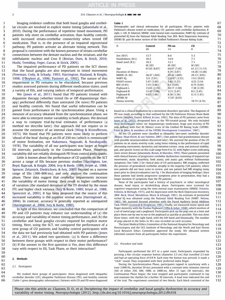
Table 1Demographic and clinical information for all participants. PD-on: patients with
Parkinson’s disease tested on medication, CD: patients with cerebellar dysfunction, R:
right, L: left, B: bilateral; MMSE: mini mental state examination; NART-IQ: estimate of
premorbid IQ from the National Adult Reading Test, BDI: Beck Depression Inventory;
UPDRS III: part III motor section of the Unified Parkinson’s Disease Rating Scale.
N Control PD-on CD20 14 8
Sex (M:F) 13:7 8:6 5:3
Handedness (R:L) 18:2 14:0 7:1
Hand used (R:L) 18:2 13:1 7:1
M (SD) M (SD) M (SD)
Age 67.65 (8.87) 64.07 (7.45) 61.13 (11.15)
Duration of illness – 8.79 (3.87) 14.38 (13.48)
MMSE (0–30) 28.47 (.943) 29.42 (.669) 29.13 (.835)
NART-IQ 121 (5.91) 122.67 (3.53) 114 (10.93)
PASAT-errors 5.47 (5.69) 5.42 (5.23) 4.25 (3.54
BDI (0-63) 7.53 (5.86) 9.5 (4.95) 8.75 (9.05)
Pegboard-L 13.65 (2.76) 10.17 (1.90) 7.38 (2.39)
Pegboard-R 13.47 (3.06) 12.5 (2.47) 8.5 (2.45)
Pegboard-B 9.65 (1.06) 8.58 (1.56) 6.13 (1.81)
UPDRS III – 17.07 (8.93) –
Ataxia severity – – 10.75 (4.74)
D.O. Claassen et al. / Neuropsychologia ] (]]]]) ]]]–]]]2
Imaging evidence confirms that both basal ganglia and cerebel-lar circuits are involved in explicit motor timing (Jahanshahi et al.,2010). During the performance of repetitive timed movement, PDpatients rely more on cerebellar activation, than healthy controls,with increased striato-cerebellar connectivity when tested Offmedication. That is, in the presence of an impaired nigrostriatalpathway, PD patients activate an alternate timing network. Thisproposal is consistent with the known presence of striato-cerebellarconnections between the dentate nucleus and the striatum, and thesubthalamic nucleus and Crus II (Bostan, Dum, & Strick, 2010;Hoshi, Tremblay, Feger, Carras, & Strick, 2005).
The impaired performance of PD patients on the SCT showsthat the basal ganglia are also important in timed movement(Freeman, Cody, & Schady, 1993; Harrington, Haaland, & Knight,1998; O’Boyleet al., 1996; Pastoret al., 1992). The nature of thisimpairment in PD remains to be elucidated, because previousstudies assessed patients during different medication states, useda variety of ISIs, and varying indices of temporal performance.
We (Jones et al., 2011) found that PD patients treated withdopamine therapy (whether tested On or Off dopaminergic ther-apy) performed differently than untreated (De novo) PD patientsand healthy controls. We found that useful information can begained from performance in the synchronization phase. Using ameasure of accuracy obtained from the synchronization phase, wewere able to interpret motor variability in both phases. We deviseda way to compute trial-by-trial estimates of performance (amethod we describe later). Our approach did not require us toassume the existence of an internal clock (Wing & Kristofferson,1973). We found that PD patients were more likely to perform‘ahead of the beat’ during at an ISI of 250 ms (which is reminiscentof motor festination, see Nakamura, Nagasaki, & Narabayashi,1978). The variability of all our participants was larger at longerISI intervals, particularly in the Continuation Phase. However,variability did not reliably distinguish PD from control participants.
Little is known about the performance of CD patients on the SCTacross a range of ISIs because previous studies (Harrington, Lee,Boyd, Rapcsak, & Knight, 2004; Ivry & Keele, 1989; Ivryet al., 1988;Spencer, Zelaznick, Diedrichsen, & Ivry, 2003) focused on a narrowrange of ISIs (300–800 ms), and only analyze the continuationphase. These data suggest that cerebellar impairments increasethe variability of motor timing: they result in higher coefficientsof variation (the standard deviation of the ITI divided by the meanITI) and higher clock variance (Ivry & Keele, 1989; Ivryet al., 1988;Spenceret al., 2003). It has been proposed that the source of thisincreased variability is the superior vermal area (Harringtonet al.,2004). In contrast, accuracy is generally reported as unimpaired(Harringtonet al., 2004; Ivry & Keele, 1989).
In light of this literature, we concluded that the comparison ofPD and CD patients may enhance our understanding of (a) theaccuracy and variability of motor timing performance, and (b) thecerebellar and basal ganglia circuits required for explicit motortiming. Here, using the SCT, we compared the performance of anew group of CD patients and healthy control participants withthe data we had previously had obtained with PD patients (Joneset al., 2011). We asked two questions: (a) Is there a differencebetween these groups with respect to their motor performance?(b) If the answer to the first question is Yes, does this differencevary with respect to ISI, Task Phase, or their interaction?
125126127128129130131132
2. Methods
2.1. Participants
We studied three groups of participants: those diagnosed with idiopathic
cerebellar disorder (CD), idiopathic Parkinson Disease (PD), and healthy controls
(Table 1 gives demographic information). Patients with CD and PD were diagnosed
Please cite this article as: Claassen, D. O., et al. Deciphering the impvariability of motor timing. Neuropsychologia (2012), http://dx.doi.o
based on a clinical evaluation by a movement disorders specialist. The diagnosis of
PD was made according to that outlined by the UK Parkinson Disease Brain Bank
criteria (Hughes, Daniel, Kilford, & Lees, 1992). The data of PD patients came from
Jones et al. (2011), designated here as the ‘PD-treated group’. We only included
their performance when ‘on’ dopaminergic medication. Stage and severity of PD
was defined with Part III (Motor) score of the Unified PD Rating Scale (UPDRS:
Fahn, & Elton, & members of the UPDRS Development Committee, 1987).
All our CD patients were classified as idiopathic late-onset cerebellar disorder
(Brusse, Maat-Kievit, & van Swieten, 2007). (One patient had granulomatous disease
with possible neurosarcoidosis discovered later). We assessed the position of the CD
patients on an ataxia severity scale, using items relating to the performance of rapid
alternating movements, dysmetria and intention tremor, sway and postural stability,
gait, and speech. Scores on this scale range from 0 to 32, with higher scores indicating
more severe disease (Jahanshahi, Brown, & Marsden, 1993). These patients presented
with clinical features of progressive cerebellar dysfunction consisting of impaired eye
movements, ataxic dysarthria, limb ataxia, and ataxic gait, without Parkinsonian
symptoms (See Table 2 for clinical data of CD participants). MR imaging confirmed
the presence of generalized cerebellar atrophy in the majority (5/8 patients), where
two patients did not have scans to review, and one patient had MR imaging many
years prior to clinical evaluation (see Fig. 1 for illustrations of imaging findings). Since
these patients had slowly progressive symptoms prior to presentation, they had a
longer duration of symptoms than the PD patients.
None of the participants had a history of psychiatric or other neurological
disease, head injury or alcohol/drug abuse. Participants were screened for
cognitive impairment using the mini-mental state examination (MMSE: Folstein,
Folstein, & McHugh, 1975), and for depression with the Beck Depression Inventory
(BDI: Beck, Ward, Mendelson, Mock, & Erbaugh, 1961). An estimate of premorbid
verbal IQ was obtained using the National Adult Reading Test (NART: Nelson,
1982). We assessed focused attention with the Paced Auditory Serial Addition
Task, PASAT (Gronwall & Wrightson, 1981). Finally, we measured motor speed and
finger dexterity with the Purdue Pegboard (Tiffin & Asher, 1948), which involves of
a set of metal pegs and a pegboard. Participants pick up the pegs one at a time and
place them one by one in one in the pegboard as quickly as possible. This was done
three times: with the right hand, with the left hand and bimanually. The number
of pegs placed in the holes in 30 s was recorded on each occasion.
The Joint Medical Ethics Committee of the National Hospital for Neurology and
Neurosurgery and the UCL Institute of Neurology and the North and East Devon
Local Research Ethics Committee approved the study. We obtained written
informed consent from all participants before the experiment.
2.2. Procedure and tasks
Participants performed the SCT in a quiet room. Participants responded by
pressing a flat circular response button (diameter 2.5 cm) that travelled 2.5 mm
and had an operating force of 0.8 N. Each time the button was pressed, it made a
‘‘click’’ sound. They responded with their preferred index finger.
During the Synchronization Phase, participants tapped in synchrony with a
tone (1000 Hz, duration 50 ms) presented with a constant inter-stimulus interval
(ISI) of either 250, 500, 1000, or 2000 ms. After 31 taps (30 intervals), the
Continuation Phase began: the tone stopped and participants continued to tap
and maintain the rhythm for a further 30 intervals. A loud tone indicated the end
of the trial. The experiment consisted of two blocks. Each block consisted of the
act of cerebellar and basal ganglia dysfunction in accuracy andrg/10.1016/j.neuropsychologia.2012.09.018

123456789
101112131415161718192021222324252627282930313233343536373839404142434445464748495051525354555657585960616263646566
676869707172737475767778798081828384858687888990919293949596979899
100101102103104105106107108109110111112113114115116117118119120121122123124125
●
●
●●
●
●
●
●
●
●
●
●
●
●●
●
●
●
●
●●
●
●
●
250 500
1000 2000
−0.05
0.00
0.05
0.10
−0.05
0.00
0.05
0.10
CD Ctrl PD−on CD Ctrl PD−on
Group
Rel
ativ
e E
rror Task Phase
●
●
Synch
Cont
Fig. 1. Selected 1.5T magnetic resonance imaging showing superior cerebellar atrophy in Participant 1 (A) T1-weight sagittal section (B) T2-weighted axial section; patient
4 (C) T2-weighted axial section; and Participant 7 (D) T2-weighted axial section.
Table 2Clinical data on cerebellar disorder patients.
Participant Sex Diagnosis Age
at
exam
Age
at
onset
Disease
duration
Examination findings Cerebellar
atrophy on
imaging
Ataxia
Rating
Scale
Ataxic eye
movement
abnormalities
Limb
dysmetria
Ataxic
dysarthria
Imbalance Cerebellar
gait
impairment
Parkinsonism
1 M ILOCA 71 20 51 11 þ � þ þ þ � ND
2 M ILOCA 61 19 43 6 þ þ þ þ þ � �
3 M ILOCA 82 78 4 17 þ ND þ þ þ � ND
4 M ILOCA 62 45 17 9 þ ND þ þ þ � þ
5 F ILOCA 59 50 9 7 þ þ � þ þ � þ
6 F ILOCA 53 44 9 9 þ þ � þ þ � þ
7 M ILOCA
neurosarcoid
46 37 9 19 þ þ þ þ þ � þ
8 F ILOCA 55 50 5 8 þ � ND þ þ � þ
ND¼Not described.
D.O. Claassen et al. / Neuropsychologia ] (]]]]) ]]]–]]] 3
presentation of four runs of trials (one of each interval type, i.e., 250, 500, 1000,
and 2000 ms), presented in counterbalanced order. We considered responses
whose inter-tap interval (ITI) was 50% longer or shorter than the target ISI to be
outliers and excluded them from the analysis.
1261271281291301311322.3. Analysis
Our analysis followed Jones et al. (2011). We found the best-fitting model to
determine the effects and interactions of categorical predictors on accuracy
and variability of repetitive tapping. We performed the analyses using
(R Development Core Team, 2012) version 2.15.0. The models were linear mixed
Please cite this article as: Claassen, D. O., et al. Deciphering the impvariability of motor timing. Neuropsychologia (2012), http://dx.doi.o
models (LMMs), computed using the function lmer (Bates, Maechler, & Bolker, 2011),
which is the best-developed tool for the computation of such models. LMMs have
supplanted traditional repeated-measures analyses because in addition to providing
estimates of the effects of interest (called fixed effects) they explicitly model the
inevitable (and random) subject-by-subject differences (called random effects),
which traditional analyses cannot. Our models explicitly take into account the
subject-by-subject variation in tapping accuracy and variability.
Furthermore, LMMs do not require us to make assumptions required by
traditional repeated-measures analyses that can lead to unsatisfactory solutions
such as quasi-F tests, by-subjects analyses, combined by-subjects and by-items
analyses, and random regression. Some of the many introductions to the topic are
provided by Kreft and De Leeuw (1998), Snijders and Bosker (1999), Raudenbush
act of cerebellar and basal ganglia dysfunction in accuracy andrg/10.1016/j.neuropsychologia.2012.09.018

123456789
101112131415161718192021222324252627282930313233343536373839404142434445464748495051525354555657585960616263646566
67686970717273747576777879808182838485868788899091929394
D.O. Claassen et al. / Neuropsychologia ] (]]]]) ]]]–]]]4
and Bryk (2002), Baayen (2008), Maxwell and Delaney (2004), and Baayen,
Davidson, and Bates (2008).
2.3.1. Accuracy
We used the relative error for each tap, RE¼(ITI–ISI)/ISI, as our measure
of accuracy. Because it is comparable across ISIs, it provides a normalized measure
of the directionality of each tap. If RE is negative then the participant is ahead of
the beat, or ‘‘leading,’’ whereas if RE is positive then the participant is behind the
beat, or ‘‘lagging.’’ For the LMM of RE, we used three categorical predictors: Group
(CD, PD-on, and control), ISI (250, 500, 1000, and 2000 ms), and Task Phase
(synchronization or continuation). These were our fixed effects. Subject-by-
subject variation of the intercept, the slope of ISI, the slope of Task Phase and
the slope of the interaction ISI�Task Phase served as random effects in our
models.
2.3.2. Variability
An ideal measure of variability should quantify the deviation of each observed
tap from the expected accuracy in a given disease state. We achieved this by
leveraging the results of our LMM of accuracy. This LMM allowed us to predict the
Group accuracy for each trial and calculate each participant’s deviation from this
prediction (the residual score for a particular participant on a given trial). Since
every participant performed 4 blocks of tapping, were able to calculate the
variability of these residual scores, trial-by-trial.
Since we were not interested in the direction of these residuals, we took their
absolute values. Then, because these scores were skewed, we brought them close
to normality by calculating the square root of these absolute values to create an
adjusted measure of variability. Our LMM of variability had the same structure as
our LMM model for accuracy.
2.3.3. Significance
We analyzed the descriptive data using independent t-tests. For our LMMs,
instead of null–hypothesis testing, we computed 95% confidence intervals for the
●●
●●
●
●
●
●
●
●
●
●
250
1000
0.20
0.25
0.30
0.35
0.20
0.25
0.30
0.35
CD Ctrl PD−on
Group
Varia
bilit
y
Fig. 2. Predicted relative error (accuracy) with each Inter-stimulus Interval (ISI). The er
data points do not imply that intermediate levels exist between the groups, but aid in pr
Parkinson Disease-treated-on; Task Phase: Synch, synchronization; Cont, continuation
Please cite this article as: Claassen, D. O., et al. Deciphering the impvariability of motor timing. Neuropsychologia (2012), http://dx.doi.o
fixed-effect predictors. (Whenever a confidence interval does not straddle zero,
then the effect of this predictor can be considered significant at po0.05.)
3. Results
3.1. Clinical variables
In order to assess group differences on clinical variables, wetested specific contrasts in motor and cognitive performance (seeTable 1). All groups were similar in age and sex. CD participantsshowed significant dexterity impairments on the Purdue peg-board task in the left, right, and bilateral upper extremities ascompared to control and PD patients (po0.001). This is consis-tent with other studies that indicate CD patients perform poorlyon this task (Brown, Jahanshahi, & Marsden, 1993). From a cognitivestandpoint, all groups showed a similar overall cognitive perfor-mance (MMSE equivalent), but CD patients performed worse on theNART estimate of premorbid verbal IQ (po0.05). PASAT and BDIscores were similar between groups.
3.2. Accuracy
To determine which of the fixed effects were important to theprediction of RE, we used a method of model comparison basedon the Akaike information criterion (AIC) (Bozdogan, 1987).Suppose we wish to compare two models A and B. Suppose alsothat model A is more parsimonious (has fewer parameters) than
9596979899
100101102103104105106107108109110111112113114115116117118119120121122123124125126127128129130131132
●●
●
●
●
●
●
●
●
●
●
●
500
2000
CD Ctrl PD−on
Task Phase●
●
Synch
Cont
ror bar represents 1 SE from the predicted relative error. N.B. Lines connecting the
esentation of the data. Group: CD, Cerebellar/Ataxic Disorders; Ctrl, control; PD-on,
.
act of cerebellar and basal ganglia dysfunction in accuracy andrg/10.1016/j.neuropsychologia.2012.09.018

123456789
101112131415161718192021222324252627282930313233343536373839404142434445464748495051525354555657585960616263646566
6768697071727374757677787980818283848586878889909192939495
D.O. Claassen et al. / Neuropsychologia ] (]]]]) ]]]–]]] 5
model B, but model B fits the data better than does model A(smaller residual sum of squares). AIC offers a way to choose thebetter model of the two—it determines the proper trade-offbetween goodness-of-fit and parsimony.
Using this criterion, we reached the following conclusions:(1) The best model for accuracy included Group, ISI, and TaskPhase, but not their interactions. (2) The next best-fitting model(with a very small AIC difference; DAIC¼0.24) also included theinteraction between ISI and Task Phase. (3) No model deemedcompetitive with the first two contained a triple interaction(Group� ISI�Task Phase). (4) Since our earlier study on Parkinson’sDisease (Jones et al., 2011) suggested that the triple interaction ofthe predictors is important, we fitted a model that included theGroup� ISI�Task Phase interaction (and of course all the lower levelinteraction and main effects). We should note, however, that thedifference in AIC value between this model and the best model waslarge by any standard. This means that one should interpret theseresults as suggestive and as guidance for further research.
Fig. 2 presents the predictions of this model as a groupcomparison between each ISI (groups on the x-axis). The errorbars represent 1 standard error from the predicted value. In ourmodel, our reference level for group was CD, for ISI, it was 250 msand for Task Phase it was the Synchronization condition. Theintercept of our model, which represents the relative error for thecombination of the three reference levels was significantly greaterthan 0, which shows that when CD patients are trying tosynchronize their tapping at a 250 ms ISI they lag behind the beat.
Fig. 2 suggested that the relative errors for CD patients werelarger than the other two groups at 250 ms ISI. We did a post-hoc
Fig. 3. Variability of motor response with each group. The error bars represent 1 SE fro
that intermediate levels exist between the ISI, but aid in presentation of the data. Group
on; Task Phase: Synch, synchronization; Cont, continuation.
Please cite this article as: Claassen, D. O., et al. Deciphering the impvariability of motor timing. Neuropsychologia (2012), http://dx.doi.o
Tukey comparison among groups for each ISI� Task Phase con-dition. Fig. 1 in the Supplementary material shows the confidenceinterval of estimates for these contrasts. The sizes of the dotsrepresent the magnitude of the differences. Only two significantcontrasts were found: (1) At the 250 ms ISI, CD patients had largerrelative errors than the PD-on patients during the continuationphase. (2) At 500 ms ISI, CD patients have smaller relative errorsthan the control group during continuation phase. Future studiesare needed to confirm other suggestive differences in Fig. 2.
3.3. Variability
Our measure of tapping variability is based on the deviationfrom predicted group relative error calculated with the full inter-action LMM in the previous analysis. By fitting several models, wefound: (1) The AICs of the top three best-fitting model were veryclose. The best model included all main effects and the interactionISI�Task Phase. (2) The second best model included all fixedeffects in the best model plus the interaction Group� ISI(DAIC¼0.01). (3) The next best model included all main effectsand all three 2-way interactions (DAIC¼0.50). (4) The model with atriple interaction (Group� ISI�Task Phase), all 2-way interactionsand all main effects was inferior to the best model (DAIC¼3.87). Butsince this interaction is a focus of our interest, we base ourconclusions on this model, keeping in mind that it is tentative,and should be considered as guidance for further research.
Fig. 3 shows the variability predicted by this model for of allthe conditions; the error bars represent 1 standard error from thepredicted value.
96979899
100101102103104105106107108109110111112113114115116117118119120121122123124125126127128129130131132
m the predicted relative error. N.B. Lines connecting the data points do not imply
: CD, Cerebellar/Ataxic Disorders; Ctrl, control; PD-on, Parkinson Disease-treated-
act of cerebellar and basal ganglia dysfunction in accuracy andrg/10.1016/j.neuropsychologia.2012.09.018

123456789
101112131415161718192021222324252627282930313233343536373839404142434445464748495051525354555657585960616263646566
67686970717273747576777879808182
D.O. Claassen et al. / Neuropsychologia ] (]]]]) ]]]–]]]6
Fig. 3 clearly showed that the variability of CD patients wasgreater than the other two groups in all conditions except for the2000 ms ISI synchronization phase. To better test the variabilitydifferences, we did a post-hoc Tukey comparison among groupsfor each ISI� Task Phase condition. Fig. 2 in the Supplementarymaterial shows the confidence intervals. The analysis confirmedthe following significant differences: (1) At the 250 ms ISI, CDpatients had greater variability than both PD-on group andhealthy control group during the synchronization phase. (2) Atthe 500 ms ISI, CD patients showed greater variability than bothPD-on group and healthy control group during the continuationphase. (3) At the 1000 ms ISI, PD-on patients showed lessvariability than both CD patients and healthy control groupduring the synchronization phase. Again, we will need more dataand follow-up studies to confirm other suggestive differences inFig. 3.
Q3
8384858687888990919293949596979899
100101102103104105106107108109110111112113114115116117118119120121122123124125126127128129130131132
4. Discussion
4.1. Summary of findings
The overall aim of this study was to apply a novel approach toidentify differences between patients with cerebellar and basalganglia dysfunction in their ability to perform an explicit motortiming task. Our observations suggest that differences betweenthese groups may be more apparent at shorter ISIs. For example,CD and PD populations appear to perform differently at theshortest ISI (250 ms). Regarding accuracy, (a) CD participantswere less accurate than PD-on patients during continuationphase, and (b) the direction of their errors was different: partici-pants with CD lag ‘behind the beat.’ In contrast we (Jones et al.,2011) have shown that PD patients, chronically treated withdopaminergic medication and tested on-medication, appear tolead, or ‘hasten’, when tapping at the 250 ISI. Finally, the motortiming of CD patients appears more variable than the motortiming of PD patients, especially at the shorter ISIs of 250 and500 ms.
4.2. Neuroanatomical substrates of explicit motor timing
In ‘explicit motor timing’ tasks, performance is assessed basedon adherence to a strictly defined temporal framework, whereasimplicit timing tasks assess ‘unconscious’ temporal expectations(Coull & Nobre, 2008). It therefore should come as no surprisethat the key neural networks active during explicit timing tasksare those involved in the integration of attentional processing andmotor control, and include networks that rely on the basalganglia, cerebellum, and frontal/parietal cortical regions. Forinstance, functional imaging studies show that cerebellar activa-tion appears in both phases of the SCT (Jancke, Himmelbach, Shah,& Zilles, 2000; Rao et al., 1997). Also, basal ganglia (putaminal)activation appears more so during the continuation phase (atleast at ISIs of 300 and 600 ms) (Rao et al., 1997). In addition,cortical regions such as the dorsolateral prefrontal cortical activ-ity, anterior cingulate, and posterior parietal cortex (Lewis &Miall, 2006; Jahanshahi et al., 2010) are required to integrateinternally generated movements.
4.3. Discrete event timing: Cerebellar and basal ganglia roles
While it is now accepted that the pathologic process of degen-erative disorders is distributed throughout the nervous system, andinvolves dysfunctional neuronal networks, nevertheless, PD andcerebellar ataxia (largely cerebellar infarcts) have been the standarddisease models for studying timing. Experimental studies of motor
Please cite this article as: Claassen, D. O., et al. Deciphering the impvariability of motor timing. Neuropsychologia (2012), http://dx.doi.o
timing in populations with neurodegenerative disorders characterizedby cerebellar dysfunction have emphasized the role of the cerebellumin discrete event timing in the millisecond range, as opposed to thebasal ganglia and related cortical structures, which control timing inthe ‘seconds’ range (Bares, Ovidiu, Husarova, & Gescheidt, 2010; Ivry,1996; Ivry et al.,, 1988; Lewis & Miall 2003; Meck, 2004). However,recent studies have provided evidence for the role of basal gangliaand cerebellar networks in estimating millisecond intervals (Wiener,Turkeltaub, & Coslett, 2010), while striatal-prefrontal cortical net-works have been shown to process supra-second intervals (Koch,Oliveri, & Caltagirone, 2009). Temporary inhibition of the rightdorsolateral prefrontal cortex results in impairments to temporalreproduction of supra-second intervals (Jones, Rosenkranz, Rothwell,& Jahanshahi, 2004; Koch et al., 2007), and frontal lobe vascularmalformations are associated with impaired accuracy and increasedvariability in estimation of millisecond and suprasecond intervals(Gooch, Wiener, Hamilton, & Coslett, 2011).
Bares and colleagues (2009) assessed patients with PD, Spino-cerebellar ataxia, and Essential tremor (a disorder characterizedby cerebellar deficits) using a predictive motor timing task. Theyfound that participants with CD showed impairments to predic-tive motor timing, along with difficulties integrating visualstimuli with motor output. Likewise, Molinari et al. (2005)observed that during synchronization at the 500 ms ISI, partici-pants with cerebellar damage could adjust to subtle changes(10 ms) in the ISI, but showed impaired accuracy, as well asincreased variability of the motor response. Together, thesestudies emphasize that the cerebellum enables accurate motortiming requiring sensorimotor integration at the millisecondlevel, perhaps accounting for our observation that CD participantscannot ‘keep-up’ at the 250 ms interval during synchronization,but do not differ from PD and controls at longer ISIs.
4.3.1. Cerebellar hypotonia and tapping speed
The clinical finding of cerebellar hypotonia is characterized byreduced tone in postural muscles, variable loudness duringspeech, or difficulty maintaining directional gaze (Fredericks,1996). In the case of finger or extremity hypotonia, the findingof ‘finger overshoot’ or dysdiadochokinesia correlates with thisphenomenon. Harringtonet al. (2004) had 12 participants withcerebellar lesions to tap for 10 s as fast as they could. For thepatients with superior cerebellar lesions the mean ITI was 231 ms(SD 95.1). Patients with inferior cerebellar damage were muchfaster (mean ITI¼186 ms, SD 31.7), as were healthy controls(mean ITI¼189.7 ms, SD¼16.0). Thus the apparent group differ-ences at the 250 ms ISI, where CD patients ‘‘lag,’’ may be evidenceof finger dysdiadochokinesia, just as ‘‘hastening’’ in PD may be acorrelate of motor festination.
4.3.2. Cerebellum and sensorimotor integration
Although cerebellar hypotonia may account for some difficul-ties in performance at an ISI of 250 ms, this pattern of laggingis more pronounced in the synchronization phase. Since thisphase requires auditory-motor integration, a deficit in that func-tion could provide an alternative account of this pattern ofperformance. Indeed, sensory feedback deficits are exacerbatedin patients with CD, which results in slower tapping speeds thanhealthy controls and PD patients (see Wittmann, von Steinbuchel,& Szelag, 2001 for a discussion on sensorimotor deficits andalterations to motor timing). Furthermore, since the earliestdescriptions of cerebellar dysfunction (Holmes, 1917), the cere-bellum has been considered a main integrator of motor feedback,not only for visual guidance and error detection, but also for theestimation of position and speed of a movement (e.g., estimating
act of cerebellar and basal ganglia dysfunction in accuracy andrg/10.1016/j.neuropsychologia.2012.09.018

Q2
123456789
101112131415161718192021222324252627282930313233343536373839404142434445464748495051525354555657585960616263646566
676869707172737475767778798081828384858687888990919293949596979899
100101102103104105106107108109110111112113114115116117118119120121122123124125
D.O. Claassen et al. / Neuropsychologia ] (]]]]) ]]]–]]] 7
how fast a ball is travelling towards a batsman) (Desmurget, &Grafton, 2000).
Because sensorimotor feedback is impaired in CD patients, atthe shortest ISIs they tap ‘behind the beat’; they are unable to‘keep up’ with the auditory tempo. Thus both clinical andbehavioral data agree that the cerebellar circuit is vital toperforming accurate, rapid rhythmic timing at the fastest inter-vals. This impairment appears only within a narrow intervalrange: neither group showed a pattern of motor timing accuracydysfunction for the longer intervals (500–2000 ms). This may bethe reason why previous studies of PD patients – which usedintervals of 300 ms or longer – have not reported difficulties withaccuracy (e.g., Harringtonet al., 2004; Ivry & Keele, 1989;Spenceret al., 2003). The range at this impairment occurs hasyet to be established.
4.3.3. Cerebellar dysfunction and tapping variability
CD patients show increased variability in motor timing(Molinari et al., 2005), and see Meck, 2004 for a comprehensivereview). Previous studies of CD have included a clinically hetero-geneous population (in terms of disease pathology, lesion size andlocation), and the repetitive tapping tasks have typically usedonly one or two short ISIs. Prior studies have not exploredperformance during the synchronization phase and have notassessed the variability of accuracy measures.
Ours is not the only study to investigate the variability ofmotor timing in both PD and CD over a wide range of ISIs.However previous studies have focused on differences in theautocovariance function for continuation tapping. Although mostof them found evidence for dysfunction in CD (Ivry & Keele, 1989;Ivryet al., 1988), some did not (Harringtonet al., 1998; Merchant,Luciana, Hooper, Majestic, & Tuite, 2008; O’Boyleet al., 1996,Spenceret al., 2003,). Moreover, some of them produced some-what equivocal results: following earlier work by Ivry et al.(1988), Harringtonet al. (2004) found that only superior, ratherthan medial lesions, resulted in elevated motor timing variability.This was later replicated by Goochet al., 2011, who also suggestthat superior lobule damage resulted in reduced accuracy andincreased variability of both sub and suprasecond timing.
In contrast to previous studies, our study has (a) a relativelyhomogeneous patient population (long-duration of symptomswith a mean of 14 years, generalized cerebellar dysfunctionwithout symptoms of basal ganglia or upper motor neurondysfunction on clinical exam), (b) ISIs in both the millisecondand seconds ranges, and (c) a modeling-based statistical approachthat allows us to assess the variability of accuracy.
We too find that the variability of motor timing CD patients isincreased, but we have been able to do this with a more sensitivemeasure in which we compare the timing of the responses ofparticipants on the one hand, to the error predicted for the clinicalgroup to which they belong. Thus our results provide an estimateof variability for each clinical group that is not affected by meaneffect measures. Using this measure, we found that the variabilityof CD participants was greater than that of other groups for ISIsless than 2000 ms. But, at the shortest ISI (250 ms), variabilitywas greater during the synchronization phase. This contrastswith our earlier work, which showed that the variability of PDparticipants was not greater than the variability of normalcontrols (Jones et al., 2011).
126127128129130131132
5. Conclusions
The present results, together with our companion paper on denovo and chronically treated PD patients (Jones et al., 2011)suggest that both patients with medically treated PD, and CD
Please cite this article as: Claassen, D. O., et al. Deciphering the impvariability of motor timing. Neuropsychologia (2012), http://dx.doi.o
show impaired motor timing, albeit in differing ways by primarilyaltering accuracy or variability, respectively. These findings sug-gest that the integrity of both the basal ganglia and cerebellumare essential for motor timing on this task. Because thesynchronization–continuation task reveals differences betweenCD, PD and healthy controls, it has great potential as a tool toexplore dysfunctions of motor timing.
We prose several avenues for future research using this tool.First, conducting longitudinal studies of the evolution of motortiming deficits in patients with degenerative ataxias, and in PDbefore and after the initiation of dopaminergic therapy would beinformative. Such a study could clarify the differential roles of thecerebellum and basal ganglia and dopamine replacement therapyin motor timing. Second, we should sample a wider clinical popula-tion. Our CD patients had severe ataxic symptoms, long duration ofillness, and appeared not to have overlapping basal ganglia dysfunc-tion. In future studies it would be instructive to include clinicalphenotypes that have overlapping symptomatology with cerebellarand basal ganglia dysfunction, such Multiple Systems Atrophy andSpinal Cerebellar Ataxia. This may shed light on the interaction ofbasal ganglia and cerebellar dysfunction on timing and motorperformance. Finally, to better test the concept of ‘‘internal clock’’,future studies addressing alterations to perceptual timing in similarclinical populations are needed.
Uncited references
Coull and Nobre (1998), Gooch, Wiener, Wencil, and Coslett(2010), Spencer and Ivry (2005).
Acknowledgments
Kristen Kanoff provided assistance in manuscript formatting.Data collection was supported by grants from the Brain ResearchTrust, UCL Institute of Neurology, and a Medical Research CouncilPh.D. studentship. A Collaborative Development Award from theBritish Council supported data analysis.
Appendix A. Supporting information
Supplementary data associated with this article can be found inthe online version at http://dx.doi.org/10.1016/j.neuropsychologia.2012.09.018.
References
Baayen, R. H. (2008). Analyzing linguistic data a practical introduction to statisticsusing R. Cambridge: Cambridge University Press.
Baayen, R. H., Davidson, D. J., & Bates, D. M. (2008). Mixed-effects modeling withcrossed random effects for subjects and items. Journal of Memory andLanguage, 59, 390–412.
Bares, M., Ovidiu, V., Husarova, L., Husarova, I., & Gescheidt, T. (2010). Predictivemotor timing performance dissociates between early diseases of the cerebel-lum and parkinson’s disease. Cerebellum, 9, 124–135.
Bates, D., Maechler, M., & Bolker, B. (2011). lme4: Linear mixed-effects modelsusing S4 classes. R package version0 .999375-42. Available from /http://lme4.r-forge.r-project.org/S.
Beck, A. T., Ward, C. H., Mendelson, M., Mock, J., & Erbaugh, J. (1961). An inventoryfor measuring depression. Archives of General Psychiatry, 4, 561–571.
Bostan, A. C., Dum, R. P., & Strick, P. L. (2010). The basal ganglia communicate withthe cerebellum. Proceedings of the National Academy of Sciences U.S.A, 107,8452–8456.
Bozdogan, H. (1987). Model selection and Akaike’s Information Criterion (AIC): thegeneral theory and its analytical extensions. Psychometrika, 52, 345–370.
Brown, R. G., Jahanshahi, M., & Marsden, C. D. (1993). The execution of bimanualmovements in patients with Parkinson’s, Huntington’s, and cerebellar disease.Journal of Neurology, Neurosurgery, and Psychiatry, 56, 295–297.
act of cerebellar and basal ganglia dysfunction in accuracy andrg/10.1016/j.neuropsychologia.2012.09.018

123456789
1011121314151617181920212223242526272829303132333435363738394041424344454647484950515253545556575859
60616263646566676869707172737475767778798081828384858687888990919293949596979899
100101102103104105106107108109110111112113114115116
D.O. Claassen et al. / Neuropsychologia ] (]]]]) ]]]–]]]8
Brusse, E., Maat-Kievet, J. A., & van Swieten, J. C. (2007). Diagnosis and manage-ment of early and late-onset cerebellar ataxia. Clinical Genetics, 71, 12–14.
Coull, J., & Nobre, A. (1998). Where and when to pay attention: the neural systemsfor directing attention to spatial locations and to time intervals as revealed byboth PET and fMRI. Journal of Neuroscience, 18, 7426–7435.
Coull, J., & Nobre, A. (2008). Dissociating explicit timing from temporal expecta-tion with fMRI. Current Opinion in Neurobiology, 18, 137–144.
Coull, J. T., Cheng, R. K., & Meck, W. H. (2011). Neuroanatomical and neurochemicalsubstrates of timing. Neuropsychopharmacology, 36, 3–25.
Desmurget, M., & Grafton, S. (2000). Forward modeling allows feedback control forfast reaching movements. Trends in Cognitive Science, 4, 423–431.
Fahn, S., Elton, R. L., & members of the UPDRS Development Committee (1987).Unified Parkinson’s Disease Rating Scale. In: S. Fahn, CD. Marsden, D. B. Calne,& M. Goldstein (Eds.), Recent Developments in Parkinson’s Disease, 2 (pp. 153–163). Florham Park (NJ): Macmillan Health Care Information.
Fierro, B., Palermo, A., Puma, A., Francolini, M., Panetta, M. L., Daniele, O., et al.(2007). Role of the cerebellum in time perception: a TMS study in normalsubjects. Journal of Neurological Sciences, 263, 107–112.
Folstein, M. F., Folstein, S. E., & McHugh, P. R. (1975). ‘‘Mini-mental state.’’ Apractical method for grading the cognitive state of patients for the clinician.Journal of Psychiatric Research, 12, 189–198.
Fredericks, C. M. (1996). Disorders of the cerebellum and Its connections. In: C.M. Fredericks, & L. K. Saladin (Eds.), Pathophysiology of the motor systems:principles and clinical presentations. F.A. Davis Co..
Freeman, J. S., Cody, F. W., & Schady, W. (1993). The influence of external timingcues upon the rhythm of voluntary movements in Parkinson’s disease. Journalof Neurology, Neurosurgery, and Psychiatry, 56, 1078–1084.
Gooch, C. M., Wiener, M., Hamilton, A., & Coslett, H. B. (2011). Temporaldiscrimination of sub- and suprasecond time intervals: a voxel-based lesionmapping analysis. Frontiers in Integrative Neuroscience, 59, 1–10.
Gooch, C. M., Wiener, M., Wencil, E. B., & Coslett, H. B. (2010). Interval timingdisruptions in subjects with cerebellar lesions. Neuropsychologia, 48,1022–1031.
Gronwall, D., & Wrightson, P. (1981). Memory and information processing capacityafter closed head injury. Journal of Neurology, Neurosurgery, and Psychiatry, 44,889–895.
Harrington, D. L., Haaland, K. Y., & Knight, R. T. (1998). Cortical networksunderlying mechanisms of time perception. Journal of Neuroscience, 18,1085–1095.
Harrington, D. L., Lee, R. R., Boyd, L. A., Rapcsak, S. Z., & Knight, R. T. (2004). Doesthe representation of time depend on the cerebellum? Effect of cerebellarstroke. Brain, 127, 561–574.
Holmes, G. (1917). The symptoms of acute cerebellar injuries due to gunshotwounds. Brain, 40, 461–535.
Hoshi, E., Tremblay, L., Feger, J., Carras, P. L., & Strick, P. L. (2005). The cerebellumcommunicates with the basal ganglia. Nature Neuroscience, 11, 1491–1493.
Hughes, A. J., Daniel, S. E., Kilford, L., & Lees, A. J. (1992). Accuracy of clinicaldiagnosis of idiopathic Parkinson’s disease: a clinico-pathological study of 100cases. Journal of Neurology, Neurosurgery, and Psychiatry, 55, 181–184.
Ivry, R. (1996). The representation of temporal information in perception andmotor control. Current Opinion in Neurobiology, 6, 851–857.
Ivry, R. B., & Hazeltine, R. E. (1995). Perception and production of temporalintervals across a range of durations: evidence for a common timing mechan-ism. Journal of Experimental Psychology: Human Perception and Performance, 21,3–18.
Ivry, R. B., & Keele, S. W. (1989). Timing functions of the cerebellum. Journal ofCognitive Neuroscience, 1, 136–152.
Ivry, R. B., Keele, S. W., & Diener, H. C. (1988). Dissociation of the lateral and medialcerebellum in movement timing and movement execution. Experimental BrainResearch, 73, 167–180.
Ivry, R. B., & Richardson, T. C. (2002). Temporal control and coordination: themultiple timer model. Brain and Cognition, 48, 117–132.
Ivry, R. B., & Schlerf, J. E. (2008). Dedicated and intrinsic models of time perception.Trends in Cognitive Sciences, 12, 273–280.
Jahanshahi, M., Brown, R. G., & Marsden, C.D (1993). A comparative study ofsimple and choice reaction time in Parkinson’s, Huntington’s and cerebellardisease. Journal of Neurology, Neurosurgery & Psychiatry, 11, 1169–1177.
Jahanshahi, M., Jones, C. R., Zijlmans, J., Katzenschlager, R., Lee, L., Quinn, N., et al.(2010). Dopaminergic modulation of striato-frontal connectivity during motortiming in Parkinson’s disease. Brain, 133, 727–745.
Jancke, L., Himmelbach, M., Shah, N. J., & Zilles, K. (2000). The effect of switchingbetween sequential and repetitive movements on cortical activation. Neuro-image, 12, 528–537.
Jones, C. R. G., Claassen, D. O., Yu, M., Spies, J. R., Malone, T., Dirnberger, G., et al.(2011). Modeling accuracy and variability of motor timing in treated and
Please cite this article as: Claassen, D. O., et al. Deciphering the impvariability of motor timing. Neuropsychologia (2012), http://dx.doi.o
untreated Parkinson’s disease and healthy controls. Frontiers in IntegratedNeuroscience, 5, 81.
Jones, C. R. G., & Jahanshahi, M. (2009). The substantia nigra, the basal ganglia,dopamine and temporal processing. Journal of Neural Transmission, 73,161–171.
Jones, C. R. G., Rosenkranz, K., Rothwell, J. C., & Jahanshahi, M. (2004). The rightdorsolateral prefrontal cortex is essential in time reproduction: an investiga-tion with repetitive transcranial magnetic stimulation. Experimental Brain
Research, 158, 366–372.Koch, G., Oliveri, M., & Caltagirone, C. (2009). Neural networks engaged in
milliseconds and seconds time processing: evidence from transcranial mag-netic stimulation and patients with cortical or subcortical dysfunction.Philosophical transactions of the Royal Society of London. Series B, Biological
sciences, 364, 1907–1918.Koch, G., Oliveri, M., Torriero, S., Salerno, S., Lo Gerfo, E., & Caltagirone, C. (2007).
Repetitive TMS of cerebellum interferes with millisecond time processing.Experimental Brain Research, 179, 291–299.
Kreft, I., & De Leeuw, J. (1998). Introducing multilevel modeling. Thousand Oaks, CA:Sage.
Lewis, P. A., & Miall, R.C (2003). Distinct systems for automatic and cognitivelycontrolled time measurement: evidence from neuroimaging. Current Opinionsin Neurobiology, 13, 250–255.
Lewis, P. A., & Miall, R. C. (2006). A right hemispheric prefrontal system forcognitive time measurement. Behavioural Processes, 28, 226–234.
Maxwell, S. E., & Delaney, H. D. (2004). Designing experiments and analyzing data: a
model comparison perspective. Mahwah, NJ: Lawrence Erlbaum Associates.Meck, W. H. (2004). Neuropsychology of timing and time perception. Brain and
Cognition, 56, 1–8.Merchant, H., Luciana, M., Hooper, C., Majestic, S., & Tuite, P. (2008). Interval
timing and Parkinson’s disease: heterogeneity in temporal performance.Experimental Brain Research, 184, 233–248.
Molinari, M., Leggio, M. G., Filippini, V., Gioia, M. C., Cerasa, A., & Thaut, M. H.(2005). Sensorimotor transduction of time information is preserved in subjectswith cerebellar damage. Brain Research Bulletin, 67, 448–458.
Nakamura, R., Nagasaki, H., & Narabayashi, H. (1978). Disturbances of rhythmformation in patients with Parkinson’s disease: Part I. Characteristics oftapping response to the periodic signals. Perceptual & Motor Skills, 46, 63–75.
Nelson, H. C. (1982). National adult reading test (NART): test manual. Windsor:NFER-Nelson.
Nichelli, P., Alway, D., & Grafman, J. (1996). Perceptual timing in cerebellardegeneration. Neuropsycologia, 9, 863–871.
O’Boyle, D. J., Freeman, J. S., & Cody, F. W. (1996). The accuracy and precision oftiming of self-paced, repetitive movements in subjects with Parkinson’sdisease. Brain, 119, 51–70.
Pastor, M. A., Jahanshahi, M., Artieda, J., & Obeso, J. A. (1992). Performance ofrepetitive wrist movements in Parkinson’s disease. Brain, 115, 875–891.
Pope, P. A., Praamstra, P., & Wing, A. M. (2006). Force and time control in theproduction of rhythmic movement sequences in Parkinson’s disease. European
Journal of Neuroscience, 23, 1643–1650.R Development Core Team (2012). R: A language and environment for statistical
computing. R Foundation for Statistical Computing, Vienna, Austria. ISBN3-900051-07-0, available from /http://www.R-project.org/S.
Rao, S. M., Harrington, D. L., Haaland, K. Y., Bobholz, J. A., Cox, R. W., & Binder, J. R.(1997). Distributed neural systems underlying the timing of movements.Journal of Neuroscience, 17, 5528–5535.
Raudenbush, S. W., & Bryk, A. S. (2002). Hierarchical linear models: applications and
data analysis methods. Newbury Park, CA: Sage.Snijders, T., & Bosker, R. (1999). Multilevel analysis: an introduction to basic and
applied multilevel analysis. London: Sage.Spencer, R. M., & Ivry, R. B. (2005). Comparison of patients with Parkinson’s
disease or cerebellar lesions in the production of periodic movements invol-ving event-based or emergent timing. Brain Cognition, 1, 84–93.
Spencer, R. M., Zelaznick, H. N., Diedrichsen, J., & Ivry, R. B. (2003). Disruptedtiming of discontinuous but not continuous movements by cerebellar lesions.Science, 300, 1437–1439.
Tiffin, J., & Asher, E. J. (1948). The Purdue pegboard; norms and studies ofreliability and validity. Journal of Applied Psychology, 32, 234–247.
Wiener, M., Turkeltaub, P., & Coslett, H. B. (2010). The image of time: a voxel-wisemeta-analysis. Neuroimage, 49, 1728–1740.
Wing, A. M., & Kristofferson, A. B. (1973). Response delays and the timing ofdiscrete motor responses. Perception & Psychophysics, 14, 5–12.
Wittmann, M., von Steinbuchel, N., & Szelag, E. (2001). Hemispheric specializationfor self-paced motor sequences. Cognitive Brain Research, 10, 341–344.
117
act of cerebellar and basal ganglia dysfunction in accuracy andrg/10.1016/j.neuropsychologia.2012.09.018
Copyright © 2022 FDOKUMEN