Shocked into Tears: Depression and Posttraumatic Stress Disorder in a Sample of Syrian Refugees
Upload
independentCategory
view
6download
0
Rt(egtq
at
C
GA
5
Acetabular Labral Tears With Underlying Chondromalacia:A Possible Association With High-Level Running
Carlos A. Guanche, M.D., and Robby S. Sikka, B.A.
Purpose: The use of hip arthroscopy has helped delineate intra-articular pathology and has enabledclinicians to further elucidate the factors responsible for injuries, such as running. The subtledevelopment of degenerative changes may be a result of repetitive impact loading associated with thissport. This study presents a population of runners with common pathologic acetabular changes. Typeof Study: Case series. Methods: Eight high-level runners with an average age of 36 years (range, 19to 45 years) were seen for complaints of increasing hip pain with running without any history ofmacrotrauma. All of the patients had either run several marathons (4), were triathletes (1), Olympicmiddle distance runners (1), or had run more than 10 miles per week for longer than 5 years (2). Plainradiographic analysis revealed no degenerative changes and an average center-edge (CE) angle of36.7° (range, 28° to 44°). Results: All patients underwent hip arthroscopy with labral debridement.In 6 patients (75%), a chondral injury of the acetabular cartilage underlying the labral tear was noted.In addition, 3 patients had ligamentum teres disruptions. Conclusions: It is possible that thedevelopment of these tears is the result of subtle instability, which may be exacerbated by running,eventually leading to labral tearing and possible ligamentum teres disruption. While perhaps con-currently, subtle acetabular dysplasia may play a role. Although this study does not confirm anassociation between running and the development of labral tears or chondral lesions in the hip, itcertainly questions whether there is an injury pattern common to this population, a “runner’s hip.”Level of Evidence: Level IV. Key Words: Hip—Arthroscopy—Hip injuries—Running—Acetab-ular labrum—Ligamentum teres.
owhwweptta1brtIct
ecently, an explosion in the knowledge of hippathology has occurred. The impetus has been
he increasing use of magnetic resonance imagingMRI) and MRI arthrography as well as arthroscopy tovaluate hip injuries. Increasingly, diagnostic and sur-ical procedures are being performed to evaluate in-ra-articular problems previously dealt with infre-uently, if at all.Tears of the acetabular labrum are often dealt with
rthroscopically. Patterson1 first described a torn ace-abular labrum associated with posterior dislocations
From the Southern California Orthopedic Institute, Van Nuys,alifornia, U.S.A.Address correspondence and reprint requests to Carlos A.uanche, M.D., Southern California Orthopedic Institute, 6815 Nobleve, Van Nuys, CA 91405, U.S.A. E-mail: [email protected]© 2005 by the Arthroscopy Association of North America
b0749-8063/05/2105-4102$30.00/0doi:10.1016/j.arthro.2005.02.016
80 Arthroscopy: The Journal of Arthroscopic and Related
f the hip in 1957. In these cases the displaced labrumas shown to be a block to concentric reduction of theip. More subtle disruptions of the acetabular labrumere not reported until 1977, when 2 cases of hip painithout dislocation or major trauma were document-
d.2 These patients were treated by arthrotomy andartial resection of the labrum. This study was the firsto show that acetabular labral tears could present ashe primary cause of hip pain and it led to furthernalysis of the acetabular labrum. Between 1986 and996, there were several reports on the relationshipetween dysplasia and acetabular labral tears.3,4 Dor-ell and Catterall5 described 1 case of acetabular labralears associated with developmental dysplasia. Alsokeda et al.6 reported on 7 patients with arthroscopi-ally documented labral tears associated with dysplas-ic conditions of the hip.
More recently, femoroacetabular impingement has
een postulated as a cause of labral damage withSurgery, Vol 21, No 5 (May), 2005: pp 580-585

raCptea
ttleatisbtoIteadaetspcovr
thamsthImi
rhrItc
t(pgapl1fapetgOlr(
S
dccpsmm
tvgsrm
tiaows
mO
581ACETABULAR LABRAL TEARS AND CHONDROMALACIA
epetitive flexion and internal rotation of the hips.7 Inddition, the converse has been postulated by Mc-arthy and Lee.8 Based on the reproduction of groinain with hip extension, rather than flexion/rotation,he mechanism of labral degeneration is that of hyper-xtension and torsional forces acting on the labrumnd subjacent articular cartilage.8
One group of patients that are prone to hip difficul-ies is runners. The subtle development of degenera-ive changes may be a result of repetitive impactoading associated with running and the rate of degen-ration may be exacerbated by the presence of subtlecetabular deformities (possibly congenital). In addi-ion, subtle instability with consequent hyperextensionn the stride phase may be a source of recurrentubluxation and possibly attritional tearing of the la-rum.9 However, the exact association between repeti-ive impact and degeneration of the hip joint as a whole,r the labrum in particular, has yet to be established.10,11
ntuitively, there may be an association between sub-le dysplasia and the development of hip joint degen-ration in competitive runners. Yet, in a study evalu-ting the association between running and theevelopment of degenerative arthritis, there did notppear to be an increased incidence of degeneration inlite runners.10 However, other studies have suggestedhat long-term, high-intensity, high-mileage runninghould not be dismissed as a potential risk factor forremature osteoarthritis of the hip.11 Thus, there is noonsensus on the effect of running on the developmentf osteoarthritis in the hip. Moreover, those with pre-ious traumatic incidents, or malalignment, are still atisk of increased intra-articular problems.
Following the report of Suzuki et al.12 on the ar-hroscopic diagnosis of acetabular labral tears in 1986,ip arthroscopy has grown. Acetabular labral injuriesre now widely recognized in a variety of athletes. Aajority of these tears occur in the anterior or antero-
uperior acetabular labrum, with the most commonype being a radial flap tear.13-15 Most of these injuriesave been documented in football, soccer, and ballet.n essence, any sport involving repetitive twistinganeuvers appears to place the athlete at risk of this
njury.16,17
Yet to date, there have been few reported cases ofunners with acetabular labral tears. It is likely that asip arthroscopy expands, an increasing number ofunners will be found to have acetabular labral tears.13
n this article, we present a series of acetabular labralears in high-level runners found to have a common
onstellation of intra-articular pathology. iMETHODS
Between March 2000 and May 2003, 162 hip ar-hroscopies were performed by the senior authorC.A.G.). A retrospective review of these patients waserformed to identify high-level runners within theroup. The definition of a high-level runner was eithern Olympic or college runner or a person havingarticipated in more than 5 marathons. Eight high-evel runners with an average age of 36 years (range,9 to 45 years) were identified. Six patients wereemale and 2 were male, with the right hip jointffected in 5. None of the subjects had any underlyingathologic condition or history of macrotrauma to thextremity. Medical records, imaging studies, and ar-hroscopic findings were reviewed. All patients wereiven the Western Ontario and McMaster Universitiessteoarthritis Index (WOMAC) survey at their fol-
ow-up examination.18 The duration of clinical andadiographic follow-up ranged from 6 to 29 monthsaverage, 14 months).
RESULTS
ymptoms and Physical Findings
All patients complained of an increasing amount ofiscomfort over the anterior groin area, with an in-reasing inability to run at their previous levels. Allases were unilateral with respect to the complaints ofain and catching. All of the patients had either runeveral marathons (4), were triathletes (1), were Olympiciddle distance runners (1), or had run more than 10iles per week for longer than 5 years (2).The symptomatology was insidious, with none of
he patients reporting even remote trauma to the in-olved extremity. In all cases, the patients had under-one an extended rehabilitation course, including coretrengthening and joint mobilization. Two patientseceived intra-articular cortisone injections and hadild relief for short periods.All of the patients complained of pain beginning at
he start of their runs, progressing in severity withncreasing mileage. In 2 of the patients, there had beenn inability to run altogether as a result of pain. Fourf the patients complained of mechanical catchingithin the joint. No patient experienced radicular
ymptoms.Physical examination of the patients revealed a nor-al and symmetrical range of motion in their hips.ne patient had a limb-length discrepancy of approx-
mately 2 cm, with the affected side being longer.

Tieaw
R
y3spp
taoabatpns
A
selrattdor
twcbt
W
ptApff
ttactcit
tgigl
lhTt
MFMFFFFFA
582 C. A. GUANCHE AND R. S. SIKKA
here was no evident muscle asymmetry in any of thenvolved limbs compared with the contralateral lowerxtremity. In all cases, there was significant pain ex-cerbation and reproduction of the symptomatologyith forced flexion with internal rotation of the hip.19
adiographic Image Analysis
All of the patients underwent plain radiographic anal-sis. The center-edge (CE) angle measurement averaged6.7° (range, 28° to 44°) (Table 1). No radiographshowed degenerative changes. There was no evidence ofre-existing slipped capital femoral epiphyses in anyatient.All patients underwent preoperative MRI. In 2 pa-
ients whose scans did not include the use of intra-rticular contrast, there was no evident labral pathol-gy. The scans were performed at outlying institutionsnd are an indication of the authors’ tertiary referralase. The tears were located predominantly in thenterosuperior portion of the labrum. MRI interpreta-ion revealed no other significant findings in any of theatients. Specifically, no cartilage or ligamentum ab-ormalities were noted either prospectively or retro-pectively.
rthroscopic Findings and Treatment
All of the patients underwent hip arthroscopy in theupine position, using fluoroscopic guidance for portalstablishment.19 All patients were documented to haveabral tears in the 10- to 12-o’clock anterosuperioregion (fight hip). In 6 patients (75%), there was alsochondral injury to the acetabular cartilage adjacent
o the labral tear. Using the Outerbridge classification,hese lesions were all grade III in nature and wereirectly under the area of labral tearing for the extentf the labral lesion (Fig 1).20 In addition, 3 casesevealed ligamentum teres disruptions (Fig 2). Two of
TABLE 1. Pa
Patient Age (yr)Average MilesRun per Week
TimeRunning atHigh Level CE (°
ale 34 40/wk 7 yr 40emale 40 20/wk 10 yr 28ale 40 35/wk 6 yr 35
emale 34 12/wk 5 yr 44emale 33 60/wk 10 yr 40emale 45 20/wk 10 yr 35emale 43 20/wk 5 yr 42emale 19 15/wk 4 yr 30
verage 36 27.75/wk 7 yr 36.7hese were in patients with MRI arthrograms and 1as in an unenhanced study. Surgical treatment in-
luded debridement of the labral tears, chondral de-ridement, and debridement of the disrupted ligamen-um.
OMAC and Follow-up
All patients completed the WOMAC scoring scaleostoperatively at an average of 14 months (range, 6o 29 months). The average value was 94 (Table 1).ll patients were able to return to running at theirreinjury level, with the patient with the 6-monthollow-up having begun to run 10-km races at lastollow-up.
DISCUSSION
The acetabular labrum is a fibrocartilaginous rimhat surrounds the perimeter of the acetabulum, withhe transverse acetabular ligament completing the rimt the base. The labrum is generally triangular inross-section and deepens the acetabulum ostensiblyo increase the constraint of the hip joint. Theoreti-ally, the congruency of the hip makes the labrum lessmportant in maintaining joint stability compared withhe glenoid labrum in the shoulder.21-24
In western cultures, the most common location forears of the labrum is the anterior articular mar-in.5,13,14,25 There are several possible reasons for this,ncluding inferior intrinsic mechanical properties,reater physical stress, and the fact that hypovascu-arity may lead to degeneration.25
Studies examining the exact contribution of theabrum in maintaining the congruency of the femoralead in extreme positions have not yet been reported.he unloaded acetabulum has a smaller diameter than
he femoral head and, in extreme positions of flexion,
emographics
WOMAC:onaffected Hip/Affected Hip
LabralTear
LigamentumTear Chondromalacia
100/100 10-2 Yes Grade III100/98 10-2 No Grade III100/92 10-2 No Grade III85/80 10-2 No NA
100/100 10-2 No Grade III100/100 10-2 Yes NA96/92 10-2 No Grade III96/90 10-2 Yes Grade III
tient D
)N
97/94
ibcwtslmibgmfu
fatvsthrawit
tlrtlilfbcusm1lbm
a
FV debridei the le
Fm
583ACETABULAR LABRAL TEARS AND CHONDROMALACIA
t is likely that there could be greater incongruityetween the femoral head and acetabulum.26-28 Thisould lead to the labrum being a load-bearing structureithin the hip.21 However, Konrath et al.29 reported
hat the labrum did not significantly contribute totress distribution or load transfer within the acetabu-um.25,29 Nonetheless, it is believed that the labrumay contribute to the negative intra-articular pressure
n the joint, thereby enhancing stability. The contri-ution of the labrum to joint stability is expected to bereatest at extremes of motion, where impingementay cause traumatic dislocation.30 Thus, the exact
unction and role of the acetabular labrum is stillndetermined.The hip joint is exposed to an inordinate amount of
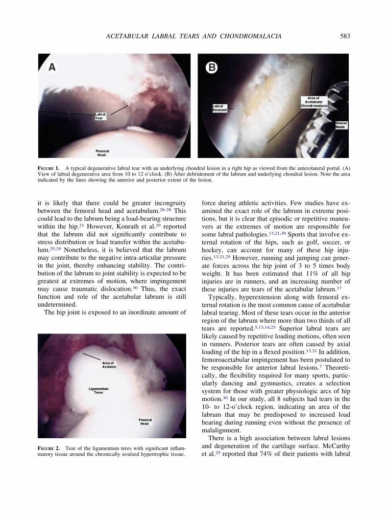
IGURE 1. A typical degenerative labral tear with an underlying ciew of labral degenerative area from 10 to 12 o’clock. (B) After
ndicated by the lines showing the anterior and posterior extent of
eIGURE 2. Tear of the ligamentum teres with significant inflam-atory tissue around the chronically avulsed hypertrophic tissue.
orce during athletic activities. Few studies have ex-mined the exact role of the labrum in extreme posi-ions, but it is clear that episodic or repetitive maneu-ers at the extremes of motion are responsible forome labral pathologies.13,21,30 Sports that involve ex-ernal rotation of the hips, such as golf, soccer, orockey, can account for many of these hip inju-ies.13,21,25 However, running and jumping can gener-te forces across the hip joint of 3 to 5 times bodyeight. It has been estimated that 11% of all hip
njuries are in runners, and an increasing number ofhese injuries are tears of the acetabular labrum.17
Typically, hyperextension along with femoral ex-ernal rotation is the most common cause of acetabularabral tearing. Most of these tears occur in the anterioregion of the labrum where more than two thirds of allears are reported.5,13,14,25 Superior labral tears areikely caused by repetitive loading motions, often seenn runners. Posterior tears are often caused by axialoading of the hip in a flexed position.13,21 In addition,emoroacetabular impingement has been postulated toe responsible for anterior labral lesions.7 Theoreti-ally, the flexibility required for many sports, partic-larly dancing and gymnastics, creates a selectionystem for those with greater physiologic arcs of hipotion.30 In our study, all 8 subjects had tears in the
0- to 12-o’clock region, indicating an area of theabrum that may be predisposed to increased loadearing during running even without the presence ofalalignment.There is a high association between labral lesions
nd degeneration of the cartilage surface. McCarthy
l lesion in a right hip as viewed from the anterolateral portal. (A)ment of the labrum and underlying chondral lesion. Note the areasion.
hondra
t al.25 reported that 74% of their patients with labral

tptaatiiltorsbsTener
alpaaaiasamhpnris
daisp
ssliam
ai
tlaockid
TaWfsWuy
tpmostipscb
oitlrtcac
hsmpm
584 C. A. GUANCHE AND R. S. SIKKA
ears had chondral damage. Moreover, in 80% of theseatients, the chondral damage was in the same zone ashe labral lesion.25 The strongest association betweenrticular damage and labral lesions was present later-lly and posteriorly. Additionally, the severity of car-ilage damage was greater in patients with labral fray-ng. In our study, 75% of the patients had chondralnjuries and all of these were grade III Outerbridgeesions. Articular cartilage degeneration originates inhe anterosuperior portion of the weight-bearing areaf the femoral head and acetabulum, probably as aesult of the high compressive stresses.25,15 An exces-ive torsional and shear force that cannot be dissipatedy the labrum causes a tear in the labrum, which theneparates the attachment to the acetabular cartilage.his watershed zone separation, once initiated, canxtend further. Continued loading may lead to delami-ation and degeneration.8 Thus, it is important tovaluate patients with labral tears for chondral inju-ies.
Tears of the ligamentum teres are uncommon. Graynd Villar31 reported on a series of 20 patients withigamentum teres pathology out of 427 hip arthrosco-ies. Of the 20 patients with tears, 7 had completevulsions. In our study, 3 of 8 patients had completevulsions of the ligamentum teres (Fig 2). These allppeared to be chronic in nature and not caused by thentraoperative traction. Tears of the ligamentum teresre often associated with labral lesions and articularurface damage.31 However, patients with completevulsions of the ligamentum often have a history ofajor joint disruption from trauma or surgery or may
ave dysplasia of the hip.31 In our study, none of theatients reported a specific macrotraumatic event, andone had dysplastic conditions. It is likely that theepetitive hyperextension from running led to subtlenstability, thereby placing an inordinate amount oftress on the ligamentum with subsequent failure.
In the 3 cases of ligamentum teres avulsions, MRIid not indicate the ligamentum lesion (2 with intra-rticular contrast). Therefore, in cases of labral tears its important to visualize the ligamentum teres. Arthro-copic debridement of the stump should then be com-leted.The authors theorize that the repetitive hyperexten-
ion seen in the stride phase of running leads to aubtle instability and increasing stress at the cartilage-abral junction. This may lead to the pathologic find-ngs documented in this study, including labral tearingnd associated chondral degeneration. Based on this
echanism, femoroacetabular impingement does not lppear to be a factor, although offset was not specif-cally analyzed.
While the emphasis of this study is to bring to lighthe possible association of high-level running withabral and acetabular degeneration, the shortcomingsre obvious. First, this is a retrospective review basedn 1 surgeon’s experience with a relatively new pro-edure. The learning curve for hip arthroscopy isnown to be high. Whether the incidence of thesenjuries is less as a result of failure to make theiagnosis prospectively is a notable criticism.The use of the WOMAC score can also be criticized.
here truly is no validated scoring scale available tonalyze an active population with hip injuries.32 The
OMAC score measures pain, stiffness, and physicalunction in patients with arthritis. It is self-administered,imple, reliable, and validated. This was the reason the
OMAC was used in this study. Although efforts arenderway to validate other scoring scales, none other haset surfaced for this purpose.32
However, the major limitation is the failure to provehe direct association of running and the describedathologic entities. While runners typically run for theajority of their training, they certainly participate in
ther activities that could contribute or perhaps beolely responsible for the documented labral and car-ilage abnormalities. The only sure way of determin-ng the veracity of the findings in this study is toerform prospective analyses on this population. Theenior author has begun such a study, but this willlearly take many years before useful conclusions cane drawn.Although the hip and pelvis account for 5% to 6%
f injuries in adult athletes and 10% to 24% of injuriesn child athletes, the hip has received less attentionhan other major joints.17 Until recently, we have beeness sophisticated in our ability to diagnose hip inju-ies with considerably less technology available toreat and diagnose these injuries.16 Through improvedlinical, imaging, and arthroscopic techniques, moreccurate associations of pathologic entities with spe-ific mechanisms may be possible.
CONCLUSIONS
Although most reports of acetabular labral injuriesave focused on soccer, football, hockey, and otherports involving twisting motions, high-level runnersay be predisposed to tears of the anterosuperior
ortion of the labrum. In cases where runners haveaintained a high intensity and high mileage for a
engthy period, it is important to consider tears of the

ladhad
bowtlt
1
1
1
1
1
1
1
1
1
1
2
2
2
2
2
2
2
2
2
2
3
3
3
585ACETABULAR LABRAL TEARS AND CHONDROMALACIA
abrum as a potential diagnosis. On arthroscopic ex-mination, it is also important to visualize the chon-ral surface in the area of the labral tear, given theigh incidence of chondral defects in our patients. It islso important to visualize the ligamentum teres andeal with any pathology in that area.Although this study does not confirm an association
etween running and the development of labral tearsr chondral lesions in the hip, it certainly questionshether there is an injury pattern that is common to
his population, a “runners hip.” Further prospective,ongitudinal studies should be planned to delineatehis further.
REFERENCES
1. Patterson JM. The torn acetabular labrum: A block to reduc-tion of a dislocated hip. J Bone Joint Surg Br 1957;39:306-309.
2. Alternberg AR. Acetabular labral tears: A cause of hip painand degenerative arthritis. South Med J 1977;70:174-175.
3. Nishina T, Saito S, Ohzono K, et al. Chiari pelvic osteotomyfor osteoarthritis: The influence of torn and detached acetab-ular labrum. J Bone Joint Surg Br 1990;72:765-769.
4. Klaue K, Durnin C, Ganz R. The acetabular rim syndrome.J Bone Joint Surg Br 1991;73:423-429.
5. Dorrell J, Catteral A. The torn acetabular labrum. J Bone JointSurg Br 1986;68:400-403.
6. Ikeda T, Awaya G, Suzuki S, et al. Torn acetabular labrum inyoung patients. J Bone Joint Surg Br 1988;70:13-16.
7. Ito K, Minka MA, Leunig M, et al. Femoroacetabular impinge-ment and the cam-effect. A MRI-based quantitative anatomicalstudy of the femoral head-neck offset. J Bone Joint Surg Br2001;83:171-176.
8. McCarthy J, Lee J. Acetabular dysplasia: A paradigm ofarthroscopic examination of chondral injuries. Clin Orthop RelRes 2002;405:122-128.
9. Philippon MJ. The role of arthroscopic thermal capsulorrhaphyin the hip. Clin Sports Med 2001;20:817-829.
0. Sohn RS, Lyle M. The effect of running on pathogenic osteo-arthritis of the hips and knees. Clin Orthop 1985;198:106-109.
1. Marti B, Knoblach MB, Tschopp A, et al. Is excessive runningpredictive of degenerative hip disease? Controlled study offormer elite athletes. BMJ 1989;299:91-93.
2. Suzuki S, Awaya G, Suzuki S, et al. Arthroscopic diagnosis ofruptured acetabular labrum. Acta Orthop Scand 1986;57:513-515.
3. Fitzgerald R. Acetabular labral tears. Clin Orthop 1995;311:60-68.
4. Farjo LA, Glick JM, Sampson TG. Hip arthroscopy for ace-tabular labrum tears. Arthroscopy 1999;15:132-137.
5. Noguchi Y, Miura H, Takasugi S, et al. Cartilage and labrumdegeneration in the dysplastic hip generally originates in theanteriosuperior weight-bearing area: An arthroscopic observa-tion. Arthroscopy 1999;15:496-506.
6. Byrd JWT, Jones KS. Hip arthroscopy in athletes. Clin SportsMed 2001;20:749-761.
7. Scopp JM, Moorman CT. The assessment of athletic hipinjury. Clin Sports Med 2001;20:647-659.
8. Bellamy N, Buchanan WW, Goldsmith CH, et al. Validationstudy of WOMAC: A health status instrument for measuringclinically important patient relevant outcomes to antirheumaticdrug therapy in patients with osteoarthritis of the hip or knee.J Rheumatol 1988;15:1833-1840.
9. Byrd JWT. Investigation of the symptomatic hip: Physicalexamination. In: Byrd JWT, ed. Operative hip arthroscopy.New York: Thieme, 1998;25-42.
0. Outerbridge R. The etiology of chondromalacia patellae. J BoneJoint Surg Br 1961;43:752-754.
1. Mason JB. Acetabular labral tears in the athlete. Clin SportsMed 2001;20:779-790.
2. Dvorak M, Duncan CP, Day B. Arthroscopic anatomy of thehip. Arthroscopy 1990;6:264-273.
3. Keene GS, Villar RN. Arthroscopic anatomy of the hip: An invivo study. Arthroscopy 1994;10:392-399.
4. Kim YT, Azuma H. The nerve endings of the acetabularlabrum. Clin Orthop 1995;320:176-181.
5. McCarthy J, Noble P, Aluisio FV, et al. Anatomy, pathologicfeatures, and treatment of acetabular labral tears. Clin Orthop2003;406:38-47.
6. Afoke NY, Byers PD, Hutton WC. The incongruous hip joint:A casting study. J Bone Joint Surg Br 1980;62:511-514.
7. Bullough P, Goodfellow J, Greenwald AS, et al. Incongruentsurfaces in the human hip joint. Nature 1968;217:1290-1294.
8. Greenwald AS, Haynes DW. Weight-bearing areas in thehuman hip joint. J Bone Joint Surg Am 1972;54:157-163.
9. Konrath GA, Hamel AJ, Olson SA, et al. The role of theacetabular labrum and the transverse acetabular ligament inload transmission in the hip. J Bone Joint Surg Am 1998;80:1781-1788.
0. Byrd JWT. Labral lesions: An elusive source of hip pain: Casereports and review of the literature. Arthroscopy 1996;12:603-612.
1. Gray AJ, Villar RN. The ligamentum teres of the hip: Anarthroscopic classification of its pathology. Arthroscopy 1997;13:575-578.
2. Christensen CP, McCarthy JC, Mittleman MA, et al. Out-
comes. In: McCarthy JC, ed. Early hip disorders. New York:Springer-Verlag, 2003;195-200.Copyright © 2022 FDOKUMEN