The Behavioral and Immunological Impact of Maternal Separation: A Matter of Timing
Upload
independentCategory
view
1download
0
6) 249–259www.elsevier.com/locate/yviro
Virology 351 (200
Different European-type vaccines against porcine reproductive andrespiratory syndrome virus have different immunological properties and
confer different protection to pigs
I. Díaz a,b, L. Darwich a,b, G. Pappaterra c, J. Pujols a,c, E. Mateu a,b,⁎
a Centre de Recerca en Sanitat Animal (CReSA), Campus UAB, 08193 Bellaterra, Spainb Departament de Sanitat i Anatomia Animals, Facultat de Veterinària, Edifici V, Universitat Autònoma de Barcelona, 08193, Bellaterra, Spain
c Institut de Recerca i Tecnologia Agroalimentària, Barcelona, Spain
Received 22 March 2005; returned to author for revision 28 February 2006; accepted 31 March 2006Available online 19 May 2006
Abstract
Immunization of piglets with two different European-type modified live vaccines against porcine reproductive and respiratory syndrome(PRRS) virus produced different outcomes. After vaccination, pigs became viremic (42 days), neutralizing antibodies did not develop, andfrequencies of virus-specific gamma-interferon-secreting cells (IFN-γ-SC) were low. Levels of interleukin-10 (IL-10) produced by peripheralblood mononuclear cells (PBMC) seemed to inversely correlate with interferon-gamma responses. After a challenge with a virulent Spanishstrain, one vaccine (V3) protected piglets against viremia while the other (V1) did not. The vaccine V3 induced the highest IFN-γ-SCfrequencies. IL-2, IL-4 or transforming growth factor-beta responses were not detected at any time for neither of the vaccines. In contrast,haptoglobin rose in sera of viremic pigs after the challenge. These results indicated a strong involvement of IFN-γ, and maybe IL-10, in thedevelopment of immunity against PRRS virus.© 2006 Elsevier Inc. All rights reserved.
Keywords: Porcine reproductive and respiratory virus; Interferon-gamma; Interleukin-10; Vaccine
Introduction
Porcine reproductive and respiratory syndrome (PRRS)emerged in Europe in the early years of the 1990 decade andrapidly spread all over the continent becoming one of the majorcauses of economic losses for swine producers. Simultaneously,the disease emerged in USA with a similar impact on pigproduction. This syndrome, caused by an arterivirus (Meulen-berg et al., 1994), is characterized in its reproductive form byabortions, stillbirths and weakly born piglets and, in young pigs,causes pneumonia and weight gain losses. PRRS virus (PRRSV)is a small positive-stranded polyadenylated RNA virus thatcontains nine open reading frames (ORFs). ORFs 1a and 1bcomprise 80% of the genome and encode the RNA polymerase;ORFs2 to5 encode four structural glycoproteins (GP2,GP3,GP4
⁎ Corresponding author. Centre de Recerca en Sanitat Animal (CReSA),Campus UAB, 08193 Bellaterra, Spain. Fax: +34 93 581 32 97.
E-mail address: [email protected] (E. Mateu).
0042-6822/$ - see front matter © 2006 Elsevier Inc. All rights reserved.doi:10.1016/j.virol.2006.03.046
and GP5) and a small non-glycosylated protein called 2b (Wu etal., 2005); ORF6 encodes a non-glycosylated integral membraneprotein (M) and ORF7 encodes the nucleocapside protein (N)(Conzelmann et al., 1993; Meulenberg et al., 1997; Snijder andMeulenberg, 1998). Currently, it is known that two distinctgenotypes of the virus exist, the European and the American, thatshare only 55–80% similarity (Meng et al., 1995).
Soon after the discovery of the virus vaccines weremarketed. The first ones were a modified live virus (MLV)vaccine called RespPRRS made of an American-type strain(Christopher-Hennings et al., 1997; Dee and Joo, 1997;Mengeling et al., 1996), and an inactivated vaccine calledCyblue, made of a European strain (Plana-Duran et al., 1997).From then, several vaccines, both attenuated and inactivated,have been commercialized.
Notwithstanding, none of the vaccines can claim fullprotection against the disease. On one hand, PRRSV, eitherwild type or attenuated, induces a low level of cell-mediatedimmunity (Meier et al., 2003) and neutralizing antibodies (NA)

Table 1Percentages of similarity between the PRRSV strains used in the study
LV V1 V3 VP21
ORF 2LV – 94.6 98.0 94.6V1 94.6 – 94.3 98.3V3 98.0 94.3 – 93.9VP21 94.6 98.3 93.9 –
ORF 3LV – 94.2 98.3 94.0V1 94.2 – 92.5 99.4V3 94.2 92.5 – 93.3VP21 94.4 99.6 93.9 –
ORF 4LV – 94.2 99.0 94.5V1 94.2 – 94.2 99.2V3 99.0 94.2 – 93.5VP21 94.5 99.2 94.5 –
ORF 5LV – 94.9 98.9 94.5V1 94.9 – 94.4 99.6V3 98.9 94.4 – 94.0VP21 94.5 99.6 94.0 –
ORF 6LV – 97.7 97.7 96.9V1 97.7 – 100 98.6V3 97.7 100 – 98.6VP21 96.9 98.6 98.6 –
ORF 7LV – 96.6 100 96.3V1 96.6 – 96.6 99.7V3 100 96.6 – 96.3VP21 96.3 99.7 96.3 –
Lelystad virus (LV, Genbank accession number M96262) was used as areference.LV = Lelystad virus, V1 = Modified live vaccine 1, V3 = Modified live vaccine2, VP21 = wild-type Spanish PRRSV strain used for the challenge.
Table 2Evolution of viremia in vaccinated pigs as determined by RT-nested PCR
Group Days post-vaccination
0 7 14 21 28 42 63 a 70 91
G1 0 5 4 5 2 1 0 4 0G2 0 5 3 3 1 2 0 0 0
a At 63 days post-vaccination, both groups were challenged with 106 TCID50
of a wild-type Spanish PRRSV strain (VP21).
250 I. Díaz et al. / Virology 351 (2006) 249–259
do not develop until a late phase of the infection (Meier et al.,2003; Vezina et al., 1996; Yoon et al., 1995). In addition, thecytokine imbalance after a vaccination or an infection suggests apossible downregulation of many cytokines (Royaee et al.,2004; Van Reeth et al., 1999). On the other hand, geneticdiversity of PRRSV is thought to influence the efficacy ofvaccines under field conditions (Labarque et al., 2004; Pesch etal., 2005) although some degree of cross protection exists(Mengeling et al., 2003a, 2003b). With this panorama, thedevelopment of improved vaccines needs a deeper understand-ing of the immunity against PRRSV.
Up to date, only one report has dealt with the evolution of theimmune responses, both humoral and cellular, after vaccinationwith a European type PRRS-modified live vaccine (MLV)(Sipos et al., 2003). However, that study was done with only onevaccine, did not challenge the animals and did not evaluatefrequencies of IFN-γ-secreting cells (IFN-γ-SC), a parameterthat seems to be related to protection (Meier et al., 2003). Theaim of the present study was to characterize the humoral and thecellular responses of pigs in the course of a vaccination with two
European-type PRRS-MLVvaccines and after a challengewith aEuropean PRRS wild-type virus.
Results
Virus sequencing and phylogenetic analysis
Similarity between vaccines and VP21 strain was above90%. Highest values were obtained for ORF7 and ORF6 andlowest similarities corresponded to envelope glycoproteins(Table 1). As a whole, VP21 was closer to V1 (both areSpanish strains) than to V3. Interestingly, in ORF6 bothvaccines had exactly the same sequence that differed fromVP21 and LV by a change in the codon starting at position14,402 in LV genome (cga → caa). That change wouldcorrespond in the amino acid sequence to a substitution of anarginine by a glutamine.
Detection of PRRSV
Viral isolation attempts in PAM or MARC-145 cells fromblood samples taken after vaccination were negative. Incontrast, all vaccinated animals were positive by nPCR atleast once during the first 6 weeks PV (Table 2). However,viremia was probably low as, with a single round RT-PCR,only six samples were positive during the first 14 days PV.After the challenge, only G1 pigs were detected as positive bynPCR (4/5) at 7 days post-infection (PI). Sequencing of thenPCR amplicons showed the virus to be the VP21 strain. Noneof the control pigs yielded nPCR-positive results or viralisolation.
Hematology
In all three groups, hematological values remained betweennormal ranges in the examined period (28 days PV). However,some parameters significantly changed in vaccinated pigs atfirst week PV, particularly for neutrophils, monocytes andplatelets (P < 0.05). Table 3 summarizes these results.
Humoral immune response
All pigs were seronegative to PRRSV at day 0. By usingELISA, all vaccinated animals seroconverted by day 14 PV(mean S/P ratio, 2.5 ± 0.2 for G1 and 2.4 ± 0.6 for G2) (Table 4).ELISA titers peaked at 21 and 28 days PV for G1 and G2,respectively (G1 = 3.2 ± 0.3 and G2 = 3.1 ± 0.3). After the
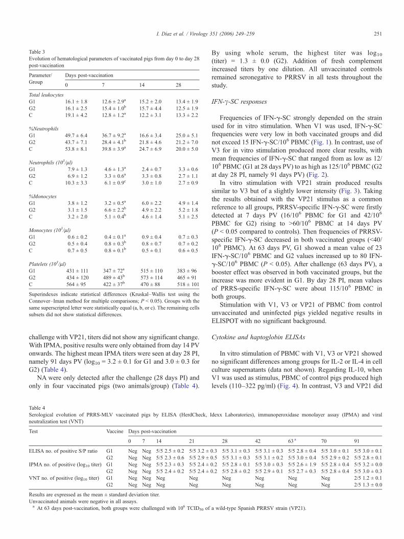
Table 3Evolution of hematological parameters of vaccinated pigs from day 0 to day 28post-vaccination
Parameter/Group
Days post-vaccination
0 7 14 28
Total leukocytesG1 16.1 ± 1.8 12.6 ± 2.9a 15.2 ± 2.0 13.4 ± 1.9G2 16.1 ± 2.5 15.4 ± 1.0b 15.7 ± 4.4 12.5 ± 1.9C 19.1 ± 4.2 12.8 ± 1.2a 12.2 ± 3.1 13.3 ± 2.2
%NeutrophilsG1 49.7 ± 6.4 36.7 ± 9.2a 16.6 ± 3.4 25.0 ± 5.1G2 43.7 ± 7.1 28.4 ± 4.1b 21.8 ± 4.6 21.2 ± 7.0C 53.8 ± 8.1 39.8 ± 3.9a 24.7 ± 6.9 20.0 ± 5.0
Neutrophils (103/μl)G1 7.9 ± 1.3 4.6 ± 1.3a 2.4 ± 0.7 3.3 ± 0.6G2 6.9 ± 1.2 3.3 ± 0.6a 3.3 ± 0.8 2.7 ± 1.1C 10.3 ± 3.3 6.1 ± 0.9c 3.0 ± 1.0 2.7 ± 0.9
%MonocytesG1 3.8 ± 1.2 3.2 ± 0.5a 6.0 ± 2.2 4.9 ± 1.4G2 3.1 ± 1.5 6.6 ± 2.2b 4.9 ± 2.2 5.2 ± 1.8C 3.2 ± 2.0 5.1 ± 0.4b 4.6 ± 1.4 5.1 ± 2.5
Monocytes (103/μl)G1 0.6 ± 0.2 0.4 ± 0.1a 0.9 ± 0.4 0.7 ± 0.3G2 0.5 ± 0.4 0.8 ± 0.3b 0.8 ± 0.7 0.7 ± 0.2C 0.7 ± 0.5 0.8 ± 0.1b 0.5 ± 0.1 0.6 ± 0.5
Platelets (103/μl)G1 431 ± 111 347 ± 72a 515 ± 110 383 ± 96G2 434 ± 120 489 ± 43b 573 ± 114 465 ± 91C 564 ± 95 422 ± 37b 470 ± 88 518 ± 101
Superindexes indicate statistical differences (Kruskal–Wallis test using theConnover–Iman method for multiple comparisons; P < 0.05). Groups with thesame superscripted letter were statistically equal (a, b, or c). The remaining cellssubsets did not show statistical differences.
251I. Díaz et al. / Virology 351 (2006) 249–259
challenge with VP21, titers did not show any significant change.With IPMA, positive results were only obtained from day 14 PVonwards. The highest mean IPMA titers were seen at day 28 PI,namely 91 days PV (log10 = 3.2 ± 0.1 for G1 and 3.0 ± 0.3 forG2) (Table 4).
NA were only detected after the challenge (28 days PI) andonly in four vaccinated pigs (two animals/group) (Table 4).
Table 4Serological evolution of PRRS-MLV vaccinated pigs by ELISA (HerdCheck,neutralization test (VNT)
Test Vaccine Days post-vaccination
0 7 14 21
ELISA no. of positive S/P ratio G1 Neg Neg 5/5 2.5 ± 0.2 5/5 3.2 ±G2 Neg Neg 5/5 2.3 ± 0.6 5/5 2.9 ±
IPMA no. of positive (log10 titer) G1 Neg Neg 5/5 2.3 ± 0.3 5/5 2.4 ±G2 Neg Neg 5/5 2.4 ± 0.2 5/5 2.4 ±
VNT no. of positive (log10 titer) G1 Neg Neg Neg NegG2 Neg Neg Neg Neg
Results are expressed as the mean ± standard deviation titer.Unvaccinated animals were negative in all assays.a At 63 days post-vaccination, both groups were challenged with 106 TCID50 of
By using whole serum, the highest titer was log10(titer) = 1.3 ± 0.0 (G2). Addition of fresh complementincreased titers by one dilution. All unvaccinated controlsremained seronegative to PRRSV in all tests throughout thestudy.
IFN-γ-SC responses
Frequencies of IFN-γ-SC strongly depended on the strainused for in vitro stimulation. When V1 was used, IFN-γ-SCfrequencies were very low in both vaccinated groups and didnot exceed 15 IFN-γ-SC/106 PBMC (Fig. 1). In contrast, use ofV3 for in vitro stimulation produced more clear results, withmean frequencies of IFN-γ-SC that ranged from as low as 12/106 PBMC (G1 at 28 days PV) to as high as 125/106 PBMC (G2at day 28 PI, namely 91 days PV) (Fig. 2).
In vitro stimulation with VP21 strain produced resultssimilar to V3 but of a slightly lower intensity (Fig. 3). Takingthe results obtained with the VP21 stimulus as a commonreference to all groups, PRRSV-specific IFN-γ-SC were firstlydetected at 7 days PV (16/106 PBMC for G1 and 42/106
PBMC for G2) rising to >60/106 PBMC at 14 days PV(P < 0.05 compared to controls). Then frequencies of PRRSV-specific IFN-γ-SC decreased in both vaccinated groups (<40/106 PBMC). At 63 days PV, G1 showed a mean value of 23IFN-γ-SC/106 PBMC and G2 values increased up to 80 IFN-γ-SC/106 PBMC (P < 0.05). After challenge (63 days PV), abooster effect was observed in both vaccinated groups, but theincrease was more evident in G1. By day 28 PI, mean valuesof PRRS-specific IFN-γ-SC were about 115/106 PBMC inboth groups.
Stimulation with V1, V3 or VP21 of PBMC from controlunvaccinated and uninfected pigs yielded negative results inELISPOT with no significant background.
Cytokine and haptoglobin ELISAs
In vitro stimulation of PBMC with V1, V3 or VP21 showedno significant differences among groups for IL-2 or IL-4 in cellculture supernatants (data not shown). Regarding IL-10, whenV1 was used as stimulus, PBMC of control pigs produced highlevels (110–322 pg/ml) (Fig. 4). In contrast, V3 and VP21 did
Idexx Laboratories), immunoperoxidase monolayer assay (IPMA) and viral
28 42 63 a 70 91
0.3 5/5 3.1 ± 0.3 5/5 3.1 ± 0.3 5/5 2.8 ± 0.4 5/5 3.0 ± 0.1 5/5 3.0 ± 0.10.5 5/5 3.1 ± 0.3 5/5 3.1 ± 0.2 5/5 3.0 ± 0.4 5/5 2.9 ± 0.2 5/5 2.8 ± 0.10.2 5/5 2.8 ± 0.1 5/5 3.0 ± 0.3 5/5 2.6 ± 1.9 5/5 2.8 ± 0.4 5/5 3.2 ± 0.00.2 5/5 2.8 ± 0.2 5/5 2.9 ± 0.1 5/5 2.7 ± 0.3 5/5 2.8 ± 0.4 5/5 3.0 ± 0.3
Neg Neg Neg Neg 2/5 1.2 ± 0.1Neg Neg Neg Neg 2/5 1.3 ± 0.0
a wild-type Spanish PRRSV strain (VP21).

Fig. 1. The figure shows the frequency of IFN-γ-secreting cells (ELISPOT) and IL-10 levels in peripheral blood mononuclear cell culture supernatants (ELISA) afterV1 in vitro stimulation in both vaccinated groups. For IL-10 results are expressed as the average of IL-10 secreted (pg/ml) (detection limit = 32 pg/ml), whereasspecific-virus IFN-γ-secreting cells are expressed as the average frequency of the specific-virus IFN-γ-secreting cells by 106 PBMC. *At 63 days post-vaccination,both groups were challenged with 106 TCID50 of a wild-type Spanish PRRSV strain (VP21).
252 I. Díaz et al. / Virology 351 (2006) 249–259
not induce IL-10 secretion in cells of control pigs (<42 pg/ml;P < 0.05).
When the specific IL-10 response was examined invaccinated pigs, stimulation of PBMC produced differentresults depending on the virus used. In G1 pigs, stimulation
Fig. 2. The figure shows the frequency of IFN-γ-secreting cells (ELISPOT) and IL-1V3 in vitro stimulation in both vaccinated groups. For IL-10 results are expressedspecific-virus IFN-γ-secreting cells are expressed as the average frequency of the spelevels (P < 0.05). (b) G2 > G1 IFN-γ-secreting cells (P < 0.05). *At 63 days post-vaPRRSV strain (VP21).
with V1 produced high levels of IL-10 until 42 days PV (150–236 pg/ml); from this date on, IL-10 could not be detected(Fig. 1). For G2 pigs, in vitro stimulation with V1 producedlower levels of IL-10 that began to decline by day 21 PV andbecame undetectable by day 70 PV. Stimulation of PBMC
0 levels in peripheral blood mononuclear cell culture supernatants (ELISA) afteras the average of IL-10 secreted (pg/ml) (detection limit = 32 pg/ml), whereascific-virus IFN-γ-secreting cells by 106 PBMC. (a) G1 and G2 > Controls IL-10ccination, both groups were challenged with 106 TCID50 of a wild-type Spanish
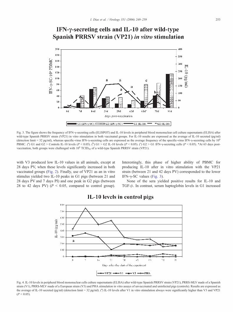
Fig. 3. The figure shows the frequency of IFN-γ-secreting cells (ELISPOT) and IL-10 levels in peripheral blood mononuclear cell culture supernatants (ELISA) afterwild-type Spanish PRRSV strain (VP21) in vitro stimulation in both vaccinated groups. For IL-10 results are expressed as the average of IL-10 secreted (pg/ml)(detection limit = 32 pg/ml), whereas specific-virus IFN-γ-secreting cells are expressed as the average frequency of the specific-virus IFN-γ-secreting cells by 106
PBMC. (a) G1 and G2 > Controls IL-10 levels (P < 0.05). (b) G1 > G2 IL-10 levels (P < 0.05). (c) G2 > G1 IFN-γ-secreting cells (P < 0.05). *At 63 days post-vaccination, both groups were challenged with 106 TCID50 of a wild-type Spanish PRRSV strain (VP21).
253I. Díaz et al. / Virology 351 (2006) 249–259
with V3 produced low IL-10 values in all animals, except at28 days PV, when these levels significantly increased in bothvaccinated groups (Fig. 2). Finally, use of VP21 as an in vitrostimulus yielded two IL-10 peaks in G1 pigs (between 21 and28 days PV and 7 days PI) and one peak in G2 pigs (between28 to 42 days PV) (P < 0.05, compared to control group).
Fig. 4. IL-10 levels in peripheral blood mononuclear cells culture supernatants (ELISAstrain (V1), PRRS-MLV made of a European strain (V3) and PHA stimulation in vitrthe average of IL-10 secreted (pg/ml) (detection limit = 32 pg/ml). (a) IL-10 levels a(P < 0.05).
Interestingly, this phase of higher ability of PBMC forproducing IL-10 after in vitro stimulation with the VP21strain (between 21 and 42 days PV) corresponded to the lowerIFN-γ-SC values (Fig. 3).
None of the sera yielded positive results for IL-10 andTGF-β. In contrast, serum haptoglobin levels in G1 increased
) after wild-type Spanish PRRSV strain (VP21), PRRS-MLVmade of a Spanisho assays of unvaccinated and uninfected pigs (controls). Results are expressed asfter V1 in vitro stimulation always were significantly higher than V3 and VP21

254 I. Díaz et al. / Virology 351 (2006) 249–259
significantly at 7 days PI (P < 0.05), returning to normality byday 14 PI (Fig. 5).
Discussion
Several studies have reported that the immune responseagainst arteriviruses is unique in its features (Balasuriya andMacLachlan, 2004; Cafruny et al., 1999; Meier et al., 2003).Regarding PRRSV, it seems that both, humoral and cellular,responses do not control the infection adequately. NA developslowly and usually appear after the resolution of viremia. Inaddition, cell-mediated immunity also rises slowly, and virus-specific IFN-γ-SC frequencies are low compared to other viralpig diseases (Meier et al., 2003). Some authors suggested that,in PRRS, immunity might play a secondary role and stated thatavailability of permissive macrophages may determine theoutcome of the infection (Diaz et al., 2005; Xiao et al., 2004). Asimilar hypothesis has been postulated for lactate dehydroge-nase-elevating virus (LDV), a related arterivirus (Onyekaba etal., 1989; Van den Broek et al., 1997).
Vaccines made of MLV or inactivated PRRSV are beingcommercialized and have shown a relative efficacy (Mengel-ing et al., 2003a, 2003b; Plana-Duran et al., 1997; VanWoensel et al., 1998). Nevertheless, this is a controversialissue. Firstly, because it is unclear how well vaccines preventthe infection and how safe they are (Labarque et al., 2003;Mengeling et al., 2003a, 2003b) and, secondly, because it isthought that the genetic diversity of PRRSV can negativelyaffect the field efficacy of vaccines (Labarque et al., 2004;Meng, 2000).
In the present study, two European-type vaccines wereexamined. To assess differences in protection, it was importantto evaluate the genetic similarity between them and thechallenge VP21 strain. As expected, main differences betweenthe PRRSV strains used were found in ORF3 and ORF5, which
Fig. 5. Kinetics of serum haptoglobin as measured by ELISA in vaccinated and contwith a wild-type Spanish PRRSV strain (VP21).
are known to be the most variable regions in the PRRSVgenome (Pirzadeh et al., 1998; Forsberg et al., 2001).
Besides this, it is interesting to note that ORF6 of bothvaccine viruses shared a change corresponding to the codonstarting at position 14,402 in LV. This change (cga → caa)produced a substitution of an arginine by a glutamine comparedto VP21 or LV. Grebennikova et al. (2004) showed that in theAmerican-type PRRSV strain NADC-8, a change in theposition 14,440 (alanine → threonine) contributed significantlyto reduce viral virulence. In our case and theirs, one amino acidpresent in virulent strains was substituted by a polar unchargedone in the attenuated viruses. This finding supports the idea thatthis site in ORF6 can be related to attenuation.
After vaccination, both G1 and G2 pigs became viremic, andsome animals remained so as late as 42 days PV. Sipos et al.(2003) reported 3 weeks of viremia in piglets using one of theEuropean-type PRRSV MLV used in the present study. Theseextended periods of viremia could significantly contribute to thespread of vaccine virus in the pig population. This fact wasreported for American-type vaccines used in Europe (Morten-sen et al., 2002; Storgaard et al., 1999). These results point outthe question of attenuation. Extended periods of viral replicationshould not be interpreted as equivalent to virulence or viceversa. As Xiao et al. (2004) suggested, in PRRS, viralreplication would occur as long as permissive macrophagesexist. Thus, with an attenuated PRRSV strain, that can bethought to produce less intense cytolytic cycles, viral replicationshould last a long time.
In our case, pigs vaccinated with V1 developed viremiawhen infected with VP21, while in G2 pigs virus was notdetectable in blood after the challenge. Genetic similarity washigher between V1 and VP21 (98.3–99.7%) than between V3and VP21 (92.3–98.6%). That fact shows that, in our case, theheterologous strain provided better protection than the homol-ogous one. This apparent contradiction could be explained by
rol pigs. (†)P < 0.05. *At 63 days post-vaccination both groups were challenged

255I. Díaz et al. / Virology 351 (2006) 249–259
the different adjuvants used with the vaccines or by differencesin the ability of each vaccine strain to induce PRRSV-specificIFN-γ-SC. In addition, the capability of V1 to stimulate IL-10responses might also have had a role.
Regarding the hematological parameters examined, weobserved some changes at 7 days PV, but all values remainedbetween the physiological ranges for pigs. This has also beenreported by Sipos et al. (2003).
By using ELISA, development of specific antibodies couldbe seen at 14 days PV in all vaccinated pigs and titers remainedhigh throughout the experiment. Interestingly, ELISA titers didnot increase significantly after inoculation of the wild-typestrain even in viremic pigs. Some authors claimed (Johnson etal., 2004) that the quantitation of the humoral response (totalantibodies) measured by ELISA can serve as an indicator of thevirulence of a given PRRSV strain. In the present case, using thesame ELISA than Johnson et al. (2004), titers after vaccinationreached a high of 3.15 (S/P ratio), similar to the values obtainedwith the most virulent strain used in Johnson's work. In ouropinion, ELISA titers can not be used to measure virulence, atleast, when European-type PRRSV strains are evaluated.
In our study, vaccinated pigs did not develop NA before thechallenge. Meier et al. (2003), using a MLV vaccine made of anAmerican-type strain, needed two doses of the vaccine to induceonly in some animals low levels of NA. Although it seems thathigh titers of NA could play a role in the outcome of theinfection (Lopez and Osorio, 2004) most experiments showed alow, delayed and sporadic development of NA (Meier et al.,2003; Mengeling et al., 1999; Vezina et al., 1996). However, asit has been observed in other arterivirus such as LDV, viremiacan be cleared in absence of NA (Balasuriya and MacLachlan,2004). In our experiment, G2 pigs achieved sterilizingimmunity without the need of detectable NA.
Regarding the cellular immune response using VP21 or V3as stimuli, levels of IFN-γ-SC rose at 14 days PV, then wereerratic, and finally rose again at 63 days PV. This figure issimilar to the data reported by us in a previous experiment,where unvaccinated pigs were experimentally infected with theVP21 strain (Diaz et al., 2005).
Just before the challenge, stimulation of PBMC with VP21produced 81 IFN-γ-SC/106 PBMC in G2, while the samestimulation only produced 21 IFN-γ-SC/106 PBMC in G1.After the challenge with VP21, G1 pigs became viremic (4/5)while G2 pigs did not. Therefore, V1 vaccine demonstrated areduced ability to induce protective immune memory comparedto V3 vaccine. The remaining pig in G1 that did not developviremia had the higher IFN-γ-SC frequency at the day ofchallenge (45 IFN-γ-SC/106 PBMC). These results add a strongevidence for the role of the IFN-γ-SC in protection againstPRRSV infection in piglets. Recently, Lowe et al. (2005)observed a strong correlation of cell-mediated immunitymeasured by IFN-γ-SC with protection against reproductivefailure in sows of commercial herds as well. As discussed later,V1 had a higher ability to induce IL-10 secretion, and this couldbe have impaired the development of IFN-γ-SC. However, itcannot be ruled out that the adjuvant used with V3 had aninfluence on that observation although some authors denied the
effect of adjuvants in PRRSV immunization (Meier et al.,2003).
Our results also showed that IFN-γ-SC frequencies wereextremely low when V1 was used for in vitro stimulation. Thiswas observed even in pigs vaccinated with V1. The impairmentof IFN-γ-SC responses when V1 was used as a stimulus couldbe explained by the ability of V1 to induce the production ofhigh levels of IL-10 in PBMC. Since this IL-10 production wasseen in control pigs, it is reasonable to think that this was anintrinsic characteristic of the V1 strain and did not involvemechanisms of the adaptative immunity. Also, PBMC arethought to be non-permissive cells for PRRSVand therefore, theIL-10 release probably did not originate from viral replication inblood cells. The explanation for this fact remains unclear but areasonable hypothesis is that IL-10 secretion was the result ofthe interaction of some of the envelope glycoproteins, the mostvariable part of the virus, with cellular receptors in monocytesor lymphocytes. However, this is only a hypothesis to be testedin further studies on PRRSV and immunity.
Regarding IL-10 secretion after recall PRRSV stimulation, itcould be seen that this cytokine peaked between days 14 and 42PV depending on the group and the strain (V3 or VP21) used.By day 63 PV, IL-10 was practically undetectable in cell culturesupernatants. Disappearance of IL-10 correlated with theresolution of the viremia after vaccination. When G1 pigswere challenged with VP21 and developed viremia, IL-10 roseagain. In a previous experiment (Diaz et al., 2005) we observedthat in pigs experimentally infected with VP21, viremia andcessation of IL-10 production were also correlated. These factssuggest either that replication of the virus induces IL-10 or,alternatively, that IL-10 release sustains viral replication.
In the present experiment, as seen in control pigs, thepotential of IL-10 secretion of PBMC changed over time. Thus,PHA stimulation produced some 700 pg/ml at 5 weeks of ageand, from 8 weeks of age on, the same stimulus only yieldedsome 200–300 pg/ml. This fact indicated an influence of age onthe ability to secrete IL-10. Interestingly, when PBMC fromvaccinated pigs were stimulated in vitro with V1, a similarpattern was seen, but production of IL-10 was lower. From42 days PV, V1 stimulation of PBMC from vaccinated pigsproduced very low or undetectable IL-10 levels. Taken together,these observations can be interpreted as a virus-specificstimulation of PBMC that took place regardless of the immunestatus of the pig. The explanation for that is not fully clear butfrom 42 days PVon, frequencies of PRRSV-specific IFN-γ-SCrose. This suggests a role for the Th1/Th2 balance in thispattern.
Results for IL-2 and IL-4 responses did not show significantdifferences between control and vaccinated pigs using any ofthe viral strains. These data support the notion that some type ofimmunomodulation takes place in the course of the infection orvaccination (Royaee et al., 2004; Sipos et al., 2003).
Haptoglobin was only detected in sera of G1 animals afterthe challenge with VP21. Those animals were the only onesthat developed viremia after the challenge. In our opinion,haptoglobin release was probably linked to the tissular damagecaused by the wild virus but not by the replication of an

256 I. Díaz et al. / Virology 351 (2006) 249–259
attenuated PRRSV strain. It is known that haptoglobin isrelated with IL-6 release in inflammatory processes (Asai etal., 1999).
In summary, the pattern of the evolution of both thehumoral and the cellular immune responses after a PRRSVvaccination is very similar to that of a PRRSV infection inpiglets (Diaz et al., 2005). Vaccine V1 had the lowest capacityto induce IFN-γ-SC and, in contrast, was able to stimulate avery high IL-10 release from PBMC. After the challenge withthe virulent virus, the response of pigs vaccinated with V1resembled that of an unvaccinated pig in terms of preventionof viremia. This observation leads to the notion that IFN-γ-SCmost probably is the main factor in protecting against PRRSVinfection as well as that IL-10 release may hamper thedevelopment of such IFN-γ-SC. Thus, the capacity of a givenPRRSV strain to induce a strong cell-mediated immunity,measured as IFN-γ-SC, may be probably modulated by theIL-10 release (Chung and Chae, 2003; Suradhat andThanawongnuwech, 2003; Suradhat et al., 2003; Diaz et al.,2005). Once the modulation of the immune responsedisappears, IFN-γ-SC frequencies rose and, when a givenfrequency is reached, the pig will be protected (Lowe et al.,2005). Nevertheless, this is only attained at late stage. On theother hand, since the ability to produce IL-10 may changewith age, this host factor may also influence the outcome ofthe immunization or the infection.
In addition, we saw that the protection afforded by a givenvaccine against a given PRRSV strain cannot be forecastedonly by a global view of the genetic similarity between thevaccine virus and the challenge virus but, maybe, can beforecasted by examining the specific ability of a given viralstrain to induce IFN-γ-SC and to modulate the immuneresponses. In consequence, the development of an effectivevaccine for piglets should take into account the balancebetween IL-10 induction and the development of IFN-γ-SC.These data can be of critical importance to develop new andbetter PRRS vaccines.
Materials and methods
Animals and housing
Thirteen healthy 3-week-old Landrace pigs were randomlyselected in a high health farm belonging to the Institut deRecerca i Tecnologia Agroalimentària (IRTA, Barcelona,Spain) that has historically been free of all major pig diseasesincluding PRRS. Animals were transported to the experimen-tal facilities, randomly divided in three groups (G1, G2 and C)and kept during 7 days to allow adaptation to the newconditions. Before the start of the experiment, pigs wereexamined for antibodies against porcine circovirus type 2,Aujeszky's disease virus, porcine parvovirus, swine influenzaand Mycoplasma hyopneumoniae. All animals were seroneg-ative for all of the abovementioned pathogens. Also, animalswere confirmed to be free of PRRSV, as determined by ELISA(Herdcheck PRRS 2XR, IDEXX Laboratories, Westbrook ME,USA).
Vaccines and virus
Animals in group G1 (n = 5) were intramuscularly (IM)vaccinated with 104.0 TCID50 of a MLV made of one SpanishPRRSV strain (V1) using water as a vehicle (2 ml). G2 pigs(n = 5) received 104.0 TCID50 of a commercial MLV made ofother European PRRSV strain (V3) (2 ml, IM) with an oil-in-water adjuvant. Vaccines were diluted with the vehiclesrecommended by each of the manufacturers. The remainingthree pigs were kept as controls (C) and received 2 ml of sterileminimal essential medium (MEM) (Sigma, Alcobendas, Spain)as a placebo.
The virulent PRRSV VP21 strain used for the challenge wasisolated in porcine alveolar macrophages (PAM) from sera ofnaturally infected pigs during an outbreak of PRRS affecting abreeding farm located in the north of Spain in December 1991.That outbreak was characterized by abortions, stillbirths and,later on, piglet mortality. The virus was shown to be of theEuropean genotype by sequencing of ORFs 2 to 7. Viral stockwas done by passage (n = 3) in PAM and titrated by means ofthe immunoperoxidase monolayer assay (IPMA) according toWensvoort et al. (1991). Before to be used, the viral stock waschecked for bacterial contamination and for the presence ofother viruses.
At 63 days post-vaccination (PV), pigs in groups G1 and G2were inoculated intranasally with 2 ml viral suspensioncontaining 106 TCID50/ml of PRRSV VP21 strain diluted insterile MEM, whilst pigs in group C only received 2 ml of sterileMEM.
Virus sequencing and phylogenetic analysis
ORFs 2 to 7 of the vaccine viruses and challenge virus wereamplified by PCR using specific primer pairs (Table 5).Sequencing was done using an ABI 3100 sequencer (AppliedBiosystems). Sequences were formatted and translated to aminoacid sequences. Alignments were done using ClustalWsoftware. As a reference, Lelystad virus (LV) sequence(Genbank accession number M96262) was used. Nucleotidesequences were deposited at Genbank under accession numbersDQ009657-DQ009659 (ORF2); DQ009654-DQ009656(ORF3); DQ067250, DQ064784, DQ064785 (ORF4);DQ009647, DQ064787, DQ064788 (ORF5), DQ009651-DQ009653 (ORF6) and DQ009648-DQ009650 (ORF7).
Samples
Blood samples were collected in duplicate (heparinized andsiliconized blood-collecting tubes) immediately before vacci-nation and then at days 7, 14, 21, 28, 42, 63, 70 and 91 PV. Serawere used for PRRSV isolation and specific RT-nested PCR(nPCR), determination of humoral responses and evaluation ofhaptoglobin, IL-10 and transforming growth factor-beta (TGF-β) levels. Heparinized blood samples were used for hemato-logical evaluation. Peripheral blood mononuclear cells (PBMC)were also obtained from heparinized blood samples and used forin vitro experiments.
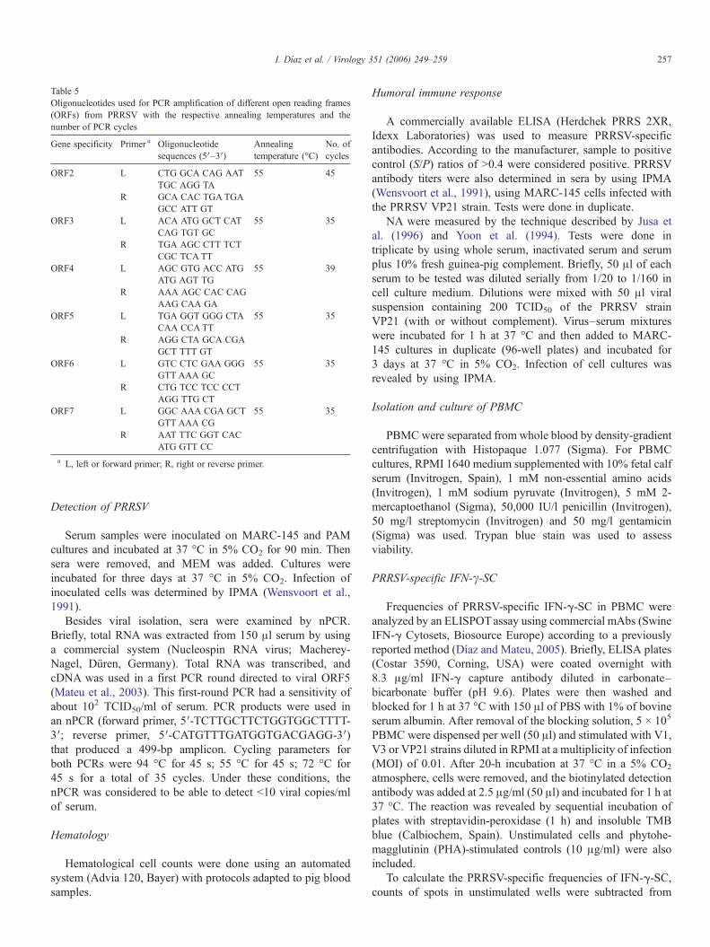
Table 5Oligonucleotides used for PCR amplification of different open reading frames(ORFs) from PRRSV with the respective annealing temperatures and thenumber of PCR cycles
Gene specificity Primer a Oligonucleotidesequences (5′–3′)
Annealingtemperature (°C)
No. ofcycles
ORF2 L CTG GCA CAG AATTGC AGG TA
55 45
R GCA CAC TGATGAGCC ATT GT
ORF3 L ACA ATG GCT CATCAG TGT GC
55 35
R TGA AGC CTT TCTCGC TCA TT
ORF4 L AGC GTG ACC ATGATG AGT TG
55 39
R AAA AGC CAC CAGAAG CAA GA
ORF5 L TGA GGT GGG CTACAA CCATT
55 35
R AGG CTA GCA CGAGCT TTT GT
ORF6 L GTC CTC GAA GGGGTT AAA GC
55 35
R CTG TCC TCC CCTAGG TTG CT
ORF7 L GGC AAA CGA GCTGTT AAA CG
55 35
R AAT TTC GGT CACATG GTT CC
a L, left or forward primer; R, right or reverse primer.
257I. Díaz et al. / Virology 351 (2006) 249–259
Detection of PRRSV
Serum samples were inoculated on MARC-145 and PAMcultures and incubated at 37 °C in 5% CO2 for 90 min. Thensera were removed, and MEM was added. Cultures wereincubated for three days at 37 °C in 5% CO2. Infection ofinoculated cells was determined by IPMA (Wensvoort et al.,1991).
Besides viral isolation, sera were examined by nPCR.Briefly, total RNA was extracted from 150 μl serum by usinga commercial system (Nucleospin RNA virus; Macherey-Nagel, Düren, Germany). Total RNA was transcribed, andcDNA was used in a first PCR round directed to viral ORF5(Mateu et al., 2003). This first-round PCR had a sensitivity ofabout 102 TCID50/ml of serum. PCR products were used inan nPCR (forward primer, 5′-TCTTGCTTCTGGTGGCTTTT-3′; reverse primer, 5′-CATGTTTGATGGTGACGAGG-3′)that produced a 499-bp amplicon. Cycling parameters forboth PCRs were 94 °C for 45 s; 55 °C for 45 s; 72 °C for45 s for a total of 35 cycles. Under these conditions, thenPCR was considered to be able to detect <10 viral copies/mlof serum.
Hematology
Hematological cell counts were done using an automatedsystem (Advia 120, Bayer) with protocols adapted to pig bloodsamples.
Humoral immune response
A commercially available ELISA (Herdchek PRRS 2XR,Idexx Laboratories) was used to measure PRRSV-specificantibodies. According to the manufacturer, sample to positivecontrol (S/P) ratios of >0.4 were considered positive. PRRSVantibody titers were also determined in sera by using IPMA(Wensvoort et al., 1991), using MARC-145 cells infected withthe PRRSV VP21 strain. Tests were done in duplicate.
NA were measured by the technique described by Jusa etal. (1996) and Yoon et al. (1994). Tests were done intriplicate by using whole serum, inactivated serum and serumplus 10% fresh guinea-pig complement. Briefly, 50 μl of eachserum to be tested was diluted serially from 1/20 to 1/160 incell culture medium. Dilutions were mixed with 50 μl viralsuspension containing 200 TCID50 of the PRRSV strainVP21 (with or without complement). Virus–serum mixtureswere incubated for 1 h at 37 °C and then added to MARC-145 cultures in duplicate (96-well plates) and incubated for3 days at 37 °C in 5% CO2. Infection of cell cultures wasrevealed by using IPMA.
Isolation and culture of PBMC
PBMC were separated from whole blood by density-gradientcentrifugation with Histopaque 1.077 (Sigma). For PBMCcultures, RPMI 1640 medium supplemented with 10% fetal calfserum (Invitrogen, Spain), 1 mM non-essential amino acids(Invitrogen), 1 mM sodium pyruvate (Invitrogen), 5 mM 2-mercaptoethanol (Sigma), 50,000 IU/l penicillin (Invitrogen),50 mg/l streptomycin (Invitrogen) and 50 mg/l gentamicin(Sigma) was used. Trypan blue stain was used to assessviability.
PRRSV-specific IFN-γ-SC
Frequencies of PRRSV-specific IFN-γ-SC in PBMC wereanalyzed by an ELISPOTassay using commercial mAbs (SwineIFN-γ Cytosets, Biosource Europe) according to a previouslyreported method (Díaz and Mateu, 2005). Briefly, ELISA plates(Costar 3590, Corning, USA) were coated overnight with8.3 μg/ml IFN-γ capture antibody diluted in carbonate–bicarbonate buffer (pH 9.6). Plates were then washed andblocked for 1 h at 37 °C with 150 μl of PBS with 1% of bovineserum albumin. After removal of the blocking solution, 5 × 10⁵
PBMC were dispensed per well (50 μl) and stimulated with V1,V3 or VP21 strains diluted in RPMI at a multiplicity of infection(MOI) of 0.01. After 20-h incubation at 37 °C in a 5% CO2
atmosphere, cells were removed, and the biotinylated detectionantibody was added at 2.5 μg/ml (50 μl) and incubated for 1 h at37 °C. The reaction was revealed by sequential incubation ofplates with streptavidin-peroxidase (1 h) and insoluble TMBblue (Calbiochem, Spain). Unstimulated cells and phytohe-magglutinin (PHA)-stimulated controls (10 μg/ml) were alsoincluded.
To calculate the PRRSV-specific frequencies of IFN-γ-SC,counts of spots in unstimulated wells were subtracted from

258 I. Díaz et al. / Virology 351 (2006) 249–259
counts in virus-stimulated wells. Frequencies of IFN-γ-SC wereexpressed as responding cells in 106 PBMC.
Cytokine (IL-2, IL-4, IL-10 and TGF-β) and haptoglobinELISAs
PBMC were seeded at a density of 2 × 10⁶ cells per well(250 μl) in 96-well plates and were mock stimulated orstimulated with V1, V3 or VP21 strains (MOI of 0.01) or PHA(10 μg/ml). After 24 h of incubation at 37 °C in 5% CO2, cellculture supernatants were collected and frozen at −80 °C untilneeded. Capture ELISAs were performed as reported previously(Darwich et al., 2003; Díaz and Mateu, 2005) using commercialpairs of mAbs (Swine IL-2, IL-4, and IL-10 cytosets, BiosourceEurope). The cut-off point of each ELISAwas calculated as themean optical density of negative controls plus three standarddeviations. Cytokine concentrations were calculated using thelinear regression formula from optical densities of the cytokinestandards provided by the manufacturer. IL-10 and TGF-β werealso measured in sera of experimental animals by using theabovementioned ELISAs. Detection limits for both cytokineswere 32 pg/ml. Serum haptoglobin levels were determined bymeans of ELISA (Haptoglobin assay phase range, Tridelta,Ireland) according to the manufacturer's instructions.
Statistical analysis
The regression line of cytokine standards in ELISA wascalculated by using SPSS v12.0 (SPSS Inc., Chicago, USA).Statistical comparisons between groups (Mann–Whitney/Krus-tal–Wallis tests) were done by using Statsdirect v2.4.1. All testsdone in the study, as well as the statistical analysis, wereperformed blind.
Acknowledgments
We thank Núria Navarro and Mercè Giménez for technicalhelp and assistance and Dr. J. Dominguez (INIA) for sharingmAbs used in this work. Ivan Díaz has been financiallysupported by Departament d'Universitats, Recerca i Societat dela Informació of the Generalitat de Catalunya. This work hasbeen funded by the Spanish Ministry of Education and Science;project BIO-2000-054-P4-03.
References
Asai, T., Mori, M., Okada, M., Uruno, K., Yazawa, S., Shibata, I., 1999.Elevated serum haptoglobin in pigs infected with porcine reproductive andrespiratory syndrome virus. Vet. Immunol. Immunopathol. 70, 143–148.
Balasuriya, U.B., MacLachlan, N.J., 2004. The immune response to equinearteritis virus: potential lessons for other arteriviruses. Vet. Immunol.Immunopathol. 102, 107–129.
Cafruny, W.A., Bradley, S.E., Rowland, R.R., 1999. Regulation of immunecomplexes during infection of mice with lactate dehydrogenase-elevatingvirus: studies with interferon-gamma gene knockout and tolerant mice.Viral. Immunol. 12, 163–173.
Christopher-Hennings, J., Nelson, E.A., Nelson, J.K., Benfield, D.A., 1997.Effects of a modified-live virus vaccine against porcine reproductive andrespiratory syndrome in boars. Am. J. Vet. Res. 58, 40–45.
Chung, H.K., Chae, C., 2003. Expression of interleukin-10 and interleukin-12 inpiglets experimentally infected with porcine reproductive and respiratorysyndrome virus (PRRSV). J. Comp. Pathol. 129, 205–212.
Conzelmann, K.K., Visser, N., Van Woensel, P., Thiel, H.J., 1993. Molecularcharacterization of porcine reproductive and respiratory syndrome virus, amember of the arterivirus group. Virology 193, 329–339.
Darwich, L., Balasch, M., Plana-Duran, J., Segales, J., Domingo, M., Mateu, E.,2003. Cytokine profiles of peripheral blood mononuclear cells from pigswith postweaning multisystemic wasting syndrome in response to mitogen,superantigen or recall viral antigens. J. Gen. Virol. 84, 3453–3457.
Dee, S.A., Joo, H., 1997. Strategies to control PRRS: a summary of field andresearch experiences. Vet. Microbiol. 55, 347–353.
Díaz, I., Mateu, E., 2005. Use of ELISPOTand ELISA to evaluate IFN-γ, IL-10and IL-4 responses in conventional pigs. Vet. Immunol. Immunopathol. 106,107–112.
Diaz, I., Darwich, L., Pappaterra, G., Pujols, J., Mateu, E., 2005. Immuneresponses of pigs after experimental infection with a European strain ofPorcine reproductive and respiratory syndrome virus. J. Gen. Virol. 86,1943–1951.
Forsberg, R., Oleksiewicz, M.B., Petersen, A.M., Hein, J., Botner, A.,Storgaard, T., 2001. A molecular clock dates the common ancestor ofEuropean-type porcine reproductive and respiratory syndrome virus at morethan 10 years before the emergence of disease. Virology 289, 174–179.
Grebennikova, T.V., Clouser, D.F., Vorwald, A.C., Musienko, M.I., Mengeling,W.L., Lager, K.M., Wesley, R.D., Biketov, S.F., Zaberezhny, A.D., Aliper,T.I., Nepoklonov E, A., 2004. Genomic characterization of virulent,attenuated, and revertant passages of a North American porcine reproductiveand respiratory syndrome virus strain. Virology 321, 383–390.
Johnson, W., Roof, M., Vaughn, E., Christopher-Hennings, J., Johnson, C.R.,Murtaugh, M.P., 2004. Pathogenic and humoral immune responses toporcine reproductive and respiratory syndrome virus (PRRSV) are related toviral load in acute infection. Vet. Immunol. Immunopathol. 102, 233–247.
Jusa, E.R., Inaba, Y., Kouno, M., Hirose, O., Shibata, I., Kubota, M., Yasuhara,H., 1996. Slow-reacting and complement-requiring neutralizing antibody inswine infected with Porcine Reproductive and Respiratory Syndrome(PRRS) virus. J. Vet. Med. Sci. 58, 749–753.
Labarque, G., Van Gucht, S., Van Reeth, K., Nauwynck, H., Pensaert, M., 2003.Respiratory tract protection upon challenge of pigs vaccinated withattenuated porcine reproductive and respiratory syndrome virus vaccines.Vet. Microbiol. 95, 187–197.
Labarque, G., Reeth, K.V., Nauwynck, H., Drexler, C., Van Gucht, S., Pensaert,M., 2004. Impact of genetic diversity of European-type porcine reproductiveand respiratory syndrome virus strains on vaccine efficacy. Vaccine 22,4183–4190.
Lopez, O.J., Osorio, F.A., 2004. Role of neutralizing antibodies in PRRSVprotective immunity. Vet. Immunol. Immunopathol. 102, 155–163.
Lowe, J.E., Husmann, R., Firkins, L.D., Zuckermann, F.A., Goldberg, T.L.,2005. Correlation of cell-mediated immunity against porcine reproductiveand respiratory syndrome virus with protection against reproductive failurein sows during outbreaks of porcine reproductive and respiratory syndromein commercial herds. J. Am. Vet. Med. Assoc. 226, 1707–1711.
Mateu, E., Martin, M., Vidal, D., 2003. Genetic diversity and phylogeneticanalysis of glycoprotein 5 of European-type porcine reproductive andrespiratory virus strains in Spain. J. Gen. Virol. 84, 529–534.
Meier, W.A., Galeota, J., Osorio, F.A., Husmann, R.J., Schnitzlein, W.M.,Zuckermann, F.A., 2003. Gradual development of the interferon-gammaresponse of swine to porcine reproductive and respiratory syndrome virusinfection or vaccination. Virology 309, 18–31.
Meng, X.J., 2000. Heterogeneity of porcine reproductive and respiratorysyndrome virus: implications for current vaccine efficacy and future vaccinedevelopment. Vet. Microbiol. 74, 309–329.
Meng, X.J., Paul, P.S., Halbur, P.G., Lum, M.A., 1995. Phylogenetic analysesof the putative M (ORF 6) and N (ORF 7) genes of porcine reproductiveand respiratory syndrome virus (PRRSV): implication for the existence oftwo genotypes of PRRSV in the U.S.A. and Europe. Arch. Virol. 140,745–755.
Mengeling, W.L., Vorwald, A.C., Lager, K.M., Brockmeier, S.L., 1996.Comparison among strains of porcine reproductive and respiratory

259I. Díaz et al. / Virology 351 (2006) 249–259
syndrome virus for their ability to cause reproductive failure. Am. J. Vet.Res. 57, 834–839.
Mengeling, W.L., Lager, K.M., Vorwald, A.C., 1999. Safety and efficacy ofvaccination of pregnant gilts against porcine reproductive and respiratorysyndrome. Am. J. Vet. Res. 60, 796–801.
Mengeling, W.L., Lager, K.M., Vorwald, A.C., Clouser, D.F., 2003a.Comparative safety and efficacy of attenuated single-strain and multi-strainvaccines for porcine reproductive and respiratory syndromeVet. Microbiol.93, 25–38.
Mengeling, W.L., Lager, K.M., Vorwald, A.C., Koehler, K.J., 2003b. Strainspecificity of the immune response of pigs following vaccination withvarious strains of porcine reproductive and respiratory syndrome virus. Vet.Microbiol. 93, 13–24.
Meulenberg, J.J., Hulst, M.M., de Meijer, E.J., Moonen, P.L., den Besten, A.,de Kluyver, E.P., Wensvoort, G., Moormann, R.J., 1994. Lelystad virusbelongs to a new virus family, comprising lactate dehydrogenase-elevatingvirus, equine arteritis virus, and simian hemorrhagic fever virus. Arch.Virol. Suppl. 9, 441–448.
Meulenberg, J.J., Petersen den Besten, A., de Kluyver, E., van Nieuwstadt, A.,Wensvoort, G., Moormann, R.J., 1997. Molecular characterization ofLelystad virus. Vet. Microbiol. 55, 197–202.
Mortensen, S., Stryhn, H., Sogaard, R., Boklund, A., Stark, K.D., Christensen,J., Willeberg, P., 2002. Risk factors for infection of sow herds with porcinereproductive and respiratory syndrome (PRRS) virus. Prev. Vet. Med. 53,83–101.
Onyekaba, C.O., Harty, J.T., Even, C., Hu, B.G., Plagemann, P.G., 1989.Persistent infection of mice by lactate dehydrogenase-elevating virus: effectsof immunosuppression on virus replication and antiviral immune responses.Virus Res. 14, 297–315.
Pesch, S., Meyer, C., Ohlinger, V.F., 2005. New insights into the geneticdiversity of European porcine reproductive and respiratory syndrome virus(PRRSV). Vet. Microbiol. 107, 31–48.
Pirzadeh, B., Gagnon, C.A., Dea, S., 1998. Genomic and antigenic variations ofporcine reproductive and respiratory syndrome virus major envelope GP5glycoprotein. Can. J. Vet. Res. 62, 170–177.
Plana-Duran, J., Bastons, M., Urniza, A., Vayreda, M., Vila, X., Mane, H., 1997.Efficacy of an inactivated vaccine for prevention of reproductive failureinduced by porcine reproductive and respiratory syndrome virus. Vet.Microbiol. 55, 361–370.
Royaee, A.R., Husmann, R.J., Dawson, H.D., Calzada-Nova, G., Schnitzlein,W.M., Zuckermann, F.A., Lunney, J.K., 2004. Deciphering the involvementof innate immune factors in the development of the host response to PRRSVvaccination. Vet. Immunol. Immunopathol. 102, 199–216.
Sipos, W., Duvigneau, C., Pietschmann, P., Holler, K., Hartl, R., Wahl, K.,Steinborn, R., Gemeiner, M., Willheim, M., Schmoll, F., 2003. Parameters
of humoral and cellular immunity following vaccination of pigs with aEuropean modified-live strain of porcine reproductive and respiratorysyndrome virus (PRRSV). Viral. Immunol. 16, 335–346.
Snijder, E.J., Meulenberg, J.J., 1998. The molecular biology of arteriviruses.J. Gen. Virol. 79, 961–979.
Storgaard, T., Oleksiewicz, M., Botner, A., 1999. Examination of the selectivepressures on a live PRRS vaccine virus. Arch. Virol. 144, 2389–2401.
Suradhat, S., Thanawongnuwech, R., 2003. Upregulation of interleukin-10 geneexpression in the leukocytes of pigs infected with porcine reproductive andrespiratory syndrome virus. J. Gen. Virol. 84, 2755–2760.
Suradhat, S., Thanawongnuwech, R., Poovorawan, Y., 2003. Upregulation of IL-10 gene expression in porcine peripheral blood mononuclear cells by porcinereproductive and respiratory syndrome virus. J. Gen. Virol. 84, 453–459.
Van den Broek, M.F., Sporri, R., Even, C., Plagemann, P.G., Hanseler, E.,Hengartner, H., Zinkernagel, R.M., 1997. Lactate dehydrogenase-elevatingvirus (LDV): lifelong coexistence of virus and LDV-specific immunity.J. Immunol. 159, 1585–1588.
Van Reeth, K., Labarque, G., Nauwynck, H., Pensaert, M., 1999. Differentialproduction of proinflammatory cytokines in the pig lung during differentrespiratory virus infections: correlations with pathogenicity. Res. Vet. Sci.67, 47–52.
Van Woensel, P.A., Liefkens, K., Demaret, S., 1998. Effect on viraemia of anAmerican and a European serotype PRRSV vaccine after challenge withEuropean wild-type strains of the virus. Vet. Rec. 142, 510–512.
Vezina, S.A., Loemba, H., Fournier, M., Dea, S., Archambault, D., 1996.Antibody production and blastogenic response in pigs experimentallyinfected with porcine reproductive and respiratory syndrome virus. Can. J.Vet. Res. 60, 94–99.
Wensvoort, G., Terpstra, C., Pol, J.M.A., 1991. Mystery swine disease in TheNetherlands: the isolation of Lelystad virus. Vet. Q. 13, 121–130.
Wu, W.H., Fang, Y., Rowland, R.R., Lawson, S.R., Christopher-Hennings, J.,Yoon, K.J., Nelson, E.A., 2005. The 2b protein as a minor structuralcomponent of PRRSV. Virus Res. 114, 177–181.
Xiao, Z., Batista, L., Dee, S., Halbur, P., Murtaugh, M.P., 2004. The level ofvirus-specific T-cell and macrophage recruitment in porcine reproductiveand respiratory syndrome virus infection in pigs is independent of virus load.J. Virol. 78, 5923–5933.
Yoon, I.J., Goyal, S.M., Molitor, T.W., 1994. A modified serum neutralizationtest for the detection of antibody to porcine reproductive and respiratorysyndrome virus in Swine sera. J. Vet. Diagn. Invest. 6, 289–292.
Yoon, K.J., Zimmerman, J.J., Swenson, S.L., McGinley, M.J., Eernisse, K.A.,Brevik, A., Rhinehart, L.L., Frey, M.L., Hill, H.T., Platt, K.B., 1995.Characterization of the humoral immune response to porcine reproductiveand respiratory syndrome (PRRS) virus infection. J. Vet. Diagn. Invest. 7,305–312.
Copyright © 2022 FDOKUMEN