The Behavioral and Immunological Impact of Maternal Separation: A Matter of Timing
10
BEHAVIORAL NEUROSCIENCE ORIGINAL RESEARCH ARTICLE published: 22 May 2014 doi: 10.3389/fnbeh.2014.00192 The behavioral and immunological impact of maternal separation: a matter of timing Susana Roque 1,2† , Ana Raquel Mesquita 1,3† , Joana A. Palha 1,2 , Nuno Sousa 1,2 and Margarida Correia-Neves 1,2 * 1 Life and Health Sciences Research Institute, School of Health Sciences, University of Minho, Braga, Portugal 2 ICVS/3B’s – PT GovernmentAssociate Laboratory, Braga, Portugal 3 Neuropsychophysiology Laboratory, Center for Research in Psychology (CIPsi), School of Psychology, University of Minho, Braga, Portugal Edited by: James P. Herman, University of Cincinnati, USA Reviewed by: Francesca R. D’Amato, National Research Council, Italy Terrence Deak, Binghamton University, USA *Correspondence: Margarida Correia-Neves, Life and Health Sciences Research Institute, School of Health Sciences, University of Minho, Campus de Gualtar, 4710-057 Braga, Portugal e-mail: mcorreianeves@ecsaude. uminho.pt † Susana Roque and Ana Raquel Mesquita have contributed equally to this work. Maternal separation (MS), an early life stressful event, has been demonstrated to trigger neuropsychiatric disorders later in life, in particular depression. Experiments using rodents subjected to MS protocols have been very informative for the establishment of this associ- ation. However, the mechanism by which MS leads to neuropsychiatric disorders is far from being understood. This is probably associated with the multifactorial nature of depression but also with the fact that different research MS protocols have been used (that vary on temporal windows and time of exposure to MS). In the present study, MS was induced in rats in two developmental periods: for 6 h per day for 14 days between postnatal days 2–15 (MS 2–15 ) and 7–20 (MS 7–20 ).These two periods were defined to differ essentially on the almost complete (MS 2–15 ) or partial (MS 7–20 ) overlap with the stress hypo-responsive period. Behavioral, immunological, and endocrine parameters, frequently associated with depressive-like behavior, were analyzed in adulthood. Irrespectively from the temporal win- dow, both MS exposure periods led to increased sera corticosterone levels. However, only MS 2–15 animals displayed depressive and anxious-like behaviors. Moreover, MS 2–15 was also the only group presenting alterations in the immune system, displaying decreased percentage of CD8 + T cells, increased spleen T cell CD4/CD8 ratio, and thymocytes with increased resistance to dexamethasone-induced cell death. A linear regression model per- formed to predict depressive-like behavior showed that both corticosterone levels and T cell CD4/CD8 ratio explained 37% of the variance observed in depressive-like behavior. Overall, these findings highlight the existence of “critical periods” for early life stressful events to exert programing effects on both central and peripheral systems, which are of relevance for distinct patterns of susceptibility to emotional disorders later in life. Keywords: maternal separation, depressive-like behavior, CD8 + T cells, T cell CD4/CD8 ratio, corticosterone, anxious-like behavior INTRODUCTION Depression is a devastating and prevalent mental disorder that causes great disability in modern societies and is predicted to rank in the second position for premature death in 2030 (Mathers and Loncar, 2006). The inability to adequately cope with stress has been implicated as an important factor on the onset and exacerbation of depression (Dinan, 2005; Cohen et al., 2007). Stressful events during the first days of life have been shown to impact on adult behavior (Aisa et al., 2007; Garner et al., 2007; Lee et al., 2007; Seckl, 2008; Mesquita et al., 2009) and to increase vulnerability to neuropsychiatric diseases (Nemeroff, 2004). The impact of early developmental stressors on neuroendocrine homeostasis, particularly on the hypothalamic–pituitary–adrenal (HPA) axis, is well-recognized (Meaney et al., 1989; Liu et al., 1997; Lehmann et al., 2002). Maternal separation (MS), one of the best well-studied developmental disruptors, is documented to interfere with the maturation process of the HPA axis (Clarke, 1993; Plot- sky and Meaney, 1993; Slotten et al., 2006) as well as with other physiological systems such as serotoninergic neurotransmission (Mesquita et al., 2007). Particular relevant for MS-induced depressive-like behavior are studies using the forced swimming test (FST), revealing increased immobility time in animals submitted to MS (3–4 h per day dur- ing the first 15 postnatal days, which overlaps most of the stress hypo-responsive period in rodents) when compared with animals reared in typical housing conditions (Ruedi-Bettschen et al., 2005; Lee et al., 2007; Lambas-Senas et al., 2009; Martisova et al., 2012). Similarly, with respect to the anhedonic dimension of depression, measured by the sucrose preference test (SPT), MS animals display decreased sucrose consumption (Michaels and Holtzman, 2007). However, these results have not been confirmed by others, with respect to both immobility time in the FST (Marais et al., 2008) and sucrose consumption in the SPT (Shalev and Kafkafi, 2002). These discrepancies in the literature, although possibly caused by small changes in the MS protocols used (Schmidt et al., 2011), deserve further investigation, specifically on the mechanisms underlying depressive-like behavior. Frontiers in Behavioral Neuroscience www.frontiersin.org May 2014 |Volume 8 | Article 192 | 1
Transcript of The Behavioral and Immunological Impact of Maternal Separation: A Matter of Timing
BEHAVIORAL NEUROSCIENCEORIGINAL RESEARCH ARTICLE
published: 22 May 2014doi: 10.3389/fnbeh.2014.00192
The behavioral and immunological impact of maternalseparation: a matter of timing
Susana Roque1,2†, Ana Raquel Mesquita1,3†, Joana A. Palha1,2, Nuno Sousa1,2 andMargarida Correia-Neves1,2*1 Life and Health Sciences Research Institute, School of Health Sciences, University of Minho, Braga, Portugal2 ICVS/3B’s – PT Government Associate Laboratory, Braga, Portugal3 Neuropsychophysiology Laboratory, Center for Research in Psychology (CIPsi), School of Psychology, University of Minho, Braga, Portugal
Edited by:James P. Herman, University ofCincinnati, USA
Reviewed by:Francesca R. D’Amato, NationalResearch Council, ItalyTerrence Deak, BinghamtonUniversity, USA
*Correspondence:Margarida Correia-Neves, Life andHealth Sciences Research Institute,School of Health Sciences, Universityof Minho, Campus de Gualtar,4710-057 Braga, Portugale-mail: [email protected]†Susana Roque and Ana RaquelMesquita have contributed equally tothis work.
Maternal separation (MS), an early life stressful event, has been demonstrated to triggerneuropsychiatric disorders later in life, in particular depression. Experiments using rodentssubjected to MS protocols have been very informative for the establishment of this associ-ation. However, the mechanism by which MS leads to neuropsychiatric disorders is far frombeing understood. This is probably associated with the multifactorial nature of depressionbut also with the fact that different research MS protocols have been used (that vary ontemporal windows and time of exposure to MS). In the present study, MS was inducedin rats in two developmental periods: for 6 h per day for 14 days between postnatal days2–15 (MS2–15) and 7–20 (MS7–20). These two periods were defined to differ essentially onthe almost complete (MS2–15) or partial (MS7–20) overlap with the stress hypo-responsiveperiod. Behavioral, immunological, and endocrine parameters, frequently associated withdepressive-like behavior, were analyzed in adulthood. Irrespectively from the temporal win-dow, both MS exposure periods led to increased sera corticosterone levels. However, onlyMS2–15 animals displayed depressive and anxious-like behaviors. Moreover, MS2–15 wasalso the only group presenting alterations in the immune system, displaying decreasedpercentage of CD8+ T cells, increased spleen T cell CD4/CD8 ratio, and thymocytes withincreased resistance to dexamethasone-induced cell death. A linear regression model per-formed to predict depressive-like behavior showed that both corticosterone levels and Tcell CD4/CD8 ratio explained 37% of the variance observed in depressive-like behavior.Overall, these findings highlight the existence of “critical periods” for early life stressfulevents to exert programing effects on both central and peripheral systems, which are ofrelevance for distinct patterns of susceptibility to emotional disorders later in life.
Keywords: maternal separation, depressive-like behavior, CD8+ T cells, T cell CD4/CD8 ratio, corticosterone,anxious-like behavior
INTRODUCTIONDepression is a devastating and prevalent mental disorder thatcauses great disability in modern societies and is predicted torank in the second position for premature death in 2030 (Mathersand Loncar, 2006). The inability to adequately cope with stresshas been implicated as an important factor on the onset andexacerbation of depression (Dinan, 2005; Cohen et al., 2007).Stressful events during the first days of life have been shownto impact on adult behavior (Aisa et al., 2007; Garner et al.,2007; Lee et al., 2007; Seckl, 2008; Mesquita et al., 2009) andto increase vulnerability to neuropsychiatric diseases (Nemeroff,2004).
The impact of early developmental stressors on neuroendocrinehomeostasis, particularly on the hypothalamic–pituitary–adrenal(HPA) axis, is well-recognized (Meaney et al., 1989; Liu et al., 1997;Lehmann et al., 2002). Maternal separation (MS), one of the bestwell-studied developmental disruptors, is documented to interferewith the maturation process of the HPA axis (Clarke, 1993; Plot-sky and Meaney, 1993; Slotten et al., 2006) as well as with other
physiological systems such as serotoninergic neurotransmission(Mesquita et al., 2007).
Particular relevant for MS-induced depressive-like behavior arestudies using the forced swimming test (FST), revealing increasedimmobility time in animals submitted to MS (3–4 h per day dur-ing the first 15 postnatal days, which overlaps most of the stresshypo-responsive period in rodents) when compared with animalsreared in typical housing conditions (Ruedi-Bettschen et al., 2005;Lee et al., 2007; Lambas-Senas et al., 2009; Martisova et al., 2012).Similarly, with respect to the anhedonic dimension of depression,measured by the sucrose preference test (SPT), MS animals displaydecreased sucrose consumption (Michaels and Holtzman, 2007).However, these results have not been confirmed by others, withrespect to both immobility time in the FST (Marais et al., 2008) andsucrose consumption in the SPT (Shalev and Kafkafi, 2002). Thesediscrepancies in the literature, although possibly caused by smallchanges in the MS protocols used (Schmidt et al., 2011), deservefurther investigation, specifically on the mechanisms underlyingdepressive-like behavior.
Frontiers in Behavioral Neuroscience www.frontiersin.org May 2014 | Volume 8 | Article 192 | 1
Roque et al. Immune–behavior consequences of maternal separation
Alterations in the immune system have been associated withboth early stressful life events and depressive-like behavior (Miller,2010). However, surprisingly, the potential interplay of theimmune system and depression has only been scarcely exploredin the MS model. So far, studies in non-human primates earlydeprived from social contact showed that there is a decrease in theratio of the two most important T lymphocyte populations, theCD4+ and the CD8+ T cells (CD4/CD8), and a significant increasein the number and activity of natural killer (NK) cells (Lewis et al.,2000). Similarly, a decrease in the percentage of CD4+ T cells andan increase in CD8+ T cells were detected in children deprivedof maternal care (Gogberashvili, 2006). However, no effect on cellnumber was observed in rodents submitted to MS (Kruschinskiet al., 2008). Considering that the function of the immune cellscan be inferred by parameters such as the type and quantity ofcytokines produced, it is of notice the short- (Dimatelis et al.,2012) and long-term (Avitsur et al., 2013) decrease in cytokinesand chemokines production observed in animals submitted to MS.
To further understand the interplay between the HPA axis andseveral parameters of the immune system and their relationshipwith depressive-like behavior, we made use of MS protocols. Theduration of the MS period used in this study (6 h/day), previouslyused by our team and others (Matthews and Robbins, 2003; Col-orado et al., 2006; Mesquita et al., 2007), was selected to induce asignificant disruption in the mother–pup interaction. Moreover, toidentify critical developmental periods relevant for the emergenceof these phenotypes, we applied the MS protocol in two neurode-velopmental time windows. One that overlaps most of the stresshypo-responsive period in rats [that usually occurs between post-natal day 4–14 (Schmidt et al., 2011)], and another in which theMS starts after the first postnatal week, where some maturation ofthe HPA axis has already occurred.
MATERIALS AND METHODSANIMALS AND MATERNAL SEPARATION PROTOCOLThe results presented in the study are originated from two inde-pendent experiments. Wistar rats (Charles River, Barcelona, Spain)were used in the experiments and maintained under standardlaboratory conditions with artificial 12 h light/dark cycle: lightson from 8:00 a.m. to 8:00 p.m., ambient temperature of 22 °C,and 55% of relative humidity; food and water were availablead libitum. The mating procedure was the same for all females.A male was introduced in the female’s cage where two virginfemales were housed, at the beginning of the dark cycle. Vagi-nal plug was examined at the beginning of the light cycle. Whenthe presence of the vaginal plug was observed the female was indi-vidually housed until delivery. In each experiment, 9–12 femalesprimiparous rats were used. Nest material was provided to eachdam and no bedding changes were performed in the last daysof pregnancy. The day on which a female rat showed a vaginalplug was designated as embryonic day 0 and the day of deliv-ery as postnatal day 0. Litters were delivered by spontaneouspartum on gestation day 22. Pups from all litters were mixedon the day of delivery, and randomly assigned to each dam;the size of each litter was adjusted to 8 (n= 4 male and n= 4female, whenever possible). Each dam and the corresponding lit-ter were randomly assigned to one of the following experimental
groups (three to five litters for each group): (a) MS from the2nd to the 15th postnatal day (MS2–15); (b) MS from the 7th tothe 20th postnatal day (MS7–20); and (c) control group with noMS (Cont).
In each experiment, pups from the MS groups were daily sepa-rated from their mothers between 9 a.m. and 3 p.m. as previouslydescribed (Mesquita et al., 2007); each litter was placed togetherin a new cage, inside an incubator at 33–35 °C in order to main-tain constantly the body temperature of the pups, as previouslydescribed (Diehl et al., 2012; Cao et al., 2013). After the 6 h of sepa-ration, each MS litter returned to their home cages, where the damremained. Pups from control litters were left undisturbed withtheir dams until the weaning day (P21). In the present study, onlymales, pair-housed at weaning, were analyzed. All experimentswere conducted in accordance with National and European regula-tions (European Union Directive 86/609/EEC) and were approvedby the National Veterinary Directorate and by the local AnimalEthical Committee.
BEHAVIORAL TESTSAt 3 months of age, behavior was evaluated in the open-field (OF)followed by the FST with a 1-day interval. The animals performedboth behavioral tests.
In the OF, the animals were individually tested during 5 minin an arena formed by a white square base (43.2 cm× 43.2 cm)surrounded with acrylic transparent walls (ENV – 515; MedAs-sociates, VT, USA). Illumination was provided by a white brightlight. The session started with the animal placed in the centerof the arena and, using a system of 16 evenly spaced infra-redsourced and sensors juxtaposed around the periphery of the foursides of the chamber (at 2.5 cm height) with the help of the track-ing software that detects solely the movement of the center of theanimal’s body (SOF-811, Med Associates, VT, USA). These sen-sors were connected to a computer, which allowed the followingparameters to be recorded: (a) time spent in the central area of thearena (10.8 cm× 10.8 cm; a measure of anxious-like behavior); (b)total distance traveled (a measure of general locomotor activity);and (c) number and duration of rears, manually recorded by twoexperimenters independently (a measure of exploratory activity).
The FST was chosen to assess behavioral despair, a measure ofdepressive-like behavior. For this test animals were placed in trans-parent acrylic cylinders with 40 cm of diameter filled with water(25 °C) to a depth (50 cm depth) such that the animals had no solidsupport for their rear paws. The test lasted for 5 min and was pre-ceded, 24 h before, by a 10 min pre-test session. At the end of eachsession, animals were dried and placed under a heating lamp for15 min before returning to their home cages. The cylinders werefilled with fresh water after each trial. A video camera was used torecord test sessions from a top angle; video recordings were laterscored by an investigator blind to the experimental details. Timeof immobility and latency to immobility were computed.
CORTICOSTERONE DETERMINATIONBlood samples were collected at sacrifice between 10 and 12a.m.; the interval between transferring animals from their undis-turbed environment to decapitation was kept under 60 s. Serumcorticosterone levels were assessed by radioimmunoassay, using
Frontiers in Behavioral Neuroscience www.frontiersin.org May 2014 | Volume 8 | Article 192 | 2

Roque et al. Immune–behavior consequences of maternal separation
ImmuChemTM Corticosterone-125I kits (MP Biomedicals, LLC,Orangeburg, NY, USA). The detection limit of the assay was7.7 ng/mL.
IMMUNE CELLS PHENOTYPINGFlow cytometryTo evaluate the phenotype of distinct immune system populationsby flow cytometry, single-cell suspensions from spleen and thymuswere prepared. Splenic erythrocytes were depleted by incubationwith a hemolytic solution (155 mM NH4Cl, 10 mM KHCO3, pH7.2) for 5 min at room temperature. For cell surface staining,5× 105 cells were used from each individual rat and incubatedwith specific antibodies, according to Table 1, for 20 min on ice.Cell surface markers were analyzed using specific antibodies forCD11 b/c (OX-42), CD45RA (OX-33) (Caltag, CA, USA), CD161(10/78), CD4 (W3/25), CD8 (G28) (BioLegend, San Diego, CA,USA), and CD3 (G4.18) (eBiosciences, San Diego, CA, USA). Cellswere fixed with 2% formaldehyde after staining. Fifty thousandevents were acquired on a FACSCalibur flow cytometer (BectonDickinson, NJ, USA) using the Cell Quest software (Becton Dick-inson, NJ, USA); analyses of the cell populations were performedusing FlowJo software (Tree Star, Ashland, OR, USA).
IN VITRO THYMOCYTE TREATMENT WITH DEXAMETHASONEThymocytes were resuspended in DMEM (supplemented with10% heat inactivated FCS, 10 mM HEPES buffer, 1 mM sodiumpyruvate, 2 mM l-glutamine, 50 µg/mL streptomycin, and50 U/mL penicillin, all from Invitrogen, CA, USA), plated into96-well plates (1.5× 10⁶ cells/mL), and treated, with or without10 µM of dexamethasone (Sigma, St Louis, USA), for 4 h. To ana-lyze cell death, cells were stained for CD4 (W3/25), CD8 (G28),annexin V, and 7AAD (BioLegend, San Diego, CA, USA), in accor-dance with Table 2 and the manufacture instructions. Cells wereanalyzed by flow cytometry as already described.
STATISTICAL ANALYSISTo calculate the number of animals used in the experiments, weperformed a power analysis (using G*Power 3.1.7). To comparethe three independent groups, an one-way ANOVA test should beused and, assuming a large effect size (f= 0.4), an alpha of 0.05,and a statistical power of 0.5, 36 animals (12/group) were needed.In order to confirm our results, we performed two independentexperiments. All dependent variables were assessed for normality.One-way ANOVA, followed by post hoc Bonferroni tests (whenmain effects were observed significant), was performed in order toassess group differences. To estimate the effect size, we calculatedthe η2 (dividing the between-groups sum of squares by the totalsum of squares); η2
≥ 0.01 indicates a small, ≥0.06 medium, and≥0.14 large effects (Cohen, 1988).
A linear regression was conducted in order to predict immo-bility in the FST, using corticosterone levels and T cell CD4/CD8ratio as potential predictors. Significance is referred as *p < 0.05.
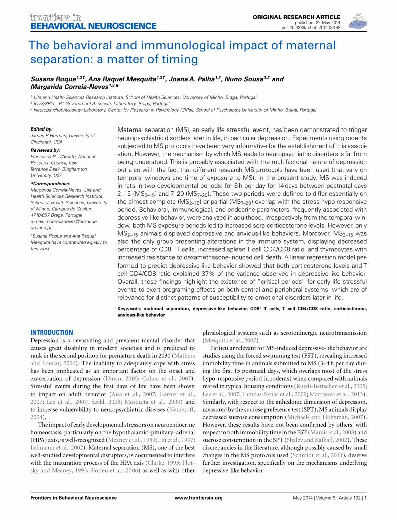
RESULTSANIMALS SUBMITTED TO MS2–15 DISPLAYED DEPRESSIVE-LIKEBEHAVIORIn the FST, only the animals from the group separated earlier(MS2–15) displayed shorter latency time to immobility (Figure 1A;
Table 1 | Antibodies used for the identification of different immune
cells.
Immune cells Phenotype spleen cells
CD4+ T cells CD3+ CD4+ (gated lymphocytes in FSC,
SSC)
CD8+ T cells CD3+ CD8+ (gated lymphocytes in FSC,
SSC)
B cells CD45Ra+ (gated lymphocytes in FSC,
SSC)
Macrophages CD11bc+ (gated in all cells except
granulocytes in FSC, SSC)
Granulocytes CD11bc+ granulocytes+ (gated
granulocytes in FSC, SSC)
NK cells CD3− CD161high+
Phenotype thymocytes
DN CD4−CD8− (gated
lymphocytes
in FSC, SSC)DP CD4+CD8+
CD4 SP CD4+CD8−
CD8 SP CD4−CD8+
Table 2 | Identification of cell death.
Phenotype
Alive Annexin V− 7AAD−
Apoptosis Annexin V+ 7AAD−
Necrosis Annexin V± 7AAD+
F 2,41= 7.92, p= 0.001 and η2= 0.28) when compared to the
Cont group (p= 0.009). Accordingly, MS2–15 displayed a signif-icant increase in the immobility time (Figure 1B; F 2,41= 4.41,p= 0.02 and η2
= 0.18) when compared with Cont animals(p= 0.049) and with MS7–20 group, in the FST (p= 0.03). Nosignificant differences were observed between MS7–20 and Contgroup (Figure 1B; p= 0.997). Considering that FST is a highlydemanding motor task, we performed the OF test to controlfor locomotor impairment that could underlie the significantreduction of activity observed in the FST. No differences wereobserved between groups in the total distance traveled in the arena(Figure 2A; F 2,42= 0.66, p= 0.52) and in the number (Figure 2B;F 2,42= 1.25, p= 0.30) and duration (Figure 2C; F 2,42= 1.4,p= 0.30) of rearings. In fact, MS, irrespectively from when itoccurred, did not affect spontaneous locomotion (Figure 2A) orexploratory behavior (Figures 2B,C). Conversely, OF results alsodemonstrated that the percentage of time spent in the center ofthe arena was significantly reduced in the MS2–15 (Figure 2D;F 2,42= 3.41, p= 0.04, and η2
= 0.14), when compared to Contanimals (p= 0.04), which is a sign of anxious-like behavior.MS7–20 did not differ from both Cont (p= 0.41) and MS2–15
(p= 0.52) groups in the percentage of time spent in the centerof the arena.
Frontiers in Behavioral Neuroscience www.frontiersin.org May 2014 | Volume 8 | Article 192 | 3
Roque et al. Immune–behavior consequences of maternal separation
FIGURE 1 | Early maternal separation (MS2–15) induceddepressive-like behavior. (A) Latency to first immobility and(B) duration of immobility in the FST were assessed in Cont, MS2–15,
and MS7–20 groups at 3 months of age. Each bar represents themean+SEM from 11 to 20 rats per group from one of twoindependent experiments.
FIGURE 2 | MS2–15 group presented anxious-like behavior in the OF test.The OF test was performed to assess (A) total distance, (B) number,(C) duration of rearings, and (D) time spent in the center of the OF arena in
Cont, MS2–15, and MS7–20 groups at 3 months of age. Each bar represents themean+SEM from 11 to 20 rats per group from one of two independentexperiments.
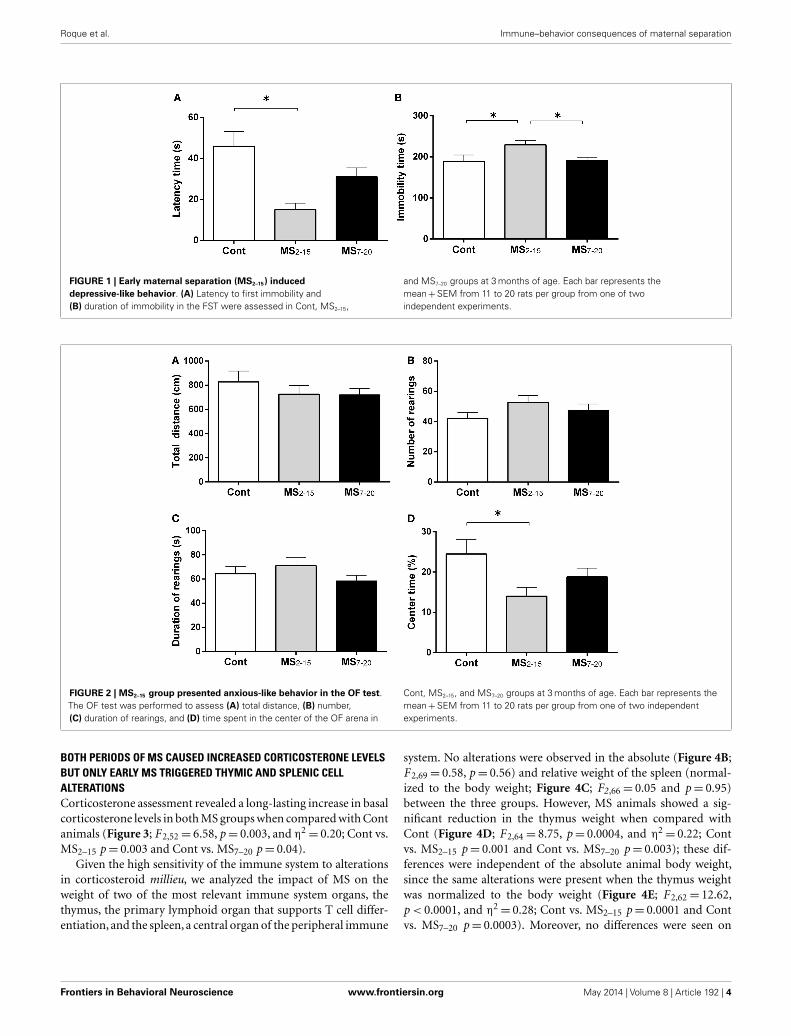
BOTH PERIODS OF MS CAUSED INCREASED CORTICOSTERONE LEVELSBUT ONLY EARLY MS TRIGGERED THYMIC AND SPLENIC CELLALTERATIONSCorticosterone assessment revealed a long-lasting increase in basalcorticosterone levels in both MS groups when compared with Contanimals (Figure 3; F 2,52= 6.58, p= 0.003, and η2
= 0.20; Cont vs.MS2–15 p= 0.003 and Cont vs. MS7–20 p= 0.04).
Given the high sensitivity of the immune system to alterationsin corticosteroid millieu, we analyzed the impact of MS on theweight of two of the most relevant immune system organs, thethymus, the primary lymphoid organ that supports T cell differ-entiation, and the spleen, a central organ of the peripheral immune
system. No alterations were observed in the absolute (Figure 4B;F 2,69= 0.58, p= 0.56) and relative weight of the spleen (normal-ized to the body weight; Figure 4C; F2,66= 0.05 and p= 0.95)between the three groups. However, MS animals showed a sig-nificant reduction in the thymus weight when compared withCont (Figure 4D; F 2,64= 8.75, p= 0.0004, and η2
= 0.22; Contvs. MS2–15 p= 0.001 and Cont vs. MS7–20 p= 0.003); these dif-ferences were independent of the absolute animal body weight,since the same alterations were present when the thymus weightwas normalized to the body weight (Figure 4E; F 2,62= 12.62,p < 0.0001, and η2
= 0.28; Cont vs. MS2–15 p= 0.0001 and Contvs. MS7–20 p= 0.0003). Moreover, no differences were seen on
Frontiers in Behavioral Neuroscience www.frontiersin.org May 2014 | Volume 8 | Article 192 | 4
Roque et al. Immune–behavior consequences of maternal separation
FIGURE 3 | Maternal separation induced increased corticosteroneserum levels. Each bar represents the mean+SEM from 16 to 21 rats pergroup from two independent experiments.
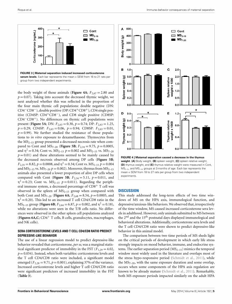
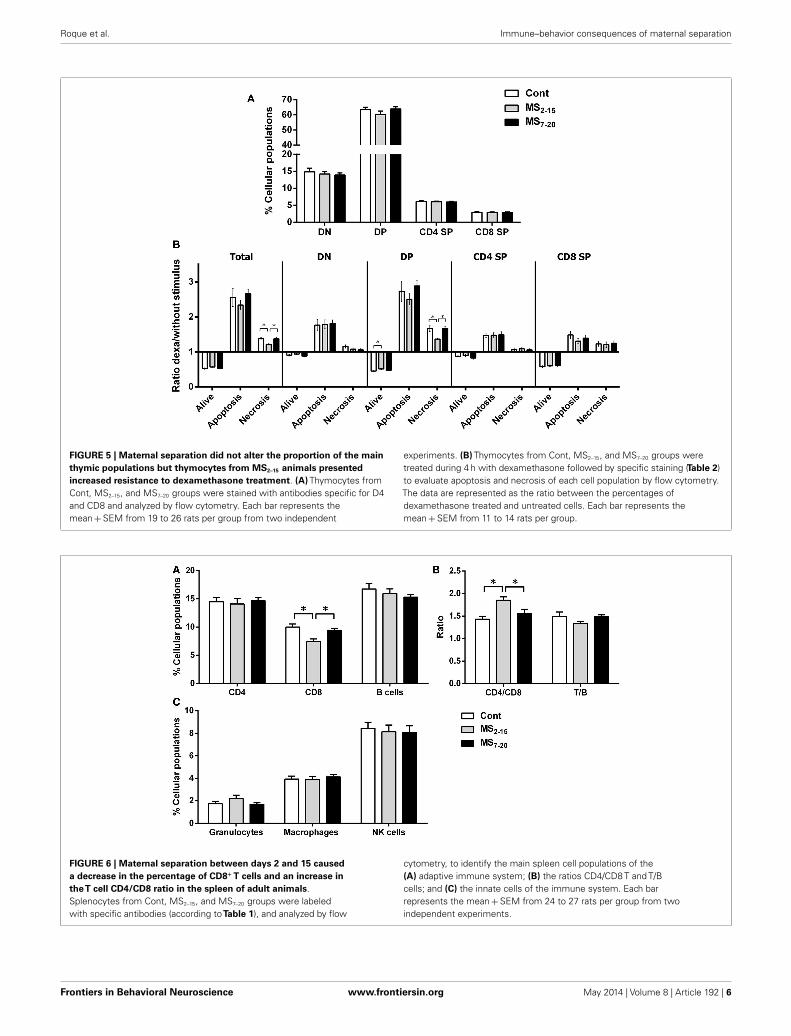
the body weight of those animals (Figure 4A; F 2,67= 2.80 andp= 0.07). Taking into account the decreased thymic weight, wenext analyzed whether this was reflected in the proportion ofthe four main thymic cell populations: double negative (DN:CD4−CD8−), double positive (DP: CD4+CD8+), CD4 single pos-itive (CD4SP: CD4+CD8−), and CD8 single positive (CD8SP:CD4−CD8+). No differences on thymic cell populations werepresent (Figure 5A; DN: F 2,63= 0.30, p= 0.74; DP: F 2,63= 1.25,p= 0.29; CD4SP: F 2,63= 0.06, p= 0.94; CD8SP: F 2,63= 0.01,p= 0.99). We further studied the resistance of those popula-tions to in vitro exposure to dexamethasone. Thymocytes fromthe MS2–15 group presented a decreased necrosis rate when com-pared to Cont and MS7–20 (Figure 5B; F 2,34= 9.75, p= 0.0005,and η2
= 0.34; Cont vs. MS2–15 p= 0.002 and MS2–15 vs. MS7–20
p= 0.01) and these alterations seemed to be mainly caused bythe decreased necrosis observed among DP cells (Figure 5B;F 2,35= 8.82, p= 0.0008, and η2
= 0.34; Cont vs. MS2–15 p= 0.004and MS2–15 vs. MS7–20 p= 0.002). Moreover, thymus from MS2–15
animals also presented a lower proportion of alive DP cells whencompared with Cont (Figure 5B; F 2,34= 5.11, p= 0.011, andη2= 0.23; Cont vs. MS2–15 p= 0.011). Regarding the periph-
eral immune system, a decreased percentage of CD8+ T cell wasobserved in the spleen of MS2–15 group when compared withboth Cont and MS7–20 (Figure 6A; F 2,68= 8.54, p= 0.0005, andη2= 0.20). This led to an increased T cell CD4/CD8 ratio in the
MS2–15 group (Figure 6B; F 2,64= 6.87, p= 0.002, and η2= 0.18),
while no alterations were seen in the T/B cells ratio. No differ-ences were observed in the other spleen cell populations analyzed(Figures 6A,C; CD4+ T cells, B cells, granulocytes, macrophages,and NK cells).
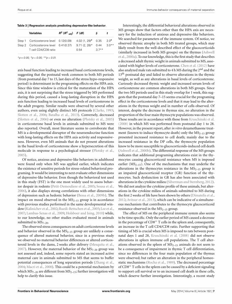
SERA CORTICOSTERONE LEVELS AND T CELL CD4/CD8 RATIO PREDICTDEPRESSIVE-LIKE BEHAVIORThe use of a linear regression model to predict depressive-likebehavior revealed that corticosterone, per se, was a marginal statis-tical significant predictor of immobility in the FST (F 1,29= 4.02,p= 0.054). Instead, when both variables: corticosterone levels andthe T cell CD4/CD8 ratio were included, a significant modelemerged (F 2,28= 9.71, p= 0.001), explaining 37% of the variance.Increased corticosterone levels and higher T cell CD4/CD8 ratiowere significant predictors of increased immobility in the FST(Table 3).
FIGURE 4 | Maternal separation caused a decrease in the thymusweight. (A) Body weight, (B) spleen weight, (C) spleen relative weight,(D) thymus weight, and (E) thymus relative weight were measured in Cont,MS2–15, and MS7–20 groups at 3 months of age. Each bar represents themean+SEM from 18 to 27 rats per group from two independentexperiments.
DISCUSSIONThis study addressed the long-term effects of two time win-dows of MS on the HPA axis, immunological function, anddepressive/anxious-like behaviors. We observed that, irrespectivelyof the time window, MS caused increased corticosterone sera lev-els in adulthood. However, only animals submitted to MS betweenthe 2nd and the 15th postnatal days displayed immunological andbehavioral alterations. Additionally, corticosterone sera levels andthe T cell CD4/CD8 ratio were shown to predict depressive-likebehavior in this animal model.
The comparison between two time periods of MS sheds lighton the critical periods of development in which early life stressstrongly impacts on mood behavior, immune, and endocrine sys-tems. The earlier separation period (MS2–15) mimics the temporalwindow most widely used in the literature and overlaps most ofthe stress hypo-responsive period (Schmidt et al., 2011), whilethe MS7–20, with the same exposure duration and some overlap,occurs when some components of the HPA axis regulation areknown to be already mature (Schmidt et al., 2011). Remarkably,both MS exposure periods impacted similarly on the adult HPA
Frontiers in Behavioral Neuroscience www.frontiersin.org May 2014 | Volume 8 | Article 192 | 5
Roque et al. Immune–behavior consequences of maternal separation
FIGURE 5 | Maternal separation did not alter the proportion of the mainthymic populations but thymocytes from MS2–15 animals presentedincreased resistance to dexamethasone treatment. (A) Thymocytes fromCont, MS2–15, and MS7–20 groups were stained with antibodies specific for D4and CD8 and analyzed by flow cytometry. Each bar represents themean+SEM from 19 to 26 rats per group from two independent
experiments. (B) Thymocytes from Cont, MS2–15, and MS7–20 groups weretreated during 4 h with dexamethasone followed by specific staining (Table 2)to evaluate apoptosis and necrosis of each cell population by flow cytometry.The data are represented as the ratio between the percentages ofdexamethasone treated and untreated cells. Each bar represents themean+SEM from 11 to 14 rats per group.
FIGURE 6 | Maternal separation between days 2 and 15 causeda decrease in the percentage of CD8+ T cells and an increase intheT cell CD4/CD8 ratio in the spleen of adult animals.Splenocytes from Cont, MS2–15, and MS7–20 groups were labeledwith specific antibodies (according toTable 1), and analyzed by flow
cytometry, to identify the main spleen cell populations of the(A) adaptive immune system; (B) the ratios CD4/CD8 T and T/Bcells; and (C) the innate cells of the immune system. Each barrepresents the mean+SEM from 24 to 27 rats per group from twoindependent experiments.
Frontiers in Behavioral Neuroscience www.frontiersin.org May 2014 | Volume 8 | Article 192 | 6
Roque et al. Immune–behavior consequences of maternal separation
Table 3 | Regression analysis predicting depressive-like behavior.
Variables R2 (R2adj) F (df) β t
Step 1 Corticosterone level 0.12(0.09) 4.02 (1, 29)# 0.35 2.0#
Step 2 Corticosterone level 0.41(0.37) 9.71 (2, 28)* 0.44 3.0**
T cell CD4/CD8 ratio 0.54 3.7**
#p=0.05; *p < 0.05; **p < 0.01.
axis basal function leading to increased basal corticosterone levels,suggesting that the postnatal week common to both MS periods(from postnatal day 7 to 15, last days of the stress hypo-responsiveperiod) is determinant in the programing effects on the HPA axis.Since this time window is critical for the maturation of the HPAaxis, it is not surprising that the stress triggered by MS performedduring this period, caused a long-lasting disruption in the HPAaxis function leading to increased basal levels of corticosterone inthe adult progeny. Similar results were observed by several otherauthors, even using slightly distinct MS protocols (Clarke, 1993;Slotten et al., 2006; Batalha et al., 2013). Conversely, decreased(Slotten et al., 2006) or even no alteration (Plotsky et al., 2005)in corticosterone basal levels in animals submitted to MS werealso reported. Overall, most literature seems to corroborate thatMS is a developmental disruptor of the neuroendocrine functionwith long-lasting effects on the HPA axis activity and responsive-ness. However, even MS animals that do not present alterationsin the basal levels of corticosterone show a hypersecretion of thismolecule in response to a psychological stressor (Plotsky et al.,2005).
Of notice, anxious and depressive-like behaviors in adulthoodwere found only when MS was applied earlier, which indicatesthe existence of sensitive periods for stress-related behavioral pro-graming. It would be interesting to next evaluate other dimensionsof depressive-like behavior. Even though the behavioral test usedin this study (FST) is the one most widely used to assess behav-ior despair in rodents (Petit-Demouliere et al., 2005; Sousa et al.,2006), it also displays strong correlations with other dimensionsof depression such as hedonic behavior (Bessa et al., 2009b). Theimpact on mood observed in the MS2–15 group is in accordancewith previous studies performed in the same developmental win-dow (Kalinichev et al., 2002; Ruedi-Bettschen et al., 2005; Lee et al.,2007; Lambas-Senas et al., 2009; Holsboer and Ising, 2010) while,to our knowledge, no other studies evaluated mood in animalssubmitted to MS7–20.
The observed stress consequences on adult corticosterone levelsand behavior observed in the MS2–15 group are unlikely a conse-quence of altered maternal behavior, since in a previous studywe observed no maternal behavior differences or altered corticos-teroid levels in the dams, 2 weeks after delivery (Mesquita et al.,2007). However, the maternal behavior of the MS7–20 group wasnot assessed and, indeed, some reports stated an increased activematernal care in animals submitted to MS that seems to bufferpotential consequences of long separation periods (Zhang et al.,2004; Macri et al., 2008). This could be a potential mechanism bywhich MS7–20 are different from MS2–15; further investigation willhelp to clarify this issue.
Interestingly, the differential behavioral alterations observed inMS groups show that factors other than the HPA axis are neces-sary for the induction of anxious and depressive-like behaviors.We searched for parameters of the immune system. Of notice, weobserved thymic atrophy in both MS treated groups, which maylikely result from the well-described effect of the glucocorticoids(similarly increased in both MS groups) on the thymus (Ashwellet al., 2000a). To our knowledge, this is the first study that describesa decreased adult thymic weight in animals submitted to MS, asso-ciated with higher levels of corticosterone. Chen et al. (2012) havealso analyzed male rats submitted to 4 h MS during the 2nd and the13th postnatal day and failed to observe alterations in the thymicweight, as well as any alterations in basal levels of corticosterone.Curiously decreased thymic weight and increased serum levels ofcorticosterone are common alterations in both MS groups. Sincethe two MS periods used in this study overlap for 1 week, this sug-gests that the postnatal day 7–15 seems crucial for the programingeffect in the corticosterone levels and that it may lead to the alter-ations in the thymus weight and in number of cells observed. Ofinterest, despite the decrease in thymus size, no alteration in theproportion of the four main thymocyte populations was observed.These results are in accordance with those from Kruschinski et al.(2008) in which MS was performed from postnatal day 1 to 28.However, in the present report, after in vitro dexamethasone treat-ment (known to induce thymocyte death) only the MS2–15 grouppresented increased resistance to cell death; mainly due to theincreased resistance in the DP cells, the thymocyte populationknow to be more susceptible to glucocorticoids-induced cell death(Ashwell et al., 2000b). The differential response of the MS groupsindicates that additional programing alterations exist in the thy-mocytes causing glucocorticoid resistance when MS is imposedearlier (MS2–15). One of the mechanisms that may underlie thealterations in the thymocytes resistance in the MS2–15 group isan impaired glucocorticoid receptor (GR) function of the thy-mocytes. Such dysfunction in GR has also been associated withalterations in the cytokine milieu (Silverman and Sternberg, 2012).We did not analyze the cytokine profile of these animals, but alter-ations in the cytokine milieu of animals submitted to MS duringthe first 2 weeks of life have been shown by others (Dimatelis et al.,2012; Avitsur et al., 2013), which can be indicative of a simultane-ous mechanism that contributes to the thymocyte glucocorticoidresistance observed in the MS2–15 group.
The effect of MS on the peripheral immune system also seemsto be time specific. Only the earlier period of MS caused a decreasein the percentage of CD8+ T cells in the spleen and, consequently,an increase in the T cell CD4/CD8 ratio. Further supporting thattiming of MS is crucial when MS is imposed to rats between post-natal days 1 and 28, Kruschinski et al. (2008) did not observealterations in spleen immune cell populations. The T cell alter-ations observed in the spleen of MS2–15 animals do not seem tobe a consequence of impairment in thymic T cell differentiation,since no differences in the four main population of the thymuswere observed; but rather an alteration in the peripheral homeo-static mechanisms (Rocha et al., 1989). The decreased percentageof CD8+ T cells in the spleen can be caused by a deficient signalingto support cell survival or to an increased cell death in these cells,which deserve further investigation. Interestingly, a recent study
Frontiers in Behavioral Neuroscience www.frontiersin.org May 2014 | Volume 8 | Article 192 | 7

Roque et al. Immune–behavior consequences of maternal separation
showed that CD8+ T cells display a more pronounced expressionof dopaminergic transporters and receptors when compared withCD4+ T cells (Mignini et al., 2013) and, dopamine was shown toinhibit the proliferation of T cells, particularly of CD8+ T cells(Saha et al., 2001). Given that MS impacts on the dopaminer-gic system, it is plausible that this might constitute an additionalmechanism for the present finding of reduced CD8+ T cells (Liet al., 2013). Moreover, these T cells are primarily involved inimmune response to pathogens (mainly virus) and tumor cells,and are also implicated in transplant rejection. Curiously, C56BL/6mice exposed to MS between postnatal days 1 and 14 were shown topresent increased susceptibility to influenza virus infection; how-ever, the levels of CD8+ T cells were not evaluated in these study(Avitsur et al., 2006).
Our findings are novel in revealing an increased T cell CD4/CD8ratio, which is a highly preserved ratio within strains of animals(Rocha et al., 1989; Sim et al., 1998). Of notice, another rodentmodel of depressive-like behavior, induced by prenatal admin-istration of dexamethasone, showed that adult males similarlydisplay increased T cell CD4/CD8 ratio and that the percentageof CD8+ T cells is negatively correlated with the latency in theFST (Roque et al., 2011). In humans and monkeys submitted toearly life stress, alterations in T cell CD4/CD8 ratio have also beenreported (Lewis et al., 2000; Gogberashvili, 2006), even thoughin the opposite direction of what we describe herein. Remark-ably, our results reveal that differences in immune cell populationsare only present in animals displaying behavioral alterations, sug-gesting that the programing effects caused by MS impact notonly in the endocrine system but also in the immune and cen-tral nervous systems, highlighting the interplay between them.Curiously, lower proportion of CD8+ T cells and a higher T cellCD4/CD8 ratio have been associated with lower levels of hip-pocampal neuronal proliferation (Huang et al., 2010). Moreover,although disputable, most studies show that MS decreases hip-pocampal cell proliferation (Hulshof et al., 2011), which in itself isassociated with depressive-like behavior (Bessa et al., 2009a; Songand Wang, 2011).
The fact that the two MS periods do not overlap in the firstand third postnatal week could also be crucial for the distincteffects, we observed in behavior and in parameters of the immunesystem. Considering that during the first postnatal week signifi-cant differences in the number and maturation process of GR inthe hypothalamus, pituitary, and hippocampus were observed (DeKloet et al., 1988), one can speculate that stressful events duringthis very immature period could be fairly impactful on long-termbehavior. In fact, early life events may lead to long-term epigeneticeffects such as down regulation of hippocampal GR through theincreased methylation patter in the coding gene (Weaver et al.,2004). A similar mechanism could also be caused by very earlyMS, since the first postnatal week is the most immature period ofthe HPA axis control. In the MS7–20 period, the HPA axis alreadypresents an increased degree of maturation and number of GRin critical regions for HPA axis control (Vazquez and Akil, 1993).Of interest, GR are solely found in the hippocampus in the sec-ond postnatal week (Vazquez and Akil, 1993). The presence of GR(even in low levels) during the second and third postnatal weekcould be crucial for the organism to respond to stressful situations
and buffer some of the long-term behavioral effects, such as anx-ious and depressive behaviors. Although speculative, this could bea likely explanation since we observed an increased anxious anddepressive-like phenotype in the earlier MS group (MS2–15) andnot in the later one (MS7–20).
As an attempt to assess the role of both corticosterone levels andT cell CD4/CD8 ratio in the depressive-like behavior observed inour MS model, we performed a regression analysis. Both predic-tors explained 37% of the immobility time in the FST, suggestinga synergetic contribution of endocrine and immunological factorsin the prediction of the depressive-like behavior observed in theMS animals. Of notice, corticosterone per se, accounted only for9% of the variance and was merely a marginal significant pre-dictor. We failed to find any predictors of anxious-like behavior.One should take into account that the depressive-like behavior, theincreased corticosterone levels, and the alterations in the immunesystem seem to be related and caused by the effect of MS in anearly life period. Even though additional studies are needed, ourdata suggests that this information might be potentially used todetermine whether the predictive model for the depressive-likebehavior can be a tool to predict if children submitted to adverseexperiences in early life are more prone to develop mood dis-orders, which could eventually help in anticipating diagnosis ofdepression.
Altogether, this study shows that the timing in which early lifestress is applied determines its long-lasting effects in several bodysystems, and that these interact in ways that may be predictive ofmood behavior alterations in the adulthood.
AUTHOR CONTRIBUTIONSS. Roque performed the immune cells phenotyping experimentsand analysis. A. R. Mesquita performed the MS protocol, thebehavior assessment and analysis, and the corticosterone mea-surement. Both S. Roque and A. R. Mesquita performed thestatistical data analysis. All authors contributed to the planning ofthe experiments, data interpretation, writing of the manuscript,and approved the final version of the manuscript.
ACKNOWLEDGMENTSWe acknowledge the Portuguese Foundation for Science andTechnology (FCT) for providing a fellowship to S. Roque(SFRH/BPD/72710/2010). This work was also supported byFCT grants (co-financed by COMPETE funds) PTDC/SAU-NEU/105180/2008 and PTDC/PSI-PCO/116612/2010 and co-financed by the Portuguese North Regional Operational Pro-gram (ON.2 – O Novo Norte) under the National StrategicReference Framework (QREN), through the European RegionalDevelopment Fund (FEDER).
REFERENCESAisa, B., Tordera, R., Lasheras, B., Del Rio, J., and Ramirez, M. J. (2007). Cogni-
tive impairment associated to HPA axis hyperactivity after maternal separationin rats. Psychoneuroendocrinology 32, 256–266. doi:10.1016/j.psyneuen.2006.12.013
Ashwell, J. D., Lu, F. W., and Vacchio, M. S. (2000a). Glucocorticoids in T cell devel-opment and function. Annu. Rev. Immunol. 18, 309–345. doi:10.1146/annurev.immunol.18.1.309
Frontiers in Behavioral Neuroscience www.frontiersin.org May 2014 | Volume 8 | Article 192 | 8

Roque et al. Immune–behavior consequences of maternal separation
Ashwell, J. D., Lu, F. W., and Vacchio, M. S. (2000b). Glucocorticoids in T cell devel-opment and function*. Annu. Rev. Immunol. 18, 309–345. doi:10.1146/annurev.immunol.18.1.309
Avitsur, R., Hunzeker, J., and Sheridan, J. F. (2006). Role of early stress in the indi-vidual differences in host response to viral infection. Brain Behav. Immun. 20,339–348. doi:10.1016/j.bbi.2005.09.006
Avitsur, R., Maayan, R., and Weizman, A. (2013). Neonatal stress modulates sick-ness behavior: role for proinflammatory cytokines. J. Neuroimmunol. 257, 59–66.doi:10.1016/j.jneuroim.2013.02.009
Batalha, V. L., Pego, J. M., Fontinha, B. M., Costenla, A. R., Valadas, J. S., Baqi,Y., et al. (2013). Adenosine A(2A) receptor blockade reverts hippocampal stress-induced deficits and restores corticosterone circadian oscillation. Mol. Psychiatry18, 320–331. doi:10.1038/mp.2012.8
Bessa, J. M., Ferreira, D., Melo, I., Marques, F., Cerqueira, J. J., Palha, J. A., et al.(2009a). The mood-improving actions of antidepressants do not depend onneurogenesis but are associated with neuronal remodeling. Mol. Psychiatry 14,739. doi:10.1038/mp.2008.119
Bessa, J. M., Mesquita, A. R., Oliveira, M., Pego, J. M., Cerqueira, J. J., Palha, J. A., et al.(2009b). A trans-dimensional approach to the behavioral aspects of depression.Front. Behav. Neurosci. 3:1. doi:10.3389/neuro.08.001.2009
Cao, X., Huang, S., Cao, J., Chen, T., Zhu, P., Zhu, R., et al. (2013). The timingof maternal separation affects morris water maze performance and long-termpotentiation in male rats. Dev. Psychobiol. doi:10.1002/dev.21130
Chen, J., Evans, A. N., Liu, Y., Honda, M., Saavedra, J. M., and Aguilera, G.(2012). Maternal deprivation in rats is associated with corticotrophin-releasinghormone (CRH) promoter hypomethylation and enhances CRH transcrip-tional responses to stress in adulthood. J. Neuroendocrinol. 24, 1055–1064.doi:10.1111/j.1365-2826.2012.02306.x
Clarke, A. S. (1993). Social rearing effects on HPA axis activity over early develop-ment and in response to stress in rhesus monkeys. Dev. Psychobiol. 26, 433–446.doi:10.1002/dev.420260802
Cohen, J. (1988). Statistical Power Analysis for the Behavioral Sciences. New York, NY:Routledge Academic.
Cohen, S., Janicki-Deverts, D., and Miller, G. E. (2007). Psychological stress anddisease. JAMA 298, 1685–1687. doi:10.1001/jama.298.14.1685
Colorado, R. A., Shumake, J., Conejo, N. M., Gonzalez-Pardo, H., and Gonzalez-Lima, F. (2006). Effects of maternal separation, early handling, and standardfacility rearing on orienting and impulsive behavior of adolescent rats. Behav.Processes 71, 51–58. doi:10.1016/j.beproc.2005.09.007
De Kloet, E. R., Rosenfeld, P., Van Eekelen, J. A., Sutanto, W., and Levine, S.(1988). Stress, glucocorticoids and development. Prog. Brain Res. 73, 101–120.doi:10.1016/S0079-6123(08)60500-2
Diehl, L. A., Alvares, L. O., Noschang, C., Engelke, D., Andreazza, A. C., Goncalves, C.A., et al. (2012). Long-lasting effects of maternal separation on an animal modelof post-traumatic stress disorder: effects on memory and hippocampal oxidativestress. Neurochem. Res. 37, 700–707. doi:10.1007/s11064-011-0660-6
Dimatelis, J. J., Pillay, N. S., Mutyaba, A. K., Russell, V. A., Daniels, W. M.,and Stein, D. J. (2012). Early maternal separation leads to down-regulation ofcytokine gene expression. Metab. Brain Dis. 27, 393–397. doi:10.1007/s11011-012-9304-z
Dinan, T. G. (2005). Stress: the shared common component in major mental ill-nesses. Eur. Psychiatry 20(Suppl. 3), S326–S328. doi:10.1016/S0924-9338(05)80184-1
Garner, B., Wood, S. J., Pantelis, C., and Van Den Buuse, M. (2007). Early maternaldeprivation reduces prepulse inhibition and impairs spatial learning ability inadulthood: no further effect of post-pubertal chronic corticosterone treatment.Behav. Brain Res. 176, 323–332. doi:10.1016/j.bbr.2006.10.020
Gogberashvili, K. (2006). Maternal deprivation, acute respiratory infections andimmune regulation. Indian J. Pediatr. 73, 995–997. doi:10.1007/BF02758305
Holsboer, F., and Ising, M. (2010). Stress hormone regulation: biological roleand translation into therapy. Annu. Rev. Psychol. 61, C101–C111. doi:10.1146/annurev.psych.093008.100321
Huang, G. J., Smith, A. L., Gray, D. H., Cosgrove, C., Singer, B. H., Edwards, A., et al.(2010). A genetic and functional relationship between T cells and cellular prolif-eration in the adult hippocampus. PLoS Biol. 8:e1000561. doi:10.1371/journal.pbio.1000561
Hulshof, H. J., Novati, A., Sgoifo, A., Luiten, P. G., Den Boer, J. A., and Meerlo, P.(2011). Maternal separation decreases adult hippocampal cell proliferation and
impairs cognitive performance but has little effect on stress sensitivity and anxi-ety in adult Wistar rats. Behav. Brain Res. 216, 552–560. doi:10.1016/j.bbr.2010.08.038
Kalinichev, M., Easterling, K. W., Plotsky, P. M., and Holtzman, S. G. (2002). Long-lasting changes in stress-induced corticosterone response and anxiety-like behav-iors as a consequence of neonatal maternal separation in Long-Evans rats. Phar-macol. Biochem. Behav. 73, 131–140. doi:10.1016/S0091-3057(02)00781-5
Kruschinski,C.,Skripuletz,T.,Bedoui,S.,Raber,K.,Straub,R. H.,Hoffmann,T., et al.(2008). Postnatal life events affect the severity of asthmatic airway inflammationin the adult rat. J. Immunol. 180, 3919–3925. doi:10.4049/jimmunol.180.6.3919
Lambas-Senas, L., Mnie-Filali, O., Certin,V., Faure, C., Lemoine, L., Zimmer, L., et al.(2009). Functional correlates for 5-HT(1A) receptors in maternally deprived ratsdisplaying anxiety and depression-like behaviors. Prog. Neuropsychopharmacol.Biol. Psychiatry 33, 262–268. doi:10.1016/j.pnpbp.2008.11.017
Lee, J. H., Kim, H. J., Kim, J. G., Ryu, V., Kim, B. T., Kang, D. W., et al. (2007).Depressive behaviors and decreased expression of serotonin reuptake transporterin rats that experienced neonatal maternal separation. Neurosci. Res. 58, 32–39.doi:10.1016/j.neures.2007.01.008
Lehmann, J., Russig, H., Feldon, J., and Pryce, C. R. (2002). Effect of a sin-gle maternal separation at different pup ages on the corticosterone stressresponse in adult and aged rats. Pharmacol. Biochem. Behav. 73, 141–145.doi:10.1016/S0091-3057(02)00788-8
Lewis, M. H., Gluck, J. P., Petitto, J. M., Hensley, L. L., and Ozer, H. (2000). Earlysocial deprivation in nonhuman primates: long-term effects on survival and cell-mediated immunity. Biol. Psychiatry 47, 119–126. doi:10.1016/S0006-3223(99)00238-3
Li, M., Xue, X., Shao, S., Shao, F., and Wang, W. (2013). Cognitive, emotional andneurochemical effects of repeated maternal separation in adolescent rats. BrainRes. 1518, 82–90. doi:10.1016/j.brainres.2013.04.026
Liu, D., Diorio, J., Tannenbaum, B., Caldji, C., Francis, D., Freedman, A., et al.(1997). Maternal care,hippocampal glucocorticoid receptors,and hypothalamic-pituitary-adrenal responses to stress. Science 277, 1659–1662. doi:10.1126/science.277.5332.1659
Macri, S., Chiarotti, F., and Wurbel, H. (2008). Maternal separation and maternalcare act independently on the development of HPA responses in male rats. Behav.Brain Res. 191, 227–234. doi:10.1016/j.bbr.2008.03.031
Marais, L., Van Rensburg, S. J., Van Zyl, J. M., Stein, D. J., and Daniels, W.M. (2008). Maternal separation of rat pups increases the risk of developingdepressive-like behavior after subsequent chronic stress by altering corticos-terone and neurotrophin levels in the hippocampus. Neurosci. Res. 61, 106–112.doi:10.1016/j.neures.2008.01.011
Martisova, E., Solas, M., Horrillo, I., Ortega, J. E., Meana, J. J., Tordera, R. M., et al.(2012). Long lasting effects of early-life stress on glutamatergic/GABAergic cir-cuitry in the rat hippocampus. Neuropharmacology 62, 1944–1953. doi:10.1016/j.neuropharm.2011.12.019
Mathers, C. D., and Loncar, D. (2006). Projections of global mortality and burden ofdisease from 2002 to 2030. PLoS Med. 3:e442. doi:10.1371/journal.pmed.0030442
Matthews, K., and Robbins, T. W. (2003). Early experience as a determinant of adultbehavioural responses to reward: the effects of repeated maternal separation inthe rat. Neurosci. Biobehav. Rev. 27, 45–55. doi:10.1016/S0149-7634(03)00008-3
Meaney, M. J., Aitken, D. H., Viau, V., Sharma, S., and Sarrieau, A. (1989). Neona-tal handling alters adrenocortical negative feedback sensitivity and hippocam-pal type II glucocorticoid receptor binding in the rat. Neuroendocrinology 50,597–604. doi:10.1159/000125287
Mesquita, A. R., Pego, J. M., Summavielle, T., Maciel, P., Almeida, O. F., and Sousa, N.(2007). Neurodevelopment milestone abnormalities in rats exposed to stress inearly life. Neuroscience 147, 1022–1033. doi:10.1016/j.neuroscience.2007.04.007
Mesquita, A. R., Wegerich, Y., Patchev, A. V., Oliveira, M., Leao, P., Sousa, N., et al.(2009). Glucocorticoids and neuro- and behavioural development. Semin. FetalNeonatal Med. 14, 130–135. doi:10.1016/j.siny.2008.11.002
Michaels, C. C., and Holtzman, S. G. (2007). Enhanced sensitivity to naltrexone-induced drinking suppression of fluid intake and sucrose consumption in mater-nally separated rats. Pharmacol. Biochem. Behav. 86, 784–796. doi:10.1016/j.pbb.2007.03.007
Mignini, F., Sabbatini, M., Capacchietti, M., Amantini, C., Bianchi, E., Artico, M.,et al. (2013). T-cell subpopulations express a different pattern of dopaminergicmarkers in intra- and extra-thymic compartments. J. Biol. Regul. Homeost. Agents27, 463–475.
Frontiers in Behavioral Neuroscience www.frontiersin.org May 2014 | Volume 8 | Article 192 | 9

Roque et al. Immune–behavior consequences of maternal separation
Miller, A. H. (2010). Depression and immunity: a role for T cells? Brain Behav.Immun. 24, 1–8. doi:10.1016/j.bbi.2009.09.009
Nemeroff, C. B. (2004). Neurobiological consequences of childhood trauma. J. Clin.Psychiatry 65(Suppl. 1), 18–28.
Petit-Demouliere, B., Chenu, F., and Bourin, M. (2005). Forced swimming testin mice: a review of antidepressant activity. Psychopharmacology (Berl.) 177,245–255. doi:10.1007/s00213-004-2048-7
Plotsky, P. M., and Meaney, M. J. (1993). Early, postnatal experience alters hypo-thalamic corticotropin-releasing factor (CRF) mRNA, median eminence CRFcontent and stress-induced release in adult rats. Brain Res. Mol. Brain Res. 18,195–200. doi:10.1016/0169-328X(93)90189-V
Plotsky, P. M., Thrivikraman, K. V., Nemeroff, C. B., Caldji, C., Sharma, S., andMeaney, M. J. (2005). Long-term consequences of neonatal rearing on centralcorticotropin-releasing factor systems in adult male rat offspring. Neuropsy-chopharmacology 30, 2192–2204. doi:10.1038/sj.npp.1300769
Rocha, B., Dautigny, N., and Pereira, P. (1989). Peripheral T lymphocytes: expansionpotential and homeostatic regulation of pool sizes and CD4/CD8 ratios in vivo.Eur. J. Immunol. 19, 905–911. doi:10.1002/eji.1830190518
Roque, S., Oliveira, T. G., Nobrega, C., Barreira-Silva, P., Nunes-Alves, C., Sousa, N.,et al. (2011). Interplay between depressive-like behavior and the immune systemin an animal model of prenatal dexamethasone administration. Front. Behav.Neurosci. 5:4. doi:10.3389/fnbeh.2011.00004
Ruedi-Bettschen, D., Pedersen, E. M., Feldon, J., and Pryce, C. R. (2005). Earlydeprivation under specific conditions leads to reduced interest in reward inadulthood in Wistar rats. Behav. Brain Res. 156, 297–310. doi:10.1016/j.bbr.2004.06.001
Saha, B., Mondal, A. C., Majumder, J., Basu, S., and Dasgupta, P. S. (2001). Phys-iological concentrations of dopamine inhibit the proliferation and cytotoxicityof human CD4+ and CD8+ T cells in vitro: a receptor-mediated mechanism.Neuroimmunomodulation 9, 23–33. doi:10.1159/000049004
Schmidt, M. V., Wang, X. D., and Meijer, O. C. (2011). Early life stress paradigmsin rodents: potential animal models of depression? Psychopharmacology (Berl.)214, 131–140. doi:10.1007/s00213-010-2096-0
Seckl, J. R. (2008). Glucocorticoids, developmental ‘programming’ and the risk ofaffective dysfunction. Prog. Brain Res. 167, 17–34. doi:10.1016/S0079-6123(07)67002-2
Shalev, U., and Kafkafi, N. (2002). Repeated maternal separation does not altersucrose-reinforced and open-field behaviors. Pharmacol. Biochem. Behav. 73,115–122. doi:10.1016/S0091-3057(02)00756-6
Silverman, M. N., and Sternberg, E. M. (2012). Glucocorticoid regulation of inflam-mation and its functional correlates: from HPA axis to glucocorticoid receptor
dysfunction. Ann. N. Y. Acad. Sci. 1261, 55–63. doi:10.1111/j.1749-6632.2012.06633.x
Sim, B. C., Aftahi, N., Reilly, C., Bogen, B., Schwartz, R. H., Gascoigne, N. R., et al.(1998). Thymic skewing of the CD4/CD8 ratio maps with the T-cell receptoralpha-chain locus. Curr. Biol. 8, 701–704. doi:10.1016/S0960-9822(98)70276-3
Slotten, H. A., Kalinichev, M., Hagan, J. J., Marsden, C. A., and Fone, K. C. (2006).Long-lasting changes in behavioural and neuroendocrine indices in the rat fol-lowing neonatal maternal separation: gender-dependent effects. Brain Res. 1097,123–132. doi:10.1016/j.brainres.2006.04.066
Song, C., and Wang, H. (2011). Cytokines mediated inflammation and decreasedneurogenesis in animal models of depression. Prog. Neuropsychopharmacol. Biol.Psychiatry 35, 760–768. doi:10.1016/j.pnpbp.2010.06.020
Sousa, N., Almeida, O. F., and Wotjak, C. T. (2006). A hitchhiker’s guide to behav-ioral analysis in laboratory rodents. Genes Brain Behav. 5(Suppl. 2), 5–24.doi:10.1111/j.1601-183X.2006.00228.x
Vazquez, D. M., and Akil, H. (1993). Pituitary-adrenal response to ether vapor in theweanling animal: characterization of the inhibitory effect of glucocorticoids onadrenocorticotropin secretion. Pediatr. Res. 34, 646–653. doi:10.1203/00006450-199311000-00017
Weaver, I. C., Cervoni, N., Champagne, F. A., D’Alessio, A. C., Sharma, S., Seckl, J.R., et al. (2004). Epigenetic programming by maternal behavior. Nat. Neurosci.7, 847–854. doi:10.1038/nn1276
Zhang, T. Y., Parent, C., Weaver, I., and Meaney, M. J. (2004). Maternal programmingof individual differences in defensive responses in the rat. Ann. N. Y. Acad. Sci.1032, 85–103. doi:10.1196/annals.1314.007
Conflict of Interest Statement: The authors declare that the research was conductedin the absence of any commercial or financial relationships that could be construedas a potential conflict of interest.
Received: 08 December 2013; accepted: 08 May 2014; published online: 22 May 2014.Citation: Roque S, Mesquita AR, Palha JA, Sousa N and Correia-Neves M (2014)The behavioral and immunological impact of maternal separation: a matter of timing.Front. Behav. Neurosci. 8:192. doi: 10.3389/fnbeh.2014.00192This article was submitted to the journal Frontiers in Behavioral Neuroscience.Copyright © 2014 Roque, Mesquita, Palha, Sousa and Correia-Neves. This is an open-access article distributed under the terms of the Creative Commons Attribution License(CC BY). The use, distribution or reproduction in other forums is permitted, providedthe original author(s) or licensor are credited and that the original publication in thisjournal is cited, in accordance with accepted academic practice. No use, distribution orreproduction is permitted which does not comply with these terms.
Frontiers in Behavioral Neuroscience www.frontiersin.org May 2014 | Volume 8 | Article 192 | 10