
High definition fibrous poly (2-ethyl-2-oxazoline) scaffolds through melt electrospinning writing
Upload
independentCategory
view
0download
0
Device-based local delivery of siRNA against mammalian target ofrapamycin (mTOR) in a murine subcutaneous implant model toinhibit fibrous encapsulation
Hironobu Takahashia,†,1, Yuwei Wanga,†, and David W. Graingera,b,*Hironobu Takahashi: [email protected]; Yuwei Wang: [email protected]; David W. Grainger:[email protected] Department of Pharmaceutics and Pharmaceutical Chemistry, University of Utah, Salt Lake City,UT 84112-5820 USAb Department of Bioengineering, University of Utah, Salt Lake City, UT 84112-5820 USA
AbstractFibrous encapsulation of surgically implant devices is associated with elevated proliferation andactivation of fibroblasts in tissues surrounding these implants, frequently causing foreign bodycomplications. Here we test the hypothesis that inhibition of the expression of mammalian target ofrapamycin (mTOR) in fibroblasts can mitigate the soft tissue implant foreign body response bysuppressing fibrotic responses around implants. In this study, mTOR was knocked down using smallinterfering RNA conjugated with branched cationic polyethylenimine (bPEI) in fibroblastic lineagecells in serum-based cell culture as shown by both gene and protein analysis. This mTOR knockdownled to an inhibition in fibroblast proliferation by 70% and simultaneous down-regulation in theexpression of type I collagen in fibroblasts in vitro. These siRNA/bPEI complexes were releasedfrom poly(ethylene glycol) (PEG)-based hydrogel coatings surrounding model polymer implants ina subcutaneous rodent model in vivo. No significant reduction in fibrous capsule thickness and mTORexpression in the foreign body capsules was observed. Observed siRNA inefficacy in this in vivoimplant model was attributed to siRNA dosing limitations in the gel delivery system, and lack oftargeting ability of the siRNA complex specifically to fibroblasts. While in vitro data supportedmTOR knock-down in fibroblast cultures, in vivo siRNA delivery must be further improved toproduce clinically relevant effects on fibrotic encapsulation around implants.
Keywordsforeign body reaction; fibrous capsule; mTOR siRNA; local delivery; fibrosis; implant
*Correspondence: David W. Grainger, Ph.D, Department of Pharmaceutics and Pharmaceutical Chemistry, University of Utah, Salt LakeCity, UT 84112-5820 USA, Phone: +1 801-581-3715, Fax: +1 801-581-3674.†Authors contributed equally to the work1Present address: Institute of Advanced Biomedical Engineering and Science Tokyo, Women’s Medical University, 8-1 Kawada-cho,Shinjuku-ku, Tokyo 162-8666, Japan, TEL: +81-3-5367-9945 Ext.6224Publisher's Disclaimer: This is a PDF file of an unedited manuscript that has been accepted for publication. As a service to our customerswe are providing this early version of the manuscript. The manuscript will undergo copyediting, typesetting, and review of the resultingproof before it is published in its final citable form. Please note that during the production process errors may be discovered which couldaffect the content, and all legal disclaimers that apply to the journal pertain.
NIH Public AccessAuthor ManuscriptJ Control Release. Author manuscript; available in PMC 2011 November 1.
Published in final edited form as:J Control Release. 2010 November 1; 147(3): 400–407. doi:10.1016/j.jconrel.2010.08.019.
NIH
-PA Author Manuscript
NIH
-PA Author Manuscript
NIH
-PA Author Manuscript
IntroductionThe foreign body reaction (FBR) at the tissue/material interface commonly contributes toabnormal inflammation, wound healing responses and tissue fibrosis without effectivemitigation.(1,2) In general, monocytes/macrophages are activated at implant surfaces andmodulate local host fibroblast function, contributing to often-excessive deposition of collagenmatrix around implanted materials (fibrotic capsule), a component of the FBR.(1,3) Recentwork (4) demonstrated that macrophage fusion observed around implants alone does notnecessarily produce implant fibrotic encapsulation. Instead, an alternative hypothesis is thatfibro-proliferation is regulated by growth factors secreted by activated macrophages.(3,5,6)Fibrogenesis induced by implants is characterized by macrophage activation and associatedelevated proliferation and activation of fibroblasts that up-regulate collagen production.Therefore, control of inflammation around implants by locally released drugs to reduce cellactivation and limit collagen encapsulation of implanted biomaterials has been reported.(7–9)
Mammalian target of rapamycin (mTOR) plays a critical role in cell cycle regulation.Rapamycin, a known inhibitor for mTOR (10), can inactivate mTOR specifically. BecausemTOR regulates cell proliferation, it has been extensively investigated as a potent target forboth anti-cancer (11) and anti-restenotic (12) therapies. Inhibition of mTOR in fibroblastsinfluences not only proliferation but also collagen production.(13,14) Rapamycin and itsanalogues are reported to effectively prevent cardiac and pulmonary fibrosis in vivo. (15,16)These previous reports describing modulation of mTOR in fibroblasts indicate that mTORcould also be a potent target to prevent implant-induced fibrosis in the context of the FBR.
RNA interference (RNAi) is a powerful tool to knock down specific mRNA expression levelsby exploiting a natural intracellular regulatory phenomenon in mammalian species.(17–19)Gene silencing using short interfering RNAs (siRNAs) has many potential therapeuticapplications.(20) However, RNAi technology has not yet been used clinically useful largelydue to challenges in dosing and effective targeted siRNA delivery systems. Local or topicalsiRNA therapeutics have been most actively investigated and successful delivery approachesinclude ocular delivery, respiratory delivery, CNS delivery, skin delivery and vaginal deliverywhere local delivery accesses cell target populations directly.(21–25) One unexplored andpromising delivery route is via combination implantable devices for local drug delivery.(26)We therefore demonstrate device-based local delivery of siRNA, testing the hypothesis thatdelivery of mTOR siRNA from poly(ethylene glycol) (PEG)-based hydrogel-coatedbiomaterials can suppress collagen encapsulation elicited from a soft tissue implant FBR.
Materials and methodsChemicals
Branched polyethylenimine (bPEI) (mol. wt.: 25,000) and dithiothreitol (DTT) were obtainedfrom Sigma-Aldrich (USA). Poly(ethylene glycol) dimethacrylate (PEGDM; mol. wt.: 7500)was synthesized as reported previously.(27) RNase-free water was prepared using diethylpyrocarbonate (DEPC) (Sigma-Aldrich). All siRNA molecules were purchased fromDharmacon (CO, USA).
Preparation of siRNA/bPEI complexesTo prepare siRNA/bPEI complexes at various anion/cation charge (NP) ratios, 2 μl of 10 μMmTOR siRNA aqueous solution (sense: GCG GAU GGC UCC UGA CUA UUU, antisense:AUA GUC AGG AGC CAU CCG CUU) was mixed with 2μl of bPEI solutions of differentconcentrations (0.016–0.64μg). The complex mixed solutions were kept at room temperature
Takahashi et al. Page 2
J Control Release. Author manuscript; available in PMC 2011 November 1.
NIH
-PA Author Manuscript
NIH
-PA Author Manuscript
NIH
-PA Author Manuscript
for 20 minutes. Then 4μl of each mixture was electrophoresed using ethidium bromide-stainedTBE-based 2% agarose gels run at 80V for 20min, followed by visualization with UV light toassess the siRNA-bPEI complex formation.
Cell culture and siRNA transfection in vitroMurine NIH 3T3 fibroblasts (American Type Culture Collection, ATCC) were plated at3×104 cells/well in a 12-well plate in Dulbecco’s modified Eagle’s medium (DMEM, GIBCO)supplemented with 10% heat-inactivated fetal bovine serum (FBS, Hyclone®, USA) and 1%penicillin-streptomycin (GIBCO), defined for all cell cultures as “complete media”, at 37°Cwith 5% CO2 overnight. Cell transfections with siRNA/bPEI complexes at fixed NP ratios incomplete media were performed subsequently. siRNA/bPEI complexes for each well areprepared by mixing 7ul of 20 μM siRNA aqueous solution with 4.48μl, 2.24μl, 1.12μl and0μl (NP 20, 10, 5 and 0) of 1mg/ml bPEI, respectively, in a total volume of 18μl with RNase-free water. After incubation at room temperature for 20 minutes, complete media was addedto achieve the final volume of 1ml, yielding a final concentration of siRNA in each well of140nM. Cell siRNA transfections were always performed in complete media. After 24-hourincubation at 37°C under 5% CO2, culture media was refreshed with 1ml complete media andthe transfected cells were further incubated.
Cell cytotoxicity and proliferation3T3 murine fibroblasts were seeded at 3×10³ cells/well in 96-well plates in complete media.After overnight incubation, cells were transfected with mTOR siRNA/bPEI complexes atdifferent NP ratios (1, 2, 5, 10, 20, and 40 prepared as described above) in complete media,maintaining siRNA concentration at 140nM. Cytotoxicity of the siRNA/bPEI complexes wasdetermined at 24 hours after initial transfection using the CellTiter 96 Aqueous One SolutionCell Proliferation Assay (Promega, USA). Media for each well was replaced with 100μl freshcomplete media containing 20μl of Cell Titer 96 Aqueous One Solution including three wellswithout cells for background subtraction. Cells were then incubated at 37°C for 2 hours andoptical absorbance at 490nm was then determined using a plate reader (TECAN GENIOS Plus).
To evaluate mTOR siRNA effects on cell proliferation, cells were plated at 3×10⁴ cells/wellin 6-well plates and transfected with mTOR siRNA/bPEI complexes at an NP ratio of 20 incomplete media. Non-targeting siRNA/bPEI complexes with the same NP ratio were used ascontrol. Cultures were refreshed with complete media 24 hours later. After incubation at 37°C for 5 days, relative numbers of cells in each well were determined using the Cell ProliferationAssay (Promega, USA). CellTiter 96 solution-containing media was transferred to 96-wellplates for optical reading at 490 nm. In addition, cultured cells were imaged at Day 5 withphase contrast microscopy prior to this assay.
Western immunoblottingCells were lysed by using M-PER Mammalian Protein Extraction reagent (Pierce, USA) with1X Halt™ protease inhibitor cocktail (Pierce). Insoluble material was removed by centrifugingat 15,000 rpm at 4°C for 5 min after 20 minutes on ice. Protein concentrations were measuredwith the Bio-Rad protein assay system (Bio-Rad, USA). Heat-denatured protein samples(8μg) were separated on 4–12% SDS-polyacrylamide gels (Invitrogen) and blotted on tocellulose membranes (Bio-Rad). After blocking with bovine serum albumin (BSA) inphosphate buffered saline containing 0.5% Tween 20 (PBST) for 1 hour at RT, the filter wasincubated overnight with antibody against murine mTOR (2983, Cell Signaling) in 5% BSA/PBST with constant shaking. After three washes with PBST, the membrane was incubated withhorseradish peroxidase (HRP)-conjugated anti-rabbit IgG (SA1-200, Affinity BioReagents).Housekeeping controls were detected with an antibody against mouse cyclophilin B (PA1-027,Affinity BioReagents) and HRP-conjugated anti-rabbit IgG. Chemiluminescence was
Takahashi et al. Page 3
J Control Release. Author manuscript; available in PMC 2011 November 1.
NIH
-PA Author Manuscript
NIH
-PA Author Manuscript
NIH
-PA Author Manuscript
produced with western blotting luminol reagent (Santa Cruz Biotechnology) and gel imagescaptured using a Molecular Imager Gel Doc XR System (Bio-Rad).
Reverse transcript polymerase chain reaction (RT-PCR)Total RNA harvests from transfected cells were isolated 48 hours after siRNA transfectionusing an RNeasy Mini Kit (Qiagen). Up to 0.5 μg of RNA was converted to cDNA with theSuperScript III 1st strand RT kit for PCR (Invitrogen). PCR primers were designed for mTOR(forward: 5′-AGC GTA TTG TTG AGG ACT GGC AGA-3′, reverse: 5′-ATC CTG GAG GTTGTT GCC TCT TGA-3′), cyclophilin B (housekeeping control, forward: 5′-GCA ATG GCAAAG GGT TTC TCC ACT-3′, reverse: 5′-AGC GCT TCC CAG ATG AGA ACT TCA-3′),and collagen type 1 alpha 1 (COL1A1) (forward: 5′-AAG AAT GGC GAT CGT GGT GAGACT-3′, reverse: 5′-TTG AGT CCG TCT TTG CCA GGA GAA-3′) using Primerquestsoftware from Integrated DNA Technologies (IDT, USA). PCR was performed with iTaq DNApolymerase (Bio-Rad), 1.5mM magnesium chloride, 200 μM each of dNTPs, 500nM of eachprimer, and 2μl of the cDNA. PCR reaction for mTOR was 95°C for 3 min, followed by 25cycles with 95°C for 30 s, 63.9°C for 30 s, and finally at 72°C for 1 min. PCR reactions forcyclophilin B and COL1A1 were 95°C for 3 min, followed by 30 cycles with 95°C for 30 s,60°C for 30 s, and finally at 72°C for 1 min. PCR products were collected for all three genesand electrophoresed using ethidium bromide-stained TBE-based 2% agarose gels run at 100Vfor 30min.
Preparation of PEG-based hydrogel release matrix and in vitro controlled release of siRNA/bPEI complexes to cultured cells
Crosslinked hydrogels releasing siRNA were prepared using PEGDM and DTT at a 1:1stoichiometric ratio of thiols to acrylates by Michael-type addition reactions.(28,29) FITC-labeled siRNA (FITC-siRNA, siGLO® Green, Dharmacon) was used for determining siRNArelease kinetics from the PEG-based hydrogels. To encapsulate siRNA in the hydrogels, 2μgof siRNA and 4.77μl of 1mg/ml bPEI (NP = 20) were mixed first and incubated at roomtemperature for 15 minutes. Volumes of stock solutions equal to either 4.25 or 8.5mg ofPEGDM and 0.09 or 0.18mg of DTT in RNase-free water were added respectively to thesiRNA/bPEI complex mixtures successively in a circular plastic mold with a parafilm bottom(6mm) diameter. Final polymerization volume was adjusted to 20μl with RNase-free water.After 5-hour incubations at 37°C, the resulting hydrogels were used for releasing study afterwashes with PBS at least three times to remove free siRNA and unreacted reagents.
The siRNA-containing hydrogels were immersed in 1ml of PBS buffer for 14 days, and thesupernatant was collected and refreshed at different time points for fluorescence intensitymeasurements using a plate reader (TECAN GENIOS Plus, excitation 485nm/emission528nm). The standard curve was prepared by using FITC-labeled siRNA PBS solutions (0,0.0125, 0.25, 0.5, 1, and 2μg/ml).
To further confirm that siRNA can be released as intact polyplexes, delivery of siRNA/bPEIcomplexes to cells was evaluated by incubating fibroblast cultures with hydrogel-releasedsiRNA/bPEI complexes. Hydrogels were incubated in 1ml complete media at 37°C. Thecomplex suspension was collected at several time intervals over 15 days. At each samplingtime except the last one, supernatant (1ml) was removed and an equivolume of fresh mediawas replaced for continued collection. Cells were plated at 3×10⁴ cells/well in a 12-well plateand then treated with the collected media. Protein harvested 3 days later was followed byWestern blotting.
Takahashi et al. Page 4
J Control Release. Author manuscript; available in PMC 2011 November 1.
NIH
-PA Author Manuscript
NIH
-PA Author Manuscript
NIH
-PA Author Manuscript
In vivo subcutaneous siRNA-releasing device implantationAll procedures were conducted as approved by the Institutional Animal Care and UseCommittee of the University of Utah. C57/BL-6 female mice (12-week-old, 20–25g, JacksonLaboratories) were maintained in a pathogen-free facility at the University of Utah. CircularMillipore filters (mixed cellulose ester, pore size: 0.45μm, diameter: 4 mm) were coated withsiRNA-containing PEG-based hydrogel under sterile conditions in a cell culture hood.PEGDM, DTT and siRNA/bPEI were mixed in a circular plastic mold with a parafilm bottom(6 mm) diameter and then one filter was placed into the middle of the viscous solution. Afterincubation at 37°C for 5 hours, the resulting hydrogel (6mm diameter, 0.7mm thickness)containing the embedded filter was used for implantation. Mice were anesthetized byintraperitoneal injection of ketamine/xylazine mixture (ketamine: 75mg/kg, xylazine: 25mg/kg). Their backs were shaved and cleaned. Dorsal incisions about 1cm long were madeperpendicularly to the longitudinal axis at the same level as the diaphragm with sterilizedsurgical scissors. Subcutaneous pockets on both sides of incision were created by blunt curvedforceps and the hydrogel-coated filters were implanted subcutaneously into the dorsal regionof mice essentially as described previously.(4,30) Identical filter pieces covered with PEG-hydrogel without siRNA were used as negative controls. Hydrogels with siRNAs targetingTGF-β1 (sense: GCA ACA ACG CCA UCU AUG AdTdT, antisense: UCA UAG AUG GCGUUG UUG CdTdT) were used as positive controls (sequence sourced from Dr. M. Gonzalez-Juarrero, Colorado State University, showing significant TGF-β1 knock-down efficacy in achronic pulmonary tuberculosis murine model, unpublished data). Cellulosic (filter paper)circular discs coated with polymer hydrogels containing mTOR-specific siRNAs werecompared to both controls. For mTOR siRNA delivery, two doses, 2μg and 10μg per implant,were tested, while a single control TGF-β1 siRNA dose (10μg) was used. After implantation,each surgical incision was closed with standard 4-0 silk sutures. Each mouse received twobilateral implants dorsally of different siRNA doses, providing 4 implants per siRNA per dose.All subjects were euthanized after 2 weeks and surrounding tissues with implants wereharvested by necropsy and fixed in 10% neutralized formalin for histological analysis asdescribed below.
Histological analyses and immunohistochemistryTissue samples were embedded in paraffin and cut into 5-μm sections after 24 hours’ fixationin 10% formalin. Three longitudinal ground sections were generated per sample and werestained with Hematoxylin and eosin (H&E) for cell nuclei and Masson’s trichrome (MTS) forcollagen encapsulation assessments (conducted at ARUP, University of Utah).(8,31) Capsulethickness for each section was estimated microscopically as the average thickness at sixdifferent random locations, and was determined per filter implant as the average thickness of3 sections per filter explant.
Immunohistochemical staining was performed by ARUP Laboratories (Salt Lake City, UT).Briefly, slides were cut at 4μm, then melted at 55°C to 60°C for 30 minutes, deparaffinized,and rehydrated in graded alcohols (100% × 2, 95% × 2, 70% × 1) for 1 minute each. Thefollowing steps were performed on the Ventana XT (Ventana Medical Systems, Tucson, AZ)at 37°C. Slides were deparaffinized with EZ Prep solution on the XT, and pretreated with CC1solution for 30 minutes (TGF beta) or 60 minutes (m-TOR) on the XT. Primary mTOR antibodywere applied for 2 hours (m-TOR 1:300), followed by the secondary antibody for 32 minutes(anti-rabbit IgG, Sigma 1:100). Detection was done by staining with Alkaline PhosphataseRed, and the counterstain was hematoxylin (Ventana) for 4 minutes. Slides were thendehydrated through graded alcohols (70% × 1, 95% × 2, 100% × 2) for 30 seconds each, dippedin 4 changes of xylene, and covered with a coverslip. Negative controls included sectionstreated with hydrogel without siRNA loading. Microscopic analysis of the FBR was performedindependently by two investigators who were not aware of the identity of the samples.
Takahashi et al. Page 5
J Control Release. Author manuscript; available in PMC 2011 November 1.
NIH
-PA Author Manuscript
NIH
-PA Author Manuscript
NIH
-PA Author Manuscript
Cell imagingLive adherent cells and histological images were captured using a Nikon Eclipse TE 2000-Umicroscope with Photometrics Coolsnap ES camera (Roper Scientific).
Statistical AnalysisANOVA followed by two-tailed student’s t-test was used to evaluate significant differencesamong groups. All in vitro experiments were repeated three times. Error bars represent standarderror of the mean. Results were considered statistically significant if p < 0.05.
ResultsOptimization of mTOR siRNA/bPEI complexes and their cytotoxicity
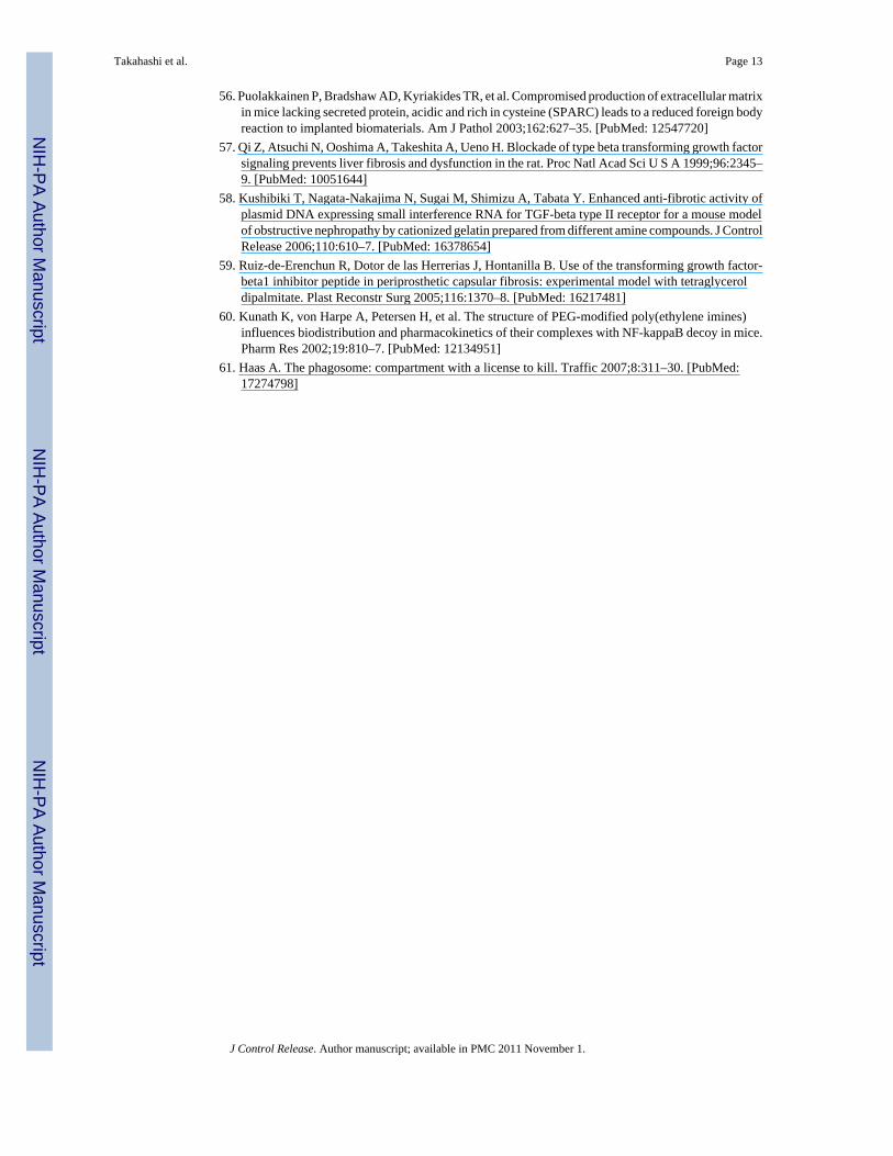
To determine the NP threshold for stable siRNA complex formation, different amounts of bPEIwere mixed with 0.14nmol mTOR siRNA at NP ratios of 0, 0.5, 1, 2, 5, 10, and 20. Figure 1ashows migration of siRNA/bPEI complexes by gel electrophoresis. With NP ratios of 0, 0.5and 1, siRNA bands migrate separately on the gel, indicating uncomplexed excess siRNA.When the NP ratio is equal to 2, the density of the siRNA band is substantially weaker. Whenthe NP ratios are = 5, no siRNA migrates freely in the gel, indicating that all siRNA moleculesare initially entrapped in bPEI complexes through electrostatic interactions. Therefore, thecomplex at NP > 5 should be appropriate for siRNA transfection.
Cytotoxicity from the siRNA complexes in serum-cultured fibroblasts in vitro was comparedamong the different NP ratios (NP = 0–40). As shown in Figure 1b, compared with cells withoutany treatment, cytotoxicity of the siRNA complexes is negligible when NP ratios are at or lessthan 20 (p = 0.18). In addition, there is no significant difference among the groups for NP ratios≤ 20.
In vitro mTOR knock-down by siRNA/bPEI complexes on fibroblast cultures3T3 Fibroblasts transfected with siRNA/bPEI complexes of different NP ratios (0, 5, 10, and20) were assayed for mTOR expression. Total protein was harvested three days after siRNAtreatment. Western blot results showed significant reductions of mTOR expression by siRNAwhen the NP ratio was 20 (Figure 2a). Furthermore, an NP ratio of 20 is sufficient to knockdown mTOR expression in fibroblasts in vitro using serum-based transfections. Therefore, theeffect of mTOR siRNA/bPEI complexes on cellular mTOR gene expression was only evaluatedby RT-PCR for NP = 20. Non-targeting siRNA/bPEI complexes with the same NP ratio wereused as controls. Compared to controls, mTOR siRNA complexed with bPEI reduces mTORmRNA expression dramatically (Figure 2b) in 3T3 fibroblast cultures in vitro.
Since mTOR positively regulates collagen type I production,(13) COL1A1 mRNA levels werealso assayed after mTOR siRNA transfection. Compared with non-targeting siRNAtransfection, COL1A1 mRNA levels were significantly suppressed by mTOR siRNA treatmentin vitro (Figure 2c).
mTOR siRNA effects on cell proliferation in vitroCell proliferation assays were performed 5 days after siRNA transfections in serum-basedcultures. As shown in Figure 3c, cell numbers in mTOR siRNA groups under NP ratio 20 aremuch less than that for control siRNA transfection groups (<30%, *p = 0.028). Itsrepresentative microscopic images of fibroblasts for control (Figure 3a) and treated (Figure3b) groups also demonstrate significant differences in cell density.
Takahashi et al. Page 6
J Control Release. Author manuscript; available in PMC 2011 November 1.
NIH
-PA Author Manuscript
NIH
-PA Author Manuscript
NIH
-PA Author Manuscript
Release of siRNA encapsulated in PEG-based hydrogelsPEG-based hydrogels were made with published methods (29) by reacting aqueous solutionsof PEGDM and DTT. Within a 20μl total reaction volume and an NP ratio of siRNA/bPEIequal to 20, 10μg of siRNA is found to be the maximum loading for successful in situ gelationwith Michael addition network chemistry. Release profiles for siRNA within the PEG-basedhydrogels for two different siRNA/PEG gel formulations were analyzed (Figure 4a). InFormulation 1 (PEGDM: 4.25 mg, DTT: 0.09 mg), approximately 50% of the siRNA wasreleased from the gel within the first 24 hours incubation, and 80% of the siRNA was releasedwithin 3 days. In Formulation 2 (PEGDM: 8.5 mg, DTT: 0.18 mg), approximately 80% of thesiRNA was released by day 7. A prolonged protein knock-down effect (up to one week) wasalso obtained by siRNA Formulation 2 compared with Formulation 1 (three days), which wasalso supported by Western blot results (Figure 4b). Formulation 2 provides longer sustainedrelease kinetics and protein expression suppression, and therefore was used for gel preparationsfor in vivo studies in subdermal implant-based siRNA release in mice.
In vivo implantation of siRNA-releasing hydrogel-coated devicesTissue harvests surrounding hydrogel-coated filter implants were stained with MTC to identifycollagen capsules (blue color). Collagen capsule thicknesses were calculated and comparedamong negative controls (hydrogels without siRNA: 71.95 ± 7.39 μm), positive controls(hydrogels loaded with TGF β1-specific siRNA: 66.82 ± 10.46 μm) and treated groups(hydrogels loaded with mTOR-specific siRNA: 2 μg dose, 96.60 ± 19.80μm; 10μg dose, 63.22± 5.95μm). Collagen capsule structure and thickness were evaluated from microscopic images(see Figure 5, foreign body capsules are demarcated with arrows). However, no significantdifference in capsule thickness or structure between these three groups was evident. In addition,H&E staining images indicate that mTOR siRNA does not significantly influence fibroblastdensity around these implants. To further investigate whether mTOR inhibition modulates thefibrotic response, tissue mTOR expression was evaluated by immunohistochemistry.Immunostaining of mTOR in tissues adjacent to the capsule (2μg and 10μg siRNA dosingcohorts) indicated no significant differences in mTOR expression in foreign body capsulesbetween siRNA-treated mice and the mice treated with blank gel. As shown in Figure 6, foreignbody capsules contain fibroblasts, inflammatory cells and ECM. After two-week implantations,no significant foreign body giant cells (FBGC) are observed around the implants. Numbers ofmacrophages were recruited to the interface, but there were no significant differences innumbers of macrophages around the implants among the groups. Therefore, despite in vitroknock-down success, it was concluded that mTOR siRNA treatments produced no significantreductions in FBR capsule thickness and protein knock-down in vivo.
DiscussionsiRNA is of substantial current interest as a sequence-specific post-transcriptional genesilencing tool for the genetic analysis and, significantly, for translational, therapeuticapplications in various mammalian cells.(32,33) In order to overcome several deliverychallenges for siRNA in therapeutics, several approaches have been used, includingconjugating siRNA with cholesterol (34), and delivery using cationic liposomes (35) andpolymer carriers.(36) Some have also been successfully utilized for systemic siRNA deliveryin mice.(35,37) Viral vectors have been described for siRNA delivery as well.(38–40)Nevertheless, overcoming viral vector oncogenicity and immunogenicity remains a significantbarrier for viral-based siRNA delivery. In general, efficiently targeting siRNA to systemicdisease sites remains a significant problem. To overcome this, lipid or polymer-based siRNAdelivery systems have been successfully used for local siRNA (e.g., topical) delivery,particularly to ophthalmic, vaginal, dermal, liver, neural, pulmonary and tumor targets.(21,23–25,41–45) Therefore, locally delivered siRNA from the surface of implantable
Takahashi et al. Page 7
J Control Release. Author manuscript; available in PMC 2011 November 1.
NIH
-PA Author Manuscript
NIH
-PA Author Manuscript
NIH
-PA Author Manuscript
subcutaneous devices was assessed for efficacy in targeting a major clinical complication ofthe foreign body response – implant-associated fibrosis.
To facilitate released siRNA internalization by mammalian cells in serum, mTOR siRNA wascomplexed with bPEI at different NP ratios (0–20) because of its known utility as a non-viralnucleic acid delivery vector.(46–48) The siRNA/bPEI migration assay (Figure 1a)demonstrates that siRNA forms stable complexes with bPEI when the NP ratio ≥ 5. Cytotoxicityfrom siRNA complexes was analyzed by culturing fibroblasts with siRNA complexes withdifferent NP ratios up to 40 in serum complete media. Significant cytotoxicity was observedonly when the NP ratio approached 40. Taken together, these results indicate that siRNA/bPEIcomplexes can be used for specific suppression of mTOR expression in fibroblast serum-basedcultures for NP ratios from 5 to 20.
Transfection of mTOR siRNA in 3T3 fibroblasts in serum-based cultures produced mTORgene silencing monitored by Western blotting three days post-transfection. Knock-down ofmTOR occurs only when the NP ratio was 20, indicating that siRNA specifically knockeddown mTOR message RNA in fibroblasts. To further exclude the possibility of non-specificknockdown of mTOR by the transfection reagent (bPEI), target gene expression was comparedbetween non-targeting siRNA and mTOR siRNA in cells. Compared to non-targeting siRNAcomplexes, mTOR siRNA complexes reduced mTOR mRNA expression in fibroblastsdramatically as shown in Figure 2b. Thus, these data confirm that mTOR siRNA suppressedthe targeted gene specifically through the RNAi mechanism.
In previous studies, mTOR was demonstrated to be essential for activating expression of thecollagen type I gene (COL1A1) in dermal fibroblasts.(13,30) A dramatic decrease (75%) inCOL1A1 mRNA expression was induced by down-regulating mTOR expression level to 56%.(13) Hence, it is not surprising that mTOR siRNA also down-regulates COL1A1 mRNAexpression by decreasing mTOR expression (Figure 2c). This supports the hypothesis thatcollagen production by fibroblasts would be blocked by mTOR siRNA transfection.
A significant role for mTOR is also promoting cell growth and proliferation by regulatingprotein synthesis. (49,50) It is therefore conceivable that mTOR knockdown may also controlor alter cell proliferation to some extent. Suppression of cell proliferation is shown afterfibroblast transfection with mTOR siRNA in cultures (Figure 3). Nearly 70% decrease in cellnumber after mTOR siRNA treatment is observed compared to non-targeting siRNAtransfections, supporting that mTOR function specifically, not cytotoxicity, was successfullysuppressed by the specific siRNA delivery to fibroblast cultures in serum.
These in vitro findings support siRNA targeting of mTOR to modulate collagen productionand implant encapsulation, analogous to rapamycin’s use against mTOR as an anti-fibroticagent.(51–54) To move this in vivo, a local delivery strategy using release from an implantsurface as a combination device model was used. PEG-based hydrogels were exploited for thisin vivo delivery system since the Michael addition chemistry in water allows mild in situpolymerization at physiological temperatures and pH in the presence of siRNA/bPEIcomplexes, control of both loading and release rates, and in situ polymerization on or aroundan implant.(28,29) To facilitate siRNA entry into cells, bPEI is used as a known complexingagent for nucleic acid delivery.(47,55) SiRNA/bPEI complexes were loaded into and releasedfrom the swollen and degrading PEG-based hydrogel networks. Two different siRNA diffusionprofiles were obtained from PEG hydrogels of two formulations, showing that encapsulatedsiRNA dosing release depends on hydrogel cross-linking. The higher cross-linked PEGhydrogels showed sustained release of siRNA to 14 days in vitro (Figure 4a). In addition, thesustained siRNA release resulted in prolonged protein knock-down to 7 days. Furthermore,
Takahashi et al. Page 8
J Control Release. Author manuscript; available in PMC 2011 November 1.
NIH
-PA Author Manuscript
NIH
-PA Author Manuscript
NIH
-PA Author Manuscript
this successful knock-down confirms that the released siRNA is active and complexed withPEI. Therefore, siRNA formulation 2 was selected for our in vivo studies.
Since encapsulated siRNA molecules exhibit both sustained release from PEG-based hydrogelsand effective mTOR and collagen knock-down in the presence of serum in vitro, the siRNAhydrogels were applied to a model soft tissue implant to reduce collagen expression andfibroblast proliferation in the context of the FBR. Based on in vitro release data showingcomplete siRNA release after two weeks, implants were harvested two weeks post-surgery.The thickness of the collagen fibrous capsule was evaluated for model circular membranedevices coated with PEG hydrogels in mice. As shown in Figure 5, no significant differencesin collagen capsule formation were found among the three groups. Compared to negativecontrols, both mTOR and control TGF-β siRNA-releasing material groups exhibited nodetectable anti-fibrotic activity in vivo. The capsule thickness for negative control groups iscomparable to the published data for the same cellulose implants harvested four weeks post-surgery in the same animal model, (i.e., 63 ± 25μm in Ref.(4), and 79 ± 40μm in Ref (56)).Previous studies have demonstrated that TGF-β is a potent target for anti-fibrotic therapy.(55,57–59) These studies show anti-fibrotic efficacy of TGF-β siRNA to prevent fibrosis invivo using different delivery systems. However, TGF-β siRNA showed no detectable anti-fibrotic activity as collagen capsule thickness was unaffected by the TGF-β siRNA in themurine implant model. Possible explanations for these non-distinguishing results includeinsufficient siRNA dosing, inefficient cellular uptake by the target fibroblasts around theimplant, and siRNA scavenging by cells other than fibroblasts at the implant site. A limitationof this design is that PEG hydrogel gelation is inhibited when siRNA complex loading exceeds10μg per polymerization. siRNA device-based dose loading cannot be increased in thisformulation to address the dosing issue in vivo. Effective gene silencing dose of siRNA in manyapplications in vivo is 1–2.5mg/kg.(21) Here, our study administered 0.4~0.5mg/kg. Thus,inefficient silencing produced by the siRNA was due partially to low dosing. As bPEI also hasmild nucleophilicity and some Michael addition capability with PEG acrylate,(60) increasingbPEI concentrations in the gelation step could alter polymerization and also affect bPEIparticipation as a cell transfection agent.
In situ tissue slice histology immunostaining for mTOR protein was performed to monitor invivo knock-down effects. In both 10μg and 2μg mTOR siRNA dose-treated groups, siRNAtreatment is unable to inhibit mTOR protein expression in the capsule. Therefore, similarcapsule thickness results from unchanged mTOR expression level in fibroblasts. Forsubcutaneous filter paper implantation in our case, FBGCs were difficult to find at the implantinterface with tissue two-week post-implantation. Plenty of macrophages were recruited to theinterface, however, there is no significant difference of the number of macrophages at theimplant and capsule interface between siRNA treated groups and controls. Immunoreactivityappears comparable in all groups and the addition of siRNA does not elicit significant changesin the FBR. In the early stage of the FBR, macrophages recruited to the implant site fuse toform FBGCs. They adhere to the surface of the hydrogel-coated implant and release severalchemicals that affect the implant surface. In this microenvironment, the hydrogel was alsosusceptible to degradation. Some fraction of siRNA/PEI complexes are likely taken up byactive capsule-associated cell endo- and phagocytosis mechanisms, followed by theirdegradation in phagolysosomes where pH remains as low as 4.(61) Hydrogel-released siRNA/PEI complexes escaping active cell phagocytosis must penetrate the dense tissue extracellularmatrix in the capsule to approach and enter target cells and then successfully escape endosomesto produce siRNA bioactivity. Hence, despite local siRNA dosing and direct release into thefibrous implant-associated capsule, siRNA polyplex dosing has low percentage success inproducing efficacy for mTOR knock-down in vivo. The apparent absence of protein knock-down in vivo is a testimony to the difficulty in getting sufficient siRNA from the implant tonearby fibroblast cells in the capsule. For device-based local siRNA delivery, the drug release
Takahashi et al. Page 9
J Control Release. Author manuscript; available in PMC 2011 November 1.
NIH
-PA Author Manuscript
NIH
-PA Author Manuscript
NIH
-PA Author Manuscript
system must overcome non-specific macrophage/FBGC capture and degradation, unless thesecells are specific targets for therapies. Therefore, device-based local siRNA delivery seekingto target collagen-producing local fibroblasts, but lacking specific cell-targeting features forfibroblasts, exhibits poor in vivo subcutaneous performance despite promising in vitro knock-down and anti-proliferative efficacy in serum-based fibroblast monocultures.
ConclusionsIn vitro cell culture results support siRNA targeting of mTOR to effectively suppress fibroblastproliferation and down-regulate type I collagen mRNA expression in serum-based fibroblastcultures. Thus, like commonly studied rapamycin, mTOR-targeted siRNA was expected toinhibit fibrotic responses around implants in vivo. Nonetheless, subcutaneous in vivo resultsdemonstrated little translation of in vitro siRNA activity to alter implant-associated fibrousencapsulation using a PEG-based hydrogel-coated implant releasing mTOR siRNA in a murinesubcutaneous implant model. Though mTOR remains a potent and attractive target for thistherapeutic purpose, and local device-based delivery is attractive for combination device localsiRNA delivery, further studies are warranted to improve in vivo efficacy in this application.These include new in vivo siRNA delivery systems, improvements in siRNA cell transfectionefficacy, degradation-resistant siRNA chemistries, and specific siRNA targeting to fibroblastsunder physiological conditions and foreign body responses.
AcknowledgmentsThe authors thank Sheryl Tripp (ARUP, University of Utah) for her immunohistochemical staining expertise, M.Weiser (UCHSC, CO), G. Burns (University of Utah) and, P. Tresco (University of Utah) for technical assistance,and R. J. Christie for providing polymer, PEGDM. This work is partially supported by NIH grant EB000894.
References1. Ratner BD. Reducing capsular thickness and enhancing angiogenesis around implant drug release
systems. J Control Release 2002;78:211–8. [PubMed: 11772462]2. Anderson JM, Rodriguez A, Chang DT. Foreign body reaction to biomaterials. Semin Immunol
2008;20:86–100. [PubMed: 18162407]3. Song E, Ouyang N, Horbelt M, Antus B, Wang M, Exton MS. Influence of alternatively and classically
activated macrophages on fibrogenic activities of human fibroblasts. Cell Immunol 2000;204:19–28.[PubMed: 11006014]
4. Kyriakides TR, Foster MJ, Keeney GE, et al. The CC chemokine ligand, CCL2/MCP1, participates inmacrophage fusion and foreign body giant cell formation. Am J Pathol 2004;165:2157–66. [PubMed:15579457]
5. Martin P, Leibovich SJ. Inflammatory cells during wound repair: the good, the bad and the ugly. TrendsCell Biol 2005;15:599–607. [PubMed: 16202600]
6. Miller KM, Anderson JM. In vitro stimulation of fibroblast activity by factors generated from humanmonocytes activated by biomedical polymers. J Biomed Mater Res 1989;23:911–30. [PubMed:2528547]
7. Norton LW, Koschwanez HE, Wisniewski NA, Klitzman B, Reichert WM. Vascular endothelialgrowth factor and dexamethasone release from nonfouling sensor coatings affect the foreign bodyresponse. J Biomed Mater Res A 2007;81:858–69. [PubMed: 17236219]
8. Blanco E, Weinberg BD, Stowe NT, Anderson JM, Gao J. Local release of dexamethasone frompolymer millirods effectively prevents fibrosis after radiofrequency ablation. J Biomed Mater Res A2006;76:174–82. [PubMed: 16265662]
9. Patil Y, Panyam J. Polymeric nanoparticles for siRNA delivery and gene silencing. Int J Pharm2009;367:195–203. [PubMed: 18940242]
10. Dumont FJ, Su Q. Mechanism of action of the immunosuppressant rapamycin. Life Sci 1996;58:373–95. [PubMed: 8594303]
Takahashi et al. Page 10
J Control Release. Author manuscript; available in PMC 2011 November 1.
NIH
-PA Author Manuscript
NIH
-PA Author Manuscript
NIH
-PA Author Manuscript
11. Law BK. Rapamycin: an anti-cancer immunosuppressant? Crit Rev Oncol Hematol 2005;56:47–60.[PubMed: 16039868]
12. Windecker S, Roffi M, Meier B. Sirolimus eluting stent: a new era in interventional cardiology?Current pharmaceutical design 2003;9:1077–94. [PubMed: 12678859]
13. Shegogue D, Trojanowska M. Mammalian target of rapamycin positively regulates collagen type Iproduction via a phosphatidylinositol 3-kinase-independent pathway. J Biol Chem 2004;279:23166–75. [PubMed: 15047702]
14. Poulalhon N, Farge D, Roos N, et al. Modulation of collagen and MMP-1 gene expression infibroblasts by the immunosuppressive drug rapamycin. A direct role as an antifibrotic agent? TheJournal of biological chemistry 2006;281:33045–52. [PubMed: 16914544]
15. Gao XM, Wong G, Wang B, et al. Inhibition of mTOR reduces chronic pressure-overload cardiachypertrophy and fibrosis. Journal of hypertension 2006;24:1663–70. [PubMed: 16877971]
16. Simler NR, Howell DC, Marshall RP, et al. The rapamycin analogue SDZ RAD attenuates bleomycin-induced pulmonary fibrosis in rats. Eur Respir J 2002;19:1124–7. [PubMed: 12108867]
17. Hannon GJ, Rossi JJ. Unlocking the potential of the human genome with RNA interference. Nature2004;431:371–8. [PubMed: 15372045]
18. Jones SW, Souza PM, Lindsay MA. siRNA for gene silencing: a route to drug target discovery.Current opinion in pharmacology 2004;4:522–7. [PubMed: 15351359]
19. Aigner A. Delivery systems for the direct aplication of siRNAs to induce RNA interference (RNAi)in vivo. journal of biomedicine and biotechnology 2006;2006:1–15.
20. de Fougerolles A, Vornlocher HP, Maraganore J, Lieberman J. Interfering with disease: a progressreport on siRNA-based therapeutics. Nat Rev Drug Discov 2007;6:443–53. [PubMed: 17541417]
21. Woodrow KA, Cu Y, Booth CJ, Saucier-Sawyer JK, Wood MJ, Saltzman WM. Intravaginal genesilencing using biodegradable polymer nanoparticles densely loaded with small-interfering RNA.Nat Mater 2009;8:526–33. [PubMed: 19404239]
22. Reich SJ, Fosnot J, Kuroki A, et al. Small interfering RNA (siRNA) targeting VEGF effectivelyinhibits ocular neovascularization in a mouse model. Mol Vis 2003;9:210–6. [PubMed: 12789138]
23. Takanashi M, Oikawa K, Sudo K, et al. Therapeutic silencing of an endogenous gene by siRNA creamin an arthritis model mouse. Gene Ther 2009;16:982–9. [PubMed: 19474812]
24. Massaro D, Massaro GD, Clerch LB. Noninvasive delivery of small inhibitory RNA and otherreagents to pulmonary alveoli in mice. Am J Physiol Lung Cell Mol Physiol 2004;287:L1066–70.[PubMed: 15234906]
25. Thakker DR, Natt F, Husken D, et al. Neurochemical and behavioral consequences of widespreadgene knockdown in the adult mouse brain by using nonviral RNA interference. Proc Natl Acad SciU S A 2004;101:17270–5. [PubMed: 15569935]
26. Wu P, Grainger DW. Drug/device combinations for local drug therapies and infection prophylaxis.Biomaterials 2006;27:2450–67. [PubMed: 16337266]
27. Christie RJ, Findley DJ, Dunfee M, Hansen RD, Olsen SC, Grainger DW. Photopolymerized hydrogelcarriers for live vaccine ballistic delivery. Vaccine 2006;24:1462–9. [PubMed: 16246467]
28. van de Wetering P, Metters AT, Schoenmakers RG, Hubbell JA. Poly(ethylene glycol) hydrogelsformed by conjugate addition with controllable swelling, degradation, and release ofpharmaceutically active proteins. J Control Release 2005;102:619–27. [PubMed: 15681084]
29. Metters A, Hubbell J. Network formation and degradation behavior of hydrogels formed by Michael-type addition reactions. Biomacromolecules 2005;6:290–301. [PubMed: 15638532]
30. Kyriakides TR, Zhu YH, Yang Z, Huynh G, Bornstein P. Altered extracellular matrix remodelingand angiogenesis in sponge granulomas of thrombospondin 2-null mice. Am J Pathol 2001;159:1255–62. [PubMed: 11583953]
31. Bryers JD, Jarvis RA, Lebo J, Prudencio A, Kyriakides TR, Uhrich K. Biodegradation of poly(anhydride-esters) into non-steroidal anti-inflammatory drugs and their effect on Pseudomonasaeruginosa biofilms in vitro and on the foreign-body response in vivo. Biomaterials 2006;27:5039–48. [PubMed: 16777217]
32. Aagaard L, Rossi JJ. RNAi therapeutics: principles, prospects and challenges. Adv Drug Deliv Rev2007;59:75–86. [PubMed: 17449137]
Takahashi et al. Page 11
J Control Release. Author manuscript; available in PMC 2011 November 1.
NIH
-PA Author Manuscript
NIH
-PA Author Manuscript
NIH
-PA Author Manuscript
33. Almeida R, Allshire RC. RNA silencing and genome regulation. Trends Cell Biol 2005;15:251–8.[PubMed: 15866029]
34. Soutschek J, Akinc A, Bramlage B, et al. Therapeutic silencing of an endogenous gene by systemicadministration of modified siRNAs. Nature 2004;432:173–8. [PubMed: 15538359]
35. Sorensen DR, Leirdal M, Sioud M. Gene silencing by systemic delivery of synthetic siRNAs in adultmice. J Mol Biol 2003;327:761–6. [PubMed: 12654261]
36. Khan A, Benboubetra M, Sayyed PZ, et al. Sustained polymeric delivery of gene silencing antisenseODNs, siRNA, DNAzymes and ribozymes: in vitro and in vivo studies. J Drug Target 2004;12:393–404. [PubMed: 15545089]
37. Sioud M, Sorensen DR. Cationic liposome-mediated delivery of siRNAs in adult mice. BiochemBiophys Res Commun 2003;312:1220–5. [PubMed: 14652004]
38. Rubinson DA, Dillon CP, Kwiatkowski AV, et al. A lentivirus-based system to functionally silencegenes in primary mammalian cells, stem cells and transgenic mice by RNA interference. Nat Genet2003;33:401–6. [PubMed: 12590264]
39. Tomar RS, Matta H, Chaudhary PM. Use of adeno-associated viral vector for delivery of smallinterfering RNA. Oncogene 2003;22:5712–5. [PubMed: 12944921]
40. Matta H, Hozayev B, Tomar R, Chugh P, Chaudhary PM. Use of lentiviral vectors for delivery ofsmall interfering RNA. Cancer Biol Ther 2003;2:206–10. [PubMed: 12750565]
41. Reich SJ, et al. Small interfering RNA (siRNA) targeting VEGF effectively inhibits ocularneovascularization in a mouse model. Mol Vision 2004;9:210–216.
42. Nakamura H, et al. RNA interference targeting transforming growth factor-β type II receptorsuppresses ocular inflammation and fibrosis. Mol Vis 2004;10:703–711. [PubMed: 15475878]
43. Luo MC, Zhang DQ, Ma SW, et al. An efficient intrathecal delivery of small interfering RNA to thespinal cord and peripheral neurons. Mol Pain 2005;1:29. [PubMed: 16191203]
44. Morrissey DV, Lockridge JA, Shaw L, et al. Potent and persistent in vivo anti-HBV activity ofchemically modified siRNAs. Nat Biotechnol 2005;23:1002–7. [PubMed: 16041363]
45. Schiffelers RM, Ansari A, Xu J, et al. Cancer siRNA therapy by tumor selective delivery with ligand-targeted sterically stabilized nanoparticle. Nucleic Acids Res 2004;32:e149. [PubMed: 15520458]
46. Park TG, Jeong JH, Kim SW. Current status of polymeric gene delivery systems. Adv Drug DelivRev 2006;58:467–86. [PubMed: 16781003]
47. Wang DA, Narang AS, Kotb M, et al. Novel branched poly(ethylenimine)-cholesterol water-solublelipopolymers for gene delivery. Biomacromolecules 2002;3:1197–207. [PubMed: 12425656]
48. Jiang G, Park K, Kim J, et al. Hyaluronic acid-polyethyleneimine conjugate for target specificintracellular delivery of siRNA. Biopolymers 2008;89:635–42. [PubMed: 18322932]
49. Achenbach TV, Brunner B, Heermeier K. Oligonucleotide-based knockdown technologies: antisenseversus RNA interference. Chembiochem 2003;4:928–35. [PubMed: 14523910]
50. Burnett PE, Barrow RK, Cohen NA, Snyder SH, Sabatini DM. RAFT1 phosphorylation of thetranslational regulators p70 S6 kinase and 4E-BP1. Proc Natl Acad Sci U S A 1998;95:1432–7.[PubMed: 9465032]
51. Wu MJ, Wen MC, Chiu YT, Chiou YY, Shu KH, Tang MJ. Rapamycin attenuates unilateral ureteralobstruction-induced renal fibrosis. Kidney Int 2006;69:2029–36. [PubMed: 16732193]
52. Neef M, Ledermann M, Saegesser H, Schneider V, Reichen J. Low-dose oral rapamycin treatmentreduces fibrogenesis, improves liver function, and prolongs survival in rats with established livercirrhosis. J Hepatol 2006;45:786–96. [PubMed: 17050028]
53. Salminen US, Maasilta PK, Taskinen EI, Alho HS, Ikonen TS, Harjula AL. Prevention of small airwayobliteration in a swine heterotopic lung allograft model. J Heart Lung Transplant 2000;19:193–206.[PubMed: 10703697]
54. Poston RS, Billingham M, Hoyt EG, et al. Rapamycin reverses chronic graft vascular disease in anovel cardiac allograft model. Circulation 1999;100:67–74. [PubMed: 10393683]
55. Kim KH, Kim HC, Hwang MY, et al. The antifibrotic effect of TGF-beta1 siRNAs in murine modelof liver cirrhosis. Biochem Biophys Res Commun 2006;343:1072–8. [PubMed: 16579972]
Takahashi et al. Page 12
J Control Release. Author manuscript; available in PMC 2011 November 1.
NIH
-PA Author Manuscript
NIH
-PA Author Manuscript
NIH
-PA Author Manuscript
56. Puolakkainen P, Bradshaw AD, Kyriakides TR, et al. Compromised production of extracellular matrixin mice lacking secreted protein, acidic and rich in cysteine (SPARC) leads to a reduced foreign bodyreaction to implanted biomaterials. Am J Pathol 2003;162:627–35. [PubMed: 12547720]
57. Qi Z, Atsuchi N, Ooshima A, Takeshita A, Ueno H. Blockade of type beta transforming growth factorsignaling prevents liver fibrosis and dysfunction in the rat. Proc Natl Acad Sci U S A 1999;96:2345–9. [PubMed: 10051644]
58. Kushibiki T, Nagata-Nakajima N, Sugai M, Shimizu A, Tabata Y. Enhanced anti-fibrotic activity ofplasmid DNA expressing small interference RNA for TGF-beta type II receptor for a mouse modelof obstructive nephropathy by cationized gelatin prepared from different amine compounds. J ControlRelease 2006;110:610–7. [PubMed: 16378654]
59. Ruiz-de-Erenchun R, Dotor de las Herrerias J, Hontanilla B. Use of the transforming growth factor-beta1 inhibitor peptide in periprosthetic capsular fibrosis: experimental model with tetraglyceroldipalmitate. Plast Reconstr Surg 2005;116:1370–8. [PubMed: 16217481]
60. Kunath K, von Harpe A, Petersen H, et al. The structure of PEG-modified poly(ethylene imines)influences biodistribution and pharmacokinetics of their complexes with NF-kappaB decoy in mice.Pharm Res 2002;19:810–7. [PubMed: 12134951]
61. Haas A. The phagosome: compartment with a license to kill. Traffic 2007;8:311–30. [PubMed:17274798]
Takahashi et al. Page 13
J Control Release. Author manuscript; available in PMC 2011 November 1.
NIH
-PA Author Manuscript
NIH
-PA Author Manuscript
NIH
-PA Author Manuscript
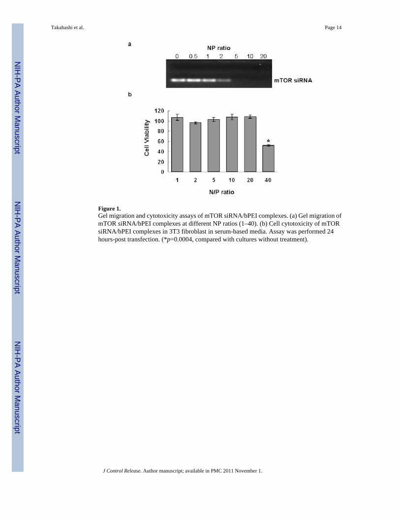
Figure 1.Gel migration and cytotoxicity assays of mTOR siRNA/bPEI complexes. (a) Gel migration ofmTOR siRNA/bPEI complexes at different NP ratios (1–40). (b) Cell cytotoxicity of mTORsiRNA/bPEI complexes in 3T3 fibroblast in serum-based media. Assay was performed 24hours-post transfection. (*p=0.0004, compared with cultures without treatment).
Takahashi et al. Page 14
J Control Release. Author manuscript; available in PMC 2011 November 1.
NIH
-PA Author Manuscript
NIH
-PA Author Manuscript
NIH
-PA Author Manuscript
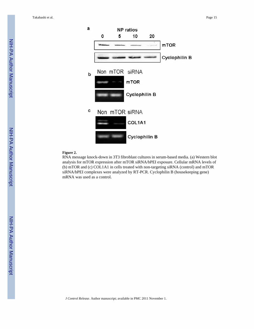
Figure 2.RNA message knock-down in 3T3 fibroblast cultures in serum-based media. (a) Western blotanalysis for mTOR expression after mTOR siRNA/bPEI exposure. Cellular mRNA levels of(b) mTOR and (c) COL1A1 in cells treated with non-targeting siRNA (control) and mTORsiRNA/bPEI complexes were analyzed by RT-PCR. Cyclophilin B (housekeeping gene)mRNA was used as a control.
Takahashi et al. Page 15
J Control Release. Author manuscript; available in PMC 2011 November 1.
NIH
-PA Author Manuscript
NIH
-PA Author Manuscript
NIH
-PA Author Manuscript
Figure 3.Microscopic images of fibroblast serum-based cultures (a) treated with non-targeting siRNA,(b) mTOR siRNA at NP ratio 20, and (c) cell proliferation in the siRNA-treated cells (NP 20,*p = 0.028), scale bar = 250μm. Images were taken 5 days after siRNA transfection. Numbersof cells treated with non-targeting siRNA was normalized to 100%.
Takahashi et al. Page 16
J Control Release. Author manuscript; available in PMC 2011 November 1.
NIH
-PA Author Manuscript
NIH
-PA Author Manuscript
NIH
-PA Author Manuscript
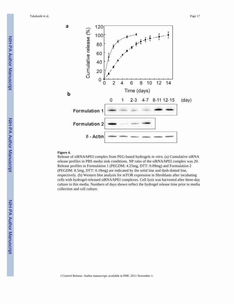
Figure 4.Release of siRNA/bPEI complex from PEG-based hydrogels in vitro. (a) Cumulative siRNArelease profiles in PBS media sink conditions. NP ratio of the siRNA/bPEI complex was 20.Release profiles in Formulation 1 (PEGDM: 4.25mg, DTT: 0.09mg) and Formulation 2(PEGDM: 8.5mg, DTT: 0.18mg) are indicated by the solid line and dash-dotted line,respectively. (b) Western blot analysis for mTOR expression in fibroblasts after incubatingcells with hydrogel-released siRNA/bPEI complexes. Cell lysis was harvested after three-dayculture in this media. Numbers of days shown reflect the hydrogel release time prior to mediacollection and cell culture.
Takahashi et al. Page 17
J Control Release. Author manuscript; available in PMC 2011 November 1.
NIH
-PA Author Manuscript
NIH
-PA Author Manuscript
NIH
-PA Author Manuscript

Figure 5.Comparison of in vivo collagen capsule thickness for murine sub-dermal hydrogel-coatedimplants explanted after two weeks for (a) negative control, no siRNA loaded, (b) 2μg mTORsiRNA loaded, (c) 10μg mTOR siRNA loaded and (d) positive control, 10 μg TGF-β siRNAloaded. Localization of fibrous capsule is marked by white arrows (scale bar = 100μm). (e)Summary of collagen capsule thickness data. P values for each individual group vs. negativecontrol without siRNA treatment are: 0.3112 (2μg mTOR siRNA), 0.3954 (10μg mTORsiRNA), and 0.7045 (10μg TGF-β siRNA).
Takahashi et al. Page 18
J Control Release. Author manuscript; available in PMC 2011 November 1.
NIH
-PA Author Manuscript
NIH
-PA Author Manuscript
NIH
-PA Author Manuscript
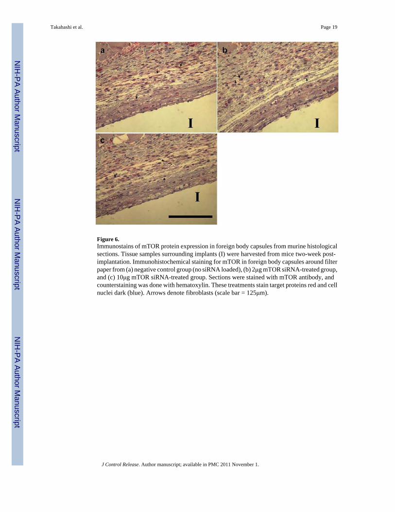
Figure 6.Immunostains of mTOR protein expression in foreign body capsules from murine histologicalsections. Tissue samples surrounding implants (I) were harvested from mice two-week post-implantation. Immunohistochemical staining for mTOR in foreign body capsules around filterpaper from (a) negative control group (no siRNA loaded), (b) 2μg mTOR siRNA-treated group,and (c) 10μg mTOR siRNA-treated group. Sections were stained with mTOR antibody, andcounterstaining was done with hematoxylin. These treatments stain target proteins red and cellnuclei dark (blue). Arrows denote fibroblasts (scale bar = 125μm).
Takahashi et al. Page 19
J Control Release. Author manuscript; available in PMC 2011 November 1.
NIH
-PA Author Manuscript
NIH
-PA Author Manuscript
NIH
-PA Author Manuscript
Copyright © 2022 FDOKUMEN