"The Solitary Reaper" by William Wordsworth analyzed by Othman A. Marzoog
Upload
khangminh22Category
view
0download
0
Citation: Apra, C.; El Arbi, A.;
Montero, A.-S.; Parker, F.; Knafo, S.
Spinal Solitary Fibrous Tumors: An
Original Multicenter Series and
Systematic Review of Presentation,
Management, and Prognosis. Cancers
2022, 14, 2839. https://doi.org/
10.3390/cancers14122839
Academic Editor: David Wong
Received: 2 May 2022
Accepted: 2 June 2022
Published: 8 June 2022
Publisher’s Note: MDPI stays neutral
with regard to jurisdictional claims in
published maps and institutional affil-
iations.
Copyright: © 2022 by the authors.
Licensee MDPI, Basel, Switzerland.
This article is an open access article
distributed under the terms and
conditions of the Creative Commons
Attribution (CC BY) license (https://
creativecommons.org/licenses/by/
4.0/).
cancers
Review
Spinal Solitary Fibrous Tumors: An Original Multicenter Seriesand Systematic Review of Presentation, Management,and PrognosisCaroline Apra 1,2,*, Amira El Arbi 3, Anne-Sophie Montero 1,2 , Fabrice Parker 3,4 and Steven Knafo 3,4,*
1 Sorbonne Université, 75013 Paris, France; [email protected] Neurosurgery Department, Pitie Salpêtrière Hospital, 75013 Paris, France3 Neurosurgery Department, Bicêtre Hospital, 94270 Kremlin-Bicêtre, France; [email protected] (A.E.A.);
[email protected] (F.P.)4 University Paris-Saclay, 91190 Gif-sur-Yvette, France* Correspondence: [email protected] (C.A.); [email protected] (S.K.)
Simple Summary: Solitary fibrous tumors are rare benign or cancerous tumors that develop inall tissues, including close to the spinal cord. These cases are exceptional and we describe theirpresentation and outcome based on 31 published cases and 10 patients on whom we operated. Thetumors can develop in any portion of the spine and cause back pain, associated with neurologicaldeficits, such as compression of a nerve or the spinal cord, in 66% of patients. Surgical removal is thefirst step towards diagnosis and treatment, but complete removal could be achieved in only 70% ofpatients, due to bleeding or spinal cord invasion. Tumors were found to recur after a mean 5.8 years(1 to 25), without identified risk factors. However, in patients with subtotal removal, radiotherapysignificantly improves the rate of recurrence. In total, spinal solitary fibrous tumors are treated byneurosurgeons on the front line but discussion in a multidisciplinary team will provide generaltreatments, especially radiotherapy after subtotal removal.
Abstract: All solitary fibrous tumors (SFT), now histologically diagnosed by a positive nuclear STAT6immunostaining, represent less than 2% of soft tissue sarcomas, with spinal SFT constituting amaximum of 2% of them, making these tumors extremely rare. We provide an up-to-date overviewof their diagnosis, treatment, and prognosis. We included 10 primary STAT6-positive SFT fromour retrospective cohort and 31 from a systematic review. Spinal pain was the most commonsymptom, in 69% of patients, and the only one in 34%, followed by spinal cord compression in 41%,radicular compression, including pain or deficit, in 36%, and urinary dysfunction specifically in 18%.Preoperative diagnosis was never obtained. Gross total resection was achieved in 71%, in the absenceof spinal cord invasion or excessive bleeding. Histologically, they were 35% grade I, 25% grade II, and40% grade III. Recurrence was observed in 43% after a mean 5.8 years (1 to 25). No significant riskfactor was identified, but adjuvant radiotherapy improved the recurrence-free survival after subtotalresection. In conclusion, spinal SFT must be treated by neurosurgeons as part of a multidisciplinaryteam. Owing to their close relationship with the spinal cord, radiotherapy should be consideredwhen gross total resection cannot be achieved, to lower the risk of recurrence.
Keywords: spine; medulla; intramedullary; solitary fibrous tumor; hemangiopericytoma; neuro-surgery; STAT6
1. Introduction
In 2016, the World Health Organization (WHO) introduced the combined term “soli-tary fibrous tumor/hemangiopericytoma” for describing connective tissue tumors of thecentral nervous system with positive STAT6 nuclear immunostaining, which was replaced
Cancers 2022, 14, 2839. https://doi.org/10.3390/cancers14122839 https://www.mdpi.com/journal/cancers

Cancers 2022, 14, 2839 2 of 11
by “solitary fibrous tumors” (SFT) alone in 2021, to conform fully with soft tissue pathol-ogy nomenclature [1]. The grouping of these two entities, which were separated untilthen, is grounded in an overlapping histological description, associated with a sharedgenetic signature: the fusion of the NGFI-A-binding protein 2 (NAB2) and signal transducerand activator of transcription 6 (STAT6) genes due to an inversion at chromosome 12q13,which is a hallmark for all SFT, regardless of their localization, since its first description in2013 [2]. SFT represent less than 2% of soft tissue sarcomas, with central nervous systemSFT constituting 20% of SFT, with only one spinal SFT for every ten intracranial lesions,making these tumors extremely rare in clinical practice [2]. We performed a retrospectivemulticenter series of spinal SFT, focusing on STAT6-positive tumors only, and added asystematic review of all published cases. The aim of this review is to provide an up-to-dateoverview of the diagnosis, treatment, and prognosis of these rare tumors, with a discussionabout individual clinical decision making.
2. Materials and Methods2.1. Original Series
All patients with a histological diagnosis of spinal SFT who underwent surgery in theneurosurgical departments of the Bicêtre and Pitié-Salpêtrière hospitals in Paris, France,between 1988 and 2020 were included. SFT diagnosis was confirmed by two expert neu-ropathologists with confirmed positive STAT6 nuclear immunostaining even for the casesdated before 2016. Medical records were reviewed for clinical and radiological presenta-tion, histopathologic features, surgical treatment, postoperative therapies, and outcomes.Tumors were graded radiologically [3]: I, extradural type; II, intradural type; and III, intra-to extradural and paravertebral type. Extent of resection was estimated from the operativereports and postoperative MRI. Tumors were graded histologically according to the 2016WHO classification [1]. Duration of follow-up was calculated as the duration from the dateof surgery to the last outpatient department visit. Recurrence was defined as local tumorgrowth on MRI, whether symptomatic or not. Ethical approval was granted by the FrenchNeurosurgical Society review board (IRB00011687; 2022/14).
2.2. Systematic Review
The literature review was performed according to the PRISMA checklist [4]. Thedatabase (PubMed) was searched for the combination of terms “spine”, “spinal”, “medulla”,“medullary”, “solitary fibrous tumor”, “hemangiopericytoma” in December 2021. Eligiblearticles reported at least one case of spinal SFT. Exclusion criteria were the absence of STAT6-positive nuclear immunostaining, spinal metastatic localizations, and the absence of clinicaldata. Clinical, radiological, and histological data, including perioperative descriptions ofthe tumors, were collected.
2.3. Statistical Analysis
All statistical analyses were performed using Microsoft Excel (version 2202). Foranalyzing risk factors for recurrence, exact Fisher test was performed due to the smallsize of the population. Schematic figures were created with http://www.BioRender.com(accessed on 10 April 2022).
3. Results3.1. Patients
We included 10 cases from our own retrospective cohort and retrieved 31 cases fromthe systematic review, as detailed in the Supplementary Flow Chart (SupplementaryFigure S1) [5–20]. All were primary SFT with a STAT6-positive pathological diagnosis.Most information was available from all cases, as detailed in Supplementary Table S1.Results are given as: all patients [review; cohort]. There were slightly more women (51%,55%/40%, sex ratio 1.05) and the mean age at diagnosis was 46 (45/47), ranging from 10 to81 (10–81/32–71). All results are detailed in Table 1.

Cancers 2022, 14, 2839 3 of 11
Table 1. Description of characteristics and prognosis, for the population presenting a spinal solitaryfibrous tumor, as diagnosed with positive STAT6 nuclear staining, for our series (n = 10) and asystematic review of the literature (n = 31).
Characteristics Our Series(n = 10)
Literature Review(n = 31)
Total(n = 41)
Population
Sex M 6 (60%) 14 (45%) 20 (49%)F 4 (40%) 17 (55%) 21 (51%)
Age, mean ± IC95 47 ± 8 45 ± 3 46 ± 4
Clinical and radiological presentations
Spinal pain 5 (50%) 22 (76%) 27 (69%)Radicular compression 6 (60%) 8 (28%) 14 (36%)Spinal cord compression 7 (70%) 9 (31%) 16 (41%)Urinary dysfunction 4(40%) 3 (10%) 7 (18%)Motor dysfunction 5 (50%) 9 (29%) 14 (34%)Sensory dysfunction 4 (40%) 6 (19%) 10 (24%)Duration of symptoms (mo) 10 ± 6 20 ± 12 17 ± 9Tumor localization Cervical 5 (50%) 7 (23%) 12 (29%)
Thoracic 4 (40%) 17 (54%) 21 (51%)Lumbar 1 (10%) 7 (23%) 8 (20%)
Tumor type I extradural 0 (0%) 2 (14%) 2 (9%)II intradural 7 (78%) 8 (57%) 15 (65%)III extra- and intradural 2 (22%) 4 (29%) 6 (26%)
Surgical and histological findings
Complete resection 7 (70%) 22 (71%) 29 (71%)Purely extramedullary tumor during surgery 5 (50%) 19 (68%) 24 (63%)Histological grading 1 1 (11%) 6 (55%) 7 (35%)
2 5 (56%) 0 (0%) 5 (25%)3 3 (33%) 5 (45%) 8 (40%)
Post-operative management and outcome
Primary adjuvant treatment None 6 (60%) 14 (77%) 30 (73%)Radiotherapy 4 (40%) 7 (23%) 11 (27%)
Documented recurrence 4 (40%) 11 (32%) 15 (37%)Time to first recurrence (mo) 128 ± 116 49 ± 42 70 ± 47
3.2. Clinical Presentation
Spinal pain was the most common symptom, found in 27 patients (69% [76%; 50%]),and as an isolated symptom in 14 (34% [45%; 0%]). Radicular symptoms, including painor deficit, were present in 14 patients (36% [28%; 60%]), whereas spinal cord compression,including increased reflexes or sensory–motor deficit, was present in 16 patients (41%[31%; 70%]), and urinary dysfunction was reported in seven patients (18%, [10%; 40%]).Symptoms typically worsened gradually, with a mean duration of clinical symptoms beforesurgery of 18 months (20/10), ranging from less than 1 month to 11 years. For patients withisolated spine pain, the mean duration of symptoms before surgery was 15 months.
3.3. Initial Radiological Findings
All patients underwent a preoperative spine MRI with contrast. Although T1 and T2MRI aspects varied, all tumors showed marked homogeneous or heterogeneous enhance-ment by gadolinium. No calcification or acute intratumoral hemorrhage was observed.CT scan was not systematically available on retrieval, although it was likely performedin clinical practice before surgery. In a few cases, scalloping was observed in extracanalartumors, but no exostosis. In our cohort, three patients had a preoperative angiographywithout embolization. Tumors were cervical in 12 patients (29% [23%; 50%]), thoracic

Cancers 2022, 14, 2839 4 of 11
in 21 patients (51% [54%; 40%]), and lumbar in eight patients (20% [23%; 10%]), whichis proportional to the length of each segment, cervical spine vertebras constituting 30%,thoracic 50%, and lumbo-sacral 20% of the whole spine. Lesions involved one to twovertebrae in most patients (93% [97%; 80%]). Spinal SFT were type I in two patients (9%[14%; 0%]), II in 15 patients (65% [57%; 78%]), and III in six patients (26% [29%; 22%]).At least four [2; 2] extracanalar lesions showed extension in the foramina, giving them adumbbell aspect.
3.4. Operative Findings
All patients underwent first-line surgery, through an isolated posterior approach withlaminectomy, or associated with an anterior approach, an arthrodesis or a thoracoscopyin one case, depending on the tumor extension. Gross total resection was achieved in29 patients (71% [71%; 70%]). The reasons for subtotal resection included spinal cordinvasion and excessive bleeding. The use of neuro-monitoring was mentioned only once,after recurrence. Operatively, tumors were identified as being purely extramedullary inpatients (63% [68%; 50%]) with or without description of dural and pial invasion, whereasother cases invaded the medulla. No intraoperative complication other than bleeding wasnoted, either in the literature or in our series. A video showing perioperative observationsis available as a Supplementary File.
3.5. Histological Findings
All tumors were diagnosed as spinal SFT with positive STAT6 nuclear immunostaining.Seven [6; 1] were classified as grade I, five [0; 5] as grade II, and eight [5; 3] grade III. Themitoses count ranged from 0 to 15 per 10 high-power fields, and Ki67 from 0% to 15%.Other reported immunostainings include CD34, vimentin, proteinS100, EMA, and SMA,but these are not reliable in SFT diagnosis. No histological evidence of medullary invasionwas described.
3.6. Adjuvant Treatment and Outcome
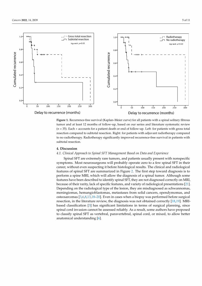
No complication, either infection, cerebrospinal fluid leakage, neurological worsening,or death, was reported after surgery. Eleven (27 [23%; 40%]) patients received immedi-ate postoperative adjuvant treatments, including 11 radiotherapy and one neoadjuvantchemotherapy. The rationale for performing these treatments was not systematic and notexplicit but these patients included one case of grade III SFT and four cases of subtotalresection. Available follow-up ranged from 12 months to 30 years for 35 [26; 9] patients.Recurrence was observed in 15 patients (43% [42%; 44%]), after a mean 5.8 years, rangingfrom 1 to 25 years, as detailed in Figure 1. No significant risk factor for recurrence couldbe identified, but there was a tendency to recur for patients with incomplete surgery orno adjuvant radiotherapy (Table 2). Survival analyses show that adjuvant radiotherapysignificantly improves the recurrence-free survival in patients with subtotal resection buthas no effect in patients with gross total resection (Figure 1). Repeated recurrences wereobserved in some cases, but data were scarce. After recurrence, nine (75% [63%; 100%])patients underwent a second surgery, with a combination of radiotherapy, carbon-ion ra-diotherapy, or proton therapy, and one was treated with Pazopanib for progressive diseaseafter third recurrence. There were no systematic data about metastasizing.
Table 2. Risk factors for recurrence in patients with a minimum 12 months of follow-up (n = 35),p-value for exact Fisher test. WHO: World Health Organization.
Risk Factor for Recurrence Recurrence (n = 15) No Recurrence (n = 20) p-Value
Intramedullary component 36% (n = 14) 26% (n = 29) 0.70Subtotal resection 53% (n = 15) 20% (n = 20) 0.07
WHO grade 3 40% (n = 5) 50% (n = 10) 1Adjuvant radiotherapy 13% (n = 15) 40% (n = 20) 0.13
Cancers 2022, 14, 2839 5 of 11
Cancers 2022, 14, x FOR PEER REVIEW 5 of 11
adjuvant radiotherapy significantly improves the recurrence-free survival in patients with
subtotal resection but has no effect in patients with gross total resection (Figure 1).
Repeated recurrences were observed in some cases, but data were scarce. After recurrence,
nine (75% [63%; 100%]) patients underwent a second surgery, with a combination of
radiotherapy, carbon-ion radiotherapy, or proton therapy, and one was treated with
Pazopanib for progressive disease after third recurrence. There were no systematic data
about metastasizing.
Figure 1. Recurrence-free survival (Kaplan–Meier curve) for all patients with a spinal solitary
fibrous tumor and at least 12 months of follow-up, based on our series and literature systematic
review (n = 35). Each + accounts for a patient death or end of follow-up. Left: for patients with gross
total resection compared to subtotal resection. Right: for patients with adjuvant radiotherapy
compared to no radiotherapy. Radiotherapy significantly improved recurrence-free survival in
patients with subtotal resection.
Table 2. Risk factors for recurrence in patients with a minimum 12 months of follow-up (n = 35), p-
value for exact Fisher test. WHO: World Health Organization.
Risk Factor for Recurrence Recurrence (n = 15) No Recurrence (n = 20) p-Value
Intramedullary component 36% (n = 14) 26% (n = 29) 0.70
Subtotal resection 53% (n = 15) 20% (n = 20) 0.07
WHO grade 3 40% (n = 5) 50% (n = 10) 1
Adjuvant radiotherapy 13% (n = 15) 40% (n = 20) 0.13
4. Discussion
4.1 Clinical Approach to Spinal SFT Management Based on Data and Experience
Spinal SFT are extremely rare tumors, and patients usually present with nonspecific
symptoms. Most neurosurgeons will probably operate zero to a few spinal SFT in their
career, without even suspecting it before histological results. The clinical and radiological
features of spinal SFT are summarized in Figure 2. The first step toward diagnosis is to
perform a spine MRI, which will allow the diagnosis of a spinal tumor. Although some
features have been described to identify spinal SFT, they are not diagnosed correctly on
MRI, because of their rarity, lack of specific features, and variety of radiological
presentations [21]. Depending on the radiological type of the lesion, they are
misdiagnosed as schwannomas, meningiomas, hemangioblastomas, metastases from
Figure 1. Recurrence-free survival (Kaplan–Meier curve) for all patients with a spinal solitary fibroustumor and at least 12 months of follow-up, based on our series and literature systematic review(n = 35). Each + accounts for a patient death or end of follow-up. Left: for patients with gross totalresection compared to subtotal resection. Right: for patients with adjuvant radiotherapy comparedto no radiotherapy. Radiotherapy significantly improved recurrence-free survival in patients withsubtotal resection.
4. Discussion4.1. Clinical Approach to Spinal SFT Management Based on Data and Experience
Spinal SFT are extremely rare tumors, and patients usually present with nonspecificsymptoms. Most neurosurgeons will probably operate zero to a few spinal SFT in theircareer, without even suspecting it before histological results. The clinical and radiologicalfeatures of spinal SFT are summarized in Figure 2. The first step toward diagnosis is toperform a spine MRI, which will allow the diagnosis of a spinal tumor. Although somefeatures have been described to identify spinal SFT, they are not diagnosed correctly on MRI,because of their rarity, lack of specific features, and variety of radiological presentations [21].Depending on the radiological type of the lesion, they are misdiagnosed as schwannomas,meningiomas, hemangioblastomas, metastases from solid cancers, ependymomas, andosteosarcomas [3,6,8,13,18–20]. Even in cases when a biopsy was performed before surgicalresection, in the literature review, the diagnosis was not obtained correctly [18,19]. MRI-based classification [3] has significant limitations in terms of surgical planning, sincespinal cord invasion cannot be assessed reliably. As a result, some authors have proposedto classify spinal SFT as vertebral, paravertebral, spinal cord, or mixed, to allow betteranatomical understanding [6].

Cancers 2022, 14, 2839 6 of 11
Cancers 2022, 14, x FOR PEER REVIEW 6 of 11
solid cancers, ependymomas, and osteosarcomas [3,6,8,13,18–20]. Even in cases when a
biopsy was performed before surgical resection, in the literature review, the diagnosis was
not obtained correctly [18,19]. MRI-based classification [3] has significant limitations in
terms of surgical planning, since spinal cord invasion cannot be assessed reliably. As a
result, some authors have proposed to classify spinal SFT as vertebral, paravertebral,
spinal cord, or mixed, to allow better anatomical understanding [6].
Figure 2. Graphical summary of the characteristics and clinico-radiological presentation of patients
with spinal solitary fibrous tumors, including radiological types of SFT on MRI.
Nevertheless, whether SFT is suspected or not, surgery remains the first-line
treatment option (Figure 3). A preoperative CT scan is recommended to assess bone
invasion in all cases of spinal tumor diagnosis. The individual decision for arthrodesis is
based on tumoral and surgical criteria, including articular process damage or
destabilization due to an exceptionally large posterior laminectomy. Spinal angiography
is useful for foraminal/anterior tumors located between T8 and L1 to identify the artery of
Adamkiewicz, whose lesion can cause definitive paraplegia due to the interruption of the
anterior spinal blood supply [22]. Preoperative embolization should be discussed every
time spinal SFT is suspected, especially for large tumors or when percutaneous
embolization is feasible and has proven to be useful in selected cases [23,24]. Preoperative
neurological electrical assessment will rarely have an impact on the surgical decision
making, except in pauci-symptomatic patients, balancing in favor of surgery when a
neurological impact arises. Perioperative neuro-monitoring is variably available in
different hospitals but could be considered whenever intramedullary invasion is
suspected. In addition, perioperative ultrasound may be useful in specific cases, when
medullar invasion is suspected or to achieve recurrence removal, this technique usually
confirming the surgeon’s own microsurgical observation. There is evidence that 5-amino-
levulinic acid induces fluorescence in spinal SFT, as in several other tumor types, which
could help to identify the limits of invasive tumors, but its clinical usefulness needs to be
proven [25]. In our experience, perioperative frozen histological analysis is seldom
conclusive, but may rule out other diagnoses. As for any spinal tumor, the goals of surgery
are to decompress the neurological structures and safely achieve resection, if possible
complete [26]. The patient should be informed of the possible subtotal surgery and need
for second-step surgery or adjuvant treatment.
Figure 2. Graphical summary of the characteristics and clinico-radiological presentation of patientswith spinal solitary fibrous tumors, including radiological types of SFT on MRI.
Nevertheless, whether SFT is suspected or not, surgery remains the first-line treatmentoption (Figure 3). A preoperative CT scan is recommended to assess bone invasion inall cases of spinal tumor diagnosis. The individual decision for arthrodesis is based ontumoral and surgical criteria, including articular process damage or destabilization dueto an exceptionally large posterior laminectomy. Spinal angiography is useful for foram-inal/anterior tumors located between T8 and L1 to identify the artery of Adamkiewicz,whose lesion can cause definitive paraplegia due to the interruption of the anterior spinalblood supply [22]. Preoperative embolization should be discussed every time spinal SFTis suspected, especially for large tumors or when percutaneous embolization is feasibleand has proven to be useful in selected cases [23,24]. Preoperative neurological electricalassessment will rarely have an impact on the surgical decision making, except in pauci-symptomatic patients, balancing in favor of surgery when a neurological impact arises.Perioperative neuro-monitoring is variably available in different hospitals but could beconsidered whenever intramedullary invasion is suspected. In addition, perioperative ul-trasound may be useful in specific cases, when medullar invasion is suspected or to achieverecurrence removal, this technique usually confirming the surgeon’s own microsurgicalobservation. There is evidence that 5-amino-levulinic acid induces fluorescence in spinalSFT, as in several other tumor types, which could help to identify the limits of invasivetumors, but its clinical usefulness needs to be proven [25]. In our experience, perioperativefrozen histological analysis is seldom conclusive, but may rule out other diagnoses. As forany spinal tumor, the goals of surgery are to decompress the neurological structures andsafely achieve resection, if possible complete [26]. The patient should be informed of thepossible subtotal surgery and need for second-step surgery or adjuvant treatment.

Cancers 2022, 14, 2839 7 of 11Cancers 2022, 14, x FOR PEER REVIEW 7 of 11
Figure 3. Graphical summary of the surgery, diagnosis, adjuvant treatments, and outcome of
patients with spinal solitary fibrous tumors.
4.2 Postoperative Decision Making in Spinal SFT Treatment
Two main questions will arise at this point: First, what information can be reliably
given to the patient concerning the tumor recurrence? Second, should any additional
treatment be performed after surgery? Spinal SFT are extremely rare tumors. However,
they are part of the SFT spectrum, which includes more common locations, including the
rare intracranial SFT, and the more frequent pleural SFT [2,27]. All these tumors share the
same genomic [2] and transcriptomic [28] identity, although they develop in different
organs. Discussing these cases in multidisciplinary meetings with oncologists and
surgeons aware of their histological rather than anatomic specificities can be of great help.
One factor that the patient must be aware of is the need for long-term follow-up, with
recurrences happening up to 25 years after the initial surgery. Recurrences in meningeal
SFT will happen in at least 37% of cases, after a mean 4.7 years, and symptomatic
metastases in 10% of cases [26]. In spinal SFT, recurrences happen in 43% of patients after
a mean 5.8 years and metastases in 11–25% [3,6]. Radiological or clinical follow-up is
usually performed from every 3–6 months in the first few years after surgery to every 5
years life-long in the absence of any event. There is no indication to screen for
asymptomatic metastases. However, it may be relevant to keep in mind that other
meningeal localizations may occur, since up to two thirds of metastases actually are
secondary intracranial or spinal SFT [3,12], and, in our experience, carcinomatous
meningitis can also develop. Overall, the 5-year survival rate ranges between 76% and
93%[3].
Prognostic risk factors for recurrence that could help to decide about adjuvant
treatment are controversial throughout the SFT literature. Recurrences in SFT in general
are more likely to happen in tumors with a high diameter (superior to 6 cm), histological
grade, necrosis, high mitotic rate, subtotal resection, absence of postoperative
radiotherapy, and some localizations, including central nervous system [27,29,30]. From
Figure 3. Graphical summary of the surgery, diagnosis, adjuvant treatments, and outcome of patientswith spinal solitary fibrous tumors.
4.2. Postoperative Decision Making in Spinal SFT Treatment
Two main questions will arise at this point: First, what information can be reliablygiven to the patient concerning the tumor recurrence? Second, should any additionaltreatment be performed after surgery? Spinal SFT are extremely rare tumors. However,they are part of the SFT spectrum, which includes more common locations, including therare intracranial SFT, and the more frequent pleural SFT [2,27]. All these tumors sharethe same genomic [2] and transcriptomic [28] identity, although they develop in differentorgans. Discussing these cases in multidisciplinary meetings with oncologists and surgeonsaware of their histological rather than anatomic specificities can be of great help.
One factor that the patient must be aware of is the need for long-term follow-up, withrecurrences happening up to 25 years after the initial surgery. Recurrences in meningeal SFTwill happen in at least 37% of cases, after a mean 4.7 years, and symptomatic metastases in10% of cases [26]. In spinal SFT, recurrences happen in 43% of patients after a mean 5.8 yearsand metastases in 11–25% [3,6]. Radiological or clinical follow-up is usually performedfrom every 3–6 months in the first few years after surgery to every 5 years life-long inthe absence of any event. There is no indication to screen for asymptomatic metastases.However, it may be relevant to keep in mind that other meningeal localizations may occur,since up to two thirds of metastases actually are secondary intracranial or spinal SFT [3,12],and, in our experience, carcinomatous meningitis can also develop. Overall, the 5-yearsurvival rate ranges between 76% and 93% [3].
Prognostic risk factors for recurrence that could help to decide about adjuvant treat-ment are controversial throughout the SFT literature. Recurrences in SFT in general aremore likely to happen in tumors with a high diameter (superior to 6 cm), histological grade,necrosis, high mitotic rate, subtotal resection, absence of postoperative radiotherapy, andsome localizations, including central nervous system [27,29,30]. From a molecular point ofview, some types of NAB2–STAT6 fusions be associated with a worse prognosis, thoughnot systematically [31]. Screening for the fusion type is not performed routinely and these

Cancers 2022, 14, 2839 8 of 11
results are not significant enough to make it necessary in clinical practice. Reviews thatfocus on spinal SFT, including us, failed to confirm any of these prognostic factors [3],except for subtotal resection in one study, which was significantly associated with a shorterrecurrence-free survival and overall survival [6]. This review also identified WHO gradesII-III as a risk factor for earlier recurrence but not survival, which could be associated withthe progression of residual grade I SFT towards grade III [26].
Whether and when to perform adjuvant radiotherapy is still a matter of debate. Thereis evidence that adjuvant radiotherapy for both extrameningeal and meningeal SFT mayimprove local control [27,32–34]. However, adjuvant radiotherapy does not prevent thedevelopment of neuroaxis or peripheral metastases [34]. Compared to extrameningeal SFT,intradural lesions more often lead to subtotal resection, which is the main risk factor forlocal recurrence. Therefore, adjuvant radiotherapy could be offered after subtotal resectionto delay local recurrence, keeping in mind that no effect on survival has been proven [3,6,34].Moreover, radiation myelopathy, although rare, could significantly alter the quality of lifeof patients with a long survival. Stereotactic radiosurgery has been used for intracranialSFT, but its use in spinal SFT is sporadic and no conclusion can be drawn. Conventionalchemotherapy gives a poor clinical benefit, and anti-angiogenic treatments are the mostpromising option [35–37], used in one patient in our series, as a third-line option.
4.3. Specificities of Spinal SFT
Although there is no controversy about the common molecular identity of SFT in alllocalizations since the description of NAB2–STAT6 fusion [2,28], spinal SFT is an ambiguousconcept based on anatomy. They are usually considered meningeal SFT because they causespinal cord compression and therefore neurological deficits. However, as illustrated bythe wide variety of radiological and operative findings, it is not clear where these tumorsarise from. Indeed, spinal SFT may well arise from the intradural space, from the verte-bra [19,20], or from the pleura. Perioperative findings support the fact that these fibroblastictumors arise from different layers, with some tumors clearly extramedullary [11], evenextradural, whereas others present obvious signs of pial, nerve roots, or even spinal cordinvasion [6,12,15]. Whether this variability is a sign of tumor aggressiveness or site of originis not clear and no histological description of medullary invasion has been reported untilnow, whereas brain invasion has been reported in intracranial SFT, as in meningiomas [1].
This anatomical ambiguity correlates with the fact that the cell of origin of SFT isnot determined: although it was advocated that meningeal SFT arise from a specificprostaglandin-D2-synthase-positive cell type, as with meningiomas [38,39], there is alsomolecular evidence that meningeal SFT probably share the same mesenchymal originas all SFT [28]. This encourages us to treat spinal SFT as nonspecific to the central ner-vous system, questioning the fact that current clinical trials for SFT exclude patients withmeningeal tumors.
5. Conclusions
Spinal SFT are extremely rare and versatile fibroblastic neoplasms with a high propen-sity to recur, representing a diagnostic and therapeutic challenge. Since clinical and ra-diological presentation does not usually allow preoperative diagnosis, surgery remainsessential to achieve both diagnosis and neurological decompression. As spinal SFT areunequivocally part of the SFT spectrum in terms of molecular identity, they should betreated as such by a multidisciplinary team rather neurosurgeons alone. However, spinalSFT present specificities owing to its close relationship with the spinal cord. In particular, itseems that radiotherapy should be considered whenever gross total resection cannot beachieved due to spinal cord pial invasion given the significant rate of recurrence. Futuredevelopments of targeted therapies or neurologically sparing radiation protocols may helpto control these tumors without damaging the surrounding neurological structures.

Cancers 2022, 14, 2839 9 of 11
Supplementary Materials: The following supporting information can be downloaded at: https://www.mdpi.com/article/10.3390/cancers14122839/s1, Figure S1: Flow chart of the literaturereview for systematic review according to the PRSIMA guidelines, Table S1: Detailed anonymousdata retrieval from our series and the systematic literature review for STAT6-positive spinal SFT.
Author Contributions: Conceptualization, C.A. and S.K.; methodology, C.A., F.P. and S.K.; formalanalysis, C.A. and S.K.; investigation, C.A., A.E.A., A.-S.M. and S.K.; data curation, A.E.A. andA.-S.M.; writing—original draft preparation, C.A.; writing—review and editing, S.K.; supervision, F.P.and S.K. All authors have read and agreed to the published version of the manuscript.
Funding: This research received no external funding.
Institutional Review Board Statement: The study was conducted in accordance with the Declarationof Helsinki, and approved by the Institutional Review Board of the French Neurosurgical Society,College de Neurochirurgie (protocol code IRB00011687, 2022/14).
Informed Consent Statement: Informed consent was obtained from all subjects involved in the study.
Data Availability Statement: Data supporting reported results can be found in SupplementaryTable S1.
Acknowledgments: The authors thank the neuropathology departments of hospitals Bicêtre (Adam)and Pitié-Salpêtrière (Mokhtari) for data retrieval and immunostaining confirmation.
Conflicts of Interest: The authors declare no conflict of interest.
References1. Louis, D.N.; Perry, A.; Wesseling, P.; Brat, D.J.; Cree, I.A.; Figarella-Branger, D.; Hawkins, C.; Ng, H.K.; Pfister, S.M.; Reifenberger,
G.; et al. The 2021 WHO Classification of Tumors of the Central Nervous System: A Summary. Neuro Oncol. 2021, 23, 1231–1251.[CrossRef] [PubMed]
2. Robinson, D.R.; Wu, Y.-M.; Kalyana-Sundaram, S.; Cao, X.; Lonigro, R.J.; Sung, Y.-S.; Chen, C.-L.; Zhang, L.; Wang, R.; Su, F.; et al.Identification of Recurrent NAB2-STAT6 Gene Fusions in Solitary Fibrous Tumor by Integrative Sequencing. Nat. Genet. 2013, 45,180–185. [CrossRef] [PubMed]
3. Liu, H.; Yang, A.; Chen, N.; Yang, J.; Qiu, X.; Zhang, J. Hemangiopericytomas in the Spine: Clinical Features, Classification,Treatment, and Long-Term Follow-up in 26 Patients. Neurosurgery 2013, 72, 16–24; discussion 24 . [CrossRef] [PubMed]
4. Page, M.J.; McKenzie, J.E.; Bossuyt, P.M.; Boutron, I.; Hoffmann, T.C.; Mulrow, C.D.; Shamseer, L.; Tetzlaff, J.M.; Akl, E.A.;Brennan, S.E.; et al. The PRISMA 2020 Statement: An Updated Guideline for Reporting Systematic Reviews. BMJ 2021, 372, n71.[CrossRef]
5. Ando, M.; Kobayashi, H.; Shinozaki-Ushiku, A.; Chikuda, H.; Matsubayashi, Y.; Yoshida, M.; Saito, Y.; Kohsaka, S.; Oda, K.;Miyagawa, K.; et al. Spinal Solitary Fibrous Tumor of the Neck: Next-Generation Sequencing-Based Analysis of GenomicAberrations. Auris Nasus Larynx 2020, 47, 1058–1063. [CrossRef]
6. Wang, J.; Zhao, K.; Han, L.; Jiao, L.; Liu, W.; Xu, Y.; Niu, H.; Ke, C.; Shu, K.; Lei, T. Solitary Fibrous Tumor/Hemangiopericytomaof Spinal Cord: A Retrospective Single-Center Study of 16 Cases. World Neurosurg. 2019, 123, e629–e638. [CrossRef]
7. Wang, L.; Yu, J.; Shu, D.; Huang, B.; Wang, Y.; Zhang, L. Primary Endodermal Hemangiopericytoma/Solitary Fibrous Tumor ofthe Cervical Spine: A Case Report and Literature Review. BMC Surg. 2021, 21, 405. [CrossRef]
8. Su, H.-Y.; Tsai, T.-H.; Yang, S.-F.; Lee, J.-Y. Dumbbell-Shaped Solitary Fibrous Tumor of Thoracic Spine. Kaohsiung J. Med. Sci. 2019,35, 517–518. [CrossRef]
9. Flores-Justa, A.; López-García, E.; García-Allut, A.; Reyes-Santías, R.M. Solitary Fibrous Tumour/Haemangiopericytoma of theSpinal Cord. Neurocirugia (Astur. Engl. E) 2018, 29, 309–313. [CrossRef]
10. Mansilla Fernández, B.; Román de Aragón, M.; Paz Solís, J.F.; García Feijoo, P.; Roda Frade, J.; Regojo Zapata, M.R. SolitaryFibrous Tumor: A Clinical Case. Neurocirugia (Astur. Engl. Ed.) 2019, 30, 33–37. [CrossRef]
11. Olmsted, Z.T.; Tabor, J.; Doron, O.; Hosseini, H.; Schneider, D.; Green, R.; Wahl, S.J.; Sciubba, D.M.; D’Amico, R.S. IntraduralExtramedullary Solitary Fibrous Tumor of the Thoracic Spinal Cord. Cureus 2021, 13, e18613. [CrossRef] [PubMed]
12. Albert, G.W.; Gokden, M. Solitary Fibrous Tumors of the Spine: A Pediatric Case Report with a Comprehensive Review of theLiterature. J. Neurosurg. Pediatr. 2017, 19, 339–348. [CrossRef] [PubMed]
13. Dauleac, C.; Vasiljevic, A.; Berhouma, M. How to Differentiate Spinal Cord Hemangiopericytoma from Common Spinal CordTumor? Neurochirurgie 2020, 66, 53–55. [CrossRef] [PubMed]
14. Murata, K.; Endo, K.; Aihara, T.; Matsuoka, Y.; Nishimura, H.; Suzuki, H.; Sawaji, Y.; Yamamoto, K.; Fukami, S.; Tanigawa, M.;et al. Salvage Carbon Ion Radiotherapy for Recurrent Solitary Fibrous Tumor: A Case Report and Literature Review. J. Orthop.Surg. (Hong Kong) 2020, 28, 2309499019896099. [CrossRef] [PubMed]
15. Wei, D.; Ma, M.; Li, H. Invasive Solitary Fibrous Tumor/Hemangiopericytoma of the Filum Terminale. World Neurosurg. 2020,139, 318–321. [CrossRef]

Cancers 2022, 14, 2839 10 of 11
16. Shukla, P.; Gulwani, H.V.; Kaur, S.; Shanmugasundaram, D. Reappraisal of Morphological and Immunohistochemical Spectrumof Intracranial and Spinal Solitary Fibrous Tumors/Hemangiopericytomas with Impact on Long-Term Follow-Up. Indian J. Cancer2018, 55, 214–221. [CrossRef]
17. Yao, Z.-G.; Wu, H.-B.; Hao, Y.-H.; Wang, X.-F.; Ma, G.-Z.; Li, J.; Li, J.-F.; Lin, C.-H.; Zhong, X.-M.; Wang, Z.; et al. Papillary SolitaryFibrous Tumor/Hemangiopericytoma: An Uncommon Morphological Form With NAB2-STAT6 Gene Fusion. J. Neuropathol. Exp.Neurol. 2019, 78, 685–693. [CrossRef]
18. Zhang, Y.-W.; Xiao, Q.; Zeng, J.-H.; Deng, L. Solitary Fibrous Tumor of the Lumbar Spine Resembling Schwannoma: A CaseReport and Review of the Literature. World Neurosurg. 2019, 124, 121–124. [CrossRef]
19. Oike, N.; Kawashima, H.; Ogose, A.; Hotta, T.; Hirano, T.; Ariizumi, T.; Yamagishi, T.; Umezu, H.; Inagawa, S.; Endo, N. AMalignant Solitary Fibrous Tumour Arising from the First Lumbar Vertebra and Mimicking an Osteosarcoma: A Case Report.World J. Surg. Oncol. 2017, 15, 100. [CrossRef]
20. Farooq, Z.; Badar, Z.; Zaccarini, D.; Tavernier, F.B.; Mohamed, A.; Mangla, R. Recurrent Solitary Fibrous Tumor of Lumbar Spinewith Vertebral Body Involvement: Imaging Features and Differential Diagnosis with Report of a Case. Radiol. Case Rep. 2016, 11,450–455. [CrossRef]
21. Mariniello, G.; Napoli, M.; Russo, C.; Briganti, F.; Giamundo, A.; Maiuri, F.; Del Basso De Caro, M.L. MRI Features of SpinalSolitary Fibrous Tumors. A Report of Two Cases and Literature Review. Neuroradiol. J. 2012, 25, 610–616. [CrossRef] [PubMed]
22. Alleyne, C.H.; Cawley, C.M.; Shengelaia, G.G.; Barrow, D.L. Microsurgical Anatomy of the Artery of Adamkiewicz and ItsSegmental Artery. Neurosurg. Focus 1998, 5, E2. [CrossRef]
23. Santillan, A.; Zink, W.; Lavi, E.; Boockvar, J.; Gobin, Y.P.; Patsalides, A. Endovascular Embolization of Cervical Hemangiopericy-toma with Onyx-18: Case Report and Review of the Literature. J. Neurointerv. Surg. 2011, 3, 304–307. [CrossRef] [PubMed]
24. El Hindy, N.; Ringelstein, A.; Forsting, M.; Sure, U.; Mueller, O. Spinal Metastasis from Malignant Meningeal IntracranialHemangiopericytoma: One-Staged Percutaneous OnyxTM Embolization and Resection—A Technical Innovation. World J. Surg.Oncol. 2013, 11, 152. [CrossRef] [PubMed]
25. Millesi, M.; Kiesel, B.; Woehrer, A.; Hainfellner, J.A.; Novak, K.; Martínez-Moreno, M.; Wolfsberger, S.; Knosp, E.; Widhalm, G.Analysis of 5-Aminolevulinic Acid-Induced Fluorescence in 55 Different Spinal Tumors. Neurosurg. Focus 2014, 36, E11. [CrossRef][PubMed]
26. Apra, C.; Mokhtari, K.; Cornu, P.; Peyre, M.; Kalamarides, M. Intracranial Solitary Fibrous Tumors/Hemangiopericytomas: FirstReport of Malignant Progression. J. Neurosurg. 2018, 128, 1719–1724. [CrossRef]
27. Demicco, E.G.; Wagner, M.J.; Maki, R.G.; Gupta, V.; Iofin, I.; Lazar, A.J.; Wang, W.-L. Risk Assessment in Solitary Fibrous Tumors:Validation and Refinement of a Risk Stratification Model. Mod. Pathol. 2017, 30, 1433–1442. [CrossRef]
28. Apra, C.; Guillemot, D.; Frouin, E.; Bouvier, C.; Mokhtari, K.; Kalamarides, M.; Pierron, G. Molecular Description of MeningealSolitary Fibrous Tumors/Hemangiopericytomas Compared to Meningiomas: Two Completely Separate Entities. J. Neurooncol.2021, 154, 327–334. [CrossRef]
29. Nakada, S.; Minato, H.; Nojima, T. Clinicopathological Differences between Variants of the NAB2-STAT6 Fusion Gene in SolitaryFibrous Tumors of the Meninges and Extra-Central Nervous System. Brain Tumor Pathol. 2016, 33, 169–174. [CrossRef]
30. Reisenauer, J.S.; Mneimneh, W.; Jenkins, S.; Mansfield, A.S.; Aubry, M.C.; Fritchie, K.J.; Allen, M.S.; Blackmon, S.H.; Cassivi, S.D.;Nichols, F.C.; et al. Comparison of Risk Stratification Models to Predict Recurrence and Survival in Pleuropulmonary SolitaryFibrous Tumor. J. Thorac. Oncol. 2018, 13, 1349–1362. [CrossRef]
31. Barthelmeß, S.; Geddert, H.; Boltze, C.; Moskalev, E.A.; Bieg, M.; Sirbu, H.; Brors, B.; Wiemann, S.; Hartmann, A.; Agaimy, A.; et al.Solitary Fibrous Tumors/Hemangiopericytomas with Different Variants of the NAB2-STAT6 Gene Fusion Are Characterized bySpecific Histomorphology and Distinct Clinicopathological Features. Am. J. Pathol. 2014, 184, 1209–1218. [CrossRef] [PubMed]
32. Haas, R.L.; Walraven, I.; Lecointe-Artzner, E.; van Houdt, W.J.; Scholten, A.N.; Strauss, D.; Schrage, Y.; Hayes, A.J.; Raut, C.P.;Fairweather, M.; et al. Management of Meningeal Solitary Fibrous Tumors/Hemangiopericytoma; Surgery Alone or Surgery plusPostoperative Radiotherapy? Acta Oncol. 2021, 60, 35–41. [CrossRef] [PubMed]
33. Haas, R.L.; Walraven, I.; Lecointe-Artzner, E.; van Houdt, W.J.; Strauss, D.; Schrage, Y.; Hayes, A.J.; Raut, C.P.; Fairweather,M.; Baldini, E.H.; et al. Extrameningeal Solitary Fibrous Tumors-Surgery Alone or Surgery plus Perioperative Radiotherapy:A Retrospective Study from the Global Solitary Fibrous Tumor Initiative in Collaboration with the Sarcoma Patients EuroNet.Cancer 2020, 126, 3002–3012. [CrossRef] [PubMed]
34. Dufour, H.; Métellus, P.; Fuentes, S.; Murracciole, X.; Régis, J.; Figarella-Branger, D.; Grisoli, F. Meningeal Hemangiopericytoma:A Retrospective Study of 21 Patients with Special Review of Postoperative External Radiotherapy. Neurosurgery 2001, 48, 756–762;discussion 762–763 .
35. Apra, C.; Alentorn, A.; Mokhtari, K.; Kalamarides, M.; Sanson, M. Pazopanib Efficacy in Recurrent Central Nervous SystemHemangiopericytomas. J. Neurooncol. 2018, 139, 369–372. [CrossRef]
36. Stacchiotti, S.; Tortoreto, M.; Baldi, G.G.; Grignani, G.; Toss, A.; Badalamenti, G.; Cominetti, D.; Morosi, C.; Dei Tos, A.P.; Festinese,F.; et al. Preclinical and Clinical Evidence of Activity of Pazopanib in Solitary Fibrous Tumour. Eur. J. Cancer 2014, 50, 3021–3028.[CrossRef]
37. de Bernardi, A.; Dufresne, A.; Mishellany, F.; Blay, J.-Y.; Ray-Coquard, I.; Brahmi, M. Novel Therapeutic Options for SolitaryFibrous Tumor: Antiangiogenic Therapy and Beyond. Cancers 2022, 14, 1064. [CrossRef]

Cancers 2022, 14, 2839 11 of 11
38. Kawashima, M.; Suzuki, S.O.; Yamashima, T.; Fukui, M.; Iwaki, T. Prostaglandin D Synthase (Beta-Trace) in Meningeal Heman-giopericytoma. Mod. Pathol. 2001, 14, 197–201. [CrossRef]
39. Peyre, M.; Salaud, C.; Clermont-Taranchon, E.; Niwa-Kawakita, M.; Goutagny, S.; Mawrin, C.; Giovannini, M.; Kalamarides, M.PDGF Activation in PGDS-Positive Arachnoid Cells Induces Meningioma Formation in Mice Promoting Tumor Progression inCombination with Nf2 and Cdkn2ab Loss. Oncotarget 2015, 6, 32713–32722. [CrossRef]
Copyright © 2022 FDOKUMEN