Detection of SS18-SSX fusion transcripts in formalin-fixed paraffin-embedded neoplasms: analysis of...
15
Detection of SS18-SSX fusion transcripts in formalin-fixed paraffin-embedded neoplasms: analysis of conventional RT-PCR, qRT-PCR and dual color FISH as diagnostic tools for synovial sarcoma Maria Fernanda C Amary 1,2 , Fitim Berisha 2 , Fabiola Del Carlo Bernardi 1 , Amanda Herbert 3 , Michelle James 4 , Jorge Se ´rgio Reis-Filho 4 , Cyril Fisher 5 , Andrew G Nicholson 6 , Roberto Tirabosco 2 , Timothy C Diss 7 and Adrienne M Flanagan 2,7,8 1 Santa Casa School of Medical Sciences and University of Sao Paulo, Sao Paulo, Brazil; 2 Department of Histopathology, Royal National Orthopaedic Hospital, Stanmore, Middlesex, UK; 3 Histopathology Department, St Thomas’ Hospital, London, UK; 4 The Breakthrough Breast Cancer Research Centre, Institute of Cancer Research, London, UK; 5 Department of Histopathology, Royal Marsden Hospital, London, UK; 6 Department of Histopathology, Royal Brompton Hospital, London, UK; 7 Institute of Orthopaedics and Musculoskeletal Science, University College London, Stanmore, Middlesex, UK and 8 Department of Histopathology, University College Hospital, London, UK Synovial Sarcoma consistently harbors t(X;18) resulting in SS18-SSX1, SS18-SSX2 and rarely SS18-SSX4 fusion transcripts. Of 328 cases included in our study, synovial sarcoma was either the primary diagnosis or was very high in the differential diagnosis in 134 cases: of these, amplifiable cDNA was obtained from 131. SS18-SSX fusion products were found in 126 (96%) cases (74 SS18-SSX1, 52 SS18-SSX2), using quantitative and 120 by conventional reverse transcriptase–polymerase chain reaction (RT-PCR). One hundred and one cases in a tissue microarray, analyzed by fluorescence in situ hybridization (FISH), revealed that 87 (86%) showed SS18 rearrangement: four RT-PCR positive cases, reported as negative for FISH, showed loss of one spectrum green signal, and 15 cases had multiple copies of the SS18 gene: both findings are potentially problematic when interpreting results. One of three cases, not analyzed by RT-PCR reaction owing to poor quality RNA, was positive by FISH. SS18-SSX1 was present in 56 monophasic and 18 biphasic synovial sarcoma: SS18-SSX2 was detected in 41 monophasic and 11 biphasic synovial sarcoma. Poorly differentiated areas were identified in 44 cases (31%). There was no statistically significant association between biphasic, monophasic and fusion type. Five cases were negative for SS18 rearrangement by all methods, three of which were pleural-sited neoplasms. Following clinical input, a diagnosis of mesothelioma was favored in one case, a sarcoma, not otherwise specified in another and a solitary fibrous tumor in the third case. The possibility of a malignant peripheral nerve sheath tumor could not be excluded in the other two cases. We concluded that the employment of a combination of molecular approaches is a powerful aid to diagnosing synovial sarcoma giving at least 96% sensitivity and 100% specificity but results must be interpreted in the light of other modalities such as clinical findings and immunohistochemical data. Modern Pathology (2007) 20, 482–496. doi:10.1038/modpathol.3800761; published online 2 March 2007 Keywords: synovial sarcoma; FISH; RT-PCR; soft tissue; SS18-SSX; SYT-SSX Synovial sarcoma is an aggressive soft tissue tumor that accounts for 5–14% of soft tissue sarcomas and classically occurs in the extremities of young adults. 1–5 As it is one of the most common soft tissue sarcoma and has been described in a wide variety of anatomical locations and in all ages the diagnosis needs to be entertained frequently. 5–15 The detection of a reciprocal translocation between chromosomes Received 11 December 2006; revised 11 January 2007; accepted 16 January 2007; published online 2 March 2007 Correspondence: Professor AM Flanagan, MD, FRC Path, PhD, Institute of Orthopaedics and Musculoskeletal Science, Univer- sity College London, Royal National Orthopaedic Hospital, Stanmore, Middlesex HA7 4LP, UK. E-mail: [email protected] Modern Pathology (2007) 20, 482–496 & 2007 USCAP, Inc All rights reserved 0893-3952/07 $30.00 www.modernpathology.org
-
Upload
independent -
Category
Documents
-
view
0 -
download
0
Transcript of Detection of SS18-SSX fusion transcripts in formalin-fixed paraffin-embedded neoplasms: analysis of...
Detection of SS18-SSX fusion transcripts informalin-fixed paraffin-embedded neoplasms:analysis of conventional RT-PCR, qRT-PCRand dual color FISH as diagnostic toolsfor synovial sarcoma
Maria Fernanda C Amary1,2, Fitim Berisha2, Fabiola Del Carlo Bernardi1, Amanda Herbert3,Michelle James4, Jorge Sergio Reis-Filho4, Cyril Fisher5, Andrew G Nicholson6,Roberto Tirabosco2, Timothy C Diss7 and Adrienne M Flanagan2,7,8
1Santa Casa School of Medical Sciences and University of Sao Paulo, Sao Paulo, Brazil; 2Departmentof Histopathology, Royal National Orthopaedic Hospital, Stanmore, Middlesex, UK; 3HistopathologyDepartment, St Thomas’ Hospital, London, UK; 4The Breakthrough Breast Cancer Research Centre, Instituteof Cancer Research, London, UK; 5Department of Histopathology, Royal Marsden Hospital, London, UK;6Department of Histopathology, Royal Brompton Hospital, London, UK; 7Institute of Orthopaedicsand Musculoskeletal Science, University College London, Stanmore, Middlesex, UK and 8Departmentof Histopathology, University College Hospital, London, UK
Synovial Sarcoma consistently harbors t(X;18) resulting in SS18-SSX1, SS18-SSX2 and rarely SS18-SSX4 fusiontranscripts. Of 328 cases included in our study, synovial sarcoma was either the primary diagnosis or was veryhigh in the differential diagnosis in 134 cases: of these, amplifiable cDNA was obtained from 131. SS18-SSXfusion products were found in 126 (96%) cases (74 SS18-SSX1, 52 SS18-SSX2), using quantitative and 120 byconventional reverse transcriptase–polymerase chain reaction (RT-PCR). One hundred and one cases in atissue microarray, analyzed by fluorescence in situ hybridization (FISH), revealed that 87 (86%) showed SS18rearrangement: four RT-PCR positive cases, reported as negative for FISH, showed loss of one spectrum greensignal, and 15 cases had multiple copies of the SS18 gene: both findings are potentially problematic wheninterpreting results. One of three cases, not analyzed by RT-PCR reaction owing to poor quality RNA, waspositive by FISH. SS18-SSX1 was present in 56 monophasic and 18 biphasic synovial sarcoma: SS18-SSX2 wasdetected in 41 monophasic and 11 biphasic synovial sarcoma. Poorly differentiated areas were identified in 44cases (31%). There was no statistically significant association between biphasic, monophasic and fusion type.Five cases were negative for SS18 rearrangement by all methods, three of which were pleural-sited neoplasms.Following clinical input, a diagnosis of mesothelioma was favored in one case, a sarcoma, not otherwisespecified in another and a solitary fibrous tumor in the third case. The possibility of a malignant peripheralnerve sheath tumor could not be excluded in the other two cases. We concluded that the employment of acombination of molecular approaches is a powerful aid to diagnosing synovial sarcoma giving at least 96%sensitivity and 100% specificity but results must be interpreted in the light of other modalities such as clinicalfindings and immunohistochemical data.Modern Pathology (2007) 20, 482–496. doi:10.1038/modpathol.3800761; published online 2 March 2007
Keywords: synovial sarcoma; FISH; RT-PCR; soft tissue; SS18-SSX; SYT-SSX
Synovial sarcoma is an aggressive soft tissue tumorthat accounts for 5–14% of soft tissue sarcomasand classically occurs in the extremities of youngadults.1–5 As it is one of the most common soft tissuesarcoma and has been described in a wide variety ofanatomical locations and in all ages the diagnosisneeds to be entertained frequently.5–15 The detectionof a reciprocal translocation between chromosomes
Received 11 December 2006; revised 11 January 2007; accepted 16January 2007; published online 2 March 2007
Correspondence: Professor AM Flanagan, MD, FRC Path, PhD,Institute of Orthopaedics and Musculoskeletal Science, Univer-sity College London, Royal National Orthopaedic Hospital,Stanmore, Middlesex HA7 4LP, UK.E-mail: [email protected]
Modern Pathology (2007) 20, 482–496& 2007 USCAP, Inc All rights reserved 0893-3952/07 $30.00
www.modernpathology.org
X and 18 t(X;18)(p11.2;q11.2)16–18 led to the identi-fication of an SS18 gene (also known as SYT),rearrangement being involved in the formation of aSS18-SSX fusion protein in synovial sarcoma.19,20
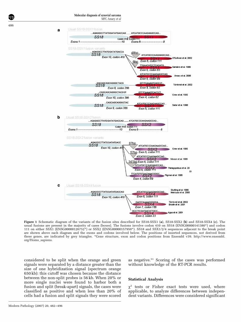
SS18-SSX1 and SS18-SSX2 fusions have beendetected in approximately 61 and 37% of casesrespectively, with the latter being associated witha better overall survival.21 In the vast majority ofsynovial sarcoma, fusion of SS18 with SSX1 andSSX2 involves the same codons: namely codon410 on SS18 (ENSG00000141380) fuses with codon111 on either SSX1 (ENSG00000126752) or SSX2(ENSG00000157950). Variant chimeric transcriptsare rare and have been described only in isolatedcases. To our knowledge, these include five variantsinvolving the fusion site of SS18-SSX1,22–26 andanother five involving the SS18-SSX2 fusionsite.20,22,27–29 SS18 also rarely forms a fusion genewith SSX4 involving the ‘usual’ SS18-SSX site atcodon 410 SS18 and codon in 111 SSX4:30–32 inaddition to this ‘usual type’, two variants have beendetected.33,34 One case of synovial sarcoma hasbeen reported to harbor a fusion involving a geneon chromosome 20 -SS18LI, a homolog of the SS18gene and the SSX1 gene.35 In addition, Sonobeet al36 reported a variant, which harbored atruncated cDNA, but involved the usual fusion inSS18-SSX1. The function of the SS18-SSX chimericprotein and its relationship to tumor developmentremains unknown, as does the mechanism throughwhich the translocation occurs.37
There are two major morphological subtypes ofsynovial sarcoma: the more common monophasictype, representing 60–75% of this tumor,5,38–41 iscomposed of short fascicles of plump spindlecells often with epithelioid areas.4,38 Identificationof well-formed glandular structures admixed withplump spindle and epithelioid cells are requiredbefore making the diagnosis of the biphasic type,and although this diagnosis is less problematic thisvariant shares microscopic features with mesothe-lioma, mixed mullerian tumors and adenocarcino-mas, which have a marked desmoplastic stromalresponse. Both subtypes may harbor a poorlydifferentiated component41,42 and if this area werebiopsied, the differential diagnosis includes a widevariety of neoplasms including ‘small round bluecell’ tumors,10 malignant peripheral nerve sheathtumor and non-sarcomatous neoplasms.43,44 This isparticularly the case when a synovial sarcomaoccurs at an unusual site such as lung or bone.5,14
In most cases, immunohistochemistry provides theinformation necessary for distinguishing these neo-plasms. However, there are situations when evenimmunohistochemistry cannot resolve the diagnos-tic problem, and in these genetic analysis for thedetection of SS18 gene rearrangement has beenfound to be a powerful diagnostic tool for makingthe diagnosis of synovial sarcoma.5,14,45–49
There is evidence to suggest that the type ofSS18-SSX fusion is associated with the histological
subtype.21,37,50 The largest study that has addressedthis issue is a multi-institutional analysis of 61biphasic synovial sarcoma of which only threeharbored the SS18-SSX2 transcript.21 Although thisfinding has been reproduced in smaller series,37,50
others have reported that the existence of a SS18-SSX2 in the biphasic variant may not be such a rareoccurrence.27,37 In addition, data on the type ofSS18-SSX fusion in synovial sarcoma with poorlydifferentiated areas are scant.39
The primary aim of this study was to analyze alarge series of formalin-fixed paraffin-embedded softtissue lesions for the SS18-SSX fusion transcriptsand compare three different molecular techniques,so that the findings could be applied to a diagnosticservice. We also correlated the SS18-SSX fusiontype with the histological subtype and the presenceof poorly differentiated areas.
Materials and methods
Selection of Cases
Three hundred and twenty-eight cases wereanalyzed for the presence of SS18-SSX fusiontranscripts. These included 134 cases, 76 resectionspecimens and 58 biopsies, where synovial sarcomawas either considered the primary diagnosis orwhere synovial sarcoma was included as part of themain differential diagnosis based on clinical infor-mation, morphology and immunohistochemistry. Ofthese 134 cases, 73 and 50 cases were retrieved fromthe archives of the Histopathology Departments ofthe Royal National Orthopaedic Hospital, and SantaCasa de Sao Paulo Central Hospital, Brazil, respec-tively. Eleven were referred from other UK hospitals.Normal tissue and tumors considered unlikely tobe synovial sarcoma (194 cases) were includedas negative controls. All cases were reviewed byMFCA, RT, AMF, and the cases that had no SS18rearrangement were also reviewed by AH, CF andAN. The study complied with Brazilian and localethical standards.
Representative paraffin-embedded material andhaematoxylin and eosin-stained slides were reviewed.Mitotic figures were counted in 10 consecutiveshigh-power fields (1 HPF¼ 0.238 mm2) in highlyproliferative ‘hot spot’ areas. Clinical data onselected cases were collected from histopathologyreports.
Classification of Synovial Sarcoma
The diagnosis of synovial sarcoma was based on themorphological features and immunohistochemistryas defined previously.4,38 All cases were classifiedas either monophasic or biphasic subtypes. Thepresence of well-defined glandular structure wasrequired for the diagnosis of the latter, whereasmonophasic synovial sarcoma was composed of
Molecular diagnosis of synovial sarcomaMFC Amary et al
483
Modern Pathology (2007) 20, 482–496
spindle cells often with epithelioid areas. Poorlydifferentiated areas were defined by the presence ofpreviously described features in five of more low-power fields: (1) cellular spindle and fasciculararchitecture closely simulating high-grade fibro-sarcoma and malignant peripheral nerve sheathtumor with areas of extensive necrosis, perivasculartumor preservation and high-mitotic activity; (2)cellular areas composed of round cells with featuresintermediate between the spindle and epithelioidcells, sometimes with rhabdoid features; and (3)the small cell variant that closely resembles PNET/Ewing sarcoma.42
Immunohistochemistry
Immunohistochemistry was performed using theVentana NexES Autostainer (Ventana Medical Sys-tems, Strasbourg, France) using antibodies to a-smooth muscle actin (1A4, 1:25 dilution, Dako, HighWycombe, Hertfordshire, UK), desmin (DE-R-11,1:10 dilution, 10 min protease pretreatment, Dako),S100 protein (polyclonal, 1:1600 dilution, 6 minprotease pretreatment, Dako), EMA (E-29, 1:5 dilu-tion, 2 min pressure cooker pretreatment, Dako),AE1/AE3 (dilution 1:50, 12 min protease pretreat-ment, Dako), MNF116 (dilution 1:100, 14 minprotease pretreatment, Dako), CD 99 (clone 12E7,dilution 1:50, 2 min pressure pretreatment, Dako)and bcl-2 (clone M0887, dilution 1:10, 2 minpressure cooker pretreatment, Dako). Diamino-benzidine was used as a chromogen in all reactions.Positive and negative controls were included ineach immunohistochemistry run.
Tissue Microarray
A tissue microarray was constructed using a manualtissue arrayer (Beecher Instruments Inc, Sun Prarie,WI, USA). Two different areas from each case weremarked in the paraffin blocks and two 0.6 mm coreswere taken for the array from all the cases describedabove for which material was available (113 cases).One hundred and seventeen cores including sali-vary gland, kidney, placenta and liver were used ascontrols and orientation markers.
RNA Extraction and Reverse Transcription
Two 10 mm or 5–10 10 mm sections were cut fromresection or biopsy specimen blocks, respectively,and placed into Eppendorf tubes. RNA wasextracted from paraffin-embedded samples usingOptimums FFPE RNA Isolation Kit (AmbionEurope, Huntington, Cambridgeshire, UK). Between1 and 3 ml of the resulting RNA samples were reversetranscribed using Superscript III First-Strand Synth-esis kit (Invitrogen, Inchinnan, Paisley, Scotland,UK) using gene-specific primers (all primers and
product sizes shown in Table 1). A G6PD (glucose-6-phosphate dehydrogenase) anti-sense primer wasincluded in the reverse transcription step.
Quantitative Polymerase Chain Reaction
Quantitative polymerase chain reaction (qPCR)amplification was performed using the iCycler iQdetection system (Bio-Rad Laboratories, Hempstead,Hertfordshire, UK) on 1ml aliquots of cDNA samplesusing specific primer sets (SYT-B-RT and SSX1-2-B)and probes designed based on the known fusiongenes and break points (see Figure 1 and Table 1).The reaction mixtures were made up to 25ml using12.5 ml of iQt Supermix (Bio-Rad Laboratories,Hempstead, Hertfordshire, UK), 5 pmol of eachprimer and 1 pmol of FAM-labelled probe and ultrapure water. Thermal cycling parameters were asfollows: 3 min at 951C followed by 55 cycles of 15 sat 941C and 1 min at 601C Fluorescence wasmonitored during the 601C annealing step.
Restriction Enzyme Digestion for Determining Subtypeof SS18-SSX Transcript Generated by qRT-PCR
Three microlitres of PCR product was mixed with0.5 ml of restriction enzyme XmnI (New EnglandBiolabs, Hitchin, Hertfordshire, UK), 1 ml of buffer(New England Biolabs) and made up to a finalvolume of 10ml with ultra pure water and digestedfor 2 h at 371C. The products were run througha 10% polyacrylamide gel revealing 56 and 39 bpproducts for a digested SS18-SSX2 transcript, andan undigested 95 bp product for the SS18-SSX1fusion.30
Conventional Polymerase Chain Reaction
PCR amplification was performed on duplicatesamples of 1ml aliquots of cDNA using specificprimer sets designed based on the known fusiongenes and break points (SSA, SSX1, SSX2 andSSX4) (Figure 1) (Table 1).49 Reactions were madeup in 25ml using 1X buffer II (Applied Biosystems,Warrington, Cheshire, UK), 200 mM of each dNTP,5 pmol of each primer, 1.5 mM MgCl2 and 1 U ofAmplitaq Gold (Applied Biosystems). A touchdownprotocol was used with cycling parameters asfollows: 7 min at 951C followed by 45 s at 941C,45 s at 661C, 1 min 30 s at 721C which was followedby reducing the annealing temperature by 11C eachcycle to 571C (10 cycles), followed by 30 cycles at561C and finally 5 min at 721C.
SS18-SSX1 and SS18-SSX2 translocations weresought using primer pairs SSA-SSX1 and SSA-SSX2, respectively (Table 1).49 Products were sepa-rated through an 8% polyacrylamide gel, stainedwith ethidium bromide and visualized under UVillumination using a BioRad Gel Doc 2000t system
Molecular diagnosis of synovial sarcomaMFC Amary et al
484
Modern Pathology (2007) 20, 482–496
(Hemel Hempstead, Hertfordshire, UK). Samplesyielding PCR products of the predicted size(108 bp) in both reactions were scored as positive.
Controls for RT-PCR
A negative (no template) and positive control (SS18-SSX fusion gene confirmed by sequencing) wereused for each experiment. The housekeeping gene,G6PD, was amplified using the same reactionconditions and cycling parameters for conventionalRT-PCR, as described above. The PCR primers weredesigned to provide the template for generation ofproducts of 86, 141 and 200 bp, from the house-keeping gene G6PD to control for RNA quality.
FISH
FISH was performed on the tissue microarrayafter optimization of the protocol using LSI SYTDual Color Break-Apart Rearrangement Probe (Vysis,Abbott Laboratories Inc., Maidenhead, Berkshire,UK). One end of the probe was labeled withspectrum oranget (telomeric, 50 to SS18, 650 Kb)and the other with spectrum greent (centromeric, 30
to SS18, 1040 Kb). Sections were deparaffinizedin xylene (3� for 5 min) and dehydrated in ethanol(3� for 3 min). Sections were pretreated usingParaffin Pretreatment Reagent Kit II (Vysis). This
involved placing the slides in 50 ml of pretreatmentsolution at 801C for 50 min, followed by 3 min indistilled water, 20 min in protease solution at 371Cwashing in distilled water for 3 min and dehydratingfor 1 min in increasing concentrations of alcohol (70,85 and 100%) after which they were air-dried. Tenmicrolitres of probe mixture (1 ml probe, 2ml dH2Oand 7 ml hybridizing buffer) were applied to theslides, overlaid with a coverslip, which was sealedwith rubber cement. Slides were denatured for 5 minat 731C and hybridized for at least 16 h at 371C in aThermoBritet hybridizer (Iris, Westwood, MA,USA). Following hybridization, the slides werewashed with 2� saline sodium citrate (SCC)/0.3%NP40 at room temperature for 5 min, at 731C for2 min and again at room temperature for 1 min. Afterair-drying in the dark, slides were counterstainedwith 10 ml of a mixture of Vectashields mountingmedium (Vector Laboratories, Orton Southgate,Peterborough, UK) containing 5 ml of Vectashieldswith 4060-diamino-2-phenylindole (DAPI) and 5mlwithout DAPI. A coverslip was applied and sealedwith nail varnish. The slides were analyzed undera fluorescent microscope (Olympus, Southall, Mid-dlesex, UK) equipped with a mercury lamp, DAPI/FITC/Rhodamine triple filter and individual DAPI,FITC and rhodamine filters using AnalySIS software(Olympus). Fifty non-overlapping nuclei, whichwere clearly identified and contained unequivocalsignals, were counted for each case. A probe was
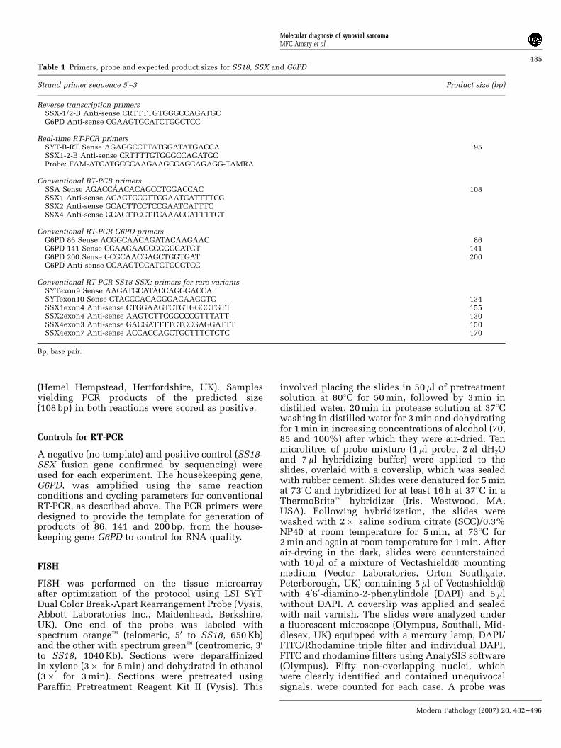
Table 1 Primers, probe and expected product sizes for SS18, SSX and G6PD
Strand primer sequence 50–30 Product size (bp)
Reverse transcription primersSSX-1/2-B Anti-sense CRTTTTGTGGGCCAGATGCG6PD Anti-sense CGAAGTGCATCTGGCTCC
Real-time RT-PCR primersSYT-B-RT Sense AGAGGCCTTATGGATATGACCA 95SSX1-2-B Anti-sense CRTTTTGTGGGCCAGATGCProbe: FAM-ATCATGCCCAAGAAGCCAGCAGAGG-TAMRA
Conventional RT-PCR primersSSA Sense AGACCAACACAGCCTGGACCAC 108SSX1 Anti-sense ACACTCCCTTCGAATCATTTTCGSSX2 Anti-sense GCACTTCCTCCGAATCATTTCSSX4 Anti-sense GCACTTCCTTCAAACCATTTTCT
Conventional RT-PCR G6PD primersG6PD 86 Sense ACGGCAACAGATACAAGAAC 86G6PD 141 Sense CCAAGAAGCCGGGCATGT 141G6PD 200 Sense GCGCAACGAGCTGGTGAT 200G6PD Anti-sense CGAAGTGCATCTGGCTCC
Conventional RT-PCR SS18-SSX: primers for rare variantsSYTexon9 Sense AAGATGCATACCAGGGACCASYTexon10 Sense CTACCCACAGGGACAAGGTC 134SSX1exon4 Anti-sense CTGGAAGTCTGTGGCCTGTT 155SSX2exon4 Anti-sense AAGTCTTCGGCCCGTTTATT 130SSX4exon3 Anti-sense GACGATTTTCTCCGAGGATTT 150SSX4exon7 Anti-sense ACCACCAGCTGCTTTCTCTC 170
Bp, base pair.
Molecular diagnosis of synovial sarcomaMFC Amary et al
485
Modern Pathology (2007) 20, 482–496
considered to be split when the orange and greensignals were separated by a distance greater than thesize of one hybridization signal (spectrum orange650 kb): this cutoff was chosen because the distancebetween the non-split probes is 56 kb. When 20% ormore single nuclei were found to harbor both afusion and split (break-apart) signals, the cases wereclassified as positive and when less than 20% ofcells had a fusion and split signals they were scored
as negative.51 Scoring of the cases was performedwithout knowledge of the RT-PCR results.
Statistical Analysis
w2 tests or Fisher exact tests were used, whereapplicable, to analyze differences between indepen-dent variants. Differences were considered significant
Figure 1 Schematic diagram of the variants of the fusion sites described for SS18-SSX1 (a), SS18-SSX2 (b) and SS18-SSX4 (c). Theusual fusions are present in the majority of cases (boxes). The fusions involve codon 410 on SS18 (ENSG00000141380*) and codon111 on either SSX1 (ENSG00000126752*) or SSX2 (ENSG00000157950*). SS18 and SSX1/2/4 sequences adjacent to the break pointare shown above each diagram and the exons and codons involved below. The positions of inserted sequences, not derived fromthese genes, are indicated by grey triangles. *Gene structure, exon and codon positions from Ensembl v39. http://www.ensembl.org/Homo_sapiens.
Molecular diagnosis of synovial sarcomaMFC Amary et al
486
Modern Pathology (2007) 20, 482–496
if Po0.05. The software SPSS 10.0 (SPSS Inc.,Chicago, IL, USA) was used for statistical analysis.
Results
Clinical and Histological Features
Synovial sarcoma was included in the differentialdiagnosis of 134 cases based on the morphologyand immunohistochemistry: in 97 of these synovialsarcoma was considered the primary diagnosis andas a strong candidate in the remaining 37 cases.Patients with these tumors presented between theages of 5 and 81, the mean age being 35 years, andthe tumors occurred equally in both sexes (female/male ratio, 1:1.06). The lower extremity was themost common location (n¼ 80), followed by upperextremity (n¼ 25), thorax (n¼ 17), head and neck(n¼ 4), pelvis (n¼ 2) and retroperitoneum (n¼ 2). Infour cases the location was unknown (Table 2). Onehundred and three (77%) cases were classified asmonophasic synovial sarcoma and 31 (23%) as
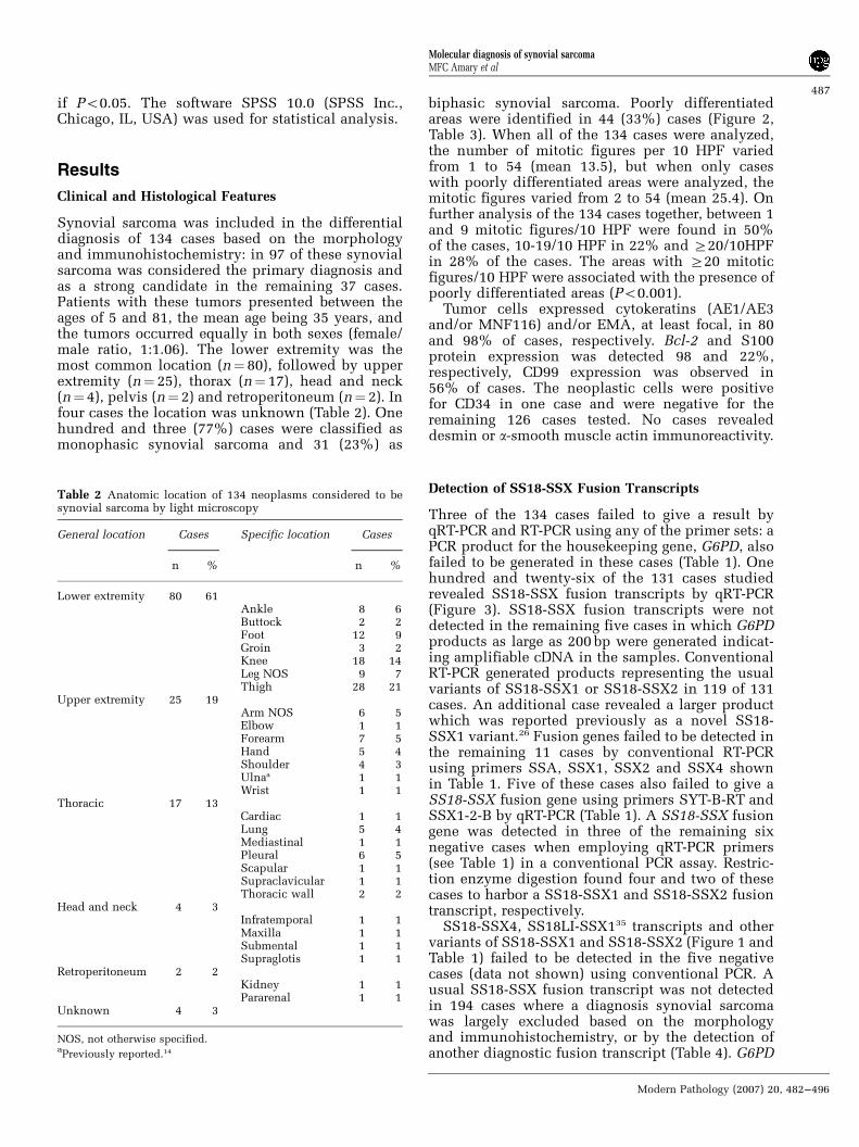
biphasic synovial sarcoma. Poorly differentiatedareas were identified in 44 (33%) cases (Figure 2,Table 3). When all of the 134 cases were analyzed,the number of mitotic figures per 10 HPF variedfrom 1 to 54 (mean 13.5), but when only caseswith poorly differentiated areas were analyzed, themitotic figures varied from 2 to 54 (mean 25.4). Onfurther analysis of the 134 cases together, between 1and 9 mitotic figures/10 HPF were found in 50%of the cases, 10-19/10 HPF in 22% and Z20/10HPFin 28% of the cases. The areas with Z20 mitoticfigures/10 HPF were associated with the presence ofpoorly differentiated areas (Po0.001).
Tumor cells expressed cytokeratins (AE1/AE3and/or MNF116) and/or EMA, at least focal, in 80and 98% of cases, respectively. Bcl-2 and S100protein expression was detected 98 and 22%,respectively, CD99 expression was observed in56% of cases. The neoplastic cells were positivefor CD34 in one case and were negative for theremaining 126 cases tested. No cases revealeddesmin or a-smooth muscle actin immunoreactivity.
Detection of SS18-SSX Fusion Transcripts
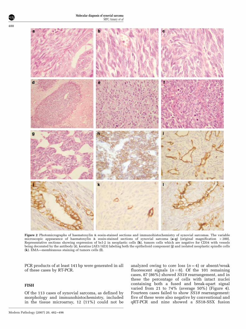
Three of the 134 cases failed to give a result byqRT-PCR and RT-PCR using any of the primer sets: aPCR product for the housekeeping gene, G6PD, alsofailed to be generated in these cases (Table 1). Onehundred and twenty-six of the 131 cases studiedrevealed SS18-SSX fusion transcripts by qRT-PCR(Figure 3). SS18-SSX fusion transcripts were notdetected in the remaining five cases in which G6PDproducts as large as 200 bp were generated indicat-ing amplifiable cDNA in the samples. ConventionalRT-PCR generated products representing the usualvariants of SS18-SSX1 or SS18-SSX2 in 119 of 131cases. An additional case revealed a larger productwhich was reported previously as a novel SS18-SSX1 variant.26 Fusion genes failed to be detected inthe remaining 11 cases by conventional RT-PCRusing primers SSA, SSX1, SSX2 and SSX4 shownin Table 1. Five of these cases also failed to give aSS18-SSX fusion gene using primers SYT-B-RT andSSX1-2-B by qRT-PCR (Table 1). A SS18-SSX fusiongene was detected in three of the remaining sixnegative cases when employing qRT-PCR primers(see Table 1) in a conventional PCR assay. Restric-tion enzyme digestion found four and two of thesecases to harbor a SS18-SSX1 and SS18-SSX2 fusiontranscript, respectively.
SS18-SSX4, SS18LI-SSX135 transcripts and othervariants of SS18-SSX1 and SS18-SSX2 (Figure 1 andTable 1) failed to be detected in the five negativecases (data not shown) using conventional PCR. Ausual SS18-SSX fusion transcript was not detectedin 194 cases where a diagnosis synovial sarcomawas largely excluded based on the morphologyand immunohistochemistry, or by the detection ofanother diagnostic fusion transcript (Table 4). G6PD
Table 2 Anatomic location of 134 neoplasms considered to besynovial sarcoma by light microscopy
General location Cases Specific location Cases
n % n %
Lower extremity 80 61Ankle 8 6Buttock 2 2Foot 12 9Groin 3 2Knee 18 14Leg NOS 9 7Thigh 28 21
Upper extremity 25 19Arm NOS 6 5Elbow 1 1Forearm 7 5Hand 5 4Shoulder 4 3Ulnaa 1 1Wrist 1 1
Thoracic 17 13Cardiac 1 1Lung 5 4Mediastinal 1 1Pleural 6 5Scapular 1 1Supraclavicular 1 1Thoracic wall 2 2
Head and neck 4 3Infratemporal 1 1Maxilla 1 1Submental 1 1Supraglotis 1 1
Retroperitoneum 2 2Kidney 1 1Pararenal 1 1
Unknown 4 3
NOS, not otherwise specified.aPreviously reported.14
Molecular diagnosis of synovial sarcomaMFC Amary et al
487
Modern Pathology (2007) 20, 482–496
PCR products of at least 141 bp were generated in allof these cases by RT-PCR.
FISH
Of the 113 cases of synovial sarcoma, as defined bymorphology and immunohistochemistry, includedin the tissue microarray, 12 (11%) could not be
analyzed owing to core loss (n¼ 4) or absent/weakfluorescent signals (n¼ 8). Of the 101 remainingcases, 87 (86%) showed SS18 rearrangement, and inthese the percentage of cells with intact nucleicontaining both a fused and break-apart signalvaried from 21 to 74% (average 50%) (Figure 4).Fourteen cases failed to show SS18 rearrangement:five of these were also negative by conventional andqRT-PCR and nine showed a SS18-SSX fusion
Figure 2 Photomicrographs of haematoxylin & eosin-stained sections and immunohistochemistry of synovial sarcomas. The variablemicroscopic appearance of haematoxylin & eosin-stained sections of synovial sarcoma (a–g) (original magnification �200).Representative sections showing expression of bcl-2 in neoplastic cells (h), tumors cells which are negative for CD34 with vesselsbeing decorated by the antibody (i), keratins (AE1/AE3) labeling both the epithelioid component (j) and isolated neoplastic spindle cells(k). EMA—membranous staining of tumors cells (l).
Molecular diagnosis of synovial sarcomaMFC Amary et al
488
Modern Pathology (2007) 20, 482–496
transcript by conventional RT-PCR and qRT-PCR(n¼ 8) and qRT-PCR only (n¼ 1). Analysis of four ofthese nine cases revealed that at least 70% of nucleicontained one fusion signal and a single spectrumorange signal, although a fusion and break-apartsignals were identified in occasional cells (2, 4, 6and 8%) (Figure 4). In the remaining five cases,which were scored as negative, a break-apart signalwas detected in 2–4 % of the cells in three cases andno break-apart signal was detected in two cases. Nocases were found to show between 10 and 20% ofthe cells with break-apart signals.
Fifteen of the 101 cases analyzed by FISH showedmultiple copies of the SS18 gene (Figure 4). Theintact nuclei of 14 of these cases showed more than20% of break-apart signals. Case 15 was classified asnegative on account of only 2% of cells showingunequivocal break-apart signals. However, this casewas positive by both conventional and qRT-PCR.
One of the three cases, which could not beanalyzed by RT-PCR, owing to no amplifiable cDNA,was positive by FISH. One was not tested by FISHowing to unavailable material and the remainingcase was tested but no signal was detected.
Correlation of SS18-SSX Fusion Genes andMorphology
Collation of all the data generated from conventionaland qRT-PCR revealed that 74 and 52 of the caseswere found to harbor SS18-SSX1 and SS18-SSX2fusion genes, respectively. The relationship betweenhistological subtypes (mono or biphasic) and fusiontranscripts is shown in Table 3, and was not foundto be statistically significant (P¼ 0.9). The presenceof poorly differentiated areas was identified in bothbiphasic (n¼ 5, 16%) and monophasic (n¼ 39, 38%)neoplasms and did not correlate with fusion type(P¼ 0.2).
Of the 31 biphasic synovial sarcoma, 30 showedareas of unequivocal glandular formation. One casewhich showed extensive squamous differentiationharbored a SS18-SSX2 fusion type.
SS18-SSX-Negative Cases
Table 5 and Figure 5 provide details of the 5 SS18-SSX fusion gene negative cases. Of the three pleuralcases, one exhibiting a biphasic and two a mono-phasic pattern, the diagnosis of synovial sarcomawas favored based on morphology in cases oneand two initially. However, following clinical and
Table 3 SS18-SSX fusion type and histological subtype
Histological subtype (n) SS18-SSX fusion type (n) Poorly differentiated areas inmorphological subtype (n)
Fusion type associated withtumours with PD areas (n)
Biphasic (31) SS18-SSX1 (18) 5 SS18-SSX1 (3)SS18-SSX2 (11) SS18-SSX2 (2)Negative (1)NA (1)
Monophasic (103) SS18-SSX1 (56) 39 SS18-SSX1 (18)SS18-SSX2 (41) SS18-SSX2 (18)Negative (4) Negative (3)NA (2)
Total (134) SS18-SSX1 (74) 44 SS18-SSX1 (21)SS18-SSX2 (52) SS18-SSX2 (20)Negative (5) Negative (3)Poor RNA (3)
PD, poorly differentiated; n, number.NA¼no amplifiable cDNA generated from the sample.
400
PCR Amp/Cycle Graph for FAM-490a
b
350
300
250
200
150
100
50
-50
-1000 2 4 6 8 10 1214 16 18 20 22 24 26 28 30 32 3436 38
Cycle
PC
R B
ase
Line
Sub
stra
cted
RF
U
L 1 2 3 4 5 6 7 8
40 42 4446 48 50 52 54 56 58
0
400
350
300
250
200
150
100
50
-50
-100
0
Figure 3 Detection of SS18-SSX fusion transcripts by the RT-PCR.Thirty-four samples analyzed by qRT-PCR and represented bydifferent lines: controls included (a). Detection of a positive SS18-SSX2 fusion transcript, by conventional RT-PCR in duplicate inan 8% polyacrylamide gel (b): lane L, molecular weight markers(50 bp); lane 1 and 5, negative control; lanes 2 and 6, positivecontrols SSX1 and SSX2 respectively; lanes 3 and 4, negative forSS18-SSX1; lanes 7 and 8, positive for SS18-SSX2.
Molecular diagnosis of synovial sarcomaMFC Amary et al
489
Modern Pathology (2007) 20, 482–496
radiological input into case 1, it was considered that‘on the balance of probabilities’ the tumor was abiphasic mesothelioma and not a synovial sarcoma.
This decision was made in view of the unilateraldisease, the history of asbestos exposure and animmunophenotype that overlapped between
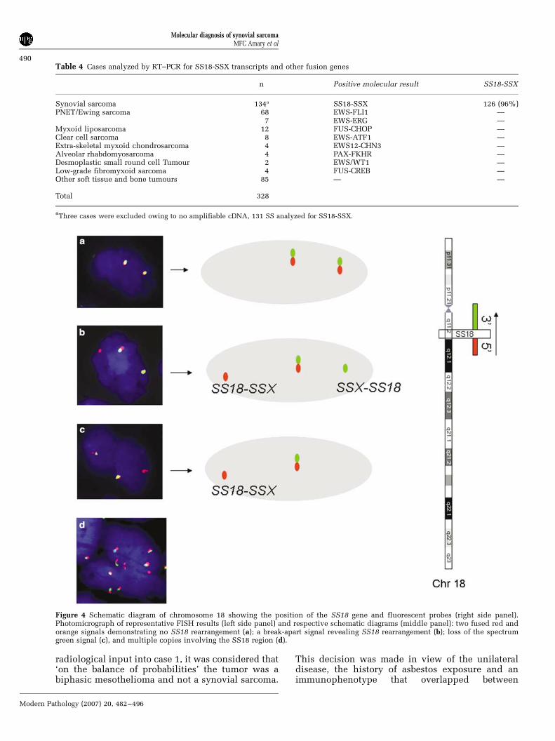
Table 4 Cases analyzed by RT–PCR for SS18-SSX transcripts and other fusion genes
n Positive molecular result SS18-SSX
Synovial sarcoma 134a SS18-SSX 126 (96%)PNET/Ewing sarcoma 68 EWS-FLI1 —
7 EWS-ERG —Myxoid liposarcoma 12 FUS-CHOP —Clear cell sarcoma 8 EWS-ATF1 —Extra-skeletal myxoid chondrosarcoma 4 EWS12-CHN3 —Alveolar rhabdomyosarcoma 4 PAX-FKHR —Desmoplastic small round cell Tumour 2 EWS/WT1 —Low-grade fibromyxoid sarcoma 4 FUS-CREB —Other soft tissue and bone tumours 85 — —
Total 328
aThree cases were excluded owing to no amplifiable cDNA, 131 SS analyzed for SS18-SSX.
Figure 4 Schematic diagram of chromosome 18 showing the position of the SS18 gene and fluorescent probes (right side panel).Photomicrograph of representative FISH results (left side panel) and respective schematic diagrams (middle panel): two fused red andorange signals demonstrating no SS18 rearrangement (a); a break-apart signal revealing SS18 rearrangement (b); loss of the spectrumgreen signal (c), and multiple copies involving the SS18 region (d).
Molecular diagnosis of synovial sarcomaMFC Amary et al
490
Modern Pathology (2007) 20, 482–496
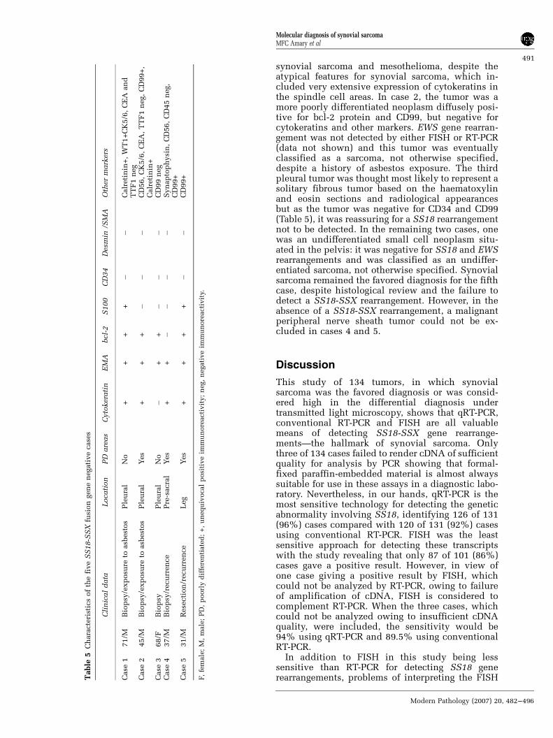
synovial sarcoma and mesothelioma, despite theatypical features for synovial sarcoma, which in-cluded very extensive expression of cytokeratins inthe spindle cell areas. In case 2, the tumor was amore poorly differentiated neoplasm diffusely posi-tive for bcl-2 protein and CD99, but negative forcytokeratins and other markers. EWS gene rearran-gement was not detected by either FISH or RT-PCR(data not shown) and this tumor was eventuallyclassified as a sarcoma, not otherwise specified,despite a history of asbestos exposure. The thirdpleural tumor was thought most likely to represent asolitary fibrous tumor based on the haematoxylinand eosin sections and radiological appearancesbut as the tumor was negative for CD34 and CD99(Table 5), it was reassuring for a SS18 rearrangementnot to be detected. In the remaining two cases, onewas an undifferentiated small cell neoplasm situ-ated in the pelvis: it was negative for SS18 and EWSrearrangements and was classified as an undiffer-entiated sarcoma, not otherwise specified. Synovialsarcoma remained the favored diagnosis for the fifthcase, despite histological review and the failure todetect a SS18-SSX rearrangement. However, in theabsence of a SS18-SSX rearrangement, a malignantperipheral nerve sheath tumor could not be ex-cluded in cases 4 and 5.
Discussion
This study of 134 tumors, in which synovialsarcoma was the favored diagnosis or was consid-ered high in the differential diagnosis undertransmitted light microscopy, shows that qRT-PCR,conventional RT-PCR and FISH are all valuablemeans of detecting SS18-SSX gene rearrange-ments—the hallmark of synovial sarcoma. Onlythree of 134 cases failed to render cDNA of sufficientquality for analysis by PCR showing that formal-fixed paraffin-embedded material is almost alwayssuitable for use in these assays in a diagnostic labo-ratory. Nevertheless, in our hands, qRT-PCR is themost sensitive technology for detecting the geneticabnormality involving SS18, identifying 126 of 131(96%) cases compared with 120 of 131 (92%) casesusing conventional RT-PCR. FISH was the leastsensitive approach for detecting these transcriptswith the study revealing that only 87 of 101 (86%)cases gave a positive result. However, in view ofone case giving a positive result by FISH, whichcould not be analyzed by RT-PCR, owing to failureof amplification of cDNA, FISH is considered tocomplement RT-PCR. When the three cases, whichcould not be analyzed owing to insufficient cDNAquality, were included, the sensitivity would be94% using qRT-PCR and 89.5% using conventionalRT-PCR.
In addition to FISH in this study being lesssensitive than RT-PCR for detecting SS18 generearrangements, problems of interpreting the FISHT
able
5C
hara
cteri
stic
sof
the
five
SS
18-S
SX
fusi
on
gen
en
egat
ive
case
s
Cli
nic
al
data
Locati
on
PD
are
as
Cyto
kera
tin
EM
Abcl-
2S
100
CD
34
Desm
in/S
MA
Oth
er
mark
ers
Case
171/M
Bio
psy
/exp
osu
reto
asb
esto
sP
leu
ral
No
++
++
��
Calr
eti
nin
+,
WT
1+
CK
5/6
,C
EA
an
dT
TF
1n
eg
Case
245/M
Bio
psy
/exp
osu
reto
asb
esto
sP
leu
ral
Yes
++
+�
��
CD
56,
CK
5/6
,C
EA
,T
TF
1n
eg,
CD
99+
,C
alr
eti
nin
+C
ase
368/F
Bio
psy
Ple
ura
lN
o�
++
��
�C
D99
neg
Case
437/M
Bio
psy
/recu
rren
ce
Pre
-sacra
lY
es
++
��
��
Syn
ap
top
hysi
n,
CD
56,
CD
45
neg,
CD
99+
Case
531/M
Rese
cti
on
/recu
rren
ce
Leg
Yes
++
++
��
CD
99+
F,
fem
ale
;M
,m
ale
;P
D,
poorl
yd
iffe
ren
tiate
d;
+,
un
equ
ivocal
posi
tive
imm
un
ore
acti
vit
y;
neg,
negati
ve
imm
un
ore
acti
vit
y.
Molecular diagnosis of synovial sarcomaMFC Amary et al
491
Modern Pathology (2007) 20, 482–496
were also encountered. These difficulties are largelythree-fold: the first, not previously reported, is thedetection of one fusion signal (green and orange)and a single spectrum orange signal, in morethan 70% of the neoplastic cells, in other words,the second spectrum green label was not observed.This observation was made in four cases, in whichthe SS18-SSX fusion gene was detected by RT-PCR,and has been found in a further two cases since the
study was closed. The SSX-SS18 transcript, unlikeits reciprocal SS18-SSX fusion gene, has beenshown to be lost in as many as two-thirds of casesof synovial sarcoma, and this loss has been asso-ciated with progression of disease.27 We, therefore,speculate that our observation is caused by deletionof the SSX-SS18 fusion gene. Our interpretation isalso based on the fact that the SS18-SSX fusion iscomposed mainly of the SS18 gene, to which the
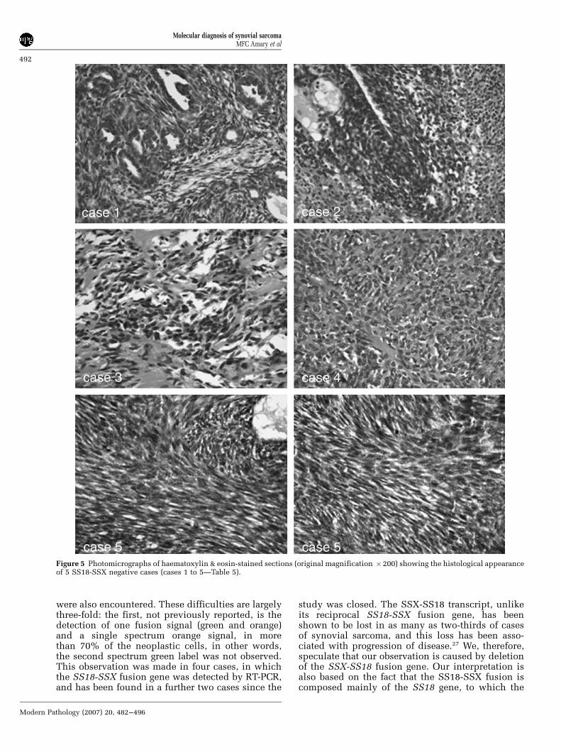
Figure 5 Photomicrographs of haematoxylin & eosin-stained sections (original magnification �200) showing the histological appearanceof 5 SS18-SSX negative cases (cases 1 to 5—Table 5).
Molecular diagnosis of synovial sarcomaMFC Amary et al
492
Modern Pathology (2007) 20, 482–496

spectrum orange label is attached via the 50-end,and spectrum green labels the SSX-SS18 fusion.Nevertheless, further studies are required to supportthis theory.
The second difficulty encountered when inter-preting FISH for the diagnosis of synovial sarcoma isthe presence of multiple signals in some cases: thisfinding represents either polyploidy or multiplecopies of a region of variable length of chromosome18 in the region of the SS18 gene. This has beenreported previously in synovial sarcoma, with one ofthe most common gains involving 18q,27 and alsoin PNET/Ewing sarcoma in chromosome 22.52 Onoccasions the signals are so numerous that the resultis difficult to interpret although in 14 of our 15 casesunequivocal break-apart signals were also identifiedallowing synovial sarcoma to be diagnosed. In all ofthese cases, interpretation of the FISH result wassupported by RT-PCR.
The final problem experienced using FISH isdetermining (a) how many cells should be countedand (b) what percentage of these cells need to bescored as positive for break-apart signals before atumor is classed as harboring a rearrangement. Inthis study, we employed the standards of Lu et al,51
but we did not encounter cases where the cellswith break-apart signals represented between 8 and21% of the 50 cells counted. In borderline cases, it isuseful to have supportive information from RT-PCRanalysis but most importantly any result mustbe interpreted in the light of the morphology andimmunohistochemistry. For these reasons, we be-lieve that there are great benefits for the histopathol-ogists reporting such cases to have experience in theinterpretation of FISH and have direct access to thistechnology.
Our results demonstrate that the primers andprobes used for RT-PCR in this study are specific forthe various subtypes and variants of the reportedSS18-SSX fusion genes. Not only were SS18-SSXfusion transcripts not detected in 194 other tumorsby RT-PCR (109 tumors in which other fusiongenes were detected and 85 other soft tissue andbone tumors with no fusion detected) but there wasalso complete concordance of positive results whenusing primers for conventional and qRT-PCR. Inthis study and in all other series except one,53 thetype of transcripts (SS18-SSX1 or SS18-SSX2) wasmutually exclusive and persisted in recurrent andmetastatic disease (data not shown). It is difficult toexplain the results of Yang et al as their studywas in-depth and appears robust. Nevertheless, it isimportant to highlight that SSX1 and SSX2 shareat least 80% homology,21 and that primer designis therefore crucial if the two genes are to bedistinguished by PCR.
The fact that six cases in this study were found toharbor the SS18-SSX fusion transcript using qRT-PCR, but not conventional RT-PCR suggests that theformer technology is more sensitive. However, thisneeds to be interpreted critically as the data show
that at least some of this apparent greater sensitivityis based on the PCR primer design and possibly thesize of the product generated rather than thetechnology employed.
This study reveals that biphasic synovial sarcomanot uncommonly harbors the SS18-SSX2 fusion, afinding that contrasts results of previous series inwhich almost all biphasic cases were shown toharbor a SS18-SSX1 fusion type.21 However, havingperformed a meta-analysis of published data we findthat SS18-SSX2 occurs significantly more frequentlyin monophasic than in biphasic synovial sarcoma(Po0.001): this meta-analysis included a total of1033 cases, of which 314 were biphasic synovialsarcoma and only 42 of these harbored theSS18-SSX2 transcript.5,20,22,27,30,37,39,46,47,49–51,54–65
The low frequency of both the SS18-SSX2 fusiontype and the biphasic subtype of synovial sarcomabest explain the discrepant results observed invarious studies. In our study, we also failed to finda significant association between poorly differen-tiated areas of synovial sarcoma and SS18 generearrangement, thereby supporting the work byGuillou et al.39
This study highlights the difficulty in distinguish-ing synovial sarcoma from mesothelioma, an im-portant issue as the thorax is now recognized as anot uncommon site in which synovial sarcomaoccur5,43 and the correct classification of tumorsnot only alters clinical management but in the caseof mesothelioma, the patient may also be eligible forfinancial compensation if there has been exposure toasbestos. We found that on microscopic assessmentalone it was not possible to distinguish definitivelybetween these two tumor types and detection of aSS18 rearrangement would have been invaluable inconfidently refuting the diagnosis of mesothelioma.Of the five SS18-SSX negative neoplasms in thisstudy, three were pleural lesions, one exhibiting abiphasic and two a monophasic pattern. A diagnosisof synovial sarcoma was strongly favored in twoof these three cases when the histopathology wasanalyzed in the absence of further clinical informa-tion. A diagnosis of a solitary fibrous tumor wasfavored in the third case and molecular geneticanalysis was performed for exclusion purposes.However, when the histopathology was reviewedin the light of all the information from the multi-disciplinary team, which included physicians,radiologists and pathologists, and the negativeresults from the molecular analysis were taken intoaccount, all three cases were considered unlikelyto represent synovial sarcoma. The value of amultidisciplinary approach is emphasized furtheras one pleural case was eventually considered tobe sarcoma ‘not otherwise specified’ rather thanmesothelioma despite exposure to asbestos.
Of the two non-pleural-based SS18-SSX negativetumors, one, situated in the pelvis, was classified asa small cell undifferentiated neoplasm, not other-wise specified, and the second occurred in the lower
Molecular diagnosis of synovial sarcomaMFC Amary et al
493
Modern Pathology (2007) 20, 482–496
limb. Despite histological review of the latter andthe failure to detect SS18-SSX rearrangements thediagnosis of synovial sarcoma was still favored.Nevertheless, as there is a well-recognized overlapboth morphologically and at a gene expression levelbetween synovial sarcoma and malignant peripheralnerve sheath tumor66,67 the latter was seriouslyconsidered as the diagnosis in both of these cases.However, as neither of these patients had stigmata ofneurofibromatosis, and the tumors did not obviouslyarising from a large nerve this diagnosis was notpossible to make with confidence.
If aberrant genetic events are to be employed asdiagnostic tests on which clinical management is tobe based, knowledge of the sensitivity and specifi-city of any method being employed is crucial. Themajority of published evidence, including ours,argues that detection of a SS18 gene rearrangementis specific for the diagnosis of synovial sarcoma,48,68
although there is some debate over the interpreta-tion of finding a SS18 fusion gene in tumorswith morphological features of malignant peripheralnerve sheath tumors.69–71 In contrast, proving 100%sensitivity is more difficult. Indeed, several groups,including ours, demonstrate it is possible to attain atleast 96% sensitivity for the detection of a SS18 generearrangement but none claim 100%.21,39,49,50,59 Forthis to be achieved in our study, it would benecessary to demonstrate, beyond all doubt, thenature of the five cases which had morphologicalfeatures of synovial sarcoma but in which a SS18gene rearrangement was not detected. Unfortu-nately, a truly robust test for diagnosing mesothe-lioma, malignant peripheral nerve sheath tumor andsolitary fibrous tumor, the favored diagnoses for ourfive cases when a SS18 rearrangement was notdetected, does not exist, and therefore claiming100% sensitivity is likely to result in developmentof dogma which tend not to be questioned—adangerous mind-set for pathologists. Although wehave screened for all the known possible variants,the possibility of the existence of a novel t(X;18)variant remains, alternatively the failure to detecta SS18 rearrangement might be explained by almostcomplete loss of the X chromosome somethingwhich has been described in advanced tumors.54
Hence, we report these rare cases descriptively, andon the basis of clinical and radiological information.
Both FISH and RT-PCR have advantages anddisadvantages, but this study shows that theycomplement each other. FISH is often favored bypathologists as they are more familiar with analyz-ing cells using a microscope than reading gels.However, unless specific probes for fusion variantsare available, FISH is less informative than RT-PCR.This begs the question ‘what information is requiredfrom molecular diagnostic investigations?’ Theanswer will vary from tumor to tumor, and maychange over time: for example, if it is demon-strated that identification of a fusion variant for aparticular tumor does not alter treatment and
provides no information about clinical outcome,identification of specific variants may be consideredirrelevant. Therefore, continued critical assessmentof why specific investigations are undertaken iscrucial if pathologists are to use available resourcesefficiently.
Acknowledgements
This work could not have been done without thework and co-operation of all the clinicians andradiologists in the London Bone and Soft TissueSarcoma Service. Thank you to those who sent casesfor molecular analysis as Professor M Novelli atUCH, Dr AG Gibson at Kent & Canterbury HospitalNHS Trust, Dr Richard Ball at Norfolk and NorwichUniversity Hospital NHS Trust, Tom Treasure atGuy’s Hospital, and Professor NA Athanasou atNuffield Orthopaedic Centre, Oxford.
Grant support: R&D funding from The RoyalNational Orthopaedic Hospital, Stanmore, UK.
MFC Amary* Research fellow supported byCoordination for the Improvement of Higher Educa-tion Personnel, CAPES—Brazil. PhD Student Uni-versity of Sao Paulo, SP, Brazil.
References
1 Russell WO, Cohen J, Edmonson JH, et al. Stagingsystem for soft tissue sarcoma. Semin Oncol 1981;8:156–159.
2 Cadman NL, Soule EH, Kelly PJ. Synovial sarcoma.Cancer 1965;18:613–627.
3 Cagle LA, Mirra JM, Storm FK, et al. Histologic featuresrelating to prognosis in synovial sarcoma. Cancer1987;59:1810–1814.
4 Weiss SW, Goldblum JR. Synovial sarcoma. In: WeissSW, Goldblum JR (eds). Enzinger and Weiss’s SoftTissue Tumors. Mosby: London, 2001, pp 309–346.
5 Begueret H, Galateau-Salle F, Guillou L, et al. Primaryintrathoracic synovial sarcoma: a clinicopathologicstudy of 40 t(X;18)-positive cases from the FrenchSarcoma Group and the Mesopath Group. Am J SurgPathol 2005;29:339–346.
6 Fisher C, Folpe AL, Hashimoto H, et al. Intra-abdominal synovial sarcoma: a clinicopathologicalstudy. Histopathology 2004;45:245–253.
7 Hisaoka M, Hashimoto H, Iwahana H, et al. Primarysynovial sarcoma of the lung: report of two casesconfirmed by molecular detection of SYT-SSX fusiongene transcripts. Histopathology 1999;34:205–210.
8 Holtz F, Magielski JE. Synovial sarcomas of the tonguebase. Arch Otolaryngol 1985;111:271–272.
9 Iyengar V, Lineberger AS, Kerman S, et al. Synovialsarcoma of the heart. Correlation with cytogeneticfindings. Arch Pathol Lab Med 1995;119:1080–1082.
10 Jun SY, Kang GH, Park SH, et al. Synovial sarcoma ofthe kidney with rhabdoid features: report of threecases. Am J Surg Pathol 2004;28:634–637.
11 Lenoir P, Ramet J, Goossens A, et al. Retropharyngealsynovial sarcoma in an infant: report of a case and its
Molecular diagnosis of synovial sarcomaMFC Amary et al
494
Modern Pathology (2007) 20, 482–496
response to chemotherapy; review of the literature.Pediatr Hematol Oncol 1991;8:45–52.
12 Ng SB, Ahmed Q, Tien SL, et al. Primary pleuralsynovial sarcoma. A case report and review of theliterature. Arch Pathol Lab Med 2003;127:85–90.
13 Nicholson A, Rigby M, Lincoln C, et al. Synovialsarcoma of the heart. Histopathology 1997;30:349–352.
14 O’Donnell P, Diss T, Whelan J, et al. Synovial sarcomawith radiological appearances of primitive neuroecto-dermal tumour/Ewing sarcoma: differentiation bymolecular genetic studies. Skeletal Radiol 2006;35:233–239.
15 Okamato S, Hisaoka M, Daa T, et al. Primarypulmonary synovial sarcoma: a clinicopathologic,immunohistochemical, and molecular study of 11cases. Human Pathol 2004;35:850–856.
16 Limon J, Mrozek K, Nedoszytko B, et al. Cytogeneticfindings in two synovial sarcomas. Cancer GenetCytogenet 1989;38:215–222.
17 Turc-Carel C, Dal Cin P, Limon J, et al. Involvementof chromosome X in primary cytogenetic changein human neoplasia: nonrandom translocation insynovial sarcoma. Proc Natl Acad Sci USA 1987;84:1981–1985.
18 Turc-Carel C, Dal CP, Limon J, et al. Translocation X;18 in synovial sarcoma. Cancer Genet Cytogenet1986;23:93.
19 Clark J, Rocques PJ, Crew AJ, et al. Identification ofnovel genes, SYT and SSX, involved in thet(X;18)(p11.2;q11.2) translocation found in humansynovial sarcoma. Nat Genet 1994;7:502–508.
20 Crew AJ, Clark J, Fisher C, et al. Fusion of SYT totwo genes, SSX1 and SSX2, encoding proteins withhomology to the Kruppel-associated box in humansynovial sarcoma. EMBO J 1995;14:2333–2340.
21 Ladanyi M, Antonescu CR, Leung DH, et al. Impactof SYT-SSX fusion type on the clinical behavior ofsynovial sarcoma: a multi-institutional retrospectivestudy of 243 patients. Cancer Res 2002;62:135–140.
22 Nilsson G, Skytting B, Xie Y, et al. The SYT-SSX1variant of synovial sarcoma is associated with a highrate of tumor cell proliferation and poor clinicaloutcome. Cancer Res 1999;59:3180–3184.
23 Safar A, Wickert R, Nelson M, et al. Characterization ofa variant SYT-SSX1 synovial sarcoma fusion tran-script. Diagn Mol Pathol 1998;7:283–287.
24 Sanders ME, van de Rijn M, Barr FG. Detection of avariant SYT-SSX1 fusion in a case of predominantlyepithelioid synovial sarcoma. Mol Diagn 1999;4:65–70.
25 O’Sullivan MJ, Humphrey PA, Dehner LP, et al. t(X;18)reverse transcriptase-polymerase chain reaction demon-strating a variant transcript. Mol Diagn 2002;4:178–180.
26 Amary MFC, Diss T, Flanagan AM. Molecular char-acterization of a novel variant of a SYT-SSX1 fusiontranscript in synovial sarcoma. Histopathology 2006,in press.
27 Panagopoulos I, Mertens F, Isaksson M, et al. Clinicalimpact of molecular and cytogenetic findings in syno-vial sarcoma. Genes Chromosomes Cancer 2001;31:362–372.
28 Fligman I, Lonardo F, Jhanwar SC, et al. Moleculardiagnosis of synovial sarcoma and characterization ofa variant SYT-SSX2 fusion transcript. Am J Pathol1995;147:1592–1599.
29 Tornkvist M, Brodin B, Bartolazzi A, et al. A novel typeof SYT/SSX fusion: methodological and biologicalimplications. Mod Pathol 2001;15:679–685.
30 Gaffney R, Chakerian A, O’Connell JX, et al. Novelfluorescent ligase detection reaction and flow cyto-metric analysis of SYT-SSX fusions in synovialsarcoma. Mol Diagn 2003;5:127–135.
31 Skytting B, Nilsson G, Brodin B, et al. A novel fusiongene, SYT-SSX4, in synovial sarcoma. J Natl CancerInst 1999;91:974–975.
32 Mancuso T, Mezzelani A, Riva C, et al. Analysis ofSYT-SSX fusion transcripts and bcl-2 expression andphosphorylation status in synovial sarcoma. Lab Invest2000;80:805–813.
33 Brodin B, Haslam K, Yang K, et al. Cloning andcharacterization of spliced fusion transcript variants ofsynovial sarcoma: SYT/SSX4, SYT/SSX4v, and SYT/SSX2v. Possible regulatory role of the fusion gene productin wild type SYT expression. Gene 2001;268:173–182.
34 Agus V, Tamborini E, Mezzelani A, et al. A novelfusion gene, SYT-SSX4, in synovial sarcoma. J NatlCancer Instit 2001;93:1347–1349.
35 Storlazzi CT, Mertens F, Mandahl N, et al. A novelfusion gene, SS18L1/SSX1, in synovial sarcoma. GenesChromosomes Cancer 2003;37:195–200.
36 Sonobe H, Takeuchi T, Liag SB, et al. A new humansynovial sarcoma cell line, HS-SY-3, with a truncatedform of hybrid SYT/SSX1 gene. Int J Cancer 1999;82:459–464.
37 Wei Y, Wang J, Zhu X, et al. Detection of SYT-SSXfusion transcripts in paraffin-embedded tissues ofsynovial sarcoma by reverse transcrition-polymerasechain reaction. Chin Med J (Engl) 2002;115:1043–1047.
38 Fisher C. Synovial sarcoma. Ann Diagn Pathol 1998;2:401–402.
39 Guillou L, Benhattar J, Bonichon F, et al. Histologicgrade, but not SYT-SSX fusion type, is an importantprognostic factor in patients with synovial sarcoma: amulticenter, retrospective analysis. J Clin Oncol2004;22:4040–4050.
40 Ladanyi M, Bridge JA. Contribution of moleculargenetic data to the classification of sarcomas. HumanPathol 2000;31:532–538.
41 Skytting B. A Scandanavian sarcoma group project.Acta Orthop Scand Suppl 2000;291:1–28.
42 Meis Kindblom JM, Stenman G, Kindblom LG. Differ-ential diagnosis of small round cell tumors. SeminDiagn Pathol 1996;13:213–241.
43 Folpe AL, Schmidt RA, Chapman D, et al. Poorlydifferentiated synovial sarcoma: immunohistochem-ical distinction from primitive neuroectodermal tu-mors and high-grade malignant peripheral nervesheath tumors. Am J Surg Pathol 1998;22:673–682.
44 de Silva MV, McMahon AD, Paterson L, et al.Identification of poorly differentiated synovial sarco-ma: a comparison of clinicopathological and cytoge-netic features with those of typical synovial sarcoma.Histopathology 2003;43:220–230.
45 Coindre JM, Pelmus M, Hostein I, et al. Shouldmolecular testing be required for diagnosing synovialsarcoma? A prospective study of 204 cases. Cancer2003;98:2700–2707.
46 Thorson JA, Weigelin HC, Ruiz RE, et al. Identificationof SYT-SSX transcripts from synovial sarcomas usingRT-multiplex PCR and capillary electrophoresis. ModPathol 2006;19:641–647.
47 Tvrdik D, Povysil C, Svatosova J, et al. Moleculardiagnosis of synovial sarcoma: RT-PCR detection ofSYT-SSX1/2 fusion transcripts in paraffin-embeddedtissue. Med Sci Monit 2005;11:MT1–MT7.
Molecular diagnosis of synovial sarcomaMFC Amary et al
495
Modern Pathology (2007) 20, 482–496
48 dos Santos NR, De Bruijn DR, Van Kessel AG.Molecular mechanisms underlying human synovialsarcoma development. Genes Chromosomes Cancer2001;30:1–14.
49 Naito N, Kawai A, Ouchida M, et al. A reversetranscriptase-polymerase chain reaction assay in thediagnosis of soft tissue sarcomas. Cancer 2000;89:1992–1998.
50 Bijwaard KE, Fetsch JF, Przygodzki R, et al. Detectionof SYT-SSX fusion transcripts in archival synovialsarcomas by real-time reverse transcriptase-polymer-ase chain reaction. Mol Diagn 2002;4:59–64.
51 Lu YJ, Birdsall S, Summersgill B, et al. Dual colourfluorescence in situ hybridization to paraffin-em-bedded samples to deduce the presence of theder(X)t(X;18)(p11.2;q11.2) and involvement of eitherthe SSX1 or SSX2 gene: a diagnostic and pro-gnostic aid for synovial sarcoma. J Pathol 1999;187:490–496.
52 Patel RM, Downs-Kelly E, Weiss SW, et al. Dual-color,break-apart fluorescence in situ hybridization for EWSgene rearrangement distinguishes clear cell sarcomaof soft tissue from malignant melanoma. Mod Pathol2005;18:1585–1590.
53 Yang K, Lui WO, Xie Y, et al. Co-existence of SYT-SSX1 and SYT-SSX2 fusions in synovial sarcomas.Oncogene 2002;21:4181–4190.
54 Guillou L, Coindre JM, Gallagher G, et al. Detection ofthe synovial sarcoma translocation t(X;18) (SYT;SSX)in paraffin-embedded tissues using reverse transcrip-tase-polymerase chain reaction: a reliable and power-ful diagnostic tool for pathologists. A molecularanalysis of 221 mesenchymal tumors fixed in differentfixatives. Human Pathol 2001;32:105–112.
55 Antonescu CR, Kawai A, Leung DH, et al. Strongassociation of SYT-SSX fusion type and morphologicepithelial differentiation in synovial sarcoma. DiagnMol Pathol 2000;9:1–8.
56 Hill DA, O’Sullivan MJ, Zhu X, et al. Practicalapplication of molecular genetic testing as an aid tothe surgical pathologic diagnosis of sarcomas: aprospective study. Am J Surg Pathol 2002;26:965–977.
57 Hiraga H, Nojima T, Abe S, et al. Diagnosis of synovialsarcoma with the reverse transcriptase-polymerasechain reaction: analyses of 84 soft tissue and bonetumors. Diagn Mol Pathol 1998;7:102–110.
58 Kawai A, Woodruff J, Healey JH, et al. Gene fusion as adeterminant of morphology and prognosis in synovialsarcoma. New Engl J Med 1998;338:153–160.
59 Lasota J, Jasinski M, Biec-Rychter M, et al. Detection ofthe SYT-SSX fusion transcripts in formaldehyde-fixed,
paraffin-embedded tissue: a reverse transcriptionpolymerase chain reaction amplification assay usefulin the diagnosis of synovial sarcoma. Mod Pathol1998;11:626–633.
60 Mezzelani A, Mariani L, Tamborini E, et al. SYT-SSXfusion genes and prognosis in synovial sarcoma. Br JCancer 2001;85:1535–1539.
61 Nikiforova MN, Groen P, Mutema G, et al. Detection ofSYT-SSX rearrangements in synovial sarcomas byreal-time one-step RT-PCR. Pediatr Dev Pathol 2005;8:162–167.
62 Shipley J, Crew J, Birdsall S, et al. Interphasefluorescence in situ hybridization and reverse tran-scription polymerase chain reaction as a diagnostic aidfor synovial sarcoma. Am J Pathol 1996;148:559–567.
63 Tamborini E, Agus V, Mezzelani A, et al. Identificationof a novel spliced variant of the SYT gene expressed innormal tissues and in synovial sarcoma. Br J Cancer2001;84:1087–1094.
64 Tsuji S, Hisaoka M, Morimitsu Y, et al. Detection ofSYT-SSX fusion transcripts in synovial sarcoma byreverse transcription-polymerase chain reaction usingarchival paraffin-embedded tissues. Am J Pathol1998;153:1807–1812.
65 Willeke F, Mechtersheimer G, Schwarzbach M, et al.Detection of SYT-SSX1/2 fusion transcripts by reversetranscriptase-polymerase chain reaction (RT-PCR) is avaluable diagnostic tool in synovial sarcoma. Eur JCancer 1998;34:2087–2093.
66 Henderson SR, Guiliano D, Presneau N, et al. Amolecular map of mesenchymal tumors. Genome Biol2005;6:R76.
67 Nagayama S, Katagiri T, Tsunoda T, et al. Genome-wide analysis of gene expression in synovial sarcomasusing a cDNA microarray. Cancer Res 2002;62:5859–5866.
68 van de Rijn M, Barr FG, Collins FG, et al. Absenceof SYT-SSX fusion products in soft tissue tumorsother than synovial sarcoma. Am J Clin Pathol 1999;112:43–49.
69 O’Sullivan MJ, Kyriakos M, Zhu X, et al. Malignantperipheral nerve sheath tumors with t(X;18). Apathologic and molecular genetic study. Mod Pathol2000;13:1336–1346.
70 Vang R, Biddle D, Harrison WR, et al. Malignantperipheral nerve sheath tumor with a t(X;18). ArchPathol Lab Med 2000;124:864–867.
71 Tamborini E, Agus V, Perrone F, et al. Lack of SYT-SSXfusion transcripts in malignant peripheral nervesheath tumors on RT-PCR analysis of 34 archivalcases. Lab Invest 2002;82:609–618.
Molecular diagnosis of synovial sarcomaMFC Amary et al
496
Modern Pathology (2007) 20, 482–496