RNA expression analysis from formalin fixed paraffin embedded tissues
11
Histochem Cell Biol (2008) 130:435–445 DOI 10.1007/s00418-008-0479-7 123 REVIEW RNA expression analysis from formalin Wxed paraYn embedded tissues Susan M. Farragher · Austin Tanney · Richard D. Kennedy · D. Paul Harkin Accepted: 5 July 2008 / Published online: 5 August 2008 © Springer-Verlag 2008 Abstract Formalin Wxation and paraYn embedding (FFPE) is the most commonly used method worldwide for tissue storage. This method preserves the tissue integrity but causes extensive damage to nucleic acids stored within the tissue. As methods for measuring gene expression such as RT-PCR and microarray are adopted into clinical prac- tice there is an increasing necessity to access the wealth of information locked in the Formalin Wxation and paraYn embedding archives. This paper reviews the progress in this Weld and discusses the unique opportunities that exist for the application of these techniques in the development of personalized medicine. Keywords FFPE · RNA proWling · Microarray · Gene expression · Personalized medicine Introduction Advances in analysis of gene expression have revolution- ised our ability to evaluate and understand the mechanisms of disease and therapeutic response. In recent years our molecular understanding of disease has been greatly advanced by the use of modern mRNA proWling techniques such as quantitative real-time PCR (qRT-PCR) and micro- array analysis. Several recent reports show that qRT-PCR is a highly sensitive and speciWc method which can provide valuable gene expression data from clinical specimens (Abrahamsen et al. 2003; Cronin et al. 2004), however only a limited number of genes can be measured at one time with this technique. In contrast microarray technology is not as sensitive or as speciWc but is widely used to investigate gene expression and allows a snapshot of transcriptional activity on a global scale. Reports from several disease studies have shown that DNA microarray technology allowed for the identiWcation of many diVerent subtypes of diseases (Perou et al. 2000; Golub et al. 1999; Bhattachar- jee et al. 2001) and that these subtypes often have important clinical implications (Sørlie et al. 2001; Shipp et al. 2002). However, applying these methods into the clinical setting has been limited due to its reliance on fresh tissue. The majority of studies to date have used high quality RNA from frozen samples however these studies have been restricted due to the small number of samples in these col- lections. On the other hand, there is a huge resource of FFPE tissues specimens held in histopathology departments around the world. These samples provide an invaluable resource for studying the molecular basis of disease, mak- ing it possible to perform large retrospective studies corre- lating molecular features with therapeutic response and clinical outcome. However extraction of RNA from these tissues has proved to be problematic due to the detrimental eVects of formalin-Wxation. Over recent years, researchers attempting to understand these adverse eVects on RNA and identify solutions to remove or diminish it have published many reports on the subject. As a result of this work RNA S.M. Farragher and A. Tanney equally contributed to this work. S. M. Farragher · A. Tanney · R. D. Kennedy · D. Paul Harkin Almac Diagnostics, Craigavon, 19 Seagoe Industrial Estate, Armagh BT635QD, UK R. D. Kennedy Belfast City Hospital Trust, Lisburn Road, Belfast BT97AB, UK D. Paul Harkin (&) Centre for Cancer Research and Cell Biology, Queen’s University Belfast, Lisburn Road, Belfast BT97BL, UK e-mail: [email protected]
-
Upload
independent -
Category
Documents
-
view
1 -
download
0
Transcript of RNA expression analysis from formalin fixed paraffin embedded tissues
Histochem Cell Biol (2008) 130:435–445
DOI 10.1007/s00418-008-0479-7REVIEW
RNA expression analysis from formalin Wxed paraYn embedded tissues
Susan M. Farragher · Austin Tanney · Richard D. Kennedy · D. Paul Harkin
Accepted: 5 July 2008 / Published online: 5 August 2008© Springer-Verlag 2008
Abstract Formalin Wxation and paraYn embedding(FFPE) is the most commonly used method worldwide fortissue storage. This method preserves the tissue integritybut causes extensive damage to nucleic acids stored withinthe tissue. As methods for measuring gene expression suchas RT-PCR and microarray are adopted into clinical prac-tice there is an increasing necessity to access the wealth ofinformation locked in the Formalin Wxation and paraYnembedding archives. This paper reviews the progress in thisWeld and discusses the unique opportunities that exist forthe application of these techniques in the development ofpersonalized medicine.
Keywords FFPE · RNA proWling · Microarray · Gene expression · Personalized medicine
Introduction
Advances in analysis of gene expression have revolution-ised our ability to evaluate and understand the mechanisms
of disease and therapeutic response. In recent years ourmolecular understanding of disease has been greatlyadvanced by the use of modern mRNA proWling techniquessuch as quantitative real-time PCR (qRT-PCR) and micro-array analysis. Several recent reports show that qRT-PCR isa highly sensitive and speciWc method which can providevaluable gene expression data from clinical specimens(Abrahamsen et al. 2003; Cronin et al. 2004), however onlya limited number of genes can be measured at one time withthis technique. In contrast microarray technology is not assensitive or as speciWc but is widely used to investigategene expression and allows a snapshot of transcriptionalactivity on a global scale. Reports from several diseasestudies have shown that DNA microarray technologyallowed for the identiWcation of many diVerent subtypes ofdiseases (Perou et al. 2000; Golub et al. 1999; Bhattachar-jee et al. 2001) and that these subtypes often have importantclinical implications (Sørlie et al. 2001; Shipp et al. 2002).However, applying these methods into the clinical settinghas been limited due to its reliance on fresh tissue. Themajority of studies to date have used high quality RNAfrom frozen samples however these studies have beenrestricted due to the small number of samples in these col-lections. On the other hand, there is a huge resource ofFFPE tissues specimens held in histopathology departmentsaround the world. These samples provide an invaluableresource for studying the molecular basis of disease, mak-ing it possible to perform large retrospective studies corre-lating molecular features with therapeutic response andclinical outcome. However extraction of RNA from thesetissues has proved to be problematic due to the detrimentaleVects of formalin-Wxation. Over recent years, researchersattempting to understand these adverse eVects on RNA andidentify solutions to remove or diminish it have publishedmany reports on the subject. As a result of this work RNA
S.M. Farragher and A. Tanney equally contributed to this work.
S. M. Farragher · A. Tanney · R. D. Kennedy · D. Paul HarkinAlmac Diagnostics, Craigavon, 19 Seagoe Industrial Estate, Armagh BT635QD, UK
R. D. KennedyBelfast City Hospital Trust, Lisburn Road, Belfast BT97AB, UK
D. Paul Harkin (&)Centre for Cancer Research and Cell Biology, Queen’s University Belfast, Lisburn Road, Belfast BT97BL, UKe-mail: [email protected]
123

436 Histochem Cell Biol (2008) 130:435–445
of suYcient quality for gene expression can now beextracted and used for both qRT-PCR and microarrayanalysis. Because of these recent advances in gene expressionproWling from formalin Wxed paraYn embedded tissue,there is considerable awareness within the scientiWc com-munity regarding the need to use these tissues together withthe appropriate gene expression technology for the devel-opment of improved prognostic assays and for use in pre-dicting response to therapy.
Challenges in working with FFPE
To date gene expression proWling from FFPE tissues hasbeen problematic, as the retrieval of RNA from FFPE mate-rial is challenging (KraVt et al. 1997). Although tissuearchitecture and proteins are preserved with paraYnembedding this method does not preserve nucleic acidsvery well resulting in RNA that is often signiWcantlydegraded (Bresters et al. 1994) (Fig. 1). Moreover, formalinWxation causes cross-linkage between nucleic acids andproteins (Finke et al. 1993; Park et al. 1996) and covalentlymodiWes RNA by the addition of monomethylol groups tothe bases, making subsequent RNA extraction, reverse tran-scription and quantitation problematic (Feldman et al.1973; Auerbach et al. 1977). Nearly 40% of adenines asopposed to 4% of uracils acquire monomethylol additionsfollowing incubation in buVered formalin (Masuda et al.1999). As a result it has been speculated that the poly A tailof Wxed mRNA is heavily modiWed (McGhee et al. 1977)inhibiting oligo (dT) primer annealing to the polyA tail andconsequently the reverse transcription reaction. In addition,the degraded mRNA may not contain a poly A tail for sub-strate binding by oligo (dT) (Srinivasan et al. 2002).
Consequently, signiWcant eVorts to improve extractionof RNA from formalin-Wxed tissue have been made byintroducing various modiWcations to the extraction steps.Masuda et al. showed that solubilization of FFPE tissuewas not possible by chaotropic agents such as guanidiniumthiocyanate, which is used in the classic method for RNAextraction from fresh tissue. Instead solubilization of FFPEtissue using proteinase K enabled the release of RNA fromthe cross-linked matrix and resulted in almost the sameRNA recovery as from a fresh tissue. However followingextraction these RNAs were still poor substrates for reversetranscription and subsequent PCR was limited to the ampli-Wcation of small targets. This poor result was attributed tothe formalin induced monomethylol additions to the nucleicacid bases preventing successful priming of oligo (dT) forcDNA synthesis. It was found that approximately half ofthe monomethylol groups could be removed simply byincubating the RNA in formalin-free buVer (Tris, pH 8.5) at70°C resulting in more ‘free’ RNA that could act as
template in reverse transcription or PCR. Extended incuba-tion of the sample in proteinase K for up to 5 days can alsobe attributable to augmented removal of these groups overthis time period (Jackson et al. 1990). There has been agreat deal of research into modifying methods for success-fully extracting usable RNA from FFPE samples (Lewiset al. 2001) and these modiWcations have been implementedin almost all commercially available FFPE extraction meth-ods today.
However this is not the end of the problem, RNA candegrade naturally in the tissue prior to Wxation and manyhospitals do not Wx the removed tissue immediately. Thissuggests a change in clinical practise for the developmentof a standardised, consistent and short time intervalbetween removal of specimen and Wxation. Additionally,degradation of RNA over time continues whilst the RNA isstored in the paraYn block through processes such as oxi-dation (Cronin et al. 2004; Ribeiro-Silva et al. 2007). Anumber of studies have established parameters aimed atlimiting the eVects of formalin Wxation. In the Wrst instanceit has been suggested that, the pre-Wxation time (timebetween surgical incision to Wxation) should be as quick aspossible to reduce degradation of RNA (Gruber et al. 1994;
Fig. 1 Analysis of integrity of RNA extracted from (a) frozen and (b)FFPE matched Lung tumour tissues using the agilent bioanalyzer.Electropherogram A shows RNA of high quality as denoted by thepresence of well-deWned 18 and 28 s ribosomal peaks. Electrophero-gram B shows degraded RNA as shown by the complete absence ofRibosomal peaks. The majority of the RNA transcripts from the FFPEsample are approximately 300 base pairs long
123
Histochem Cell Biol (2008) 130:435–445 437
Srinivasan et al. 2002). Secondly, it has been recommendedthat the Wxation time should be kept within 12–48 h aslonger Wxation times can cause further degradation of thesamples (Abrahamsen et al. 2003). Finally it was suggestedthat formalin should be buVered to a neutral pH as pro-longed tissue hypoxia reduces pH in tissues locally,decreasing the yield of nucleic acids. This reduction in pHhas been reported to cause degradation of nucleic acids(Srinivasan et al. 2002). A number of studies suggested thatWxation in neutral buVered formalin (NBF) should occur a4°C as this causes the least amount of degradation ofnucleic acids (Yagi et al. 1996; Noguchi et al. 1997; Tok-uda et al. 1990; Srinivasan et al. 2002). It is clear thereforethat the adoption of standard operating procedures, at leastwithin the conWnes of deWned clinical trials and preferablyin all cases, may markedly improve the quality of RNAextracted from FFPE tissue.
Real-time PCR for expression analysis of FFPE tissues
Successful extraction and ampliWcation of RNA from FFPEtissue has been reported since the late 1980’s (Jackson et al.1989; Stanta et al. 1991; von Weizsäcker et al. 1991; Finkeet al. 1993). von Weisäcker and colleagues described a pro-tocol to retrospectively analyse gene expression in Wxed tis-sues. Subsequently Finke and colleagues reported on thesuccessful extraction of RNA suitable for PCR ampliWca-tion and identiWed useful housekeeping genes. The deve-lopment of real-time quantitative PCR (qRT-PCR) has had asigniWcant impact on the study of the molecular basis ofdisease (Holland et al. 1991; Gibson et al. 1996; Heid et al.1996). This technique has all the advantages of conven-tional PCR such as high speciWcity and sensitivity, but hasthe added advantage of enabling quantiWcation as well asdetection, thus enabling gene expression to be measuredquantitatively.
QuantiWcation of gene expression from FFPE tissue iswell suited to qRT-PCR interrogation. Many groups havereported the ability to amplify small amounts of RNA fromFFPE tissues using this approach (Specht et al. 2001; God-frey et al. 2000; Cronin et al. 2004). The results from thesestudies can be used to establish practical recommendationsfor gene expression analysis from FFPE derived RNAusing qRT-PCR (Godfrey et al. 2000; Lewis et al. 2001).The major advantage of qRT-PCR is the ability to use veryshort RNA fragments for ampliWcation, which suggests thatthis method is well suited to the ampliWcation of highlydegraded RNA in FFPE tissue. RNA fragments extractedfrom FFPE tissue can be as short as 200 bp and it is there-fore suggested that ampliWcation of fragments longer thanthis should not be attempted. Several studies have shownthat amplicons of less than »130 bp are optimal and showvery high success rates (Godfrey et al. 2000; Abrahamsen
et al. 2003). Reverse transcription is the most importantpart of the qRT-PCR process and success at this stage isessential for the generation of cDNA of suYcient yield andquality. Unfortunately, monomethylol groups are neverremoved completely following RNA extraction and puriW-cation, and these groups, which preferentially bind to ade-nines, can inhibit substrate annealing of oligo (dT) to thepoly A tail. In addition, in a fragmented mRNA moleculethe polyA tail may not even be present having being frac-tured from the molecule during the formalin-Wxation par-aYn embedding process. This loss of priming is the mainreason for unsuccessful reverse transcription. Conse-quently, it is recommended, that the reverse transcriptionstep is primed either with a gene speciWc primer or randomhexamers to ensure detection of a transcript of interest.Normalising results to reference genes is also extremelyimportant to ensure reliability of gene expression resultsand as a result selection of suitable housekeeping genes ismandatory. A model housekeeping gene is one that is con-stantly expressed at moderate levels across all samples inthe study. It must also have the same ampliWcationeYciency (produce the same amount of ampliWcation prod-uct per cycle) and when ampliWed it must be of similarlength to the gene it is being normalised against. However,despite these optimised procedures RT-PCR from FFPE tis-sue consistently obtains lower Ct values (»5 Cts lower)than for matched fresh frozen tissues with the same inputRNA (Gruber et al. 1994; Godfrey et al. 2000; Li et al.2008). Indicating that a large portion of the RNA is still notaccessible for cDNA ampliWcation despite improvements toRNA extraction methods from FFPE tissue. However thefollowing studies show that the reduced portion of accessi-ble RNA appears to be suYcient to obtain accurate, speciWcgene expression results from this diYcult tissue.
One of the early concerns with measuring gene expres-sion from Wxed tissue was the uncertainty as to whethergene expression levels would accurately reXect the levelsprior to Wxation. A study by Godfrey and colleaguesutilizing qRT-PCR addressed this question by examining theexpression levels of 9 mRNA species with regard to RNAhalf life, pre-Wxation time, and diVerential eVect of Wxationon RNA populations in fresh frozen prostate tissue and thesame tissue Wxed in 10% buVered formalin. SubsequentqRT-PCR gave similar mRNA expression levels from bothapproaches, despite the observation of signiWcant mRNAdegradation in the FFPE samples. This data suggests thatmRNA expression levels derived from FFPE tissue reXectthe actual expression level in the original tissue samplesregardless of the variable eVects of Wxation (Godfrey et al.2000).
To examine this in more detail, Specht and colleaguesused qRT-PCR to analyze the expression of a panel of can-cer-relevant genes, including of EGF-R, HER-2/neu, FGF-
123

438 Histochem Cell Biol (2008) 130:435–445
R4, p21/WAF1/Cip1, and MDM2 in matched frozen andFFPE xenograft sections. The authors reported no signiW-cant diVerences between gene expression levels obtainedwith both approaches and neither Wxation time nor gradeadversely aVected these results. Subsequently the compara-bility of gene expression analysis results from matched fro-zen and FFPE tissue was conWrmed with colorectalcarcinoma samples and lymph node metastases from fourpatients, further demonstrating the value of the qRT-PCRfrom FFPE tissues for clinical research (Specht et al. 2001).The same authors analysed the expression level of HER-2/neu in 26 esophageal Barrett’s adenocarcinoma FFPE sam-ples by qRT-PCR and IHC. It was concluded that HER-2/neu status assessed by FISH analysis and immunohisto-chemistry closely correlated with HER-2/neu gene overex-pression measured by TaqMan qRT-PCR.
In 2004 Genomic Health (http://www.genomic-health.com) published a paper that took one step furthertowards the clinical application of this approach. Croninand colleagues assessed the potential for using breast can-cer FFPE material for qPCR based testing using multiana-lyte assays, one measuring 48 genes and one 92 genes. Inthis experiment FFPE and frozen samples were used thathad been prepared from the same breast tumor in 1995. Inthe Wrst case, all 48 genes proWled in FFPE RNA yieldedmeasurable values that were similar to that generated fromthe matched frozen sample. In the second experiment usinga 92-gene assay only one of the tested genes failed to yielda signal. In an extension of the study the authors comparedthe mRNA levels of the estrogen receptor (ER), progesteronereceptor (PR), and HER2, to their respective protein levelsas determined by standard diagnostic assays based on IHC,for 62 primary breast cancer FFPE samples. Consistent withthe reproducibility of the qRT-PCR approach approximately90% concordance was obtained between the two approaches(Cronin et al. 2004).
DNA microarray analysis of FFPE samples
Recent advances in oligonucleotide microarray technologyhave transformed our ability to comprehensively evaluatethe genome of diseased tissues. Unlike qRT-PCR assaysthat focus on a small selected numbers of genes, microarrayanalysis allows interrogation of the expression level of tensof thousands of genes in a single experiment and hasquickly become the method of choice for retrospective pre-dictive and prognostic gene proWling. However, the pre-ferred source of mRNA for microarray proWling has alwaysbeen snap frozen tissue material (Elkahloun et al. 2002).And the poor quality associated with RNA from FFPE hasoften been cited as a primary reason for the continued useof fresh frozen tissues, in the clinical application of micro-array technology (van’t Veer and Bernards 2008).
However in spite of these diYculties, there have been afew recent reports of microarray analysis of RNAextracted from FFPE tissues using T7 in-vitro transcription(IVT) oligo (dT) methodologies which have generatedvalid, reproducible biological data. One such study carriedout as a collaboration between Arcturis Bioscience and theFox Chase Cancer Centre investigated the quality of datathat could be retrieved from matched fresh frozen andFFPE material obtained by laser capture microdissection(LCM) and achieved excellent retention of genes betweenthe two storage mediums (Coudry et al. 2007). This studyused mRNA ampliWcation and labelling kit developed byArcturis and a 22,000-oligonucleotide array from AgilentTechnologies. The samples had been stored for up to 5years, and demonstrated that short pre-Wxation timeresulted in the best quality mRNA. The observation thattime to Wxation aVects mRNA quality has also beenreported in a number of other studies (Start et al. 1992;Lukiw and Bazan 1997; Mizuno et al. 1998; Fitzpatricket al. 2002). The concordance that was seen between fro-zen and FFPE samples was extremely high, with the Pear-son correlation coeYcient ranging from 0.8 in the lowestquality sample to 0.97 in the highest quality. However inthis study for a number of matched tissues higher percentpresents were obtained from FFPE tissue than theirmatched frozen counterpart. This is an interesting result,considering the understanding implicit in FFPE RNAstudies that formalin-Wxed paraYn embedding of tissues hasa detrimental eVect on RNA stored in those tissues. Thisincrease in percentage present calls could be due to cross-hybridisation inferring probe detection where it was not infact present. We have found in our own studies that FFPEextracted mRNA is more susceptible to cross hybridisationthan high quality RNA (Winter in preparation). Also,eVorts to optimise probe length found that longer probessuch as the 60-mers used by Agilent enjoy stronger signalintensity but also suVer from increased tendency tocross-hybridisation (Relógio et al. 2002). The lack ofmismatch (MM) probes on Agilent arrays means thiscross-hybridisation is not readily detectable. Another recentstudy, comparing the same sample stored as FFPE andfresh tissue achieved excellent retention of genes betweenthe two storage mediums, although percentage presentcalls were very low for both frozen and FFPE samples(»20%) (Frank et al. 2007).
Penland and colleagues proved that reliable transcriptinformation is retained in FFPE tissues by correctly classi-fying tumour type and subtype from a cohort of 157 FFPEtumours ranging from 2 to 8 years old. In this study, theresearchers found that only 25% of the unselected FFPEsamples provided mRNA of suYcient quality for successfulexpression analysis, citing both Wxation itself and hetero-geneity in Wxation techniques and storage conditions as the
123
Histochem Cell Biol (2008) 130:435–445 439
primary reason for this. The authors suggested that around40% of those transcripts that pass standard bioinformaticWltering in frozen samples would not pass similar Wlterswhen extracted from FFPE tissue. Despite these limitations,unsupervised clustering of the data was able to correctlyclassify tumours and identiWed tumour tissue of origin inthree unclassiWed carcinomas (Penland et al. 2007).
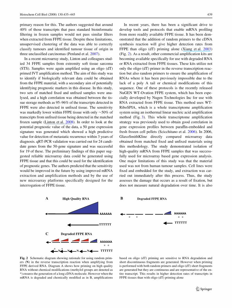
In a recent microarray study, Linton and colleagues stud-ied 34 FFPE samples from extremity soft tissue sarcoma(STS). Samples were again ampliWed using an oligo (dT)primed IVT ampliWcation method. The aim of this study wasto identify if biologically relevant data could be obtainedfrom the FFPE material, with a secondary aim of potentiallyidentifying prognostic markers in this disease. In this study,two sets of matched Wxed and unWxed samples were ana-lysed, and a high correlation was obtained between the tis-sue storage methods as 95–96% of the transcripts detected inFFPE were also detected in unWxed tissue. The sensitivitywas markedly lower within FFPE tissue with only »50% oftranscripts from unWxed tissue being detected in the matchedfrozen sample (Linton et al. 2008). In order to look at thepotential prognostic value of the data, a 50 gene expressionsignature was generated which showed a high predictivevalue for detection of metastatic recurrence within 3 years ofdiagnosis. qRT-PCR validation was carried out for 24 candi-date genes from the 50-gene signature and was successfulfor 19 of these. The preliminary Wndings of this paper sug-gested reliable microarray data could be generated usingFFPE tissue and that this could be used for the identiWcationof prognostic genes. The authors predicted that the sensitivitywould be improved in the future by using improved mRNAextraction and ampliWcation methods and by the use ofnew microarray platforms speciWcally designed for theinterrogation of FFPE tissue.
In recent years, there has been a signiWcant drive todevelop tools and protocols that enable mRNA proWlingfrom more readily available FFPE tissue. It has been dem-onstrated that the addition of random primers to the cDNAsynthesis reaction will give higher detection rates fromFFPE than oligo (dT) priming alone (Xiang et al. 2003)(Fig. 2). As a result, other commercial ampliWcation kits arebecoming available speciWcally for use with degraded RNAor RNA extracted from FFPE tissues. These kits utilize notonly the oligo (dT) primer in the reverse transcription reac-tion but also random primers to ensure the ampliWcation ofRNAs where it has been previously impossible due to thelack of a poly A tail or chemical modiWcations of thissequence. One of these protocols is the recently releasedNuGEN WT-Ovation FFPE system, which has been espe-cially developed by Nugen Technologies Inc for use withRNA extracted from FFPE tissue. This method uses WT-RiboSPIA, which is a whole transcriptome ampliWcationsystem using an isothermal linear nucleic acid ampliWcationmethod (Fig. 3). This whole transcriptome ampliWcationstrategy was previously used to obtain good correlation ingene expression proWles between paraYn-embedded andfresh frozen cell pellets (Scicchitano et al. 2006). In 2006,GlaxoSmithKline directly compared microarray dataobtained from matched Wxed and unWxed materials usingthis methodology. The study demonstrated isolation ofhigh-quality mRNA from FFPE samples that was success-fully used for microarray based gene expression analysis.One major limitations of this study was that the materialused was not from human tumour samples. Cell lines wereWxed and embedded for the study, and extraction was car-ried out immediately after this process. Thus, the studyassesses the damage that occurs as a result of Wxation, butdoes not measure natural degradation over time. It is also
Fig. 2 Schematic diagram showing rationale for using random prim-ers (N) in the reverse transcription reaction when amplifying fromFFPE derived RNA. Diagram A shows how priming on high qualityRNA without chemical modiWcations (methylol groups are denoted as*) ensures the generation of a long cDNA molecule. However when themRNA is degraded and chemically modiWed as in B, ampliWcations
based on oligo (dT) priming are sensitive to RNA degradation andshort discontinuous fragments are generated. However when primingis performed with both random primers and oligo (dT) short fragmentsare generated but they are continuous and are representative of the en-tire transcript. This results in higher detection rates of transcripts inFFPE tissues than with oligo (dT) priming alone
High Quality RNA Degraded FFPE RNA
TTTTTT
AAAAAA AAAAAA
* ** * TTTTTT
AAAAAA
* *TTTTTT* * NNNN
C
A B
Degraded FFPE RNA
123

440 Histochem Cell Biol (2008) 130:435–445
important to note that the cell pellets used in this study weresmall and Wxed rapidly, which likely contributed to the rel-ative high quality of the mRNA (Scicchitano et al. 2006).In our in-house studies we have consistently found the sig-nal intensities for FFPE samples are lower when analysinggene expression from FFPE samples than their correspond-ing fresh sample on microarrays. One of the advantages wehave identiWed of using the WT-RiboSPIA technologywhen compared to IVT for amplifying RNA from FFPE tis-sues is the lower background obtained. This lower back-ground means that there is more signal above backgroundfor gene detection, which is particularly important whenidentifying diVerentially, regulated genes.
Another technology used in measuring gene expressionfrom FFPE samples is the DASL (cDNA-mediated anneal-ing, selection, extension and ligation) assay developed byIllumina (Illumina Inc., San Diego, CA, USA). The mecha-nism behind the DASL assay also uses priming with ran-dom hexamers in the cDNA synthesis stage but with nooligo-d(T) priming. The assay works from as little as 50 ngof total RNA to analyse 300–400 transcripts. This is a sig-niWcant increase over what is readily achievable by multi-plex RT-PCR, but is still 10–20 fold less than can beachieved by using microarray analysis. An initial study byBibikova et al. found that 90% of the genes detected infresh frozen samples were detected in FFPE tissue; however,the gene expression proWles from FFPE did not correlate
exactly with the proWles from fresh samples (R2 = 0.69)(Bibikova et al. 2004). Subsequently Bibikova and col-leagues were able to identify a 16-gene signature that wasable to predict prostate cancer relapse after radical prosta-tectomy and correlated well with Gleason score (Bibikovaet al. 2007). The speciWcity and sensitivity of this gene sig-nature, however, remains to be reported and the authors aimfor this test to be validated in a future larger study. How-ever, this result demonstrates the clinical and prognosticvalue of measuring gene expression from FFPE derivedRNA. With the development of high-density microarrayplatforms that work with FFPE tissue the DASL assay maynot be an ideal discovery tool. However, while not directlyapplicable in the biomarker discovery process, the DASLassay may have a role in the validation of transcripts previ-ously identiWed by high-density microarray platforms.
Almac Diagnostics (http://www.almacgroup.com), haveadopted a diVerent approach to extract robust data fromFFPE derived RNA. Almac have developed a range of highdensity Disease SpeciWc MicroArrays (DSAs™) manufac-tured on the gold standard AVymetrix platform, which cap-ture as completely as possible all transcripts transcribed in aspeciWc disease setting such as breast, colorectal or non-small cell lung cancer. The actual content of the DSA isderived using a combination of high throughput 3�-basedsequencing, data mining and expression analysis (Tanneyet al. 2008) (Fig. 4). Because Almac Diagnostics utilised a3�-based sequencing approach it was possible to designprobes at the actual 3�-end of each transcript. Technicallythis is extremely important as most linear ampliWcationtechnologies include a reverse transcription step using anoligo (dT) primer. This has the eVect of enriching for 3�
information, particularly from degraded RNA typicallyderived from FFPE. It has previously been suggested that amore 3� biased design may improve the identiWcation ofderegulated genes from FFPE samples (Scicchitano et al.2006). The combination of extra disease speciWc contentand actual 3� design results in substantially greater detec-tion of robust gene expression data from FFPE samples.
An example study was performed in-house to demon-strate the utility of this array. The Lung Cancer DSAresearch tool was used to study a Quad Set™ (Asterand)from a single patient (Fig. 5), representing matchedNSCLC tumour and normal lung tissues, both of whichwere snap frozen and formalin-Wxed paraYn embedded.For each of the four samples (FF-tumour, FF-normal,FFPE-tumour, FFPE-normal), Wve technical replicates wereproWled. For RNA ampliWcation and labelling, the WT-Ovation™ FFPE RNA AmpliWcation System was used(Nugen Technologies, CA, USA). This system uses a com-bination of random hexamer priming in addition to oligo(dT) priming. The coeYcients of variance and correlationcomputed for the expression indices of probesets called
Fig. 3 Overview of the Nugen whole transcriptome RiboSPIA (WT-RiboSPIA) ampliWcation process used in the WT-Ovation™ FFPESystem. Note that both oligo (dT) and random primers are used in thereverse transcription step
123

Histochem Cell Biol (2008) 130:435–445 441
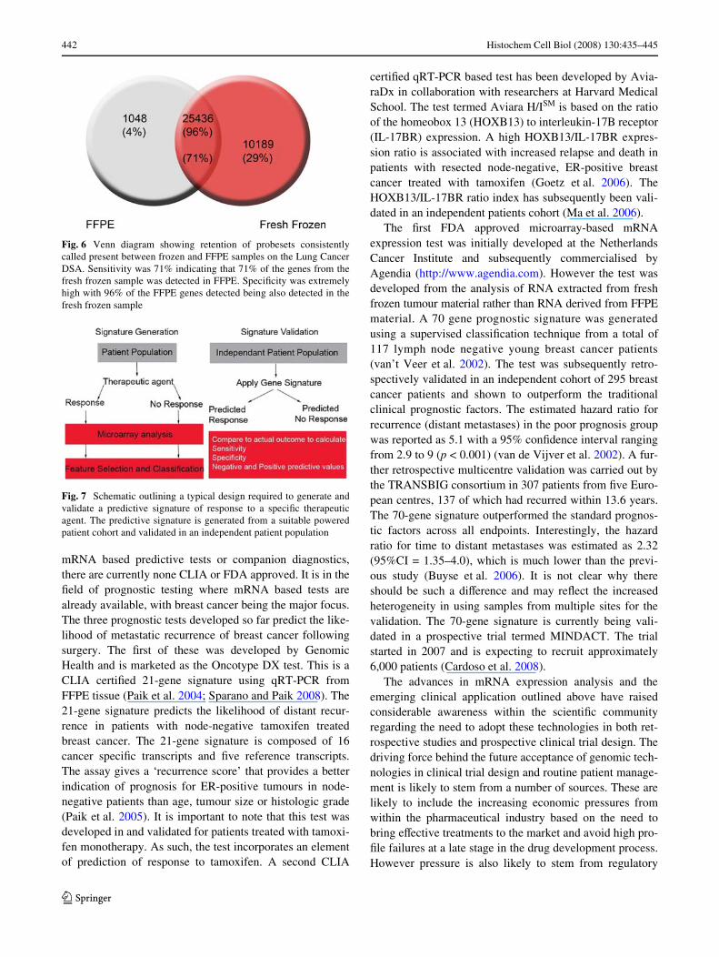
consistently present among technical replicates demon-strated similar and very low levels of noise in the data andequally high levels of correlation on both arrays (Table 1).When the present calls were analysed in this experiment,we observed a remarkably high level of present calls inboth frozen and FFPE samples. When we examined the
overlap between fresh frozen and FFPE tissue expressiondata, we observed that despite the inevitable loss of infor-mation in FFPE tissue, both the speciWcity and the sensitivitywere very high. The sensitivity was 71%, indicating that thetranscripts detected in frozen material could be detected inFFPE material and the speciWcity was 96% with the vastmajority of the transcripts detected in FFPE also detected infrozen samples (Fig. 6). These Wgures were signiWcantlyhigher than previously reported (Linton et al. 2008; Scic-chitano et al. 2006) and provided conWdence that reliablehigh quality data can be derived from FFPE tissue by theLung Cancer DSA research tool. In order to apply the DSAtechnology to a more biological relevant question, Hoseyet al., utilized the Breast Cancer DSA to carry out FFPE-based proWling comparing 17 BRCA1 mutant and 14matched sporadic breast cancers. A total of 636 probesetswere identiWed as being diVerentially expressed betweenthe two cohorts. One of the transcripts identiWed as down-regulated in the BRCA1 mutant tumours was estrogenreceptor 1 (ESR1) that codes for the estrogen receptor alpha(ER�). The authors went on to conWrm that BRCA1 modu-lates response to antiestrogens by transcriptional regulationof ESR1 in an OCT1 dependent manner (Hosey et al.2007). The study was signiWcant because it provided amodel to explain the observation that BRCA1 deWcienttumours are typically ER� negative.
Clinical application of mRNA expression analysis
To date, the majority of predictive tests that are availableare DNA or protein-based rather than mRNA-based. Whilethere is currently a great deal of focus on developing
Fig. 4 Schematic representa-tion of the generation of the Can-cer DSA research tools showing parallel approaches (in house sequencing, public sequence database mining and gene expression proWling) used to characterise the NSCLC tran-scriptome, from which the Lung Cancer DSA was designed
Fig. 5 Schematic representation of the process used to generate theLung Quad Set. Matched normal and tumour lung tissues pieces are re-moved from a single patient. The tumour and normal tissue pieces arethen cut in two, to form mirror images of the other. One half is storedin liquid nitrogen and the other half is paraYn embedded
Table 1 Reproducibility of data from frozen and FFPE sam-ples proWled on the Lung Cancer DSA™ research tool
Storage method CoeYcient of variation in normal tissue (%)
CoeYcient of variation in tumour tissue (%)
Correlation coeYcient normal–normal (%)
Correlation coeYcient tumour–tumour (%)
FFPE 5.2 5.3 98.59 98.70
Frozen 5.6 5.2 98.10 98.52
123
442 Histochem Cell Biol (2008) 130:435–445
mRNA based predictive tests or companion diagnostics,there are currently none CLIA or FDA approved. It is in theWeld of prognostic testing where mRNA based tests arealready available, with breast cancer being the major focus.The three prognostic tests developed so far predict the like-lihood of metastatic recurrence of breast cancer followingsurgery. The Wrst of these was developed by GenomicHealth and is marketed as the Oncotype DX test. This is aCLIA certiWed 21-gene signature using qRT-PCR fromFFPE tissue (Paik et al. 2004; Sparano and Paik 2008). The21-gene signature predicts the likelihood of distant recur-rence in patients with node-negative tamoxifen treatedbreast cancer. The 21-gene signature is composed of 16cancer speciWc transcripts and Wve reference transcripts.The assay gives a ‘recurrence score’ that provides a betterindication of prognosis for ER-positive tumours in node-negative patients than age, tumour size or histologic grade(Paik et al. 2005). It is important to note that this test wasdeveloped in and validated for patients treated with tamoxi-fen monotherapy. As such, the test incorporates an elementof prediction of response to tamoxifen. A second CLIA
certiWed qRT-PCR based test has been developed by Avia-raDx in collaboration with researchers at Harvard MedicalSchool. The test termed Aviara H/ISM is based on the ratioof the homeobox 13 (HOXB13) to interleukin-17B receptor(IL-17BR) expression. A high HOXB13/IL-17BR expres-sion ratio is associated with increased relapse and death inpatients with resected node-negative, ER-positive breastcancer treated with tamoxifen (Goetz et al. 2006). TheHOXB13/IL-17BR ratio index has subsequently been vali-dated in an independent patients cohort (Ma et al. 2006).
The Wrst FDA approved microarray-based mRNAexpression test was initially developed at the NetherlandsCancer Institute and subsequently commercialised byAgendia (http://www.agendia.com). However the test wasdeveloped from the analysis of RNA extracted from freshfrozen tumour material rather than RNA derived from FFPEmaterial. A 70 gene prognostic signature was generatedusing a supervised classiWcation technique from a total of117 lymph node negative young breast cancer patients(van’t Veer et al. 2002). The test was subsequently retro-spectively validated in an independent cohort of 295 breastcancer patients and shown to outperform the traditionalclinical prognostic factors. The estimated hazard ratio forrecurrence (distant metastases) in the poor prognosis groupwas reported as 5.1 with a 95% conWdence interval rangingfrom 2.9 to 9 (p < 0.001) (van de Vijver et al. 2002). A fur-ther retrospective multicentre validation was carried out bythe TRANSBIG consortium in 307 patients from Wve Euro-pean centres, 137 of which had recurred within 13.6 years.The 70-gene signature outperformed the standard prognos-tic factors across all endpoints. Interestingly, the hazardratio for time to distant metastases was estimated as 2.32(95%CI = 1.35–4.0), which is much lower than the previ-ous study (Buyse et al. 2006). It is not clear why thereshould be such a diVerence and may reXect the increasedheterogeneity in using samples from multiple sites for thevalidation. The 70-gene signature is currently being vali-dated in a prospective trial termed MINDACT. The trialstarted in 2007 and is expecting to recruit approximately6,000 patients (Cardoso et al. 2008).
The advances in mRNA expression analysis and theemerging clinical application outlined above have raisedconsiderable awareness within the scientiWc communityregarding the need to adopt these technologies in both ret-rospective studies and prospective clinical trial design. Thedriving force behind the future acceptance of genomic tech-nologies in clinical trial design and routine patient manage-ment is likely to stem from a number of sources. These arelikely to include the increasing economic pressures fromwithin the pharmaceutical industry based on the need tobring eVective treatments to the market and avoid high pro-Wle failures at a late stage in the drug development process.However pressure is also likely to stem from regulatory
Fig. 6 Venn diagram showing retention of probesets consistentlycalled present between frozen and FFPE samples on the Lung CancerDSA. Sensitivity was 71% indicating that 71% of the genes from thefresh frozen sample was detected in FFPE. SpeciWcity was extremelyhigh with 96% of the FFPE genes detected being also detected in thefresh frozen sample
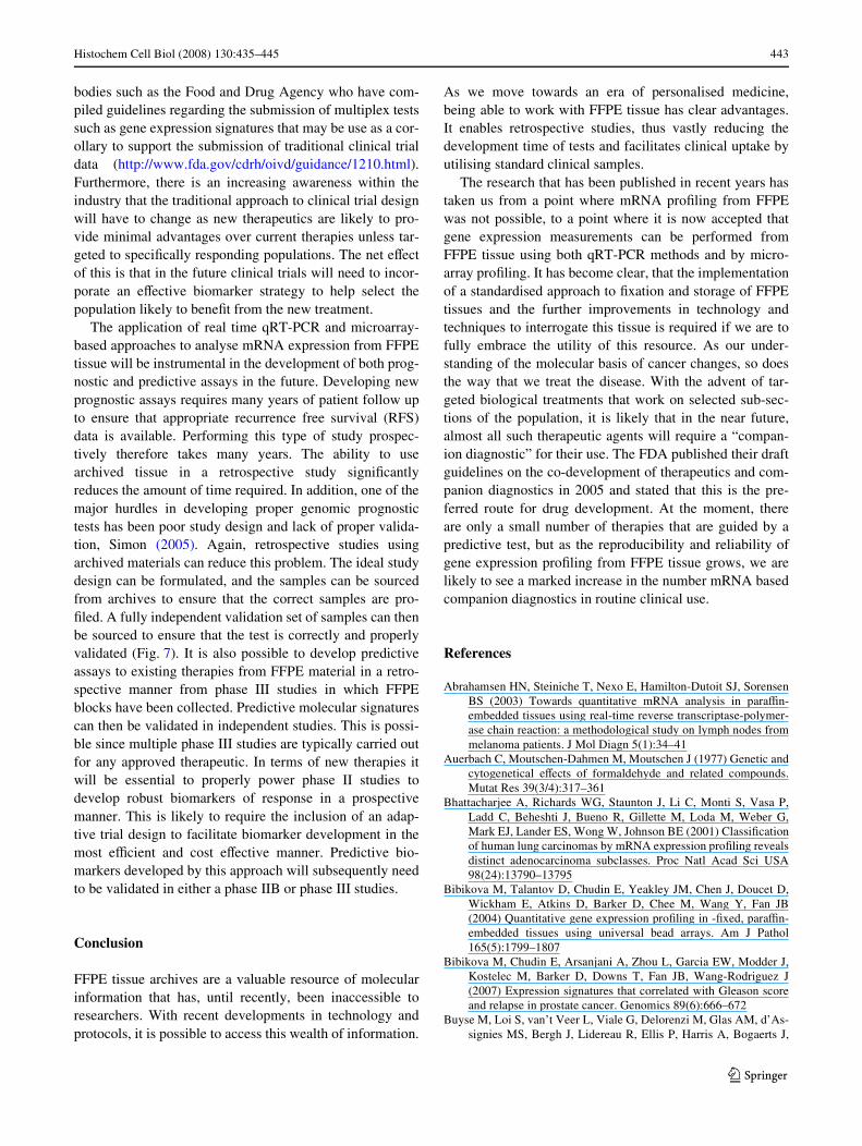
Fig. 7 Schematic outlining a typical design required to generate andvalidate a predictive signature of response to a speciWc therapeuticagent. The predictive signature is generated from a suitable poweredpatient cohort and validated in an independent patient population
123
Histochem Cell Biol (2008) 130:435–445 443
bodies such as the Food and Drug Agency who have com-piled guidelines regarding the submission of multiplex testssuch as gene expression signatures that may be use as a cor-ollary to support the submission of traditional clinical trialdata (http://www.fda.gov/cdrh/oivd/guidance/1210.html).Furthermore, there is an increasing awareness within theindustry that the traditional approach to clinical trial designwill have to change as new therapeutics are likely to pro-vide minimal advantages over current therapies unless tar-geted to speciWcally responding populations. The net eVectof this is that in the future clinical trials will need to incor-porate an eVective biomarker strategy to help select thepopulation likely to beneWt from the new treatment.
The application of real time qRT-PCR and microarray-based approaches to analyse mRNA expression from FFPEtissue will be instrumental in the development of both prog-nostic and predictive assays in the future. Developing newprognostic assays requires many years of patient follow upto ensure that appropriate recurrence free survival (RFS)data is available. Performing this type of study prospec-tively therefore takes many years. The ability to usearchived tissue in a retrospective study signiWcantlyreduces the amount of time required. In addition, one of themajor hurdles in developing proper genomic prognostictests has been poor study design and lack of proper valida-tion, Simon (2005). Again, retrospective studies usingarchived materials can reduce this problem. The ideal studydesign can be formulated, and the samples can be sourcedfrom archives to ensure that the correct samples are pro-Wled. A fully independent validation set of samples can thenbe sourced to ensure that the test is correctly and properlyvalidated (Fig. 7). It is also possible to develop predictiveassays to existing therapies from FFPE material in a retro-spective manner from phase III studies in which FFPEblocks have been collected. Predictive molecular signaturescan then be validated in independent studies. This is possi-ble since multiple phase III studies are typically carried outfor any approved therapeutic. In terms of new therapies itwill be essential to properly power phase II studies todevelop robust biomarkers of response in a prospectivemanner. This is likely to require the inclusion of an adap-tive trial design to facilitate biomarker development in themost eYcient and cost eVective manner. Predictive bio-markers developed by this approach will subsequently needto be validated in either a phase IIB or phase III studies.
Conclusion
FFPE tissue archives are a valuable resource of molecularinformation that has, until recently, been inaccessible toresearchers. With recent developments in technology andprotocols, it is possible to access this wealth of information.
As we move towards an era of personalised medicine,being able to work with FFPE tissue has clear advantages.It enables retrospective studies, thus vastly reducing thedevelopment time of tests and facilitates clinical uptake byutilising standard clinical samples.
The research that has been published in recent years hastaken us from a point where mRNA proWling from FFPEwas not possible, to a point where it is now accepted thatgene expression measurements can be performed fromFFPE tissue using both qRT-PCR methods and by micro-array proWling. It has become clear, that the implementationof a standardised approach to Wxation and storage of FFPEtissues and the further improvements in technology andtechniques to interrogate this tissue is required if we are tofully embrace the utility of this resource. As our under-standing of the molecular basis of cancer changes, so doesthe way that we treat the disease. With the advent of tar-geted biological treatments that work on selected sub-sec-tions of the population, it is likely that in the near future,almost all such therapeutic agents will require a “compan-ion diagnostic” for their use. The FDA published their draftguidelines on the co-development of therapeutics and com-panion diagnostics in 2005 and stated that this is the pre-ferred route for drug development. At the moment, thereare only a small number of therapies that are guided by apredictive test, but as the reproducibility and reliability ofgene expression proWling from FFPE tissue grows, we arelikely to see a marked increase in the number mRNA basedcompanion diagnostics in routine clinical use.
References
Abrahamsen HN, Steiniche T, Nexo E, Hamilton-Dutoit SJ, SorensenBS (2003) Towards quantitative mRNA analysis in paraYn-embedded tissues using real-time reverse transcriptase-polymer-ase chain reaction: a methodological study on lymph nodes frommelanoma patients. J Mol Diagn 5(1):34–41
Auerbach C, Moutschen-Dahmen M, Moutschen J (1977) Genetic andcytogenetical eVects of formaldehyde and related compounds.Mutat Res 39(3/4):317–361
Bhattacharjee A, Richards WG, Staunton J, Li C, Monti S, Vasa P,Ladd C, Beheshti J, Bueno R, Gillette M, Loda M, Weber G,Mark EJ, Lander ES, Wong W, Johnson BE (2001) ClassiWcationof human lung carcinomas by mRNA expression proWling revealsdistinct adenocarcinoma subclasses. Proc Natl Acad Sci USA98(24):13790–13795
Bibikova M, Talantov D, Chudin E, Yeakley JM, Chen J, Doucet D,Wickham E, Atkins D, Barker D, Chee M, Wang Y, Fan JB(2004) Quantitative gene expression proWling in -Wxed, paraYn-embedded tissues using universal bead arrays. Am J Pathol165(5):1799–1807
Bibikova M, Chudin E, Arsanjani A, Zhou L, Garcia EW, Modder J,Kostelec M, Barker D, Downs T, Fan JB, Wang-Rodriguez J(2007) Expression signatures that correlated with Gleason scoreand relapse in prostate cancer. Genomics 89(6):666–672
Buyse M, Loi S, van’t Veer L, Viale G, Delorenzi M, Glas AM, d’As-signies MS, Bergh J, Lidereau R, Ellis P, Harris A, Bogaerts J,
123
444 Histochem Cell Biol (2008) 130:435–445
Therasse P, Floore A, Amakrane M, Piette F, Rutgers E, SotiriouC, Cardoso F, Piccart MJ, TRANSBIG Consortium (2006) Vali-dation and clinical utility of a 70-gene prognostic signature forwomen with node-negative breast cancer. J Natl Cancer Inst98(17):1183–1192
Bresters D, Schipper ME, Reesink HW, Boeser-Nunnink BD, CuypersHT (1994) The duration of Wxation inXuences the yield of HCVcDNA-PCR products from formalin-Wxed, paraYn-embeddedliver tissue. J Virol Methods 48(2/3):267–272
Cardoso F, Van’t Veer L, Rutgers E, Loi S, Mook S, Piccart-GebhartMJ (2008) Clinical application of the 70-gene proWle: the MIN-DACT trial. J Clin Oncol 26(5):729–735
Coudry RA, Meireles SI, Stoyanova R, Cooper HS, Carpino A, WangX, Engstrom PF, Clapper ML (2007) Successful application ofmicroarray technology to microdissected -Wxed, paraYn-embed-ded tissue. J Mol Diagn 9(1):70–79
Cronin M, Pho M, Dutta D, Stephans JC, Shak S, Kiefer MC, EstebanJM, Baker JB (2004) Measurement of gene expression in archivalparaYn-embedded tissues: development and performance of a92-gene reverse transcriptase-polymerase chain reaction assay.Am J Pathol 164(1):35–42
Elkahloun AG, Gaudet J, Robinson GS, Sgroi DC (2002) In situ geneexpression analysis of cancer using laser capture microdissection,microarrays and real time quantitative PCR. Cancer Biol Ther1(4):354–358
Feldman MY (1973) Reactions of nucleic acids and nucleoproteinswith formaldehyde. Prog Nucleic Acid Res Mol Biol 13:1–49
Finke J, Fritzen R, Ternes P, Lange W, Dölken G (1993) An improvedstrategy and a useful housekeeping gene for RNA analysisfrom -Wxed, paraYn-embedded tissues by PCR. Biotechniques14(3):448–453
Fitzpatrick R, Casey O, Morris D, Smith T, Powell R, Sreenan J (2002)Post-mortem stability of RNA isolated from bovine reproductivetissues. Biochim Biophys Acta 1574:10–14
Frank M, Döring C, Metzler D, Eckerle S, Hansmann ML (2007) Glo-bal gene expression proWling of formalin-Wxed paraYn-embed-ded tumor samples: a comparison to snap-frozen material usingoligonucleotide microarrays. Virchows Arch 450(6):699–711
Gibson UE, Heid CA, Williams PM (1996) A novel method for realtime quantitative RT-PCR. Genome Res 6:995–1001
Godfrey TE, Kim SH, Chavira M, RuV DW, Warren RS, Gray JW,Jensen RH (2000) Quantitative mRNA expression analysis from-Wxed, paraYn-embedded tissues using 5� nuclease quantitativereverse transcription-polymerase chain reaction. J Mol Diagn2(2):84–91
Golub TR, Slonim DK, Tamayo P, Huard C, Gaasenbeek M, MesirovJP, Coller H, Loh ML, Downing JR, Caligiuri MA, BloomWeldCD, Lander ES (1999) Molecular classiWcation of cancer: classdiscovery and class prediction by gene expression monitoring.Science 286(5439):531–537
Goetz MP, Suman VJ, Ingle JN, Nibbe AM, Visscher DW, ReynoldsCA, Lingle WL, Erlander M, Ma XJ, Sgroi DC, Perez EA, CouchFJ (2006) A two-gene expression ratio of homeobox 13 and inter-leukin-17B receptor for prediction of recurrence and survival inwomen receiving adjuvant tamoxifen. Clin Cancer Res 12(7 Pt1):2080–2087
Gruber AD, Moennig V, Hewicker-Trautwein M, Trautwein G (1994)EVect of formalin Wxation and long-term storage on the detect-ability of bovine viral-diarrhoea-virus (BVDV) RNA in archivalbrain tissue using polymerase chain reaction. Zentralbl Veterin-armed B 41(10):654–661
Heid CA, Stevens J, Livak KJ, Williams PM (1996) Real time quanti-tative PCR. Genome Res 6:986–994
Holland PM, Abramson RD, Watson R, Gelfand DH (1991) Detectionof speciWc polymerase chain reaction product by utilizing the
5�–3� exonuclease activity of Thermus aquaticus DNA polymer-ase. Proc Natl Acad Sci USA 88:7276–7280
Hosey AM, Gorski JJ, Murray MM, Quinn JE, Chung WY, StewartGE, James CR, Farragher SM, Mulligan JM, Scott AN, DervanPA, Johnston PG, Couch FJ, Daley PA, Kay E, McCann A, Mu-llan PB, Harkin DP (2007) Molecular basis for estrogen receptor� deWciency in BRCA1-linked breast cancer. J Natl Cancer Inst99(22):1683–1694
Jackson DP, Quirke P, Lewis F, Boylson AW, Sloan JM, Robertson D,Taylor GR (1989) Detection of measules virus RNA in paraYn-embedded tissues (letter). Lancet 33(8651):1391
Jackson DP, Lewis FA, Taylor GR, Boylson AW, Quirke P (1990)Tissue extraction of DNA and RNA and analysis by the polymerasechain reaction. J Clin Pathol 43:499–504
KraVt AE, Duncan BW, Bijwaard KE, Taubenberger JK, Lichy JH(1997) Optimization of the isolation and ampliWcation of RNAfrom formalin-Wxed paraYn-embedded tissue: the Armed ForcesInstitute of Pathology experience and literature review. MolDiagn 2:217–230
Lewis F, Maughan NJ, Smith V, Hillan K, Quirke P (2001) Unlockingthe archive–gene expression in paraYn-embedded tissue. J Pathol195(1):66–71
Li J, Smyth P, Cahill S, Denning K, Flavin R, Aherne S, Pirotta M,Guenther SM, O’Leary JJ, Sheils O (2008) Improved RNA qual-ity and TaqMan Pre-ampliWcation method (PreAmp) to enhanceexpression analysis from formalin Wxed paraYn embedded (FFPE)materials. BMC Biotechnol 8(10):1–11
Linton KM, Hey Y, Saunders E, Jeziorska M, Denton J, Wilson CL,Swindell R, Dibben S, Miller CJ, Pepper SD, Radford JA,Freemont AJ (2008) Acquisition of biologically relevant geneexpression data by AVymetrix microarray analysis of archival -WxedparaYn-embedded tumours. Br J Cancer 98(8):1403–1414
Lukiw W, Bazan N (1997) Cyclooxegenase2 RNA message abundance,stability and hypervariability in sporadic Alzheimer neocortex.J Neurosci Res 50:937–945
Ma XJ, Hilsenbeck SG, Wang W, Ding L, Sgroi DC, Bender RA, OsborneCK, Allred DC, Erlander MG (2006) The HOXB13:IL17BRexpression index is a prognostic factor in early-stage breastcancer. J Clin Oncol 24(28):4611–4619
Masuda N, Ohnishi T, Kawamoto S, Monden M, Okubo K (1999)Analysis of chemical modiWcation of RNA from -Wxed samplesand optimization of molecular biology applications for suchsamples. Nucleic Acids Res 27(22):4436–4443
McGhee JD, von Hippel PH (1977) Formaldehyde as a probe of DNAstructure. 3. Equilibrium denaturation of DNA and syntheticpolynucleotides. Biochem 16(15):3267–3276
Mizuno T, Nagamura H, Iwamoto K, Ito T, Fukuhura T, TokunagaM, Tokuoka S et al (1998) RNA from decades old archivaltissue blocks for retrospective studies. Diagn Mol Pathol7:202–208
Noguchi M, Furuya S, Takeuchi T, Hirohashi S (1997) ModiWed for-malin and methanol Wxation methods for molecular biological andmorphological analyses. Pathol Int 47(10):685–691
Paik S, Shak S, Tang G, Kim C, Baker J, Cronin M, Baehner FL,Walker MG, Watson D, Park T, Hiller W, Fisher ER, WickerhamDL, Bryant J, Wolmark N (2004) A multigene assay to predictrecurrence of tamoxifen-treated, node-negative breast cancer.N Engl J Med 351(27):2817–2826
Paik S, Kim CY, Song YK, Kim WS (2005) Technology insight: appli-cation of molecular techniques to -Wxed paraYn-embedded tis-sues from breast cancer. Nat Clin Pract Oncol 2(5):246–254
Park YN, Abe K, Li H, Hsuih T, Thung SN, Zhang DY (1996) Detec-tion of hepatitis C virus RNA using ligation-dependent polymer-ase chain reaction in formalin-Wxed, paraYn-embedded livertissues. Am J Pathol 149(5):1485–1491
123
Histochem Cell Biol (2008) 130:435–445 445
Penland SK, Keku TO, Torrice C, He X, Krishnamurthy J, HoadleyKA, Woosley JT, Thomas NE, Perou CM, Sandler RS, SharplessNE (2007a) RNA expression analysis of -Wxed paraYn-embeddedtumors. Lab Invest 87(4):383–391
Penland SK, Keku TO, Torrice C, He X, Krishnamurthy J, HoadleyKA, Woosley JT, Thomas NE, Perou CM, Sandler RS, SharplessNE (2007b) RNA expression analysis of formalin-Wxed paraYn-embedded tumors. Lab Invest 87(4):383–391
Perou CM, Sørlie T, Eisen MB, van de Rijn M, JeVrey SS, Rees CA,Pollack JR, Ross DT, Johnsen H, Akslen LA, Fluge O, Pergamen-schikov A, Williams C, Zhu SX, Lønning PE, Børresen-Dale AL,Brown PO, Botstein D (2000) Molecular portraits of humanbreast tumours. Nature 406(6797):747–752
Relógio A, Schwager C, Richter A, Ansorge W, Valcárcel J (2002)Optimization of oligonucleotide-based DNA microarrays. NucleicAcids Res 30(11):e51
Ribeiro-Silva A, Zhang H, JeVrey SS (2007) RNA extraction from tenyear old -Wxed paraYn-embedded breast cancer samples: acomparison of column puriWcation and magnetic bead-basedtechnologies. BMC Mol Biol 8:118
Scicchitano MS, Dalmas DA, Bertiaux MA, Anderson SM, Turner LR,Thomas RA, Mirable R, Boyce RW (2006) Preliminary compari-son of quantity, quality, and microarray performance of RNAextracted from -Wxed, paraYn-embedded, and unWxed frozentissue samples. J Histochem Cytochem 54(11):1229–1237
Shipp MA, Ross KN, Tamayo P, Weng AP, Kutok JL, Aguiar RC,Gaasenbeek M, Angelo M, Reich M, Pinkus GS, Ray TS, KovalMA, Last KW, Norton A, Lister TA, Mesirov J, Neuberg DS,Lander ES, Aster JC, Golub TR (2002) DiVuse large B-cell lym-phoma outcome prediction by gene-expression proWling andsupervised machine learning. Nat Med 8(1):68–74
Simon R (2005) Roadmap for developing and validating therapeuticallyrelevant genomic classiWers. J Clin Oncol 23(29):7332–7341
Sørlie T, Perou CM, Tibshirani R, Aas T, Geisler S, Johnsen H, Hastie T,Eisen MB, van de Rijn M, JeVrey SS, Thorsen T, Quist H, MateseJC, Brown PO, Botstein D, Eystein Lønning P, Børresen-Dale AL(2001) Gene expression patterns of breast carcinomas distinguishtumor subclasses with clinical implications. Proc Natl Acad SciUSA 98(19):10869–10874
Sparano JA, Paik S (2008) Development of the 21-gene assay and itsapplication in clinical practice and clinical trials. J Clin Oncol26(5):721–728
Specht K, Richter T, Müller U, Walch A, Werner M, HöXer H(2001) Quantitative gene expression analysis in microdissectedarchival -Wxed and paraYn-embedded tumor tissue. Am J Pathol158(2):419–429
Srinivasan M, Sedmak D, Jewell S (2002) EVect of Wxatives and tissueprocessing on the content and integrity of nucleic acids. Am JPathol 161(6):1961–1971
Stanta G, Schneider C (1991) RNA extracted from paraYn-embeddedhuman tissues is amenable to analysis by PCR ampliWcation. Bio-techniques 11:304–308
Start R, Cross S, Smith J (1992) Assessment of specimen Wxation in asurgical pathology service. J Clin Pathol 45:546–547
Tanney A, Oliver GR, Farztdinov V, Kennedy RD, Mulligan JM, Ful-ton CE, Farragher SM, Field JK, Johnston PG, Harkin DP, Prout-ski V, Mulligan KA (2008) Generation of a non-small cell lungcancer transcriptome microarray. BMC Med Genomics 1(1):20
Tokuda Y, Nakamura T, Satonaka K, Maeda S, Doi K, Baba S,Sugiyama T (1990) Fundamental study on the mechanism ofDNA degradation in tissues Wxed in formaldehyde. J Clin Pathol43(9):748–751
van de Vijver MJ, He YD, Van’t Veer LJ, Dai H, Hart AA, VoskuilDW, Schreiber GJ, Peterse JL, Roberts C, Marton MJ, Parrish M,Atsma D, Witteveen A, Glas A, Delahaye L, van der Velde T,Bartelink H, Rodenhuis S, Rutgers ET, Friend SH, Bernards R(2002) A gene-expression signature as a predictor of survival inbreast cancer. N Engl J Med 347(25):1999–2009
van’t Veer LJ, Bernards R (2008) Enabling personalized cancermedicine through analysis of gene-expression patterns. Nature452(7187):564–570
von Weizsäcker F, Labeit S, Koch HK, Oehlert W, Gerok W, BlumHE (1991) A simple and rapid method for the detection ofRNA in -Wxed, paraYn-embedded tissues by PCR ampliWcation.Biochem Biophys Res Commun 174(1):176–180
WolV AC, Hammond ME, Schwartz JN, Hagerty KL, Allred DC, CoteRJ, Dowsett M, Fitzgibbons PL, Hanna WM, Langer A, McShaneLM, Paik S, Pegram MD, Perez EA, Press MF, Rhodes A, SturgeonC, Taube SE, Tubbs R, Vance GH, van de Vijver M, Wheeler TM,Hayes DF, American Society of Clinical Oncology, College ofAmerican Pathologists (2007) American Society of ClinicalOncology/College of American Pathologists guideline recom-mendations for human epidermal growth factor receptor 2 testingin breast cancer. J Clin Oncol 25(1):118–145
Xiang CC, Chen M, Ma L, Phan QN, Inman JM, Koshich OA,Brownstein MJ (2003) A new strategy to amplify degraded RNAfrom small tissue samples for microarray studies. Nucleic AcidsRes 31(9):1–5
Yagi N, Satonaka K, Horio M, Shimogaki H, Tokuda Y, Maeda S(1996) The role of DNase and EDTA on DNA degradation informaldehyde Wxed tissues. Biotech Histochem 71(3):123–129
123