Shenmai injection as an adjuvant treatment for chronic cor ...
Upload
independentCategory
view
3download
0
The
Journ
al o
f Exp
erim
enta
l M
edic
ine
The Journal of Experimental Medicine • Volume 198, Number 2, July 21, 2003 281–291http://www.jem.org/cgi/doi/10.1084/jem.20030645
281
Dendritic Cells Are Responsible for the Capacity of CpG Oligodeoxynucleotides to Act as an Adjuvant for Protective Vaccine Immunity Against
Leishmania major
in Mice
Javeed A. Shah,
1
Patricia A. Darrah,
1
David R. Ambrozak,
2
Tara N. Turon,
1
Susana Mendez,
3
Joanna Kirman,
1
Chang-You Wu,
1
Nicolas Glaichenhaus,
4
and Robert A. Seder
1
1
Cellular Immunology Section and
2
Human Immunology Section, Vaccine Research Center, and
3
Intracellular Parasite Biology Section, Laboratory of Parasitic Diseases, National Institute of Allergy and Infectious Diseases, National Institutes of Health, Bethesda, MD 20892
4
Centre National de la Recherche Scientifique, UMR 6097, Institute de Pharmacologie Moléculaire et Cellulaire, 06560 Valbonne, France
Abstract
Vaccination with leishmanial Ag and CpG oligodeoxynucleotides (ODN) confers sustainedcellular immunity and protection to infectious challenge up to 6 mo after immunization. Todefine the cellular mechanism by which CpG ODN mediate their adjuvant effects in vivo, thefunctional capacity of distinct dendritic cell (DC) subsets was assessed in the lymph nodes (LNs)of BALB/c mice, 36 h after immunization with the leishmanial antigen (LACK) and CpGODN. After this immunization, there was a striking decrease in the frequency of theCD11c
�
B220
�
plasmacytoid DCs with a proportionate increase in CD11c
�
CD8
�
B220
�
cells.CD11c
�
CD8
�
B220
�
cells were the most potent producers of interleukin (IL)-12 p70 and in-terferon (IFN)-
�
, while plasmacytoid DCs were the only subset capable of secreting IFN-
�
. Interms of antigen presenting capacity, plasmacytoid DCs were far less efficient compared withthe other DC subsets. To certify that DCs were responsible for effective vaccination, we iso-lated CD11c
�
and CD11c
�
cells 36 h after immunization and used such cells to elicit protec-tive immunity after adoptive transfer in naive,
Leishmania major
susceptible BALB/c mice.CD11c
�
cells but not 10-fold higher numbers of CD11c
�
cells from such immunized micemediated protection. Therefore, the combination of LACK antigen and CpG ODN adjuvantleads to the presence of CD11c
�
DCs in the draining LN that are capable of vaccinating naivemice in the absence of further antigen or adjuvant.
Key words: plasmacytoid dendritic cell • Toll-like receptor •
Leishmania major
• vaccine • Th1 cells
Introduction
Currently there are no uniformly effective vaccines for in-fections such as
Mycobacterium tuberculosis
, malaria, and HIV.For such infections, the cellular immune response compris-ing primarily Th1 and CD8
�
effector T cells has beenshown to be critical in mediating protection against infec-tion and/or development of disease (1). Potential difficultiesin generating potent and durable cellular immune responsesin vivo with current vaccines may be due to multiple factors.First, induction of Th1 cells requires a differentiation processthat involves both Ag stimulation and a Th1-inducing cyto-kine (2, 3). Second, generation of CD8
�
effector T cells re-
quires intracellular processing of Ag, which is most readilyelicited by either live-attenuated or DNA vaccines; how-ever, live-attenuated vaccines for diseases such as HIV or
M. tuberculosis
might be precluded for safety and/or manu-facturing considerations. In addition, DNA vaccines are farless potent for inducing immune responses in humans thanin rodents. One recent approach that has shown promisefor generating broad cellular immune responses in vivo isuse of Toll-like receptor (TLR)
*
ligands as vaccine adju-vants (4).
Address correspondence to Dr. Robert A. Seder, VRC, NIAID, 40 Con-vent Dr., Room 40/3512, NIH, Bethesda, MD 20892. Phone: 301-594-8483; Fax: 301-480-2565; E-mail: [email protected]
*
Abbreviations used in this paper:
CpG ODN, cytosine phosphate guanosineoligodeoxynucleotide(s); LACK, Leishmania homolog of the receptor foractivated C kinase; SAC,
Staphylococcus aureus
Cowens; TLR, Toll-likereceptor.
on January 23, 2014jem
.rupress.orgD
ownloaded from
Published July 21, 2003
The
Journ
al o
f Exp
erim
enta
l M
edic
ine
282
DCs Mediate Immunity by CpG ODN Immunization
At present, there are 10 TLR receptors that respond todifferent pathogen-associated molecular patterns (5). Oneof the most potent inducers of both innate and adaptiveimmunity is mediated through the interaction of cytosinephosphate guanosine oligodeoxynucleotide (CpG ODN)with TLR 9 (4, 6). At the initiation of an immune re-sponse, this interaction leads to increased expression of co-stimulatory markers such as CD40, CD80, and CD86 andproduction of proinflammatory cytokines such as TNF-
�
,IL-12, and IL-6 from DCs (7–9). In turn, this facilitates in-duction of both Th1 and CD8
�
T cell responses (10–13).The physiologic relevance for the immune effects of CpGODN in vivo was demonstrated in a mouse vaccine modelof
Leishmania major
infection in which mice immunizedwith heat-killed or recombinant leishmanial Ag and CpGODN had sustained cellular immune responses and wereprotected up to 6 mo after immunization (14). Similarly, inother studies, vaccination with plasmid DNA encodingvarious leishmanial Ag also conferred long-term protectionin a Th1 and CD8
�
T cell–dependent manner (15). Thus,while the aforementioned vaccine studies elucidated theimmune correlates of protection, it remained to be provenhow these different types of vaccine formulations were ini-tiating such responses at the level of the APC. In this re-gard, a common feature of plasmid DNA and CpG ODN
�
protein vaccines is the ability to transfect (16) and activate(17) DCs, respectively.
CD11c
�
DCs can direct the differentiation of CD4
�
Tcells into Th1- or Th2-type cells (18) as well as CD8
�
Tcell responses either directly or through cross-priming (19).The ability of DCs to induce such responses depends onthe specific subset (20), the maturation state (21), and theconditions of stimulation (22). The great majority of priorstudies analyzing the function of DCs have used either invitro–generated or in vitro–cultured bone marrow orsplenic DCs. In addition, characterization of ex vivo-derived DCs has been limited to nonpathogenic Ag. Thus,there has not been a systematic study using an entirely invivo model in which subsets of DCs from peripheral LNswere characterized after immunization with a protein de-rived from a specific pathogen plus CpG ODN for theirability to present the Ag, secrete cytokines, and mediateprotection to an infectious challenge after adoptive transferinto naive mice.
The aim of the studies presented here was to determinethe cellular mechanism by which CpG ODN were mediat-ing protective immunity in vivo when used as a vaccineadjuvant with a leishmanial Ag. For the leishmanial Ag, weused the Leishmania homologue of the receptor for acti-vated C kinase (LACK) protein. LACK is a highly con-served, immunodominant Ag with a single I-A
d
–restrictedepitope that has been shown to regulate the natural courseof infection in BALB/c mice and been useful in vaccinestudies for determining the requirements for Th1 andCD8
�
T cells in maintaining long-term memory responsessufficient to mediate protection (15, 23–25). We hypothe-sized that DCs from mice immunized with LACK
�
CpGODN mediate T cell immunity and protection following
transfer into in naive,
Leishmania major
–susceptible BALB/cmice. The results of this study provide in vivo evidence forthe cellular mechanism by which CpG ODN, which pre-viously has been shown to have demonstrable adjuvant ac-tivity, mediates protective immunity in vivo.
Materials and Methods
Mice.
Female BALB/c mice were purchased from the Divi-sion of Cancer Treatment (National Cancer Institute, NationalInstitutes of Health). Mice were maintained in the Vaccine Re-search Center Animal Care Facility (Bethesda, MD) under patho-gen-free conditions.
ODN.
For all in vivo immunizations, phosphorothioate-modified ODN sequence 1826 containing two CpG motifs(underlined: TCATGACGTTCCTGACGTT) was provided byColey Pharmaceutical Group. To assess production of IFN-
�
under in vitro stimulation, ODN sequence 1585 containing oneCpG motif (underlined: GGGGTCAACGTTGAGGGGGG) wasused. The ODN contained endotoxin levels
�
0.1 EU/mg usingthe limulus amebocyte lysis assay (Associates of Cape Cod, Inc.).
Immunization.
To generate CD11c
�
DCs in vivo, naiveBALB/c mice (20–25 mice/group) were injected s.c. in bothhind footpads and flanks with sterile PBS, CpG ODN (25
�
g), orrecombinant leishmanial LACK protein (50
�
g; Paragon Bioser-vices, Inc.) with or without CpG ODN. This protein contained
�
5.0 EU/dose. Each injection was suspended in sterile PBS andgiven in a volume of 50
�
l. For some experiments, mice werevaccinated s.c. in the footpad with 50
�
g of LACK protein witheither 25
�
g CpG ODN or 1
�
g IL-12 protein and boosted 2 wklater as described previously (26).
DC Isolation.
At various times after immunization, mice fromeach group were killed and their draining popliteal and inguinalLN passed through a 100-
�
m cell strainer and digested for 30min at 37
�
C by 1 mg/ml collagenase D and 15
�
g/ml DnaseI(Roche). All subsequent procedures were performed on ice inHBSS. The cells were blocked using 1 mg/ml purified rat IgG or1:100 2.4G2 Ab (BD Biosciences). CD11c
�
cells were positivelyselected using magnetized Ab for CD11c (N418; Miltenyi Bio-tec). After this step, there were
�
60% CD11c
�
cells. CD11c
�
DCs were further enriched by staining with FITC-labeled anti-CD3 and APC-labeled anti-CD19 and sorted for to remove suchcells. After cell sorting, there were
95% CD11c
�
CD3
�
CD19
�
cells. To isolate specific DC subsets, CD11c
�
cells enriched fol-lowing magnetic bead separation were stained with FITC-labeledanti-CD3 and CD19, PE-labeled anti-CD11c (HL3), PerCP-labeled anti-B220, or APC-labeled anti-CD8 (Ly-2) and sortedon a FACSVantage™ cell sorter. The resulting population wasconsistently
99% CD11c
�
, with fewer than 0.5% T cells.CD11c
�
cells were further depleted of CD3
�
cells by cell sortingresulting in a population that was
90% CD19
�
cells.
T Cell Proliferation Assay.
CD4
�
T cell hybridoma LMR 7.5,detects LACK in the context of MHC class II expressing I-A
d
(27). To detect the antigen presenting capacity of LACK fromAPC immediately ex vivo, 10
5
LMR 7.5 hybridoma cells werecultured with varying numbers of CD11c
�
or CD11c
�
cells frommice immunized with PBS, LACK protein, CpG ODN, orLACK
�
CpG ODN or IL-12 protein. Supernatants were har-vested 24 h later and assayed for IL-2 content by ELISA usingOptEIA kits (BD Biosciences). As a positive control for the abil-ity of APC to present LACK independent of processing of theLACK protein in vivo, 10
5
LMR 7.5 hybridoma cells were cul-
on January 23, 2014jem
.rupress.orgD
ownloaded from
Published July 21, 2003
The
Journ
al o
f Exp
erim
enta
l M
edic
ine
283
Shah et al.
tured with varying numbers of CD11c
�
or CD11c
�
cells fromnaive or immunized mice with 10
�
g/ml LACK peptide (aminoacids 156–173; FSPSLEHPIVVSSW). The detection limit for IL-2production was 3 pg/ml.
Adoptive Transfer of CD11c
�
and CD11c
�
Cells.
Varyingnumbers of CD11c
�
DC (10
4
–5
10
4
) or CD11c
�
cells (5
10
4
–5
10
5
) from mice immunized with PBS, CpG ODN,LACK
�
CpG ODN, or IL-12 protein were sorted, resuspendedin sterile HBSS (50
�
l), and transferred s.c. into the footpad ofnaive BALB/c mice. Mice were boosted 2 wk later in an identi-cal manner.
LACK-specific Production of IFN-
�
from BALB/c Mice AfterAdoptive Transfer of CD11c
�
Cells from Immunized Mice.
Afteradoptive transfer of CD11c
�
DCs into naive BALB/c mice,LACK-specific production of IFN-
�
was assessed from LN be-fore infection or spleens after infection by culturing 2
10
5
totalLN or spleen cells/200
�
l in flat-bottom 96-well plates with orwithout LACK protein (10
�
g/ml). Supernatants were harvested48 or 72 h later and analyzed for IFN-
�
content by ELISA usingOptEIA kits. The detection limit for IFN-
�
was 62.5 pg/ml.
Assessment of Cytokine Production from DCs Isolated After Immuni-zation.
2 d after immunization, total CD11c
�
or sorted subsetsof DCs (10
4
/ml) were isolated from draining LNs, added to 96-well tissue culture plates, and stimulated in media alone, Staphylo-coccus aureus Cowens (SAC; 1:1,000) and/or anti-CD40 Ab (1�g/ml), CpG ODN (5 �g/ml), or CpG ODN � anti-CD40 Ab.Supernatants were collected at 24–48 h for IL-12 p40, p70, andIL-10 assays. IL-12 p40, p70, and IL-10 were detected usingOptEIA kits. The limit of detection for IL-12 p40 was 125 pg/ml; for IL-10, 62.5 pg/ml; and for IL-12 p70, 7.8 pg/ml. For de-tection of IFN-�, cells were stimulated with CpG ODN andpoly I:C (25 �g/ml). IFN-� was detected using kits purchasedfrom R&D Systems. The limit of detection was 16 pg/ml.
Characterization of Cell Surface Markers on DCs. After enrich-ment of CD11c� DCs from mice immunized with LACK, CpGODN, both, or neither, cells were stained with the following Ab:FITC-labeled anti-I-Ad (AMS32.1), anti-CD40 (3/23), anti-CD80 (16–10A1), anti-CD86 (GL1), APC-labeled CD11b (M1/70), or Ly-6G/C (RB6–8C5). The relative expression of thesemarkers was assessed using a FACSCalibur™ flow cytometer(Becton Dickinson) and analyzed using Flow Jo software.
Infectious Challenge. L. major clone V1 (MHOM/IL/80/Friedlin) promastigotes were grown as described previously (26).Briefly, infective-stage metacyclic promastigotes of L. major wereisolated from stationary cultures (4–5 d old) by negative selectionusing peanut agglutinin (Vector Laboratories). BALB/c micewere infected 2 wk after boost with 105 metacyclic promastigotes.Parasites were injected into the footpad s.c. contralateral from thesite in which they had received CD11c� or CD11c� cells fromimmunized mice. Weekly footpad swelling measurements using ametric caliper were recorded.
Preparation of Bone Marrow–derived DCs for Detection of LACK-Specific CD4� and CD8� IFN-� Responses. To measure LACK-specific T cell immune responses after adoptive transfer, we usednormal bone marrow–derived DCs from BALB/c mice (1.2 106) alone or pulsed with LACK protein anti-CD40, and CpGODN. These were cocultured for 2 h with total splenocytes (6 106) from BALB/c mice that had received CD11c� or CD11c�
cells from mice immunized with LACK � CpG ODN. BFA (10�g/ml) was then added and 4 h later cells were fixed, permeabi-lized, and stained with FITC-labeled anti-CD3� (145–2C11),PE-labeled anti-CD8� (Ly-2), PerCP-labeled anti-CD4 (L3T4),or APC-labeled anti-IFN-� (XMG1.2). The frequency of CD4�
and CD8� T cells producing IFN-� was assessed after collecting300,000 total events in each group.
ResultsCD11c� Cells From Mice Immunized With LACK � CpG
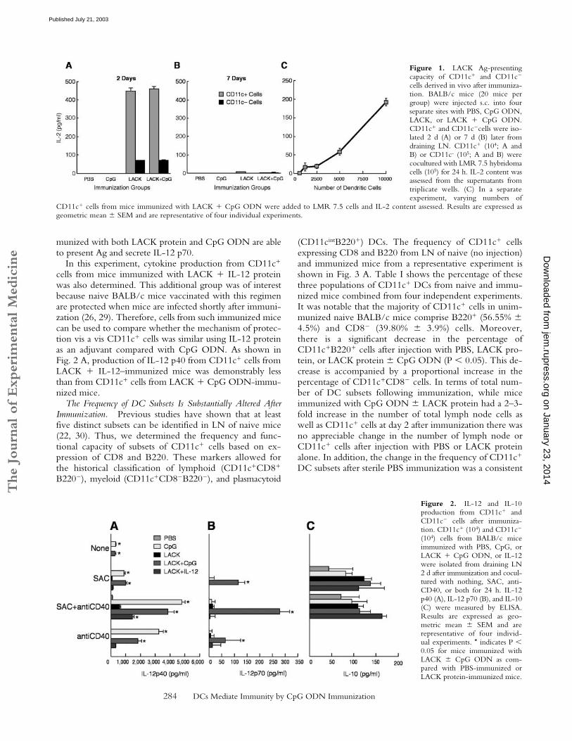
ODN Present Ag to LACK-Specific CD4� T Cells. To de-termine the mechanism by which CpG ODN confers pro-tection as a vaccine adjuvant with the leishmanial LACKprotein, we first assessed the capacity of various populationsof APCs to present LACK to a LACK-specific CD4� Tcell hybridoma (LMR 7.5) following immunization (27).Naive BALB/c mice were immunized s.c. with PBS, CpGODN alone, or LACK � CpG ODN. Draining LNs werethen harvested at various times post immunization, andCD11c� and CD11c� cells were isolated by cell sortingand added to LMR 7.5 cells to determine their capacity topresent LACK Ag. As shown in Fig. 1 A, CD11c� cells(104) isolated 2 d after immunization with LACK proteinor LACK � CpG ODN induced substantial amounts ofIL-2 from the LMR 7.5 cells. Moreover, as few as 103
CD11c� cells from such immunized mice were sufficientto induce IL-2 from the LMR 7.5 cells (Fig. 1 C). In con-trast, CD11c� cells from mice immunized with PBS orCpG ODN alone did not induce any IL-2 productionfrom LMR 7.5 cells. These data demonstrate that onlyCD11c� cells from mice immunized with LACK � CpGODN have acquired the capacity in vivo to present LACKto the LMR 7.5 cells. When assessed 7 d after immuniza-tion, there was no detectable IL-2 produced by the LMR7.5 cells from CD11c� cells in any of the groups (Fig. 1 B).Consequently, for the remainder of the experiments shownbelow, CD11c� cells were isolated from draining LN 36–48 h after immunization.
In comparing the efficiency of antigen presentation be-tween CD11c� DCs and CD11c� cells (90% B cells),while the same number of CD11c� cells (104) from miceimmunized with LACK � CpG ODN did not induce anyIL-2 from LMR 7.5 cells (unpublished data), a smallamount of IL-2 was induced from LMR 7.5 cells using 105
CD11c� cells (Fig. 1 A).CD11c� Cells from Mice Immunized with LACK and CpG
ODN Produce IL-12. Cytokines released from CD11c�
cells have a strong influence on the CD4� T cell response(2, 28). CD11c� cells isolated 2 d after immunization withPBS, CpG ODN, or LACK � CpG ODN were analyzedfor production of IL-12 p40, p70, and IL-10 after restimula-tion in vitro. As shown in Fig. 2 A, CD11c� cells frommice immunized with CpG ODN or LACK � CpG ODNhad significant production of IL-12 p40 in response to SAC,anti-CD40, or both (P � 0.05). There was no detectableIL-12 p40 from CD11c� cells from any of the immunizedgroups (data not shown). IL-12 p70 was only detected frommice immunized with LACK � CpG ODN (Fig. 2 B; P �0.05). Last, there were no significant differences in IL-10production in response to SAC � anti-CD40 stimulationbetween any of the immunized groups (Fig. 2 C). Togetherwith the previous figure, these data show that only mice im-
on January 23, 2014jem
.rupress.orgD
ownloaded from
Published July 21, 2003
The
Journ
al o
f Exp
erim
enta
l M
edic
ine
284 DCs Mediate Immunity by CpG ODN Immunization
munized with both LACK protein and CpG ODN are ableto present Ag and secrete IL-12 p70.
In this experiment, cytokine production from CD11c�
cells from mice immunized with LACK � IL-12 proteinwas also determined. This additional group was of interestbecause naive BALB/c mice vaccinated with this regimenare protected when mice are infected shortly after immuni-zation (26, 29). Therefore, cells from such immunized micecan be used to compare whether the mechanism of protec-tion vis a vis CD11c� cells was similar using IL-12 proteinas an adjuvant compared with CpG ODN. As shown inFig. 2 A, production of IL-12 p40 from CD11c� cells fromLACK � IL-12–immunized mice was demonstrably lessthan from CD11c� cells from LACK � CpG ODN-immu-nized mice.
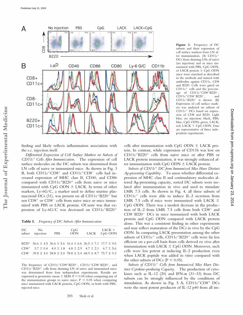
The Frequency of DC Subsets Is Substantially Altered AfterImmunization. Previous studies have shown that at leastfive distinct subsets can be identified in LN of naive mice(22, 30). Thus, we determined the frequency and func-tional capacity of subsets of CD11c� cells based on ex-pression of CD8 and B220. These markers allowed forthe historical classification of lymphoid (CD11c�CD8�
B220�), myeloid (CD11c�CD8�B220�), and plasmacytoid
(CD11cintB220�) DCs. The frequency of CD11c� cellsexpressing CD8 and B220 from LN of naive (no injection)and immunized mice from a representative experiment isshown in Fig. 3 A. Table I shows the percentage of thesethree populations of CD11c� DCs from naive and immu-nized mice combined from four independent experiments.It was notable that the majority of CD11c� cells in unim-munized naive BALB/c mice comprise B220� (56.55% �4.5%) and CD8� (39.80% � 3.9%) cells. Moreover,there is a significant decrease in the percentage ofCD11c�B220� cells after injection with PBS, LACK pro-tein, or LACK protein � CpG ODN (P � 0.05). This de-crease is accompanied by a proportional increase in thepercentage of CD11c�CD8� cells. In terms of total num-ber of DC subsets following immunization, while miceimmunized with CpG ODN � LACK protein had a 2–3-fold increase in the number of total lymph node cells aswell as CD11c� cells at day 2 after immunization there wasno appreciable change in the number of lymph node orCD11c� cells after injection with PBS or LACK proteinalone. In addition, the change in the frequency of CD11c�
DC subsets after sterile PBS immunization was a consistent
Figure 1. LACK Ag-presentingcapacity of CD11c� and CD11c�
cells derived in vivo after immuniza-tion. BALB/c mice (20 mice pergroup) were injected s.c. into fourseparate sites with PBS, CpG ODN,LACK, or LACK � CpG ODN.CD11c� and CD11c�cells were iso-lated 2 d (A) or 7 d (B) later fromdraining LN. CD11c� (104; A andB) or CD11c- (105; A and B) werecocultured with LMR 7.5 hybridomacells (105) for 24 h. IL-2 content wasassessed from the supernatants fromtriplicate wells. (C) In a separateexperiment, varying numbers of
CD11c� cells from mice immunized with LACK � CpG ODN were added to LMR 7.5 cells and IL-2 content assessed. Results are expressed asgeometric mean � SEM and are representative of four individual experiments.
Figure 2. IL-12 and IL-10production from CD11c� andCD11c� cells after immuniza-tion. CD11c� (104) and CD11c�
(104) cells from BALB/c miceimmunized with PBS, CpG, orLACK � CpG ODN, or IL-12were isolated from draining LN2 d after immunization and cocul-tured with nothing, SAC, anti-CD40, or both for 24 h. IL-12p40 (A), IL-12 p70 (B), and IL-10(C) were measured by ELISA.Results are expressed as geo-metric mean � SEM and arerepresentative of four individ-ual experiments. * indicates P �0.05 for mice immunized withLACK � CpG ODN as com-pared with PBS-immunized orLACK protein-immunized mice.
on January 23, 2014jem
.rupress.orgD
ownloaded from
Published July 21, 2003
The
Journ
al o
f Exp
erim
enta
l M
edic
ine
285 Shah et al.
finding and likely reflects inflammation association withthe s.c. injection itself.
Differential Expression of Cell Surface Markers on Subsets ofCD11c� Cells After Immunization. The expression of cellsurface molecules on the DC subsets was determined fromLN cells of naive or immunized mice. As shown in Fig. 3B, both CD11c�CD8� and CD11c�CD8� cells had in-creased expression of MHC class II, CD40, and CD86compared with CD11c�B220� cells from naive or miceimmunized with CpG ODN � LACK. In terms of othermarkers, Ly-6G/C, a marker used to define murine plas-macytoid DCs (31), was present on all CD11c�B220� butnot CD8� or CD8� cells from naive mice or mice immu-nized with PBS or LACK protein. Of note was that ex-pression of Ly-6G/C was decreased on CD11c�B220�
cells after immunization with CpG ODN � LACK pro-tein. In contrast, while expression of CD11b was low onCD11c�B220� cells from naive mice or after PBS orLACK protein immunization, it was strongly enhanced af-ter immunization with CpG ODN � LACK protein.
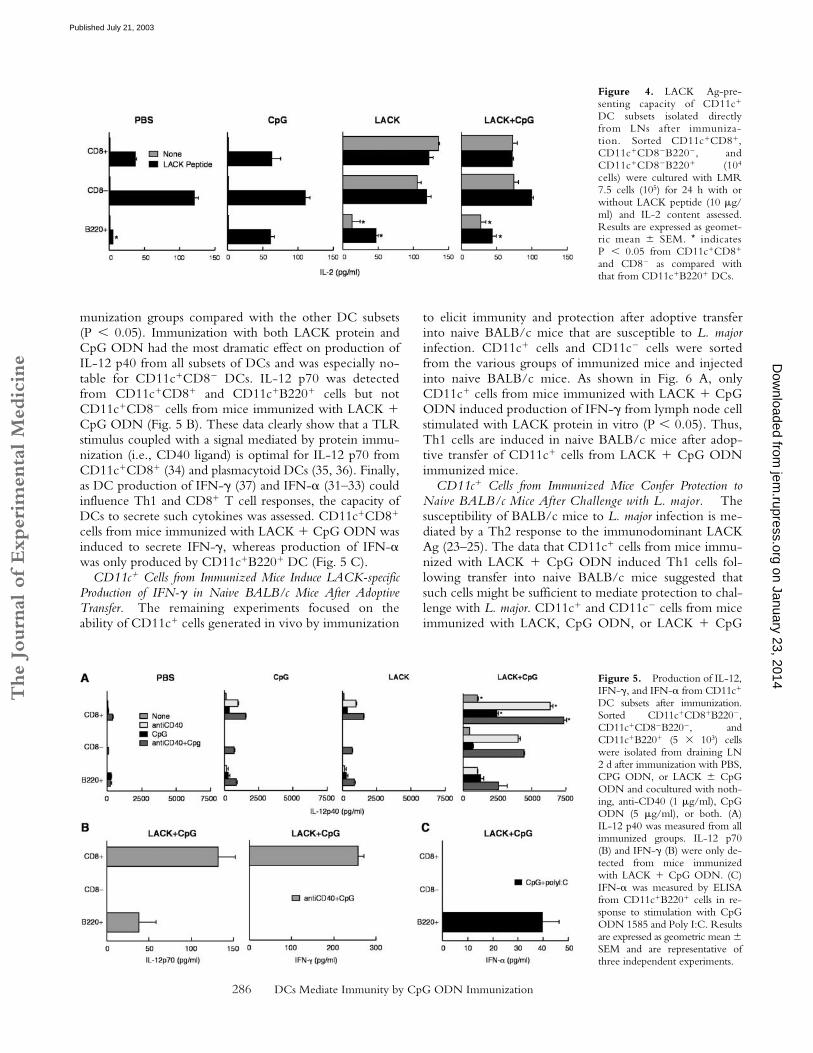
Subsets of CD11c� DC from Immunized Mice Have DistinctAg-presenting Capability. To assess whether differential ex-pression of MHC class II and costimulatory molecules al-tered Ag-presenting capacity, sorted DC subsets were iso-lated after immunization in vivo and used to stimulateLMR 7.5 cells. As shown in Fig. 4, all three subsets ofCD11c� cells were able to induce IL-2 secretion fromLMR 7.5 cells if mice were immunized with LACK �CpG ODN. There was a modest decrease in the produc-tion of IL-2 from LMR 7.5 cells from both CD8� andCD8�B220� DCs in mice immunized with both LACKprotein and CpG ODN compared with LACK proteinalone. This was a consistent finding in other experimentsand may reflect maturation of the DCs in vivo by the CpGODN. In comparing LACK presentation among the othersubsets of CD11c� cells, CD11c�B220� cells were far lessefficient on a per-cell basis from cells derived ex vivo afterimmunization with LACK � CpG ODN. Moreover, suchcells were less potent at inducing IL-2 production evenwhen LACK peptide was added in vitro compared withthe other subsets of DCs (P � 0.05).
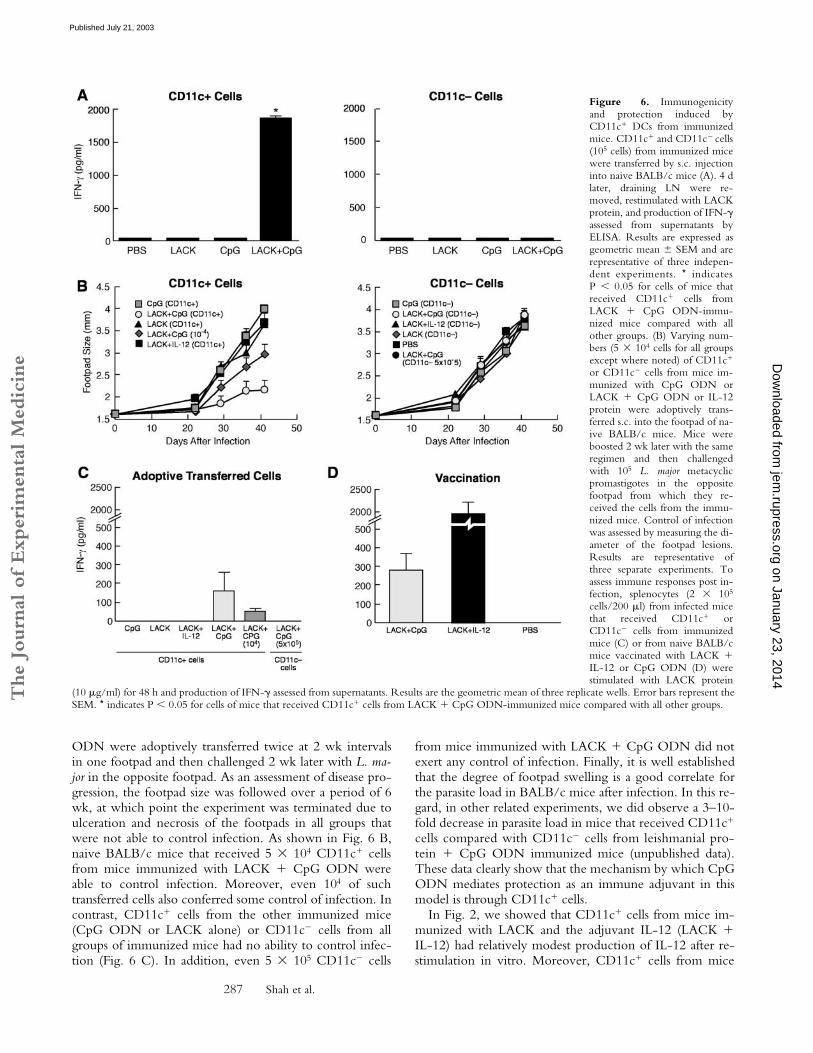
Subsets of CD11c� Cells from Immunized Mice Have Dis-tinct Cytokine-producing Capacity. The production of cyto-kines such as IL-12 (20) and IFN-� (31–33) from DCsubsets can be strongly influenced by the conditions ofstimulation. As shown in Fig. 5 A, CD11c�CD8� DCswere the most potent producers of IL-12 p40 from all im-
Figure 3. Frequency of DCsubsets and their expression ofcell surface markers from LN af-ter immunization. (A) CD11c�
DCs from draining LNs of naive(no injection) and or mice im-munized with PBS, CpG ODN,or LACK protein � CpG ODNmice were enriched as describedin the methods and stained withantibodies against CD11c, CD8and B220. Cells were gated onCD11c� cells and the percent-age of CD11c�CD8�B220�,CD11c�CD8�B220�, andCD11c�B220� is shown. (B)Expression of cell surface mark-ers was analyzed on subsets ofCD11c� DCs based on expres-sion of CD8 and B220. Lightblue, no injection; black, PBS;blue, CpG ODN; green, LACK;red, LACK � CpG ODN. Dataare representative of three inde-pendent experiments.
Table I. Frequency of DC Subsets After Immunization
DCsubset
No injection PBS
CpGODN LACK
LACK �CpG ODN
B220� 56.6 � 4.5 36.6 � 5.6 16.4 � 6.6 26.5 � 7.1 17.7 � 9.0CD8� 3.7 � 0.4 4.0 � 1.8 4.8 � 2.9 4.7 � 2.1 6.7 � 5.6CD8� 39.8 � 4.0 58.8 � 5.0 78.8 � 5.4 68.9 � 8.7 75.7 � 9.3
The frequency of CD11c�CD8�B220�, CD11c�CD8�B220�, andCD11c�B220� cells from draining LN of naive and immunized micewas determined from four independent experiments. Results areexpressed as geometric mean � SEM: P � 0.05 when comparing any ofthe immunization groups to naive mice; P � 0.05 when comparingmice immunized with LACK protein, CpG ODN, or both with PBS-injected mice.
on January 23, 2014jem
.rupress.orgD
ownloaded from
Published July 21, 2003
The
Journ
al o
f Exp
erim
enta
l M
edic
ine
286 DCs Mediate Immunity by CpG ODN Immunization
munization groups compared with the other DC subsets(P � 0.05). Immunization with both LACK protein andCpG ODN had the most dramatic effect on production ofIL-12 p40 from all subsets of DCs and was especially no-table for CD11c�CD8� DCs. IL-12 p70 was detectedfrom CD11c�CD8� and CD11c�B220� cells but notCD11c�CD8� cells from mice immunized with LACK �CpG ODN (Fig. 5 B). These data clearly show that a TLRstimulus coupled with a signal mediated by protein immu-nization (i.e., CD40 ligand) is optimal for IL-12 p70 fromCD11c�CD8� (34) and plasmacytoid DCs (35, 36). Finally,as DC production of IFN-� (37) and IFN-� (31–33) couldinfluence Th1 and CD8� T cell responses, the capacity ofDCs to secrete such cytokines was assessed. CD11c�CD8�
cells from mice immunized with LACK � CpG ODN wasinduced to secrete IFN-�, whereas production of IFN-�was only produced by CD11c�B220� DC (Fig. 5 C).
CD11c� Cells from Immunized Mice Induce LACK-specificProduction of IFN-� in Naive BALB/c Mice After AdoptiveTransfer. The remaining experiments focused on theability of CD11c� cells generated in vivo by immunization
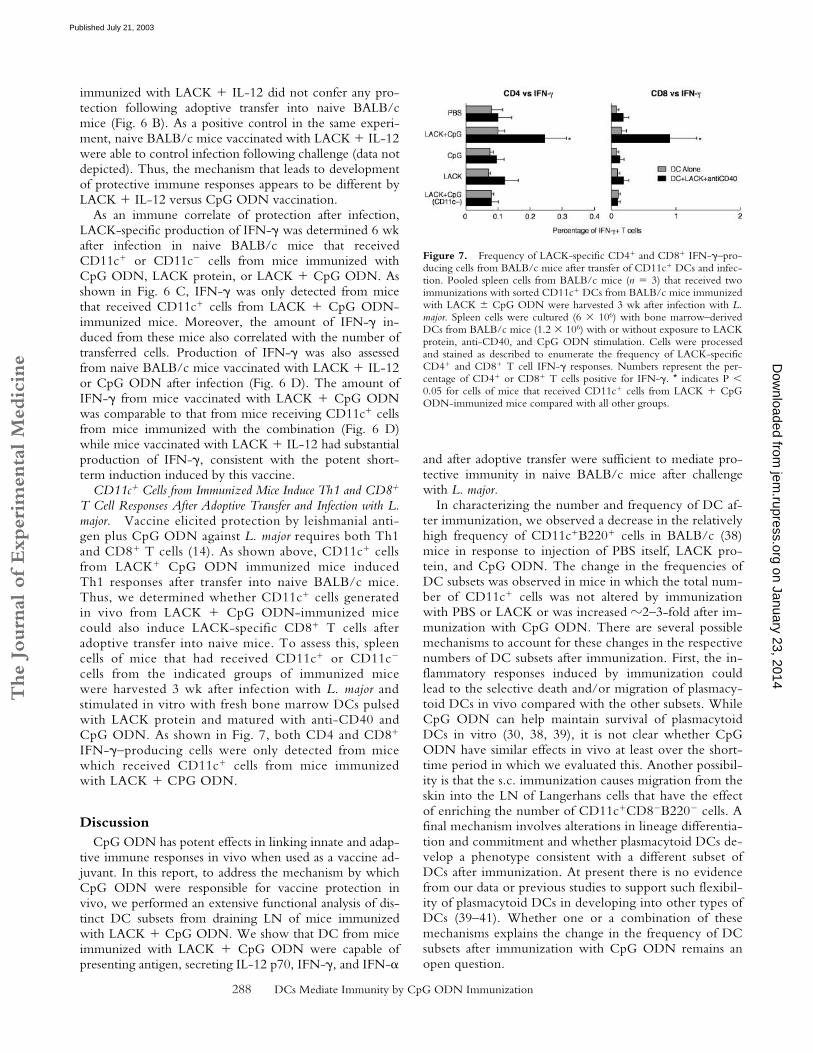
to elicit immunity and protection after adoptive transferinto naive BALB/c mice that are susceptible to L. majorinfection. CD11c� cells and CD11c� cells were sortedfrom the various groups of immunized mice and injectedinto naive BALB/c mice. As shown in Fig. 6 A, onlyCD11c� cells from mice immunized with LACK � CpGODN induced production of IFN-� from lymph node cellstimulated with LACK protein in vitro (P � 0.05). Thus,Th1 cells are induced in naive BALB/c mice after adop-tive transfer of CD11c� cells from LACK � CpG ODNimmunized mice.
CD11c� Cells from Immunized Mice Confer Protection toNaive BALB/c Mice After Challenge with L. major. Thesusceptibility of BALB/c mice to L. major infection is me-diated by a Th2 response to the immunodominant LACKAg (23–25). The data that CD11c� cells from mice immu-nized with LACK � CpG ODN induced Th1 cells fol-lowing transfer into naive BALB/c mice suggested thatsuch cells might be sufficient to mediate protection to chal-lenge with L. major. CD11c� and CD11c� cells from miceimmunized with LACK, CpG ODN, or LACK � CpG
Figure 4. LACK Ag-pre-senting capacity of CD11c�
DC subsets isolated directlyfrom LNs after immuniza-tion. Sorted CD11c�CD8�,CD11c�CD8�B220�, andCD11c�CD8�B220� (104
cells) were cultured with LMR7.5 cells (105) for 24 h with orwithout LACK peptide (10 �g/ml) and IL-2 content assessed.Results are expressed as geomet-ric mean � SEM. * indicatesP � 0.05 from CD11c�CD8�
and CD8� as compared withthat from CD11c�B220� DCs.
Figure 5. Production of IL-12,IFN-�, and IFN-� from CD11c�
DC subsets after immunization.Sorted CD11c�CD8�B220�,CD11c�CD8�B220�, andCD11c�B220� (5 103) cellswere isolated from draining LN2 d after immunization with PBS,CPG ODN, or LACK � CpGODN and cocultured with noth-ing, anti-CD40 (1 �g/ml), CpGODN (5 �g/ml), or both. (A)IL-12 p40 was measured from allimmunized groups. IL-12 p70(B) and IFN-� (B) were only de-tected from mice immunizedwith LACK � CpG ODN. (C)IFN-� was measured by ELISAfrom CD11c�B220� cells in re-sponse to stimulation with CpGODN 1585 and Poly I:C. Resultsare expressed as geometric mean �SEM and are representative ofthree independent experiments.
on January 23, 2014jem
.rupress.orgD
ownloaded from
Published July 21, 2003
The
Journ
al o
f Exp
erim
enta
l M
edic
ine
287 Shah et al.
ODN were adoptively transferred twice at 2 wk intervalsin one footpad and then challenged 2 wk later with L. ma-jor in the opposite footpad. As an assessment of disease pro-gression, the footpad size was followed over a period of 6wk, at which point the experiment was terminated due toulceration and necrosis of the footpads in all groups thatwere not able to control infection. As shown in Fig. 6 B,naive BALB/c mice that received 5 104 CD11c� cellsfrom mice immunized with LACK � CpG ODN wereable to control infection. Moreover, even 104 of suchtransferred cells also conferred some control of infection. Incontrast, CD11c� cells from the other immunized mice(CpG ODN or LACK alone) or CD11c� cells from allgroups of immunized mice had no ability to control infec-tion (Fig. 6 C). In addition, even 5 105 CD11c� cells
from mice immunized with LACK � CpG ODN did notexert any control of infection. Finally, it is well establishedthat the degree of footpad swelling is a good correlate forthe parasite load in BALB/c mice after infection. In this re-gard, in other related experiments, we did observe a 3–10-fold decrease in parasite load in mice that received CD11c�
cells compared with CD11c� cells from leishmanial pro-tein � CpG ODN immunized mice (unpublished data).These data clearly show that the mechanism by which CpGODN mediates protection as an immune adjuvant in thismodel is through CD11c� cells.
In Fig. 2, we showed that CD11c� cells from mice im-munized with LACK and the adjuvant IL-12 (LACK �IL-12) had relatively modest production of IL-12 after re-stimulation in vitro. Moreover, CD11c� cells from mice
Figure 6. Immunogenicityand protection induced byCD11c� DCs from immunizedmice. CD11c� and CD11c� cells(105 cells) from immunized micewere transferred by s.c. injectioninto naive BALB/c mice (A). 4 dlater, draining LN were re-moved, restimulated with LACKprotein, and production of IFN-�assessed from supernatants byELISA. Results are expressed asgeometric mean � SEM and arerepresentative of three indepen-dent experiments. * indicatesP � 0.05 for cells of mice thatreceived CD11c� cells fromLACK � CpG ODN-immu-nized mice compared with allother groups. (B) Varying num-bers (5 104 cells for all groupsexcept where noted) of CD11c�
or CD11c� cells from mice im-munized with CpG ODN orLACK � CpG ODN or IL-12protein were adoptively trans-ferred s.c. into the footpad of na-ive BALB/c mice. Mice wereboosted 2 wk later with the sameregimen and then challengedwith 105 L. major metacyclicpromastigotes in the oppositefootpad from which they re-ceived the cells from the immu-nized mice. Control of infectionwas assessed by measuring the di-ameter of the footpad lesions.Results are representative ofthree separate experiments. Toassess immune responses post in-fection, splenocytes (2 105
cells/200 �l) from infected micethat received CD11c� orCD11c� cells from immunizedmice (C) or from naive BALB/cmice vaccinated with LACK �IL-12 or CpG ODN (D) werestimulated with LACK protein
(10 �g/ml) for 48 h and production of IFN-� assessed from supernatants. Results are the geometric mean of three replicate wells. Error bars represent theSEM. * indicates P � 0.05 for cells of mice that received CD11c� cells from LACK � CpG ODN-immunized mice compared with all other groups.
on January 23, 2014jem
.rupress.orgD
ownloaded from
Published July 21, 2003
The
Journ
al o
f Exp
erim
enta
l M
edic
ine
288 DCs Mediate Immunity by CpG ODN Immunization
immunized with LACK � IL-12 did not confer any pro-tection following adoptive transfer into naive BALB/cmice (Fig. 6 B). As a positive control in the same experi-ment, naive BALB/c mice vaccinated with LACK � IL-12were able to control infection following challenge (data notdepicted). Thus, the mechanism that leads to developmentof protective immune responses appears to be different byLACK � IL-12 versus CpG ODN vaccination.
As an immune correlate of protection after infection,LACK-specific production of IFN-� was determined 6 wkafter infection in naive BALB/c mice that receivedCD11c� or CD11c� cells from mice immunized withCpG ODN, LACK protein, or LACK � CpG ODN. Asshown in Fig. 6 C, IFN-� was only detected from micethat received CD11c� cells from LACK � CpG ODN-immunized mice. Moreover, the amount of IFN-� in-duced from these mice also correlated with the number oftransferred cells. Production of IFN-� was also assessedfrom naive BALB/c mice vaccinated with LACK � IL-12or CpG ODN after infection (Fig. 6 D). The amount ofIFN-� from mice vaccinated with LACK � CpG ODNwas comparable to that from mice receiving CD11c� cellsfrom mice immunized with the combination (Fig. 6 D)while mice vaccinated with LACK � IL-12 had substantialproduction of IFN-�, consistent with the potent short-term induction induced by this vaccine.
CD11c� Cells from Immunized Mice Induce Th1 and CD8�
T Cell Responses After Adoptive Transfer and Infection with L.major. Vaccine elicited protection by leishmanial anti-gen plus CpG ODN against L. major requires both Th1and CD8� T cells (14). As shown above, CD11c� cellsfrom LACK� CpG ODN immunized mice inducedTh1 responses after transfer into naive BALB/c mice.Thus, we determined whether CD11c� cells generatedin vivo from LACK � CpG ODN-immunized micecould also induce LACK-specific CD8� T cells afteradoptive transfer into naive mice. To assess this, spleencells of mice that had received CD11c� or CD11c�
cells from the indicated groups of immunized micewere harvested 3 wk after infection with L. major andstimulated in vitro with fresh bone marrow DCs pulsedwith LACK protein and matured with anti-CD40 andCpG ODN. As shown in Fig. 7, both CD4 and CD8�
IFN-�–producing cells were only detected from micewhich received CD11c� cells from mice immunizedwith LACK � CPG ODN.
DiscussionCpG ODN has potent effects in linking innate and adap-
tive immune responses in vivo when used as a vaccine ad-juvant. In this report, to address the mechanism by whichCpG ODN were responsible for vaccine protection invivo, we performed an extensive functional analysis of dis-tinct DC subsets from draining LN of mice immunizedwith LACK � CpG ODN. We show that DC from miceimmunized with LACK � CpG ODN were capable ofpresenting antigen, secreting IL-12 p70, IFN-�, and IFN-�
and after adoptive transfer were sufficient to mediate pro-tective immunity in naive BALB/c mice after challengewith L. major.
In characterizing the number and frequency of DC af-ter immunization, we observed a decrease in the relativelyhigh frequency of CD11c�B220� cells in BALB/c (38)mice in response to injection of PBS itself, LACK pro-tein, and CpG ODN. The change in the frequencies ofDC subsets was observed in mice in which the total num-ber of CD11c� cells was not altered by immunizationwith PBS or LACK or was increased �2–3-fold after im-munization with CpG ODN. There are several possiblemechanisms to account for these changes in the respectivenumbers of DC subsets after immunization. First, the in-flammatory responses induced by immunization couldlead to the selective death and/or migration of plasmacy-toid DCs in vivo compared with the other subsets. WhileCpG ODN can help maintain survival of plasmacytoidDCs in vitro (30, 38, 39), it is not clear whether CpGODN have similar effects in vivo at least over the short-time period in which we evaluated this. Another possibil-ity is that the s.c. immunization causes migration from theskin into the LN of Langerhans cells that have the effectof enriching the number of CD11c�CD8�B220� cells. Afinal mechanism involves alterations in lineage differentia-tion and commitment and whether plasmacytoid DCs de-velop a phenotype consistent with a different subset ofDCs after immunization. At present there is no evidencefrom our data or previous studies to support such flexibil-ity of plasmacytoid DCs in developing into other types ofDCs (39–41). Whether one or a combination of thesemechanisms explains the change in the frequency of DCsubsets after immunization with CpG ODN remains anopen question.
Figure 7. Frequency of LACK-specific CD4� and CD8� IFN-�–pro-ducing cells from BALB/c mice after transfer of CD11c� DCs and infec-tion. Pooled spleen cells from BALB/c mice (n 3) that received twoimmunizations with sorted CD11c� DCs from BALB/c mice immunizedwith LACK � CpG ODN were harvested 3 wk after infection with L.major. Spleen cells were cultured (6 106) with bone marrow–derivedDCs from BALB/c mice (1.2 106) with or without exposure to LACKprotein, anti-CD40, and CpG ODN stimulation. Cells were processedand stained as described to enumerate the frequency of LACK-specificCD4� and CD8� T cell IFN-� responses. Numbers represent the per-centage of CD4� or CD8� T cells positive for IFN-�. * indicates P �0.05 for cells of mice that received CD11c� cells from LACK � CpGODN-immunized mice compared with all other groups.
on January 23, 2014jem
.rupress.orgD
ownloaded from
Published July 21, 2003
The
Journ
al o
f Exp
erim
enta
l M
edic
ine
289 Shah et al.
In assessing the antigen presenting capacity of DCs afterimmunization, we found that CD11c� cells capable of pre-senting LACK to the LACK-specific LMR 7.5 hybridomacould be detected from draining LN as early as 18 h afterimmunization, which peaked at 36–48 h and was notpresent at 7 d. The kinetics of the response are consistentwith previous reports showing intimate contact of T cellsand DCs occurring within hours after antigenic challenge(42) that begin to dissociate after 36–48 h (43). These datasuggest that CD11c� cells capable of presenting LACKdied, migrated, and/or lost expression of LACK/MHCcomplexes on their cell surface from LN 2 d after immuni-zation. In terms of the relative potency of Ag presentation,both CD11c�CD8� and CD8� cells were substantiallymore efficient than CD11c�B220� DCs in presenting Agimmediately ex vivo. Thus, due to the relatively high fre-quency of CD11c�CD8� cells in the LN; they wouldlikely have the most prominent role in initiating T cell im-mune responses (44). These data are consistent with thefinding that only CD11c�CD11b�CD8� DCs are capableof presenting LACK Ag after infection with L. major (44a).In contrast, the relative deficiency of B220� cells for Agpresentation compared with the CD8� and CD8� mightinvolve a qualitative difference in Ag processing or presen-tation resulting in diminished ability to assemble peptide–MHC complexes (45). Moreover, CD11c�B220� DCshave a demonstrable decrease in expression of MHC classII and other costimulatory markers (31, 38, 45). Finally, thehigh sensitivity of the LMR 7.5 hybridoma for detectingLACK Ag might have overestimated even the relativelylimited Ag-presenting capacity of CD11c�B220� cells. Inthis regard, recent reports showing that freshly isolated na-ive or virally exposed splenic plasmacytoid DCs were un-able to induce proliferation of naive CD4� T cells (45, 46)but could promote expansion of previously activated Tcells (45) might be analogous to our studies showing thatplasmacytoid DCs induce low but detectable IL-2 produc-tion from a T cell hybridoma. While these data do not ab-solutely preclude a role of LN plasmacytoid DCs in Agpresentation with protein and CpG ODN immunization invivo, they suggest that the relatively high frequency of suchcells in normal LN of BALB/c mice might be more in-volved in maintaining tolerance in their steady-state andupon activation provide stimulation through their produc-tion of cytokines.
In addition to Ag presentation, DCs strongly influencesthe qualitative and quantitative aspects of cellular immuneresponses. CD11c�CD8�B220� DCs were the most po-tent producers of IL-12 p40 and p70 (20) and the only DCsubset able to secrete IFN-� after immunization withLACK � CpG ODN. Plasmacytoid DCs also producedIL-12 p70 and IFN-� after immunization. Thus, due to therelatively low frequency of CD11c� CD8�B220� DCs inLN of naive or immunized mice and the limited capacity ofplasmacytoid DCs for Ag presentation, these particular sub-sets of DCs through production of IL-12 p70, IFN-�, andIFN-� likely mediate their effects by providing a favorablemilieu to activate Th1 (28, 47, 48) and CD8� T cell (49)
responses. A final point is whether the effects of CpGODN on DC subsets are direct and/or indirect. In mice, ithas recently been shown that CD8��, CD8�� and plasma-cytoid DCs derived from spleens had comparable mRNAexpression for TLR-9 and all could be induced to secreteIL-12 p40 after stimulation with CpG ODN (46, 50).These data are consistent with our findings that CpG ODNdirectly activates all the DC subsets studied here with in-duction of inflammatory cytokines and up-regulation ofcostimulatory molecules. In addition, CpG ODN likelyhave substantial indirect effects for generating adaptive cel-lular immune responses. Such indirect effects could bethrough IL-12 and IFN-� inducing NK production ofIFN-� which would enhance Th1 responses. In addition,IFN-� induced from plasmacytoid DC can also enhancethe maturation of other DC subsets (51). Thus, the effect ofCpG ODN on DC subsets and T cell responses involvesboth direct and indirect mechanisms. In summary we spec-ulate that there is a cooperative effect with potential spe-cialization of the various DC subsets in vivo for inducing afunctional cellular immune response. For the model studiedhere, CD11c�CD8� would be primarily involved in anti-gen presentation, while CD11c�CD8�B220� and plasma-cytoid DCs provide a favorable cytokine milieu for en-hancing adaptive immune responses.
The demonstration that in vivo-derived LN CD11c�
DCs after immunization with LACK � CpG ODN aresufficient to mediate immunity and protection against anintracellular infection provides strong evidence for howCpG ODN mediate their effects as an adjuvant in vivo.With regard to how the transferred CD11c� DCs mediatestheir effects in vivo, there are two potential mechanisms.First is the importance of IL-12 from the transferredCD11c� cells from immunized mice in priming Th1 re-sponses in the recipient mice. It has been shown recentlythat wild-type but not IL-12–deficient Langerhans cellspulsed in vitro with a variety of leishmanial Ag includingLACK could confer protection (52). Thus, it is likely thatIL-12 would be required from the transferred CD11c�
cells. The second consideration is whether the induction ofTh1 and/or CD8� T cell responses after adoptive transferof cells from mice immunized with LACK protein � CpGODN into naive mice is direct and/or indirect via cross-priming from recipient cells.
To conclude, these studies focused exclusively on pe-ripheral LN DCs. While there is heterogeneity among DCpopulations and function between LN, spleen, and bonemarrow–derived DCs, we believe these results examininghow peripheral LN DCs are altered by s.c. immunizationhas direct relevance for vaccine design in humans. In thisregard, despite the broad distribution of TLR-9 expressionamongst mouse DC subsets (50), its expression in primatesand humans is limited to B cells and plasmacytoid DCs(53). Thus for humans, the antigen presenting capacity andproduction of cytokines of plasmacytoid DCs in vivo willlargely determine whether CpG ODN alone are effectivevaccine adjuvants for inducing adaptive cellular immuneresponse in people. If indeed such cells are not optimal for
on January 23, 2014jem
.rupress.orgD
ownloaded from
Published July 21, 2003
The
Journ
al o
f Exp
erim
enta
l M
edic
ine
290 DCs Mediate Immunity by CpG ODN Immunization
antigen presentation as in the mouse, combination strate-gies aimed at targeting additional DC populations usingother TLR ligands or combinations of TLR ligands mightbe necessary for efficient generation of cellular responses inhumans. Nevertheless, this report showing that freshly iso-lated CD11c� but not CD11c� cells from mice immunizedusing CpG ODN as an adjuvant are capable of transferringprotective immunity should provide a more targeted andrational approach to vaccine development for diseases re-quiring cellular immune responses.
We thank Brenda Rae Marshall for editorial assistance.J.A. Shah is an HHMI-NIH Research Scholar.
Submitted: 21 April 2003Revised: 13 May 2003Accepted: 13 May 2003
References1. Seder, R.A., and A.V. Hill. 2000. Vaccines against intracellular
infections requiring cellular immunity. Nature. 406:793–798.2. O’Garra, A. 1998. Cytokines induce the development of
functionally heterogeneous T helper cell subsets. Immunity.8:275–283.
3. Murphy, K.M., W. Ouyang, J.D. Farrar, J. Yang, S. Ranga-nath, H. Asnagli, M. Afkarian, and T.L. Murphy. 2000. Sig-naling and transcription in T helper development. Annu. Rev.Immunol. 18:451–494.
4. Krieg, A.M. 2002. CpG motifs in bacterial DNA and theirimmune effects. Annu. Rev. Immunol. 20:709–760.
5. Barton, G.M., and R. Medzhitov. 2002. Control of adaptiveimmune responses by Toll-like receptors. Curr. Opin. Immu-nol. 14:380–383.
6. Akira, S., K. Takeda, and T. Kaisho. 2001. Toll-like recep-tors: critical proteins linking innate and acquired immunity.Nat. Immunol. 2:675–680.
7. Klinman, D.M., A.K. Yi, S.L. Beaucage, J. Conover, andA.M. Krieg. 1996. CpG motifs present in bacteria DNA rap-idly induce lymphocytes to secrete interleukin 6, interleukin12, and interferon �. Proc. Natl. Acad. Sci. USA. 93:2879–2883.
8. Sparwasser, T., R.M. Vabulas, B. Villmow, G.B. Lipford, andH. Wagner. 2000. Bacterial CpG-DNA activates dendriticcells in vivo: T helper cell-independent cytotoxic T cell re-sponses to soluble proteins. Eur. J. Immunol. 30:3591–3597.
9. Hartmann, G., G.J. Weiner, and A.M. Krieg. 1999. CpGDNA: a potent signal for growth, activation, and maturationof human dendritic cells. Proc. Natl. Acad. Sci. USA. 96:9305–9310.
10. Chu, R.S., O.S. Targoni, A.M. Krieg, P.V. Lehmann, andC.V. Harding. 1997. CpG oligodeoxynucleotides act as adju-vants that switch on T helper 1 (Th1) immunity. J. Exp. Med.186:1623–1631.
11. Roman, M., E. Martin-Orozco, J.S. Goodman, M.D.Nguyen, Y. Sato, A. Ronaghy, R.S. Kornbluth, D.D. Rich-man, D.A. Carson, and E. Raz. 1997. ImmunostimulatoryDNA sequences function as T helper-1-promoting adjuvants.Nat. Med. 3:849–854.
12. Cho, H.J., K. Takabayashi, P.M. Cheng, M.D. Nguyen, M.Corr, S. Tuck, and E. Raz. 2000. Immunostimulatory DNA-based vaccines induce cytotoxic lymphocyte activity by a
T-helper cell-independent mechanism. Nat. Biotechnol. 18:509–514.
13. Davila, E., and E. Celis. 2000. Repeated administration ofcytosine-phosphorothiolated guanine-containing oligonucle-otides together with peptide/protein immunization results inenhanced CTL responses with anti-tumor activity. J. Immu-nol. 165:539–547.
14. Rhee, E.G., S. Mendez, J.A. Shah, C.Y. Wu, J.R. Kirman,T.N. Turon, D.F. Davey, H. Davis, D.M. Klinman, R.N.Coler, et al. 2002. Vaccination with heat-killed Leishmaniaantigen or recombinant leishmanial protein and CpG oli-godeoxynucleotides induces long-term memory CD4� andCD8� T cell responses and protection against Leishmania ma-jor infection. J. Exp. Med. 195:1565–1573.
15. Gurunathan, S., D.L. Sacks, D.R. Brown, S.L. Reiner, H.Charest, N. Glaichenhaus, and R.A. Seder. 1997. Vaccina-tion with DNA encoding the immunodominant LACK para-site antigen confers protective immunity to mice infectedwith Leishmania major. J. Exp. Med. 186:1137–1147.
16. Casares, S., K. Inaba, T.D. Brumeanu, R.M. Steinman, andC.A. Bona. 1997. Antigen presentation by dendritic cells af-ter immunization with DNA encoding a major histocompat-ibility complex class II-restricted viral epitope. J. Exp. Med.186:1481–1486.
17. Sparwasser, T., E.S. Koch, R.M. Vabulas, K. Heeg, G.B.Lipford, J.W. Ellwart, and H. Wagner. 1998. Bacterial DNAand immunostimulatory CpG oligonucleotides trigger matu-ration and activation of murine dendritic cells. Eur. J. Immu-nol. 28:2045–2054.
18. Maldonado-Lopez, R., and M. Moser. 2001. Dendritic cellsubsets and the regulation of Th1/Th2 responses. Semin. Im-munol. 13:275–282.
19. Heath, W.R., and F.R. Carbone. 2001. Cross-presentation,dendritic cells, tolerance and immunity. Annu. Rev. Immunol.19:47–64.
20. Maldonado-Lopez, R., T. De Smedt, P. Michel, J. Godfroid,B. Pajak, C. Heirman, K. Thielemans, O. Leo, J. Urbain, andM. Moser. 1999. CD8�� and CD8�- subclasses of dendriticcells direct the development of distinct T helper cells in vivo.J. Exp. Med. 189:587–592.
21. Lanzavecchia, A., and F. Sallusto. 2001. Regulation of T cellimmunity by dendritic cells. Cell. 106:263–266.
22. Shortman, K., and Y.J. Liu. 2002. Mouse and human den-dritic cell subtypes. Nat. Rev. Immunol. 2:151–161.
23. Mougneau, E., F. Altare, A.E. Wakil, S. Zheng, T. Coppola,Z.E. Wang, R. Waldmann, R.M. Locksley, and N. Glai-chenhaus. 1995. Expression cloning of a protective Leishma-nia antigen. Science. 268:563–566.
24. Julia, V., M. Rassoulzadegan, and N. Glaichenhaus. 1996.Resistance to Leishmania major induced by tolerance to a sin-gle antigen. Science. 274:421–423.
25. Launois, P., I. Maillard, S. Pingel, K.G. Swihart, I. Xenarios,H. Acha-Orbea, H. Diggelmann, R.M. Locksley, H.R. Mac-Donald, and J.A. Louis. 1997. IL-4 rapidly produced by V�4V�8 CD4� T cells instructs Th2 development and susceptibil-ity to Leishmania major in BALB/c mice. Immunity. 6:541–549.
26. Stobie, L., S. Gurunathan, C. Prussin, D.L. Sacks, N. Glai-chenhaus, C.Y. Wu, and R.A. Seder. 2000. The role of anti-gen and IL-12 in sustaining Th1 memory cells in vivo: IL-12is required to maintain memory/effector Th1 cells sufficientto mediate protection to an infectious parasite challenge. Proc.Natl. Acad. Sci. USA. 97:8427–8432.
27. Malherbe, L., C. Filippi, V. Julia, G. Foucras, M. Moro, H.
on January 23, 2014jem
.rupress.orgD
ownloaded from
Published July 21, 2003
The
Journ
al o
f Exp
erim
enta
l M
edic
ine
291 Shah et al.
Appel, K. Wucherpfennig, J.C. Guery, and N. Glaichenhaus.2000. Selective activation and expansion of high-affinityCD4� T cells in resistant mice upon infection with Leishma-nia major. Immunity. 13:771–782.
28. Trinchieri, G. 1995. Interleukin-12: a proinflammatory cyto-kine with immunoregulatory functions that bridge innate re-sistance and antigen-specific adaptive immunity. Annu. Rev.Immunol. 13:251–276.
29. Afonso, L.C., T.M. Scharton, L.Q. Vieira, M. Wysocka, G.Trinchieri, and P. Scott. 1994. The adjuvant effect of inter-leukin-12 in a vaccine against Leishmania major. Science. 263:235–237.
30. Henri, S., D. Vremec, A. Kamath, J. Waithman, S. Williams,C. Benoist, K. Burnham, S. Saeland, E. Handman, and K.Shortman. 2001. The dendritic cell populations of mouselymph nodes. J. Immunol. 167:741-748.30.
31. Asselin-Paturel, C., A. Boonstra, M. Dalod, I. Durand, N.Yessaad, C. Dezutter-Dambuyant, A. Vicari, A. O’Garra, C.Biron, F. Briere, and G. Trinchieri. 2001. Mouse type I IFN-producing cells are immature APCs with plasmacytoid mor-phology. Nat. Immunol. 2:1144–1150.
32. Siegal, F.P., N. Kadowaki, M. Shodell, P.A. Fitzgerald-Bocarsly, K. Shah, S. Ho, S. Antonenko, and Y.J. Liu. 1999.The nature of the principal type 1 interferon-producing cellsin human blood. Science. 284:1835–1837.
33. Rissoan, M.C., V. Soumelis, N. Kadowaki, G. Grouard, F.Briere, R. Malefyt, and Y.J. Liu. 1999. Reciprocal control ofT helper cell and dendritic cell differentiation. Science. 283:1183–1186.
34. Schulz, O., A.D. Edwards, M. Schito, J. Aliberti, S. Manick-asingham, A. Sher, and C. Reis e Sousa. 2000. CD40 trigger-ing of heterodimeric IL-12 p70 production by dendritic cellsin vivo requires a microbial priming signal. Immunity. 13:453–462.
35. Krug, A., A. Towarowski, S. Britsch, S. Rothenfusser, V. Hor-nung, R. Bals, T. Giese, H. Engelmann, S. Endres, A.M.Krieg, and G. Hartmann. 2001. Toll-like receptor expressionreveals CpG DNA as a unique microbial stimulus for plasma-cytoid dendritic cells which synergizes with CD40 ligand toinduce high amounts of IL-12. Eur. J. Immunol. 31:3036–3037.
36. Cella, M., F. Facchetti, A. Lanzavecchia, and M. Colonna.2000. Plasmacytoid dendritic cells activated by influenza virusand CD40L drive a potent Th1 polarization. Nat. Immunol.305-310.
37. Ohteki, T., T. Fukao, K. Suzue, C. Maki, M. Ito, M. Naka-mura, and S. Koyasu. 1999. Interleukin 12-dependent inter-feron � production by CD8�� lymphoid dendritic cells. J.Exp. Med. 189:1981–1986.
38. Nakano, H., M. Yanagita, and M.D. Gunn. 2001.CD11c�B220�Gr-1� cells in mouse lymph nodes and spleendisplay characteristics of plasmacytoid dendritic cells. J. Exp.Med. 194:1171–1178.
39. O’Keeffe, M., H. Hochrein, D. Vremec, I. Caminschi, J.L.Miller, E.M. Anders, L. Wu, M.H. Lahoud, S. Henri, B.Scott, et al. 2002. Mouse plasmacytoid cells: long-lived cells,heterogeneous in surface phenotype and function, that differ-entiate into CD8� dendritic cells only after microbial stimu-lus. J. Exp. Med. 196:1307–1319.
40. del Hoyo, G.M., P. Martin, H.H. Vargas, S. Ruiz, C.F. Arias,and C. Ardavin. 2002. Characterization of a common precur-sor population for dendritic cells. Nature. 415:1043–1047.
41. Martinez del Hoyo, G., P. Martin, C.F. Arias, A.R. Marin,and D. Ardavin. 2002. CD8�� dendritic cells originate from
the CD8�� dendritic cell subset by a maturation process in-volving CD8�, DEC-205, and CD24 up-regulation. Blood.99:999–1004.
42. Jenkins, M.K., A. Khoruts, E. Ingulli, D.L. Mueller, S.J. Mc-Sorley, R.L. Reinhardt, A. Itano, A., and K.A. Pape. 2001.In vivo activation of antigen-specific CD4 T cells. Annu.Rev. Immunol. 19:23-45.
43. Stoll, S., J. Delon, T.M. Brotz, and R.N. Germain. 2002.Dynamic imaging of T cell-dendritic cell interactions inlymph nodes. Science. 296:1873–1876.
44. Smith, A.L., and B. Fazekas de St. Groth. 1999. Antigen-pulsed CD8a� dendritic cells generate an immune responseafter subcutaneous injection without homing to the draininglymph node. J. Exp. Med. 189:593-598.
44a.Filippi, C., S. Hugues, J. Cazareth, V. Julia, N. Glaichenhaus,and S. Ugolini. 2003. CD4� T cell polarization in mice ismodulated by strain-specific major histocompatibility com-plex–independent differences within dendritic cells. J. Exp.Med. 198:201–209.
45. Krug, A., R. Veeraswamy, A. Pekosz, O. Kanagawa, E.R.Unanue, M. Colonna, and M. Cella. 2003. Interferon-pro-ducing cells fail to induce proliferation of naïve T cells butcan promote expansion and T helper 1 differentiation of anti-gen-experienced unpolarized T cells. J. Exp. Med. 197:899–906.
46. Boonstra, A., C. Asselin-Paturel, M. Gilliet, C. Crain, G.Trinchieri, Y.J. Liu, and A. O’Garra. 2003. Flexibility ofmouse classical and plasmacytoid-derived dendritic cells in di-recting T helper type 1 and 2 cell development: dependencyon antigen dose and differential Toll-like receptor ligation. J.Exp. Med. 197:101–109.
47. Parronchi, P., S. Mohapatra, S. Sampognaro, L. Giannarini,U. Wahn, P. Chong, S. Mohapatra, E. Maggi, H. Renz, andS. Romagnani. Effects of interferon-� on cytokine profile, Tcell receptor repertoire and peptide reactivity of human aller-gen-specific T cells. Eur. J. Immunol. 26:697-703.
48. Rogge, L., L. Barberis-Maino, M. Biffi, N. Passini, D.H.Presky, U. Gubler, and F. Sinigaglia. Selective expression ofan interleukin-12 receptor component by human T helper 1cells. J. Exp. Med. 185:825-831.
49. Sprent, J., and C.D. Surh. 2002. T cell memory. Annu. Rev.Immunol. 20:551–579.
50. Edwards, A.D., S.S. Diebold, E.M.C. Slack, H. Tomizawa,H. Hemmi, T. Kaisho, S. Akira, and C. Reis e Sousa. 2003.Toll-like receptor expression in murine DC subsets: lack ofTLR7 expression by CD8�� DC correlates with unrespon-siveness to imidazoquinolines. Eur. J. Immunol. 33:827–833.
51. Dalod, M. T. Hamilton, R. Salomon, T.P. Salazaar-Mather,S.C. Henry, J.D. Hamilton, and C.A. Biron. 2003. Dendriticcell responses to early murine cytomegalovirus infection: sub-set functional specialization and differential regulation by in-terferon �/�. J. Exp. Med. 197:885–898.
52. Berberich, C., J.R. Ramirez-Pineda, C. Hambrecht, G. Al-ber, Y.A. Skeiky, and H. Moll. 2003. Dendritic cell (DC)-based protection against an intracellular pathogen is de-pendent upon DC-derived IL-12 and can be induced bymolecularly defined antigens. J. Immunol. 170:3171–3179.
53. Hornung, V.S., S. Rothenfusser, S. Britsch, A. Krug, B. Jahrs-dorfer, T. Giese, S. Endres, S., and G. Hartmann. 2002.Quantitative expression of Toll-like receptor 1-10 mRNA incellular subsets of human peripheral blood mononuclear cellsand sensitivity to CpG oligodeoxynucleotides. J. Immunol.168:4531-4537.
on January 23, 2014jem
.rupress.orgD
ownloaded from
Published July 21, 2003
Copyright © 2022 FDOKUMEN