Netcentric Information Orchestration - TU Delft Repositories
Upload
khangminh22Category
view
1download
0
Delft University of Technology
Engineering biotin synthesis; towards vitamin independency of Saccharomyces cerevisiae
Wronska, A.K.
DOI10.4233/uuid:bd748c70-2cda-4094-945f-0f0577700367Publication date2022Document VersionFinal published versionCitation (APA)Wronska, A. K. (2022). Engineering biotin synthesis; towards vitamin independency of Saccharomycescerevisiae. https://doi.org/10.4233/uuid:bd748c70-2cda-4094-945f-0f0577700367
Important noteTo cite this publication, please use the final published version (if applicable).Please check the document version above.
CopyrightOther than for strictly personal use, it is not permitted to download, forward or distribute the text or part of it, without the consentof the author(s) and/or copyright holder(s), unless the work is under an open content license such as Creative Commons.
Takedown policyPlease contact us and provide details if you believe this document breaches copyrights.We will remove access to the work immediately and investigate your claim.
This work is downloaded from Delft University of Technology.For technical reasons the number of authors shown on this cover page is limited to a maximum of 10.

Engineering biotin synthesis; towards vitamin independency of Saccharomyces cerevisiae
Dissertation for the purpose of obtaining the degree of doctor
at Delft University of Technology
by the authority of the Rector Magnificus prof. dr. ir. T.H.J.J. van der Hagen
chair of the Board for Doctorates
to be defended publicly on
Friday 20th of May 2022 at 12:30 o’clock
by
Anna Kristina WRONSKA
Master of Science in Biotechnology,
Technical University Braunschweig, Germany
born in Braunschweig, Germany
This dissertation has been approved by the promotors.
Composition of the doctoral committee: Rector Magnificus chairperson Prof. dr. ir. J.M.G. Daran Delft University of Technology, promotor Prof. dr. J.T. Pronk Delft University of Technology, promotor Independent members: Prof. dr. U. Hanefeld Delft University of Technology Prof. dr. I. Borodina Technical University of Denmark Prof. dr. L.M. Veenhoff University of Groningen Dr. ir. M.E. Klijn Delft University of Technology Dr. ir. H. Roubos Royal DSM N.V., Delft Reserve member: Prof. dr. ir. M.C.M. van Loosdrecht Delft University of Technology
The research presented in this thesis was performed at the Industrial Microbiology section, Department of Biotechnology, Faculty of Applied Sciences, Delft University of Technology, the Netherlands. This research was funded by the European Union’s Horizon 2020 research and innovation program under the Marie Skłodowska-Curie action PAcMEN grant agreement No. 722287.
Layout Anna Wronska Cover Anna Wronska Printed by ProefschriftMaken | proefschriftmaken.nl ISBN 978-94-6423-708-5
© 2022 by Anna Kristina Wronska
All rights reserved. No part of this publication may be reproduced, stored in a retrieval system, or transmitted, in any form or by any means, electronically, mechanically, by photo-copying, recording or otherwise, without the prior written permission of the author.
Contents
Summary ................................................................................................................................ 5
Samenvatting ....................................................................................................................... 9
Chapter 1 |Introduction ................................................................................................. 15
Chapter 2 |Exploiting the diversity of Saccharomycotina yeasts to engineer
biotin-independent growth of Saccharomyces cerevisiae .......................................39
Chapter 3 |Engineering oxygen-independent biotin biosynthesis in
Saccharomyces cerevisiae ............................................................................................ 69
Chapter 4 |Engineering class-B vitamin biosynthesis in Saccharomyces
cerevisiae ............................................................................................................................ 99
Outlook .............................................................................................................................. 131
Bibliography ...................................................................................................................... 135
Acknowledgments .......................................................................................................... 149
Curriculum vitae .............................................................................................................. 154
List of Publications ........................................................................................................... 155
List of Patents .................................................................................................................... 155

Summary Every century brings its own challenges, but the 21st century is the first in which a global transition towards circularity is required to ensure human existence on this planet. Exhaustion of planetary resources, such as oil and rare elements, must be prevented and sustainable circular value chains introduced into our industry and economy. In addition to new challenges, every century also brings new and unique solutions. Today, biotechnology may provide some of the most relevant solutions by providing scientists with the ability to decipher the code of life represented by an organism’s DNA as well as with the tools to edit this code. Especially fast-reproducing microorganisms have a great potential to serve as cell factories, which can convert renewable raw materials into chemicals, materials and food ingredients and thereby support a circular bio-based economy. Recently developed biotechnological tools enable us to rewrite (‘edit’) the blueprint for these microbial cell factories with unprecedented precisions and at unprecedented rates. A myriad of life forms evolved over billions of years to adapt to an incredibly diverse number of habitats on our planet, which led to an immense diversity in survival strategies and metabolic capabilities. Recombining these naturally occurring DNA codes and ‘novel-to-nature’ DNA sequences generated in laboratories offers unique possibilities for development of novel cell factories to address challenges in our century and beyond.
Baker’s yeast, Saccharomyces cerevisiae is one of the most intensively studied microorganisms and, as a cell factory, has a long history of successful application in industrial applications. Its story of success began thousands of years ago when processes for production of wine, beer and bread-making were first invented and, over many centuries, improved. Application of yeasts probably started as serendipitous discovery rather than as an invention, when yeast cells from the environment ‘contaminated’ sugar-containing food products and, by accident, turned sugars into ethanol and carbon dioxide, thus yielding the first alcoholic beverages and rising dough. All essential nutrients that yeast require for growth and fermentation were either present in the food product or generated by other microorganisms that inadvertently entered these early fermentation processes. Such a co-existence of multiple microbial species is a natural phenomenon that helps organisms thrive, but in man-made industrial settings such undefined mixed populations are often difficult to control and optimize. When researchers discovered that pure cultures of individual yeast strains were very efficient in producing transport fuels and other interesting chemicals, they therefore developed growth media that contained all essential and non-essential nutrients required for optimal yeast growth, to make these yeast cell factories as productive as possible. For over a century now, yeast cell factories have been under continual development. Classical strain improvement strategies to obtain high-producing strains, later combined with recombinant-DNA technology (genetic engineering) brought microbial production systems to a next level and helped pave the way towards a sustainable bio-based
4 |

Summary Every century brings its own challenges, but the 21st century is the first in which a global transition towards circularity is required to ensure human existence on this planet. Exhaustion of planetary resources, such as oil and rare elements, must be prevented and sustainable circular value chains introduced into our industry and economy. In addition to new challenges, every century also brings new and unique solutions. Today, biotechnology may provide some of the most relevant solutions by providing scientists with the ability to decipher the code of life represented by an organism’s DNA as well as with the tools to edit this code. Especially fast-reproducing microorganisms have a great potential to serve as cell factories, which can convert renewable raw materials into chemicals, materials and food ingredients and thereby support a circular bio-based economy. Recently developed biotechnological tools enable us to rewrite (‘edit’) the blueprint for these microbial cell factories with unprecedented precisions and at unprecedented rates. A myriad of life forms evolved over billions of years to adapt to an incredibly diverse number of habitats on our planet, which led to an immense diversity in survival strategies and metabolic capabilities. Recombining these naturally occurring DNA codes and ‘novel-to-nature’ DNA sequences generated in laboratories offers unique possibilities for development of novel cell factories to address challenges in our century and beyond.
Baker’s yeast, Saccharomyces cerevisiae is one of the most intensively studied microorganisms and, as a cell factory, has a long history of successful application in industrial applications. Its story of success began thousands of years ago when processes for production of wine, beer and bread-making were first invented and, over many centuries, improved. Application of yeasts probably started as serendipitous discovery rather than as an invention, when yeast cells from the environment ‘contaminated’ sugar-containing food products and, by accident, turned sugars into ethanol and carbon dioxide, thus yielding the first alcoholic beverages and rising dough. All essential nutrients that yeast require for growth and fermentation were either present in the food product or generated by other microorganisms that inadvertently entered these early fermentation processes. Such a co-existence of multiple microbial species is a natural phenomenon that helps organisms thrive, but in man-made industrial settings such undefined mixed populations are often difficult to control and optimize. When researchers discovered that pure cultures of individual yeast strains were very efficient in producing transport fuels and other interesting chemicals, they therefore developed growth media that contained all essential and non-essential nutrients required for optimal yeast growth, to make these yeast cell factories as productive as possible. For over a century now, yeast cell factories have been under continual development. Classical strain improvement strategies to obtain high-producing strains, later combined with recombinant-DNA technology (genetic engineering) brought microbial production systems to a next level and helped pave the way towards a sustainable bio-based
Summary | 5
industry. However, while studying and developing product pathways for yeast strains employed in these processes, the specific requirements of these hosts regarding essential nutrients (vitamins) did not always receive attention. Use of generic media, containing excess amounts of vitamins to ensure high productivity, increase overall production costs, complicate down-stream processing and increase contamination risks. The research described in this thesis explores genetic engineering strategies in which heterologous DNA sequences are introduced to improve vitamin synthesis under industrially relevant conditions, with the goal to enable development of fully vitamin-independent (prototrophic) S. cerevisiae strains. The research focusses on a number of compounds that are routinely added to synthetic media for cultivation of S. cerevisiae that, based on their role in human nutrition, are referred to as B-type vitamins. A special focus was laid upon one of the more expensive B vitamins, biotin. The pathway by which some S. cerevisiae strains synthesize biotin is still not completely resolved. By a combination of laboratory evolution, genome analysis and genetic engineering, different strategies were designed and tested to obtain biotin prototrophic and fully vitamin-independent S. cerevisiae strains.
Chapter 1 provides an introduction to B-type vitamins that are commonly added to synthetic media used for cultivation of S. cerevisiae, their roles in yeast metabolism and the pathways for their de novo synthesis. Furthermore, the occurrence of genes involved in synthesis of B-type vitamins in genomes of different Saccharomyces species was analyzed to explore their potential vitamin requirements and options to improve established medium compositions. In addition, such comparisons can help metabolic engineers identify potential bottlenecks in synthesis of these vitamins, which are often cofactors or cofactor precursors for key enzymes in metabolic pathways. In Chapter 2, the biodiversity of Saccharomycotina was explored for biotin prototrophic species to identify highly active orthologs of Bio1, an enzyme involved in biotin synthesis that was previously shown to rate limiting in de novo biotin synthesis by S. cerevisiae. Six orthologous BIO1 genes, which based on literature information on S. cerevisiae Bio1 were assumed to encode pimelate-CoA ligase, were inferred from BLAST analysis with the genomes of six biotin prototrophic yeasts and heterologously expressed in biotin-auxotrophic S. cerevisiae strains. One of the six BIO1 orthologs, isolated from the yeast Cyberlindnera fabianii rendered conferred different laboratory and industrial S. cerevisiae strains with the ability to grow fast on media that were devoid of biotin. Many yeast-based industrial processes may benefit from introducing C. fabianii BIO1, provided that all the other biotin synthesis genes are functional. This study illustrates how harvesting information from the rapidly growing databases of strains and genome sequences can help address biotechnological challenges. Furthermore, the newly discovered CfBio1 enzyme provided a chance to study biotin metabolism further. Based on in silico analysis, a hypothesis was formulated that, instead of being CoA-ligases, Bio1 enzymes are dioxygenases that cleave fatty acyl-CoAs with molecular oxygen and thereby synthesize pimeloyl-CoA. Further studies on the enzymatic mechanism of CfBio1 may finally resolve the full pathway for biotin synthesis in yeast. The discovery in Chapter 2 that yeast biotin synthesis required oxygen
contributed to the understanding of overall nutritional needs of yeast microbial cell factories. Since some of the largest industrial processes involving yeasts are performed under anaerobic conditions, it was of interest to explore strategies to also make anaerobic cultures of S. cerevisiae biotin independent. Since the CfBio1-expressing strains described in Chapter 2 would, when used in a cultivation, require aeration in order to grow without biotin supplementation, another strategy was explored in Chapter 3 to obtain an oxygen-independent, biotin-prototrophic S. cerevisiae strain. To this end, the well-studied Escherichia coli pathway for de novo synthesis of biotin in was transplanted into biotin-auxotrophic S. cerevisiae backgrounds. By using CRISPR/Cas9 technology, expression cassettes for the required ten E. coli genes were integrated into the yeast genome in a single transformation. The resulting strains showed instantaneous anaerobic growth on medium without biotin. This result showed that strategy is a straightforward way to implement anaerobic biotin prototrophy and illustrates the enormous potential of modern genome-editing techniques to accelerate improvement of microbial strains towards industrially relevant phenotypes. After oxygen- and biotin-independent growth of the resulting strain was further optimized by evolutionary engineering, causal mutations for faster growth were successfully transferred to an industrial ethanol-producing strain. The results presented in Chapter 3 thereby provide a basis for development of yeast strains whose performance in large-scale bioethanol production processes can no longer be affected by biotin availability in industrial feedstocks. This Chapter shows how integration of laboratory evolution and rational genetic engineering can overcome metabolic engineering challenges. In particular, observation of genome duplication and hypothetically micro-homology-mediated end-joining, resulting in the same segmental aneuploidy in independently evolved mutants, demonstrated the power of accurate, high-coverage whole-genome sequencing and bioinformatics for identifying causal mutations in laboratory-evolved strains. In Chapter 4, metabolic engineering efforts towards single-vitamin prototrophies were merged to engineer a fully vitamin-independent S. cerevisiae strain. Overexpression of native and heterologous vitamin biosynthesis genes yielded a strain that was able to grow on a simple mineral salt medium and glucose without substantial changes growth rate relative to vitamin-supplemented cultures of the parental, non-engineered strain. This endeavour is an example of how modular genetic engineering strategy can quickly establish a complex and industrially relevant phenotype. In addition, the strain platform described in Chapter 4 provides a basis to explore context-dependency of its vitamin independence and any trade-offs associated with its engineered phenotype.
6 | Summary
industry. However, while studying and developing product pathways for yeast strains employed in these processes, the specific requirements of these hosts regarding essential nutrients (vitamins) did not always receive attention. Use of generic media, containing excess amounts of vitamins to ensure high productivity, increase overall production costs, complicate down-stream processing and increase contamination risks. The research described in this thesis explores genetic engineering strategies in which heterologous DNA sequences are introduced to improve vitamin synthesis under industrially relevant conditions, with the goal to enable development of fully vitamin-independent (prototrophic) S. cerevisiae strains. The research focusses on a number of compounds that are routinely added to synthetic media for cultivation of S. cerevisiae that, based on their role in human nutrition, are referred to as B-type vitamins. A special focus was laid upon one of the more expensive B vitamins, biotin. The pathway by which some S. cerevisiae strains synthesize biotin is still not completely resolved. By a combination of laboratory evolution, genome analysis and genetic engineering, different strategies were designed and tested to obtain biotin prototrophic and fully vitamin-independent S. cerevisiae strains.
Chapter 1 provides an introduction to B-type vitamins that are commonly added to synthetic media used for cultivation of S. cerevisiae, their roles in yeast metabolism and the pathways for their de novo synthesis. Furthermore, the occurrence of genes involved in synthesis of B-type vitamins in genomes of different Saccharomyces species was analyzed to explore their potential vitamin requirements and options to improve established medium compositions. In addition, such comparisons can help metabolic engineers identify potential bottlenecks in synthesis of these vitamins, which are often cofactors or cofactor precursors for key enzymes in metabolic pathways. In Chapter 2, the biodiversity of Saccharomycotina was explored for biotin prototrophic species to identify highly active orthologs of Bio1, an enzyme involved in biotin synthesis that was previously shown to rate limiting in de novo biotin synthesis by S. cerevisiae. Six orthologous BIO1 genes, which based on literature information on S. cerevisiae Bio1 were assumed to encode pimelate-CoA ligase, were inferred from BLAST analysis with the genomes of six biotin prototrophic yeasts and heterologously expressed in biotin-auxotrophic S. cerevisiae strains. One of the six BIO1 orthologs, isolated from the yeast Cyberlindnera fabianii rendered conferred different laboratory and industrial S. cerevisiae strains with the ability to grow fast on media that were devoid of biotin. Many yeast-based industrial processes may benefit from introducing C. fabianii BIO1, provided that all the other biotin synthesis genes are functional. This study illustrates how harvesting information from the rapidly growing databases of strains and genome sequences can help address biotechnological challenges. Furthermore, the newly discovered CfBio1 enzyme provided a chance to study biotin metabolism further. Based on in silico analysis, a hypothesis was formulated that, instead of being CoA-ligases, Bio1 enzymes are dioxygenases that cleave fatty acyl-CoAs with molecular oxygen and thereby synthesize pimeloyl-CoA. Further studies on the enzymatic mechanism of CfBio1 may finally resolve the full pathway for biotin synthesis in yeast. The discovery in Chapter 2 that yeast biotin synthesis required oxygen
contributed to the understanding of overall nutritional needs of yeast microbial cell factories. Since some of the largest industrial processes involving yeasts are performed under anaerobic conditions, it was of interest to explore strategies to also make anaerobic cultures of S. cerevisiae biotin independent. Since the CfBio1-expressing strains described in Chapter 2 would, when used in a cultivation, require aeration in order to grow without biotin supplementation, another strategy was explored in Chapter 3 to obtain an oxygen-independent, biotin-prototrophic S. cerevisiae strain. To this end, the well-studied Escherichia coli pathway for de novo synthesis of biotin in was transplanted into biotin-auxotrophic S. cerevisiae backgrounds. By using CRISPR/Cas9 technology, expression cassettes for the required ten E. coli genes were integrated into the yeast genome in a single transformation. The resulting strains showed instantaneous anaerobic growth on medium without biotin. This result showed that strategy is a straightforward way to implement anaerobic biotin prototrophy and illustrates the enormous potential of modern genome-editing techniques to accelerate improvement of microbial strains towards industrially relevant phenotypes. After oxygen- and biotin-independent growth of the resulting strain was further optimized by evolutionary engineering, causal mutations for faster growth were successfully transferred to an industrial ethanol-producing strain. The results presented in Chapter 3 thereby provide a basis for development of yeast strains whose performance in large-scale bioethanol production processes can no longer be affected by biotin availability in industrial feedstocks. This Chapter shows how integration of laboratory evolution and rational genetic engineering can overcome metabolic engineering challenges. In particular, observation of genome duplication and hypothetically micro-homology-mediated end-joining, resulting in the same segmental aneuploidy in independently evolved mutants, demonstrated the power of accurate, high-coverage whole-genome sequencing and bioinformatics for identifying causal mutations in laboratory-evolved strains. In Chapter 4, metabolic engineering efforts towards single-vitamin prototrophies were merged to engineer a fully vitamin-independent S. cerevisiae strain. Overexpression of native and heterologous vitamin biosynthesis genes yielded a strain that was able to grow on a simple mineral salt medium and glucose without substantial changes growth rate relative to vitamin-supplemented cultures of the parental, non-engineered strain. This endeavour is an example of how modular genetic engineering strategy can quickly establish a complex and industrially relevant phenotype. In addition, the strain platform described in Chapter 4 provides a basis to explore context-dependency of its vitamin independence and any trade-offs associated with its engineered phenotype.
Summary | 7

Samenvatting Iedere eeuw stelt de mensheid voor uitdagingen, maar de 21e eeuw is de eerste waarin een wereldwijde transitie naar circulariteit nodig is om het menselijk voortbestaan op deze planeet zeker te stellen. Uitputting van natuurlijke hulpbronnen, zoals olie en zeldzame elementen, moet worden voorkomen en duurzame circulaire waardeketens moeten in onze industrie en economie worden verankerd. Naast nieuwe uitdagingen brengt elke eeuw ook nieuwe en unieke oplossingen. Dankzij de huidige ontwikkelingen in de biotechnologie zijn wetenschappers in staat om de code van het leven, vastgelegd in het DNA, te ontcijferen en hebben ze de middelen om deze te bewerken. Vooral snel reproducerende micro-organismen hebben een groot potentieel om als “celfabrieken” te functioneren, door hernieuwbare grondstoffen om te zetten in chemicaliën, materialen en voedselingrediënten en daarmee een circulaire “bio-based” economie te ondersteunen. Recent ontwikkelde biotechnologische gereedschappen, zoals bijvoorbeeld CRISPR-Cas-techniek, stellen ons in staat om de blauwdruk voor deze microbiële celfabrieken met ongekende precisie en snelheid te herschrijven. Een enorme verscheidenheid van levensvormen is in de loop van miljarden jaren geëvolueerd door zich aan te passen aan de vele, zeer uiteenlopende habitats op onze planeet. Deze evolutie heeft geleid tot een enorme diversiteit aan overlevingsstrategieën en metabolische eigenschappen. Het recombineren van deze natuurlijk geëvolueerde DNA-codes en 'nieuw-voor-de-natuur'-DNA-volgorden die in laboratoria zijn gegenereerd, biedt unieke mogelijkheden voor ontwikkeling van nieuwe celfabrieken om uitdagingen in deze en volgende eeuwen aan te pakken.
Bakkersgist, Saccharomyces cerevisiae, is een van de meest intensief bestudeerde micro-organismen en heeft een lange geschiedenis van succesvol gebruik als celfabriek in industriële toepassingen. Dit succesverhaal begon duizenden jaren geleden toen de processen voor de productie van wijn, bier en brood voor het eerst werden uitgevonden en gedurende vele eeuwen geoptimaliseerd. Gebruik van gist begon waarschijnlijk als een toevallige ontdekking in plaats van als uitvinding, toen gistcellen uit de omgeving suikerhoudende voedingsproducten 'verontreinigden' en vervolgens per ongeluk suikers omzetten in ethanol en koolstofdioxide, waardoor de eerste alcoholische dranken en brood ontstonden. Alle essentiële voedingsstoffen die gist nodig heeft voor groei en fermentatie waren ofwel aanwezig in het voedingsproduct of aangeleverd door andere micro-organismen die onbedoeld in deze vroege fermentatieprocessen terechtkwamen. Het naast elkaar bestaan van verschillende samenwerkende microbiële soorten is een natuurlijk fenomeen dat organismen helpt gedijen. Echter, in industriële omgevingen zijn dergelijke ongedefinieerde gemengde populaties vaak moeilijk te beheersen en te optimaliseren. Daarom worden in de industrie voor grote biotechnologische processen die gebruik maken van gisten zogenaamde reinculturen gebruikt die uit nakomelingen van een enkele cel bestaan. Voor productieprocessen met gisten
8 |
Samenvatting Iedere eeuw stelt de mensheid voor uitdagingen, maar de 21e eeuw is de eerste waarin een wereldwijde transitie naar circulariteit nodig is om het menselijk voortbestaan op deze planeet zeker te stellen. Uitputting van natuurlijke hulpbronnen, zoals olie en zeldzame elementen, moet worden voorkomen en duurzame circulaire waardeketens moeten in onze industrie en economie worden verankerd. Naast nieuwe uitdagingen brengt elke eeuw ook nieuwe en unieke oplossingen. Dankzij de huidige ontwikkelingen in de biotechnologie zijn wetenschappers in staat om de code van het leven, vastgelegd in het DNA, te ontcijferen en hebben ze de middelen om deze te bewerken. Vooral snel reproducerende micro-organismen hebben een groot potentieel om als “celfabrieken” te functioneren, door hernieuwbare grondstoffen om te zetten in chemicaliën, materialen en voedselingrediënten en daarmee een circulaire “bio-based” economie te ondersteunen. Recent ontwikkelde biotechnologische gereedschappen, zoals bijvoorbeeld CRISPR-Cas-techniek, stellen ons in staat om de blauwdruk voor deze microbiële celfabrieken met ongekende precisie en snelheid te herschrijven. Een enorme verscheidenheid van levensvormen is in de loop van miljarden jaren geëvolueerd door zich aan te passen aan de vele, zeer uiteenlopende habitats op onze planeet. Deze evolutie heeft geleid tot een enorme diversiteit aan overlevingsstrategieën en metabolische eigenschappen. Het recombineren van deze natuurlijk geëvolueerde DNA-codes en 'nieuw-voor-de-natuur'-DNA-volgorden die in laboratoria zijn gegenereerd, biedt unieke mogelijkheden voor ontwikkeling van nieuwe celfabrieken om uitdagingen in deze en volgende eeuwen aan te pakken.
Bakkersgist, Saccharomyces cerevisiae, is een van de meest intensief bestudeerde micro-organismen en heeft een lange geschiedenis van succesvol gebruik als celfabriek in industriële toepassingen. Dit succesverhaal begon duizenden jaren geleden toen de processen voor de productie van wijn, bier en brood voor het eerst werden uitgevonden en gedurende vele eeuwen geoptimaliseerd. Gebruik van gist begon waarschijnlijk als een toevallige ontdekking in plaats van als uitvinding, toen gistcellen uit de omgeving suikerhoudende voedingsproducten 'verontreinigden' en vervolgens per ongeluk suikers omzetten in ethanol en koolstofdioxide, waardoor de eerste alcoholische dranken en brood ontstonden. Alle essentiële voedingsstoffen die gist nodig heeft voor groei en fermentatie waren ofwel aanwezig in het voedingsproduct of aangeleverd door andere micro-organismen die onbedoeld in deze vroege fermentatieprocessen terechtkwamen. Het naast elkaar bestaan van verschillende samenwerkende microbiële soorten is een natuurlijk fenomeen dat organismen helpt gedijen. Echter, in industriële omgevingen zijn dergelijke ongedefinieerde gemengde populaties vaak moeilijk te beheersen en te optimaliseren. Daarom worden in de industrie voor grote biotechnologische processen die gebruik maken van gisten zogenaamde reinculturen gebruikt die uit nakomelingen van een enkele cel bestaan. Voor productieprocessen met gisten
Samenvatting | 9

worden voedingsoplossingen (media) gebruikt die alle essentiële en niet-essentiële voedingsstoffen bevatten die nodig zijn om een zo hoog mogelijke productiviteit te behalen. Naast mediumoptimalisatie werden voor het optimaliseren van productieprocessen al vroeg klassieke stamverbeteringstechnieken gebruikt, later aangevuld met recombinant-DNA-technologie (genetische modificatie). Deze methoden brachten microbiële productiesystemen naar een hoger niveau en hielpen de weg vrij te maken voor een duurzame “bio-based” industrie. Door een sterke nadruk op het verbeteren van productiviteit en de optimalisatie van productvormingsroutes werden de specifieke benodigdheden van het gastheerorganisme met betrekking tot essentiële voedingsstoffen (vitamines) vaak veronachtzaamd hetgeen uiteindelijk tot een suboptimaal proces kan leiden. Veel toegepaste generieke media in de biotechnologie bevatten vaak een overmaat aan vitaminen om een hoge productiviteit zeker te stellen Deze toevoegingen verhogen echter de productiekosten, kunnen productzuivering bemoeilijken en geven daarnaast een verhoogd risico op microbiële contaminatie van processen. Het onderzoek in dit proefschrift beschrijft strategieën voor genetische modificatie om volledig vitamine-onafhankelijke (prototrofe) S. cerevisiae-stammen te genereren. Hiertoe werden heterologe DNA-volgorden geïntroduceerd om de vitamine-synthese onder industrieel relevante condities te verbeteren. Het onderzoek richt zich op een aantal verbindingen die routinematig worden toegevoegd aan synthetische media voor het kweken van S. cerevisiae en die, op basis van hun rol in de menselijke voeding, worden aangeduid als B-type vitamines. In het onderzoek werd in het bijzonder aandacht besteed aan een van de duurdere B-vitamines, biotine. De route waarlangs sommige S. cerevisiae-stammen biotine synthetiseren, is nog steeds niet volledig opgelost. Door gebruik van een combinatie van laboratorium-evolutie, genoom-analyse en genetische modificatie werden verschillende strategieën ontworpen en getest om biotine-prototrofe en volledig vitamine-onafhankelijke S. cerevisiae-stammen te verkrijgen.
Hoofdstuk 1 geeft een inleiding over de B-type vitamines die vaak worden toegevoegd aan synthetische media die worden gebruikt voor het kweken van S. cerevisiae, hun rol in de giststofwisseling en de routes voor hun de novo bio-synthese. Bovendien zijn de bij synthese van B-type vitamines betrokken genen in het genoom van verschillende Saccharomyces-soorten geanalyseerd om hun potentiële vitaminebehoeften te onderzoeken en daarmee rationele optimalisatie van gevestigde mediumsamenstellingen mogelijk te maken. Eerder verricht onderzoek naar de rol van B-vitaminen geeft aanknopingspunten voor identificatie van mogelijke knelpunten in de synthese van deze vitamines, die vaak co-factoren of cofactor-bouwstoffen zijn voor belangrijke enzymen in stofwisselingsroutes. In Hoofdstuk 2 werd de biodiversiteit van Saccharomycotina gisten onderzocht voor ondersoorten die biotine zelf kunnen maken (prototrofen) om zeer actieve orthologen van Bio1, een enzym dat betrokken is bij biotine-synthese, te identificeren. Eerder was aangetoond dat Bio1 snelheidsbeperkend was voor de reeds aanwezige biotine-synthesecapaciteit in S. cerevisiae. Zes kandidaat BIO1-orthologen, waarvan op basis
van literatuurinformatie over S. cerevisiae Bio1 werd aangenomen dat ze coderen voor pimelaat-CoA-ligase, werden geïdentificeerd door BLAST-analyse met de genomen van zes biotine-prototrofe gisten. Deze genen werden vervolgens heteroloog tot expressie gebracht in biotine-auxotrofe S. cerevisiae-stammen. Een van de zes BIO1-orthologen, geïsoleerd uit de gist Cyberlindnera fabianii, gaf verschillende laboratorium- en industriële S. cerevisiae-stammen het vermogen om snel te groeien op medium zonder biotine. Veel op gist gebaseerde industriële processen kunnen baat hebben bij de introductie van C. fabianii BIO1, op voorwaarde dat alle andere biotinesynthese-genen functioneel zijn. Deze studie illustreert hoe het verzamelen van informatie uit de snelgroeiende databases van stammen en genoom-sequenties kan helpen bij het oplossen van biotechnologische uitdagingen. Bovendien bood het nieuw ontdekte CfBio1-enzym de kans om het biotine metabolisme in gisten verder te bestuderen. Op basis van in silico-analyse werd de hypothese geformuleerd dat Bio1-enzymen, in plaats van CoA-ligases, dioxygenases zijn die vetacyl-CoA's splitsen met moleculaire zuurstof en daardoor pimeloyl-CoA synthetiseren. Verdere studies naar het enzymatische mechanisme van CfBio1 kunnen helpen om eindelijk de volledige route voor biotine-synthese in gist op te helderen. De ontdekking in Hoofdstuk 2 dat voor de synthese van biotine in gist zuurstof nodig was, droeg bij aan de kennis over de algemene voedingsbehoeften van gistcelfabrieken. Aangezien enkele van de grootste industriële processen waarbij gisten betrokken zijn worden uitgevoerd onder anaërobe omstandigheden, was het van belang om strategieën te onderzoeken die het mogelijk maken om ook anaërobe culturen van S. cerevisiae biotine-onafhankelijk te maken. De in hoofdstuk 2 beschreven giststammen die CfBio1 tot expressie brengen, hebben beluchting nodig om te kunnen groeien zonder biotinetoevoeging. In Hoofdstuk 3 werd een strategie onderzocht om een zuurstofonafhankelijke, biotine-prototrofe S. cerevisiae stam te verkrijgen. Hiervoor werd de goed bestudeerde Escherichia coli-route voor de novo-synthese van biotine geïntroduceerd in biotine-auxotrofe S. cerevisiae-achtergronden. Door gebruik te maken van CRISPR-Cas technologie werden expressie-cassettes voor de tien benodigde E. coli-genen in één transformatie geïntegreerd in het gistgenoom. De resulterende stammen vertoonden onmiddellijke anaërobe groei op medium zonder biotine. Dit resultaat toonde aan dat deze strategie een eenvoudige manier is om anaërobe biotine-prototrofie te implementeren en illustreert het enorme potentieel van moderne genoom-bewerkingstechnieken om de verbetering van microbiële stammen naar industrieel relevante fenotypes te versnellen. Nadat de zuurstof- en biotine-onafhankelijke groei van de resulterende stam verder was geoptimaliseerd door laboratoriumevolutie, werden causale mutaties voor snellere groei met succes overgebracht naar een industriële ethanolproducerende stam. De in Hoofdstuk 3 gepresenteerde resultaten vormen daarmee een basis voor de ontwikkeling van giststammen waarvan de prestaties in grootschalige bio-ethanol productieprocessen niet langer worden beïnvloed door de beschikbaarheid van biotine in industriële grondstoffen. Dit hoofdstuk laat tevens zien hoe de integratie van laboratorium-evolutie en rationele genetische modificatie uitdagingen in het verbeteren van stofwisselingsnetwerken
10 | Samenvatting

worden voedingsoplossingen (media) gebruikt die alle essentiële en niet-essentiële voedingsstoffen bevatten die nodig zijn om een zo hoog mogelijke productiviteit te behalen. Naast mediumoptimalisatie werden voor het optimaliseren van productieprocessen al vroeg klassieke stamverbeteringstechnieken gebruikt, later aangevuld met recombinant-DNA-technologie (genetische modificatie). Deze methoden brachten microbiële productiesystemen naar een hoger niveau en hielpen de weg vrij te maken voor een duurzame “bio-based” industrie. Door een sterke nadruk op het verbeteren van productiviteit en de optimalisatie van productvormingsroutes werden de specifieke benodigdheden van het gastheerorganisme met betrekking tot essentiële voedingsstoffen (vitamines) vaak veronachtzaamd hetgeen uiteindelijk tot een suboptimaal proces kan leiden. Veel toegepaste generieke media in de biotechnologie bevatten vaak een overmaat aan vitaminen om een hoge productiviteit zeker te stellen Deze toevoegingen verhogen echter de productiekosten, kunnen productzuivering bemoeilijken en geven daarnaast een verhoogd risico op microbiële contaminatie van processen. Het onderzoek in dit proefschrift beschrijft strategieën voor genetische modificatie om volledig vitamine-onafhankelijke (prototrofe) S. cerevisiae-stammen te genereren. Hiertoe werden heterologe DNA-volgorden geïntroduceerd om de vitamine-synthese onder industrieel relevante condities te verbeteren. Het onderzoek richt zich op een aantal verbindingen die routinematig worden toegevoegd aan synthetische media voor het kweken van S. cerevisiae en die, op basis van hun rol in de menselijke voeding, worden aangeduid als B-type vitamines. In het onderzoek werd in het bijzonder aandacht besteed aan een van de duurdere B-vitamines, biotine. De route waarlangs sommige S. cerevisiae-stammen biotine synthetiseren, is nog steeds niet volledig opgelost. Door gebruik van een combinatie van laboratorium-evolutie, genoom-analyse en genetische modificatie werden verschillende strategieën ontworpen en getest om biotine-prototrofe en volledig vitamine-onafhankelijke S. cerevisiae-stammen te verkrijgen.
Hoofdstuk 1 geeft een inleiding over de B-type vitamines die vaak worden toegevoegd aan synthetische media die worden gebruikt voor het kweken van S. cerevisiae, hun rol in de giststofwisseling en de routes voor hun de novo bio-synthese. Bovendien zijn de bij synthese van B-type vitamines betrokken genen in het genoom van verschillende Saccharomyces-soorten geanalyseerd om hun potentiële vitaminebehoeften te onderzoeken en daarmee rationele optimalisatie van gevestigde mediumsamenstellingen mogelijk te maken. Eerder verricht onderzoek naar de rol van B-vitaminen geeft aanknopingspunten voor identificatie van mogelijke knelpunten in de synthese van deze vitamines, die vaak co-factoren of cofactor-bouwstoffen zijn voor belangrijke enzymen in stofwisselingsroutes. In Hoofdstuk 2 werd de biodiversiteit van Saccharomycotina gisten onderzocht voor ondersoorten die biotine zelf kunnen maken (prototrofen) om zeer actieve orthologen van Bio1, een enzym dat betrokken is bij biotine-synthese, te identificeren. Eerder was aangetoond dat Bio1 snelheidsbeperkend was voor de reeds aanwezige biotine-synthesecapaciteit in S. cerevisiae. Zes kandidaat BIO1-orthologen, waarvan op basis
van literatuurinformatie over S. cerevisiae Bio1 werd aangenomen dat ze coderen voor pimelaat-CoA-ligase, werden geïdentificeerd door BLAST-analyse met de genomen van zes biotine-prototrofe gisten. Deze genen werden vervolgens heteroloog tot expressie gebracht in biotine-auxotrofe S. cerevisiae-stammen. Een van de zes BIO1-orthologen, geïsoleerd uit de gist Cyberlindnera fabianii, gaf verschillende laboratorium- en industriële S. cerevisiae-stammen het vermogen om snel te groeien op medium zonder biotine. Veel op gist gebaseerde industriële processen kunnen baat hebben bij de introductie van C. fabianii BIO1, op voorwaarde dat alle andere biotinesynthese-genen functioneel zijn. Deze studie illustreert hoe het verzamelen van informatie uit de snelgroeiende databases van stammen en genoom-sequenties kan helpen bij het oplossen van biotechnologische uitdagingen. Bovendien bood het nieuw ontdekte CfBio1-enzym de kans om het biotine metabolisme in gisten verder te bestuderen. Op basis van in silico-analyse werd de hypothese geformuleerd dat Bio1-enzymen, in plaats van CoA-ligases, dioxygenases zijn die vetacyl-CoA's splitsen met moleculaire zuurstof en daardoor pimeloyl-CoA synthetiseren. Verdere studies naar het enzymatische mechanisme van CfBio1 kunnen helpen om eindelijk de volledige route voor biotine-synthese in gist op te helderen. De ontdekking in Hoofdstuk 2 dat voor de synthese van biotine in gist zuurstof nodig was, droeg bij aan de kennis over de algemene voedingsbehoeften van gistcelfabrieken. Aangezien enkele van de grootste industriële processen waarbij gisten betrokken zijn worden uitgevoerd onder anaërobe omstandigheden, was het van belang om strategieën te onderzoeken die het mogelijk maken om ook anaërobe culturen van S. cerevisiae biotine-onafhankelijk te maken. De in hoofdstuk 2 beschreven giststammen die CfBio1 tot expressie brengen, hebben beluchting nodig om te kunnen groeien zonder biotinetoevoeging. In Hoofdstuk 3 werd een strategie onderzocht om een zuurstofonafhankelijke, biotine-prototrofe S. cerevisiae stam te verkrijgen. Hiervoor werd de goed bestudeerde Escherichia coli-route voor de novo-synthese van biotine geïntroduceerd in biotine-auxotrofe S. cerevisiae-achtergronden. Door gebruik te maken van CRISPR-Cas technologie werden expressie-cassettes voor de tien benodigde E. coli-genen in één transformatie geïntegreerd in het gistgenoom. De resulterende stammen vertoonden onmiddellijke anaërobe groei op medium zonder biotine. Dit resultaat toonde aan dat deze strategie een eenvoudige manier is om anaërobe biotine-prototrofie te implementeren en illustreert het enorme potentieel van moderne genoom-bewerkingstechnieken om de verbetering van microbiële stammen naar industrieel relevante fenotypes te versnellen. Nadat de zuurstof- en biotine-onafhankelijke groei van de resulterende stam verder was geoptimaliseerd door laboratoriumevolutie, werden causale mutaties voor snellere groei met succes overgebracht naar een industriële ethanolproducerende stam. De in Hoofdstuk 3 gepresenteerde resultaten vormen daarmee een basis voor de ontwikkeling van giststammen waarvan de prestaties in grootschalige bio-ethanol productieprocessen niet langer worden beïnvloed door de beschikbaarheid van biotine in industriële grondstoffen. Dit hoofdstuk laat tevens zien hoe de integratie van laboratorium-evolutie en rationele genetische modificatie uitdagingen in het verbeteren van stofwisselingsnetwerken
Samenvatting | 11
kan overwinnen. Tijdens dit onderzoek werd in geëvolueerde giststammen zowel genoom-duplicatie als door microhomologie gemedieerde end-joining waargenomen, waarbij een specifieke segmentele aneuploïdie werd waargenomen in onafhankelijk geëvolueerde mutanten. Deze resultaten tonen de kracht aan van nauwkeurige, ‘high coverage’ sequencing van het hele genoom en bio-informatica voor het identificeren van causale mutaties in laboratorium-geëvolueerde stammen. In Hoofdstuk 4 werden strategieën voor het elimineren van auxotrofieën voor individuele vitaminen gecombineerd om een volledig vitamine-onafhankelijke S. cerevisiae-stam te ontwikkelen. Overexpressie van natieve en heterologe vitamine-biosynthese genen leverde een stam op die in staat was om te groeien op een eenvoudig medium bestaande uit minerale zouten en glucose, zonder dat dit leidde tot substantiële veranderingen in de groeisnelheid ten opzichte van vitamine-aangevulde culturen van de niet-gemodificeerde ouderstam. Dit onderzoek is een voorbeeld van hoe een modulaire genetische modificatiestrategie kan bijdragen aan snelle introductie van een complex en industrieel relevant fenotype. Bovendien biedt het stamplatform dat in Hoofdstuk 4 wordt beschreven een basis om de context-afhankelijkheid van de vitamine-onafhankelijkheid en eventuele compromissen die verband houden met het vitamine-onafhankelijke fenotype te onderzoeken.
12 | Samenvatting

kan overwinnen. Tijdens dit onderzoek werd in geëvolueerde giststammen zowel genoom-duplicatie als door microhomologie gemedieerde end-joining waargenomen, waarbij een specifieke segmentele aneuploïdie werd waargenomen in onafhankelijk geëvolueerde mutanten. Deze resultaten tonen de kracht aan van nauwkeurige, ‘high coverage’ sequencing van het hele genoom en bio-informatica voor het identificeren van causale mutaties in laboratorium-geëvolueerde stammen. In Hoofdstuk 4 werden strategieën voor het elimineren van auxotrofieën voor individuele vitaminen gecombineerd om een volledig vitamine-onafhankelijke S. cerevisiae-stam te ontwikkelen. Overexpressie van natieve en heterologe vitamine-biosynthese genen leverde een stam op die in staat was om te groeien op een eenvoudig medium bestaande uit minerale zouten en glucose, zonder dat dit leidde tot substantiële veranderingen in de groeisnelheid ten opzichte van vitamine-aangevulde culturen van de niet-gemodificeerde ouderstam. Dit onderzoek is een voorbeeld van hoe een modulaire genetische modificatiestrategie kan bijdragen aan snelle introductie van een complex en industrieel relevant fenotype. Bovendien biedt het stamplatform dat in Hoofdstuk 4 wordt beschreven een basis om de context-afhankelijkheid van de vitamine-onafhankelijkheid en eventuele compromissen die verband houden met het vitamine-onafhankelijke fenotype te onderzoeken.
| 13

Chapter 1 |
Introduction
Adapted from the publication entitled
“Vitamin requirements and biosynthesis in Saccharomyces cerevisiae”
by Thomas Perli#, Anna K. Wronska#, Raúl A. Ortiz-Merino, Jack T. Pronk and Jean-Marc Daran
#These authors contributed equally to this work.
Yeast 2020; 37: 283-304. DOI: 10.1002/yea.3461
“No animal can live on only pure protein, fat, and carbohydrates, but other dietary factors are required for life” [1]. This observation eventually led to the vitamine (later changed to vitamin) theory established by Casimir Funk [2]. An organic compound is defined as a vitamin if it is essential, cannot be synthesized by the organism itself, and therefore needs to be taken up from the environment [3]. Whether a compound is a vitamin therefore depends on the organism studied and, potentially, on growth conditions.
Chemically defined media for cultivation of yeasts (CDMY) are essential for fundamental as well as applied research. In contrast to complex media, which contain non-defined components such as yeast extract and/or peptone, defined media enable the generation of highly reproducible data, independent variation of the concentrations of individual nutrients and, in applied settings, design of balanced media for high-biomass-density cultivation and application of defined nutrient limitation regimes. The use of CMDY prevents thus unwanted variations. Lot to lot variation of the complex raw materials as yeast extract may lead to up to 50% difference in growth rate and biomass levels that [4, 5]. The control of process variability is not only crucial to be in line with FDA regulations but also for maintaining high productivity and maximize process economics [6]. The CDMY that are now used in yeast research laboratories around the world are based on an early investigation of the requirements for riboflavin (B2), biotin (B7), thiamine (B1), pyridoxine (B6), inositol (B8), nicotinic acid (B3) and pantothenate (B5) of over a hundred yeast species [7]. With the exception of riboflavin, which could be universally omitted, yeast species exhibited diverse auxotrophy patterns for the remaining six compounds, which were therefore included in the first CDMY. para-Aminobenzoic acid (pABA, formerly referred to as vitamin B10) was later added as it was found to stimulate growth of brewing yeasts [8]. These seven compounds with riboflavin (vitamin B2) and folate (vitamin B9) are still included in the widely used CDMY known as Yeast Nitrogen Base [9, 10] (YNB; Table 1.1). The concentration of the vitamins contained in YNB have been empirically defined but without quantitative assessing the exact yeast requirement [11]. In another popular CDMY, often referred to as Verduyn medium (Table 1.1), concentrations of media components were adjusted to support yeast biomass concentrations up to 10 g L-1 in aerobic, glucose-limited cultures that exhibit a fully respiratory metabolism [12, 13].
While meant to suit all S. cerevisiae strains, it may happen that in specific growth conditions or for specific strains these recipes have to be adjusted. Strains of the popular S. cerevisiae BY lineage [14] require additional inositol to support fast growth until glucose exhaustion in YNB medium [15]. Inositol concentration represents one of main difference between the YNB and the Verduyn media, the latest containing an inositol (B8) concentration 12.5-fold higher to prevent occurrence of undesired growth limitation (Table 1.1). Information of yeast biomass vitamin content (per gramDW) would allow to prepare tailor-made media based on exact nutritional requirements. However, data of intracellular vitamin concentrations remain scarce and difficult to
compare. As an example the range of measured intracellular biotin concentration in S. cerevisiae varies by order of magnitude likely influenced by the used detection method that oscillates between bioassay based on growth of an auxotroph organism (1.4-1.5 µg/g) [16], immunodetection (0.053-0.004 ng/g) [17] or liquid chromatography. It is obvious that more complete and accurate quantitative information regarding intracellular vitamin concentration is needed. This knowledge will be key to further understand the physiological role of class B vitamin in yeast metabolism.
Based on their essentiality in the human diet, the molecules precedently mentioned can all be classified as B vitamins, which are water-soluble compounds involved in cell metabolism. However, as will be discussed below, they have widely different chemical structures and roles in cellular metabolism [3]. Early studies already demonstrated that growth of some yeasts, including Saccharomyces species, was not strictly dependent on addition of all of these compounds, although omission of individual compounds might result in sub-optimal growth [18-20]. These observations suggested that these yeast strains could de novo synthesize some of these compounds, in which cases they should formally not be referred to as vitamins but, if their addition leads to improved growth, as growth factors.
Table 1.1 | Composition of Yeast Nitrogen Base (YNB) [26] and Synthetic Media (SM) [13] for aerobic growth. Values are for 1 L of media.
Yeast Nitrogen Base w/o amino acids
Verduyn Synthetic Media
Nitrogen source Ammonium sulfate ((NH4)2SO4) 5 g 5 g Salts Potassium phosphate monobasic
(KH2PO4) 850 mg 3 g
Potassium phosphate dibasic (K2HPO4) 150 mg Sodium chloride (NaCl) 100 mg Calcium chloride (CaCl2) 100 mg 3.39 mg Boric acid (H3BO3) 0.5 mg 1 mg Copper sulfate (CuSO4) 0.04 mg 0.19 mg Cobalt chloride (CoCl2) 0.16 mg Potassium iodide (KI) 0.1 mg 0.1 mg Ferric chloride (FeCl3) 0.2 mg Iron sulfate heptahydrate (FeSO4 7·H2O) 3 mg Magnesium sulfate (MgSO4) 0.5 g 0.244 g Manganese chloride (MnCl2) 0.64 mg Manganese sulfate (MnSO4) 0.4 mg Sodium molybdate (Na2MoO4) 0.2 mg 0.34 mg Zinc sulfate (ZnSO4) 0.4 mg 2.53 mg EDTA 15 mg
Growth factors Biotin 0.002 mg 0.05 mg Calcium pantothenate 0.4 mg 1 mg Folic acid 0.002 mg Inositol 2 mg 25 mg Nicotinic acid 0.4 mg 1 mg para-Aminobenzoic acid 0.2 mg 0.2 mg Pyridoxine 0.4 mg 0.82 mg Riboflavin 0.2 mg Thiamine 0.32 mg 0.79 mg
16 | Chapter 1

“No animal can live on only pure protein, fat, and carbohydrates, but other dietary factors are required for life” [1]. This observation eventually led to the vitamine (later changed to vitamin) theory established by Casimir Funk [2]. An organic compound is defined as a vitamin if it is essential, cannot be synthesized by the organism itself, and therefore needs to be taken up from the environment [3]. Whether a compound is a vitamin therefore depends on the organism studied and, potentially, on growth conditions.
Chemically defined media for cultivation of yeasts (CDMY) are essential for fundamental as well as applied research. In contrast to complex media, which contain non-defined components such as yeast extract and/or peptone, defined media enable the generation of highly reproducible data, independent variation of the concentrations of individual nutrients and, in applied settings, design of balanced media for high-biomass-density cultivation and application of defined nutrient limitation regimes. The use of CMDY prevents thus unwanted variations. Lot to lot variation of the complex raw materials as yeast extract may lead to up to 50% difference in growth rate and biomass levels that [4, 5]. The control of process variability is not only crucial to be in line with FDA regulations but also for maintaining high productivity and maximize process economics [6]. The CDMY that are now used in yeast research laboratories around the world are based on an early investigation of the requirements for riboflavin (B2), biotin (B7), thiamine (B1), pyridoxine (B6), inositol (B8), nicotinic acid (B3) and pantothenate (B5) of over a hundred yeast species [7]. With the exception of riboflavin, which could be universally omitted, yeast species exhibited diverse auxotrophy patterns for the remaining six compounds, which were therefore included in the first CDMY. para-Aminobenzoic acid (pABA, formerly referred to as vitamin B10) was later added as it was found to stimulate growth of brewing yeasts [8]. These seven compounds with riboflavin (vitamin B2) and folate (vitamin B9) are still included in the widely used CDMY known as Yeast Nitrogen Base [9, 10] (YNB; Table 1.1). The concentration of the vitamins contained in YNB have been empirically defined but without quantitative assessing the exact yeast requirement [11]. In another popular CDMY, often referred to as Verduyn medium (Table 1.1), concentrations of media components were adjusted to support yeast biomass concentrations up to 10 g L-1 in aerobic, glucose-limited cultures that exhibit a fully respiratory metabolism [12, 13].
While meant to suit all S. cerevisiae strains, it may happen that in specific growth conditions or for specific strains these recipes have to be adjusted. Strains of the popular S. cerevisiae BY lineage [14] require additional inositol to support fast growth until glucose exhaustion in YNB medium [15]. Inositol concentration represents one of main difference between the YNB and the Verduyn media, the latest containing an inositol (B8) concentration 12.5-fold higher to prevent occurrence of undesired growth limitation (Table 1.1). Information of yeast biomass vitamin content (per gramDW) would allow to prepare tailor-made media based on exact nutritional requirements. However, data of intracellular vitamin concentrations remain scarce and difficult to
compare. As an example the range of measured intracellular biotin concentration in S. cerevisiae varies by order of magnitude likely influenced by the used detection method that oscillates between bioassay based on growth of an auxotroph organism (1.4-1.5 µg/g) [16], immunodetection (0.053-0.004 ng/g) [17] or liquid chromatography. It is obvious that more complete and accurate quantitative information regarding intracellular vitamin concentration is needed. This knowledge will be key to further understand the physiological role of class B vitamin in yeast metabolism.
Based on their essentiality in the human diet, the molecules precedently mentioned can all be classified as B vitamins, which are water-soluble compounds involved in cell metabolism. However, as will be discussed below, they have widely different chemical structures and roles in cellular metabolism [3]. Early studies already demonstrated that growth of some yeasts, including Saccharomyces species, was not strictly dependent on addition of all of these compounds, although omission of individual compounds might result in sub-optimal growth [18-20]. These observations suggested that these yeast strains could de novo synthesize some of these compounds, in which cases they should formally not be referred to as vitamins but, if their addition leads to improved growth, as growth factors.
Table 1.1 | Composition of Yeast Nitrogen Base (YNB) [26] and Synthetic Media (SM) [13] for aerobic growth. Values are for 1 L of media.
Yeast Nitrogen Base w/o amino acids
Verduyn Synthetic Media
Nitrogen source Ammonium sulfate ((NH4)2SO4) 5 g 5 g Salts Potassium phosphate monobasic
(KH2PO4) 850 mg 3 g
Potassium phosphate dibasic (K2HPO4) 150 mg Sodium chloride (NaCl) 100 mg Calcium chloride (CaCl2) 100 mg 3.39 mg Boric acid (H3BO3) 0.5 mg 1 mg Copper sulfate (CuSO4) 0.04 mg 0.19 mg Cobalt chloride (CoCl2) 0.16 mg Potassium iodide (KI) 0.1 mg 0.1 mg Ferric chloride (FeCl3) 0.2 mg Iron sulfate heptahydrate (FeSO4 7·H2O) 3 mg Magnesium sulfate (MgSO4) 0.5 g 0.244 g Manganese chloride (MnCl2) 0.64 mg Manganese sulfate (MnSO4) 0.4 mg Sodium molybdate (Na2MoO4) 0.2 mg 0.34 mg Zinc sulfate (ZnSO4) 0.4 mg 2.53 mg EDTA 15 mg
Growth factors Biotin 0.002 mg 0.05 mg Calcium pantothenate 0.4 mg 1 mg Folic acid 0.002 mg Inositol 2 mg 25 mg Nicotinic acid 0.4 mg 1 mg para-Aminobenzoic acid 0.2 mg 0.2 mg Pyridoxine 0.4 mg 0.82 mg Riboflavin 0.2 mg Thiamine 0.32 mg 0.79 mg
Introduction | 17
1
It is well established that vitamin and/or growth-factor requirements of yeasts are not only species dependent, but can also strongly vary with growth conditions. In particular, ergosterol and unsaturated fatty acids, whose synthesis by S. cerevisiae requires molecular oxygen, are routinely included in CDMY for anaerobic yeast cultivation [21, 22]. These anaerobic nutritional requirements of yeasts are addressed in several dedicated reviews [23, 24] and will not be discussed here. For information on the applications and physiological impacts of artificially introduced auxotrophic requirements in S. cerevisiae, readers are referred to a previous mini-review [25].
This Chapter aims to review current knowledge on the capability of S. cerevisiae for de novo synthesis of the seven ‘vitamins’ that are commonly added to CDMY and on the pathways and genes involved in their biosynthesis. Riboflavin (B2) and folic acid (B9) that are only present in YNB will not be discussed further. S. cerevisiae and more generally Saccharomycotina yeasts are B2 prototroph under both aerobic and anaerobic conditions [7]. Folic acid (B9) synthesis depends on pABA as a rate limiting precursor, whose de novo synthesis and metabolic implication are reviewed below. In addition, based on the existing knowledge on S. cerevisiae and a comparative analysis of the genomes of Saccharomyces species, we present a brief assessment of the distribution of these metabolic pathways across Saccharomyces species.
Vitamins that act as enzyme cofactors
Pyridoxine (B6)
Pyridoxine (PN), pyridoxal (PL), pyridoxamine (PM) and their phosphorylated derivatives pyridoxine 5’-phosphate (PNP) and pyridoxamine 5’-phosphate (PMP) can be interconverted intracellularly and together form the B6 vitamers. A vitamer is defined as a molecule having a similar structure and the same nutritional impact as the biologically active form of the vitamin. Pyridoxine was isolated and synthesized after its identification as a substance preventing dermatitis in rats [27-29]. Its chemical structure is characterized by a tetra-substituted pyrimidine ring with one methyl, one hydroxyl and two methyl-hydroxyl groups (Figure 1.1). Pyridoxine was first reported to stimulate yeast growth in 1939 [30]. Although mainly supplied to CDMY as the vitamer pyridoxine, pyridoxal 5’-phosphate (PLP) is the active form. PLP serves as co-enzyme and/or substrate for at least 50 S. cerevisiae enzymes involved in amino-acid, glucose and lipid metabolism, as well as in thiamine biosynthesis and regulation (Table 1.2). PLP formation from PM, PN or PL involves a salvage pathway that is widespread in nature [31]. These three vitamers can be imported in S. cerevisiae by the high-affinity proton symporter Tpn1 [32]. In the yeast cytosol, PN, PM and PL are phosphorylated to form PNP, PMP and PLP, respectively, most probably by the putative pyridoxine kinase Bud16. PNP and PMP are subsequently oxidized to PLP by the pyridoxine oxidase Pdx3 [33].
De novo synthesis of PLP by S. cerevisiae [34] involves a single reaction catalysed by PLP synthase, which is a heterodimeric enzyme [35] (Figure 1.1). Its glutamine-
hydrolase subunit (Sno) catalyses the hydrolysis of L-glutamine, producing L-glutamate and ammonia [36]. Ammonia generated in this reaction is not released from the enzyme, but channelled to the active site of the synthase subunit (Snz), which condenses it with D-ribulose 5-phosphate and D-glyceraldehyde 3-phosphate to yield PLP [37]. The Snz protein not only catalyses PLP formation but also isomerizes dihydroxyacetone-phosphate and ribose-5-phosphate to glyceraldehyde-phosphate and ribulose-5-phosphate, respectively, with the latter being the favoured substrate for PLP formation [38].
The S. cerevisiae genome carries three members of the SNO and SNZ genes families (SNO1,2 and 3, SNZ1,2 and 3). These SNO and SNZ genes form co-localized gene pairs, each expressed from a single bi-directional promoter. The SNZ1/SNO1 pair has been shown to be involved in de novo PLP biosynthesis and its transcription is activated in late stationary phase [39]. Transcriptional activation of SNZ1/SNO1 under amino acid starvation, mediated by the Gcn4 master regulator, is consistent with the PLP requirement of aminotransferases [40]. The SNZ1/SNO1 gene pair is co-regulated by the adenine and histidine biosynthesis transcription factor Bas1 [41-43] in the presence of glycine [44]. In contrast to the SNZ1/SNO1 gene pair, which is located in the middle of the right arm of CHRXIII, SNZ2/SNO2 and SNZ3/SNO3 are found in sub-telomeric regions of CHRXIV and VI, respectively and are flanked by the thiamine biosynthetic genes THI12 and THI5, respectively. Their increased expression upon thiamine depletion is consistent with the role of PLP in thiamine biosynthesis [45]. The demonstration that Snz proteins can directly interact with Thi5 proteins [46] further shows the interaction of pyridoxine and thiamine biosynthesis (Figure 1.1).
Table 1.2 | S. cerevisiae S288C proteins requiring pyridoxal-5-phosphate, thiamine diphosphate and biotin as cofactor or as substrate. Protein lists were obtained through advanced search in UNIPROT and manually curated (https://www.uniprot.org).
Cofactor Protein Protein name Pyridoxal-5-phosphate
Uga1 4-aminobutyrate aminotransferase [47] Hem1 5-aminolevulinate synthase [48] Arg8 Acetylornithine aminotransferase* [49] Bio3 Adenosylmethionine-8-amino-7-oxononanoate aminotransferase [50] Agx1 Alanine-glyoxylate aminotransferase 1 [51] Abz2 Aminodeoxychorismate lyase [52] Aro9 Aromatic amino acid aminotransferase 2 [53] Aro8 Aromatic/aminoadipate aminotransferase [54] Aat2 Aspartate aminotransferase 2 [55] Aat1 Aspartate aminotransferase 1 [56] Bat2 Branched-chain-amino-acid aminotransferase 2 [57] Bat1 Branched-chain-amino-acid aminotransferase 1 [57] Cha1 Catabolic L-serine/threonine dehydratase [58] Str3 Cystathionine β-lyase [59] Cys4 Cystathionine β-synthase [60] Cys3 Cystathionine gamma-lyase [61] Str2 Cystathionine gamma-synthase [62] Nfs1 Cysteine desulfurase [63] Dsd1 D-serine dehydratase [64] Gad1 Glutamate decarboxylase [65] Gcv2 Glycine dehydrogenase [66] Gph1 Glycogen phosphorylase [67]
18 | Chapter 1

It is well established that vitamin and/or growth-factor requirements of yeasts are not only species dependent, but can also strongly vary with growth conditions. In particular, ergosterol and unsaturated fatty acids, whose synthesis by S. cerevisiae requires molecular oxygen, are routinely included in CDMY for anaerobic yeast cultivation [21, 22]. These anaerobic nutritional requirements of yeasts are addressed in several dedicated reviews [23, 24] and will not be discussed here. For information on the applications and physiological impacts of artificially introduced auxotrophic requirements in S. cerevisiae, readers are referred to a previous mini-review [25].
This Chapter aims to review current knowledge on the capability of S. cerevisiae for de novo synthesis of the seven ‘vitamins’ that are commonly added to CDMY and on the pathways and genes involved in their biosynthesis. Riboflavin (B2) and folic acid (B9) that are only present in YNB will not be discussed further. S. cerevisiae and more generally Saccharomycotina yeasts are B2 prototroph under both aerobic and anaerobic conditions [7]. Folic acid (B9) synthesis depends on pABA as a rate limiting precursor, whose de novo synthesis and metabolic implication are reviewed below. In addition, based on the existing knowledge on S. cerevisiae and a comparative analysis of the genomes of Saccharomyces species, we present a brief assessment of the distribution of these metabolic pathways across Saccharomyces species.
Vitamins that act as enzyme cofactors
Pyridoxine (B6)
Pyridoxine (PN), pyridoxal (PL), pyridoxamine (PM) and their phosphorylated derivatives pyridoxine 5’-phosphate (PNP) and pyridoxamine 5’-phosphate (PMP) can be interconverted intracellularly and together form the B6 vitamers. A vitamer is defined as a molecule having a similar structure and the same nutritional impact as the biologically active form of the vitamin. Pyridoxine was isolated and synthesized after its identification as a substance preventing dermatitis in rats [27-29]. Its chemical structure is characterized by a tetra-substituted pyrimidine ring with one methyl, one hydroxyl and two methyl-hydroxyl groups (Figure 1.1). Pyridoxine was first reported to stimulate yeast growth in 1939 [30]. Although mainly supplied to CDMY as the vitamer pyridoxine, pyridoxal 5’-phosphate (PLP) is the active form. PLP serves as co-enzyme and/or substrate for at least 50 S. cerevisiae enzymes involved in amino-acid, glucose and lipid metabolism, as well as in thiamine biosynthesis and regulation (Table 1.2). PLP formation from PM, PN or PL involves a salvage pathway that is widespread in nature [31]. These three vitamers can be imported in S. cerevisiae by the high-affinity proton symporter Tpn1 [32]. In the yeast cytosol, PN, PM and PL are phosphorylated to form PNP, PMP and PLP, respectively, most probably by the putative pyridoxine kinase Bud16. PNP and PMP are subsequently oxidized to PLP by the pyridoxine oxidase Pdx3 [33].
De novo synthesis of PLP by S. cerevisiae [34] involves a single reaction catalysed by PLP synthase, which is a heterodimeric enzyme [35] (Figure 1.1). Its glutamine-
hydrolase subunit (Sno) catalyses the hydrolysis of L-glutamine, producing L-glutamate and ammonia [36]. Ammonia generated in this reaction is not released from the enzyme, but channelled to the active site of the synthase subunit (Snz), which condenses it with D-ribulose 5-phosphate and D-glyceraldehyde 3-phosphate to yield PLP [37]. The Snz protein not only catalyses PLP formation but also isomerizes dihydroxyacetone-phosphate and ribose-5-phosphate to glyceraldehyde-phosphate and ribulose-5-phosphate, respectively, with the latter being the favoured substrate for PLP formation [38].
The S. cerevisiae genome carries three members of the SNO and SNZ genes families (SNO1,2 and 3, SNZ1,2 and 3). These SNO and SNZ genes form co-localized gene pairs, each expressed from a single bi-directional promoter. The SNZ1/SNO1 pair has been shown to be involved in de novo PLP biosynthesis and its transcription is activated in late stationary phase [39]. Transcriptional activation of SNZ1/SNO1 under amino acid starvation, mediated by the Gcn4 master regulator, is consistent with the PLP requirement of aminotransferases [40]. The SNZ1/SNO1 gene pair is co-regulated by the adenine and histidine biosynthesis transcription factor Bas1 [41-43] in the presence of glycine [44]. In contrast to the SNZ1/SNO1 gene pair, which is located in the middle of the right arm of CHRXIII, SNZ2/SNO2 and SNZ3/SNO3 are found in sub-telomeric regions of CHRXIV and VI, respectively and are flanked by the thiamine biosynthetic genes THI12 and THI5, respectively. Their increased expression upon thiamine depletion is consistent with the role of PLP in thiamine biosynthesis [45]. The demonstration that Snz proteins can directly interact with Thi5 proteins [46] further shows the interaction of pyridoxine and thiamine biosynthesis (Figure 1.1).
Table 1.2 | S. cerevisiae S288C proteins requiring pyridoxal-5-phosphate, thiamine diphosphate and biotin as cofactor or as substrate. Protein lists were obtained through advanced search in UNIPROT and manually curated (https://www.uniprot.org).
Cofactor Protein Protein name Pyridoxal-5-phosphate
Uga1 4-aminobutyrate aminotransferase [47] Hem1 5-aminolevulinate synthase [48] Arg8 Acetylornithine aminotransferase* [49] Bio3 Adenosylmethionine-8-amino-7-oxononanoate aminotransferase [50] Agx1 Alanine-glyoxylate aminotransferase 1 [51] Abz2 Aminodeoxychorismate lyase [52] Aro9 Aromatic amino acid aminotransferase 2 [53] Aro8 Aromatic/aminoadipate aminotransferase [54] Aat2 Aspartate aminotransferase 2 [55] Aat1 Aspartate aminotransferase 1 [56] Bat2 Branched-chain-amino-acid aminotransferase 2 [57] Bat1 Branched-chain-amino-acid aminotransferase 1 [57] Cha1 Catabolic L-serine/threonine dehydratase [58] Str3 Cystathionine β-lyase [59] Cys4 Cystathionine β-synthase [60] Cys3 Cystathionine gamma-lyase [61] Str2 Cystathionine gamma-synthase [62] Nfs1 Cysteine desulfurase [63] Dsd1 D-serine dehydratase [64] Gad1 Glutamate decarboxylase [65] Gcv2 Glycine dehydrogenase [66] Gph1 Glycogen phosphorylase [67]
Vitamins that act as enzyme cofactors | 19
1
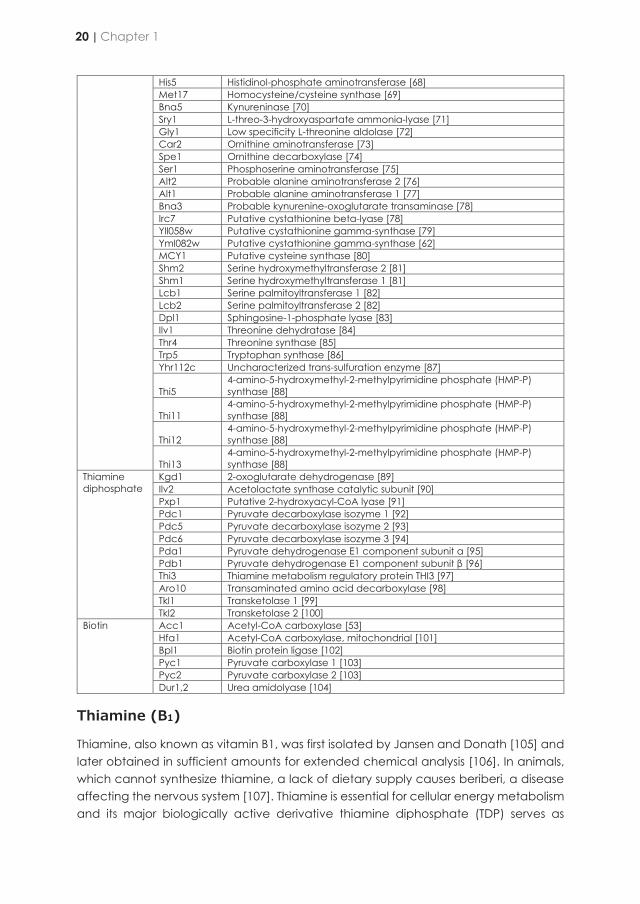
His5 Histidinol-phosphate aminotransferase [68] Met17 Homocysteine/cysteine synthase [69] Bna5 Kynureninase [70] Sry1 L-threo-3-hydroxyaspartate ammonia-lyase [71] Gly1 Low specificity L-threonine aldolase [72] Car2 Ornithine aminotransferase [73] Spe1 Ornithine decarboxylase [74] Ser1 Phosphoserine aminotransferase [75] Alt2 Probable alanine aminotransferase 2 [76] Alt1 Probable alanine aminotransferase 1 [77] Bna3 Probable kynurenine-oxoglutarate transaminase [78] Irc7 Putative cystathionine beta-lyase [78] Yll058w Putative cystathionine gamma-synthase [79] Yml082w Putative cystathionine gamma-synthase [62] MCY1 Putative cysteine synthase [80] Shm2 Serine hydroxymethyltransferase 2 [81] Shm1 Serine hydroxymethyltransferase 1 [81] Lcb1 Serine palmitoyltransferase 1 [82] Lcb2 Serine palmitoyltransferase 2 [82] Dpl1 Sphingosine-1-phosphate lyase [83] Ilv1 Threonine dehydratase [84] Thr4 Threonine synthase [85] Trp5 Tryptophan synthase [86] Yhr112c Uncharacterized trans-sulfuration enzyme [87]
Thi5 4-amino-5-hydroxymethyl-2-methylpyrimidine phosphate (HMP-P) synthase [88]
Thi11 4-amino-5-hydroxymethyl-2-methylpyrimidine phosphate (HMP-P) synthase [88]
Thi12 4-amino-5-hydroxymethyl-2-methylpyrimidine phosphate (HMP-P) synthase [88]
Thi13 4-amino-5-hydroxymethyl-2-methylpyrimidine phosphate (HMP-P) synthase [88]
Thiamine diphosphate
Kgd1 2-oxoglutarate dehydrogenase [89] Ilv2 Acetolactate synthase catalytic subunit [90] Pxp1 Putative 2-hydroxyacyl-CoA lyase [91] Pdc1 Pyruvate decarboxylase isozyme 1 [92] Pdc5 Pyruvate decarboxylase isozyme 2 [93] Pdc6 Pyruvate decarboxylase isozyme 3 [94] Pda1 Pyruvate dehydrogenase E1 component subunit α [95] Pdb1 Pyruvate dehydrogenase E1 component subunit β [96] Thi3 Thiamine metabolism regulatory protein THI3 [97] Aro10 Transaminated amino acid decarboxylase [98] Tkl1 Transketolase 1 [99] Tkl2 Transketolase 2 [100]
Biotin Acc1 Acetyl-CoA carboxylase [53] Hfa1 Acetyl-CoA carboxylase, mitochondrial [101] Bpl1 Biotin protein ligase [102] Pyc1 Pyruvate carboxylase 1 [103] Pyc2 Pyruvate carboxylase 2 [103] Dur1,2 Urea amidolyase [104]
Thiamine (B1)
Thiamine, also known as vitamin B1, was first isolated by Jansen and Donath [105] and later obtained in sufficient amounts for extended chemical analysis [106]. In animals, which cannot synthesize thiamine, a lack of dietary supply causes beriberi, a disease affecting the nervous system [107]. Thiamine is essential for cellular energy metabolism and its major biologically active derivative thiamine diphosphate (TDP) serves as
cofactor for a variety of enzymes, including pyruvate and oxoglutarate dehydrogenases, transketolases, 2-hydroxy-3-oxoadipate synthase, acetolactate synthase and 2-oxo acid decarboxylases (Table 1.2). As an electrophilic cofactor, TDP forms covalent intermediates with enzyme substrates. Thiamine can also perform intramolecular proton transfers, which is a rare function among cofactors [108]. It has been proposed that a general stress-protective role of thiamine in S. cerevisiae is partially unrelated to its role as a cofactor [109]. Thiamine is synthesized de novo by plants and many microorganisms including yeast species.
Thiamine consists of two substituted aromatic moieties, 4-amino-2-methyl-5-pyrimidyl (HMP) and 5-(2-hydroxylethyl)-4-methylthiazolium (HET), which are connected by a methylene bridge (Figure 1.1). In addition to free thiamine and the biologically active form thiamine diphosphate (TDP), thiamine monophosphate (TMP), and thiamine triphosphate (TTP) are also found intracellularly. All thiamine-prototrophic organisms synthesize TDP via condensation of the precursors 5-(2-hydroxylethyl)-4-methyl thiazole phosphate (HET-P) and 4-amino-2-methyl-5-pyrimidine diphosphate (HMP-PP) to TMP by TMP-diphosphorylase (Thi6 in S. cerevisiae) (Figure 1.1). While bacteria can synthesize TDP from TMP in a single reaction, catalysed by a TMP kinase, eukaryotes utilize a pathway in which TMP is first dephosphorylated to thiamine, which is then pyrophosphorylated to TDP by a thiamine pyrophosphokinase (Thi80 in S. cerevisiae) [110]. Two transporters involved in the acquisition of exogenous thiamine have been identified in S. cerevisiae: a high-affinity transporter encoded by THI10 [111] and a periplasmic acid phosphatase encoded by PHO3 that releases thiamine from thiamine phosphates [112].
In S. cerevisiae, the thiamine precursor HMP-PP is synthesized in two steps. First, 4-amino-2-methyl-5-pyrimidine phosphate (HMP-P) is formed from pyridoxal-5-phosphate and histidine. The histidine used for HMP-P synthesis is provided from the active site of HMP-P synthase in a suicide reaction [88, 113]. HMP-P synthase is encoded by four highly similar S. cerevisiae genes (THI5, THI11, THI12, and THI13). These genes are located in sub-telomeric regions of different chromosomes, suggesting that an increased copy number conferred a selective advantage in thiamine-poor environments [114]. In a second step, HMP-P is phosphorylated to HMP-PP by HMP-P kinase in an ATP-dependent reaction [115]. The S. cerevisiae genome harbours two paralogous genes encoding HMP-P kinase, THI20, and THI21, of which the former encodes the major isoform [114]. Thi20 is a trifunctional protein that displays thiamine biosynthesis and thiamine degradation activities in a single protein. Its N-terminal domain is active as HMP and HMP-P kinase, while its C-terminal domain has thiaminase II activity [116]. Although molecular oxygen is not directly required for HMP biosynthesis, activity of this branch of the thiamine biosynthetic pathway was shown to be oxygen dependent [114]. However, based on gene deletion studies it has been proposed that S. cerevisiae can still synthesize the pyrimidyl moiety under anaerobic conditions via an alternative, as yet unidentified, pathway [117].
20 | Chapter 1
His5 Histidinol-phosphate aminotransferase [68] Met17 Homocysteine/cysteine synthase [69] Bna5 Kynureninase [70] Sry1 L-threo-3-hydroxyaspartate ammonia-lyase [71] Gly1 Low specificity L-threonine aldolase [72] Car2 Ornithine aminotransferase [73] Spe1 Ornithine decarboxylase [74] Ser1 Phosphoserine aminotransferase [75] Alt2 Probable alanine aminotransferase 2 [76] Alt1 Probable alanine aminotransferase 1 [77] Bna3 Probable kynurenine-oxoglutarate transaminase [78] Irc7 Putative cystathionine beta-lyase [78] Yll058w Putative cystathionine gamma-synthase [79] Yml082w Putative cystathionine gamma-synthase [62] MCY1 Putative cysteine synthase [80] Shm2 Serine hydroxymethyltransferase 2 [81] Shm1 Serine hydroxymethyltransferase 1 [81] Lcb1 Serine palmitoyltransferase 1 [82] Lcb2 Serine palmitoyltransferase 2 [82] Dpl1 Sphingosine-1-phosphate lyase [83] Ilv1 Threonine dehydratase [84] Thr4 Threonine synthase [85] Trp5 Tryptophan synthase [86] Yhr112c Uncharacterized trans-sulfuration enzyme [87]
Thi5 4-amino-5-hydroxymethyl-2-methylpyrimidine phosphate (HMP-P) synthase [88]
Thi11 4-amino-5-hydroxymethyl-2-methylpyrimidine phosphate (HMP-P) synthase [88]
Thi12 4-amino-5-hydroxymethyl-2-methylpyrimidine phosphate (HMP-P) synthase [88]
Thi13 4-amino-5-hydroxymethyl-2-methylpyrimidine phosphate (HMP-P) synthase [88]
Thiamine diphosphate
Kgd1 2-oxoglutarate dehydrogenase [89] Ilv2 Acetolactate synthase catalytic subunit [90] Pxp1 Putative 2-hydroxyacyl-CoA lyase [91] Pdc1 Pyruvate decarboxylase isozyme 1 [92] Pdc5 Pyruvate decarboxylase isozyme 2 [93] Pdc6 Pyruvate decarboxylase isozyme 3 [94] Pda1 Pyruvate dehydrogenase E1 component subunit α [95] Pdb1 Pyruvate dehydrogenase E1 component subunit β [96] Thi3 Thiamine metabolism regulatory protein THI3 [97] Aro10 Transaminated amino acid decarboxylase [98] Tkl1 Transketolase 1 [99] Tkl2 Transketolase 2 [100]
Biotin Acc1 Acetyl-CoA carboxylase [53] Hfa1 Acetyl-CoA carboxylase, mitochondrial [101] Bpl1 Biotin protein ligase [102] Pyc1 Pyruvate carboxylase 1 [103] Pyc2 Pyruvate carboxylase 2 [103] Dur1,2 Urea amidolyase [104]
Thiamine (B1)
Thiamine, also known as vitamin B1, was first isolated by Jansen and Donath [105] and later obtained in sufficient amounts for extended chemical analysis [106]. In animals, which cannot synthesize thiamine, a lack of dietary supply causes beriberi, a disease affecting the nervous system [107]. Thiamine is essential for cellular energy metabolism and its major biologically active derivative thiamine diphosphate (TDP) serves as
cofactor for a variety of enzymes, including pyruvate and oxoglutarate dehydrogenases, transketolases, 2-hydroxy-3-oxoadipate synthase, acetolactate synthase and 2-oxo acid decarboxylases (Table 1.2). As an electrophilic cofactor, TDP forms covalent intermediates with enzyme substrates. Thiamine can also perform intramolecular proton transfers, which is a rare function among cofactors [108]. It has been proposed that a general stress-protective role of thiamine in S. cerevisiae is partially unrelated to its role as a cofactor [109]. Thiamine is synthesized de novo by plants and many microorganisms including yeast species.
Thiamine consists of two substituted aromatic moieties, 4-amino-2-methyl-5-pyrimidyl (HMP) and 5-(2-hydroxylethyl)-4-methylthiazolium (HET), which are connected by a methylene bridge (Figure 1.1). In addition to free thiamine and the biologically active form thiamine diphosphate (TDP), thiamine monophosphate (TMP), and thiamine triphosphate (TTP) are also found intracellularly. All thiamine-prototrophic organisms synthesize TDP via condensation of the precursors 5-(2-hydroxylethyl)-4-methyl thiazole phosphate (HET-P) and 4-amino-2-methyl-5-pyrimidine diphosphate (HMP-PP) to TMP by TMP-diphosphorylase (Thi6 in S. cerevisiae) (Figure 1.1). While bacteria can synthesize TDP from TMP in a single reaction, catalysed by a TMP kinase, eukaryotes utilize a pathway in which TMP is first dephosphorylated to thiamine, which is then pyrophosphorylated to TDP by a thiamine pyrophosphokinase (Thi80 in S. cerevisiae) [110]. Two transporters involved in the acquisition of exogenous thiamine have been identified in S. cerevisiae: a high-affinity transporter encoded by THI10 [111] and a periplasmic acid phosphatase encoded by PHO3 that releases thiamine from thiamine phosphates [112].
In S. cerevisiae, the thiamine precursor HMP-PP is synthesized in two steps. First, 4-amino-2-methyl-5-pyrimidine phosphate (HMP-P) is formed from pyridoxal-5-phosphate and histidine. The histidine used for HMP-P synthesis is provided from the active site of HMP-P synthase in a suicide reaction [88, 113]. HMP-P synthase is encoded by four highly similar S. cerevisiae genes (THI5, THI11, THI12, and THI13). These genes are located in sub-telomeric regions of different chromosomes, suggesting that an increased copy number conferred a selective advantage in thiamine-poor environments [114]. In a second step, HMP-P is phosphorylated to HMP-PP by HMP-P kinase in an ATP-dependent reaction [115]. The S. cerevisiae genome harbours two paralogous genes encoding HMP-P kinase, THI20, and THI21, of which the former encodes the major isoform [114]. Thi20 is a trifunctional protein that displays thiamine biosynthesis and thiamine degradation activities in a single protein. Its N-terminal domain is active as HMP and HMP-P kinase, while its C-terminal domain has thiaminase II activity [116]. Although molecular oxygen is not directly required for HMP biosynthesis, activity of this branch of the thiamine biosynthetic pathway was shown to be oxygen dependent [114]. However, based on gene deletion studies it has been proposed that S. cerevisiae can still synthesize the pyrimidyl moiety under anaerobic conditions via an alternative, as yet unidentified, pathway [117].
Vitamins that act as enzyme cofactors | 21
1

Figure 1.1 | Pyridoxal-5’-phosphate (PLP) and thiamine diphosphate (TDP) de novo synthesis pathway in S. cerevisiae. D-glyceraldehyde 3-phosphate, L-glutamine and keto-D-ribose 5-phosphate are converted to PLP by the catalytic activity of the gene products of SNO1,2,3 and SNZ1,2,3. Gcn4 acts as positive regulator of de novo PLP biosynthesis while Bas1 acts as an inhibitor. Gcn4 is inhibited by amino acids and activated under amino-acid starvation. Bas1 instead is upregulated in the presence of glycine. Pyridoxine (PN), pyridoxamine (PM) and pyridoxal (PL) are imported by Tpn1. PN is converted at the expense of ATP to pyridoxine-5’-phosphate (PNP) by Bud16 whereupon Pdx3, produces PLP and hydrogen peroxide in an oxygen-dependent reaction. Similarly, PLP can be formed starting from PM in two steps by action of Bud16 and Pdx3, with pyridoxamine-5’-phosphate (PMP) as intermediate. Moreover, PL can also be converted to PLP by action of Bud16. PLP is used as cofactor or converted to 4-amino-2-methyl-5-pyrimidine phosphate (HMP-P) by one of the four homologous enzymes Thi5, Thi11, Thi12 and Thi13, under consumption of L-histidine. HMP-P is the intermediate for the formation of the pyrimidyl moiety of thiamine (shown in cyan). Thi20 and Thi21 further phosphorylate HMP-P to 4-amino-2-methyl-5-pyrimidine diphosphate (HMP-PP). The thiazole moiety (shown in yellow) is synthesized by activity of Thi4 in a suicide mechanism, leading to 5-(2-hyroxylethyl)-4-methylthiazole phosphate (HET-P). HMP-PP and HET-P are merged by the gene product of THI6 to thiamine phosphate (TMP). The following reaction catalysed by an acid phosphatase (EC number 3.1.3.2) yields thiamine. Thiamine can be taken up with the aid of the transporter Thi10. Finally, thiamine is converted to its biologically active form thiamine diphosphate (TDP) under consumption of ATP by Thi80. Pdc2, Thi2 and Thi3 are responsible for the upregulation of transcription of Thi5/11/12/13, Thi20/21, Thi6 and Thi4. Alcohol and methyl substitutions on the pyridoxine pyrimide ring are shown in magenta and purple respectively. Metabolites, proteins and positive regulators are shown in bold, blue and green respectively.
For the synthesis of the thiazole moiety, eukaryotic cells use a single enzyme to form HET-P from glycine and NAD+, encoded by THI4 in S. cerevisiae [118]. Thi4 acts as a substrate in the reaction by providing the sulfur atom needed for thiazole formation in an iron-dependent sulfide transfer from a conserved cysteine. Therefore, similar to Thi5, Thi4 acts as a suicide enzyme undergoing only a single catalytic turnover [119-121]. Under thiamine-depleted conditions, Thi5 and Thi4 are among the most abundant proteins in S. cerevisiae [122]. Strains harbouring a THI4 deletion have an increased sensitivity to DNA damaging agents such as UV light and methyl methanesulfonate. The mechanism of this protection is not fully understood [114, 123].
Involvement of two suicide enzymes makes de novo thiamine biosynthesis in yeast an energetically very expensive process: for each molecule of thiamine produced, two complete proteins (Thi4 and Thi5/11/12/13) have to be synthesized and degraded. Tight regulation of thiamine synthesis occurs mainly at the transcriptional level [124, 125]. As a result, the THI regulon is repressed in the presence of high intracellular levels of TDP. A strain carrying a partially inactivated form of Thi80 was shown to constitutively express the THI genes, suggesting that TDP is the molecule acting in this negative feedback regulation loop [126]. Three positive regulators for thiamine biosynthesis have been identified to date: Thi2, Thi3, and Pdc2 [97, 127, 128] . Elimination of any of these three proteins abolishes THI genes expression. The expression of THI2 and THI3, but not PDC2, strongly increased under thiamine-depleted conditions[129]. Deletion of THI2 results in repression of all THI genes except for THI10, whereas deletion of THI3 causes repression of all THI genes. Thi3, which binds TDP, was originally proposed to also act as a 2-oxo acid decarboxylase involved in the Ehrlich for fusel alcohol biosynthesis [130] but this conclusion was later refuted [131-133]. A strain that only carried a thi3 allele encoding a protein unable to bind TDP showed constitutive expression of THI genes in thiamine-containing medium, suggesting that Thi3 acts as a TDP sensor. However, Thi3 lacks a clear DNA-binding motif and is likely to act through interaction with other proteins, such as Thi2 and Pdc2. Pdc2 is a transcriptional regulator that activates both THI genes and PDC genes encoding pyruvate decarboxylases [128, 134]. These regulatory proteins therefore provide an interesting link between the biosynthesis of pyruvate decarboxylase, the most highly expressed TDP-dependent enzyme in S. cerevisiae, and its cofactor. A regulatory link between the biosynthesis of thiamine and that of nicotinic acid, another member of the B-complex vitamins, was demonstrated when the NAD+-dependent histone deacetylase Hst1 was found to act as a repressor of basal THI-gene expression [135].
Biotin (B7)
During the first half of the 20th century, biotin was discovered as an essential growth factor for various organisms [136, 137]. Biotin plays an important role as co-enzyme in carboxylases involved in fatty acid synthesis, sugar and amino acid metabolism [138]. The cytosolic (Acc1) [139] and mitochondrial (Hfa1) acetyl-CoA carboxylases [140], pyruvate carboxylase (Pyc1,2) [141], urea carboxylase (Dur1,2) [142] and a tRNA-
22 | Chapter 1
Figure 1.1 | Pyridoxal-5’-phosphate (PLP) and thiamine diphosphate (TDP) de novo synthesis pathway in S. cerevisiae. D-glyceraldehyde 3-phosphate, L-glutamine and keto-D-ribose 5-phosphate are converted to PLP by the catalytic activity of the gene products of SNO1,2,3 and SNZ1,2,3. Gcn4 acts as positive regulator of de novo PLP biosynthesis while Bas1 acts as an inhibitor. Gcn4 is inhibited by amino acids and activated under amino-acid starvation. Bas1 instead is upregulated in the presence of glycine. Pyridoxine (PN), pyridoxamine (PM) and pyridoxal (PL) are imported by Tpn1. PN is converted at the expense of ATP to pyridoxine-5’-phosphate (PNP) by Bud16 whereupon Pdx3, produces PLP and hydrogen peroxide in an oxygen-dependent reaction. Similarly, PLP can be formed starting from PM in two steps by action of Bud16 and Pdx3, with pyridoxamine-5’-phosphate (PMP) as intermediate. Moreover, PL can also be converted to PLP by action of Bud16. PLP is used as cofactor or converted to 4-amino-2-methyl-5-pyrimidine phosphate (HMP-P) by one of the four homologous enzymes Thi5, Thi11, Thi12 and Thi13, under consumption of L-histidine. HMP-P is the intermediate for the formation of the pyrimidyl moiety of thiamine (shown in cyan). Thi20 and Thi21 further phosphorylate HMP-P to 4-amino-2-methyl-5-pyrimidine diphosphate (HMP-PP). The thiazole moiety (shown in yellow) is synthesized by activity of Thi4 in a suicide mechanism, leading to 5-(2-hyroxylethyl)-4-methylthiazole phosphate (HET-P). HMP-PP and HET-P are merged by the gene product of THI6 to thiamine phosphate (TMP). The following reaction catalysed by an acid phosphatase (EC number 3.1.3.2) yields thiamine. Thiamine can be taken up with the aid of the transporter Thi10. Finally, thiamine is converted to its biologically active form thiamine diphosphate (TDP) under consumption of ATP by Thi80. Pdc2, Thi2 and Thi3 are responsible for the upregulation of transcription of Thi5/11/12/13, Thi20/21, Thi6 and Thi4. Alcohol and methyl substitutions on the pyridoxine pyrimide ring are shown in magenta and purple respectively. Metabolites, proteins and positive regulators are shown in bold, blue and green respectively.
For the synthesis of the thiazole moiety, eukaryotic cells use a single enzyme to form HET-P from glycine and NAD+, encoded by THI4 in S. cerevisiae [118]. Thi4 acts as a substrate in the reaction by providing the sulfur atom needed for thiazole formation in an iron-dependent sulfide transfer from a conserved cysteine. Therefore, similar to Thi5, Thi4 acts as a suicide enzyme undergoing only a single catalytic turnover [119-121]. Under thiamine-depleted conditions, Thi5 and Thi4 are among the most abundant proteins in S. cerevisiae [122]. Strains harbouring a THI4 deletion have an increased sensitivity to DNA damaging agents such as UV light and methyl methanesulfonate. The mechanism of this protection is not fully understood [114, 123].
Involvement of two suicide enzymes makes de novo thiamine biosynthesis in yeast an energetically very expensive process: for each molecule of thiamine produced, two complete proteins (Thi4 and Thi5/11/12/13) have to be synthesized and degraded. Tight regulation of thiamine synthesis occurs mainly at the transcriptional level [124, 125]. As a result, the THI regulon is repressed in the presence of high intracellular levels of TDP. A strain carrying a partially inactivated form of Thi80 was shown to constitutively express the THI genes, suggesting that TDP is the molecule acting in this negative feedback regulation loop [126]. Three positive regulators for thiamine biosynthesis have been identified to date: Thi2, Thi3, and Pdc2 [97, 127, 128] . Elimination of any of these three proteins abolishes THI genes expression. The expression of THI2 and THI3, but not PDC2, strongly increased under thiamine-depleted conditions[129]. Deletion of THI2 results in repression of all THI genes except for THI10, whereas deletion of THI3 causes repression of all THI genes. Thi3, which binds TDP, was originally proposed to also act as a 2-oxo acid decarboxylase involved in the Ehrlich for fusel alcohol biosynthesis [130] but this conclusion was later refuted [131-133]. A strain that only carried a thi3 allele encoding a protein unable to bind TDP showed constitutive expression of THI genes in thiamine-containing medium, suggesting that Thi3 acts as a TDP sensor. However, Thi3 lacks a clear DNA-binding motif and is likely to act through interaction with other proteins, such as Thi2 and Pdc2. Pdc2 is a transcriptional regulator that activates both THI genes and PDC genes encoding pyruvate decarboxylases [128, 134]. These regulatory proteins therefore provide an interesting link between the biosynthesis of pyruvate decarboxylase, the most highly expressed TDP-dependent enzyme in S. cerevisiae, and its cofactor. A regulatory link between the biosynthesis of thiamine and that of nicotinic acid, another member of the B-complex vitamins, was demonstrated when the NAD+-dependent histone deacetylase Hst1 was found to act as a repressor of basal THI-gene expression [135].
Biotin (B7)
During the first half of the 20th century, biotin was discovered as an essential growth factor for various organisms [136, 137]. Biotin plays an important role as co-enzyme in carboxylases involved in fatty acid synthesis, sugar and amino acid metabolism [138]. The cytosolic (Acc1) [139] and mitochondrial (Hfa1) acetyl-CoA carboxylases [140], pyruvate carboxylase (Pyc1,2) [141], urea carboxylase (Dur1,2) [142] and a tRNA-
Vitamins that act as enzyme cofactors | 23
1
aminoacylation cofactor (Arc1) [143] are the only biotin-dependent enzymatic activities in S. cerevisiae (Table 1.2). Covalent linkage of the carboxyl group of biotin to an ε-lysine residue of apo-Acc1 and apo-Pyc1 and 2 is catalysed by the biotin protein ligase, Bpl1 [102, 144]. While not characterised, a similar mechanism is likely to occur for the mitochondrial acetyl-CoA carboxylase [101, 145]. Biotin can be taken up via the proton symporter Vht1 [146]. Alternatively, the biotin intermediates 8-amino-7-oxonanote (KAPA) and 7,8-diaminopelargonate (DAPA) can be transported into yeast via the Bio5 membrane protein [50].
The molecular structure of biotin is characterized by an imidazole, or ureido ring, fused with a sulfur-containing tetrahydrothiophene ring, substituted with a valeric acid chain (Figure 1.2). The reactions involved in the formation of the ring structures of biotin from KAPA are highly conserved among yeast and bacteria and require three steps starting with the conversion of KAPA to DAPA. This reaction is catalysed by Bio3, a DAPA aminotransferase that requires S-adenosyl-methionine (SAM) and pyridoxal 5’-phosphate (PLP) as cofactors. The following step, catalysed by the dethiobiotin synthetase Bio4, converts DAPA to dethiobiotin at the expense of ATP [50]. In the final step, the biotin synthase Bio2, a mitochondrial iron-sulfur-cluster protein, converts dethiobiotin to biotin by incorporating a sulfur atom [147], presumably acting as a suicide enzyme [148].
The pathway for synthesis of the valeric side chain of biotin remains elusive and probably involves Bio1 and Bio6, both of which are required for biotin-independent growth of S. cerevisiae [149]. Presence of Bio1 and Bio6 is strain dependent. For example, the reference strain S288C [150] lacks these two genes and is unable to grow on CDMY lacking biotin [151]. In contrast, sake strains of S. cerevisiae [152], S. cerevisiae strains isolated from cachaça fermentations [153], and the laboratory strains A364a [149] and CEN.PK113-7D [151, 154] do carry these two genes and exhibit growth, albeit very slowly, on CDMY without biotin. BIO6 has been proposed to have evolved from a duplication and neo-functionalization of BIO3, after BIO3 and BIO4 had been simultaneously acquired by horizontal gene transfer, with BIO1 similarly having evolved from duplication and neo-functionalization of the uncharacterized ORF YJR154W [149].
In view of its 55% amino-acid sequence similarity with Escherichia coli BioA, BIO6 probably encodes an adenosylmethionine-8-amino-7-oxononanoate transaminase [152]. The initial formation of the pimeloyl thioester in S. cerevisiae remains unclear. Hall and Dietrich [149] proposed that BIO1 encodes a coenzyme A (CoA) ligase that activates pimelic acid, a C7 dicarboxylic acid, to pimeloyl-CoA. Although such a CoA ligase (BioW) was identified in the gram-positive bacterium Bacillus subtilis [155], S. cerevisiae Bio1 protein does not show similarity to that enzyme. Additionally, biosynthesis of pimelic acid by S. cerevisiae has not been reported and pimelic acid feeding to a strain carrying the full biotin biosynthesis pathway was not able to stimulate growth on medium lacking biotin [156].
Figure 1.2 | Biotin de novo biosynthesis pathway in S. cerevisiae. Biotin is composed of an ureido and a tetrahydrothiophene ring (shown in cyan) fused to a valeric acid chain (shown in yellow). The five final steps of de novo biotin synthesis are carried out by Bio1, Bio6, Bio3, Bio4 and Bio2. Origin of pimelic acid remains elusive in S. cerevisiae (indicated by question mark(?)). Pimeloyl-CoA formed by Bio1 is converted via 8-amino-7-oxonanoate (KAPA) to 7,8-diaminopelargonate (DAPA) by Bio6 and Bio3. DAPA is subsequently converted by Bio4 to dethiobiotin and finally to biotin by Bio2. The intermediate KAPA and biotin can be imported via the membrane transporters Bio5 and Vht1, respectively. In the absence of biotin, the regulator Vhr1 upregulates expression of genes encoding the transporters Vht1 and Bio5 as well as Bio2. In iron and amino acid rich conditions the transcriptional regulator genes AFT1 and GCN4 are transcriptionally repressed, which under iron and amino acid scarce conditions would not activate transcription of BIO3, BIO4 and BIO2 and relieve BIO5 expression. Metabolites, proteins, positive and negative regulators are shown in bold, blue, green and red respectively.
24 | Chapter 1
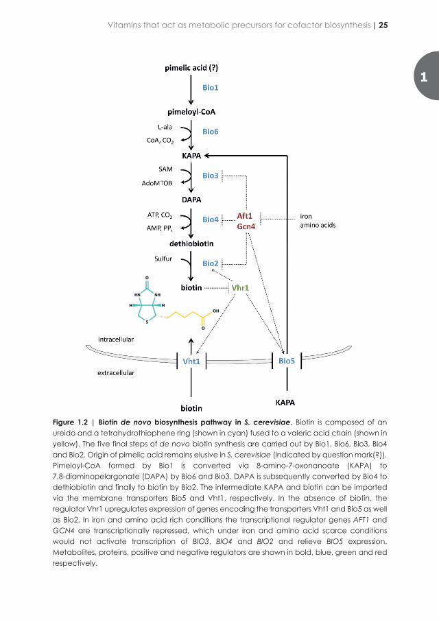
aminoacylation cofactor (Arc1) [143] are the only biotin-dependent enzymatic activities in S. cerevisiae (Table 1.2). Covalent linkage of the carboxyl group of biotin to an ε-lysine residue of apo-Acc1 and apo-Pyc1 and 2 is catalysed by the biotin protein ligase, Bpl1 [102, 144]. While not characterised, a similar mechanism is likely to occur for the mitochondrial acetyl-CoA carboxylase [101, 145]. Biotin can be taken up via the proton symporter Vht1 [146]. Alternatively, the biotin intermediates 8-amino-7-oxonanote (KAPA) and 7,8-diaminopelargonate (DAPA) can be transported into yeast via the Bio5 membrane protein [50].
The molecular structure of biotin is characterized by an imidazole, or ureido ring, fused with a sulfur-containing tetrahydrothiophene ring, substituted with a valeric acid chain (Figure 1.2). The reactions involved in the formation of the ring structures of biotin from KAPA are highly conserved among yeast and bacteria and require three steps starting with the conversion of KAPA to DAPA. This reaction is catalysed by Bio3, a DAPA aminotransferase that requires S-adenosyl-methionine (SAM) and pyridoxal 5’-phosphate (PLP) as cofactors. The following step, catalysed by the dethiobiotin synthetase Bio4, converts DAPA to dethiobiotin at the expense of ATP [50]. In the final step, the biotin synthase Bio2, a mitochondrial iron-sulfur-cluster protein, converts dethiobiotin to biotin by incorporating a sulfur atom [147], presumably acting as a suicide enzyme [148].
The pathway for synthesis of the valeric side chain of biotin remains elusive and probably involves Bio1 and Bio6, both of which are required for biotin-independent growth of S. cerevisiae [149]. Presence of Bio1 and Bio6 is strain dependent. For example, the reference strain S288C [150] lacks these two genes and is unable to grow on CDMY lacking biotin [151]. In contrast, sake strains of S. cerevisiae [152], S. cerevisiae strains isolated from cachaça fermentations [153], and the laboratory strains A364a [149] and CEN.PK113-7D [151, 154] do carry these two genes and exhibit growth, albeit very slowly, on CDMY without biotin. BIO6 has been proposed to have evolved from a duplication and neo-functionalization of BIO3, after BIO3 and BIO4 had been simultaneously acquired by horizontal gene transfer, with BIO1 similarly having evolved from duplication and neo-functionalization of the uncharacterized ORF YJR154W [149].
In view of its 55% amino-acid sequence similarity with Escherichia coli BioA, BIO6 probably encodes an adenosylmethionine-8-amino-7-oxononanoate transaminase [152]. The initial formation of the pimeloyl thioester in S. cerevisiae remains unclear. Hall and Dietrich [149] proposed that BIO1 encodes a coenzyme A (CoA) ligase that activates pimelic acid, a C7 dicarboxylic acid, to pimeloyl-CoA. Although such a CoA ligase (BioW) was identified in the gram-positive bacterium Bacillus subtilis [155], S. cerevisiae Bio1 protein does not show similarity to that enzyme. Additionally, biosynthesis of pimelic acid by S. cerevisiae has not been reported and pimelic acid feeding to a strain carrying the full biotin biosynthesis pathway was not able to stimulate growth on medium lacking biotin [156].
Figure 1.2 | Biotin de novo biosynthesis pathway in S. cerevisiae. Biotin is composed of an ureido and a tetrahydrothiophene ring (shown in cyan) fused to a valeric acid chain (shown in yellow). The five final steps of de novo biotin synthesis are carried out by Bio1, Bio6, Bio3, Bio4 and Bio2. Origin of pimelic acid remains elusive in S. cerevisiae (indicated by question mark(?)). Pimeloyl-CoA formed by Bio1 is converted via 8-amino-7-oxonanoate (KAPA) to 7,8-diaminopelargonate (DAPA) by Bio6 and Bio3. DAPA is subsequently converted by Bio4 to dethiobiotin and finally to biotin by Bio2. The intermediate KAPA and biotin can be imported via the membrane transporters Bio5 and Vht1, respectively. In the absence of biotin, the regulator Vhr1 upregulates expression of genes encoding the transporters Vht1 and Bio5 as well as Bio2. In iron and amino acid rich conditions the transcriptional regulator genes AFT1 and GCN4 are transcriptionally repressed, which under iron and amino acid scarce conditions would not activate transcription of BIO3, BIO4 and BIO2 and relieve BIO5 expression. Metabolites, proteins, positive and negative regulators are shown in bold, blue, green and red respectively.
Vitamins that act as metabolic precursors for cofactor biosynthesis | 25
1

Laboratory evolution studies highlighted the role of the enigmatic Bio1 protein in biotin prototrophy of S. cerevisiae. Prolonged cultivation of the laboratory strain CEN.PK113-7D in biotin-free accelerostats yielded an evolved strain that showed the same high specific growth rate (0.36 h-1) in the absence and presence of biotin. Whole-genome re-sequencing of evolved isolates revealed a massive 20 to 40-fold amplification of the physically linked BIO1 and BIO6 gene copies [154]. Overexpression of BIO1, but not BIO6, from a multi-copy plasmid sufficed to increase specific growth rates of the non-evolved strain on biotin-free CMDY without biotin from 0.01 h-1 to 0.15 h-1. Despite its unknown function, these results show that BIO1 is a key bottleneck of in de novo biotin synthesis in S. cerevisiae [154]. Strategies to generate biotin-prototrophic S. cerevisiae strains are likely to benefit from elucidation of the reaction catalysed by Bio1.
The biotin biosynthetic genes BIO5, BIO2, BIO4, BIO3, BIO6, VHT1 and BPL1 showed a concerted upregulation during biotin starvation [17, 152]. The promoter regions of BIO5, VHT1, BIO2 and BPL1 contains an upstream activating element that, in the absence of biotin, is bound by the transcription factor Vhr1 which upregulates transcription. This activation ensures expression of biotin and DAPA transporters, de novo biotin synthesis and enzyme biotinylation [157]. The transcriptional regulation of the biotin permease gene VHT1 is additionally controlled by the transcription factors Aft1 and Gcn4, which are involved in iron homeostasis and global control of nitrogen metabolism, respectively. Down-regulation of biotin biosynthesis genes and a parallel upregulation of biotin transport upon low availability of iron and/or nitrogen [158] has been interpreted as a strategy to decrease the metabolic burden of de novo biotin synthesis under these conditions [17].
Vitamins that act as metabolic precursors for cofactor biosynthesis
Pantothenic acid (B5)
Vitamin B5 was discovered by 1933 [159] and, based on its presence in all animal tissues, named pantothenate (παντοθεν, from everywhere). Pantothenate is not a co-factor, but a key precursor for synthesis of coenzyme A (CoA) and acyl carrier protein (ACP), which play key roles in metabolism. When supplied to media, pantothenate is imported into S. cerevisiae by the plasma-membrane pantothenate-proton symporter Fen2 [32]. Only plants and microorganisms, including fungi, can perform de novo pantothenate biosynthesis. However, most sake strains of S. cerevisiae strains are entirely auxotrophic for pantothenate when grown in media that exclusively contain organic nitrogen sources and, in some cases, also when an inorganic nitrogen source is provided [160]. Many S. cerevisiae strains can synthesize pantothenic acid. In such strains, removal of the molecule from the medium typically results in impaired growth on glucose, but not on glycerol or acetate [161].
Figure 1.3 | Pantothenate de novo synthesis pathway in S. cerevisiae and transcription profiles of pantothenate biosynthetic genes under different growth conditions. Pantothenate can be imported by the proton symporter Fen2 or synthesized de novo by condensation of pantoate (shown in cyan) and β-alanine (shown in yellow) in an ATP-dependent reaction catalysed by Pan6. Pantoate is formed in a two-step pathway from 2-keto-isovalerate catalysed by Ecm31 and Pan5 with 2-dehydropantoate as intermediate. β-alanine is formed starting from spermine by the enzymes Fms1 and Ald2-3 via 3-aminopropanal.
Pantothenate is formed by fusion of pantoate and β-alanine, in a reaction catalysed by pantoate-beta-alanine ligase (Pan6 in S. cerevisiae, Figure 1.3). In S. cerevisiae, β-alanine is produced from spermine in two steps [161]. The first step is catalysed by the polyamine oxidase Fms1, which produces 3-aminopropanal from spermine. 3-Aminopropanal is then oxidized to β-alanine by the cytosolic aldehyde dehydrogenases Ald2 and Ald3. The reaction catalysed by Fms1 has been reported to be rate limiting for pantothenate biosynthesis and Fms1 overexpression results in the secretion of pantothenic acid [162]. Pantoate is synthesized in S. cerevisiae from 2-keto-isovalerate, an intermediate of the valine biosynthesis. After conversion of 2-keto-isovalerate into 2-dehydropantoate [163] by keto-pantoate hydroxymethyltransferase (Ecm31), 2-dehydropantoate is transformed into pantoate by 2-dehydropantoate 2-reductase (Pan5) in an NADPH-dependent reduction [164].
26 | Chapter 1

Laboratory evolution studies highlighted the role of the enigmatic Bio1 protein in biotin prototrophy of S. cerevisiae. Prolonged cultivation of the laboratory strain CEN.PK113-7D in biotin-free accelerostats yielded an evolved strain that showed the same high specific growth rate (0.36 h-1) in the absence and presence of biotin. Whole-genome re-sequencing of evolved isolates revealed a massive 20 to 40-fold amplification of the physically linked BIO1 and BIO6 gene copies [154]. Overexpression of BIO1, but not BIO6, from a multi-copy plasmid sufficed to increase specific growth rates of the non-evolved strain on biotin-free CMDY without biotin from 0.01 h-1 to 0.15 h-1. Despite its unknown function, these results show that BIO1 is a key bottleneck of in de novo biotin synthesis in S. cerevisiae [154]. Strategies to generate biotin-prototrophic S. cerevisiae strains are likely to benefit from elucidation of the reaction catalysed by Bio1.
The biotin biosynthetic genes BIO5, BIO2, BIO4, BIO3, BIO6, VHT1 and BPL1 showed a concerted upregulation during biotin starvation [17, 152]. The promoter regions of BIO5, VHT1, BIO2 and BPL1 contains an upstream activating element that, in the absence of biotin, is bound by the transcription factor Vhr1 which upregulates transcription. This activation ensures expression of biotin and DAPA transporters, de novo biotin synthesis and enzyme biotinylation [157]. The transcriptional regulation of the biotin permease gene VHT1 is additionally controlled by the transcription factors Aft1 and Gcn4, which are involved in iron homeostasis and global control of nitrogen metabolism, respectively. Down-regulation of biotin biosynthesis genes and a parallel upregulation of biotin transport upon low availability of iron and/or nitrogen [158] has been interpreted as a strategy to decrease the metabolic burden of de novo biotin synthesis under these conditions [17].
Vitamins that act as metabolic precursors for cofactor biosynthesis
Pantothenic acid (B5)
Vitamin B5 was discovered by 1933 [159] and, based on its presence in all animal tissues, named pantothenate (παντοθεν, from everywhere). Pantothenate is not a co-factor, but a key precursor for synthesis of coenzyme A (CoA) and acyl carrier protein (ACP), which play key roles in metabolism. When supplied to media, pantothenate is imported into S. cerevisiae by the plasma-membrane pantothenate-proton symporter Fen2 [32]. Only plants and microorganisms, including fungi, can perform de novo pantothenate biosynthesis. However, most sake strains of S. cerevisiae strains are entirely auxotrophic for pantothenate when grown in media that exclusively contain organic nitrogen sources and, in some cases, also when an inorganic nitrogen source is provided [160]. Many S. cerevisiae strains can synthesize pantothenic acid. In such strains, removal of the molecule from the medium typically results in impaired growth on glucose, but not on glycerol or acetate [161].
Figure 1.3 | Pantothenate de novo synthesis pathway in S. cerevisiae and transcription profiles of pantothenate biosynthetic genes under different growth conditions. Pantothenate can be imported by the proton symporter Fen2 or synthesized de novo by condensation of pantoate (shown in cyan) and β-alanine (shown in yellow) in an ATP-dependent reaction catalysed by Pan6. Pantoate is formed in a two-step pathway from 2-keto-isovalerate catalysed by Ecm31 and Pan5 with 2-dehydropantoate as intermediate. β-alanine is formed starting from spermine by the enzymes Fms1 and Ald2-3 via 3-aminopropanal.
Pantothenate is formed by fusion of pantoate and β-alanine, in a reaction catalysed by pantoate-beta-alanine ligase (Pan6 in S. cerevisiae, Figure 1.3). In S. cerevisiae, β-alanine is produced from spermine in two steps [161]. The first step is catalysed by the polyamine oxidase Fms1, which produces 3-aminopropanal from spermine. 3-Aminopropanal is then oxidized to β-alanine by the cytosolic aldehyde dehydrogenases Ald2 and Ald3. The reaction catalysed by Fms1 has been reported to be rate limiting for pantothenate biosynthesis and Fms1 overexpression results in the secretion of pantothenic acid [162]. Pantoate is synthesized in S. cerevisiae from 2-keto-isovalerate, an intermediate of the valine biosynthesis. After conversion of 2-keto-isovalerate into 2-dehydropantoate [163] by keto-pantoate hydroxymethyltransferase (Ecm31), 2-dehydropantoate is transformed into pantoate by 2-dehydropantoate 2-reductase (Pan5) in an NADPH-dependent reduction [164].
Vitamins that act as metabolic precursors for cofactor biosynthesis | 27
1
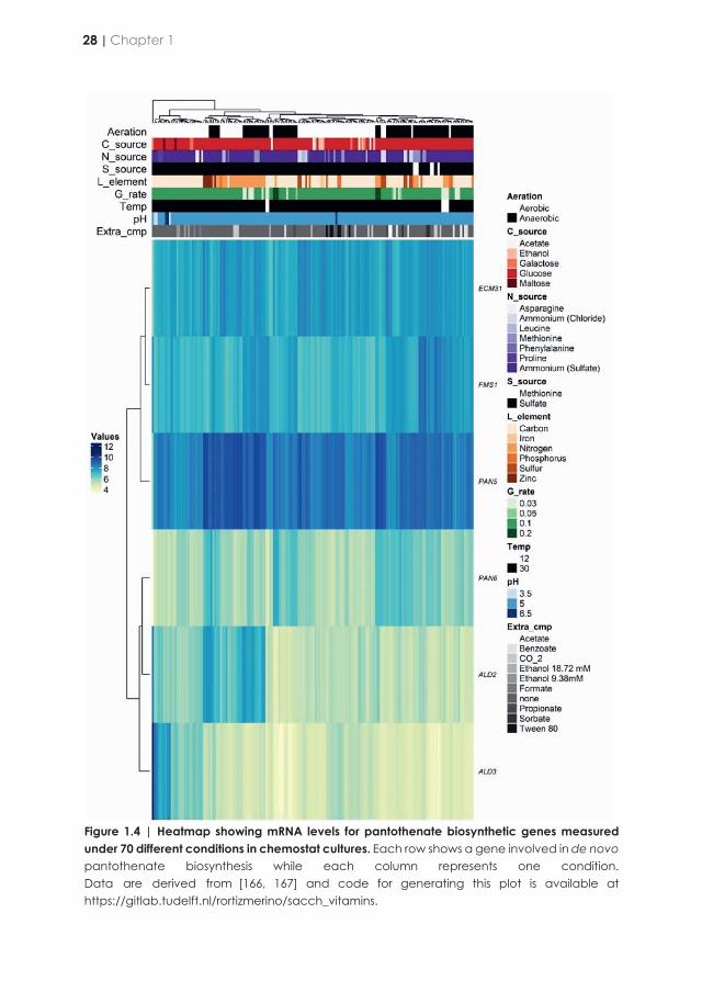
Figure 1.4 | Heatmap showing mRNA levels for pantothenate biosynthetic genes measured under 70 different conditions in chemostat cultures. Each row shows a gene involved in de novo pantothenate biosynthesis while each column represents one condition. Data are derived from [166, 167] and code for generating this plot is available at https://gitlab.tudelft.nl/rortizmerino/sacch_vitamins.
In comparison with the regulation of the other biosynthetic pathways discussed in this review, regulation of pantothenate acid biosynthesis in S. cerevisiae has not been intensively studied and, therefore, is still incompletely understood. Expression of ECM31 and PAN6 was shown to be low, constitutive and unaffected by extracellular pantothenate concentrations [165], while transcript levels of the pantothenic acid biosynthetic genes (ECM31, PAN5, FMS1, ALD2, ALD3 and PAN6) across 70 different culture conditions [166, 167] (Figure 1.4) did not reveal indications for co-regulation.
para-Aminobenzoic acid (B10)
Para-aminobenzoic acid (pABA), also known as vitamin B10, is a water-soluble B complex vitamin. It was discovered in 1920’s [168, 169]. A temporary sunscreen application after the WW2 was soon withdrawn as it caused dermatitis and auto-immune responses [170, 171]. pABA is an important intermediate in the biosynthesis of folates, a class of co-factors involved in transfer of C1-units in nucleic acid and amino-acid metabolism, as well as in ubiquinone biosynthesis [172, 173]. Folates result from the association of three precursors pABA (B10), GTP and glutamate, out of which pABA is the less abundant intracellularly and limit folic acid (B9) synthesis. Additionally, growth deficiency in the presence of pABA and absence of folic acid has not been reported before, making this vitamin dispensable for CDMY.
Figure 1.5 | para-Aminobenzoic acid (pABA) and myo-inositol de novo synthesis pathways in S. cerevisiae. (A) The genes ABZ1 and ABZ2 code for a two-step pathway producing pABA from chorismate via the intermediate 4-amino-4-deoxychorismate. Chorismate is synthesized from erythrose-4-phosphate (E4P) and phosphoenolpyruvate (PEP) via the shikimate pathway. In addition to being precursor for pABA biosynthesis, chorismate also serves as precursor for tryptophan, phenylalanine and tyrosine biosynthesis. (B) myo-Inositol is formed from glucose-6-phosphate via Ino1 yielding L-myo-inositol-1-phosphate, which is in a second step converted to myo-inositol by Inm1 or Inm2. INO2, INO4 genes encode INO1 transcriptional activators while OPI1 encodes the antagonist regulator of the gene encoding the initial step of inositol synthesis (INO1). Metabolites, proteins, positive regulators and positive regulators are shown in bold, blue, green and red respectively.
28 | Chapter 1

Figure 1.4 | Heatmap showing mRNA levels for pantothenate biosynthetic genes measured under 70 different conditions in chemostat cultures. Each row shows a gene involved in de novo pantothenate biosynthesis while each column represents one condition. Data are derived from [166, 167] and code for generating this plot is available at https://gitlab.tudelft.nl/rortizmerino/sacch_vitamins.
In comparison with the regulation of the other biosynthetic pathways discussed in this review, regulation of pantothenate acid biosynthesis in S. cerevisiae has not been intensively studied and, therefore, is still incompletely understood. Expression of ECM31 and PAN6 was shown to be low, constitutive and unaffected by extracellular pantothenate concentrations [165], while transcript levels of the pantothenic acid biosynthetic genes (ECM31, PAN5, FMS1, ALD2, ALD3 and PAN6) across 70 different culture conditions [166, 167] (Figure 1.4) did not reveal indications for co-regulation.
para-Aminobenzoic acid (B10)
Para-aminobenzoic acid (pABA), also known as vitamin B10, is a water-soluble B complex vitamin. It was discovered in 1920’s [168, 169]. A temporary sunscreen application after the WW2 was soon withdrawn as it caused dermatitis and auto-immune responses [170, 171]. pABA is an important intermediate in the biosynthesis of folates, a class of co-factors involved in transfer of C1-units in nucleic acid and amino-acid metabolism, as well as in ubiquinone biosynthesis [172, 173]. Folates result from the association of three precursors pABA (B10), GTP and glutamate, out of which pABA is the less abundant intracellularly and limit folic acid (B9) synthesis. Additionally, growth deficiency in the presence of pABA and absence of folic acid has not been reported before, making this vitamin dispensable for CDMY.
Figure 1.5 | para-Aminobenzoic acid (pABA) and myo-inositol de novo synthesis pathways in S. cerevisiae. (A) The genes ABZ1 and ABZ2 code for a two-step pathway producing pABA from chorismate via the intermediate 4-amino-4-deoxychorismate. Chorismate is synthesized from erythrose-4-phosphate (E4P) and phosphoenolpyruvate (PEP) via the shikimate pathway. In addition to being precursor for pABA biosynthesis, chorismate also serves as precursor for tryptophan, phenylalanine and tyrosine biosynthesis. (B) myo-Inositol is formed from glucose-6-phosphate via Ino1 yielding L-myo-inositol-1-phosphate, which is in a second step converted to myo-inositol by Inm1 or Inm2. INO2, INO4 genes encode INO1 transcriptional activators while OPI1 encodes the antagonist regulator of the gene encoding the initial step of inositol synthesis (INO1). Metabolites, proteins, positive regulators and positive regulators are shown in bold, blue, green and red respectively.
Vitamins that act as metabolic precursors for cofactor biosynthesis | 29
1
In S. cerevisiae, pABA biosynthesis starts from chorismate which, as indicated by its name (χωρίζω; to separate) is located at the intersection of the biosynthesis of i) tyrosine and phenylalanine, ii) tryptophan, and iii) pABA and folates. Conversion of chorismate in to pABA involves two enzyme reactions (Figure 1.5A). First, amino-deoxy-chorismate synthase (Abz1) uses glutamine as amino donor to produce 4-amino-4-deoxy-chorismate. Subsequently, amino-deoxy-chorismate lyase (Abz2) removes the pyruvate moiety of chorismate, resulting in pABA [174]. Chorismate is a key intermediate of the shikimate pathway for aromatic amino-acid biosynthesis. The shikimate pathway is tightly regulated, not only transcriptionally but also by allosteric feedback regulation of its first committed enzyme, 3-Deoxy-D-arabinoheptulosonate 7-phosphate (DAHP) synthase. S. cerevisiae contains two isoenzymes of DAHP, Aro3 and Aro4, which are feedback inhibited by phenylalanine and tyrosine, respectively [175, 176]. This regulation ensures that intracellular chorismate availability is strongly influenced by aromatic amino-acid concentrations. ABZ1 and ABZ2, which encode the key enzymes of the pABA pathway, are transcribed constitutively [177], suggesting that any regulation of pABA biosynthesis occurs is post-transcriptional. Rates of fermentation and nitrogen assimilation of S. cerevisiae strains have been correlated with specific alleles of ABZ1, thereby linking pABA synthesis to overall strain performance [177, 178]. This genetic heterogeneity has been exploited to engineer S. cerevisiae for pABA production by overexpressing ABZ1-2 alleles from wine strains that encode highly active enzymes [179].
Inositol (B8)
Of the seven organic supplements that are added to commonly used CDMY, only inositol (Table 1.1) is not a cofactor or cofactor precursor. First isolated in 1928 [180], inositol is a polyol (cyclohexane-1,2,3,4,5,6-hexol) that serves as precursor for phosphatidylinositol (PI), a main constituent of phospholipid membranes [181]. Upon its cleavage into inositol phosphate (IP) and diacylglycerol by phospholipase C, PI also plays a central role in inositol-phosphate signalling [182]. Moreover, inositol is a precursor for the synthesis of glycosylphosphatidylinositol (GPI) anchor proteins [183].
Myo-inositol is physiologically the most common stereoisomer among the eight possible inositol enantiomers. In organisms capable of synthesizing myo-inositol, it is formed from glucose-6-phosphate via two enzyme-catalysed reactions. The genes involved in the S. cerevisiae inositol biosynthesis pathway were discovered by complementation of inositol-requiring mutants [184] (Figure 1.5B). First, L-myo-inositol 1-phosphate (IP) is generated from glucose-6-phosphate by L-myo-inositol 1-phosphate synthase (Ino1) [185]. Subsequently, myo-inositol is generated by dephosphorylation of L-myo-inositol 1-phosphate by the heterodimeric enzyme inositol 3-phosphate monophosphatase (Inm1/Inm2) [186] Lipid metabolism in eukaryotic cells, including yeasts, is rigorously regulated. Yeast cells continuously monitor lipid status and quickly respond to alterations by a dual regulatory control. Many insights into how the yeast cells regulate their phospholipid metabolism derive from research on regulatory
responses to variations in the inositol content of growth media [187]. Analysis of inositol-auxotrophic S. cerevisiae strains enabled discovery of INO2 and INO4, which encode positive transcriptional regulators for INO1 and a large number of other genes involved in phospholipid synthesis [188]. A key negative feedback mechanism for transcriptional regulation was discovered by the characterization of mutants able to secrete inositol, a phenotype also referred to as the Opi- phenotype [189]. The transcriptional factor Opi1 was shown to act as a negative regulator in the presence of inositol, with some mutations in OPI1 resulting in constitutive INO1 expression. The Opi- phenotype has also been involved in identifying other S. cerevisiae genes in phospholipid biosynthesis, transcription, protein processing, and trafficking [190].
Nicotinic acid (B3)
Nicotinic acid, also known as niacin, was first isolated from liver in 1937 and was identified as “pellagra-preventing factor” and “anti-black tongue factor” [191]. Together with nicotinamide, it makes up the vitamin B3 complex. Nicotinic acid is an important precursor for the essential redox cofactors nicotinamide adenine dinucleotide (NAD+) and nicotinamide adenine dinucleotide phosphate (NADP+). S. cerevisiae can either obtain NAD+ from de novo biosynthesis or from salvage routes that regenerate NAD+ from its nicotinamide degradation products [192, 193] (Figure 1.6). These pathways converge at the level of nicotinic acid mononucleotide (NaMN) and share the last two reactions towards NAD+ formation.
In the de novo biosynthesis pathway, NaMN is synthesized from L-tryptophan in a series of six enzymatic reactions (catalysed by Bna1-2 and Bna4-7) and one spontaneous reaction (Figure 1.6). Three of the enzymes involved in the de novo biosynthesis pathway, indoleamine 2,3 dioxygenase (Bna2), kynurenine 3-monooxygenase (Bna4), and 3-hydroxyanthranilate 3,4-dioxygenase (Bna1), require molecular oxygen as a substrate, thereby explaining the strict requirement of anaerobic S. cerevisiae cultures for nicotinic acid supplementation [70]. In the salvage pathway, nicotinamide and nicotinic acid are converted to NaMN via the so-called Preiss-Handler pathway I [194, 195] which involves Pnc1 and Npti1 as key enzymes. Extracellular nicotinic acid can be imported into yeast cells by the plasma-membrane transporter Tna1 and then used to form NAD+ through the salvage pathway [196, 197].
In yeast, there are other four additional pathways for NAD+ biosynthesis: two salvage pathways from nicotinamide riboside (NR) and two salvage pathways from nicotinic acid riboside (NaR) [198-200]. Three of these salvage pathways converge first with the Preiss-Handler NAD+ salvage pathway and then with the de novo NAD pathway (Figure 1.6). In the NR salvage pathway I. which is not connected to the other pathways, NR is first phosphorylated to NMN by the Nrk1 kinase and then adenylated to NAD+ by Nma1 or Nma2 (Figure 1.6).
30 | Chapter 1
In S. cerevisiae, pABA biosynthesis starts from chorismate which, as indicated by its name (χωρίζω; to separate) is located at the intersection of the biosynthesis of i) tyrosine and phenylalanine, ii) tryptophan, and iii) pABA and folates. Conversion of chorismate in to pABA involves two enzyme reactions (Figure 1.5A). First, amino-deoxy-chorismate synthase (Abz1) uses glutamine as amino donor to produce 4-amino-4-deoxy-chorismate. Subsequently, amino-deoxy-chorismate lyase (Abz2) removes the pyruvate moiety of chorismate, resulting in pABA [174]. Chorismate is a key intermediate of the shikimate pathway for aromatic amino-acid biosynthesis. The shikimate pathway is tightly regulated, not only transcriptionally but also by allosteric feedback regulation of its first committed enzyme, 3-Deoxy-D-arabinoheptulosonate 7-phosphate (DAHP) synthase. S. cerevisiae contains two isoenzymes of DAHP, Aro3 and Aro4, which are feedback inhibited by phenylalanine and tyrosine, respectively [175, 176]. This regulation ensures that intracellular chorismate availability is strongly influenced by aromatic amino-acid concentrations. ABZ1 and ABZ2, which encode the key enzymes of the pABA pathway, are transcribed constitutively [177], suggesting that any regulation of pABA biosynthesis occurs is post-transcriptional. Rates of fermentation and nitrogen assimilation of S. cerevisiae strains have been correlated with specific alleles of ABZ1, thereby linking pABA synthesis to overall strain performance [177, 178]. This genetic heterogeneity has been exploited to engineer S. cerevisiae for pABA production by overexpressing ABZ1-2 alleles from wine strains that encode highly active enzymes [179].
Inositol (B8)
Of the seven organic supplements that are added to commonly used CDMY, only inositol (Table 1.1) is not a cofactor or cofactor precursor. First isolated in 1928 [180], inositol is a polyol (cyclohexane-1,2,3,4,5,6-hexol) that serves as precursor for phosphatidylinositol (PI), a main constituent of phospholipid membranes [181]. Upon its cleavage into inositol phosphate (IP) and diacylglycerol by phospholipase C, PI also plays a central role in inositol-phosphate signalling [182]. Moreover, inositol is a precursor for the synthesis of glycosylphosphatidylinositol (GPI) anchor proteins [183].
Myo-inositol is physiologically the most common stereoisomer among the eight possible inositol enantiomers. In organisms capable of synthesizing myo-inositol, it is formed from glucose-6-phosphate via two enzyme-catalysed reactions. The genes involved in the S. cerevisiae inositol biosynthesis pathway were discovered by complementation of inositol-requiring mutants [184] (Figure 1.5B). First, L-myo-inositol 1-phosphate (IP) is generated from glucose-6-phosphate by L-myo-inositol 1-phosphate synthase (Ino1) [185]. Subsequently, myo-inositol is generated by dephosphorylation of L-myo-inositol 1-phosphate by the heterodimeric enzyme inositol 3-phosphate monophosphatase (Inm1/Inm2) [186] Lipid metabolism in eukaryotic cells, including yeasts, is rigorously regulated. Yeast cells continuously monitor lipid status and quickly respond to alterations by a dual regulatory control. Many insights into how the yeast cells regulate their phospholipid metabolism derive from research on regulatory
responses to variations in the inositol content of growth media [187]. Analysis of inositol-auxotrophic S. cerevisiae strains enabled discovery of INO2 and INO4, which encode positive transcriptional regulators for INO1 and a large number of other genes involved in phospholipid synthesis [188]. A key negative feedback mechanism for transcriptional regulation was discovered by the characterization of mutants able to secrete inositol, a phenotype also referred to as the Opi- phenotype [189]. The transcriptional factor Opi1 was shown to act as a negative regulator in the presence of inositol, with some mutations in OPI1 resulting in constitutive INO1 expression. The Opi- phenotype has also been involved in identifying other S. cerevisiae genes in phospholipid biosynthesis, transcription, protein processing, and trafficking [190].
Nicotinic acid (B3)
Nicotinic acid, also known as niacin, was first isolated from liver in 1937 and was identified as “pellagra-preventing factor” and “anti-black tongue factor” [191]. Together with nicotinamide, it makes up the vitamin B3 complex. Nicotinic acid is an important precursor for the essential redox cofactors nicotinamide adenine dinucleotide (NAD+) and nicotinamide adenine dinucleotide phosphate (NADP+). S. cerevisiae can either obtain NAD+ from de novo biosynthesis or from salvage routes that regenerate NAD+ from its nicotinamide degradation products [192, 193] (Figure 1.6). These pathways converge at the level of nicotinic acid mononucleotide (NaMN) and share the last two reactions towards NAD+ formation.
In the de novo biosynthesis pathway, NaMN is synthesized from L-tryptophan in a series of six enzymatic reactions (catalysed by Bna1-2 and Bna4-7) and one spontaneous reaction (Figure 1.6). Three of the enzymes involved in the de novo biosynthesis pathway, indoleamine 2,3 dioxygenase (Bna2), kynurenine 3-monooxygenase (Bna4), and 3-hydroxyanthranilate 3,4-dioxygenase (Bna1), require molecular oxygen as a substrate, thereby explaining the strict requirement of anaerobic S. cerevisiae cultures for nicotinic acid supplementation [70]. In the salvage pathway, nicotinamide and nicotinic acid are converted to NaMN via the so-called Preiss-Handler pathway I [194, 195] which involves Pnc1 and Npti1 as key enzymes. Extracellular nicotinic acid can be imported into yeast cells by the plasma-membrane transporter Tna1 and then used to form NAD+ through the salvage pathway [196, 197].
In yeast, there are other four additional pathways for NAD+ biosynthesis: two salvage pathways from nicotinamide riboside (NR) and two salvage pathways from nicotinic acid riboside (NaR) [198-200]. Three of these salvage pathways converge first with the Preiss-Handler NAD+ salvage pathway and then with the de novo NAD pathway (Figure 1.6). In the NR salvage pathway I. which is not connected to the other pathways, NR is first phosphorylated to NMN by the Nrk1 kinase and then adenylated to NAD+ by Nma1 or Nma2 (Figure 1.6).
Vitamins that act as metabolic precursors for cofactor biosynthesis | 31
1
NAD+ and NADP+ are essential redox cofactors for many oxido-reductases [201]. In addition to its role as a redox co-factor, NAD+ is a substrate for several enzymes in yeast including sirtuin protein deacetylases (Sir2, Hst1-4) and cyclic ADP-ribose (cADPR) synthases (Tpt1) [202, 203]. These enzymes have important roles in the maintenance and regulation of chromatin structure, calcium signalling, life-span and DNA repair [193, 204-207]. NAD+ is also a precursor for NADP+ which, like NAD+ is involved in many cellular redox reactions [208]. Intracellular NAD+ levels are controlled by a complex regulation network. Hst1 (Homologue of Sir2) acts as a NAD+ sensor that represses BNA genes when NAD+ is abundant. Hst1 does not bind the DNA directly but interacts with Rfm1 and Sum1 to form a repressor complex.
Mac1, which was previously characterized as a copper-sensing transcription factor, has been shown to also be involved in regulation of BNA genes, together with Hst1 [192, 209-211]. When NA is abundantly available, NA salvage metabolism is preferred over use of the de novo biosynthetic pathway, which is repressed by Hst1 [192, 211]. In S. cerevisiae, NAD+ metabolism is regulated together with phosphate and purine nucleotide metabolism, although the exact mechanisms remain uncharacterised [212, 213]. NR can be produced and stored in vacuoles and then released into the cytosol by the Fun26 transporter, thereby enabling cells to feed NR stores into NAD+ synthesis [214, 215].
Figure 1.6 | Nicotinic acid de novo synthesis and salvage pathway in S. cerevisiae. Nicotinamide adenine dinucleotide (NAD+) is de novo synthesized from L-tryptophan in nine catalytic steps involving the Bna enzyme family and enzymes Nma1, Nma2 and Qns1. Nicotinic acid can be imported into the cell via Tna1 and enters the NAD synthesis pathway as nicotinic acid mononucleotide (NaMN) by catalytic activity of Npt1. Similarly, nicotinamide riboside (NaR) can be salvaged by catalytic activity of Nrk1 to form NaMN. NaR can be also converted to nicotinic acid by Urh1 and Pnp1. Nrk1 also converts nicotinamide riboside (NR) into nicotinamide nucleotide (NMN) subsequently converted to NAD+ by Nma1 and Nma2. NR is imported by activity of Nrt1 transporter and might be used by Pnp1 or Urh1 to form nicotinamide. Alternatively, nicotinamide can be synthesized via Sir2 from NAD+. Pnc1 uses nicotinamide to form nicotinic acid. The regulators Hst1 (with aid of Rfm1 and Sum1) and Mac1 repress the expression of genes encoding Bna enzymes upon binding to NAD+ and nicotinic acid. Metabolites, proteins and negative regulators are shown in bold, blue and red respectively.
32 | Chapter 1

NAD+ and NADP+ are essential redox cofactors for many oxido-reductases [201]. In addition to its role as a redox co-factor, NAD+ is a substrate for several enzymes in yeast including sirtuin protein deacetylases (Sir2, Hst1-4) and cyclic ADP-ribose (cADPR) synthases (Tpt1) [202, 203]. These enzymes have important roles in the maintenance and regulation of chromatin structure, calcium signalling, life-span and DNA repair [193, 204-207]. NAD+ is also a precursor for NADP+ which, like NAD+ is involved in many cellular redox reactions [208]. Intracellular NAD+ levels are controlled by a complex regulation network. Hst1 (Homologue of Sir2) acts as a NAD+ sensor that represses BNA genes when NAD+ is abundant. Hst1 does not bind the DNA directly but interacts with Rfm1 and Sum1 to form a repressor complex.
Mac1, which was previously characterized as a copper-sensing transcription factor, has been shown to also be involved in regulation of BNA genes, together with Hst1 [192, 209-211]. When NA is abundantly available, NA salvage metabolism is preferred over use of the de novo biosynthetic pathway, which is repressed by Hst1 [192, 211]. In S. cerevisiae, NAD+ metabolism is regulated together with phosphate and purine nucleotide metabolism, although the exact mechanisms remain uncharacterised [212, 213]. NR can be produced and stored in vacuoles and then released into the cytosol by the Fun26 transporter, thereby enabling cells to feed NR stores into NAD+ synthesis [214, 215].
Figure 1.6 | Nicotinic acid de novo synthesis and salvage pathway in S. cerevisiae. Nicotinamide adenine dinucleotide (NAD+) is de novo synthesized from L-tryptophan in nine catalytic steps involving the Bna enzyme family and enzymes Nma1, Nma2 and Qns1. Nicotinic acid can be imported into the cell via Tna1 and enters the NAD synthesis pathway as nicotinic acid mononucleotide (NaMN) by catalytic activity of Npt1. Similarly, nicotinamide riboside (NaR) can be salvaged by catalytic activity of Nrk1 to form NaMN. NaR can be also converted to nicotinic acid by Urh1 and Pnp1. Nrk1 also converts nicotinamide riboside (NR) into nicotinamide nucleotide (NMN) subsequently converted to NAD+ by Nma1 and Nma2. NR is imported by activity of Nrt1 transporter and might be used by Pnp1 or Urh1 to form nicotinamide. Alternatively, nicotinamide can be synthesized via Sir2 from NAD+. Pnc1 uses nicotinamide to form nicotinic acid. The regulators Hst1 (with aid of Rfm1 and Sum1) and Mac1 repress the expression of genes encoding Bna enzymes upon binding to NAD+ and nicotinic acid. Metabolites, proteins and negative regulators are shown in bold, blue and red respectively.
Vitamins that act as metabolic precursors for cofactor biosynthesis | 33
1

Systematic search for components of the class B vitamin biosynthesis pathways in Saccharomyces species
Although strain-to-strain differences occur, the S. cerevisiae pan-genome harbour all necessary genetic information to synthesize inositol, biotin, thiamine, nicotinic acid, pantothenate, pyridoxine and pABA. Since the work of Burkholder, McVeigh and Moyer in 1944, no systematic analysis has been performed to assess growth factor requirements of different species within the Saccharomyces genus. To explore this issue, we screened the genomes of the type strains of Saccharomyces species for annotated sequences homologous to the structural genes encoding enzymes involved in biosynthesis of class B vitamins in S. cerevisiae [216]. Based on this screen, the genomes of most Saccharomyces type species encode complete biosynthetic pathways for these compounds (Figure 1.7). Two notable exceptions are S. arboricola, which misses key genes required for biosynthesis of pyridoxine, thiamine, and biotin (SNO2/3, SNZ2/3, THI5-13, BIO1) and S. kudriavzevii which lacks genes involved in biosynthesis of pyridoxine, pantothenate, pABA and inositol (SNO1, FMS1, PAN6, ABZ1/2, INO1). Absence of SNO2/3 in S. paradoxus should not compromise its pyridoxine prototrophy as its genome does harbour the main paralog SNO1.
Some Saccharomyces species show higher copy numbers for individual vitamin biosynthesis genes than S. cerevisiae. In particular, S. jurei harbours additional copies of SNO2/3, SNZ2/3, THI5 and THI11-13, while S. paradoxus carries two copies of BIO1 and BIO6. These genes are all located in subtelomeric regions in S. cerevisiae. Subtelomeric regions are known hotspots for genetic plasticity that contain many gene families involved in interaction between the cell and its environment [217]. Assuming conserved synteny within the Saccharomyces genus, these gene amplifications may therefore reflect evolutionary adaptations to the environmental conditions these different species were exposed to.
Table 1.3 | Saccharomyces species involved in a comparative genome analysis of the presence of vitamin biosynthesis genes.
TaxID Species Strain Other Identifiers Reference Biosample 1080349 S. eubayanus CBS12357T NBRC111513T [219] NA 226127 S. uvarum CBS7001T MCYC623T [220] SAMN13069661 1160507 S. arboricola CBS10644T H-6 T [221] SAMN13069660 226230 S. kudriavzevii CBS8840T IFO1802T [220] NA 226126 S. mikatae CBS8839T IFO1815T [220] SAMN13069662 1987369 S. jurei CBS14759T NCYC3947T [222] SAMN13069663 226125 S. paradoxus CBS432T NRRLY-17217T [223] SAMN13069659 559292 S. cerevisiae S288C CBS8803 [150] NA 889517 S. cerevisiae CEN.PK113-7D CBS8340 [224] SAMN13069664
Biosamples can be accessed under bioproject accession PRJNA578688 (https://www.ncbi.nlm.nih.gov/bioproject)
Figure 1.7 | Occurrence of vitamin biosynthesis genus in Saccharomyces species. A homology search was conducted using HMMER v3 [218] with S. cerevisiae S288C proteins as queries (left side row names) against a database of annotated proteins from the Saccharomyces species listed in the column headers. For Bio1 and Bio6, S. cerevisiae K7 proteins were used as queries (indicated with *) since S288C is known to lack such proteins. Available genome annotations from species in the monophyletic Saccharomyces clade (formerly known as sensu stricto; Table 1.3) were used to build a protein sequence database. Besides S. cerevisiae S288C and CEN.PK113-7D, sequences in the database belong to type strains. This database was then searched for sequence homologs using the queries listed on the left-hand side. Queries are grouped and labelled on the right-hand side and depending on the biosynthetic pathway they are involved in. Boxes are coloured depending on the number of hits (e-value > 1E-5, percentage of alignment > 75%) obtained by each query on each strain. The colour code is shown at the bottom. Hits from queries belonging to the same biosynthetic pathway were ranked according to lowest e-value, then highest percentage of alignment and best hits were uniquely assigned to each query (i.e. a sequence considered as best hit is never used more than once and best hits with a count > 1 are all identical). This last step accounts for the presence of paralogs and the high level of similarity between proteins in the same pathway, especially in the pyridoxine and thiamine pathways (see Thi5 and Thi20 for instance). Code for this search is available in https://gitlab.tudelft.nl/rortizmerino/sacch_vitamins and sequences are deposited under BioProject accession PRJNA578688 as indicated in Table 1.3.
34 | Chapter 1

Systematic search for components of the class B vitamin biosynthesis pathways in Saccharomyces species
Although strain-to-strain differences occur, the S. cerevisiae pan-genome harbour all necessary genetic information to synthesize inositol, biotin, thiamine, nicotinic acid, pantothenate, pyridoxine and pABA. Since the work of Burkholder, McVeigh and Moyer in 1944, no systematic analysis has been performed to assess growth factor requirements of different species within the Saccharomyces genus. To explore this issue, we screened the genomes of the type strains of Saccharomyces species for annotated sequences homologous to the structural genes encoding enzymes involved in biosynthesis of class B vitamins in S. cerevisiae [216]. Based on this screen, the genomes of most Saccharomyces type species encode complete biosynthetic pathways for these compounds (Figure 1.7). Two notable exceptions are S. arboricola, which misses key genes required for biosynthesis of pyridoxine, thiamine, and biotin (SNO2/3, SNZ2/3, THI5-13, BIO1) and S. kudriavzevii which lacks genes involved in biosynthesis of pyridoxine, pantothenate, pABA and inositol (SNO1, FMS1, PAN6, ABZ1/2, INO1). Absence of SNO2/3 in S. paradoxus should not compromise its pyridoxine prototrophy as its genome does harbour the main paralog SNO1.
Some Saccharomyces species show higher copy numbers for individual vitamin biosynthesis genes than S. cerevisiae. In particular, S. jurei harbours additional copies of SNO2/3, SNZ2/3, THI5 and THI11-13, while S. paradoxus carries two copies of BIO1 and BIO6. These genes are all located in subtelomeric regions in S. cerevisiae. Subtelomeric regions are known hotspots for genetic plasticity that contain many gene families involved in interaction between the cell and its environment [217]. Assuming conserved synteny within the Saccharomyces genus, these gene amplifications may therefore reflect evolutionary adaptations to the environmental conditions these different species were exposed to.
Table 1.3 | Saccharomyces species involved in a comparative genome analysis of the presence of vitamin biosynthesis genes.
TaxID Species Strain Other Identifiers Reference Biosample 1080349 S. eubayanus CBS12357T NBRC111513T [219] NA 226127 S. uvarum CBS7001T MCYC623T [220] SAMN13069661 1160507 S. arboricola CBS10644T H-6 T [221] SAMN13069660 226230 S. kudriavzevii CBS8840T IFO1802T [220] NA 226126 S. mikatae CBS8839T IFO1815T [220] SAMN13069662 1987369 S. jurei CBS14759T NCYC3947T [222] SAMN13069663 226125 S. paradoxus CBS432T NRRLY-17217T [223] SAMN13069659 559292 S. cerevisiae S288C CBS8803 [150] NA 889517 S. cerevisiae CEN.PK113-7D CBS8340 [224] SAMN13069664
Biosamples can be accessed under bioproject accession PRJNA578688 (https://www.ncbi.nlm.nih.gov/bioproject)
Figure 1.7 | Occurrence of vitamin biosynthesis genus in Saccharomyces species. A homology search was conducted using HMMER v3 [218] with S. cerevisiae S288C proteins as queries (left side row names) against a database of annotated proteins from the Saccharomyces species listed in the column headers. For Bio1 and Bio6, S. cerevisiae K7 proteins were used as queries (indicated with *) since S288C is known to lack such proteins. Available genome annotations from species in the monophyletic Saccharomyces clade (formerly known as sensu stricto; Table 1.3) were used to build a protein sequence database. Besides S. cerevisiae S288C and CEN.PK113-7D, sequences in the database belong to type strains. This database was then searched for sequence homologs using the queries listed on the left-hand side. Queries are grouped and labelled on the right-hand side and depending on the biosynthetic pathway they are involved in. Boxes are coloured depending on the number of hits (e-value > 1E-5, percentage of alignment > 75%) obtained by each query on each strain. The colour code is shown at the bottom. Hits from queries belonging to the same biosynthetic pathway were ranked according to lowest e-value, then highest percentage of alignment and best hits were uniquely assigned to each query (i.e. a sequence considered as best hit is never used more than once and best hits with a count > 1 are all identical). This last step accounts for the presence of paralogs and the high level of similarity between proteins in the same pathway, especially in the pyridoxine and thiamine pathways (see Thi5 and Thi20 for instance). Code for this search is available in https://gitlab.tudelft.nl/rortizmerino/sacch_vitamins and sequences are deposited under BioProject accession PRJNA578688 as indicated in Table 1.3.
Systematic search for components of the class B vitamin biosynthesis pathways in Saccharomyces species | 35
1
Scope and outline of this thesis
Although strain-to-strain differences do occur, the genomes of most S. cerevisiae strains harbour all genetic information required for synthesis of inositol, biotin, thiamine, nicotinic acid, pantothenate, pyridoxine and pABA. However, since endogenous synthesis does not always meet the demands for fast growth and high product titres, yeast cultivation in research and industry often involves supplementation of these ‘vitamins’ to growth media. The goals of the research described in this thesis were to gain a deeper understanding of the biochemistry and genetics of vitamin metabolism in S. cerevisiae and, based on this understanding, to design and test metabolic engineering strategies for construction of fully vitamin-independent yeast strains.
Biotin is one of the most expensive among B-type vitamins that are routinely added to synthetic media for cultivation of yeasts and the pathway for its de novo synthesis by yeasts remains incompletely understood. In Chapter 2, yeast biodiversity was explored in an attempt to address a bottleneck in biotin synthesis by S. cerevisiae that had been identified in earlier laboratory evolution experiments. Based on expression of different orthologs of S. cerevisiae Bio1 in a bio1Δ genetic background, a new and straightforward metabolic engineering strategy for conferring full biotin prototrophy was designed and tested in laboratory and industrial S. cerevisiae strains. In addition to this application-inspired goal, the research described in Chapter 3 also provided a new insight into the role of oxygen in biotin synthesis by yeast.
While Chapter 2 outlines a strategy for conferring aerobic biotin prototrophy to laboratory and industrial S. cerevisiae strains, it did not offer a perspective for eliminating a complete biotin auxotrophy of anaerobic cultures of this yeast. Therefore, Chapter 3 explored a metabolic engineering strategy in which an entire bacterial pathway, involving ten different bacterial genes, for conversion of malonyl-CoA into the biotin-biosynthesis intermediate KAPA, was introduced into S. cerevisiae. In addition to CRISPR-based genome editing and in vivo assembly of the expression cassettes required for expression of the heterologous pathway, laboratory evolution and reverse engineering of causal mutations were applied to identify and reverse causal mutations for fast anaerobic, biotin-independent growth.
Chapter 4 addresses the ambitious goal to construct an S. cerevisiae platform that is fully prototrophic for all B-type vitamins that are routinely added to synthetic media for aerobic cultivation of this yeast. To this end, the strategy for eliminating the biotin requirement of aerobic S. cerevisiae culture was combined with similar strategies focused on other vitamins, which were inspired by a separate study [225] performed in the context of the same European Union-funded project (PAcMEN) in which the present PhD project was embedded.
36 | Chapter 1

Scope and outline of this thesis
Although strain-to-strain differences do occur, the genomes of most S. cerevisiae strains harbour all genetic information required for synthesis of inositol, biotin, thiamine, nicotinic acid, pantothenate, pyridoxine and pABA. However, since endogenous synthesis does not always meet the demands for fast growth and high product titres, yeast cultivation in research and industry often involves supplementation of these ‘vitamins’ to growth media. The goals of the research described in this thesis were to gain a deeper understanding of the biochemistry and genetics of vitamin metabolism in S. cerevisiae and, based on this understanding, to design and test metabolic engineering strategies for construction of fully vitamin-independent yeast strains.
Biotin is one of the most expensive among B-type vitamins that are routinely added to synthetic media for cultivation of yeasts and the pathway for its de novo synthesis by yeasts remains incompletely understood. In Chapter 2, yeast biodiversity was explored in an attempt to address a bottleneck in biotin synthesis by S. cerevisiae that had been identified in earlier laboratory evolution experiments. Based on expression of different orthologs of S. cerevisiae Bio1 in a bio1Δ genetic background, a new and straightforward metabolic engineering strategy for conferring full biotin prototrophy was designed and tested in laboratory and industrial S. cerevisiae strains. In addition to this application-inspired goal, the research described in Chapter 3 also provided a new insight into the role of oxygen in biotin synthesis by yeast.
While Chapter 2 outlines a strategy for conferring aerobic biotin prototrophy to laboratory and industrial S. cerevisiae strains, it did not offer a perspective for eliminating a complete biotin auxotrophy of anaerobic cultures of this yeast. Therefore, Chapter 3 explored a metabolic engineering strategy in which an entire bacterial pathway, involving ten different bacterial genes, for conversion of malonyl-CoA into the biotin-biosynthesis intermediate KAPA, was introduced into S. cerevisiae. In addition to CRISPR-based genome editing and in vivo assembly of the expression cassettes required for expression of the heterologous pathway, laboratory evolution and reverse engineering of causal mutations were applied to identify and reverse causal mutations for fast anaerobic, biotin-independent growth.
Chapter 4 addresses the ambitious goal to construct an S. cerevisiae platform that is fully prototrophic for all B-type vitamins that are routinely added to synthetic media for aerobic cultivation of this yeast. To this end, the strategy for eliminating the biotin requirement of aerobic S. cerevisiae culture was combined with similar strategies focused on other vitamins, which were inspired by a separate study [225] performed in the context of the same European Union-funded project (PAcMEN) in which the present PhD project was embedded.
Scope and outline of this thesis | 37
1

Chapter 2 |
Exploiting the diversity of Saccharomycotina yeasts to engineer
biotin-independent growth of Saccharomyces cerevisiae
Anna K. Wronska, Meinske P. Haak, Ellen Geraats, Eva Bruins Slot, Marcel van den Broek, Jack T. Pronk and Jean-Marc Daran
Essentially as published in Applied and Environmental Microbiology
2020; 86 (12) e00270-20 DOI: 10.1128/AEM.00270-20
Abstract
Biotin, an important co-factor for carboxylases, is essential for all kingdoms of life. Since native biotin synthesis does not always suffice for fast growth and product formation, microbial cultivation in research and industry often requires supplementation of biotin. De novo biotin biosynthesis in yeasts is not fully understood, which hinders attempts to optimize the pathway in these industrially relevant microorganisms. Previous work based on laboratory evolution of Saccharomyces cerevisiae for biotin prototrophy identified Bio1, whose catalytic function remains unresolved, as a bottleneck in biotin synthesis. This study aimed at eliminating this bottleneck in the S. cerevisiae laboratory strain CEN.PK113-7D. A screening of 35 Saccharomycotina yeasts identified six species that grew fast without biotin supplementation. Overexpression of the ScBIO1 ortholog isolated from one of these biotin prototrophs, Cyberlindnera fabianii, enabled fast growth of strain CEN.PK113-7D in biotin-free medium. Similar results were obtained by single overexpression of CfBIO1 in other laboratory and industrial S. cerevisiae strains. However, biotin prototrophy was restricted to aerobic conditions, probably reflecting the involvement of oxygen in the reaction catalysed by the putative oxidoreductase CfBio1. In aerobic cultures on biotin-free medium, S. cerevisiae strains expressing CfBio1 showed a decreased susceptibility to contamination by biotin-auxotrophic S. cerevisiae. This study illustrates how the vast Saccharomycotina genomic resource may be used to improve physiological characteristics of industrially relevant S. cerevisiae.
Importance
The reported metabolic engineering strategy to enable optimal growth in the absence of biotin is of direct relevance for large-scale industrial applications of S. cerevisiae. Important benefits of biotin prototrophy include cost reduction during preparation of chemically-defined industrial growth media, as well as a lower susceptibility of biotin-prototrophic strains to contamination by auxotrophic microorganisms. The observed oxygen dependency of biotin synthesis by the engineered strains is relevant for further studies on the elucidation of fungal biotin biosynthesis pathways.
Introduction
Biotin is a class B vitamin (B7) and an essential co-factor for higher eukaryotes such as mammals and birds, which have to acquire it from their diet [226]. In contrast, most prokaryotes, plants and some fungi can synthesize biotin [227]. In S. cerevisiae, biotin serves as an important co-factor for enzymes catalysing carbon-dioxide transfers: the cytosolic (Acc1) and mitochondrial (Hfa1) acetyl-CoA carboxylases generating malonyl-CoA for fatty acid synthesis [228] as well as the pyruvate carboxylases (Pyc1,2) responsible for anaplerotic formation of oxaloacetate [141]. In addition, biotin is involved in the reaction catalysed by urea amidolyase (Dur1,2), which releases ammonia and carbon dioxide from urea [142]. The only biotin-dependent protein in S. cerevisiae that is not a carboxylase is Arc1, which is involved in tRNA aminoacylation [229].
A fast, widely applicable metabolic engineering strategy for construction of fully biotin-prototrophic yeast strains has significant economic and technological relevance. Omission of biotin from media formulations would enable design of cheaper, easier to handle media with a longer shelf life. Such advantages have been reported for heterologous protein production with a Komagatella phaffi (formerly known as Pichia pastoris) strain expressing the S. cerevisiae biotin biosynthesis genes BIO1 and BIO6. However, this biotin-prototrophic strain exhibited slower growth in the absence of biotin than in its presence [230]. Additionally, use of fully biotin-prototrophic strains of yeast in fermentation processes that do not contain biotin may reduce the impact of contamination with ‘wild’ yeast or even bacteria strains that grow slower or not at all in the absence of this cofactor [231-233].
The genomes of most S. cerevisiae strains carry all genes necessary to encode all known enzymes in the yeast biotin synthesis pathway. In some cases, S. cerevisiae strains lack one or more biosynthetic genes, resulting in a full biotin auxotrophy (e.g., the reference strain S288C misses both BIO1 and BIO6). In the intensively studied S. cerevisiae strain CEN.PK113-7D [234-236], all biotin pathway genes are present [151]. Nevertheless, cultivation of this strain on synthetic medium devoid of biotin results in a much lower specific growth rate than cultivation on biotin-containing medium [151, 237]. Growth defects in absence of biotin were observed for several S. cerevisiae strains as well as for other budding yeasts. The extent to which growth was impaired was hypothesized to be dependent on the BIO1-BIO6 gene cluster copy number of the specific strain [149]. Evolutionary engineering of strain CEN.PK113-7D for biotin-independent growth enabled the selection of mutants whose growth in biotin-free media was as fast as growth of the reference strain in the presence of biotin. This evolved phenotype was linked to a 20- to 40-fold amplification of the BIO1-BIO6 gene cluster originally located on chromosome I. Karyotyping and genome sequencing revealed that extensive chromosomal rearrangements and formation of neo-chromosomes contributed to this amplification [154]. Although these experiments showed that fast biotin-independent growth of S. cerevisiae is possible, the responsible
40 | Chapter 2

Abstract
Biotin, an important co-factor for carboxylases, is essential for all kingdoms of life. Since native biotin synthesis does not always suffice for fast growth and product formation, microbial cultivation in research and industry often requires supplementation of biotin. De novo biotin biosynthesis in yeasts is not fully understood, which hinders attempts to optimize the pathway in these industrially relevant microorganisms. Previous work based on laboratory evolution of Saccharomyces cerevisiae for biotin prototrophy identified Bio1, whose catalytic function remains unresolved, as a bottleneck in biotin synthesis. This study aimed at eliminating this bottleneck in the S. cerevisiae laboratory strain CEN.PK113-7D. A screening of 35 Saccharomycotina yeasts identified six species that grew fast without biotin supplementation. Overexpression of the ScBIO1 ortholog isolated from one of these biotin prototrophs, Cyberlindnera fabianii, enabled fast growth of strain CEN.PK113-7D in biotin-free medium. Similar results were obtained by single overexpression of CfBIO1 in other laboratory and industrial S. cerevisiae strains. However, biotin prototrophy was restricted to aerobic conditions, probably reflecting the involvement of oxygen in the reaction catalysed by the putative oxidoreductase CfBio1. In aerobic cultures on biotin-free medium, S. cerevisiae strains expressing CfBio1 showed a decreased susceptibility to contamination by biotin-auxotrophic S. cerevisiae. This study illustrates how the vast Saccharomycotina genomic resource may be used to improve physiological characteristics of industrially relevant S. cerevisiae.
Importance
The reported metabolic engineering strategy to enable optimal growth in the absence of biotin is of direct relevance for large-scale industrial applications of S. cerevisiae. Important benefits of biotin prototrophy include cost reduction during preparation of chemically-defined industrial growth media, as well as a lower susceptibility of biotin-prototrophic strains to contamination by auxotrophic microorganisms. The observed oxygen dependency of biotin synthesis by the engineered strains is relevant for further studies on the elucidation of fungal biotin biosynthesis pathways.
Introduction
Biotin is a class B vitamin (B7) and an essential co-factor for higher eukaryotes such as mammals and birds, which have to acquire it from their diet [226]. In contrast, most prokaryotes, plants and some fungi can synthesize biotin [227]. In S. cerevisiae, biotin serves as an important co-factor for enzymes catalysing carbon-dioxide transfers: the cytosolic (Acc1) and mitochondrial (Hfa1) acetyl-CoA carboxylases generating malonyl-CoA for fatty acid synthesis [228] as well as the pyruvate carboxylases (Pyc1,2) responsible for anaplerotic formation of oxaloacetate [141]. In addition, biotin is involved in the reaction catalysed by urea amidolyase (Dur1,2), which releases ammonia and carbon dioxide from urea [142]. The only biotin-dependent protein in S. cerevisiae that is not a carboxylase is Arc1, which is involved in tRNA aminoacylation [229].
A fast, widely applicable metabolic engineering strategy for construction of fully biotin-prototrophic yeast strains has significant economic and technological relevance. Omission of biotin from media formulations would enable design of cheaper, easier to handle media with a longer shelf life. Such advantages have been reported for heterologous protein production with a Komagatella phaffi (formerly known as Pichia pastoris) strain expressing the S. cerevisiae biotin biosynthesis genes BIO1 and BIO6. However, this biotin-prototrophic strain exhibited slower growth in the absence of biotin than in its presence [230]. Additionally, use of fully biotin-prototrophic strains of yeast in fermentation processes that do not contain biotin may reduce the impact of contamination with ‘wild’ yeast or even bacteria strains that grow slower or not at all in the absence of this cofactor [231-233].
The genomes of most S. cerevisiae strains carry all genes necessary to encode all known enzymes in the yeast biotin synthesis pathway. In some cases, S. cerevisiae strains lack one or more biosynthetic genes, resulting in a full biotin auxotrophy (e.g., the reference strain S288C misses both BIO1 and BIO6). In the intensively studied S. cerevisiae strain CEN.PK113-7D [234-236], all biotin pathway genes are present [151]. Nevertheless, cultivation of this strain on synthetic medium devoid of biotin results in a much lower specific growth rate than cultivation on biotin-containing medium [151, 237]. Growth defects in absence of biotin were observed for several S. cerevisiae strains as well as for other budding yeasts. The extent to which growth was impaired was hypothesized to be dependent on the BIO1-BIO6 gene cluster copy number of the specific strain [149]. Evolutionary engineering of strain CEN.PK113-7D for biotin-independent growth enabled the selection of mutants whose growth in biotin-free media was as fast as growth of the reference strain in the presence of biotin. This evolved phenotype was linked to a 20- to 40-fold amplification of the BIO1-BIO6 gene cluster originally located on chromosome I. Karyotyping and genome sequencing revealed that extensive chromosomal rearrangements and formation of neo-chromosomes contributed to this amplification [154]. Although these experiments showed that fast biotin-independent growth of S. cerevisiae is possible, the responsible
Introduction | 41
2
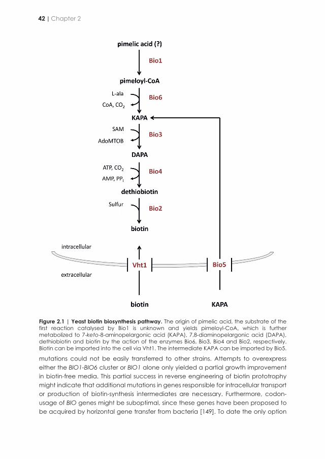
Figure 2.1 | Yeast biotin biosynthesis pathway. The origin of pimelic acid, the substrate of the first reaction catalysed by Bio1 is unknown and yields pimeloyl-CoA, which is further metabolized to 7-keto-8-aminopelargonic acid (KAPA), 7,8-diaminopelargonic acid (DAPA), dethiobiotin and biotin by the action of the enzymes Bio6, Bio3, Bio4 and Bio2, respectively. Biotin can be imported into the cell via Vht1. The intermediate KAPA can be imported by Bio5.
mutations could not be easily transferred to other strains. Attempts to overexpress either the BIO1-BIO6 cluster or BIO1 alone only yielded a partial growth improvement in biotin-free media. This partial success in reverse engineering of biotin prototrophy might indicate that additional mutations in genes responsible for intracellular transport or production of biotin-synthesis intermediates are necessary. Furthermore, codon-usage of BIO genes might be suboptimal, since these genes have been proposed to be acquired by horizontal gene transfer from bacteria [149]. To date the only option
to obtain full biotin prototrophy in S. cerevisiae is to perform laboratory evolution experiments which are time consuming and, moreover, lead to massive chromosomal rearrangements that might result in mutations that could negatively affect other cellular functions [238].
The genetic information required for biosynthesis of the fused-ring structure of biotin (catalysed by Bio3, Bio4 and Bio2) is well characterized in S. cerevisiae ([50, 239], Figure 2.1). In contrast, the origin of the pimeloyl moiety contributing to the valeric-acid chain remains elusive (Figure 2.1) [240]. Hitherto, ScBIO1 was hypothesised to act as a CoA-ligase that activates free pimelic acid for biotin synthesis, similar to the enzyme encoded by bioW in Bacillus subtilis [155]. The hypothetical product of such a ligase, pimeloyl-CoA, could be converted into 8-amino-7-oxononanoate by yeast KAPA synthase (Bio6) using L-alanine and pyridoxal-5-phosphate. However, no evidence is available for utilization of free pimelic acid by yeast. The lack of knowledge on the origin of the pimeloyl moiety in S. cerevisiae has so far hindered further targeted molecular improvement of biotin synthesis.
The aim of this study was to explore new metabolic engineering solutions to enable fast, unimpaired growth of S. cerevisiae without biotin supplementation. To this end, a set of Saccharomycotina yeasts were screened for specific growth rate in biotin-free synthetic medium lacking biotin. After identification of the best-performing species, the genome sequences of these yeasts were interrogated for ScBIO1 orthologs. The identified orthologous genes were functionally characterized in S. cerevisiae CEN.PK113-7D and evaluated for their ability to support fast growth in the absence of biotin, under aerobic as well as under anaerobic conditions. A promising candidate gene was expressed in several industrial and laboratory S. cerevisiae strains and its impact was evaluated. Furthermore, it was investigated how cultivation of an engineered biotin-prototrophic S. cerevisiae strain on biotin-free medium affected the impact of an intentional contamination with an auxotrophic strain.
Results
Characterization of biotin requirement in Saccharomycotina yeasts
Determination of nutritional requirements is a standard procedure in classical yeast taxonomy. While biotin requirements of individual species are reported in the yeast taxonomy reference book “The Yeasts” [241], this information is only qualitative. To capture quantitative information on biotin requirements, we determined the specific growth rates of a set of 35 biotechnologically relevant chosen yeasts belonging to the Saccharomycotina sub-phylum in biotin-free synthetic medium (biotin-free SMD). Of these 35 species, eleven exhibited growth, albeit at different rates. We focused further analysis on fast-growing species, based on an arbitrary threshold specific growth rate of 0.25 h-1. Only strains belonging to the species Yarrowia lipolytica, Pichia kudriavzevii (syn. Candida krusei and Issatchenkia orientalis) [242], Wickerhamomyces ciferrii,
42 | Chapter 2
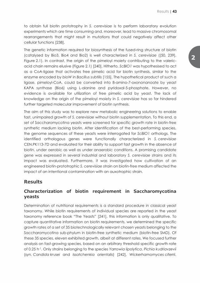
Figure 2.1 | Yeast biotin biosynthesis pathway. The origin of pimelic acid, the substrate of the first reaction catalysed by Bio1 is unknown and yields pimeloyl-CoA, which is further metabolized to 7-keto-8-aminopelargonic acid (KAPA), 7,8-diaminopelargonic acid (DAPA), dethiobiotin and biotin by the action of the enzymes Bio6, Bio3, Bio4 and Bio2, respectively. Biotin can be imported into the cell via Vht1. The intermediate KAPA can be imported by Bio5.
mutations could not be easily transferred to other strains. Attempts to overexpress either the BIO1-BIO6 cluster or BIO1 alone only yielded a partial growth improvement in biotin-free media. This partial success in reverse engineering of biotin prototrophy might indicate that additional mutations in genes responsible for intracellular transport or production of biotin-synthesis intermediates are necessary. Furthermore, codon-usage of BIO genes might be suboptimal, since these genes have been proposed to be acquired by horizontal gene transfer from bacteria [149]. To date the only option
to obtain full biotin prototrophy in S. cerevisiae is to perform laboratory evolution experiments which are time consuming and, moreover, lead to massive chromosomal rearrangements that might result in mutations that could negatively affect other cellular functions [238].
The genetic information required for biosynthesis of the fused-ring structure of biotin (catalysed by Bio3, Bio4 and Bio2) is well characterized in S. cerevisiae ([50, 239], Figure 2.1). In contrast, the origin of the pimeloyl moiety contributing to the valeric-acid chain remains elusive (Figure 2.1) [240]. Hitherto, ScBIO1 was hypothesised to act as a CoA-ligase that activates free pimelic acid for biotin synthesis, similar to the enzyme encoded by bioW in Bacillus subtilis [155]. The hypothetical product of such a ligase, pimeloyl-CoA, could be converted into 8-amino-7-oxononanoate by yeast KAPA synthase (Bio6) using L-alanine and pyridoxal-5-phosphate. However, no evidence is available for utilization of free pimelic acid by yeast. The lack of knowledge on the origin of the pimeloyl moiety in S. cerevisiae has so far hindered further targeted molecular improvement of biotin synthesis.
The aim of this study was to explore new metabolic engineering solutions to enable fast, unimpaired growth of S. cerevisiae without biotin supplementation. To this end, a set of Saccharomycotina yeasts were screened for specific growth rate in biotin-free synthetic medium lacking biotin. After identification of the best-performing species, the genome sequences of these yeasts were interrogated for ScBIO1 orthologs. The identified orthologous genes were functionally characterized in S. cerevisiae CEN.PK113-7D and evaluated for their ability to support fast growth in the absence of biotin, under aerobic as well as under anaerobic conditions. A promising candidate gene was expressed in several industrial and laboratory S. cerevisiae strains and its impact was evaluated. Furthermore, it was investigated how cultivation of an engineered biotin-prototrophic S. cerevisiae strain on biotin-free medium affected the impact of an intentional contamination with an auxotrophic strain.
Results
Characterization of biotin requirement in Saccharomycotina yeasts
Determination of nutritional requirements is a standard procedure in classical yeast taxonomy. While biotin requirements of individual species are reported in the yeast taxonomy reference book “The Yeasts” [241], this information is only qualitative. To capture quantitative information on biotin requirements, we determined the specific growth rates of a set of 35 biotechnologically relevant chosen yeasts belonging to the Saccharomycotina sub-phylum in biotin-free synthetic medium (biotin-free SMD). Of these 35 species, eleven exhibited growth, albeit at different rates. We focused further analysis on fast-growing species, based on an arbitrary threshold specific growth rate of 0.25 h-1. Only strains belonging to the species Yarrowia lipolytica, Pichia kudriavzevii (syn. Candida krusei and Issatchenkia orientalis) [242], Wickerhamomyces ciferrii,
Results | 43
2
Cyberlindnera fabianii (syn. Candida fabianii), Lachancea kluyveri and Torulaspora delbrueckii met this criterion, with specific growth rates in biotin-free SMD ranging from 0.27 to 0.64 h-1. Specific growth rates of Y. lipolytica, P. kudriavzevii and C. fabianii were not significantly higher in biotin-supplemented SMD. The other strains all showed lower specific growth rates in biotin-free media than in biotin-supplemented media, however not below 60% of the growth rate on SMD supplemented with biotin (Figure 2.2). Consistent with earlier observations [149, 151, 154], S. cerevisiae strain CEN.PK113-7D exhibited a specific growth rate of 0.39 ± 0.01 h-1 on biotin-supplemented SMD, but grew extremely slowly (μ < 0.01 h-1) on biotin-free SMD.
Figure 2.2 | Specific growth rates of Saccharomycotina species in SMD (+/white) and biotin-free SMD (-/grey). The bars represent average and standard deviation of three biological replicates. The data are derived from culture optical density (OD660) measurements over time during the exponential growth phase of shake flask batch cultures of Y. lypolytica W29, P. kudriavzevii CBS 5147, W. ciferrii CBS 111, C. fabianii CBS 5481, L. kluyveri CBS 3082, T. delbrueckii CBS 813 and S. cerevisiae CEN.PK113-7D. Statistical significance between growth rates in SMD and biotin-free SMD of each strain was determined using the Holm-Sidak method. *denotes comparison with pvalue lower than 5.0E-2.
Identification of putative ScBIO1 orthologs in Y. lipolytica, P. kudriavzevii, W. ciferrii, C. fabianii, L. kluyveri and T. delbrueckii.
The growth performance of Y. lipolytica, P. kudriavzevii, W. ciferrii, C. fabianii, L. kluyveri and T. delbrueckii in biotin-free SMD indicated that the Bio1 ‘bottleneck’ in the biotin biosynthesis pathway of S. cerevisiae CEN.PK113-7D was absent in these species. We therefore hypothesized that biotin prototrophy of these strains might be enabled by ScBIO1 orthologs encoding enzymes with superior catalytic or regulatory properties.
Identification of putative ScBIO1 orthologs was performed using the Basic Local Alignment Search Tool (BLAST) (https://blast.ncbi.nlm.nih.gov/Blast.cgi) [243], more specifically tBLASTn, which uses a protein sequence as query against translation of a nucleotide database. Use of ScBio1 as a query revealed candidate proteins in five out of the six species (P. kudriavzevii, W. ciferri, L. kluyveri, T. delbrueckii and C. fabianii) but not in Yarrowia lipolytica (Table 2.1). Subsequently, the search was repeated with the ScBIO1 orthologs found in P. kudriavzevii, W. ciferri, L. kluyveri, T. delbrueckii and C. fabianii as queries and using the Y. lipolytica W29 genome assembly as a database (PRJNA601425). Each five searches identified the same 346-amino-acids peptide sequence encoded by open reading frame YALI0A10010p (Table 2.1). Pair-wise comparisons of the identified putative Bio1 amino acid sequences using ClustalΩ [244] showed that the sequences of LkBio1 and TdBio1 showed the highest amino-acid sequence similarities to the ScBio1 sequence (43-44%), while YlBio1 had the lowest similarity to the other homologs. Although its size matched perfectly with ScBio1, YlBio1 only showed 15% to 20% amino-acid sequence similarity with the ScBio1 orthologous peptides (Figure 2.3). Out of this set of putative Bio1 homologs CfBio1 and WcBio1 exhibited the highest pair-wise amino-acid similarity (62%), which is in line with the phylogenetic proximity of these yeast species [241].
Table 2.1 | Identification of heterologous BIO1 gene by basic local alignment search tool tBLASTn, best hits for querying ScBio1 protein sequencea, or Td, Lk, Cf, Wc and PkBio1b.
Species taxon ID (NCBI) Accession No Sequence Open reading frame coordinates
T. delbrueckii txid1076872 aHE616748.1 Chromosome 7 18080-19111 bp
L. kluyveri txid4934 aAACE03000004.1 Chromosome D SKLU-Cont10108
1095811-1094765 bp
C. fabianii txid36022 aBCGI01000001.1 Scaffold 0 1136478-1137572 bp
W. ciferrii txid1041607 aCAIF01000264.1 Contig 00264 39352-40449 bp
P. kudriavzevii txid4909 aCP028531.1 Chromosome 1 1085523-1084447 bp
Y. lypolytica txid284591 bNC_006067.1 Chromosome A 1097298-1096261 bp
To investigate whether expression of the ScBIO1 orthologs was able to promote growth of S. cerevisiae CEN.PK113-7D in biotin-free medium, each BIO1 gene was isolated from its respective genome and cloned under the transcriptional control of the S. cerevisiae PYK1 promoter (ScPYK1p) and the S. cerevisiae BIO1 terminator (ScBIO1t). Each expression module was integrated at the chromosomal SGA1 locus of the Spycas9-expressing, CEN.PK-derived S. cerevisiae strain IMX585 by CRISPR/Cas9-mediated genome editing [245]. The resulting strains IMX1862 (YlBIO1↑), IMX1861 (PkBIO1↑), IMX1863 (WcBIO1↑), IMX1859 (CfBIO1↑), IMX1858 (LkBIO1↑), and IMX1857 (TdBIO1↑) and the control strain IMX1511 (ScBIO1↑) expressing the S. cerevisiae BIO1 gene under the same regulatory sequences as the heterologous BIO1 genes, were grown on biotin-containing and biotin-free SMD.
44 | Chapter 2
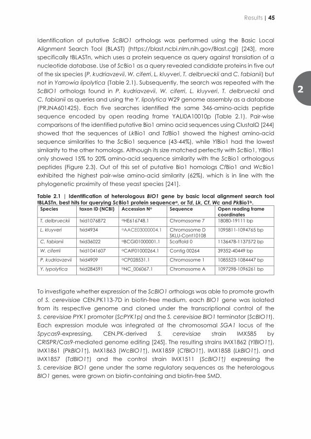
Cyberlindnera fabianii (syn. Candida fabianii), Lachancea kluyveri and Torulaspora delbrueckii met this criterion, with specific growth rates in biotin-free SMD ranging from 0.27 to 0.64 h-1. Specific growth rates of Y. lipolytica, P. kudriavzevii and C. fabianii were not significantly higher in biotin-supplemented SMD. The other strains all showed lower specific growth rates in biotin-free media than in biotin-supplemented media, however not below 60% of the growth rate on SMD supplemented with biotin (Figure 2.2). Consistent with earlier observations [149, 151, 154], S. cerevisiae strain CEN.PK113-7D exhibited a specific growth rate of 0.39 ± 0.01 h-1 on biotin-supplemented SMD, but grew extremely slowly (μ < 0.01 h-1) on biotin-free SMD.
Figure 2.2 | Specific growth rates of Saccharomycotina species in SMD (+/white) and biotin-free SMD (-/grey). The bars represent average and standard deviation of three biological replicates. The data are derived from culture optical density (OD660) measurements over time during the exponential growth phase of shake flask batch cultures of Y. lypolytica W29, P. kudriavzevii CBS 5147, W. ciferrii CBS 111, C. fabianii CBS 5481, L. kluyveri CBS 3082, T. delbrueckii CBS 813 and S. cerevisiae CEN.PK113-7D. Statistical significance between growth rates in SMD and biotin-free SMD of each strain was determined using the Holm-Sidak method. *denotes comparison with pvalue lower than 5.0E-2.
Identification of putative ScBIO1 orthologs in Y. lipolytica, P. kudriavzevii, W. ciferrii, C. fabianii, L. kluyveri and T. delbrueckii.
The growth performance of Y. lipolytica, P. kudriavzevii, W. ciferrii, C. fabianii, L. kluyveri and T. delbrueckii in biotin-free SMD indicated that the Bio1 ‘bottleneck’ in the biotin biosynthesis pathway of S. cerevisiae CEN.PK113-7D was absent in these species. We therefore hypothesized that biotin prototrophy of these strains might be enabled by ScBIO1 orthologs encoding enzymes with superior catalytic or regulatory properties.
Identification of putative ScBIO1 orthologs was performed using the Basic Local Alignment Search Tool (BLAST) (https://blast.ncbi.nlm.nih.gov/Blast.cgi) [243], more specifically tBLASTn, which uses a protein sequence as query against translation of a nucleotide database. Use of ScBio1 as a query revealed candidate proteins in five out of the six species (P. kudriavzevii, W. ciferri, L. kluyveri, T. delbrueckii and C. fabianii) but not in Yarrowia lipolytica (Table 2.1). Subsequently, the search was repeated with the ScBIO1 orthologs found in P. kudriavzevii, W. ciferri, L. kluyveri, T. delbrueckii and C. fabianii as queries and using the Y. lipolytica W29 genome assembly as a database (PRJNA601425). Each five searches identified the same 346-amino-acids peptide sequence encoded by open reading frame YALI0A10010p (Table 2.1). Pair-wise comparisons of the identified putative Bio1 amino acid sequences using ClustalΩ [244] showed that the sequences of LkBio1 and TdBio1 showed the highest amino-acid sequence similarities to the ScBio1 sequence (43-44%), while YlBio1 had the lowest similarity to the other homologs. Although its size matched perfectly with ScBio1, YlBio1 only showed 15% to 20% amino-acid sequence similarity with the ScBio1 orthologous peptides (Figure 2.3). Out of this set of putative Bio1 homologs CfBio1 and WcBio1 exhibited the highest pair-wise amino-acid similarity (62%), which is in line with the phylogenetic proximity of these yeast species [241].
Table 2.1 | Identification of heterologous BIO1 gene by basic local alignment search tool tBLASTn, best hits for querying ScBio1 protein sequencea, or Td, Lk, Cf, Wc and PkBio1b.
Species taxon ID (NCBI) Accession No Sequence Open reading frame coordinates
T. delbrueckii txid1076872 aHE616748.1 Chromosome 7 18080-19111 bp
L. kluyveri txid4934 aAACE03000004.1 Chromosome D SKLU-Cont10108
1095811-1094765 bp
C. fabianii txid36022 aBCGI01000001.1 Scaffold 0 1136478-1137572 bp
W. ciferrii txid1041607 aCAIF01000264.1 Contig 00264 39352-40449 bp
P. kudriavzevii txid4909 aCP028531.1 Chromosome 1 1085523-1084447 bp
Y. lypolytica txid284591 bNC_006067.1 Chromosome A 1097298-1096261 bp
To investigate whether expression of the ScBIO1 orthologs was able to promote growth of S. cerevisiae CEN.PK113-7D in biotin-free medium, each BIO1 gene was isolated from its respective genome and cloned under the transcriptional control of the S. cerevisiae PYK1 promoter (ScPYK1p) and the S. cerevisiae BIO1 terminator (ScBIO1t). Each expression module was integrated at the chromosomal SGA1 locus of the Spycas9-expressing, CEN.PK-derived S. cerevisiae strain IMX585 by CRISPR/Cas9-mediated genome editing [245]. The resulting strains IMX1862 (YlBIO1↑), IMX1861 (PkBIO1↑), IMX1863 (WcBIO1↑), IMX1859 (CfBIO1↑), IMX1858 (LkBIO1↑), and IMX1857 (TdBIO1↑) and the control strain IMX1511 (ScBIO1↑) expressing the S. cerevisiae BIO1 gene under the same regulatory sequences as the heterologous BIO1 genes, were grown on biotin-containing and biotin-free SMD.
Results | 45
2
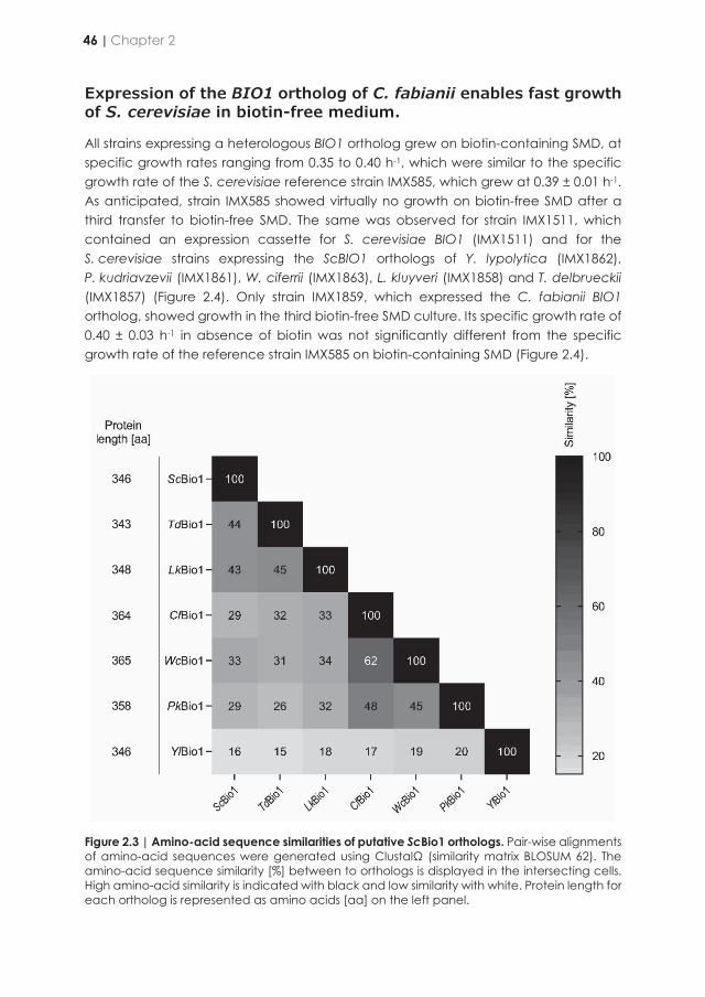
Expression of the BIO1 ortholog of C. fabianii enables fast growth of S. cerevisiae in biotin-free medium.
All strains expressing a heterologous BIO1 ortholog grew on biotin-containing SMD, at specific growth rates ranging from 0.35 to 0.40 h-1, which were similar to the specific growth rate of the S. cerevisiae reference strain IMX585, which grew at 0.39 ± 0.01 h-1. As anticipated, strain IMX585 showed virtually no growth on biotin-free SMD after a third transfer to biotin-free SMD. The same was observed for strain IMX1511, which contained an expression cassette for S. cerevisiae BIO1 (IMX1511) and for the S. cerevisiae strains expressing the ScBIO1 orthologs of Y. lypolytica (IMX1862), P. kudriavzevii (IMX1861), W. ciferrii (IMX1863), L. kluyveri (IMX1858) and T. delbrueckii (IMX1857) (Figure 2.4). Only strain IMX1859, which expressed the C. fabianii BIO1 ortholog, showed growth in the third biotin-free SMD culture. Its specific growth rate of 0.40 ± 0.03 h-1 in absence of biotin was not significantly different from the specific growth rate of the reference strain IMX585 on biotin-containing SMD (Figure 2.4).
Figure 2.3 | Amino-acid sequence similarities of putative ScBio1 orthologs. Pair-wise alignments of amino-acid sequences were generated using ClustalΩ (similarity matrix BLOSUM 62). The amino-acid sequence similarity [%] between to orthologs is displayed in the intersecting cells. High amino-acid similarity is indicated with black and low similarity with white. Protein length for each ortholog is represented as amino acids [aa] on the left panel.
All strains expressing ScBIO1 orthologs, including IMX1859, still harboured the native ScBIO1 gene at its original locus. To verify that expression of the CfBIO1 gene sufficed to achieve full biotin prototrophy, the native ScBIO1 copy was deleted in strain IMX1859, yielding strain IMX1860 (Scbio1∆ CfBIO1↑). The specific growth rate of IMX1860 in SMD was determined as 0.39 ± 0.01 h-1 and in biotin-free SMD as 0.36 ± 0.00 h-1, which is highly similar to the specific growth rates of strain IMX1859 on these media (Figure 2.4). Despite a slight difference in biotin-containing and biotin-free media, the specific growth rates of strain IMX1860 indicated that CfBIO1 was sufficient to confer S. cerevisiae strains from the CEN.PK lineage with the ability to grow fast in the absence of biotin.
Figure 2.4 | Specific growth rates of S. cerevisiae CEN.PK113-7D derived strains expressing ScBIO1 orthologs in SMD (+/white) and biotin-free SMD (-/growth). The bars data represent average and standard deviation of three biological replicates and are derived from measurement of culture optical density (OD660) over time during the exponential growth phase of shake flask batch cultures of strains IMX585, IMX1511 (ScBIO1↑), IMX1862 (YlBIO1↑), IMX1861 (PkBIO1↑), IMX1863 (WcBIO1↑), IMX1859 (CfBIO1↑), IMX1858 (LkBIO1↑), IMX1857 (TdBIO1↑) and IMX1860 (Scbio1Δ CfBIO1↑). Statistical significance between growth rates in SMD and biotin-free SMD of each strain was determined using the Holm-Sidak method providing pvalue a. Statistical significance between growth rates of IMX585 grown on SMD and the respective specific growth rates of engineered strains in SMD and biotin-free SMD was determined using the Holm-Sidak method providing pvalue b. * denotes comparison with pvalue lower than 5.0E-2.
46 | Chapter 2
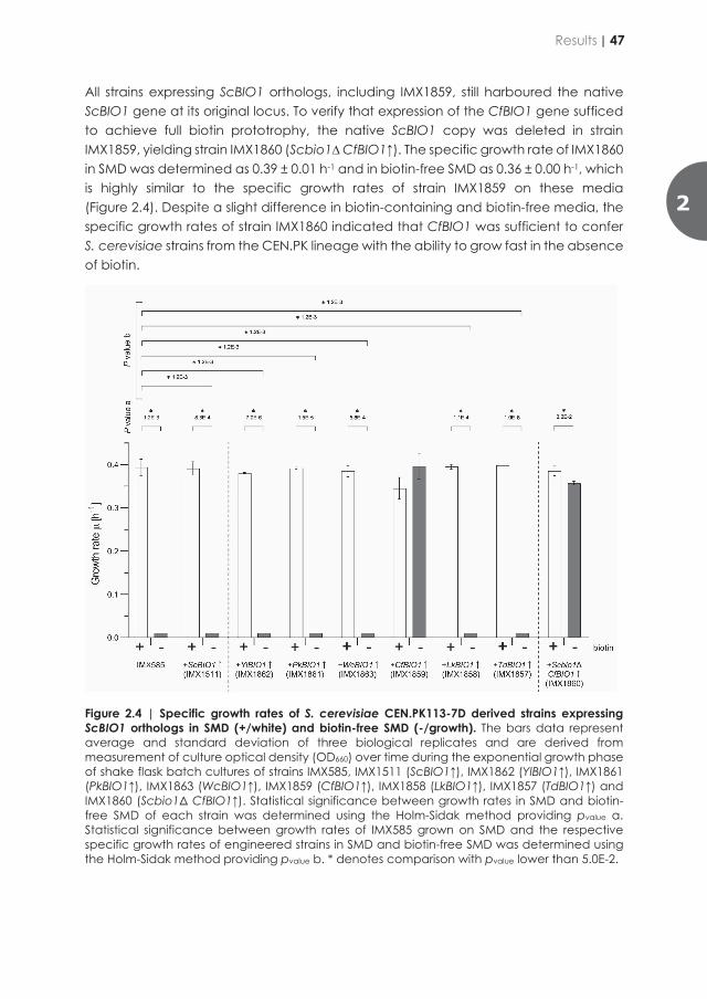
Expression of the BIO1 ortholog of C. fabianii enables fast growth of S. cerevisiae in biotin-free medium.
All strains expressing a heterologous BIO1 ortholog grew on biotin-containing SMD, at specific growth rates ranging from 0.35 to 0.40 h-1, which were similar to the specific growth rate of the S. cerevisiae reference strain IMX585, which grew at 0.39 ± 0.01 h-1. As anticipated, strain IMX585 showed virtually no growth on biotin-free SMD after a third transfer to biotin-free SMD. The same was observed for strain IMX1511, which contained an expression cassette for S. cerevisiae BIO1 (IMX1511) and for the S. cerevisiae strains expressing the ScBIO1 orthologs of Y. lypolytica (IMX1862), P. kudriavzevii (IMX1861), W. ciferrii (IMX1863), L. kluyveri (IMX1858) and T. delbrueckii (IMX1857) (Figure 2.4). Only strain IMX1859, which expressed the C. fabianii BIO1 ortholog, showed growth in the third biotin-free SMD culture. Its specific growth rate of 0.40 ± 0.03 h-1 in absence of biotin was not significantly different from the specific growth rate of the reference strain IMX585 on biotin-containing SMD (Figure 2.4).
Figure 2.3 | Amino-acid sequence similarities of putative ScBio1 orthologs. Pair-wise alignments of amino-acid sequences were generated using ClustalΩ (similarity matrix BLOSUM 62). The amino-acid sequence similarity [%] between to orthologs is displayed in the intersecting cells. High amino-acid similarity is indicated with black and low similarity with white. Protein length for each ortholog is represented as amino acids [aa] on the left panel.
All strains expressing ScBIO1 orthologs, including IMX1859, still harboured the native ScBIO1 gene at its original locus. To verify that expression of the CfBIO1 gene sufficed to achieve full biotin prototrophy, the native ScBIO1 copy was deleted in strain IMX1859, yielding strain IMX1860 (Scbio1∆ CfBIO1↑). The specific growth rate of IMX1860 in SMD was determined as 0.39 ± 0.01 h-1 and in biotin-free SMD as 0.36 ± 0.00 h-1, which is highly similar to the specific growth rates of strain IMX1859 on these media (Figure 2.4). Despite a slight difference in biotin-containing and biotin-free media, the specific growth rates of strain IMX1860 indicated that CfBIO1 was sufficient to confer S. cerevisiae strains from the CEN.PK lineage with the ability to grow fast in the absence of biotin.
Figure 2.4 | Specific growth rates of S. cerevisiae CEN.PK113-7D derived strains expressing ScBIO1 orthologs in SMD (+/white) and biotin-free SMD (-/growth). The bars data represent average and standard deviation of three biological replicates and are derived from measurement of culture optical density (OD660) over time during the exponential growth phase of shake flask batch cultures of strains IMX585, IMX1511 (ScBIO1↑), IMX1862 (YlBIO1↑), IMX1861 (PkBIO1↑), IMX1863 (WcBIO1↑), IMX1859 (CfBIO1↑), IMX1858 (LkBIO1↑), IMX1857 (TdBIO1↑) and IMX1860 (Scbio1Δ CfBIO1↑). Statistical significance between growth rates in SMD and biotin-free SMD of each strain was determined using the Holm-Sidak method providing pvalue a. Statistical significance between growth rates of IMX585 grown on SMD and the respective specific growth rates of engineered strains in SMD and biotin-free SMD was determined using the Holm-Sidak method providing pvalue b. * denotes comparison with pvalue lower than 5.0E-2.
Results | 47
2
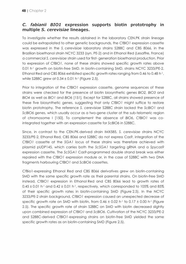
C. fabianii BIO1 expression supports biotin prototrophy in multiple S. cerevisiae lineages.
To investigate whether the results obtained in the laboratory CEN.PK strain lineage could be extrapolated to other genetic backgrounds, the CfBIO1 expression cassette was expressed in the S. cerevisiae laboratory strains S288C and CBS 8066, in the Brazilian bioethanol strain NCYC 3233 (syn. PE-2) and in Ethanol Red (Lesaffre, France) a commercial S. cerevisiae strain used for first- generation bioethanol production. Prior to expression of CfBIO1, none of these strains showed specific growth rates above 0.01 h-1 growth on biotin-free SMD. In biotin-containing SMD, strains NCYC 3233/PE-2, Ethanol Red and CBS 8066 exhibited specific growth rates ranging from 0.46 to 0.48 h-1, while S288C grew at 0.34 ± 0.01 h-1 (Figure 2.5).
Prior to integration of the CfBIO1 expression cassette, genome sequences of these strains were checked for the presence of biotin biosynthetic genes BIO2, BIO3 and BIO4 as well as BIO1 and BIO6 [151]. Except for S288C, all strains showed presence of these five biosynthetic genes, suggesting that only CfBIO1 might suffice to restore biotin prototrophy. The reference S. cerevisiae S288C strain lacked the ScBIO1 and ScBIO6 genes, which usually occur as a two-gene cluster at the sub-telomeric region of chromosome I [150]. To complement the absence of BIO6, CfBIO1 was co-integrated together with an expression cassette for ScBIO6 in S288C.
Since, in contrast to the CEN.PK-derived strain IMX585, S. cerevisiae strains NCYC 3233/PE-2, Ethanol Red, CBS 8066 and S288C do not express Cas9, integration of the CfBIO1 cassette at the SGA1 locus of these strains was therefore achieved with plasmid pUDP145, which carries both the ScSGA1 targeting gRNA and a Spycas9 expression cassette. The ScSGA1 Cas9-programmed double strand break was either repaired with the CfBIO1 expression module or, in the case of S288C with two DNA fragments harbouring CfBIO1 and ScBIO6 cassettes.
CfBio1-expressing Ethanol Red and CBS 8066 derivatives grew on biotin-containing SMD with the same specific growth rate as their parental strains. On biotin-free SMD instead, CfBIO1 expression in Ethanol Red and CBS 8066 lead to growth rates of 0.45 ± 0.01 h-1 and 0.42 ± 0.01 h-1, respectively, which corresponded to 105% and 85% of their specific growth rates in biotin-containing SMD (Figure 2.5). In the NCYC 3233/PE-2 strain background, CfBIO1 expression caused an unexpected decrease of specific growth rate on SMD with biotin, from 0.46 ± 0.02 h-1 to 0.17 ± 0.00 h-1 (Figure 2.5). The specific growth rate of strain S288C on SMD with biotin decreased slightly upon combined expression of CfBIO1 and ScBIO6. Cultivation of the NCYC 3233/PE-2 and S288C-derived CfBIO1-expressing strains on biotin-free SMD yielded the same specific growth rates as on biotin-containing SMD (Figure 2.5).
Figure 2.5 | Specific growth rates of S. cerevisiae strains engineered for growth without biotin in SMD (+/black) and biotin-free SMD (-/white). The bars data represent average and standard deviation of three biological replicates and specific growth rates are calculated from measurement of culture optical density (OD660) over time during the exponential growth phase of shake flask batch cultures of Ethanol Red, CBS 8066, NCYC 3233/PE-2 and S288C as well as the respective engineered strains IMX2101 (Ethanol Red CfBIO1↑), IMX2104 (CBS 8066 CfBIO1↑), IMX2090 (NCYC 3233/PE-2 CfBIO1↑) and IMX2103 (S288C CfBIO1↑ ScBIO6↑). Statistical significance between growth rates in SMD and biotin-free SMD of each strain was determined using the Holm-Sidak method providing pvalue a.) Statistical significance between growth rates of the initial and the engineered strain on SMD was determined using the Holm-Sidak method providing pvalue b.) Statistical significance between growth rates of the initial and the engineered strain on biotin-free SMD was determined using the Holm-Sidak method providing pvalue c.) * denotes comparison with pvalue lower than 5.0E-2.
A biotin-prototrophic S. cerevisiae strain expressing CfBIO1 outcompetes an auxotrophic strain.
A possible advantage of biotin-prototrophic strains upon contamination of cultures with biotin auxotrophs was investigated by co-cultivation of biotin-auxotrophic and biotin-prototrophic S. cerevisiae strains. S. cerevisiae strains IMX585 (Spycas9) and IMK827 (Spycas9 Scbio1Δ) were equipped with an expression cassette encoding Venus fluorescent protein, yielding strains IMX2212 and IMX2240, respectively. The biotin-prototrophic strain IMX1860 (Spycas9 Scbio1Δ CfBIO1↑) was engineered to overexpress the mRuby2 fluorescence protein, yielding strain IMX2238. First, the strains IMX2240 (Spycas9 Scbio1Δ Venus↑) and IMX2238 (Spycas9 Scbio1Δ mRuby2↑ CfBIO1↑) were mixed in a 40:60 ratio. The relative abundance of the two fluorescent populations was monitored by flow cytometry during consecutive batch cultivation cycles, both in SMD with biotin and in biotin-free SMD. In biotin-containing media, the relative
48 | Chapter 2
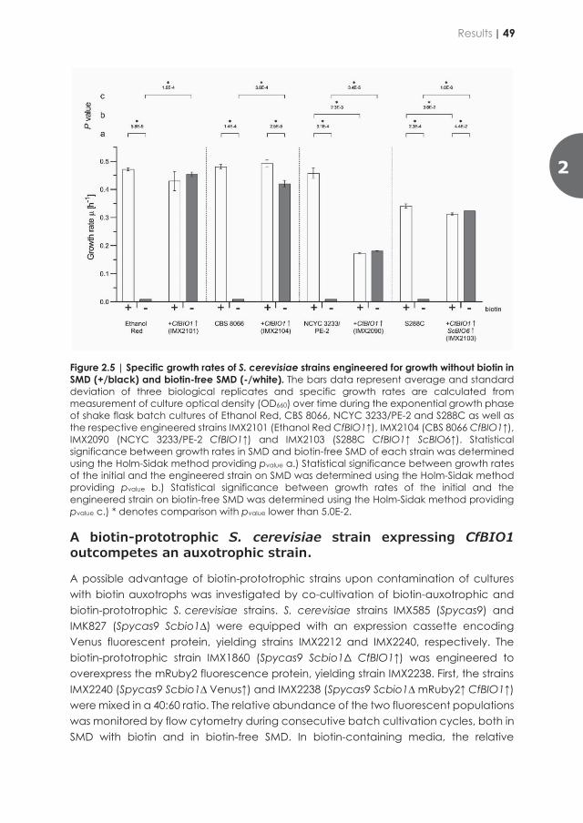
C. fabianii BIO1 expression supports biotin prototrophy in multiple S. cerevisiae lineages.
To investigate whether the results obtained in the laboratory CEN.PK strain lineage could be extrapolated to other genetic backgrounds, the CfBIO1 expression cassette was expressed in the S. cerevisiae laboratory strains S288C and CBS 8066, in the Brazilian bioethanol strain NCYC 3233 (syn. PE-2) and in Ethanol Red (Lesaffre, France) a commercial S. cerevisiae strain used for first- generation bioethanol production. Prior to expression of CfBIO1, none of these strains showed specific growth rates above 0.01 h-1 growth on biotin-free SMD. In biotin-containing SMD, strains NCYC 3233/PE-2, Ethanol Red and CBS 8066 exhibited specific growth rates ranging from 0.46 to 0.48 h-1, while S288C grew at 0.34 ± 0.01 h-1 (Figure 2.5).
Prior to integration of the CfBIO1 expression cassette, genome sequences of these strains were checked for the presence of biotin biosynthetic genes BIO2, BIO3 and BIO4 as well as BIO1 and BIO6 [151]. Except for S288C, all strains showed presence of these five biosynthetic genes, suggesting that only CfBIO1 might suffice to restore biotin prototrophy. The reference S. cerevisiae S288C strain lacked the ScBIO1 and ScBIO6 genes, which usually occur as a two-gene cluster at the sub-telomeric region of chromosome I [150]. To complement the absence of BIO6, CfBIO1 was co-integrated together with an expression cassette for ScBIO6 in S288C.
Since, in contrast to the CEN.PK-derived strain IMX585, S. cerevisiae strains NCYC 3233/PE-2, Ethanol Red, CBS 8066 and S288C do not express Cas9, integration of the CfBIO1 cassette at the SGA1 locus of these strains was therefore achieved with plasmid pUDP145, which carries both the ScSGA1 targeting gRNA and a Spycas9 expression cassette. The ScSGA1 Cas9-programmed double strand break was either repaired with the CfBIO1 expression module or, in the case of S288C with two DNA fragments harbouring CfBIO1 and ScBIO6 cassettes.
CfBio1-expressing Ethanol Red and CBS 8066 derivatives grew on biotin-containing SMD with the same specific growth rate as their parental strains. On biotin-free SMD instead, CfBIO1 expression in Ethanol Red and CBS 8066 lead to growth rates of 0.45 ± 0.01 h-1 and 0.42 ± 0.01 h-1, respectively, which corresponded to 105% and 85% of their specific growth rates in biotin-containing SMD (Figure 2.5). In the NCYC 3233/PE-2 strain background, CfBIO1 expression caused an unexpected decrease of specific growth rate on SMD with biotin, from 0.46 ± 0.02 h-1 to 0.17 ± 0.00 h-1 (Figure 2.5). The specific growth rate of strain S288C on SMD with biotin decreased slightly upon combined expression of CfBIO1 and ScBIO6. Cultivation of the NCYC 3233/PE-2 and S288C-derived CfBIO1-expressing strains on biotin-free SMD yielded the same specific growth rates as on biotin-containing SMD (Figure 2.5).
Figure 2.5 | Specific growth rates of S. cerevisiae strains engineered for growth without biotin in SMD (+/black) and biotin-free SMD (-/white). The bars data represent average and standard deviation of three biological replicates and specific growth rates are calculated from measurement of culture optical density (OD660) over time during the exponential growth phase of shake flask batch cultures of Ethanol Red, CBS 8066, NCYC 3233/PE-2 and S288C as well as the respective engineered strains IMX2101 (Ethanol Red CfBIO1↑), IMX2104 (CBS 8066 CfBIO1↑), IMX2090 (NCYC 3233/PE-2 CfBIO1↑) and IMX2103 (S288C CfBIO1↑ ScBIO6↑). Statistical significance between growth rates in SMD and biotin-free SMD of each strain was determined using the Holm-Sidak method providing pvalue a.) Statistical significance between growth rates of the initial and the engineered strain on SMD was determined using the Holm-Sidak method providing pvalue b.) Statistical significance between growth rates of the initial and the engineered strain on biotin-free SMD was determined using the Holm-Sidak method providing pvalue c.) * denotes comparison with pvalue lower than 5.0E-2.
A biotin-prototrophic S. cerevisiae strain expressing CfBIO1 outcompetes an auxotrophic strain.
A possible advantage of biotin-prototrophic strains upon contamination of cultures with biotin auxotrophs was investigated by co-cultivation of biotin-auxotrophic and biotin-prototrophic S. cerevisiae strains. S. cerevisiae strains IMX585 (Spycas9) and IMK827 (Spycas9 Scbio1Δ) were equipped with an expression cassette encoding Venus fluorescent protein, yielding strains IMX2212 and IMX2240, respectively. The biotin-prototrophic strain IMX1860 (Spycas9 Scbio1Δ CfBIO1↑) was engineered to overexpress the mRuby2 fluorescence protein, yielding strain IMX2238. First, the strains IMX2240 (Spycas9 Scbio1Δ Venus↑) and IMX2238 (Spycas9 Scbio1Δ mRuby2↑ CfBIO1↑) were mixed in a 40:60 ratio. The relative abundance of the two fluorescent populations was monitored by flow cytometry during consecutive batch cultivation cycles, both in SMD with biotin and in biotin-free SMD. In biotin-containing media, the relative
Results | 49
2

abundance of the two strains remained constant after inoculation (Figure 2.6; top panel). Instead, in biotin-free medium (Figure 2.6; bottom panel), within five hours of cultivation, 75% of the population already consisted of mRuby2-expressing, biotin-prototrophic cells, while during the third and fourth batch cultivation cycles, 99% of the culture consisted of biotin-prototrophic cells. In a similar experiment, the biotin prototroph IMX2238 (Spycas9 Scbio1Δ mRuby2↑ CfBIO1↑) was inoculated together with a fluorescent CEN.PK113-7D derived strain IMX2212 (Spycas9 Venus↑), which carries all native biotin biosynthetic genes (Figure 2.7). The initial ratio of 60:40 was maintained three batches in biotin-containing SMD (Figure 2.7; top panel).
Figure 2.6 | Culture optical density (OD660) and fluorescent population distribution of mRuby2 expressing strain IMX2238 (Spycas9 Scbio1Δ mRuby2↑ CfBIO1↑; biotin prototroph) and Venus expressing strain IMX2240 (Spycas9 Scbio1ΔΔ Venus↑; biotin auxotroph) over time during co-cultivation in four consecutive batch culture cycles on SMD (top) and biotin-free SMD (bottom). Both conditions were analysed in two independent replicates each. Each OD660 time-point was sampled and analysed by flow cytometry. The distribution of mRuby2 (red bar) and Venus (green bar) fluorescent cells from the total of fluorescent cells [%] at each time-point is represented in the bar graph above (connected by arrow).
In contrast, in biotin-free SMD, abundance of the auxotroph was reduced to 2% of the total fluorescent population after three batch cultivation cycles (Figure 2.7; bottom panel). These results indicated that. while expression of CfBIO1 (IMX2238) provided a significant growth advantage in biotin-free SMD, biotin is not released into the medium in sufficient amounts to support growth of a complete biotin auxotroph or of a strain with a very low biosynthesis capacity such as CEN.PK113-7D.
Figure 2.7 | Culture optical density (OD660) and fluorescent population distribution of mRuby2 expressing strain IMX2238 (Spycas9 Scbio1Δ mRuby2↑ CfBIO1↑; biotin prototroph) and Venus expressing strain IMX2212 (Spycas9 Venus↑; biotin auxotroph) over time during co-cultivation in three consecutive batch culture cycles on SMD (top) and biotin-free SMD (bottom). Both conditions were analysed in two independent replicates each. Each OD660 time-point was sampled and analysed by flow cytometry. The distribution of mRuby2 (red bar) and Venus (green bar) fluorescent cells from the total of fluorescent cells [%] at each time-point is represented in the bar graph above (connected by arrow).
Bio1 proteins show similarity to iron(II)-dependent oxidoreductases and biotin synthesis requires oxygen.
For a first exploration of the unknown reaction catalysed by CfBio1, its protein structure was predicted by homology modelling with Phyre2. The CfBio1 structure showed high structural similarity to an Aspergillus nidulans phytanoyl-CoA dioxygenase (5DAQ, DOI: 10.2210/pdb5daq/pdb, EC 1.14.11.18). This oxidoreductase enzyme catalyses an iron(II)-dependent reaction in which phytanoyl-CoA is oxidized to 2-hydroxyphytanoyl-CoA with 2-oxo-glutarate and oxygen as co-substrates, yielding succinate and carbon dioxide as by-products [246]. The same approach was applied to ScBio1 that showed high structural similarity to another putative oxygenase (4NAO, DOI:10.2210/pdb4NAO/pdb, EC 1.14.11.18). These two homology-‘hits’ were retrieved for all the orthologous Bio1 candidate polypeptides, except for YlBio1, which instead
50 | Chapter 2

abundance of the two strains remained constant after inoculation (Figure 2.6; top panel). Instead, in biotin-free medium (Figure 2.6; bottom panel), within five hours of cultivation, 75% of the population already consisted of mRuby2-expressing, biotin-prototrophic cells, while during the third and fourth batch cultivation cycles, 99% of the culture consisted of biotin-prototrophic cells. In a similar experiment, the biotin prototroph IMX2238 (Spycas9 Scbio1Δ mRuby2↑ CfBIO1↑) was inoculated together with a fluorescent CEN.PK113-7D derived strain IMX2212 (Spycas9 Venus↑), which carries all native biotin biosynthetic genes (Figure 2.7). The initial ratio of 60:40 was maintained three batches in biotin-containing SMD (Figure 2.7; top panel).
Figure 2.6 | Culture optical density (OD660) and fluorescent population distribution of mRuby2 expressing strain IMX2238 (Spycas9 Scbio1Δ mRuby2↑ CfBIO1↑; biotin prototroph) and Venus expressing strain IMX2240 (Spycas9 Scbio1ΔΔ Venus↑; biotin auxotroph) over time during co-cultivation in four consecutive batch culture cycles on SMD (top) and biotin-free SMD (bottom). Both conditions were analysed in two independent replicates each. Each OD660 time-point was sampled and analysed by flow cytometry. The distribution of mRuby2 (red bar) and Venus (green bar) fluorescent cells from the total of fluorescent cells [%] at each time-point is represented in the bar graph above (connected by arrow).
In contrast, in biotin-free SMD, abundance of the auxotroph was reduced to 2% of the total fluorescent population after three batch cultivation cycles (Figure 2.7; bottom panel). These results indicated that. while expression of CfBIO1 (IMX2238) provided a significant growth advantage in biotin-free SMD, biotin is not released into the medium in sufficient amounts to support growth of a complete biotin auxotroph or of a strain with a very low biosynthesis capacity such as CEN.PK113-7D.
Figure 2.7 | Culture optical density (OD660) and fluorescent population distribution of mRuby2 expressing strain IMX2238 (Spycas9 Scbio1Δ mRuby2↑ CfBIO1↑; biotin prototroph) and Venus expressing strain IMX2212 (Spycas9 Venus↑; biotin auxotroph) over time during co-cultivation in three consecutive batch culture cycles on SMD (top) and biotin-free SMD (bottom). Both conditions were analysed in two independent replicates each. Each OD660 time-point was sampled and analysed by flow cytometry. The distribution of mRuby2 (red bar) and Venus (green bar) fluorescent cells from the total of fluorescent cells [%] at each time-point is represented in the bar graph above (connected by arrow).
Bio1 proteins show similarity to iron(II)-dependent oxidoreductases and biotin synthesis requires oxygen.
For a first exploration of the unknown reaction catalysed by CfBio1, its protein structure was predicted by homology modelling with Phyre2. The CfBio1 structure showed high structural similarity to an Aspergillus nidulans phytanoyl-CoA dioxygenase (5DAQ, DOI: 10.2210/pdb5daq/pdb, EC 1.14.11.18). This oxidoreductase enzyme catalyses an iron(II)-dependent reaction in which phytanoyl-CoA is oxidized to 2-hydroxyphytanoyl-CoA with 2-oxo-glutarate and oxygen as co-substrates, yielding succinate and carbon dioxide as by-products [246]. The same approach was applied to ScBio1 that showed high structural similarity to another putative oxygenase (4NAO, DOI:10.2210/pdb4NAO/pdb, EC 1.14.11.18). These two homology-‘hits’ were retrieved for all the orthologous Bio1 candidate polypeptides, except for YlBio1, which instead
Results | 51
2
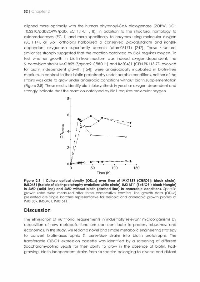
aligned more optimally with the human phytanoyl-CoA dioxygenase (2OPW, DOI: 10.2210/pdb2OPW/pdb, EC 1.14.11.18). In addition to the structural homology to oxidoreductases (EC 1) and more specifically to enzymes using molecular oxygen (EC 1.14), all Bio1 orthologs harboured a conserved 2-oxoglutarate and iron(II)-dependent oxygenase superfamily domain (pfam03171) [247]. These structural similarities strongly suggested that the reaction catalysed by Bio1 requires oxygen. To test whether growth in biotin-free medium was indeed oxygen-dependent, the S. cerevisiae strains IMX1859 (Spycas9 CfBIO1↑) and IMS0481 (CEN.PK113-7D evolved for biotin independent growth [154]) were anaerobically incubated in biotin-free medium. In contrast to their biotin prototrophy under aerobic conditions, neither of the strains was able to grow under anaerobic conditions without biotin supplementation (Figure 2.8). These results identify biotin biosynthesis in yeast as oxygen-dependent and strongly indicate that the reaction catalysed by Bio1 requires molecular oxygen.
Figure 2.8 | Culture optical density (OD660) over time of IMX1859 (CfBIO1↑; black circle), IMS0481 (isolate of biotin prototrophy evolution; white circle), IMX1511 (ScBIO1↑; black triangle) in SMD (solid line) and SMD without biotin (dashed line) in anaerobic conditions. Specific growth rates were measured after three consecutive transfers. The growth data (OD660) presented are single batches representative for aerobic and anaerobic growth profiles of IMX1859, IMS0481, IMX1511.
Discussion
The elimination of nutritional requirements in industrially relevant microorganisms by acquisition of new metabolic functions can contribute to process robustness and economics. In this study, we report a novel and simple metabolic engineering strategy to convert biotin-auxotrophic S. cerevisiae strains into biotin prototrophs. The transferable CfBIO1 expression cassette was identified by a screening of different Saccharomycotina yeasts for their ability to grow in the absence of biotin. Fast-growing, biotin-independent strains from six species belonging to diverse and distant
phylogenetic groups were identified (Y. lipolytica from the Yarrowia family, P. krudiavzevii from the the Pichiaceae family, W. ciferrii and C. fabianii from the Phaffomycetaceae family and L. kluyveri and T. delbrueckii from the Saccharomycetaceae family). Some of these yeasts have already been explored for their biotechnological properties such as Y. lipolytica for the production of oleochemicals [248], P. kudriavzevii as a multi-stress tolerant yeast for bioethanol [249] and succinic acid production [250] and W. ciferrii for the production of sphingolipids [251]. However, their biotin-prototrophic phenotype has not yet been exploited. C. fabianii (syn. Hansenula fabianii, Pichia fabianii, and Lindnera fabianii) is not an industrial yeast and has, in rare cases, been isolated from patients with blood infections. Its biotin prototrophy might be related to its opportunistic behaviour by providing a selective advantage in low-nutrient environments [252].
Establishing sustainable and cost-effective preparation of growth media remains an important factor in industrial process design and economics [253-255]. The benefit of microbes growing independently from the addition of organic growth factors such as biotin enables increased standardisation as well as reduced costs and time for media preparation and simplified down-stream processing [154, 256]. Economic incentives to develop simpler growth media are especially strong for production of commodity chemicals and in strictly regulated processes that require purified media components [257]. As demonstrated in this study, biotin-independent S. cerevisiae strains did not secrete amounts of biotin in the medium that enabled cross-feeding of fast-growing auxotrophic yeasts. This observation suggests that fermentation processes based on such prototrophs may be less susceptible to contamination by biotin-requiring ‘wild’ S. cerevisiae strains or other biotin-auxotrophic contaminants [258].
Inspection of available draft genome assemblies of the biotin-prototrophic yeast species led to the identification of candidate BIO1 genes. Surprisingly, only the expression of the BIO1 ortholog from C. fabianii supported fast growth of S. cerevisiae in biotin-free medium. Even the BIO1 ortholog from W. ciferrii, which belongs to the same phylogenetic subdivision of the Saccharomycotina tree as C. fabianii, failed to support growth of S. cerevisiae in absence of biotin. Without a functional analysis of these BIO1 orthologs in their original hosts, the possibility remains that these open reading frames do not encode for the enzyme performing the initial step in biotin biosynthesis, but have a different physiological role. We also cannot exclude differences in post-translation regulation between the donor organism and S. cerevisiae which could result in non-functional proteins. As shown for the elucidated biotin biosynthetic pathways in prokaryotic model organisms such as Escherichia coli and B. subtilis the precursor initiating the biosynthesis could differ. In E. coli, biotin is produced by the so-called BioC-BioH pathway, which diverts malonyl-CoA from fatty acid synthesis by masking the ϖ-carboxyl group of a malonyl-CoA thioester, resulting in recognition of this uncommon substrate by the E. coli fatty-acid synthesis machinery. In two successive rounds of fatty acid synthesis the malonyl thioester methyl ester is elongated yielding pimeloyl-ACP methyl ester which reversibly esterified to free
52 | Chapter 2

aligned more optimally with the human phytanoyl-CoA dioxygenase (2OPW, DOI: 10.2210/pdb2OPW/pdb, EC 1.14.11.18). In addition to the structural homology to oxidoreductases (EC 1) and more specifically to enzymes using molecular oxygen (EC 1.14), all Bio1 orthologs harboured a conserved 2-oxoglutarate and iron(II)-dependent oxygenase superfamily domain (pfam03171) [247]. These structural similarities strongly suggested that the reaction catalysed by Bio1 requires oxygen. To test whether growth in biotin-free medium was indeed oxygen-dependent, the S. cerevisiae strains IMX1859 (Spycas9 CfBIO1↑) and IMS0481 (CEN.PK113-7D evolved for biotin independent growth [154]) were anaerobically incubated in biotin-free medium. In contrast to their biotin prototrophy under aerobic conditions, neither of the strains was able to grow under anaerobic conditions without biotin supplementation (Figure 2.8). These results identify biotin biosynthesis in yeast as oxygen-dependent and strongly indicate that the reaction catalysed by Bio1 requires molecular oxygen.
Figure 2.8 | Culture optical density (OD660) over time of IMX1859 (CfBIO1↑; black circle), IMS0481 (isolate of biotin prototrophy evolution; white circle), IMX1511 (ScBIO1↑; black triangle) in SMD (solid line) and SMD without biotin (dashed line) in anaerobic conditions. Specific growth rates were measured after three consecutive transfers. The growth data (OD660) presented are single batches representative for aerobic and anaerobic growth profiles of IMX1859, IMS0481, IMX1511.
Discussion
The elimination of nutritional requirements in industrially relevant microorganisms by acquisition of new metabolic functions can contribute to process robustness and economics. In this study, we report a novel and simple metabolic engineering strategy to convert biotin-auxotrophic S. cerevisiae strains into biotin prototrophs. The transferable CfBIO1 expression cassette was identified by a screening of different Saccharomycotina yeasts for their ability to grow in the absence of biotin. Fast-growing, biotin-independent strains from six species belonging to diverse and distant
phylogenetic groups were identified (Y. lipolytica from the Yarrowia family, P. krudiavzevii from the the Pichiaceae family, W. ciferrii and C. fabianii from the Phaffomycetaceae family and L. kluyveri and T. delbrueckii from the Saccharomycetaceae family). Some of these yeasts have already been explored for their biotechnological properties such as Y. lipolytica for the production of oleochemicals [248], P. kudriavzevii as a multi-stress tolerant yeast for bioethanol [249] and succinic acid production [250] and W. ciferrii for the production of sphingolipids [251]. However, their biotin-prototrophic phenotype has not yet been exploited. C. fabianii (syn. Hansenula fabianii, Pichia fabianii, and Lindnera fabianii) is not an industrial yeast and has, in rare cases, been isolated from patients with blood infections. Its biotin prototrophy might be related to its opportunistic behaviour by providing a selective advantage in low-nutrient environments [252].
Establishing sustainable and cost-effective preparation of growth media remains an important factor in industrial process design and economics [253-255]. The benefit of microbes growing independently from the addition of organic growth factors such as biotin enables increased standardisation as well as reduced costs and time for media preparation and simplified down-stream processing [154, 256]. Economic incentives to develop simpler growth media are especially strong for production of commodity chemicals and in strictly regulated processes that require purified media components [257]. As demonstrated in this study, biotin-independent S. cerevisiae strains did not secrete amounts of biotin in the medium that enabled cross-feeding of fast-growing auxotrophic yeasts. This observation suggests that fermentation processes based on such prototrophs may be less susceptible to contamination by biotin-requiring ‘wild’ S. cerevisiae strains or other biotin-auxotrophic contaminants [258].
Inspection of available draft genome assemblies of the biotin-prototrophic yeast species led to the identification of candidate BIO1 genes. Surprisingly, only the expression of the BIO1 ortholog from C. fabianii supported fast growth of S. cerevisiae in biotin-free medium. Even the BIO1 ortholog from W. ciferrii, which belongs to the same phylogenetic subdivision of the Saccharomycotina tree as C. fabianii, failed to support growth of S. cerevisiae in absence of biotin. Without a functional analysis of these BIO1 orthologs in their original hosts, the possibility remains that these open reading frames do not encode for the enzyme performing the initial step in biotin biosynthesis, but have a different physiological role. We also cannot exclude differences in post-translation regulation between the donor organism and S. cerevisiae which could result in non-functional proteins. As shown for the elucidated biotin biosynthetic pathways in prokaryotic model organisms such as Escherichia coli and B. subtilis the precursor initiating the biosynthesis could differ. In E. coli, biotin is produced by the so-called BioC-BioH pathway, which diverts malonyl-CoA from fatty acid synthesis by masking the ϖ-carboxyl group of a malonyl-CoA thioester, resulting in recognition of this uncommon substrate by the E. coli fatty-acid synthesis machinery. In two successive rounds of fatty acid synthesis the malonyl thioester methyl ester is elongated yielding pimeloyl-ACP methyl ester which reversibly esterified to free
Discussion | 53
2
pimeloyl-ACP, which subsequently enters biotin synthesis [259]. In B. subtilis two distinct pathways have been identified. The indispensable route via the pimeloyl-CoA synthetase BioW [260] activates free pimelic acid with co-enzyme A yielding pimeloyl-CoA [155], which is used by B. subtilis BioF to produce KAPA [261]. Alternatively, a second, apparently redundant route [262] via a cytochrome P450-dependent carbon-bond-cleaving oxygenase encoded by bioI can generate pimeloyl-ACP from long chain (C14, C16 or C18) acyl-ACP molecules [263]. So it cannot be excluded that similarly the precursor to produce pimeloyl-ACP or -CoA differs in between these yeasts and S. cerevisiae.
While the expression of CfBIO1 in the NCYC 3233 (PE-2) strain resulted in biotin prototrophy as in any other genetic background tested, the simple integration of this gene at the SGA1 locus negatively impacted cell growth by reducing by half the strain specific growth rate, a phenomenon that remains enigmatic. This difference in phenotype might be the result of unwanted mutations introduce during the strain transformation procedure. As previously shown induction of aneuploidy is relatively frequent and associated with an aneuploidy associated stress response (AASR) that may lead to strain growth rate reduction [264, 265]. However, the methodology to detect this type of mutations has not used in the framework of this study such as full genome sequencing of the PE-2 derived transformants. This might also be linked to the SGA1 genetic context, such that integration at that locus in NCYC 3233 might have disturbed expression of surrounding genes. Deregulation of XBP1, distal to SGA1 on CHRIX might lead to slower growth. XBP1 is a transcriptional repressor that acts on promoter of cyclin genes and whose overexpression is known to results in decrease growth rate [266]. Integration at a different chromosomal site would be sufficient to falsify this hypothesis.
Although the amino-acid similarities of the six Bio1 orthologs ranged from 15 to 62%, all contained common structural motifs typical for phytanoyl-CoA dioxygenases and 2-oxoglutarate-iron(II)-dependent dioxygenases. These oxidoreductases catalyse reactions that incorporate oxygen from molecular oxygen (O2) into their substrates, while oxidizing 2-oxoglutarate to succinate and carbon dioxide [267]. Consistent with the involvement of an oxygenase in biotin synthesis, our results show that de novo biotin biosynthesis represents a previously unidentified oxygen-requiring process in S. cerevisiae and other biotin-prototrophic Saccharomycotina yeasts, in addition to well-known oxygen requiring processes such as the synthesis of ergosterol, unsaturated fatty acids [21 , 22] and nicotinic acid [268].
An oxygen requirement for biotin synthesis has also been reported for B. subtilis, in which pimeloyl-ACP is generated by cleavage of a carbon bond of an acyl-ACP (C14, C16 or C18) molecule by the cytochrome P450-dependent, oxygen-requiring enzyme BioI [263]. This mechanism provides a direct link between biotin synthesis and fatty-acid synthesis. Five of the biotin-prototrophic yeasts identified in this study, Y. lipolytica [269], W. ciferri , P. kudriavzevii , T. delbrueckii and C. fabianii are classified as oleaginous yeasts [270] which might support the link between biotin and fatty acid
biosynthesis also in yeast. Biochemical studies on the reaction(s) catalysed by ScBio1 and similar proteins are essential to finally elucidate one of the very few missing reactions in fungal vitamin biosynthesis [271].
Materials and Methods
Strains, media and maintenance
Strains of 35 Saccharomycotina yeasts were tested for growth in the absence of biotin. The S. cerevisiae strains used and constructed in this study belong to the CEN.PK, S288C, Ethanol Red, NCYC 3233/PE-2 and CBS 8066 lineages (Table 2.2). Yeast strains were grown on YP medium (10 g L−1 yeast extract, 20 g L−1 peptone) or on synthetic medium (SM) containing 3.0 g L−1 KH2PO4, 5.0 g L−1 (NH4)2SO4, 0.5 g L−1 MgSO4 · 7H2O, 1 mL L−1 trace element solution (4.5 mg L-1 ZnSO4 · 7 H2O, 0.3 mg L-1 CoCl2 · 6 H2O, 1 mg L-1 MnCl2 · 4 H2O, 0.3 mg L-1 CuSO4 · 5 H2O, 4.5 mg L-1 CaCl2 · 2 H2O, 3 mg L-1 FeSO4 · 7 H2O, 0.4 mg L-1 NaMoO4 · 2 H2O, 1 mg L-1 H3BO3, 0.1 mg L-1 KI, 15 mg L-1 EDTA), and 1 mL L−1 vitamin solution (0.05 g L-1 D-(+)-biotin, 1.0 g L-1 D-calcium pantothenate, 1.0 g L-1 nicotinic acid, 25 g L-1 myo-inositol, 1.0 g L-1 thiamine hydrochloride, 1.0 g L-1 pyridoxal hydrochloride, 0.2 g L-1 4-aminobenzoic acid) [13]. The pH was adjusted to 6 with 2 M KOH prior to autoclaving at 120oC for 20 min. Vitamin solutions were sterilized by filtration and added to the sterile medium. Concentrated sugar solutions were autoclaved at 110oC for 20 min and added to the sterile medium to give a final concentration of 20 g L−1 glucose (YPD and SMD). Biotin-free SM was prepared similarly, but biotin was omitted from the vitamin solution. Similarly, after autoclaving concentrated glucose solution at 110oC for 20 min, glucose was added to biotin-free SM to a final concentration of 20 g L-1 (biotin-free SMD). Solid media contained 1.5% bacto agar and, when indicated, acetamide for SMD acetamide (20 g L−1 glucose, 1.2 g L−1 acetamide, 3.0 g L−1 KH2PO4, 6.6 g L−1 K2SO4, 0.5 g L−1 MgSO4 · 7H2O, 1 mL L−1 trace element solution and 1 mL L−1 vitamin solution) and 200 mg L-1 hygromycin for YPD hygromycin. E. coli cells (XL1-Blue, Agilent Technologies, Santa Clara, CA) were grown in lysogeny broth (LB) medium (5.0 g L-1 yeast extract, 10 g L-1 bacto trypton, 5.0 g L-1 NaCl) supplemented with 25 mg L-1 chloramphenicol, 100 mg L-1 ampicillin or 50 mg L-1 kanamycin. Solid LB medium contained 2.0% bacto agar. For maintenance, stock cultures of yeast strains were grown on YPD if not specified differently and E. coli cultures on LB medium with appropriate antibiotic marker until late exponential phase, complemented with sterile glycerol to a final concentration of 30% (v/v) and stored at −80oC as 1 mL aliquots until further use.
Shake flask cultivation conditions
Cultivation experiments for determination of biotin requirements of yeasts were performed as follows: 1 mL aliquot of a stock culture was inoculated in 100 mL SMD in a 500-mL-shake flask and incubated for 20 h at 30oC. A second 100 mL SMD culture
54 | Chapter 2
pimeloyl-ACP, which subsequently enters biotin synthesis [259]. In B. subtilis two distinct pathways have been identified. The indispensable route via the pimeloyl-CoA synthetase BioW [260] activates free pimelic acid with co-enzyme A yielding pimeloyl-CoA [155], which is used by B. subtilis BioF to produce KAPA [261]. Alternatively, a second, apparently redundant route [262] via a cytochrome P450-dependent carbon-bond-cleaving oxygenase encoded by bioI can generate pimeloyl-ACP from long chain (C14, C16 or C18) acyl-ACP molecules [263]. So it cannot be excluded that similarly the precursor to produce pimeloyl-ACP or -CoA differs in between these yeasts and S. cerevisiae.
While the expression of CfBIO1 in the NCYC 3233 (PE-2) strain resulted in biotin prototrophy as in any other genetic background tested, the simple integration of this gene at the SGA1 locus negatively impacted cell growth by reducing by half the strain specific growth rate, a phenomenon that remains enigmatic. This difference in phenotype might be the result of unwanted mutations introduce during the strain transformation procedure. As previously shown induction of aneuploidy is relatively frequent and associated with an aneuploidy associated stress response (AASR) that may lead to strain growth rate reduction [264, 265]. However, the methodology to detect this type of mutations has not used in the framework of this study such as full genome sequencing of the PE-2 derived transformants. This might also be linked to the SGA1 genetic context, such that integration at that locus in NCYC 3233 might have disturbed expression of surrounding genes. Deregulation of XBP1, distal to SGA1 on CHRIX might lead to slower growth. XBP1 is a transcriptional repressor that acts on promoter of cyclin genes and whose overexpression is known to results in decrease growth rate [266]. Integration at a different chromosomal site would be sufficient to falsify this hypothesis.
Although the amino-acid similarities of the six Bio1 orthologs ranged from 15 to 62%, all contained common structural motifs typical for phytanoyl-CoA dioxygenases and 2-oxoglutarate-iron(II)-dependent dioxygenases. These oxidoreductases catalyse reactions that incorporate oxygen from molecular oxygen (O2) into their substrates, while oxidizing 2-oxoglutarate to succinate and carbon dioxide [267]. Consistent with the involvement of an oxygenase in biotin synthesis, our results show that de novo biotin biosynthesis represents a previously unidentified oxygen-requiring process in S. cerevisiae and other biotin-prototrophic Saccharomycotina yeasts, in addition to well-known oxygen requiring processes such as the synthesis of ergosterol, unsaturated fatty acids [21 , 22] and nicotinic acid [268].
An oxygen requirement for biotin synthesis has also been reported for B. subtilis, in which pimeloyl-ACP is generated by cleavage of a carbon bond of an acyl-ACP (C14, C16 or C18) molecule by the cytochrome P450-dependent, oxygen-requiring enzyme BioI [263]. This mechanism provides a direct link between biotin synthesis and fatty-acid synthesis. Five of the biotin-prototrophic yeasts identified in this study, Y. lipolytica [269], W. ciferri , P. kudriavzevii , T. delbrueckii and C. fabianii are classified as oleaginous yeasts [270] which might support the link between biotin and fatty acid
biosynthesis also in yeast. Biochemical studies on the reaction(s) catalysed by ScBio1 and similar proteins are essential to finally elucidate one of the very few missing reactions in fungal vitamin biosynthesis [271].
Materials and Methods
Strains, media and maintenance
Strains of 35 Saccharomycotina yeasts were tested for growth in the absence of biotin. The S. cerevisiae strains used and constructed in this study belong to the CEN.PK, S288C, Ethanol Red, NCYC 3233/PE-2 and CBS 8066 lineages (Table 2.2). Yeast strains were grown on YP medium (10 g L−1 yeast extract, 20 g L−1 peptone) or on synthetic medium (SM) containing 3.0 g L−1 KH2PO4, 5.0 g L−1 (NH4)2SO4, 0.5 g L−1 MgSO4 · 7H2O, 1 mL L−1 trace element solution (4.5 mg L-1 ZnSO4 · 7 H2O, 0.3 mg L-1 CoCl2 · 6 H2O, 1 mg L-1 MnCl2 · 4 H2O, 0.3 mg L-1 CuSO4 · 5 H2O, 4.5 mg L-1 CaCl2 · 2 H2O, 3 mg L-1 FeSO4 · 7 H2O, 0.4 mg L-1 NaMoO4 · 2 H2O, 1 mg L-1 H3BO3, 0.1 mg L-1 KI, 15 mg L-1 EDTA), and 1 mL L−1 vitamin solution (0.05 g L-1 D-(+)-biotin, 1.0 g L-1 D-calcium pantothenate, 1.0 g L-1 nicotinic acid, 25 g L-1 myo-inositol, 1.0 g L-1 thiamine hydrochloride, 1.0 g L-1 pyridoxal hydrochloride, 0.2 g L-1 4-aminobenzoic acid) [13]. The pH was adjusted to 6 with 2 M KOH prior to autoclaving at 120oC for 20 min. Vitamin solutions were sterilized by filtration and added to the sterile medium. Concentrated sugar solutions were autoclaved at 110oC for 20 min and added to the sterile medium to give a final concentration of 20 g L−1 glucose (YPD and SMD). Biotin-free SM was prepared similarly, but biotin was omitted from the vitamin solution. Similarly, after autoclaving concentrated glucose solution at 110oC for 20 min, glucose was added to biotin-free SM to a final concentration of 20 g L-1 (biotin-free SMD). Solid media contained 1.5% bacto agar and, when indicated, acetamide for SMD acetamide (20 g L−1 glucose, 1.2 g L−1 acetamide, 3.0 g L−1 KH2PO4, 6.6 g L−1 K2SO4, 0.5 g L−1 MgSO4 · 7H2O, 1 mL L−1 trace element solution and 1 mL L−1 vitamin solution) and 200 mg L-1 hygromycin for YPD hygromycin. E. coli cells (XL1-Blue, Agilent Technologies, Santa Clara, CA) were grown in lysogeny broth (LB) medium (5.0 g L-1 yeast extract, 10 g L-1 bacto trypton, 5.0 g L-1 NaCl) supplemented with 25 mg L-1 chloramphenicol, 100 mg L-1 ampicillin or 50 mg L-1 kanamycin. Solid LB medium contained 2.0% bacto agar. For maintenance, stock cultures of yeast strains were grown on YPD if not specified differently and E. coli cultures on LB medium with appropriate antibiotic marker until late exponential phase, complemented with sterile glycerol to a final concentration of 30% (v/v) and stored at −80oC as 1 mL aliquots until further use.
Shake flask cultivation conditions
Cultivation experiments for determination of biotin requirements of yeasts were performed as follows: 1 mL aliquot of a stock culture was inoculated in 100 mL SMD in a 500-mL-shake flask and incubated for 20 h at 30oC. A second 100 mL SMD culture
Materials and Methods | 55
2
was started by inoculating 2 mL of the first shake flask culture. When the second culture reached mid-exponential phase (OD660 of 3-5) it was used to inoculate a third culture at an OD660 of 0.1-0.3. Similarly, a 1 mL aliquot of a thawed stock culture was inoculated in 100 mL biotin-free SMD in a 500-mL-shake flask and incubated for 20 h at 30oC. A second 100 mL biotin-free SMD culture was started by inoculating 2 mL of the first shake flask culture. If the second culture reached mid-exponential phase (OD660 of 3-5) it was used to inoculate a third culture at an OD660 of 0.1-0.3. Shake flasks were incubated as biological duplicates at 30oC and 200 rpm in an Innova incubator (Brunswick Scientific, Edison, NJ). Strain IMX585 and CEN.PK113-7D, which consistently failed to grow on biotin-free SMD in the third culture, were used as a negative control in all growth experiments. Growth was monitored by following cultures OD660 of an appropriate dilution of the third shake flask culture measured with a Jenway 7200 Spectrophotometer (Cole-Palmer, Stone, United Kingdom). Specific growth rates were calculated from a minimum of six data points during exponential growth covering 3-4 doublings of OD660. Growth rate was calculated using the equation 1: 𝑋𝑋 𝑋 𝑋𝑋𝑒𝑒 in which µ indicates the exponential growth rate. All aerobic shake flask experiments were carried out in an Innova shaker incubator (New Brunswick Scientific, Edison, NJ) set at 30°C and 200 rpm. For growth profiling under anaerobic conditions first and second culture were grown in 40 mL SMD or biotin-free SMD in a 50 mL shake flask as described previously in aerobic conditions. 200 µL sample of mid-exponentially growing cells from the second culture were transferred to an anaerobic workstation (Bactron, Sheldon Manufacturing, Cornelius, OR) at 30oC and used to inoculate the third culture. Oxygen entry through the air lock of the anaerobic workstation was minimized with the use of a regenerated Pd catalyst for H2-dependent oxygen removal that was introduced into the chamber. Concentrated solutions of Tween 80 and ergosterol were added to the medium aiming for final concentrations of 420 mg L−1 and 10 mg L−1 respectively. In order to deplete all the nutrients from the aerobic growth phase a fourth culture was inoculated from exponentially growing cells. OD660 of the fourth culture was measured with a Ultrospec® 10 cell density meter (Biochrom, Harvard Bioscience, Cambridge, United Kingdom) which was placed inside the anaerobic workstation. All anaerobic experiments were carried out in biological duplicates as described before [277]. In case of co-cultivations the strains were inoculated in 100 mL SMD and biotin-free SMD in a 500-mL-shake flask by addition of a 1 mL aliquot of a stock culture and incubated for 20 h at 30oC. A second 100 mL SMD culture was started by inoculating 2 mL of the first shake flask culture. When the second culture reached mid-exponential phase (OD660 of 3-5) it was used to inoculate a third culture at an OD660 of 0.1-0.3 and an approximate 60:40 mix of the two strains from the same medium (biotin-supplemented or biotin-free). After reaching late-exponential phase co-cultures were transferred into fresh medium to an OD660 of 0.2. The fifth and sixth consecutive cultures were started similarly by transferring stationary phase cultures from the previous batch. During the third, fourth, fifth and sixth culture, samples were taken to analyse OD660 and population distribution by flow cytometry.
Tabl
e 2.
2 |
List o
f stra
ins
used
in th
is st
udy.
St
rain
Ye
ast s
peci
es
Gen
otyp
e De
scrip
tion
Refe
renc
e C
BS 5
679
Schi
zosa
ccha
rom
yces
japo
nicu
s W
ild ty
pe
[2
72]
CBS
103
95
Schi
zosa
ccha
rom
yces
pom
be
Wild
type
[272
] W
29
Yarro
wia
lipol
ytic
a W
ild ty
pe
[2
72]
CBS
199
3 Ku
raish
ia c
aps
ula
ta
Wild
type
[272
] C
BS 4
732
Oga
taea
pol
ymor
pha
Wild
type
[272
] C
BS 1
1895
O
gata
ea p
ara
poly
mor
pha
Wild
type
[272
] C
BS 2
499
Bret
tano
myc
es b
ruxe
llens
is W
ild ty
pe
[2
72]
CBS
514
7 Pi
chia
kud
riavz
evii
Wild
type
[272
] C
BS 2
352
Hyp
hopi
chia
bur
toni
i W
ild ty
pe
[2
72]
CBS
767
D
eba
ryom
yces
ha
nsen
ii W
ild ty
pe
[2
72]
CBS
605
4 Sc
heffe
rsom
yces
stip
itis
Wild
type
[272
] C
BS 7
064
Mille
rozy
ma
farin
osa
Wild
type
[272
] C
BS 1
11
Wic
kerh
am
omyc
es c
iferri
i W
ild ty
pe
[2
72]
CBS
548
1 C
yber
lind
nera
fabi
ani
i W
ild ty
pe
[2
72]
CBS
308
2 La
chan
cea
kluy
veri
Wild
type
[272
] C
BS 6
340
Lach
anc
ea th
erm
otol
era
ns
Wild
type
[272
]
CBS
270
.75
Erem
othe
cium
cym
bala
riae
Wild
type
[272
] C
BS 6
556
Kluy
vero
myc
es m
arx
ianu
s W
ild ty
pe
[2
72]
CBS
235
9 Kl
uyve
rom
yces
lact
is W
ild ty
pe
[2
72]
CBS
813
To
rula
spor
a d
elbr
ueck
ii W
ild ty
pe
[2
72]
CBS
732
Zy
gosa
ccha
rom
yces
roux
ii W
ild ty
pe
[2
72]
CBS
216
3 V
and
erw
alto
zym
a po
lysp
ora
Wild
type
[272
] C
BS 4
417
Tetra
pisis
pora
pha
ffii
Wild
type
[272
] C
BS 4
309
Nau
mov
ozym
a ca
stel
lii W
ild ty
pe
[2
72]
CBS
251
7 Ka
zach
sta
nia
afri
cana
W
ild ty
pe
[2
72]
CBS
863
8 Ka
zach
stan
ia b
uld
eri
Wild
type
[272
] C
BS 2
170
Na
kase
omyc
es d
elph
ensis
W
ild ty
pe
[2
72]
CBS
579
2 Sa
ccha
rom
yces
bay
anus
W
ild ty
pe
[2
72]
CBS
123
57
Sacc
haro
myc
es e
ubay
anus
W
ild ty
pe
[2
73]
CBS
106
44
Sacc
haro
myc
es a
rbor
icol
us
Wild
type
[272
] C
BS 8
840
Sacc
haro
myc
es k
udria
vzev
ii W
ild ty
pe
[2
72]
CBS
883
9 Sa
ccha
rom
yces
mik
ata
e W
ild ty
pe
[2
72]
CBS
119
0 Sa
ccha
rom
yces
par
adox
us
Wild
type
[272
] C
BS 6
412
Sa
ccha
rom
yces
cer
evisi
ae
Wild
type
Ky
okai
No.
7
[272
] C
EN.P
K113
-7D
Sa
ccha
rom
yces
cer
evisi
ae
MA
Ta M
AL2
-8c
SUC
2 La
bora
tory
stra
in
[274
]
56 | Chapter 2

was started by inoculating 2 mL of the first shake flask culture. When the second culture reached mid-exponential phase (OD660 of 3-5) it was used to inoculate a third culture at an OD660 of 0.1-0.3. Similarly, a 1 mL aliquot of a thawed stock culture was inoculated in 100 mL biotin-free SMD in a 500-mL-shake flask and incubated for 20 h at 30oC. A second 100 mL biotin-free SMD culture was started by inoculating 2 mL of the first shake flask culture. If the second culture reached mid-exponential phase (OD660 of 3-5) it was used to inoculate a third culture at an OD660 of 0.1-0.3. Shake flasks were incubated as biological duplicates at 30oC and 200 rpm in an Innova incubator (Brunswick Scientific, Edison, NJ). Strain IMX585 and CEN.PK113-7D, which consistently failed to grow on biotin-free SMD in the third culture, were used as a negative control in all growth experiments. Growth was monitored by following cultures OD660 of an appropriate dilution of the third shake flask culture measured with a Jenway 7200 Spectrophotometer (Cole-Palmer, Stone, United Kingdom). Specific growth rates were calculated from a minimum of six data points during exponential growth covering 3-4 doublings of OD660. Growth rate was calculated using the equation 1: 𝑋𝑋 𝑋 𝑋𝑋𝑒𝑒 in which µ indicates the exponential growth rate. All aerobic shake flask experiments were carried out in an Innova shaker incubator (New Brunswick Scientific, Edison, NJ) set at 30°C and 200 rpm. For growth profiling under anaerobic conditions first and second culture were grown in 40 mL SMD or biotin-free SMD in a 50 mL shake flask as described previously in aerobic conditions. 200 µL sample of mid-exponentially growing cells from the second culture were transferred to an anaerobic workstation (Bactron, Sheldon Manufacturing, Cornelius, OR) at 30oC and used to inoculate the third culture. Oxygen entry through the air lock of the anaerobic workstation was minimized with the use of a regenerated Pd catalyst for H2-dependent oxygen removal that was introduced into the chamber. Concentrated solutions of Tween 80 and ergosterol were added to the medium aiming for final concentrations of 420 mg L−1 and 10 mg L−1 respectively. In order to deplete all the nutrients from the aerobic growth phase a fourth culture was inoculated from exponentially growing cells. OD660 of the fourth culture was measured with a Ultrospec® 10 cell density meter (Biochrom, Harvard Bioscience, Cambridge, United Kingdom) which was placed inside the anaerobic workstation. All anaerobic experiments were carried out in biological duplicates as described before [277]. In case of co-cultivations the strains were inoculated in 100 mL SMD and biotin-free SMD in a 500-mL-shake flask by addition of a 1 mL aliquot of a stock culture and incubated for 20 h at 30oC. A second 100 mL SMD culture was started by inoculating 2 mL of the first shake flask culture. When the second culture reached mid-exponential phase (OD660 of 3-5) it was used to inoculate a third culture at an OD660 of 0.1-0.3 and an approximate 60:40 mix of the two strains from the same medium (biotin-supplemented or biotin-free). After reaching late-exponential phase co-cultures were transferred into fresh medium to an OD660 of 0.2. The fifth and sixth consecutive cultures were started similarly by transferring stationary phase cultures from the previous batch. During the third, fourth, fifth and sixth culture, samples were taken to analyse OD660 and population distribution by flow cytometry.
Tabl
e 2.
2 |
List o
f stra
ins
used
in th
is st
udy.
St
rain
Ye
ast s
peci
es
Gen
otyp
e De
scrip
tion
Refe
renc
e C
BS 5
679
Schi
zosa
ccha
rom
yces
japo
nicu
s W
ild ty
pe
[2
72]
CBS
103
95
Schi
zosa
ccha
rom
yces
pom
be
Wild
type
[272
] W
29
Yarro
wia
lipol
ytic
a W
ild ty
pe
[2
72]
CBS
199
3 Ku
raish
ia c
aps
ula
ta
Wild
type
[272
] C
BS 4
732
Oga
taea
pol
ymor
pha
Wild
type
[272
] C
BS 1
1895
O
gata
ea p
ara
poly
mor
pha
Wild
type
[272
] C
BS 2
499
Bret
tano
myc
es b
ruxe
llens
is W
ild ty
pe
[2
72]
CBS
514
7 Pi
chia
kud
riavz
evii
Wild
type
[272
] C
BS 2
352
Hyp
hopi
chia
bur
toni
i W
ild ty
pe
[2
72]
CBS
767
D
eba
ryom
yces
ha
nsen
ii W
ild ty
pe
[2
72]
CBS
605
4 Sc
heffe
rsom
yces
stip
itis
Wild
type
[272
] C
BS 7
064
Mille
rozy
ma
farin
osa
Wild
type
[272
] C
BS 1
11
Wic
kerh
am
omyc
es c
iferri
i W
ild ty
pe
[2
72]
CBS
548
1 C
yber
lind
nera
fabi
ani
i W
ild ty
pe
[2
72]
CBS
308
2 La
chan
cea
kluy
veri
Wild
type
[272
] C
BS 6
340
Lach
anc
ea th
erm
otol
era
ns
Wild
type
[272
]
CBS
270
.75
Erem
othe
cium
cym
bala
riae
Wild
type
[272
] C
BS 6
556
Kluy
vero
myc
es m
arx
ianu
s W
ild ty
pe
[2
72]
CBS
235
9 Kl
uyve
rom
yces
lact
is W
ild ty
pe
[2
72]
CBS
813
To
rula
spor
a d
elbr
ueck
ii W
ild ty
pe
[2
72]
CBS
732
Zy
gosa
ccha
rom
yces
roux
ii W
ild ty
pe
[2
72]
CBS
216
3 V
and
erw
alto
zym
a po
lysp
ora
Wild
type
[272
] C
BS 4
417
Tetra
pisis
pora
pha
ffii
Wild
type
[272
] C
BS 4
309
Nau
mov
ozym
a ca
stel
lii W
ild ty
pe
[2
72]
CBS
251
7 Ka
zach
sta
nia
afri
cana
W
ild ty
pe
[2
72]
CBS
863
8 Ka
zach
stan
ia b
uld
eri
Wild
type
[272
] C
BS 2
170
Na
kase
omyc
es d
elph
ensis
W
ild ty
pe
[2
72]
CBS
579
2 Sa
ccha
rom
yces
bay
anus
W
ild ty
pe
[2
72]
CBS
123
57
Sacc
haro
myc
es e
ubay
anus
W
ild ty
pe
[2
73]
CBS
106
44
Sacc
haro
myc
es a
rbor
icol
us
Wild
type
[272
] C
BS 8
840
Sacc
haro
myc
es k
udria
vzev
ii W
ild ty
pe
[2
72]
CBS
883
9 Sa
ccha
rom
yces
mik
ata
e W
ild ty
pe
[2
72]
CBS
119
0 Sa
ccha
rom
yces
par
adox
us
Wild
type
[272
] C
BS 6
412
Sa
ccha
rom
yces
cer
evisi
ae
Wild
type
Ky
okai
No.
7
[272
] C
EN.P
K113
-7D
Sa
ccha
rom
yces
cer
evisi
ae
MA
Ta M
AL2
-8c
SUC
2 La
bora
tory
stra
in
[274
]
Materials and Methods | 57
2
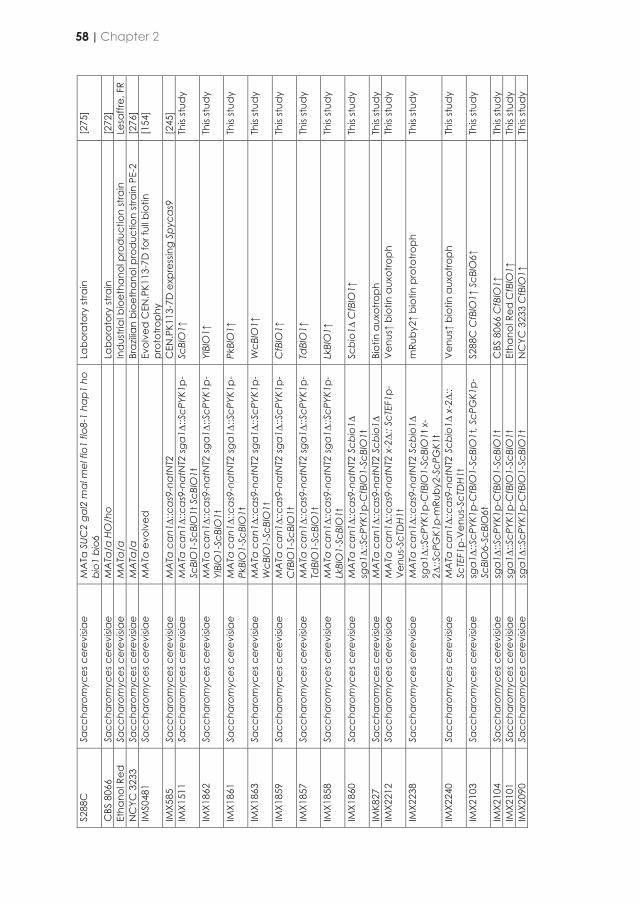
S288
C
Sacc
haro
myc
es c
erev
isiae
M
ATα
SUC
2 ga
l2 m
al m
el fl
o1 fl
o8-1
hap
1 ho
bi
o1 b
io6
Labo
rato
ry st
rain
[2
75]
CBS
806
6 Sa
ccha
rom
yces
cer
evisi
ae
MA
Ta/α
HO
/ho
Labo
rato
ry st
rain
[2
72]
Etha
nol R
ed
Sacc
haro
myc
es c
erev
isiae
M
ATa
/α
Ind
ustri
al b
ioet
hano
l pro
duc
tion
stra
in
Lesa
ffre,
FR
NC
YC 3
233
Sacc
haro
myc
es c
erev
isiae
M
ATa
/α
Braz
ilian
bioe
than
ol p
rod
uctio
n st
rain
PE-
2 [2
76]
IMS0
481
Sacc
haro
myc
es c
erev
isiae
M
ATa
evo
lved
Ev
olve
d C
EN.P
K113
-7D
for f
ull b
iotin
pr
otot
roph
y
[154
]
IMX5
85
Sacc
haro
myc
es c
erev
isiae
M
ATa
ca
n1∆:
:cas
9-na
tNT2
C
EN.P
K113
-7D
exp
ress
ing
Spyc
as9
[2
45]
IMX1
511
Sacc
haro
myc
es c
erev
isiae
M
ATa
ca
n1∆:
:cas
9-na
tNT2
sga
1∆::S
cPYK
1p-
ScBI
O1-
ScBI
O1t
ScB
IO1t
Sc
BIO
1↑
This
stud
y
IMX1
862
Sacc
haro
myc
es c
erev
isiae
M
ATa
ca
n1∆:
:cas
9-na
tNT2
sga
1∆::S
cPYK
1p-
YlBI
O1-
ScBI
O1t
Yl
BIO
1↑
This
stud
y
IMX1
861
Sacc
haro
myc
es c
erev
isiae
M
ATa
ca
n1∆:
:cas
9-na
tNT2
sga
1∆::S
cPYK
1p-
PkBI
O1-
ScBI
O1t
Pk
BIO
1↑
This
stud
y
IMX1
863
Sacc
haro
myc
es c
erev
isiae
M
ATa
ca
n1∆:
:ca
s9-n
atN
T2 sg
a1∆
::ScP
YK1p
-W
cBIO
1-Sc
BIO
1t
WcB
IO1↑
Th
is st
udy
IMX1
859
Sacc
haro
myc
es c
erev
isiae
M
ATa
ca
n1∆:
:ca
s9-n
atN
T2 sg
a1∆
::ScP
YK1p
-C
fBIO
1-Sc
BIO
1t
CfB
IO1↑
Th
is st
udy
IMX1
857
Sacc
haro
myc
es c
erev
isiae
M
ATa
ca
n1∆:
:ca
s9-n
atN
T2 sg
a1∆
::ScP
YK1p
-Td
BIO
1-Sc
BIO
1t
TdBI
O1↑
Th
is st
udy
IMX1
858
Sacc
haro
myc
es c
erev
isiae
M
ATa
ca
n1∆:
:ca
s9-n
atN
T2 sg
a1∆
::ScP
YK1p
-Lk
BIO
1-Sc
BIO
1t
LkBI
O1↑
Th
is st
udy
IMX1
860
Sacc
haro
myc
es c
erev
isiae
M
ATa
ca
n1∆:
:cas
9-na
tNT2
Scb
io1∆
sg
a1∆
::ScP
YK1p
-CfB
IO1-
ScBI
O1t
Sc
bio1∆
CfB
IO1↑
Th
is st
udy
IMK8
27
Sacc
haro
myc
es c
erev
isiae
M
ATa
ca
n1∆:
:cas
9-na
tNT2
Scb
io1∆
Bi
otin
aux
otro
ph
This
stud
y IM
X221
2 Sa
ccha
rom
yces
cer
evisi
ae
MA
Ta c
an1∆:
:cas
9-na
tNT2
x-2∆:
: ScT
EF1p
-V
enus
-ScT
DH1
t V
enus↑
biot
in a
uxot
roph
Th
is st
udy
IMX2
238
Sacc
haro
myc
es c
erev
isiae
M
ATa
ca
n1∆:
:cas
9-na
tNT2
Scb
io1∆
sg
a1∆
::ScP
YK1p
-CfB
IO1-
ScBI
O1t
x-
2∆::S
cPG
K1p-
mRu
by2-
ScPG
K1t
mRu
by2↑
bio
tin p
roto
troph
Th
is st
udy
IMX2
240
Sacc
haro
myc
es c
erev
isiae
M
ATa
ca
n1∆:
:cas
9-na
tNT2
Scb
io1∆
x-2∆:
: Sc
TEF1
p-V
enus
-ScT
DH
1t
Ven
us↑
biot
in a
uxot
roph
Th
is st
udy
IMX2
103
Sacc
haro
myc
es c
erev
isiae
sg
a1∆:
:ScP
YK1p
-CfB
IO1-
ScBI
O1t
, ScP
GK1
p-Sc
BIO
6-Sc
BIO
6t
S288
C C
fBIO
1↑ S
cBIO
6↑
This
stud
y
IMX2
104
Sacc
haro
myc
es c
erev
isiae
sg
a1∆
::ScP
YK1p
-CfB
IO1-
ScBI
O1t
C
BS 8
066
CfB
IO1↑
Th
is st
udy
IMX2
101
Sacc
haro
myc
es c
erev
isiae
sg
a1∆
::ScP
YK1p
-CfB
IO1-
ScBI
O1t
Et
hano
l Red
CfB
IO1↑
Th
is st
udy
IMX2
090
Sacc
haro
myc
es c
erev
isiae
sg
a1∆
::ScP
YK1p
-CfB
IO1-
ScBI
O1t
N
CYC
323
3 C
fBIO
1↑
This
stud
y
Molecular biology techniques
DNA fragments were amplified by PCR amplification with Phusion Hot Start II High Fidelity Polymerase (Thermo Fisher Scientific, Landsmeer, Netherlands) and desalted or PAGE-purified oligonucleotide primers (Sigma-Aldrich, St. Louis, MO) (Table 2.3) according to manufacturers’ instructions. For diagnostic PCR analysis of constructed plasmids and strains DreamTaq polymerase (Thermo Fisher Scientific) was used according to manufacturers’ recommendations. PCR products were separated by electrophoresis on 1% (w/v) agarose gels in Tris-acetate-EDTA (TAE) buffer (Thermo Fisher Scientific) with SERVA DNA Stain G (1:100,000) (Serva, Heidelberg, Germany) and, if required, purified with a Zymoclean Gel DNA Recovery kit (Zymo Research, Irvine, CA) or GenElute PCR Clean-Up kit (Sigma-Aldrich). Yeast strains of the CEN.PK lineage and S288C, NCYC 3233/PE-2 were transformed by the lithium acetate (LiAc) method. Yeast strains Ethanol Red and CBS 8066 were transformed using electroporation as previously described [278] with a 2 mm cuvette (Bio-Rad, Hercules, CA) using a Gene Pulser Xcell electroporation system (Bio-Rad). Yeast genomic DNA was isolated using the YeaStar Genomic DNA kit (Zymo Research) or using the SDS-LiAc protocol [279]. E. coli cells were chemically transformed and plated on selective LB agar medium. Plasmids from selected clones were isolated from E. coli with a Sigma GenElute Plasmid kit (Sigma-Aldrich).
Table 2.2 | Primers used in this study. Primer No. Sequence 5’ → 3’ 3841 CACCTTTCGAGAGGACGATG 4892 TACAGACACGACGCATGG 5328 ATTTTAGCGTAAAGGATGGG 5941 GCTGGCCTTTTGCTCACATG 6005 GATCATTTATCTTTCACTGCGGAGAAG 6006 GTTTTAGAGCTAGAAATAGCAAGTTAAAATAAGGCTAGTC 7428 TGTGATGATGTTTTATTTGTTTTGATTGG 7469 GGAGTTGACCGTCTTAACAG 8737 ATAACGGGTTTCTCGCTGAA 10235 TTGTGCGGTTTCGTTTAGGG 10320 CATGCGCGGATGACACGAAC 10325 AGTCATCCGAGCGTGTATTG 10873 ACGTGCGGAATAGGAATCTC 11037 AGTTCTTTGTAGAACAGCTCTC 11614 GCATCGTCTCATCGGTCTCATATGATGAACACAAAATCACTCG 11615 ATGCCGTCTCAGGTCTCAGGATTTACTCTTTATCGTCATAAATAATCTTG 11618 GCATCGTCTCATCGGTCTCAATCCATATCATCATTTCTGCCACAAATATATG 11619 ATGCCGTCTCAGGTCTCACAGCCGGTAGCTTGACGTGCGGAATAG 11898 CGCGGAAACGGGTATTAGGG 11899 CTAGATCCGGTAAGCGACAG 11915 GAGTGAGTGCTTTGTTCAATGG 11945 AGCATCACCTTCACCTTCAC 12086 TTTACAATATAGTGATAATCGTGGACTAGAGCAAGATTTCAAATAAGTAACAGCAGCAAAGCCGATAATT
GCAGACGAAC 12108 TAAATATCTAGGAAATACACTTGTGTATACTTCTCGCTTTCCTTTTATTTTTTTTTTTGTCATGCTCAGCCGGTAGCT
TG 12223 CCAGGTGGCGTGCTAAACTTTTATAATGTATAAAAACCACCACCTCATAAAGTTTACTGGATATCATCATTT
CTGCCACAAATATATGTACTGAGTCTATACGTCAAAGTAAAAAAATAA 12224 TTATTTTTTTACTTTGACGTATAGACTCAGTACATATATTTGTGGCAGAAATGATGATATCCAGTAAACTTTATGA
GGTGGTGGTTTTTATACATTATAAAAGTTTAGCACGCCACCTGG
58 | Chapter 2
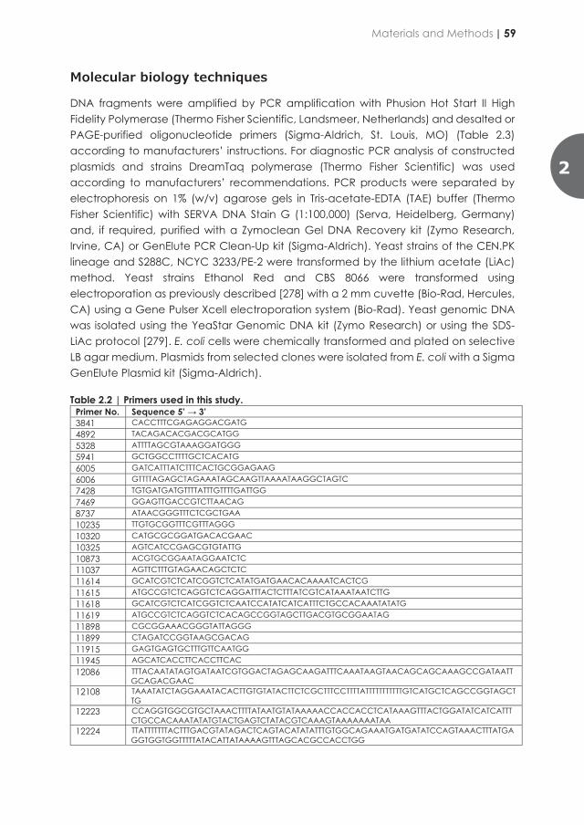
S288
C
Sacc
haro
myc
es c
erev
isiae
M
ATα
SUC
2 ga
l2 m
al m
el fl
o1 fl
o8-1
hap
1 ho
bi
o1 b
io6
Labo
rato
ry st
rain
[2
75]
CBS
806
6 Sa
ccha
rom
yces
cer
evisi
ae
MA
Ta/α
HO
/ho
Labo
rato
ry st
rain
[2
72]
Etha
nol R
ed
Sacc
haro
myc
es c
erev
isiae
M
ATa
/α
Ind
ustri
al b
ioet
hano
l pro
duc
tion
stra
in
Lesa
ffre,
FR
NC
YC 3
233
Sacc
haro
myc
es c
erev
isiae
M
ATa
/α
Braz
ilian
bioe
than
ol p
rod
uctio
n st
rain
PE-
2 [2
76]
IMS0
481
Sacc
haro
myc
es c
erev
isiae
M
ATa
evo
lved
Ev
olve
d C
EN.P
K113
-7D
for f
ull b
iotin
pr
otot
roph
y
[154
]
IMX5
85
Sacc
haro
myc
es c
erev
isiae
M
ATa
ca
n1∆:
:cas
9-na
tNT2
C
EN.P
K113
-7D
exp
ress
ing
Spyc
as9
[2
45]
IMX1
511
Sacc
haro
myc
es c
erev
isiae
M
ATa
ca
n1∆:
:cas
9-na
tNT2
sga
1∆::S
cPYK
1p-
ScBI
O1-
ScBI
O1t
ScB
IO1t
Sc
BIO
1↑
This
stud
y
IMX1
862
Sacc
haro
myc
es c
erev
isiae
M
ATa
ca
n1∆:
:cas
9-na
tNT2
sga
1∆::S
cPYK
1p-
YlBI
O1-
ScBI
O1t
Yl
BIO
1↑
This
stud
y
IMX1
861
Sacc
haro
myc
es c
erev
isiae
M
ATa
ca
n1∆:
:cas
9-na
tNT2
sga
1∆::S
cPYK
1p-
PkBI
O1-
ScBI
O1t
Pk
BIO
1↑
This
stud
y
IMX1
863
Sacc
haro
myc
es c
erev
isiae
M
ATa
ca
n1∆:
:ca
s9-n
atN
T2 sg
a1∆
::ScP
YK1p
-W
cBIO
1-Sc
BIO
1t
WcB
IO1↑
Th
is st
udy
IMX1
859
Sacc
haro
myc
es c
erev
isiae
M
ATa
ca
n1∆:
:ca
s9-n
atN
T2 sg
a1∆
::ScP
YK1p
-C
fBIO
1-Sc
BIO
1t
CfB
IO1↑
Th
is st
udy
IMX1
857
Sacc
haro
myc
es c
erev
isiae
M
ATa
ca
n1∆:
:ca
s9-n
atN
T2 sg
a1∆
::ScP
YK1p
-Td
BIO
1-Sc
BIO
1t
TdBI
O1↑
Th
is st
udy
IMX1
858
Sacc
haro
myc
es c
erev
isiae
M
ATa
ca
n1∆:
:ca
s9-n
atN
T2 sg
a1∆
::ScP
YK1p
-Lk
BIO
1-Sc
BIO
1t
LkBI
O1↑
Th
is st
udy
IMX1
860
Sacc
haro
myc
es c
erev
isiae
M
ATa
ca
n1∆:
:cas
9-na
tNT2
Scb
io1∆
sg
a1∆
::ScP
YK1p
-CfB
IO1-
ScBI
O1t
Sc
bio1∆
CfB
IO1↑
Th
is st
udy
IMK8
27
Sacc
haro
myc
es c
erev
isiae
M
ATa
ca
n1∆:
:cas
9-na
tNT2
Scb
io1∆
Bi
otin
aux
otro
ph
This
stud
y IM
X221
2 Sa
ccha
rom
yces
cer
evisi
ae
MA
Ta c
an1∆:
:cas
9-na
tNT2
x-2∆:
: ScT
EF1p
-V
enus
-ScT
DH1
t V
enus↑
biot
in a
uxot
roph
Th
is st
udy
IMX2
238
Sacc
haro
myc
es c
erev
isiae
M
ATa
ca
n1∆:
:cas
9-na
tNT2
Scb
io1∆
sg
a1∆
::ScP
YK1p
-CfB
IO1-
ScBI
O1t
x-
2∆::S
cPG
K1p-
mRu
by2-
ScPG
K1t
mRu
by2↑
bio
tin p
roto
troph
Th
is st
udy
IMX2
240
Sacc
haro
myc
es c
erev
isiae
M
ATa
ca
n1∆:
:cas
9-na
tNT2
Scb
io1∆
x-2∆:
: Sc
TEF1
p-V
enus
-ScT
DH
1t
Ven
us↑
biot
in a
uxot
roph
Th
is st
udy
IMX2
103
Sacc
haro
myc
es c
erev
isiae
sg
a1∆:
:ScP
YK1p
-CfB
IO1-
ScBI
O1t
, ScP
GK1
p-Sc
BIO
6-Sc
BIO
6t
S288
C C
fBIO
1↑ S
cBIO
6↑
This
stud
y
IMX2
104
Sacc
haro
myc
es c
erev
isiae
sg
a1∆
::ScP
YK1p
-CfB
IO1-
ScBI
O1t
C
BS 8
066
CfB
IO1↑
Th
is st
udy
IMX2
101
Sacc
haro
myc
es c
erev
isiae
sg
a1∆
::ScP
YK1p
-CfB
IO1-
ScBI
O1t
Et
hano
l Red
CfB
IO1↑
Th
is st
udy
IMX2
090
Sacc
haro
myc
es c
erev
isiae
sg
a1∆
::ScP
YK1p
-CfB
IO1-
ScBI
O1t
N
CYC
323
3 C
fBIO
1↑
This
stud
y
Molecular biology techniques
DNA fragments were amplified by PCR amplification with Phusion Hot Start II High Fidelity Polymerase (Thermo Fisher Scientific, Landsmeer, Netherlands) and desalted or PAGE-purified oligonucleotide primers (Sigma-Aldrich, St. Louis, MO) (Table 2.3) according to manufacturers’ instructions. For diagnostic PCR analysis of constructed plasmids and strains DreamTaq polymerase (Thermo Fisher Scientific) was used according to manufacturers’ recommendations. PCR products were separated by electrophoresis on 1% (w/v) agarose gels in Tris-acetate-EDTA (TAE) buffer (Thermo Fisher Scientific) with SERVA DNA Stain G (1:100,000) (Serva, Heidelberg, Germany) and, if required, purified with a Zymoclean Gel DNA Recovery kit (Zymo Research, Irvine, CA) or GenElute PCR Clean-Up kit (Sigma-Aldrich). Yeast strains of the CEN.PK lineage and S288C, NCYC 3233/PE-2 were transformed by the lithium acetate (LiAc) method. Yeast strains Ethanol Red and CBS 8066 were transformed using electroporation as previously described [278] with a 2 mm cuvette (Bio-Rad, Hercules, CA) using a Gene Pulser Xcell electroporation system (Bio-Rad). Yeast genomic DNA was isolated using the YeaStar Genomic DNA kit (Zymo Research) or using the SDS-LiAc protocol [279]. E. coli cells were chemically transformed and plated on selective LB agar medium. Plasmids from selected clones were isolated from E. coli with a Sigma GenElute Plasmid kit (Sigma-Aldrich).
Table 2.2 | Primers used in this study. Primer No. Sequence 5’ → 3’ 3841 CACCTTTCGAGAGGACGATG 4892 TACAGACACGACGCATGG 5328 ATTTTAGCGTAAAGGATGGG 5941 GCTGGCCTTTTGCTCACATG 6005 GATCATTTATCTTTCACTGCGGAGAAG 6006 GTTTTAGAGCTAGAAATAGCAAGTTAAAATAAGGCTAGTC 7428 TGTGATGATGTTTTATTTGTTTTGATTGG 7469 GGAGTTGACCGTCTTAACAG 8737 ATAACGGGTTTCTCGCTGAA 10235 TTGTGCGGTTTCGTTTAGGG 10320 CATGCGCGGATGACACGAAC 10325 AGTCATCCGAGCGTGTATTG 10873 ACGTGCGGAATAGGAATCTC 11037 AGTTCTTTGTAGAACAGCTCTC 11614 GCATCGTCTCATCGGTCTCATATGATGAACACAAAATCACTCG 11615 ATGCCGTCTCAGGTCTCAGGATTTACTCTTTATCGTCATAAATAATCTTG 11618 GCATCGTCTCATCGGTCTCAATCCATATCATCATTTCTGCCACAAATATATG 11619 ATGCCGTCTCAGGTCTCACAGCCGGTAGCTTGACGTGCGGAATAG 11898 CGCGGAAACGGGTATTAGGG 11899 CTAGATCCGGTAAGCGACAG 11915 GAGTGAGTGCTTTGTTCAATGG 11945 AGCATCACCTTCACCTTCAC 12086 TTTACAATATAGTGATAATCGTGGACTAGAGCAAGATTTCAAATAAGTAACAGCAGCAAAGCCGATAATT
GCAGACGAAC 12108 TAAATATCTAGGAAATACACTTGTGTATACTTCTCGCTTTCCTTTTATTTTTTTTTTTGTCATGCTCAGCCGGTAGCT
TG 12223 CCAGGTGGCGTGCTAAACTTTTATAATGTATAAAAACCACCACCTCATAAAGTTTACTGGATATCATCATTT
CTGCCACAAATATATGTACTGAGTCTATACGTCAAAGTAAAAAAATAA 12224 TTATTTTTTTACTTTGACGTATAGACTCAGTACATATATTTGTGGCAGAAATGATGATATCCAGTAAACTTTATGA
GGTGGTGGTTTTTATACATTATAAAAGTTTAGCACGCCACCTGG
Materials and Methods | 59
2
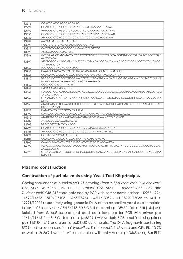
12616 CGAGTCAGTGAGCGAGGAAG 12991 GCATCGTCTCATCGGTCTCATATGGCGTCTAAGAACCAAAA 12992 ATGCCGTCTCAGGTCTCAGGATCTACTCAAAAACTGCATGGA 13038 GCATCGTCTCATCGGTCTCATATGACGTTTAGTAAGAACTTAAC 13039 ATGCCGTCTCAGGTCTCAGGATCTATTCGATAACAGGATAAA 13287 AACAAGATCCGAGTACTTAG 13290 TTCGTCTCTCTCACACTTATACGGGTCGTTAGT 13291 CACGTCTCATGAGCCCGGAATAAATCTAGTGGC 13293 ATATCGTCCCTAGTCAATTC 13596 GCTGAAGATTTATCATACTATTCCTCCGCTCGTTTCTTTTTTCAGTGAGGTGTGTCGTGATGAACTGGCCGAT
AATTGCAGA 13597 ATTCTCGCCAAGGCATTACCATCCCATGTAAGAACGGAATAAAACAGCATTCGAAGGTTATGATGACC
CCGTCGTCTCATT 13662 TCCTCGGGCAGAGAAACTCG 13963 CAAATAAAACATCATCACATATGACACATATAAGTAACTCGGAGTATC 13964 GCAGAAATGATGATATGGATTTATATACGAACTACTTTACAAACATCA 14139 TGCGCATGTTTCGGCGTTCGAAACTTCTCCGCAGTGAAAGATAAATGATCAGGAAACGCTACTCGGAG
TAGTTTTAGAGCTAGAAATAGCAAGTTAAAATAAG 14162 GGCACCTCTGGCTTGTCTTC 14167 TACTCCGAGTAGCGTTTCCT 14661 TTAGGGAGCACATCCATGCCAATAGCTCGACAAGCGGCGAGAGCCTTGCACCTATGCTATCAATAGG
CACACTGTAATTG 14662 GTATATATATATATTTGATGTAAATATCTAGGAAATACACTTGTGTATACTTCTCGCTTTCTAAACTGAGCACAA
GTTTC 14663 ATAGCATAGGTGCAAGGCTCTCGCCGCTTGTCGAGCTATTGGCATGGATGTGCTCCCTAATAGCTTGAC
GTGCGGAATAG 14891 CATATCATCATTTCTGCCACAAATAT 14892 CAATCAAAACAAATAAAACATCATCACAATGAGTTTCAACTACGAAGACTG 14893 ATATTTGTGGCAGAAATGATGATATGTTAGTCGTATAAAACTTTACATACTT 14907 AATGCAATGGAGCTTGAGAC 14909 TGGTCGTCTCTATGCAAAGG 14925 GCATCGTCTCATCGGTCTCATATGTGCTGTACATGTACATACCA 14926 ATGCCGTCTCAGGTCTCAGGATTAGGCGCGTAAAGTTATTAC 14928 GGGAGGTCGCAATATCTCTG 15104 GCAGAAATGATGATATGGATTTAATTAACATCTGAGACTT 15105 CAAATAAAACATCATCACATATGACTGTTATAGATACCAATGA 16792 TCACAGAGGGATCCCGTTACCCATCTATGCTGAAGATTTATCATACTATTCCTCCGCTCGGCCTTGCCAA
CAGGGAGTTC 16793 GTCATAACTCAATTTGCCTATTTCTTACGGCTTCTCATAAAACGTCCCACACTATTCAGGCGTTCAGGGTAA
TATATTTT
Plasmid construction
Construction of part plasmids using Yeast Tool Kit principle.
Coding sequences of putative ScBIO1 orthologs from Y. lipolytica W29, P. kudriavzevii CBS 5147, W. ciferrii CBS 111, C. fabianii CBS 5481, L. kluyveri CBS 3082 and T. delbrueckii CBS 813 were obtained by PCR with primer combinations 14925/14926, 14892/14893, 15104/15105, 13963/13964, 13291/13039 and 13290/13038 as well as 12991/12992 respectively using genomic DNA of the respective yeast as a template. In case of S. cerevisiae CEN.PK113-7D BIO1, the plasmid pUDE450 (Table 2.4) [154] was isolated from E. coli cultures and used as a template for PCR with primer pair 11614/11615. The ScBIO1 terminator (ScBIO1t) was similarly PCR amplified using primer pair 11618/11619 and plasmid pUDE450 as template. The DNA fragments containing BIO1 coding sequences from Y. lypolytica, T. delbrueckii, L. kluyverii and CEN.PK113-7D as well as ScBIO1t were in vitro assembled with entry vector pUD565 using BsmBI-T4
ligase directed Golden Gate cloning [280] resulting in Yeast Tool Kit type 3 plasmids pGGkp243, pGGkp169, pGGkp178 and pGGkp080 respectively and a Yeast Tool Kit type 4 plasmid with ScBIO1t pGGkp078 (Table 2.4). In order to remove a BsaI restriction recognition site in the coding sequence of LkBIO1, the open reading frame was PCR amplified from L. kluyveri CBS 3082 genomic DNA using two primer pairs 13291/13039 and 13290/13038 with overhangs allowing for BsmBI-T4 ligase directed Golden Gate cloning of the two DNA fragments into entry vector pUD565 [281], leaving a sequence without BsaI site behind and resulting in the Yeast Tool Kit type 3 plasmid pGGkp178. After in vitro assembly, plasmids were transformed into E. coli and plated on LB chloramphenicol for selection. The Yeast Tool Kit type plasmids pGGkp080, pGGkp169 pGGkp078 and pGGkp178 were confirmed by diagnostic PCR with primer pair 12616/4892, 12616/13287, 12616/10235and 12616/13290 respectively. Yeast Tool Kit type plasmid pGGkp243 was confirmed by restriction analysis with restriction enzymes PvuII and DraI. The promoter ScPYK1p was synthesized by GeneArt (Thermo Fisher Scientific) and cloned as plasmid pGGkp117. The Yeast Tool Kit type plasmids were stored in transformed E. coli cultures.
Construction of transcriptional modules
The control S. cerevisiae CEN.PK113-7D BIO1 transcriptional module was constructed by Golden Gate cloning combining DNA fragments with compatible overhangs from plasmids pGGkd017, pGGkp117, pGGkp080, pGGkp078 yielding plasmid pUDE718. The entry plasmid pGGkd017 was also constructed by Golden Gate cloning combining DNA fragments with compatible overhangs from pYTK002, pYTK047, pYTK072, pYTK074, pYTK082 and pYTK083 [280]. The T. delbrueckii BIO1 transcriptional module was constructed by Golden Gate cloning combining DNA fragments with compatible overhangs from plasmids pGGkd015, pGGkp117, pGGkp169, pGGkp078 yielding plasmid pUD788. The entry plasmid pGGkd015 was obtained by Golden Gate cloning with Yeast Tool Kit type plasmids pYTK002, pYTK047, pYTK067 and pYTK095. The L. kluyverii BIO1 transcriptional module was constructed by Golden Gate cloning combining DNA fragments with compatible overhangs from plasmids pGGkd015, pGGkp117, pGGkp178, pGGkp078 yielding plasmid pUD789. The Y. lipolytica BIO1 transcriptional module was constructed by Golden Gate cloning combining DNA fragment with compatible overhangs from plasmids pGGkd015, pGGkp117, pGGkp243, pGGkp078 yielding plasmid pUD989. The transcriptional modules of P. kudriavzevii, W. ciferrii and C. fabianii BIO1 genes were constructed by Gibson assembly [282] (New England Biolabs, Ipswich, MA) using pUDE718 as plasmid backbone which includes the ScPYK1p and ScBIO1t sequences. The BIO1 genes from P. kudriavzevii CBS 5147, W. ciferrii CBS 111, C. fabianii CBS 5481 were amplified by primer pairs 14892/14893, 15104/15105, 13963/13964 respectively using genomic DNA of the respective yeast as a template. DNA fragments were assembled with linearized pUDE718 backbone using primer pair 7428/14891 yielding plasmids pUD988, pUD990 and pUD790 respectively. The assembly mixes were transformed into E. coli and plated
60 | Chapter 2
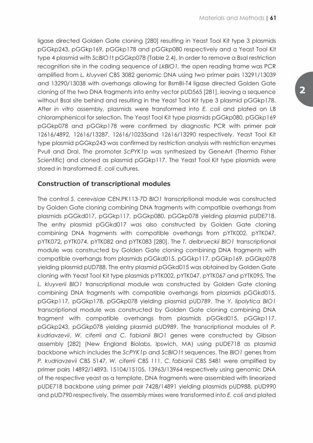
12616 CGAGTCAGTGAGCGAGGAAG 12991 GCATCGTCTCATCGGTCTCATATGGCGTCTAAGAACCAAAA 12992 ATGCCGTCTCAGGTCTCAGGATCTACTCAAAAACTGCATGGA 13038 GCATCGTCTCATCGGTCTCATATGACGTTTAGTAAGAACTTAAC 13039 ATGCCGTCTCAGGTCTCAGGATCTATTCGATAACAGGATAAA 13287 AACAAGATCCGAGTACTTAG 13290 TTCGTCTCTCTCACACTTATACGGGTCGTTAGT 13291 CACGTCTCATGAGCCCGGAATAAATCTAGTGGC 13293 ATATCGTCCCTAGTCAATTC 13596 GCTGAAGATTTATCATACTATTCCTCCGCTCGTTTCTTTTTTCAGTGAGGTGTGTCGTGATGAACTGGCCGAT
AATTGCAGA 13597 ATTCTCGCCAAGGCATTACCATCCCATGTAAGAACGGAATAAAACAGCATTCGAAGGTTATGATGACC
CCGTCGTCTCATT 13662 TCCTCGGGCAGAGAAACTCG 13963 CAAATAAAACATCATCACATATGACACATATAAGTAACTCGGAGTATC 13964 GCAGAAATGATGATATGGATTTATATACGAACTACTTTACAAACATCA 14139 TGCGCATGTTTCGGCGTTCGAAACTTCTCCGCAGTGAAAGATAAATGATCAGGAAACGCTACTCGGAG
TAGTTTTAGAGCTAGAAATAGCAAGTTAAAATAAG 14162 GGCACCTCTGGCTTGTCTTC 14167 TACTCCGAGTAGCGTTTCCT 14661 TTAGGGAGCACATCCATGCCAATAGCTCGACAAGCGGCGAGAGCCTTGCACCTATGCTATCAATAGG
CACACTGTAATTG 14662 GTATATATATATATTTGATGTAAATATCTAGGAAATACACTTGTGTATACTTCTCGCTTTCTAAACTGAGCACAA
GTTTC 14663 ATAGCATAGGTGCAAGGCTCTCGCCGCTTGTCGAGCTATTGGCATGGATGTGCTCCCTAATAGCTTGAC
GTGCGGAATAG 14891 CATATCATCATTTCTGCCACAAATAT 14892 CAATCAAAACAAATAAAACATCATCACAATGAGTTTCAACTACGAAGACTG 14893 ATATTTGTGGCAGAAATGATGATATGTTAGTCGTATAAAACTTTACATACTT 14907 AATGCAATGGAGCTTGAGAC 14909 TGGTCGTCTCTATGCAAAGG 14925 GCATCGTCTCATCGGTCTCATATGTGCTGTACATGTACATACCA 14926 ATGCCGTCTCAGGTCTCAGGATTAGGCGCGTAAAGTTATTAC 14928 GGGAGGTCGCAATATCTCTG 15104 GCAGAAATGATGATATGGATTTAATTAACATCTGAGACTT 15105 CAAATAAAACATCATCACATATGACTGTTATAGATACCAATGA 16792 TCACAGAGGGATCCCGTTACCCATCTATGCTGAAGATTTATCATACTATTCCTCCGCTCGGCCTTGCCAA
CAGGGAGTTC 16793 GTCATAACTCAATTTGCCTATTTCTTACGGCTTCTCATAAAACGTCCCACACTATTCAGGCGTTCAGGGTAA
TATATTTT
Plasmid construction
Construction of part plasmids using Yeast Tool Kit principle.
Coding sequences of putative ScBIO1 orthologs from Y. lipolytica W29, P. kudriavzevii CBS 5147, W. ciferrii CBS 111, C. fabianii CBS 5481, L. kluyveri CBS 3082 and T. delbrueckii CBS 813 were obtained by PCR with primer combinations 14925/14926, 14892/14893, 15104/15105, 13963/13964, 13291/13039 and 13290/13038 as well as 12991/12992 respectively using genomic DNA of the respective yeast as a template. In case of S. cerevisiae CEN.PK113-7D BIO1, the plasmid pUDE450 (Table 2.4) [154] was isolated from E. coli cultures and used as a template for PCR with primer pair 11614/11615. The ScBIO1 terminator (ScBIO1t) was similarly PCR amplified using primer pair 11618/11619 and plasmid pUDE450 as template. The DNA fragments containing BIO1 coding sequences from Y. lypolytica, T. delbrueckii, L. kluyverii and CEN.PK113-7D as well as ScBIO1t were in vitro assembled with entry vector pUD565 using BsmBI-T4
ligase directed Golden Gate cloning [280] resulting in Yeast Tool Kit type 3 plasmids pGGkp243, pGGkp169, pGGkp178 and pGGkp080 respectively and a Yeast Tool Kit type 4 plasmid with ScBIO1t pGGkp078 (Table 2.4). In order to remove a BsaI restriction recognition site in the coding sequence of LkBIO1, the open reading frame was PCR amplified from L. kluyveri CBS 3082 genomic DNA using two primer pairs 13291/13039 and 13290/13038 with overhangs allowing for BsmBI-T4 ligase directed Golden Gate cloning of the two DNA fragments into entry vector pUD565 [281], leaving a sequence without BsaI site behind and resulting in the Yeast Tool Kit type 3 plasmid pGGkp178. After in vitro assembly, plasmids were transformed into E. coli and plated on LB chloramphenicol for selection. The Yeast Tool Kit type plasmids pGGkp080, pGGkp169 pGGkp078 and pGGkp178 were confirmed by diagnostic PCR with primer pair 12616/4892, 12616/13287, 12616/10235and 12616/13290 respectively. Yeast Tool Kit type plasmid pGGkp243 was confirmed by restriction analysis with restriction enzymes PvuII and DraI. The promoter ScPYK1p was synthesized by GeneArt (Thermo Fisher Scientific) and cloned as plasmid pGGkp117. The Yeast Tool Kit type plasmids were stored in transformed E. coli cultures.
Construction of transcriptional modules
The control S. cerevisiae CEN.PK113-7D BIO1 transcriptional module was constructed by Golden Gate cloning combining DNA fragments with compatible overhangs from plasmids pGGkd017, pGGkp117, pGGkp080, pGGkp078 yielding plasmid pUDE718. The entry plasmid pGGkd017 was also constructed by Golden Gate cloning combining DNA fragments with compatible overhangs from pYTK002, pYTK047, pYTK072, pYTK074, pYTK082 and pYTK083 [280]. The T. delbrueckii BIO1 transcriptional module was constructed by Golden Gate cloning combining DNA fragments with compatible overhangs from plasmids pGGkd015, pGGkp117, pGGkp169, pGGkp078 yielding plasmid pUD788. The entry plasmid pGGkd015 was obtained by Golden Gate cloning with Yeast Tool Kit type plasmids pYTK002, pYTK047, pYTK067 and pYTK095. The L. kluyverii BIO1 transcriptional module was constructed by Golden Gate cloning combining DNA fragments with compatible overhangs from plasmids pGGkd015, pGGkp117, pGGkp178, pGGkp078 yielding plasmid pUD789. The Y. lipolytica BIO1 transcriptional module was constructed by Golden Gate cloning combining DNA fragment with compatible overhangs from plasmids pGGkd015, pGGkp117, pGGkp243, pGGkp078 yielding plasmid pUD989. The transcriptional modules of P. kudriavzevii, W. ciferrii and C. fabianii BIO1 genes were constructed by Gibson assembly [282] (New England Biolabs, Ipswich, MA) using pUDE718 as plasmid backbone which includes the ScPYK1p and ScBIO1t sequences. The BIO1 genes from P. kudriavzevii CBS 5147, W. ciferrii CBS 111, C. fabianii CBS 5481 were amplified by primer pairs 14892/14893, 15104/15105, 13963/13964 respectively using genomic DNA of the respective yeast as a template. DNA fragments were assembled with linearized pUDE718 backbone using primer pair 7428/14891 yielding plasmids pUD988, pUD990 and pUD790 respectively. The assembly mixes were transformed into E. coli and plated
Materials and Methods | 61
2

on LB ampicillin for selection. The transcriptional module plasmids were confirmed by diagnostic PCR with the forward primer 10320 and a gene-specific reverse primer as follows: 13287 for TdBIO1, 13293 for LkBIO1, 14928 for YlBIO1, 4892 for ScBIO1, 14909 for PkBIO1, 14907 for WcBIO1 and 14162 for CfBIO1. The transcriptional module for expression of fluorophore mRuby2 was constructed by Golden Gate cloning combining DNA fragment with compatible overhangs from plasmids pGGKd005, pYTK011, pYTK046, pYTK054. The entry plasmid pGGkd005 was constructed by Golden Gate cloning combining DNA fragments with compatible overhangs from pYTK002, pYTK047, pYTK067, pYTK079, pYTK083 and pYTK081. The assembly mixes were transformed into E. coli and plated on LB ampicillin for selection. The mRuby2 transcriptional module plasmid was confirmed by diagnostic PCR with primer pair 10320/10325 and stored as pUDE480 in transformed E. coli cultures.
Spycas9 expressing plasmids
The gRNAScSGA1 expressing plasmid pUDR244 was constructed by in vitro Gibson assembly. The linearized pROS11 plasmid, obtained by PCR with 6005/6006 was assembled together with a PCR amplified fragment using primer 14139 and pROS11 as a template as previously described [245]. The plasmid DNA was isolated from E. coli and the correct assembly of plasmid pUDR244 confirmed by diagnostic PCR with primers 3841/14167/5941. Similarly, plasmid pUDR376 was assembled with linearized pROS11 and the PCR amplified DNA fragment but using primer 10866. Assembly of pUDP145 was performed in vitro by BsaI–T4 DNA ligase directed Golden Gate cloning with the gRNA entry plasmid pUDP002 [283] with a de novo-synthesized DNA fragment (GeneArt, Thermo Fisher Scientific) encoding a gRNA targeting the ScSGA1.
Strain construction
The BIO1 transcriptional modules were PCR amplified by using primer pair 12086/12108 adding specific sequences for homologous recombination into the SGA1 locus in S. cerevisiae directed by CRISPR/Cas9 [245]. The transcriptional module was amplified from plasmid pUD788 for TdBIO1, from plasmid pUD789 for LkBIO1, from plasmid pUD989 for YlBIO1, from plasmid pUDE718 for ScBIO1, from plasmid pUD988 for PkBIO1, from plasmid pUD990 for WcBIO1 and from plasmid pUD790 for CfBIO1. Targeting at the ScSGA1 locus in IMX585 was directed by CRISPR/Cas9 and a target-specific gRNA expressing plasmid [245]. Each transcriptional module was co-transformed with plasmid pUDR119 expressing the gRNA to target Cas9 to ScSGA1 in strain IMX585 using the LiAc transformation protocol. The transformed cells were plated on selective SMD acetamide and incubated for 3 days at 30°C. Genomic DNA of transformants was isolated using the SDS/LiAc protocol [279]. The desired genotype was confirmed using primer pair 11898/11899 and a gene-specific primer pair with 11898 as forward primer and the following reverse primers: 13287 for TdBIO1, 13293 for LkBIO1, 14928 for YlBIO1, 4892 for ScBIO1, 14909 for PkBIO1, 14907 for WcBIO1 and 14162 for CfBIO1.
Table 2.4 | Plasmids used in this study. Name Characteristics Reference pGGkd005 hph CEN6/ARS4 bla ColE1 Gfp dropout This study pGGkd015 Bla ColE1 Gfp dropout [281] pGGkd017 URA3 2μ bla ColE1 Gfp dropout This study pGGKp078 cat ScBIO1t This study pGGKp080 cat ScBIO1 This study pGGkp117 cat ScPYK1p This study pGGkp169 cat TdBIO1 This study pGGkp178 cat LkBIO1 This study pGGkp243 cat YlBIO1 This study pROS11 amdS 2μ gRNACAN1 gRNAADE2 [245] pUD565 cat Gfp dropout [281] pUD788 bla ColE1 ScPYK1p-TdBIO1-ScBIO1t This study pUD789 bla ColE1 ScPYK1p-LkBIO1-ScBIO1t This study pUD790 URA3 2μ bla ColE1 ScPYK1p-CfBIO1-ScBIO1t This study pUD988 URA3 2μ bla ColE1 ScPYK1p-PkBIO1-ScBIO1t This study pUD989 bla ColE1 ScPYK1p-YlBIO1-ScBIO1t This study pUD990 URA3 2μ bla ColE1 ScPYK1p-WcBIO1-ScBIO1t This study pUDC193 URA3 CEN6/ARS4 bla ColE1 ScTEF1p-Venus-ScTDH1t This study pUDE448 URA3 2μ bla ColE1 ScPGK1p-ScBIO6-ScBIO6t [154] pUDE450 URA3 2μ bla ColE1 ScPYK1p-ScBIO1-ScBIO1t [154] pUDE480 hph CEN6/ARS4 bla ColE1 ScPGK1p-mRuby2-ScPGK1t This study pUDE718 URA3 2μ bla ColE1 ScPYK1p-ScBIO1-ScBIO1t This study pUDP002 hph panARS(OPT) bla ColE1 ScTDH3p- BsaI site -ScCYC1t AaTEF1p-
Spcas9D147Y P411T-ScPHO5t [283]
pUDP145 hph panARS(OPT) bla ColE1 ScTDH3p-HH-gRNASGA1-HDV-ScCYC1t This study pUDR119 amdS 2μ ScSNR52p-gRNASGA1-SUP4t [245] pUDR244 amdS 2μ ScSNR52p-gRNABIO1-SUP4t ScSNR52p-gRNABIO1-SUP4t This study pUDR376 amdS 2μ bla ColE1 ScSNR52p-gRNAX-2-SUP4t This study pYTK002 cat ConLS [280] pYTK011 cat ScPGK1p [280] pYTK046 cat mRuby2 [280] pYTK047 cat Gfp dropout [280] pYTK054 cat ScPGK1t [280] pYTK067 cat ConR1 [280] pYTK072 cat ConRE [280] pYTK074 cat URA3 [280] pYTK079 cat hph [280] pYTK081 cat CEN6/ARS4 [280] pYTK082 cat 2μ [280] pYTK083 bla ColE1 [280] pYTK095 bla ColE1 Gfp dropout [280]
The correct clone was inoculated in 20 mL non-selective YPD for plasmid removal and incubated for 24 h at 30°C. The cells were plated on YPD agar in order to obtain single colony isolates. One isolate was restreaked on both SMD acetamide and YPD. When no growth was observed on SMD acetamide the respective clone was once again confirmed by PCR with gene-specific primers. Furthermore, the genetic modification at the ScSGA1 locus was verified by Sanger sequencing (BaseClear, Leiden, Netherlands) using primers 11898/11899 to PCR amplify the modified locus and further using primers 11898, 11915 and 10235 for sequencing. The strain with the transcriptional module coding for TdBIO1 was stocked as IMX1857, LkBIO1 as IMX1858, YlBIO1 as IMX1862, ScBIO1 as IMX1511, PkBIO1 as IMX1861, WcBIO1 as IMX1863 and CfBIO1 as IMX1859 in SMD.
62 | Chapter 2

on LB ampicillin for selection. The transcriptional module plasmids were confirmed by diagnostic PCR with the forward primer 10320 and a gene-specific reverse primer as follows: 13287 for TdBIO1, 13293 for LkBIO1, 14928 for YlBIO1, 4892 for ScBIO1, 14909 for PkBIO1, 14907 for WcBIO1 and 14162 for CfBIO1. The transcriptional module for expression of fluorophore mRuby2 was constructed by Golden Gate cloning combining DNA fragment with compatible overhangs from plasmids pGGKd005, pYTK011, pYTK046, pYTK054. The entry plasmid pGGkd005 was constructed by Golden Gate cloning combining DNA fragments with compatible overhangs from pYTK002, pYTK047, pYTK067, pYTK079, pYTK083 and pYTK081. The assembly mixes were transformed into E. coli and plated on LB ampicillin for selection. The mRuby2 transcriptional module plasmid was confirmed by diagnostic PCR with primer pair 10320/10325 and stored as pUDE480 in transformed E. coli cultures.
Spycas9 expressing plasmids
The gRNAScSGA1 expressing plasmid pUDR244 was constructed by in vitro Gibson assembly. The linearized pROS11 plasmid, obtained by PCR with 6005/6006 was assembled together with a PCR amplified fragment using primer 14139 and pROS11 as a template as previously described [245]. The plasmid DNA was isolated from E. coli and the correct assembly of plasmid pUDR244 confirmed by diagnostic PCR with primers 3841/14167/5941. Similarly, plasmid pUDR376 was assembled with linearized pROS11 and the PCR amplified DNA fragment but using primer 10866. Assembly of pUDP145 was performed in vitro by BsaI–T4 DNA ligase directed Golden Gate cloning with the gRNA entry plasmid pUDP002 [283] with a de novo-synthesized DNA fragment (GeneArt, Thermo Fisher Scientific) encoding a gRNA targeting the ScSGA1.
Strain construction
The BIO1 transcriptional modules were PCR amplified by using primer pair 12086/12108 adding specific sequences for homologous recombination into the SGA1 locus in S. cerevisiae directed by CRISPR/Cas9 [245]. The transcriptional module was amplified from plasmid pUD788 for TdBIO1, from plasmid pUD789 for LkBIO1, from plasmid pUD989 for YlBIO1, from plasmid pUDE718 for ScBIO1, from plasmid pUD988 for PkBIO1, from plasmid pUD990 for WcBIO1 and from plasmid pUD790 for CfBIO1. Targeting at the ScSGA1 locus in IMX585 was directed by CRISPR/Cas9 and a target-specific gRNA expressing plasmid [245]. Each transcriptional module was co-transformed with plasmid pUDR119 expressing the gRNA to target Cas9 to ScSGA1 in strain IMX585 using the LiAc transformation protocol. The transformed cells were plated on selective SMD acetamide and incubated for 3 days at 30°C. Genomic DNA of transformants was isolated using the SDS/LiAc protocol [279]. The desired genotype was confirmed using primer pair 11898/11899 and a gene-specific primer pair with 11898 as forward primer and the following reverse primers: 13287 for TdBIO1, 13293 for LkBIO1, 14928 for YlBIO1, 4892 for ScBIO1, 14909 for PkBIO1, 14907 for WcBIO1 and 14162 for CfBIO1.
Table 2.4 | Plasmids used in this study. Name Characteristics Reference pGGkd005 hph CEN6/ARS4 bla ColE1 Gfp dropout This study pGGkd015 Bla ColE1 Gfp dropout [281] pGGkd017 URA3 2μ bla ColE1 Gfp dropout This study pGGKp078 cat ScBIO1t This study pGGKp080 cat ScBIO1 This study pGGkp117 cat ScPYK1p This study pGGkp169 cat TdBIO1 This study pGGkp178 cat LkBIO1 This study pGGkp243 cat YlBIO1 This study pROS11 amdS 2μ gRNACAN1 gRNAADE2 [245] pUD565 cat Gfp dropout [281] pUD788 bla ColE1 ScPYK1p-TdBIO1-ScBIO1t This study pUD789 bla ColE1 ScPYK1p-LkBIO1-ScBIO1t This study pUD790 URA3 2μ bla ColE1 ScPYK1p-CfBIO1-ScBIO1t This study pUD988 URA3 2μ bla ColE1 ScPYK1p-PkBIO1-ScBIO1t This study pUD989 bla ColE1 ScPYK1p-YlBIO1-ScBIO1t This study pUD990 URA3 2μ bla ColE1 ScPYK1p-WcBIO1-ScBIO1t This study pUDC193 URA3 CEN6/ARS4 bla ColE1 ScTEF1p-Venus-ScTDH1t This study pUDE448 URA3 2μ bla ColE1 ScPGK1p-ScBIO6-ScBIO6t [154] pUDE450 URA3 2μ bla ColE1 ScPYK1p-ScBIO1-ScBIO1t [154] pUDE480 hph CEN6/ARS4 bla ColE1 ScPGK1p-mRuby2-ScPGK1t This study pUDE718 URA3 2μ bla ColE1 ScPYK1p-ScBIO1-ScBIO1t This study pUDP002 hph panARS(OPT) bla ColE1 ScTDH3p- BsaI site -ScCYC1t AaTEF1p-
Spcas9D147Y P411T-ScPHO5t [283]
pUDP145 hph panARS(OPT) bla ColE1 ScTDH3p-HH-gRNASGA1-HDV-ScCYC1t This study pUDR119 amdS 2μ ScSNR52p-gRNASGA1-SUP4t [245] pUDR244 amdS 2μ ScSNR52p-gRNABIO1-SUP4t ScSNR52p-gRNABIO1-SUP4t This study pUDR376 amdS 2μ bla ColE1 ScSNR52p-gRNAX-2-SUP4t This study pYTK002 cat ConLS [280] pYTK011 cat ScPGK1p [280] pYTK046 cat mRuby2 [280] pYTK047 cat Gfp dropout [280] pYTK054 cat ScPGK1t [280] pYTK067 cat ConR1 [280] pYTK072 cat ConRE [280] pYTK074 cat URA3 [280] pYTK079 cat hph [280] pYTK081 cat CEN6/ARS4 [280] pYTK082 cat 2μ [280] pYTK083 bla ColE1 [280] pYTK095 bla ColE1 Gfp dropout [280]
The correct clone was inoculated in 20 mL non-selective YPD for plasmid removal and incubated for 24 h at 30°C. The cells were plated on YPD agar in order to obtain single colony isolates. One isolate was restreaked on both SMD acetamide and YPD. When no growth was observed on SMD acetamide the respective clone was once again confirmed by PCR with gene-specific primers. Furthermore, the genetic modification at the ScSGA1 locus was verified by Sanger sequencing (BaseClear, Leiden, Netherlands) using primers 11898/11899 to PCR amplify the modified locus and further using primers 11898, 11915 and 10235 for sequencing. The strain with the transcriptional module coding for TdBIO1 was stocked as IMX1857, LkBIO1 as IMX1858, YlBIO1 as IMX1862, ScBIO1 as IMX1511, PkBIO1 as IMX1861, WcBIO1 as IMX1863 and CfBIO1 as IMX1859 in SMD.
Materials and Methods | 63
2

Deletion of the native ScBIO1 locus in strain IMX1859 and IMX585 was directed by CRISPR/Cas9 using a plasmid pUDR244 (Table 2.4) which was co-transformed with annealed repair oligo-nucleotides 12223/12224 in strain IMX1859 and IMX585 using the LiAc transformation protocol using SMD acetamide as selection marker and the deletion was. confirmed using primer pair 7469/10873.
To achieve integration of the CfBIO1 module at the SGA1 locus of CBS 8066, NCYC 3233/PE-2 and Ethanol Red the plasmid pUDP145 was co-transformed with a PCR amplified DNA fragment using primer pair 12086/12108 and pUD790 as template. In contrast to CBS 8066, NCYC 3233/PE-2 and Ethanol Red the S. cerevisiae strains S288C misses both the ScBIO1 and ScBIO6 gene [150], therefore the CfBIO1 transcriptional module was amplified using primer pair 12086/14663 from pUD790 and an additional transcriptional module harbouring ScBIO6 was amplified using primer pair 14661/14662 and plasmid pUDE448 as template. These two DNA fragments harboured homologous flanks allowing for in vivo assembly into the ScSGA1 locus after co-transformation with pUDP145. Transformants selected on YPD hygromycin were tested for the desired genotype using primer pair 11898/11899 and using a CfBIO1-specific PCR with primer pair 11898/14162. In case of transformation into strain S288C an additional diagnostic PCR with primer pair 8737/11899 was conducted. After counter-selection the strain with the CfBIO1 and ScBIO6 transcriptional module in S288C was stored as IMX2103. CBS 8066 expressing CfBIO1 was stored as IMX2104, Ethanol Red expressing CfBIO1 as IMX2101 and NCYC 3233/PE-2 expressing CfBIO1 as IMX2090 in SMD.
The Venus fluorophore transcriptional module was PCR amplified from pUDC193 by using primer pair 16792/16793. The mRuby2 fluorophore transcriptional module was PCR amplified from plasmid pUDE480 with primer pair 13596/13597. These two linear DNA fragments contained homologous flanks to the intergenic region X-2 [284] to enable for integration at this site when co-transformed with pUDR376 into a Cas9 expressing strain background. The Venus fluorophore gene was integrated into IMX585 and IMK827 and the mRuby2 fluorophore into IMX1860. Genomic DNA of transformants selected on SMD acetamide was isolated using the SDS-LiAc protocol. The desired genotype was confirmed by PCR using primer pair 13662/11037 and a gene-specific primer pair with 13662 as forward primer and 5328 as reverse primer to confirm mRuby2 expression cassette integration. The primer pair 13662/11945 was used to confirm the integration of the Venus expression cassette at the X-2 intergenic site. IMK827 with Venus fluorescence was stored as IMX2240, IMX585 with Venus fluorescence was stored as IMX2212 and IMX1860 with mRuby2 fluorescence as IMX2238 in SMD.
Flow cytometric analysis
Samples from aerobic 100 mL cultures in 500-mL-shake flasks were vortexed thoroughly to disrupt cell aggregates and used for flow cytometry on a BD FACSAria™ II SORP Cell Sorter (BD Biosciences, Franklin Lakes, NJ) equipped with 355, 445, 488, 561 and 640 nm lasers and a 70 μm nozzle, and operated with filtered FACSFlow™ (BD Biosciences).
Cytometer performance was evaluated prior to each experiment by running a CS&T cycle with CS&T Beads (BD Biosciences). The fluorophore mRuby2 was excited by the 561 nm laser and emission was detected through a 582 nm bandpass filter with a bandwidth of 15 nm. The fluorophore Venus was excited by the 488 nm laser and emission was detected through a 545 nm bandpass filter with a bandwidth of 30 nm. For each sample, 10,000 events were analysed and the same gating strategy was applied to all samples from the same culture. The reference sample for no fluorescent cells was a mid-exponentially growing culture of IMX585 on SMD. The Venus and mRuby2 fluorescence reference were obtained from mid-exponentially aerobic cultures on SMD of IMX2240 or IMX2212 and IMX2238 respectively. Cells without fluorescence, ‘doublets’ and with Venus and mRuby2 fluorescence were selected in a Venus/mRuby2 plot.
Sequence analysis and structural modelling
Genomic DNA of Y. lypolytica W29 was sequenced in-house on a Miseq sequencer (Illumina, San Diego, CA) to obtain a 300-cycle paired-end library with a fragment length of 550 bp using PCR-free library preparation yielding 4.28 million reads with a total sequence of 1.27 giga base. De novo assembly was performed using SPAdes (version 3.9.0) producing a 20.48 megabase genome comprising 409 contigs and a N50 of 181.71 kb in 36 contigs. tBLASTn (https://blast.ncbi.nlm.nih.gov) was used for identification of BIO1 orthologs. ScBIO1 [151, 224] amino acid sequence was queried against translation of whole genome shotgun (wgs) or nucleotide collection data of single yeast species. In a reciprocal analysis the yeast specific best hits with a minimum of 80% coverage were aligned using tBLASTn against S. cerevisiae CEN.PK113-7D nucleotide sequence to verify the similarity to the ScBIO1 sequence. The identified putative BIO1 amino acid sequences (Figure 2.3) were pair-wise aligned using ClustalΩ (Scoring matrix BLOSUM 62) [244] to determine amino-acid sequence similarities. The BIO1 structural model was generated by homology modelling using Phyre2 [285].
Data availability
The sequencing data assembly of the Yarrowia lipolytica strain W29 was deposited at NCBI (https://www.ncbi.nlm.nih.gov/) under BioProject accession number PRJNA601425.
Acknowledgements
We thank Wijb Dekker for construction of plasmid pUDR376. A.K.W and J.-M. G.D. were supported for this work by the European Union’s Horizon 2020 research and innovation programme under the Marie Skłodowska-Curie action PAcMEN (grant agreement No 722287). J.T.P is funded by an Advanced Grant of the European Research Council (grant No 694633). A.K.W., J.-M.G.D. and J.T.P. designed experiments. A.K.W performed physiological analysis of Saccharomycotina yeasts, identified ScBIO1 orthologs and performed amino acid sequence analysis, constructed most of the plasmids and yeast
64 | Chapter 2
Deletion of the native ScBIO1 locus in strain IMX1859 and IMX585 was directed by CRISPR/Cas9 using a plasmid pUDR244 (Table 2.4) which was co-transformed with annealed repair oligo-nucleotides 12223/12224 in strain IMX1859 and IMX585 using the LiAc transformation protocol using SMD acetamide as selection marker and the deletion was. confirmed using primer pair 7469/10873.
To achieve integration of the CfBIO1 module at the SGA1 locus of CBS 8066, NCYC 3233/PE-2 and Ethanol Red the plasmid pUDP145 was co-transformed with a PCR amplified DNA fragment using primer pair 12086/12108 and pUD790 as template. In contrast to CBS 8066, NCYC 3233/PE-2 and Ethanol Red the S. cerevisiae strains S288C misses both the ScBIO1 and ScBIO6 gene [150], therefore the CfBIO1 transcriptional module was amplified using primer pair 12086/14663 from pUD790 and an additional transcriptional module harbouring ScBIO6 was amplified using primer pair 14661/14662 and plasmid pUDE448 as template. These two DNA fragments harboured homologous flanks allowing for in vivo assembly into the ScSGA1 locus after co-transformation with pUDP145. Transformants selected on YPD hygromycin were tested for the desired genotype using primer pair 11898/11899 and using a CfBIO1-specific PCR with primer pair 11898/14162. In case of transformation into strain S288C an additional diagnostic PCR with primer pair 8737/11899 was conducted. After counter-selection the strain with the CfBIO1 and ScBIO6 transcriptional module in S288C was stored as IMX2103. CBS 8066 expressing CfBIO1 was stored as IMX2104, Ethanol Red expressing CfBIO1 as IMX2101 and NCYC 3233/PE-2 expressing CfBIO1 as IMX2090 in SMD.
The Venus fluorophore transcriptional module was PCR amplified from pUDC193 by using primer pair 16792/16793. The mRuby2 fluorophore transcriptional module was PCR amplified from plasmid pUDE480 with primer pair 13596/13597. These two linear DNA fragments contained homologous flanks to the intergenic region X-2 [284] to enable for integration at this site when co-transformed with pUDR376 into a Cas9 expressing strain background. The Venus fluorophore gene was integrated into IMX585 and IMK827 and the mRuby2 fluorophore into IMX1860. Genomic DNA of transformants selected on SMD acetamide was isolated using the SDS-LiAc protocol. The desired genotype was confirmed by PCR using primer pair 13662/11037 and a gene-specific primer pair with 13662 as forward primer and 5328 as reverse primer to confirm mRuby2 expression cassette integration. The primer pair 13662/11945 was used to confirm the integration of the Venus expression cassette at the X-2 intergenic site. IMK827 with Venus fluorescence was stored as IMX2240, IMX585 with Venus fluorescence was stored as IMX2212 and IMX1860 with mRuby2 fluorescence as IMX2238 in SMD.
Flow cytometric analysis
Samples from aerobic 100 mL cultures in 500-mL-shake flasks were vortexed thoroughly to disrupt cell aggregates and used for flow cytometry on a BD FACSAria™ II SORP Cell Sorter (BD Biosciences, Franklin Lakes, NJ) equipped with 355, 445, 488, 561 and 640 nm lasers and a 70 μm nozzle, and operated with filtered FACSFlow™ (BD Biosciences).
Cytometer performance was evaluated prior to each experiment by running a CS&T cycle with CS&T Beads (BD Biosciences). The fluorophore mRuby2 was excited by the 561 nm laser and emission was detected through a 582 nm bandpass filter with a bandwidth of 15 nm. The fluorophore Venus was excited by the 488 nm laser and emission was detected through a 545 nm bandpass filter with a bandwidth of 30 nm. For each sample, 10,000 events were analysed and the same gating strategy was applied to all samples from the same culture. The reference sample for no fluorescent cells was a mid-exponentially growing culture of IMX585 on SMD. The Venus and mRuby2 fluorescence reference were obtained from mid-exponentially aerobic cultures on SMD of IMX2240 or IMX2212 and IMX2238 respectively. Cells without fluorescence, ‘doublets’ and with Venus and mRuby2 fluorescence were selected in a Venus/mRuby2 plot.
Sequence analysis and structural modelling
Genomic DNA of Y. lypolytica W29 was sequenced in-house on a Miseq sequencer (Illumina, San Diego, CA) to obtain a 300-cycle paired-end library with a fragment length of 550 bp using PCR-free library preparation yielding 4.28 million reads with a total sequence of 1.27 giga base. De novo assembly was performed using SPAdes (version 3.9.0) producing a 20.48 megabase genome comprising 409 contigs and a N50 of 181.71 kb in 36 contigs. tBLASTn (https://blast.ncbi.nlm.nih.gov) was used for identification of BIO1 orthologs. ScBIO1 [151, 224] amino acid sequence was queried against translation of whole genome shotgun (wgs) or nucleotide collection data of single yeast species. In a reciprocal analysis the yeast specific best hits with a minimum of 80% coverage were aligned using tBLASTn against S. cerevisiae CEN.PK113-7D nucleotide sequence to verify the similarity to the ScBIO1 sequence. The identified putative BIO1 amino acid sequences (Figure 2.3) were pair-wise aligned using ClustalΩ (Scoring matrix BLOSUM 62) [244] to determine amino-acid sequence similarities. The BIO1 structural model was generated by homology modelling using Phyre2 [285].
Data availability
The sequencing data assembly of the Yarrowia lipolytica strain W29 was deposited at NCBI (https://www.ncbi.nlm.nih.gov/) under BioProject accession number PRJNA601425.
Acknowledgements
We thank Wijb Dekker for construction of plasmid pUDR376. A.K.W and J.-M. G.D. were supported for this work by the European Union’s Horizon 2020 research and innovation programme under the Marie Skłodowska-Curie action PAcMEN (grant agreement No 722287). J.T.P is funded by an Advanced Grant of the European Research Council (grant No 694633). A.K.W., J.-M.G.D. and J.T.P. designed experiments. A.K.W performed physiological analysis of Saccharomycotina yeasts, identified ScBIO1 orthologs and performed amino acid sequence analysis, constructed most of the plasmids and yeast
Materials and Methods | 65
2
strains, characterized constructed yeast strains, performed flow cytometric analysis of yeast cultures. E.B. performed initial physiological analysis of Saccharomycotina yeasts. M.P.H. constructed strains IMX1857, IMX1858, IMX1859, IMX1860 and IMK827 and performed physiological characterization of these strains. E.G. constructed strain IMX2238 and IMX2240. M.V.D.B. conducted de novo assembly of the Y. lipolytica W29 genome sequence. A.K.W. and J.-M.G.D. wrote the manuscript. All authors read and commented upon the manuscript and approved the final version.
Conflicts of interest
A.K.W., J.-M.G.D. and J.T.P. are inventors on a patent application related to this work. The remaining authors declare no competing interests.
66 | Chapter 2

strains, characterized constructed yeast strains, performed flow cytometric analysis of yeast cultures. E.B. performed initial physiological analysis of Saccharomycotina yeasts. M.P.H. constructed strains IMX1857, IMX1858, IMX1859, IMX1860 and IMK827 and performed physiological characterization of these strains. E.G. constructed strain IMX2238 and IMX2240. M.V.D.B. conducted de novo assembly of the Y. lipolytica W29 genome sequence. A.K.W. and J.-M.G.D. wrote the manuscript. All authors read and commented upon the manuscript and approved the final version.
Conflicts of interest
A.K.W., J.-M.G.D. and J.T.P. are inventors on a patent application related to this work. The remaining authors declare no competing interests.
2
Acknowledgements | 67

Chapter 3 |
Engineering oxygen-independent biotin biosynthesis
in Saccharomyces cerevisiae
Anna K. Wronska, Marcel van den Broek, Thomas Perli, Erik A. F. de Hulster, Jack T. Pronk and Jean-Marc Daran
Essentially as published in
Metabolic Engineering 2021 (67); 88-103
DOI: 10.1016/j.ymben.2021.05.006
Abstract
An oxygen requirement for de novo biotin synthesis in Saccharomyces cerevisiae precludes the application of biotin-prototrophic strains in anoxic processes that use biotin-free media. To overcome this issue, this study explores introduction of the oxygen-independent Escherichia coli biotin-biosynthesis pathway in S. cerevisiae. Implementation of this pathway required expression of seven E. coli genes involved in fatty-acid synthesis and three E. coli genes essential for the formation of a pimelate thioester, key precursor of biotin synthesis. A yeast strain expressing these genes readily grew in biotin-free medium, irrespective of the presence of oxygen. However, the engineered strain exhibited specific growth rates 25% lower in biotin-free media than in biotin-supplemented media. Following adaptive laboratory evolution in anoxic cultures, evolved cell lines that no longer showed this growth difference in controlled bioreactors, were characterized by genome sequencing and proteome analyses. The evolved isolates exhibited a whole-genome duplication accompanied with an alteration in the relative gene dosages of biosynthetic pathway genes. These alterations resulted in a reduced abundance of the enzymes catalyzing the first three steps of the E. coli biotin pathway. The evolved pathway configuration was reverse engineered in the diploid industrial S. cerevisiae strain Ethanol Red. The resulting strain grew at nearly the same rate in biotin-supplemented and biotin-free media non-controlled batches performed in an anaerobic chamber. This study established a unique genetic engineering strategy to enable biotin-independent anoxic growth of S. cerevisiae and demonstrated its portability in industrial strain backgrounds.
Introduction
Typical industrial substrates derived from plant biomass such as sugarcane juice, starch, and ligno-cellulosic hydrolysates are subjected to harsh physical-chemical treatments that result in lowering nutritional properties [276] by affecting stability of vitamins [286-289]. In these substrates, biotin concentration is ranging from 10 to 80 ppb [290, 291]. Preloading of cells with vitamins during biomass propagation [292] or supplementing vitamins during fermentation showed positive impact on yeast fermentation performance [293-295] and significantly reduced occurrences of stuck wine fermentations [296, 297]. Thus, the estimation and the provision of the proper nutritional requirements of a microbial strain for industrial application are key points to improve robustness of a fermentation process [298]. In this context, vitamin prototrophic yeast strains could be highly beneficial.
Although most S. cerevisiae strains harbor all genes necessary to encode all known enzymes of the biotin biosynthesis pathway, these strains are bradytroph for biotin, exhibiting very low growth on media devoid of biotin. Evolutionary engineering and rational metabolic engineering strategies led to the selection of yeast strains whose growth in biotin-free medium was as fast as the growth of the reference strain in the presence of biotin [299, 300]. But in both cases, acquisition of the biotin prototroph phenotype was restricted to the presence of oxygen [300].
Several essential carboxylation reactions in eukaryotes and prokaryotes require biotin as a cofactor [240]. Despite its essentiality for prototrophic growth, de novo synthesis of biotin is restricted to bacteria and a limited number of plant and fungal species. The well-studied biochemical reactions involved in assembly of the fused heterocyclic rings of biotin are conserved among yeasts, bacteria and plants [301]. This assembly pathway starts with a thioester of either coenzyme A (CoA) or acyl carrier protein (Acp) with the 7-carbon dicarboxylic acid pimelate. This thioester is then further converted in four successive enzymatic steps catalyzed by 8-amino-7-oxononaote (7-keto-8-aminopelargonic acid, KAPA) synthase (EC 2.3.1.47), 7,8-diamino-nonanoate (DAPA) synthase (EC 2.6.1.62), dethiobiotin synthetase (EC 6.3.3.3) and biotin synthase (EC 2.8.1.6) to finally yield biotin [302]. Recently, a novel reaction involved in biotin synthesis was reported for cyanobacteria. In this reaction, the single-turnover suicide enzyme BioU converts KAPA to DAPA, using its Lys124 residue as an amino donor [303] (Figure 3.1).
The pathway for synthesis of the pimeloyl thioester that contributes to the valerate side chain of biotin is much less conserved and the origin of the pimeloyl moiety in eukaryotes remains elusive. The recent characterization of Bio1 from Cyberlindnera fabianii and Saccharomyces cerevisiae, an enzyme whose activity remains unresolved but which is essential for pimeloyl-thioester formation in yeast [149], revealed that it catalyzes an oxygen-dependent reaction [300]. A similar oxygen dependency has been reported for the Bacillus subtilis P450-enzyme BioI (Figure 3.1), which performs oxidative cleavage of ACP-bound long-chain fatty and thereby
70 | Chapter 3
Abstract
An oxygen requirement for de novo biotin synthesis in Saccharomyces cerevisiae precludes the application of biotin-prototrophic strains in anoxic processes that use biotin-free media. To overcome this issue, this study explores introduction of the oxygen-independent Escherichia coli biotin-biosynthesis pathway in S. cerevisiae. Implementation of this pathway required expression of seven E. coli genes involved in fatty-acid synthesis and three E. coli genes essential for the formation of a pimelate thioester, key precursor of biotin synthesis. A yeast strain expressing these genes readily grew in biotin-free medium, irrespective of the presence of oxygen. However, the engineered strain exhibited specific growth rates 25% lower in biotin-free media than in biotin-supplemented media. Following adaptive laboratory evolution in anoxic cultures, evolved cell lines that no longer showed this growth difference in controlled bioreactors, were characterized by genome sequencing and proteome analyses. The evolved isolates exhibited a whole-genome duplication accompanied with an alteration in the relative gene dosages of biosynthetic pathway genes. These alterations resulted in a reduced abundance of the enzymes catalyzing the first three steps of the E. coli biotin pathway. The evolved pathway configuration was reverse engineered in the diploid industrial S. cerevisiae strain Ethanol Red. The resulting strain grew at nearly the same rate in biotin-supplemented and biotin-free media non-controlled batches performed in an anaerobic chamber. This study established a unique genetic engineering strategy to enable biotin-independent anoxic growth of S. cerevisiae and demonstrated its portability in industrial strain backgrounds.
Introduction
Typical industrial substrates derived from plant biomass such as sugarcane juice, starch, and ligno-cellulosic hydrolysates are subjected to harsh physical-chemical treatments that result in lowering nutritional properties [276] by affecting stability of vitamins [286-289]. In these substrates, biotin concentration is ranging from 10 to 80 ppb [290, 291]. Preloading of cells with vitamins during biomass propagation [292] or supplementing vitamins during fermentation showed positive impact on yeast fermentation performance [293-295] and significantly reduced occurrences of stuck wine fermentations [296, 297]. Thus, the estimation and the provision of the proper nutritional requirements of a microbial strain for industrial application are key points to improve robustness of a fermentation process [298]. In this context, vitamin prototrophic yeast strains could be highly beneficial.
Although most S. cerevisiae strains harbor all genes necessary to encode all known enzymes of the biotin biosynthesis pathway, these strains are bradytroph for biotin, exhibiting very low growth on media devoid of biotin. Evolutionary engineering and rational metabolic engineering strategies led to the selection of yeast strains whose growth in biotin-free medium was as fast as the growth of the reference strain in the presence of biotin [299, 300]. But in both cases, acquisition of the biotin prototroph phenotype was restricted to the presence of oxygen [300].
Several essential carboxylation reactions in eukaryotes and prokaryotes require biotin as a cofactor [240]. Despite its essentiality for prototrophic growth, de novo synthesis of biotin is restricted to bacteria and a limited number of plant and fungal species. The well-studied biochemical reactions involved in assembly of the fused heterocyclic rings of biotin are conserved among yeasts, bacteria and plants [301]. This assembly pathway starts with a thioester of either coenzyme A (CoA) or acyl carrier protein (Acp) with the 7-carbon dicarboxylic acid pimelate. This thioester is then further converted in four successive enzymatic steps catalyzed by 8-amino-7-oxononaote (7-keto-8-aminopelargonic acid, KAPA) synthase (EC 2.3.1.47), 7,8-diamino-nonanoate (DAPA) synthase (EC 2.6.1.62), dethiobiotin synthetase (EC 6.3.3.3) and biotin synthase (EC 2.8.1.6) to finally yield biotin [302]. Recently, a novel reaction involved in biotin synthesis was reported for cyanobacteria. In this reaction, the single-turnover suicide enzyme BioU converts KAPA to DAPA, using its Lys124 residue as an amino donor [303] (Figure 3.1).
The pathway for synthesis of the pimeloyl thioester that contributes to the valerate side chain of biotin is much less conserved and the origin of the pimeloyl moiety in eukaryotes remains elusive. The recent characterization of Bio1 from Cyberlindnera fabianii and Saccharomyces cerevisiae, an enzyme whose activity remains unresolved but which is essential for pimeloyl-thioester formation in yeast [149], revealed that it catalyzes an oxygen-dependent reaction [300]. A similar oxygen dependency has been reported for the Bacillus subtilis P450-enzyme BioI (Figure 3.1), which performs oxidative cleavage of ACP-bound long-chain fatty and thereby
3
Introduction | 71
generates pimeloyl-thioester for biotin synthesis [263]. Expression of C. fabianii Bio1 conferred full biotin prototrophy to oxic cultures of multiple laboratory and industrial strains of S. cerevisiae [300]. However, due to the oxygen dependence of this enzyme, this strategy is not applicable in large-scale anoxic processes such as the yeast-based production of ethanol and isobutanol.
Prokaryotic metabolism offers options for pimeloyl-thioester biosynthesis that are independent of molecular oxygen and might be suitable for implementation in S. cerevisiae to meet biotin demands in processes performed in absence of oxygen. In B. subtilis, pimeloyl-CoA can be formed by BioW, a pimeloyl-CoA synthetase that converts free pimelic acid to pimeloyl-CoA in presence of ATP and free CoA [262]. The substrate of BioW, pimelic acid (heptanedioic acid), has been proposed to be derived from fatty acid synthesis [304]. In Escherichia coli, a divergent pathway for pimelate thioester synthesis has been elucidated [259]. This pathway is intertwined with fatty acid synthesis and is initiated by SAM-dependent methylation of malonyl-CoA by the malonyl-[Acp] O-methyltransferase encoded by bioC, yielding malonyl-CoA or malonyl-[Acp]) [305]. The methyl group of malonyl-CoA methyl ester mimics the methyl ends of fatty acyl chains and removes the charge of the carboxyl group. Malonyl-CoA methyl ester then undergoes two cycles of chain elongation by a modified type-II fatty acid synthesis pathway involving FabB, a 3-oxoacyl-[Acp]-synthase (EC 2.3.1.41), as well as FabI (EC 1.3.1.9), FabZ (EC 4.2.1.59) and FabG (EC 1.1.1.100), which produce methyl pimeloyl-[Acp]. In a final step, BioH, a pimeloyl-[Acp] methyl esterase, removes the methyl group from pimeloyl-[Acp] methyl ester, thus preventing further elongation [306]. The released pimeloyl-[Acp] is then used by BioF, the first enzyme of the canonical pathway for formation of the hetero-bi-cyclic ring of biotin, which is a homolog of S. cerevisiae Bio6. BioF produces KAPA, which is the link between all hitherto described pathways for de novo syntheses of biotin. KAPA can be converted to biotin by DAPA synthase (Bio3, BioA) (or, in cyanobacteria, by (S)-8-amino-7-oxononanoate synthase BioU [303], dethiobiotin synthetase (Bio4, BioU) and biotin synthase (Bio2, BioB) [307] (Figure 3.1).
Since the multi-step prokaryotic pathway for biotin synthesis via malonyl-CoA methyl ester is not known to involve oxygen-requiring enzymes, its introduction into S. cerevisiae provides a possible strategy for de novo synthesis of biotin in anoxic cultures. To investigate this strategy, the E. coli genes encoding enzymes involved in KAPA synthesis, comprising fabD, bioC, fabB, fabG, fabZ, fabI, bioH, bioF, acpP and acpS, were expressed in S. cerevisiae. Individual transformants were evolved for fast growth in biotin-free medium conditions in absence of oxygen. Evolved biotin-prototrophic lineages were characterized by whole-genome re-sequencing and observed genetic changes were reverse engineered into S. cerevisiae Ethanol Red, a commercial yeast strain applied in industrial bioethanol production processes.
Figure 3.1 | Biotin biosynthesis pathways in Escherichia coli (blue), Bacillus subtilis (green), cyanobacteria (red) and yeast (orange). The E. coli-derived steps for biotin synthesis (blue) start from the acyl-carrier protein (AcpP), which is converted from its inactive apo-form into holo-[Acp] by the holo-[Acp] synthase AcpS. The malonyl-CoA-[Acp] protein transacylase FabD (EC 2.3.1.39) uses holo-[Acp] to attach the acyl-carrier protein to malonyl-CoA. The resulting malonyl-[Acp] receives a methyl group by SAM-dependent activity of the malonyl-[Acp] O methyltransferase BioC (EC 2.1.1.197). The four-carbon (C4) molecule is elongated by the 3-oxoacyl-[Acp] synthase FabB (EC 2.3.1.41). The enoyl-[Acp] reductase FabI (EC 1.3.1.9), 3-hydroxyl-[Acp] dehydratase FabZ (EC 4.2.1.59) and the 3-oxoacyl-[Acp] reductase FabG (EC 1.1.1.100) convert the product of this reaction to glutaryl-[Acp] methyl ester, which is in a subsequent step further elongated by FabB. The eight-carbon (C8) molecule is once more processed by FabI, FabZ and FabG. After two cycles of elongation the pimeloyl moiety is complete and the pimeloyl-[Acp] methyl ester esterase BioH (EC 3.1.1.85) enzyme activity removes the methyl group from pimeloyl-[Acp] methyl ester. The resulting pimeloyl-[Acp] enters after conversion by an 8-amino-7-oxononanoate synthase BioF (EC 2.3.1.47) to KAPA the yeast biotin synthesis (orange). The pathway is prolonged by three more enzymatic steps catalysed by the yeast enzymes adenosylmethionine-8-amino-7-oxononanoate aminotransferase Bio3 (EC 2.6.1.62), dethiobiotin synthetase Bio4 (EC 6.3.3.3) and biotin synthase Bio2 (EC 2.8.1.6) or in E. coli (blue) or B. subtilis (green) via the adenosylmethionine-8-amino-7-oxononanoate aminotransferase BioA (EC 2.6.1.62) or the (S)-8-amino-7-oxononanoate synthase BioU (EC 2.6.1.-) in cyanobacteria (red), the ATP-dependent dethiobiotin synthetase BioD (EC 6.3.3.3) and biotin synthase BioB (EC 2.8.1.6) to synthesise biotin. KAPA synthesis in yeast is proposed to start with pimelic acid, derived from an unknown source indicated with (?). Pimelic-acid conversion towards KAPA involves two enzymes in yeast, the putative pimeloyl-CoA synthetase Bio1 (EC 6.2.1.14) and the 7,8-diamino-pelargonic acid aminotransferase Bio6 (EC 2.3.1.47), with one of them involving putatively oxygen in the reaction. KAPA synthesis in B. subtilis (green) starts with the synthesis of a pimeloyl-thioester by either CoA-dependent conversion of pimelic acid by the 6-carboxyhexanoate CoA ligase BioW (EC 6.2.1.14) or oxygen-dependent cleavage of a long chain acyl-[Acp] by the biotin biosynthesis cytochrome P450 BioI (EC 1.14.14.46).
72 | Chapter 3
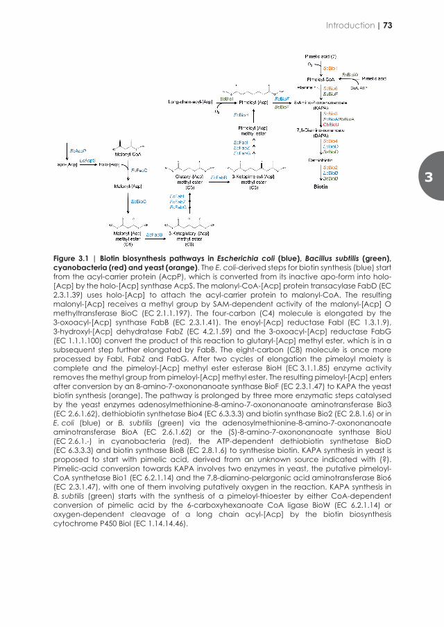
generates pimeloyl-thioester for biotin synthesis [263]. Expression of C. fabianii Bio1 conferred full biotin prototrophy to oxic cultures of multiple laboratory and industrial strains of S. cerevisiae [300]. However, due to the oxygen dependence of this enzyme, this strategy is not applicable in large-scale anoxic processes such as the yeast-based production of ethanol and isobutanol.
Prokaryotic metabolism offers options for pimeloyl-thioester biosynthesis that are independent of molecular oxygen and might be suitable for implementation in S. cerevisiae to meet biotin demands in processes performed in absence of oxygen. In B. subtilis, pimeloyl-CoA can be formed by BioW, a pimeloyl-CoA synthetase that converts free pimelic acid to pimeloyl-CoA in presence of ATP and free CoA [262]. The substrate of BioW, pimelic acid (heptanedioic acid), has been proposed to be derived from fatty acid synthesis [304]. In Escherichia coli, a divergent pathway for pimelate thioester synthesis has been elucidated [259]. This pathway is intertwined with fatty acid synthesis and is initiated by SAM-dependent methylation of malonyl-CoA by the malonyl-[Acp] O-methyltransferase encoded by bioC, yielding malonyl-CoA or malonyl-[Acp]) [305]. The methyl group of malonyl-CoA methyl ester mimics the methyl ends of fatty acyl chains and removes the charge of the carboxyl group. Malonyl-CoA methyl ester then undergoes two cycles of chain elongation by a modified type-II fatty acid synthesis pathway involving FabB, a 3-oxoacyl-[Acp]-synthase (EC 2.3.1.41), as well as FabI (EC 1.3.1.9), FabZ (EC 4.2.1.59) and FabG (EC 1.1.1.100), which produce methyl pimeloyl-[Acp]. In a final step, BioH, a pimeloyl-[Acp] methyl esterase, removes the methyl group from pimeloyl-[Acp] methyl ester, thus preventing further elongation [306]. The released pimeloyl-[Acp] is then used by BioF, the first enzyme of the canonical pathway for formation of the hetero-bi-cyclic ring of biotin, which is a homolog of S. cerevisiae Bio6. BioF produces KAPA, which is the link between all hitherto described pathways for de novo syntheses of biotin. KAPA can be converted to biotin by DAPA synthase (Bio3, BioA) (or, in cyanobacteria, by (S)-8-amino-7-oxononanoate synthase BioU [303], dethiobiotin synthetase (Bio4, BioU) and biotin synthase (Bio2, BioB) [307] (Figure 3.1).
Since the multi-step prokaryotic pathway for biotin synthesis via malonyl-CoA methyl ester is not known to involve oxygen-requiring enzymes, its introduction into S. cerevisiae provides a possible strategy for de novo synthesis of biotin in anoxic cultures. To investigate this strategy, the E. coli genes encoding enzymes involved in KAPA synthesis, comprising fabD, bioC, fabB, fabG, fabZ, fabI, bioH, bioF, acpP and acpS, were expressed in S. cerevisiae. Individual transformants were evolved for fast growth in biotin-free medium conditions in absence of oxygen. Evolved biotin-prototrophic lineages were characterized by whole-genome re-sequencing and observed genetic changes were reverse engineered into S. cerevisiae Ethanol Red, a commercial yeast strain applied in industrial bioethanol production processes.
Figure 3.1 | Biotin biosynthesis pathways in Escherichia coli (blue), Bacillus subtilis (green), cyanobacteria (red) and yeast (orange). The E. coli-derived steps for biotin synthesis (blue) start from the acyl-carrier protein (AcpP), which is converted from its inactive apo-form into holo-[Acp] by the holo-[Acp] synthase AcpS. The malonyl-CoA-[Acp] protein transacylase FabD (EC 2.3.1.39) uses holo-[Acp] to attach the acyl-carrier protein to malonyl-CoA. The resulting malonyl-[Acp] receives a methyl group by SAM-dependent activity of the malonyl-[Acp] O methyltransferase BioC (EC 2.1.1.197). The four-carbon (C4) molecule is elongated by the 3-oxoacyl-[Acp] synthase FabB (EC 2.3.1.41). The enoyl-[Acp] reductase FabI (EC 1.3.1.9), 3-hydroxyl-[Acp] dehydratase FabZ (EC 4.2.1.59) and the 3-oxoacyl-[Acp] reductase FabG (EC 1.1.1.100) convert the product of this reaction to glutaryl-[Acp] methyl ester, which is in a subsequent step further elongated by FabB. The eight-carbon (C8) molecule is once more processed by FabI, FabZ and FabG. After two cycles of elongation the pimeloyl moiety is complete and the pimeloyl-[Acp] methyl ester esterase BioH (EC 3.1.1.85) enzyme activity removes the methyl group from pimeloyl-[Acp] methyl ester. The resulting pimeloyl-[Acp] enters after conversion by an 8-amino-7-oxononanoate synthase BioF (EC 2.3.1.47) to KAPA the yeast biotin synthesis (orange). The pathway is prolonged by three more enzymatic steps catalysed by the yeast enzymes adenosylmethionine-8-amino-7-oxononanoate aminotransferase Bio3 (EC 2.6.1.62), dethiobiotin synthetase Bio4 (EC 6.3.3.3) and biotin synthase Bio2 (EC 2.8.1.6) or in E. coli (blue) or B. subtilis (green) via the adenosylmethionine-8-amino-7-oxononanoate aminotransferase BioA (EC 2.6.1.62) or the (S)-8-amino-7-oxononanoate synthase BioU (EC 2.6.1.-) in cyanobacteria (red), the ATP-dependent dethiobiotin synthetase BioD (EC 6.3.3.3) and biotin synthase BioB (EC 2.8.1.6) to synthesise biotin. KAPA synthesis in yeast is proposed to start with pimelic acid, derived from an unknown source indicated with (?). Pimelic-acid conversion towards KAPA involves two enzymes in yeast, the putative pimeloyl-CoA synthetase Bio1 (EC 6.2.1.14) and the 7,8-diamino-pelargonic acid aminotransferase Bio6 (EC 2.3.1.47), with one of them involving putatively oxygen in the reaction. KAPA synthesis in B. subtilis (green) starts with the synthesis of a pimeloyl-thioester by either CoA-dependent conversion of pimelic acid by the 6-carboxyhexanoate CoA ligase BioW (EC 6.2.1.14) or oxygen-dependent cleavage of a long chain acyl-[Acp] by the biotin biosynthesis cytochrome P450 BioI (EC 1.14.14.46).
3
Introduction | 73

Materials and methods
Strains, media and maintenance
The S. cerevisiae strains used in this study are derived from the CEN.PK [308, 309] and Ethanol Red lineages (Leaf, Lesaffre, Marcq-en-Baroeul, France) (Table 3.1). Yeast strains were grown on YP medium (10 g L−1 yeast extract [BD Biosciences, Vianen, NL], 20 g L−1 peptone [BD Biosciences]) or on chemically defined medium (SM) containing 3.0 g L−1 KH2PO4, 5.0 g L−1 (NH4)2SO4, 0.5 g L−1 MgSO4, 7H2O, 1 mL L−1 trace element solution, and 1 mL L−1 vitamin solution (0.05 g L-1 D-(+)-biotin, 1.0 g L-1 D-calcium pantothenate, 1.0 g L-1 nicotinic acid, 25 g L-1 myo-inositol, 1.0 g L-1 thiamine hydrochloride, 1.0 g L-1 pyridoxal hydrochloride, 0.2 g L-1 4-aminobenzoic acid) [13]. The pH was adjusted to 6 with 2 M KOH prior to autoclaving at 121oC for 20 min. Vitamin solutions were sterilized by filtration and added to the sterile medium. Concentrated sugar solutions were autoclaved at 110°C for 20 min and added to the sterile medium to give a final concentration of 20 g L−1 glucose (YPD and SMD). Biotin-free SM was prepared similarly, but biotin was omitted from the vitamin solution (1.0 g L-1 D-calcium pantothenate, 1.0 g L-1 nicotinic acid, 25 g L-1 myo-inositol, 1.0 g L-1 thiamine hydrochloride, 1.0 g L-1 pyridoxol hydrochloride, 0.2 g L-1 4-aminobenzoic acid) [299]. Similarly, after autoclaving concentrated glucose solution at 110°C for 20 min, glucose was added to biotin-free SM to a final concentration of 20 g L-1 (biotin-free SMD). Solid media contained 2% (w/v) Bacto agar (BD Biosciences) and, when indicated, acetamide for SMD acetamide (20 g L−1 glucose, 1.2 g L−1 acetamide, 3.0 g L−1 KH2PO4, 6.6 g L−1 K2SO4, 0.5 g L−1 MgSO4 7H2O, 1 mL L−1 trace element solution and 1 mL L−1 vitamin solution) [310], 200 mg L-1 hygromycin for YPD hygromycin and 200 mg L-1 G418 (geneticin) for YPD geneticin. Where indicated, unsaturated fatty acids and/or sterols were added to autoclaved media as Tween 80 (polyethylene glycol sorbate monooleate, Merck, Darmstadt, Germany) and ergosterol (≥95% pure, Sigma-Aldrich, St. Louis, MO). 800-fold concentrated stock solutions of these “anaerobic” growth factors were prepared as described previously and incubated at 80°C for 20 min before diluting them in growth medium, yielding final concentrations of 420 mg L−1 Tween 80 and 10 mg L−1 ergosterol [311]. E. coli cells (XL1-Blue, Agilent Technologies, Santa Clara, CA) were grown in Lysogeny broth (LB) medium (5.0 g L-1 yeast extract, 10 g L-1 Bacto trypton [BD Biosciences], 5.0 g L-1 NaCl) supplemented with 25 mg L-1 chloramphenicol, 100 mg L-1 ampicillin or 50 mg L-1 kanamycin for selection. Solid LB medium contained 2% bacto agar. Unless indicated otherwise, stock cultures for strain maintenance were prepared by growing yeast strains on YPD and E. coli cultures on LB with appropriate antibiotic markers. After reaching late exponential phase, cultures were complemented with sterile glycerol to a final concentration of 30% (v/v) and stored at −80°C as 1 mL aliquots until use.
Shake flask cultivations
For cultivation experiments for determination of specific growth rates, 1 mL aliquot of a stock culture was inoculated in 100 mL SMD in a 500-mL-shake flask and incubated for 20 h at 30°C. A second 100 mL SMD culture was started by inoculating 2 mL of the first shake flask culture. When the second culture reached mid-exponential phase, which corresponded to an optical density at 660 nm (OD660) of 3-5, an aliquot was used to inoculate a third culture at an OD660 of 0.1-0.3. For biotin-free growth studies, the pre-cultivation steps were performed in biotin-free SMD. Strains S. cerevisiae IMX585 and CEN.PK113-7D, which consistently failed to grow on biotin-free SMD in the third culture, were included as negative controls in all growth experiments. Growth was monitored by measuring OD660 of accurately diluted culture samples of the third shake-flask culture with a Jenway 7200 Spectrophotometer (Cole-Palmer, Stone, United Kingdom). Specific growth rates were calculated from a minimum number of six data points collected during exponential growth and covering 3-4 doublings of OD660. Specific growth rate was calculated using the equation 𝑋𝑋 𝑋 𝑋𝑋𝑒𝑒 in which µ indicates the exponential growth rate. All oxic shake-flask experiments were carried out as biological duplicates in an Innova shaker incubator (New Brunswick Scientific, Edison, NJ) set at 30°C and 200 rpm. To test if growth rate averages observed for different combinations of strains and medium composition are significantly different, one-way analyses of variance (ANOVA) and Tukey’s multiple comparison test with α = 0.05 were performed using GraphPad Prism 8.2.1 software (GraphPad Software, Inc., San Diego, CA). For growth profiling under anoxic conditions, the first and second pre-culture were grown in 100 mL SMD or biotin-free SMD in a 500-mL-shake flask as described previously. A 200 µL sample of mid-exponential-phase (OD660 of 3-5) cells from the second culture was then transferred to a Shel Lab Bactron 300 anaerobic workstation (Sheldon Manufacturing Inc., Cornelius, OR) operated at 30°C. The gas mixture used for flushing the workspace and air lock consisted of 85% N2, 10% CO2 and 5% H2. An IKA KS 260 Basic orbital shaker platform (Dijkstra Verenigde BV, Lelystad, The Netherlands) placed in the anaerobic chamber was set at 200 rpm. A palladium catalyst for hydrogen-dependent oxygen removal was introduced into the chamber to reduce oxygen contamination. Cultures were grown in 50-mL shake flasks containing 40 mL SMD or biotin-free SMD. Concentrated solutions of ergosterol and/or Tween 80 were added as indicated. Sterile growth media were pre-incubated in the anaerobic chamber for at least 48 h prior to inoculation to allow for removal of oxygen. Growth experiments in the anaerobic chamber were started by inoculating shake flasks, containing SMD or biotin-free SMD, with 200 µL of an exponentially growing oxic pre-culture. Growth was measured by periodic measurements of the OD600 with an Ultrospec 10 cell-density meter (Biochrom, Cambridge, UK) placed inside the anaerobic chamber. Strains IMX585 and CEN.PK113-7D grown in SMD without “anaerobic” growth factors were used as controls for absence of oxygen in all anoxic
74 | Chapter 3
Materials and methods
Strains, media and maintenance
The S. cerevisiae strains used in this study are derived from the CEN.PK [308, 309] and Ethanol Red lineages (Leaf, Lesaffre, Marcq-en-Baroeul, France) (Table 3.1). Yeast strains were grown on YP medium (10 g L−1 yeast extract [BD Biosciences, Vianen, NL], 20 g L−1 peptone [BD Biosciences]) or on chemically defined medium (SM) containing 3.0 g L−1 KH2PO4, 5.0 g L−1 (NH4)2SO4, 0.5 g L−1 MgSO4, 7H2O, 1 mL L−1 trace element solution, and 1 mL L−1 vitamin solution (0.05 g L-1 D-(+)-biotin, 1.0 g L-1 D-calcium pantothenate, 1.0 g L-1 nicotinic acid, 25 g L-1 myo-inositol, 1.0 g L-1 thiamine hydrochloride, 1.0 g L-1 pyridoxal hydrochloride, 0.2 g L-1 4-aminobenzoic acid) [13]. The pH was adjusted to 6 with 2 M KOH prior to autoclaving at 121oC for 20 min. Vitamin solutions were sterilized by filtration and added to the sterile medium. Concentrated sugar solutions were autoclaved at 110°C for 20 min and added to the sterile medium to give a final concentration of 20 g L−1 glucose (YPD and SMD). Biotin-free SM was prepared similarly, but biotin was omitted from the vitamin solution (1.0 g L-1 D-calcium pantothenate, 1.0 g L-1 nicotinic acid, 25 g L-1 myo-inositol, 1.0 g L-1 thiamine hydrochloride, 1.0 g L-1 pyridoxol hydrochloride, 0.2 g L-1 4-aminobenzoic acid) [299]. Similarly, after autoclaving concentrated glucose solution at 110°C for 20 min, glucose was added to biotin-free SM to a final concentration of 20 g L-1 (biotin-free SMD). Solid media contained 2% (w/v) Bacto agar (BD Biosciences) and, when indicated, acetamide for SMD acetamide (20 g L−1 glucose, 1.2 g L−1 acetamide, 3.0 g L−1 KH2PO4, 6.6 g L−1 K2SO4, 0.5 g L−1 MgSO4 7H2O, 1 mL L−1 trace element solution and 1 mL L−1 vitamin solution) [310], 200 mg L-1 hygromycin for YPD hygromycin and 200 mg L-1 G418 (geneticin) for YPD geneticin. Where indicated, unsaturated fatty acids and/or sterols were added to autoclaved media as Tween 80 (polyethylene glycol sorbate monooleate, Merck, Darmstadt, Germany) and ergosterol (≥95% pure, Sigma-Aldrich, St. Louis, MO). 800-fold concentrated stock solutions of these “anaerobic” growth factors were prepared as described previously and incubated at 80°C for 20 min before diluting them in growth medium, yielding final concentrations of 420 mg L−1 Tween 80 and 10 mg L−1 ergosterol [311]. E. coli cells (XL1-Blue, Agilent Technologies, Santa Clara, CA) were grown in Lysogeny broth (LB) medium (5.0 g L-1 yeast extract, 10 g L-1 Bacto trypton [BD Biosciences], 5.0 g L-1 NaCl) supplemented with 25 mg L-1 chloramphenicol, 100 mg L-1 ampicillin or 50 mg L-1 kanamycin for selection. Solid LB medium contained 2% bacto agar. Unless indicated otherwise, stock cultures for strain maintenance were prepared by growing yeast strains on YPD and E. coli cultures on LB with appropriate antibiotic markers. After reaching late exponential phase, cultures were complemented with sterile glycerol to a final concentration of 30% (v/v) and stored at −80°C as 1 mL aliquots until use.
Shake flask cultivations
For cultivation experiments for determination of specific growth rates, 1 mL aliquot of a stock culture was inoculated in 100 mL SMD in a 500-mL-shake flask and incubated for 20 h at 30°C. A second 100 mL SMD culture was started by inoculating 2 mL of the first shake flask culture. When the second culture reached mid-exponential phase, which corresponded to an optical density at 660 nm (OD660) of 3-5, an aliquot was used to inoculate a third culture at an OD660 of 0.1-0.3. For biotin-free growth studies, the pre-cultivation steps were performed in biotin-free SMD. Strains S. cerevisiae IMX585 and CEN.PK113-7D, which consistently failed to grow on biotin-free SMD in the third culture, were included as negative controls in all growth experiments. Growth was monitored by measuring OD660 of accurately diluted culture samples of the third shake-flask culture with a Jenway 7200 Spectrophotometer (Cole-Palmer, Stone, United Kingdom). Specific growth rates were calculated from a minimum number of six data points collected during exponential growth and covering 3-4 doublings of OD660. Specific growth rate was calculated using the equation 𝑋𝑋 𝑋 𝑋𝑋𝑒𝑒 in which µ indicates the exponential growth rate. All oxic shake-flask experiments were carried out as biological duplicates in an Innova shaker incubator (New Brunswick Scientific, Edison, NJ) set at 30°C and 200 rpm. To test if growth rate averages observed for different combinations of strains and medium composition are significantly different, one-way analyses of variance (ANOVA) and Tukey’s multiple comparison test with α = 0.05 were performed using GraphPad Prism 8.2.1 software (GraphPad Software, Inc., San Diego, CA). For growth profiling under anoxic conditions, the first and second pre-culture were grown in 100 mL SMD or biotin-free SMD in a 500-mL-shake flask as described previously. A 200 µL sample of mid-exponential-phase (OD660 of 3-5) cells from the second culture was then transferred to a Shel Lab Bactron 300 anaerobic workstation (Sheldon Manufacturing Inc., Cornelius, OR) operated at 30°C. The gas mixture used for flushing the workspace and air lock consisted of 85% N2, 10% CO2 and 5% H2. An IKA KS 260 Basic orbital shaker platform (Dijkstra Verenigde BV, Lelystad, The Netherlands) placed in the anaerobic chamber was set at 200 rpm. A palladium catalyst for hydrogen-dependent oxygen removal was introduced into the chamber to reduce oxygen contamination. Cultures were grown in 50-mL shake flasks containing 40 mL SMD or biotin-free SMD. Concentrated solutions of ergosterol and/or Tween 80 were added as indicated. Sterile growth media were pre-incubated in the anaerobic chamber for at least 48 h prior to inoculation to allow for removal of oxygen. Growth experiments in the anaerobic chamber were started by inoculating shake flasks, containing SMD or biotin-free SMD, with 200 µL of an exponentially growing oxic pre-culture. Growth was measured by periodic measurements of the OD600 with an Ultrospec 10 cell-density meter (Biochrom, Cambridge, UK) placed inside the anaerobic chamber. Strains IMX585 and CEN.PK113-7D grown in SMD without “anaerobic” growth factors were used as controls for absence of oxygen in all anoxic
3
Materials and Methods | 75

experiments [311]. All shake flask experiments were carried out as biological duplicates.
Molecular biology techniques
DNA fragments were amplified by PCR amplification with Phusion Hot Start II High Fidelity Polymerase (Thermo Fisher Scientific, Landsmeer, The Netherlands) and desalted or PAGE-purified oligonucleotide primers (Sigma-Aldrich) (Table 3.3). For diagnostic PCR analysis DreamTaq polymerase (Thermo Fisher Scientific) was used according to manufacturers’ recommendations. PCR products were separated by gel electrophoresis and, if required, purified with a Zymoclean Gel DNA Recovery kit (Zymo Research, Irvine, CA) or GenElute PCR Clean-Up kit (Sigma-Aldrich). Assembly of DNA fragments was, if not mentioned differently, by Golden Gate cloning based on the Yeast Tool Kit methodology [313]. Yeast strains of the CEN.PK lineage were transformed by the lithium acetate (LiAc) method [314]. S. cerevisiae Ethanol Red was transformed using electroporation as previously described [278]. Electroporated cells were plated on selective YPD hygromycin or YPD geneticin (G418) agar medium. Genomic DNA of transformants was isolated using the YeaStar Genomic DNA kit (Zymo Research) or with the SDS/LiAc protocol [315]. E. coli cells were chemically transformed [316] and plated on selective LB agar. Plasmids from selected clones were isolated from E. coli with a Sigma GenElute Plasmid kit (Sigma-Aldrich) and verified by restriction analysis (Thermo Fisher Scientific) according to the manufacturer’s recommendations or by diagnostic PCR.
Plasmid construction
Construction of part plasmids using Yeast Tool Kit
Coding sequences of EcfabD, EcbioC, EcfabB, EcfabG, EcfabZ, EcfabI, EcbioH, EcbioF, EcacpP and EcacpS were codon optimized for expression in S. cerevisiae using JCat [317] and synthesized by GeneArt (Thermo Fisher Scientific). E. coli cells were chemically transformed with the plasmids harbouring the coding sequences together with 5’ and 3’ flanks compatible with the YTK type 3 BsaI sites [313] and after selection for the antibiotic marker stored as Yeast Tool Kit type plasmids pUD671, pUD663, pUD664, pUD665, pUD666, pUD667, pUD668, pUD669, pUD661, pUD662 (Table 3.3).
The promoter sequence ScPFK2p was obtained by PCR application from genomic DNA of CEN.PK113-7D using primer pair 9630/9631. The promoter sequence was introduced in the entry vector pUD565 [281] using BsmBI-T4 ligase directed Golden Gate cloning resulting in Yeast Tool Kit type 2 plasmids pGGkp031. Correct assembly was confirmed by restriction analysis with enzyme PvuII (Thermo Fisher Scientific) according to manufacturer’s recommendations. The Yeast Tool Kit type plasmid was propagated in E. coli grown in liquid LB chloramphenicol at 37°C and stored at -80°C.
Table 3.1 | List of strains used in this study Strain Genotype Reference
or source CEN.PK113-7D MATa MAL2-8c SUC2 [308] CEN.PK-122 MATa/MATα [308] IMS0481 Single colony isolate of CEN.PK113-7D evolved in synthetic medium
without biotin [299]
IMX1859 MATa can1Δ::cas9-natNT2 Scsga1∆::ScPYK1p-CfBIO1-ScBIO1t [300] IMX585 MATa can1Δ::cas9-natNT2 [312] IMX2600 MATa can1Δ::cas9-natNT2 This study IMX2035 MATa can1Δ::cas9-natNT2 Scsga1∆::SkADH1p-EcfabD-
ScADH1t_SkTDH2p-EcbioC-ScTEF2t_SkPDC1p-EcfabB-ScPYK1t_SkFBA1p-EcfabG-ScFBA1t_SePDC1p-EcfabZ-ScPDC1t_ScENO2p-EcfabI-ScPFK2t_ScPYK1p-EcbioH-ScPGI1t_ScPFK2p-EcbioF-ScTPIt_ScPGI1p-EcacpP-ScGPM1t_ScHXK2p-EcacpS-ScTDH3t
This study
IMX2122 MATa can1Δ::cas9-natNT2 Scsga1∆::SkADH1p-EcfabD-ScADH1t_SkTDH2p-EcbioC-ScTEF2t_SkPDC1p-EcfabB-ScPYK1t_SkFBA1p-EcfabG-ScFBA1t_SePDC1p-EcfabZ-ScPDC1t_ScENO2p-EcfabI-ScPFK2t_ScPYK1p-EcbioH-ScPGI1t_ScPFK2p-EcbioF-ScTPIt_ScPGI1p-EcacpP-ScGPM1t_ScHXK2p-EcacpS-ScTDH3t Scbio1Δ
This study
IMS0994 Single colony isolate of IMX2122 evolved under anoxic conditions without biotin in bioreactor A
This study
IMS0995 Single colony isolate of IMX2122 evolved under anoxic conditions without biotin in bioreactor B
This study
Ethanol Red MATa/α (diploid prototrophic industrial bioethanol production strain) F.R. Lesaffre IMX2555 Ethanol Red Scsga1∆::SkADH1p-EcfabD-ScADH1t_SkTDH2p-EcbioC-
ScTEF2t_SkPDC1p-EcfabB-ScPYK1t_SkFBA1p-EcfabG-ScFBA1t_SePDC1p-EcfabZ-ScPDC1t_ScENO2p-EcfabI-ScPFK2t_ScPYK1p-EcbioH-ScPGI1t_ScPFK2p-EcbioF-ScTPIt_ScPGI1p-EcacpP-ScGPM1t_ScHXK2p-EcacpS-ScTDH3t/Scsga1∆::SkADH1p-EcfabD-ScADH1t_SkTDH2p-EcbioC-ScTEF2t_SkPDC1p-EcfabB-ScPYK1t_SkFBA1p-EcfabG-ScFBA1t_SePDC1p-EcfabZ-ScPDC1t_ScENO2p-EcfabI-ScPFK2t_ScPYK1p-EcbioH-ScPGI1t_ScPFK2p-EcbioF-ScTPIt_ScPGI1p-EcacpP-ScGPM1t_ScHXK2p-EcacpS-ScTDH3t
This study
IMX2632 Ethanol Red Scsga1∆::SkADH1p-EcfabD-ScADH1t_SkTDH2p-EcbioC-ScTEF2t_SkPDC1p-EcfabB-ScPYK1t_SkFBA1p-EcfabG-ScFBA1t_SePDC1p-EcfabZ-ScPDC1t_ScENO2p-EcfabI-ScPFK2t_ScPYK1p-EcbioH-ScPGI1t_ScPFK2p-EcbioF-ScTPIt_ScPGI1p-EcacpP-ScGPM1t_ScHXK2p-EcacpS-ScTDH3t/Scsga1∆::AgTEFp-kanMX-AgTEFt_SkFBA1p-EcfabG-ScFBA1t_SePDC1p-EcfabZ-ScPDC1t_ScENO2p-EcfabI-ScPFK2t_ScPYK1p-EcbioH-ScPGI1t_ScPFK2p-EcbioF-ScTPIt_ScPGI1p-EcacpP-ScGPM1t_ScHXK2p-EcacpS-ScTDH3t
This study
IMX2706 MATa can1Δ::cas9-natNT2 Scsga1∆::SkTDH2p-EcbioC-ScTEF2t_ScPYK1p-EcbioH-ScPGI1t_ScPFK2p-EcbioF∆-ScTPIt
This study
IMX2707 MATa can1Δ::cas9-natNT2 Scsga1∆::SkADH1p-EcfabD-ScADH1t_SkTDH2p-EcbioC-ScTEF2t_SkPDC1p-EcfabB-ScPYK1t_SkFBA1p-EcfabG-ScFBA1t_SePDC1p-EcfabZ-ScPDC1t_ScENO2p-EcfabI-ScPFK2t_ScPYK1p-EcbioH-ScPGI1t_ScPFK2p-EcbioF∆-ScTPIt_ScPGI1p-EcacpP-ScGPM1t_ScHXK2p-EcacpS-ScTDH3t
This study
The terminator sequences ScFBA1t, ScTPI1t and ScPGI1t were obtained by PCR with primer combinations 10757/10758, 10765/10766 and 10771/10772, respectively using genomic DNA of S. cerevisiae CEN.PK113-7D as template. The terminator sequences were cloned in pUD565 using BsmBI-T4 DNA ligase directed Golden Gate cloning yielding the Yeast Tool Kit type 4 plasmids pGGkp046, pGGkp042 and pGGkp044
76 | Chapter 3
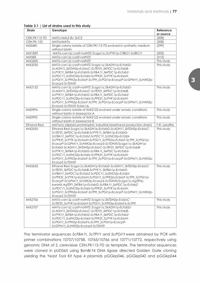
experiments [311]. All shake flask experiments were carried out as biological duplicates.
Molecular biology techniques
DNA fragments were amplified by PCR amplification with Phusion Hot Start II High Fidelity Polymerase (Thermo Fisher Scientific, Landsmeer, The Netherlands) and desalted or PAGE-purified oligonucleotide primers (Sigma-Aldrich) (Table 3.3). For diagnostic PCR analysis DreamTaq polymerase (Thermo Fisher Scientific) was used according to manufacturers’ recommendations. PCR products were separated by gel electrophoresis and, if required, purified with a Zymoclean Gel DNA Recovery kit (Zymo Research, Irvine, CA) or GenElute PCR Clean-Up kit (Sigma-Aldrich). Assembly of DNA fragments was, if not mentioned differently, by Golden Gate cloning based on the Yeast Tool Kit methodology [313]. Yeast strains of the CEN.PK lineage were transformed by the lithium acetate (LiAc) method [314]. S. cerevisiae Ethanol Red was transformed using electroporation as previously described [278]. Electroporated cells were plated on selective YPD hygromycin or YPD geneticin (G418) agar medium. Genomic DNA of transformants was isolated using the YeaStar Genomic DNA kit (Zymo Research) or with the SDS/LiAc protocol [315]. E. coli cells were chemically transformed [316] and plated on selective LB agar. Plasmids from selected clones were isolated from E. coli with a Sigma GenElute Plasmid kit (Sigma-Aldrich) and verified by restriction analysis (Thermo Fisher Scientific) according to the manufacturer’s recommendations or by diagnostic PCR.
Plasmid construction
Construction of part plasmids using Yeast Tool Kit
Coding sequences of EcfabD, EcbioC, EcfabB, EcfabG, EcfabZ, EcfabI, EcbioH, EcbioF, EcacpP and EcacpS were codon optimized for expression in S. cerevisiae using JCat [317] and synthesized by GeneArt (Thermo Fisher Scientific). E. coli cells were chemically transformed with the plasmids harbouring the coding sequences together with 5’ and 3’ flanks compatible with the YTK type 3 BsaI sites [313] and after selection for the antibiotic marker stored as Yeast Tool Kit type plasmids pUD671, pUD663, pUD664, pUD665, pUD666, pUD667, pUD668, pUD669, pUD661, pUD662 (Table 3.3).
The promoter sequence ScPFK2p was obtained by PCR application from genomic DNA of CEN.PK113-7D using primer pair 9630/9631. The promoter sequence was introduced in the entry vector pUD565 [281] using BsmBI-T4 ligase directed Golden Gate cloning resulting in Yeast Tool Kit type 2 plasmids pGGkp031. Correct assembly was confirmed by restriction analysis with enzyme PvuII (Thermo Fisher Scientific) according to manufacturer’s recommendations. The Yeast Tool Kit type plasmid was propagated in E. coli grown in liquid LB chloramphenicol at 37°C and stored at -80°C.
Table 3.1 | List of strains used in this study Strain Genotype Reference
or source CEN.PK113-7D MATa MAL2-8c SUC2 [308] CEN.PK-122 MATa/MATα [308] IMS0481 Single colony isolate of CEN.PK113-7D evolved in synthetic medium
without biotin [299]
IMX1859 MATa can1Δ::cas9-natNT2 Scsga1∆::ScPYK1p-CfBIO1-ScBIO1t [300] IMX585 MATa can1Δ::cas9-natNT2 [312] IMX2600 MATa can1Δ::cas9-natNT2 This study IMX2035 MATa can1Δ::cas9-natNT2 Scsga1∆::SkADH1p-EcfabD-
ScADH1t_SkTDH2p-EcbioC-ScTEF2t_SkPDC1p-EcfabB-ScPYK1t_SkFBA1p-EcfabG-ScFBA1t_SePDC1p-EcfabZ-ScPDC1t_ScENO2p-EcfabI-ScPFK2t_ScPYK1p-EcbioH-ScPGI1t_ScPFK2p-EcbioF-ScTPIt_ScPGI1p-EcacpP-ScGPM1t_ScHXK2p-EcacpS-ScTDH3t
This study
IMX2122 MATa can1Δ::cas9-natNT2 Scsga1∆::SkADH1p-EcfabD-ScADH1t_SkTDH2p-EcbioC-ScTEF2t_SkPDC1p-EcfabB-ScPYK1t_SkFBA1p-EcfabG-ScFBA1t_SePDC1p-EcfabZ-ScPDC1t_ScENO2p-EcfabI-ScPFK2t_ScPYK1p-EcbioH-ScPGI1t_ScPFK2p-EcbioF-ScTPIt_ScPGI1p-EcacpP-ScGPM1t_ScHXK2p-EcacpS-ScTDH3t Scbio1Δ
This study
IMS0994 Single colony isolate of IMX2122 evolved under anoxic conditions without biotin in bioreactor A
This study
IMS0995 Single colony isolate of IMX2122 evolved under anoxic conditions without biotin in bioreactor B
This study
Ethanol Red MATa/α (diploid prototrophic industrial bioethanol production strain) F.R. Lesaffre IMX2555 Ethanol Red Scsga1∆::SkADH1p-EcfabD-ScADH1t_SkTDH2p-EcbioC-
ScTEF2t_SkPDC1p-EcfabB-ScPYK1t_SkFBA1p-EcfabG-ScFBA1t_SePDC1p-EcfabZ-ScPDC1t_ScENO2p-EcfabI-ScPFK2t_ScPYK1p-EcbioH-ScPGI1t_ScPFK2p-EcbioF-ScTPIt_ScPGI1p-EcacpP-ScGPM1t_ScHXK2p-EcacpS-ScTDH3t/Scsga1∆::SkADH1p-EcfabD-ScADH1t_SkTDH2p-EcbioC-ScTEF2t_SkPDC1p-EcfabB-ScPYK1t_SkFBA1p-EcfabG-ScFBA1t_SePDC1p-EcfabZ-ScPDC1t_ScENO2p-EcfabI-ScPFK2t_ScPYK1p-EcbioH-ScPGI1t_ScPFK2p-EcbioF-ScTPIt_ScPGI1p-EcacpP-ScGPM1t_ScHXK2p-EcacpS-ScTDH3t
This study
IMX2632 Ethanol Red Scsga1∆::SkADH1p-EcfabD-ScADH1t_SkTDH2p-EcbioC-ScTEF2t_SkPDC1p-EcfabB-ScPYK1t_SkFBA1p-EcfabG-ScFBA1t_SePDC1p-EcfabZ-ScPDC1t_ScENO2p-EcfabI-ScPFK2t_ScPYK1p-EcbioH-ScPGI1t_ScPFK2p-EcbioF-ScTPIt_ScPGI1p-EcacpP-ScGPM1t_ScHXK2p-EcacpS-ScTDH3t/Scsga1∆::AgTEFp-kanMX-AgTEFt_SkFBA1p-EcfabG-ScFBA1t_SePDC1p-EcfabZ-ScPDC1t_ScENO2p-EcfabI-ScPFK2t_ScPYK1p-EcbioH-ScPGI1t_ScPFK2p-EcbioF-ScTPIt_ScPGI1p-EcacpP-ScGPM1t_ScHXK2p-EcacpS-ScTDH3t
This study
IMX2706 MATa can1Δ::cas9-natNT2 Scsga1∆::SkTDH2p-EcbioC-ScTEF2t_ScPYK1p-EcbioH-ScPGI1t_ScPFK2p-EcbioF∆-ScTPIt
This study
IMX2707 MATa can1Δ::cas9-natNT2 Scsga1∆::SkADH1p-EcfabD-ScADH1t_SkTDH2p-EcbioC-ScTEF2t_SkPDC1p-EcfabB-ScPYK1t_SkFBA1p-EcfabG-ScFBA1t_SePDC1p-EcfabZ-ScPDC1t_ScENO2p-EcfabI-ScPFK2t_ScPYK1p-EcbioH-ScPGI1t_ScPFK2p-EcbioF∆-ScTPIt_ScPGI1p-EcacpP-ScGPM1t_ScHXK2p-EcacpS-ScTDH3t
This study
The terminator sequences ScFBA1t, ScTPI1t and ScPGI1t were obtained by PCR with primer combinations 10757/10758, 10765/10766 and 10771/10772, respectively using genomic DNA of S. cerevisiae CEN.PK113-7D as template. The terminator sequences were cloned in pUD565 using BsmBI-T4 DNA ligase directed Golden Gate cloning yielding the Yeast Tool Kit type 4 plasmids pGGkp046, pGGkp042 and pGGkp044
3
Materials and Methods | 77
respectively. After assembly and transformation into E. coli, plasmids harbouring the terminator sequences were confirmed by restriction analysis with enzyme SspI (Thermo Fisher Scientific) according to manufacturer’s recommendations. The Yeast Tool Kit type plasmids were stored in transformed E. coli cultures.
The promoter sequence ScHXK2p was synthesized by GeneArt (Thermo Fisher Scientific) and is harboured by Yeast Tool Kit type 2 plasmid pGGkp096. The Yeast Tool Kit type plasmid was propagated in a chemically transformed E. coli culture in liquid LB chloramphenicol medium grown at 37°C on a rotary shaker and subsequently stored at -80°C.
Construction of gRNA-expressing plasmid pUDR791
The gRNAEcBIOF expressing plasmid pUDR791 was constructed in vitro by Gibson assembly. The linearized pROS11 plasmid, obtained by PCR with 6005/6006 was assembled with a PCR amplified fragment using primer 18409 and pROS11 as a template [312]. Plasmid DNA was isolated from E. coli and correct assembly of plasmid pUDR791 was confirmed by diagnostic PCR with primers 18457/3841/5941.
Construction of expression cassettes
The E. coli fabD expression cassette was constructed by BsaI–T4 DNA ligase directed Golden Gate cloning combining DNA fragments with compatible overhangs from plasmids pGGkd015, pGGkp062, pUD671, pGGkp037 yielding plasmid pUD978. The next expression plasmids were constructed following a similar cloning principle. The E. coli bioC expression cassette was constructed by combining DNA fragments with compatible overhangs from plasmids pGGkd015, pGGkp063, pUD663, pGGkp038 yielding plasmid pUD979. The E. coli fabB expression cassette was constructed by combining DNA fragments with compatible overhangs from plasmids pGGkd015, pGGkp064, pUD664, pGGkp040 yielding plasmid pUD980. The E. coli fabG expression cassette was constructed by combining DNA fragments with compatible overhangs from plasmids pGGkd015, pGGkp065, pUD665, pGGkp046 yielding plasmid pUD981. The E. coli fabZ expression cassette was constructed by combining DNA fragments with compatible overhangs from plasmids pGGkd015, pGGkp074, pUD666, pGGkp045 yielding plasmid pUD982. The E. coli fabI expression cassette was constructed by combining DNA fragments with compatible overhangs from plasmids pGGkd015, pGGkp028, pUD667, pGGkp103 yielding plasmid pUD983. The E. coli bioH expression cassette was constructed by combining DNA fragments with compatible overhangs from plasmids pGGkd015, pGGkp117, pUD668, pGGkp044 yielding plasmid pUD984. The E. coli bioF expression cassette was constructed by combining DNA fragments with compatible overhangs from plasmids pGGkd015, pGGkp031, pUD669, pGGkp042 yielding plasmid pUD985. The E. coli acpP expression cassettee was constructed by combining DNA fragments with compatible overhangs from plasmids pGGkd015, pGGkp033, pUD661, pGGkp048 yielding plasmid pUD986. The E. coli acpS expression
cassette was constructed by combining DNA fragments with compatible overhangs from plasmids pGGkd015, pGGkp096, pUD662, pGGkp041 yielding plasmid pUD987. After assembly reaction and transformation of E. coli with the plasmids carrying the expression cassettes, four to eight colonies were selected for each plasmid, followed by isolation of plasmid DNA.
Table 3.2 | List of primers used in this study. Primer No. Sequence 5’ → 3’ 1719 TCCATCCGGTCTTTATCGAC 7469 GGAGTTGACCGTCTTAACAG 9630 AAGCATCGTCTCATCGGTCTCAAACGTATTCTTAGTGGATAACATGCG 9631 TTATGCCGTCTCAGGTCTCACATATTTTAGGCTGGTATCTTGATTC 10320 CATGCGCGGATGACACGAAC 10325 AGTCATCCGAGCGTGTATTG 10757 AAGCATCGTCTCATCGGTCTCAATCCGTTAATTCAAATTAATTGATATAGTTTTTTAATG 10758 TTATGCCGTCTCAGGTCTCACAGCCGCGAACTCCAAAATGAGC 10765 AAGCATCGTCTCATCGGTCTCAATCCGATTAATATAATTATATAAAAATATTATCTTCTTTTC 10766 TTATGCCGTCTCAGGTCTCACAGCCGGTACACTTCTGAGTAAC 10771 AAGCATCGTCTCATCGGTCTCAATCCACAAATCGCTCTTAAATATATACC 10772 TTATGCCGTCTCAGGTCTCACAGCGAAATAGGACCTGATATCCTCC 10873 ACGTGCGGAATAGGAATCTC 11898 CGCGGAAACGGGTATTAGGG 11899 CTAGATCCGGTAAGCGACAG 12223 CCAGGTGGCGTGCTAAACTTTTATAATGTATAAAAACCACCACCTCATAAAGTTTACTGGATATCATCATT
TCTGCCACAAATATATGTACTGAGTCTATACGTCAAAGTAAAAAAATAA 12224 TTATTTTTTTACTTTGACGTATAGACTCAGTACATATATTTGTGGCAGAAATGATGATATCCAGTAAACTTTATG
AGGTGGTGGTTTTTATACATTATAAAAGTTTAGCACGCCACCTGG 12450 TCTGTCAGTTGGTTAAGCGCCGCTACGATTACTACACATGCCACAGACTGATCTACAATGTATCCTCCTT
TTAAACAGTTGATG 12455 ACATTGCATGGAATCAGGGCCTCAATATGTGGGAGAATGCATGAGTACGCGAGCGATCCTCCTGGT
CAAACTTCAGAACTAAG 12655 TTTACAATATAGTGATAATCGTGGACTAGAGCAAGATTTCAAATAAGTAACAGCAGCAAATCCGATTTCC
GTGGTTGATG 12656 CCAGGGCTCAAATGGCATAAACACTGATGGAACAGGTAGCATCGAACGTGTGTCAAACGCATGTTA
GCGTCAACAACAAG 12657 GGCACAGACGAATCACTGACTGATCTGTACCACTGCGTCGACATAACTTTCCAGAAGCGCGTGGGT
GCGTCAACTACATC 12658 TGAGCCAGTGCATTCCATCGATGCAGATTCGCGTCCACGTAACGTATCGGAAGCATAGGCAACAAT
GCCAACCCCTCTAC 12659 TGTGAGCAGTCATCCACTCGGCATAAGCCTGAATTGCACCATATCCTTGGAAGCCTGGGCGAAGCT
ATCTTCCGGTTATG 12660 TCCTCGACGCGATGGCATATCCAGTGTGATAACGTATGAGAAGGTACTGGAAGCTACTGCAACACTA
AACGAAGGCTATC 12663 CACTGCGTGTTAAGGATATGCCTAAGGATACATGACACGCATAGCTCATTAACCGGCACGTGGATAA
CATGCGGCATTTC 12664 GCCGCGTAGACAATAGATCACCATCTAGTTGAATCCTGAGAGACTATCTCTAATGACCCGGGTAAAG
TACAGCTACATTC 12665 GCGTTTGACACACGTTCGATGCTACCTGTTCCATCAGTGTTTATGCCATTTGAGCCCTGGACACACCGA
GATTCATCAAC 12666 GCGCTTCTGGAAAGTTATGTCGACGCAGTGGTACAGATCAGTCAGTGATTCGTCTGTGCCATATACATA
CGCTGACATGG 12667 GCCTATGCTTCCGATACGTTACGTGGACGCGAATCTGCATCGATGGAATGCACTGGCTCATCGCCAT
CCTGATAATCATG 12668 GCCCAGGCTTCCAAGGATATGGTGCAATTCAGGCTTATGCCGAGTGGATGACTGCTCACATTGAAAT
GACTCCGCAGTGG 12669 GCAGTAGCTTCCAGTACCTTCTCATACGTTATCACACTGGATATGCCATCGCGTCGAGGATTCCTTGGTT
CCACTAATTC 12674 GAAAAAACTAGATCCGGTAAGCGACAGATCTTTGAATTTGTTTATAGCCGACTCTAAGTCCAGAATCGTT
ATCCTGGCGG 12745 AGCGTAGATAGAAGCGTCAG 12746 TCCAGTTGGTGACGTTAAGG 12747 TCAGCACCCAAGTCTTCAAC 12749 TCCAGATAGCCCATTCGTTG
78 | Chapter 3
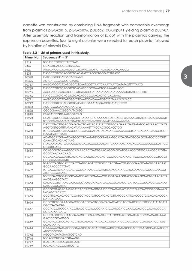
respectively. After assembly and transformation into E. coli, plasmids harbouring the terminator sequences were confirmed by restriction analysis with enzyme SspI (Thermo Fisher Scientific) according to manufacturer’s recommendations. The Yeast Tool Kit type plasmids were stored in transformed E. coli cultures.
The promoter sequence ScHXK2p was synthesized by GeneArt (Thermo Fisher Scientific) and is harboured by Yeast Tool Kit type 2 plasmid pGGkp096. The Yeast Tool Kit type plasmid was propagated in a chemically transformed E. coli culture in liquid LB chloramphenicol medium grown at 37°C on a rotary shaker and subsequently stored at -80°C.
Construction of gRNA-expressing plasmid pUDR791
The gRNAEcBIOF expressing plasmid pUDR791 was constructed in vitro by Gibson assembly. The linearized pROS11 plasmid, obtained by PCR with 6005/6006 was assembled with a PCR amplified fragment using primer 18409 and pROS11 as a template [312]. Plasmid DNA was isolated from E. coli and correct assembly of plasmid pUDR791 was confirmed by diagnostic PCR with primers 18457/3841/5941.
Construction of expression cassettes
The E. coli fabD expression cassette was constructed by BsaI–T4 DNA ligase directed Golden Gate cloning combining DNA fragments with compatible overhangs from plasmids pGGkd015, pGGkp062, pUD671, pGGkp037 yielding plasmid pUD978. The next expression plasmids were constructed following a similar cloning principle. The E. coli bioC expression cassette was constructed by combining DNA fragments with compatible overhangs from plasmids pGGkd015, pGGkp063, pUD663, pGGkp038 yielding plasmid pUD979. The E. coli fabB expression cassette was constructed by combining DNA fragments with compatible overhangs from plasmids pGGkd015, pGGkp064, pUD664, pGGkp040 yielding plasmid pUD980. The E. coli fabG expression cassette was constructed by combining DNA fragments with compatible overhangs from plasmids pGGkd015, pGGkp065, pUD665, pGGkp046 yielding plasmid pUD981. The E. coli fabZ expression cassette was constructed by combining DNA fragments with compatible overhangs from plasmids pGGkd015, pGGkp074, pUD666, pGGkp045 yielding plasmid pUD982. The E. coli fabI expression cassette was constructed by combining DNA fragments with compatible overhangs from plasmids pGGkd015, pGGkp028, pUD667, pGGkp103 yielding plasmid pUD983. The E. coli bioH expression cassette was constructed by combining DNA fragments with compatible overhangs from plasmids pGGkd015, pGGkp117, pUD668, pGGkp044 yielding plasmid pUD984. The E. coli bioF expression cassette was constructed by combining DNA fragments with compatible overhangs from plasmids pGGkd015, pGGkp031, pUD669, pGGkp042 yielding plasmid pUD985. The E. coli acpP expression cassettee was constructed by combining DNA fragments with compatible overhangs from plasmids pGGkd015, pGGkp033, pUD661, pGGkp048 yielding plasmid pUD986. The E. coli acpS expression
cassette was constructed by combining DNA fragments with compatible overhangs from plasmids pGGkd015, pGGkp096, pUD662, pGGkp041 yielding plasmid pUD987. After assembly reaction and transformation of E. coli with the plasmids carrying the expression cassettes, four to eight colonies were selected for each plasmid, followed by isolation of plasmid DNA.
Table 3.2 | List of primers used in this study. Primer No. Sequence 5’ → 3’ 1719 TCCATCCGGTCTTTATCGAC 7469 GGAGTTGACCGTCTTAACAG 9630 AAGCATCGTCTCATCGGTCTCAAACGTATTCTTAGTGGATAACATGCG 9631 TTATGCCGTCTCAGGTCTCACATATTTTAGGCTGGTATCTTGATTC 10320 CATGCGCGGATGACACGAAC 10325 AGTCATCCGAGCGTGTATTG 10757 AAGCATCGTCTCATCGGTCTCAATCCGTTAATTCAAATTAATTGATATAGTTTTTTAATG 10758 TTATGCCGTCTCAGGTCTCACAGCCGCGAACTCCAAAATGAGC 10765 AAGCATCGTCTCATCGGTCTCAATCCGATTAATATAATTATATAAAAATATTATCTTCTTTTC 10766 TTATGCCGTCTCAGGTCTCACAGCCGGTACACTTCTGAGTAAC 10771 AAGCATCGTCTCATCGGTCTCAATCCACAAATCGCTCTTAAATATATACC 10772 TTATGCCGTCTCAGGTCTCACAGCGAAATAGGACCTGATATCCTCC 10873 ACGTGCGGAATAGGAATCTC 11898 CGCGGAAACGGGTATTAGGG 11899 CTAGATCCGGTAAGCGACAG 12223 CCAGGTGGCGTGCTAAACTTTTATAATGTATAAAAACCACCACCTCATAAAGTTTACTGGATATCATCATT
TCTGCCACAAATATATGTACTGAGTCTATACGTCAAAGTAAAAAAATAA 12224 TTATTTTTTTACTTTGACGTATAGACTCAGTACATATATTTGTGGCAGAAATGATGATATCCAGTAAACTTTATG
AGGTGGTGGTTTTTATACATTATAAAAGTTTAGCACGCCACCTGG 12450 TCTGTCAGTTGGTTAAGCGCCGCTACGATTACTACACATGCCACAGACTGATCTACAATGTATCCTCCTT
TTAAACAGTTGATG 12455 ACATTGCATGGAATCAGGGCCTCAATATGTGGGAGAATGCATGAGTACGCGAGCGATCCTCCTGGT
CAAACTTCAGAACTAAG 12655 TTTACAATATAGTGATAATCGTGGACTAGAGCAAGATTTCAAATAAGTAACAGCAGCAAATCCGATTTCC
GTGGTTGATG 12656 CCAGGGCTCAAATGGCATAAACACTGATGGAACAGGTAGCATCGAACGTGTGTCAAACGCATGTTA
GCGTCAACAACAAG 12657 GGCACAGACGAATCACTGACTGATCTGTACCACTGCGTCGACATAACTTTCCAGAAGCGCGTGGGT
GCGTCAACTACATC 12658 TGAGCCAGTGCATTCCATCGATGCAGATTCGCGTCCACGTAACGTATCGGAAGCATAGGCAACAAT
GCCAACCCCTCTAC 12659 TGTGAGCAGTCATCCACTCGGCATAAGCCTGAATTGCACCATATCCTTGGAAGCCTGGGCGAAGCT
ATCTTCCGGTTATG 12660 TCCTCGACGCGATGGCATATCCAGTGTGATAACGTATGAGAAGGTACTGGAAGCTACTGCAACACTA
AACGAAGGCTATC 12663 CACTGCGTGTTAAGGATATGCCTAAGGATACATGACACGCATAGCTCATTAACCGGCACGTGGATAA
CATGCGGCATTTC 12664 GCCGCGTAGACAATAGATCACCATCTAGTTGAATCCTGAGAGACTATCTCTAATGACCCGGGTAAAG
TACAGCTACATTC 12665 GCGTTTGACACACGTTCGATGCTACCTGTTCCATCAGTGTTTATGCCATTTGAGCCCTGGACACACCGA
GATTCATCAAC 12666 GCGCTTCTGGAAAGTTATGTCGACGCAGTGGTACAGATCAGTCAGTGATTCGTCTGTGCCATATACATA
CGCTGACATGG 12667 GCCTATGCTTCCGATACGTTACGTGGACGCGAATCTGCATCGATGGAATGCACTGGCTCATCGCCAT
CCTGATAATCATG 12668 GCCCAGGCTTCCAAGGATATGGTGCAATTCAGGCTTATGCCGAGTGGATGACTGCTCACATTGAAAT
GACTCCGCAGTGG 12669 GCAGTAGCTTCCAGTACCTTCTCATACGTTATCACACTGGATATGCCATCGCGTCGAGGATTCCTTGGTT
CCACTAATTC 12674 GAAAAAACTAGATCCGGTAAGCGACAGATCTTTGAATTTGTTTATAGCCGACTCTAAGTCCAGAATCGTT
ATCCTGGCGG 12745 AGCGTAGATAGAAGCGTCAG 12746 TCCAGTTGGTGACGTTAAGG 12747 TCAGCACCCAAGTCTTCAAC 12749 TCCAGATAGCCCATTCGTTG
3
Materials and Methods | 79
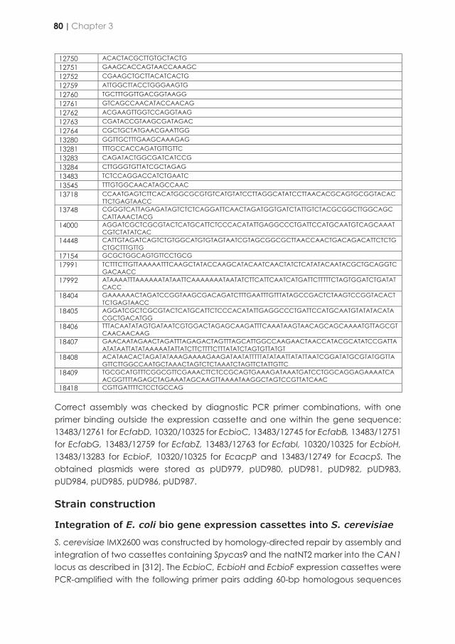
Correct assembly was checked by diagnostic PCR primer combinations, with one primer binding outside the expression cassette and one within the gene sequence: 13483/12761 for EcfabD, 10320/10325 for EcbioC, 13483/12745 for EcfabB, 13483/12751 for EcfabG, 13483/12759 for EcfabZ, 13483/12763 for EcfabI, 10320/10325 for EcbioH, 13483/13283 for EcbioF, 10320/10325 for EcacpP and 13483/12749 for EcacpS. The obtained plasmids were stored as pUD979, pUD980, pUD981, pUD982, pUD983, pUD984, pUD985, pUD986, pUD987.
Strain construction
Integration of E. coli bio gene expression cassettes into S. cerevisiae
S. cerevisiae IMX2600 was constructed by homology-directed repair by assembly and integration of two cassettes containing Spycas9 and the natNT2 marker into the CAN1 locus as described in [312]. The EcbioC, EcbioH and EcbioF expression cassettes were PCR-amplified with the following primer pairs adding 60-bp homologous sequences
12750 ACACTACGCTTGTGCTACTG 12751 GAAGCACCAGTAACCAAAGC 12752 CGAAGCTGCTTACATCACTG 12759 ATTGGCTTACCTGGGAAGTG 12760 TGCTTTGGTTGACGGTAAGG 12761 GTCAGCCAACATACCAACAG 12762 ACGAAGTTGGTCCAGGTAAG 12763 CGATACCGTAAGCGATAGAC 12764 CGCTGCTATGAACGAATTGG 13280 GGTTGCTTTGAAGCAAAGAG 13281 TTTGCCACCAGATGTTGTTC 13283 CAGATACTGGCGATCATCCG 13284 CTTGGGTGTTATCGCTAGAG 13483 TCTCCAGGACCATCTGAATC 13545 TTTGTGGCAACATAGCCAAC 13718 CCAATGAGTCTTCACATGGCGCGTGTCATGTATCCTTAGGCATATCCTTAACACGCAGTGCGGTACAC
TTCTGAGTAACC 13748 CGGGTCATTAGAGATAGTCTCTCAGGATTCAACTAGATGGTGATCTATTGTCTACGCGGCTTGGCAGC
CATTAAACTACG 14000 AGGATCGCTCGCGTACTCATGCATTCTCCCACATATTGAGGCCCTGATTCCATGCAATGTCAGCAAAT
CGTCTATATCAC 14448 CATTGTAGATCAGTCTGTGGCATGTGTAGTAATCGTAGCGGCGCTTAACCAACTGACAGACATTCTCTG
CTGCTTTGTTG 17154 GCGCTGGCAGTGTTCCTGCG 17991 TCTTTCTTGTTAAAAATTTCAAGCTATACCAAGCATACAATCAACTATCTCATATACAATACGCTGCAGGTC
GACAACC 17992 ATAAAATTTAAAAAATATAATTCAAAAAAATAATATCTTCATTCAATCATGATTCTTTTTCTAGTGGATCTGATAT
CACC 18404 GAAAAAACTAGATCCGGTAAGCGACAGATCTTTGAATTTGTTTATAGCCGACTCTAAGTCCGGTACACT
TCTGAGTAACC 18405 AGGATCGCTCGCGTACTCATGCATTCTCCCACATATTGAGGCCCTGATTCCATGCAATGTATATACATA
CGCTGACATGG 18406 TTTACAATATAGTGATAATCGTGGACTAGAGCAAGATTTCAAATAAGTAACAGCAGCAAAATGTTAGCGT
CAACAACAAG 18407 GAACAATAGAACTAGATTTAGAGACTAGTTTAGCATTGGCCAAGAACTAACCATACGCATATCCGATTA
ATATAATTATATAAAAATATTATCTTCTTTTCTTTATATCTAGTGTTATGT 18408 ACATAACACTAGATATAAAGAAAAGAAGATAATATTTTTATATAATTATATTAATCGGATATGCGTATGGTTA
GTTCTTGGCCAATGCTAAACTAGTCTCTAAATCTAGTTCTATTGTTC 18409 TGCGCATGTTTCGGCGTTCGAAACTTCTCCGCAGTGAAAGATAAATGATCCTGGCAGGAGAAAATCA
ACGGTTTTAGAGCTAGAAATAGCAAGTTAAAATAAGGCTAGTCCGTTATCAAC 18418 CGTTGATTTTCTCCTGCCAG
[319]: 18406/18405 for EcbioC (pUD979), 12455/12450 for EcbioH (pUD984) and 14448/18404 for EcbioF (pUD985). Targeting at the ScSGA1 locus in IMX2600 was directed by Cas9 activity and a target-specific gRNA expressing plasmid. The strain was co-transformed with the EcbioC, EcbioH and EcbioF expression cassette fragments and the plasmid pUDR119 expressing the gRNA to target Cas9 activity to the ScSGA1 locus [318] using the LiAc transformation protocol. Transformed cells were plated on selective SMD with acetamide and incubated for 3 days at 30°C. Genomic DNA of colonies was isolated and the desired genotype confirmed by diagnostic PCR using primer combinations 11898/13545, 13284/13281, 13280/13283 and 1719/11899. A verified clone was inoculated in 20 mL non-selective YPD for plasmid removal and incubated for 24 h at 30°C. Cells were plated on YPD agar to obtain single colony isolates. One isolate was re-streaked on both selective medium and YPD. When no growth was observed on selective medium the respective clone was again checked by diagnostic PCR with above-mentioned primer combinations. The strain with in vivo assembled expression cassettes of the E. coli bio genes into ScSGA1 was stored as IMX2706.
Integration of E. coli KAPA synthesis in IMX585 and Ethanol Red
Expression cassettes were PCR-amplified with the following primer pairs, thereby adding 60-bp homologous sequences [320] to enable in vivo assembly at the ScSGA1 locus: 12655/12665 for EcfabD (pUD978), 12656/12666 for EcbioC (pUD979), 12657/12667 for EcfabB (pUD980), 12658/12668 for EcfabG (pUD981), 12659/12669 for EcfabZ (pUD982), 12660/14000 for EcfabI (pUD983), 12455/12450 for EcbioH (pUD984), 14448/13718 for EcbioF (pUD985), 12663/13748 for EcacpP (pUD986) and 12664/12674 for EcacpS (pUD987). The resulting expression cassettes were integrated at the ScSGA1 locus in IMX585 and Ethanol Red, by transformation of specific gRNA encoded on plasmid pUDR119 in case of IMX585 and in case of Ethanol Red by plasmid pUDP145. Targeting at the ScSGA1 locus in IMX585 was directed by strain-intrinsic Cas9 activity and in Ethanol Red by expression of Spycas9 from plasmid pUDP145 [283]. Yeast strains were co-transformed with the respective plasmids and the EcfabD, EcbioC, EcfabB, EcfabG, EcfabZ, EcfabI, EcbioH, EcbioF, EcacpP and EcacpS expression cassettes using the LiAc transformation protocol.
Table 3.3 | List of plasmids constructed and used in this study. Name Characteristics Reference or source pGGkd015 bla ColE1 Gfp dropout [281] pGGkp028 cat ColE1 ScENO2p [281] pGGkp031 cat ColE1 ScPFK2p This study pGGkp033 cat ColE1 ScPGI1p [281] pGGkp037 cat ColE1 ScADH1t [281] pGGkp038 cat ColE1ScTEF2t [281] pGGkp040 cat ColE1 ScPYK1t [281] pGGkp041 cat ColE1 ScTDH3t [281] pGGkp042 cat ColE1 ScTPIt This study pGGkp044 cat ColE1 ScPGI1t This study pGGkp045 cat ColE1 ScPDC1t [281] pGGkp046 cat ColE1 ScFBA1t This study
80 | Chapter 3
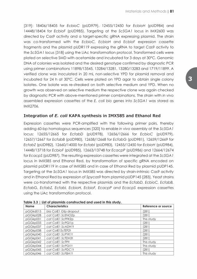
Correct assembly was checked by diagnostic PCR primer combinations, with one primer binding outside the expression cassette and one within the gene sequence: 13483/12761 for EcfabD, 10320/10325 for EcbioC, 13483/12745 for EcfabB, 13483/12751 for EcfabG, 13483/12759 for EcfabZ, 13483/12763 for EcfabI, 10320/10325 for EcbioH, 13483/13283 for EcbioF, 10320/10325 for EcacpP and 13483/12749 for EcacpS. The obtained plasmids were stored as pUD979, pUD980, pUD981, pUD982, pUD983, pUD984, pUD985, pUD986, pUD987.
Strain construction
Integration of E. coli bio gene expression cassettes into S. cerevisiae
S. cerevisiae IMX2600 was constructed by homology-directed repair by assembly and integration of two cassettes containing Spycas9 and the natNT2 marker into the CAN1 locus as described in [312]. The EcbioC, EcbioH and EcbioF expression cassettes were PCR-amplified with the following primer pairs adding 60-bp homologous sequences
12750 ACACTACGCTTGTGCTACTG 12751 GAAGCACCAGTAACCAAAGC 12752 CGAAGCTGCTTACATCACTG 12759 ATTGGCTTACCTGGGAAGTG 12760 TGCTTTGGTTGACGGTAAGG 12761 GTCAGCCAACATACCAACAG 12762 ACGAAGTTGGTCCAGGTAAG 12763 CGATACCGTAAGCGATAGAC 12764 CGCTGCTATGAACGAATTGG 13280 GGTTGCTTTGAAGCAAAGAG 13281 TTTGCCACCAGATGTTGTTC 13283 CAGATACTGGCGATCATCCG 13284 CTTGGGTGTTATCGCTAGAG 13483 TCTCCAGGACCATCTGAATC 13545 TTTGTGGCAACATAGCCAAC 13718 CCAATGAGTCTTCACATGGCGCGTGTCATGTATCCTTAGGCATATCCTTAACACGCAGTGCGGTACAC
TTCTGAGTAACC 13748 CGGGTCATTAGAGATAGTCTCTCAGGATTCAACTAGATGGTGATCTATTGTCTACGCGGCTTGGCAGC
CATTAAACTACG 14000 AGGATCGCTCGCGTACTCATGCATTCTCCCACATATTGAGGCCCTGATTCCATGCAATGTCAGCAAAT
CGTCTATATCAC 14448 CATTGTAGATCAGTCTGTGGCATGTGTAGTAATCGTAGCGGCGCTTAACCAACTGACAGACATTCTCTG
CTGCTTTGTTG 17154 GCGCTGGCAGTGTTCCTGCG 17991 TCTTTCTTGTTAAAAATTTCAAGCTATACCAAGCATACAATCAACTATCTCATATACAATACGCTGCAGGTC
GACAACC 17992 ATAAAATTTAAAAAATATAATTCAAAAAAATAATATCTTCATTCAATCATGATTCTTTTTCTAGTGGATCTGATAT
CACC 18404 GAAAAAACTAGATCCGGTAAGCGACAGATCTTTGAATTTGTTTATAGCCGACTCTAAGTCCGGTACACT
TCTGAGTAACC 18405 AGGATCGCTCGCGTACTCATGCATTCTCCCACATATTGAGGCCCTGATTCCATGCAATGTATATACATA
CGCTGACATGG 18406 TTTACAATATAGTGATAATCGTGGACTAGAGCAAGATTTCAAATAAGTAACAGCAGCAAAATGTTAGCGT
CAACAACAAG 18407 GAACAATAGAACTAGATTTAGAGACTAGTTTAGCATTGGCCAAGAACTAACCATACGCATATCCGATTA
ATATAATTATATAAAAATATTATCTTCTTTTCTTTATATCTAGTGTTATGT 18408 ACATAACACTAGATATAAAGAAAAGAAGATAATATTTTTATATAATTATATTAATCGGATATGCGTATGGTTA
GTTCTTGGCCAATGCTAAACTAGTCTCTAAATCTAGTTCTATTGTTC 18409 TGCGCATGTTTCGGCGTTCGAAACTTCTCCGCAGTGAAAGATAAATGATCCTGGCAGGAGAAAATCA
ACGGTTTTAGAGCTAGAAATAGCAAGTTAAAATAAGGCTAGTCCGTTATCAAC 18418 CGTTGATTTTCTCCTGCCAG
[319]: 18406/18405 for EcbioC (pUD979), 12455/12450 for EcbioH (pUD984) and 14448/18404 for EcbioF (pUD985). Targeting at the ScSGA1 locus in IMX2600 was directed by Cas9 activity and a target-specific gRNA expressing plasmid. The strain was co-transformed with the EcbioC, EcbioH and EcbioF expression cassette fragments and the plasmid pUDR119 expressing the gRNA to target Cas9 activity to the ScSGA1 locus [318] using the LiAc transformation protocol. Transformed cells were plated on selective SMD with acetamide and incubated for 3 days at 30°C. Genomic DNA of colonies was isolated and the desired genotype confirmed by diagnostic PCR using primer combinations 11898/13545, 13284/13281, 13280/13283 and 1719/11899. A verified clone was inoculated in 20 mL non-selective YPD for plasmid removal and incubated for 24 h at 30°C. Cells were plated on YPD agar to obtain single colony isolates. One isolate was re-streaked on both selective medium and YPD. When no growth was observed on selective medium the respective clone was again checked by diagnostic PCR with above-mentioned primer combinations. The strain with in vivo assembled expression cassettes of the E. coli bio genes into ScSGA1 was stored as IMX2706.
Integration of E. coli KAPA synthesis in IMX585 and Ethanol Red
Expression cassettes were PCR-amplified with the following primer pairs, thereby adding 60-bp homologous sequences [320] to enable in vivo assembly at the ScSGA1 locus: 12655/12665 for EcfabD (pUD978), 12656/12666 for EcbioC (pUD979), 12657/12667 for EcfabB (pUD980), 12658/12668 for EcfabG (pUD981), 12659/12669 for EcfabZ (pUD982), 12660/14000 for EcfabI (pUD983), 12455/12450 for EcbioH (pUD984), 14448/13718 for EcbioF (pUD985), 12663/13748 for EcacpP (pUD986) and 12664/12674 for EcacpS (pUD987). The resulting expression cassettes were integrated at the ScSGA1 locus in IMX585 and Ethanol Red, by transformation of specific gRNA encoded on plasmid pUDR119 in case of IMX585 and in case of Ethanol Red by plasmid pUDP145. Targeting at the ScSGA1 locus in IMX585 was directed by strain-intrinsic Cas9 activity and in Ethanol Red by expression of Spycas9 from plasmid pUDP145 [283]. Yeast strains were co-transformed with the respective plasmids and the EcfabD, EcbioC, EcfabB, EcfabG, EcfabZ, EcfabI, EcbioH, EcbioF, EcacpP and EcacpS expression cassettes using the LiAc transformation protocol.
Table 3.3 | List of plasmids constructed and used in this study. Name Characteristics Reference or source pGGkd015 bla ColE1 Gfp dropout [281] pGGkp028 cat ColE1 ScENO2p [281] pGGkp031 cat ColE1 ScPFK2p This study pGGkp033 cat ColE1 ScPGI1p [281] pGGkp037 cat ColE1 ScADH1t [281] pGGkp038 cat ColE1ScTEF2t [281] pGGkp040 cat ColE1 ScPYK1t [281] pGGkp041 cat ColE1 ScTDH3t [281] pGGkp042 cat ColE1 ScTPIt This study pGGkp044 cat ColE1 ScPGI1t This study pGGkp045 cat ColE1 ScPDC1t [281] pGGkp046 cat ColE1 ScFBA1t This study
3
Materials and Methods | 81
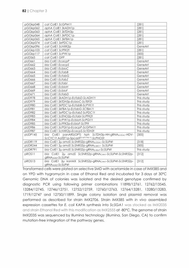
pGGkp048 cat ColE1 ScGPM1t [281] pGGkp062 aphA ColE1 SkADH1p [281] pGGkp063 aphA ColE1 SkTDH3p [281] pGGkp064 aphA ColE1 SkPDC1p [281] pGGkp065 aphA ColE1 SkFBA1p [281] pGGkp074 cat ColE1 SePDC1p [281] pGGkp096 cat ColE1 ScHXK2p GeneArt pGGkp103 cat ColE1 ScPFK2t [281] pGGkp117 cat ColE1 ScPYK1p [300] pUD565 cat ColE1 GFP [281] pUD661 bla ColE1 EcacpP GeneArt pUD662 bla ColE1 EcacpS GeneArt pUD663 bla ColE1 EcbioC GeneArt pUD664 bla ColE1 EcfabB GeneArt pUD665 bla ColE1 EcfabG GeneArt pUD666 bla ColE1 EcfabZ GeneArt pUD667 bla ColE1 EcfabI GeneArt pUD668 bla ColE1 EcbioH GeneArt pUD669 bla ColE1 EcbioF GeneArt pUD671 bla ColE1 EcfabD GeneArt pUD978 bla ColE1 SkADH1p-EcfabD-ScADH1t This study pUD979 bla ColE1 SkTDH3p-EcbioC-ScTEF2t This study pUD980 bla ColE1 SkPDC1p-EcfabB-ScPYK1t This study pUD981 bla ColE1 SkFBA1p-EcfabG-SCfBA1t This study pUD982 bla ColE1 SePDC1p-EcfabZ-ScPDC1t This study pUD983 bla ColE1 ScENO2p-EcfabI-ScPFK2t This study pUD984 bla ColE1 ScPYK1p-EcbioH-ScPGI1t This study pUD985 bla ColE1 ScPFK2p-EcbioF-ScTPIt This study pUD986 bla ColE1 ScPGI1p-EcacpP-ScGPM1t This study pUD987 bla ColE1 ScHXK2p-EcacpS-ScTDH3t This study pUDP145 bla ColE1 panARS(OPT) hph ScTDH3p-HH-gRNAScSGA1-HDV-
ScCYC1t AaTEF1p-Spcas9D147Y P411T-ScPHO5t [300]
pUDR119 bla ColE1 2μ amdS ScSNR52p-gRNAScSGA1-ScSUP4t [318] pUDR244 bla ColE1 2μ amdS ScSNR52p-gRNAScBIO1- ScSUP4t [300] pUDR791 bla ColE1 2μ amdS ScSNR52p-gRNAEcBioF-ScSUP4t This study pROS11 bla ColE1 2μ amdS ScSNR52p-gRNACAN1-ScSUP4t-ScSNR52p-
gRNAADE2-ScSUP4t [312]
pROS13 bla ColE1 2μ kanMX ScSNR52p-gRNACAN1-ScSUP4t-ScSNR52p-gRNAADE2-ScSUP4t
[312]
Transformed cells were plated on selective SMD with acetamide in case of IMX585 and on YPD with hygromycin in case of Ethanol Red and incubated for 3 days at 30°C Genomic DNA of colonies was isolated and the desired genotype confirmed by diagnostic PCR using following primer combinations 11898/12761, 12762/13545, 13284/12745, 12746/12751, 12752/12759, 12760/12763, 12764/13281, 13280/13283, 1719/12747 and 12750/11899. Single colony isolation and plasmid removal was performed as described for strain IMX2706. Strain IMX585 with in vivo assembled expression cassettes for E. coli KAPA synthesis into ScSGA1 was stocked as IMX2035 and strain Ethanol Red with this modification as IMX2555 at -80°C. The genome of strain IMX2035 was sequenced by Illumina technology (Illumina, San Diego, CA) to confirm mutation-free integration of the pathway genes.
Gene deletion
To delete the native ScBIO1 locus in S. cerevisiae IMX2035, it was co-transformed with plasmid pUDR244 [300] and a repair DNA fragment resulting from the annealing of oligo-nucleotides 12223/12224. Transformed cells were plated on selective SMD acetamide and incubated for 3 days at 30°C. Genomic DNA of colonies was isolated and the desired genotype confirmed by diagnostic PCR using primer pair 7469/10873. A verified clone was inoculated in 20 mL non-selective YPD for plasmid removal and incubated for 24 h at 30°C. Cells were plated on YPD agar in order to obtain single colony isolates. One isolate was re-streaked on both SMD acetamide and YPD. When no growth was observed on SMD acetamide the respective clone was once again confirmed by diagnostic PCR and stored as IMX2122. Similarly, to delete the heterologously expressed EcbioF gene, strain IMX2035 was co-transformed with plasmid pUDR791 and a repair DNA fragment resulting from the annealing of the oligo-nucleotides 18407/18408. After growth on selective SMD acetamide, genotyping of the resulting colonies was carried out by diagnostic PCR with primer pair 1719/12747. After plasmid removal a single colony was isolated and stored as IMX2707.
Deletion of EcfabD, EcbioC and EcfabB in S. cerevisiae IMX2555, which was derived from the diploid S. cerevisiae strain Ethanol Red containing the KAPA synthesis pathway, was performed by transformation with and integration of a deletion cassette. The transformed linear DNA fragment contained 60-bp flanks homologous to the SkADH1 promoter and the intergenic region between the EcfabB and EcfabG expressional units and the KanMX expression cassette conferring resistance to geneticin [321]. The linear DNA fragment with the deletion cassette was obtained by PCR with the primer pair 17991/17992 using plasmid pROS13 as a template. Upon homologous recombination, the deletion cassette replaced one of the two copies of the three expression cassettes for EcfabD, EcbioC and EcfabB. Electroporated cells were plated on selective YPD G418 agar plates and incubated for 5 days at 30°C. Genomic DNA of transformants was isolated and the desired genotype was confirmed by diagnostic PCR using following primer combinations 11898/12761, 12762/13545, 13284/12745, 11898/12562, 12751/17154. The correct clone was re-streaked on YPD agar to obtain single colony isolates. A single colony was once again confirmed by diagnostic PCR with above-mentioned primer combinations and inoculated for stocking in 20 mL non-selective YPD. The Ethanol Red strain with the integration of the KanMX cassette in the ScSGA1 locus was stored as IMX2632.
Batch cultivation in bioreactors
Physiological characterization of S. cerevisiae IMX2122 (Scbio1Δ ↑EcKAPA pathway) was performed in anoxic bioreactors (Applikon, Delft, The Netherlands) with a working volume of 1.0 L. All cultures were grown on biotin-free SMD; anoxic cultures were supplemented with sterile solutions of the “anaerobic” growth factors ergosterol (10 mg L−1) and Tween 80 (420 mg L−1), as well as with 0.2 g L−1 sterile antifoam C
82 | Chapter 3
pGGkp048 cat ColE1 ScGPM1t [281] pGGkp062 aphA ColE1 SkADH1p [281] pGGkp063 aphA ColE1 SkTDH3p [281] pGGkp064 aphA ColE1 SkPDC1p [281] pGGkp065 aphA ColE1 SkFBA1p [281] pGGkp074 cat ColE1 SePDC1p [281] pGGkp096 cat ColE1 ScHXK2p GeneArt pGGkp103 cat ColE1 ScPFK2t [281] pGGkp117 cat ColE1 ScPYK1p [300] pUD565 cat ColE1 GFP [281] pUD661 bla ColE1 EcacpP GeneArt pUD662 bla ColE1 EcacpS GeneArt pUD663 bla ColE1 EcbioC GeneArt pUD664 bla ColE1 EcfabB GeneArt pUD665 bla ColE1 EcfabG GeneArt pUD666 bla ColE1 EcfabZ GeneArt pUD667 bla ColE1 EcfabI GeneArt pUD668 bla ColE1 EcbioH GeneArt pUD669 bla ColE1 EcbioF GeneArt pUD671 bla ColE1 EcfabD GeneArt pUD978 bla ColE1 SkADH1p-EcfabD-ScADH1t This study pUD979 bla ColE1 SkTDH3p-EcbioC-ScTEF2t This study pUD980 bla ColE1 SkPDC1p-EcfabB-ScPYK1t This study pUD981 bla ColE1 SkFBA1p-EcfabG-SCfBA1t This study pUD982 bla ColE1 SePDC1p-EcfabZ-ScPDC1t This study pUD983 bla ColE1 ScENO2p-EcfabI-ScPFK2t This study pUD984 bla ColE1 ScPYK1p-EcbioH-ScPGI1t This study pUD985 bla ColE1 ScPFK2p-EcbioF-ScTPIt This study pUD986 bla ColE1 ScPGI1p-EcacpP-ScGPM1t This study pUD987 bla ColE1 ScHXK2p-EcacpS-ScTDH3t This study pUDP145 bla ColE1 panARS(OPT) hph ScTDH3p-HH-gRNAScSGA1-HDV-
ScCYC1t AaTEF1p-Spcas9D147Y P411T-ScPHO5t [300]
pUDR119 bla ColE1 2μ amdS ScSNR52p-gRNAScSGA1-ScSUP4t [318] pUDR244 bla ColE1 2μ amdS ScSNR52p-gRNAScBIO1- ScSUP4t [300] pUDR791 bla ColE1 2μ amdS ScSNR52p-gRNAEcBioF-ScSUP4t This study pROS11 bla ColE1 2μ amdS ScSNR52p-gRNACAN1-ScSUP4t-ScSNR52p-
gRNAADE2-ScSUP4t [312]
pROS13 bla ColE1 2μ kanMX ScSNR52p-gRNACAN1-ScSUP4t-ScSNR52p-gRNAADE2-ScSUP4t
[312]
Transformed cells were plated on selective SMD with acetamide in case of IMX585 and on YPD with hygromycin in case of Ethanol Red and incubated for 3 days at 30°C Genomic DNA of colonies was isolated and the desired genotype confirmed by diagnostic PCR using following primer combinations 11898/12761, 12762/13545, 13284/12745, 12746/12751, 12752/12759, 12760/12763, 12764/13281, 13280/13283, 1719/12747 and 12750/11899. Single colony isolation and plasmid removal was performed as described for strain IMX2706. Strain IMX585 with in vivo assembled expression cassettes for E. coli KAPA synthesis into ScSGA1 was stocked as IMX2035 and strain Ethanol Red with this modification as IMX2555 at -80°C. The genome of strain IMX2035 was sequenced by Illumina technology (Illumina, San Diego, CA) to confirm mutation-free integration of the pathway genes.
Gene deletion
To delete the native ScBIO1 locus in S. cerevisiae IMX2035, it was co-transformed with plasmid pUDR244 [300] and a repair DNA fragment resulting from the annealing of oligo-nucleotides 12223/12224. Transformed cells were plated on selective SMD acetamide and incubated for 3 days at 30°C. Genomic DNA of colonies was isolated and the desired genotype confirmed by diagnostic PCR using primer pair 7469/10873. A verified clone was inoculated in 20 mL non-selective YPD for plasmid removal and incubated for 24 h at 30°C. Cells were plated on YPD agar in order to obtain single colony isolates. One isolate was re-streaked on both SMD acetamide and YPD. When no growth was observed on SMD acetamide the respective clone was once again confirmed by diagnostic PCR and stored as IMX2122. Similarly, to delete the heterologously expressed EcbioF gene, strain IMX2035 was co-transformed with plasmid pUDR791 and a repair DNA fragment resulting from the annealing of the oligo-nucleotides 18407/18408. After growth on selective SMD acetamide, genotyping of the resulting colonies was carried out by diagnostic PCR with primer pair 1719/12747. After plasmid removal a single colony was isolated and stored as IMX2707.
Deletion of EcfabD, EcbioC and EcfabB in S. cerevisiae IMX2555, which was derived from the diploid S. cerevisiae strain Ethanol Red containing the KAPA synthesis pathway, was performed by transformation with and integration of a deletion cassette. The transformed linear DNA fragment contained 60-bp flanks homologous to the SkADH1 promoter and the intergenic region between the EcfabB and EcfabG expressional units and the KanMX expression cassette conferring resistance to geneticin [321]. The linear DNA fragment with the deletion cassette was obtained by PCR with the primer pair 17991/17992 using plasmid pROS13 as a template. Upon homologous recombination, the deletion cassette replaced one of the two copies of the three expression cassettes for EcfabD, EcbioC and EcfabB. Electroporated cells were plated on selective YPD G418 agar plates and incubated for 5 days at 30°C. Genomic DNA of transformants was isolated and the desired genotype was confirmed by diagnostic PCR using following primer combinations 11898/12761, 12762/13545, 13284/12745, 11898/12562, 12751/17154. The correct clone was re-streaked on YPD agar to obtain single colony isolates. A single colony was once again confirmed by diagnostic PCR with above-mentioned primer combinations and inoculated for stocking in 20 mL non-selective YPD. The Ethanol Red strain with the integration of the KanMX cassette in the ScSGA1 locus was stored as IMX2632.
Batch cultivation in bioreactors
Physiological characterization of S. cerevisiae IMX2122 (Scbio1Δ ↑EcKAPA pathway) was performed in anoxic bioreactors (Applikon, Delft, The Netherlands) with a working volume of 1.0 L. All cultures were grown on biotin-free SMD; anoxic cultures were supplemented with sterile solutions of the “anaerobic” growth factors ergosterol (10 mg L−1) and Tween 80 (420 mg L−1), as well as with 0.2 g L−1 sterile antifoam C
3
Materials and Methods | 83
(Sigma-Aldrich). These conditions were maintained by sparging cultures with a gas mixture of N2/CO2 (90/10%, < 10 ppm oxygen) at a rate of 0.5 L min−1. Culture pH was maintained at 5.0 by automatic addition of 2 M KOH. All cultures were grown at a stirrer speed of 800 rpm and at a temperature of 30°C. Oxygen diffusion in the bioreactors was minimized by using Neoprene tubing and Viton O-rings, and evaporation was minimized by cooling of outlet gas to 4°C. Oxic conditions were maintained by sparging with pressurised air at a rate of 0.5 L min−1. For bioreactor inocula, a 1 mL aliquot of a thawed stock culture of strain IMX2122 was inoculated in 100 mL biotin-free SMD in a 500 mL-shake flask and incubated for 20 h at 30°C. A second 100 mL biotin-free SMD culture was started by inoculating 2 mL of the first shake flask culture. Shake flasks were incubated at 30°C and 200 rpm in an Innova incubator (Brunswick Scientific). When the second culture reached mid-exponential phase (OD660 of 3-5) it was used to inoculate the bioreactors at an OD660 of 0.1-0.3. Growth in the bioreactor was monitored based on the CO2 concentration in the off-gas. Specific growth rates were calculated from CO2 concentration values collected during exponential growth and covering 3-4 doublings. Specific growth rate was calculated using the equation 𝑋𝑋 𝑋 𝑋𝑋𝑒𝑒 in which µ indicates the exponential growth rate. After anaerobic cultures had reached a first CO2 production peak and the CO2 percentage in the off-gas subsequently decreased below more than 20% of the previously measured value, a computer-controlled peristaltic pump automatically removed ca. 90% of the culture volume, leaving ca. 10% as an inoculum for the next batch cultivation cycle that occurred after refilling the reactor with fresh medium. Specific growth rates in absence of oxygen were determined from the CO2 profile after two empty-refill cycles in order to deplete “anaerobic” growth factors from the pre-cultures that were run in presence of oxygen [311].
Laboratory evolution
Laboratory evolution of S. cerevisiae IMX2122 (Scbio1Δ ↑EcKAPA pathway) for fast anoxic growth without biotin supplementation was performed in sequential-batch bioreactor cultures. Empty-refill cycles in two independent anaerobic bioreactors, operated as described above, were continued until no further increase of the specific growth rate was observed for at least five consecutive batch cultivation cycles. Single-colony isolates from reactor A were obtained after 109 cycles and from reactor B after 100 cycles by plating on biotin-free SMD.
Whole-genome sequence analysis
DNA of S. cerevisiae strains IMX2035, IMX2122, IMS0994 and IMS0995 grown in shake-flask cultures with SMD was isolated with a Qiagen Blood & Cell Culture DNA kit (Qiagen, Germantown, MD), following manufacturer’s specifications. Paired-end sequencing was performed on a 350-bp PCR-free insert library using an Illumina HiSeq PE150 sequencer (Novogene Company Limited, Hong Kong). Sequence data was mapped to the CEN.PK113-7D genome [309], to which the sequences of the integrated expression cassettes for the heterologous genes EcfabD, EcbioC, EcfabB,
EcfabG, EcfabZ, EcfabI, EcbioH, EcbioF, EcacpP and EcacpS were manually added. Data processing and chromosome copy number analysis were carried out as described previously [299, 322].
Ploidy analysis by flow cytometry
For determination of ploidy, frozen aliquots of S. cerevisiae strains IMX2035, IMX2122 and the evolved strains IMS0994 and IMS0995 were thawed and used to inoculate 20-mL cultures on SMD (IMX2035 and IMX2122) or on biotin-free SMD (IMS0994 and IMS0995). After incubation at 30°C until mid-exponential phase, cells were harvested, washed twice with demineralized water and stored in 70% ethanol at 4 °C. Sample preparation and staining were performed as described previously [323, 324]. Samples were processed using a BD Accuri C6 flow cytometer (BD Biosciences, San Jose, CA) and analysed using the FlowJo software package (Flowjo LLC, Ashland, OR). S. cerevisiae strains CEN.PK113-7D and CEN.PK122 were used a haploid and diploid references, respectively.
Proteome analysis
Frozen aliquots of S. cerevisiae strains IMX2122 (Scbio1Δ ↑EcKAPA pathway), IMS0994 (evolution A IMX2122), and IMS0995 (evolution B IMX2122) were thawed and used to inoculate wake-up cultures in 20 mL biotin-free SMD. After overnight incubation at 30°C, these cultures were used to inoculate two independent 100 mL cultures at a starting OD660 of 0.2. Once these cultures reached an OD660 of 4, 1 mL was collected and centrifuged at 3000 x g for 5 min, yielding a cell pellet with a volume of approximately 60 μL. After protein extraction and trypsin digestion [325], extracted peptides were re-suspended in 30 µL of 3% acetonitrile/0.01% trifluoroacetic acid. The peptide concentration was measured using a Nanodrop spectrophotometer (Thermo Scientific) at a wavelength of 280 nm. A total of 1 μg of sample was injected in a CapLC system (Thermo Scientific) coupled to an Orbitrap Q-exactive HF-X mass spectrometer (Thermo Scientific). First, samples were captured at a flow rate of 10 μL/min on a pre-column (µ-pre-column C18 PepMap 100, 5 µm, 100 Å) and subsequently peptides were separated on a 15 cm C18 easy spray column (PepMap RSLC C18 2 µm, 104 pm, 150 µm x 15 cm) using a flow rate of 1.2 µL min-1. A linear gradient from 4% to 76% acetonitrile in water was applied over 60 min. While spraying the samples into the mass spectrometer the instrument was operated in data dependent mode using settings as previously described in [326]. Data analysis was performed using Proteome discover 2.4 (Thermo Scientific) with fixed modifications set to carbamidomethyl (C), variable modifications set to oxidation of methionine residues, search mass tolerance set to 20 ppm, MS/MS tolerance set to 20 ppm, trypsin selected as restriction enzyme and allowing one missed cleavage. False Discovery Rate (FDR) was set at 0.1% and the match between runs window was set to 0.7 min. Quantification was only based on unique peptides and normalization between samples was based on total peptide amount. For protein search, a protein database consisting of the S. cerevisiae S288C proteome amino-acid sequences together with
84 | Chapter 3
(Sigma-Aldrich). These conditions were maintained by sparging cultures with a gas mixture of N2/CO2 (90/10%, < 10 ppm oxygen) at a rate of 0.5 L min−1. Culture pH was maintained at 5.0 by automatic addition of 2 M KOH. All cultures were grown at a stirrer speed of 800 rpm and at a temperature of 30°C. Oxygen diffusion in the bioreactors was minimized by using Neoprene tubing and Viton O-rings, and evaporation was minimized by cooling of outlet gas to 4°C. Oxic conditions were maintained by sparging with pressurised air at a rate of 0.5 L min−1. For bioreactor inocula, a 1 mL aliquot of a thawed stock culture of strain IMX2122 was inoculated in 100 mL biotin-free SMD in a 500 mL-shake flask and incubated for 20 h at 30°C. A second 100 mL biotin-free SMD culture was started by inoculating 2 mL of the first shake flask culture. Shake flasks were incubated at 30°C and 200 rpm in an Innova incubator (Brunswick Scientific). When the second culture reached mid-exponential phase (OD660 of 3-5) it was used to inoculate the bioreactors at an OD660 of 0.1-0.3. Growth in the bioreactor was monitored based on the CO2 concentration in the off-gas. Specific growth rates were calculated from CO2 concentration values collected during exponential growth and covering 3-4 doublings. Specific growth rate was calculated using the equation 𝑋𝑋 𝑋 𝑋𝑋𝑒𝑒 in which µ indicates the exponential growth rate. After anaerobic cultures had reached a first CO2 production peak and the CO2 percentage in the off-gas subsequently decreased below more than 20% of the previously measured value, a computer-controlled peristaltic pump automatically removed ca. 90% of the culture volume, leaving ca. 10% as an inoculum for the next batch cultivation cycle that occurred after refilling the reactor with fresh medium. Specific growth rates in absence of oxygen were determined from the CO2 profile after two empty-refill cycles in order to deplete “anaerobic” growth factors from the pre-cultures that were run in presence of oxygen [311].
Laboratory evolution
Laboratory evolution of S. cerevisiae IMX2122 (Scbio1Δ ↑EcKAPA pathway) for fast anoxic growth without biotin supplementation was performed in sequential-batch bioreactor cultures. Empty-refill cycles in two independent anaerobic bioreactors, operated as described above, were continued until no further increase of the specific growth rate was observed for at least five consecutive batch cultivation cycles. Single-colony isolates from reactor A were obtained after 109 cycles and from reactor B after 100 cycles by plating on biotin-free SMD.
Whole-genome sequence analysis
DNA of S. cerevisiae strains IMX2035, IMX2122, IMS0994 and IMS0995 grown in shake-flask cultures with SMD was isolated with a Qiagen Blood & Cell Culture DNA kit (Qiagen, Germantown, MD), following manufacturer’s specifications. Paired-end sequencing was performed on a 350-bp PCR-free insert library using an Illumina HiSeq PE150 sequencer (Novogene Company Limited, Hong Kong). Sequence data was mapped to the CEN.PK113-7D genome [309], to which the sequences of the integrated expression cassettes for the heterologous genes EcfabD, EcbioC, EcfabB,
EcfabG, EcfabZ, EcfabI, EcbioH, EcbioF, EcacpP and EcacpS were manually added. Data processing and chromosome copy number analysis were carried out as described previously [299, 322].
Ploidy analysis by flow cytometry
For determination of ploidy, frozen aliquots of S. cerevisiae strains IMX2035, IMX2122 and the evolved strains IMS0994 and IMS0995 were thawed and used to inoculate 20-mL cultures on SMD (IMX2035 and IMX2122) or on biotin-free SMD (IMS0994 and IMS0995). After incubation at 30°C until mid-exponential phase, cells were harvested, washed twice with demineralized water and stored in 70% ethanol at 4 °C. Sample preparation and staining were performed as described previously [323, 324]. Samples were processed using a BD Accuri C6 flow cytometer (BD Biosciences, San Jose, CA) and analysed using the FlowJo software package (Flowjo LLC, Ashland, OR). S. cerevisiae strains CEN.PK113-7D and CEN.PK122 were used a haploid and diploid references, respectively.
Proteome analysis
Frozen aliquots of S. cerevisiae strains IMX2122 (Scbio1Δ ↑EcKAPA pathway), IMS0994 (evolution A IMX2122), and IMS0995 (evolution B IMX2122) were thawed and used to inoculate wake-up cultures in 20 mL biotin-free SMD. After overnight incubation at 30°C, these cultures were used to inoculate two independent 100 mL cultures at a starting OD660 of 0.2. Once these cultures reached an OD660 of 4, 1 mL was collected and centrifuged at 3000 x g for 5 min, yielding a cell pellet with a volume of approximately 60 μL. After protein extraction and trypsin digestion [325], extracted peptides were re-suspended in 30 µL of 3% acetonitrile/0.01% trifluoroacetic acid. The peptide concentration was measured using a Nanodrop spectrophotometer (Thermo Scientific) at a wavelength of 280 nm. A total of 1 μg of sample was injected in a CapLC system (Thermo Scientific) coupled to an Orbitrap Q-exactive HF-X mass spectrometer (Thermo Scientific). First, samples were captured at a flow rate of 10 μL/min on a pre-column (µ-pre-column C18 PepMap 100, 5 µm, 100 Å) and subsequently peptides were separated on a 15 cm C18 easy spray column (PepMap RSLC C18 2 µm, 104 pm, 150 µm x 15 cm) using a flow rate of 1.2 µL min-1. A linear gradient from 4% to 76% acetonitrile in water was applied over 60 min. While spraying the samples into the mass spectrometer the instrument was operated in data dependent mode using settings as previously described in [326]. Data analysis was performed using Proteome discover 2.4 (Thermo Scientific) with fixed modifications set to carbamidomethyl (C), variable modifications set to oxidation of methionine residues, search mass tolerance set to 20 ppm, MS/MS tolerance set to 20 ppm, trypsin selected as restriction enzyme and allowing one missed cleavage. False Discovery Rate (FDR) was set at 0.1% and the match between runs window was set to 0.7 min. Quantification was only based on unique peptides and normalization between samples was based on total peptide amount. For protein search, a protein database consisting of the S. cerevisiae S288C proteome amino-acid sequences together with
3
Materials and Methods | 85
the sequences of the heterologously expressed proteins was used. Each strain was analyzed in independent biological duplicate samples. Data processing and analysis of differentially expressed proteins was conducted as previously described in [325]. Enrichment analysis of up- and downregulated proteins in the isolates was performed using the GO Enrichment Analysis [327].
Results
Expression of the E. coli KAPA-biosynthesis pathway supports biotin-independent growth of S. cerevisiae in absence of oxygen.
Of the currently known prokaryotic biotin-biosynthesis pathways (Figure 3.1), only the variant that occurs in E. coli starts with malonyl-CoA, a key precursor for lipid synthesis in S. cerevisiae. To complete the malonyl-CoA conversion into pimeloyl-CoA, only EcbioC and EcbioH would be required in S. cerevisiae assuming that the other reactions could be performed by the native fatty acid elongation machinery. We also included EcbioF since it is unclear whether ScBio6, the protein ortholog of EcBioF, can use pimeloyl-[Acp] as substrate. Integration of these three E. coli genes at the SGA1 locus yielded S. cerevisiae IMX2706. Even after prolonged oxic incubation in biotin-free synthetic medium, this engineered strain did not show growth on biotin-free synthetic medium. To investigate whether this inability was related to the different organization of the prokaryotic and yeast fatty-acid-synthesis machineries, we introduced an additional set of expression cassettes for E. coli proteins involved in conversion of malonyl-[Acp]-methyl ester into pimeloyl-[Acp]-methyl ester. In addition to EcbioC, H and F, five genes involved in fatty-acid biosynthesis (EcfabD, EcfabB, EcfabG, EcfabZ, EcfabI) and two genes involved in acyl carrier protein formation (EcacpP and EcacpS) were introduced. In E. coli, the concerted action of the enzymes encoded by these genes converts malonyl-CoA into 8-amino-7-oxo-nonanoate (KAPA), a metabolic intermediate of the native S. cerevisiae biotin pathway. Using the SpyCas9-expressing strain IMX585, the ten expression cassettes were integrated at the SGA1 locus, yielding S. cerevisiae IMX2035 (↑EcKAPA pathway; Figure 2.2A)
Figure 3.2 | Expression of E. coli KAPA biosynthesis pathway in S. cerevisiae. (A) Schematic overview of genetic modifications introduced at the ScSGA1 locus. A Cas9-induced cut in the ScSGA1 coding sequence and in vivo homologous recombination enabled integration of expression cassettes for ten E. coli genes with different promoters (green) and terminators (yellow). Intergenic regions consisted of synthetic 60-bp-homologous recombination sequences [320]. (B) Bar graphs representing average specific growth rates of S. cerevisiae strains CEN.PK113-7D, IMS0481 (evolved for biotin prototrophy [299]), IMX1859 (↑CfBIO1,[300]), IMX2035 (↑EcKAPA pathway) and IMX2122 (Scbio1Δ ↑EcKAPA pathway) under oxic conditions on glucose synthetic medium with (+, black) and without (-, white) biotin. (C) Bar graphs representing average specific growth rates of S. cerevisiae strains CEN.PK113-7D, IMS0481, IMX1859 and IMX2035 under anoxic conditions on glucose synthetic medium with (+, black) and without (-, white) biotin. Averages and deviations of the bar graphs were calculated from independent duplicate cultures. Brackets between two bar graphs show the p-value, which was derived from significance testing of the difference between observed growth rates by one-way analyses of variance (ANOVA) and Tukey’s multiple comparison test using GraphPad prism 8.2.1 software (significance threshold p-value < 5.0E-02).
86 | Chapter 3
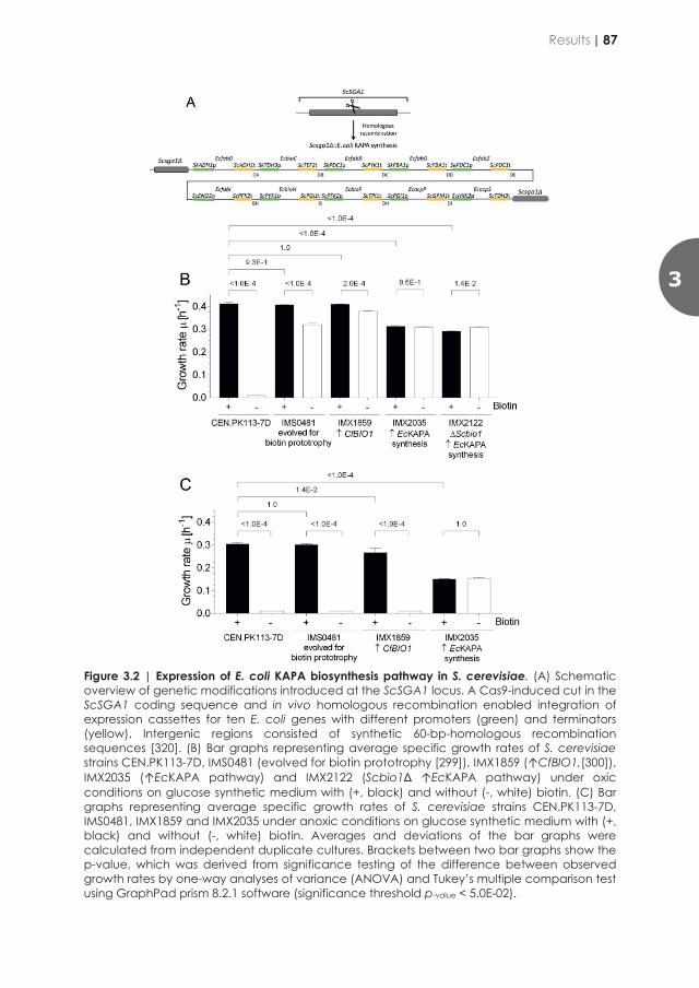
the sequences of the heterologously expressed proteins was used. Each strain was analyzed in independent biological duplicate samples. Data processing and analysis of differentially expressed proteins was conducted as previously described in [325]. Enrichment analysis of up- and downregulated proteins in the isolates was performed using the GO Enrichment Analysis [327].
Results
Expression of the E. coli KAPA-biosynthesis pathway supports biotin-independent growth of S. cerevisiae in absence of oxygen.
Of the currently known prokaryotic biotin-biosynthesis pathways (Figure 3.1), only the variant that occurs in E. coli starts with malonyl-CoA, a key precursor for lipid synthesis in S. cerevisiae. To complete the malonyl-CoA conversion into pimeloyl-CoA, only EcbioC and EcbioH would be required in S. cerevisiae assuming that the other reactions could be performed by the native fatty acid elongation machinery. We also included EcbioF since it is unclear whether ScBio6, the protein ortholog of EcBioF, can use pimeloyl-[Acp] as substrate. Integration of these three E. coli genes at the SGA1 locus yielded S. cerevisiae IMX2706. Even after prolonged oxic incubation in biotin-free synthetic medium, this engineered strain did not show growth on biotin-free synthetic medium. To investigate whether this inability was related to the different organization of the prokaryotic and yeast fatty-acid-synthesis machineries, we introduced an additional set of expression cassettes for E. coli proteins involved in conversion of malonyl-[Acp]-methyl ester into pimeloyl-[Acp]-methyl ester. In addition to EcbioC, H and F, five genes involved in fatty-acid biosynthesis (EcfabD, EcfabB, EcfabG, EcfabZ, EcfabI) and two genes involved in acyl carrier protein formation (EcacpP and EcacpS) were introduced. In E. coli, the concerted action of the enzymes encoded by these genes converts malonyl-CoA into 8-amino-7-oxo-nonanoate (KAPA), a metabolic intermediate of the native S. cerevisiae biotin pathway. Using the SpyCas9-expressing strain IMX585, the ten expression cassettes were integrated at the SGA1 locus, yielding S. cerevisiae IMX2035 (↑EcKAPA pathway; Figure 2.2A)
Figure 3.2 | Expression of E. coli KAPA biosynthesis pathway in S. cerevisiae. (A) Schematic overview of genetic modifications introduced at the ScSGA1 locus. A Cas9-induced cut in the ScSGA1 coding sequence and in vivo homologous recombination enabled integration of expression cassettes for ten E. coli genes with different promoters (green) and terminators (yellow). Intergenic regions consisted of synthetic 60-bp-homologous recombination sequences [320]. (B) Bar graphs representing average specific growth rates of S. cerevisiae strains CEN.PK113-7D, IMS0481 (evolved for biotin prototrophy [299]), IMX1859 (↑CfBIO1,[300]), IMX2035 (↑EcKAPA pathway) and IMX2122 (Scbio1Δ ↑EcKAPA pathway) under oxic conditions on glucose synthetic medium with (+, black) and without (-, white) biotin. (C) Bar graphs representing average specific growth rates of S. cerevisiae strains CEN.PK113-7D, IMS0481, IMX1859 and IMX2035 under anoxic conditions on glucose synthetic medium with (+, black) and without (-, white) biotin. Averages and deviations of the bar graphs were calculated from independent duplicate cultures. Brackets between two bar graphs show the p-value, which was derived from significance testing of the difference between observed growth rates by one-way analyses of variance (ANOVA) and Tukey’s multiple comparison test using GraphPad prism 8.2.1 software (significance threshold p-value < 5.0E-02).
3
Results | 87
This engineered strain showed immediate oxic growth on biotin-free synthetic medium, at a specific growth rate of 0.31 ± 0.01 h-1. Under the same conditions, the reference strain CEN.PK113-7D was unable to grow [225, 299, 300] (Figure 3.2B). Compared to previous S. cerevisiae strains engineered (IMX1859, [300] or evolved (IMS0481, [299] for biotin prototrophy, IMX2035 grew approximately 25% slower in biotin-supplemented as well as biotin-free media (Figure 2.2B). However, in contrast to these other biotin-prototrophic strains, strain IMX2035 (↑EcKAPA pathway) showed anoxic growth in biotin-free medium, at specific growth rate of 0.15 ± 0.003 h-1. Also, in absence of oxygen, the specific growth rate of strain IMX2035 on biotin-supplement medium was lower than observed in cultures of reference strains (Figure 3.2C). These results demonstrated that expression of the E. coli KAPA pathway in S. cerevisiae supports conversion of malonyl-CoA into KAPA and promotes biotin-independent anoxic growth of S. cerevisiae. The functionality of the EcKAPA pathway in S. cerevisiae IMX2035 enabled us to evaluate whether the orthologs ScBIO6 and EcbioF are functionally redundant. To this end, EcbioF was deleted in strain IMX2035, yielding strain IMX2707. This deletion strain did not grow on biotin-free medium, indicating that the yeast 7,8-diamino-pelargonic acid aminotransferase ScBio6 cannot functionally replace the E. coli 8-amino-7-oxononanoate synthase EcBioF.
Laboratory evolution for fast biotin-independent anoxic growth
To exclude the possibility that activity of the native S. cerevisiae biotin pathway interfered with the interpretation of results, ScBIO1 was deleted in strain IMX2035 (↑EcKAPA pathway), yielding strain IMX2122 (Scbio1Δ ↑EcKAPA pathway). ScBio1 is proposed to catalyse an as yet unidentified reaction for synthesis of pimeloyl-CoA. In oxic cultures, strain IMX2122 showed similar specific growth rates on biotin-supplemented and biotin-free media (specific growth rates of 0.29 ± 0.00 h-1 and
0.31 ± 0.00 h-1, respectively, Figure 3.2B). As anticipated, strain IMX2122 grew without oxygen on biotin-free medium, at a specific growth rate of 0.20 ± 0.00 h-1 (Figure 3.3C). As observed for strain IMX2035 (↑EcKAPA pathway) biotin supplementation did not restore the specific growth rate of strain IMX2122 to that of reference strain CEN.PK113-7D, which in both cultivation regimes on biotin-supplemented media exhibits specific growth rates of 0.32 - 0.33 h-1 [328, 329] and 0.38 - 0.40 h-1 [330] ,respectively. To explore the evolvability of full biotin prototrophy, strain IMX2122 (Scbio1Δ ↑EcKAPA pathway) was grown in two independent, anoxic sequential batch reactors (SBRs) on biotin-free synthetic medium. Throughout the course of SBR cultivation, the specific growth rate of the yeast populations in the two reactions increased to close to 0.32 h-1, which corresponded closely to the reported specific growth rate on the congenic CEN.PK113-7D reference strain in absence of oxygen on chemically defined medium with biotin [328, 329] (Figure 3.3A, B). After 436 (109 batch cycles) and 400 generations (100 batch cycles) for reactor A and B, respectively, single colony isolates (SCI) were
obtained from each reactor (IMS0994 from reactor A and IMS0995 from reactor B). Both these SCI‘s showed specific growth rates on biotin-free medium of 0.39 ± 0.01 h-1. Under anoxic conditions, specific growth rates of the SCI’s were 0.33 ± 0.01 h-1 and 0.33 ± 0.02 h-1, respectively. These specific growth rates are virtually identical to those measured in this study for the reference strain CEN.PK113-7D during growth on biotin-containing synthetic medium under both cultivation regimes (0.41 ± 0.01 h-1 and 0.31± 0.01 h-1, respectively). Compared to the specific growth rates of their parental strain IMX2122 on biotin-free medium in presence and absence of oxygen, those of the two SCI’s had increased by 34% (p = 2E-04) and 57% (p = 1E-04), respectively (Figure 3.3C).
Figure 3.3 | Laboratory evolution of the engineered biotin-prototrophic S. cerevisiae strain IMX2122. (A) Specific growth rates of anoxic sequential batch cycles [n] of strain IMX2122 (Scbio1Δ ↑EcKAPA pathway) on biotin-free medium, reactor A. (B) Specific growth rates of anoxic sequential batch cycles [n] of strain IMX2122 on biotin-free medium, reactor B. (C) Bar graphs represent average specific growth rates of the parental strain S. cerevisiae IMX2122 and evolved isolates IMS0994 (evolution A IMX2122) and IMS0995 (evolution B IMX2122) on synthetic medium without biotin under oxic (+, black) and anoxic (-, white) conditions. The growth rate means and deviations of the bar graphs were calculated from biological duplicates. Brackets between two bar graphs show the p-value, which was derived from significance testing of the difference between observed growth rates by one-way analyses of variance (ANOVA) and Tukey’s multiple comparison test using GraphPad prism 8.2.1 software (significance threshold p-value < 5.0E-02).
88 | Chapter 3

This engineered strain showed immediate oxic growth on biotin-free synthetic medium, at a specific growth rate of 0.31 ± 0.01 h-1. Under the same conditions, the reference strain CEN.PK113-7D was unable to grow [225, 299, 300] (Figure 3.2B). Compared to previous S. cerevisiae strains engineered (IMX1859, [300] or evolved (IMS0481, [299] for biotin prototrophy, IMX2035 grew approximately 25% slower in biotin-supplemented as well as biotin-free media (Figure 2.2B). However, in contrast to these other biotin-prototrophic strains, strain IMX2035 (↑EcKAPA pathway) showed anoxic growth in biotin-free medium, at specific growth rate of 0.15 ± 0.003 h-1. Also, in absence of oxygen, the specific growth rate of strain IMX2035 on biotin-supplement medium was lower than observed in cultures of reference strains (Figure 3.2C). These results demonstrated that expression of the E. coli KAPA pathway in S. cerevisiae supports conversion of malonyl-CoA into KAPA and promotes biotin-independent anoxic growth of S. cerevisiae. The functionality of the EcKAPA pathway in S. cerevisiae IMX2035 enabled us to evaluate whether the orthologs ScBIO6 and EcbioF are functionally redundant. To this end, EcbioF was deleted in strain IMX2035, yielding strain IMX2707. This deletion strain did not grow on biotin-free medium, indicating that the yeast 7,8-diamino-pelargonic acid aminotransferase ScBio6 cannot functionally replace the E. coli 8-amino-7-oxononanoate synthase EcBioF.
Laboratory evolution for fast biotin-independent anoxic growth
To exclude the possibility that activity of the native S. cerevisiae biotin pathway interfered with the interpretation of results, ScBIO1 was deleted in strain IMX2035 (↑EcKAPA pathway), yielding strain IMX2122 (Scbio1Δ ↑EcKAPA pathway). ScBio1 is proposed to catalyse an as yet unidentified reaction for synthesis of pimeloyl-CoA. In oxic cultures, strain IMX2122 showed similar specific growth rates on biotin-supplemented and biotin-free media (specific growth rates of 0.29 ± 0.00 h-1 and
0.31 ± 0.00 h-1, respectively, Figure 3.2B). As anticipated, strain IMX2122 grew without oxygen on biotin-free medium, at a specific growth rate of 0.20 ± 0.00 h-1 (Figure 3.3C). As observed for strain IMX2035 (↑EcKAPA pathway) biotin supplementation did not restore the specific growth rate of strain IMX2122 to that of reference strain CEN.PK113-7D, which in both cultivation regimes on biotin-supplemented media exhibits specific growth rates of 0.32 - 0.33 h-1 [328, 329] and 0.38 - 0.40 h-1 [330] ,respectively. To explore the evolvability of full biotin prototrophy, strain IMX2122 (Scbio1Δ ↑EcKAPA pathway) was grown in two independent, anoxic sequential batch reactors (SBRs) on biotin-free synthetic medium. Throughout the course of SBR cultivation, the specific growth rate of the yeast populations in the two reactions increased to close to 0.32 h-1, which corresponded closely to the reported specific growth rate on the congenic CEN.PK113-7D reference strain in absence of oxygen on chemically defined medium with biotin [328, 329] (Figure 3.3A, B). After 436 (109 batch cycles) and 400 generations (100 batch cycles) for reactor A and B, respectively, single colony isolates (SCI) were
obtained from each reactor (IMS0994 from reactor A and IMS0995 from reactor B). Both these SCI‘s showed specific growth rates on biotin-free medium of 0.39 ± 0.01 h-1. Under anoxic conditions, specific growth rates of the SCI’s were 0.33 ± 0.01 h-1 and 0.33 ± 0.02 h-1, respectively. These specific growth rates are virtually identical to those measured in this study for the reference strain CEN.PK113-7D during growth on biotin-containing synthetic medium under both cultivation regimes (0.41 ± 0.01 h-1 and 0.31± 0.01 h-1, respectively). Compared to the specific growth rates of their parental strain IMX2122 on biotin-free medium in presence and absence of oxygen, those of the two SCI’s had increased by 34% (p = 2E-04) and 57% (p = 1E-04), respectively (Figure 3.3C).
Figure 3.3 | Laboratory evolution of the engineered biotin-prototrophic S. cerevisiae strain IMX2122. (A) Specific growth rates of anoxic sequential batch cycles [n] of strain IMX2122 (Scbio1Δ ↑EcKAPA pathway) on biotin-free medium, reactor A. (B) Specific growth rates of anoxic sequential batch cycles [n] of strain IMX2122 on biotin-free medium, reactor B. (C) Bar graphs represent average specific growth rates of the parental strain S. cerevisiae IMX2122 and evolved isolates IMS0994 (evolution A IMX2122) and IMS0995 (evolution B IMX2122) on synthetic medium without biotin under oxic (+, black) and anoxic (-, white) conditions. The growth rate means and deviations of the bar graphs were calculated from biological duplicates. Brackets between two bar graphs show the p-value, which was derived from significance testing of the difference between observed growth rates by one-way analyses of variance (ANOVA) and Tukey’s multiple comparison test using GraphPad prism 8.2.1 software (significance threshold p-value < 5.0E-02).
3
Results | 89
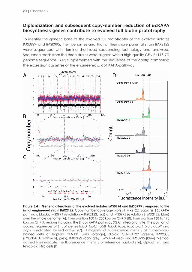
Diploidization and subsequent copy-number reduction of EcKAPA biosynthesis genes contribute to evolved full biotin prototrophy
To identify the genetic basis of the evolved full prototrophy of the evolved isolates IMS0994 and IMS0995, their genomes and that of their share parental strain IMX2122 were sequenced with Illumina short-read sequencing technology and analysed. Sequence reads from the three strains were aligned with a high-quality CEN.PK113-7D genome sequence [309] supplemented with the sequence of the contig comprising the expression cassettes of the engineered E. coli KAPA-pathway.
Figure 3.4 | Genetic alterations of the evolved isolates IMS0994 and IMS0995 compared to the initial engineered strain IMX2122. Copy number coverage plots of IMX2122 (Scbio1Δ ↑EcKAPA pathway, black), IMS0994 (evolution A IMX2122, red) and IMS0995 (evolution B IMX2122, blue) over the whole genome (A), from position 100 to 250 kbp on CHRIX (B), from position 168 to 195 kbp on CHRIX, regions including the E. coli KAPA pathway SGA1 integration site. The position of coding sequences of E. coli genes fabD, bioC, fabB, fabG, fabZ, fabI, bioH, bioF, acpP and acpS is indicated by red arrows (C). Histograms of fluorescence intensity of nucleic-acid-stained cells of haploid CEN.PK113-7D (orange), diploid CEN.PK122 (green), IMX2035 ((↑EcKAPA pathway), grey), IMX2122 (dark grey), IMS0994 (red) and IMS0995 (blue). Vertical dashed lines indicate the fluorescence intensity of reference haploid (1n), diploid (2n) and tetraploid (4n) cells (D).
Mapped data were analysed for copy number variations (CNVs), structural modifications and single nucleotide variations (SNVs) in annotated coding sequences. Prior to sequence data analysis, the nominal strain ploidy of IMX2035, IMX2122, IMS0994 and IMS0995 was analysed by nucleic acid staining and subsequent flow-cytometry analysis. The genetically engineered strains IMX2035 and IMX2122 exhibited the same ploidy as the haploid reference strain CEN.PK113-7D. In contrast, a higher fluorescence intensity of both evolved SCI’s (IMS0994 and IMS0995) corresponded with that of the diploid reference strain CEN.PK122 (Figure 3.4) and indicated that a whole-genome duplication had occurred in two independent evolution experiments. CNV analysis of strains IMX2122 and IMS0994-5 did reveal a segmental aneuploidy of the engineered SGA1 locus in which the E. coli KAPA pathway was integrated. As anticipated, the read coverage over the contig harboring the E. coli KAPA-pathway cassettes in the parental strain IMX2122 was the same as that of the rest of the genome. In contrast, the evolved SCI’s IMS0994 and IMS0995 showed a 50% lower coverage for a region comprising the three contiguous expression cassettes for EcfabD, EcbioC and EcfabB (Figure 3.4 A-B-C). This coverage reduction relative to the rest of the genome was consistent with the overall 2n ploidy of the evolved isolates (Figure 3.4 C-D). While no homozygous SNVs were found in coding regions of the two evolved SCI’s, a single homozygous SNV in IMS0994 was identified in the intergenic region between PTR2 and MLP1 on CHRXI. In addition, the two SCI’s harbored a small number of heterozygous SNVs that caused amino-acid changes in the peptide sequence encoded by the mutated allele. In IMS0994 nine heterozygous SNVs occurring in coding sequences were found to be distributed over five genes (FLO11, AGA1, MFα1, TIF3 and ADE3). Similarly, IMS0995 harbored ten heterozygous SNVs scattered over the coding sequences of four genes (GLT1, MFα1, ECM38 and TIF3). Out of these heterozygous SNVs, five observed in TIF3 and one detected in MFα1 were shared by the two evolved isolates suggesting that these SNVs might originate from stock cultures used to inoculate the evolution cultures. None of the affected genes showed an obvious functional relationship with biotin-related cellular processes and the individual impact of these SNVs was not further studied.
To investigate the impact of the altered gene dosage of three E. coli KAPA biosynthesis genes in the evolved strains, levels of the E. coli KAPA pathway proteins were quantified in strains IMX2122, IMS0994 and IMS0995. Consistent with their lower copy number relative to the remainder of the genome in the evolved SCI’s, abundances of the 3-oxoacyl-[Acp] synthase EcFabB, the malonyl CoA-acyl carrier protein transacylase EcFabD and the malonyl-[Acp] O-methyltransferase EcBioC in IMS0994 and IMS0995 were at least 1.8-fold lower than those of the non-evolved parental strain IMX2122 (Figure 3.5). Despite the change in ploidy, no differences in average protein abundance were observed between the three strains (Figure 3.5A). While all expressed heterologous proteins were detected, ScBio2 was the only native biotin-synthesis pathway detected in the samples.
90 | Chapter 3
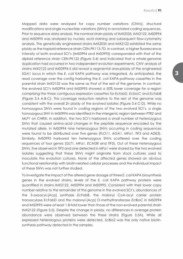
Diploidization and subsequent copy-number reduction of EcKAPA biosynthesis genes contribute to evolved full biotin prototrophy
To identify the genetic basis of the evolved full prototrophy of the evolved isolates IMS0994 and IMS0995, their genomes and that of their share parental strain IMX2122 were sequenced with Illumina short-read sequencing technology and analysed. Sequence reads from the three strains were aligned with a high-quality CEN.PK113-7D genome sequence [309] supplemented with the sequence of the contig comprising the expression cassettes of the engineered E. coli KAPA-pathway.
Figure 3.4 | Genetic alterations of the evolved isolates IMS0994 and IMS0995 compared to the initial engineered strain IMX2122. Copy number coverage plots of IMX2122 (Scbio1Δ ↑EcKAPA pathway, black), IMS0994 (evolution A IMX2122, red) and IMS0995 (evolution B IMX2122, blue) over the whole genome (A), from position 100 to 250 kbp on CHRIX (B), from position 168 to 195 kbp on CHRIX, regions including the E. coli KAPA pathway SGA1 integration site. The position of coding sequences of E. coli genes fabD, bioC, fabB, fabG, fabZ, fabI, bioH, bioF, acpP and acpS is indicated by red arrows (C). Histograms of fluorescence intensity of nucleic-acid-stained cells of haploid CEN.PK113-7D (orange), diploid CEN.PK122 (green), IMX2035 ((↑EcKAPA pathway), grey), IMX2122 (dark grey), IMS0994 (red) and IMS0995 (blue). Vertical dashed lines indicate the fluorescence intensity of reference haploid (1n), diploid (2n) and tetraploid (4n) cells (D).
Mapped data were analysed for copy number variations (CNVs), structural modifications and single nucleotide variations (SNVs) in annotated coding sequences. Prior to sequence data analysis, the nominal strain ploidy of IMX2035, IMX2122, IMS0994 and IMS0995 was analysed by nucleic acid staining and subsequent flow-cytometry analysis. The genetically engineered strains IMX2035 and IMX2122 exhibited the same ploidy as the haploid reference strain CEN.PK113-7D. In contrast, a higher fluorescence intensity of both evolved SCI’s (IMS0994 and IMS0995) corresponded with that of the diploid reference strain CEN.PK122 (Figure 3.4) and indicated that a whole-genome duplication had occurred in two independent evolution experiments. CNV analysis of strains IMX2122 and IMS0994-5 did reveal a segmental aneuploidy of the engineered SGA1 locus in which the E. coli KAPA pathway was integrated. As anticipated, the read coverage over the contig harboring the E. coli KAPA-pathway cassettes in the parental strain IMX2122 was the same as that of the rest of the genome. In contrast, the evolved SCI’s IMS0994 and IMS0995 showed a 50% lower coverage for a region comprising the three contiguous expression cassettes for EcfabD, EcbioC and EcfabB (Figure 3.4 A-B-C). This coverage reduction relative to the rest of the genome was consistent with the overall 2n ploidy of the evolved isolates (Figure 3.4 C-D). While no homozygous SNVs were found in coding regions of the two evolved SCI’s, a single homozygous SNV in IMS0994 was identified in the intergenic region between PTR2 and MLP1 on CHRXI. In addition, the two SCI’s harbored a small number of heterozygous SNVs that caused amino-acid changes in the peptide sequence encoded by the mutated allele. In IMS0994 nine heterozygous SNVs occurring in coding sequences were found to be distributed over five genes (FLO11, AGA1, MFα1, TIF3 and ADE3). Similarly, IMS0995 harbored ten heterozygous SNVs scattered over the coding sequences of four genes (GLT1, MFα1, ECM38 and TIF3). Out of these heterozygous SNVs, five observed in TIF3 and one detected in MFα1 were shared by the two evolved isolates suggesting that these SNVs might originate from stock cultures used to inoculate the evolution cultures. None of the affected genes showed an obvious functional relationship with biotin-related cellular processes and the individual impact of these SNVs was not further studied.
To investigate the impact of the altered gene dosage of three E. coli KAPA biosynthesis genes in the evolved strains, levels of the E. coli KAPA pathway proteins were quantified in strains IMX2122, IMS0994 and IMS0995. Consistent with their lower copy number relative to the remainder of the genome in the evolved SCI’s, abundances of the 3-oxoacyl-[Acp] synthase EcFabB, the malonyl CoA-acyl carrier protein transacylase EcFabD and the malonyl-[Acp] O-methyltransferase EcBioC in IMS0994 and IMS0995 were at least 1.8-fold lower than those of the non-evolved parental strain IMX2122 (Figure 3.5). Despite the change in ploidy, no differences in average protein abundance were observed between the three strains (Figure 3.5A). While all expressed heterologous proteins were detected, ScBio2 was the only native biotin-synthesis pathway detected in the samples.
3
Results | 91
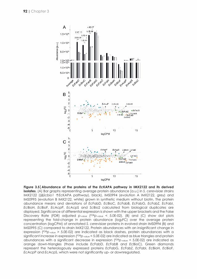
Figure 3.5Abundance of the proteins of the EcKAPA pathway in IMX2122 and its derived isolates. (A) Bar graphs representing average protein abundance [a.u.] in S. cerevisiae strains IMX2122 ((ΔScbio1 ↑EcKAPA pathway), black), IMS0994 (evolution A IMX2122, grey) and IMS0995 (evolution B IMX2122, white) grown in synthetic medium without biotin. The protein abundance means and deviations of EcFabD, EcBioC, EcFabB, EcFabG, EcFabZ, EcFabI, EcBioH, EcBioF, EcAcpP, EcAcpS and ScBio2 calculated from biological duplicates are displayed. Significance of differential expression is shown with the upper brackets and the False Discovery Rate (FDR) adjusted p-value (FDRp-value < 5.0E-02). (B) and (C) show dot plots representing the fold-change in protein abundance (log2FC) over the average protein concentration (logCPM) of annotated S. cerevisiae proteins in evolved strain IMS0994 (B) and IMS0995 (C) compared to strain IMX2122. Protein abundances with an insignificant change in expression (FDRp-value > 5.0E-02) are indicated as black dashes, protein abundances with a significant increase in expression (FDRp-value < 5.0E-02) are indicated as blue triangles and protein abundances with a significant decrease in expression (FDRp-value < 5.0E-02) are indicated as orange down-triangles (those include EcFabD, EcFabB and EcBioC). Green diamonds represent the heterologously expressed proteins EcFabG, EcFabZ, EcFabI, EcBioH, EcBioF, EcAcpP and EcAcpS, which were not significantly up- or downregulated.
Only 44 native yeast proteins in strain IMS0994 and 48 in strain IMS0995 showed a significantly different abundance relative to the parental strain IMX2122, of which 22 showed a unidirectional difference in the two isolates (Figures 3.5B and 3.5C). Not fewer than 20 and 26 proteins exhibited a 2-fold reduction at least of their abundance in IMS0994 and IMS0995 relative to IMX2122, respectively. Concomitantly, 14 and 10 proteins exhibited a 2-fold increase at least of their abundance in IMS0994 and IMS0995 relative to IMX2122, respectively (Figures 3.5B and 3.5C). Proteins that showed a lower level in the two SCI’s did not show GO-categories related to metabolic processes, whereas proteins that showed a higher level in IMS0994 (Bonferronip-value = 9.98E-07) or IMS0995 (Bonferronip-value = 3.41E-02) indicated an overrepresentation of proteins belonging to the GO-category ‘ATP metabolic process’ (GO:0046034). As members of this GO category, the ATP synthase subunit Atp20 as well as the cytochrome c oxidase subunits Cox5A and Cox13 showed higher levels in both isolates. In IMS0995, the cytochrome c oxidase subunit Cox4 and, in IMS0994, the cytochrome b-c1 complex subunit Qcr8, ATP synthase subunits Atp7 and Atp4 as well as cytochrome c oxidase subunit Cox9 also showed increased levels.
Reverse engineering gene dosage of the E. coli KAPA biosynthesis pathway contributes to improve both an- and oxic growth rate of the industrial diploid strain Ethanol Red.
To test whether altered gene dosage of the first three genes of the oxygen-independent KAPA biosynthesis pathway relative to the downstream genes, and the corresponding lower level of the encoded proteins, was critical to enhance growth of engineered strains in biotin-free conditions, we engineered the diploid industrial strain Ethanol Red. Using CRISPR/Cas9, which enables the simultaneous modification of all gene copies in polyploid strains [278], the ten heterologous genes were introduced at the SGA1 locus. In contrast to the parental strain Ethanol Red, the resulting strain IMX2555 readily grew in biotin-free medium under oxic as well as under anoxic conditions. However, in both cultivation conditions, strain IMX2555 grew slower than Ethanol Red in biotin-supplemented medium (0.34 ± 0.01 h-1 versus 0.45 ± 0.01 h-1 and 0.20 ± 0.01 h-1 versus 0.42 ± 0.00 h-1, respectively; Figure 3.6). Specific growth rates of strain IMX2555 were not affected by the presence or absence of biotin (Figure 3.6)
To reproduce the genotype observed in the evolved isolates IMS0994-5, a copy of EcfabD, EcbioC and EcfabB was deleted in IMX2555 by ‘pre-CRISPR’ marker-assisted homologous recombination [321] as it enables deletion of only one of the two copies of a targeted region in diploid strains. The deletion yielded the heterozygous diploid strain IMX2632 (↑EcfabD EcbioC EcfabB,G,Z,I EcbioH,F EcacpP,S / ↑EcfabG,Z,I EcbioH,F EcacpP,S). The specific growth rate of strain IMX2632 in anaerobic cultures on biotin-free medium was significantly higher than that of its parental strain IMS2555 (p value < 1.0E-04; 0.30 ± 0.01 h-1 versus 0.19±0.00 h-1). A smaller but significantly higher specific growth rate (p-value < 1.0E-04; 0.38 ± 0.01 h-1 versus 0.34 ± 0.00 h-1) was observed in oxic cultures. Despite these improvements, the engineered strain IMX2632 still grew
92 | Chapter 3

Figure 3.5Abundance of the proteins of the EcKAPA pathway in IMX2122 and its derived isolates. (A) Bar graphs representing average protein abundance [a.u.] in S. cerevisiae strains IMX2122 ((ΔScbio1 ↑EcKAPA pathway), black), IMS0994 (evolution A IMX2122, grey) and IMS0995 (evolution B IMX2122, white) grown in synthetic medium without biotin. The protein abundance means and deviations of EcFabD, EcBioC, EcFabB, EcFabG, EcFabZ, EcFabI, EcBioH, EcBioF, EcAcpP, EcAcpS and ScBio2 calculated from biological duplicates are displayed. Significance of differential expression is shown with the upper brackets and the False Discovery Rate (FDR) adjusted p-value (FDRp-value < 5.0E-02). (B) and (C) show dot plots representing the fold-change in protein abundance (log2FC) over the average protein concentration (logCPM) of annotated S. cerevisiae proteins in evolved strain IMS0994 (B) and IMS0995 (C) compared to strain IMX2122. Protein abundances with an insignificant change in expression (FDRp-value > 5.0E-02) are indicated as black dashes, protein abundances with a significant increase in expression (FDRp-value < 5.0E-02) are indicated as blue triangles and protein abundances with a significant decrease in expression (FDRp-value < 5.0E-02) are indicated as orange down-triangles (those include EcFabD, EcFabB and EcBioC). Green diamonds represent the heterologously expressed proteins EcFabG, EcFabZ, EcFabI, EcBioH, EcBioF, EcAcpP and EcAcpS, which were not significantly up- or downregulated.
Only 44 native yeast proteins in strain IMS0994 and 48 in strain IMS0995 showed a significantly different abundance relative to the parental strain IMX2122, of which 22 showed a unidirectional difference in the two isolates (Figures 3.5B and 3.5C). Not fewer than 20 and 26 proteins exhibited a 2-fold reduction at least of their abundance in IMS0994 and IMS0995 relative to IMX2122, respectively. Concomitantly, 14 and 10 proteins exhibited a 2-fold increase at least of their abundance in IMS0994 and IMS0995 relative to IMX2122, respectively (Figures 3.5B and 3.5C). Proteins that showed a lower level in the two SCI’s did not show GO-categories related to metabolic processes, whereas proteins that showed a higher level in IMS0994 (Bonferronip-value = 9.98E-07) or IMS0995 (Bonferronip-value = 3.41E-02) indicated an overrepresentation of proteins belonging to the GO-category ‘ATP metabolic process’ (GO:0046034). As members of this GO category, the ATP synthase subunit Atp20 as well as the cytochrome c oxidase subunits Cox5A and Cox13 showed higher levels in both isolates. In IMS0995, the cytochrome c oxidase subunit Cox4 and, in IMS0994, the cytochrome b-c1 complex subunit Qcr8, ATP synthase subunits Atp7 and Atp4 as well as cytochrome c oxidase subunit Cox9 also showed increased levels.
Reverse engineering gene dosage of the E. coli KAPA biosynthesis pathway contributes to improve both an- and oxic growth rate of the industrial diploid strain Ethanol Red.
To test whether altered gene dosage of the first three genes of the oxygen-independent KAPA biosynthesis pathway relative to the downstream genes, and the corresponding lower level of the encoded proteins, was critical to enhance growth of engineered strains in biotin-free conditions, we engineered the diploid industrial strain Ethanol Red. Using CRISPR/Cas9, which enables the simultaneous modification of all gene copies in polyploid strains [278], the ten heterologous genes were introduced at the SGA1 locus. In contrast to the parental strain Ethanol Red, the resulting strain IMX2555 readily grew in biotin-free medium under oxic as well as under anoxic conditions. However, in both cultivation conditions, strain IMX2555 grew slower than Ethanol Red in biotin-supplemented medium (0.34 ± 0.01 h-1 versus 0.45 ± 0.01 h-1 and 0.20 ± 0.01 h-1 versus 0.42 ± 0.00 h-1, respectively; Figure 3.6). Specific growth rates of strain IMX2555 were not affected by the presence or absence of biotin (Figure 3.6)
To reproduce the genotype observed in the evolved isolates IMS0994-5, a copy of EcfabD, EcbioC and EcfabB was deleted in IMX2555 by ‘pre-CRISPR’ marker-assisted homologous recombination [321] as it enables deletion of only one of the two copies of a targeted region in diploid strains. The deletion yielded the heterozygous diploid strain IMX2632 (↑EcfabD EcbioC EcfabB,G,Z,I EcbioH,F EcacpP,S / ↑EcfabG,Z,I EcbioH,F EcacpP,S). The specific growth rate of strain IMX2632 in anaerobic cultures on biotin-free medium was significantly higher than that of its parental strain IMS2555 (p value < 1.0E-04; 0.30 ± 0.01 h-1 versus 0.19±0.00 h-1). A smaller but significantly higher specific growth rate (p-value < 1.0E-04; 0.38 ± 0.01 h-1 versus 0.34 ± 0.00 h-1) was observed in oxic cultures. Despite these improvements, the engineered strain IMX2632 still grew
3
Results | 93
slower than observed for the Ethanol Red strain in both biotin supplemented cultures (Figure 3.6), suggesting additional tuning of gene dosages of KAPA-pathway cassettes and/or other mutations are required for full anoxic biotin prototrophy in engineered strains.
Figure 3.6 | Growth of S. cerevisiae Ethanol Red and engineered strains expressing E. coli KAPA synthesis genes. (A) Bar graphs representing average specific growth rates of S. cerevisiae strains Ethanol Red (diploid, industrial ethanol producer), IMX2555 (Ethanol Red ↑EcKAPA pathway) and IMX2632 (Ethanol Red ↑ EcKAPA pathway / fabD,B bioCΔ) under oxic conditions on synthetic medium with (+, black) and without (-, white) biotin. (B) specific growth rates of S. cerevisiae strains Ethanol Red, IMX2555 and IMX2632 under anoxic conditions on synthetic medium with (+, black) and without (-, white) biotin. The bars represent averages and standard deviations from two biological replicates. Statistical significance between growth rates in SMD with and without biotin, and between strains grown in the same conditions using one-way analyses of variance (ANOVA) and Tukey’s multiple comparison test using GraphPad prism 8.2.1 software (p-value < 5.0E-02) is indicated.
Discussion
The native yeast pathway for biotin biosynthesis, for which the first committed reaction remains to be resolved, is oxygen dependent [300]. This study shows that functional expression of the E. coli KAPA pathway yields S. cerevisiae strains that are biotin prototrophic irrespective of the applied oxygen regime and whose specific growth rates can be further improved by tuning of the expression levels of specific KAPA-pathway enzymes. Prokaryotic biosynthesis pathways have previously been transferred between bacteria to increase biotin production by bacterial hosts such as Pseudomonas mutabilis [331], Agrobacterium sp. [332] and E. coli [333]. For functional expression of the E. coli KAPA pathway in S. cerevisiae, the different organization of prokaryotic and eukaryotic fatty-acid biosynthesis needed to be considered. In the type-II FAS system of E. coli, individual reactions in fatty-acid synthesis are catalysed by separate proteins [334]. In contrast, the type-I FAS system of S. cerevisiae and other fungi harbours all catalytic sites required for fatty-acid biosynthesis in domains of a
large, multi-functional single polypeptide or, as in S. cerevisiae, two polypeptides [335, 336]. Despite this structural difference, functional replacement of the S. cerevisiae type I-FAS complex by the E. coli type-II FAS system has been demonstrated [337]. In this study, expression of only EcbioC, H and F in S. cerevisiae did not support biotin prototrophy. This observation suggested that the yeast type-I FAS complex cannot convert malonyl-CoA methyl ester into pimeloyl-[Acp] or, alternatively, that the location of the acyl-carrier function on a distinct domain within a large multifunctional protein prevented EcBioC from accessing its substrate. While the S. cerevisiae genome additionally encodes a soluble acyl carrier protein (Acp1) and its activating enzyme phosphopantetheine:protein transferase (Ppt2), these proteins participate in mitochondrial fatty-acid synthesis and are located in the mitochondrial matrix [338]. This localization issue was circumvented by additionally expressing the E. coli fatty-acid synthesis genes EcfabD,B, G, Z, I as well as EcacpS and P and, thereby, enabling cytosolic synthesis of pimeloyl-[Acp]. In the engineered biotin-prototrophic strain IMX2035, conversion of pimeloyl-[Acp] to 7-keto-8-aminopelargonic acid (KAPA) was enabled by expression of EcbioF. Deletion of EcbioF from this strain led to loss of its biotin prototrophy. Apparently, like its B. subtilis ortholog BioF, S. cerevisiae Bio6 cannot convert pimeloyl-[Acp] to KAPA but specifically requires pimeloyl-CoA as a substrate [261]. The biotin auxotrophy of the EcbioF deletion strain was unlikely to be caused by an insufficient expression level of Bio6 since expression of ScBio1 or CfBio1 suffices to confer oxic biotin prototrophy in CEN.PK strains [300].
In metabolic engineering, optimization of productivity and yield often requires balancing of the relative levels of enzymes in product pathways [339]. Such balancing may be especially challenging when, as in the present study, the product pathway is strongly intertwined with core metabolic processes of the microbial host. Optimal enzyme levels can be explored by in vitro [340] or in vivo [341, 342] approaches for combinatorial variation of the amounts of relevant enzymes. Our results illustrate how adaptive laboratory evolution [343, 344], combined with access to a high-quality reference genome [309], modern sequencing technologies, proteomics and a streamlined bioinformatics pipeline [345, 346] can provide a powerful alternative approach to gain relevant information on pathway balancing. Evolution of strain IMX2122 for faster biotin-independent growth involved a whole-genome duplication and subsequent reduction of the copy number of three genes of the heterologous biotin-biosynthesis pathway. Ploidy changes from haploid to diploid and from tetraploid to diploid have been reported in previous studies on evolving yeast populations subjected to strong selection pressures such as repetitive carbon-source switching [347] and ethanol stress [348]. A whole-genome duplication was also observed after prolonged cultivation (over 1000 generations) of haploid S. cerevisiae strains on complex medium [349]. However, based on several shared homozygous and heterozygous SNVs in independently evolved isolates, we cannot exclude the possibility that a small subpopulation of diploid cells was already present in the predominantly haploid stock cultures with which the evolution experiments were inoculated.
94 | Chapter 3

slower than observed for the Ethanol Red strain in both biotin supplemented cultures (Figure 3.6), suggesting additional tuning of gene dosages of KAPA-pathway cassettes and/or other mutations are required for full anoxic biotin prototrophy in engineered strains.
Figure 3.6 | Growth of S. cerevisiae Ethanol Red and engineered strains expressing E. coli KAPA synthesis genes. (A) Bar graphs representing average specific growth rates of S. cerevisiae strains Ethanol Red (diploid, industrial ethanol producer), IMX2555 (Ethanol Red ↑EcKAPA pathway) and IMX2632 (Ethanol Red ↑ EcKAPA pathway / fabD,B bioCΔ) under oxic conditions on synthetic medium with (+, black) and without (-, white) biotin. (B) specific growth rates of S. cerevisiae strains Ethanol Red, IMX2555 and IMX2632 under anoxic conditions on synthetic medium with (+, black) and without (-, white) biotin. The bars represent averages and standard deviations from two biological replicates. Statistical significance between growth rates in SMD with and without biotin, and between strains grown in the same conditions using one-way analyses of variance (ANOVA) and Tukey’s multiple comparison test using GraphPad prism 8.2.1 software (p-value < 5.0E-02) is indicated.
Discussion
The native yeast pathway for biotin biosynthesis, for which the first committed reaction remains to be resolved, is oxygen dependent [300]. This study shows that functional expression of the E. coli KAPA pathway yields S. cerevisiae strains that are biotin prototrophic irrespective of the applied oxygen regime and whose specific growth rates can be further improved by tuning of the expression levels of specific KAPA-pathway enzymes. Prokaryotic biosynthesis pathways have previously been transferred between bacteria to increase biotin production by bacterial hosts such as Pseudomonas mutabilis [331], Agrobacterium sp. [332] and E. coli [333]. For functional expression of the E. coli KAPA pathway in S. cerevisiae, the different organization of prokaryotic and eukaryotic fatty-acid biosynthesis needed to be considered. In the type-II FAS system of E. coli, individual reactions in fatty-acid synthesis are catalysed by separate proteins [334]. In contrast, the type-I FAS system of S. cerevisiae and other fungi harbours all catalytic sites required for fatty-acid biosynthesis in domains of a
large, multi-functional single polypeptide or, as in S. cerevisiae, two polypeptides [335, 336]. Despite this structural difference, functional replacement of the S. cerevisiae type I-FAS complex by the E. coli type-II FAS system has been demonstrated [337]. In this study, expression of only EcbioC, H and F in S. cerevisiae did not support biotin prototrophy. This observation suggested that the yeast type-I FAS complex cannot convert malonyl-CoA methyl ester into pimeloyl-[Acp] or, alternatively, that the location of the acyl-carrier function on a distinct domain within a large multifunctional protein prevented EcBioC from accessing its substrate. While the S. cerevisiae genome additionally encodes a soluble acyl carrier protein (Acp1) and its activating enzyme phosphopantetheine:protein transferase (Ppt2), these proteins participate in mitochondrial fatty-acid synthesis and are located in the mitochondrial matrix [338]. This localization issue was circumvented by additionally expressing the E. coli fatty-acid synthesis genes EcfabD,B, G, Z, I as well as EcacpS and P and, thereby, enabling cytosolic synthesis of pimeloyl-[Acp]. In the engineered biotin-prototrophic strain IMX2035, conversion of pimeloyl-[Acp] to 7-keto-8-aminopelargonic acid (KAPA) was enabled by expression of EcbioF. Deletion of EcbioF from this strain led to loss of its biotin prototrophy. Apparently, like its B. subtilis ortholog BioF, S. cerevisiae Bio6 cannot convert pimeloyl-[Acp] to KAPA but specifically requires pimeloyl-CoA as a substrate [261]. The biotin auxotrophy of the EcbioF deletion strain was unlikely to be caused by an insufficient expression level of Bio6 since expression of ScBio1 or CfBio1 suffices to confer oxic biotin prototrophy in CEN.PK strains [300].
In metabolic engineering, optimization of productivity and yield often requires balancing of the relative levels of enzymes in product pathways [339]. Such balancing may be especially challenging when, as in the present study, the product pathway is strongly intertwined with core metabolic processes of the microbial host. Optimal enzyme levels can be explored by in vitro [340] or in vivo [341, 342] approaches for combinatorial variation of the amounts of relevant enzymes. Our results illustrate how adaptive laboratory evolution [343, 344], combined with access to a high-quality reference genome [309], modern sequencing technologies, proteomics and a streamlined bioinformatics pipeline [345, 346] can provide a powerful alternative approach to gain relevant information on pathway balancing. Evolution of strain IMX2122 for faster biotin-independent growth involved a whole-genome duplication and subsequent reduction of the copy number of three genes of the heterologous biotin-biosynthesis pathway. Ploidy changes from haploid to diploid and from tetraploid to diploid have been reported in previous studies on evolving yeast populations subjected to strong selection pressures such as repetitive carbon-source switching [347] and ethanol stress [348]. A whole-genome duplication was also observed after prolonged cultivation (over 1000 generations) of haploid S. cerevisiae strains on complex medium [349]. However, based on several shared homozygous and heterozygous SNVs in independently evolved isolates, we cannot exclude the possibility that a small subpopulation of diploid cells was already present in the predominantly haploid stock cultures with which the evolution experiments were inoculated.
3
Discussion | 95
Diploidy enabled tuning of the levels of EcFabD, EcBioC and EcFabB relative to other KAPA pathway enzymes by gene deletion (Figures 4 and 5). Micro-homology-mediated end joining (MMEJ), an error-prone repair mechanism that involves alignment of micro-homologous sequences before joining, is typically associated with deletions and insertions that mark the original break site. In yeast, MMEJ is enhanced by homologous flanking sequences of at least 12 nucleotides [350]. Analysis of the break-point sequence in the evolved strains revealed a 18-bp (5'-CTGGTCACTCTTTGGGTG-3') direct repeat in EcfabD (positions 265-283) and in EcfabB (positions 991 and 1009) that perfectly flanked the heterozygous deletion. This observation strongly suggests that MMEJ was responsible for the deletion [351]. Deliberate introduction of short direct repeats in between clustered expression cassettes introduced into diploid or tetraploid strains by Cas9-medidated integration, followed by adaptive laboratory evolution, may be an attractive approach for exploring optimal gene dosages in heterologously expressed pathways whose in vivo activity can be coupled to growth or survival. Deletion of a copy of EcfabB, D and EcbioC in the evolved diploid strains is likely to have mitigated a too strong competition for malonyl-CoA between the heterologously expressed KAPA pathway and native fatty-acid synthesis. This interpretation is consistent with the observed sub-optimal growth of the non-evolved parental strain on biotin-supplemented medium. The relevance of the segmental aneuploidy in the evolved strains was demonstrated by its reconstruction in the diploid industrial S. cerevisiae strain Ethanol Red. The anoxic specific growth rate of the thus engineered biotin-prototrophic strain was ca. 25% lower than that of biotin-supplemented cultures of non-engineered Ethanol Red. Although further targeted engineering and/or laboratory evolution is required for industrial implementation, our results demonstrate the feasibility of introducing anoxic biotin prototrophy into industrial S. cerevisiae strains. Growth of wild-type S. cerevisiae strains on chemically defined media in absence of oxygen depends on supplementation of several nutrients, including ergosterol [352], nicotinic acid [353], pantothenate [354] and biotin [300]. Although essential for fast growth, the unsaturated fatty acid requirement of S. cerevisiae for anoxic growth is not absolute [311, 355]. Several metabolic strategies have recently been studied to eliminate these biosynthetic oxygen requirements. Expression of a squalene-tetrahymanol cyclase gene from Tetrahymena thermophila was shown to enable synthesis of the sterol surrogate tetrahymanol and anoxic growth of S. cerevisiae in sterol-free media [356]. Similarly, expression of fungal genes encoding an L-aspartate oxidase (NadB) and a quinolinate synthase (NadA) enabled nicotinic acid prototrophy without oxygen, while expression of heterologous L-aspartate-decarboxylases (AdcA) supported anoxic growth in the absence of pantothenate [354]. In terms of anoxic synthesis of cofactors, this leaves the puzzling case of thiamine, whose synthesis by yeast has been reported to be oxygen-dependent although the enzymes involved do not appear to require molecular oxygen [357]. Further research on engineering anoxic cofactor synthesis in yeast is therefore not only relevant for the
development of robust, prototrophic and feedstock-agnostic yeast strains for application in anoxic processes, but also for fundamental understanding of native biosynthetic pathways.
Conclusions
Functional expression of ten E. coli enzymes involved in KAPA synthesis enabled biotin-prototrophic growth of S. cerevisiae irrespective of oxygen supply. Adaptive laboratory evolution, genome resequencing, proteomics and reverse engineering of observed copy-number differences in a naive strain identified balancing of the relative levels of KAPA pathway enzymes as a key requirement for fast biotin-prototrophic growth. This metabolic engineering strategy can be used to construct S. cerevisiae cell factories for anaerobic bioprocesses based on feedstocks with low or variable biotin contents.
Data availability
The genome sequencing data of the S. cerevisiae strains IMX2035, IMX2122, IMS0994 and IMS0995 can be found in the NCBI archive BioProject under the accession number PRJNA717156. The codon optimised sequences of the heterologous genes used in this study and the raw data used to draw graphs on Figures 2, 3, 5 and 6 are available at the 4TU.Centre for research data repository (https://researchdata.4tu.nl/) under the https://doi.org/10.4121/14308007.
Acknowledgments
We thank Pilar de la Torre Cortés for whole genome sequencing, Marijke Luttik for guidance during ploidy analysis, Jonna Bouwknegt, Sanne Wiersma and Wijb Dekker for instructions on anaerobic chamber experiments. We thank Tune Wulff from DTU Biosustain for the proteomics analysis. A.K.W., J.-M.G.D., and J.T.P. designed experiments. A.K.W. performed all experiments, except the proteome samples, which were prepared by T.P. E.A.F.D.H provided experimental support and valuable input on the design of bioreactor experiments. M.V.D.B. developed methods and wrote scripts for whole genome sequence and proteome analysis. A.K.W., J.-M.G.D. and J.T.P. wrote the manuscript. All authors read and commented on the manuscript and approved the final version. A.K.W., J.-M.G.D., and J.T.P. are inventors on a patent application (WO2020234215 (A1)) related to this work. The remaining authors have no competing interests to declare.
Funding
A.K.W., T.P. and J.-M.G.D. were supported by the European Union’s Horizon 2020 research and innovation program under the Marie Skłodowska-Curie action PAcMEN (grant agreement no. 722287). J.T.P. is funded by an Advanced Grant of the European Research Council (grant no. 694633).
96 | Chapter 3
Diploidy enabled tuning of the levels of EcFabD, EcBioC and EcFabB relative to other KAPA pathway enzymes by gene deletion (Figures 4 and 5). Micro-homology-mediated end joining (MMEJ), an error-prone repair mechanism that involves alignment of micro-homologous sequences before joining, is typically associated with deletions and insertions that mark the original break site. In yeast, MMEJ is enhanced by homologous flanking sequences of at least 12 nucleotides [350]. Analysis of the break-point sequence in the evolved strains revealed a 18-bp (5'-CTGGTCACTCTTTGGGTG-3') direct repeat in EcfabD (positions 265-283) and in EcfabB (positions 991 and 1009) that perfectly flanked the heterozygous deletion. This observation strongly suggests that MMEJ was responsible for the deletion [351]. Deliberate introduction of short direct repeats in between clustered expression cassettes introduced into diploid or tetraploid strains by Cas9-medidated integration, followed by adaptive laboratory evolution, may be an attractive approach for exploring optimal gene dosages in heterologously expressed pathways whose in vivo activity can be coupled to growth or survival. Deletion of a copy of EcfabB, D and EcbioC in the evolved diploid strains is likely to have mitigated a too strong competition for malonyl-CoA between the heterologously expressed KAPA pathway and native fatty-acid synthesis. This interpretation is consistent with the observed sub-optimal growth of the non-evolved parental strain on biotin-supplemented medium. The relevance of the segmental aneuploidy in the evolved strains was demonstrated by its reconstruction in the diploid industrial S. cerevisiae strain Ethanol Red. The anoxic specific growth rate of the thus engineered biotin-prototrophic strain was ca. 25% lower than that of biotin-supplemented cultures of non-engineered Ethanol Red. Although further targeted engineering and/or laboratory evolution is required for industrial implementation, our results demonstrate the feasibility of introducing anoxic biotin prototrophy into industrial S. cerevisiae strains. Growth of wild-type S. cerevisiae strains on chemically defined media in absence of oxygen depends on supplementation of several nutrients, including ergosterol [352], nicotinic acid [353], pantothenate [354] and biotin [300]. Although essential for fast growth, the unsaturated fatty acid requirement of S. cerevisiae for anoxic growth is not absolute [311, 355]. Several metabolic strategies have recently been studied to eliminate these biosynthetic oxygen requirements. Expression of a squalene-tetrahymanol cyclase gene from Tetrahymena thermophila was shown to enable synthesis of the sterol surrogate tetrahymanol and anoxic growth of S. cerevisiae in sterol-free media [356]. Similarly, expression of fungal genes encoding an L-aspartate oxidase (NadB) and a quinolinate synthase (NadA) enabled nicotinic acid prototrophy without oxygen, while expression of heterologous L-aspartate-decarboxylases (AdcA) supported anoxic growth in the absence of pantothenate [354]. In terms of anoxic synthesis of cofactors, this leaves the puzzling case of thiamine, whose synthesis by yeast has been reported to be oxygen-dependent although the enzymes involved do not appear to require molecular oxygen [357]. Further research on engineering anoxic cofactor synthesis in yeast is therefore not only relevant for the
development of robust, prototrophic and feedstock-agnostic yeast strains for application in anoxic processes, but also for fundamental understanding of native biosynthetic pathways.
Conclusions
Functional expression of ten E. coli enzymes involved in KAPA synthesis enabled biotin-prototrophic growth of S. cerevisiae irrespective of oxygen supply. Adaptive laboratory evolution, genome resequencing, proteomics and reverse engineering of observed copy-number differences in a naive strain identified balancing of the relative levels of KAPA pathway enzymes as a key requirement for fast biotin-prototrophic growth. This metabolic engineering strategy can be used to construct S. cerevisiae cell factories for anaerobic bioprocesses based on feedstocks with low or variable biotin contents.
Data availability
The genome sequencing data of the S. cerevisiae strains IMX2035, IMX2122, IMS0994 and IMS0995 can be found in the NCBI archive BioProject under the accession number PRJNA717156. The codon optimised sequences of the heterologous genes used in this study and the raw data used to draw graphs on Figures 2, 3, 5 and 6 are available at the 4TU.Centre for research data repository (https://researchdata.4tu.nl/) under the https://doi.org/10.4121/14308007.
Acknowledgments
We thank Pilar de la Torre Cortés for whole genome sequencing, Marijke Luttik for guidance during ploidy analysis, Jonna Bouwknegt, Sanne Wiersma and Wijb Dekker for instructions on anaerobic chamber experiments. We thank Tune Wulff from DTU Biosustain for the proteomics analysis. A.K.W., J.-M.G.D., and J.T.P. designed experiments. A.K.W. performed all experiments, except the proteome samples, which were prepared by T.P. E.A.F.D.H provided experimental support and valuable input on the design of bioreactor experiments. M.V.D.B. developed methods and wrote scripts for whole genome sequence and proteome analysis. A.K.W., J.-M.G.D. and J.T.P. wrote the manuscript. All authors read and commented on the manuscript and approved the final version. A.K.W., J.-M.G.D., and J.T.P. are inventors on a patent application (WO2020234215 (A1)) related to this work. The remaining authors have no competing interests to declare.
Funding
A.K.W., T.P. and J.-M.G.D. were supported by the European Union’s Horizon 2020 research and innovation program under the Marie Skłodowska-Curie action PAcMEN (grant agreement no. 722287). J.T.P. is funded by an Advanced Grant of the European Research Council (grant no. 694633).
3
Conclusions | 97

Chapter 4 |
Engineering class-B vitamin biosynthesis in Saccharomyces cerevisiae
Thomas Perli#, Anna K. Wronska#, Marcel van den Broek, Erik A. F. de Hulster, Jack T. Pronk and Jean-Marc Daran
#These authors contributed equally to this work.
Unpublished manuscript
Chapter 4 |
Engineering class-B vitamin biosynthesis in Saccharomyces cerevisiae
Thomas Perli#, Anna K. Wronska#, Marcel van den Broek, Erik A. F. de Hulster, Jack T. Pronk and Jean-Marc Daran
#These authors contributed equally to this work.
Unpublished manuscript
Abstract
Saccharomyces cerevisiae is bradytroph for class B vitamins, it means that yeast cells exhibit slower growth in the absence of an external source of these metabolites. Alleviating these nutritional requirements for optimal growth performance would represent a valuable phenotypic characteristic for industrial strains since this would result in cheaper processes that would also be less susceptible to contaminations. In the present study, suboptimal growth of S. cerevisiae in absence of either pantothenic acid, para-aminobenzoic acid (pABA), pyridoxine, inositol and biotin were corrected by single or double gene overexpression of native FMS1, ABZ1/ABZ2, SNZ1/SNO1, INO1 and the Cyberlindnera fabianii BIO1, respectively. Several strategies were attempted to improve growth of S. cerevisiae CEN.PK113-7D in absence of thiamine, revealing that overexpression of THI4 and THI4/THI5 was able to improve growth up to 83% of the maximum specific growth rate of the reference CEN.PK113-7D in medium including all vitamins. Although the initial aim of this study was to combine all identified mutations in a single strain, the engineered strain IMX2210 only harboured genes to correct biotin, pABA, pantothenate and inositol bradotrophies. Firstly, this strain was fast-growing at a maximum specific growth rate of 0.28 ± 0.01 h-1 in medium devoid of all vitamins. Secondly, this strain exhibited physiological variables in aerobic glucose limited chemostat cultures at a dilution rate of 0.1 h-1 in absence of vitamins similar to that of the reference strain CEN.PK113-7D grown in the same conditions but in a fully supplemented complete medium. These physiological similarities were further emphasized by the limited differences observed in comparative transcriptome analysis from the chemostat culture grown cells that were essentially affecting genes of the class B vitamins biosynthetic pathways. This work paves the way towards construction of the first fast growing vitamin-independent S. cerevisiae strain.
Introduction
A vitamin is an organic compound that an organism needs in small quantities to properly run metabolic functions. It also implies that this nutrient is essential and cannot be synthesized by the organism, either at all or not in sufficient quantities, and consequently has to be obtained through diet. Traditional chemically defined media used to cultivate the budding yeast Saccharomyces cerevisiae includes seven different class B vitamins (thiamine [B1], pyridoxine [B6], pantothenate [B5], nicotinic acid [B3], biotin [B7], myo-inositol [B8] and para-aminobenzoate (B10) that act as enzyme cofactors, with the exception of inositol which acts mainly as phospholipid precursor [240].
Multiple studies have shown the dependency of vitamin dosages on productivity of yeast processes which employ pathways and enzymes requiring vitamins. Increased productions of triterpenoid and n-butanol could be achieved by increasing pantothenate dosages by either supplementation of higher quantity or deregulating the expression of the genes involved in its synthesis [162]. Pantothenate is a precursor of coenzyme A, whose availability was identified as bottleneck in the biosynthesis of these two compounds in S. cerevisiae [358]. In other examples, optimization of fumarate and succinate biosynthesis in S. cerevisiae required supplementation of higher concentration of biotin, a cofactor used by the pyruvate carboxylase that converts pyruvate to oxaloacetate a key enzyme in these metabolic pathways [359, 360]. The production of high yield yeast biomass or yeast extract as food-additive for human or animal diet requires the addition of vitamins to the yeast propagation medium [361]. From these examples it is obvious that performance optimizations through nutritional supplementation have a direct impact on the overall process cost.
Contrasting with the need to supply vitamins for optimal growth of S. cerevisiae, the genome sequence of this yeast carries complete pathways for class B vitamins biosynthesis (thiamine, pyridoxine, pantothenate, nicotinic acid, biotin, inositol and para-aminobenzoate). This genetic resource was recently exploited and resulted in almost complete alleviation of single vitamin requirement. Through adaptive laboratory evolution and subsequent reverse engineering of causative mutations, a small set of genetic alterations was identified to solve individual thiamine, pyridoxine, pantothenate and para-aminobenzoic acid requirement for fast growth of S. cerevisiae on synthetic media devoid of the respective vitamin [225]. In other case the genetic solution proposed by evolutionary engineering might be too complex to be reversed and transferred to multiple backgrounds. Evolution for biotin independent growth involved a massive 20- to 40-fold amplification of the BIO1-BIO6 genes cluster that encode respectively the first two enzymatic steps of the pathway [154]. However, a simpler solution to solve biotin dependency was more recently found by screening yeast genetic biodiversity. The expression of the S. cerevisiae BIO1 ortholog from the biotin prototrophic yeast Cyberlindnera fabianii immediately restored wild-type growth in media lacking biotin [362].
100 | Chapter 4
Abstract
Saccharomyces cerevisiae is bradytroph for class B vitamins, it means that yeast cells exhibit slower growth in the absence of an external source of these metabolites. Alleviating these nutritional requirements for optimal growth performance would represent a valuable phenotypic characteristic for industrial strains since this would result in cheaper processes that would also be less susceptible to contaminations. In the present study, suboptimal growth of S. cerevisiae in absence of either pantothenic acid, para-aminobenzoic acid (pABA), pyridoxine, inositol and biotin were corrected by single or double gene overexpression of native FMS1, ABZ1/ABZ2, SNZ1/SNO1, INO1 and the Cyberlindnera fabianii BIO1, respectively. Several strategies were attempted to improve growth of S. cerevisiae CEN.PK113-7D in absence of thiamine, revealing that overexpression of THI4 and THI4/THI5 was able to improve growth up to 83% of the maximum specific growth rate of the reference CEN.PK113-7D in medium including all vitamins. Although the initial aim of this study was to combine all identified mutations in a single strain, the engineered strain IMX2210 only harboured genes to correct biotin, pABA, pantothenate and inositol bradotrophies. Firstly, this strain was fast-growing at a maximum specific growth rate of 0.28 ± 0.01 h-1 in medium devoid of all vitamins. Secondly, this strain exhibited physiological variables in aerobic glucose limited chemostat cultures at a dilution rate of 0.1 h-1 in absence of vitamins similar to that of the reference strain CEN.PK113-7D grown in the same conditions but in a fully supplemented complete medium. These physiological similarities were further emphasized by the limited differences observed in comparative transcriptome analysis from the chemostat culture grown cells that were essentially affecting genes of the class B vitamins biosynthetic pathways. This work paves the way towards construction of the first fast growing vitamin-independent S. cerevisiae strain.
Introduction
A vitamin is an organic compound that an organism needs in small quantities to properly run metabolic functions. It also implies that this nutrient is essential and cannot be synthesized by the organism, either at all or not in sufficient quantities, and consequently has to be obtained through diet. Traditional chemically defined media used to cultivate the budding yeast Saccharomyces cerevisiae includes seven different class B vitamins (thiamine [B1], pyridoxine [B6], pantothenate [B5], nicotinic acid [B3], biotin [B7], myo-inositol [B8] and para-aminobenzoate (B10) that act as enzyme cofactors, with the exception of inositol which acts mainly as phospholipid precursor [240].
Multiple studies have shown the dependency of vitamin dosages on productivity of yeast processes which employ pathways and enzymes requiring vitamins. Increased productions of triterpenoid and n-butanol could be achieved by increasing pantothenate dosages by either supplementation of higher quantity or deregulating the expression of the genes involved in its synthesis [162]. Pantothenate is a precursor of coenzyme A, whose availability was identified as bottleneck in the biosynthesis of these two compounds in S. cerevisiae [358]. In other examples, optimization of fumarate and succinate biosynthesis in S. cerevisiae required supplementation of higher concentration of biotin, a cofactor used by the pyruvate carboxylase that converts pyruvate to oxaloacetate a key enzyme in these metabolic pathways [359, 360]. The production of high yield yeast biomass or yeast extract as food-additive for human or animal diet requires the addition of vitamins to the yeast propagation medium [361]. From these examples it is obvious that performance optimizations through nutritional supplementation have a direct impact on the overall process cost.
Contrasting with the need to supply vitamins for optimal growth of S. cerevisiae, the genome sequence of this yeast carries complete pathways for class B vitamins biosynthesis (thiamine, pyridoxine, pantothenate, nicotinic acid, biotin, inositol and para-aminobenzoate). This genetic resource was recently exploited and resulted in almost complete alleviation of single vitamin requirement. Through adaptive laboratory evolution and subsequent reverse engineering of causative mutations, a small set of genetic alterations was identified to solve individual thiamine, pyridoxine, pantothenate and para-aminobenzoic acid requirement for fast growth of S. cerevisiae on synthetic media devoid of the respective vitamin [225]. In other case the genetic solution proposed by evolutionary engineering might be too complex to be reversed and transferred to multiple backgrounds. Evolution for biotin independent growth involved a massive 20- to 40-fold amplification of the BIO1-BIO6 genes cluster that encode respectively the first two enzymatic steps of the pathway [154]. However, a simpler solution to solve biotin dependency was more recently found by screening yeast genetic biodiversity. The expression of the S. cerevisiae BIO1 ortholog from the biotin prototrophic yeast Cyberlindnera fabianii immediately restored wild-type growth in media lacking biotin [362].
4
Introduction | 101
The resolution of vitamin B nutritional requirements would enable the first application of a mineral medium base that would just need to be supplemented with a carbon source for S. cerevisiae cultivation. Such medium would present several advantages such as an economical benefit, a simpler recipe, a higher stability, and lower susceptibility to contamination which should overall result in an increased process robustness.
The goals of this study were to investigate whether strategies can be found to eliminate single vitamin requirements and if the found solutions could be combined yielding a vitamin-independent S. cerevisiae strain. Next, the engineered strain was characterized for growth performance in batch and chemostat cultures and compared to a parental strain requiring vitamin supplementation for optimal growth. Finally, transcriptome data of the engineered strain grown in absence of vitamins was analyzed and compared to its isogenic reference grown in presence of vitamins.
Material and Methods
Strains, media, and maintenance
The S. cerevisiae strains used and constructed in this study (Table 4.1) all derived from the CEN.PK lineage [224, 308]. Yeast cultures were grown in either YP (10 g L-1 Bacto yeast extract, 20 g L-1 Bacto peptone) or synthetic medium (SM) [363]. The YP and SM medium were autoclaved at 121°C for 20 min. SM medium was then supplemented with 1 mL L-1 of filter-sterilized vitamin solution (0.05 g L-1 D-(+)-biotin, 1.0 g L-1 D-calcium pantothenate, 1.0 g L-1 nicotinic acid, 25 g L-1 myo-inositol, 1.0 g L-1 thiamine hydrochloride, 1.0 g L-1 pyridoxal hydrochloride, 0.20 g L-1 4-aminobenzoic acid). A concentrated glucose solution was autoclaved separately at 110°C and added to SM and YP at a final concentration of 20 g L-1 yielding SMD and YPD, respectively. Where indicated, biotin, thiamine, pyridoxine, para-aminobenzoic acid, inositol or pantothenic acid were omitted from the vitamin solution, yielding biotin-free SMD (SMDΔbio), thiamine-free SMD (SMDΔthi), pyridoxine-free SMD (SMDΔpyr), para-aminobenzoic acid-free SMD (SMDΔpABA), inositol-free SMD (SMDΔino), and pantothenic acid-free SMD (SMDΔpan), respectively. For medium without vitamins the vitamin solution was not added to obtain SMDΔvitamins. Drop-in media where only one vitamin was provided were prepared by adding 1 mL L-1 of concentrated solution of either 0.05 g L-1 D-(+)-biotin or 1.0 g L-1 thiamine hydrochloride to SMD to form SMDΔvitamins+bio or SMDΔvitamins+thi, respectively. Yeast strains were inoculated in 100 mL medium in 500-mL-shake flasks or in 20 mL medium in 100-mL-shake flasks and incubated at 30°C, 200 rpm in an Innova Incubator (Brunswick Scientific, Edison, NJ). Solid media were prepared by adding 1.5% Bacto agar (BD Biosciences, Franklin Lakes, NJ) and, when indicated, acetamide for SMG acetamide (20 g L−1 glucose, 1.2 g L−1
acetamide, 3.0 g L−1 KH2PO4, 6.6 g L−1 K2SO4, 0.5 g L−1 MgSO4 7H2O, 1 mL L−1 trace element solution and 1 mL L−1 vitamin solution) [364] or 200 mg L-1 G418 (geneticin) for YPD G418 were added for selection of transformants.
E. coli cells (XL1-Blue, Agilent Technologies, Santa Clara, CA) were grown in lysogenic broth (LB) medium (5.0 g L-1 yeast extract, 10 g L-1 bacto trypton, 5.0 g L-1 NaCl) supplemented with 25 mg L-1 chloramphenicol, 100 mg L-1 ampicillin or 50 mg L-1 kanamycin. Solid LB medium contained 2% bacto agar. After reaching late exponential phase, glycerol to a final concentration of 30% (v/v) was added to each culture prior storing at −80°C.
Table 4.1 | S. cerevisiae strains used in this study. * = sequence most likely missing in the strain; not verified by sequencing.
Name Relevant genotype Parental strain Reference CEN.PK113-7D MATaMAL2-8c [308] CEN.PK113-5D MATa ura3-52 MAL2-8c [308] IMX585 MATa can1Δ::cas9-natNT2 CEN.PK113-7D [245] IMX1651 ura3-52Δ::ScTDH3p-THI4-ScTDH1t URA3 CEN.PK113-5D This study IMX1652 ura3-52Δ::ScCCW12p-THI5-ScENO2t URA3 CEN.PK113-5D This study IMX1653 ura3-52Δ::ScTDH3p-EcThiC-ScTDH1t URA3 CEN.PK113-5D This study IMX1654 ura3-52Δ::ScTDH3p-THI4-ScTDH1t ScCCW12p-THI5-
ScENO2t URA3 CEN.PK113-5D This study
IMX1655 ura3-52Δ::ScTDH3p-SNO1-ScTDH1t ScCCW12p-SNZ1-ScENO2t URA3
CEN.PK113-5D This study
IMX1859 can1Δ::cas9-natNT2 sga1∆::ScPYK1p-CfBIO1-ScBIO1t
IMX585 [362]
IMX2044 ura3-52Δ::ScTDH3p-INO1-ScTDH1t URA3 CEN.PK113-5D This study IMX2045 ura3-52Δ::ScTEF1p-FMS1-ScADH1t URA3 CEN.PK113-5D This study IMX2088 ura3-52Δ::ScTDH3p-ABZ1-ScTDH1t ScCCW12p-
ABZ2-ScENO2t URA3 CEN.PK113-5D This study
IMX2089 sga1Δ::ScTEF1p-FMS1-ScADH1t ScTDH3p-INO1-ScTDH1t ScTDH3p-SNO1-ScTDH1t* ScCCW12p-SNZ1-ScENO2t ScPGK1p-THI4-ScSSA1t* ScTDH3p-ABZ1-ScTDH1t ScCCW12p-ABZ2-ScENO2t*
IMX585 This study
IMX2210 sga1Δ::ScTEF1p-FMS1-ScADH1t ScTDH3p-INO1-ScTDH1t ScCCW12p-SNZ1-ScENO2t ScTDH3p-ABZ1-ScTDH1t YPRcTau3Δ::ScPYK1p-CfBIO1-ScBIO1t
IMX2089 This study
IMX2325 Δx-2::ScPGI1p-THI2-ScGPM1t ScHXK2p-THI3-ScTDH3t sga1Δ::ScTEF1p-FMS1-ScADH1t ScTDH3p-INO1-ScTDH1t ScCCW12p-SNZ1-ScENO2t ScTDH3p-ABZ1-ScTDH1t YPRcTau3Δ::ScPYK1p-CfBIO1-ScBIO1t
IMX2210 This study
Molecular biology techniques
DNA was PCR amplified with Phusion Hot Start II High Fidelity Polymerase (Thermo Scientific) and desalted or PAGE-purified oligonucleotide primers (Sigma Aldrich) by following manufacturers’ instructions. DreamTaq polymerase (Thermo Scientific) was used for diagnostic PCR. Primers used in this study are shown in Table 4.2. PCR products were separated by agarose-gel electrophoresis and purified with either the GenElutePCR Clean-Up Kit (Sigma Aldrich) or with the Zymoclean Gel DNA Recovery Kit (Zymo Research). E. coli XL1-blue cells (Agilent Technologies) were used for chemical transformation and plasmid propagation [316]. Plasmids were purified from E. coli using a Sigma GenElute Plasmid Kit (Sigma Aldrich) and verified by either restriction analysis or by diagnostic PCR.
Yeast genomic DNA was isolated with the SDS-LiAc protocol [365]. Yeast strains were transformed with the lithium acetate method [366]. After genotyping by diagnostic
102 | Chapter 4
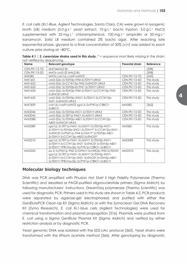
The resolution of vitamin B nutritional requirements would enable the first application of a mineral medium base that would just need to be supplemented with a carbon source for S. cerevisiae cultivation. Such medium would present several advantages such as an economical benefit, a simpler recipe, a higher stability, and lower susceptibility to contamination which should overall result in an increased process robustness.
The goals of this study were to investigate whether strategies can be found to eliminate single vitamin requirements and if the found solutions could be combined yielding a vitamin-independent S. cerevisiae strain. Next, the engineered strain was characterized for growth performance in batch and chemostat cultures and compared to a parental strain requiring vitamin supplementation for optimal growth. Finally, transcriptome data of the engineered strain grown in absence of vitamins was analyzed and compared to its isogenic reference grown in presence of vitamins.
Material and Methods
Strains, media, and maintenance
The S. cerevisiae strains used and constructed in this study (Table 4.1) all derived from the CEN.PK lineage [224, 308]. Yeast cultures were grown in either YP (10 g L-1 Bacto yeast extract, 20 g L-1 Bacto peptone) or synthetic medium (SM) [363]. The YP and SM medium were autoclaved at 121°C for 20 min. SM medium was then supplemented with 1 mL L-1 of filter-sterilized vitamin solution (0.05 g L-1 D-(+)-biotin, 1.0 g L-1 D-calcium pantothenate, 1.0 g L-1 nicotinic acid, 25 g L-1 myo-inositol, 1.0 g L-1 thiamine hydrochloride, 1.0 g L-1 pyridoxal hydrochloride, 0.20 g L-1 4-aminobenzoic acid). A concentrated glucose solution was autoclaved separately at 110°C and added to SM and YP at a final concentration of 20 g L-1 yielding SMD and YPD, respectively. Where indicated, biotin, thiamine, pyridoxine, para-aminobenzoic acid, inositol or pantothenic acid were omitted from the vitamin solution, yielding biotin-free SMD (SMDΔbio), thiamine-free SMD (SMDΔthi), pyridoxine-free SMD (SMDΔpyr), para-aminobenzoic acid-free SMD (SMDΔpABA), inositol-free SMD (SMDΔino), and pantothenic acid-free SMD (SMDΔpan), respectively. For medium without vitamins the vitamin solution was not added to obtain SMDΔvitamins. Drop-in media where only one vitamin was provided were prepared by adding 1 mL L-1 of concentrated solution of either 0.05 g L-1 D-(+)-biotin or 1.0 g L-1 thiamine hydrochloride to SMD to form SMDΔvitamins+bio or SMDΔvitamins+thi, respectively. Yeast strains were inoculated in 100 mL medium in 500-mL-shake flasks or in 20 mL medium in 100-mL-shake flasks and incubated at 30°C, 200 rpm in an Innova Incubator (Brunswick Scientific, Edison, NJ). Solid media were prepared by adding 1.5% Bacto agar (BD Biosciences, Franklin Lakes, NJ) and, when indicated, acetamide for SMG acetamide (20 g L−1 glucose, 1.2 g L−1
acetamide, 3.0 g L−1 KH2PO4, 6.6 g L−1 K2SO4, 0.5 g L−1 MgSO4 7H2O, 1 mL L−1 trace element solution and 1 mL L−1 vitamin solution) [364] or 200 mg L-1 G418 (geneticin) for YPD G418 were added for selection of transformants.
E. coli cells (XL1-Blue, Agilent Technologies, Santa Clara, CA) were grown in lysogenic broth (LB) medium (5.0 g L-1 yeast extract, 10 g L-1 bacto trypton, 5.0 g L-1 NaCl) supplemented with 25 mg L-1 chloramphenicol, 100 mg L-1 ampicillin or 50 mg L-1 kanamycin. Solid LB medium contained 2% bacto agar. After reaching late exponential phase, glycerol to a final concentration of 30% (v/v) was added to each culture prior storing at −80°C.
Table 4.1 | S. cerevisiae strains used in this study. * = sequence most likely missing in the strain; not verified by sequencing.
Name Relevant genotype Parental strain Reference CEN.PK113-7D MATaMAL2-8c [308] CEN.PK113-5D MATa ura3-52 MAL2-8c [308] IMX585 MATa can1Δ::cas9-natNT2 CEN.PK113-7D [245] IMX1651 ura3-52Δ::ScTDH3p-THI4-ScTDH1t URA3 CEN.PK113-5D This study IMX1652 ura3-52Δ::ScCCW12p-THI5-ScENO2t URA3 CEN.PK113-5D This study IMX1653 ura3-52Δ::ScTDH3p-EcThiC-ScTDH1t URA3 CEN.PK113-5D This study IMX1654 ura3-52Δ::ScTDH3p-THI4-ScTDH1t ScCCW12p-THI5-
ScENO2t URA3 CEN.PK113-5D This study
IMX1655 ura3-52Δ::ScTDH3p-SNO1-ScTDH1t ScCCW12p-SNZ1-ScENO2t URA3
CEN.PK113-5D This study
IMX1859 can1Δ::cas9-natNT2 sga1∆::ScPYK1p-CfBIO1-ScBIO1t
IMX585 [362]
IMX2044 ura3-52Δ::ScTDH3p-INO1-ScTDH1t URA3 CEN.PK113-5D This study IMX2045 ura3-52Δ::ScTEF1p-FMS1-ScADH1t URA3 CEN.PK113-5D This study IMX2088 ura3-52Δ::ScTDH3p-ABZ1-ScTDH1t ScCCW12p-
ABZ2-ScENO2t URA3 CEN.PK113-5D This study
IMX2089 sga1Δ::ScTEF1p-FMS1-ScADH1t ScTDH3p-INO1-ScTDH1t ScTDH3p-SNO1-ScTDH1t* ScCCW12p-SNZ1-ScENO2t ScPGK1p-THI4-ScSSA1t* ScTDH3p-ABZ1-ScTDH1t ScCCW12p-ABZ2-ScENO2t*
IMX585 This study
IMX2210 sga1Δ::ScTEF1p-FMS1-ScADH1t ScTDH3p-INO1-ScTDH1t ScCCW12p-SNZ1-ScENO2t ScTDH3p-ABZ1-ScTDH1t YPRcTau3Δ::ScPYK1p-CfBIO1-ScBIO1t
IMX2089 This study
IMX2325 Δx-2::ScPGI1p-THI2-ScGPM1t ScHXK2p-THI3-ScTDH3t sga1Δ::ScTEF1p-FMS1-ScADH1t ScTDH3p-INO1-ScTDH1t ScCCW12p-SNZ1-ScENO2t ScTDH3p-ABZ1-ScTDH1t YPRcTau3Δ::ScPYK1p-CfBIO1-ScBIO1t
IMX2210 This study
Molecular biology techniques
DNA was PCR amplified with Phusion Hot Start II High Fidelity Polymerase (Thermo Scientific) and desalted or PAGE-purified oligonucleotide primers (Sigma Aldrich) by following manufacturers’ instructions. DreamTaq polymerase (Thermo Scientific) was used for diagnostic PCR. Primers used in this study are shown in Table 4.2. PCR products were separated by agarose-gel electrophoresis and purified with either the GenElutePCR Clean-Up Kit (Sigma Aldrich) or with the Zymoclean Gel DNA Recovery Kit (Zymo Research). E. coli XL1-blue cells (Agilent Technologies) were used for chemical transformation and plasmid propagation [316]. Plasmids were purified from E. coli using a Sigma GenElute Plasmid Kit (Sigma Aldrich) and verified by either restriction analysis or by diagnostic PCR.
Yeast genomic DNA was isolated with the SDS-LiAc protocol [365]. Yeast strains were transformed with the lithium acetate method [366]. After genotyping by diagnostic
4
Materials and Methods | 103
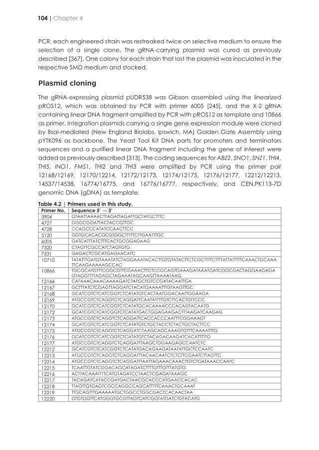
PCR, each engineered strain was restreaked twice on selective medium to ensure the selection of a single clone. The gRNA-carrying plasmid was cured as previously described [367]. One colony for each strain that lost the plasmid was inoculated in the respective SMD medium and stocked.
Plasmid cloning
The gRNA-expressing plasmid pUDR538 was Gibson assembled using the linearized pROS12, which was obtained by PCR with primer 6005 [245], and the X-2 gRNA containing linear DNA fragment amplified by PCR with pROS12 as template and 10866 as primer. Integration plasmids carrying a single gene expression module were cloned by BsaI-mediated (New England Biolabs, Ipswich, MA) Golden Gate Assembly using pYTK096 as backbone. The Yeast Tool Kit DNA parts for promoters and terminators sequences and a purified linear DNA fragment including the gene of interest were added as previously described [313]. The coding sequences for ABZ2, SNO1, SNZ1, THI4, THI5, INO1, FMS1, THI2 and THI3 were amplified by PCR using the primer pair 12168/12169, 12170/12214, 12172/12173, 12174/12175, 12176/12177, 12212/12213, 14537/14538, 16774/16775, and 16776/16777, respectively, and CEN.PK113-7D genomic DNA (gDNA) as template.
Table 4.2 | Primers used in this study. Primer No. Sequence 5’ → 3’ 3904 GTAATTAAAACTTAGATTAGATTGCTATGCTTTC 4727 GGGCGGATTACTACCGTTGC 4728 CCAGCCCATATCCAACTTCC 5120 GGTGCACACGCGTGGCTTTTTCTTGAATTTGC 6005 GATCATTTATCTTTCACTGCGGAGAAG 7320 CTAGTTCGCCATCTAGTGTG 7331 GAGACTCGCATGAGAACATC 10710 TATATTTGATGTAAATATCTAGGAAATACACTTGTGTATACTTCTCGCTTTTCTTTTATTATTTTTCAAACTGCAAA
TTCAAGAAAAAGCCAC 10866 TGCGCATGTTTCGGCGTTCGAAACTTCTCCGCAGTGAAAGATAAATGATCGGCGACTAGGAAGAGA
GTAGGTTTTAGAGCTAGAAATAGCAAGTTAAAATAAG 12166 CATAAACAAACAAAAGATCTATGCTGTCCGATACAATTGA 12167 GCTTTATCTCGAGTTAGGATCTACATGAAAATTTGTAAGTTGC 12168 GCATCGTCTCATCGGTCTCATATGTCACTAATGGACAATTGGAAGA 12169 ATGCCGTCTCAGGTCTCAGGATTCAATATTTTGTCTTCACTGTTCCC 12170 GCATCGTCTCATCGGTCTCATATGCACAAAACCCACAGTACAATG 12172 GCATCGTCTCATCGGTCTCATATGACTGGAGAAGACTTTAAGATCAAGAG 12173 ATGCCGTCTCAGGTCTCAGGATTCACCACCCAATTTCGGAAAGT 12174 GCATCGTCTCATCGGTCTCATATGTCTGCTACCTCTACTGCTACTTCC 12175 ATGCCGTCTCAGGTCTCAGGATCTAAGCAGCAAAGTGTTTCAAAATTTG 12176 GCATCGTCTCATCGGTCTCATATGTCTACAGACAAGATCACATTTTTG 12177 ATGCCGTCTCAGGTCTCAGGATTTAAGCTGGAAGAGCCAATCTC 12212 GCATCGTCTCATCGGTCTCATATGACAGAAGATAATATTGCTCCAATC 12213 ATGCCGTCTCAGGTCTCAGGATTTACAACAATCTCTCTTCGAATCTTAGTTC 12214 ATGCCGTCTCAGGTCTCAGGATTTAATTAGAAACAAACTGTCTGATAAACCAATC 12215 TCAATTGTATCGGACAGCATAGATCTTTTGTTTGTTTATGTG 12216 ACTTACAAATTTTCATGTAGATCCTAACTCGAGATAAAGC 12217 TACAGATCATACCGATGACTAACGCACCCATGAACCACAC 12218 TTAGTTGTGAGTCGCCAGGCCAGCATTTTTCAAACTGCAAAT 12219 TTGCAGTTTGAAAAATGCTGGCCTGGCGACTCACAACTAA 12220 GTGTGGTTCATGGGTGCGTTAGTCATCGGTATGATCTGTACATG
12664 GCCGCGTAGACAATAGATCACCATCTAGTTGAATCCTGAGAGACTATCTCTAATGACCCGGGTAAAGTACAGCTACATTC
13389 TACTGCTACTTTAATTTTATGCCTGGCGACTCACAACTAA 13390 CTCACTCTTTCCTTACTCACCAGCATTTTTCAAACTGCAA 13391 TTAGTTGTGAGTCGCCAGGCATAAAATTAAAGTAGCAGTACTTCA 13392 TTGCAGTTTGAAAAATGCTGGTGAGTAAGGAAAGAGTGAGG 13662 TCCTCGGGCAGAGAAACTCG 13663 GTGAGCCTCTTACCTGTTTG 13748 CGGGTCATTAGAGATAGTCTCTCAGGATTCAACTAGATGGTGATCTATTGTCTACGCGGCTTGGCAGC
CATTAAACTACG 14162 GGCACCTCTGGCTTGTCTTC 14486 GGGGACGTTCTTCACCTCCTTG 14537 GCATCGTCTCATCGGTCTCATATGAATACAGTTTCACCAGC 14538 ATGCCGTCTCAGGTCTCAGGATCTATTTCAGTAAGTCAGAGATTCG 14743 TTTACAATATAGTGATAATCGTGGACTAGAGCAAGATTTCAAATAAGTAACAGCAGCAAACCTTGCCAA
CAGGGAGTTC 14745 TCAGCGTGTTGTAATGATGCGCCATGAATTAGAATGCGTGATGATGTGCAAAGTGCCGTCGCATGCC
GGTAGAGGTG 14746 GACGGCACTTTGCACATCATCACGCATTCTAATTCATGGCGCATCATTACAACACGCTGACAGTTCGA
GTTTATCATTATCAATACTGC 14747 GCTACATCTTCCGTACTATGCTGTAGTCTCATGGTCGAGTTCTATTGCTGTTCGGCGGCACCGTTCAGG
GTAATATATTTTAACCG 14748 TGCCGCCGAACAGCAATAGAACTCGACCATGAGACTACAGCATAGTACGGAAGATGTAGCCAGTT
CGAGTTTATCATTATCAATACTGC 14749 CTCCACTGTACTGCATGTAGCATTCGCCGATCTGCATGATGTGTGACATTCTGCTATCGGATAAAATTAA
AGTAGCAGTACTTCAACCATTAG 14750 CCGATAGCAGAATGTCACACATCATGCAGATCGGCGAATGCTACATGCAGTACAGTGGAGCAGTTC
GAGTTTATCATTATCAATACTGC 16107 AGAATGATTTACAATCTAGTCGCAAAAACAAGTACAGTGCTGACGTCCCATCTTTTAATGGCCGATAATT
GCAGACGAAC 16108 CATTACCAATGAATGCTGTTTTTGCAGAAATAACGAGATATCTGCAATAAAAGCAAAAGTCATGCTCAG
CCGGTAGCTTG 16774 GCATCGTCTCATCGGTCTCATATGATCAATAGTAAGAGGCA 16775 ATGCCGTCTCAGGTCTCAGGATCTAGTCCTGCATGGCATATA 16776 GCATCGTCTCATCGGTCTCATATGAATTCTAGCTATACACA 16777 ATGCCGTCTCAGGTCTCAGGATTCAGTATCCAACTTGATTTT 16778 TCACAGAGGGATCCCGTTACCCATCTATGCTGAAGATTTATCATACTATTCCTCCGCTCGTATTCTTAGTG
GATAACATG 16779 GTCATAACTCAATTTGCCTATTTCTTACGGCTTCTCATAAAACGTCCCACACTATTCAGGGTAACTTCAGA
ATCGTTATC
Then, the ABZ2, SNO1, SNZ1, THI4, THI5, INO1, FMS1, THI2 and THI3 linear DNA fragments were gel purified and assembled by Golden Gate cloning with pYTK096 and the promoter/terminator part plasmids pYTK010/pYTK055, pYTK009/pYTK056, pYTK010/pYTK055, pYTK011/pYTK052, pYTK010/pYTK055, pYTK009/pYTK056, pYTK013/pYTK053, pGGkp033/pGGkp048, and pGGkp096/pGGkp041, respectively, to yield plasmid pUDI177, pUDI178, pUDI179, pUDI201, pUDI181, pUDI182, pUDI211, pUD1103, and pUD1104, respectively (Table 4.3).
Plasmid pUDI176 for the expression of ABZ1 was cloned by Gibson Assembly by combining equimolar amounts of a backbone fragment amplified with primer pair 12215/12216 and pUDI178 as a template together with an insert fragment amplified using primers 12166/12167 and CEN.PK113-7D gDNA as template. Plasmid pUDI188 for the expression of EcthiC was cloned by BsaI-Golden Gate Assembly. Plasmid pUD727, containing the S. cerevisiae codon-optimized version of EcthiC was synthesized by GeneArt (Thermo Fisher Scientific) and cloned with pYTK096, pYTK009 and pYTK056. Two-genes-integration plasmids pUDI183, pUDI184 and pUDI185 were cloned by
104 | Chapter 4
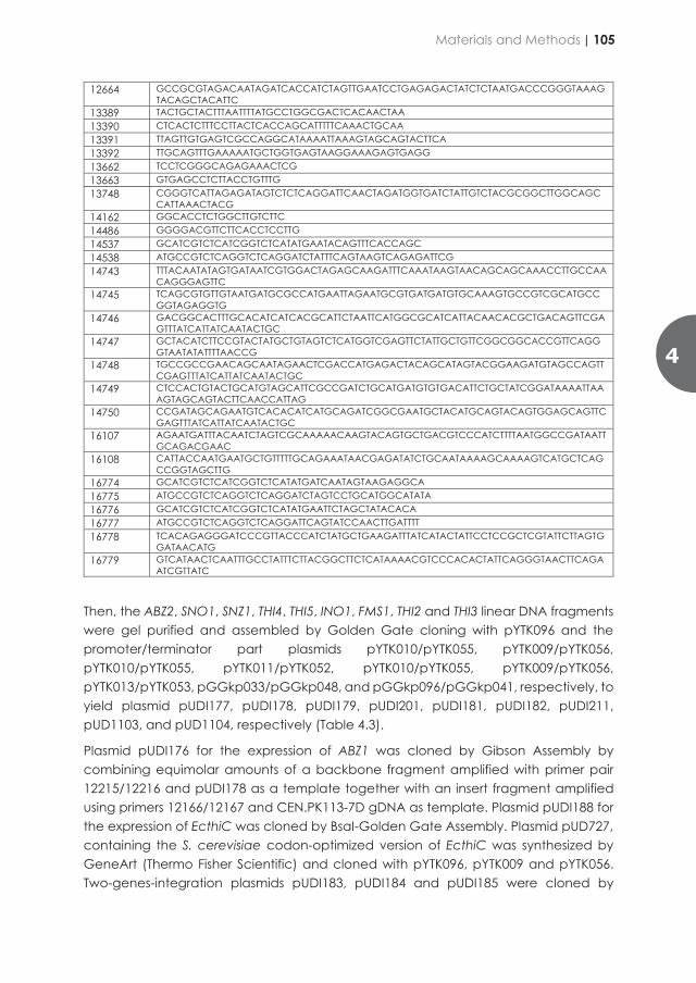
PCR, each engineered strain was restreaked twice on selective medium to ensure the selection of a single clone. The gRNA-carrying plasmid was cured as previously described [367]. One colony for each strain that lost the plasmid was inoculated in the respective SMD medium and stocked.
Plasmid cloning
The gRNA-expressing plasmid pUDR538 was Gibson assembled using the linearized pROS12, which was obtained by PCR with primer 6005 [245], and the X-2 gRNA containing linear DNA fragment amplified by PCR with pROS12 as template and 10866 as primer. Integration plasmids carrying a single gene expression module were cloned by BsaI-mediated (New England Biolabs, Ipswich, MA) Golden Gate Assembly using pYTK096 as backbone. The Yeast Tool Kit DNA parts for promoters and terminators sequences and a purified linear DNA fragment including the gene of interest were added as previously described [313]. The coding sequences for ABZ2, SNO1, SNZ1, THI4, THI5, INO1, FMS1, THI2 and THI3 were amplified by PCR using the primer pair 12168/12169, 12170/12214, 12172/12173, 12174/12175, 12176/12177, 12212/12213, 14537/14538, 16774/16775, and 16776/16777, respectively, and CEN.PK113-7D genomic DNA (gDNA) as template.
Table 4.2 | Primers used in this study. Primer No. Sequence 5’ → 3’ 3904 GTAATTAAAACTTAGATTAGATTGCTATGCTTTC 4727 GGGCGGATTACTACCGTTGC 4728 CCAGCCCATATCCAACTTCC 5120 GGTGCACACGCGTGGCTTTTTCTTGAATTTGC 6005 GATCATTTATCTTTCACTGCGGAGAAG 7320 CTAGTTCGCCATCTAGTGTG 7331 GAGACTCGCATGAGAACATC 10710 TATATTTGATGTAAATATCTAGGAAATACACTTGTGTATACTTCTCGCTTTTCTTTTATTATTTTTCAAACTGCAAA
TTCAAGAAAAAGCCAC 10866 TGCGCATGTTTCGGCGTTCGAAACTTCTCCGCAGTGAAAGATAAATGATCGGCGACTAGGAAGAGA
GTAGGTTTTAGAGCTAGAAATAGCAAGTTAAAATAAG 12166 CATAAACAAACAAAAGATCTATGCTGTCCGATACAATTGA 12167 GCTTTATCTCGAGTTAGGATCTACATGAAAATTTGTAAGTTGC 12168 GCATCGTCTCATCGGTCTCATATGTCACTAATGGACAATTGGAAGA 12169 ATGCCGTCTCAGGTCTCAGGATTCAATATTTTGTCTTCACTGTTCCC 12170 GCATCGTCTCATCGGTCTCATATGCACAAAACCCACAGTACAATG 12172 GCATCGTCTCATCGGTCTCATATGACTGGAGAAGACTTTAAGATCAAGAG 12173 ATGCCGTCTCAGGTCTCAGGATTCACCACCCAATTTCGGAAAGT 12174 GCATCGTCTCATCGGTCTCATATGTCTGCTACCTCTACTGCTACTTCC 12175 ATGCCGTCTCAGGTCTCAGGATCTAAGCAGCAAAGTGTTTCAAAATTTG 12176 GCATCGTCTCATCGGTCTCATATGTCTACAGACAAGATCACATTTTTG 12177 ATGCCGTCTCAGGTCTCAGGATTTAAGCTGGAAGAGCCAATCTC 12212 GCATCGTCTCATCGGTCTCATATGACAGAAGATAATATTGCTCCAATC 12213 ATGCCGTCTCAGGTCTCAGGATTTACAACAATCTCTCTTCGAATCTTAGTTC 12214 ATGCCGTCTCAGGTCTCAGGATTTAATTAGAAACAAACTGTCTGATAAACCAATC 12215 TCAATTGTATCGGACAGCATAGATCTTTTGTTTGTTTATGTG 12216 ACTTACAAATTTTCATGTAGATCCTAACTCGAGATAAAGC 12217 TACAGATCATACCGATGACTAACGCACCCATGAACCACAC 12218 TTAGTTGTGAGTCGCCAGGCCAGCATTTTTCAAACTGCAAAT 12219 TTGCAGTTTGAAAAATGCTGGCCTGGCGACTCACAACTAA 12220 GTGTGGTTCATGGGTGCGTTAGTCATCGGTATGATCTGTACATG
12664 GCCGCGTAGACAATAGATCACCATCTAGTTGAATCCTGAGAGACTATCTCTAATGACCCGGGTAAAGTACAGCTACATTC
13389 TACTGCTACTTTAATTTTATGCCTGGCGACTCACAACTAA 13390 CTCACTCTTTCCTTACTCACCAGCATTTTTCAAACTGCAA 13391 TTAGTTGTGAGTCGCCAGGCATAAAATTAAAGTAGCAGTACTTCA 13392 TTGCAGTTTGAAAAATGCTGGTGAGTAAGGAAAGAGTGAGG 13662 TCCTCGGGCAGAGAAACTCG 13663 GTGAGCCTCTTACCTGTTTG 13748 CGGGTCATTAGAGATAGTCTCTCAGGATTCAACTAGATGGTGATCTATTGTCTACGCGGCTTGGCAGC
CATTAAACTACG 14162 GGCACCTCTGGCTTGTCTTC 14486 GGGGACGTTCTTCACCTCCTTG 14537 GCATCGTCTCATCGGTCTCATATGAATACAGTTTCACCAGC 14538 ATGCCGTCTCAGGTCTCAGGATCTATTTCAGTAAGTCAGAGATTCG 14743 TTTACAATATAGTGATAATCGTGGACTAGAGCAAGATTTCAAATAAGTAACAGCAGCAAACCTTGCCAA
CAGGGAGTTC 14745 TCAGCGTGTTGTAATGATGCGCCATGAATTAGAATGCGTGATGATGTGCAAAGTGCCGTCGCATGCC
GGTAGAGGTG 14746 GACGGCACTTTGCACATCATCACGCATTCTAATTCATGGCGCATCATTACAACACGCTGACAGTTCGA
GTTTATCATTATCAATACTGC 14747 GCTACATCTTCCGTACTATGCTGTAGTCTCATGGTCGAGTTCTATTGCTGTTCGGCGGCACCGTTCAGG
GTAATATATTTTAACCG 14748 TGCCGCCGAACAGCAATAGAACTCGACCATGAGACTACAGCATAGTACGGAAGATGTAGCCAGTT
CGAGTTTATCATTATCAATACTGC 14749 CTCCACTGTACTGCATGTAGCATTCGCCGATCTGCATGATGTGTGACATTCTGCTATCGGATAAAATTAA
AGTAGCAGTACTTCAACCATTAG 14750 CCGATAGCAGAATGTCACACATCATGCAGATCGGCGAATGCTACATGCAGTACAGTGGAGCAGTTC
GAGTTTATCATTATCAATACTGC 16107 AGAATGATTTACAATCTAGTCGCAAAAACAAGTACAGTGCTGACGTCCCATCTTTTAATGGCCGATAATT
GCAGACGAAC 16108 CATTACCAATGAATGCTGTTTTTGCAGAAATAACGAGATATCTGCAATAAAAGCAAAAGTCATGCTCAG
CCGGTAGCTTG 16774 GCATCGTCTCATCGGTCTCATATGATCAATAGTAAGAGGCA 16775 ATGCCGTCTCAGGTCTCAGGATCTAGTCCTGCATGGCATATA 16776 GCATCGTCTCATCGGTCTCATATGAATTCTAGCTATACACA 16777 ATGCCGTCTCAGGTCTCAGGATTCAGTATCCAACTTGATTTT 16778 TCACAGAGGGATCCCGTTACCCATCTATGCTGAAGATTTATCATACTATTCCTCCGCTCGTATTCTTAGTG
GATAACATG 16779 GTCATAACTCAATTTGCCTATTTCTTACGGCTTCTCATAAAACGTCCCACACTATTCAGGGTAACTTCAGA
ATCGTTATC
Then, the ABZ2, SNO1, SNZ1, THI4, THI5, INO1, FMS1, THI2 and THI3 linear DNA fragments were gel purified and assembled by Golden Gate cloning with pYTK096 and the promoter/terminator part plasmids pYTK010/pYTK055, pYTK009/pYTK056, pYTK010/pYTK055, pYTK011/pYTK052, pYTK010/pYTK055, pYTK009/pYTK056, pYTK013/pYTK053, pGGkp033/pGGkp048, and pGGkp096/pGGkp041, respectively, to yield plasmid pUDI177, pUDI178, pUDI179, pUDI201, pUDI181, pUDI182, pUDI211, pUD1103, and pUD1104, respectively (Table 4.3).
Plasmid pUDI176 for the expression of ABZ1 was cloned by Gibson Assembly by combining equimolar amounts of a backbone fragment amplified with primer pair 12215/12216 and pUDI178 as a template together with an insert fragment amplified using primers 12166/12167 and CEN.PK113-7D gDNA as template. Plasmid pUDI188 for the expression of EcthiC was cloned by BsaI-Golden Gate Assembly. Plasmid pUD727, containing the S. cerevisiae codon-optimized version of EcthiC was synthesized by GeneArt (Thermo Fisher Scientific) and cloned with pYTK096, pYTK009 and pYTK056. Two-genes-integration plasmids pUDI183, pUDI184 and pUDI185 were cloned by
4
Materials and Methods | 105
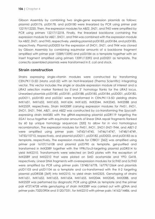
Gibson Assembly by combining two single-gene expression plasmids as follows: plasmid pUDI176, pUDI178, and pUDI180 were linearized by PCR using primer pair 12219/12220. Then, the expression modules for ABZ2, SNZ1, and THI5 were amplified by PCR using primers 12217/12218. Finally, the linearized backbone containing the expression module for ABZ1, SNO1, and THI4 was combined with the expression module for ABZ2, SNZ1, and THI5, respectively, yielding plasmid pUDI183, pUDI184, and pUDI185, respectively. Plasmid pUDI203 for the expression of SNO1, SNZ1, and THI4 was cloned by Gibson Assembly by combining equimolar amounts of a backbone fragment amplified with primer pair 13389/13390 and pUDI184 as a template together with an insert fragment amplified using primers 13391/13392 and pUDI201 as template. The correctly assembled plasmids were transformed In E. coli and stock.
Strain construction
Strains expressing single-vitamin modules were constructed by transforming CEN.PK113-5D (Mata ura3-52) with an NotI-linearized (Thermo Scientific) integrating vector. This vector includes the single or double expression module of interest and a URA3 selection marker flanked by 5’and 3’ homology flanks for the URA3 locus. Linearized plasmids pUDI180, pUDI181, pUDI188, pUDI185, pUDI184, pUDI201, pUDI182, pUDI211, pUDI183 and pUD261 were transformed in CEN.PK113-5D yielding strains IMX1651, IMX1652, IMX1653, IMX1654, IMX1655, IMX2044, IMX2045, IMX2088 and IMX2559, respectively. Strain IMX2089 carrying expression modules for FMS1, INO1, SNO1, SNZ1, THI4, ABZ1, and ABZ2 was constructed by co-transforming the Spycas9-expressing strain IMX585 with the gRNA-expressing plasmid pUDR119 targeting the SGA1 locus together with equimolar amounts of linear DNA repair fragments flanked by 60 bp unique homology sequences [320] to allow for in vivo homologous recombination. The expression modules for FMS1, INO1, SNO1-SNZ1-THI4, and ABZ1-2 were amplified using primer pairs 14743/14745, 14746/14747, 14748/14749, 14750/10710, respectively, and plasmid pUDI211, pUDI182, pUDI203, and pUDI183 as a template, respectively. The expression module for CfBIO1 [362] was amplified using primer pair 16107/16108 and plasmid pUD790 as template, gel-purified and transformed in IMX2089 together with the YPRcTau3-targeting plasmid pUDR514 to yield IMX2210. Transformants were selected on SMD plates with the exception of IMX2089 and IMX2210 that were plated on SMD acetamide and YPD G418, respectively. Linear DNA fragments with overexpression modules for ScTHI2 and ScTHI3 were amplified by PCR using primer pairs 13748/16778, 16779/12664 and plasmids pUD1103 and pUD1104 as a template and co-transformed with the X-2 targeting plasmid pUDR538 [369] into IMX2210, to yield strain IMX2325. Genotyping of strains IMX1651, IMX1652, IMX1653, IMX1654, IMX1655, IMX2044, IMX2045, IMX2088, and IMX2559 was performed by diagnostic PCR using gDNA as template and the primer pair 4727/4728 while genotyping of strain IMX2089 was carried out with gDNA and primer pairs 7320/3904 and 5120/7331, for IMX2210 with primer pairs 14162/14486, and
for IMX2325 with primer pairs 13662/16781 and 13663/16782 respectively. After genotyping, a correct clone was selected and stocked.
Shake flask cultures
For determination of growth rates under oxic condition, a frozen aliquot was thawed and used to inoculate a 20 mL culture in a 100 mL shake flask. After overnight incubation at 30°C the grown culture was used to inoculate a pre-culture in 100 mL of fresh medium in a 500-mL-shake flask. When the second culture reached mid-exponential phase, which was equivalent to an optical density at 660 nm (OD660) of 3-5, an aliquot was used to inoculate a third 100 mL-culture with an OD660 of 0.2. Growth was monitored by measuring OD660 over time in the third culture using a Jenway 7200 Spectrophotometer (Cole-Palmer, Stone, United Kingdom). Specific growth rates were calculated by fitting the exponential growth function (𝑋𝑋 𝑋 𝑋𝑋𝑒𝑒 where µ indicates the growth rate) to a minimum of five data points and three-to-four-time doublings during exponential growth. All oxic shake flask experiments were carried out as at least biological duplicates and incubated in an Innova incubator (New Brunswick Scientific) set at 30°C and 200 rpm.
Batch and chemostat cultures in lab-scale bioreactors
Physiological characterization of engineered S. cerevisiae strain IMX2210 was performed in biologically independent quadruplicates in 2-L bioreactors (Applikon, Delft, The Netherlands) with 1-L working volume. Thawed stocks of strain IMX2210 were grown in 100 mL either SMD or SMDΔvitamins in 500-mL-shake flasks. After overnight incubation at 30°C, 200 rpm, each culture was used to inoculate a second flask containing the same relative media. Exponentially growing cells (OD660 of 4-5) from the second flask were washed twice in demi-water by centrifuging for 5 min, 3000 x g, re-suspended in 50 mL of sterile water and used to inoculate each reactor to a starting OD660 of 0.15-0.25. An oxic batch culture with 20 g L-1 glucose was followed by an oxic, carbon-limited chemostat culture at a dilution rate of 0.10 h−1 with 7.5 g L-1 glucose in the medium as previously described [370, 371]. The temperature was controlled at 30°C, and the pH was maintained at 5.0 by automatic addition of 2.0 M KOH. To maintain oxic conditions, air at a rate of 0.5 L min−1 was sparged through the culture which was stirred at 800 rpm. All cultures were grown on either SMD or vitamin-free SMD (SMDΔvitamins) supplemented with 0.2 g L−1 sterile pluronic PE6100 (BASF, Ludwigshafen, Germany) to avoid foam formation. For characterization of growth in batch conditions the OD660 was measured every hour using a Jenway 7200 spectrophotometer while five dry weight (DW) biomass samples were taken during the exponential growth phase and used to generate an OD660/DW (g L-1) calibration line to convert each OD660 value to DW. The carbon dioxide and oxygen concentration in the gas outflow from the bioreactors were analyzed by a Rosemount NGA 2000 analyzer (Baar, Switzerland), after cooling of the gas by a condenser (2°C) and drying using a PermaPure Dryer (model MD 110-8P-4; Inacom Instruments, Veenendaal, The
106 | Chapter 4
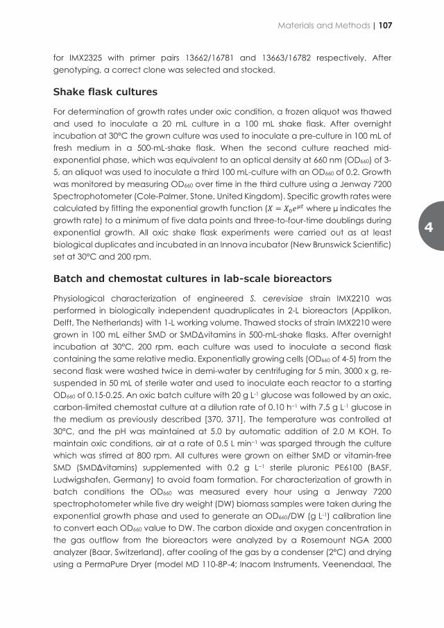
Gibson Assembly by combining two single-gene expression plasmids as follows: plasmid pUDI176, pUDI178, and pUDI180 were linearized by PCR using primer pair 12219/12220. Then, the expression modules for ABZ2, SNZ1, and THI5 were amplified by PCR using primers 12217/12218. Finally, the linearized backbone containing the expression module for ABZ1, SNO1, and THI4 was combined with the expression module for ABZ2, SNZ1, and THI5, respectively, yielding plasmid pUDI183, pUDI184, and pUDI185, respectively. Plasmid pUDI203 for the expression of SNO1, SNZ1, and THI4 was cloned by Gibson Assembly by combining equimolar amounts of a backbone fragment amplified with primer pair 13389/13390 and pUDI184 as a template together with an insert fragment amplified using primers 13391/13392 and pUDI201 as template. The correctly assembled plasmids were transformed In E. coli and stock.
Strain construction
Strains expressing single-vitamin modules were constructed by transforming CEN.PK113-5D (Mata ura3-52) with an NotI-linearized (Thermo Scientific) integrating vector. This vector includes the single or double expression module of interest and a URA3 selection marker flanked by 5’and 3’ homology flanks for the URA3 locus. Linearized plasmids pUDI180, pUDI181, pUDI188, pUDI185, pUDI184, pUDI201, pUDI182, pUDI211, pUDI183 and pUD261 were transformed in CEN.PK113-5D yielding strains IMX1651, IMX1652, IMX1653, IMX1654, IMX1655, IMX2044, IMX2045, IMX2088 and IMX2559, respectively. Strain IMX2089 carrying expression modules for FMS1, INO1, SNO1, SNZ1, THI4, ABZ1, and ABZ2 was constructed by co-transforming the Spycas9-expressing strain IMX585 with the gRNA-expressing plasmid pUDR119 targeting the SGA1 locus together with equimolar amounts of linear DNA repair fragments flanked by 60 bp unique homology sequences [320] to allow for in vivo homologous recombination. The expression modules for FMS1, INO1, SNO1-SNZ1-THI4, and ABZ1-2 were amplified using primer pairs 14743/14745, 14746/14747, 14748/14749, 14750/10710, respectively, and plasmid pUDI211, pUDI182, pUDI203, and pUDI183 as a template, respectively. The expression module for CfBIO1 [362] was amplified using primer pair 16107/16108 and plasmid pUD790 as template, gel-purified and transformed in IMX2089 together with the YPRcTau3-targeting plasmid pUDR514 to yield IMX2210. Transformants were selected on SMD plates with the exception of IMX2089 and IMX2210 that were plated on SMD acetamide and YPD G418, respectively. Linear DNA fragments with overexpression modules for ScTHI2 and ScTHI3 were amplified by PCR using primer pairs 13748/16778, 16779/12664 and plasmids pUD1103 and pUD1104 as a template and co-transformed with the X-2 targeting plasmid pUDR538 [369] into IMX2210, to yield strain IMX2325. Genotyping of strains IMX1651, IMX1652, IMX1653, IMX1654, IMX1655, IMX2044, IMX2045, IMX2088, and IMX2559 was performed by diagnostic PCR using gDNA as template and the primer pair 4727/4728 while genotyping of strain IMX2089 was carried out with gDNA and primer pairs 7320/3904 and 5120/7331, for IMX2210 with primer pairs 14162/14486, and
for IMX2325 with primer pairs 13662/16781 and 13663/16782 respectively. After genotyping, a correct clone was selected and stocked.
Shake flask cultures
For determination of growth rates under oxic condition, a frozen aliquot was thawed and used to inoculate a 20 mL culture in a 100 mL shake flask. After overnight incubation at 30°C the grown culture was used to inoculate a pre-culture in 100 mL of fresh medium in a 500-mL-shake flask. When the second culture reached mid-exponential phase, which was equivalent to an optical density at 660 nm (OD660) of 3-5, an aliquot was used to inoculate a third 100 mL-culture with an OD660 of 0.2. Growth was monitored by measuring OD660 over time in the third culture using a Jenway 7200 Spectrophotometer (Cole-Palmer, Stone, United Kingdom). Specific growth rates were calculated by fitting the exponential growth function (𝑋𝑋 𝑋 𝑋𝑋𝑒𝑒 where µ indicates the growth rate) to a minimum of five data points and three-to-four-time doublings during exponential growth. All oxic shake flask experiments were carried out as at least biological duplicates and incubated in an Innova incubator (New Brunswick Scientific) set at 30°C and 200 rpm.
Batch and chemostat cultures in lab-scale bioreactors
Physiological characterization of engineered S. cerevisiae strain IMX2210 was performed in biologically independent quadruplicates in 2-L bioreactors (Applikon, Delft, The Netherlands) with 1-L working volume. Thawed stocks of strain IMX2210 were grown in 100 mL either SMD or SMDΔvitamins in 500-mL-shake flasks. After overnight incubation at 30°C, 200 rpm, each culture was used to inoculate a second flask containing the same relative media. Exponentially growing cells (OD660 of 4-5) from the second flask were washed twice in demi-water by centrifuging for 5 min, 3000 x g, re-suspended in 50 mL of sterile water and used to inoculate each reactor to a starting OD660 of 0.15-0.25. An oxic batch culture with 20 g L-1 glucose was followed by an oxic, carbon-limited chemostat culture at a dilution rate of 0.10 h−1 with 7.5 g L-1 glucose in the medium as previously described [370, 371]. The temperature was controlled at 30°C, and the pH was maintained at 5.0 by automatic addition of 2.0 M KOH. To maintain oxic conditions, air at a rate of 0.5 L min−1 was sparged through the culture which was stirred at 800 rpm. All cultures were grown on either SMD or vitamin-free SMD (SMDΔvitamins) supplemented with 0.2 g L−1 sterile pluronic PE6100 (BASF, Ludwigshafen, Germany) to avoid foam formation. For characterization of growth in batch conditions the OD660 was measured every hour using a Jenway 7200 spectrophotometer while five dry weight (DW) biomass samples were taken during the exponential growth phase and used to generate an OD660/DW (g L-1) calibration line to convert each OD660 value to DW. The carbon dioxide and oxygen concentration in the gas outflow from the bioreactors were analyzed by a Rosemount NGA 2000 analyzer (Baar, Switzerland), after cooling of the gas by a condenser (2°C) and drying using a PermaPure Dryer (model MD 110-8P-4; Inacom Instruments, Veenendaal, The
4
Materials and Methods | 107

Netherlands). Steady state sampling of the chemostat cultures for extracellular metabolites and sugar was performed by rapid sampling technique using cold steel beads pre-cooled to -20°C [372].
Whole genome sequencing
DNA of S. cerevisiae strain IMX2210 was grown in shake-flask cultures with SMD and genomic DNA was isolated with a Qiagen Blood & Cell Culture DNA kit (Qiagen, Hilden, Germany), following manufacturer's specifications. 150bp paired-end sequencing was performed on a 350-bp TruSeq PCR-free insert library using an Illumina NovaSeq 6000 sequencer (Macrogen Europe, Amsterdam, the Netherlands). Sequence data was mapped to the CEN.PK113-7D genome [224] to which the sequences of the integrated expression cassettes for the heterologous genes FMS1, INO1, SNO1, SNZ1, THI4, ABZ1, ABZ2, CfBIO1 were manually added. Data processing and single nucleotide variation (SNV) analysis were carried out as described previously [151, 373].
Total RNA isolation and transcriptomics analysis
Steady state chemostat cultures were sampled directly from the reactor in liquid nitrogen as previously described [374]. Cells were stored at -80°C for a maximum of two weeks before RNA was further purified using a phenolic acid/chloroform method as previously described [375]. Total RNA was quantified by using a Qubit RNA BR Assay Kit (Thermo Fisher Scientific) following manufacturer instructions. Total RNA quality was assessed by Nanodrop (Thermo Fisher Scientific) measurement followed by RNA ScreenTape analysis using a Tapestation System (Agilent Technologies) following manufacturer’s instructions. RNA libraries were prepared with the TruSeq Stranded mRNA LT protocol (Illumina, San Diego, CA, #15031047) and subjected to 150 bp paired-end sequencing on a NovaSeq 6000 (Illumina) sequencer (Macrogen). RNAseq libraries were mapped with STAR aligner (version 2.5.3a) [376] to the CEN.PK113-7D genome [224] to which the sequence of CfBIO1 were manually added. Expression was quantified by applying featureCounts (version 1.6.0) in paired-end mode and reversely stranded [377].
Differential gene expression (DGE) analysis was performed by using edgeR (version 3.30.3) with TMM normalization [378]. For functional interpretation of the DGE analysis, enrichment of gene ontology terms (GO) according to the biological process was conducted with YeastMine (https://www.yeastgenome.org/goTermFinder). Analysis used a binomial test and a Bonferroni correction for multiple testing. Gene ontology terms with a p-value < 0.01 were deemed as being enriched. Mean values of mRNA counts of the genes which in the DGE analysis showed -1 < log2 fold-change (log2FC) > 1 with a false discovery rate (FDR) < 5.0E-2 were compared between CEN.PK113-7D in SMD, IMX2210 in SMD and IMX2210 in SMDΔvitamins.
108 | Chapter 4
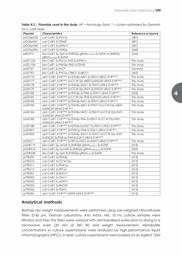
Table 4.3 | Plasmids used in this study. HF = homology flank. * = codon-optimized by GeneArt for S. cerevisiae.
Plasmid Characteristics Reference or source pGGkp033 cat ColE1 ScPGI1p [281] pGGkp041 cat ColE1 ScTDH3t [281] pGGkp048 cat ColE1 ScGPM1t [281] pGGkp096 cat ColE1 ScHXK2p [368] pROS12 bla ColE1 2μ hph ScSNR52p-gRNAScCAN1-ScSUP4t ScSNR52p-
gRNAScADE2-ScSUP4t [245]
pUD1103 bla ColE1 ScPGI1p-THI2-ScGPM1t This study pUD1104 bla ColE1 ScHXK2p-THI3-ScTDH3t This study pUD727 bla ColE1 EcthiC* GeneArt pUD790 bla ColE1 ScPYK1p-CfBIO1-ScBIO1t [362] pUDI176 aph ColE1 5’HFURA3 ScTDH3p-ABZ1-ScTDH1t URA3 3’HFURA3 This study pUDI177 aph ColE1 5’HFURA3 ScCCW12p-ABZ2-ScENO2t URA3 3’HFURA3 This study pUDI178 aph ColE1 5’HFURA3 ScTDH3p-SNO1-ScTDH1t URA3 3’HFURA3 This study pUDI179 aph ColE1 5’HFURA3 ScCCW12p-SNZ1-ScENO2t URA3 3’HFURA3 This study pUDI180 aph ColE1 5’HFURA3 ScTDH3p-ScTHI4-ScTDH1t URA3 3’HFURA3 [225] pUDI181 aph ColE1 5’HFURA3 ScCCW12p-THI5-ScENO2t URA3 3’HFURA3 This study pUDI182 aph ColE1 5’HFURA3 ScTDH3p-INO1-ScTDH1t URA3 3’HFURA3 This study pUDI183 aph ColE1 5’HFURA3 ScTDH3p-ABZ1-ScTDH1t ScCCW12p-ABZ2-
ScENO2t URA3 3’HFURA3 This study
pUDI184 aph ColE1 5’HFURA3 ScTDH3p-SNO1-ScTDH1t ScCCW12p-SNZ1-ScENO2t URA3 3’HFURA3
This study
pUDI185 aph ColE1 5’HFURA3 ScTDH3p-THI4-ScTDH1t ScCCW12p-THI5-ScENO2t URA3 3’HFURA3
This study
pUDI188 aph ColE1 5’HFURA3 ScTDH3p-EcthiC*-ScTDH1t URA3 3’HFURA3 This study pUDI201 aph ColE1 5’HFURA3 ScPGK1p-THI4-ScSSA1t URA3 3’HFURA3 This study pUDI203 aph ColE1 5’HFURA3 ScTDH3p-SNO1-ScTDH1t ScCCW12p-SNZ1-
ScENO2t ScPGK1p-THI4-ScSSA1t URA3 3’HFURA3 This study
pUDI211 aph ColE1 5’HFURA3 ScTEF1p-FMS1-ScADH1t URA3 3’HFURA3 This study pUDR119 bla ColE1 2μ amdS ScSNR52p-gRNAScSGA1-ScSUP4t [318] pUDR514 bla ColE1 2μ kanMX ScSNR52p-gRNAYPRcTau3-ScSUP4t [225] pUDR538 bla ColE1 2μ hph ScSNR52p-gRNAX−2-ScSUP4t This study pYTK009 cat ColE1 ScTDH3p [313] pYTK010 cat ColE1 ScCCW12p [313] pYTK011 cat ColE1 ScPGK1p [313] pYTK013 cat ColE1 ScTEF1p [313] pYTK051 cat ColE1 ScENO1t [313] pYTK052 cat ColE1 ScSSA1t [313] pYTK053 cat ColE1 ScADH1t [313] pYTK055 cat ColE1 ScENO2t [313] pYTK056 cat ColE1 ScTDH1t [313] pYTK096 aph ColE1 5’HFURA3 sfGFP URA3 3’HFURA3 [313]
Analytical methods
Biomass dry weight measurements were performed using pre-weighed nitrocellulose filters (0.45 µm, Gelman Laboratory, Ann Arbor, MI). 10 mL culture samples were filtrated and then the filters were washed with demineralized water prior to drying in a microwave oven (20 min at 360 W) and weight measurement. Metabolite concentrations in culture supernatants were analyzed by high-performance liquid chromatography (HPLC). In brief, culture supernatants were loaded on an Agilent 1260
4
Materials and Methods | 109

HPLC (Agilent Technologies) fitted with a Bio-Rad HPX 87 H column (Bio-Rad, Hercules, CA). The flow rate was set at 0.6 mL min-1 and 0.5 g L-1 H2SO4 was used as eluent. An Agilent refractive-index detector and an Agilent 1260 VWD detector were used to detect the metabolites [379]. An evaporation constant of 0.008 divided by the volume in liters, was used to correct HPLC measurements of ethanol in the culture supernatants, taking into account changes in volume caused by sampling [380].
Statistical analysis
Statistical significance of differences between measurements from replicate cultures were calculated by using a one-way analysis of variance (ANOVA). When specific pairs of means were chosen for comparison in the experimental design, the Fisher's Least Significant Difference (LSD) test was used to calculate p-values. When all means within an experiment were compared to each other the p-values were corrected for multiple comparisons using Tukey’s correction for multiple comparison by GraphPad Prism version 8.2.1 for Windows, (GraphPad Software, San Diego, California USA, www.graphpad.com). Significant differences were assumed when p-value > 5.0E-2. The batch and chemostat culture determined data were compared by using two-tailed student’s t-test.
Data availability
All measurement and calculations used to prepare figure 4.2, 4.3, 4.5, 4.6, 4.7 and 4.8 as well as table 4.4, 4.5 and 4.7 of the manuscript as well as DNA sequencing data of S. cerevisiae strain IMX2210 are available at the 4TU.Centre for research data repository (https://researchdata.4tu.nl/) under 10.4121/17185607 (data under public embargo and accessible through the following reviewer link: https://figshare.com/s/d8b0128171d8c85ce091. The RNA sequencing data of Saccharomyces cerevisiae strain IMX2210 and CEN.PK113-7D are available at the Gene Expression Omnibus database (https://www.ncbi.nlm.nih.gov/geo/) under accession number GSE191212 (data under public embargo and accessible through the following reviewer link: that can be opened using the token “uvahuimsdpanrkv”.
Results
Selection of engineering strategies to eliminate single vitamin requirements
To start engineering S. cerevisiae for vitamin independent growth, strategies suitable for removing individual vitamin requirements were evaluated (Figure 4.1).
Figure 4.1 | Overview of vitamin biosynthetic pathways and genetic modifiations studied in this work. Schematic representation of the yeast native biosynthetic pathway for thiamine (A) starting with the reaction of D-glyceraldehyde-3-phosphate, L-glutamine and ribulose-5-phospahte to pyridoxal 5’-phosphate (PLP) by the PLP synthase Sno1-2-3/Snz1-2-3. PLP can also be synthesized by two reactions for pyridoxine catalyzed by the pyridoxal kinase Bud16 and the Pyridoxamine 5'-phosphate oxidase Pdx3. The Thi5 protein family resembling the function of a 4-amino-5-hydroxymethyl-2-methylpyrimidine phosphate synthase makes hydroxymethylpyrimidine phosphate (HMP-P). The Phosphomethylpyrimidine synthase ThiC from Escherichia coli converts 5-phosphoribosyl-5-aminoimidazole (AIR) to HMP-P. Hydroxymethylpyrimidine diphosphate (HMP-PP), which is produced by Thi20-21, and hydroxyethylthiazole phosphate (HET-P), which is made from NAD+ and glycine by action of Thi4, a thiamine thiazole synthase, are combined by the action of Thi6 into thiamine. Thi2 and Thi3 are thimaine biosynthesis regulators pABA (B) is made from chorismate and 4-amino-4-deoxychorismate by catalytic activity of ABZ1 and ABZ2, an aminodeoxychorismate synthase and aminodeoxychorismate lyase, respectively. myo-Inositol (C) is synthesized from glucose-6-phosphate via L-myo-inositol-1-phosphate by action of Ino1 an inositol-3-phosphate synthase and Inm1-2 (inositol-1-monophosphatase). Pantothenate (D) is made by reaction of pantoate and β-alanine catalyzed by Pan6 (pantoate-β-alanine ligase). Pantoate is synthesized from 2-keto-isovalerate and the reactions of 3-methyl-2-oxobutanoate hydroxymethyltransferase (Ecm31) and 2-dehydropantoate 2-reductase (Pan5) and β-alanine from spermine by polyamine oxidase (Fms1) and aldehyde dehydrogenase (Ald2-3). Biotin synthesis (E) starts with a putative precursor pimelic acid, which is indicated by (?). The first enzyme from Cyberlindnera fabianii (CfBio1) catalyses the synthesis of pimeloyl-CoA, which is converted by 7,8-diamino-pelargonic acid aminotransferase Bio6, adenosylmethionine-8-amino-7-oxononanoate aminotransferase Bio3, 8-amino-7-oxononanoate synthase Bio4 and biotin synthase Bio2 to biotin. The intermediates are 7-keto-8-aminopelargonic acid (KAPA), 7,8-diaminopelargonic acid (DAPA) and dethiobiotin. The names of overexpressed enzymes in this study are green. Unmodified enzyme names are shown in black and intermediates and products in grey.
110 | Chapter 4

HPLC (Agilent Technologies) fitted with a Bio-Rad HPX 87 H column (Bio-Rad, Hercules, CA). The flow rate was set at 0.6 mL min-1 and 0.5 g L-1 H2SO4 was used as eluent. An Agilent refractive-index detector and an Agilent 1260 VWD detector were used to detect the metabolites [379]. An evaporation constant of 0.008 divided by the volume in liters, was used to correct HPLC measurements of ethanol in the culture supernatants, taking into account changes in volume caused by sampling [380].
Statistical analysis
Statistical significance of differences between measurements from replicate cultures were calculated by using a one-way analysis of variance (ANOVA). When specific pairs of means were chosen for comparison in the experimental design, the Fisher's Least Significant Difference (LSD) test was used to calculate p-values. When all means within an experiment were compared to each other the p-values were corrected for multiple comparisons using Tukey’s correction for multiple comparison by GraphPad Prism version 8.2.1 for Windows, (GraphPad Software, San Diego, California USA, www.graphpad.com). Significant differences were assumed when p-value > 5.0E-2. The batch and chemostat culture determined data were compared by using two-tailed student’s t-test.
Data availability
All measurement and calculations used to prepare figure 4.2, 4.3, 4.5, 4.6, 4.7 and 4.8 as well as table 4.4, 4.5 and 4.7 of the manuscript as well as DNA sequencing data of S. cerevisiae strain IMX2210 are available at the 4TU.Centre for research data repository (https://researchdata.4tu.nl/) under 10.4121/17185607 (data under public embargo and accessible through the following reviewer link: https://figshare.com/s/d8b0128171d8c85ce091. The RNA sequencing data of Saccharomyces cerevisiae strain IMX2210 and CEN.PK113-7D are available at the Gene Expression Omnibus database (https://www.ncbi.nlm.nih.gov/geo/) under accession number GSE191212 (data under public embargo and accessible through the following reviewer link: that can be opened using the token “uvahuimsdpanrkv”.
Results
Selection of engineering strategies to eliminate single vitamin requirements
To start engineering S. cerevisiae for vitamin independent growth, strategies suitable for removing individual vitamin requirements were evaluated (Figure 4.1).
Figure 4.1 | Overview of vitamin biosynthetic pathways and genetic modifiations studied in this work. Schematic representation of the yeast native biosynthetic pathway for thiamine (A) starting with the reaction of D-glyceraldehyde-3-phosphate, L-glutamine and ribulose-5-phospahte to pyridoxal 5’-phosphate (PLP) by the PLP synthase Sno1-2-3/Snz1-2-3. PLP can also be synthesized by two reactions for pyridoxine catalyzed by the pyridoxal kinase Bud16 and the Pyridoxamine 5'-phosphate oxidase Pdx3. The Thi5 protein family resembling the function of a 4-amino-5-hydroxymethyl-2-methylpyrimidine phosphate synthase makes hydroxymethylpyrimidine phosphate (HMP-P). The Phosphomethylpyrimidine synthase ThiC from Escherichia coli converts 5-phosphoribosyl-5-aminoimidazole (AIR) to HMP-P. Hydroxymethylpyrimidine diphosphate (HMP-PP), which is produced by Thi20-21, and hydroxyethylthiazole phosphate (HET-P), which is made from NAD+ and glycine by action of Thi4, a thiamine thiazole synthase, are combined by the action of Thi6 into thiamine. Thi2 and Thi3 are thimaine biosynthesis regulators pABA (B) is made from chorismate and 4-amino-4-deoxychorismate by catalytic activity of ABZ1 and ABZ2, an aminodeoxychorismate synthase and aminodeoxychorismate lyase, respectively. myo-Inositol (C) is synthesized from glucose-6-phosphate via L-myo-inositol-1-phosphate by action of Ino1 an inositol-3-phosphate synthase and Inm1-2 (inositol-1-monophosphatase). Pantothenate (D) is made by reaction of pantoate and β-alanine catalyzed by Pan6 (pantoate-β-alanine ligase). Pantoate is synthesized from 2-keto-isovalerate and the reactions of 3-methyl-2-oxobutanoate hydroxymethyltransferase (Ecm31) and 2-dehydropantoate 2-reductase (Pan5) and β-alanine from spermine by polyamine oxidase (Fms1) and aldehyde dehydrogenase (Ald2-3). Biotin synthesis (E) starts with a putative precursor pimelic acid, which is indicated by (?). The first enzyme from Cyberlindnera fabianii (CfBio1) catalyses the synthesis of pimeloyl-CoA, which is converted by 7,8-diamino-pelargonic acid aminotransferase Bio6, adenosylmethionine-8-amino-7-oxononanoate aminotransferase Bio3, 8-amino-7-oxononanoate synthase Bio4 and biotin synthase Bio2 to biotin. The intermediates are 7-keto-8-aminopelargonic acid (KAPA), 7,8-diaminopelargonic acid (DAPA) and dethiobiotin. The names of overexpressed enzymes in this study are green. Unmodified enzyme names are shown in black and intermediates and products in grey.
4
Results | 111
Pyridoxine
In absence of pyridoxine (SMDΔpyr), S. cerevisiae CEN.PK113-7D grew with a growth rate of 0.29 ± 0.01 h-1 which represented a 28% reduction compared to the growth rate of the same strain in the presence of pyridoxine (Figure 4.2A). It was previously established that mutations in the gene encoding the transcriptional regulator Bas1 restored wild type growth in absence of pyridoxine through the relief of the transcriptional repression of gene SNZ1 involved in pyridoxal-phosphate synthesis [225, 381]. To mitigate unwanted effects due the dual activating and repressing regulatory functions of Bas1, deregulation of SNZ1 and SNO1 was evaluated (Figure 4.1). In absence of vitamin B6 the strain IMX1655 that overexpressed the pyridoxal-5’-phosphate biosynthetic genes ScSNO1 and ScSNZ1 grew at a growth rate of 0.37 ± 0.01 h-1. Although significantly slower than the parental strain grown in presence of pyridoxine (p = 1.6E-2), IMX1655 exhibited a growth rate identical to that of BAS1 mutants previously reported [225].
para-Aminobenzoic acid
In absence of para-aminobenzoic acid (pABA), the strain CEN.PK113-7D exhibited a severe growth reduction (81%) relative to growth in presence of pABA. However, S. cerevisiae previously showed fast evolvability of the genes encoding enzymes of the chorismate node, ABZ1 and ARO7 [225]. Mutations in these two genes could restore growth rates that were undiscernible from the CEN.PK113-7D growth rate in presence of pABA. Inspired by these earlier results and following a strategy similar to that applied for pyridoxine, the two genes, ABZ1 and ABZ2 encoding enzymes involved in the conversion of chorismate to pABA (Figure 4.1) were overexpressed. The resulting strain IMX2088 recovered a growth rate of 0.38 ± 0.00 h-1, that did not significantly differ from the growth rate of CEN.PK113-7D on SMD (Figure 4.2B).
Inositol
Although growth improvement in absence of inositol can be rapidly rescued by adaptive laboratory evolution, no mutations have been linked to this phenotype. However, deregulation or constitutive expression of INO1 has been linked to an inositol excretion phenotype [189]. The strain IMX2044 that overexpresses ScINO1 (Figure 4.1) could grow in absence of inositol as fast as CEN.PK113-7D cultivated on regular SMD medium. IMX2044 (ScINO1) grew with a growth rate of 0.37 ± 0.01 h-1 in SMDΔino that was significantly improved relative to CEN.PK113-7D grown on the same medium (µSMDΔino-CEN.PK113-7D = 0.31 ± 0.00 h-1) (Figure 4.2C).
Pantothenate
Although previous research showed that mutations in global regulators as GAL11 and TUP1 had positive effect on growth rate in absence of pantothenate [225], the proteins encoded by these two genes have a central regulatory role and affect the expression of multiple genes [382]. To avoid the introduction of a massive transcriptional
rearrangement, other options were investigated. Pantothenic acid independent growth was achieved by overexpression of gene ScFMS1 in CEN.PK113-7D coding for a polyamine oxidase synthesizing β-alanine (Figure 4.1), the precursor of pantothenic acid synthesis [383]. The resulting strain IMX2045 was able to grow at a rate of 0.38 ± 0.01 h-1, which was an improvement of 124% when compared to growth of CEN.PK113-7D on the same medium (µSMDΔpan,CEN.PK113-7D = 0.17 ± 0.00 h-1) and a restoration of growth as observed for CEN.PK113-7D on SMD (Figure 4.2D).
Figure 4.2 | Engineering fast-growth in the absence of single vitamins. Growth rate of the non-engineered CEN.PK117-7D strain (dark grey) and engineered derivatives (white) in synthetic medium with pyridoxine (A), pABA (B), inositol (C) and pantothenate (D) added (+) or ommitted (-). Gene(s) that were overexpressed in the engineered strains are reported below the relative bar. Statistical analysis was based on a one-way analysis of variance analysis (ANOVA) followed by Tukey’s multiple comparisons test (GraphPad Software, San Diego, California USA, www.graphpad.com) with means of independent cultures (n=3; except n=2 for CEN.PK117-7D in SMDΔpABA and IMX2088). Significant differences were assumed when p-value > 5.0E-2.
112 | Chapter 4

Pyridoxine
In absence of pyridoxine (SMDΔpyr), S. cerevisiae CEN.PK113-7D grew with a growth rate of 0.29 ± 0.01 h-1 which represented a 28% reduction compared to the growth rate of the same strain in the presence of pyridoxine (Figure 4.2A). It was previously established that mutations in the gene encoding the transcriptional regulator Bas1 restored wild type growth in absence of pyridoxine through the relief of the transcriptional repression of gene SNZ1 involved in pyridoxal-phosphate synthesis [225, 381]. To mitigate unwanted effects due the dual activating and repressing regulatory functions of Bas1, deregulation of SNZ1 and SNO1 was evaluated (Figure 4.1). In absence of vitamin B6 the strain IMX1655 that overexpressed the pyridoxal-5’-phosphate biosynthetic genes ScSNO1 and ScSNZ1 grew at a growth rate of 0.37 ± 0.01 h-1. Although significantly slower than the parental strain grown in presence of pyridoxine (p = 1.6E-2), IMX1655 exhibited a growth rate identical to that of BAS1 mutants previously reported [225].
para-Aminobenzoic acid
In absence of para-aminobenzoic acid (pABA), the strain CEN.PK113-7D exhibited a severe growth reduction (81%) relative to growth in presence of pABA. However, S. cerevisiae previously showed fast evolvability of the genes encoding enzymes of the chorismate node, ABZ1 and ARO7 [225]. Mutations in these two genes could restore growth rates that were undiscernible from the CEN.PK113-7D growth rate in presence of pABA. Inspired by these earlier results and following a strategy similar to that applied for pyridoxine, the two genes, ABZ1 and ABZ2 encoding enzymes involved in the conversion of chorismate to pABA (Figure 4.1) were overexpressed. The resulting strain IMX2088 recovered a growth rate of 0.38 ± 0.00 h-1, that did not significantly differ from the growth rate of CEN.PK113-7D on SMD (Figure 4.2B).
Inositol
Although growth improvement in absence of inositol can be rapidly rescued by adaptive laboratory evolution, no mutations have been linked to this phenotype. However, deregulation or constitutive expression of INO1 has been linked to an inositol excretion phenotype [189]. The strain IMX2044 that overexpresses ScINO1 (Figure 4.1) could grow in absence of inositol as fast as CEN.PK113-7D cultivated on regular SMD medium. IMX2044 (ScINO1) grew with a growth rate of 0.37 ± 0.01 h-1 in SMDΔino that was significantly improved relative to CEN.PK113-7D grown on the same medium (µSMDΔino-CEN.PK113-7D = 0.31 ± 0.00 h-1) (Figure 4.2C).
Pantothenate
Although previous research showed that mutations in global regulators as GAL11 and TUP1 had positive effect on growth rate in absence of pantothenate [225], the proteins encoded by these two genes have a central regulatory role and affect the expression of multiple genes [382]. To avoid the introduction of a massive transcriptional
rearrangement, other options were investigated. Pantothenic acid independent growth was achieved by overexpression of gene ScFMS1 in CEN.PK113-7D coding for a polyamine oxidase synthesizing β-alanine (Figure 4.1), the precursor of pantothenic acid synthesis [383]. The resulting strain IMX2045 was able to grow at a rate of 0.38 ± 0.01 h-1, which was an improvement of 124% when compared to growth of CEN.PK113-7D on the same medium (µSMDΔpan,CEN.PK113-7D = 0.17 ± 0.00 h-1) and a restoration of growth as observed for CEN.PK113-7D on SMD (Figure 4.2D).
Figure 4.2 | Engineering fast-growth in the absence of single vitamins. Growth rate of the non-engineered CEN.PK117-7D strain (dark grey) and engineered derivatives (white) in synthetic medium with pyridoxine (A), pABA (B), inositol (C) and pantothenate (D) added (+) or ommitted (-). Gene(s) that were overexpressed in the engineered strains are reported below the relative bar. Statistical analysis was based on a one-way analysis of variance analysis (ANOVA) followed by Tukey’s multiple comparisons test (GraphPad Software, San Diego, California USA, www.graphpad.com) with means of independent cultures (n=3; except n=2 for CEN.PK117-7D in SMDΔpABA and IMX2088). Significant differences were assumed when p-value > 5.0E-2.
4
Results | 113
Nicotinic acid and biotin
Previous studies indicated that the presence of nicotinic acid (niacin, Vitamin B3) was dispensable for oxic growth of S. cerevisiae [70]. Based on these data no genetic modifications aiming at improving nicotinic acid provision were investigated. For biotin, an already confirmed strategy consisting in the overexpression of BIO1 from the biotin prototroph Phaffomycetaceae yeast Cyberlindnera fabianii was selected [362] (Figure 4.1).
Evaluation of genotypes to reduce thiamine dependency of S. cerevisiae growth
Of all class B vitamins, removing thiamine dependency was the most problematic after biotin. Thiamine remained the only vitamin for which ALE did not yield isolates whose growth was as fast as the parental strain in presence of thiamine [225]. Several point mutations in CNB1 that encodes the regulatory subunit of calcineurin, in PMR1 that encodes a high affinity Ca2+/Mn2+ P-type ATPase, and in FRE2 that encodes a cell-surface iron reductase as well as the overexpression of ScTHI4 have been shown to act positively on growth rate in absence of thiamine [225]. The thiazole synthase ScThi4 that is involved in the synthesis of the thiazole moiety of thiamine operates as a co-substrate in the reaction by providing a sulfur group for thiazole formation; this implies that Thi4 undergoes a single turnover suicide reaction [384]. This unusual catalytic role identifies Thi4 as a potential bottleneck in thiamine biosynthesis and was therefore chosen for this overexpression study.
The omission of thiamin in liquid culture reduced the CEN.PK113-7D cell growth by 25% (µSMDΔthi = 0.29 ± 0.00 h-1 vs. µSMD = 0.40 ± 0.01 h-1, Figure 4.3). The ScTHI4 overexpression (IMX1651) resulted in a significant but moderate increase in growth rate (µSMDΔthi-IMX1651
= 0.32 ± 0.01 h-1 vs. µSMD = 0.39 ± 0.01 h-1). This increase can also be observed in strain IMX1653, which overexpresses both ScTHI4 and ScTHI5 (µSMDΔthi-IMX1653 = 0.33 ± 0.01 h-1). Thi4 is not the only suicide enzyme in the thiamine biosynthetic pathway.
Indeed, ScThi5 that is responsible for the formation of the pyrimidine heterocycle in the thiamine biosynthesis pathway, catalyzes the synthesis of hydroxymethylpyrimidine phosphate from pyridoxal phosphate (Figure 4.1) and a histidine residue located in the Thi5 active site, resulting in a single turnover reaction [385]. Conversely to ScThi4 overexpression, that of ScTHI5 did not yield a growth improvement; this was confirmed by the phenotype of IMX1654 (ScTHI4-5) that was not different to that of IMX1651 (ScTHI4) (Figure 4.3).
Two other strategies were attempted, firstly the genes encoding the THI regulon transcriptional regulators, ScTHI2 and ScTHI3 were overexpressed (IMX2325) as described earlier to have a positive effect [386]. Secondly the E. coli gene thiC which, as opposed to ScThi5, encodes a phosphomethylpyrimidine synthase that provides 4-amino-2-methyl-5-(phosphor-oxymethyl) pyrimidine in a non-suicide mechanism [387], was overexpressed in strain IMX1653 (Figure 4.1). Of all the attempted strategies,
only the THI4 overexpression yielded a significant positive impact on the strains growth rate in medium without thiamine (Figure 4.3).
Figure 4.3 | Engineering fast-growth in the absence of thiamine. Growth rate of the non-engineered CEN.PK117-7D strain (dark grey) and engineered derivatives (white) in synthetic medium with thiamine (+) and without thiamine (-). Gene(s) that were overexpressed in the engineered strains are reported below the relative bar. Statistical analysis of means of independent cultures (n=3; except n=2 for IMX2325) was conducted using one-way analysis of variance (ANOVA) followed by significance testing of specific mean pairs by Fisher's Least Significant Difference (LSD) test (GraphPad Software, www.graphpad.com) with significant difference when p-value > 5.0E-2.
Construction of a S. cerevisiae strain able to grow on mineral medium supplemented with glucose as carbon source.
To eliminate the dependencies for thiamine, pyridoxine, inositol, pABA and pantothenic acid, the overexpression of the S. cerevisiae genes THI4, SNO1 and SNZ1, INO1, ABZ1 and ABZ2, and FMS1 were attempted by integration at the SGA1 locus of the Spycas9-expressing strain IMX585, which would yield strain IMX2089. The remaining biotin requirement was eliminated by integrating and constitutively expressing the BIO1 ortholog from C. fabianii [362] in IMX2089.
The whole genome of strain IMX2210 was sequenced to verify the correct strain construction. Mapping of the sequencing data on the CEN.PK113-7D reference genome [224] added of the expected engineered gene cluster revealed that several genes (THI4, SNO1 and ABZ2) were missing (Figure 4.4). This indicated that some of the ORFs were not correctly integrated in the SGA1 locus as expected for strain IMX2210,
114 | Chapter 4
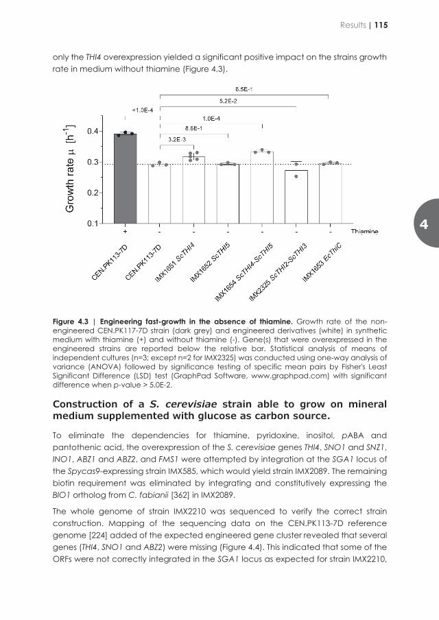
Nicotinic acid and biotin
Previous studies indicated that the presence of nicotinic acid (niacin, Vitamin B3) was dispensable for oxic growth of S. cerevisiae [70]. Based on these data no genetic modifications aiming at improving nicotinic acid provision were investigated. For biotin, an already confirmed strategy consisting in the overexpression of BIO1 from the biotin prototroph Phaffomycetaceae yeast Cyberlindnera fabianii was selected [362] (Figure 4.1).
Evaluation of genotypes to reduce thiamine dependency of S. cerevisiae growth
Of all class B vitamins, removing thiamine dependency was the most problematic after biotin. Thiamine remained the only vitamin for which ALE did not yield isolates whose growth was as fast as the parental strain in presence of thiamine [225]. Several point mutations in CNB1 that encodes the regulatory subunit of calcineurin, in PMR1 that encodes a high affinity Ca2+/Mn2+ P-type ATPase, and in FRE2 that encodes a cell-surface iron reductase as well as the overexpression of ScTHI4 have been shown to act positively on growth rate in absence of thiamine [225]. The thiazole synthase ScThi4 that is involved in the synthesis of the thiazole moiety of thiamine operates as a co-substrate in the reaction by providing a sulfur group for thiazole formation; this implies that Thi4 undergoes a single turnover suicide reaction [384]. This unusual catalytic role identifies Thi4 as a potential bottleneck in thiamine biosynthesis and was therefore chosen for this overexpression study.
The omission of thiamin in liquid culture reduced the CEN.PK113-7D cell growth by 25% (µSMDΔthi = 0.29 ± 0.00 h-1 vs. µSMD = 0.40 ± 0.01 h-1, Figure 4.3). The ScTHI4 overexpression (IMX1651) resulted in a significant but moderate increase in growth rate (µSMDΔthi-IMX1651
= 0.32 ± 0.01 h-1 vs. µSMD = 0.39 ± 0.01 h-1). This increase can also be observed in strain IMX1653, which overexpresses both ScTHI4 and ScTHI5 (µSMDΔthi-IMX1653 = 0.33 ± 0.01 h-1). Thi4 is not the only suicide enzyme in the thiamine biosynthetic pathway.
Indeed, ScThi5 that is responsible for the formation of the pyrimidine heterocycle in the thiamine biosynthesis pathway, catalyzes the synthesis of hydroxymethylpyrimidine phosphate from pyridoxal phosphate (Figure 4.1) and a histidine residue located in the Thi5 active site, resulting in a single turnover reaction [385]. Conversely to ScThi4 overexpression, that of ScTHI5 did not yield a growth improvement; this was confirmed by the phenotype of IMX1654 (ScTHI4-5) that was not different to that of IMX1651 (ScTHI4) (Figure 4.3).
Two other strategies were attempted, firstly the genes encoding the THI regulon transcriptional regulators, ScTHI2 and ScTHI3 were overexpressed (IMX2325) as described earlier to have a positive effect [386]. Secondly the E. coli gene thiC which, as opposed to ScThi5, encodes a phosphomethylpyrimidine synthase that provides 4-amino-2-methyl-5-(phosphor-oxymethyl) pyrimidine in a non-suicide mechanism [387], was overexpressed in strain IMX1653 (Figure 4.1). Of all the attempted strategies,
only the THI4 overexpression yielded a significant positive impact on the strains growth rate in medium without thiamine (Figure 4.3).
Figure 4.3 | Engineering fast-growth in the absence of thiamine. Growth rate of the non-engineered CEN.PK117-7D strain (dark grey) and engineered derivatives (white) in synthetic medium with thiamine (+) and without thiamine (-). Gene(s) that were overexpressed in the engineered strains are reported below the relative bar. Statistical analysis of means of independent cultures (n=3; except n=2 for IMX2325) was conducted using one-way analysis of variance (ANOVA) followed by significance testing of specific mean pairs by Fisher's Least Significant Difference (LSD) test (GraphPad Software, www.graphpad.com) with significant difference when p-value > 5.0E-2.
Construction of a S. cerevisiae strain able to grow on mineral medium supplemented with glucose as carbon source.
To eliminate the dependencies for thiamine, pyridoxine, inositol, pABA and pantothenic acid, the overexpression of the S. cerevisiae genes THI4, SNO1 and SNZ1, INO1, ABZ1 and ABZ2, and FMS1 were attempted by integration at the SGA1 locus of the Spycas9-expressing strain IMX585, which would yield strain IMX2089. The remaining biotin requirement was eliminated by integrating and constitutively expressing the BIO1 ortholog from C. fabianii [362] in IMX2089.
The whole genome of strain IMX2210 was sequenced to verify the correct strain construction. Mapping of the sequencing data on the CEN.PK113-7D reference genome [224] added of the expected engineered gene cluster revealed that several genes (THI4, SNO1 and ABZ2) were missing (Figure 4.4). This indicated that some of the ORFs were not correctly integrated in the SGA1 locus as expected for strain IMX2210,
4
Results | 115
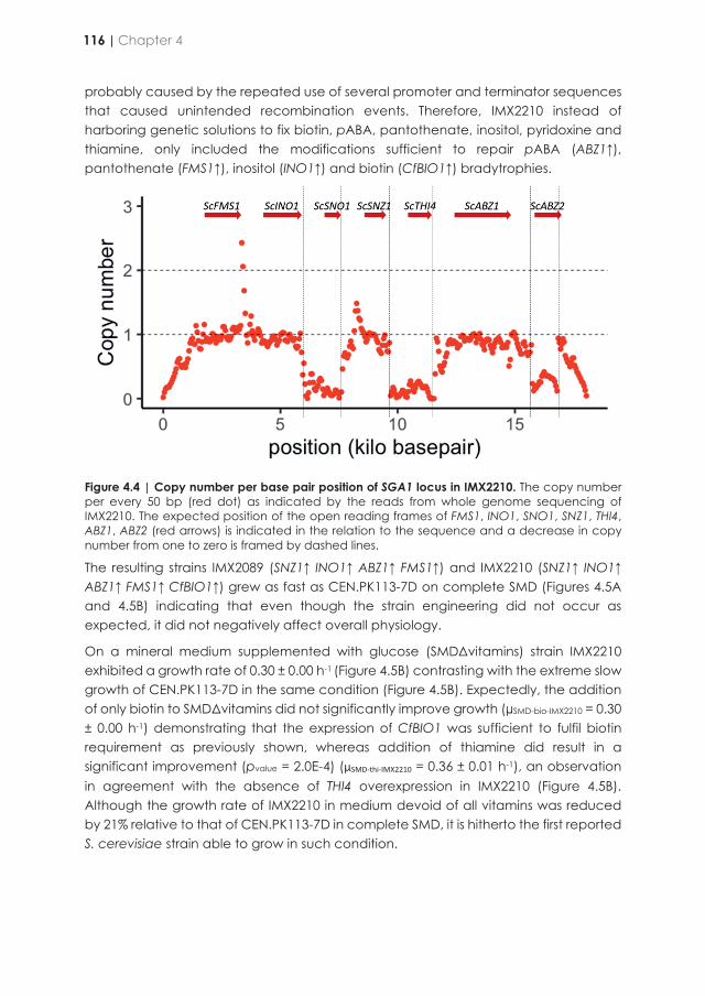
probably caused by the repeated use of several promoter and terminator sequences that caused unintended recombination events. Therefore, IMX2210 instead of harboring genetic solutions to fix biotin, pABA, pantothenate, inositol, pyridoxine and thiamine, only included the modifications sufficient to repair pABA (ABZ1↑), pantothenate (FMS1↑), inositol (INO1↑) and biotin (CfBIO1↑) bradytrophies.
Figure 4.4 | Copy number per base pair position of SGA1 locus in IMX2210. The copy number per every 50 bp (red dot) as indicated by the reads from whole genome sequencing of IMX2210. The expected position of the open reading frames of FMS1, INO1, SNO1, SNZ1, THI4, ABZ1, ABZ2 (red arrows) is indicated in the relation to the sequence and a decrease in copy number from one to zero is framed by dashed lines.
The resulting strains IMX2089 (SNZ1↑ INO1↑ ABZ1↑ FMS1↑) and IMX2210 (SNZ1↑ INO1↑ ABZ1↑ FMS1↑ CfBIO1↑) grew as fast as CEN.PK113-7D on complete SMD (Figures 4.5A and 4.5B) indicating that even though the strain engineering did not occur as expected, it did not negatively affect overall physiology.
On a mineral medium supplemented with glucose (SMDΔvitamins) strain IMX2210 exhibited a growth rate of 0.30 ± 0.00 h-1 (Figure 4.5B) contrasting with the extreme slow growth of CEN.PK113-7D in the same condition (Figure 4.5B). Expectedly, the addition of only biotin to SMDΔvitamins did not significantly improve growth (µSMD-bio-IMX2210 = 0.30 ± 0.00 h-1) demonstrating that the expression of CfBIO1 was sufficient to fulfil biotin requirement as previously shown, whereas addition of thiamine did result in a significant improvement (pvalue = 2.0E-4) (µSMD-thi-IMX2210 = 0.36 ± 0.01 h-1), an observation in agreement with the absence of THI4 overexpression in IMX2210 (Figure 4.5B). Although the growth rate of IMX2210 in medium devoid of all vitamins was reduced by 21% relative to that of CEN.PK113-7D in complete SMD, it is hitherto the first reported S. cerevisiae strain able to grow in such condition.
Figure 4.5 | Engineering fast-growth in the absence of vitamins. Growth rate of the non-engineered CEN.PK117-7D strain (dark grey) and engineered derivatives (white) IMX2089 (A) and IMX2210 (B) in synthetic medium with vitamins (+), without vitamins (-) or a single vitamin added. Statistical analysis was based on a one-way analysis of variance (ANOVA) followed in case of panel A by Tukey’s multiple comparisons correction and in case of panel B by Fisher's Least Significant Difference (LSD) test (GraphPad Software, www.graphpad.com) with significant differences between the means of independent cultures (n=3; except for IMX2210 n=2) when p-value > 5.0E-2.
116 | Chapter 4

probably caused by the repeated use of several promoter and terminator sequences that caused unintended recombination events. Therefore, IMX2210 instead of harboring genetic solutions to fix biotin, pABA, pantothenate, inositol, pyridoxine and thiamine, only included the modifications sufficient to repair pABA (ABZ1↑), pantothenate (FMS1↑), inositol (INO1↑) and biotin (CfBIO1↑) bradytrophies.
Figure 4.4 | Copy number per base pair position of SGA1 locus in IMX2210. The copy number per every 50 bp (red dot) as indicated by the reads from whole genome sequencing of IMX2210. The expected position of the open reading frames of FMS1, INO1, SNO1, SNZ1, THI4, ABZ1, ABZ2 (red arrows) is indicated in the relation to the sequence and a decrease in copy number from one to zero is framed by dashed lines.
The resulting strains IMX2089 (SNZ1↑ INO1↑ ABZ1↑ FMS1↑) and IMX2210 (SNZ1↑ INO1↑ ABZ1↑ FMS1↑ CfBIO1↑) grew as fast as CEN.PK113-7D on complete SMD (Figures 4.5A and 4.5B) indicating that even though the strain engineering did not occur as expected, it did not negatively affect overall physiology.
On a mineral medium supplemented with glucose (SMDΔvitamins) strain IMX2210 exhibited a growth rate of 0.30 ± 0.00 h-1 (Figure 4.5B) contrasting with the extreme slow growth of CEN.PK113-7D in the same condition (Figure 4.5B). Expectedly, the addition of only biotin to SMDΔvitamins did not significantly improve growth (µSMD-bio-IMX2210 = 0.30 ± 0.00 h-1) demonstrating that the expression of CfBIO1 was sufficient to fulfil biotin requirement as previously shown, whereas addition of thiamine did result in a significant improvement (pvalue = 2.0E-4) (µSMD-thi-IMX2210 = 0.36 ± 0.01 h-1), an observation in agreement with the absence of THI4 overexpression in IMX2210 (Figure 4.5B). Although the growth rate of IMX2210 in medium devoid of all vitamins was reduced by 21% relative to that of CEN.PK113-7D in complete SMD, it is hitherto the first reported S. cerevisiae strain able to grow in such condition.
Figure 4.5 | Engineering fast-growth in the absence of vitamins. Growth rate of the non-engineered CEN.PK117-7D strain (dark grey) and engineered derivatives (white) IMX2089 (A) and IMX2210 (B) in synthetic medium with vitamins (+), without vitamins (-) or a single vitamin added. Statistical analysis was based on a one-way analysis of variance (ANOVA) followed in case of panel A by Tukey’s multiple comparisons correction and in case of panel B by Fisher's Least Significant Difference (LSD) test (GraphPad Software, www.graphpad.com) with significant differences between the means of independent cultures (n=3; except for IMX2210 n=2) when p-value > 5.0E-2.
4
Results | 117

Physiological characterization of vitamin independent strain IMX2210 in aerobic glucose limited chemostat culture.
Although not entirely repaired, IMX2210 is capable of growth in medium devoid of all class B vitamins. The physiology of strain IMX2210 was characterized in controlled oxic bioreactors in batch and glucose-limited chemostat cultures in both SMD and SMDΔvitamins.
Table 4.4 | Growth rate µ, biomass yield YX/S, biomass specific glucose consumption rate (qGlucose), biomass specific ethanol production rate (qEthanol), of oxic batch cultures of strain IMX2210 grown on either SMD or SMDΔvitamins. Data are compared to previously published data for the reference strain CEN.PK113-7D grown in SMD [389]. Errors represent the standard deviation of the mean (n=2). Statistical significance determined using the student’s t-test, with alpha = 0.05. * = significant different results. n.a. = not measured/applicable
Strain IMX2210 CEN.PK113-7D p-values Medium SMD (1) SMDΔvitamins (2) SMD (3) 1 vs. 2 2 vs. 3 1 vs. 3
µ [h-1] n.a. 0.28±0.01 0.37±0.01 n.a. 1.7E-2* n.a. YXS [gx/gS] 0.14±0.01 0.15±0.01 0.13±0.01 8.2E-1 3.9E-1 5.6E-1 qGlucose
[mmol gX-1 h-1] 6.03±0.29 5.92±1.02 14.8±1.1 9.3E-1 2.8E-2* 6.4E-2*
qEthanol [mmol gX-1 h-1]
8.04±0.41 9.71±3.38 22.0±0.5 7.1E-1 7.4E-2 2.7E-3*
During the batch phase of the chemostat cultures in the absence of all vitamins (SMDΔvitamins), strain IMX2210 showed a growth rate of 0.28 ± 0.01 h-1, which is reduced compared to growth rates of similar batch cultures with CEN.PK113-7D in SMD (Table 4.4, p-value = 1.7E-2). The strain displayed a respiro-fermentative metabolism and a regular diauxic shift in which the ethanol formed during the glucose phase was subsequently completely re-consumed typical of oxic batches of S. cerevisiae (Figure 4.6) [388]. The biomass yield for strain IMX2210, grown either on SMD or on SMDΔvitamins, was comparable to the CEN.PK113-7D biomass yield in an oxic batch cultures (Table 4.4). The quantitative data for the biomass specific glucose uptake rate and ethanol production rate for strain IMX2210 on SMD were slightly lower compared to CEN.PK113-7D data and did not change significantly when IMX2210 was grown on SMDΔvitamins (Table 4.4).
The batches were followed by an oxic glucose-limited chemostat culture at a dilution rate of 0.1 h-1. Irrespective of the culture medium, IMX2210 exhibited identical steady state biomass yield, biomass specific oxygen consumption rate, biomass specific carbon dioxide production rate, and respiratory quotient. These quantitative data were almost identical to those measured for the reference CEN.PK113-7D strain in oxic glucose limited chemostat at the same dilution rate [389] (Table 4.5). Only biomass specific glucose uptake rate of IMX2210 compared to CEN.PK113-7D was slightly reduced according to the student’s t-test, as well as oxygen consumption rate and carbon recovery when grown on SMD.
Figure 4.6 | Biomass and metabolite concentration over time of IMX2210 cultures in SMDΔvitamins in oxic batch bioreactors. Values for biomass (), glucose (), ethanol (), glycerol (), and acetate () are shown over time. Error bars represent the standard error of the mean (n=4).
IMX2210 transcriptome analysis
Chemostat cultivation offers a number of advantages for genome-wide transcriptome studies because it enables cell cultivation under tightly defined environmental conditions. This cultivation mode in particular is very well suited to compare conditions or strains that exhibit difference in growth rate in batch cultures. Aerobic glucose-limited chemostat-cultures of IMX2210 in SMD and SMDΔvitamins were sampled for analysis of the transcriptome. RNA-seq data of the cultivation with vitamins, were also compared with RNA seq-based transcriptome data of the reference strain CEN.PK113-7D grown in identical vitamin containing cultivation conditions. The pairwise IMX2210 versus CEN.PK113-7D grown in SMD comparison revealed that 123 genes were differently expressed between the two strains, 31 were upregulated in IMX2210 and 92 genes exhibited a reverse profile (Ifold changeI > 2 with a qFDR-value < 0.05) (Supplemental data).
The upregulated genes set was enriched for genes involved in siderophore transmembrane transport (GO:0044718; GO: ENB1, SIT1, ARN1, FET3 and FRE3) and in water-soluble vitamin biosynthetic process (GO:0042364: SNZ1 (+36-fold), INO1 (+7.8-fold), ABZ1 (31.7-fold), FMS1 (+26.7-fold)) (Table 4.6). The latter category corresponded to four overexpressed genes to eliminate pantothenic acid, para-aminobenzoic acid (pABA), pyridoxine, inositol bradytrophies, to which we could add the heterologous gene CfBIO1. Although it was not possible to calculate a fold
0 10 20 30 400
2
4
6
8
10
0
50
100
150
200
Time (h)
Biom
ass
(g L
-1)
glyc
erol
, ace
tate
(mM
)
IMX2210 SMDΔvitamins
Biomass (g/L) Glucose (mM) Ethanol (mM) Glycerol (mM) Acetate (mM)
Glucose, ethanol (m
M)
118 | Chapter 4
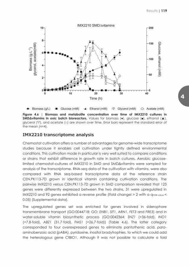
Physiological characterization of vitamin independent strain IMX2210 in aerobic glucose limited chemostat culture.
Although not entirely repaired, IMX2210 is capable of growth in medium devoid of all class B vitamins. The physiology of strain IMX2210 was characterized in controlled oxic bioreactors in batch and glucose-limited chemostat cultures in both SMD and SMDΔvitamins.
Table 4.4 | Growth rate µ, biomass yield YX/S, biomass specific glucose consumption rate (qGlucose), biomass specific ethanol production rate (qEthanol), of oxic batch cultures of strain IMX2210 grown on either SMD or SMDΔvitamins. Data are compared to previously published data for the reference strain CEN.PK113-7D grown in SMD [389]. Errors represent the standard deviation of the mean (n=2). Statistical significance determined using the student’s t-test, with alpha = 0.05. * = significant different results. n.a. = not measured/applicable
Strain IMX2210 CEN.PK113-7D p-values Medium SMD (1) SMDΔvitamins (2) SMD (3) 1 vs. 2 2 vs. 3 1 vs. 3
µ [h-1] n.a. 0.28±0.01 0.37±0.01 n.a. 1.7E-2* n.a. YXS [gx/gS] 0.14±0.01 0.15±0.01 0.13±0.01 8.2E-1 3.9E-1 5.6E-1 qGlucose
[mmol gX-1 h-1] 6.03±0.29 5.92±1.02 14.8±1.1 9.3E-1 2.8E-2* 6.4E-2*
qEthanol [mmol gX-1 h-1]
8.04±0.41 9.71±3.38 22.0±0.5 7.1E-1 7.4E-2 2.7E-3*
During the batch phase of the chemostat cultures in the absence of all vitamins (SMDΔvitamins), strain IMX2210 showed a growth rate of 0.28 ± 0.01 h-1, which is reduced compared to growth rates of similar batch cultures with CEN.PK113-7D in SMD (Table 4.4, p-value = 1.7E-2). The strain displayed a respiro-fermentative metabolism and a regular diauxic shift in which the ethanol formed during the glucose phase was subsequently completely re-consumed typical of oxic batches of S. cerevisiae (Figure 4.6) [388]. The biomass yield for strain IMX2210, grown either on SMD or on SMDΔvitamins, was comparable to the CEN.PK113-7D biomass yield in an oxic batch cultures (Table 4.4). The quantitative data for the biomass specific glucose uptake rate and ethanol production rate for strain IMX2210 on SMD were slightly lower compared to CEN.PK113-7D data and did not change significantly when IMX2210 was grown on SMDΔvitamins (Table 4.4).
The batches were followed by an oxic glucose-limited chemostat culture at a dilution rate of 0.1 h-1. Irrespective of the culture medium, IMX2210 exhibited identical steady state biomass yield, biomass specific oxygen consumption rate, biomass specific carbon dioxide production rate, and respiratory quotient. These quantitative data were almost identical to those measured for the reference CEN.PK113-7D strain in oxic glucose limited chemostat at the same dilution rate [389] (Table 4.5). Only biomass specific glucose uptake rate of IMX2210 compared to CEN.PK113-7D was slightly reduced according to the student’s t-test, as well as oxygen consumption rate and carbon recovery when grown on SMD.
Figure 4.6 | Biomass and metabolite concentration over time of IMX2210 cultures in SMDΔvitamins in oxic batch bioreactors. Values for biomass (), glucose (), ethanol (), glycerol (), and acetate () are shown over time. Error bars represent the standard error of the mean (n=4).
IMX2210 transcriptome analysis
Chemostat cultivation offers a number of advantages for genome-wide transcriptome studies because it enables cell cultivation under tightly defined environmental conditions. This cultivation mode in particular is very well suited to compare conditions or strains that exhibit difference in growth rate in batch cultures. Aerobic glucose-limited chemostat-cultures of IMX2210 in SMD and SMDΔvitamins were sampled for analysis of the transcriptome. RNA-seq data of the cultivation with vitamins, were also compared with RNA seq-based transcriptome data of the reference strain CEN.PK113-7D grown in identical vitamin containing cultivation conditions. The pairwise IMX2210 versus CEN.PK113-7D grown in SMD comparison revealed that 123 genes were differently expressed between the two strains, 31 were upregulated in IMX2210 and 92 genes exhibited a reverse profile (Ifold changeI > 2 with a qFDR-value < 0.05) (Supplemental data).
The upregulated genes set was enriched for genes involved in siderophore transmembrane transport (GO:0044718; GO: ENB1, SIT1, ARN1, FET3 and FRE3) and in water-soluble vitamin biosynthetic process (GO:0042364: SNZ1 (+36-fold), INO1 (+7.8-fold), ABZ1 (31.7-fold), FMS1 (+26.7-fold)) (Table 4.6). The latter category corresponded to four overexpressed genes to eliminate pantothenic acid, para-aminobenzoic acid (pABA), pyridoxine, inositol bradytrophies, to which we could add the heterologous gene CfBIO1. Although it was not possible to calculate a fold
0 10 20 30 400
2
4
6
8
10
0
50
100
150
200
Time (h)
Biom
ass
(g L
-1)
glyc
erol
, ace
tate
(mM
)
IMX2210 SMDΔvitamins
Biomass (g/L) Glucose (mM) Ethanol (mM) Glycerol (mM) Acetate (mM)
Glucose, ethanol (m
M)
4
Results | 119
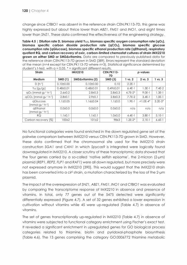
change since CfBIO1 was absent in the reference strain CEN.PK113-7D, this gene was highly expressed but about thrice lower than ABZ1, FMS1 and INO1, and eight times lower than SNZ1. These data confirmed the effectiveness of the engineering strategy.
Table 4.5 | Dilution rate D, biomass yield YX/S, biomass specific oxygen consumption rate (qO2), biomass specific carbon dioxide production rate (qCO2), biomass specific glucose consumption rate (qGlucose), biomass specific ethanol production rate (qEthanol), respiratory quotient RQ, and carbon recovery of oxic, carbon-limited chemostat cultures of strain IMX2210 grown on either SMD or SMDΔvitamins. Data are compared to previously published data for the reference strain CEN.PK113-7D grown in SMD [389]. Errors represent the standard deviation of the mean (n=4 except for CEN.PK113-7D where n=3). Statistical significance determined by student’s t-test, with α = 0.05. * = significant different results.
Strain IMX2210 CEN.PK113-7D
p-values
Medium SMD (1) SMDΔvitamins (2) SMD (3) 1 vs. 2 2 vs. 3 1 vs. 3 D [h-1] 0.10±0.00 0.10±0.00 0.10 2.2E-1
YXS [gx/gS] 0.48±0.01 0.48±0.01 0.49±0.01 6.4E-1 1.5E-1 7.4E-2 qO2 [mmol gX-1 h-1] 2.6±0.2 2.8±0.3 2.8±0.3 4.7E-2* 9.0E-1 1.3E-1
qCO2 [mmol gX-1 h-1] 2.8±0.1 2.9±0.1 2.8±0.3 7.7E-2 3.4E-1 1.0E-1 qGlucose
[mmol gX-1 h-1] 1.1±0.05 1.16±0.04 1.1±0.0 1.9E-1 <1.0E-4* 3.2E-3*
qEthanol [mmol gX-1 h-1]
0.0±0.0 0.0±0.0 0.0±0.0 n/a n/a n/a
RQ 1.1±0.1 1.1±0.1 1.0±0.0 4.4E-1 3.8E-1 5.1E-1 Carbon recovery [%] 100±2 101±2 98±3 1.2E-2* 2.1E-1 6.6E-1
No functional categories were found enriched in the down regulated gene set of the pairwise comparison between IMX2210 versus CEN.PK113-7D grown in SMD. However, these data confirmed that the chromosomal site used for the IMX2210 strain construction SGA1 and CAN1 in which Spycas9 is integrated were logically found downregulated in IMX2210. A closer scrutiny of these transcriptomic data showed that the four genes carried by a so-called ‘native selfish episome’, the 2-micron (2-μm) plasmid (REP1, REP2, FLP1 and RAF1) were all down regulated, but more precisely were not expressed anymore in IMX2210 [390]. This would suggest that the IMX2210 strain has been converted into a cir0 strain, a mutation characterized by the loss of the 2-μm plasmid.
The impact of the overexpression of SNZ1, ABZ1, FMS1, INO1 and CfBIO1 was evaluated by comparing the transcriptome response of IMX2210 in absence and presence of vitamins. In total, only 77 genes out of the 5475 detected were significantly differentially expressed (Figure 4.7). A set of 32 genes exhibited a lower expression in cultivation without vitamins while 45 were up-regulated (Table 4.7) in absence of vitamins.
The set of genes transcriptionally up-regulated in IMX2210 (Table 4.7) in absence of vitamins were subjected to functional category enrichment using Fischer’s exact test. It revealed a significant enrichment in upregulated genes for GO biological process categories related to thiamine, biotin and pyridoxal-phosphate biosynthesis (Table 4.6). The 13 genes comprising the category GO:0006772 thiamine metabolic
process, were all upregulated in IMX2210 when vitamins were omitted to the culture medium (THI12, THI6, THI2,1 SNZ3, THI2, THI20, SNZ2, THI11, THI4, THI5, THI22, THI13 and PET18). The upregulation of PET18 [391], THI73, THI74 [392] and THI7 [393] was already reported in starving or low thiamine conditions.
Similarly, all six genes (BIO2, BIO5, BIO4, BIO3 and ScBIO1 and BIO6) comprising the GO category “biotin biosynthetic process (GO:0006768)” related genes were up-regulated in vitamin less conditions. This was accompanied with the upregulation of the biotin transporter gene VTH1 as well. The last set of enriched categories was pyridoxal phosphate biosynthesis related genes (GO:0042822) BUD16, SNZ3, SNO3 and SNZ2 (Table 4.7, Figure 4.7). Conversely, the gene SNO4 that encodes a cysteine protease hypothesized to be involved in pyridoxine metabolism [394] was down-regulated. In total a minimum of 26 genes, which represented 83% of the upregulated genes were related to class B vitamins (thiamine, biotin and pyridoxine) biosynthesis.
Although, this was not significantly overrepresented, several genes encoding vitamin dependent enzymes were also upregulated in the transcriptome of IMX2210 grown in absence of vitamins. Expression of proteins requiring vitamins as cofactors was up-regulated. The pyridoxal-phosphate dependent alanine-transaminase gene (ALT1) [77] expression was increased by 2.1-fold, similarly the expression of the thiamine dependent broad range 2-oxo acid decarboxylase encoding gene ARO10 [131, 395] and of the pyruvate decarboxylase isoform gene PDC5 [394] were increased by 3.1 and 3.2-fold, respectively. The highest alterations in mRNA abundance were observed for THI4, THI5, THI12, THI13, THI11 and BIO5 as well as for FCY22, a nucleobase cation symporter gene [396] and YCR102C, a stress tolerance gene [397] (Figure 4.7).
120 | Chapter 4
change since CfBIO1 was absent in the reference strain CEN.PK113-7D, this gene was highly expressed but about thrice lower than ABZ1, FMS1 and INO1, and eight times lower than SNZ1. These data confirmed the effectiveness of the engineering strategy.
Table 4.5 | Dilution rate D, biomass yield YX/S, biomass specific oxygen consumption rate (qO2), biomass specific carbon dioxide production rate (qCO2), biomass specific glucose consumption rate (qGlucose), biomass specific ethanol production rate (qEthanol), respiratory quotient RQ, and carbon recovery of oxic, carbon-limited chemostat cultures of strain IMX2210 grown on either SMD or SMDΔvitamins. Data are compared to previously published data for the reference strain CEN.PK113-7D grown in SMD [389]. Errors represent the standard deviation of the mean (n=4 except for CEN.PK113-7D where n=3). Statistical significance determined by student’s t-test, with α = 0.05. * = significant different results.
Strain IMX2210 CEN.PK113-7D
p-values
Medium SMD (1) SMDΔvitamins (2) SMD (3) 1 vs. 2 2 vs. 3 1 vs. 3 D [h-1] 0.10±0.00 0.10±0.00 0.10 2.2E-1
YXS [gx/gS] 0.48±0.01 0.48±0.01 0.49±0.01 6.4E-1 1.5E-1 7.4E-2 qO2 [mmol gX-1 h-1] 2.6±0.2 2.8±0.3 2.8±0.3 4.7E-2* 9.0E-1 1.3E-1
qCO2 [mmol gX-1 h-1] 2.8±0.1 2.9±0.1 2.8±0.3 7.7E-2 3.4E-1 1.0E-1 qGlucose
[mmol gX-1 h-1] 1.1±0.05 1.16±0.04 1.1±0.0 1.9E-1 <1.0E-4* 3.2E-3*
qEthanol [mmol gX-1 h-1]
0.0±0.0 0.0±0.0 0.0±0.0 n/a n/a n/a
RQ 1.1±0.1 1.1±0.1 1.0±0.0 4.4E-1 3.8E-1 5.1E-1 Carbon recovery [%] 100±2 101±2 98±3 1.2E-2* 2.1E-1 6.6E-1
No functional categories were found enriched in the down regulated gene set of the pairwise comparison between IMX2210 versus CEN.PK113-7D grown in SMD. However, these data confirmed that the chromosomal site used for the IMX2210 strain construction SGA1 and CAN1 in which Spycas9 is integrated were logically found downregulated in IMX2210. A closer scrutiny of these transcriptomic data showed that the four genes carried by a so-called ‘native selfish episome’, the 2-micron (2-μm) plasmid (REP1, REP2, FLP1 and RAF1) were all down regulated, but more precisely were not expressed anymore in IMX2210 [390]. This would suggest that the IMX2210 strain has been converted into a cir0 strain, a mutation characterized by the loss of the 2-μm plasmid.
The impact of the overexpression of SNZ1, ABZ1, FMS1, INO1 and CfBIO1 was evaluated by comparing the transcriptome response of IMX2210 in absence and presence of vitamins. In total, only 77 genes out of the 5475 detected were significantly differentially expressed (Figure 4.7). A set of 32 genes exhibited a lower expression in cultivation without vitamins while 45 were up-regulated (Table 4.7) in absence of vitamins.
The set of genes transcriptionally up-regulated in IMX2210 (Table 4.7) in absence of vitamins were subjected to functional category enrichment using Fischer’s exact test. It revealed a significant enrichment in upregulated genes for GO biological process categories related to thiamine, biotin and pyridoxal-phosphate biosynthesis (Table 4.6). The 13 genes comprising the category GO:0006772 thiamine metabolic
process, were all upregulated in IMX2210 when vitamins were omitted to the culture medium (THI12, THI6, THI2,1 SNZ3, THI2, THI20, SNZ2, THI11, THI4, THI5, THI22, THI13 and PET18). The upregulation of PET18 [391], THI73, THI74 [392] and THI7 [393] was already reported in starving or low thiamine conditions.
Similarly, all six genes (BIO2, BIO5, BIO4, BIO3 and ScBIO1 and BIO6) comprising the GO category “biotin biosynthetic process (GO:0006768)” related genes were up-regulated in vitamin less conditions. This was accompanied with the upregulation of the biotin transporter gene VTH1 as well. The last set of enriched categories was pyridoxal phosphate biosynthesis related genes (GO:0042822) BUD16, SNZ3, SNO3 and SNZ2 (Table 4.7, Figure 4.7). Conversely, the gene SNO4 that encodes a cysteine protease hypothesized to be involved in pyridoxine metabolism [394] was down-regulated. In total a minimum of 26 genes, which represented 83% of the upregulated genes were related to class B vitamins (thiamine, biotin and pyridoxine) biosynthesis.
Although, this was not significantly overrepresented, several genes encoding vitamin dependent enzymes were also upregulated in the transcriptome of IMX2210 grown in absence of vitamins. Expression of proteins requiring vitamins as cofactors was up-regulated. The pyridoxal-phosphate dependent alanine-transaminase gene (ALT1) [77] expression was increased by 2.1-fold, similarly the expression of the thiamine dependent broad range 2-oxo acid decarboxylase encoding gene ARO10 [131, 395] and of the pyruvate decarboxylase isoform gene PDC5 [394] were increased by 3.1 and 3.2-fold, respectively. The highest alterations in mRNA abundance were observed for THI4, THI5, THI12, THI13, THI11 and BIO5 as well as for FCY22, a nucleobase cation symporter gene [396] and YCR102C, a stress tolerance gene [397] (Figure 4.7).
4
Results | 121
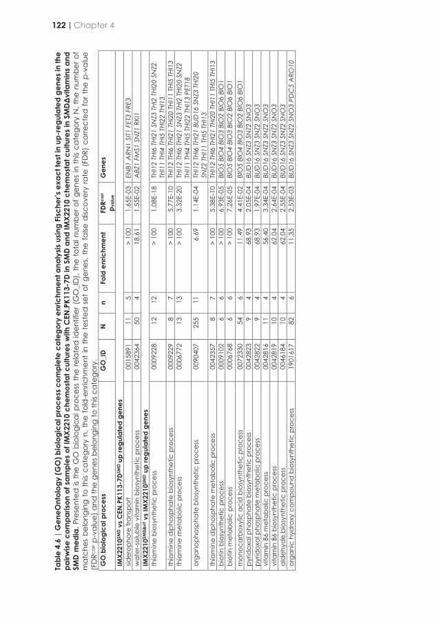
Tabl
e 4.
6 |
Gen
eOnt
olog
y (G
O) b
iolo
gica
l pro
cess
com
plet
e ca
tego
ry e
nric
hmen
t ana
lysis
usin
g Fi
sche
r’s e
xact
test
in u
p-re
gula
ted
gene
s in
the
pairw
ise c
ompa
rison
of s
ampl
es o
f IM
X221
0 ch
emos
tat c
ultu
res
with
CEN
.PK1
13-7
D in
SM
D an
d IM
X221
0 ch
emos
tat c
ultu
res
in S
MDΔ
vita
min
s an
d SM
D m
edia
. Pre
sent
ed is
the
GO
bio
logi
cal p
roce
ss th
e re
late
d id
entif
ier (
GO
_ID
), th
e to
tal n
umbe
r of g
enes
in th
is ca
tego
ry N
, the
num
ber o
f m
atch
es b
elon
ging
to
this
cate
gory
n, t
he fo
ld-e
nric
hmen
t in
the
tes
ted
set
of g
enes
, the
false
disc
over
y ra
te (
FDR)
cor
rect
ed fo
r the
p-v
alu
e (F
DRc
orr p
-val
ue) a
nd th
e ge
nes b
elon
ging
to th
is ca
tego
ry.
GO
bio
logi
cal p
roce
ss
GO
_ID
N
n Fo
ld e
nric
hmen
t FD
Rcor
r p v
alue
G
enes
IMX2
210S
MD
vs C
EN.P
K113
-7DS
MD
up re
gula
ted
gene
s sid
erop
hore
tran
spor
t 00
1589
1 11
5
> 10
0 1.
65E-
03
ENB1
ARN
1 SI
T1 F
ET3
FRE3
w
ater
-sol
uble
vita
min
bio
synt
hetic
pro
cess
00
4236
4 50
4
18.6
1 1.
55E-
02
ABZ
1 FM
S1 S
NZ1
RKI
1 IM
X221
0SM
DΔ Δvi
t vs
IMX2
210S
MD
up re
gula
ted
gene
s th
iam
ine
bios
ynth
etic
pro
cess
00
0922
8 12
12
>
100
1.08
E-18
TH
I12
THI6
TH
I21
SNZ3
TH
I2 T
HI20
SN
Z2
THI1
1 TH
I4 T
HI5
TH
I22
THI1
3 th
iam
ine
dip
hosp
hate
bio
synt
hetic
pro
cess
00
0922
9 8
7 >
100
5.77
E-10
TH
I12
THI6
TH
I21
THI2
0 TH
I11
THI5
THI
13
thia
min
e m
etab
olic
pro
cess
00
0677
2 13
13
>
100
3.32
E-20
TH
I12
THI6
TH
I21
SNZ3
TH
I2 T
HI20
SN
Z2
THI1
1 TH
I4 T
HI5
THI2
2 TH
I13
PET1
8 or
gano
phos
phat
e bi
osyn
thet
ic p
roce
ss
0090
407
255
11
6.69
1.
14E-
04
THI1
2 TH
I6 T
HI21
BUD
16 S
NZ3
THI
20
SNZ2
TH
I11
THI5
TH
I13
thia
min
e d
ipho
spha
te m
etab
olic
pro
cess
00
4235
7 8
7 >
100
5.38
E-10
TH
I12
THI6
TH
I21
THI2
0 TH
I11
THI5
THI
13
biot
in b
iosy
nthe
tic p
roce
ss
0009
102
6 6
> 10
0 6.
93E-
05
BIO
5 BI
O4
BIO
3 BI
O2
BIO
6 BI
O1
biot
in m
etab
olic
pro
cess
00
0676
8 6
6 >
100
7.26
E-05
BI
O5
BIO
4 BI
O3
BIO
2 BI
O6
BIO
1 m
onoc
arbo
xylic
aci
d b
iosy
nthe
tic p
roce
ss
0072
330
54
6 11
.49
4.41
E-02
BI
O5
BIO
4 BI
O3
BIO
2 BI
O6
BIO
1 py
ridox
al p
hosp
hate
bio
synt
hetic
pro
cess
00
4282
3 9
4 68
.93
2.05
E-04
BU
D16
SN
Z3 S
NZ2
SN
O3
pyrid
oxal
pho
spha
te m
etab
olic
pro
cess
00
4282
2 9
4 68
.93
1.97
E-04
BU
D16
SN
Z3 S
NZ2
SN
O3
vita
min
B6
met
abol
ic p
roce
ss
0042
816
11
4 56
.40
3.34
E-04
BU
D16
SN
Z3 S
NZ2
SN
O3
vita
min
B6
bios
ynth
etic
pro
cess
00
4281
9 10
4
62.0
4 2.
64E-
04
BUD
16 S
NZ3
SN
Z2 S
NO
3 al
deh
yde
bios
ynth
etic
pro
cess
00
4618
4 10
4
62.0
4 2.
55E-
04
BUD
16 S
NZ3
SN
Z2 S
NO
3 or
gani
c hy
dro
xy c
ompo
und
bio
synt
hetic
pro
cess
19
0161
7 82
6
11.3
5 2.
53E-
03
BUD
16 S
NZ3
SN
Z2 S
NO
3 PD
C5
ARO
10
Figure 4.7 | Differentially expressed genes between IMX2210 chemostat cultures with SMD and SMD without vitamins. Genes presented were plotted by their -log10 of the False Discovery Rate (-log10(FDR)) against the log2 of the fold change (log2FC). Genes with colored labels have a log2FC > 1 with a -log10(FDR) > 2 and are grouped in thiamine metabolism related genes (purple), biotin metabolism related genes (light blue) and pyridoxine metabolism related genes (dark blue).
Discussion
Availability of vitamin prototrophic yeast strains would result in costs reduction for preparation of industrial growth media and would allow exploration of characteristic of strains that have hitherto never been investigated under harsh industrial conditions [240, 298]. Although, the initial ambition of this work was to construct such a strain by combining overexpression of only eight genes (THI4, SNO1 and SNZ1, INO1, ABZ1 and ABZ2, FMS1 and CfBIO1) that in pair or individually were able to improve growth of S. cerevisiae in absence of a single vitamin (Figure 4.2), the constructed strain was not exactly matching the initial expectation. It combined two major genetic alterations, it missed recombination of the seven cassettes for expression of the S. cerevisiae genes, and during this process, it also lost the native 2-μm plasmid. While the latter mutation did not have direct impact on the ability of the strain to grow in absence of vitamins, it did not permit to easily correct the absence of THI4, SNO1 and ABZ2, since cir0 strains cannot maintain recombinant 2-μm plasmids. This absence prevented the subsequent transformation of CRISPR plasmid enabling the expression of the gRNA necessary to target the integration of the missing genes into a chromosomal site. For this reason, the strain IMX2210 evaluated in this work was only engineered for biosynthesis of p-aminobenzoate, pantothenate, inositol [240] and biotin [362].
122 | Chapter 4

Tabl
e 4.
6 |
Gen
eOnt
olog
y (G
O) b
iolo
gica
l pro
cess
com
plet
e ca
tego
ry e
nric
hmen
t ana
lysis
usin
g Fi
sche
r’s e
xact
test
in u
p-re
gula
ted
gene
s in
the
pairw
ise c
ompa
rison
of s
ampl
es o
f IM
X221
0 ch
emos
tat c
ultu
res
with
CEN
.PK1
13-7
D in
SM
D an
d IM
X221
0 ch
emos
tat c
ultu
res
in S
MDΔ
vita
min
s an
d SM
D m
edia
. Pre
sent
ed is
the
GO
bio
logi
cal p
roce
ss th
e re
late
d id
entif
ier (
GO
_ID
), th
e to
tal n
umbe
r of g
enes
in th
is ca
tego
ry N
, the
num
ber o
f m
atch
es b
elon
ging
to
this
cate
gory
n, t
he fo
ld-e
nric
hmen
t in
the
tes
ted
set
of g
enes
, the
false
disc
over
y ra
te (
FDR)
cor
rect
ed fo
r the
p-v
alu
e (F
DRc
orr p
-val
ue) a
nd th
e ge
nes b
elon
ging
to th
is ca
tego
ry.
GO
bio
logi
cal p
roce
ss
GO
_ID
N
n Fo
ld e
nric
hmen
t FD
Rcor
r p v
alue
G
enes
IMX2
210S
MD
vs C
EN.P
K113
-7DS
MD
up re
gula
ted
gene
s sid
erop
hore
tran
spor
t 00
1589
1 11
5
> 10
0 1.
65E-
03
ENB1
ARN
1 SI
T1 F
ET3
FRE3
w
ater
-sol
uble
vita
min
bio
synt
hetic
pro
cess
00
4236
4 50
4
18.6
1 1.
55E-
02
ABZ
1 FM
S1 S
NZ1
RKI
1 IM
X221
0SM
DΔΔvi
t vs
IMX2
210S
MD
up re
gula
ted
gene
s th
iam
ine
bios
ynth
etic
pro
cess
00
0922
8 12
12
>
100
1.08
E-18
TH
I12
THI6
TH
I21
SNZ3
TH
I2 T
HI20
SN
Z2
THI1
1 TH
I4 T
HI5
TH
I22
THI1
3 th
iam
ine
dip
hosp
hate
bio
synt
hetic
pro
cess
00
0922
9 8
7 >
100
5.77
E-10
TH
I12
THI6
TH
I21
THI2
0 TH
I11
THI5
THI
13
thia
min
e m
etab
olic
pro
cess
00
0677
2 13
13
>
100
3.32
E-20
TH
I12
THI6
TH
I21
SNZ3
TH
I2 T
HI20
SN
Z2
THI1
1 TH
I4 T
HI5
THI2
2 TH
I13
PET1
8 or
gano
phos
phat
e bi
osyn
thet
ic p
roce
ss
0090
407
255
11
6.69
1.
14E-
04
THI1
2 TH
I6 T
HI21
BUD
16 S
NZ3
THI
20
SNZ2
TH
I11
THI5
TH
I13
thia
min
e d
ipho
spha
te m
etab
olic
pro
cess
00
4235
7 8
7 >
100
5.38
E-10
TH
I12
THI6
TH
I21
THI2
0 TH
I11
THI5
THI
13
biot
in b
iosy
nthe
tic p
roce
ss
0009
102
6 6
> 10
0 6.
93E-
05
BIO
5 BI
O4
BIO
3 BI
O2
BIO
6 BI
O1
biot
in m
etab
olic
pro
cess
00
0676
8 6
6 >
100
7.26
E-05
BI
O5
BIO
4 BI
O3
BIO
2 BI
O6
BIO
1 m
onoc
arbo
xylic
aci
d b
iosy
nthe
tic p
roce
ss
0072
330
54
6 11
.49
4.41
E-02
BI
O5
BIO
4 BI
O3
BIO
2 BI
O6
BIO
1 py
ridox
al p
hosp
hate
bio
synt
hetic
pro
cess
00
4282
3 9
4 68
.93
2.05
E-04
BU
D16
SN
Z3 S
NZ2
SN
O3
pyrid
oxal
pho
spha
te m
etab
olic
pro
cess
00
4282
2 9
4 68
.93
1.97
E-04
BU
D16
SN
Z3 S
NZ2
SN
O3
vita
min
B6
met
abol
ic p
roce
ss
0042
816
11
4 56
.40
3.34
E-04
BU
D16
SN
Z3 S
NZ2
SN
O3
vita
min
B6
bios
ynth
etic
pro
cess
00
4281
9 10
4
62.0
4 2.
64E-
04
BUD
16 S
NZ3
SN
Z2 S
NO
3 al
deh
yde
bios
ynth
etic
pro
cess
00
4618
4 10
4
62.0
4 2.
55E-
04
BUD
16 S
NZ3
SN
Z2 S
NO
3 or
gani
c hy
dro
xy c
ompo
und
bio
synt
hetic
pro
cess
19
0161
7 82
6
11.3
5 2.
53E-
03
BUD
16 S
NZ3
SN
Z2 S
NO
3 PD
C5
ARO
10
Figure 4.7 | Differentially expressed genes between IMX2210 chemostat cultures with SMD and SMD without vitamins. Genes presented were plotted by their -log10 of the False Discovery Rate (-log10(FDR)) against the log2 of the fold change (log2FC). Genes with colored labels have a log2FC > 1 with a -log10(FDR) > 2 and are grouped in thiamine metabolism related genes (purple), biotin metabolism related genes (light blue) and pyridoxine metabolism related genes (dark blue).
Discussion
Availability of vitamin prototrophic yeast strains would result in costs reduction for preparation of industrial growth media and would allow exploration of characteristic of strains that have hitherto never been investigated under harsh industrial conditions [240, 298]. Although, the initial ambition of this work was to construct such a strain by combining overexpression of only eight genes (THI4, SNO1 and SNZ1, INO1, ABZ1 and ABZ2, FMS1 and CfBIO1) that in pair or individually were able to improve growth of S. cerevisiae in absence of a single vitamin (Figure 4.2), the constructed strain was not exactly matching the initial expectation. It combined two major genetic alterations, it missed recombination of the seven cassettes for expression of the S. cerevisiae genes, and during this process, it also lost the native 2-μm plasmid. While the latter mutation did not have direct impact on the ability of the strain to grow in absence of vitamins, it did not permit to easily correct the absence of THI4, SNO1 and ABZ2, since cir0 strains cannot maintain recombinant 2-μm plasmids. This absence prevented the subsequent transformation of CRISPR plasmid enabling the expression of the gRNA necessary to target the integration of the missing genes into a chromosomal site. For this reason, the strain IMX2210 evaluated in this work was only engineered for biosynthesis of p-aminobenzoate, pantothenate, inositol [240] and biotin [362].
4
Discussion | 123
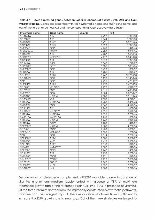
Table 4.7 | Over-expressed genes between IMX2210 chemostat cultures with SMD and SMD without vitamins. Genes are presented with their systematic name and their gene name and log2 of the fold change (log2FC) and the corresponding False Discovery Rate (FDR).
Systematic name Gene name Log2FC FDR YGR144W THI4 7.497 0.00E+00 YFL058W THI5 6.064 0.00E+00 YNL332W THI12 5.421 3.22E-275 YDL244W THI13 5.023 0.00E+00 YNR056C BIO5 4.740 1.49E-65 YER060W-A FCY22 4.408 3.80E-36 YJR156C THI11 4.287 1.36E-215 YCR102C YCR102C 4.177 3.94E-120 YBR240C THI2 3.670 0.00E+00 YPL265W DIP5 3.562 1.24E-27 YCR020C PET18 3.536 1.38E-236 YNR058W BIO3 3.423 1.05E-82 YLR237W THI7 3.285 1.89E-77 YOL055C THI20 3.247 3.19E-280 YGR286C BIO2 3.128 2.14E-140 BIO6 BIO6 2.996 1.62E-139 YLR004C THI73 2.973 7.62E-153 YEL073C YEL073C 2.929 6.31E-97 YFL059W SNZ3 2.815 5.40E-100 YNR057C BIO4 2.604 3.02E-142 YNL331C AAD14 2.589 2.21E-145 YGR065C VHT1 2.538 4.22E-91 YJR157W YJR157W 2.482 8.40E-43 YNL333W SNZ2 2.348 4.52E-56 YPL214C THI6 2.140 1.65E-98 YJL213W YJL213W 2.095 8.67E-38 YJR154W YJR154W 2.095 3.23E-66 YMR317W YMR317W 1.759 1.83E-03 YJR155W AAD10 1.740 5.32E-70 YLR134W PDC5 1.700 1.41E-29 YDR380W ARO10 1.608 4.75E-04 YFL060C SNO3 1.603 5.03E-31 YDR541C YDR541C 1.530 1.95E-38 BIO1 BIO1 1.514 2.98E-15 YDR438W THI74 1.506 3.86E-77 YKR053C YSR3 1.413 3.53E-04 YPR121W THI22 1.350 1.81E-35 YIL169C YAR068W 1.307 1.09E-06 YDR508C GNP1 1.267 1.43E-58 YPL258C THI21 1.239 5.95E-14 YNL334C SNO2 1.221 1.04E-04 YGL263W COS12 1.122 7.88E-08 YEL029C BUD16 1.047 2.73E-18 YLR089C ALT1 1.037 7.78E-37 YGR287C IMA1 1.009 9.27E-16
Despite an incomplete gene complement, IMX2210 was able to grow in absence of vitamins in a mineral medium supplemented with glucose at 78% of maximum theoretical growth rate of the reference strain CEN.PK113-7D in presence of vitamins. Of the three vitamins derived from the improperly constructed biosynthetic pathways, thiamine had the strongest impact. The sole addition of vitamin B1 was sufficient to increase IMX2210 growth rate to near μmax. Out of the three strategies envisaged to
eliminate thiamine dependency in S. cerevisiae, the most promising ones involved the overexpression of THI4 whose translational product acts as sulfur source for thiazole formation in a single turnover reaction. The transcriptome analysis of IMX2210 revealed that THI4 was massively upregulated in absence of thiamine already, being the top expressed gene in IMX2210 (Table 4.7, Figure 4.7). This would suggest that further alleviation of thiamine dependency for growth would require an even higher increase of the THI4 transcription. This could be achieved by increasing the native THI4 expression cassettes copy number or combining THI4 expression with the CCW12 promoter that was used to drive the overexpression of SNZ1 in IMX2210, being the most expressed gene in IMX2210 (Supplemental data). In both situations, we cannot exclude that the reuse of such a strong promoter might in the end affect the expression of the respective native gene resulting in titration of specific transcription factors and consequently affect the strain fitness. Alteration of expression of CCW12, which is involved in the structure and maintenance of the cell wall, has already been reported to cause reduction of growth rate [398, 399]. Alternatively, synthetic regulatory sequences generated by a machine learning approach derived from natural genomic data that demonstrated high transcriptional activity without disturbing cell growth [399], might represent a workable solution.
The ability to grow the S. cerevisiae strain IMX2210 in absence of vitamins offered a unique opportunity to evaluate the transcriptional regulation of the genes involved in class-B vitamin biosynthesis. Distinct from the role of the transcriptional regulator Vhr1 in the transcriptional activation of the VHT1 and BIO5 genes that encodes a biotin and a 7-keto-8-aminopelargonic acid (KAPA) transporters respectively, little is known about the regulation of the biotin genes in S. cerevisiae [157]. The regulation of VHT1 and BIO5 by Vhr1 occurs through a cis-regulatory motif (AATCAn8TGAYT) and it is dependent on the concentration of biotin in the medium. The transcriptional activity of the VHT1 promoter was maximal at biotin concentrations ranging from 0.05 to 0.5 μg L-1, concentration at least hundred-fold lower than the biotin concentration used in SMD medium (50 μg L-1). Our results were in line with this previous report, although necessary to grow on SMD, VHT1 and BIO5 showed low expression in CEN.PK113-7D and IMX2210 in this condition. The expression of the two transporter genes along the other biotin biosynthetic genes (BIO1, 2, 3, 4 and 6) (Figure 4.1) were highly upregulated in vitamin-free conditions (Figure 4.7, Supplemental data). Search in regulatory sequences of these genes confirmed occurrence of the Vhr1 cis regulatory motif in VHT1 and also revealed such occurrence in BIO2, BIO1 and BIO6 promoters. No such motif could be found in the 52-bp BIO5 promoter conversely to what has been previously suggested [157], as well as in the 220-bp bidirectional BIO4-BIO3 promoters. Analysis using of extended sequences including the coding of the physically clustered BIO5, BIO4 and BIO3 genes identified a motif within BIO4 open reading frame that might serve for the regulation of the three contiguous genes. This peculiar organization and the role of this regulatory motif in the regulation of these genes will have to be further explored. The absence of obvious Vhr1 cis-regulatory sequence might also indicate that Vhr1 might not be the only transcriptional activator
124 | Chapter 4

Table 4.7 | Over-expressed genes between IMX2210 chemostat cultures with SMD and SMD without vitamins. Genes are presented with their systematic name and their gene name and log2 of the fold change (log2FC) and the corresponding False Discovery Rate (FDR).
Systematic name Gene name Log2FC FDR YGR144W THI4 7.497 0.00E+00 YFL058W THI5 6.064 0.00E+00 YNL332W THI12 5.421 3.22E-275 YDL244W THI13 5.023 0.00E+00 YNR056C BIO5 4.740 1.49E-65 YER060W-A FCY22 4.408 3.80E-36 YJR156C THI11 4.287 1.36E-215 YCR102C YCR102C 4.177 3.94E-120 YBR240C THI2 3.670 0.00E+00 YPL265W DIP5 3.562 1.24E-27 YCR020C PET18 3.536 1.38E-236 YNR058W BIO3 3.423 1.05E-82 YLR237W THI7 3.285 1.89E-77 YOL055C THI20 3.247 3.19E-280 YGR286C BIO2 3.128 2.14E-140 BIO6 BIO6 2.996 1.62E-139 YLR004C THI73 2.973 7.62E-153 YEL073C YEL073C 2.929 6.31E-97 YFL059W SNZ3 2.815 5.40E-100 YNR057C BIO4 2.604 3.02E-142 YNL331C AAD14 2.589 2.21E-145 YGR065C VHT1 2.538 4.22E-91 YJR157W YJR157W 2.482 8.40E-43 YNL333W SNZ2 2.348 4.52E-56 YPL214C THI6 2.140 1.65E-98 YJL213W YJL213W 2.095 8.67E-38 YJR154W YJR154W 2.095 3.23E-66 YMR317W YMR317W 1.759 1.83E-03 YJR155W AAD10 1.740 5.32E-70 YLR134W PDC5 1.700 1.41E-29 YDR380W ARO10 1.608 4.75E-04 YFL060C SNO3 1.603 5.03E-31 YDR541C YDR541C 1.530 1.95E-38 BIO1 BIO1 1.514 2.98E-15 YDR438W THI74 1.506 3.86E-77 YKR053C YSR3 1.413 3.53E-04 YPR121W THI22 1.350 1.81E-35 YIL169C YAR068W 1.307 1.09E-06 YDR508C GNP1 1.267 1.43E-58 YPL258C THI21 1.239 5.95E-14 YNL334C SNO2 1.221 1.04E-04 YGL263W COS12 1.122 7.88E-08 YEL029C BUD16 1.047 2.73E-18 YLR089C ALT1 1.037 7.78E-37 YGR287C IMA1 1.009 9.27E-16
Despite an incomplete gene complement, IMX2210 was able to grow in absence of vitamins in a mineral medium supplemented with glucose at 78% of maximum theoretical growth rate of the reference strain CEN.PK113-7D in presence of vitamins. Of the three vitamins derived from the improperly constructed biosynthetic pathways, thiamine had the strongest impact. The sole addition of vitamin B1 was sufficient to increase IMX2210 growth rate to near μmax. Out of the three strategies envisaged to
eliminate thiamine dependency in S. cerevisiae, the most promising ones involved the overexpression of THI4 whose translational product acts as sulfur source for thiazole formation in a single turnover reaction. The transcriptome analysis of IMX2210 revealed that THI4 was massively upregulated in absence of thiamine already, being the top expressed gene in IMX2210 (Table 4.7, Figure 4.7). This would suggest that further alleviation of thiamine dependency for growth would require an even higher increase of the THI4 transcription. This could be achieved by increasing the native THI4 expression cassettes copy number or combining THI4 expression with the CCW12 promoter that was used to drive the overexpression of SNZ1 in IMX2210, being the most expressed gene in IMX2210 (Supplemental data). In both situations, we cannot exclude that the reuse of such a strong promoter might in the end affect the expression of the respective native gene resulting in titration of specific transcription factors and consequently affect the strain fitness. Alteration of expression of CCW12, which is involved in the structure and maintenance of the cell wall, has already been reported to cause reduction of growth rate [398, 399]. Alternatively, synthetic regulatory sequences generated by a machine learning approach derived from natural genomic data that demonstrated high transcriptional activity without disturbing cell growth [399], might represent a workable solution.
The ability to grow the S. cerevisiae strain IMX2210 in absence of vitamins offered a unique opportunity to evaluate the transcriptional regulation of the genes involved in class-B vitamin biosynthesis. Distinct from the role of the transcriptional regulator Vhr1 in the transcriptional activation of the VHT1 and BIO5 genes that encodes a biotin and a 7-keto-8-aminopelargonic acid (KAPA) transporters respectively, little is known about the regulation of the biotin genes in S. cerevisiae [157]. The regulation of VHT1 and BIO5 by Vhr1 occurs through a cis-regulatory motif (AATCAn8TGAYT) and it is dependent on the concentration of biotin in the medium. The transcriptional activity of the VHT1 promoter was maximal at biotin concentrations ranging from 0.05 to 0.5 μg L-1, concentration at least hundred-fold lower than the biotin concentration used in SMD medium (50 μg L-1). Our results were in line with this previous report, although necessary to grow on SMD, VHT1 and BIO5 showed low expression in CEN.PK113-7D and IMX2210 in this condition. The expression of the two transporter genes along the other biotin biosynthetic genes (BIO1, 2, 3, 4 and 6) (Figure 4.1) were highly upregulated in vitamin-free conditions (Figure 4.7, Supplemental data). Search in regulatory sequences of these genes confirmed occurrence of the Vhr1 cis regulatory motif in VHT1 and also revealed such occurrence in BIO2, BIO1 and BIO6 promoters. No such motif could be found in the 52-bp BIO5 promoter conversely to what has been previously suggested [157], as well as in the 220-bp bidirectional BIO4-BIO3 promoters. Analysis using of extended sequences including the coding of the physically clustered BIO5, BIO4 and BIO3 genes identified a motif within BIO4 open reading frame that might serve for the regulation of the three contiguous genes. This peculiar organization and the role of this regulatory motif in the regulation of these genes will have to be further explored. The absence of obvious Vhr1 cis-regulatory sequence might also indicate that Vhr1 might not be the only transcriptional activator
4
Discussion | 125
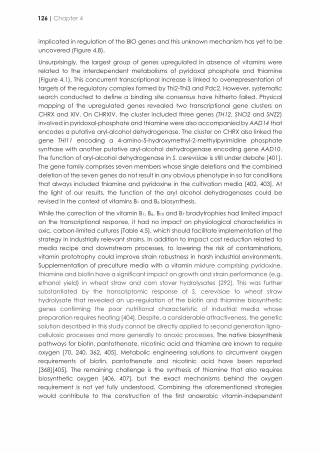
implicated in regulation of the BIO genes and this unknown mechanism has yet to be uncovered (Figure 4.8).
Unsurprisingly, the largest group of genes upregulated in absence of vitamins were related to the interdependent metabolisms of pyridoxal phosphate and thiamine (Figure 4.1). This concurrent transcriptional increase is linked to overrepresentation of targets of the regulatory complex formed by Thi2-Thi3 and Pdc2. However, systematic search conducted to define a binding site consensus have hitherto failed. Physical mapping of the upregulated genes revealed two transcriptional gene clusters on CHRX and XIV. On CHRXIV, the cluster included three genes (TH12, SNO2 and SNZ2) involved in pyridoxal-phosphate and thiamine were also accompanied by AAD14 that encodes a putative aryl-alcohol dehydrogenase. The cluster on CHRX also linked the gene THI11 encoding a 4-amino-5-hydroxymethyl-2-methylpyrimidine phosphate synthase with another putative aryl-alcohol dehydrogenase encoding gene AAD10. The function of aryl-alcohol dehydrogenase in S. cerevisiae is still under debate [401]. The gene family comprises seven members whose single deletions and the combined deletion of the seven genes do not result in any obvious phenotype in so far conditions that always included thiamine and pyridoxine in the cultivation media [402, 403]. At the light of our results, the function of the aryl alcohol dehydrogenases could be revised in the context of vitamins B1 and B6 biosynthesis.
While the correction of the vitamin B1, B6, B10 and B7 bradytrophies had limited impact on the transcriptional response, it had no impact on physiological characteristics in oxic, carbon-limited cultures (Table 4.5), which should facilitate implementation of the strategy in industrially relevant strains. In addition to impact cost reduction related to media recipe and downstream processes, to lowering the risk of contaminations, vitamin prototrophy could improve strain robustness in harsh industrial environments. Supplementation of preculture media with a vitamin mixture comprising pyridoxine, thiamine and biotin have a significant impact on growth and strain performance (e.g. ethanol yield) in wheat straw and corn stover hydrolysates [292]. This was further substantiated by the transcriptomic response of S. cerevisiae to wheat straw hydrolysate that revealed an up-regulation of the biotin and thiamine biosynthetic genes confirming the poor nutritional characteristic of industrial media whose preparation requires heating [404]. Despite, a considerable attractiveness, the genetic solution described in this study cannot be directly applied to second generation ligno-cellulosic processes and more generally to anoxic processes. The native biosynthesis pathways for biotin, pantothenate, nicotinic acid and thiamine are known to require oxygen [70, 240, 362, 405]. Metabolic engineering solutions to circumvent oxygen requirements of biotin, pantothenate and nicotinic acid have been reported [368][405]. The remaining challenge is the synthesis of thiamine that also requires biosynthetic oxygen [406, 407], but the exact mechanisms behind the oxygen requirement is not yet fully understood. Combining the aforementioned strategies would contribute to the construction of the first anaerobic vitamin-independent
S. cerevisiae strain, that in a near future might prove to be a key phenotypic characteristic of industrial strains.
Figure 4.8 | New insight in regulation of the biotin and thiamine biosynthetic genes. (A) Position of Vhr1 putative cis regulatory sequence (AATCAn8TGAYT) in BIO locus. The promoter sequences are represented in grey and the coding sequences in black. The AATCAn8TGAYT motif is indicated with a dark blue arrow. Downwards arrows indicate motif located on the (+) strand conversely upwards arrows indicate motif on the (-) strand. Search for motif in the BIO loci was performed using the regulatory sequence analysis tools (RSAT; http://rsat.sb-roscoff.fr/; [400]) using the pattern matching function. (B) Co-expression of genes surrounding THI11 and TH12 on CHRX and XIV, respectively. The bar graphs represent the normalized transcript expression of the strains IMX2210 grown in absence of class B vitamins (black), in presence of class B vitamins (white) and CEN.PK113-7D grown in presence of vitamins (grey).
126 | Chapter 4
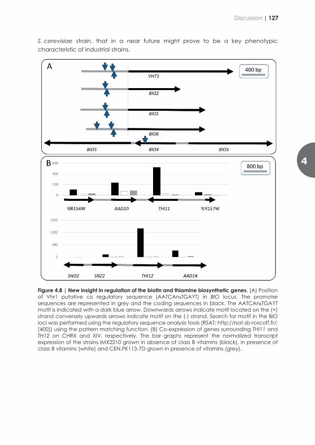
implicated in regulation of the BIO genes and this unknown mechanism has yet to be uncovered (Figure 4.8).
Unsurprisingly, the largest group of genes upregulated in absence of vitamins were related to the interdependent metabolisms of pyridoxal phosphate and thiamine (Figure 4.1). This concurrent transcriptional increase is linked to overrepresentation of targets of the regulatory complex formed by Thi2-Thi3 and Pdc2. However, systematic search conducted to define a binding site consensus have hitherto failed. Physical mapping of the upregulated genes revealed two transcriptional gene clusters on CHRX and XIV. On CHRXIV, the cluster included three genes (TH12, SNO2 and SNZ2) involved in pyridoxal-phosphate and thiamine were also accompanied by AAD14 that encodes a putative aryl-alcohol dehydrogenase. The cluster on CHRX also linked the gene THI11 encoding a 4-amino-5-hydroxymethyl-2-methylpyrimidine phosphate synthase with another putative aryl-alcohol dehydrogenase encoding gene AAD10. The function of aryl-alcohol dehydrogenase in S. cerevisiae is still under debate [401]. The gene family comprises seven members whose single deletions and the combined deletion of the seven genes do not result in any obvious phenotype in so far conditions that always included thiamine and pyridoxine in the cultivation media [402, 403]. At the light of our results, the function of the aryl alcohol dehydrogenases could be revised in the context of vitamins B1 and B6 biosynthesis.
While the correction of the vitamin B1, B6, B10 and B7 bradytrophies had limited impact on the transcriptional response, it had no impact on physiological characteristics in oxic, carbon-limited cultures (Table 4.5), which should facilitate implementation of the strategy in industrially relevant strains. In addition to impact cost reduction related to media recipe and downstream processes, to lowering the risk of contaminations, vitamin prototrophy could improve strain robustness in harsh industrial environments. Supplementation of preculture media with a vitamin mixture comprising pyridoxine, thiamine and biotin have a significant impact on growth and strain performance (e.g. ethanol yield) in wheat straw and corn stover hydrolysates [292]. This was further substantiated by the transcriptomic response of S. cerevisiae to wheat straw hydrolysate that revealed an up-regulation of the biotin and thiamine biosynthetic genes confirming the poor nutritional characteristic of industrial media whose preparation requires heating [404]. Despite, a considerable attractiveness, the genetic solution described in this study cannot be directly applied to second generation ligno-cellulosic processes and more generally to anoxic processes. The native biosynthesis pathways for biotin, pantothenate, nicotinic acid and thiamine are known to require oxygen [70, 240, 362, 405]. Metabolic engineering solutions to circumvent oxygen requirements of biotin, pantothenate and nicotinic acid have been reported [368][405]. The remaining challenge is the synthesis of thiamine that also requires biosynthetic oxygen [406, 407], but the exact mechanisms behind the oxygen requirement is not yet fully understood. Combining the aforementioned strategies would contribute to the construction of the first anaerobic vitamin-independent
S. cerevisiae strain, that in a near future might prove to be a key phenotypic characteristic of industrial strains.
Figure 4.8 | New insight in regulation of the biotin and thiamine biosynthetic genes. (A) Position of Vhr1 putative cis regulatory sequence (AATCAn8TGAYT) in BIO locus. The promoter sequences are represented in grey and the coding sequences in black. The AATCAn8TGAYT motif is indicated with a dark blue arrow. Downwards arrows indicate motif located on the (+) strand conversely upwards arrows indicate motif on the (-) strand. Search for motif in the BIO loci was performed using the regulatory sequence analysis tools (RSAT; http://rsat.sb-roscoff.fr/; [400]) using the pattern matching function. (B) Co-expression of genes surrounding THI11 and TH12 on CHRX and XIV, respectively. The bar graphs represent the normalized transcript expression of the strains IMX2210 grown in absence of class B vitamins (black), in presence of class B vitamins (white) and CEN.PK113-7D grown in presence of vitamins (grey).
4
Discussion | 127

Acknowledgments
We thank Dewi P.I. Moonen and Ellen Geraats who constructed and characterized strains used in this study. This work was supported by the European Union’s Horizon 2020 research and innovation programme under the Marie Skłodowska-Curie action PAcMEN (grant agreement No 722287). J.T.P. acknowledges support by an Advanced Grant of the European Research Council (grant No. 694633). Authors have no competing interests to declare.
Author contributions
AKW, TP and JMD designed the experiments and wrote a first version of the manuscript. All authors critically read this version, provided input and approved the final version. AKW, TP, constructed the yeast strains and performed shake flask culture characterization. EFAdH, AKW and TP performed the bioreactor experiments. MvdB, TP, and AKW prepared RNA libraries and analysed the transcriptome data.
128 | Chapter 4

Acknowledgments
We thank Dewi P.I. Moonen and Ellen Geraats who constructed and characterized strains used in this study. This work was supported by the European Union’s Horizon 2020 research and innovation programme under the Marie Skłodowska-Curie action PAcMEN (grant agreement No 722287). J.T.P. acknowledges support by an Advanced Grant of the European Research Council (grant No. 694633). Authors have no competing interests to declare.
Author contributions
AKW, TP and JMD designed the experiments and wrote a first version of the manuscript. All authors critically read this version, provided input and approved the final version. AKW, TP, constructed the yeast strains and performed shake flask culture characterization. EFAdH, AKW and TP performed the bioreactor experiments. MvdB, TP, and AKW prepared RNA libraries and analysed the transcriptome data.
4
Acknowledgments | 129

Outlook Biotechnology and genetic engineering have helped to overcome many challenges of our time. During this study, answers to challenges faced in the field of energy and health could be identified. The insights might serve as an example for solving problems in the future.
To the general public, the most visible demonstrations of the potential of biotechnological processes and products based on genetic modification are probably related to medical applications, which range from the successful replacement of slaughterhouse-derived porcine insulin by safe, body-identical recombinant human albumin to the fast development and roll-out of Covid-19 vaccines. In the coming decades, a different and even more important contribution of biotechnology is needed to enable a swift transition of our society and economy to low-carbon-emission, circular processes for production of chemicals, transport fuels and food. Current projections for replacing fossil-based fuels for road transport, shipping and aviation by sustainable, low-carbon-emission alternatives all include a significant contribution of microbial biofuels production, such as yeast-based bioethanol production. At the same time, biotechnology is expected to play a major role in the replacement of fossil-based bulk and fine chemicals by microbially production of the same compounds or of functional analogs. Especially until the environmental and economic costs of using fossil feedstocks are included in consumer prices, cell factories for production for most of these products need to become more cost-effective to be able to compete with their established, cheap fossil-based counterparts. A lower price of growth media, as well as improved stability, simplicity and scalability of processes by elimination of growth-factor requirements, which was the subject of the research described in this study, can contribute to driving down costs of production.
Current yeast-based biotechnological processes predominantly use sugars, derived from agriculture, as feedstocks. Processes for production of high-added-value compounds generally use high-purity glucose or sucrose as carbon source, chemically defined media and are performed in aerobic fed-batch cultures. In such cultures, eliminating of individual vitamins from media, e.g., by using biotin-prototrophic yeast strains (Chapter 2) or strains that are completely prototrophic (Chapter 4) simplifies medium composition and reduces costs. Moreover, it can reduce risk of contamination with auxotrophic ‘wild’ yeast of bacteria. In the case of biotin, it is particularly interesting to investigate whether this approach can also confer advantages for processes that require high activities of biotin dependent enzymes, such as yeast-based production of C4-dicarboxylic acids with pyruvate carboxylase or processes that use urea as nitrogen source with the aid of urease.
To meet future demands for bulk products of engineered yeast strains, such as ethanol and isobutanol, feedstock use should move from glucose and sucrose to hydrolysates
130 |
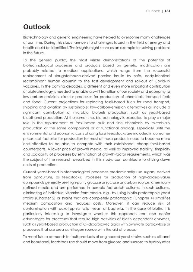
Outlook Biotechnology and genetic engineering have helped to overcome many challenges of our time. During this study, answers to challenges faced in the field of energy and health could be identified. The insights might serve as an example for solving problems in the future.
To the general public, the most visible demonstrations of the potential of biotechnological processes and products based on genetic modification are probably related to medical applications, which range from the successful replacement of slaughterhouse-derived porcine insulin by safe, body-identical recombinant human albumin to the fast development and roll-out of Covid-19 vaccines. In the coming decades, a different and even more important contribution of biotechnology is needed to enable a swift transition of our society and economy to low-carbon-emission, circular processes for production of chemicals, transport fuels and food. Current projections for replacing fossil-based fuels for road transport, shipping and aviation by sustainable, low-carbon-emission alternatives all include a significant contribution of microbial biofuels production, such as yeast-based bioethanol production. At the same time, biotechnology is expected to play a major role in the replacement of fossil-based bulk and fine chemicals by microbially production of the same compounds or of functional analogs. Especially until the environmental and economic costs of using fossil feedstocks are included in consumer prices, cell factories for production for most of these products need to become more cost-effective to be able to compete with their established, cheap fossil-based counterparts. A lower price of growth media, as well as improved stability, simplicity and scalability of processes by elimination of growth-factor requirements, which was the subject of the research described in this study, can contribute to driving down costs of production.
Current yeast-based biotechnological processes predominantly use sugars, derived from agriculture, as feedstocks. Processes for production of high-added-value compounds generally use high-purity glucose or sucrose as carbon source, chemically defined media and are performed in aerobic fed-batch cultures. In such cultures, eliminating of individual vitamins from media, e.g., by using biotin-prototrophic yeast strains (Chapter 2) or strains that are completely prototrophic (Chapter 4) simplifies medium composition and reduces costs. Moreover, it can reduce risk of contamination with auxotrophic ‘wild’ yeast of bacteria. In the case of biotin, it is particularly interesting to investigate whether this approach can also confer advantages for processes that require high activities of biotin dependent enzymes, such as yeast-based production of C4-dicarboxylic acids with pyruvate carboxylase or processes that use urea as nitrogen source with the aid of urease.
To meet future demands for bulk products of engineered yeast strains, such as ethanol and isobutanol, feedstock use should move from glucose and sucrose to hydrolysates
Outlook | 131
of lignocellulosic biomass derived from ‘energy crops’ or non-food agricultural residues. Composition of these feedstocks can vary based on plant species, season, location and storage, resulting in variable contents of growth factors. Moreover, deconstruction of plant biomass required to release fermentable sugars from such feedstocks can involve high temperatures and extreme pH values which may inactivate vitamins [408-410]. In addition, some vitamins can also be chemically inactivated, as exemplified by biotin, which can be inactivated by exposure to aldehydes. Vitamin starvation has been proven to negatively affect ethanol yields and cell viability and be the cause of ‘stuck’ fermentations in a nitrogen-source dependent manner [296]. Usage of corn steep liquor as a source of vitamins, as an inexpensive alternative to yeast extract [411], in some cases still results in a requirement for addition of yeast extract and peptone as sources of vitamins to obtain highest ethanol production [412]. In addition to increasing costs, such complex medium components can also lead to increased risk of contamination with, for example, lactic acid bacteria [258]. In view of the inherent variability of lignocellulosic (‘second generation’) feedstocks, experimental analysis across a range of feedstocks and hydrolysis procedures is required to assess the impact of the metabolic engineering strategies presented in Chapters 3 and 4. Another process that could benefit from medium simplification is the production of yeast extracts for animal feed applications, which requires addition of vitamins in the cell propagation phase.
Further optimization of biotin prototrophic S. cerevisiae strains, as developed in Chapter 2 and 3, might serve as an alternative for chemical synthesis of biotin as a vitamin and feed additive. Its current synthesis uses crude oil for production of the raw materials following the method developed by Goldberg and Steinbach, and is therefore not considered to be sustainable. Biotin-producing microbes can provide a more sustainable way of biotin production, since carbon dioxide, chemical waste emissions as well as air pollution and water usage are expected to be considerably lower than for chemical biotin synthesis. Sourcing from a biological process might, moreover, be preferred by end-consumers if the biotin is added to food and cosmetic products. New microbial biotin production processes conducted locally for the European food and health market (‘onshoring’), might increase the traceability and end-consumer acceptance of biotin and related-products. Research efforts to achieve microbial biotin production processes using Pseudomonas, Escherichia coli or Corynebacterium glutamicum as well as Bifidobacteria prove the interest in a sustainable solution for biotin production. However, these processes did not yet reach cost-competitiveness with chemical synthesis. Naturally occurring, fast-growing biotin-prototrophic yeasts such as Cyberlindnera fabianii (Chapter 2) may provide an interesting starting point for classical strain-improvement and/or genome-editing programmes to increase biotin content.
Awareness about the importance of the quality of food products is increasing: “The nutritional content of what we eat determines the composition of our cell membranes, bone marrow, blood, hormones, tissue, organs, skin, hair. Our bodies are replacing
billions of cells every day — and using the foods we consume as the source”, as stated by the Brain World Magazine in January 2018. Although application of genetically modified yeasts for food products might encounter challenges related to consumer acceptance, vitamin-producing yeasts would offer interesting nutritional characteristics. Similarly to Golden rice, a genetically engineered variety of Oryza sativa that produces β-carotene intended to be grown and consumed as fortified food in areas with a shortage of dietary vitamin A [413], access to yeasts or yeast-fermented products enriched in vitamins as such as vitamin B9 (folate) could contribute to diet fortification. Vitamin B9 deficiency causes anaemia in adults and when occurring during pregnancy it causes neural tube defects of the new-born [414, 415]. Addition of vitamins D and B12 to milk is another example for fortified foods to prevent vitamin deficiencies. Nutritionally enhanced yeast and yeast extracts generated from vitamin-overproducing strains could play an important role as fortified food platforms [416, 417]. With the current rapid rise of consumer interest in alternative protein sources to replace animal protein, yeast biomass (‘single cell protein’) is becoming an interesting option as protein-rich food ingredient. In addition to the use of dedicated yeast strains as, enhance vitamin contents of yeast biomass could also increase the nutritional value of excess yeast produced by the brewing and bioethanol industries [361] and thereby improve its monetary value as an animal food ingredient.
In addition to providing options for industrial application, the research described in this thesis raises a number of fundamental scientific questions that can be pursued in further research. The most urgent question, arising from the research described in Chapter 2, is the identification of the reaction catalysed by Bio1 orthologs in yeast. Purification of Bio1, followed by in vitro incubation with acyl-CoA esters of different lengths and identification of products by mass spectrometry, NMR and/or other methods may resolve whether this enzyme indeed, as postulated in Chapter 2, catalysis an oxidative cleavage of an acyl-CoA ester that directly leads to formation of pimeloyl-CoA. In addition, it would be highly interesting to investigate why, out of a sample of five different Bio1 orthologs from biotin-prototrophic yeast species, only the C. fabianii enzyme conferred biotin prototrophy to S. cerevisiae. Availability of prototrophic S. cerevisiae strains also raises interesting questions about the context dependency of their prototrophies. This question not only refers to the availability of oxygen (Chapter 2) but also to possible increase vitamin conditions under conditions that require a high activity of enzymes whose activity involves a specific cofactor. In addition, quantitative physiological research on such strains may resolve trade-offs between prototrophy and fitness, for example related to resource allocation in strains in which specific enzymes involved in vitamin biosynthesis need to be overexpressed, with the single-turnover Thi4 and Thi5 enzymes of S. cerevisiae as interesting model systems.
132 | Outlook
of lignocellulosic biomass derived from ‘energy crops’ or non-food agricultural residues. Composition of these feedstocks can vary based on plant species, season, location and storage, resulting in variable contents of growth factors. Moreover, deconstruction of plant biomass required to release fermentable sugars from such feedstocks can involve high temperatures and extreme pH values which may inactivate vitamins [408-410]. In addition, some vitamins can also be chemically inactivated, as exemplified by biotin, which can be inactivated by exposure to aldehydes. Vitamin starvation has been proven to negatively affect ethanol yields and cell viability and be the cause of ‘stuck’ fermentations in a nitrogen-source dependent manner [296]. Usage of corn steep liquor as a source of vitamins, as an inexpensive alternative to yeast extract [411], in some cases still results in a requirement for addition of yeast extract and peptone as sources of vitamins to obtain highest ethanol production [412]. In addition to increasing costs, such complex medium components can also lead to increased risk of contamination with, for example, lactic acid bacteria [258]. In view of the inherent variability of lignocellulosic (‘second generation’) feedstocks, experimental analysis across a range of feedstocks and hydrolysis procedures is required to assess the impact of the metabolic engineering strategies presented in Chapters 3 and 4. Another process that could benefit from medium simplification is the production of yeast extracts for animal feed applications, which requires addition of vitamins in the cell propagation phase.
Further optimization of biotin prototrophic S. cerevisiae strains, as developed in Chapter 2 and 3, might serve as an alternative for chemical synthesis of biotin as a vitamin and feed additive. Its current synthesis uses crude oil for production of the raw materials following the method developed by Goldberg and Steinbach, and is therefore not considered to be sustainable. Biotin-producing microbes can provide a more sustainable way of biotin production, since carbon dioxide, chemical waste emissions as well as air pollution and water usage are expected to be considerably lower than for chemical biotin synthesis. Sourcing from a biological process might, moreover, be preferred by end-consumers if the biotin is added to food and cosmetic products. New microbial biotin production processes conducted locally for the European food and health market (‘onshoring’), might increase the traceability and end-consumer acceptance of biotin and related-products. Research efforts to achieve microbial biotin production processes using Pseudomonas, Escherichia coli or Corynebacterium glutamicum as well as Bifidobacteria prove the interest in a sustainable solution for biotin production. However, these processes did not yet reach cost-competitiveness with chemical synthesis. Naturally occurring, fast-growing biotin-prototrophic yeasts such as Cyberlindnera fabianii (Chapter 2) may provide an interesting starting point for classical strain-improvement and/or genome-editing programmes to increase biotin content.
Awareness about the importance of the quality of food products is increasing: “The nutritional content of what we eat determines the composition of our cell membranes, bone marrow, blood, hormones, tissue, organs, skin, hair. Our bodies are replacing
billions of cells every day — and using the foods we consume as the source”, as stated by the Brain World Magazine in January 2018. Although application of genetically modified yeasts for food products might encounter challenges related to consumer acceptance, vitamin-producing yeasts would offer interesting nutritional characteristics. Similarly to Golden rice, a genetically engineered variety of Oryza sativa that produces β-carotene intended to be grown and consumed as fortified food in areas with a shortage of dietary vitamin A [413], access to yeasts or yeast-fermented products enriched in vitamins as such as vitamin B9 (folate) could contribute to diet fortification. Vitamin B9 deficiency causes anaemia in adults and when occurring during pregnancy it causes neural tube defects of the new-born [414, 415]. Addition of vitamins D and B12 to milk is another example for fortified foods to prevent vitamin deficiencies. Nutritionally enhanced yeast and yeast extracts generated from vitamin-overproducing strains could play an important role as fortified food platforms [416, 417]. With the current rapid rise of consumer interest in alternative protein sources to replace animal protein, yeast biomass (‘single cell protein’) is becoming an interesting option as protein-rich food ingredient. In addition to the use of dedicated yeast strains as, enhance vitamin contents of yeast biomass could also increase the nutritional value of excess yeast produced by the brewing and bioethanol industries [361] and thereby improve its monetary value as an animal food ingredient.
In addition to providing options for industrial application, the research described in this thesis raises a number of fundamental scientific questions that can be pursued in further research. The most urgent question, arising from the research described in Chapter 2, is the identification of the reaction catalysed by Bio1 orthologs in yeast. Purification of Bio1, followed by in vitro incubation with acyl-CoA esters of different lengths and identification of products by mass spectrometry, NMR and/or other methods may resolve whether this enzyme indeed, as postulated in Chapter 2, catalysis an oxidative cleavage of an acyl-CoA ester that directly leads to formation of pimeloyl-CoA. In addition, it would be highly interesting to investigate why, out of a sample of five different Bio1 orthologs from biotin-prototrophic yeast species, only the C. fabianii enzyme conferred biotin prototrophy to S. cerevisiae. Availability of prototrophic S. cerevisiae strains also raises interesting questions about the context dependency of their prototrophies. This question not only refers to the availability of oxygen (Chapter 2) but also to possible increase vitamin conditions under conditions that require a high activity of enzymes whose activity involves a specific cofactor. In addition, quantitative physiological research on such strains may resolve trade-offs between prototrophy and fitness, for example related to resource allocation in strains in which specific enzymes involved in vitamin biosynthesis need to be overexpressed, with the single-turnover Thi4 and Thi5 enzymes of S. cerevisiae as interesting model systems.
Outlook | 133

Bibliography 1. Gowland Hopkins, F., The analyst and the medical man. Analyst, 1906. 31: p. 385b-404. 2. Funk, C., The vitamines. 2nd ed, ed. D.H. E. 1922, Baltimore: Williams & Wilkins Company. 3. Combs, G.F., The vitamins : fundamental aspects in nutrition and health. 3 ed. 2008: Elsevier Academic Press.
583. 4. Potvin, J., et al., An automatic turbidimetric method to screen yeast extracts as fermentation nutrient
ingredients. Journal of Microbiological Methods, 1997. 29(3): p. 153-160. 5. Zhang, J., et al., Toward consistent and productive complex media for industrial fermentations: studies on
yeast extract for a recombinant yeast fermentation process. Biotechnology and Bioengineering, 2003. 82(6): p. 640-52.
6. Dahod, S.K., R. Greasham, and M. Kennedy, Raw materials selection and medium development for industrial fermentation processes. Manual of Industrial Microbiology and Biotechnology, Third Edition, 2010: p. 659-668.
7. Burkholder, P.R., I. McVeigh, and D. Moyer, Studies on some growth factors of yeasts. Journal of Bacteriology, 1944. 48(4): p. 385-91.
8. Cutts, N.S. and C. Rainbow, Studies of a yeast exacting towards para-aminobenzoic acid. Journal of General Microbiology, 1950. 4(2): p. 150-5.
9. Wickerham, L.J., Taxonomy of yeasts. Technical Bulletin U.S. Dept. Agriculture, Washington DC, 1951. 1029. 10. Sherman, F., G.R. Fink, and J. Hicks, Methods in yeast genetics: A laboratory course manual. 1987, Cold Spring
Harbor: Cold Spring Harbor Laboratory Press. 11. Wickerham, L.J., A critical evaluation of the nitrogen assimilation tests as commonly used in the classification
of the yeasts. Journal of Bacteriology, 1946. 51(5): p. 6-6. 12. Bruinenberg, P.M., J.P. van Dijken, and W.A. Scheffers, An enzymic analysis of NADPH production and
consumption in Candida utilis. Journal of General Microbiology, 1983. 129(4): p. 965-71. 13. Verduyn, C., et al., Effect of benzoic acid on metabolic fluxes in yeasts: a continuous-culture study on the
regulation of respiration and alcoholic fermentation. Yeast, 1992. 8(7): p. 501-17. 14. Brachmann, C.B., et al., Designer deletion strains derived from Saccharomyces cerevisiae S288C: a useful set
of strains and plasmids for PCR-mediated gene disruption and other applications. Yeast, 1998. 14(2): p. 115-32.
15. Hanscho, M., et al., Nutritional requirements of the BY series of Saccharomyces cerevisiae strains for optimum growth. Fems Yeast Research, 2012. 12(7): p. 796-808.
16. Emery, W.B., N. McLeod, and F.A. Robinson, Comparative microbiological assays of members of the vitamin B complex in yeast and liver extracts. Biochemical Journal, 1946. 40(3): p. 426-32.
17. Pirner, H.M. and J. Stolz, Biotin sensing in Saccharomyces cerevisiae is mediated by a conserved DNA element and requires the activity of biotin-protein ligase. Journal of Biological Chemistry, 2006. 281(18): p. 12381-9.
18. de Kock, S.H., J.C. du Preez, and S.G. Kilian, The effect of vitamins and amino acids on glucose uptake in aerobic chemostat cultures of three Saccharomyces cerevisiae strains. Systematic and Applied Microbiology, 2000. 23(1): p. 41-6.
19. Rogosa, M., Vitamin requirements of lactose-fermenting and certain other yeasts. Journal of Bacteriology, 1944. 47(2): p. 159-70.
20. Burkholder, P.R., Vitamin deficiencies in yeasts. American Journal of Botany, 1943. 30(3): p. 206-211. 21. Andreasen, A.A. and T.J. Stier, Anaerobic nutrition of Saccharomyces cerevisiae. II. Unsaturated fatty acid
requirement for growth in a defined medium. Journal of Cellular and Comparative Physiology, 1954. 43(3): p. 271-81.
22. Andreasen, A.A. and T.J. Stier, Anaerobic nutrition of Saccharomyces cerevisiae. I. Ergosterol requirement for growth in a defined medium. Journal of Cellular and Comparative Physiology, 1953. 41(1): p. 23-36.
23. Snoek, I.S. and H.Y. Steensma, Why does Kluyveromyces lactis not grow under anaerobic conditions? Comparison of essential anaerobic genes of Saccharomyces cerevisiae with the Kluyveromyces lactis genome. FEMS Yeast Research, 2006. 6(3): p. 393-403.
24. Rosenfeld, E. and B. Beauvoit, Role of the non-respiratory pathways in the utilization of molecular oxygen by Saccharomyces cerevisiae. Yeast, 2003. 20(13): p. 1115-44.
25. Pronk, J.T., Auxotrophic yeast strains in fundamental and applied research. Applied and environmental microbiology, 2002. 68: p. 2095-100.
26. Guthrie, C. and G.R. Fink, Getting started with yeast. Methods in Enzymology, 1991. 194: p. 3-21. 27. Keresztesy, J.C. and J.R. Stevens, Vitamin B6. Journal of the American Chemical Society, 1938. 60: p. 1267-
1268. 28. Lepkovsky, S., Crystalline Factor I. Science, 1938. 87(2251): p. 169-70. 29. Harris, S.A. and K. Folkers, Synthesis of vitamin B6. Journal of the American Chemical Society, 1939. 61: p. 1245-
1247. 30. Schultz, A.S., L. Atkin, and C.N. Frey, Vitamin B6 a growth promoting factor for yeast. Journal of the American
Chemical Society, 1939. 61(7): p. 1931-1931. 31. di Salvo, M.L., R. Contestabile, and M.K. Safo, Vitamin B6 salvage enzymes: mechanism, structure and
regulation. Biochimica and Biophysica Acta, 2011. 1814(11): p. 1597-608. 32. Stolz, J. and M. Vielreicher, Tpn1p, the plasma membrane vitamin B6 transporter of Saccharomyces
cerevisiae. Journal of Biological Chemistry, 2003. 278(21): p. 18990-6. 33. Loubbardi, A., et al., Sterol uptake induced by an impairment of pyridoxal phosphate synthesis in
Saccharomyces cerevisiae: cloning and sequencing of the PDX3 gene encoding pyridoxine (pyridoxamine) phosphate oxidase. Journal of Bacteriology, 1995. 177(7): p. 1817-23.
134 |
Bibliography 1. Gowland Hopkins, F., The analyst and the medical man. Analyst, 1906. 31: p. 385b-404. 2. Funk, C., The vitamines. 2nd ed, ed. D.H. E. 1922, Baltimore: Williams & Wilkins Company. 3. Combs, G.F., The vitamins : fundamental aspects in nutrition and health. 3 ed. 2008: Elsevier Academic Press.
583. 4. Potvin, J., et al., An automatic turbidimetric method to screen yeast extracts as fermentation nutrient
ingredients. Journal of Microbiological Methods, 1997. 29(3): p. 153-160. 5. Zhang, J., et al., Toward consistent and productive complex media for industrial fermentations: studies on
yeast extract for a recombinant yeast fermentation process. Biotechnology and Bioengineering, 2003. 82(6): p. 640-52.
6. Dahod, S.K., R. Greasham, and M. Kennedy, Raw materials selection and medium development for industrial fermentation processes. Manual of Industrial Microbiology and Biotechnology, Third Edition, 2010: p. 659-668.
7. Burkholder, P.R., I. McVeigh, and D. Moyer, Studies on some growth factors of yeasts. Journal of Bacteriology, 1944. 48(4): p. 385-91.
8. Cutts, N.S. and C. Rainbow, Studies of a yeast exacting towards para-aminobenzoic acid. Journal of General Microbiology, 1950. 4(2): p. 150-5.
9. Wickerham, L.J., Taxonomy of yeasts. Technical Bulletin U.S. Dept. Agriculture, Washington DC, 1951. 1029. 10. Sherman, F., G.R. Fink, and J. Hicks, Methods in yeast genetics: A laboratory course manual. 1987, Cold Spring
Harbor: Cold Spring Harbor Laboratory Press. 11. Wickerham, L.J., A critical evaluation of the nitrogen assimilation tests as commonly used in the classification
of the yeasts. Journal of Bacteriology, 1946. 51(5): p. 6-6. 12. Bruinenberg, P.M., J.P. van Dijken, and W.A. Scheffers, An enzymic analysis of NADPH production and
consumption in Candida utilis. Journal of General Microbiology, 1983. 129(4): p. 965-71. 13. Verduyn, C., et al., Effect of benzoic acid on metabolic fluxes in yeasts: a continuous-culture study on the
regulation of respiration and alcoholic fermentation. Yeast, 1992. 8(7): p. 501-17. 14. Brachmann, C.B., et al., Designer deletion strains derived from Saccharomyces cerevisiae S288C: a useful set
of strains and plasmids for PCR-mediated gene disruption and other applications. Yeast, 1998. 14(2): p. 115-32.
15. Hanscho, M., et al., Nutritional requirements of the BY series of Saccharomyces cerevisiae strains for optimum growth. Fems Yeast Research, 2012. 12(7): p. 796-808.
16. Emery, W.B., N. McLeod, and F.A. Robinson, Comparative microbiological assays of members of the vitamin B complex in yeast and liver extracts. Biochemical Journal, 1946. 40(3): p. 426-32.
17. Pirner, H.M. and J. Stolz, Biotin sensing in Saccharomyces cerevisiae is mediated by a conserved DNA element and requires the activity of biotin-protein ligase. Journal of Biological Chemistry, 2006. 281(18): p. 12381-9.
18. de Kock, S.H., J.C. du Preez, and S.G. Kilian, The effect of vitamins and amino acids on glucose uptake in aerobic chemostat cultures of three Saccharomyces cerevisiae strains. Systematic and Applied Microbiology, 2000. 23(1): p. 41-6.
19. Rogosa, M., Vitamin requirements of lactose-fermenting and certain other yeasts. Journal of Bacteriology, 1944. 47(2): p. 159-70.
20. Burkholder, P.R., Vitamin deficiencies in yeasts. American Journal of Botany, 1943. 30(3): p. 206-211. 21. Andreasen, A.A. and T.J. Stier, Anaerobic nutrition of Saccharomyces cerevisiae. II. Unsaturated fatty acid
requirement for growth in a defined medium. Journal of Cellular and Comparative Physiology, 1954. 43(3): p. 271-81.
22. Andreasen, A.A. and T.J. Stier, Anaerobic nutrition of Saccharomyces cerevisiae. I. Ergosterol requirement for growth in a defined medium. Journal of Cellular and Comparative Physiology, 1953. 41(1): p. 23-36.
23. Snoek, I.S. and H.Y. Steensma, Why does Kluyveromyces lactis not grow under anaerobic conditions? Comparison of essential anaerobic genes of Saccharomyces cerevisiae with the Kluyveromyces lactis genome. FEMS Yeast Research, 2006. 6(3): p. 393-403.
24. Rosenfeld, E. and B. Beauvoit, Role of the non-respiratory pathways in the utilization of molecular oxygen by Saccharomyces cerevisiae. Yeast, 2003. 20(13): p. 1115-44.
25. Pronk, J.T., Auxotrophic yeast strains in fundamental and applied research. Applied and environmental microbiology, 2002. 68: p. 2095-100.
26. Guthrie, C. and G.R. Fink, Getting started with yeast. Methods in Enzymology, 1991. 194: p. 3-21. 27. Keresztesy, J.C. and J.R. Stevens, Vitamin B6. Journal of the American Chemical Society, 1938. 60: p. 1267-
1268. 28. Lepkovsky, S., Crystalline Factor I. Science, 1938. 87(2251): p. 169-70. 29. Harris, S.A. and K. Folkers, Synthesis of vitamin B6. Journal of the American Chemical Society, 1939. 61: p. 1245-
1247. 30. Schultz, A.S., L. Atkin, and C.N. Frey, Vitamin B6 a growth promoting factor for yeast. Journal of the American
Chemical Society, 1939. 61(7): p. 1931-1931. 31. di Salvo, M.L., R. Contestabile, and M.K. Safo, Vitamin B6 salvage enzymes: mechanism, structure and
regulation. Biochimica and Biophysica Acta, 2011. 1814(11): p. 1597-608. 32. Stolz, J. and M. Vielreicher, Tpn1p, the plasma membrane vitamin B6 transporter of Saccharomyces
cerevisiae. Journal of Biological Chemistry, 2003. 278(21): p. 18990-6. 33. Loubbardi, A., et al., Sterol uptake induced by an impairment of pyridoxal phosphate synthesis in
Saccharomyces cerevisiae: cloning and sequencing of the PDX3 gene encoding pyridoxine (pyridoxamine) phosphate oxidase. Journal of Bacteriology, 1995. 177(7): p. 1817-23.
Bibliography | 135

34. KONDO, H., et al., Pyridoxine biosynthesis in yeast: participation of ribose 5-phosphate ketol-isomerase. The Biochemical journal, 2004. 379: p. 65-70.
35. Raschle, T., N. Amrhein, and T.B. Fitzpatrick, On the two components of pyridoxal 5'-phosphate synthase from Bacillus subtilis. Journal of Biological Chemistry, 2005. 280(37): p. 32291-300.
36. Bauer, J.A., et al., Three-dimensional structure of YaaE from Bacillus subtilis, a glutaminase implicated in pyridoxal-5'-phosphate biosynthesis. Journal of Biological Chemistry, 2004. 279(4): p. 2704-11.
37. Hanes, J.W., et al., Mechanistic studies on pyridoxal phosphate synthase: the reaction pathway leading to a chromophoric intermediate. Journal of American Chemical Society, 2008. 130(10): p. 3043-52.
38. Zhang, X., et al., Structural insights into the catalytic mechanism of the yeast pyridoxal 5-phosphate synthase Snz1. Biochemical Journal, 2010. 432(3): p. 445-50.
39. Padilla, P.A., et al., The highly conserved, coregulated SNO and SNZ gene families in Saccharomyces cerevisiae respond to nutrient limitation. Journal of Bacteriology, 1998. 180(21): p. 5718-26.
40. Natarajan, K., et al., Transcriptional profiling shows that Gcn4p is a master regulator of gene expression during amino acid starvation in yeast. Molecular and Cellular Biology, 2001. 21(13): p. 4347-68.
41. Tice-Baldwin, K., G.R. Fink, and K.T. Arndt, BAS1 has a Myb motif and activates HIS4 transcription only in combination with BAS2. Science, 1989. 246(4932): p. 931-5.
42. Daignan-Fornier, B. and G.R. Fink, Coregulation of purine and histidine biosynthesis by the transcriptional activators BAS1 and BAS2. Proceedings of the National Academy of Sciences, USA, 1992. 89(15): p. 6746-50.
43. Mieczkowski, P.A., et al., Global analysis of the relationship between the binding of the Bas1p transcription factor and meiosis-specific double-strand DNA breaks in Saccharomyces cerevisiae. Molecular and Cellular Biology, 2006. 26(3): p. 1014-27.
44. Subramanian, M., et al., Transcriptional regulation of the one-carbon metabolism regulon in Saccharomyces cerevisiae by Bas1p. Molecular Microbiology, 2005. 57(1): p. 53-69.
45. Paxhia, M.D. and D.M. Downs, SNZ3 encodes a PLP synthase involved in thiamine synthesis in Saccharomyces cerevisiae. Genes | Genomes | Genetics (Bethesda), 2019. 9(2): p. 335-344.
46. Rodriguez-Navarro, S., et al., Functional analysis of yeast gene families involved in metabolism of vitamins B1 and B6. Yeast, 2002. 19(14): p. 1261-76.
47. Andersen, G., et al., A gene duplication led to specialized γ-aminobutyrate and β-alanine aminotransferase in yeast. FEBS Journal, 2007. 274: p. 1804-1817.
48. Volland, C. and F. Felix, Isolation and properties of 5-aminolevulinate synthase from the yeast Saccharomyces cerevisiae. European Journal of Biochemistry, 1984. 142(3): p. 551-7.
49. Hilger, F. and R.K. Mortimer, Genetic mapping of ARG1 and ARG8 in Saccharomyces cerevisiae by trisomic analysis combined with interallelic complementation. Journal of Bacteriology, 1980. 141(1): p. 270-4.
50. Phalip, V., et al., Characterization of the biotin biosynthesis pathway in Saccharomyces cerevisiae and evidence for a cluster containing BIO5, a novel gene involved in vitamer uptake. Gene, 1999. 232(1): p. 43-51.
51. Takada, Y. and T. Noguchi, Characteristics of alanine: glyoxylate aminotransferase from Saccharomyces cerevisiae, a regulatory enzyme in the glyoxylate pathway of glycine and serine biosynthesis from tricarboxylic acid-cycle intermediates. Biochemical Journal, 1985. 231(1): p. 157-63.
52. Dai, Y.-N., et al., Structure and catalytic mechanism of yeast 4-amino-4-deoxychorismate lyase. Journal of Biological Chemistry, 2013. 288: p. 22985-92.
53. Zhang, H., et al., Crystal structure of the carboxyltransferase domain of acetyl-coenzyme A carboxylase. Science, 2003. 299(5615): p. 2064-7.
54. Karsten, W.E., et al., Mechanism of the aromatic aminotransferase encoded by the ARO8 gene from Saccharomyces cerevisiae. Archives in Biochemistry and Biophysics, 2011. 516(1): p. 67-74.
55. Yagi, T. and H. Kagamiyama, Aspartate: 2-oxoglutarate aminotransferase from bakers' yeast: crystallization and characterization. Journal of Biochemistry, 1982. 92: p. 35-43.
56. Morin, P.J., G.S. Subramanian, and T.D. Gilmore, AAT1, a gene encoding a mitochondrial aspartate aminotransferase in Saccharomyces cerevisiae. Biochimica and Biophysica Acta, 1992. 1171(2): p. 211-4.
57. Eden, A., G. Simchen, and N. Benvenisty, Two yeast homologs of ECA39, a target for c-Myc regulation, code for cytosolic and mitochondrial branched-chain amino acid aminotransferases. Journal of Biological Chemistry, 1996. 271(34): p. 20242-5.
58. Bornaes, C., J.G. Petersen, and S. Holmberg, Serine and threonine catabolism in Saccharomyces cerevisiae: the CHA1 polypeptide is homologous with other serine and threonine dehydratases. Genetics, 1992. 131(3): p. 531-9.
59. Holt, S., et al., Engineering Saccharomyces cerevisiae to release 3-Mercaptohexan-1-ol during fermentation through overexpression of an S. cerevisiae gene, STR3, for improvement of wine aroma. Applied and Environmental Microbiology, 2011. 77(11): p. 3626-32.
60. Jhee, K.H., P. McPhie, and E.W. Miles, Domain architecture of the heme-independent yeast cystathionine b-synthase provides insights into mechanisms of catalysis and regulation. Biochemistry, 2000. 39(34): p. 10548-56.
61. Messerschmidt, A., et al., Determinants of enzymatic specificity in the Cys-Met-metabolism PLP-dependent enzymes family: crystal structure of cystathionine g-lyase from yeast and intrafamiliar structure comparison. Biological Chemistry, 2003. 384(3): p. 373-86.
62. Hansen, J. and P.F. Johannesen, Cysteine is essential for transcriptional regulation of the sulfur assimilation genes in Saccharomyces cerevisiae. Molecular and General Genetics, 2000. 263(3): p. 535-42.
63. Pandey, A., et al., Persulfide formation on mitochondrial cysteine desulfurase: enzyme activation by a eukaryote-specific interacting protein and Fe-S cluster synthesis. Biochemical Journal, 2012. 448(2): p. 171-87.
64. Ito, T., et al., A novel zinc-dependent D-serine dehydratase from Saccharomyces cerevisiae. Biochemical Journal, 2008. 409(2): p. 399-406.
65. Coleman, S.T., et al., Expression of a glutamate decarboxylase homologue is required for normal oxidative stress tolerance in Saccharomyces cerevisiae. Journal of Biological Chemistry, 2001. 276(1): p. 244-50.
66. Sinclair, D.A., S.P. Hong, and I.W. Dawes, Specific induction by glycine of the gene for the P-subunit of glycine decarboxylase from Saccharomyces cerevisiae. Molecular Microbiology, 1996. 19(3): p. 611-23.
67. Becker, J.U., R. Wingender-Drissen, and E. Schiltz, Purification and properties of phosphorylase from baker's yeast. Archives in Biochemistry and Biophysics, 1983. 225(2): p. 667-78.
68. Alifano, P., et al., Histidine biosynthetic pathway and genes: structure, regulation, and evolution. Microbiology Reviews, 1996. 60(1): p. 44-69.
69. Yamagata, S. and K. Takeshima, O-acetylserine and O-acetylhomoserine sulfhydrylase of yeast - further purification and characterization as a pyridoxal enzyme. Journal of Biochemistry, 1976. 80(4): p. 777-785.
70. Panozzo, C., et al., Aerobic and anaerobic NAD+ metabolism in Saccharomyces cerevisiae. FEBS Letters, 2002. 517(1-3): p. 97-102.
71. Wada, M., S. Nakamori, and H. Takagi, Serine racemase homologue of Saccharomyces cerevisiae has L-threo-3-hydroxyaspartate dehydratase activity. Fems Microbiology Letters, 2003. 225(2): p. 189-93.
72. Liu, J.Q., et al., The GLY1 gene of Saccharomyces cerevisiae encodes a low-specific L-threonine aldolase that catalyzes cleavage of L-allo-threonine and L-threonine to glycine - expression of the gene in Escherichia coli and purification and characterization of the enzyme. European Journal of Biochemistry, 1997. 245(2): p. 289-93.
73. Degols, G., J.C. Jauniaux, and J.M. Wiame, Molecular characterization of transposable-element-associated mutations that lead to constitutive L-ornithine aminotransferase expression in Saccharomyces cerevisiae. European Journal of Biochemistry, 1987. 165(2): p. 289-96.
74. Tyagi, A.K., C.W. Tabor, and H. Tabor, Ornithine decarboxylase from Saccharomyces cerevisiae. Purification, properties, and regulation of activity. Journal of Biological Chemistry, 1981. 256(23): p. 12156-63.
75. Melcher, K., et al., Molecular analysis of the yeast SER1 gene encoding 3-phosphoserine aminotransferase: regulation by general control and serine repression. Current Genetics, 1995. 27(6): p. 501-8.
76. Garcia-Campusano, F., et al., ALT1-encoded alanine aminotransferase plays a central role in the metabolism of alanine in Saccharomyces cerevisiae. Canadian Journal of Microbiology, 2009. 55(4): p. 368-74.
77. Duff, S.M., et al., The enzymology of alanine aminotransferase (AlaAT) isoforms from Hordeum vulgare and other organisms, and the HvAlaAT crystal structure. Archives of Biochemistry and Biophysics, 2012. 528(1): p. 90-101.
78. Wogulis, M., et al., Identification of formyl kynurenine formamidase and kynurenine aminotransferase from Saccharomyces cerevisiae using crystallographic, bioinformatic and biochemical evidence. Biochemistry, 2008. 47(6): p. 1608-21.
79. Zhang, N., et al., Functional Analysis of six novel ORFs on the left arm of Chromosome XII of Saccharomyces cerevisiae reveals three of them responding to S-starvation. Yeast, 2001. 18(4): p. 325-34.
80. Hughes, A.L., et al., Selective sorting and destruction of mitochondrial membrane proteins in aged yeast. Elife, 2016. 5.
81. Nakamura, K.D., R.W. Trewyn, and L.W. Parks, Purification and characterization of serine transhydroxy-methylase from Saccharomyces cerevisiae. Biochimica and Biophysica Acta, 1973. 327(2): p. 328-35.
82. Perry, D.K., Serine palmitoyltransferase: role in apoptotic de novo ceramide synthesis and other stress responses. Biochimica and Biophysica Acta, 2002. 1585(2-3): p. 146-52.
83. Saba, J.D., et al., The BST1 gene of Saccharomyces cerevisiae is the sphingosine-1-phosphate lyase. Journal of Biological Chemistry, 1997. 272(42): p. 26087-90.
84. Karassevitch, Y. and H. Robichon-Szulmajster, Reversible dissociation of threonine deaminase in an ilvl mutant of Saccharomyces cerevisiae. Molecular and General Genetics, 1972. 117(2): p. 113-23.
85. Parsot, C., Evolution of biosynthetic pathways: a common ancestor for threonine synthase, threonine dehydratase and D-serine dehydratase. EMBO Journal, 1986. 5(11): p. 3013-9.
86. Bartholmes, P., H. Boker, and R. Jaenicke, Purification of tryptophan synthase from Saccharomyces cerevisiae and partial activity of its nicked subunits. European Journal of Biochemistry, 1979. 102(1): p. 167-72.
87. Arlt, H., A. Perz, and C. Ungermann, An overexpression screen in Saccharomyces cerevisiae identifies novel genes that affect endocytic protein trafficking. Traffic, 2011. 12(11): p. 1592-603.
88. Coquille, S., et al., The last piece in the vitamin B1 biosynthesis puzzle: structural and functional insight into yeast 4-amino-5-hydroxymethyl-2-methylpyrimidine phosphate (HMP-P) synthase. Journal of Biological Chemistry, 2012. 287(50): p. 42333-43.
89. Repetto, B. and A. Tzagoloff, Structure and regulation of KGD1, the structural gene for yeast a-ketoglutarate dehydrogenase. Molecular and Cellular Biology, 1989. 9(6): p. 2695-705.
90. Falco, S.C., K.S. Dumas, and K.J. Livak, Nucleotide sequence of the yeast ILV2 gene which encodes acetolactate synthase. Nucleic Acids Research, 1985. 13(11): p. 4011-27.
91. Notzel, C., et al., Identification of new fungal peroxisomal matrix proteins and revision of the PTS1 consensus. Traffic, 2016. 17(10): p. 1110-24.
92. Schmitt, H.D., M. Ciriacy, and F.K. Zimmermann, The synthesis of yeast pyruvate decarboxylase is regulated by large variations in the messenger RNA level. Molecular and General Genetics, 1983. 192(1-2): p. 247-52.
93. Hohmann, S. and H. Cederberg, Autoregulation may control the expression of yeast pyruvate decarboxylase structural genes PDC1 and PDC5. European Journal of Biochemistry, 1990. 188(3): p. 615-21.
94. Hohmann, S., Characterization of PDC6, a third structural gene for pyruvate decarboxylase in Saccharomyces cerevisiae. Journal of Bacteriology, 1991. 173(24): p. 7963-9.
95. Zeeman, A.M., et al., Inactivation of the Kluyveromyces lactis KlPDA1 gene leads to loss of pyruvate dehydrogenase activity, impairs growth on glucose and triggers aerobic alcoholic fermentation. Microbiology, 1998. 144: p. 3437-46.
136 | Bibliography
34. KONDO, H., et al., Pyridoxine biosynthesis in yeast: participation of ribose 5-phosphate ketol-isomerase. The Biochemical journal, 2004. 379: p. 65-70.
35. Raschle, T., N. Amrhein, and T.B. Fitzpatrick, On the two components of pyridoxal 5'-phosphate synthase from Bacillus subtilis. Journal of Biological Chemistry, 2005. 280(37): p. 32291-300.
36. Bauer, J.A., et al., Three-dimensional structure of YaaE from Bacillus subtilis, a glutaminase implicated in pyridoxal-5'-phosphate biosynthesis. Journal of Biological Chemistry, 2004. 279(4): p. 2704-11.
37. Hanes, J.W., et al., Mechanistic studies on pyridoxal phosphate synthase: the reaction pathway leading to a chromophoric intermediate. Journal of American Chemical Society, 2008. 130(10): p. 3043-52.
38. Zhang, X., et al., Structural insights into the catalytic mechanism of the yeast pyridoxal 5-phosphate synthase Snz1. Biochemical Journal, 2010. 432(3): p. 445-50.
39. Padilla, P.A., et al., The highly conserved, coregulated SNO and SNZ gene families in Saccharomyces cerevisiae respond to nutrient limitation. Journal of Bacteriology, 1998. 180(21): p. 5718-26.
40. Natarajan, K., et al., Transcriptional profiling shows that Gcn4p is a master regulator of gene expression during amino acid starvation in yeast. Molecular and Cellular Biology, 2001. 21(13): p. 4347-68.
41. Tice-Baldwin, K., G.R. Fink, and K.T. Arndt, BAS1 has a Myb motif and activates HIS4 transcription only in combination with BAS2. Science, 1989. 246(4932): p. 931-5.
42. Daignan-Fornier, B. and G.R. Fink, Coregulation of purine and histidine biosynthesis by the transcriptional activators BAS1 and BAS2. Proceedings of the National Academy of Sciences, USA, 1992. 89(15): p. 6746-50.
43. Mieczkowski, P.A., et al., Global analysis of the relationship between the binding of the Bas1p transcription factor and meiosis-specific double-strand DNA breaks in Saccharomyces cerevisiae. Molecular and Cellular Biology, 2006. 26(3): p. 1014-27.
44. Subramanian, M., et al., Transcriptional regulation of the one-carbon metabolism regulon in Saccharomyces cerevisiae by Bas1p. Molecular Microbiology, 2005. 57(1): p. 53-69.
45. Paxhia, M.D. and D.M. Downs, SNZ3 encodes a PLP synthase involved in thiamine synthesis in Saccharomyces cerevisiae. Genes | Genomes | Genetics (Bethesda), 2019. 9(2): p. 335-344.
46. Rodriguez-Navarro, S., et al., Functional analysis of yeast gene families involved in metabolism of vitamins B1 and B6. Yeast, 2002. 19(14): p. 1261-76.
47. Andersen, G., et al., A gene duplication led to specialized γ-aminobutyrate and β-alanine aminotransferase in yeast. FEBS Journal, 2007. 274: p. 1804-1817.
48. Volland, C. and F. Felix, Isolation and properties of 5-aminolevulinate synthase from the yeast Saccharomyces cerevisiae. European Journal of Biochemistry, 1984. 142(3): p. 551-7.
49. Hilger, F. and R.K. Mortimer, Genetic mapping of ARG1 and ARG8 in Saccharomyces cerevisiae by trisomic analysis combined with interallelic complementation. Journal of Bacteriology, 1980. 141(1): p. 270-4.
50. Phalip, V., et al., Characterization of the biotin biosynthesis pathway in Saccharomyces cerevisiae and evidence for a cluster containing BIO5, a novel gene involved in vitamer uptake. Gene, 1999. 232(1): p. 43-51.
51. Takada, Y. and T. Noguchi, Characteristics of alanine: glyoxylate aminotransferase from Saccharomyces cerevisiae, a regulatory enzyme in the glyoxylate pathway of glycine and serine biosynthesis from tricarboxylic acid-cycle intermediates. Biochemical Journal, 1985. 231(1): p. 157-63.
52. Dai, Y.-N., et al., Structure and catalytic mechanism of yeast 4-amino-4-deoxychorismate lyase. Journal of Biological Chemistry, 2013. 288: p. 22985-92.
53. Zhang, H., et al., Crystal structure of the carboxyltransferase domain of acetyl-coenzyme A carboxylase. Science, 2003. 299(5615): p. 2064-7.
54. Karsten, W.E., et al., Mechanism of the aromatic aminotransferase encoded by the ARO8 gene from Saccharomyces cerevisiae. Archives in Biochemistry and Biophysics, 2011. 516(1): p. 67-74.
55. Yagi, T. and H. Kagamiyama, Aspartate: 2-oxoglutarate aminotransferase from bakers' yeast: crystallization and characterization. Journal of Biochemistry, 1982. 92: p. 35-43.
56. Morin, P.J., G.S. Subramanian, and T.D. Gilmore, AAT1, a gene encoding a mitochondrial aspartate aminotransferase in Saccharomyces cerevisiae. Biochimica and Biophysica Acta, 1992. 1171(2): p. 211-4.
57. Eden, A., G. Simchen, and N. Benvenisty, Two yeast homologs of ECA39, a target for c-Myc regulation, code for cytosolic and mitochondrial branched-chain amino acid aminotransferases. Journal of Biological Chemistry, 1996. 271(34): p. 20242-5.
58. Bornaes, C., J.G. Petersen, and S. Holmberg, Serine and threonine catabolism in Saccharomyces cerevisiae: the CHA1 polypeptide is homologous with other serine and threonine dehydratases. Genetics, 1992. 131(3): p. 531-9.
59. Holt, S., et al., Engineering Saccharomyces cerevisiae to release 3-Mercaptohexan-1-ol during fermentation through overexpression of an S. cerevisiae gene, STR3, for improvement of wine aroma. Applied and Environmental Microbiology, 2011. 77(11): p. 3626-32.
60. Jhee, K.H., P. McPhie, and E.W. Miles, Domain architecture of the heme-independent yeast cystathionine b-synthase provides insights into mechanisms of catalysis and regulation. Biochemistry, 2000. 39(34): p. 10548-56.
61. Messerschmidt, A., et al., Determinants of enzymatic specificity in the Cys-Met-metabolism PLP-dependent enzymes family: crystal structure of cystathionine g-lyase from yeast and intrafamiliar structure comparison. Biological Chemistry, 2003. 384(3): p. 373-86.
62. Hansen, J. and P.F. Johannesen, Cysteine is essential for transcriptional regulation of the sulfur assimilation genes in Saccharomyces cerevisiae. Molecular and General Genetics, 2000. 263(3): p. 535-42.
63. Pandey, A., et al., Persulfide formation on mitochondrial cysteine desulfurase: enzyme activation by a eukaryote-specific interacting protein and Fe-S cluster synthesis. Biochemical Journal, 2012. 448(2): p. 171-87.
64. Ito, T., et al., A novel zinc-dependent D-serine dehydratase from Saccharomyces cerevisiae. Biochemical Journal, 2008. 409(2): p. 399-406.
65. Coleman, S.T., et al., Expression of a glutamate decarboxylase homologue is required for normal oxidative stress tolerance in Saccharomyces cerevisiae. Journal of Biological Chemistry, 2001. 276(1): p. 244-50.
66. Sinclair, D.A., S.P. Hong, and I.W. Dawes, Specific induction by glycine of the gene for the P-subunit of glycine decarboxylase from Saccharomyces cerevisiae. Molecular Microbiology, 1996. 19(3): p. 611-23.
67. Becker, J.U., R. Wingender-Drissen, and E. Schiltz, Purification and properties of phosphorylase from baker's yeast. Archives in Biochemistry and Biophysics, 1983. 225(2): p. 667-78.
68. Alifano, P., et al., Histidine biosynthetic pathway and genes: structure, regulation, and evolution. Microbiology Reviews, 1996. 60(1): p. 44-69.
69. Yamagata, S. and K. Takeshima, O-acetylserine and O-acetylhomoserine sulfhydrylase of yeast - further purification and characterization as a pyridoxal enzyme. Journal of Biochemistry, 1976. 80(4): p. 777-785.
70. Panozzo, C., et al., Aerobic and anaerobic NAD+ metabolism in Saccharomyces cerevisiae. FEBS Letters, 2002. 517(1-3): p. 97-102.
71. Wada, M., S. Nakamori, and H. Takagi, Serine racemase homologue of Saccharomyces cerevisiae has L-threo-3-hydroxyaspartate dehydratase activity. Fems Microbiology Letters, 2003. 225(2): p. 189-93.
72. Liu, J.Q., et al., The GLY1 gene of Saccharomyces cerevisiae encodes a low-specific L-threonine aldolase that catalyzes cleavage of L-allo-threonine and L-threonine to glycine - expression of the gene in Escherichia coli and purification and characterization of the enzyme. European Journal of Biochemistry, 1997. 245(2): p. 289-93.
73. Degols, G., J.C. Jauniaux, and J.M. Wiame, Molecular characterization of transposable-element-associated mutations that lead to constitutive L-ornithine aminotransferase expression in Saccharomyces cerevisiae. European Journal of Biochemistry, 1987. 165(2): p. 289-96.
74. Tyagi, A.K., C.W. Tabor, and H. Tabor, Ornithine decarboxylase from Saccharomyces cerevisiae. Purification, properties, and regulation of activity. Journal of Biological Chemistry, 1981. 256(23): p. 12156-63.
75. Melcher, K., et al., Molecular analysis of the yeast SER1 gene encoding 3-phosphoserine aminotransferase: regulation by general control and serine repression. Current Genetics, 1995. 27(6): p. 501-8.
76. Garcia-Campusano, F., et al., ALT1-encoded alanine aminotransferase plays a central role in the metabolism of alanine in Saccharomyces cerevisiae. Canadian Journal of Microbiology, 2009. 55(4): p. 368-74.
77. Duff, S.M., et al., The enzymology of alanine aminotransferase (AlaAT) isoforms from Hordeum vulgare and other organisms, and the HvAlaAT crystal structure. Archives of Biochemistry and Biophysics, 2012. 528(1): p. 90-101.
78. Wogulis, M., et al., Identification of formyl kynurenine formamidase and kynurenine aminotransferase from Saccharomyces cerevisiae using crystallographic, bioinformatic and biochemical evidence. Biochemistry, 2008. 47(6): p. 1608-21.
79. Zhang, N., et al., Functional Analysis of six novel ORFs on the left arm of Chromosome XII of Saccharomyces cerevisiae reveals three of them responding to S-starvation. Yeast, 2001. 18(4): p. 325-34.
80. Hughes, A.L., et al., Selective sorting and destruction of mitochondrial membrane proteins in aged yeast. Elife, 2016. 5.
81. Nakamura, K.D., R.W. Trewyn, and L.W. Parks, Purification and characterization of serine transhydroxy-methylase from Saccharomyces cerevisiae. Biochimica and Biophysica Acta, 1973. 327(2): p. 328-35.
82. Perry, D.K., Serine palmitoyltransferase: role in apoptotic de novo ceramide synthesis and other stress responses. Biochimica and Biophysica Acta, 2002. 1585(2-3): p. 146-52.
83. Saba, J.D., et al., The BST1 gene of Saccharomyces cerevisiae is the sphingosine-1-phosphate lyase. Journal of Biological Chemistry, 1997. 272(42): p. 26087-90.
84. Karassevitch, Y. and H. Robichon-Szulmajster, Reversible dissociation of threonine deaminase in an ilvl mutant of Saccharomyces cerevisiae. Molecular and General Genetics, 1972. 117(2): p. 113-23.
85. Parsot, C., Evolution of biosynthetic pathways: a common ancestor for threonine synthase, threonine dehydratase and D-serine dehydratase. EMBO Journal, 1986. 5(11): p. 3013-9.
86. Bartholmes, P., H. Boker, and R. Jaenicke, Purification of tryptophan synthase from Saccharomyces cerevisiae and partial activity of its nicked subunits. European Journal of Biochemistry, 1979. 102(1): p. 167-72.
87. Arlt, H., A. Perz, and C. Ungermann, An overexpression screen in Saccharomyces cerevisiae identifies novel genes that affect endocytic protein trafficking. Traffic, 2011. 12(11): p. 1592-603.
88. Coquille, S., et al., The last piece in the vitamin B1 biosynthesis puzzle: structural and functional insight into yeast 4-amino-5-hydroxymethyl-2-methylpyrimidine phosphate (HMP-P) synthase. Journal of Biological Chemistry, 2012. 287(50): p. 42333-43.
89. Repetto, B. and A. Tzagoloff, Structure and regulation of KGD1, the structural gene for yeast a-ketoglutarate dehydrogenase. Molecular and Cellular Biology, 1989. 9(6): p. 2695-705.
90. Falco, S.C., K.S. Dumas, and K.J. Livak, Nucleotide sequence of the yeast ILV2 gene which encodes acetolactate synthase. Nucleic Acids Research, 1985. 13(11): p. 4011-27.
91. Notzel, C., et al., Identification of new fungal peroxisomal matrix proteins and revision of the PTS1 consensus. Traffic, 2016. 17(10): p. 1110-24.
92. Schmitt, H.D., M. Ciriacy, and F.K. Zimmermann, The synthesis of yeast pyruvate decarboxylase is regulated by large variations in the messenger RNA level. Molecular and General Genetics, 1983. 192(1-2): p. 247-52.
93. Hohmann, S. and H. Cederberg, Autoregulation may control the expression of yeast pyruvate decarboxylase structural genes PDC1 and PDC5. European Journal of Biochemistry, 1990. 188(3): p. 615-21.
94. Hohmann, S., Characterization of PDC6, a third structural gene for pyruvate decarboxylase in Saccharomyces cerevisiae. Journal of Bacteriology, 1991. 173(24): p. 7963-9.
95. Zeeman, A.M., et al., Inactivation of the Kluyveromyces lactis KlPDA1 gene leads to loss of pyruvate dehydrogenase activity, impairs growth on glucose and triggers aerobic alcoholic fermentation. Microbiology, 1998. 144: p. 3437-46.
Bibliography | 137
96. Miran, S.G., J.E. Lawson, and L.J. Reed, Characterization of PDH b1, the structural gene for the pyruvate dehydrogenase b-subunit from Saccharomyces cerevisiae. Proceedings of the national Academy of Sciences, USA, 1993. 90(4): p. 1252-6.
97. Nishimura, H., et al., A positive regulatory gene, THI3, is required for thiamine metabolism in Saccharomyces cerevisiae. Journal of Bacteriology, 1992. 174(14): p. 4701-6.
98. Vuralhan, Z., et al., Identification and characterization of phenylpyruvate decarboxylase genes in Saccharomyces cerevisiae. Applied and Environmental Microbiology, 2003. 69(8): p. 4534-41.
99. Kochetov, G. and I.A. Sevostyanova, Binding of the coenzyme and formation of the transketolase active center. IUBMB Life, 2005. 57(7): p. 491-7.
100. Schaaff-Gerstenschlager, I., et al., TKL2, a second transketolase gene of Saccharomyces cerevisiae. Cloning, sequence and deletion analysis of the gene. European Journal of Biochemistry, 1993. 217(1): p. 487-92.
101. Hoja, U., et al., Pleiotropic phenotype of acetyl-CoA-carboxylase-defective yeast cells - viability of a BPL1-amber mutation depending on its readthrough by normal tRNA(Gln)(CAG). European Journal of Biochemistry, 1998. 254(3): p. 520-6.
102. Cronan, J.E., Jr. and J.C. Wallace, The gene encoding the biotin-apoprotein ligase of Saccharomyces cerevisiae. Fems Microbiology Letters, 1995. 130(2-3): p. 221-9.
103. Walker, M.E., et al., Yeast pyruvate carboxylase: identification of two genes encoding isoenzymes. Biochemical and Biophysical Research Communications, 1991. 176(3): p. 1210-7.
104. Genbauffe, F.S. and T.G. Cooper, The urea amidolyase (DUR1,2) gene of Saccharomyces cerevisiae. DNA Sequence, 1991. 2(1): p. 19-32.
105. Jansen, B.C.P. and W.F. Donath, On the isolation of the anti-beri-beri vitamin. Proceedings of the Koninklijke Akademie Van Wetenschappen Te Amsterdam, 1926. 29(6/10): p. 1390-1400.
106. Williams, R.R., R.E. Waterman, and J.C. Keresztesy, Larger yields of crystalline antineuritic vitamin. Journal of the American Chemical Society, 1934. 56: p. 1187-1191.
107. Weiss, S. and R.W. Wilkins, The nature of the cardiovascular disturbances in nutritional deficiency states (beriberi). Annals of Internal Medicine, 1937. 11(1): p. 104-148.
108. Nemeria, N.S., et al., Reaction mechanisms of thiamin diphosphate enzymes: defining states of ionization and tautomerization of the cofactor at individual steps. FEBS Journal, 2009. 276(9): p. 2432-46.
109. Wolak, N., et al., Thiamine increases the resistance of baker's yeast Saccharomyces cerevisiae against oxidative, osmotic and thermal stress, through mechanisms partly independent of thiamine diphosphate-bound enzymes. FEMS Yeast Research, 2014. 14(8): p. 1249-62.
110. Muller, I.B., et al., The vitamin B1 metabolism of Staphylococcus aureus is controlled at enzymatic and transcriptional levels. PLoS One, 2009. 4(11): p. e7656.
111. Enjo, F., et al., Isolation and characterization of a thiamin transport gene, THI10, from Saccharomyces cerevisiae. Journal of Biological Chemistry, 1997. 272(31): p. 19165-70.
112. Nosaka, K., et al., A possible role for acid phosphatase with thiamin-binding activity encoded by PHO3 in yeast. FEMS Microbiology Letters, 1989. 51(1): p. 55-9.
113. Lai, R.Y., et al., Thiamin pyrimidine biosynthesis in Candida albicans: a remarkable reaction between histidine and pyridoxal phosphate. Journal of American Chemical Society, 2012. 134(22): p. 9157-9.
114. Wightman, R. and P.A. Meacock, The THI5 gene family of Saccharomyces cerevisiae: distribution of homologues among the hemiascomycetes and functional redundancy in the aerobic biosynthesis of thiamin from pyridoxine. Microbiology, 2003. 149(Pt 6): p. 1447-60.
115. Kawasaki, Y., et al., Biosynthesis of hydroxymethylpyrimidine pyrophosphate in Saccharomyces cerevisiae. Current Genetics, 2005. 47(3): p. 156-62.
116. Haas, A.L., N.P. Laun, and T.P. Begley, Thi20, a remarkable enzyme from Saccharomyces cerevisiae with dual thiamin biosynthetic and degradation activities. Bioorganic Chemistry, 2005. 33(4): p. 338-44.
117. Tanaka, K., et al., Biosynthesis of thiamin under anaerobic conditions in Saccharomyces cerevisiae. Biol Pharm Bull, 2000. 23(1): p. 108-11.
118. Praekelt, U.M., K.L. Byrne, and P.A. Meacock, Regulation of THI4 (MOL1), a thiamine-biosynthetic gene of Saccharomyces cerevisiae. Yeast, 1994. 10(4): p. 481-90.
119. Chatterjee, A., et al., Biosynthesis of thiamin thiazole in eukaryotes: conversion of NAD to an advanced intermediate. Journal of American Chemical Society, 2007. 129(10): p. 2914-22.
120. Chatterjee, A., et al., Biosynthesis of the thiamin-thiazole in eukaryotes: identification of a thiazole tautomer intermediate. Journal of American Chemical Society, 2008. 130(34): p. 11394-8.
121. Chatterjee, A., et al., Saccharomyces cerevisiae THI4p is a suicide thiamine thiazole synthase. Nature, 2011. 478(7370): p. 542-6.
122. Muller, E.H., et al., Thiamine repression and pyruvate decarboxylase autoregulation independently control the expression of the Saccharomyces cerevisiae PDC5 gene. FEBS Letters, 1999. 449: p. 245-50.
123. Machado, C.R., et al., Dual role for the yeast THI4 gene in thiamine biosynthesis and DNA damage tolerance. Journal of Molecular Biology, 1997. 273(1): p. 114-21.
124. Hohmann, S. and P.A. Meacock, Thiamin metabolism and thiamin diphosphate-dependent enzymes in the yeast Saccharomyces cerevisiae: genetic regulation. Biochimica and Biophysica Acta, 1998. 1385(2): p. 201-19.
125. Nosaka, K., Recent progress in understanding thiamin biosynthesis and its genetic regulation in Saccharomyces cerevisiae. Applied Microbiology and Biotechnology, 2006. 72(1): p. 30-40.
126. Nishimura, H., et al., A constitutive thiamine metabolism mutation, thi80, causing reduced thiamine pyrophosphokinase activity in Saccharomyces cerevisiae. Journal of Bacteriology, 1991. 173(8): p. 2716-9.
127. Nishimura, H., et al., Cloning and characteristics of a positive regulatory gene, THI2 (PHO6), of thiamin biosynthesis in Saccharomyces cerevisiae. FEBS Letters, 1992. 297(1-2): p. 155-8.
128. Hohmann, S., Characterisation of PDC2, a gene necessary for high level expression of pyruvate decarboxylase structural genes in Saccharomyces cerevisiae. Molecular and General Genetics, 1993. 241(5-6): p. 657-66.
129. Nosaka, K., et al., Genetic regulation mediated by thiamin pyrophosphate-binding motif in Saccharomyces cerevisiae. Molecular Microbiology, 2005. 58(2): p. 467-79.
130. Dickinson, J.R., et al., A 13C nuclear magnetic resonance investigation of the metabolism of leucine to isoamyl alcohol in Saccharomyces cerevisiae. Journal of Biological Chemistry, 1997. 272(43): p. 26871-8.
131. Romagnoli, G., et al., Substrate specificity of thiamine pyrophosphate-dependent 2-oxo-acid decarboxylases in Saccharomyces cerevisiae. Applied and Environmental Microbiology, 2012. 78(21): p. 7538-48.
132. Vuralhan, Z., et al., Physiological characterization of the ARO10-dependent, broad-substrate-specificity 2-oxo acid decarboxylase activity of Saccharomyces cerevisiae. Applied and Environmental Microbiology, 2005. 71(6): p. 3276-84.
133. Hazelwood, L.A., et al., The Ehrlich pathway for fusel alcohol production: a century of research on Saccharomyces cerevisiae metabolism. Applied and Environmental Microbiology, 2008. 74(8): p. 2259-66.
134. Mojzita, D. and S. Hohmann, Pdc2 coordinates expression of the THI regulon in the yeast Saccharomyces cerevisiae. Molecular Genetics and Genomics, 2006. 276(2): p. 147-61.
135. Li, M., et al., Thiamine biosynthesis in Saccharomyces cerevisiae is regulated by the NAD+-dependent histone deacetylase Hst1. Molecular and Cellular Biology, 2010. 30(13): p. 3329-41.
136. Kogl, F. and D.G.F.B. Kostermans, The consitutition-specificity of the hetero-auxins. 16. Announcement on the plant growth materials. Hoppe-Seylers Zeitschrift Fur Physiologische Chemie, 1935. 235: p. 201-216.
137. Gyorgy, P., et al., The possible identity of vitamin H with biotin and coenzyme R. Science, 1940. 91(2358): p. 243-5.
138. Lardy, H.A., R.L. Potter, and R.H. Harris, Metabolic functions of biotin; the role of biotin in bicarbonate utilization by Lactobacillus arabinosus studied with 14C. Journal of Biological Chemistry, 1949. 179(2): p. 721-31.
139. Wakil, S.J., E.B. Titchener, and D.M. Gibson, Evidence for the participation of biotin in the enzymic synthesis of fatty acids. Biochimica and Biophysica Acta, 1958. 29(1): p. 225-6.
140. Hoja, U., et al., HFA1 encoding an organelle-specific acetyl-CoA carboxylase controls mitochondrial fatty acid synthesis in Saccharomyces cerevisiae. Journal of Biological Chemistry, 2004. 279(21): p. 21779-86.
141. Morris, C.P., F. Lim, and J.C. Wallace, Yeast pyruvate carboxylase: gene isolation. Biochemical and Biophysical Research Communications, 1987. 145(1): p. 390-6.
142. Roon, R.J., J. Hampshire, and B. Levenberg, Urea amidolyase. The involvement of biotin in urea cleavage. Journal of Biological Chemistry, 1972. 247(23): p. 7539-45.
143. Kim, H.S., et al., Identification of the tRNA-binding protein Arc1p as a novel target of in vivo biotinylation in Saccharomyces cerevisiae. Journal of Biological Chemistry, 2004. 279(41): p. 42445-52.
144. Mishina, M., R. Roggenkamp, and E. Schweizer, Yeast mutants defective in acetyl-coenzyme A carboxylase and biotin: apocarboxylase ligase. European Journal of Biochemistry, 1980. 111: p. 79-87.
145. Suomi, F., et al., Expression and evolution of the non-canonically translated yeast mitochondrial acetyl-CoA carboxylase Hfa1p. PLoS One, 2014. 9(12): p. e114738.
146. Stolz, J., et al., Identification of the plasma membrane H+-biotin symporter of Saccharomyces cerevisiae by rescue of a fatty acid-auxotrophic mutant. Journal of Biological Chemistry, 1999. 274: p. 18741-6.
147. Berkovitch, F., et al., Crystal structure of biotin synthase, an S-adenosylmethionine-dependent radical enzyme. Science, 2004. 303(5654): p. 76-9.
148. Jarrett, J.T., The novel structure and chemistry of iron-sulfur clusters in the adenosylmethionine-dependent radical enzyme biotin synthase. Archives in Biochemistry and Biophysics, 2005. 433(1): p. 312-21.
149. Hall, C. and F.S. Dietrich, The reacquisition of biotin prototrophy in Saccharomyces cerevisiae involved horizontal gene transfer, gene duplication and gene clustering. Genetics, 2007. 177(4): p. 2293-307.
150. Goffeau, A., et al., Life with 6000 genes. Science, 1996. 274(5287): p. 546, 563-7. 151. Nijkamp, J.F., et al., De novo sequencing, assembly and analysis of the genome of the laboratory strain
Saccharomyces cerevisiae CEN.PK113-7D, a model for modern industrial biotechnology. Microbial Cell Factories, 2012. 11: p. 36.
152. Wu, H., K. Ito, and H. Shimoi, Identification and characterization of a novel biotin biosynthesis gene in Saccharomyces cerevisiae. Applied and Environmental Microbiology, 2005. 71(11): p. 6845-55.
153. Barbosa, R., et al., Multiple rounds of artificial selection promote microbe secondary domestication - The case of cachaça yeasts. Genome Biology & Evolution, 2018. 10(8): p. 1939-1955.
154. Bracher, J.M., et al., Laboratory evolution of a biotin-requiring Saccharomyces cerevisiae strain for full biotin prototrophy and identification of causal mutations. Applied and Environmental Microbiology, 2017. 83(16): p. 1-16.
155. Manandhar, M. and J.E. Cronan, Pimelic acid, the first precursor of the Bacillus subtilis biotin synthesis pathway, exists as the free acid and is assembled by fatty acid synthesis. Molecular Microbiology, 2017. 104(4): p. 595-607.
156. Ohsugi, M. and Y. Imanishi, Microbiological activity of biotin-vitamers. J Nutr Sci Vitaminol (Tokyo), 1985. 31(6): p. 563-72.
157. Weider, M., et al., Vhr1p, a new transcription factor from budding yeast, regulates biotin-dependent expression of VHT1 and BIO5. Journal of Biological Chemistry, 2006. 281(19): p. 13513-24.
158. Shakoury-Elizeh, M., et al., Transcriptional remodeling in response to iron deprivation in Saccharomyces cerevisiae. Molecular Biology of the Cell, 2004. 15(3): p. 1233-43.
159. Williams, R.J., et al., “Pantothenic acid”, a growth determinant of universal biological occurrence. Journal of the American Chemical Society, 1933. 55(7): p. 2912-2927.
160. Sugama, S., Sake brewery yeast (II): requirement of pantothenic acid in a medium containing inorganic nitrogen source. Journal of the Brewing Society of Japan 1965. 60: p. 453-456.
138 | Bibliography
96. Miran, S.G., J.E. Lawson, and L.J. Reed, Characterization of PDH b1, the structural gene for the pyruvate dehydrogenase b-subunit from Saccharomyces cerevisiae. Proceedings of the national Academy of Sciences, USA, 1993. 90(4): p. 1252-6.
97. Nishimura, H., et al., A positive regulatory gene, THI3, is required for thiamine metabolism in Saccharomyces cerevisiae. Journal of Bacteriology, 1992. 174(14): p. 4701-6.
98. Vuralhan, Z., et al., Identification and characterization of phenylpyruvate decarboxylase genes in Saccharomyces cerevisiae. Applied and Environmental Microbiology, 2003. 69(8): p. 4534-41.
99. Kochetov, G. and I.A. Sevostyanova, Binding of the coenzyme and formation of the transketolase active center. IUBMB Life, 2005. 57(7): p. 491-7.
100. Schaaff-Gerstenschlager, I., et al., TKL2, a second transketolase gene of Saccharomyces cerevisiae. Cloning, sequence and deletion analysis of the gene. European Journal of Biochemistry, 1993. 217(1): p. 487-92.
101. Hoja, U., et al., Pleiotropic phenotype of acetyl-CoA-carboxylase-defective yeast cells - viability of a BPL1-amber mutation depending on its readthrough by normal tRNA(Gln)(CAG). European Journal of Biochemistry, 1998. 254(3): p. 520-6.
102. Cronan, J.E., Jr. and J.C. Wallace, The gene encoding the biotin-apoprotein ligase of Saccharomyces cerevisiae. Fems Microbiology Letters, 1995. 130(2-3): p. 221-9.
103. Walker, M.E., et al., Yeast pyruvate carboxylase: identification of two genes encoding isoenzymes. Biochemical and Biophysical Research Communications, 1991. 176(3): p. 1210-7.
104. Genbauffe, F.S. and T.G. Cooper, The urea amidolyase (DUR1,2) gene of Saccharomyces cerevisiae. DNA Sequence, 1991. 2(1): p. 19-32.
105. Jansen, B.C.P. and W.F. Donath, On the isolation of the anti-beri-beri vitamin. Proceedings of the Koninklijke Akademie Van Wetenschappen Te Amsterdam, 1926. 29(6/10): p. 1390-1400.
106. Williams, R.R., R.E. Waterman, and J.C. Keresztesy, Larger yields of crystalline antineuritic vitamin. Journal of the American Chemical Society, 1934. 56: p. 1187-1191.
107. Weiss, S. and R.W. Wilkins, The nature of the cardiovascular disturbances in nutritional deficiency states (beriberi). Annals of Internal Medicine, 1937. 11(1): p. 104-148.
108. Nemeria, N.S., et al., Reaction mechanisms of thiamin diphosphate enzymes: defining states of ionization and tautomerization of the cofactor at individual steps. FEBS Journal, 2009. 276(9): p. 2432-46.
109. Wolak, N., et al., Thiamine increases the resistance of baker's yeast Saccharomyces cerevisiae against oxidative, osmotic and thermal stress, through mechanisms partly independent of thiamine diphosphate-bound enzymes. FEMS Yeast Research, 2014. 14(8): p. 1249-62.
110. Muller, I.B., et al., The vitamin B1 metabolism of Staphylococcus aureus is controlled at enzymatic and transcriptional levels. PLoS One, 2009. 4(11): p. e7656.
111. Enjo, F., et al., Isolation and characterization of a thiamin transport gene, THI10, from Saccharomyces cerevisiae. Journal of Biological Chemistry, 1997. 272(31): p. 19165-70.
112. Nosaka, K., et al., A possible role for acid phosphatase with thiamin-binding activity encoded by PHO3 in yeast. FEMS Microbiology Letters, 1989. 51(1): p. 55-9.
113. Lai, R.Y., et al., Thiamin pyrimidine biosynthesis in Candida albicans: a remarkable reaction between histidine and pyridoxal phosphate. Journal of American Chemical Society, 2012. 134(22): p. 9157-9.
114. Wightman, R. and P.A. Meacock, The THI5 gene family of Saccharomyces cerevisiae: distribution of homologues among the hemiascomycetes and functional redundancy in the aerobic biosynthesis of thiamin from pyridoxine. Microbiology, 2003. 149(Pt 6): p. 1447-60.
115. Kawasaki, Y., et al., Biosynthesis of hydroxymethylpyrimidine pyrophosphate in Saccharomyces cerevisiae. Current Genetics, 2005. 47(3): p. 156-62.
116. Haas, A.L., N.P. Laun, and T.P. Begley, Thi20, a remarkable enzyme from Saccharomyces cerevisiae with dual thiamin biosynthetic and degradation activities. Bioorganic Chemistry, 2005. 33(4): p. 338-44.
117. Tanaka, K., et al., Biosynthesis of thiamin under anaerobic conditions in Saccharomyces cerevisiae. Biol Pharm Bull, 2000. 23(1): p. 108-11.
118. Praekelt, U.M., K.L. Byrne, and P.A. Meacock, Regulation of THI4 (MOL1), a thiamine-biosynthetic gene of Saccharomyces cerevisiae. Yeast, 1994. 10(4): p. 481-90.
119. Chatterjee, A., et al., Biosynthesis of thiamin thiazole in eukaryotes: conversion of NAD to an advanced intermediate. Journal of American Chemical Society, 2007. 129(10): p. 2914-22.
120. Chatterjee, A., et al., Biosynthesis of the thiamin-thiazole in eukaryotes: identification of a thiazole tautomer intermediate. Journal of American Chemical Society, 2008. 130(34): p. 11394-8.
121. Chatterjee, A., et al., Saccharomyces cerevisiae THI4p is a suicide thiamine thiazole synthase. Nature, 2011. 478(7370): p. 542-6.
122. Muller, E.H., et al., Thiamine repression and pyruvate decarboxylase autoregulation independently control the expression of the Saccharomyces cerevisiae PDC5 gene. FEBS Letters, 1999. 449: p. 245-50.
123. Machado, C.R., et al., Dual role for the yeast THI4 gene in thiamine biosynthesis and DNA damage tolerance. Journal of Molecular Biology, 1997. 273(1): p. 114-21.
124. Hohmann, S. and P.A. Meacock, Thiamin metabolism and thiamin diphosphate-dependent enzymes in the yeast Saccharomyces cerevisiae: genetic regulation. Biochimica and Biophysica Acta, 1998. 1385(2): p. 201-19.
125. Nosaka, K., Recent progress in understanding thiamin biosynthesis and its genetic regulation in Saccharomyces cerevisiae. Applied Microbiology and Biotechnology, 2006. 72(1): p. 30-40.
126. Nishimura, H., et al., A constitutive thiamine metabolism mutation, thi80, causing reduced thiamine pyrophosphokinase activity in Saccharomyces cerevisiae. Journal of Bacteriology, 1991. 173(8): p. 2716-9.
127. Nishimura, H., et al., Cloning and characteristics of a positive regulatory gene, THI2 (PHO6), of thiamin biosynthesis in Saccharomyces cerevisiae. FEBS Letters, 1992. 297(1-2): p. 155-8.
128. Hohmann, S., Characterisation of PDC2, a gene necessary for high level expression of pyruvate decarboxylase structural genes in Saccharomyces cerevisiae. Molecular and General Genetics, 1993. 241(5-6): p. 657-66.
129. Nosaka, K., et al., Genetic regulation mediated by thiamin pyrophosphate-binding motif in Saccharomyces cerevisiae. Molecular Microbiology, 2005. 58(2): p. 467-79.
130. Dickinson, J.R., et al., A 13C nuclear magnetic resonance investigation of the metabolism of leucine to isoamyl alcohol in Saccharomyces cerevisiae. Journal of Biological Chemistry, 1997. 272(43): p. 26871-8.
131. Romagnoli, G., et al., Substrate specificity of thiamine pyrophosphate-dependent 2-oxo-acid decarboxylases in Saccharomyces cerevisiae. Applied and Environmental Microbiology, 2012. 78(21): p. 7538-48.
132. Vuralhan, Z., et al., Physiological characterization of the ARO10-dependent, broad-substrate-specificity 2-oxo acid decarboxylase activity of Saccharomyces cerevisiae. Applied and Environmental Microbiology, 2005. 71(6): p. 3276-84.
133. Hazelwood, L.A., et al., The Ehrlich pathway for fusel alcohol production: a century of research on Saccharomyces cerevisiae metabolism. Applied and Environmental Microbiology, 2008. 74(8): p. 2259-66.
134. Mojzita, D. and S. Hohmann, Pdc2 coordinates expression of the THI regulon in the yeast Saccharomyces cerevisiae. Molecular Genetics and Genomics, 2006. 276(2): p. 147-61.
135. Li, M., et al., Thiamine biosynthesis in Saccharomyces cerevisiae is regulated by the NAD+-dependent histone deacetylase Hst1. Molecular and Cellular Biology, 2010. 30(13): p. 3329-41.
136. Kogl, F. and D.G.F.B. Kostermans, The consitutition-specificity of the hetero-auxins. 16. Announcement on the plant growth materials. Hoppe-Seylers Zeitschrift Fur Physiologische Chemie, 1935. 235: p. 201-216.
137. Gyorgy, P., et al., The possible identity of vitamin H with biotin and coenzyme R. Science, 1940. 91(2358): p. 243-5.
138. Lardy, H.A., R.L. Potter, and R.H. Harris, Metabolic functions of biotin; the role of biotin in bicarbonate utilization by Lactobacillus arabinosus studied with 14C. Journal of Biological Chemistry, 1949. 179(2): p. 721-31.
139. Wakil, S.J., E.B. Titchener, and D.M. Gibson, Evidence for the participation of biotin in the enzymic synthesis of fatty acids. Biochimica and Biophysica Acta, 1958. 29(1): p. 225-6.
140. Hoja, U., et al., HFA1 encoding an organelle-specific acetyl-CoA carboxylase controls mitochondrial fatty acid synthesis in Saccharomyces cerevisiae. Journal of Biological Chemistry, 2004. 279(21): p. 21779-86.
141. Morris, C.P., F. Lim, and J.C. Wallace, Yeast pyruvate carboxylase: gene isolation. Biochemical and Biophysical Research Communications, 1987. 145(1): p. 390-6.
142. Roon, R.J., J. Hampshire, and B. Levenberg, Urea amidolyase. The involvement of biotin in urea cleavage. Journal of Biological Chemistry, 1972. 247(23): p. 7539-45.
143. Kim, H.S., et al., Identification of the tRNA-binding protein Arc1p as a novel target of in vivo biotinylation in Saccharomyces cerevisiae. Journal of Biological Chemistry, 2004. 279(41): p. 42445-52.
144. Mishina, M., R. Roggenkamp, and E. Schweizer, Yeast mutants defective in acetyl-coenzyme A carboxylase and biotin: apocarboxylase ligase. European Journal of Biochemistry, 1980. 111: p. 79-87.
145. Suomi, F., et al., Expression and evolution of the non-canonically translated yeast mitochondrial acetyl-CoA carboxylase Hfa1p. PLoS One, 2014. 9(12): p. e114738.
146. Stolz, J., et al., Identification of the plasma membrane H+-biotin symporter of Saccharomyces cerevisiae by rescue of a fatty acid-auxotrophic mutant. Journal of Biological Chemistry, 1999. 274: p. 18741-6.
147. Berkovitch, F., et al., Crystal structure of biotin synthase, an S-adenosylmethionine-dependent radical enzyme. Science, 2004. 303(5654): p. 76-9.
148. Jarrett, J.T., The novel structure and chemistry of iron-sulfur clusters in the adenosylmethionine-dependent radical enzyme biotin synthase. Archives in Biochemistry and Biophysics, 2005. 433(1): p. 312-21.
149. Hall, C. and F.S. Dietrich, The reacquisition of biotin prototrophy in Saccharomyces cerevisiae involved horizontal gene transfer, gene duplication and gene clustering. Genetics, 2007. 177(4): p. 2293-307.
150. Goffeau, A., et al., Life with 6000 genes. Science, 1996. 274(5287): p. 546, 563-7. 151. Nijkamp, J.F., et al., De novo sequencing, assembly and analysis of the genome of the laboratory strain
Saccharomyces cerevisiae CEN.PK113-7D, a model for modern industrial biotechnology. Microbial Cell Factories, 2012. 11: p. 36.
152. Wu, H., K. Ito, and H. Shimoi, Identification and characterization of a novel biotin biosynthesis gene in Saccharomyces cerevisiae. Applied and Environmental Microbiology, 2005. 71(11): p. 6845-55.
153. Barbosa, R., et al., Multiple rounds of artificial selection promote microbe secondary domestication - The case of cachaça yeasts. Genome Biology & Evolution, 2018. 10(8): p. 1939-1955.
154. Bracher, J.M., et al., Laboratory evolution of a biotin-requiring Saccharomyces cerevisiae strain for full biotin prototrophy and identification of causal mutations. Applied and Environmental Microbiology, 2017. 83(16): p. 1-16.
155. Manandhar, M. and J.E. Cronan, Pimelic acid, the first precursor of the Bacillus subtilis biotin synthesis pathway, exists as the free acid and is assembled by fatty acid synthesis. Molecular Microbiology, 2017. 104(4): p. 595-607.
156. Ohsugi, M. and Y. Imanishi, Microbiological activity of biotin-vitamers. J Nutr Sci Vitaminol (Tokyo), 1985. 31(6): p. 563-72.
157. Weider, M., et al., Vhr1p, a new transcription factor from budding yeast, regulates biotin-dependent expression of VHT1 and BIO5. Journal of Biological Chemistry, 2006. 281(19): p. 13513-24.
158. Shakoury-Elizeh, M., et al., Transcriptional remodeling in response to iron deprivation in Saccharomyces cerevisiae. Molecular Biology of the Cell, 2004. 15(3): p. 1233-43.
159. Williams, R.J., et al., “Pantothenic acid”, a growth determinant of universal biological occurrence. Journal of the American Chemical Society, 1933. 55(7): p. 2912-2927.
160. Sugama, S., Sake brewery yeast (II): requirement of pantothenic acid in a medium containing inorganic nitrogen source. Journal of the Brewing Society of Japan 1965. 60: p. 453-456.
Bibliography | 139

161. White, W.H., P.L. Gunyuzlu, and J.H. Toyn, Saccharomyces cerevisiae is capable of de novo pantothenic acid biosynthesis involving a novel pathway of b-alanine production from spermine. Journal of Biological Chemistry, 2001. 276(14): p. 10794-800.
162. Schadeweg, V. and E. Boles, Increasing n-butanol production with Saccharomyces cerevisiae by optimizing acetyl-CoA synthesis, NADH levels and trans-2-enoyl-CoA reductase expression. Biotechnology for Biofuels, 2016. 9: p. 257.
163. Lussier, M., et al., Large scale identification of genes involved in cell surface biosynthesis and architecture in Saccharomyces cerevisiae. Genetics, 1997. 147(2): p. 435-50.
164. Patil, K.R. and J. Nielsen, Uncovering transcriptional regulation of metabolism by using metabolic network topology. Proceedings of the national Academy of Sciences, USA, 2005. 102(8): p. 2685-9.
165. Olzhausen, J., S. Schubbe, and H.J. Schuller, Genetic analysis of coenzyme A biosynthesis in the yeast Saccharomyces cerevisiae: identification of a conditional mutation in the pantothenate kinase gene CAB1. Current Genetics, 2009. 55(2): p. 163-73.
166. Knijnenburg, T.A., et al., Exploiting combinatorial cultivation conditions to infer transcriptional regulation. BMC Genomics, 2007. 8: p. 25.
167. Knijnenburg, T.A., et al., Combinatorial effects of environmental parameters on transcriptional regulation in Saccharomyces cerevisiae: a quantitative analysis of a compendium of chemostat-based transcriptome data. BMC Genomics, 2009. 10: p. 53.
168. Behaghel, O., S. Rothman, and W. Schultze, Beziehungen zwischen selektiver Ultraviolett-Absorption mit chemischer Konstitution. Strahlentherapie, 1928. 28: p. 110-114.
169. Rothman, S., Die Beeinflussung der Lichentzündung und der Pigmentierung durch Novokain-Einspritzungen. Strahlentherapie, 1926. 22: p. 729-735.
170. Mackie, B.S. and L.E. Mackie, The PABA story. Australasian Journal of Dermatology, 1999. 40(1): p. 51-3. 171. Rothman, S. and J. Rubin, Sunburn and para-aminobenzoic acid. Journal of investigation in dermatology,
1942. 5: p. 445-457. 172. Brown, G.M., R.A. Weisman, and D.A. Molnar, The biosynthesis of folic acid: I. Substrate and cofactor
requirements for enzymatic synthesis by cell-free extracts of Escherichia coli. Journal of Biological Chemistry, 1961. 236: p. 2534-2543.
173. Pierrel, F., et al., Involvement of mitochondrial ferredoxin and para-aminobenzoic acid in yeast coenzyme Q biosynthesis. Chemical Biology, 2010. 17(5): p. 449-59.
174. Marbois, B., et al., para-Aminobenzoic acid is a precursor in coenzyme Q6 biosynthesis in Saccharomyces cerevisiae. Journal of Biological Chemistry, 2010. 285(36): p. 27827-38.
175. Braus, G.H., Aromatic amino acid biosynthesis in the yeast Saccharomyces cerevisiae: a model system for the regulation of a eukaryotic biosynthetic pathway. Microbiological Reviews, 1991. 55(3): p. 349-70.
176. Luttik, M.A., et al., Alleviation of feedback inhibition in Saccharomyces cerevisiae aromatic amino acid biosynthesis: quantification of metabolic impact. Metabolic Engineering, 2008. 10(3-4): p. 141-53.
177. Ambroset, C., et al., Deciphering the molecular basis of wine yeast fermentation traits using a combined genetic and genomic approach. G3 (Bethesda), 2011. 1(4): p. 263-81.
178. Steyer, D., et al., QTL mapping of the production of wine aroma compounds by yeast. BMC Genomics, 2012. 13: p. 573.
179. Averesch, N.J., G. Winter, and J.O. Kromer, Production of para-aminobenzoic acid from different carbon-sources in engineered Saccharomyces cerevisiae. Microbial Cell Factories, 2016. 15: p. 89.
180. Eastcott, E.V., Wildiers' bios - The isolation and identification of "bios I". Journal of Physical Chemistry, 1928. 32: p. 1094-1111.
181. White, M.J., J.M. Lopes, and S.A. Henry, Inositol metabolism in yeasts. Advances in Microbial Physiology, 1991. 32: p. 1-51.
182. Yoko-o, T., et al., The putative phosphoinositide-specific phospholipase C gene, PLC1, of the yeast Saccharomyces cerevisiae is important for cell growth. Proceedings of the national Academy of Sciences, USA, 1993. 90(5): p. 1804-1808.
183. Pittet, M. and A. Conzelmann, Biosynthesis and function of GPI proteins in the yeast Saccharomyces cerevisiae. Biochimica and Biophysica Acta, 2007. 1771(3): p. 405-20.
184. Culbertson, M.R., T.F. Donahue, and S.A. Henry, Control of inositol biosynthesis in Saccharomyces cerevisiae; inositol-phosphate synthetase mutants. Journal of Bacteriology, 1976. 126(1): p. 243-50.
185. Donahue, T.F. and S.A. Henry, myo-Inositol-1-phosphate synthase. Characteristics of the enzyme and identification of its structural gene in yeast. Journal of Biological Chemistry, 1981. 256(13): p. 7077-85.
186. Murray, M. and M.L. Greenberg, Expression of yeast INM1 encoding inositol monophosphatase is regulated by inositol, carbon source and growth stage and is decreased by lithium and valproate. Molecular Microbiology, 2000. 36(3): p. 651-61.
187. Henry, S.A., M.L. Gaspar, and S.A. Jesch, The response to inositol: regulation of glycerolipid metabolism and stress response signaling in yeast. Chemistry and Physics of Lipids, 2014. 180: p. 23-43.
188. Henry, S.A., S.D. Kohlwein, and G.M. Carman, Metabolism and regulation of glycerolipids in the yeast Saccharomyces cerevisiae. Genetics, 2012. 190(2): p. 317-49.
189. Greenberg, M.L., B. Reiner, and S.A. Henry, Regulatory mutations of inositol biosynthesis in yeast: isolation of inositol-excreting mutants. Genetics, 1982. 100(1): p. 19-33.
190. Hancock, L.C., R.P. Behta, and J.M. Lopes, Genomic analysis of the Opi- phenotype. Genetics, 2006. 173(2): p. 621-34.
191. Elvehjem, C.A., et al., The isolation and identification of the anti-black tongue factor. Journal of Biological Chemistry, 1938. 123(1): p. 137-149.
192. Bedalov, A., et al., NAD+-dependent deacetylase Hst1p controls biosynthesis and cellular NAD+ levels in Saccharomyces cerevisiae. Molecular and Cellular Biology, 2003. 23(19): p. 7044-54.
193. Lin, S.J. and L. Guarente, Nicotinamide adenine dinucleotide, a metabolic regulator of transcription, longevity and disease. Current Opinion in Cell Biology, 2003. 15(2): p. 241-6.
194. Preiss, J. and P. Handler, Biosynthesis of diphosphopyridine nucleotide. I. Identification of intermediates. Journal of Biological Chemistry, 1958. 233(2): p. 488-92.
195. Preiss, J. and P. Handler, Enzymatic synthesis of nicotinamide mononucleotide. Journal of Biological Chemistry, 1957. 225(2): p. 759-70.
196. Klebl, F., M. Zillig, and N. Sauer, Transcription of the yeast TNA1 gene is not only regulated by nicotinate but also by p-aminobenzoate. FEBS Letters, 2000. 481(1): p. 86-7.
197. Llorente, B. and B. Dujon, Transcriptional regulation of the Saccharomyces cerevisiae DAL5 gene family and identification of the high affinity nicotinic acid permease TNA1 (YGR260w). FEBS Lett, 2000. 475(3): p. 237-41.
198. Bieganowski, P. and C. Brenner, Discoveries of nicotinamide riboside as a nutrient and conserved NRK genes establish a Preiss-Handler independent route to NAD+ in fungi and humans. Cell, 2004. 117(4): p. 495-502.
199. Belenky, P., et al., Nicotinamide riboside promotes Sir2 silencing and extends lifespan via Nrk and Urh1/Pnp1/Meu1 pathways to NAD+. Cell, 2007. 129(3): p. 473-84.
200. Tempel, W., et al., Nicotinamide riboside kinase structures reveal new pathways to NAD+. PLoS Biology, 2007. 5(10): p. e263.
201. Voet, D., J.G. Voet, and C.W. Pratt, Fundamentals of biochemistry life at the molecular level. 2006: John Wiley and Sons.
202. Wierman, M.B. and J.S. Smith, Yeast sirtuins and the regulation of aging. FEMS Yeast Research, 2014. 14(1): p. 73-88.
203. Culver, G.M., et al., A 2'-phosphotransferase implicated in tRNA splicing is essential in Saccharomyces cerevisiae. Journal of Biological Chemistry, 1997. 272(20): p. 13203-10.
204. Rusche, L.N., A.L. Kirchmaier, and J. Rine, The establishment, inheritance, and function of silenced chromatin in Saccharomyces cerevisiae. Annual Review of Biochemistry, 2003. 72: p. 481-516.
205. Bürkle, A., Poly (ADP-ribose). The most elaborate metabolite of NAD+. FEBS Journal, 2005. 272(18): p. 4576-89. 206. Chini, E.N., CD38 as a regulator of cellular NAD: a novel potential pharmacological target for metabolic
conditions. Current Pharmaceutical Design, 2009. 15(1): p. 57-63. 207. Kato, M. and S.J. Lin, Regulation of NAD+ metabolism, signaling and compartmentalization in the yeast
Saccharomyces cerevisiae. DNA Repair, 2014. 23: p. 49-58. 208. Kawai, S., et al., Molecular cloning and identification of UTR1 of a yeast Saccharomyces cerevisiae as a gene
encoding an NAD kinase. FEMS Microbiology Letters, 2001. 200(2): p. 181-4. 209. Laurenson, P. and J. Rine, SUM1-1: a suppressor of silencing defects in Saccharomyces cerevisiae. Genetics,
1991. 129(3): p. 685-96. 210. McCord, R., et al., Rfm1, a novel tethering factor required to recruit the Hst1 histone deacetylase for repression
of middle sporulation genes. Molecular and Cellular Biology, 2003. 23(6): p. 2009-16. 211. James Theoga Raj, C., et al., The copper-sensing transcription factor Mac1, the histone deacetylase Hst1,
and nicotinic acid regulate de novo NAD+ biosynthesis in budding yeast. Journal of Biological Chemistry, 2019. 294(14): p. 5562-5575.
212. Lu, S.P. and S.J. Lin, Regulation of yeast sirtuins by NAD+ metabolism and calorie restriction. Biochimica and Biophysica Acta, 2010. 1804(8): p. 1567-75.
213. Pinson, B., et al., Dual control of NAD+ synthesis by purine metabolites in yeast. Elife, 2019. 8. 214. Lu, S.P. and S.J. Lin, Phosphate-responsive signaling pathway is a novel component of NAD+ metabolism in
Saccharomyces cerevisiae. Journal of Biological Chemistry, 2011. 286: p. 14271-14281. 215. Boswell-Casteel, R.C., et al., FUN26 (function unknown now 26) protein from Saccharomyces cerevisiae is a
broad selectivity, high affinity, nucleoside and nucleobase transporter. Journal of Biological Chemistry, 2014. 289(35): p. 24440-51.
216. Jelier, R., et al., Predicting phenotypic variation in yeast from individual genome sequences. Nature Genetics, 2011. 43(12): p. 1270-4.
217. Winzeler, E.A., et al., Genetic diversity in yeast assessed with whole-genome oligonucleotide arrays. Genetics, 2003. 163(1): p. 79-89.
218. Eddy, S.R., Accelerated Profile HMM Searches. PLoS Computational Biology, 2011. 7(10): p. e1002195. 219. Baker, E., et al., The genome sequence of Saccharomyces eubayanus and the domestication of lager-
brewing yeasts. Molecular Biology and Evolution, 2015. 32(11): p. 2818-31. 220. Scannell, D.R., et al., The awesome power of yeast evolutionary genetics: new genome sequences and strain
resources for the Saccharomyces sensu stricto genus. Genes | Genomes | Genetics (Bethesda), 2011. 1(1): p. 11-25.
221. Liti, G., et al., High quality de novo sequencing and assembly of the Saccharomyces arboricolus genome. BMC Genomics, 2013. 14: p. 69.
222. Naseeb, S., et al., Saccharomyces jurei sp. nov., isolation and genetic identification of a novel yeast species from Quercus robur. International Journal of Systematic and Evolutionary Microbiology, 2017. 67(6): p. 2046-2052.
223. Yue, J.X., et al., Contrasting evolutionary genome dynamics between domesticated and wild yeasts. Nature Genetics, 2017. 49(6): p. 913-924.
224. Salazar, A.N., et al., Nanopore sequencing enables near-complete de novo assembly of Saccharomyces cerevisiae reference strain CEN.PK113-7D. FEMS Yeast Research, 2017. 17(7).
225. Perli, T., et al., Adaptive laboratory evolution and reverse engineering of single-vitamin prototrophies in Saccharomyces cerevisiae. Appl Environ Microbiol, 2020. 86(12).
226. Zempleni, J., S.S. Wijeratne, and Y.I. Hassan, Biotin. Biofactors, 2009. 35(1): p. 36-46. 227. Landy, M. and D.M. Dicken, Biotin synthesis by microorganisms. Proceedings of the Society for Experimental
Biology and Medicine, 1941. 46: p. 449-452.
140 | Bibliography
161. White, W.H., P.L. Gunyuzlu, and J.H. Toyn, Saccharomyces cerevisiae is capable of de novo pantothenic acid biosynthesis involving a novel pathway of b-alanine production from spermine. Journal of Biological Chemistry, 2001. 276(14): p. 10794-800.
162. Schadeweg, V. and E. Boles, Increasing n-butanol production with Saccharomyces cerevisiae by optimizing acetyl-CoA synthesis, NADH levels and trans-2-enoyl-CoA reductase expression. Biotechnology for Biofuels, 2016. 9: p. 257.
163. Lussier, M., et al., Large scale identification of genes involved in cell surface biosynthesis and architecture in Saccharomyces cerevisiae. Genetics, 1997. 147(2): p. 435-50.
164. Patil, K.R. and J. Nielsen, Uncovering transcriptional regulation of metabolism by using metabolic network topology. Proceedings of the national Academy of Sciences, USA, 2005. 102(8): p. 2685-9.
165. Olzhausen, J., S. Schubbe, and H.J. Schuller, Genetic analysis of coenzyme A biosynthesis in the yeast Saccharomyces cerevisiae: identification of a conditional mutation in the pantothenate kinase gene CAB1. Current Genetics, 2009. 55(2): p. 163-73.
166. Knijnenburg, T.A., et al., Exploiting combinatorial cultivation conditions to infer transcriptional regulation. BMC Genomics, 2007. 8: p. 25.
167. Knijnenburg, T.A., et al., Combinatorial effects of environmental parameters on transcriptional regulation in Saccharomyces cerevisiae: a quantitative analysis of a compendium of chemostat-based transcriptome data. BMC Genomics, 2009. 10: p. 53.
168. Behaghel, O., S. Rothman, and W. Schultze, Beziehungen zwischen selektiver Ultraviolett-Absorption mit chemischer Konstitution. Strahlentherapie, 1928. 28: p. 110-114.
169. Rothman, S., Die Beeinflussung der Lichentzündung und der Pigmentierung durch Novokain-Einspritzungen. Strahlentherapie, 1926. 22: p. 729-735.
170. Mackie, B.S. and L.E. Mackie, The PABA story. Australasian Journal of Dermatology, 1999. 40(1): p. 51-3. 171. Rothman, S. and J. Rubin, Sunburn and para-aminobenzoic acid. Journal of investigation in dermatology,
1942. 5: p. 445-457. 172. Brown, G.M., R.A. Weisman, and D.A. Molnar, The biosynthesis of folic acid: I. Substrate and cofactor
requirements for enzymatic synthesis by cell-free extracts of Escherichia coli. Journal of Biological Chemistry, 1961. 236: p. 2534-2543.
173. Pierrel, F., et al., Involvement of mitochondrial ferredoxin and para-aminobenzoic acid in yeast coenzyme Q biosynthesis. Chemical Biology, 2010. 17(5): p. 449-59.
174. Marbois, B., et al., para-Aminobenzoic acid is a precursor in coenzyme Q6 biosynthesis in Saccharomyces cerevisiae. Journal of Biological Chemistry, 2010. 285(36): p. 27827-38.
175. Braus, G.H., Aromatic amino acid biosynthesis in the yeast Saccharomyces cerevisiae: a model system for the regulation of a eukaryotic biosynthetic pathway. Microbiological Reviews, 1991. 55(3): p. 349-70.
176. Luttik, M.A., et al., Alleviation of feedback inhibition in Saccharomyces cerevisiae aromatic amino acid biosynthesis: quantification of metabolic impact. Metabolic Engineering, 2008. 10(3-4): p. 141-53.
177. Ambroset, C., et al., Deciphering the molecular basis of wine yeast fermentation traits using a combined genetic and genomic approach. G3 (Bethesda), 2011. 1(4): p. 263-81.
178. Steyer, D., et al., QTL mapping of the production of wine aroma compounds by yeast. BMC Genomics, 2012. 13: p. 573.
179. Averesch, N.J., G. Winter, and J.O. Kromer, Production of para-aminobenzoic acid from different carbon-sources in engineered Saccharomyces cerevisiae. Microbial Cell Factories, 2016. 15: p. 89.
180. Eastcott, E.V., Wildiers' bios - The isolation and identification of "bios I". Journal of Physical Chemistry, 1928. 32: p. 1094-1111.
181. White, M.J., J.M. Lopes, and S.A. Henry, Inositol metabolism in yeasts. Advances in Microbial Physiology, 1991. 32: p. 1-51.
182. Yoko-o, T., et al., The putative phosphoinositide-specific phospholipase C gene, PLC1, of the yeast Saccharomyces cerevisiae is important for cell growth. Proceedings of the national Academy of Sciences, USA, 1993. 90(5): p. 1804-1808.
183. Pittet, M. and A. Conzelmann, Biosynthesis and function of GPI proteins in the yeast Saccharomyces cerevisiae. Biochimica and Biophysica Acta, 2007. 1771(3): p. 405-20.
184. Culbertson, M.R., T.F. Donahue, and S.A. Henry, Control of inositol biosynthesis in Saccharomyces cerevisiae; inositol-phosphate synthetase mutants. Journal of Bacteriology, 1976. 126(1): p. 243-50.
185. Donahue, T.F. and S.A. Henry, myo-Inositol-1-phosphate synthase. Characteristics of the enzyme and identification of its structural gene in yeast. Journal of Biological Chemistry, 1981. 256(13): p. 7077-85.
186. Murray, M. and M.L. Greenberg, Expression of yeast INM1 encoding inositol monophosphatase is regulated by inositol, carbon source and growth stage and is decreased by lithium and valproate. Molecular Microbiology, 2000. 36(3): p. 651-61.
187. Henry, S.A., M.L. Gaspar, and S.A. Jesch, The response to inositol: regulation of glycerolipid metabolism and stress response signaling in yeast. Chemistry and Physics of Lipids, 2014. 180: p. 23-43.
188. Henry, S.A., S.D. Kohlwein, and G.M. Carman, Metabolism and regulation of glycerolipids in the yeast Saccharomyces cerevisiae. Genetics, 2012. 190(2): p. 317-49.
189. Greenberg, M.L., B. Reiner, and S.A. Henry, Regulatory mutations of inositol biosynthesis in yeast: isolation of inositol-excreting mutants. Genetics, 1982. 100(1): p. 19-33.
190. Hancock, L.C., R.P. Behta, and J.M. Lopes, Genomic analysis of the Opi- phenotype. Genetics, 2006. 173(2): p. 621-34.
191. Elvehjem, C.A., et al., The isolation and identification of the anti-black tongue factor. Journal of Biological Chemistry, 1938. 123(1): p. 137-149.
192. Bedalov, A., et al., NAD+-dependent deacetylase Hst1p controls biosynthesis and cellular NAD+ levels in Saccharomyces cerevisiae. Molecular and Cellular Biology, 2003. 23(19): p. 7044-54.
193. Lin, S.J. and L. Guarente, Nicotinamide adenine dinucleotide, a metabolic regulator of transcription, longevity and disease. Current Opinion in Cell Biology, 2003. 15(2): p. 241-6.
194. Preiss, J. and P. Handler, Biosynthesis of diphosphopyridine nucleotide. I. Identification of intermediates. Journal of Biological Chemistry, 1958. 233(2): p. 488-92.
195. Preiss, J. and P. Handler, Enzymatic synthesis of nicotinamide mononucleotide. Journal of Biological Chemistry, 1957. 225(2): p. 759-70.
196. Klebl, F., M. Zillig, and N. Sauer, Transcription of the yeast TNA1 gene is not only regulated by nicotinate but also by p-aminobenzoate. FEBS Letters, 2000. 481(1): p. 86-7.
197. Llorente, B. and B. Dujon, Transcriptional regulation of the Saccharomyces cerevisiae DAL5 gene family and identification of the high affinity nicotinic acid permease TNA1 (YGR260w). FEBS Lett, 2000. 475(3): p. 237-41.
198. Bieganowski, P. and C. Brenner, Discoveries of nicotinamide riboside as a nutrient and conserved NRK genes establish a Preiss-Handler independent route to NAD+ in fungi and humans. Cell, 2004. 117(4): p. 495-502.
199. Belenky, P., et al., Nicotinamide riboside promotes Sir2 silencing and extends lifespan via Nrk and Urh1/Pnp1/Meu1 pathways to NAD+. Cell, 2007. 129(3): p. 473-84.
200. Tempel, W., et al., Nicotinamide riboside kinase structures reveal new pathways to NAD+. PLoS Biology, 2007. 5(10): p. e263.
201. Voet, D., J.G. Voet, and C.W. Pratt, Fundamentals of biochemistry life at the molecular level. 2006: John Wiley and Sons.
202. Wierman, M.B. and J.S. Smith, Yeast sirtuins and the regulation of aging. FEMS Yeast Research, 2014. 14(1): p. 73-88.
203. Culver, G.M., et al., A 2'-phosphotransferase implicated in tRNA splicing is essential in Saccharomyces cerevisiae. Journal of Biological Chemistry, 1997. 272(20): p. 13203-10.
204. Rusche, L.N., A.L. Kirchmaier, and J. Rine, The establishment, inheritance, and function of silenced chromatin in Saccharomyces cerevisiae. Annual Review of Biochemistry, 2003. 72: p. 481-516.
205. Bürkle, A., Poly (ADP-ribose). The most elaborate metabolite of NAD+. FEBS Journal, 2005. 272(18): p. 4576-89. 206. Chini, E.N., CD38 as a regulator of cellular NAD: a novel potential pharmacological target for metabolic
conditions. Current Pharmaceutical Design, 2009. 15(1): p. 57-63. 207. Kato, M. and S.J. Lin, Regulation of NAD+ metabolism, signaling and compartmentalization in the yeast
Saccharomyces cerevisiae. DNA Repair, 2014. 23: p. 49-58. 208. Kawai, S., et al., Molecular cloning and identification of UTR1 of a yeast Saccharomyces cerevisiae as a gene
encoding an NAD kinase. FEMS Microbiology Letters, 2001. 200(2): p. 181-4. 209. Laurenson, P. and J. Rine, SUM1-1: a suppressor of silencing defects in Saccharomyces cerevisiae. Genetics,
1991. 129(3): p. 685-96. 210. McCord, R., et al., Rfm1, a novel tethering factor required to recruit the Hst1 histone deacetylase for repression
of middle sporulation genes. Molecular and Cellular Biology, 2003. 23(6): p. 2009-16. 211. James Theoga Raj, C., et al., The copper-sensing transcription factor Mac1, the histone deacetylase Hst1,
and nicotinic acid regulate de novo NAD+ biosynthesis in budding yeast. Journal of Biological Chemistry, 2019. 294(14): p. 5562-5575.
212. Lu, S.P. and S.J. Lin, Regulation of yeast sirtuins by NAD+ metabolism and calorie restriction. Biochimica and Biophysica Acta, 2010. 1804(8): p. 1567-75.
213. Pinson, B., et al., Dual control of NAD+ synthesis by purine metabolites in yeast. Elife, 2019. 8. 214. Lu, S.P. and S.J. Lin, Phosphate-responsive signaling pathway is a novel component of NAD+ metabolism in
Saccharomyces cerevisiae. Journal of Biological Chemistry, 2011. 286: p. 14271-14281. 215. Boswell-Casteel, R.C., et al., FUN26 (function unknown now 26) protein from Saccharomyces cerevisiae is a
broad selectivity, high affinity, nucleoside and nucleobase transporter. Journal of Biological Chemistry, 2014. 289(35): p. 24440-51.
216. Jelier, R., et al., Predicting phenotypic variation in yeast from individual genome sequences. Nature Genetics, 2011. 43(12): p. 1270-4.
217. Winzeler, E.A., et al., Genetic diversity in yeast assessed with whole-genome oligonucleotide arrays. Genetics, 2003. 163(1): p. 79-89.
218. Eddy, S.R., Accelerated Profile HMM Searches. PLoS Computational Biology, 2011. 7(10): p. e1002195. 219. Baker, E., et al., The genome sequence of Saccharomyces eubayanus and the domestication of lager-
brewing yeasts. Molecular Biology and Evolution, 2015. 32(11): p. 2818-31. 220. Scannell, D.R., et al., The awesome power of yeast evolutionary genetics: new genome sequences and strain
resources for the Saccharomyces sensu stricto genus. Genes | Genomes | Genetics (Bethesda), 2011. 1(1): p. 11-25.
221. Liti, G., et al., High quality de novo sequencing and assembly of the Saccharomyces arboricolus genome. BMC Genomics, 2013. 14: p. 69.
222. Naseeb, S., et al., Saccharomyces jurei sp. nov., isolation and genetic identification of a novel yeast species from Quercus robur. International Journal of Systematic and Evolutionary Microbiology, 2017. 67(6): p. 2046-2052.
223. Yue, J.X., et al., Contrasting evolutionary genome dynamics between domesticated and wild yeasts. Nature Genetics, 2017. 49(6): p. 913-924.
224. Salazar, A.N., et al., Nanopore sequencing enables near-complete de novo assembly of Saccharomyces cerevisiae reference strain CEN.PK113-7D. FEMS Yeast Research, 2017. 17(7).
225. Perli, T., et al., Adaptive laboratory evolution and reverse engineering of single-vitamin prototrophies in Saccharomyces cerevisiae. Appl Environ Microbiol, 2020. 86(12).
226. Zempleni, J., S.S. Wijeratne, and Y.I. Hassan, Biotin. Biofactors, 2009. 35(1): p. 36-46. 227. Landy, M. and D.M. Dicken, Biotin synthesis by microorganisms. Proceedings of the Society for Experimental
Biology and Medicine, 1941. 46: p. 449-452.
Bibliography | 141
228. Hasslacher, M., et al., Acetyl-Coa Carboxylase from Yeast Is an Essential Enzyme and Is Regulated by Factors That Control Phospholipid-Metabolism. Journal of Biological Chemistry, 1993. 268(15): p. 10946-10952.
229. Simos, G., et al., The yeast protein Arc1p binds to tRNA and functions as a cofactor for the methionyl- and glutamyl-tRNA synthetases. EMBO J, 1996. 15(19): p. 5437-48.
230. Gasser, B., M. Dragosits, and D. Mattanovich, Engineering of biotin-prototrophy in Pichia pastoris for robust production processes. Metab Eng, 2010. 12(6): p. 573-80.
231. Beckner, M., M.L. Ivey, and T.G. Phister, Microbial contamination of fuel ethanol fermentations. Letters in Applied Microbiology, 2011. 53(4): p. 387-394.
232. Carvalho-Netto, O.V., et al., Saccharomyces cerevisiae transcriptional reprograming due to bacterial contamination during industrial scale bioethanol production. Microbial Cell Factories, 2015. 14.
233. Ponomarova, O., et al., Yeast creates a niche for symbiotic lactic acid bacteria through nitrogen overflow. Cell Systems, 2017. 5(4): p. 345-357 e6.
234. van Dijken, J.P., et al., An interlaboratory comparison of physiological and genetic properties of four Saccharomyces cerevisiae strains. Enzyme Microb Technol, 2000. 26(9-10): p. 706-714.
235. Caspeta, L. and J. Nielsen, Thermotolerant Yeast Strains Adapted by Laboratory Evolution Show Trade-Off at Ancestral Temperatures and Preadaptation to Other Stresses. mBio, 2015. 6(4): p. e00431.
236. Jansen, M.L.A., et al., Saccharomyces cerevisiae strains for second-generation ethanol production: from academic exploration to industrial implementation. FEMS Yeast Research, 2017. 17(5): p. fox044.
237. Bracher, J.M., et al., Laboratory Evolution of a Biotin-Requiring Saccharomyces cerevisiae Strain for Full Biotin Prototrophy and Identification of Causal Mutations. Applied and Environmental Microbiology, 2017. 83(16): p. e00892-17.
238. Mans, R., J.G. Daran, and J.T. Pronk, Under pressure: evolutionary engineering of yeast strains for improved performance in fuels and chemicals production. Current Opinion in Biotechnology, 2018. 50: p. 47-56.
239. Zhang, S., et al., The gene for biotin synthase from Saccharomyces cerevisiae: cloning, sequencing, and complementation of Escherichia coli strains lacking biotin synthase. Archives of Biochemistry and Biophysics, 1994. 309(1): p. 29-35.
240. Perli, T., et al., Vitamin requirements and biosynthesis in Saccharomyces cerevisiae. Yeast, 2020. 37(4): p. 283-304.
241. Kurtzman, C.P., J.W. Fell, and T. Boekhout, The Yeasts: A taxonomy study. 5th ed. 2011, Burlington: Elsevier Science.
242. Douglass, A.P., et al., Population genomics shows no distinction between pathogenic Candida krusei and environmental Pichia kudriavzevii: One species, four names. PLoS Pathog, 2018. 14(7): p. e1007138.
243. Altschul, S.F., et al., Basic local alignment search tool. J Mol Biol, 1990. 215(3): p. 403-10. 244. Madeira, F., et al., The EMBL-EBI search and sequence analysis tools APIs in 2019. Nucleic Acids Res, 2019.
47(W1): p. W636-W641. 245. Mans, R., et al., CRISPR/Cas9: a molecular Swiss army knife for simultaneous introduction of multiple genetic
modifications in Saccharomyces cerevisiae. FEMS Yeast Res, 2015. 15(2). 246. Mihalik, S.J., A.M. Rainville, and P.A. Watkins, Phytanic acid a-oxidation in rat liver peroxisomes. Production of
a-hydroxyphytanoyl-CoA and formate is enhanced by dioxygenase cofactors. Eur J Biochem, 1995. 232(2): p. 545-51.
247. El-Gebali, S., et al., The Pfam protein families database in 2019. Nucleic Acids Research, 2019. 47(D1): p. D427-D432.
248. Shabbir Hussain, M., et al., Recent advances in bioengineering of the oleaginous yeast Yarrowia lipolytica. AIMS Bioengineering, 2016. 3(4): p. 493-514.
249. Mukherjee, V., et al., Phenotypic landscape of non-conventional yeast species for different stress tolerance traits desirable in bioethanol fermentation. Biotechnol Biofuels, 2017. 10: p. 216.
250. Xiao, H., et al., Exploiting Issatchenkia orientalis SD108 for succinic acid production, in Microbial Cell Factories. 2014, BioMed Central Ltd.
251. Schorsch, C., E. Boles, and S. Schaffer, Biotechnological production of sphingoid bases and their applications. Applied Microbiology and Biotechnology, 2013. 97: p. 4301-4308.
252. Gabaldon, T., M.A. Naranjo-Ortiz, and M. Marcet-Houben, Evolutionary genomics of yeast pathogens in the Saccharomycotina. FEMS Yeast Res, 2016. 16(6).
253. Chalon, M.C., et al., Microbiological culture broth designed from food waste. J Environ Manage, 2013. 115: p. 1-4.
254. Salgado, J.M., et al., Development of cost-effective media to increase the economic potential for larger-scale bioproduction of natural food additives by Lactobacillus rhamnosus, Debaryomyces hansenii , and Aspergillus niger. J Agric Food Chem, 2009. 57(21): p. 10414-28.
255. Wisselink, H.W., et al., Novel evolutionary engineering approach for accelerated utilization of glucose, xylose, and arabinose mixtures by engineered Saccharomyces cerevisiae strains. Appl Environ Microbiol, 2009. 75(4): p. 907-14.
256. Hopton, J.W. and M. Woodbine, Fat synthesis by yeasts. I. A comparative assessment of Hansenula species. Journal of Applied Bacteriology, 1960. 23(2): p. 283-290.
257. Peters-Wendisch, P., et al., Engineering biotin prototrophic Corynebacterium glutamicum strains for amino acid, diamine and carotenoid production. J Biotechnol, 2014. 192 Pt B: p. 346-54.
258. Skinner, K.A. and T.D. Leathers, Bacterial contaminants of fuel ethanol production. J Ind Microbiol Biotechnol, 2004. 31(9): p. 401-8.
259. Lin, S., R.E. Hanson, and J.E. Cronan, Biotin synthesis begins by hijacking the fatty acid synthetic pathway. Nat Chem Biol, 2010. 6(9): p. 682-8.
260. Ploux, O., et al., Investigation of the first step of biotin biosynthesis in Bacillus sphaericus. Purification and characterization of the pimeloyl-CoA synthase, and uptake of pimelate. Biochem J, 1992. 287 ( Pt 3): p. 685-90.
261. Manandhar, M. and J.E. Cronan, A canonical biotin synthesis enzyme, 8-amino-7-oxononanoate synthase (BioF), utilizes different acyl chain donors in Bacillus subtilis and Escherichia coli. Appl Environ Microbiol, 2018. 84(1).
262. Bower, S., et al., Cloning, sequencing, and characterization of the Bacillus subtilis biotin biosynthetic operon. J Bacteriol, 1996. 178(14): p. 4122-30.
263. Stok, J.E. and J. De Voss, Expression, purification, and characterization of BioI: a carbon-carbon bond cleaving cytochrome P450 involved in biotin biosynthesis in Bacillus subtilis. Arch Biochem Biophys, 2000. 384(2): p. 351-60.
264. Santaguida, S. and A. Amon, Short- and long-term effects of chromosome mis-segregation and aneuploidy. Nature Reviews Molecular Cell Biology, 2015. 16(8): p. 473-485.
265. Gorter de Vries, A.R., J.T. Pronk, and J.G. Daran, Industrial Relevance of Chromosomal Copy Number Variation in Saccharomyces Yeasts. Appl Environ Microbiol, 2017. 83(11).
266. Mai, B. and L. Breeden, Xbp1, a stress-induced transcriptional repressor of the Saccharomyces cerevisiae Swi4/Mbp1 family. Molecular and Cellular Biology, 1997. 17(11): p. 6491-6501.
267. McDonough, M.A., et al., Structure of human phytanoyl-CoA 2-hydroxylase identifies molecular mechanisms of Refsum disease. J Biol Chem, 2005. 280(49): p. 41101-10.
268. Heilmann, H.D. and F. Lingens, On the regulation of nicotinic acid biosynthesis in Saccharomyces cerevisiae. Hoppe Seylers Z Physiol Chem, 1968. 349(2): p. 231-6.
269. Beopoulos, A., et al., Yarrowia lipolytica as a model for bio-oil production. Prog Lipid Res, 2009. 48(6): p. 375-87.
270. Sitepu, I.R., et al., Oleaginous yeasts for biodiesel: current and future trends in biology and production. Biotechnol Adv, 2014. 32(7): p. 1336-1360.
271. Shapiro, M.M., V. Chakravartty, and J.E. Cronan, Remarkable diversity in the enzymes catalyzing the last step in synthesis of the pimelate moiety of biotin. PLoS One, 2012. 7(11): p. e49440.
272. Kurtzman, C.P., J.W. Fell, and T. Boekhout, The Yeasts, a Taxonomic Study. Yeasts: A Taxonomic Study, Vols 1-3, 5th Edition, 2011: p. Xiii-+.
273. Libkind, D., et al., Microbe domestication and the identification of the wild genetic stock of lager-brewing yeast. Proc Natl Acad Sci U S A, 2011. 108(35): p. 14539-44.
274. Entian, K.D. and P. Kötter, 25 yeast genetic strain and plasmid collections. Methods in Microbiology, 2007. 36: p. 629-666.
275. Mortimer, R.K. and J.R. Johnston, Genealogy of principal strains of the yeast genetic stock center. Genetics, 1986. 113(1): p. 35-43.
276. Basso, L.C., et al., Yeast selection for fuel ethanol production in Brazil. FEMS Yeast Res, 2008. 8(7): p. 1155-63. 277. Dekker, W.J.C., et al., Anaerobic growth of Saccharomyces cerevisiae CEN.PK113-7D does not depend on
synthesis or supplementation of unsaturated fatty acids. FEMS Yeast Res, 2019. 19(6). 278. Gorter de Vries, A.R., et al., CRISPR-Cas9 mediated gene deletions in lager yeast Saccharomyces pastorianus.
Microb Cell Fact, 2017. 16(1): p. 222. 279. Looke, M., K. Kristjuhan, and A. Kristjuhan, Extraction of genomic DNA from yeasts for PCR-based applications.
Biotechniques, 2011. 50(5): p. 325-8. 280. Lee, M.E., et al., A highly characterized yeast toolkit for modular, multipart assembly. ACS Synth Biol, 2015.
4(9): p. 975-86. 281. Hassing, E.J., et al., Connecting central carbon and aromatic amino acid metabolisms to improve de novo
2-phenylethanol production in Saccharomyces cerevisiae. Metab Eng, 2019. 56: p. 165-180. 282. Gibson, D.G., et al., Enzymatic assembly of DNA molecules up to several hundred kilobases. Nat Methods,
2009. 6(5): p. 343-5. 283. Juergens, H., et al., Genome editing in Kluyveromyces and Ogataea yeasts using a broad-host-range
Cas9/gRNA co-expression plasmid. FEMS Yeast Res, 2018. 18(3). 284. Mikkelsen, M.D., et al., Microbial production of indolylglucosinolate through engineering of a multi-gene
pathway in a versatile yeast expression platform. Metab Eng, 2012. 14(2): p. 104-11. 285. Kelley, L.A., et al., The Phyre2 web portal for protein modeling, prediction and analysis. Nat Protoc, 2015. 10(6):
p. 845-58. 286. Brown, G.B. and V. Du Vigneaud, The effect of certain reagents on the activity of biotin. J Biol Chem, 1941.
141: p. 85–89. 287. Mauri, L.M., et al., Review: Kinetic parameters for thiamine degradation in foods and model solutions of high
water activity. International Journal of Food Science & Technology, 1989. 24(1): p. 1-9. 288. Saidi, B. and J.J. Warthesen, Influence of pH and light on the kinetics of vitamin B6 degradation. J Agric Food
Chem, 1983. 31(4): p. 876-880. 289. Schnellbaecher, A., et al., Vitamins in cell culture media: Stability and stabilization strategies. Biotechnol
Bioeng, 2019. 116(6): p. 1537-1555. 290. Jackson, W.R. and T.J. Macek, B Complex Vitamins in Sugar Cane and Sugar Cane Juice. Ind Eng Chem,
1944. 36(3): p. 261-263. 291. Pejin, D., et al., Dependence of biotin content on molasses composition. J Food Qual, 1996. 19(5): p. 353-361. 292. van Dijk, M., et al., Nutrient-supplemented propagation of Saccharomyces cerevisiae improves its
lignocellulose fermentation ability. AMB Express, 2020. 10(1): p. 157. 293. Alfenore, S., et al., Improving ethanol production and viability of Saccharomyces cerevisiae by a vitamin
feeding strategy during fed-batch process. Appl Microbiol Biotechnol, 2002. 60(1-2): p. 67-72. 294. Brandberg, T., et al., Continuous fermentation of wheat-supplemented lignocellulose hydrolysate with
different types of cell retention. Biotechnol Bioeng, 2007. 98(1): p. 80-90. 295. Brandberg, T., et al., Continuous fermentation of undetoxified dilute acid lignocellulose hydrolysate by
Saccharomyces cerevisiae ATCC 96581 using cell recirculation. Biotechnol Prog, 2005. 21(4): p. 1093-101.
142 | Bibliography
228. Hasslacher, M., et al., Acetyl-Coa Carboxylase from Yeast Is an Essential Enzyme and Is Regulated by Factors That Control Phospholipid-Metabolism. Journal of Biological Chemistry, 1993. 268(15): p. 10946-10952.
229. Simos, G., et al., The yeast protein Arc1p binds to tRNA and functions as a cofactor for the methionyl- and glutamyl-tRNA synthetases. EMBO J, 1996. 15(19): p. 5437-48.
230. Gasser, B., M. Dragosits, and D. Mattanovich, Engineering of biotin-prototrophy in Pichia pastoris for robust production processes. Metab Eng, 2010. 12(6): p. 573-80.
231. Beckner, M., M.L. Ivey, and T.G. Phister, Microbial contamination of fuel ethanol fermentations. Letters in Applied Microbiology, 2011. 53(4): p. 387-394.
232. Carvalho-Netto, O.V., et al., Saccharomyces cerevisiae transcriptional reprograming due to bacterial contamination during industrial scale bioethanol production. Microbial Cell Factories, 2015. 14.
233. Ponomarova, O., et al., Yeast creates a niche for symbiotic lactic acid bacteria through nitrogen overflow. Cell Systems, 2017. 5(4): p. 345-357 e6.
234. van Dijken, J.P., et al., An interlaboratory comparison of physiological and genetic properties of four Saccharomyces cerevisiae strains. Enzyme Microb Technol, 2000. 26(9-10): p. 706-714.
235. Caspeta, L. and J. Nielsen, Thermotolerant Yeast Strains Adapted by Laboratory Evolution Show Trade-Off at Ancestral Temperatures and Preadaptation to Other Stresses. mBio, 2015. 6(4): p. e00431.
236. Jansen, M.L.A., et al., Saccharomyces cerevisiae strains for second-generation ethanol production: from academic exploration to industrial implementation. FEMS Yeast Research, 2017. 17(5): p. fox044.
237. Bracher, J.M., et al., Laboratory Evolution of a Biotin-Requiring Saccharomyces cerevisiae Strain for Full Biotin Prototrophy and Identification of Causal Mutations. Applied and Environmental Microbiology, 2017. 83(16): p. e00892-17.
238. Mans, R., J.G. Daran, and J.T. Pronk, Under pressure: evolutionary engineering of yeast strains for improved performance in fuels and chemicals production. Current Opinion in Biotechnology, 2018. 50: p. 47-56.
239. Zhang, S., et al., The gene for biotin synthase from Saccharomyces cerevisiae: cloning, sequencing, and complementation of Escherichia coli strains lacking biotin synthase. Archives of Biochemistry and Biophysics, 1994. 309(1): p. 29-35.
240. Perli, T., et al., Vitamin requirements and biosynthesis in Saccharomyces cerevisiae. Yeast, 2020. 37(4): p. 283-304.
241. Kurtzman, C.P., J.W. Fell, and T. Boekhout, The Yeasts: A taxonomy study. 5th ed. 2011, Burlington: Elsevier Science.
242. Douglass, A.P., et al., Population genomics shows no distinction between pathogenic Candida krusei and environmental Pichia kudriavzevii: One species, four names. PLoS Pathog, 2018. 14(7): p. e1007138.
243. Altschul, S.F., et al., Basic local alignment search tool. J Mol Biol, 1990. 215(3): p. 403-10. 244. Madeira, F., et al., The EMBL-EBI search and sequence analysis tools APIs in 2019. Nucleic Acids Res, 2019.
47(W1): p. W636-W641. 245. Mans, R., et al., CRISPR/Cas9: a molecular Swiss army knife for simultaneous introduction of multiple genetic
modifications in Saccharomyces cerevisiae. FEMS Yeast Res, 2015. 15(2). 246. Mihalik, S.J., A.M. Rainville, and P.A. Watkins, Phytanic acid a-oxidation in rat liver peroxisomes. Production of
a-hydroxyphytanoyl-CoA and formate is enhanced by dioxygenase cofactors. Eur J Biochem, 1995. 232(2): p. 545-51.
247. El-Gebali, S., et al., The Pfam protein families database in 2019. Nucleic Acids Research, 2019. 47(D1): p. D427-D432.
248. Shabbir Hussain, M., et al., Recent advances in bioengineering of the oleaginous yeast Yarrowia lipolytica. AIMS Bioengineering, 2016. 3(4): p. 493-514.
249. Mukherjee, V., et al., Phenotypic landscape of non-conventional yeast species for different stress tolerance traits desirable in bioethanol fermentation. Biotechnol Biofuels, 2017. 10: p. 216.
250. Xiao, H., et al., Exploiting Issatchenkia orientalis SD108 for succinic acid production, in Microbial Cell Factories. 2014, BioMed Central Ltd.
251. Schorsch, C., E. Boles, and S. Schaffer, Biotechnological production of sphingoid bases and their applications. Applied Microbiology and Biotechnology, 2013. 97: p. 4301-4308.
252. Gabaldon, T., M.A. Naranjo-Ortiz, and M. Marcet-Houben, Evolutionary genomics of yeast pathogens in the Saccharomycotina. FEMS Yeast Res, 2016. 16(6).
253. Chalon, M.C., et al., Microbiological culture broth designed from food waste. J Environ Manage, 2013. 115: p. 1-4.
254. Salgado, J.M., et al., Development of cost-effective media to increase the economic potential for larger-scale bioproduction of natural food additives by Lactobacillus rhamnosus, Debaryomyces hansenii , and Aspergillus niger. J Agric Food Chem, 2009. 57(21): p. 10414-28.
255. Wisselink, H.W., et al., Novel evolutionary engineering approach for accelerated utilization of glucose, xylose, and arabinose mixtures by engineered Saccharomyces cerevisiae strains. Appl Environ Microbiol, 2009. 75(4): p. 907-14.
256. Hopton, J.W. and M. Woodbine, Fat synthesis by yeasts. I. A comparative assessment of Hansenula species. Journal of Applied Bacteriology, 1960. 23(2): p. 283-290.
257. Peters-Wendisch, P., et al., Engineering biotin prototrophic Corynebacterium glutamicum strains for amino acid, diamine and carotenoid production. J Biotechnol, 2014. 192 Pt B: p. 346-54.
258. Skinner, K.A. and T.D. Leathers, Bacterial contaminants of fuel ethanol production. J Ind Microbiol Biotechnol, 2004. 31(9): p. 401-8.
259. Lin, S., R.E. Hanson, and J.E. Cronan, Biotin synthesis begins by hijacking the fatty acid synthetic pathway. Nat Chem Biol, 2010. 6(9): p. 682-8.
260. Ploux, O., et al., Investigation of the first step of biotin biosynthesis in Bacillus sphaericus. Purification and characterization of the pimeloyl-CoA synthase, and uptake of pimelate. Biochem J, 1992. 287 ( Pt 3): p. 685-90.
261. Manandhar, M. and J.E. Cronan, A canonical biotin synthesis enzyme, 8-amino-7-oxononanoate synthase (BioF), utilizes different acyl chain donors in Bacillus subtilis and Escherichia coli. Appl Environ Microbiol, 2018. 84(1).
262. Bower, S., et al., Cloning, sequencing, and characterization of the Bacillus subtilis biotin biosynthetic operon. J Bacteriol, 1996. 178(14): p. 4122-30.
263. Stok, J.E. and J. De Voss, Expression, purification, and characterization of BioI: a carbon-carbon bond cleaving cytochrome P450 involved in biotin biosynthesis in Bacillus subtilis. Arch Biochem Biophys, 2000. 384(2): p. 351-60.
264. Santaguida, S. and A. Amon, Short- and long-term effects of chromosome mis-segregation and aneuploidy. Nature Reviews Molecular Cell Biology, 2015. 16(8): p. 473-485.
265. Gorter de Vries, A.R., J.T. Pronk, and J.G. Daran, Industrial Relevance of Chromosomal Copy Number Variation in Saccharomyces Yeasts. Appl Environ Microbiol, 2017. 83(11).
266. Mai, B. and L. Breeden, Xbp1, a stress-induced transcriptional repressor of the Saccharomyces cerevisiae Swi4/Mbp1 family. Molecular and Cellular Biology, 1997. 17(11): p. 6491-6501.
267. McDonough, M.A., et al., Structure of human phytanoyl-CoA 2-hydroxylase identifies molecular mechanisms of Refsum disease. J Biol Chem, 2005. 280(49): p. 41101-10.
268. Heilmann, H.D. and F. Lingens, On the regulation of nicotinic acid biosynthesis in Saccharomyces cerevisiae. Hoppe Seylers Z Physiol Chem, 1968. 349(2): p. 231-6.
269. Beopoulos, A., et al., Yarrowia lipolytica as a model for bio-oil production. Prog Lipid Res, 2009. 48(6): p. 375-87.
270. Sitepu, I.R., et al., Oleaginous yeasts for biodiesel: current and future trends in biology and production. Biotechnol Adv, 2014. 32(7): p. 1336-1360.
271. Shapiro, M.M., V. Chakravartty, and J.E. Cronan, Remarkable diversity in the enzymes catalyzing the last step in synthesis of the pimelate moiety of biotin. PLoS One, 2012. 7(11): p. e49440.
272. Kurtzman, C.P., J.W. Fell, and T. Boekhout, The Yeasts, a Taxonomic Study. Yeasts: A Taxonomic Study, Vols 1-3, 5th Edition, 2011: p. Xiii-+.
273. Libkind, D., et al., Microbe domestication and the identification of the wild genetic stock of lager-brewing yeast. Proc Natl Acad Sci U S A, 2011. 108(35): p. 14539-44.
274. Entian, K.D. and P. Kötter, 25 yeast genetic strain and plasmid collections. Methods in Microbiology, 2007. 36: p. 629-666.
275. Mortimer, R.K. and J.R. Johnston, Genealogy of principal strains of the yeast genetic stock center. Genetics, 1986. 113(1): p. 35-43.
276. Basso, L.C., et al., Yeast selection for fuel ethanol production in Brazil. FEMS Yeast Res, 2008. 8(7): p. 1155-63. 277. Dekker, W.J.C., et al., Anaerobic growth of Saccharomyces cerevisiae CEN.PK113-7D does not depend on
synthesis or supplementation of unsaturated fatty acids. FEMS Yeast Res, 2019. 19(6). 278. Gorter de Vries, A.R., et al., CRISPR-Cas9 mediated gene deletions in lager yeast Saccharomyces pastorianus.
Microb Cell Fact, 2017. 16(1): p. 222. 279. Looke, M., K. Kristjuhan, and A. Kristjuhan, Extraction of genomic DNA from yeasts for PCR-based applications.
Biotechniques, 2011. 50(5): p. 325-8. 280. Lee, M.E., et al., A highly characterized yeast toolkit for modular, multipart assembly. ACS Synth Biol, 2015.
4(9): p. 975-86. 281. Hassing, E.J., et al., Connecting central carbon and aromatic amino acid metabolisms to improve de novo
2-phenylethanol production in Saccharomyces cerevisiae. Metab Eng, 2019. 56: p. 165-180. 282. Gibson, D.G., et al., Enzymatic assembly of DNA molecules up to several hundred kilobases. Nat Methods,
2009. 6(5): p. 343-5. 283. Juergens, H., et al., Genome editing in Kluyveromyces and Ogataea yeasts using a broad-host-range
Cas9/gRNA co-expression plasmid. FEMS Yeast Res, 2018. 18(3). 284. Mikkelsen, M.D., et al., Microbial production of indolylglucosinolate through engineering of a multi-gene
pathway in a versatile yeast expression platform. Metab Eng, 2012. 14(2): p. 104-11. 285. Kelley, L.A., et al., The Phyre2 web portal for protein modeling, prediction and analysis. Nat Protoc, 2015. 10(6):
p. 845-58. 286. Brown, G.B. and V. Du Vigneaud, The effect of certain reagents on the activity of biotin. J Biol Chem, 1941.
141: p. 85–89. 287. Mauri, L.M., et al., Review: Kinetic parameters for thiamine degradation in foods and model solutions of high
water activity. International Journal of Food Science & Technology, 1989. 24(1): p. 1-9. 288. Saidi, B. and J.J. Warthesen, Influence of pH and light on the kinetics of vitamin B6 degradation. J Agric Food
Chem, 1983. 31(4): p. 876-880. 289. Schnellbaecher, A., et al., Vitamins in cell culture media: Stability and stabilization strategies. Biotechnol
Bioeng, 2019. 116(6): p. 1537-1555. 290. Jackson, W.R. and T.J. Macek, B Complex Vitamins in Sugar Cane and Sugar Cane Juice. Ind Eng Chem,
1944. 36(3): p. 261-263. 291. Pejin, D., et al., Dependence of biotin content on molasses composition. J Food Qual, 1996. 19(5): p. 353-361. 292. van Dijk, M., et al., Nutrient-supplemented propagation of Saccharomyces cerevisiae improves its
lignocellulose fermentation ability. AMB Express, 2020. 10(1): p. 157. 293. Alfenore, S., et al., Improving ethanol production and viability of Saccharomyces cerevisiae by a vitamin
feeding strategy during fed-batch process. Appl Microbiol Biotechnol, 2002. 60(1-2): p. 67-72. 294. Brandberg, T., et al., Continuous fermentation of wheat-supplemented lignocellulose hydrolysate with
different types of cell retention. Biotechnol Bioeng, 2007. 98(1): p. 80-90. 295. Brandberg, T., et al., Continuous fermentation of undetoxified dilute acid lignocellulose hydrolysate by
Saccharomyces cerevisiae ATCC 96581 using cell recirculation. Biotechnol Prog, 2005. 21(4): p. 1093-101.
Bibliography | 143

296. Bohlscheid, J.C., et al., The influence of nitrogen and biotin interactions on the performance of Saccharomyces in alcoholic fermentations. J Appl Microbiol, 2007. 102(2): p. 390-400.
297. Medina, K., et al., Growth of non-Saccharomyces yeasts affects nutrient availability for Saccharomyces cerevisiae during wine fermentation. Int J Food Microbiol, 2012. 157(2): p. 245-50.
298. Hahn-Hagerdal, B., et al., Role of cultivation media in the development of yeast strains for large scale industrial use. Microb Cell Fact, 2005. 4: p. 31.
299. Bracher, J.M., et al., Laboratory Evolution of a Biotin-Requiring Saccharomyces cerevisiae Strain for Full Biotin Prototrophy and Identification of Causal Mutations. Appl Environ Microbiol, 2017. 83(16): p. AEM.00892-17.
300. Wronska, A.K., et al., Exploiting the Diversity of Saccharomycotina Yeasts To Engineer Biotin-Independent Growth of Saccharomyces cerevisiae. Appl Environ Microbiol, 2020. 86(12): p. AEM.00270-20.
301. Patton, D.A., et al., An embryo-defective mutant of arabidopsis disrupted in the final step of biotin synthesis. Plant Physiol, 1998. 116(3): p. 935-46.
302. Streit, W.R. and P. Entcheva, Biotin in microbes, the genes involved in its biosynthesis, its biochemical role and perspectives for biotechnological production. Appl Microbiol Biotechnol, 2003. 61(1): p. 21-31.
303. Sakaki, K., et al., A suicide enzyme catalyzes multiple reactions for biotin biosynthesis in cyanobacteria. Nat Chem Biol, 2020. 16(4): p. 415-422.
304. Manandhar, M. and J.E. Cronan, Pimelic acid, the first precursor of the Bacillus subtilis biotin synthesis pathway, exists as the free acid and is assembled by fatty acid synthesis. Mol Microbiol, 2017. 104(4): p. 595-607.
305. Lin, S. and J.E. Cronan, The BioC O-methyltransferase catalyzes methyl esterification of malonyl-acyl carrier protein, an essential step in biotin synthesis. J Biol Chem, 2012. 287(44): p. 37010-20.
306. Agarwal, V., et al., Structure of the enzyme-acyl carrier protein (Acp) substrate gatekeeper complex required for biotin synthesis. Proc Natl Acad Sci U S A, 2012. 109(43): p. 17406-11.
307. Otsuka, A.J., et al., The Escherichia coli biotin biosynthetic enzyme sequences predicted from the nucleotide sequence of the bio operon. J Biol Chem, 1988. 263(36): p. 19577-85.
308. Entian, K.D. and P. Kotter, Yeast genetic strain and plasmid collections. Yeast Gene Analysis, Second Edition, 2007. 36: p. 629-666.
309. Salazar, A.N., et al., Nanopore sequencing enables near-complete de novo assembly of Saccharomyces cerevisiae reference strain CEN.PK113-7D. FEMS Yeast Res, 2017. 17(7): p. fox074.
310. Solis-Escalante, D., et al., amdSYM, a new dominant recyclable marker cassette for Saccharomyces cerevisiae. FEMS Yeast Res, 2013. 13(1): p. 126-39.
311. Dekker, W.J.C., et al., Anaerobic growth of Saccharomyces cerevisiae CEN.PK113-7D does not depend on synthesis or supplementation of unsaturated fatty acids. FEMS Yeast Res, 2019. 19(6): p. foz060.
312. Mans, R., et al., CRISPR/Cas9: a molecular Swiss army knife for simultaneous introduction of multiple genetic modifications in Saccharomyces cerevisiae. FEMS Yeast Res, 2015. 15(2): p. fov004.
313. Lee, M.E., et al., A highly characterized yeast toolkit for modular, multipart assembly. ACS Synth Biol, 2015. 4(9): p. 975-986.
314. Gietz, R.D. and R.A. Woods, Transformation of yeast by lithium acetate/single-stranded carrier DNA/polyethylene glycol method. Method Enzymol, 2002. 350: p. 87-96.
315. Looke, M., K. Kristjuhan, and A. Kristjuhan, Extraction of genomic DNA from yeasts for PCR-based applications. Biotechniques, 2011. 50(5): p. 325-.
316. Inoue, H., H. Nojima, and H. Okayama, High efficiency transformation of Escherichia coli with plasmids. Gene, 1990. 96(1): p. 23-8.
317. Grote, A., et al., JCat: a novel tool to adapt codon usage of a target gene to its potential expression host. Nucleic Acids Res, 2005. 33(Web Server issue): p. W526-31.
318. Papapetridis, I., et al., Optimizing anaerobic growth rate and fermentation kinetics in Saccharomyces cerevisiae strains expressing Calvin-cycle enzymes for improved ethanol yield. Biotechnol Biofuels, 2018. 11: p. 17.
319. Kuijpers, N.G., et al., One-step assembly and targeted integration of multigene constructs assisted by the I-SceI meganuclease in Saccharomyces cerevisiae. FEMS Yeast Res, 2013. 13(8): p. 769-81.
320. Kuijpers, N.G., et al., A versatile, efficient strategy for assembly of multi-fragment expression vectors in Saccharomyces cerevisiae using 60 bp synthetic recombination sequences. Microb Cell Fact, 2013. 12: p. 47.
321. Wach, A., et al., New heterologous modules for classical or PCR-based gene disruptions in Saccharomyces cerevisiae. Yeast, 1994. 10(13): p. 1793-808.
322. Nijkamp, J.F., et al., De novo detection of copy number variation by co-assembly. Bioinformatics, 2012. 28(24): p. 3195-202.
323. Haase, S.B., Cell cycle analysis of budding yeast using SYTOX Green. Curr Protoc Cytom, 2004. Chapter 7: p. Unit 7 23.
324. van den Broek, M., et al., Chromosomal Copy Number Variation in Saccharomyces pastorianus Is Evidence for Extensive Genome Dynamics in Industrial Lager Brewing Strains. Appl Environ Microbiol, 2015. 81(18): p. 6253-67.
325. Boonekamp, F.J., et al., Design and experimental evaluation of a minimal, innocuous watermarking strategy to distinguish near-identical DNA and RNA sequences. ACS Synth Biol, 2020. 9(6): p. 1361-1375.
326. Perli, T., et al., Engineering heterologous molybdenum-cofactor-biosynthesis and nitrate-assimilation pathways enables nitrate utilization by Saccharomyces cerevisiae. Metab Eng, 2021. 65: p. 11-29.
327. Mi, H., et al., PANTHER version 14: more genomes, a new PANTHER GO-slim and improvements in enrichment analysis tools. Nucleic Acids Res, 2019. 47(D1): p. D419-D426.
328. Bakker, B.M., et al., Stoichiometry and compartmentation of NADH metabolism in Saccharomyces cerevisiae. FEMS Microbiol Rev, 2001. 25(1): p. 15-37.
329. Papapetridis, I., et al., Improving ethanol yield in acetate-reducing Saccharomyces cerevisiae by cofactor engineering of 6-phosphogluconate dehydrogenase and deletion of ALD6. Microb Cell Fact, 2016. 15: p. 67.
330. van Maris, A.J., et al., Modulating the distribution of fluxes among respiration and fermentation by overexpression of HAP4 in Saccharomyces cerevisiae. FEMS Yeast Res, 2001. 1(2): p. 139-49.
331. Xiao, F., et al., Multi-level metabolic engineering of Pseudomonas mutabilis ATCC31014 for efficient production of biotin. Metab Eng, 2020. 61: p. 406-415.
332. Shaw, N., et al., Biotin production under limiting growth conditions by Agrobacterium/Rhizobium HK4 transformed with a modified Escherichia coli bio operon. J Ind Microbiol Biotechnol, 1999. 22(6): p. 590-599.
333. Bali, A.P., et al., Improved biotin, thiamine, and lipoic acid biosynthesis by engineering the global regulator IscR. Metab Eng, 2020. 60: p. 97-109.
334. White, S.W., et al., The structural biology of type II fatty acid biosynthesis. Annu Rev Biochem, 2005. 74: p. 791-831.
335. Lomakin, I.B., Y. Xiong, and T.A. Steitz, The crystal structure of yeast fatty acid synthase, a cellular machine with eight active sites working together. Cell, 2007. 129(2): p. 319-32.
336. Tehlivets, O., K. Scheuringer, and S.D. Kohlwein, Fatty acid synthesis and elongation in yeast. Biochim Biophys Acta, 2007. 1771(3): p. 255-70.
337. Fernandez-Moya, R., et al., Functional replacement of the Saccharomyces cerevisiae fatty acid synthase with a bacterial type II system allows flexible product profiles. Biotechnol Bioeng, 2015. 112(12): p. 2618-23.
338. Brody, S., et al., Mitochondrial acyl carrier protein is involved in lipoic acid synthesis in Saccharomyces cerevisiae. FEBS Lett, 1997. 408(2): p. 217-20.
339. Naseri, G. and M.A.G. Koffas, Application of combinatorial optimization strategies in synthetic biology. Nat Commun, 2020. 11(1): p. 2446.
340. Xiao, X., X. Yu, and C. Khosla, Metabolic flux between unsaturated and saturated fatty acids is controlled by the FabA:FabB ratio in the fully reconstituted fatty acid biosynthetic pathway of Escherichia coli. Biochemistry, 2013. 52(46): p. 8304-12.
341. Lian, J., et al., Combinatorial metabolic engineering using an orthogonal tri-functional CRISPR system. Nat Commun, 2017. 8(1): p. 1688.
342. Naseri, G., et al., COMPASS for rapid combinatorial optimization of biochemical pathways based on artificial transcription factors. Nat Commun, 2019. 10(1): p. 2615.
343. Mans, R., J.G. Daran, and J.T. Pronk, Under pressure: evolutionary engineering of yeast strains for improved performance in fuels and chemicals production. Curr Opin Biotechnol, 2018. 50: p. 47-56.
344. Sandberg, T.E., et al., The emergence of adaptive laboratory evolution as an efficient tool for biological discovery and industrial biotechnology. Metab Eng, 2019. 56: p. 1-16.
345. Herrgard, M. and G. Panagiotou, Analyzing the genomic variation of microbial cell factories in the era of "New Biotechnology". Comput Struct Biotechnol J, 2012. 3: p. e201210012.
346. Oud, B., et al., Genome-wide analytical approaches for reverse metabolic engineering of industrially relevant phenotypes in yeast. FEMS Yeast Res, 2012. 12(2): p. 183-96.
347. Oud, B., et al., Genome duplication and mutations in ACE2 cause multicellular, fast-sedimenting phenotypes in evolved Saccharomyces cerevisiae. Proc Natl Acad Sci U S A, 2013. 110(45): p. E4223-31.
348. Voordeckers, K., et al., Adaptation to high ethanol reveals complex evolutionary pathways. PLoS Genet, 2015. 11(11): p. e1005635.
349. Gerstein, A.C., et al., Genomic convergence toward diploidy in Saccharomyces cerevisiae. PLoS Genet, 2006. 2(9): p. e145.
350. Deng, S.K., et al., RPA antagonizes microhomology-mediated repair of DNA double-strand breaks. Nat Struct Mol Biol, 2014. 21(4): p. 405-12.
351. Seol, J.H., E.Y. Shim, and S.E. Lee, Microhomology-mediated end joining: Good, bad and ugly. Mutat Res, 2018. 809: p. 81-87.
352. Andreasen, A.A.S., T. J., Anaerobic nutrition of Saccharomyces cerevisiae. I. Ergosterol requirement for growth in a defined medium. J Cell Physiol, 1953. 41: p. 23–36.
353. Panozzo, C., et al., Aerobic and anaerobic NAD+ metabolism in Saccharomyces cerevisiae. FEBS Lett, 2002. 517(1-3): p. 97-102.
354. Perli, T., et al., Identification of oxygen-independent pathways for pyridine-nucleotide and Coenzyme-A synthesis in anaerobic fungi by expression of candidate genes in yeast. bioRxiv, 2020. 2020.07.06.189415: p. doi: https://doi.org/10.1101/2020.07.06.189415.
355. da Costa, B.L.V., et al., Anaerobiosis revisited: growth of Saccharomyces cerevisiae under extremely low oxygen availability. Appl Microbiol Biotechnol, 2018. 102(5): p. 2101-2116.
356. Wiersma, S.J., et al., Squalene-tetrahymanol cyclase expression enables sterol-independent growth of Saccharomyces cerevisiae. Appl Environ Microbiol, 2020. 86(17).
357. Wightman, R. and P.A. Meacock, The THI5 gene family of Saccharomyces cerevisiae: distribution of homologues among the hemiascomycetes and functional redundancy in the aerobic biosynthesis of thiamin from pyridoxine. Microbiology (Reading), 2003. 149(Pt 6): p. 1447-1460.
358. Ebert, B.E., E. Czarnotta, and L.M. Blank, Physiologic and metabolic characterization of Saccharomyces cerevisiae reveals limitations in the synthesis of the triterpene squalene. FEMS Yeast Res, 2018. 18(8).
359. Yan, D., et al., Construction of reductive pathway in Saccharomyces cerevisiae for effective succinic acid fermentation at low pH value. Bioresour Technol, 2014. 156: p. 232-239.
360. Xu, G.Q., et al., Fumaric acid production in Saccharomyces cerevisiae by simultaneous use of oxidative and reductive routes. Bioresource Technology, 2013. 148: p. 91-96.
361. Bekatorou, A., C. Psarianos, and A.A. Koutinas, Production of food grade yeasts. Food Technology and Biotechnology, 2006. 44(3): p. 407-415.
362. Wronska, A.K., et al., Exploiting the diversity of Saccharomycotina yeasts to engineer biotin-independent growth of Saccharomyces cerevisiae. Appl Environ Microbiol, 2020.
363. Verduyn, C., et al., Effect of benzoic acid on metabolic fluxes in yeasts: a continuous-culture study on the regulation of respiration and alcoholic fermentation. Yeast, 1992. 8(7): p. 501-517.
144 | Bibliography
296. Bohlscheid, J.C., et al., The influence of nitrogen and biotin interactions on the performance of Saccharomyces in alcoholic fermentations. J Appl Microbiol, 2007. 102(2): p. 390-400.
297. Medina, K., et al., Growth of non-Saccharomyces yeasts affects nutrient availability for Saccharomyces cerevisiae during wine fermentation. Int J Food Microbiol, 2012. 157(2): p. 245-50.
298. Hahn-Hagerdal, B., et al., Role of cultivation media in the development of yeast strains for large scale industrial use. Microb Cell Fact, 2005. 4: p. 31.
299. Bracher, J.M., et al., Laboratory Evolution of a Biotin-Requiring Saccharomyces cerevisiae Strain for Full Biotin Prototrophy and Identification of Causal Mutations. Appl Environ Microbiol, 2017. 83(16): p. AEM.00892-17.
300. Wronska, A.K., et al., Exploiting the Diversity of Saccharomycotina Yeasts To Engineer Biotin-Independent Growth of Saccharomyces cerevisiae. Appl Environ Microbiol, 2020. 86(12): p. AEM.00270-20.
301. Patton, D.A., et al., An embryo-defective mutant of arabidopsis disrupted in the final step of biotin synthesis. Plant Physiol, 1998. 116(3): p. 935-46.
302. Streit, W.R. and P. Entcheva, Biotin in microbes, the genes involved in its biosynthesis, its biochemical role and perspectives for biotechnological production. Appl Microbiol Biotechnol, 2003. 61(1): p. 21-31.
303. Sakaki, K., et al., A suicide enzyme catalyzes multiple reactions for biotin biosynthesis in cyanobacteria. Nat Chem Biol, 2020. 16(4): p. 415-422.
304. Manandhar, M. and J.E. Cronan, Pimelic acid, the first precursor of the Bacillus subtilis biotin synthesis pathway, exists as the free acid and is assembled by fatty acid synthesis. Mol Microbiol, 2017. 104(4): p. 595-607.
305. Lin, S. and J.E. Cronan, The BioC O-methyltransferase catalyzes methyl esterification of malonyl-acyl carrier protein, an essential step in biotin synthesis. J Biol Chem, 2012. 287(44): p. 37010-20.
306. Agarwal, V., et al., Structure of the enzyme-acyl carrier protein (Acp) substrate gatekeeper complex required for biotin synthesis. Proc Natl Acad Sci U S A, 2012. 109(43): p. 17406-11.
307. Otsuka, A.J., et al., The Escherichia coli biotin biosynthetic enzyme sequences predicted from the nucleotide sequence of the bio operon. J Biol Chem, 1988. 263(36): p. 19577-85.
308. Entian, K.D. and P. Kotter, Yeast genetic strain and plasmid collections. Yeast Gene Analysis, Second Edition, 2007. 36: p. 629-666.
309. Salazar, A.N., et al., Nanopore sequencing enables near-complete de novo assembly of Saccharomyces cerevisiae reference strain CEN.PK113-7D. FEMS Yeast Res, 2017. 17(7): p. fox074.
310. Solis-Escalante, D., et al., amdSYM, a new dominant recyclable marker cassette for Saccharomyces cerevisiae. FEMS Yeast Res, 2013. 13(1): p. 126-39.
311. Dekker, W.J.C., et al., Anaerobic growth of Saccharomyces cerevisiae CEN.PK113-7D does not depend on synthesis or supplementation of unsaturated fatty acids. FEMS Yeast Res, 2019. 19(6): p. foz060.
312. Mans, R., et al., CRISPR/Cas9: a molecular Swiss army knife for simultaneous introduction of multiple genetic modifications in Saccharomyces cerevisiae. FEMS Yeast Res, 2015. 15(2): p. fov004.
313. Lee, M.E., et al., A highly characterized yeast toolkit for modular, multipart assembly. ACS Synth Biol, 2015. 4(9): p. 975-986.
314. Gietz, R.D. and R.A. Woods, Transformation of yeast by lithium acetate/single-stranded carrier DNA/polyethylene glycol method. Method Enzymol, 2002. 350: p. 87-96.
315. Looke, M., K. Kristjuhan, and A. Kristjuhan, Extraction of genomic DNA from yeasts for PCR-based applications. Biotechniques, 2011. 50(5): p. 325-.
316. Inoue, H., H. Nojima, and H. Okayama, High efficiency transformation of Escherichia coli with plasmids. Gene, 1990. 96(1): p. 23-8.
317. Grote, A., et al., JCat: a novel tool to adapt codon usage of a target gene to its potential expression host. Nucleic Acids Res, 2005. 33(Web Server issue): p. W526-31.
318. Papapetridis, I., et al., Optimizing anaerobic growth rate and fermentation kinetics in Saccharomyces cerevisiae strains expressing Calvin-cycle enzymes for improved ethanol yield. Biotechnol Biofuels, 2018. 11: p. 17.
319. Kuijpers, N.G., et al., One-step assembly and targeted integration of multigene constructs assisted by the I-SceI meganuclease in Saccharomyces cerevisiae. FEMS Yeast Res, 2013. 13(8): p. 769-81.
320. Kuijpers, N.G., et al., A versatile, efficient strategy for assembly of multi-fragment expression vectors in Saccharomyces cerevisiae using 60 bp synthetic recombination sequences. Microb Cell Fact, 2013. 12: p. 47.
321. Wach, A., et al., New heterologous modules for classical or PCR-based gene disruptions in Saccharomyces cerevisiae. Yeast, 1994. 10(13): p. 1793-808.
322. Nijkamp, J.F., et al., De novo detection of copy number variation by co-assembly. Bioinformatics, 2012. 28(24): p. 3195-202.
323. Haase, S.B., Cell cycle analysis of budding yeast using SYTOX Green. Curr Protoc Cytom, 2004. Chapter 7: p. Unit 7 23.
324. van den Broek, M., et al., Chromosomal Copy Number Variation in Saccharomyces pastorianus Is Evidence for Extensive Genome Dynamics in Industrial Lager Brewing Strains. Appl Environ Microbiol, 2015. 81(18): p. 6253-67.
325. Boonekamp, F.J., et al., Design and experimental evaluation of a minimal, innocuous watermarking strategy to distinguish near-identical DNA and RNA sequences. ACS Synth Biol, 2020. 9(6): p. 1361-1375.
326. Perli, T., et al., Engineering heterologous molybdenum-cofactor-biosynthesis and nitrate-assimilation pathways enables nitrate utilization by Saccharomyces cerevisiae. Metab Eng, 2021. 65: p. 11-29.
327. Mi, H., et al., PANTHER version 14: more genomes, a new PANTHER GO-slim and improvements in enrichment analysis tools. Nucleic Acids Res, 2019. 47(D1): p. D419-D426.
328. Bakker, B.M., et al., Stoichiometry and compartmentation of NADH metabolism in Saccharomyces cerevisiae. FEMS Microbiol Rev, 2001. 25(1): p. 15-37.
329. Papapetridis, I., et al., Improving ethanol yield in acetate-reducing Saccharomyces cerevisiae by cofactor engineering of 6-phosphogluconate dehydrogenase and deletion of ALD6. Microb Cell Fact, 2016. 15: p. 67.
330. van Maris, A.J., et al., Modulating the distribution of fluxes among respiration and fermentation by overexpression of HAP4 in Saccharomyces cerevisiae. FEMS Yeast Res, 2001. 1(2): p. 139-49.
331. Xiao, F., et al., Multi-level metabolic engineering of Pseudomonas mutabilis ATCC31014 for efficient production of biotin. Metab Eng, 2020. 61: p. 406-415.
332. Shaw, N., et al., Biotin production under limiting growth conditions by Agrobacterium/Rhizobium HK4 transformed with a modified Escherichia coli bio operon. J Ind Microbiol Biotechnol, 1999. 22(6): p. 590-599.
333. Bali, A.P., et al., Improved biotin, thiamine, and lipoic acid biosynthesis by engineering the global regulator IscR. Metab Eng, 2020. 60: p. 97-109.
334. White, S.W., et al., The structural biology of type II fatty acid biosynthesis. Annu Rev Biochem, 2005. 74: p. 791-831.
335. Lomakin, I.B., Y. Xiong, and T.A. Steitz, The crystal structure of yeast fatty acid synthase, a cellular machine with eight active sites working together. Cell, 2007. 129(2): p. 319-32.
336. Tehlivets, O., K. Scheuringer, and S.D. Kohlwein, Fatty acid synthesis and elongation in yeast. Biochim Biophys Acta, 2007. 1771(3): p. 255-70.
337. Fernandez-Moya, R., et al., Functional replacement of the Saccharomyces cerevisiae fatty acid synthase with a bacterial type II system allows flexible product profiles. Biotechnol Bioeng, 2015. 112(12): p. 2618-23.
338. Brody, S., et al., Mitochondrial acyl carrier protein is involved in lipoic acid synthesis in Saccharomyces cerevisiae. FEBS Lett, 1997. 408(2): p. 217-20.
339. Naseri, G. and M.A.G. Koffas, Application of combinatorial optimization strategies in synthetic biology. Nat Commun, 2020. 11(1): p. 2446.
340. Xiao, X., X. Yu, and C. Khosla, Metabolic flux between unsaturated and saturated fatty acids is controlled by the FabA:FabB ratio in the fully reconstituted fatty acid biosynthetic pathway of Escherichia coli. Biochemistry, 2013. 52(46): p. 8304-12.
341. Lian, J., et al., Combinatorial metabolic engineering using an orthogonal tri-functional CRISPR system. Nat Commun, 2017. 8(1): p. 1688.
342. Naseri, G., et al., COMPASS for rapid combinatorial optimization of biochemical pathways based on artificial transcription factors. Nat Commun, 2019. 10(1): p. 2615.
343. Mans, R., J.G. Daran, and J.T. Pronk, Under pressure: evolutionary engineering of yeast strains for improved performance in fuels and chemicals production. Curr Opin Biotechnol, 2018. 50: p. 47-56.
344. Sandberg, T.E., et al., The emergence of adaptive laboratory evolution as an efficient tool for biological discovery and industrial biotechnology. Metab Eng, 2019. 56: p. 1-16.
345. Herrgard, M. and G. Panagiotou, Analyzing the genomic variation of microbial cell factories in the era of "New Biotechnology". Comput Struct Biotechnol J, 2012. 3: p. e201210012.
346. Oud, B., et al., Genome-wide analytical approaches for reverse metabolic engineering of industrially relevant phenotypes in yeast. FEMS Yeast Res, 2012. 12(2): p. 183-96.
347. Oud, B., et al., Genome duplication and mutations in ACE2 cause multicellular, fast-sedimenting phenotypes in evolved Saccharomyces cerevisiae. Proc Natl Acad Sci U S A, 2013. 110(45): p. E4223-31.
348. Voordeckers, K., et al., Adaptation to high ethanol reveals complex evolutionary pathways. PLoS Genet, 2015. 11(11): p. e1005635.
349. Gerstein, A.C., et al., Genomic convergence toward diploidy in Saccharomyces cerevisiae. PLoS Genet, 2006. 2(9): p. e145.
350. Deng, S.K., et al., RPA antagonizes microhomology-mediated repair of DNA double-strand breaks. Nat Struct Mol Biol, 2014. 21(4): p. 405-12.
351. Seol, J.H., E.Y. Shim, and S.E. Lee, Microhomology-mediated end joining: Good, bad and ugly. Mutat Res, 2018. 809: p. 81-87.
352. Andreasen, A.A.S., T. J., Anaerobic nutrition of Saccharomyces cerevisiae. I. Ergosterol requirement for growth in a defined medium. J Cell Physiol, 1953. 41: p. 23–36.
353. Panozzo, C., et al., Aerobic and anaerobic NAD+ metabolism in Saccharomyces cerevisiae. FEBS Lett, 2002. 517(1-3): p. 97-102.
354. Perli, T., et al., Identification of oxygen-independent pathways for pyridine-nucleotide and Coenzyme-A synthesis in anaerobic fungi by expression of candidate genes in yeast. bioRxiv, 2020. 2020.07.06.189415: p. doi: https://doi.org/10.1101/2020.07.06.189415.
355. da Costa, B.L.V., et al., Anaerobiosis revisited: growth of Saccharomyces cerevisiae under extremely low oxygen availability. Appl Microbiol Biotechnol, 2018. 102(5): p. 2101-2116.
356. Wiersma, S.J., et al., Squalene-tetrahymanol cyclase expression enables sterol-independent growth of Saccharomyces cerevisiae. Appl Environ Microbiol, 2020. 86(17).
357. Wightman, R. and P.A. Meacock, The THI5 gene family of Saccharomyces cerevisiae: distribution of homologues among the hemiascomycetes and functional redundancy in the aerobic biosynthesis of thiamin from pyridoxine. Microbiology (Reading), 2003. 149(Pt 6): p. 1447-1460.
358. Ebert, B.E., E. Czarnotta, and L.M. Blank, Physiologic and metabolic characterization of Saccharomyces cerevisiae reveals limitations in the synthesis of the triterpene squalene. FEMS Yeast Res, 2018. 18(8).
359. Yan, D., et al., Construction of reductive pathway in Saccharomyces cerevisiae for effective succinic acid fermentation at low pH value. Bioresour Technol, 2014. 156: p. 232-239.
360. Xu, G.Q., et al., Fumaric acid production in Saccharomyces cerevisiae by simultaneous use of oxidative and reductive routes. Bioresource Technology, 2013. 148: p. 91-96.
361. Bekatorou, A., C. Psarianos, and A.A. Koutinas, Production of food grade yeasts. Food Technology and Biotechnology, 2006. 44(3): p. 407-415.
362. Wronska, A.K., et al., Exploiting the diversity of Saccharomycotina yeasts to engineer biotin-independent growth of Saccharomyces cerevisiae. Appl Environ Microbiol, 2020.
363. Verduyn, C., et al., Effect of benzoic acid on metabolic fluxes in yeasts: a continuous-culture study on the regulation of respiration and alcoholic fermentation. Yeast, 1992. 8(7): p. 501-517.
Bibliography | 145

364. Solis-Escalante, D., et al., amdSYM, a new dominant recyclable marker cassette for Saccharomyces cerevisiae. FEMS Yeast Res, 2013. 13(1): p. 126-139.
365. Looke, M., K. Kristjuhan, and A. Kristjuhan, Extraction of genomic DNA from yeasts for PCR-based applications. Biotechniques, 2011. 50(5): p. 325-+.
366. Gietz, R.D. and R.A. Woods, Transformation of yeast by lithium acetate/single-stranded carrier DNA/polyethylene glycol method. Guide to Yeast Genetics and Molecular and Cell Biology, Pt B, 2002. 350: p. 87-96.
367. Mans, R., et al., A protocol for introduction of multiple genetic modifications in Saccharomyces cerevisiae using CRISPR/Cas9. FEMS Yeast Res, 2018. 18(7).
368. Wronska, A.K., et al., Engineering oxygen-independent biotin biosynthesis in Saccharomyces cerevisiae. Metab Eng, 2021. 67: p. 88-103.
369. Mikkelsen, M.D., et al., Microbial production of indolylglucosinolate through engineering of a multi-gene pathway in a versatile yeast expression platform. Metab Eng, 2012. 14(2): p. 104-111.
370. van den Berg, M.A., et al., The two acetyl-coenzyme A synthetases of Saccharomyces cerevisiae differ with respect to kinetic properties and transcriptional regulation. J Biol Chem, 1996. 271(46): p. 28953-28959.
371. Daran-Lapujade, P., et al., Chemostat-based micro-array analysis in baker's yeast. Adv Microb Physiol, 2009. 54: p. 257-311.
372. Mashego, M.R., et al., Critical evaluation of sampling techniques for residual glucose determination in carbon-limited chemostat culture of Saccharomyces cerevisiae. Biotechnol Bioeng, 2003. 83(4): p. 395-399.
373. Bracher, J.M., et al., Laboratory evolution of a biotin-requiring Saccharomyces cerevisiae strain for full biotin prototrophy and identification of causal mutations. Appl Environ Microbiol, 2017. 83(16): p. 1-16.
374. Piper, M.D., et al., Reproducibility of oligonucleotide microarray transcriptome analyses. An interlaboratory comparison using chemostat cultures of Saccharomyces cerevisiae. J Biol Chem, 2002. 277(40): p. 37001-37008.
375. Tai, S.L., et al., Two-dimensional transcriptome analysis in chemostat cultures. Combinatorial effects of oxygen availability and macronutrient limitation in Saccharomyces cerevisiae. J Biol Chem, 2005. 280(1): p. 437-447.
376. Dobin, A., et al., STAR: ultrafast universal RNA-seq aligner. Bioinformatics, 2013. 29(1): p. 15-21. 377. Liao, Y., G.K. Smyth, and W. Shi, featureCounts: an efficient general purpose program for assigning sequence
reads to genomic features. Bioinformatics, 2014. 30(7): p. 923-930. 378. Robinson, M.D., D.J. McCarthy, and G.K. Smyth, edgeR: a bioconductor package for differential expression
analysis of digital gene expression data. Bioinformatics, 2010. 26(1): p. 139-140. 379. Verhoeven, M.D., et al., Mutations in PMR1 stimulate xylose isomerase activity and anaerobic growth on xylose
of engineered Saccharomyces cerevisiae by influencing manganese homeostasis. Sci Rep, 2017. 7: p. 46155. 380. Guadalupe Medina, V., et al., Elimination of glycerol production in anaerobic cultures of a Saccharomyces
cerevisiae strain engineered to use acetic acid as an electron acceptor. Appl Environ Microbiol, 2010. 76(1): p. 190-195.
381. Daignan-Fornier, B. and G.R. Fink, Coregulation of purine and histidine biosynthesis by the transcriptional activators BAS1 and BAS2. Proceedings of the National Academy of Sciences, USA, 1992. 89(15): p. 6746-6750.
382. van de Peppel, J., et al., Mediator expression profiling epistasis reveals a signal transduction pathway with antagonistic submodules and highly specific downstream targets. Mol Cell, 2005. 19(4): p. 511-522.
383. Landry, J. and R. Sternglanz, Yeast Fms1 is a FAD-utilizing polyamine oxidase. Biochem Biophys Res Commun, 2003. 303(3): p. 771-776.
384. Chatterjee, A., et al., Saccharomyces cerevisiae THI4p is a suicide thiamine thiazole synthase. Nature, 2011. 478(7370): p. 542-546.
385. Coquille, S., et al., The last piece in the vitamin B1 biosynthesis puzzle: structural and functional insight into yeast 4-amino-5-hydroxymethyl-2-methylpyrimidine phosphate (HMP-P) synthase. Journal of Biological Chemistry, 2012. 287(50): p. 42333-42343.
386. Nosaka, K., et al., Genetic regulation mediated by thiamin pyrophosphate-binding motif in Saccharomyces cerevisiae. Molecular Microbiology, 2005. 58(2): p. 467-479.
387. Lawhorn, B.G., R.A. Mehl, and T.P. Begley, Biosynthesis of the thiamin pyrimidine: the reconstitution of a remarkable rearrangement reaction. Org Biomol Chem, 2004. 2(17): p. 2538-2546.
388. van Maris, A.J., et al., Overproduction of threonine aldolase circumvents the biosynthetic role of pyruvate decarboxylase in glucose-limited chemostat cultures of Saccharomyces cerevisiae. Appl Environ Microbiol, 2003. 69(4): p. 2094-2099.
389. Boer, V.M., et al., The genome-wide transcriptional responses of Saccharomyces cerevisiae grown on glucose in aerobic chemostat cultures limited for carbon, nitrogen, phosphorus, or sulfur. J Biol Chem, 2003. 278(5): p. 3265-3274.
390. Rizvi, S.M.A., H.K. Prajapati, and S.K. Ghosh, The 2 micron plasmid: a selfish genetic element with an optimized survival strategy within Saccharomyces cerevisiae. Curr Genet, 2018. 64(1): p. 25-42.
391. Llorente, B., C. Fairhead, and B. Dujon, Genetic redundancy and gene fusion in the genome of the Baker's yeast Saccharomyces cerevisiae: functional characterization of a three-member gene family involved in the thiamine biosynthetic pathway. Mol Microbiol, 1999. 32(6): p. 1140-1152.
392. Mojzita, D. and S. Hohmann, Pdc2 coordinates expression of the THI regulon in the yeast Saccharomyces cerevisiae. Molecular Genetics and Genomics, 2006. 276(2): p. 147-161.
393. Singleton, C.K., Identification and characterization of the thiamine transporter gene of Saccharomyces cerevisiae. Gene, 1997. 199(1-2): p. 111-121.
394. Hohmann, S. and H. Cederberg, Autoregulation may control the expression of yeast pyruvate decarboxylase structural genes PDC1 and PDC5. European Journal of Biochemistry, 1990. 188(3): p. 615-621.
395. Vuralhan, Z., et al., Identification and characterization of phenylpyruvate decarboxylase genes in Saccharomyces cerevisiae. Applied and Environmental Microbiology, 2003. 69(8): p. 4534-4541.
396. Brethes, D., et al., Purine-cytosine permease of Saccharomyces cerevisiae. Effect of external pH on nucleobase uptake and binding. Eur J Biochem, 1992. 210(3): p. 785-791.
397. Chen, H., et al., Improvement of inhibitor tolerance in Saccharomyces cerevisiae by overexpression of the quinone oxidoreductase family gene YCR102C. FEMS Yeast Res, 2019. 19(6).
398. Seidel, J. and W. Tanner, Characterization of two new genes down-regulated by alpha-factor. Yeast, 1997. 13(9): p. 809-17.
399. Zrimec, J., et al., Supervised generative design of regulatory DNA for gene expression control. bioRxiv, 2021: p. 2021.07.15.452480.
400. Nguyen, Nga Thi T., et al., RSAT 2018: regulatory sequence analysis tools 20th anniversary. Nucleic Acids Research, 2018. 46(W1): p. W209-W214.
401. Yang, D.D., et al., Deciphering the Origin, Evolution, and Physiological Function of the Subtelomeric Aryl-Alcohol Dehydrogenase Gene Family in the Yeast Saccharomyces cerevisiae. Appl Environ Microbiol, 2018. 84(1).
402. Delneri, D., D.C. Gardner, and S.G. Oliver, Analysis of the seven-member AAD gene set demonstrates that genetic redundancy in yeast may be more apparent than real. Genetics, 1999. 153(4): p. 1591-600.
403. Delneri, D., et al., Disruption of seven hypothetical aryl alcohol dehydrogenase genes from Saccharomyces cerevisiae and construction of a multiple knock-out strain. Yeast, 1999. 15(15): p. 1681-9.
404. van Dijk, M., et al., RNA sequencing reveals metabolic and regulatory changes leading to more robust fermentation performance during short-term adaptation of Saccharomyces cerevisiae to lignocellulosic inhibitors. Biotechnol Biofuels, 2021. 14(1): p. 201.
405. Perli, T., et al., Identification of Oxygen-Independent Pathways for Pyridine Nucleotide and Coenzyme A Synthesis in Anaerobic Fungi by Expression of Candidate Genes in Yeast. mBio, 2021. 12(3): p. e0096721.
406. Wightman, R. and P.A. Meacock, The THI5 gene family of Saccharomyces cerevisiae: distribution of homologues among the hemiascomycetes and functional redundancy in the aerobic biosynthesis of thiamin from pyridoxine. Microbiology, 2003. 149(Pt 6): p. 1447-1460.
407. Tanaka, K., et al., Biosynthesis of thiamin under anaerobic conditions in Saccharomyces cerevisiae. Biol Pharm Bull, 2000. 23(1): p. 108-111.
408. Mauri, L.M., et al., Kinetic-parameters for thiamine degradation in foods and model solutions of high water activity International Journal of Food Science and Technology, 1989. 24(1): p. 1-9.
409. Saidi, B. and J.J. Warthesen, Influence of pH and light on the kinetics of vitamin B6 degradation. Journal of Agricultural and Food Chemistry, 1983. 31(4): p. 876-880.
410. Brown, G.B. and V. Du Vigneaud, The effect of certain reagents on the activity of biotin. Journal of Biological Chemistry, 1941. 141(1): p. 85-89.
411. Sarks, C., et al., Studying the rapid bioconversion of lignocellulosic sugars into ethanol using high cell density fermentations with cell recycle. Biotechnol Biofuels, 2014. 7: p. 73.
412. Kadam, K.L. and M.M. Newman, Development of a low-cost fermentation medium for ethanol production from biomass. Appl Microbiol Biotechnol, 1997. 47(6): p. 625-9.
413. Tang, G.W., et al., Golden Rice is an effective source of vitamin A. American Journal of Clinical Nutrition, 2009. 89(6): p. 1776-1783.
414. Basset, G.J.C., et al., Folate synthesis and metabolism in plants and prospects for biofortification. Crop Science, 2005. 45(2): p. 449-453.
415. Yang, Z.Y. and S.L. Huffman, Review of fortified food and beverage products for pregnant and lactating women and their impact on nutritional status. Maternal and Child Nutrition, 2011. 7: p. 19-43.
416. Black, L.J., et al., An Updated Systematic Review and Meta-Analysis of the Efficacy of Vitamin D Food Fortification. Journal of Nutrition, 2012. 142(6): p. 1102-1108.
417. Ryan-Harshman, M. and W. Aldoori, Vitamin B12 and health. Canadian Family Physician, 2008. 54(4): p. 536-541.
146 | Bibliography

364. Solis-Escalante, D., et al., amdSYM, a new dominant recyclable marker cassette for Saccharomyces cerevisiae. FEMS Yeast Res, 2013. 13(1): p. 126-139.
365. Looke, M., K. Kristjuhan, and A. Kristjuhan, Extraction of genomic DNA from yeasts for PCR-based applications. Biotechniques, 2011. 50(5): p. 325-+.
366. Gietz, R.D. and R.A. Woods, Transformation of yeast by lithium acetate/single-stranded carrier DNA/polyethylene glycol method. Guide to Yeast Genetics and Molecular and Cell Biology, Pt B, 2002. 350: p. 87-96.
367. Mans, R., et al., A protocol for introduction of multiple genetic modifications in Saccharomyces cerevisiae using CRISPR/Cas9. FEMS Yeast Res, 2018. 18(7).
368. Wronska, A.K., et al., Engineering oxygen-independent biotin biosynthesis in Saccharomyces cerevisiae. Metab Eng, 2021. 67: p. 88-103.
369. Mikkelsen, M.D., et al., Microbial production of indolylglucosinolate through engineering of a multi-gene pathway in a versatile yeast expression platform. Metab Eng, 2012. 14(2): p. 104-111.
370. van den Berg, M.A., et al., The two acetyl-coenzyme A synthetases of Saccharomyces cerevisiae differ with respect to kinetic properties and transcriptional regulation. J Biol Chem, 1996. 271(46): p. 28953-28959.
371. Daran-Lapujade, P., et al., Chemostat-based micro-array analysis in baker's yeast. Adv Microb Physiol, 2009. 54: p. 257-311.
372. Mashego, M.R., et al., Critical evaluation of sampling techniques for residual glucose determination in carbon-limited chemostat culture of Saccharomyces cerevisiae. Biotechnol Bioeng, 2003. 83(4): p. 395-399.
373. Bracher, J.M., et al., Laboratory evolution of a biotin-requiring Saccharomyces cerevisiae strain for full biotin prototrophy and identification of causal mutations. Appl Environ Microbiol, 2017. 83(16): p. 1-16.
374. Piper, M.D., et al., Reproducibility of oligonucleotide microarray transcriptome analyses. An interlaboratory comparison using chemostat cultures of Saccharomyces cerevisiae. J Biol Chem, 2002. 277(40): p. 37001-37008.
375. Tai, S.L., et al., Two-dimensional transcriptome analysis in chemostat cultures. Combinatorial effects of oxygen availability and macronutrient limitation in Saccharomyces cerevisiae. J Biol Chem, 2005. 280(1): p. 437-447.
376. Dobin, A., et al., STAR: ultrafast universal RNA-seq aligner. Bioinformatics, 2013. 29(1): p. 15-21. 377. Liao, Y., G.K. Smyth, and W. Shi, featureCounts: an efficient general purpose program for assigning sequence
reads to genomic features. Bioinformatics, 2014. 30(7): p. 923-930. 378. Robinson, M.D., D.J. McCarthy, and G.K. Smyth, edgeR: a bioconductor package for differential expression
analysis of digital gene expression data. Bioinformatics, 2010. 26(1): p. 139-140. 379. Verhoeven, M.D., et al., Mutations in PMR1 stimulate xylose isomerase activity and anaerobic growth on xylose
of engineered Saccharomyces cerevisiae by influencing manganese homeostasis. Sci Rep, 2017. 7: p. 46155. 380. Guadalupe Medina, V., et al., Elimination of glycerol production in anaerobic cultures of a Saccharomyces
cerevisiae strain engineered to use acetic acid as an electron acceptor. Appl Environ Microbiol, 2010. 76(1): p. 190-195.
381. Daignan-Fornier, B. and G.R. Fink, Coregulation of purine and histidine biosynthesis by the transcriptional activators BAS1 and BAS2. Proceedings of the National Academy of Sciences, USA, 1992. 89(15): p. 6746-6750.
382. van de Peppel, J., et al., Mediator expression profiling epistasis reveals a signal transduction pathway with antagonistic submodules and highly specific downstream targets. Mol Cell, 2005. 19(4): p. 511-522.
383. Landry, J. and R. Sternglanz, Yeast Fms1 is a FAD-utilizing polyamine oxidase. Biochem Biophys Res Commun, 2003. 303(3): p. 771-776.
384. Chatterjee, A., et al., Saccharomyces cerevisiae THI4p is a suicide thiamine thiazole synthase. Nature, 2011. 478(7370): p. 542-546.
385. Coquille, S., et al., The last piece in the vitamin B1 biosynthesis puzzle: structural and functional insight into yeast 4-amino-5-hydroxymethyl-2-methylpyrimidine phosphate (HMP-P) synthase. Journal of Biological Chemistry, 2012. 287(50): p. 42333-42343.
386. Nosaka, K., et al., Genetic regulation mediated by thiamin pyrophosphate-binding motif in Saccharomyces cerevisiae. Molecular Microbiology, 2005. 58(2): p. 467-479.
387. Lawhorn, B.G., R.A. Mehl, and T.P. Begley, Biosynthesis of the thiamin pyrimidine: the reconstitution of a remarkable rearrangement reaction. Org Biomol Chem, 2004. 2(17): p. 2538-2546.
388. van Maris, A.J., et al., Overproduction of threonine aldolase circumvents the biosynthetic role of pyruvate decarboxylase in glucose-limited chemostat cultures of Saccharomyces cerevisiae. Appl Environ Microbiol, 2003. 69(4): p. 2094-2099.
389. Boer, V.M., et al., The genome-wide transcriptional responses of Saccharomyces cerevisiae grown on glucose in aerobic chemostat cultures limited for carbon, nitrogen, phosphorus, or sulfur. J Biol Chem, 2003. 278(5): p. 3265-3274.
390. Rizvi, S.M.A., H.K. Prajapati, and S.K. Ghosh, The 2 micron plasmid: a selfish genetic element with an optimized survival strategy within Saccharomyces cerevisiae. Curr Genet, 2018. 64(1): p. 25-42.
391. Llorente, B., C. Fairhead, and B. Dujon, Genetic redundancy and gene fusion in the genome of the Baker's yeast Saccharomyces cerevisiae: functional characterization of a three-member gene family involved in the thiamine biosynthetic pathway. Mol Microbiol, 1999. 32(6): p. 1140-1152.
392. Mojzita, D. and S. Hohmann, Pdc2 coordinates expression of the THI regulon in the yeast Saccharomyces cerevisiae. Molecular Genetics and Genomics, 2006. 276(2): p. 147-161.
393. Singleton, C.K., Identification and characterization of the thiamine transporter gene of Saccharomyces cerevisiae. Gene, 1997. 199(1-2): p. 111-121.
394. Hohmann, S. and H. Cederberg, Autoregulation may control the expression of yeast pyruvate decarboxylase structural genes PDC1 and PDC5. European Journal of Biochemistry, 1990. 188(3): p. 615-621.
395. Vuralhan, Z., et al., Identification and characterization of phenylpyruvate decarboxylase genes in Saccharomyces cerevisiae. Applied and Environmental Microbiology, 2003. 69(8): p. 4534-4541.
396. Brethes, D., et al., Purine-cytosine permease of Saccharomyces cerevisiae. Effect of external pH on nucleobase uptake and binding. Eur J Biochem, 1992. 210(3): p. 785-791.
397. Chen, H., et al., Improvement of inhibitor tolerance in Saccharomyces cerevisiae by overexpression of the quinone oxidoreductase family gene YCR102C. FEMS Yeast Res, 2019. 19(6).
398. Seidel, J. and W. Tanner, Characterization of two new genes down-regulated by alpha-factor. Yeast, 1997. 13(9): p. 809-17.
399. Zrimec, J., et al., Supervised generative design of regulatory DNA for gene expression control. bioRxiv, 2021: p. 2021.07.15.452480.
400. Nguyen, Nga Thi T., et al., RSAT 2018: regulatory sequence analysis tools 20th anniversary. Nucleic Acids Research, 2018. 46(W1): p. W209-W214.
401. Yang, D.D., et al., Deciphering the Origin, Evolution, and Physiological Function of the Subtelomeric Aryl-Alcohol Dehydrogenase Gene Family in the Yeast Saccharomyces cerevisiae. Appl Environ Microbiol, 2018. 84(1).
402. Delneri, D., D.C. Gardner, and S.G. Oliver, Analysis of the seven-member AAD gene set demonstrates that genetic redundancy in yeast may be more apparent than real. Genetics, 1999. 153(4): p. 1591-600.
403. Delneri, D., et al., Disruption of seven hypothetical aryl alcohol dehydrogenase genes from Saccharomyces cerevisiae and construction of a multiple knock-out strain. Yeast, 1999. 15(15): p. 1681-9.
404. van Dijk, M., et al., RNA sequencing reveals metabolic and regulatory changes leading to more robust fermentation performance during short-term adaptation of Saccharomyces cerevisiae to lignocellulosic inhibitors. Biotechnol Biofuels, 2021. 14(1): p. 201.
405. Perli, T., et al., Identification of Oxygen-Independent Pathways for Pyridine Nucleotide and Coenzyme A Synthesis in Anaerobic Fungi by Expression of Candidate Genes in Yeast. mBio, 2021. 12(3): p. e0096721.
406. Wightman, R. and P.A. Meacock, The THI5 gene family of Saccharomyces cerevisiae: distribution of homologues among the hemiascomycetes and functional redundancy in the aerobic biosynthesis of thiamin from pyridoxine. Microbiology, 2003. 149(Pt 6): p. 1447-1460.
407. Tanaka, K., et al., Biosynthesis of thiamin under anaerobic conditions in Saccharomyces cerevisiae. Biol Pharm Bull, 2000. 23(1): p. 108-111.
408. Mauri, L.M., et al., Kinetic-parameters for thiamine degradation in foods and model solutions of high water activity International Journal of Food Science and Technology, 1989. 24(1): p. 1-9.
409. Saidi, B. and J.J. Warthesen, Influence of pH and light on the kinetics of vitamin B6 degradation. Journal of Agricultural and Food Chemistry, 1983. 31(4): p. 876-880.
410. Brown, G.B. and V. Du Vigneaud, The effect of certain reagents on the activity of biotin. Journal of Biological Chemistry, 1941. 141(1): p. 85-89.
411. Sarks, C., et al., Studying the rapid bioconversion of lignocellulosic sugars into ethanol using high cell density fermentations with cell recycle. Biotechnol Biofuels, 2014. 7: p. 73.
412. Kadam, K.L. and M.M. Newman, Development of a low-cost fermentation medium for ethanol production from biomass. Appl Microbiol Biotechnol, 1997. 47(6): p. 625-9.
413. Tang, G.W., et al., Golden Rice is an effective source of vitamin A. American Journal of Clinical Nutrition, 2009. 89(6): p. 1776-1783.
414. Basset, G.J.C., et al., Folate synthesis and metabolism in plants and prospects for biofortification. Crop Science, 2005. 45(2): p. 449-453.
415. Yang, Z.Y. and S.L. Huffman, Review of fortified food and beverage products for pregnant and lactating women and their impact on nutritional status. Maternal and Child Nutrition, 2011. 7: p. 19-43.
416. Black, L.J., et al., An Updated Systematic Review and Meta-Analysis of the Efficacy of Vitamin D Food Fortification. Journal of Nutrition, 2012. 142(6): p. 1102-1108.
417. Ryan-Harshman, M. and W. Aldoori, Vitamin B12 and health. Canadian Family Physician, 2008. 54(4): p. 536-541.
Bibliography | 147

Acknowledgments My PhD journey at the TU Delft was a wonderful experience and shaped me as a scientist and person. Looking back at this PhD-rollercoaster, I had a whole lot of fun, some difficult climbs but first and foremost always lovely and amazing people joining me on this ride. I am grateful for all of you.
Ich möchte mich als allererstes bei euch, Mama und Papa, bedanken. Eure Unterstützung auf meinem gesamten Lebensweg, der mich nun in die Niederlande gebracht hat, ist nicht zu übertreffen und ich danke euch tausend Mal und vom ganzen Herzen dafür.
Jean-Marc, I want to express my sincere gratitude to you for your guidance and support during my PhD. You made a significant contribution to me becoming an independent researcher. Your critical feedback and trust, as well as your creativity made me enjoy our work together. You did not only push me to perform at my best in my research but also to contribute to university politics during my time in the PhD councils. I appreciate very much your support in reaching my goals.
Jack, I am thanking you very much for all your optimism and support. Your energy influenced my personal and professional development positively. In difficult times you were always ready to find a solution together and in happy times I could always count on a very positive reaction from your side. Having you as co-promoter on board of my project was a great pleasure and honour. Thank you for all your contributions!
Jasmijn, so many memories pop into my mind when I think about my time with you as colleague, partner in crime and most importantly as one of my closest friends. You are my paranymph, obviously. Who would better be suited than you to stand site-by-site with me when the day comes, for which we both worked so hard? And who would better be suited than you when it comes to making a cabaret? You were the one being there at almost all the memorable moments I had in Delft (this should actually disqualify you if I want to keep my pride, but okay). My thanks to you are as big as all the mountains we climbed together and I know that sounds very cheesy, but that is how it is. You are always there with chocolate and wine and an endless portion of understanding. Do not change the way you see the world. It is one of the most inspiring views I got to see and I learned and keep learning so much from you.
Erik, you are like a rock in the surf. Always there to help, always there to support and to find a solution together. And I am not only referring to problems with fermentations. You are there as a friend, you listen, you give advice and I want to thank you for that. I am very honoured that you are my paranymph and stand next to me on one of the most important days of my professional life. Stay as you are!
Thomas, thank you for all the fun we had together on our PAcMEN-PhD journey. With you I did not only share the ‘yeast-vitamin-problem’, tedious experiments and writing articles, we also got to see many places together on our PhD-travel adventures:
148 |

Acknowledgments My PhD journey at the TU Delft was a wonderful experience and shaped me as a scientist and person. Looking back at this PhD-rollercoaster, I had a whole lot of fun, some difficult climbs but first and foremost always lovely and amazing people joining me on this ride. I am grateful for all of you.
Ich möchte mich als allererstes bei euch, Mama und Papa, bedanken. Eure Unterstützung auf meinem gesamten Lebensweg, der mich nun in die Niederlande gebracht hat, ist nicht zu übertreffen und ich danke euch tausend Mal und vom ganzen Herzen dafür.
Jean-Marc, I want to express my sincere gratitude to you for your guidance and support during my PhD. You made a significant contribution to me becoming an independent researcher. Your critical feedback and trust, as well as your creativity made me enjoy our work together. You did not only push me to perform at my best in my research but also to contribute to university politics during my time in the PhD councils. I appreciate very much your support in reaching my goals.
Jack, I am thanking you very much for all your optimism and support. Your energy influenced my personal and professional development positively. In difficult times you were always ready to find a solution together and in happy times I could always count on a very positive reaction from your side. Having you as co-promoter on board of my project was a great pleasure and honour. Thank you for all your contributions!
Jasmijn, so many memories pop into my mind when I think about my time with you as colleague, partner in crime and most importantly as one of my closest friends. You are my paranymph, obviously. Who would better be suited than you to stand site-by-site with me when the day comes, for which we both worked so hard? And who would better be suited than you when it comes to making a cabaret? You were the one being there at almost all the memorable moments I had in Delft (this should actually disqualify you if I want to keep my pride, but okay). My thanks to you are as big as all the mountains we climbed together and I know that sounds very cheesy, but that is how it is. You are always there with chocolate and wine and an endless portion of understanding. Do not change the way you see the world. It is one of the most inspiring views I got to see and I learned and keep learning so much from you.
Erik, you are like a rock in the surf. Always there to help, always there to support and to find a solution together. And I am not only referring to problems with fermentations. You are there as a friend, you listen, you give advice and I want to thank you for that. I am very honoured that you are my paranymph and stand next to me on one of the most important days of my professional life. Stay as you are!
Thomas, thank you for all the fun we had together on our PAcMEN-PhD journey. With you I did not only share the ‘yeast-vitamin-problem’, tedious experiments and writing articles, we also got to see many places together on our PhD-travel adventures:
Acknowledgments | 149

Copenhagen, Lausanne, Aachen and Milan, and it was a great adventure! The drinks and activities we had with all the other PAcMEN-PhD-fellows will not be forgotten.
Raúl, my Mexican friend! So many thanks for being there with Mezcal, jam sessions at your place, all the good Mexican music and philosophical talks. I always left your place a bit more ‘tranquilo’ and that helped me in many stressful situations during my PhD journey. Also, I would like to thank you for your contribution to my research from a bioinformatic perspective. Our collaboration and scientific discussions were always fun. Thanks for being you!
Marcel, thanks a lot for your contributions to my work and your never-ending patience. Your walks around the hallway were often an occasion for a little chat between us. I am glad that we did not only have small talks and that I had the chance to get to know you a bit better. Thanks for being such a kind person.
Pilar, such an inspiring and strong person you are. I am glad I had the chance to meet you at IMB. Thank you so much for all the support, technical and emotional. And thank you for showing me Seville, your home. You are coming from such a beautiful place and I understand that you came back to it. I wish you all the best there!
Marijke, IMB would not have been the same without you. You are a lovely person and a great professional. I felt taken serious by you and I appreciate that a lot, thanks for the atmosphere you create at IMB. I also very much appreciate that you thoroughly checked my thesis for typos and layout mistakes. Thank you!
Without you, Jannie, Astrid and Apilena, IMB would not function as good. Thanks for all the effort you put to make our organization work and your kindness to support if needed.
My amazing students, Eva, Meinske, Ellen, Nikki, Joel and Valérie: Thousand thanks to every single one of you. Each of you embarked for a short while on my PhD journey and I had a lot of fun working together with you. I saw you grow as scientists in parallel with me, which is a bonding experience. I am convinced that all of you will develop into great professionals and I am so happy to have had the opportunity to join your journeys as well. All the best to all of you!
Paola, my good friend, thank you so much for being there in difficult and happy times with open ears and your unique sense of humour. The coffee breaks with you were always unique and the dinners with you were full of great stories and delicious food. I miss having you close by and hope to visit you soon in France. I am so happy that you showed me Sicily, your home and culture. Thanks for all of that!
Nicolò, I trust you. I probably always did, otherwise I would have not stepped in all the traps, jokes and pranks you prepared for me at work and outside work. I trust you, because you are a true friend. You are always willing to see the best in people, which is a gift. Thanks for all the talks and never-to-be-forgotten fun we had together. You are one in a million.
Jonna, so much fun I had with you over the PhD years, thank you so much! I will never forget my first day when I was seated next to you. I felt a bit intimidated by you, but I learned very fast how sweet you are and what a big heart you have. We had so many good parties and nice dinners and a great trip to Poland together, which will always be one of the best memories of my PhD time. I am looking forward to make many more memories with you!
Michał, I miss having you close by. Thank you so much for being there as friend in the beginning of my PhD and thank you for always inviting me over to Poland and Germany. The funny moments we had together are countless and I hope to have many more laughs with you together in the future. Alles Gute in Deutschland und bis bald!
Pascale, Eline, Nicolò, Nicole and Maxim it was a great pleasure to teach the course Metabolic Reprogramming with you. Especially, I would like to thank you, Pascale, for your guidance and trust. I learned so much during this course about yeast metabolism and about teaching. Correcting the exams was a great adventure together with all of you and I enjoyed it a lot. Thank you!
I would also like to thank all the people I met at IMB over the years for all the great fun and some of you I would like to thank in particular. Wijb, I would like to thank you for being such a great and relaxed office mate and later co-paranymph. It was such great fun to organize together with you the cabaret for Thomas. Let’s stay in touch! Charlotte, thanks for being such a great partner in crime when creating the cabaret for Nicolò and the fun we had together on many different parties and drinks! Lara, you are such a lovely and straight forward person. I always knew you would tell me what you really think and you still do! I am happy I met you. Arthur, thanks for always believing in me. I will never forget your support during my PhD. Xavier, thanks for the fun time on the IMB ski trip. Aurin and Mark, I enjoyed every party you have joined. Especially Milan was so great, thanks for these memories! Nicole, thanks for listening to all my ‘I-am-such-a-knowledgeable-fourth-year-PhD-student-talks’ when you joined the group as baby PhD. Good luck with finishing all the great projects you have in mind! Sanne, thanks for being a great partner in organizing the employee meetings. I learned and enjoyed a lot. Christiaan, thanks for being so kind and helping me a lot with questions in the fermentation lab and for being a food-delivering friend in difficult times.
I am very grateful that I could join the European PhD training network PAcMEN. Thanks to Prof. Irina Borodina and Dr Michael Krogh Jensen for coordinating this network and giving me the opportunity to do my secondment at DTU in Denmark. Such a pleasure to have met you during this part of my professional life and I hope to stay in touch. I would also like to thank my PAcMEN fellows for the great time we had during our meet-ups. Helén, Christoph, Pierre, Roy, Vasil, Gang, Søren, Jonathan, Wasti, Homa, Khalil, Paul, Muen, Leslie and José you are all great and inspiring people and I wish every single one of you all the best for your future career path. Helén thanks for being such
150 | Acknowledgments

Copenhagen, Lausanne, Aachen and Milan, and it was a great adventure! The drinks and activities we had with all the other PAcMEN-PhD-fellows will not be forgotten.
Raúl, my Mexican friend! So many thanks for being there with Mezcal, jam sessions at your place, all the good Mexican music and philosophical talks. I always left your place a bit more ‘tranquilo’ and that helped me in many stressful situations during my PhD journey. Also, I would like to thank you for your contribution to my research from a bioinformatic perspective. Our collaboration and scientific discussions were always fun. Thanks for being you!
Marcel, thanks a lot for your contributions to my work and your never-ending patience. Your walks around the hallway were often an occasion for a little chat between us. I am glad that we did not only have small talks and that I had the chance to get to know you a bit better. Thanks for being such a kind person.
Pilar, such an inspiring and strong person you are. I am glad I had the chance to meet you at IMB. Thank you so much for all the support, technical and emotional. And thank you for showing me Seville, your home. You are coming from such a beautiful place and I understand that you came back to it. I wish you all the best there!
Marijke, IMB would not have been the same without you. You are a lovely person and a great professional. I felt taken serious by you and I appreciate that a lot, thanks for the atmosphere you create at IMB. I also very much appreciate that you thoroughly checked my thesis for typos and layout mistakes. Thank you!
Without you, Jannie, Astrid and Apilena, IMB would not function as good. Thanks for all the effort you put to make our organization work and your kindness to support if needed.
My amazing students, Eva, Meinske, Ellen, Nikki, Joel and Valérie: Thousand thanks to every single one of you. Each of you embarked for a short while on my PhD journey and I had a lot of fun working together with you. I saw you grow as scientists in parallel with me, which is a bonding experience. I am convinced that all of you will develop into great professionals and I am so happy to have had the opportunity to join your journeys as well. All the best to all of you!
Paola, my good friend, thank you so much for being there in difficult and happy times with open ears and your unique sense of humour. The coffee breaks with you were always unique and the dinners with you were full of great stories and delicious food. I miss having you close by and hope to visit you soon in France. I am so happy that you showed me Sicily, your home and culture. Thanks for all of that!
Nicolò, I trust you. I probably always did, otherwise I would have not stepped in all the traps, jokes and pranks you prepared for me at work and outside work. I trust you, because you are a true friend. You are always willing to see the best in people, which is a gift. Thanks for all the talks and never-to-be-forgotten fun we had together. You are one in a million.
Jonna, so much fun I had with you over the PhD years, thank you so much! I will never forget my first day when I was seated next to you. I felt a bit intimidated by you, but I learned very fast how sweet you are and what a big heart you have. We had so many good parties and nice dinners and a great trip to Poland together, which will always be one of the best memories of my PhD time. I am looking forward to make many more memories with you!
Michał, I miss having you close by. Thank you so much for being there as friend in the beginning of my PhD and thank you for always inviting me over to Poland and Germany. The funny moments we had together are countless and I hope to have many more laughs with you together in the future. Alles Gute in Deutschland und bis bald!
Pascale, Eline, Nicolò, Nicole and Maxim it was a great pleasure to teach the course Metabolic Reprogramming with you. Especially, I would like to thank you, Pascale, for your guidance and trust. I learned so much during this course about yeast metabolism and about teaching. Correcting the exams was a great adventure together with all of you and I enjoyed it a lot. Thank you!
I would also like to thank all the people I met at IMB over the years for all the great fun and some of you I would like to thank in particular. Wijb, I would like to thank you for being such a great and relaxed office mate and later co-paranymph. It was such great fun to organize together with you the cabaret for Thomas. Let’s stay in touch! Charlotte, thanks for being such a great partner in crime when creating the cabaret for Nicolò and the fun we had together on many different parties and drinks! Lara, you are such a lovely and straight forward person. I always knew you would tell me what you really think and you still do! I am happy I met you. Arthur, thanks for always believing in me. I will never forget your support during my PhD. Xavier, thanks for the fun time on the IMB ski trip. Aurin and Mark, I enjoyed every party you have joined. Especially Milan was so great, thanks for these memories! Nicole, thanks for listening to all my ‘I-am-such-a-knowledgeable-fourth-year-PhD-student-talks’ when you joined the group as baby PhD. Good luck with finishing all the great projects you have in mind! Sanne, thanks for being a great partner in organizing the employee meetings. I learned and enjoyed a lot. Christiaan, thanks for being so kind and helping me a lot with questions in the fermentation lab and for being a food-delivering friend in difficult times.
I am very grateful that I could join the European PhD training network PAcMEN. Thanks to Prof. Irina Borodina and Dr Michael Krogh Jensen for coordinating this network and giving me the opportunity to do my secondment at DTU in Denmark. Such a pleasure to have met you during this part of my professional life and I hope to stay in touch. I would also like to thank my PAcMEN fellows for the great time we had during our meet-ups. Helén, Christoph, Pierre, Roy, Vasil, Gang, Søren, Jonathan, Wasti, Homa, Khalil, Paul, Muen, Leslie and José you are all great and inspiring people and I wish every single one of you all the best for your future career path. Helén thanks for being such
Acknowledgments | 151
a great friend when we met in Delft and Copenhagen and also in other places in Europe. It was always so much fun to travel together with you and have a drink. Let’s stay in touch!
Such a great experience I shared with you Florence, Marta, Stefan, Britte, Albert and Chema when we formed together the BT PhD committee. Thanks to all of you for the effort we put together to organise two awesome BT symposia. It was a whole lot of fun.
Marina and Irene, you two brought a little bit of Italy for me to Delft, to our home and I loved that so much. Your culture and especially food is amazing and I am so grateful I met you both in Delft and won you as friends.
Frau und Herr Mrosan, auf euch kann ich mich verlassen. Danke vielmals, für eure Unterstützung und guten Zuspruch in den ganzen Jahren meiner Schulzeit, Studienzeit und nun auch im Arbeitsleben. Ihr seid immer willkommen, wo auch immer ich gerade bin.
Meine deutschen Freunde sind für mich besonders wichtig, denn sie machen einen Teil meiner Heimat aus. Andrea, danke dir für die schönen Reisen, die wir zusammen gemacht haben und deine tollen Ideen. Du bist einzigartig. Meike, ich bin so froh, dass ich dich in Delft getroffen habe. Du bist so lieb und hast immer ein offenes Ohr, vielen Dank für die vielen Stunden zusammen. Steffen, danke für den ganzen Spaß, den wir beim Shoppen, Essen oder Wandern zusammen hatten. Ich wünsche dir alles Gute in den USA! Bianka, vielen Dank, dass du immer für mich da bist und für die vielen tollen Besuche mit Kevin bei mir. Ihr seid immer herzlich willkommen. Jessi und Melanie, meine liebsten Freunde, ihr seid meine Anker. Anker, die mich an meine Heimat erinnern und mich zu mir selbst zurück holen wenn es notwendig ist. Danke, für die unzählbaren Stunden am Telefon und die tollen Besuche. Ich will euch niemals missen.
Sebastiaan, our journey has just begun and we met only at the end of my PhD journey, which is known as the hardest part. Nevertheless, you always have an open ear and I am so grateful for your endless support and understanding. Thanks for being next to me.
152 | Acknowledgments

a great friend when we met in Delft and Copenhagen and also in other places in Europe. It was always so much fun to travel together with you and have a drink. Let’s stay in touch!
Such a great experience I shared with you Florence, Marta, Stefan, Britte, Albert and Chema when we formed together the BT PhD committee. Thanks to all of you for the effort we put together to organise two awesome BT symposia. It was a whole lot of fun.
Marina and Irene, you two brought a little bit of Italy for me to Delft, to our home and I loved that so much. Your culture and especially food is amazing and I am so grateful I met you both in Delft and won you as friends.
Frau und Herr Mrosan, auf euch kann ich mich verlassen. Danke vielmals, für eure Unterstützung und guten Zuspruch in den ganzen Jahren meiner Schulzeit, Studienzeit und nun auch im Arbeitsleben. Ihr seid immer willkommen, wo auch immer ich gerade bin.
Meine deutschen Freunde sind für mich besonders wichtig, denn sie machen einen Teil meiner Heimat aus. Andrea, danke dir für die schönen Reisen, die wir zusammen gemacht haben und deine tollen Ideen. Du bist einzigartig. Meike, ich bin so froh, dass ich dich in Delft getroffen habe. Du bist so lieb und hast immer ein offenes Ohr, vielen Dank für die vielen Stunden zusammen. Steffen, danke für den ganzen Spaß, den wir beim Shoppen, Essen oder Wandern zusammen hatten. Ich wünsche dir alles Gute in den USA! Bianka, vielen Dank, dass du immer für mich da bist und für die vielen tollen Besuche mit Kevin bei mir. Ihr seid immer herzlich willkommen. Jessi und Melanie, meine liebsten Freunde, ihr seid meine Anker. Anker, die mich an meine Heimat erinnern und mich zu mir selbst zurück holen wenn es notwendig ist. Danke, für die unzählbaren Stunden am Telefon und die tollen Besuche. Ich will euch niemals missen.
Sebastiaan, our journey has just begun and we met only at the end of my PhD journey, which is known as the hardest part. Nevertheless, you always have an open ear and I am so grateful for your endless support and understanding. Thanks for being next to me.
| 153
Curriculum vitae Anna Wronska was born on the 1st of August 1993 in Braunschweig, Germany. Anna grew up in Wolfsburg, Germany and completed her high-school diploma at the Ratsgymnasium Wolfsburg in 2011. In the same year, Anna enrolled in the biotechnology bachelor programme at the Technical University of Braunschweig and performed her bachelor thesis at the microbiology section in Prof. Martina Jahns group about iron homeostasis of the human pathogen Clostridium difficile. In the same year she participated in the TU Braunschweig-2013 iGEM team, engineering a synthetic consortium of three inter-dependent Escherichia coli strains based on quorum-sensing, awarded with the price for ‘Best New Application’. A year after she participated with the TU Braunschweig-2014 iGEM team with a project to engineer an immobilized methane-degrading Escherichia coli strain to reduce methane emissions of cattle. In 2013, Anna started a master in Cellular Biotechnology at the Technical University of Braunschweig. During her master studies she worked at the Braunschweig Helmholtz Centre for Infection Research as a research assistant at the Cell Biology group of Prof. Dr. Theresia Stradal and absolved an internship at the Microbial drugs group of Prof. Mark Stadler for identification of new antimicrobial drugs from filamentous fungi. She performed her master thesis at Prof. Jens Nielsen’s Systems and Synthetic Biology group at the Chalmers University in Gothenburg, Sweden and engineered Saccharomyces cerevisiae for 2-butanol production under supervision of Prof. Christer Larsson. After completing her studies, Anna moved to the Netherlands and started a PhD as part of the Marie-Curie-Skłodowska-action PAcMEN ITN programme, at the Industrial Microbiology group at the Delft University of Technology under the co-supervision of Prof. Jean-Marc Daran and Prof. Jack Pronk. The results generated during Anna’s PhD project are basis of this thesis. During her PhD, Anna supervised six student research projects, was inventor on a patent application and was member and chair-woman of the department- and faculty-wide PhD councils. In March 2021, Anna accepted a Scientist position at Animal Health Vision International in Zwolle, the Netherlands.
List of Publications Perli, T*., Wronska, A. K.*, Ortiz-Merino R. A., Pronk, J. T., Daran, J. M., 2020. Vitamin requirements and biosynthesis in Saccharomyces cerevisiae. Yeast. 37.
Wronska, A. K., Haak, M. P., Geraats, E., Bruins-Slot, E., van den Broek, M., Pronk, J. T., Daran J. M., 2020. Exploiting the diversity of Saccharomycotina yeasts to engineer biotin-independent growth of Saccharomyces cerevisiae. Appl Environ Microbiol. 86.
Wronska, A. K., van den Broek, M., Perli, T., de Hulster, E. A. F., Pronk, J. T., Daran, J. M., 2021. Engineering oxygen-independent biotin biosynthesis in Saccharomyces cerevisiae. Metab Eng. 67.
List of Patents Wronska, A. K., Pronk, J. T., & Daran, J. G., 2020. Biotin prototrophy.
154 | Curriculum vitae

Curriculum vitae Anna Wronska was born on the 1st of August 1993 in Braunschweig, Germany. Anna grew up in Wolfsburg, Germany and completed her high-school diploma at the Ratsgymnasium Wolfsburg in 2011. In the same year, Anna enrolled in the biotechnology bachelor programme at the Technical University of Braunschweig and performed her bachelor thesis at the microbiology section in Prof. Martina Jahns group about iron homeostasis of the human pathogen Clostridium difficile. In the same year she participated in the TU Braunschweig-2013 iGEM team, engineering a synthetic consortium of three inter-dependent Escherichia coli strains based on quorum-sensing, awarded with the price for ‘Best New Application’. A year after she participated with the TU Braunschweig-2014 iGEM team with a project to engineer an immobilized methane-degrading Escherichia coli strain to reduce methane emissions of cattle. In 2013, Anna started a master in Cellular Biotechnology at the Technical University of Braunschweig. During her master studies she worked at the Braunschweig Helmholtz Centre for Infection Research as a research assistant at the Cell Biology group of Prof. Dr. Theresia Stradal and absolved an internship at the Microbial drugs group of Prof. Mark Stadler for identification of new antimicrobial drugs from filamentous fungi. She performed her master thesis at Prof. Jens Nielsen’s Systems and Synthetic Biology group at the Chalmers University in Gothenburg, Sweden and engineered Saccharomyces cerevisiae for 2-butanol production under supervision of Prof. Christer Larsson. After completing her studies, Anna moved to the Netherlands and started a PhD as part of the Marie-Curie-Skłodowska-action PAcMEN ITN programme, at the Industrial Microbiology group at the Delft University of Technology under the co-supervision of Prof. Jean-Marc Daran and Prof. Jack Pronk. The results generated during Anna’s PhD project are basis of this thesis. During her PhD, Anna supervised six student research projects, was inventor on a patent application and was member and chair-woman of the department- and faculty-wide PhD councils. In March 2021, Anna accepted a Scientist position at Animal Health Vision International in Zwolle, the Netherlands.
List of Publications Perli, T*., Wronska, A. K.*, Ortiz-Merino R. A., Pronk, J. T., Daran, J. M., 2020. Vitamin requirements and biosynthesis in Saccharomyces cerevisiae. Yeast. 37.
Wronska, A. K., Haak, M. P., Geraats, E., Bruins-Slot, E., van den Broek, M., Pronk, J. T., Daran J. M., 2020. Exploiting the diversity of Saccharomycotina yeasts to engineer biotin-independent growth of Saccharomyces cerevisiae. Appl Environ Microbiol. 86.
Wronska, A. K., van den Broek, M., Perli, T., de Hulster, E. A. F., Pronk, J. T., Daran, J. M., 2021. Engineering oxygen-independent biotin biosynthesis in Saccharomyces cerevisiae. Metab Eng. 67.
List of Patents Wronska, A. K., Pronk, J. T., & Daran, J. G., 2020. Biotin prototrophy.
List of Publications | 155

Copyright © 2022 FDOKUMEN