Remodeling of airway epithelium and lung extracellular matrix ...
Upload
independentCategory
view
2download
0
Deficiency of Smad7 Enhances Cardiac RemodelingInduced by Angiotensin II Infusion in a Mouse Model ofHypertensionLi Hua Wei1, Xiao Ru Huang2, Yang Zhang1, You Qi Li2, Hai-yong Chen1, Rainer Heuchel3, Bryan P. Yan1,
Cheuk-Man Yu1, Hui Yao Lan1,2*
1 Department of Medicine and Therapeutics, Prince of Wales Hospital, The Chinese University of Hong Kong, Hong Kong, China, 2 Li Ka Shing Institute of Health Sciences,
The Chinese University of Hong Kong, Hong Kong, China, 3 Department of Clinical Science, Intervention and Technology, Karolinska Institutet, Stockholm, Sweden
Abstract
Smad7 has been shown to negatively regulate fibrosis and inflammation, but its role in angiotensin II (Ang II)-inducedhypertensive cardiac remodeling remains unknown. Therefore, the present study investigated the role of Smad7 inhypertensive cardiopathy induced by angiotensin II infusion. Hypertensive cardiac disease was induced in Smad7 geneknockout (KO) and wild-type (WT) mice by subcutaneous infusion of Ang II (1.46 mg/kg/day) for 28 days. Although equallevels of high blood pressure were developed in both Smad7 KO and WT mice, Smad7 KO mice developed more severecardiac injury as demonstrated by impairing cardiac function including a significant increase in left ventricular (LV) mass(P,0.01),reduction of LV ejection fraction(P,0.001) and fractional shortening(P,0.001). Real-time PCR, Western blot andimmunohistochemistry detected that deletion of Smad7 significantly enhanced Ang II-induced cardiac fibrosis andinflammation, including upregulation of collagen I, a-SMA, interleukin-1b, TNF-a, and infiltration of CD3+ T cells and F4/80+
macrophages. Further studies revealed that enhanced activation of the Sp1-TGFb/Smad3-NF-kB pathways anddownregulation of miR-29 were mechanisms though which deletion of Smad7 promoted Ang II-mediated cardiacremodeling. In conclusions, Smad7 plays a protective role in AngII-mediated cardiac remodeling via mechanisms involvingthe Sp1-TGF-b/Smad3-NF.kB-miR-29 regulatory network.
Citation: Wei LH, Huang XR, Zhang Y, Li YQ, Chen H-y, et al. (2013) Deficiency of Smad7 Enhances Cardiac Remodeling Induced by Angiotensin II Infusion in aMouse Model of Hypertension. PLoS ONE 8(7): e70195. doi:10.1371/journal.pone.0070195
Editor: Ryuichi Morishita, Osaka University Graduate School of Medicine, Japan
Received April 23, 2013; Accepted June 15, 2013; Published July 23, 2013
Copyright: � 2013 Wei et al. This is an open-access article distributed under the terms of the Creative Commons Attribution License, which permits unrestricteduse, distribution, and reproduction in any medium, provided the original author and source are credited.
Funding: This work was supported by grants from Research Grants Council of Hong Kong (RGC GRF468711, CUHK5/CRF/09 and CUHK3/CRF/12R to H.Y.L.;CUHK9/CRF/10 to C.M.Y); and the Focused Investments Scheme A and B from the Chinese University of Hong Kong. The funders had no role in study design, datacollection and analysis, decision to publish, or preparation of the manuscript.
Competing Interests: The authors have declared that no competing interests exist.
* E-mail: [email protected]
Introduction
Hypertensive heart disease is a major global health problem and
a considerable cause of cardiovascular morbidity and mortality
[1]. Fibrosis and inflammation are two key pathologic features in
hypertensive cardiac remodeling which causes left ventricular (LV)
hypertrophy and can lead to LV dysfunction and heart failure [2–
4]. Angiotensin II (Ang II) is a well-established mediator of the
renin-angiotensin system (RAS) in the pathogenesis of hyperten-
sive heart disease [3–5]. In addition to its hypertensive effect, Ang
II may induce cardiac remodeling by promoting cardiac
hypertrophy, inflammation, and fibrosis via a number of signaling
pathway [3–5]. It has been reported that nuclear factor kB (NF-
kB) pathway plays an important role in Ang II-induced cardiac
inflammation [6]. Recent studies showed that Ang II mediates
cardiovascular fibrosis via transforming growth factor-b (TGF-b)-
dependent and independent Smad signaling [7,8]. It is now well
accepted that upon binding to its receptor, TGF-b1 activates two
downstream proteins, Smad2 and Smad3, to exert its biological
effects, which are negatively regulated by Smad7, a downstream
inhibitory Smad in TGF-b signaling [9]. In the context of fibrosis,
we and other investigators have found that both TGF-b1 and Ang
II can act by stimulating Smad3, not Smad2, to mediate fibrosis
in vitro and in a number of diseases including obstructive
nephropathy, Ang II-induced hypertensive nephropathy and
cardiopathy, ischemic cardiac remodeling [10–15], which is
inhibited by Smad7 under a variety of pathological condi-
tions[14–18]. In addition, we have also identified that Smad7
plays a key role in negatively regulating renal inflammation by
blocking activation of NF-kB signaling pathway [18–20]. Thus,
Smad7 is an important inhibitor of both TGF-b/Smad and NF-
kB signaling pathways and plays a negatively regulating role in
renal fibrosis and inflammation. However, the role of Smad7 in
hypertensive cardiac remodeling remains unclear. Therefore, in
the present study, by using Smad7 KO mice, we investigated the
functional role of Smad7 in Ang II-induced hypertensive cardiac
disease.
Materials and Methods
Mouse Model of Ang II–induced HypertensionSmad7 KO and their littermate WT mice CD-1 background,
male, aged 8–10 weeks, 30–35 g) were used in this study. The
generation of the Smad7 KO mice by functionally deleting Smad7
gene exon I has been described previously and the genotype was
PLOS ONE | www.plosone.org 1 July 2013 | Volume 8 | Issue 7 | e70195

Figure 1. Mice lacking Smad7 develop more severe cardiac dysfunction. (A) Effect of Smad7 deletion on the systolic blood pressure inresponse to Ang II infusion. (B) Representative echocardiography at day 28 after saline or Ang II infusion in Smad7 WT and KO mice. Results show thatSmad7 KO mice significantly increase LV mass, reduce LVEF and FS when compared with Smad7 WT mice after Ang II infusion. Data represent themean6SE for a group of 6 mice. **P,0.01, ***P,0.001 vs saline control; ##P,0.01, ###P,0.001 vs Ang II–infused Smad7 WT mice.doi:10.1371/journal.pone.0070195.g001
Role of Smad7 in Hypertensive Cardiac Remodeling
PLOS ONE | www.plosone.org 2 July 2013 | Volume 8 | Issue 7 | e70195

confirmed by PCR [21]. Hypertensive cardiac remodeling was
induced by subcutaneous infusion of Ang II at a dose of 1.46 mg/
kg per day for 28 days via osmotic minipumps (Model2004, ALZA
Corp, Palo Alto, CA) as described previously [11,12]. Control
mice received saline instead of AngII. Mice were euthanized at day
28 after Ang II infusion. Systolic blood pressure was measured in
conscious and calm mouse by the non-invasive tail-cuff method
using the CODA blood pressure system (Kent Scientific,
Torrington, CT) according to the manufacturer’s instruction.
LV tissues were collected for immunohistochemistry, real-time
PCR, and western blot analysis. The experimental procedures
were approved by the Animal Ethics Committee of The Chinese
University of Hong Kong (Permit No. 1165-05).
EchocardiographyTransthoracic echocardiography was performed in both Smad7
KO and WT mice before and at day 28 after Ang II infusion.
Echocardiography was conducted using a Vevo770 high resolution
ultrasound imaging system (VisualSonics Inc., Toronto, Canada)
with a RMV 707B scanhead (30 MHz) (VisualSonics Inc.,
Toronto, Canada). The LV ejection fraction (LVEF =
[(LVDD32LVSD3)/LVDD3]6100%), fractional shortening
(FS = [(LVDD2LVSD)/LVDD]6100%) and LV mass (mg) =
1.0556[(IVS+LVDD+ LVPW)32(LVDD)3] were calculated.
ImmunohistochemistryImmunohistochemistry was performed in paraffin sections using
a microwave-based antigen retrieval method [22]. The antibodies
used in this study included: collagen I (Southern Biotech Inc.,
Figure 2. Loss of Smad7 promotes collagen I expression during cardiac fibrosis in response to Ang II infusion. (A and B)Immunohistochemical staining and quantitative analysis of cardiac collagen I accumulation. (C) Real-time PCR analysis of collagen I (Col-I) mRNAexpression. (D and E ) Western blot analysis of collagen I expression. Data represent the mean6SE for a group of 6 mice. *P,0.05, **P,0.01,***P,0.001 vs saline control; #P,0.05, ###P,0.001 vs Ang II–infused Smad7 WT mice. Scale bar = 100 mm.doi:10.1371/journal.pone.0070195.g002
Role of Smad7 in Hypertensive Cardiac Remodeling
PLOS ONE | www.plosone.org 3 July 2013 | Volume 8 | Issue 7 | e70195

Birmingham, AL), a-SMA(R&D, Minneapolis, MN), TNFa and
IL-1b (Santa Cruz Biotechnology, Santa Cruz, CA), CD3+T cells
(Abcam, Cambridge, UK), macrophages (F4/80+) (Serotec,
Oxford, UK). All slides (except sections stained with antibodies
against a-SMA) were counterstained with haematoxylin. The
percentage of positive staining for collagen I, a-SMA, TNFa, IL-
1b was measured by a quantitative image-analysis system (Image-
Pro Plus 6.5, Media Cybernetics, Silver Spring, MD) in 5
consecutive fields of LV tissues under a 620 power field of
microscope, while positive cells per mm2 for CD3 and F4/80 were
counted under the 620 power field of microscope in 5 random
areas of LV tissues using a 0.25 mm2 graticule fitted in the
eyepiece of the microscope as previously described [12].
Real-time PCRThe RNA was extracted LV tissues and real-time PCR analysis
was performed by Bio-Rad iQ SYBR Green supermix with the
Opticon2 (Bio-Rad, Hercules, CA, USA) using primers of collagen
I, a-SMA, IL-1b, TNFa, and GAPDH as described previously
[12,18]. Reaction specificity was confirmed by melting curve
analysis. The ratio against housekeeping gene GAPDH for
individual mRNA was calculated and expressed as mean6-
standard errors (SE). In addition, miR-29b expression was
detected by real-time PCR using the Taqman microRNA Assay
(Applied Biosystems, Foster City, CA) with small nuclear RNA U6
as an endogenous control for normalization as previously
described [23].
Figure 3. Loss of Smad7 promotes a-SMA expression during cardiac fibrosis in response to Ang II infusion. (A and B)Immunohistochemical staining and quantitative analysis of cardiac a-SMA accumulation. (C) Real-time PCR analysis of a-SMA mRNA expression. (Dand E ) Western blot analysis of a-SMA expression. Data represent the mean6SE for a group of 6 mice. *P,0.05, **P,0.01, ***P,0.001 vs salinecontrol; #P,0.05, ##P,0.01, ###P,0.001 vs Ang II–infused Smad7 WT mice. Scale bar = 100 mm.doi:10.1371/journal.pone.0070195.g003
Role of Smad7 in Hypertensive Cardiac Remodeling
PLOS ONE | www.plosone.org 4 July 2013 | Volume 8 | Issue 7 | e70195

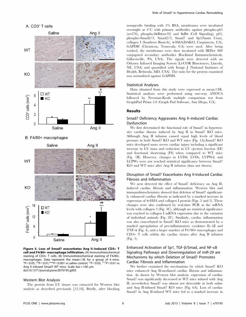
Figure 4. Loss of Smad7 aggravates proinflammation expression during cardiac inflammation in response to Ang II infusion. (A) IL-1b expression. (B) TNF-a expression. (i) Immunohistochemistry (IHC); (ii) quantitative analysis of immunohistochemical staining (IHC); (iii) real-timePCR analysis. Data represent the mean6SE for a group of 6 mice. *P,0.05, ***P,0.001 vs saline control; #P,0.05, ##P,0.01, ###P,0.001 vs Ang II–infused Smad7 WT mice. Scale bar = 100 mm.doi:10.1371/journal.pone.0070195.g004
Role of Smad7 in Hypertensive Cardiac Remodeling
PLOS ONE | www.plosone.org 5 July 2013 | Volume 8 | Issue 7 | e70195
Western Blot AnalysisThe protein from LV tissues was extracted for Western blot
analysis as described previously [12,18]. Briefly, after blocking
nonspecific binding with 5% BSA, membranes were incubated
overnight at 4uC with primary antibodies against phospho-p65
(ser276), phospho-IkBa(ser32) and IkBa (Cell Signaling), p65,
phospho-Smad2/3, Smad2/3, Smad7 and Sp1(Santa Cruz),
collagen I (Southern Biotech), a-SMA(DAKO, Carpinteria, CA),
GAPDH (Chemicon, Temecula, CA) were used. After being
washed, the membranes were then incubated with IRDye 800
conjugated secondary antibodies (Rockland Immunochemicals,
Gilbertsville, PA, USA). The signals were detected with an
Odyssey Infrared Imaging System (Li-COR Biosciences, Lincoln,
NE, USA) and quantified with Image J (National Institutes of
Health, Bethesda, MD, USA). The ratio for the protein examined
was normalized against GAPDH.
Statistical AnalysesData obtained from this study were expressed as mean6SE.
Statistical analyses were performed using one-way ANOVA
followed by Newman-Keuls multiple comparison test from
GraphPad Prism 5.0 (Graph Pad Software, San Diego, CA).
Results
Smad7 Deficiency Aggravates Ang II–induced CardiacDysfunction
We first determined the functional role of Smad7 in hyperten-
sive cardiac disease induced by Ang II in Smad7 KO mice.
Although Ang II infusion caused equal high levels of blood
pressure in both Smad7 KO and WT mice (Fig. 1A),Smad7 KO
mice developed more severe cardiac injury including a significant
increase in LV mass and reduction in LV ejection fraction (EF)
and fractional shortening (FS) when compared to WT mice
(Fig. 1B). However, changes in LVDd, LVDs, LVPWd, and
LCPWs were not reached statistical significance between Smad7
KO and WT mice after Ang II infusion (data not shown).
Disruption of Smad7 Exacerbates Ang II-induced CardiacFibrosis and Inflammation
We next detected the effect of Smad7 deficiency on Ang II-
induced cardiac fibrosis and inflammation. Western blot and
immunohistochemistry showed that deletion of Smad7 significant-
ly enhanced cardiac fibrosis as indicated by a marked increase in
expression of a-SMA and collagen I protein (Figs. 2 and 3). These
changes were also confirmed by real-time PCR at the mRNA
levels with collagen 3 (Fig. 3C), although no statistical significance
was reached in collagen I mRNA expression due to the variation
of individual animals (Fig. 2C). Similarly, cardiac inflammation
was also exacerbated in Smad7 KO mice as demonstrated by a
marked upregulation of pro-inflammatory cytokines IL-1b and
TNF-a (Fig. 4), and a larger number of F4/80+ macrophages and
CD3+ T cells within the cardiac tissues after Ang II infusion
(Fig. 5).
Enhanced Activation of Sp1, TGF-b/Smad, and NF-kBSignaling Pathways and Downregulation of miR-29 areMechanisms by which Deletion of Smad7 PromotesCardiac Fibrosis and Inflammation
We further examined the mechanisms by which Smad7 KO
mice enhanced Ang II-mediated cardiac fibrosis and inflamma-
tion. As shown by Western blot analysis, expression of cardiac
Smad7 was significantly decreased in WT mice infused with Ang
II; nevertheless Smad7 was almost not detectable in both saline
and Ang II-infused Smad7 KO mice (Fig. 6A). Loss of cardiac
Smad7 in Ang II-infused WT mice led to a marked increase in
Figure 5. Loss of Smad7 exacerbates Ang II–induced CD3+ Tcell and F4/80+ macrophage infiltration. (A) Immunohistochemicalstaining of CD3+ T cells. (B) Immunohistochemical staining of F4/80+macrophages. Data represent the mean6SE for a group of 6 mice.*P,0.05, **P,0.01,***P,0.001 vs saline control; #P,0.05, ##P,0.01 vsAng II–infused Smad7 WT mice. Scale bar = 100 mm.doi:10.1371/journal.pone.0070195.g005
Role of Smad7 in Hypertensive Cardiac Remodeling
PLOS ONE | www.plosone.org 6 July 2013 | Volume 8 | Issue 7 | e70195

phosphorylation of Smad2/3, which was further enhanced in Ang
II-infused Smad7 KO mice (Fig. 6B). Real-time PCR and
immunohistochemistry also showed that enhanced Smad2/3
signaling in Ang II-infused Smad7 KO mice was associated with
a further upregulation of cardiac TGF-b1 expression (Fig. 6C),
demonstrating that deletion of Smad7 enhanced TGF-b/Smad
signaling in the hypertensive cardiac disease. In addition, we also
found that loss of cardiac Smad7 largely enhanced activation of
NF-kB signaling as demonstrated by higher levels of phosphor-
ylated IkBa and NF-kB/p65 in Smad7 KO mice when compared
with Ang II-infused Smad7 WT mice (Fig. 7A, B).
Figure 6. Disruption of Smad7 enhances Ang II-induced activation of TGF-b/Smad signaling during cardiac fibrosis. (A) Western blotanalysis of Smad7. (B) Western blot analysis of phosphorylated Smad2/3. (C) Immunohistochemical and real-time PCR analysis of TGF-b1 expression.Results show that deletion of Smad7 enhances TGFb1 expression and Smad2/3 signaling in response to Ang II. Data represent the mean6SE for agroup of 6 mice. *P,0.05, ***P,0.001vs saline control; #P,0.05,##P,0.01, ###P,0.001 vs Smad7 WT mice. Scale bar = 100 mm.doi:10.1371/journal.pone.0070195.g006
Role of Smad7 in Hypertensive Cardiac Remodeling
PLOS ONE | www.plosone.org 7 July 2013 | Volume 8 | Issue 7 | e70195

Since Sp1, a ubiquitous transcription factor, is required for Ang
II–induced fibrotic and inflammatory response [24,25]. we
examined Sp1 expression in the hypertensive heart and found
that Ang II infusion largely upregulated cardiac Sp1 in Smad7
WT mice, which was further increased in Smad7 KO mice
(Fig. 7C). Moreover, it has been shown that loss of miR-29b is
associated with cardiac fibrosis and is negatively regulated by both
TGF-b/Smad3 and NF-kB-YY1 [23,26,27], we examined
whether deletion of Smad7 causes enhanced Ang II-induced loss
of cardiac miR-29b during cardiac remodeling. As shown in
Figure 7D, real-time PCR showed that miR-29b was significantly
decreased in the hypertensive heart of Smad7 WT mice, which
became almost undetectable in Ang II-infused Smad7 KO mice.
Figure 7. Smad7 deficiency enhances Ang II-induced NF-kB signaling activation, upregulation of Sp1, but loss of miR-29b. (A)Western blot analysis of phosphorylated IkBa. (B) Western blot analysis of phosphorylated NF-kB/p65. (C) Western blot analysis of Sp1. (D) Real-timePCR analysis of miR-29b expression. Results show that deletion of Smad7 enhances activation of NF-kB signaling by promoting phosphorylation ofIkBa and p65, promotes Sp1 expression, but induces loss of cardiac miR-29b. Data represent the mean6SE for a group of 6 mice. *P,0.05, **P,0.01,***P,0.001vs saline control; #P,0.05,###P,0.001 vs Smad7 WT mice.doi:10.1371/journal.pone.0070195.g007
Role of Smad7 in Hypertensive Cardiac Remodeling
PLOS ONE | www.plosone.org 8 July 2013 | Volume 8 | Issue 7 | e70195

Discussion
Increasing evidence demonstrates that Ang II is a driving force
in cardiac fibrosis, inflammation and cardiac dysfunction [1,28].
The present study provided new evidence for a protective role of
Smad7 in Ang II-induced hypertensive cardiac remodeling. We
detected that loss of Smad7 enhanced Ang II-induced cardiac
remodeling and cardiac dysfunction. Enhanced activation of Sp1-
TGF-b/Smad-NF-kB signaling pathways and loss of cardiac miR-
29 were key mechanisms by which deficiency of Smad7 promoted
Ang II-mediated cardiopathy.
Loss of Smad7 was a key mechanism by which Ang II induces
cardiac fibrosis. Consistent with the previous observation that
decreased cardiac Smad7 contributes to cardiac fibrosis [29,30],
our current study revealed that cardiac Smad7 expression was
markedly reduced in response to Ang II infusion, resulting in
impaired cardiac function including an increase in LV mass,
reduction of LVEF and FS. Beyond this observation, by using
Smad 7 KO mice, we provided a direct evidence for a functional
role of Smad7 in Ang II-induced hypertensive cardiac remodeling.
We found that mice lacking Smad7 developed more severe cardiac
fibrosis in response to Ang II infusion and had more severe cardiac
dysfunction including a significant increase in LV mass, a fall of
LVEF and FS when compared with Smad7 WT mice. Once
Smad7 is lost, Ang II-induced activation of Smad3 via both TGF-
b-dependent and independent pathways is enhanced [7,8], which
results in enhanced Smad3-mediated fibrosis as previously
reported in vitro and in a number of mouse models induced by
Ang II [8,11,12,14,15], postmyocardiac infarction [13], obstruc-
tive and diabetic nephropathy [18,31]. The observation that the
lack of Smad7 promoted Ang II-induced cardiac fibrosis and
dysfunction identified a critically protective role for Smad7 in Ang
II-mediated cardiac remodeling.
Loss of Smad7 may also be a mechanism whereby Ang II
induces cardiac inflammation via a NF-kB-dependent mechanism.
We have previously showed that Smad7 is able to block
inflammatory responses by preventing NF-kB from activation
[18–20]. It is well accepted that Ang II is capable of activating NF-
kB to mediate cardiovascular inflammation [6]. We have
previously detected that Smad7 is able to block NF-kB-dependent
inflammation by inducing IkBa, an inhibitor of NF-kB, or
preventing it from degradation in a number of experimental
models and in vitro [18–20]. In the present study, the finding that
disruption of Smad7 enhanced further NF-kB activation added
new evidence that Smad7 protects against Ang II-induced cardiac
inflammation through inhibition of the NF-kB pathway.
Upregulation of Sp1 pathway may also contribute to promote
Ang II-mediated cardiac remodeling in Smad7 KO mice. Sp1 is
required for Ang II–induced fibrotic and inflammatory response
[24,25]. It has been demonstrated that Sp1 can interact with both
Smad3 and NF-kB to play a critical role in fibrosis and
inflammation [32–34]. Thus, Ang II-induced activation of Sp1/
Smad3/NF-kB pathways may cooperate in the development of
cardiac fibrosis and inflammation as seen in WT mice. Deletion of
Smad7 enhanced further activation of this Sp1/Smad3/NF-kB
axis in response to Ang II, which may well explain enhanced
cardiac fibrosis and inflammation when Smad7 is disrupted.
Interestingly, we also found that loss of miR-29 may be an
additional mechanism through which disruption of Smad7
enhances Ang II-mediated cardiac fibrosis and inflammation.
miR-29b is negatively regulated by both TGF-b/Smad3 and NF-
kB-YY1 regulatory circuit [23,26,27]. Increasing evidence shows
that down-regulation of miR-29 is associated with fibrosis in a
number of disease models including ischemic cardiac remodeling
[26], while overexpression of miR-29b is capable of inhibiting
Smad3-mediated kidney and lung fibrosis [23,35]. miR-29b exerts
an anti-fibrotic function through direct targeting of the 39UTR
regions in the mRNA for collagens I, III and IV and fibrillin and
elastin [26]. It is also reported that miR-29b can interact with Sp1
to form the Sp1/NFkB/HDAC/miR-29b regulatory network in
myeloid leukemia [36]. All these findings suggest that Ang II-
induced loss of miR-29b via TGF-b/Smad3 and NF-kB-depen-
dent pathways may also be an additional mechanism by which
deletion of Smad7 promotes Ang II-induced cardiac remodeling.
In summary, Smad7 plays a protective role in Ang II-mediated
cardiac fibrosis and inflammation and cardiac dysfunction.
Upregulation of the Sp1-TGF-b/Smad-NF-kB pathway and loss
of miR-29b may be mechanisms by which deletion of Smad7
enhances hypertensive cardiac remodeling. These findings suggest
that Smad7 may be a novel therapeutic agent for hypertensive
cardiovascular disease.
Author Contributions
Conceived and designed the experiments: HYL. Performed the experi-
ments: LHW XRH YQL YZ HYC. Analyzed the data: LHW XRH YQL
YZ HYC. Contributed reagents/materials/analysis tools: RH. Wrote the
paper: BPY CMY RH HYL.
References
1. Drazner MH (2011) The progression of hypertensive heart disease. Circulation
123: 327–334.
2. Mann DL (2002) Inflammatory mediators and the failing heart: past, present,
and the foreseeable future. Circ Res 91: 988–998.
3. Jia L, Li Y, Xiao C, Du J (2012) Angiotensin II induces inflammation leading to
cardiac remodeling. Front Biosci 17: 221–231.
4. Shahbaz AU, Sun Y, Bhattacharya SK, Ahokas RA, Gerling IC, et al.(2010)
Fibrosis in hypertensive heart disease: molecular pathways and cardioprotective
strategies. J Hypertens 28 Suppl 1: S25–S32.
5. Mehta PK, Griendling KK (2007) Angiotensin II cell signaling: physiological
and pathological effects in the cardiovascular system. Am J Physiol Cell Physiol
292: C82–C97.
6. Muller DN, Dechend R, Mervaala EM, Park JK, Schmidt F, et al. (2000) NF-kB
inhibition ameliorates angiotensin II-induced inflammatory damage in rats.
Hypertension 35: 193–201.
7. Rodriguez-Vita J, Sanchez-Lopez E, Esteban V, Ruperez M, Egido J, et al.
(2005) Angiotensin II activates the Smad pathway in vascular smooth muscle
cells by a transforming growth factor-beta-independent mechanism. Circulation
111: 2509–2517.
8. Wang W, Huang XR, Canlas E, Oka K, Truong LD, et al. (2006) Essential role
of Smad3 in angiotensin II-induced vascular fibrosis. Circ Res 98: 1032–1039.
9. Kavsak P, Rasmussen RK, Causing CG, Bonni S, Zhu H, et al.(2000) Smad7
binds to Smurf2 to form an E3 ubiquitin ligase that targets the TGF beta
receptor for degradation. Mol Cell 6: 1365–1375.
10. Meng XM, Huang XR, Chung AC, Qin W, Shao X, et al. (2010) Smad2
protects against TGF-beta/Smad3-mediated renal fibrosis. J Am Soc Nephrol
21: 1477–1487.
11. Liu Z, Huang XR, Lan HY (2012) Smad3 mediates ANG II-induced
hypertensive kidney disease in mice. Am J Physiol Renal Physiol 302: F986–
F997.
12. Huang XR, Chung AC, Yang F, Yue W, Deng C, et al.(2010) Smad3 mediates
cardiac inflammation and fibrosis in angiotensin II-induced hypertensive cardiac
remodeling. Hypertension 55: 1165–1171.
13. Bujak M, Ren G, Kweon HJ, Dobaczewski M, Reddy A, et al. (2007)
Frangogiannis NG. Essential role of Smad3 in infarct healing and in the
pathogenesis of cardiac remodeling. Circulation 116: 2127–2138.
14. Yang F, Chung AC, Huang XR, Lan HY (2009) Angiotensin II induces
connective tissue growth factor and collagen I expression via transforming
growth factor-beta-dependent and -independent Smad pathways: the role of
Smad3. Hypertension54: 877–884.
15. Yang F, Huang XR, Chung AC, Hou CC, Lai KN, et al. (2010) Essential role
for Smad3 in angiotensin II-induced tubular epithelial-mesenchymal transition.
J Pathol 221: 390–401.
Role of Smad7 in Hypertensive Cardiac Remodeling
PLOS ONE | www.plosone.org 9 July 2013 | Volume 8 | Issue 7 | e70195

16. Lan HY, Mu W, Tomita N, Huang XR, Li JH, et al. (2003) Inhibition of renal
fibrosis by gene transfer of inducible Smad7 using ultrasound-microbubble
system in rat UUO model. J Am Soc Nephrol 14: 1535–1548.
17. Hou CC, Wang W, Huang XR, Fu P, Chen TH, et al. (2005) Ultrasound-
microbubble-mediated gene transfer of inducible Smad7 blocks transforming
growth factor-beta signaling and fibrosis in rat remnant kidney. Am J Pathol
166: 761–771.
18. Chen HY, Huang XR, Wang W, Li JH, Heuchel RL, et al. (2011) The
protective role of Smad7 in diabetic kidney disease: mechanism and therapeutic
potential. Diabetes 60: 590–601.
19. Wang W, Huang XR, Li AG, Liu F, Li JH, et al.(2005) Signaling mechanism of
TGF-b1 in prevention of renal inflammation: role of Smad7. J Am Soc Nephrol
16: 1371–1383.
20. Ng YY, Hou CC, Wang W, Huang XR, Lan HY (2005) Blockade of NFkappaB
activation and renal inflammation by ultrasound-mediated gene transfer of
Smad7 in rat remnant kidney. Kidney Int Suppl S83–S91.
21. Li R, Rosendahl A,Brodin G,Cheng AM, Ahgren A, et al.(2006) Deletion of
exon I of SMAD7 in mice results in altered B cell responses. J Immunol 176:
6777–6784.
22. Lan HY, Mu W, Nikolic-Paterson DJ, Atkins RC (1995) A novel, simple,
reliable, and sensitive method for multiple immunoenzyme staining: use of
microwave oven heating to block antibody crossreactivity and retrieve antigens.
J Histochem Cytochem 43: 97–102.
23. Qin W, Chung AC, Huang XR, Meng XM, Hui DS, et al. (2011) TGF-b/
Smad3 signaling promotes renal fibrosis by inhibiting miR-29. J Am Soc
Nephrol 22: 1462–1474.
24. Motojima M, Ando T, Yoshioka T (2000) Sp1-like activity mediates angiotensin-
II-induced plasminogen-activator inhibitor type-1 (PAI-1) gene expression in
mesangial cells. Biochem J349: 435–441.
25. Zhao X, Martin MM, Elton TS (2001) The transcription factors Sp1 and Sp3
are required for human angiotensin II type 1 receptor gene expression in H295-
R cells. Biochim Biophys Acta 1522: 195–206.
26. van Rooij E, Sutherland LB, Thatcher JE, DiMaio JM, Naseem RH, et al.
(2008) Dysregulation of microRNAs aftermyocardial infarction reveals a role ofmiR-29 in cardiac fibrosis. ProcNatl Acad Sci U S A 105: 13027–13032.
27. Wang H, Garzon R, Sun H, Ladner KJ, Singh R, et al. (2008) NF-kappaB-YY1-
miR-29 regulatory circuitry in skeletal myogenesis and rhabdomyosarcoma.Cancer Cell 14: 369–381.
28. Leask A (2010) Potential therapeutic targets for cardiac fibrosis: TGFbeta,angiotensin, endothelin, CCN2, and PDGF, partners in fibroblast activation.
Circ Res 106: 1675–1680.
29. He X, Gao X, Peng L, Wang S, Zhu Y, et al. (2011) Atrial fibrillation inducesmyocardial fibrosis through angiotensin II type 1 receptor-specific Arkadia-
mediated downregulation of Smad7. Circ Res 108: 164–175.30. Wang B, Hao J, Jones SC, Yee MS, Roth JC, et al. (2002) Decreased Smad 7
expression contributes to cardiac fibrosis in the infarcted rat heart. Am J PhysiolHeart Circ Physiol 282: H1685–H1696.
31. Chung AC, Huang XR, Zhou L, Heuchel R, Lai KN, et al. (2009) Disruption of
the Smad7 gene promotes renal fibrosis and inflammation in unilateral ureteralobstruction (UUO) in mice. Nephrol Dial Transplant 24: 1443–1454.
32. Traylor A, Hock T, Hill-Kapturczak N (2007) Specificity protein 1 and Smad-dependent regulation of human heme oxygenase-1 gene by transforming growth
factor-beta1 in renal epithelial cells. Am J Physiol Renal Physiol 293: F885–
F894.33. Poncelet AC, Schnaper HW (2001) Sp1 and Smad proteins cooperate to
mediate transforming growth factor-b1-induced a2(I) collagen expression inhuman glomerular mesangial cells. J Biol Chem 276: 6983–6992.
34. Perkins ND, Agranoff AB, Pascal E, Nabel GJ (1994) An interaction between theDNA-binding domains of RelA(p65) and Sp1 mediates human immunodefi-
ciency virus gene activation. Mol Cell Biol 14: 6570–6583.
35. Xiao J, Meng XM, Huang XR, Chung AC, Feng YL, et al. (2012) miR-29inhibits bleomycin-induced pulmonary fibrosis in Mice. Mol Ther 20: 1251–
1260.36. Liu S, Wu LC, Pang J, Santhanam R, Schwind S, et al. (2010) Sp1/NFkappaB/
HDAC/miR-29b regulatory network in KIT-driven myeloid leukemia. Cancer
Cell 17: 333–347.
Role of Smad7 in Hypertensive Cardiac Remodeling
PLOS ONE | www.plosone.org 10 July 2013 | Volume 8 | Issue 7 | e70195
Copyright © 2022 FDOKUMEN