Decoding of Calcium Oscillations by Phosphorylation Cycles: Analytic Results
13
Decoding of Calcium Oscillations by Phosphorylation Cycles: Analytic Results Carlos Salazar, Antonio Zaccaria Politi, and Thomas Ho ¨fer Research Group Modeling of Biological Systems, German Cancer Research Center, Heidelberg, Germany ABSTRACT Experimental studies have demonstrated that Ca 21 -regulated proteins are sensitive to the frequency of Ca 21 oscillations, and several mathematical models for specific proteins have provided insight into the mechanisms involved. Because of the large number of Ca 21 -regulated proteins in signal transduction, metabolism and gene expression, it is desirable to establish in general terms which molecular properties shape the response to oscillatory Ca 21 signals. Here we address this question by analyzing in detail a model of a prototypical Ca 21 -decoding module, consisting of a target protein whose activity is controlled by a Ca 21 -activated kinase and the counteracting phosphatase. We show that this module can decode the frequency of Ca 21 oscillations, at constant average Ca 21 signal, provided that the Ca 21 spikes are narrow and the oscillation frequency is sufficiently low—of the order of the phosphatase rate constant or below. Moreover, Ca 21 oscillations activate the target more efficiently than a constant signal when Ca 21 is bound cooperatively and with low affinity. Thus, the rate constants and the Ca 21 affinities of the target-modifying enzymes can be tuned in such a way that the module responds optimally to Ca 21 spikes of a certain amplitude and frequency. Frequency sensitivity is further enhanced when the limited duration of the external stimulus driving Ca 21 signaling is accounted for. Thus, our study identifies molecular parameters that may be involved in establishing the specificity of cellular responses downstream of Ca 21 oscillations. INTRODUCTION Cell signaling induced by extracellular stimuli is often ac- companied by an increase in the cytosolic calcium concen- tration [Ca 21 ] c that, ultimately, regulates a plethora of cellular processes, including secretion, contraction, learning, and proliferation (1–4). Such regulation is mediated by Ca 21 -dependent enzymes that, in turn, modify downstream targets commonly by phosphorylation. In many cell types, changes in [Ca 21 ] c occur as repetitive spikes that increase their frequency with the strength of the stimulus (5,6). Infor- mation can also be encoded in the amplitude of Ca 21 oscil- lations, which may change with the extracellular stimulus (7–9) and may also depend on the subcellular localization of the target (10,11). Understanding how an extracellular stimulus, encoded in the frequency and amplitude of Ca 21 oscillations, is interpreted by different physiological pro- cesses is one of the major challenges in the study of Ca 21 signaling. Various experiments have suggested that Ca 21 oscilla- tions may be advantageous compared to a constant rise in [Ca 21 ] c because they increase the efficiency of gene expres- sion at low levels of stimulation (12,13). A long-standing question is how a ubiquitous messenger like Ca 21 achieves specificity in its action. Recent data indicate the ability of calmodulin and protein kinase C (PKC) to decode the am- plitude of Ca 21 oscillatory signal into an appropriate cellular response (14,15). It has been shown that the sensitivity to Ca 21 oscillations of different transcription factors such as the nuclear factor of activated T cells (NFAT), nuclear factor kappa B (NFkB) (12,13,16), Ca 21 -dependent enzymes such as calmodulin (CaM) kinase II (17), and mitochondria (18,19), can depend on the oscillation frequency. Moreover, when the stimulus is transient, the effect of Ca 21 oscillations can be maximized within a certain range of frequencies (13,16,20). In Ca 21 oscillations, higher frequency implies a higher density of practically uniformly shaped Ca 21 spikes and thus also a higher average [Ca 21 ] c . Thus, the question arises whether the observed increases in target response with rising oscillation frequency are simply due to an increase in the average Ca 21 signal or present a true frequency decoding. Previous theoretical work has focused on how frequency- encoded information is processed by Ca 21 -regulated proteins (21–23). Frequency decoding is characterized by higher aver- age levels of phosphorylated protein when the frequency of Ca 21 oscillations is increased. Detailed models of protein phosphorylation driven by regular Ca 21 oscillations have been developed for CaM kinase II (14,24,25), glycogen phosphorylase (26,27), and mitogen-activated protein kinase cascades (28,29). Decoding of Ca 21 signals more complex than periodic spiking (e.g., bursting oscillations) has been re- cently theoretically explored (30,31). Numerical simulations have shown that Ca 21 oscillations can be more efficient for protein phosphorylation compared to constant Ca 21 signals of equal average [Ca 21 ] c . The nonlinear dependence of pro- tein activation on Ca 21 stimulation, arising from diverse mechanisms, such as cooperative Ca 21 binding or zero-order doi: 10.1529/biophysj.107.113084 Submitted July 24, 2007, and accepted for publication September 26, 2007. Carlos Salazar and Antonio Zaccaria Politi contributed equally to this work. Address reprint requests to Carlos Salazar, E-mail: c.salazar@dkfz-heidelberg. de; or Thomas Ho ¨fer, E-mail: [email protected]. Antonio Politi’s present address is the Dept. of Mathematics, University of Auckland, 38 Princes St., Auckland, New Zealand. Editor: Ian Parker. Ó 2008 by the Biophysical Society 0006-3495/08/02/1203/13 $2.00 Biophysical Journal Volume 94 February 2008 1203–1215 1203
-
Upload
independent -
Category
Documents
-
view
1 -
download
0
Transcript of Decoding of Calcium Oscillations by Phosphorylation Cycles: Analytic Results

Decoding of Calcium Oscillations by Phosphorylation Cycles:Analytic Results
Carlos Salazar, Antonio Zaccaria Politi, and Thomas HoferResearch Group Modeling of Biological Systems, German Cancer Research Center, Heidelberg, Germany
ABSTRACT Experimental studies have demonstrated that Ca21-regulated proteins are sensitive to the frequency of Ca21
oscillations, and several mathematical models for specific proteins have provided insight into the mechanisms involved.Because of the large number of Ca21-regulated proteins in signal transduction, metabolism and gene expression, it is desirableto establish in general terms which molecular properties shape the response to oscillatory Ca21 signals. Here we address thisquestion by analyzing in detail a model of a prototypical Ca21-decoding module, consisting of a target protein whose activity iscontrolled by a Ca21-activated kinase and the counteracting phosphatase. We show that this module can decode the frequencyof Ca21 oscillations, at constant average Ca21 signal, provided that the Ca21 spikes are narrow and the oscillation frequency issufficiently low—of the order of the phosphatase rate constant or below. Moreover, Ca21 oscillations activate the target moreefficiently than a constant signal when Ca21 is bound cooperatively and with low affinity. Thus, the rate constants and the Ca21
affinities of the target-modifying enzymes can be tuned in such a way that the module responds optimally to Ca21 spikes of acertain amplitude and frequency. Frequency sensitivity is further enhanced when the limited duration of the external stimulusdriving Ca21 signaling is accounted for. Thus, our study identifies molecular parameters that may be involved in establishing thespecificity of cellular responses downstream of Ca21 oscillations.
INTRODUCTION
Cell signaling induced by extracellular stimuli is often ac-
companied by an increase in the cytosolic calcium concen-
tration [Ca21]c that, ultimately, regulates a plethora of
cellular processes, including secretion, contraction, learning,
and proliferation (1–4). Such regulation is mediated by
Ca21-dependent enzymes that, in turn, modify downstream
targets commonly by phosphorylation. In many cell types,
changes in [Ca21]c occur as repetitive spikes that increase
their frequency with the strength of the stimulus (5,6). Infor-
mation can also be encoded in the amplitude of Ca21 oscil-
lations, which may change with the extracellular stimulus
(7–9) and may also depend on the subcellular localization
of the target (10,11). Understanding how an extracellular
stimulus, encoded in the frequency and amplitude of Ca21
oscillations, is interpreted by different physiological pro-
cesses is one of the major challenges in the study of Ca21
signaling.
Various experiments have suggested that Ca21 oscilla-
tions may be advantageous compared to a constant rise in
[Ca21]c because they increase the efficiency of gene expres-
sion at low levels of stimulation (12,13). A long-standing
question is how a ubiquitous messenger like Ca21 achieves
specificity in its action. Recent data indicate the ability of
calmodulin and protein kinase C (PKC) to decode the am-
plitude of Ca21 oscillatory signal into an appropriate cellular
response (14,15). It has been shown that the sensitivity to
Ca21 oscillations of different transcription factors such as the
nuclear factor of activated T cells (NFAT), nuclear factor
kappa B (NFkB) (12,13,16), Ca21-dependent enzymes such
as calmodulin (CaM) kinase II (17), and mitochondria (18,19),
can depend on the oscillation frequency. Moreover, when the
stimulus is transient, the effect of Ca21 oscillations can be
maximized within a certain range of frequencies (13,16,20).
In Ca21 oscillations, higher frequency implies a higher
density of practically uniformly shaped Ca21 spikes and thus
also a higher average [Ca21]c. Thus, the question arises
whether the observed increases in target response with rising
oscillation frequency are simply due to an increase in the
average Ca21 signal or present a true frequency decoding.
Previous theoretical work has focused on how frequency-
encoded information is processed by Ca21-regulated proteins
(21–23). Frequency decoding is characterized by higher aver-
age levels of phosphorylated protein when the frequency of
Ca21 oscillations is increased. Detailed models of protein
phosphorylation driven by regular Ca21 oscillations have
been developed for CaM kinase II (14,24,25), glycogen
phosphorylase (26,27), and mitogen-activated protein kinase
cascades (28,29). Decoding of Ca21 signals more complex
than periodic spiking (e.g., bursting oscillations) has been re-
cently theoretically explored (30,31). Numerical simulations
have shown that Ca21 oscillations can be more efficient for
protein phosphorylation compared to constant Ca21 signals
of equal average [Ca21]c. The nonlinear dependence of pro-
tein activation on Ca21 stimulation, arising from diverse
mechanisms, such as cooperative Ca21 binding or zero-order
doi: 10.1529/biophysj.107.113084
Submitted July 24, 2007, and accepted for publication September 26, 2007.
Carlos Salazar and Antonio Zaccaria Politi contributed equally to this work.
Address reprint requests to Carlos Salazar, E-mail: c.salazar@dkfz-heidelberg.
de; or Thomas Hofer, E-mail: [email protected].
Antonio Politi’s present address is the Dept. of Mathematics, University of
Auckland, 38 Princes St., Auckland, New Zealand.
Editor: Ian Parker.
� 2008 by the Biophysical Society
0006-3495/08/02/1203/13 $2.00
Biophysical Journal Volume 94 February 2008 1203–1215 1203

ultrasensitivity, turns out to be of major importance for an
efficient processing of oscillatory signals. Furthermore, these
models predict that the Ca21 oscillation frequency can dis-
criminate among different signaling pathways.
Although these theoretical studies have provided insight
into the mechanisms involved in calcium decoding, their
conclusions are based on numerical simulations of detailed
models for specific proteins (reviewed in (32)). Because of
the large numbers of Ca21-regulated proteins in signal trans-
duction and gene transcription, it is desirable to establish in
general terms which molecular properties shape the response
to Ca21 oscillations. Investigating Ca21 decoding in an ana-
lytically tractable model may provide more insight into the
following issues:
How sensitively can a target protein respond to changes
in the frequency of calcium oscillations?
Under which conditions are Ca21 oscillations more potent
than a constant signal in activating a target protein?
Which properties determine the existence of an optimal
activating signal for a particular target?
Under which conditions can target proteins be differen-
tially activated by Ca21 signals?
Some challenges for such analysis are the nonstationarity of
the Ca21 signal and the nonlinearity of the Ca21 dependent
processes. The description of Ca21 oscillations using a piece-
wise constant function can facilitate an analytical solution of
the kinetic equations (31,33,34). Indeed, such square-shaped
pulses have been used in several experiments (12,13,17) and
for numerical simulations (20,35).
In this article, we attempt to establish in general terms
which molecular properties determine the target response
to oscillatory Ca21 signals. To this aim, we employed an
analytically tractable model of a prototypical Ca21-decoding
module, consisting of a target protein controlled by a Ca21-
activated kinase and the counteracting phosphatase. The
modeling of Ca21 oscillations by square-shaped pulses
allowed the derivation of a general formula for the target
activity as a function of three dimensionless parameters.
Using this formula, we first asked whether a true frequency
decoding, at constant average Ca21 signal, is possible. We
then examined the conditions under which oscillatory signals
are more potent in activating the target protein than constant
signals with the same average calcium. To assess the kinetics
of the target, we defined an activation time that quantifies
the time necessary to reach the maximal target activity. The
expressions for the activation time and target activity were
used to analyze signaling specificity. In particular, we inves-
tigated which molecular parameters determine the optimal
signal shape for a limited amount of calcium. Collectively,
our results demonstrate that the nonlinear activation of a
protein by a Ca21-dependent kinase represents a core system
for decoding Ca21 oscillations, and rationalize why oscilla-
tory stimuli increase the efficiency and specificity of cell
signaling.
MATHEMATICAL MODEL
We consider an activation-inactivation cycle of a target pro-
tein X (Fig. 1 A). Its activation is induced by phosphorylation
through a Ca21-dependent kinase Y, and it is inactivated by
dephosphorylation. Cooperative activation of the kinase by
Ca21, as observed, for example, for Ca21/CaM-dependent
kinases (36,37), is described by the binding of n ions of
Ca21. The fractions of active kinase Y and phosphorylated
(active) target protein X obey, in the simplest model,
dY
dt¼ aYSðtÞnð1� YÞ � bYY; (1)
dX
dt¼ aXYTYð1� XÞ � bXX; (2)
where YT represents the total concentration of kinase, S(t)describes the time course of the Ca21 signal, aY and bY are the
rate constants of Ca21 binding and release, and the rate con-
stants of phosphorylation and dephosphorylation are denoted
by aX and bX, respectively.
In many cases, Ca21 binding will be much faster than
(de)phosphorylation reactions, so that we can assume the
binding equilibrium, dY/dt � 0. Equation 2 then becomes
dX
dt¼ aðSÞ � ðaðSÞ1 bÞX; (3)
where, for simplicity, b ¼ bX. Between calcium spikes, the
kinase activity is negligible so that the response time of the
target protein is b�1.
The dependence of the phosphorylation rate constant on
Ca21 concentration takes the form of the Hill equation
aðSÞ ¼ aðS=KSÞn
ðS=KSÞn 1 1; (4)
with the maximal value a ¼ aXYT; the half-saturation constant
KS ¼ffiffiffiffiffiffiffiffiffiffiffiffiffiffiffibY=aY;
np
and the Hill-coefficient n. Hill equations
provide good fits for more realistic Ca21 binding mechanisms,
in which case n can take noninteger values (20,30).
Cytosolic Ca21 oscillations consist of a series of spikes
separated by intervals of resting Ca21 concentration (38). To
enable analytic treatments, Ca21 oscillations are described
by a piecewise constant function
SðtÞ ¼S0; iT # t , iT 1 D
0; iT 1 D , t , ði 1 1ÞT
�i ¼ 0; . . . ; N; (5)
characterized by period T, amplitude S0, and spike width D
(Fig. 1 B). For simplicity, the basal Ca21 concentration has
been set to zero.
RESULTS
The mean activity of the target protein dependson three dimensionless parameters
Let us assume that before the onset of calcium oscillations, the
target protein is completely inactive (i.e., unphosphorylated).
1204 Salazar et al.
Biophysical Journal 94(4) 1203–1215

The target’s response to the oscillations goes through an
initial transient to reach an (approximately) periodic state
(Fig. 1 C). The mean target activity �Xi during the ith
oscillation cycle is
�Xi ¼1
T
Z ði11ÞT
iT
XðtÞdt: (6)
The average is a good measure of the target protein’s re-
sponse if the amplitude of the protein activity oscillations is
not too large, that is, when the system integrates the
oscillations. This has been seen in the responses of gene
expression rates and of mitochondrial metabolism to calcium
oscillations (12,16,18,19). However, even if the target
amplitudes were large, the average is a useful diagnostic of
how sensitively the system responds to the oscillatory signal.
The approximation for the Ca21 spikes (see Eq. 5) allows
the derivation of an analytic expression for the average �Xi:After some algebra, one finds for its stationary value�X ¼ lim
i/N�Xi (see Appendix A):
�X ¼ Xmax g 1vs
1 1 s
ð1� e�gð11sÞ=vÞð1� e
�ð1�gÞ=vÞ1� e
�ð11gsÞ=v
" #: (7)
This equation shows how the original parameters combine
into three dimensionless parameters that govern the response
of the system, as follows here.
The effective activation rate s during the calcium spike
s ¼ a
b
ðS0=KSÞn
1 1 ðS0=KSÞn; (8)
measures how the calcium amplitude S0 is sensed by the
kinase with respect to binding affinity (1/KS) and coopera-
tivity of kinase activation (Hill coefficient n), and how fast
phosphorylation of the target is compared to its dephospho-
rylation (a=b).
The relative oscillation frequency v,
v ¼ 1
bT; (9)
expresses how fast the oscillations are compared with the
basal response time of the protein b�1 (see Eq. 3). Thus, the
parameter combines the two timescales in the problem. Large
v means fast calcium oscillations and/or a slow response
time of the phosphorylation cycle, in which case the phos-
phorylation cycle will integrate over the oscillations and the
amplitude of the oscillations in target activity is small. Con-
versely, if v is small, the target activity will closely follow
each cycle of the calcium oscillations.
The duty ratio of the oscillations g,
g ¼ D
T; (10)
gives the part of the oscillation period during which calcium
is elevated and the kinase is active. Hence, the average
calcium concentration in the oscillations is
�S ¼ gS0: (11)
The maximal activity of the target protein Xmax is a function
of s,
Xmax ¼s
1 1 s: (12)
Target activity is sensitive to the frequency ofnarrow calcium spikes
It turns out that the mean target activity �X is a monotonically
increasing function of all three parameters on their natural
intervals s 2[0,N), v 2[0,N), and g 2[0,1). Increases in the
activation rate, oscillation frequency, or duty ratio would
therefore all raise the target activity. As experiments on many
cell types show that the dose of the external stimuli (e.g.,
FIGURE 1 Model for the decoding of Ca21 oscillations
by a target protein. (A) Model scheme. (B) Oscillations in
the Ca21 concentration S(t) have period T, amplitude S0,
spike width D, and average �S: (C) The calcium signal
induces oscillations in the phosphorylated (active) target
protein. After an initial transient, the time-average target
activity reaches the steady value �X:
Decoding of Ca21 Oscillations 1205
Biophysical Journal 94(4) 1203–1215

hormones and local mediators) controls primarily the fre-
quency of calcium oscillations and leaves the amplitude
unchanged, we asked how sensitively the target protein can
respond to variations in frequency. In the limit of slow
oscillations (or a rapidly responding protein), we obtained
the target activity as
�Xðv/0Þ ¼ g Xmax ¼sg
1 1 s: (13)
For fast oscillations (or a slowly responding protein), we
found
�Xðv/NÞ ¼ ð1 1 sÞg1 1 sg
Xmax ¼sg
1 1 sg: (14)
The frequency sensitivity of the target’s response to calcium
oscillations with constant amplitude becomes maximal,
maximizeg
�Xðv/NÞ � �Xðv/0Þ½ �; (15a)
s ¼ const:; (15b)
when the duty ratio obeys
gopt ¼ffiffiffiffiffiffiffiffiffiffiffiffi1 1 sp
� 1
s: (16)
The optimal duty ratio gopt lies between 0.5 (for s¼ 0) and 0
(for s / N). Hence, calcium oscillations whose frequency
can efficiently drive changes in target activity have duty
ratios below 0.5, i.e., the spikes occupy less than half of the
oscillation. Interestingly, the observed calcium oscillations,
for example in liver cells, have duty ratios between 0.05 and
0.5 (39).
Importantly, when activation rate s and duty ratio g are
constant and only the relative oscillation frequency v is
varied, the average calcium concentration �S remains constant
(Fig. 2 A). Therefore, the system can respond purely to
changes in the frequency of the calcium signal if the calcium
spikes are sufficiently narrow compared to the period of the
oscillations. An example of this true frequency decoding is
shown in Fig. 2 B. Note that the asymptotic value �Xðv/NÞis practically reached already for v $ 1. This implies that the
target protein activity becomes frequency-insensitive when
the oscillation period gets in the range of the basal response
time of the target protein (see Eq. 9). We have found this to
be the typical behavior for a wide range of parameter values.
These results demonstrate that true frequency decoding, at
constant average Ca21 signal, can occur provided that the
Ca21 spikes are narrow and the oscillation frequency is of
the order of the target inactivation rate or below.
Oscillations are more potent than constant signalswhen the kinase binds calcium cooperatively andwith low affinity
Given a certain amount of calcium per unit time, what is the
signal shape that best activates the target protein? For the
piecewise constant oscillatory signal (Eq. 5), the shape at a
given calcium average �S is uniquely defined by fixing period
T and duty ratio g, where the latter automatically defines the
amplitude S0 ¼ �S=g: Setting the frequency, the optimal sig-
nal shape is therefore the solution of
maximizeg
�X; (17a)
�S ¼ gS0 ¼ const: (17b)
Using Eq. 14, for high-frequency oscillations, one obtains
the maximal target response for
gmax ¼�S=KSffiffiffiffiffiffiffiffiffiffiffin� 1np : (18)
If calcium activation of the kinase is noncooperative or neg-
atively cooperative, n # 1, there is no gmax. Indeed, for
n # 1, �X is a monotonically increasing function of g when
the average calcium signal is constrained (Eq. 17b). Therefore,
the maximal response will be obtained at g ¼ 1 (that is, the
constant calcium signal).
However, when calcium activation of the kinase displays
positive cooperativity, n . 1, gmax can be below 1. There-
fore, an oscillatory signal can be more potent in activating
the target than a constant signal. The critical case occurs
obviously when the maximum is attained for g ¼ 1 (constant
signal). From Eq. 18, we then found that if the condition
KS .S0ffiffiffiffiffiffiffiffiffiffiffi
n� 1np (19)
holds, the maximum of �X occurs for 0 , g , 1, and hence
oscillations with the duty ratio given by Eq. 18 activate
the target most strongly. In particular, this oscillatory signal
FIGURE 2 Target activity is sensitive to the fre-
quency of narrow calcium spikes. (A) True frequency
encoding occurs when the relative frequency v
changes while both amplitude S0 and average �S stay
constant. (B) An example of true frequency decoding is
shown here. The mean target activity �X is plotted
against v using Eq. 7. The values of �X in the limit of
very slow and very fast oscillations (Eqs. 13 and 14,
respectively) are indicated by dashed lines. Parameters:
s ¼ 10, g ¼ 0.25.
1206 Salazar et al.
Biophysical Journal 94(4) 1203–1215

is more potent than a constant signal of the same average
calcium.
The functionffiffiffiffiffiffiffiffiffiffiffin� 1np
lies between 1 and 1.321 for a co-
operative calcium decoder with 2 , n , N (the maximum is
attained at n ¼ 4.5911). Therefore, an efficient decoder must
have comparatively weak calcium affinity, with dissociation
constant KS lying around the peak concentration of the cal-
cium spike S0 or above.
Equation 19, for the superiority of oscillations over con-
stant calcium signals, holds for any duty ratio. However,
calcium oscillations typically have rather low duty ratios
(g , 0.5). In this case, the requirement for calcium affinity
becomes less stringent. At a given duty ratio g, oscillations
are superior over constant signals if the calcium sensitivity
of the kinase, relative to the spike amplitude, satisfies (see
Appendix B):
KS
S0
.
ffiffiffiffiffiffiffiffiffiffiffiffiffiffiffiffiffiffiffig
n � gn11
g � gn
n
s: (20)
The decrease of the critical dissociation constant with de-
creasing duty ratio is shown in Fig. 3 for two distinct values
of n. So far, all expressions were derived for the case of high-
frequency oscillations. We obtained very similar expressions
for the general case (Appendix B).
In summary, our theory shows that Ca21 oscillations acti-
vate the target of a Ca21-dependent kinase more efficiently
than a constant signal if 1), Ca21 acts cooperatively on the
kinase (n . 1); and 2), the Ca21 affinity of the kinase is rather
low. As an example, we consider typical cytoplasmic Ca21
spikes with S0¼ 800 nM and g¼ 0.3, yielding a critical KS of
300–440 nM for Hill coefficient of 2–4. Indeed, a range of
prominent calcium-activated enzymes appears to satisfy the
conditions as an efficient oscillation decoder (Table 1).
Target responses to modulations of Ca21
frequency and amplitude
The case of true frequency decoding, in which the shape of
Ca21 oscillations is varied but the average Ca21 concentra-
tion seen by the protein remains unchanged, has been ex-
perimentally studied by means of an engineered control of
calcium oscillations. However, in biological reality this case
does not seem characteristic. Calcium spikes have a typical
width, which is independent of the oscillation period (39).
Therefore, an increase in the spike frequency at constant
spike amplitude will be accompanied by an increase in the
duty ratio g and hence the average calcium level �S: We have
designated this case as biological frequency encoding.
Changes in oscillation period T affect v as well as g (since
D ¼ const.) while s ¼ const. (Fig. 4 A).
We compared the target activity achieved with constant
signals and frequency-encoded oscillatory signals of the
same average calcium �S: In the absence of cooperativity in
Ca21 binding (n ¼ 1), a constant signal (Fig. 4 B, dashedline) is always more efficient in activating the target protein
than an oscillatory signal (solid lines). On the contrary, when
the kinase is activated cooperatively by Ca21 (shown for
FIGURE 3 Critical Ca21 sensitivity for an efficient decoding by oscilla-
tions. The critical Ca21 dissociation constant KS (relative to the spike
amplitude S0), above which Ca21 oscillations are more potent than a
constant Ca21 signal, is plotted as a function of the duty ratio g. Curves were
obtained using Eq. 20. Parameters: n ¼ 2 (dashed line), n ¼ 4 (solid line).
TABLE 1 Examples of calcium-regulated proteins
Protein Hill coefficient, n Dissociation constant, KS Localization References
Phospholipase Cd 2–3 0.4–2 mM Cytoplasm, plasma membrane (50,51)
Protein kinase Cb* 2.3 35 mM Cytoplasm, plasma membrane (52)
Calmoduliny 1.8 25 mM Cytoplasm (53)
Ca21/Calmodulin-dependent protein kinase IIz 4.4 1.6–5.8 mM Dendritic spines (54)
Calcineurinz 2.8–3 0.6–1.3 mM Cytoplasm (55)
Troponin C§ 2.8 4.3 mM Muscle (56)
Mitochondrial Ca21 uniporter 2 10–70 mM Mitochondria (57)
*Ca21 binding to the C2 domain of PKC was measured in vitro in the absence of phospholipids (52).yCalmodulin contains four Ca21 binding sites. In vitro experiments by Linse et al. (53) suggest dissociation constants of 20, 0.6, 40, and 5 mM at
physiological salt concentrations. The values shown in the table correspond to the effective Hill coefficient and dissociation constant assuming a sequential
binding of Ca21 (58).zEffective dissociation constant and Hill coefficient for calcineurin bound to calmodulin.§Troponin C has two low-affinity and two high-affinity Ca21 binding sites. The reported dissociation constants range from 0.2 to 1 mM for the low-affinity
sites and from 15.6 to 40 mM for the high-affinity sites (56). The value shown in the table corresponds to the contraction of a reconstituted muscle.
Decoding of Ca21 Oscillations 1207
Biophysical Journal 94(4) 1203–1215

n ¼ 4), the detection of Ca21 oscillations with lower aver-
age concentration is considerably enhanced compared to the
constant signal (Fig. 4 C, solid and dashed lines, respec-
tively). In particular, targets with slow activation-inactivation
cycles are more efficiently activated than rapidly responding
targets (compare the solid lines in Fig. 4, B and C).
Differences in spike amplitudes are particularly relevant in
the context of subcellular localization of the target, because
in restricted spaces such as between the ER and nearby
mitochondria the calcium amplitude can be much higher than
in the bulk cytoplasm. Thus, we have defined this second
strategy to activate the target protein as biological amplitude
encoding. The calcium amplitude S0 (and hence s) varies,
while v ¼ const., g ¼ const. This has the consequence that
average calcium �S ¼ g S0 also varies (Fig. 4 D). In the
absence of cooperativity (n ¼ 1), as for frequency encoding,
a constant signal (Fig. 4 E, dashed line) is more efficient in
activating the target protein than an oscillatory signal (solidlines). In the presence of cooperativity (n ¼ 4, Fig. 4 F),
oscillations enhance signaling efficiency by shifting the
activation threshold of the target protein to weaker Ca21
stimuli (lower values of �S). However, the maximal value of
the mean target activity �X is then also reduced.
Thus, at low levels of stimulation, Ca21 oscillations can
be more potent in activating a target than constant signals of
the same average calcium irrespective of whether the infor-
mation is transmitted by the amplitude or by the frequency
of oscillations. However, this requires cooperativity in the
calcium binding.
Calcium oscillations increase the specificity oftarget activation by sensing its response kinetics
So far, we have examined the activation of a single target
protein. A long-standing question is how a ubiquitous mes-
senger like Ca21, with a large number of downstream targets,
can elicit selective responses. Experiments suggest that such
specificity may arise from differences in Ca21 sensitivity and
response kinetics among the downstream targets. Therefore,
we compared a slow, insensitive phosphorylation cycle 1
with a fast, sensitive cycle 2 (b1 , b2, KS1 . KS2).
When the Ca21 concentration remains constant, the more
sensitive protein 2 will be preferentially activated (Fig. 5 A).
If both phosphorylation cycles differ only in their response
kinetics, their target proteins would be equally activated. The
question arises whether oscillations can sense the response
kinetics of phosphorylation cycles and so increase target
specificity compared to a constant signal. Can oscillatory sig-
nals upregulate one target protein and downregulate another
and vice versa? We demonstrated above that, at a given
FIGURE 4 Encoding and decoding
of Ca21 frequency and amplitude. (A)
Biological frequency encoding: Oscil-
lation period T changes, and therewith
the calcium average �S; while amplitude
S0 remains constant. (B and C) Fre-
quency decoding: Target activity �X in
response to oscillatory (solid lines) or
constant (dashed line) Ca21 signals.
For the oscillatory signal, the average
Ca21 concentration �S is increased by
reducing the oscillation period T. (B and
C) Correspond to noncooperative (n ¼1) and cooperative (n ¼ 4) calcium
binding, respectively. (D) Biological
amplitude encoding: Oscillation ampli-
tude S0 changes, and therewith the
calcium average �S; while the period Tremains constant. (E and F) Amplitude
decoding: Target activity �X in response
to oscillatory (solid lines) or constant
(dashed line) Ca21 signals. For the
oscillatory signal, �S is changed by
increasing the oscillation amplitude S0.
Panels E and F correspond to nonco-
operative (n ¼ 1) and cooperative
(n ¼ 4) calcium binding, respectively.
Parameters: a=b ¼ 10; KS ¼ 1 mM,
S0 ¼ 1.5 mM, D ¼ 10 s; in panels Band C, b ¼ 0.01, 0.1, 1/s; in panels E
and F, b¼ 0.1/s, and g ¼ 0.1, 0.25, 0.5.
1208 Salazar et al.
Biophysical Journal 94(4) 1203–1215

calcium average, a maximal target activity is obtained for an
oscillatory signal of amplitude S0 ¼ KS
ffiffiffiffiffiffiffiffiffiffiffiffiffiffiffiffiffiffiffiffiffiffiffiffiffiffiffiffiffiffiffiffiffiffiffiffiffiðn� 1Þ=ða=b 1 1Þn
p(see Appendix B). This optimal amplitude depends not
only on Ca21 sensitivity but also on the response kinetics
of the cycle. The activities of proteins 1 and 2 become max-
imal at distinct signal amplitudes and thus they are selec-
tively activated by Ca21 oscillations (Fig. 5 B).
In the case of amplitude-encoded signals, protein 2 pre-
vails at low levels of stimulation due to its stronger Ca21
sensitivity, whereas the slowly responding protein 1 predom-
inates at higher Ca21 concentrations (Fig. 5 C). On the other
hand, frequency encoding provides three different scenarios,
depending on the oscillation amplitude. At sufficiently low
or sufficiently high amplitudes, one of the two proteins
prevails irrespective of the oscillation period. For middle
amplitudes, however, protein 1 and protein 2 will be pref-
erentially activated at low and high oscillation frequencies,
respectively (Fig. 5 D). Thus, Ca21 oscillations ensure a
more specific regulation of target proteins because they can
also sense the response kinetics of phosphorylation cycles.
Phosphorylation cycle integrates Ca21
oscillations when their frequency is larger thanthe target inactivation rate
Phosphorylation cycles can either closely follow each cycle
of the Ca21 oscillations or rather integrate over the oscil-
lations. The phosphorylation cycle integrates the Ca21 signal
if the target activity continuously increases after each Ca21
oscillation. To investigate under which conditions a phosphor-
ylation cycle behaves as an integrator of Ca21 oscillations,
we need to evaluate how fast the target protein responds to
the Ca21 stimulus. Therefore, we defined the characteristic
time t (see Fig. 6 A)
t ¼ Q�X; with Q ¼ T +
N
i¼0
�X � �Xi; (21)
which describes the mean time needed to attain the stationary
target activity �X in response to an oscillatory signal. Equation
21 resembles the characteristic time previously defined for a
constant signal (40). A general expression for t has been
derived in Appendix A (Eq. 35). For fast oscillations, or a
slowly responding protein, one obtains
t
T� v
1 1 sg� 1� g
2: (22)
As shown in Fig. 6 B, for a large range of periods, Eq. 22
(dashed lines) is a good approximation of the exact solution
(solid lines). The ratio t/T gives the mean number of Ca21
spikes needed to attain the stationary activity �X; and thus is a
measure of whether the phosphorylation cycles integrates
over the oscillations. When t/T , 1, the stationary activity �Xwill be reached during the first oscillation; this is always the
case when v ¼ 1/bT , 1. Thus, when the oscillation period
is larger than the characteristic time 1/b of target inactiva-
tion, the Ca21 signal is not integrated by the system. More-
over, large signal amplitudes (large s) can also reduce the
number of spikes necessary to attain �X: Together, our results
show that phosphorylation cycles integrate Ca21 oscillations
if their amplitude is low and their frequency is larger than the
inactivation rate of the target.
FIGURE 5 Specificity of target activation. Pro-
tein 1 is less Ca21-sensitive and responds slower
than protein 2. (A) A constant Ca21 signal prefer-
entially activates protein 2. (B) When the average is
kept constant, an optimal oscillatory signal exists
for each target protein. (C) Amplitude-encoded
oscillatory signals activate primarily protein 2 at
low amplitudes and protein 1 at high amplitudes.
(D) Frequency-encoded signals preferentially acti-
vate protein 2 at low amplitudes and protein 1 at
high amplitudes, irrespective of the oscillation
period. Protein parameters: a1 ¼ 0:1=s;b1 ¼0:01=s; a2 ¼ 10=s;b2 ¼ 1=s; n1 ¼ n2 ¼ 4;KS1 ¼3 mM;KS2 ¼ 1 mM:Ca2 1 signal parameters: Spike
width D ¼ 20 s; in panel C, g ¼ 0.5; in panel D,
S0 ¼ 1,2,4 mM.
Decoding of Ca21 Oscillations 1209
Biophysical Journal 94(4) 1203–1215
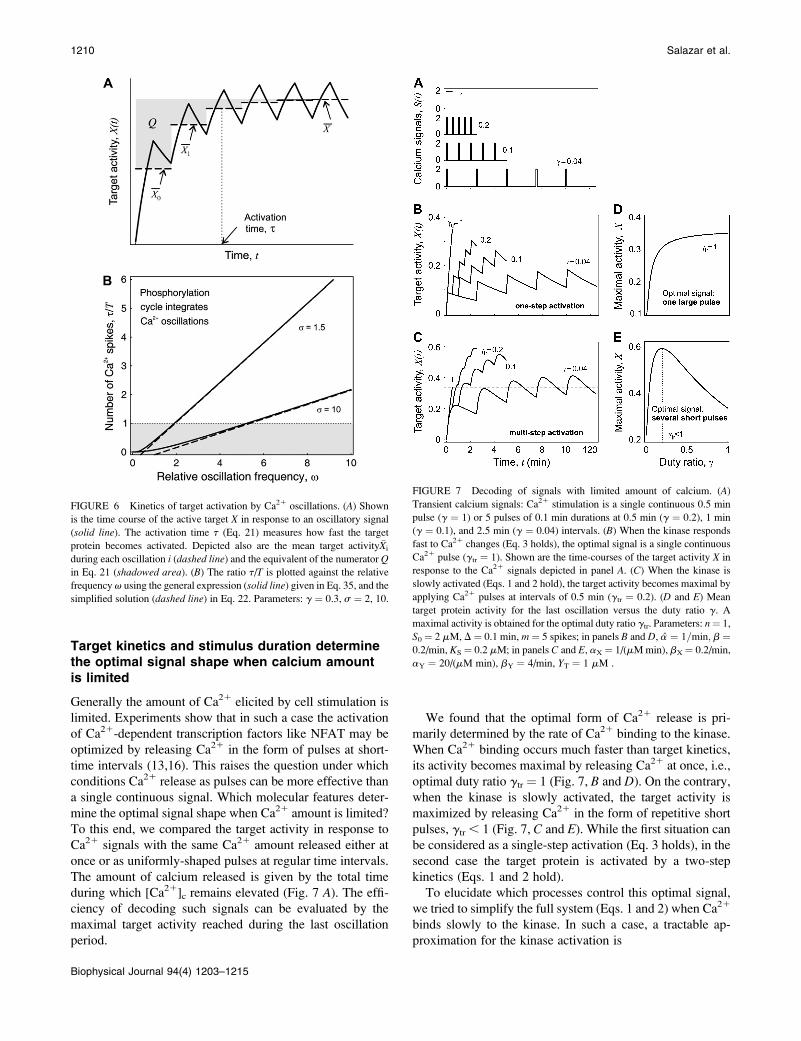
Target kinetics and stimulus duration determinethe optimal signal shape when calcium amountis limited
Generally the amount of Ca21 elicited by cell stimulation is
limited. Experiments show that in such a case the activation
of Ca21-dependent transcription factors like NFAT may be
optimized by releasing Ca21 in the form of pulses at short-
time intervals (13,16). This raises the question under which
conditions Ca21 release as pulses can be more effective than
a single continuous signal. Which molecular features deter-
mine the optimal signal shape when Ca21 amount is limited?
To this end, we compared the target activity in response to
Ca21 signals with the same Ca21 amount released either at
once or as uniformly-shaped pulses at regular time intervals.
The amount of calcium released is given by the total time
during which [Ca21]c remains elevated (Fig. 7 A). The effi-
ciency of decoding such signals can be evaluated by the
maximal target activity reached during the last oscillation
period.
We found that the optimal form of Ca21 release is pri-
marily determined by the rate of Ca21 binding to the kinase.
When Ca21 binding occurs much faster than target kinetics,
its activity becomes maximal by releasing Ca21 at once, i.e.,
optimal duty ratio gtr ¼ 1 (Fig. 7, B and D). On the contrary,
when the kinase is slowly activated, the target activity is
maximized by releasing Ca21 in the form of repetitive short
pulses, gtr , 1 (Fig. 7, C and E). While the first situation can
be considered as a single-step activation (Eq. 3 holds), in the
second case the target protein is activated by a two-step
kinetics (Eqs. 1 and 2 hold).
To elucidate which processes control this optimal signal,
we tried to simplify the full system (Eqs. 1 and 2) when Ca21
binds slowly to the kinase. In such a case, a tractable ap-
proximation for the kinase activation is
FIGURE 6 Kinetics of target activation by Ca21 oscillations. (A) Shown
is the time course of the active target X in response to an oscillatory signal
(solid line). The activation time t (Eq. 21) measures how fast the target
protein becomes activated. Depicted also are the mean target activity �Xi
during each oscillation i (dashed line) and the equivalent of the numerator Qin Eq. 21 (shadowed area). (B) The ratio t/T is plotted against the relative
frequency v using the general expression (solid line) given in Eq. 35, and the
simplified solution (dashed line) in Eq. 22. Parameters: g ¼ 0.3, s ¼ 2, 10.
FIGURE 7 Decoding of signals with limited amount of calcium. (A)
Transient calcium signals: Ca21 stimulation is a single continuous 0.5 min
pulse (g ¼ 1) or 5 pulses of 0.1 min durations at 0.5 min (g ¼ 0.2), 1 min
(g ¼ 0.1), and 2.5 min (g ¼ 0.04) intervals. (B) When the kinase responds
fast to Ca21 changes (Eq. 3 holds), the optimal signal is a single continuous
Ca21 pulse (gtr ¼ 1). Shown are the time-courses of the target activity X in
response to the Ca21 signals depicted in panel A. (C) When the kinase is
slowly activated (Eqs. 1 and 2 hold), the target activity becomes maximal by
applying Ca21 pulses at intervals of 0.5 min (gtr ¼ 0.2). (D and E) Mean
target protein activity for the last oscillation versus the duty ratio g. A
maximal activity is obtained for the optimal duty ratio gtr. Parameters: n¼ 1,
S0¼ 2 mM, D¼ 0.1 min, m ¼ 5 spikes; in panels B and D, a ¼ 1=min; b ¼0.2/min, KS ¼ 0.2 mM; in panels C and E, aX¼ 1/(mM min), bX¼ 0.2/min,
aY ¼ 20/(mM min), bY ¼ 4/min, YT ¼ 1 mM .
1210 Salazar et al.
Biophysical Journal 94(4) 1203–1215

YðtÞ ¼ �Yð1� e�t=tÞ; (23)
where �Y ¼ aYSn0g=ðaYSn
0g 1 bYÞis the stationary kinase
activity and t ¼ ðaYSn0g 1 bYÞ�1
its activation time. For the
target protein, one obtains
XðtÞ ¼ �X 1� e�bXt � bXt e
�t=t
1� bXt
!; (24)
where �X ¼ �YaX=bX is the stationary target activity. The
term in brackets gives the fraction of �X attained after time t.Changes in the duty ratio g exert opposite effects on these
two terms. On the one hand, the kinase activity �Y increases
with g leading to a higher value of �X: On the other hand,
because the total signal duration ttot ¼ mD/g (being m the
number of spikes) decreases with g, a lower fraction of �Xwill be reached. The optimal signal shape arises from the
tradeoff between both opposing effects.
Using Eq. 24, we determined how this optimal signal
shape depends on the kinetics of the phosphorylation cycle
and on the time during which [Ca21]c remains elevated. If
the target protein is rapidly dephosphorylated (bX� 1), the
optimal signal shape will then be characterized by a rapid
succession of spikes and a short total duration (Fig. 8). Con-
versely, for slowly responding targets, the optimal signal
shape has a slow spiking frequency and lasts for longer. This
optimal signal is also affected by the time mD during which
[Ca21]c remains elevated. We found that the larger this time,
the higher the optimal duty ratio will be (Fig. 8). These
analytical results were confirmed by numerical simulation of
the full system (compare in the solid squares with the solidlines in Fig. 8).
In summary, when Ca21 amount is limited, calcium release
in the form of spikes is more effective than a single continuous
release if target activation occurs in a two-step kinetics. This
condition is fulfilled when Ca21 binds slowly to the kinase.
The optimal signal shape is determined by the amount of
calcium and the kinetics of the phosphorylation cycle.
DISCUSSION
In this article, the decoding of Ca21 oscillations has been
theoretically analyzed in a minimal model of protein acti-
vation. Such a model comprises the activation-inactivation
cycle of a target protein controlled by a Ca21-dependent
kinase and the counteracting phosphatase. The mimicking of
Ca21 oscillations by square-shaped pulses allowed for an
analytical solution of the kinetic equations. To quantify how
sensitively the target protein responds to an oscillatory cal-
cium signal, we derived expressions for the mean target ac-
tivity and for the activation time. Both depend on three
dimensionless quantities that govern the response of the
system. Two of these dimensionless quantities (the effective
activation rate and the relative oscillation frequency) com-
bine kinetic properties of the target activation with charac-
teristics of the Ca21 signal. This fact indicates that the
timescales of Ca21 oscillations and target response are coupled
through the decoding mechanism and should not be analyzed
separately.
With our model we aimed to answer several questions
concerning the decoding of Ca21 oscillations:
How does a protein respond to changes in the oscillation
frequency and when does it function as a signal inte-
grator?
What are the advantages of having an oscillatory signal?
Under which conditions is the system response maxi-
mized?
To what extent do Ca21 oscillations confer target speci-
ficity?
These four issues will be discussed below.
Frequency sensitivity
Although an increase in the oscillation frequency, leaving the
amplitude and average unchanged, always causes an increase
in the mean target activity, the magnitude of these changes
depends on the inactivation rate of the target and on the duty
ratio of oscillations. In case target inactivation is faster than
the oscillation frequency, the target activity would simply
oscillate along with the Ca21 oscillation and the target does
not integrate the signal. By increasing the oscillation fre-
quency, such that Ca21 oscillations are faster than target in-
activation, the target protein would not fully inactivate between
each Ca21 oscillation and behaves as a signal integrator. In
FIGURE 8 Effect of kinetic parameters on the optimal transient signal.
Optimal duty ratio gtr of a transient Ca21 signal as a function of the
dephosphorylation rate constant bX of the target protein. The ratio aX/bX is
kept constant. If the target protein is rapidly inactivated (bX � 1), the
optimal Ca21 signal is characterized by a high duty ratio. The optimal duty
ratio will also increase by increasing the total duration of the Ca21 spikes
(upper line). The solid lines are calculated with the analytical approximation
(Eq. 24); the solid squares are obtained after numerically solving the full
system (Eqs. 2 and 3). Parameters: n ¼ 1, S0 ¼ 2 mM, D ¼ 0.2 and 0.4 min
(lower and upper line, respectively), m¼ 5 spikes; aX/bX/mM, aY¼ 1/(mM
min), bY ¼ 1/min, YT ¼ 1 mM.
Decoding of Ca21 Oscillations 1211
Biophysical Journal 94(4) 1203–1215

this case, one observes a continuous increase in the target
activity at each oscillation cycle. The critical point, over
which the target response becomes frequency-insensitive,
appears when the oscillation frequency gets in the range of
the target inactivation rate. Our analysis demonstrates that
true frequency decoding, at constant average Ca21 signal,
can indeed occur provided that Ca21 spikes are narrow and
the oscillation frequency is of the order of the target inac-
tivation rate or below.
Oscillations versus constant signals
We compared the target response to a constant signal and to a
sustained oscillatory signal of the same average Ca21 con-
centration. Our analysis demonstrates that Ca21 oscillations
are more potent in activating the target protein than a con-
stant signal if 1), Ca21 acts cooperatively on the kinase; and
2), the Ca21 sensitivity of the kinase (expressed by the dis-
sociation constant) lies around the peak concentration of the
calcium spike or above. Under these conditions, Ca21 oscil-
lations reduce the effective threshold for the target activation.
Taking into account the typical values for amplitude and
duty ratio of Ca21 oscillations, the predicted critical affinity
values lie in the range of the experimentally measured Ca21
sensitivities. Furthermore, we found that target proteins are
more efficiently activated by oscillatory signals at low levels
of stimulation irrespective of whether the information has
been encoded in the amplitude or in the frequency of oscil-
lations. This study provides a theoretical support to the ex-
perimental findings on Ca21-dependent gene expression by
Dolmetsch et al. (12) and on Ca21-dependent enzyme acti-
vation by Eshete and Fields (41), Kupzig et al. (42), and to
the numerical simulations by Gall et al. (26).
Optimal signals and target specificity
We also asked, what is the calcium signal shape that best
activates a particular target protein? Our analysis demon-
strates the existence of an optimal shape, which is deter-
mined by the Ca21 sensitivity and kinetics of target response.
Thus, by varying the characteristics of Ca21 oscillations,
target proteins can be differentially activated. To investigate
this issue, we compared two phosphorylation cycles with dis-
tinct Ca21 sensitivities and (in)activation kinetics. We found
that under specific conditions, Ca21 oscillations can upreg-
ulate one protein and downregulate the other one and vice
versa. If the cycles only differ in their (in)activation kinetics, the
slowly responding protein would be always stronger-activated
by Ca21 oscillations than the rapidly responding protein. Such
a behavior has been observed experimentally for genes regu-
lated by the two calcium-dependent transcription factors NFAT
and NFkB, where the latter factor responds slower to changes
in calcium concentration (12).
Cellular responses depend not only on the frequency and
amplitude of Ca21 oscillations but also on the duration and
number of Ca21 spikes (43). Hence, we examined the
specificity of target activation using time-limited Ca21
oscillations. Specifically, we compared the target response
when Ca21 stimulation of the same total duration, i.e., the
time during which calcium concentration remains elevated,
is applied as either a single continuous stimulation or as
pulses of short duration at distinct time intervals. Releasing
Ca21 at once maximizes the target response when kinase
(in)activation or target (de)phosphorylation is fast compared
to the duration of the Ca21 transient. However, when the
proteins respond slowly, the target activity becomes maxi-
mal by releasing Ca21 in the form of pulses with an optimal
frequency. We demonstrated analytically that this optimal
oscillation frequency arises from the tradeoff between two
opposing effects. On the one hand, the kinase activity in-
creases with the oscillation frequency leading to a higher
stationary target activity. On the other hand, the total signal
duration decreases with the frequency so that a lower fraction
of the stationary target activity will be reached. Similar
conclusions were arrived at by Marhl et al. on the basis of
numerical simulations (28,34).
Our theoretical analysis of target activation by transient
Ca21 oscillations is also consistent with the experiments of
Li et al. (16) and Tomida et al. (13), who found that the
nuclear localization of the transcription factor NFAT is
optimized when the Ca21 pulses are applied at short-time
intervals. A determinant factor to achieve this effect is the
temporal dissociation between Ca21 signals and nuclear
translocation of NFAT. Ca21-dependent dephosphorylation
of NFAT proceeds faster than its nuclear translocation and
rephosphorylation (44). Consequently, Ca21 oscillations can
induce a buildup of dephosphorylated NFAT in the cyto-
plasm, allowing nuclear translocation of NFAT even during
the interspike interval, provided that this interval is shorter
than the lifetime of dephosphorylated NFAT (13). These
experiments point out to the existence of a molecular Ca21
memory in the mechanism of NFAT activation, where an
oscillatory input is transformed into a nearly stationary
output.
Molecular sensors of calcium signals
Diverse experiments on Ca21 decoding have suggested the
existence of molecular sensors capable of interpreting com-
plex temporal Ca21 signals into the correct physiological
response. A classic example of such decoders is the small
molecule calmodulin, which activates several kinases as well
as the phosphatase calcineurin (14,17). More recently, the
kinase PKC (15,45) and the small GTPase Ras (46,47) have
been also proposed as potential Ca21 decoders. These sen-
sors contain specialized Ca21-binding domains such as the
C2 domain with a high structural diversity, allowing the
binding of multiple targets with distinct Ca21 sensitivities
and activation kinetics. Other elements to consider are the
compartmentalization and cross-interactions among signaling
1212 Salazar et al.
Biophysical Journal 94(4) 1203–1215

molecules. So the question arises: what is the ideal processor
for decoding complex Ca21 signals, and what minimal
features should it have?
Our study demonstrates that a system consisting of the
nonlinear regulation of a target protein by a Ca21-activated
kinase and the counteracting phosphatase contains the
minimal features required for deciphering temporal Ca21
signals. Autophosphorylation of the target protein (e.g., CaM
kinase II) can be considered as a special case of this model,
where the Ca21-dependent kinase is itself the target protein.
Yet despite its simplicity, this minimal model is able to
reproduce all features of Ca21 signaling decoding that have
been observed in detailed models (25,26,30). In our model,
nonlinear activation of the target protein arises from the
cooperative Ca21 binding to the kinase. Other mechanisms
that can generate nonlinear responses, such as multiple
phosphorylation and feedback regulatory loops, may also be
implicated in the decoding of Ca21 signals and should be
considered in future studies (48,49). Deciphering of complex
Ca21 signals presumably involves activation of multiple
Ca21 sensors instead of a central decoder. Complex decod-
ing patterns such as signal integration and summation might
emerge from combining single properties of the individual
decoders. Therefore, it would be worth extending this ap-
proach to a system consisting of multiple Ca21 decoders.
APPENDIX A: TARGET ACTIVITY ANDACTIVATION TIME
Here, we derive the formulas for the mean target activity �X and the activation
time t. The linear differential equation (Eq. 3), describing the kinetics of the
target protein X, can be separately solved for the spike and interspike
intervals (see Eq. 5). The solution of Eq. 3 reads
XðtÞ ¼ XiðzÞ ¼ e�ða01bÞz
Ai 1 Xmax; 0 # z , D
e�bz
Bi; D # z # T:
�(25)
For simplicity, we have introduced the internal time z ¼ t � iT within an
oscillation cycle z2[0,T]. The target activity during the ith cycle is denoted
by Xi(z). The phosphorylation rate a0 during a Ca21 spike (see Eq. 4) is
given by
a0 ¼ aðS0Þ ¼ aðS0=KSÞn
ðS0=KSÞn 1 1; (26)
and the maximal target activity is Xmax ¼ ða0Þ=ða0 1 bÞ: The coefficients Ai
and Bi are determined from
XiðTÞ ¼ Xi11ð0Þ and XiðD�Þ ¼ XiðD1 Þ; (27)
leading to the difference equations
Ai11 ¼ e�ða0D1bTÞAi � Xmaxð1� e�bðT�DÞÞ and
Bi ¼ e�a0D
Ai 1 XmaxebD: (28)
The initial condition X0(0) ¼ 0 gives A0 ¼ �Xmax. Equation 28 is solved
with the Ansatz Ai ¼ a 1 be�iða0D 1 bTÞ; where e�iða0D 1 bTÞ is a solution of
the homogeneous difference equation. One then obtains
Ai ¼ AN 1 1 e�ði11ÞðbT1a0DÞe
ða01bÞD � 1
1� e�bðT�DÞ
!: (29)
After a sufficiently large number of cycles (i / N), the coefficient Ai
approaches AN ¼ �Xmaxð1� e�bðT�DÞÞ= ð1� e�ðbT 1 a0DÞÞ: Equations 28
and 29 give the second coefficient
Bi ¼ BNð1� e�ði11ÞðbT1a0DÞÞ; (30)
with BN ¼ XmaxðebD � e�a0DÞ=ð1� e�ðbT1a0DÞÞ: The dynamics of the target
protein during the ith cycle is then described by
The quantities X1(z) and X�(z) denote the stationary target activity during a
Ca21 spike (0 # z , D) and during the interspike interval (D # z # T),
respectively,
X1 ðzÞ ¼ Xmax 1� 1� e�bðT�DÞ
1� e�ðbT1a0DÞe
�ða0 1 bÞz
!;
X�ðzÞ ¼ Xmax
ebD � e
�a0D
1� e�ðbT1a0DÞ
� �e�bz: (32)
After a sufficiently large number of oscillation cycles (i / N), the target
activity approaches X1(z) and X�(z).
The mean target activity �Xi during the ith cycle is defined by �Xi ¼ð1Þ=ðTÞ
R T
0XiðzÞdz: Thus, integration of Eq. 31 leads to
XðtÞ ¼ XiðzÞ ¼ X1 ðzÞð1� e
�iðbT1a0DÞÞ1 Xmaxð1� e�ða01bÞzÞe�iðbT1a0DÞ
; 0 # z , D
X�ðzÞð1� e
�ði11ÞðbT1a0DÞÞ; D # z # T:
�(31)
�Xi ¼ Xmax
D
T1
a0
bTða0 1 bÞð1� e
�Dða01bÞÞð1� e�bðT�DÞÞ
1� e�ðbT1a0DÞ
" #ð1� e
�iðbT1a0DÞÞ
1 e�iðbT1a0DÞXmax
D
T1ð1� e
�ða01bÞDÞ a0
a0 1 b� e
�bðT�DÞ� �bT
0@
1A: (33)
Decoding of Ca21 Oscillations 1213
Biophysical Journal 94(4) 1203–1215

According to Eq. 33, the mean target activity �X; i.e., for i / N, reads
�X ¼ Xmax
D
T1
a0
bTða0 1 bÞð1� e
�Dða01bÞÞð1� e�bðT�DÞÞ
1� e�ðbT1a0DÞ
" #:
(34)
Equation 34 is equivalent to Eq. 7, where the dimensionless parameters
s ¼ a0/b, v ¼ 1/bT, and g ¼ D/T have been introduced.
The activation time t of the target protein, defined in Eq. 21, can be
calculated using Eqs. 33 and 34. After some algebra, one obtains
t ¼ T
1� e�ða0D1bTÞ
� T½bD 1 ð1� e�ða01bÞDÞðXmax � e
�bðT�DÞÞ�bDð1� e
�ða0D1bTÞÞ1 Xmaxð1� e�ða01bÞDÞð1� e
�bðT�DÞÞ:
(35)
A Taylor expansion of Eq. 35 for T / 0, leaving D/T constant, yields
t � a0
D
T1 b
� ��1
�T � D
2: (36)
Equation 36 is equivalent to Eq. 22 after introducing the dimensionless
parameters.
APPENDIX B: OPTIMAL CA21 SIGNALS
The optimal signal shape at a given average �S is the solution of ð@ �XÞ=ð@gÞ ¼0; where the amplitude S0 has been replaced by S0 ¼ �S=g: Using Eq. 13, for
low-frequency oscillations (v / 0), one obtains the maximal target
response for
gmax ¼ �S=KS
ffiffiffiffiffiffiffiffiffiffiffiffiffiffiffiffiffia=b 1 1
n� 1
n
r: (37)
Using Eq. 14, for high-frequency oscillations (v / N), one gets
gmax ¼ �S=KS
ffiffiffiffiffiffiffiffiffiffiffi1
n� 1
n
r: (38)
Numerical evaluation of Eq. 7 shows that gmax is a decreasing function of v.
Thus, Eqs. 37 and 38 give the upper and lower bound for gmax, respectively.
Oscillations and a constant signal of equal average �S have the same
efficiency in activating the target protein when �X � ða�SnÞ=ððb 1 aÞ�Sn 1
bKnSÞ ¼ 0; where the second term corresponds to the target activity obtained
with the constant signal. Solving the above equation yields a critical
dissociation constant (KS/S0)crit. When KS/S0 . (KS/S0)crit, oscillations are
the more potent activating signals. For high frequency oscillations (v / N,
Eq. 14 holds), one gets
KS
S0
� �crit
¼
ffiffiffiffiffiffiffiffiffiffiffiffiffiffiffiffiffiffiffig
n � gn11
g � gn
n
s; (39a)
and
limg/1
KS
S0
� �crit
¼ 1ffiffiffiffiffiffiffiffiffiffiffin� 1np : (39b)
These formulas correspond to Eqs. 20 and 19, respectively. For low
frequencies (v / 0, Eq. 13 holds) the critical dissociation constant reads
KS
S0
� �crit
¼
ffiffiffiffiffiffiffiffiffiffiffiffiffiffiffiffiffiffiffiffiffiffiffiffiffiffiffiffiffiffiffiffiffiffiffiffiffiffiffiffig
n � gn 1 1
g � gn ða=b 1 1Þn
s; (40a)
and
limg/1
KS
S0
� �crit
¼ffiffiffiffiffiffiffiffiffiffiffiffiffiffiffiffiffia=b 1 1
n� 1
n
r: (40b)
Since �Xðv/0Þ # �X # �Xðv/NÞ; Eqs. 39 and 40 give the lower and upper
bound for (KS/S0)crit, respectively. Therefore, the condition for the
superiority of oscillations over a constant signal is ðKSÞ=ðS0Þ.ffiffiffiffiffiffiffiffiffiffiffiffiffiffiffiffiffiffiffiffiffiffiffiffiffiffiffiffiffiffiffiffiffiffiffiffiffiffiffiða=b 1 1Þ=ðn� 1Þ:n
pThis is a more strict condition than Eq. 19, which
is valid for any frequency or duty ratio.
The work was supported by the German Research Foundation (Deutschen
Forschungsgemeinschaft) through the Collaborative Research Center
Theoretical Biology (grant No. SFB 618) and by the German Federal
Ministry of Education and Research through the Systems Biology Com-
petence Network of Hepatocytes.
REFERENCES
1. Berridge, M. J., M. D. Bootman, and P. Lipp. 1998. Calcium—a lifeand death signal. Nature. 395:645–648.
2. Lewis, R. S. 2001. Calcium signaling mechanisms in T-lymphocytes.Annu. Rev. Immunol. 19:497–521.
3. Feske, S., H. Okamura, P. G. Hogan, and A. Rao. 2003. Ca21/calcineurin signaling in cells of the immune system. Biochem. Biophys.Res. Commun. 311:1117–1132.
4. Bito, H., and S. Takemoto-Kimura. 2003. Ca21/CREB/CBP-dependentgene regulation: a shared mechanism critical in long-term synapticplasticity and neuronal survival. Cell Calcium. 34:425–430.
5. Berridge, M. J., M. D. Bootman, and H. L. Roderick. 2003. Ca21
signaling: dynamics, homeostasis and remodeling. Nat. Rev. Mol. CellBiol. 4:517–529.
6. Gaspers, L. D., and A. P. Thomas. 2005. Calcium signaling in liver.Cell Calcium. 38:329–342.
7. Schofl, C., G. Brabant, R. D. Hesch, A. von zur Muhlen, P. H.Cobbold, and K. S. Cuthbertson. 1993. Temporal patterns of a1-receptor stimulation regulate amplitude and frequency of calciumtransients. Am. J. Physiol. 265:C1030–C1036.
8. D’Andrea, P., F. Codazzi, D. Zacchetti, J. Meldolesi, and F. Grohovaz.1994. Oscillations of cytosolic calcium in rat chromaffin cells: dualmodulation in frequency and amplitude. Biochem. Biophys. Res.Commun. 205:1264–1269.
9. Dolmetsch, R. E., R. S. Lewis, C. C. Goodnow, and J. I. Healy. 1997.Differential activation of transcription factors induced by Ca21 re-sponse amplitude and duration. Nature. 386:855–858.
10. Berridge, M. J. 2006. Calcium microdomains: organization andfunction. Cell Calcium. 40:405–412.
11. Rizzuto, R., and T. Pozzan. 2006. Microdomains of intracellular Ca21:molecular determinants and functional consequences. Physiol. Rev. 86:369–408.
12. Dolmetsch, R. E., K. Xu, and R. S. Lewis. 1998. Calcium oscillationsincrease the efficiency and specificity of gene expression. Nature. 392:933–936.
13. Tomida, T., K. Hirose, S. Takizawa, F. Shibasaki, and M. Iino. 2003.NFAT functions as a working memory of Ca21 signals in decodingCa21 oscillation. EMBO J. 22:3825–3832.
14. Frey, N., T. A. McKinsey, and E. N. Olson. 2000. Decoding calciumsignals involved in cardiac growth and function. Nat. Med. 6:1221–1227.
15. Reither, G., M. Schaefer, and P. Lipp. 2006. PKCa: a versatile key fordecoding the cellular calcium toolkit. J. Cell Biol. 174:521–533.
16. Li, W., J. Llopis, M. Whitney, G. Zlokarnik, and R. Y. Tsien. 1998.Cell-permeant caged InsP3 ester shows that Ca21 spike frequency canoptimize gene expression. Nature. 392:936–941.
17. De Koninck, P., and H. Schulman. 1998. Sensitivity of CaM kinase IIto the frequency of Ca21 oscillations. Science. 279:227–230.
1214 Salazar et al.
Biophysical Journal 94(4) 1203–1215

18. Hajnoczky, G., L. D. Robb-Gaspers, M. B. Seitz, and A. P. Thomas.1995. Decoding of cytosolic calcium oscillations in the mitochondria.Cell. 82:415–424.
19. Robb-Gaspers, L. D., P. Burnett, G. A. Rutter, R. M. Denton, R.Rizzuto, and A. P. Thomas. 1998. Integrating cytosolic calcium signalsinto mitochondrial metabolic responses. EMBO J. 17:4987–5000.
20. Dupont, G., G. Houart, and P. De Koninck. 2003. Sensitivity of CaMkinase II to the frequency of Ca21 oscillations: a simple model. CellCalcium. 34:485–497.
21. Goldbeter, A., G. Dupont, and M. J. Berridge. 1990. Minimal model forsignal-induced Ca21 oscillations and for their frequency encoding throughprotein phosphorylation. Proc. Natl. Acad. Sci. USA. 87:1461–1465.
22. Dupont, G., and A. Goldbeter. 1992. Protein phosphorylation driven byintracellular calcium oscillations: a kinetic analysis. Biophys. Chem.42:257–270.
23. Schuster, S., M. Marhl, and T. Hofer. 2002. Modeling of simple andcomplex calcium oscillations. Eur. J. Biochem. 269:1333–1355.
24. Dupont, G., and A. Goldbeter. 1998. CaM kinase II as frequencydecoder of Ca21 oscillations. Bioessays. 20:607–610.
25. Prank, K., L. Laer, A. von zur Muhlen, G. Brabant, and C. Schofl.1998. Decoding of intracellular calcium spikes train. Europhys. Lett.42:143–147.
26. Gall, D., E. Baus, and G. Dupont. 2000. Activation of the liverglycogen phosphorylase by Ca21 oscillations: a theoretical study. J.Theor. Biol. 207:445–454.
27. Rozi, A., and Y. Jia. 2003. A theoretical study of effects of cytosolicCa21 oscillations on activation of glycogen phosphorylase. Biophys.Chem. 106:193–202.
28. Marhl, M., M. Perc, and S. Schuster. 2005. Selective regulation ofcellular processes via protein cascades acting as band-pass filters fortime-limited oscillations. FEBS Lett. 579:5461–5465.
29. Marhl, M., and V. Grubelnik. 2007. Role of cascades in convertingoscillatory signals into stationary step-like responses. Biosystems.87:58–67.
30. Larsen, A. Z., L. F. Olsen, and U. Kummer. 2004. On the encoding anddecoding of calcium signals in hepatocytes. Biophys. Chem. 107:83–99.
31. Schuster, S., B. Knoke, and M. Marhl. 2005. Differential regulation ofproteins by bursting calcium oscillations—a theoretical study. Biosys-tems. 81:49–63.
32. Larsen, A. Z., and U. Kummer. 2003. Information processing incalcium signal transduction. Lect. Notes Phys. 5623:153–178.
33. Salazar, C., A. Politi, and T. Hofer. 2004. Decoding of calciumoscillations by phosphorylation cycles. In Proceedings of FourthInternational Workshop on Bioinformatics and Systems Biology. H.Mamitsuka, T. Smith, H. G. Holzhutter, M. Kanehisa, C. DeLisi, R.Heinrich, and S. Miyano, editors. Kyoto, Japan.
34. Marhl, M., M. Perc, and S. Schuster. 2006. A minimal model for decodingof time-limited Ca21 oscillations. Biophys. Chem. 120:161–167.
35. Li, Y., and A. Goldbeter. 1992. Pulsatile signaling in intercellularcommunication. Periodic stimuli are more efficient than random orchaotic signals in a model based on receptor desensitization. Biophys.J. 61:161–171.
36. Zhang, M., and T. Yuang. 1998. Molecular mechanisms of calmod-ulin’s functional versatility. Biochem. Cell Biol. 76:313–323.
37. Yang, J. J., A. Gawthrop, and Y. Ye. 2003. Obtaining site-specific calcium-binding affinities of calmodulin. Protein Pept. Lett. 10:331–345.
38. Thomas, A. P., G. S. Bird, G. Hajnoczky, L. D. Robb-Gaspers, andJ. W. Putney, Jr. 1996. Spatial and temporal aspects of cellular calciumsignaling. FASEB J. 10:1505–1507.
39. Rooney, T. A., E. J. Sass, and A. P. Thomas. 1989. Characterization ofcytosolic calcium oscillations induced by phenylephrine and vasopres-sin in single Fura-2-loaded hepatocytes. J. Biol. Chem. 264:17131–17141.
40. Heinrich, R., and T. A. Rapoport. 1975. Mathematical analysis ofmultienzyme systems. II. Steady state and transient control. Biosys-tems. 7:130–136.
41. Eshete, F., and R. D. Fields. 2001. Spike frequency decoding andautonomous activation of Ca21-Calmodulin-dependent protein kinaseII in dorsal root ganglion neurons. J. Neurosci. 21:6694–6705.
42. Kupzig, S., S. A. Walker, and P. J. Cullen. 2005. The frequencies ofcalcium oscillations are optimized for efficient calcium-mediatedactivation of Ras and the ERK/MAPK cascade. Proc. Natl. Acad.Sci. USA. 102:7577–7582.
43. Li, Y., G. X. Wang, M. Xin, H. M. Yang, X. J. Wu, and T. Li. 2004.The parameters of guard cell calcium oscillation encodes stomataloscillation and closure in Vicia faba. Plant Sci. 166:415–421.
44. Salazar, C., and T. Hofer. 2003. Allosteric regulation of the transcrip-tion factor NFAT1 by multiple phosphorylation sites: a mathematicalanalysis. J. Mol. Biol. 327:31–45.
45. Oancea, E., and T. Meyer. 1998. Protein kinase C as a molecular machinefor decoding calcium and diacylglycerol signals. Cell. 95:307–318.
46. Walker, S. A., P. J. Lockyer, and P. J. Cullen. 2003. The Ras binaryswitch: an ideal processor for decoding complex Ca21 signals?Biochem. Soc. Trans. 31:966–969.
47. Cullen, P. J. 2006. Decoding complex Ca21 signals through themodulation of Ras signaling. Curr. Opin. Cell Biol. 18:157–161.
48. Politi, A., L. D. Gaspers, A. P. Thomas, and T. Hofer. 2006. Models ofIP3 and Ca21 oscillations: frequency encoding and identification ofunderlying feedbacks. Biophys. J. 90:3120–3133.
49. Salazar, C., and T. Hofer. 2007. Versatile regulation of multisiteprotein phosphorylation by the order of phosphate processing andprotein-protein interactions. FEBS J. 274:1046–1061.
50. Cifuentes, M. E., L. Honkanen, and M. J. Rebecchi. 1993. Proteolyticfragments of phosphoinositide-specific phospholipase Cd1. Catalyticand membrane binding properties. J. Biol. Chem. 268:11586–11593.
51. Rebecchi, M. J., R. Eberhardt, T. Delaney, S. Ali, and R. Bittman.1993. Hydrolysis of short acyl chain inositol lipids by phospholipaseCd 1. J. Biol. Chem. 268:1735–1741.
52. Kohout, S. C., S. Corbalan-Garcia, A. Torrecillas, J. C. Gomez-Fernandez, and J. J. Falke. 2002. C2 domains of protein kinase Cisoforms a, b, and g: Activation parameters and calcium stoichiom-etries of the membrane-bound state. Biochemistry. 41:11411–11424.
53. Linse, S., A. Helmersson, and S. Forsen. 1991. Calcium binding tocalmodulin and its globular domains. J. Biol. Chem. 266:8050–8054.
54. Bradshaw, J. M., Y. Kubota, T. Meyer, and H. Schulman. 2003. Anultrasensitive Ca21/calmodulin-dependent protein kinase II-proteinphosphatase 1 switch facilitates specificity in postsynaptic calciumsignaling. Proc. Natl. Acad. Sci. USA. 100:10512–10517.
55. Stemmer, P. M., and C. B. Klee. 1994. Dual calcium ion regulation ofcalcineurin by calmodulin and calcineurin B. Biochemistry. 33:6859–6866.
56. da Silva, A. C., A. H. de Araujo, O. Herzberg, J. Moult, M. Sorenson,and F. C. Reinach. 1993. Troponin-C mutants with increased calciumaffinity. Eur. J. Biochem. 213:599–604.
57. Vinogradov, A., and A. Scarpa. 1973. The initial velocities of calciumuptake by rat liver mitochondria. J. Biol. Chem. 248:5527–5531.
58. Holmes, W. R. 2000. Models of calmodulin trapping and CaM kinaseII activation in a dendritic spine. J. Comput. Neurosci. 8:65–85.
Decoding of Ca21 Oscillations 1215
Biophysical Journal 94(4) 1203–1215