Déchiffrage du rôle de la protéine XPC dans la réparation par ...
271
THÈSE Pour obtenir le grade de DOCTEUR DE L’UNIVERSITE GRENOBLE ALPES Spécialité : BIS - Biotechnologie, instrumentation, signal et imagerie pour la biologie, la médecine et l'environnement Arrêté ministériel : 25 mai 2016 Présentée par Nour FAYYAD Thèse dirigée par le professeur Walid RACHIDI, Université Grenoble Alpes (UGA) Préparée au sein du DRF/IRIG/DIESE/SyMMES/CIBEST-CEA, Grenoble Dans l'École doctorale Ingénierie pour la santé la Cognition et l'Environnement (EDISCE) Déchiffrage du rôle de la protéine XPC dans la réparation par excision de base (BER) et le stress oxydant Thèse soutenue publiquement le 30 Juin 2021, devant le jury composé de : Monsieur Walid RACHIDI Professeur, UGA, directeur de thèse Monsieur Michel SEVE Professeur, UGA, Président Madame Patricia ROUSSELLE Directrice de recherche, CNRS, Examinatrice Madame Pascale COHEN Professeur, Université de Lyon, Rapporteur Madame Anna CAMPALANS Chercheur, CEA, Rapporteur
-
Upload
khangminh22 -
Category
Documents
-
view
4 -
download
0
Transcript of Déchiffrage du rôle de la protéine XPC dans la réparation par ...

THÈSE
Pour obtenir le grade de
DOCTEUR DE L’UNIVERSITE GRENOBLE ALPES
Spécialité : BIS - Biotechnologie, instrumentation, signal et imagerie
pour la biologie, la médecine et l'environnement
Arrêté ministériel : 25 mai 2016
Présentée par
Nour FAYYAD
Thèse dirigée par le professeur Walid RACHIDI, Université Grenoble
Alpes (UGA)
Préparée au sein du DRF/IRIG/DIESE/SyMMES/CIBEST-CEA,
Grenoble
Dans l'École doctorale Ingénierie pour la santé la Cognition et
l'Environnement (EDISCE)
Déchiffrage du rôle de la protéine XPC
dans la réparation par excision de base
(BER) et le stress oxydant
Thèse soutenue publiquement le 30 Juin 2021,
devant le jury composé de :
Monsieur Walid RACHIDI Professeur, UGA, directeur de thèse
Monsieur Michel SEVE Professeur, UGA, Président
Madame Patricia ROUSSELLE Directrice de recherche, CNRS, Examinatrice Madame Pascale COHEN Professeur, Université de Lyon, Rapporteur
Madame Anna CAMPALANS Chercheur, CEA, Rapporteur

1
Abstract
eroderma pigmentosum C (XPC) protein initiates global genome-nucleotide excision
repair (GG-NER) pathway to remove UV-induced DNA lesions such as pyrimidine
(6-4) pyrimidone photoproducts [(6-4) PPs] and cyclobutane pyrimidine dimers
(CPDs). XPC deficient (XP-C) patients show a persistence of such lesions triggering high skin
cancer incidences. They also suffer from internal cancers that could be due to the accumulation
of oxidative DNA damage. Such base lesions, including 8-oxoguanine (8-oxoGua), are usually
repaired by the base excision repair (BER) pathway. Despite growing evidence about how XPC
enhances the activity of several BER DNA glycosylases, the effect of XPC mutations on other
BER factors and their activities is still elusive. Herein, we seek to answer this open question
by characterizing normal and XP-C fibroblasts derived from patients, optimizing the
conditions, and dividing our project into two parts.
In part one, we showed a global downregulation of BER's genes in XP-C cells post-UVB
compared to normal controls. Furthermore, the major proteins linked to oxidative DNA damage
repair (OGG1, MYH, and APE1) were downregulated. This led to an ineffectiveness of BER
in excising UVB-induced oxidative DNA damage. In part two, we investigated whether
balancing the cellular redox state by treating XP-C cells with different drugs could boost their
BER's activity post-UVB. We showed that nicotinamide (NIC) and N-acetyl cysteine (NAC)
pretreatments increase glutathione level, decrease ROS level, and enhance BER's gene
expression and activity. Meanwhile, buthionine sulfoximine/dimethylfumuate (BSO/DMF)
pretreatment depletes glutathione level, increases ROS level, and impairs BER's gene
expression and activity.
Based on these results, we propose that the pretreatment with drugs that could enhance
glutathione's level may protect XP-C cells from an imbalanced redox state that affects the DNA
repair. This could pave the way for therapeutic strategies for XP-patients and other DNA repair-
deficient patients.
Future work is required to check the efficiency of such treatments on 3D reconstructed skin
and in vivo models. Additionally, studying the interactome linking XPC and glutathione
signaling could be interesting.
Keywords: Ultraviolet irradiation-B (UVB), xeroderma pigmentosum C (XPC), nucleotide excision repair (NER), bulky
lesions [CPDs and (6-4) PPs)], base excision repair (BER), oxidative DNA lesions (8-oxoguanine), reactive oxygen species
(ROS), oxidative stress, glutathione (GSH), nicotinamide (NIC), N-acetylcysteine (NAC), buthionine sulfoximine/dimethyl
fumarate (BSO/DMF)
X

2
Résumé
La protéine Xeroderma pigmentosum C (XPC) initie la réparation globale du génome par
excision de nucléotides (GG-NER) pour éliminer les lésions de l'ADN induites par les
rayonnements UV, telles que les photoproduits de pyrimidine (6-4) [(6-4) PPs] et les dimères
de cyclobutane de pyrimidine (CPDs). Les patients déficients en XPC (XP-C) présentent une
persistance de ces lésions, déclenchant ainsi une forte incidence de cancers cutanés. Ces
patients souffrent également de cancers internes qui pourraient être dus à l'accumulation de
lésions d’oxydation de l'ADN. Ces dernières, dont la 8-oxoguanine (8-oxoGua), sont
généralement réparées par excision de bases (BER). Malgré les preuves, de plus en plus
tangibles, concernant l’implication de la protéine XPC dans l'activité de plusieurs glycosylases
clés de la voie BER, l'effet des mutations de XPC sur les autres facteurs de cette voie reste
encore peu connu. Le but de ce travail de thèse est de répondre à cette question ouverte en
caractérisant la modulation de la voie BER dans les cellules normales et les cellules XP-C
issues de patients.
Dans un premier temps, nous avons montré un effondrement global de l’expression de plusieurs
gènes importants de la voie BER dans les cellules XP-C par rapport aux cellules témoins après
irradiation aux UVB. En outre, les principales protéines liées à la réparation des dommages
d’oxydation de l'ADN (OGG1, MYH, et APE1) ont été déréglées. Cela a conduit à une
inefficacité du BER dans l'excision des purines oxydées induites par les UVB. Dans un
deuxième temps, nous avons cherché à savoir si la modulation de l'état redox en traitant les
cellules avec différents médicaments pharmacologiques pouvait restaurer l'activité de BER
après irradiation aux UVB. Nous avons montré que les prétraitements par le nicotinamide
(NIC) et le N-acétyl cystéine (NAC) augmentent le niveau de glutathion, diminuent la
génération des espèces réactives de l'oxygène (ROS), et augmentent l'activité du BER après
irradiation aux UVB. Cependant, le prétraitement à la buthionine sulfoximine/diméthylfumate
(BSO/DMF) inhibe le glutathion, augmente la production des ROS, et diminue l'activité du
BER.
Sur la base de ces résultats, nous pourrions proposer que le prétraitement avec des médicaments
qui pourraient augmenter le niveau de glutathion puisse protéger les cellules XP-C d'un état
redox déséquilibré qui affecte la réparation de l'ADN. Cela pourrait ouvrir la voie à des
stratégies thérapeutiques pour les patients XP et d'autres patients souffrant des maladies
génétiques de réparation de l'ADN.
Des travaux futurs sont nécessaires pour vérifier l'efficacité de ces traitements au niveau de la
peau reconstruite en 3D et sur des modèles pré-cliniques in vivo. En outre, l'étude de
l'interactome reliant XPC et la signalisation du glutathion pourrait être intéressante.
Mots-clés : Rayonnement ultraviolet B (UVB), xeroderma pigmentosum C (XPC), réparation par excision de nucléotides
(NER), lésions de l'ADN [CPD et (6-4) PP)], réparation par excision de bases (BER), lésions d’oxydation de l'ADN (8-
oxoguanine), espèces réactives de l'oxygène (ROS), stress oxydatif, glutathion (GSH), nicotinamide (NIC), N-acétylcystéine
(NAC), buthionine sulfoximine/fumarate de diméthyle (BSO/DMF)

3
"I am among those who think that science has great beauty. A scientist in his laboratory is not only a technician: he is also a child placed before
natural phenomena which impress him like a fairy tale."
~Marie Curie

4
Acknowledgment
First, I would like to express my gratitude to my supervisor, Pr. Walid RACHIDI, for his
invaluable supervision, immense knowledge, and support during my PhD degree. I would also
like to thank Professor Bassam BADRAN and doctors Hussein and Mohammad KAZAN, from
the Lebanese University, for their support whenever needed.
Furthermore, I would like to thank my CSI members, doctors Hamid Reza REZVANI and
Christine DEMEILLIERS, from who I benefited knowledge and experiences. They were
incredibly helpful and friendly. I enjoyed our meetings.
My sincere thanks also go to professors Michele SEVE, Pascale COHEN, Patricia
ROUSSELLE, and Doctor Anna CAMPALANS for accepting to be members of my PhD's
committee. I also appreciate the doctoral school, EDISCE, for the funding and needed
guidance, and he CEA, especially Doctor Frédéric CHANDEZON, and to our collaborators.
I was blessed to be supported by my team, CIBEST. They were always friendly, kind, and
helpful. I will never forget the support and guidance of doctors Pascale DELANGLE, Thierry
DOUKI, Jean Luc RAVANAT, Marie CARRIERE, and Jean BRETON. Sylvain CAILLAT
was my IT hero. David BEAL helped me a lot and was always there for me when needed. I
learned a lot from him, and I enormously appreciate everything he did for me during my studies.
I want to thank my friends for their support, particularly Abir and Anna.
Finally, I am indebted to my family. My mother, father, brother, and sister-in-law are my
everything, and I was blessed to have their constant support in every step I make. They have
always believed in me. I could not ask for more than their love and encouragement.
"Mom, I dedicate this PhD for you."
I was also blessed to have the constant support of my love, Hasan. His tremendous
understanding, love, and encouragement always motivated and strengthened me. He never
stopped believing in me and always encouraged me; for that, I love him endlessly.

5
Table of Contents
Abstract ...................................................................................................................................... 1
Résumé ....................................................................................................................................... 2
Acknowledgment ....................................................................................................................... 4
Table of Contents ....................................................................................................................... 5
List of Figures ............................................................................................................................ 9
List of Tables ........................................................................................................................... 13
List of Abbreviations ............................................................................................................... 14
Preamble .................................................................................................................................... 1
Bibliographic Review ................................................................................................................ 4
1. Chapter One: ROS, Oxidative stress, and the skin....................................................... 5
1.1. Definition and origin ................................................................................................... 5
1.1.1. Oxidative stress targets ........................................................................................ 7
1.1.2. Endogenous sources for most common ROS..................................................... 10
1.1.3. Exogenous sources ............................................................................................. 12
1.1.4. ROS and signaling pathways ............................................................................. 26
1.2. Defense mechanisms against oxidative stress ........................................................... 28
1.3. Pathologies linked to oxidative stress ........................................................................... 33
1.4. Summary for ROS......................................................................................................... 34
2. Chapter Two: The Base Excision Repair Pathway (BER) ............................................. 35
2.1. Overview of BER pathway ....................................................................................... 35
2.2. BER factors roles and post-translational modifications ............................................ 37
2.3. BER and human disorders: aging, oxidative stress, and ROS .................................. 42
2.4. BER variants/mutations and cancer (skin and internal) ............................................ 43
2.5. BER and cell cycle .................................................................................................... 45
2.6. BER targeted treatment: The debased side of BER .................................................. 46
2.7. BER and Drugs: Acetohexamide (ACETO) ............................................................. 48
2.8. BER and other repair pathways ................................................................................. 49
3. Chapter Three: Nucleotide Excision Repair (NER) ....................................................... 52
3.1. Overview of NER pathway ........................................................................................... 52
3.1.1. Photoproducts: CPDs vs (6-4) PPs ......................................................................... 53
3.1.2. NER factors roles and post-translational modifications ........................................ 55
3.2. NER and cell cycle........................................................................................................ 57
3.3. NER and human disorders ............................................................................................ 59
4. Chapter Four: XPC the Bridge Between BER and NER ............................................... 61

6
4.1. XPC expression and interactions .................................................................................. 61
4.2. XPC’s regulation ........................................................................................................... 62
4.3. XPC’s role ..................................................................................................................... 63
4.3.1. Canonical role ........................................................................................................ 63
4.3.2. Other roles .............................................................................................................. 63
4.4. XPC disorder ................................................................................................................. 67
4.4.1. Clinical features ..................................................................................................... 67
4.4.2. Clinical treatments ................................................................................................. 68
4.5. XPC mutations/polymorphic variants and cancer......................................................... 70
5. Representative Summary .................................................................................................. 73
Materials and Methods ............................................................................................................. 74
1. Cell culture and treatments ........................................................................................... 75
1.1. Cell culture ......................................................................................................... 75
1.2. Cell treatments ................................................................................................... 78
1.3. Immunocytochemistry (Immunofluorescence) and associated microscopy ...... 80
1.4. Short-term cytotoxicity (MTT) .......................................................................... 81
2. Transcriptional and translational genes’ expressions ................................................... 82
2.1. Gene expression by RT-qPCR ........................................................................... 82
2.2. Western blot ....................................................................................................... 84
3. Detection of DNA lesions ............................................................................................. 85
3.1. HPLC-MS/MS: detection of bulky lesions ........................................................ 85
3.2. Comet Assay ± Fpg: detection of oxidized purines (8-oxoGua) ....................... 86
4. Studying oxidative stress .............................................................................................. 87
▪ Part Two “Ameliorating the DNA repair of XP-C cells by modulating their
redox state via pharmacological treatments” .............................................................. 87
4.1. ROS assay .......................................................................................................... 87
4.2. Glutathione assay ............................................................................................... 87
Results & Discussion ............................................................................................................... 89
Part One “Deciphering the Role of XPC in BER and Oxidative Stress” ......................... 90
1. Chapter One: Characterization of Primary Fibroblasts ................................................. 91
1.1. Impaired XPC gene and protein expression in XP-C fibroblasts ...................... 91
1.2. Impaired NER capacities in XP-C primary fibroblasts compared to control cells
92
1.3. Similar UVB-induced photosensitivity between XP-C and normal primary
fibroblasts ......................................................................................................................... 95
1.4. Higher ROS level in XP-C1 fibroblasts ............................................................. 97
1.5. Higher photoresistance in XP-C1 fibroblasts to solar simulation ...................... 98
Briefing of the characterization .................................................................................... 99

7
2. Chapter Two: Effect of XP-C Mutations on BER’s Expression and Excision Activity
100
2.1. Effect of XP-C on BER’s mRNA expression post-UVB........................................ 100
2.2. Effect of XPC mutations on BER’s protein expression post-UVB ........................ 105
2.3. Effect of XP-C on BER’s activity post-UVB ......................................................... 107
3. Chapter Three: Effect of XP-C on Some Genes Linked to Cell Cycle Regulation ....... 113
Conclusion and Perspective ............................................................................................................. 117
Schematic Summary ................................................................................................................. 118
Part Two “Ameliorating the DNA repair of XP-C cells by modulating their redox state
via pharmacological treatments”........................................................................................ 119
1. Chapter One: Characterization of Cell lines ............................................................... 121
1.1. Impaired XPC gene and protein expression in XP-C cell line......................... 121
1.2. Higher UVB-induced photosensitivity in XP-C cells compared to control ..... 122
1.3. Impaired NER capacities in XP-C cells compared to control .......................... 123
Briefing of the characterization .................................................................................. 125
2. Chapter Two: The Effect of Nicotinamide (NIC) on UVB-Induced Oxidative Stress
and DNA Repair in XP-C and Normal Cells ..................................................................... 126
2.1. Characterization of NIC-treated normal and XP-C cells ................................. 126
2.2. Effect of NIC on XP-C and normal cells’ redox state ..................................... 128
2.3. Effect of NIC on XP-C and normal cells’ PARP1 protein expression ............ 133
2.4. Effect of NIC on XP-C and normal cells’ P53 gene expression ...................... 136
2.5. Effect of NIC on XP-C and normal cells’ BER gene expression .................... 137
2.6. Effect of NIC on XP-C and normal cells’ UVB-induced BER and NER activity
141
Conclusion and Perspective ..................................................................................................... 145
3. Chapter Three: The effect of N-acetylcysteine (NAC) on UVB-Induced Oxidative
Stress and DNA Repair in XP-C and Normal Cells .......................................................... 146
3.1. Characterization of NAC-treated normal and XP-C cells ................................ 146
3.2. Effect of NAC on XP-C and normal cells’ redox state .................................... 148
3.3. Effect of NAC on XP-C and normal cells’ PARP1 protein expression ........... 153
3.4. Effect of NAC on XP-C and normal cells’ P53 gene expression .................... 155
3.5. Effect of NAC on XP-C and normal cells’ BER gene expression ................... 156
3.6. Effect of NAC on XP-C and normal cells’ UVB-induced BER and NER activity
158
Conclusion and Perspective ..................................................................................................... 162
4. Chapter Four: The Effect of Buthionine sulfoximine/Dimethylfumurate (BSO/DMF)
on UVB-Induced Oxidative Stress and DNA Repair in XP-C and Normal Cells ............. 163
4.1. Characterization of BSO/DMF-treated normal and XP-C cells ...................... 163
4.2. Effect of BSO/DMF on XP-C and normal cells’ redox state........................... 165

8
4.3. Effect of BSO/DMF on XP-C and normal cells’ PARP1 protein expression.. 169
4.4. Effect of BSO/DMF on XP-C and normal cells’ P53 gene expression ........... 170
4.5. Effect of BSO/DMF on XP-C and normal cells’ BER’s gene expression ....... 172
4.6. Effect of BSO/DMF on XP-C and normal cells’ UVB-induced BER and NER
activity 175
Conclusion and Perspective ..................................................................................................... 178
Schematic Summary .............................................................................................................. 179
General Discussion ................................................................................................................ 180
• Part One “Deciphering the Role of XPC in BER and Oxidative Stress” ............ 180
• Part Two “Ameliorating the DNA repair of XP-C cells by modulating their redox
state via pharmacological treatments”........................................................................... 183
Conclusion and Perspectives.................................................................................................. 188
Proposed schematic summary ................................................................................................ 189
References .............................................................................................................................. 190
Annex 1-Preliminary Results ................................................................................................. 218
• Part One “Deciphering the Role of XPC in BER and Oxidative Stress” ............ 218
Annex 2-Research Article ...................................................................................................... 220
Annex 3 -Review Article ....................................................................................................... 234
Annex 4 -Other Activities ...................................................................................................... 253
Abstract .................................................................................................................................. 254
Résumé ................................................................................................................................... 255

9
List of Figures
Bibliographic Review
Figure 1. Effect of different ROS exposure concentrations on the cell
Figure 2. ROS and its DNA damage repair
Figure 3. Summary of the ROS production from different cellular sources and some of the main
types
Figure 4. Electron transport chain with electron and proton leakage
Figure 5. A modified scheme showing the different skin cancers (SCC, BCC, and melanoma)
arising post-solar irradiation
Figure 6. Distribution of CPDs (<>) and (6-4) PPs (6-4) at the four possible bipyrimidine sites
within the DNA upon UVA vs UVB
Figure 7. A scheme presenting the effect of UV on DNA
Figure 8. Skin layer components and UV
Figure 9. A modified scheme representing intrinsic aging vs photoaging
Figure 10. ROS: mechanisms of actions and alterations
Figure 11. A simplified schematic representation of BER pathway
Figure 12. Post-translational modifications (PTMs) of BER factors
Figure 13. Cell cycle regulated BER genes
Figure 14. A modified schematic representation of NER pathway
Figure 15. Post-translational modifications (PTMs) of NER factors
Figure 16. The link between NER mutated genes and 3 different disorders (Xeroderma
Pigmentosum "XP", Trichothyodystrophy "TTD", and Cockayne syndrome "CS")
Figure 17. A modified schematic representation of the human XPC protein
Figure 18. A pie chart categorizing interactors/roles of XPC
Figure 19. Clinical features of a Mahori XP-C patient at 6 and 8 years-old
Figure 20. XP-C patients' precautions and proposed treatments
Figure 21. CRISPR/pass mechanism of action
Representative Summary
Figure 22. Schematic summary of the role of XPC post-UV irradiation
Materials and Methods
Figure 23. Morphological images of normal and XP-C primary fibroblasts
Figure 24. Morphological images of normal and XP-C SV40 transformed fibroblasts
Figure 25. A summary of the thesis workflow
Figure 26. Cellular treatments and collection
Figure 27. Bioblock Scientific with UVB lamp (312nm, 15W) used by our laboratory,
CIBEST/CEA
Figure 28. LS1000 Solar Simulator used by our laboratory, CIBEST/CEA
Figure 29. Cell viability versus different UVB doses (J/cm²)
Figure 30. The CFX96 thermal cycler C1000-touch used by our laboratory, CIBEST/CEA
Figure 31. Western blot instruments used by our laboratory, CIBEST/CEA
Results and Discussion
Part one
Figure 32. Lower XPC mRNA level in XP-C fibroblasts compared to normal control, at basal
level

10
Figure 33. Absence of XPC protein in XP-C primary fibroblasts compared to normal control,
at basal level
Figure 34. Deficient (6-4) PPs repair in XP-C primary fibroblasts compared to normal control,
post-UVB irradiation
Figure 35. Kinetics of bulky lesions' [CPDs and (6-4) PPs] repair in XP-C1 versus normal
fibroblast post-UVB irradiation
Figure 36. Similar photosensitivity between normal and XP-C primary fibroblasts
Figure 37. Kinetics of ROS level in XP-C1 and normal primary fibroblasts
Figure 38. Higher photo-resistance in XP-C1 primary fibroblast compared to normal control
Figure 39. Downregulated BER-associated gene transcription in XP-C fibroblasts compared to
normal control, post-UVB irradiation
Figure 40. Downregulated BER-associated accessory gene transcription in XP-C fibroblasts
compared to normal control, post-UVB irradiation
Figure 41. Downregulated BER-associated OGG1 protein level in XP-C fibroblasts compared
to normal control, post-UVB irradiation
Figure 42. Downregulated BER-associated MYH protein level in XP-C fibroblasts compared
to normal control, post-UVB irradiation
Figure 43. Downregulated BER-associated APE1 protein level in XP-C fibroblasts compared
to normal control, post-UVB irradiation
Figure 44. The undamaged (comet head) and damaged (comet tail) DNA ± FPG in normal and
XP-C1 fibroblasts and positive control H2O2
Figure 45. Downregulated BER excision repair capacities in XP-C primary fibroblast
compared to normal fibroblasts, post-UVB irradiation Figure 46. A simple graphical representation of BER excision repair in XP-C primary
fibroblasts compared to normal fibroblasts, post-UVB irradiation
Figure 47. Different P53 mRNA level between XP-C and normal fibroblasts
Figure 48. Different BER-associated P53 protein level in XP-C fibroblasts compared to normal
control, post-UVB irradiation
Figure 49. Different GADD45a mRNA level between XP-C and normal fibroblasts
Schematic Summary
Figure 50. The difference between normal and XP-C fibroblasts in response to UVB stress
Part Two
Figure 51. Impaired XPC mRNA and protein expressions in XP-C cells compared to normal
control, at basal level
Figure 52. Higher photosensitivity in XP-C cells compared to normal control
Figure 53. Deficient (6-4) PPs lesions repair in XP-C cells compared to normal control
Figure 54. Deficient CPDs lesions repair in XP-C cells compared to normal control
• Nicotinamide (NIC)
Figure 55. NIC dose response curve
Figure 56. Similar UVB-induced photosensitivity in each cell line (normal and XP-C), with
and without NIC
Figure 57. Effect of NIC on ROS level in normal and XP-C cells, post-UVB irradiation
Figure 58. The turnover of glutathione (GSH)
Figure 59. Effect of NIC on the UVB-induced IR/NIR glutathione (GSH) level in normal and
XP-C cells
Figure 60. Effect of NIC on detoxificants' mRNA expression at basal and UVB levels in normal
and XP-C cells

11
Figure 61. Effect of NIC on Nrf2 mRNA expression at basal and UVB levels in normal and
XP-C cells
Figure 62. Effect of NIC on Nrf2 protein expression at basal and UVB levels in normal and
XP-C cells
Figure 63. Effect of NIC on PARP1 protein expression at basal and UVB levels in normal and
XP-C cells
Figure 64. Band images showing the effect of NIC on PARP1 protein expression at basal and
UVB levels in normal and XP-C cells
Figure 65. Effect of NIC on cleaved PARP1 protein expression at basal and UVB levels in
normal and XP-C cells
Figure 66. Effect of NIC on P53 mRNA expression at basal and UVB levels in normal and XP-
C cells
Figure 67. Effect of NIC on P53 protein expression at basal and UVB levels in normal and XP-
C cells
Figure 68. Effect of NIC on BER mRNA expression at basal and UVB levels in normal and
XP-C cells
Figure 69. Effect of NIC on BER protein expression at basal and UVB levels in normal and
XP-C cells
Figure 70. Effect of different treatments (NIC, NAC, BSO/DMF) on BER protein expression
at basal and UVB levels in normal and XP-C cells
Figure 71. Effect of NIC on UVB-induced-(6-4) PPs kinetic repair in normal and XP-C cells
Figure 72. Effect of NIC on UVB-induced-CPDs kinetic repair in normal and XP-C cells
Figure 73. Effect of NIC on alkaline and oxidized purines repair in normal and XP-C cells,
post-UVB irradiation
• N-acetylcysteine (NAC)
Figure 74. NAC dose response curve
Figure 75. Similar UVB-induced photosensitivity in each cell line (normal and XP-C), with
and without NAC
Figure 76. Effect of NAC on ROS level in normal and XP-C cells, post-UVB irradiation.
Figure 77. The induced IR/NIR glutathione (GSH) level in normal and XP-C cells
Figure 78. Effect of NAC on the UVB-induced IR/NIR glutathione (GSH) level in normal and
XP-C cells
Figure 79. Effect of NAC on the detoxificants' mRNA expression at basal and UVB levels in
normal and XP-C cells
Figure 80. Effect of NAC on the Nrf2 mRNA expression at basal and UVB levels in normal
and XP-C cells
Figure 81. Effect of NAC on the Nrf2 protein expression at basal and UVB levels in normal
and XP-C cells
Figure 82. Effect of NAC on the PARP1 protein expression at basal and UVB levels in normal
and XP-C cells
Figure 83. Effect of NAC on cleaved PARP1 protein expression at basal and UVB levels in
normal and XP-C cells
Figure 84. Effect of NAC on the P53 mRNA expression at basal and UVB levels in normal
and XP-C cells
Figure 85. Effect of NAC on the P53 protein expression at basal and UVB levels in normal and
XP-C cells
Figure 86. Effect of NAC on BER mRNA expression at basal and UVB levels in normal and
XP-C cells

12
Figure 87. Effect of NAC on BER protein expression at basal and UVB levels in normal and
XP-C cells
Figure 88. Effect of NAC on UVB-induced (6-4) PPs kinetic repair in normal and XP-C cells
Figure 89. Effect of NAC on UVB-induced CPDs kinetic repair in normal and XP-C cells
Figure 90. Effect of NAC on alkaline and oxidized purines repair in normal and XP-C cells,
post-UVB irradiation
• Buthionine sulfoximine/Dimethylfumurate (BSO/DMF)
Figure 91. BSO/DMF dose response curve
Figure 92. Higher photosensitivity in each cell line (normal and XP-C) in presence of
BSO/DMF compared to its untreated control
Figure 93. Effect of BSO/DMF on ROS level in normal and XP-C cells, post-UVB irradiation
Figure 94. Effect of BSO/DMF on UVB-induced IR/NIR glutathione (GSH) level in normal
and XP-C cells
Figure 95. Effect of BSO/DMF on the detoxificants' mRNA expression at basal and UVB levels
in normal and XP-C cells
Figure 96. Effect of BSO/DMF on the Nrf2 mRNA expression at basal and UVB levels in
normal and XP-C cells
Figure 97. Effect of BSO/DMF on the Nrf2 protein expression at basal and UVB levels in
normal and XP-C cells
Figure 98. Effect of BSO/DMF on the PARP1 protein expression at basal and UVB levels in
normal and XP-C cells
Figure 99. Effect of BSO/DMF on cleaved PARP1 protein expression at basal and UVB levels
in normal and XP-C cells
Figure 100. Effect of BSO/DMF on the P53 mRNA expression at basal and UVB levels in
normal and XP-C cells
Figure 101. Effect of BSO/DMF on the P53 protein expression at basal and UVB levels in
normal and XP-C cells
Figure 102. Effect of BSO/DMF on BER mRNA expression at basal and UVB levels in normal
and XP-C cells
Figure 103. Effect of BSO/DMF on BER protein expression at basal and UVB levels in normal
and XP-C cells
Figure 104. Effect of BSO/DMF on UVB-induced (6-4) PPs kinetic repair in normal and XP-
C cells
Figure 105. Effect of BSO/DMF on UVB-induced CPDs kinetic repair in normal and XP-C
cells
Figure 106. Effect of BSO/DMF on alkaline and oxidized purines repair in normal and XP-C
cells, post-UVB irradiation
Schematic Summary
Figure 107. Effect of NIC, NAC, and BSO/DMF on cells post-UVB.
Proposed Schematic Summary
Figure 108. Suggested mechanism of action of NIC on cells.
Annex 1-Preliminary Results
Supplementary figure 1. TT and TC (6-4) PPs kinetic repair in normal vs XP-C1 fibroblasts.
Supplementary figure 2. TT, TC, CC, CT CPDs kinetic repair in normal vs XP-C1 fibroblasts.

13
List of Tables
Bibliographic Review
Table 1. A brief possible interaction of BER factors with other DNA repair proteins/enzymes
Materials and Methods
Table 2. Main characteristics of the XP-C patients involved in the study
Table 3. Main characteristics of the XP-C transformed cell line involved in the study
Table 4. Oligonucleotides' sequences that were used in RT-qPCR experiments as gene primers
Table 5. Antibodies that were used in western blot experiments
Table 6. The transitions used for our analysis based on specific chromatographic conditions
and mass spectrometry features
Results & Discussion
Part One
Table 7. Measurement of LD50 for normal and XP-C primary fibroblasts post-UVB irradiation
Part two
Table 8. Level of dimeric photoproducts [CPDs and (6-4) PPs] per million normal DNA bases

14
List of Abbreviations
A Adenosine triphosphate= ATP
Apurinic/apyrimidinic (AP)
endonuclease= APE1
B Base excision repair= BER
Basal cell carcinoma= BCC
C Cyclobutane pyrimidine dimers= CPDs
D Deoxyguanosine triphosphate= dGTP
Deoxyribnucleic acid= DNA
Dihydrorhodamine 123= DHR123
Dimethylfumurate= DMF
Dimethyl sulfoxide= DMSO
5,5-dithio-bis-(2-nitrobenzoic acid=
DTNB
3-(4,5- dimethylthiazol-2-yl)-2,5-
diphenyltetrazolium bromide = MTT
DNA damage response= DDR
DNA polymerase β= POLβ
Double-strand breaks= DSBs
Dulbecco’s Modified Eagle Medium=
DMEM medium
E Ethylenediaminetetraacetic acid= EDTA
F Fetal Bovine Serum= FBS
Flap endonuclease 1= FEN1
Formamidopyrimidine glycosylase= FPG
G Growth Arrest and DNA Damage
Inducible Alpha= GADD45a
Global genome excision repair= GG-NER
Gluceradlehdye-3-phosophate
dehydrogenase= GAPDH
Glutathione= GSH
Glutathione disulfide= GSSG
Glutathione peroxidase= GPx
Glutathione-S-transferases= GST
Guanine= G
H Hydrochloric acid= HCL
hydrogen peroxide= H2O2
hydroxyl radical= •OH
L L-buthionine sulfoximine= BSO
Ligase III= LIG3
M Melanoma= MSC
Metaphosphoric acid= MPA
MutY DNA glycosylase= MYH
N N-acetylcysteine= NAC
NADPH oxidase= NOX
Nicotinamide= NIC
Nicotinamide adenine dinucleotide=
NAD+
Nicotinamide adenine dinucleotide
phosphate= NADPH
2-(N-morpholino)ethanesulfonic acid=
MES
Nonmelanoma skin cancer= NMSC
Nucleotide excision repair= NER
Nuclear factor E2-related factor 2= Nrf2
O 8-oxo-7,8-dihydroguanine= 8-oxoGua
8-oxoguanine DNA glycosylase= OGG1
P Paraformaldehyde= PFA
Phosphate-Buffered Saline= PBS
Poly(ADP-Ribose) Polymerase= PARP
Polymerase Chain Reaction= PCR
Pyrimidine (6-4) pyrimidone
photoproducts= (6-4) PPs

1
R Reactive oxygen species= ROS
Ribonucleic acid= RNA
S Single-strand breaks= SSBs
Sirtuin 1= SIRT1
Singlet oxygen= 1O2
Sodium n-Dodecyl Sulfate= SDS
Superoxide anion= O2−•
Squamous cell carcinoma= SCC
Superoxide dismutase= SOD
T Triethanolamine= TEAM
Tumor protein 53= P53
U Ultraviolet radiation B= UVB
Ultraviolet radiation A= UVA
X Xeroderma pigmentosum C= XPC
X-ray repair cross-complementing protein
1= XRCC1

1
Preamble

2
The skin is considered as the primary external barrier protecting the body against biomolecules
damage and mutations due to solar, acute and chronic, ultraviolet (UV) radiation. However, a
failure in such guardianship leads to skin inflammation, hyperpigmentation, photoaging, and
skin cancer (melanoma and non-melanoma) (Ryu et al. 2010; Biniek, Levi, and Dauskardt
2012). 300,000 melanoma and over 1 million non-melanoma cancer cases were reported
worldwide in 2018 ("Skin Cancer" 2018). These malignancies are mostly due to accumulated
UV-induced DNA damage. For example, Australia has the highest rates of skin cancer, majorly
due to the exposure to high UV radiation (Olsen et al. 2015). The nature of such DNA lesions
depends on the wavelength of the incident photons. Ultraviolet B (UVB, 280-320 nm), the most
energetic solar radiation at the earth's surface, induces the formation of bulky lesions,
cyclobutane pyrimidine dimers (CPDs) and pyrimidine (6-4) pyrimidone photoproducts [(6-4)
PPs] (Cadet and Douki 2018). Almost fifty percent of the UVB-induced macromolecular
damage is attributable to the formation of reactive oxygen species (ROS), which lead to
oxidative DNA damage (Wölfle et al. 2011). Less energetic but 20-time more intense
ultraviolet A (UVA, 320-400 nm) induces the formation of CPDs alongside a wide variety of
oxidatively generated lesions such as single-strand breaks and oxidized bases. Among those,
8-oxo-7,8-dihydroguanine (8-oxoguanine, 8-oxoGua) is the most common oxidative
premutagenic DNA lesion (Cadet and Douki 2018). If left unrepaired, 8-oxoGua causes G:C
to T:A transversion in proto-oncogenes, such as KRAS and P53, promoting internal
tumorigeneses (lung, breast, ovarian, gastric, and colorectal cancers) (Vodicka et al. 2020;
Yoshihara et al. 2014). This may be due to the failure of specific DNA repair pathways to
recognize and repair such DNA lesions at distinct cell cycle phases, leading to genomic
instability. Base excision repair pathway (BER), a highly conserved pathway from bacteria to
humans, is accountable for repairing the vast majority of endogenous base damage, including
alkylation, deamination, depurination, single-strand breaks (SSBs), and most importantly
oxidized purines, including 8-oxoGua, through long or short-patch sub-pathways (Krokan and
Bjørås 2013; Wallace 2014). BER removes approximately 40,000 endogenous base lesions per
human cell per day (Wallace 2014). Despite that these small base lesions do not significantly
distort the DNA helix structure, they are considered mutagenic (Krokan and Bjørås 2013).
Several polymorphism variants and mutations in different BER components favorize the
development of numerous internal cancers, such as colorectal, lung, colon, breast, ovarian, and
bladder cancers (Wallace 2014). Interestingly, such types of cancers are also present once XPC
protein is lost or mutated.

3
XPC is an initiator of the global genome-nucleotide excision repair pathway (GG-NER). It
recognizes bulky lesions, such as CPDs and (6-4) PPs throughout the whole genome.
Evidence had shown a correlation between XPC, BER, and internal cancers. XP-C patients
were diagnosed with hematological, brain glioma, gastric, thyroid, lung, and gynecological
cancers (Yurchenko et al. 2020; Zebian et al. 2019). In parallel, xpc-/- mice models
demonstrated high incidences of liver and lung malignancies (Yurchenko et al. 2020; Zebian
et al. 2019). This could be explained by XPC-BER interactions. For instance, multiple studies
showed that XPC interacts with DNA glycosylases [3- methyladenine DNA glycosylase
(MPG), thymine DNA glycosylase (TDG), single-strand selective monofunctional uracil DNA
glycosylase (SMUG1), 8-oxoguanine DNA glycosylase (OGG1)] and Apurinic/apyrimidinic
endonuclease (APE1) (Zebian et al. 2019; de Melo et al. 2016). However, little is mentioned
about how other BER factors could adapt to XPC mutations and whether we could manipulate
BER's background activity. In the first part of our project: we monitored the outcome of XPC
mutations on BER and showed that it hinders BER's global gene expression and excision
activity. Could this be via an upregulated oxidative stress level? And could a balanced redox
status enhance BER's effectiveness? To answer such questions, we experimented with the
second part of our project. We treated the normal and XP-C cells with pharmacological
(antioxidant/oxidant) treatments to modulate the redox state and check whether this could
impact BER's activity.
Based on our results, we were able to critically examine and monitor the cross-link between
BER and XPC in a detailed manner, considering the different aspects, including several XPC
mutations, different cell lines, treatments, etc.… Also, we propose that enhancing the
antioxidant capacity by upregulating glutathione level could boost BER's efficiency in XP-C
cells. By this, we could pave the way for therapeutic strategies that could target not only XP
patients per se but also other DNA repair-deficient patients to improve and boost their
background DNA repair.

4
Bibliographic Review

5
Part of this bibliographic introduction was published as a chapter in a book entitled "Immunology and Cancer
Biology" (refer to Annex). However, we wanted to be original within the manuscript, so we paraphrased and
changed between the chapter and the introduction.
1. Chapter One: ROS, Oxidative stress, and the skin
1.1. Definition and origin
Reactive Oxygen Species (ROS) are generated in several cellular compartments, including the
plasma membrane, peroxisome, endoplasmic reticulum, in the cytoplasm, and most
prominently, on the membranes of the mitochondria. This could be in the form of radicals with
a free electron (superoxide, hydroxyl, peroxyl, and alkoxyl radicals…) or chemically stable
non-radicals (hydrogen peroxide, peroxynitrite, hypochlorous acid, and ozone…) as the main
mechanism in photoaging and carcinogenesis (Li, Jia, and Trush 2016; Di Meo et al. 2016;
Deng et al. 2018). ROS could also be generated via exogenous sources, including irradiation
(UV and IR), alkylating agents, pollutants, toxins, drugs, smoking, etc.…
Among ROS, superoxide anion (O2−•), hydroxyl radical (•OH), and hydrogen peroxide (H2O2)
are the most common in triggering oxidative stress (Birben et al. 2012). In 1985, the concept
of unbalanced oxidants/antioxidants was used amongst the industry to explain such a
phenomenon. They had invested billions of dollars in research to test antioxidants as free
radical scavengers to diminish oxidative stress. However, such a supplementation failed to
provide health benefits and rather contributed to a transition to what we now know about ROS
(Go and Jones 2017). As shown in figure 1, at normal conditions, oxidative eustress is a
continuous low level of ROS production as a regulatory mediator in different signaling
processes, including inflammation, cell cycle regulation (entry to S-phase), adenosine
triphosphate (ATP) production, the energy required for a multitude of cellular responses and
functions, and as a detoxifying natural defense (Di Meo et al. 2016; D. Wu and Cederbaum
2003; Roy et al. 2017; Bae et al. 2011). It was reported that ROS are involved in normal muscle
contraction where they behave as signals to modulate adaptations of muscle to exercise and as
T lymphocytes' enhancers by promoting interleukin-2 (IL-2) production and regulating
Tregulator/Teffector balance (Roy et al. 2017). This enhances local inflammation and immune
response. Macrophages and neutrophils are other sources of ROS where they contain reduced
nicotinamide adenine dinucleotide phosphate (NADPH) oxidase enzymes that will generate
O2−• and H2O2 to protect the body from infections (D. Wu and Cederbaum 2003). Upon their
interaction with cellular chloride ions, hypochlorite (an active ingredient in bleach) will be
produced to destroy pathogens (D. Wu and Cederbaum 2003).

6
Similarly, non-phagocytic cells (fibroblasts, keratinocytes, endothelial cells, pancreatic cells,
and cardiac myocytes) release ROS to regulate different cellular transductions (cell cycle,
growth, differentiation, NADPH oxidase) (Ahmad et al. 2017; Hirobe 2014a).
Once an imbalance occurs between ROS and antioxidants, the accumulation of ROS shifts from
being advantageous to detrimental by which oxidative distress, high-level supraphysiological
oxidative stress, ensues (Di Meo et al. 2016; Sies 2019). It is the major cause of human
morbidity and mortality (Go and Jones 2017). Oxidative stress leads to oxidative
macromolecular damages (lipids, proteins, and DNA) due to the unique electronic properties
of ROS's excited oxygen electron, consequently leading to various pathological disorders (Li,
Jia, and Trush 2016; Di Meo et al. 2016; Heck et al. 2003). Moderate oxidative stress causes
altered cellular function (the main contributor in carcinogenesis), whereas overt oxidative
stress triggers cell death (oncosis, apoptosis, and autophagy) (Li, Jia, and Trush 2016). Also, it
oxidizes the triple guanine repeats at the end of the telomeres provoking their breakage and
suppression (Barrera 2012).
Figure 1. Effect of different ROS exposure concentrations on the cell. At steady state, ROS are controlled and
addressed to specific targets for redox signaling and homeostasis (oxidative eustress). Higher ROS doses lead to
disrupted redox signaling and molecular damage causing severe pathophysiological diseases (oxidative distress).
If left unreduced, ROS will accumulate in cells leading to their destruction and death.
Although we focused in our studies on oxidants/antioxidants balance from radical/radical
scavenger balance to study oxidative stress, it is worth mentioning that in the 2000s, scientists
started talking about thiol/disulfide systems as an attributive to oxidative stress by regulating

7
the redox status of proteins and molecules containing thiol/disulfide groups, including
glutathione (GSH). This will contribute to cellular signaling and proteins' modifications at
structural and functional levels (Go and Jones 2017).
Such ROS defense mechanisms act as memory protective systems that are responsible for
adapting the genome to the variable environmental resources and challenges until their
flexibility decreases with aging. Moreover, they are highly selective and systematic. For
instance, H2O2 is targeted by NADPH oxidase without any detectable changes in the
thiol/disulfide systems (Thioredoxin "Trx" or GSH/GSSG systems….) (Go and Jones 2017).
Understanding such a mechanism via studying the human exposome could herald new
therapeutic strategies that prevent and manage diseases, aging, DNA repair, and regeneration
(juvenile treatments.…).
1.1.1. Oxidative stress targets
• Lipids
Phospholipids are the main components of the cellular membrane and contain polyunsaturated
fatty acids that are sensitive to peroxidation. A single •OH can result in multi-polyunsaturated
fatty acids' peroxidation that will destroy the membranes and result in reactive products. These
products may interact with the protein and DNA, leading to their damage (D. Wu and
Cederbaum 2003). Lipid peroxidation products [hexanal, 4-hydroxynonenal (4-HNE),
Malondialdehyde (MDA), acrolein] were identified to induce cell death (autophagy, apoptosis,
ferroptosis), atherogenesis, inflammatory responses, and carcinogenesis. For instance,
researchers revealed an upregulation in lipid peroxidation in colorectal, ependymal glial,
thyroid cancer tissues, lung, and invasive breast carcinomas (Barrera 2012; Zabłocka-
Słowińska et al. 2019). This may be due to an upregulated point mutations in tumor-suppressor
genes, downregulated antioxidants' serum level, and increased inflammation level (Zabłocka-
Słowińska et al. 2019).
• Proteins
Proteins play several roles, whether in cellular functions or signaling. They are made up of
approximately 20 amino acids that are ROS sensitive. For example, histidine, methionine, and
cysteine amino acids are the most susceptible to an attack by •OH. Amino acids' oxidation may
lead to protein physical and chemical variations (structure, cross-linkage, function); eventually,
proteins will lose their functions/identity and be eliminated from cells (D. Wu and Cederbaum
2003). Nevertheless, some oxidized proteins interact with other products leading to their
aggregation and not degradation (Cecarini et al. 2007). This induces cellular dysfunction,

8
higher oxidative stress, aging, and multiple diseases (Cecarini et al. 2007). For instance, high
protein oxidation was detected in several cancer patients (gastric, colorectal, and lung) (Ma et
al. 2013; Cecarini et al. 2007). This could be explained by multiple reasons, including the
deficiency in DNA repair. For example, oxidation of some BER factors (OGG1, APE1, and
PARP1), double-strand break repair factors (Ku), and O6-
Methylguanine methyltransferase (MGMT) reduce their activity and DNA binding capacity
(Alnajjar and Sweasy 2019).
• Deoxyribonucleic acid (DNA)
DNA is the cellular genetic material consisting of nucleotides' pairing and a sugar-phosphate
backbone. It sculpts its proteins. Henceforth, any defect in the DNA can result in deleterious
consequences, including miscoded, malfunctioned, or inactivated proteins. Also, Ribonucleic
acid (RNA) is directly affected by the DNA's structure, stability, and expression. ROS can
target the DNA, causing DNA strand breaks and nucleotide modifications. Sometimes, the
cellular defense and repair mechanisms fail to repair and protect it. This leads to permanent
DNA changes, potentially inimically affecting the cell (D. Wu and Cederbaum 2003).
o 8-oxoGua: The main concern
Over 100 different types of oxidative DNA modifications have already been identified in the
mammalian genome. However, in the nucleus and mitochondria, 8-oxoGua is the main
oxidized product due to guanine (G). Guanine has a low redox potential, making it the most
vulnerable base and the most susceptible to oxidation (Nakabeppu 2014; Aguiar et al. 2013).
It is estimated that approximately 104 8-oxoGua lesions per single nucleus are formed per day
(Nakabeppu 2014). The strong correlation between ROS and 8-oxoGua allowed us to consider
the latter as a cellular biomarker for oxidative stress and its consecutive spontaneous and
induced carcinogenesis (Nakabeppu 2014; Aguiar et al. 2013). It has been shown that 8-
oxoGua could form post-stress (chemicals, radiation..) via the following pathways: directly,
upon the oxidation of guanine in the DNA, and indirectly (i) upon the oxidation of
deoxyguanosine triphosphate (dGTPs) in the nucleotide pool into 8-oxodGTPs that will be
incorporated into the DNA by DNA polymerases and/or by (ii) metabolizing 7,8-dihydro-8-
oxo-2′-deoxyguanosine (8-oxodG) into 8-oxodGTP that will also be incorporated to the DNA
(Mundt et al. 2008). High levels of 8-oxodG were found to be excreted from cancer patients
(bladder, lung, colorectal, prostate carcinomas...), leading it to be regarded as a disease and
oxidative stress biomarker (Mundt et al. 2008; Sova et al. 2010). It can be detected and
measured in urine or serum samples as an oxidative stress biomarker by

9
immunohistochemistry, enzyme-linked immunosorbent assay (ELISA), high-pressure liquid
chromatography coupled to mass spectrometric or electrochemical detection (HPLC-MS/MS;
HPLC-EC) (Sova et al. 2010). Case in point, studies showed that 8-oxoGua was detected by
immunohistochemistry analysis for 24 hours post-UVB exposure in epidermal cells (Kunisada
et al. 2005). This may be due to the direct oxidation by UVB or the increase in the inflammatory
state where neutrophils and macrophages will induce oxidative stress (Kunisada et al. 2005).
Hence, it is reasonable to speculate that unrepaired 8-oxoGua is the main factor triggering
carcinogenesis post-UV.
If left unrepaired, 8-oxoGua will mimic thymine (T) to be able to pair with Adenine (A),
forming G:C to T:A transversion (Aguiar et al. 2013). Such transversions were detected in RAS
oncogenes and P53 tumor-suppressor genes in several cancers, including non-melanoma skin
cancer (Kunisada et al. 2005). Therefore, the body developed a "GO-system" as a triple defense
mechanism, including MTH1, MYH, and OGG1 enzymes (discussed later, in BER section)
(Aguiar et al. 2013).
Briefly, as shown in figure 2, MTH1 hydrolyzes 8-oxodGTP in the nucleotide pool to its
monophosphate form (8-oxodGMP ), preventing its incorporation into the DNA, while OGG1
and MYH are responsible for excising 8-oxoGua opposite cytosine (C) and removing the
Adenine (A) in the 8-oxoGua:A mispair, respectively (Aguiar et al. 2013).

10
Figure 2. ROS and its DNA damage repair. ROS can target and oxidize guanine either in the cellular pool or at
the level of DNA. A) ROS oxidizes guanine of G:C to be repaired by OGG1. If these mutations persist where
Adenine (A) replaces cytosine (C), B) MYH will excise Adenine that is in front of the oxidized guanine. C) Once
MYH and OGG1 are dysregulated, carcinogenesis risk elevates. This is due to the accumulation of G:C to T:A
transversions mutations. D) In parallel, ROS can produce free oxidized guanine (8-oxo-dGTP). It will be targeted
by the MTH1 enzyme to prevent its incorporation into the DNA. MTH1 will dephosphorylate it to have 8-oxo-
dGMP. 8-oxo-dGMP will be dephosphorylated by 5’nucleotidase into 8-oxo-dG. G) Similarly to 8-oxoGua, if left
not excreted, it can induce carcinogenesis. This is due to converting into 8-oxo-dGTP via multi-step enzymes
(purine nucleoside phosphorylase, hypoxanthine-guanine phosphoribosyltransferase, ribonucleotide diphosphate
reductase…) that could be then incorporated into the DNA if MTH1 is dysregulated. F) Upon excretion, it can be
detected in the urine. Both 8-oxoGua and 8-oxodG act as oxidative stress and cancer biomarkers.
1.1.2. Endogenous sources for most common ROS
• Superoxide radical (O2−•)
It is formed by the addition of an extra electron to the molecular oxygen. This is mediated by
xanthine oxidase, a cytosolic and peroxidase enzyme, nicotinamide adenine dinucleotide
phosphate (NADPH) oxidase, found in granulocytes, monocytes, and macrophages, or by
mitochondrial electron transport system, during the process of oxidative phosphorylation
(OXPHOS), in which molecular oxygen (O2) is reduced to water in the electron transport chain
(Birben et al. 2012; Snezhkina et al. 2019) (figure 3).
Figure 3. Summary of the ROS production from different cellular sources and some of the main types
(Koňaříková and Prokisch 2015). Most of the ROS are produced in/by the mitochondria, including the respiratory
chain that will release ROS to the matrix, or cytosol from the inner mitochondrial membrane (IMM) and outer
mitochondrial membrane (OMM), respectively. Other proteins or organelles can also contribute to ROS

11
production. Respiratory chain complexes are displayed in blue, other ROS contributors in green, organelles in
violet.
Mitochondrial glycerophosphate dehydrogenase (mGPDH), ketoglutharate dehydrogenases (-KGDH), electron
transfer flavoprotein (ETF), ETF ubiquinone oxidoreductase (ETF Qo), pyruvate dehydrogenase (PDH),
alternative oxidase (AO), dihydroorotate dehydrogenase (DHODH), external NADH dehydrogenase (NADH
DH), protein p66Shc, cytochrome (cyt) b5 reductase, monoamine oxidase (MAO) and nitric oxide synthase (NOS).
Generally, around 1 to 3 percent of electrons leak from the system and produce superoxide
(Birben et al. 2012). As shown in figure 4, complex I (sites IQ and IF), Q oxidoreductase,
pyruvate dehydrogenase, and 2-oxoglutarate dehydrogenase produce superoxide radical (O2−•)
to the mitochondrial matrix (MM) while complex III (site IIIQo) and glycerol 3-phosphate
dehydrogenase generate ROS into the intermembrane mitochondrial space (IMS) (Snezhkina
et al. 2019). Another mitochondrial site of O2−• production is the cytochrome (CYP) catalytic
cycle that gives rise to O2−• and H2O2 byproducts. Other mitochondrial proteins, such as
NADH-cytochrome b5 reductase and complex II (succinate dehydrogenase were also shown
to generate O2−• in the mitochondria (Snezhkina et al. 2019; Koňaříková and Prokisch 2015).
Figure 4. Electron transport chain with electron and proton leakage (R.-Z. Zhao et al. 2019). Electrons derived
from oxidizable substrates pass through CI/III/IV or CII/III/IV in an exergonic process that drives the proton
pumping into the IMS of CI, CIII, and CIV that may drive ATP synthesis at CV or be consumed by UCP. O2−•
are produced in IF and IQ of CI, IIF of CII, and IIIQo of CIII. It will be released by the latter into the IMS to be
converted into H2O2 by superoxide dismutase 1 (SOD1) that may diffuse into the cytoplasm. The red, black, and
blue arrows represent electron pathways, substrate reactions, and proton circuits across the IMM, respectively.
The complexes (I, II, III, IV, and V) are cyan-colored. Q= ubiquinone, C=cytochrome C, IMM=inner
mitochondrial membrane, IMS=intermembrane space, OMM=outer mitochondrial membrane, and
UCP=uncoupling protein.

12
• Hydrogen peroxide (H2O2)
More than 30 cellular enzymes have been recognized in the mitochondria, peroxisome, and
endoplasmic reticulum to produce H2O2 (figure 3) (Sies 2019; Snezhkina et al. 2019).
At the mitochondrial membrane, manganese superoxide dismutase (Mn-SOD) converts the
O2−• to H2O2 [reaction I. (I)] that will be further converted to a hydroxyl radical (•OH) via
Fenton reaction, by removing an electron from the participating metal ion (Snezhkina et al.
2019) [reaction I. (II)]. In parallel, CYP enzymes, members of the cytochrome (CYP) catalytic
cycle, metabolize organic substrates to produce O2−• and H2O2 byproducts (Snezhkina et al.
2019). Similarly, monoamine oxidases (MAO) and dihydroorotate dehydrogenase produce this
ROS byproduct (Snezhkina et al. 2019; Koňaříková and Prokisch 2015). Additionally, studies
have identified several H2O2-producing flavin oxidases [NADPH oxidase (NOX), xanthine
oxidase (XOD)] in the peroxisome. As shown in reaction II, xanthine oxidase (XOD) catalyzes
the oxidation of xanthine and hypoxanthine to uric acid and O2−• and H2O2 byproducts
(Snezhkina et al. 2019; Del Río and López-Huertas 2016).
• Hydroxyl radical (•OH)
•OH is a highly reactive and aggressive ROS that has a short lifespan. It interacts with organic
and inorganic biomolecules, including DNA, proteins, lipids, sugars, and metals. This leads to
their oxidative damage upon hydrogen abstraction, addition, and electron transfer. •OH are
emerged from Fenton reaction [reaction I. (II)] as discussed in the H2O2 part, where H2O2 and
O2−• interact in the presence of free iron or copper ions, and from water radiolysis (water
molecules decomposition) (D. Wu and Cederbaum 2003; Pisoschi and Pop 2015).
1.1.3. Exogenous sources
Several sources were identified to trigger ROS production, especially based on the lifestyle we
are living. These include cigarette smoke, ozone exposure, hyperoxia, and most importantly
irradiation (ultraviolet and ionizing) (Birben et al. 2012).
Hypoxanthine + H2O + O2 ⇌ Xanthine + H2O2 (I)
Xanthine + H2O + O2 ⇌ Uric acid + H2O2 (II)
Reaction II.
Reaction I.
2O2−• + 2H+ → H2O2+O2 (I)
Fe2+ + H2O2 → Fe3+ +•OH + OH- (II)

13
• Cigarette smoke and alcohol consumption
Cigarette smoking and alcohol are part of the lifestyle of most people despite their identified
toxicity. First, second, and third-hand cigarette smoke (mainstream and side stream) are toxic
due to ROS production, such as O2−•, H2O2, and other reactive radicals, mainly by the
combustion process (Birben et al. 2012; J. Zhao and Hopke 2012). Additionally, the inhalation
of the smoke activates immune cells, neutrophils, and macrophages, further increasing the
oxidant-ROS injury (Birben et al. 2012). Similarly, alcohol consumption, under certain
conditions as chronic or acute exposure, can lead to excessive free radicals generation,
including •OH, and/or reduction of the antioxidants' activity resulting in oxidative stress and
peroxidation of cellular molecules (D. Wu and Cederbaum 2003). This is due to increased
cytochrome P450 2E1 (CYP2E1) enzyme, conversion of xanthine dehydrogenase into xanthine
oxidase form, and increased free iron in the cell (D. Wu and Cederbaum 2003).
• Air pollutant: ozone exposure
Air pollution induces inflammation-related cascade and oxidative stress in the lung, vascular,
and heart tissues (Lodovici and Bigagli 2011). An increase in the ambient outdoor ozone
exposure leads to the generation of ROS, such as O2−•, H2O2, •OH, nitric oxide (NO),
peroxynitrite, and hypochlorous acid that will cause lipid peroxidation, reduction in pulmonary
functions, and release of some inflammatory mediators (Birben et al. 2012; Voter et al. 2001).
• Radiation
o Ionizing radiation (IR)
IR originates from natural sources, such as soil, water, vegetation, and fabricated sources, such
as x-rays and medical devices. It has been found that IR induces cells to accumulate in the
G2/M phase leading to high cellular and mitochondrial oxidative stress (Yamamori et al. 2012).
IR leads to the instantaneous formation of water radiolysis products, including ROS, upon
series of reactive combinations (H2O→ → → → H2O2, and •OH) (Yamamori et al. 2012). In
parallel, it interacts with O2 to convert radicals to H2O2 that will further interact with free metal
ions (Fenton reaction), such as Fe and Cu, to induce oxidative stress (Birben et al. 2012).
Researchers showed that plasma membrane-bound NADPH oxidase enzymes generate O2−•
and H2O2. Consequently, signal cascades are activated, leading to P53-dependent cell death
(Birben et al. 2012). IR is also able to form Guanine (G) radicals that contribute to oxidatively
damaged genetic code and a transient decrease in the intracellular level of glutathione (GSH)
antioxidant (Birben et al. 2012).

14
o Ultraviolet radiation (UV) : photooxidative Stress
Terrestrial life is dependent on solar radiation, including UV, visible (light), infrared, ionizing,
and microwave radiations. Approximately 5 percent of the solar radiation is UV that is
subdivided into UVA (315–400 nm), UVB (280–315 nm), and UVC (100–280 nm) (Humans
2012). Based on WHO and reports, all UVC and most of UVB are absorbed by the ozone
stratospheric ozone. Hence, the UV radiation reaching the earth's surface comprises about 95
percent UVA and 5 percent UVB (Humans 2012; "WHO | Ultraviolet Radiation and Health"
n.d.). Nowadays, due to the digital revolution and increased interest in the detrimental and
beneficial effects of UVR, the sun has not become the sole source of UV, rather with the advent
of artificial sources, additional exposure sources have increased (indoor tanning lamps,
phototherapy equipment…) (Humans 2012).
a. Solar radiation: the hero
Moderate exposure to solar radiation is recommended due to its pleasant consequences.
Tanning makes people feel better and more relaxed since sunlight improves their energy and
elevates their mood and complacency (Juzeniene and Moan 2012). This mood enhancement
and relaxation is also linked to the production of an opioid β-endorphin via stimulating
proopiomelanocortin (POMC) promoter in keratinocytes (Juzeniene and Moan 2012).
Gruesomely, frequent and chronic UV exposure may result in a tanning addiction (Juzeniene
and Moan 2012). Vitamin D is another beneficial effect of exposure to UVB sunlight. It is
produced upon cholesterol metabolism in the liver and kidney to process calcium, for normal
teeth and bone development, lower diabetic and cardiovascular risk, and to regulate at least
1,000 different genes governing virtually every tissue in the body to reduce depression and
enhance immunity (Powers and Murphy 2019; Mead 2008)… The American Journal of
Clinical Nutrition states that calcium and vitamin D reduce expected cancer incidence rates by
50 to 77 percent in postmenopausal women (Mead 2008).
b. Solar radiation: the villain
Scientists are concerned about the dark side of solar radiation since it is considered as a
complete carcinogen. Repeated exposure to extensive solar radiation increases the risk of skin
cancer and photoaging (Mead 2008). This is due to the production of oxidative and bulky
lesions (discussed in section 1.1.2. and afterward). For example, ROS, one of the main UV-
products, can lead to DNA damages, henceforth, single-and/or double-strand breaks, base
modifications, mutagenesis (in proto-oncogenes and tumor suppressor genes; P53, etc..), and
carcinogenesis. Unfortunately, organic sunscreens deceived people by not acting as protectors

15
rather enhance UV-induced ROS once penetrating the epidermis (Shen et al. 2014). Due to
such dramatic events, several skin cancers arose becoming the most common cancer
worldwide, especially in white-skin residents (figure 5) (Mead 2008). However, skin cancer
may not develop instantaneously rather after a latency of time from being exposed to the
carcinogen to appear markedly with age. It is estimated that UV causes almost 65 percent of
melanoma and 90 percent of non-melanoma skin cancers (D'Orazio et al. 2013). In 2018, more
than 0.2 million and 1 million global cases of melanoma and non-melanoma skin cancer cases
were reported, respectively ("2020-Campaign-Report-GC-Version-MPA_1.Pdf" n.d.).
b.1. Non-melanoma skin cancer (NMSC)
Basal cell carcinoma (BCC) and squamous cell carcinoma (SCC) are the two most common
subtypes of NMSC, arising from epidermal keratinocytes, with almost 2 to 3 million global
cases diagnosed every year (figure 5) (Narayanan, Saladi, and Fox 2010). Other rare NMSCs
include Merkel cell carcinoma, sebaceous carcinoma, and apocrine adenocarcinoma
(Ciążyńska et al. 2021). NMSC incidences represent 96 percent of all skin malignancies and
are triggered mainly by UVB-irradiation due to the accumulation of the DNA bulky lesions,
CPDs and (6-4) PPs (Ming et al. 2011). They are mainly found in sun-exposed areas, as head
and neck regions, and are inversely proportional to skin pigmentation in the population
(Narayanan, Saladi, and Fox 2010). That is why incidences differ between distinct latitude
regions and are high in tropical areas (Lomas, Leonardi‐Bee, and Bath‐Hextall 2012). Although
BCC and SCC share many similarities, they have significant etiological differences as different
incidence rates (Lomas, Leonardi‐Bee, and Bath‐Hextall 2012). Even though BCCs account
for more than 80 percent of all NMSCs, SCCs are 10-fold higher in metastasis and mortality
risks upon chronic UVB exposure (Narayanan, Saladi, and Fox 2010).
Both NMSC types are less fatal than melanoma due to their tendency to remain confined to
their primary site of disease, making it easier to extirpate the tumors by resection, microsurgery,
or cryosurgery (D'Orazio et al. 2013).
❖ Basal cell carcinoma (BCC)
BCC's development is mainly sporadic, driven by mutations initiating P53 tumor suppressor
gene molecular alterations and the activation of an intracellular hedgehog pathway that will
induce basal cell proliferation. These mutations may be due to reduced clearance of UV-
induced DNA lesions upon low-penetrance genetic polymorphisms of DNA repair enzymes
(Feller et al. 2016). For example, nevoid basal cell carcinoma is characterized by the
development of multiple BCCs. It has been recently linked to a concomitant downregulation

16
of BER's gene expression and activity, resulting in accumulated oxidative DNA damage that
could explain BCC patients' clinical phenotype (Charazac et al. 2020).
❖ Squamous cell carcinoma (SCC)
SCC's development is a multi-step process starting from a precursor skin outer layer lesions
called actinic keratosis (AKs) (Feller et al. 2016). It originates from keratinocyte
stem/progenitor cells of the basal cell layer of the epidermis. Its metastasis is achieved by the
secretion of matrix metalloproteinases (MMP)-2/9, higher tumor thickness, alteration of the
cell cycle, cell proliferation, DNA repair, etc (Feller et al. 2016; Feraudy et al. 2010). Malignant
keratinocytes of SCC show UV-induced signature mutations, as found in P53, as proof of a
direct link between UV and its development, RAS and Src kinases activation, and NFκB
blockade (Feller et al. 2016; Feraudy et al. 2010). In addition, SCCs tumor cells had been shown
to harbor more UVA (G to T) than UVB (C to T) signature mutations, suggesting a direct role
of UVA-induced oxidized lesions in human skin carcinomas (Pfeifer and Besaratinia 2012).
b.2. Melanoma (MSC)
Melanoma is less common than NMSC but is the most lethal and invasive skin cancer (figure
5) (Watson, Holman, and Maguire-Eisen 2016). It is estimated that more than 287,000 new
cases occur worldwide each year, where it is curable if detected at early stages by nevi surgical
excision (D'Orazio et al. 2013; "2020-Campaign-Report-GC-Version-MPA_1.Pdf" n.d.). Once
it is lately diagnosed, metastasis, poor diagnosis, and lethality ensue (Narayanan, Saladi, and
Fox 2010). In 2020, 100,350 American patients were diagnosed with melanoma and 6,850
patients died from the disease (Siegel, Miller, and Jemal 2020). Advanced treatments (targeted
therapy, immunotherapy…) showed a temporarily enhanced prognosis in metastatic patients
but did not entirely prevent secondary resistance or relapse (Davis, Shalin, and Tackett 2019).
Nevertheless, a better understanding of melanomagenesis's genetic and mechanistic basis paves
the way to enhance treatments to achieve a specific and more lasting effect (Davis, Shalin, and
Tackett 2019).
Melanomagensis results from a combination of constitutional (genetic..) and environmental
factors (UV..) (Watson, Holman, and Maguire-Eisen 2016). Some of the constitutional non-
modifiable risk facts: (i) a high number of nevi, skin moles, (ii) a family history of melanoma,
and (iii) skin phototype (melanin degree in hair/eyes/skin). Meanwhile, the major
environmental factor is chronic and intensive UV exposure, particularly during childhood
(Watson, Holman, and Maguire-Eisen 2016). These events lead to several mutations in genes
linked to cell growth, proliferation, and metabolism (BRCA1, BRAF, PTEN, P53...),

17
dysregulating signaling cascades (constitutive activation of the MAPK signaling cascade…);
ultimately induce cancer metastasis and invasion (Feller et al. 2016; Watson, Holman, and
Maguire-Eisen 2016; Davis, Shalin, and Tackett 2019).
The link between UV and melanoma has been controversial. Some say that UV-induced
signature mutations' are not common in melanoma despite the link between UV irradiation and
its development. Therefore, it is evident that UV by itself does not necessarily cause MSC;
instead, an accumulation of different factors could lead to such a complex aetiopathogenesis
(Feller et al. 2016). For example, several gene mutations (BRAF and CDKN2A) and progressive
DNA damage are the characteristics of indirect UV-induced oxidative damage and loss of
melanosomes' membranes' integrity with consequent leakage of ROS into melanoma cells'
cytoplasm (Feller et al. 2016). Meanwhile, other studies confirmed by exon and genome
sequencing the presence and importance of unrepaired UV-signature mutations (C > T or CC
> TT transitions at dipyrimidine sites) at the non-transcribed regions and active promoters of
melanoma cell lines' genome and CPDs in UV-induced melanoma. This may be due to the S-
phase 6-4 PPs and CPDs repair deficiency (80 percent less repair compared to control) and
ATR (Rad3-related kinase) depletion, where ATR is a DNA repair protein acting downstream
NER (Budden et al. 2016; Chhabra et al. 2019).
Figure 5. A modified scheme showing the different skin cancers (SCC, BCC, and melanoma) arising post-solar
irradiation ("Types of Skin Cancer: Can You Spot Them?" .2018 ; ThingLink n.d.). Keratinocytes form in the
deep basal layer as basal cells. Then, they gradually migrate upwards upon differentiation becoming squamous
cells. Upon sun exposure, different skin cancers occur. SCC resembles the disease of cells on the skin's surface,
BCC starts at the basal cells, and melanoma arises in the pigment cells-melanocytes.
18
c. UV components
It acts as the main interest in research due to activating various signaling cascades and to severe
consequences. Upon UV-induced DNA damage, NFκB/PI3-K/MAPK/P53/ATR signaling
cascades will be activated to stimulate cell cycle arrest and DNA repair. In case of repair failure
or extreme damage, mutations accumulate, leading to skin disorders (inflammation, hyper-
pigmentation, aging, melanoma, non-melanoma skin cancer), internal cancers, and other
pathologies (Ryu et al. 2010; Budden et al. 2016).
c.1. UVA
UVA comprises over 95 percent of UV radiation incidents. Even though it is weakly absorbed
by the DNA, it can lead to lethal damages in the latter and other skin biomolecules, especially
after the surge in human exposure to UVA due to outdoor leisure. These damages occur upon
an interaction with endogenous photosensitizers, i.e., endogenous chromophores (flavins,
porphyrins, NADPH oxidase, and melanin…), henceforth, triggering ROS generation that will
interact with guanine at the DNA level leading to 8-oxoGua (Birben et al. 2012; Douki et al.
2003; Brem et al. 2017). H2O2, O2−•, NO, •OH, and singlet oxygen (1O2) have been detected
in UVA-initiated responses where •OH has a minor contribution to the formation of oxidative
DNA lesions on the contrary to 1O2 (Douki et al. 2003; Valencia and Kochevar 2008). UVA
may promote the formation of •OH via the photosensitized production of 1O2, inducing a wide
range of further DNA damage (Douki et al. 2003). 1O2, the first excited state of oxygen, and
O2−• are formed by the electron transfer from the excited state of the photosensitizer exclusively
during irradiation due to their short lifetimes. However, this duration is enough to produce
additional ROS that will extend after UVA (Mundt et al. 2008). NOX1 mainly produces ROS
in the mitochondria and plasma membrane in most cellular sources, including keratinocytes.
This was proven by Valencia et al., who used siRNA against NOX1 to show that UVA-induced
ROS decreases. Once O2−• is formed by NOX1 and released to the extracellular space, it
undergoes dismutation into H2O2 that can diffuse into the same cell, or nearby cells, to elicit
responses and further ROS from endogenous sources. Valencia et al. proposed that NOX1 is
activated by one of two possible mechanisms: (i) 1O2 increases [Ca2+] or (ii) ceramide that will
activate Rac, a member of the Rho family of small GTPases, consequently activating NOX1
(Valencia and Kochevar 2008). Surprisingly, this ROS and its oxidative DNA damage are not
the major consequences of UVA. The use of alkaline modified gel electrophoresis (with repair
enzymes) and HPLC-MS/MS showed that single-strand breaks, oxidized DNA damage
(essentially 8-oxoGua), and CPDs (mainly TT CPDs) are formed in a 1:1:3:10 ratio, as

19
presented in figures 6 and 7 (Douki et al. 2003). These CPDs are likely formed by
photosensitization; triplet energy transfer from an excited chromophore mainly to thymine (due
to its lowest triplet state energy). Photosensitization processes may be involved in
photocarcinogenesis, particularly in the induction of melanoma (Mouret et al. 2006).
However, UVA induces neither 6-4 (PP)s nor their Dewar isomers (Douki et al. 2003).
Figure 6. Distribution of CPDs (<>) and (6-4)PPs (6-4) at the four possible bipyrimidine sites within the DNA
upon UVA vs UVB (Douki et al. 2003). Results represent the yield of formation (±SD) obtained by linear
regression of lesion level with respect to the applied dose.
c.2. UVB
UVB generates ROS and bulky lesions [CPDs and (6-4) PPs] (figure 7). Although it represents
almost 5 percent of the solar UV-radiation, it is expected to induce the most significant damage
due to its absorption by the DNA in the skin's epidermis, which in turn will induce DNA
damage response (DDR) network (Douki et al. 2003; Hosseini et al. 2018). DDR will trigger
different cellular fates based on the type and extent of damage and DNA repair capacity. If left
unrepaired, apoptosis, disorders, and skin cancer may form, primarily NMSC (BCCs and
SCCs) (Ryu et al. 2010; Hosseini et al. 2018). A study had shown, by flow cytometric analysis
of the fluorescent intensity following DCFH2-DA staining, that UVB-irradiation upregulates
intracellular ROS in dermal fibroblasts and keratinocytes compared to sham control (Ryu et al.
2010; Deng et al. 2018). Such an irradiation has been shown to generate excessive ROS
quantities that swiftly overpower the antioxidant defense mechanisms (H. R. Rezvani, Ged, et
al. 2008). The production occurs straightway and 2-3 hours post-irradiation (H. R. Rezvani,
Ged, et al. 2008). These ROS include •OH, O2−•, 1O2, and H2O2 (Deng et al. 2018). Upon UVB,
catalase could also be converted into reactive intermediates by shifting between its catalytic
and peroxidative activity that antioxidants will detoxify (Heck et al. 2003). Once antioxidants
are limited, ROS accumulate to induce oxidative stress (Heck et al. 2003). In parallel, UVB

20
has been shown to stimulate several signaling pathways that contribute to ROS production.
Ryu et al. suggested that ROS generation is linked to the BLT2-NOX1 pathway in a P53-
independent manner (Ryu et al. 2010, 2). This may lead to apoptotic cell death via a ROS–
ASK1–JNK/p38 kinase signaling cascade (Ryu et al. 2010). Nonetheless, it is not the only
pathway; the EGF-linked signaling cascade had been shown to participate in UVB-generated
ROS in keratinocytes (Ryu et al. 2010).
Furthermore, direct absorption of UVB by DNA results in dimerization between adjacent
pyrimidine bases producing CPDs and (6-4) PPs (Douki et al. 2003). CPDs are four-membered
ring lesions formed by the coupling of two covalent bonds between adjacent pyrimidines, while
(6-4) PPs consist of a covalent bond between carbon at the 6th position of one ring and that at
the 4th position of the ring on the adjacent 3' pyrimidine (Amaro-Ortiz, Yan, and D'Orazio
2014). It is estimated that 105 UV-induced DNA damages (single-strand DNA breaks, bases,
bulky lesions) are produced in every skin cell per day, leading to UV-signature mutations
(D'Orazio et al. 2013; Hoeijmakers 2009). Exposure of keratinocytes to the sun for one day can
also induce inflammation and oxidative damage (Hoeijmakers 2009). The resulting bulky
photoproducts distort the DNA helix, which will halt transcription and DNA replication if left
unrepaired (Budden and Bowden 2013). Upon chronic exposure to UVB, (6-4) PPs may be
converted into their Dewar isomers (Douki et al. 2003). In UVB-irradiated cells, as shown in
figure 6, TT and TC lesions were the most reactive sites observed in both photoproducts, where
the overall ratio of CPDs to (6-4) PPs was 3:1. They are known to be mutagenic events
contributing to skin tumors due to their high proportion in mutated P53 (TC to TT or CC to TT
transitions). CPDs at CC and (6-4) PPs at TT are infrequent UVB-induced photolesions (Douki
et al. 2003; Mouret et al. 2006).
c.3. UVA-UVB interactions
UVA and UVB have been suggested to have a synergetic effect. Notably, UVA showed an
enhancement of UVB-induced immune responses' suppression and cytotoxicity. This may be
due to that UVA damages DNA repair proteins compromising the DNA repair (non-
homologous DNA end joining, OGG1 and MYH glycosylases of BER, NER...) by which the
DNA becomes more vulnerable to the toxic effect of UVB (Karran and Brem 2016). Some
studies showed that UVA and UVB-produced CPDs, if left unrepaired, strongly contribute to
skin cancer mutations, most profoundly, mutation hotspots in the P53-tumor suppressor gene
leading to skin cancer (Pfeifer and Besaratinia 2012). On the other side, exposure to both UV

21
irradiations can upregulate cytokines and increase T-regulatory cells' activity to prevent
autoimmune diseases (Mead 2008).
c.4. UVC
Even though the atmospheric ozone layers absorb UVC, researchers used artificial lights in
their studies due to UVC's action as an anti-microbial approach. Some studies showed that
UVC produces ROS, such as H2O2 and 1O2, eventually inducing 8-oxoGua (Gomes et al. 2005).
In addition, UVC has been categorized as the most lethal range of wavelengths strongly
absorbed by microorganisms resulting in nucleic acids' damage; therefore, it is considered
germicidal (Dai et al. 2012). This is due to pyrimidine molecules' dimerization that will block
replication and prevent microorganisms from growth. This became the glimmer of hope
towards inactivating antibiotic-resistant microorganisms. Scientists have started testing it in
vivo and clinical trials locally on infected areas without damaging the surrounding cells (Dai et
al. 2012). Interestingly, 222 nm short-wavelength UVC has shown to be not absorbed by
mammalian cells and is efficient in eliminating viruses and bacteria (Narita et al. 2020).
Figure 7. A scheme presenting the effect of UV on DNA. UVC is blocked from penetrating the earth due to the
ozone layer, which makes it the least impactable. UVB is partially blocked and can form (6-4) PPs (TT and TC),
CPDs (CC, TT, CT, and TC), and oxidative DNA lesions (8-oxoGuanine). UVA produces CPDs (TT, CT, and TC)
and oxidative DNA lesions (8-oxoguanine). The red arrow represents blockage by the ozone layer; the green
arrow represents UVB; the brown arrow represents UVA. The degree of color represents the difference in lesions,
while + represents how much the isoforms are confluent in UVB vs UVA.

22
d. Skin: UV-barrier
The skin comprises two primary layers, the epidermis and dermis, consisting of epithelial,
mesenchymal, glandular, and neurovascular components. Usually, it is surmised that the upper
epidermal layers' biological and physical characteristics protect the basal layer against UV
penetration and the subsequent DNA damage, while the dermal layer participates in many skin
physiologic responses (Mouret et al. 2006; D'Orazio et al. 2013). Interestingly, UVA-induced
DNA lesions, including CPDs, are not efficiently prevented solely by skin, where UV poses a
double risk to the skin by both increasing the biomechanical driving force for damage and
decreasing skin's natural ability to resist (Biniek, Levi, and Dauskardt 2012; Mouret et al.
2006). This leads to acute (erythema….) and chronic (photoaging….) conditions and various
skin cancers upon photochemical reactions (Biniek, Levi, and Dauskardt 2012). Reconstruction
of human skin models by adding a differentiated epidermal layer (keratinocytes) over a dermal
layer (fibroblasts) was the first silver lining behind investigating the biological effects of UVA
and UVB on epidermal keratinocytes and dermal fibroblasts, at physiological state (Bernerd,
Marionnet, and Duval 2012). As shown in figure 8, UVB only penetrates the epidermis,
whereas UVA can penetrate the dermis despite that UVB has higher energy than the latter
(Shen et al. 2014). Thus, excessive UVB could lead to skin burns, DNA damage and promotes
skin cancers (Mead 2008).
Figure 8. Skin layer components and UV. UVB penetrates the epidermis while UVA penetrates further into the
dermis. The epidermis and dermis contain many cells, most importantly, keratinocytes + melanocytes and
fibroblasts, respectively.

23
d.1. Skin layer components
❖ Fibroblasts
Although fibroblasts are heterogeneous and found in most tissues, we will focus on those
present dominantly in the dermal skin layer due to their vital role in dermal-epidermal
interactions, homeostasis, skin diseases, and carcinomas. Once activated, they synthesize and
secrete extracellular matrix (collagen, proteoglycans, growth factors, fibronectin, and
proteases) for skin’s structural integrity through acting as an autocrine and paracrine loop
(Tracy, Minasian, and Caterson 2016; Ghetti et al. 2018). Primarily, fibroblast-secreted factors
[epidermal growth factor (EGF), keratinocyte growth factor (KGF), and insulin-like growth
factor (IGF)] promote epidermal keratinocytes’ proliferation, differentiation, survival, repair,
and migration for epidermal homeostasis and photocarcinogenesis resistance post-acute-UV
exposure. This was well elaborated by Fernandez et al. They showed that UVB-induced CPDs
were higher and more persistent in primo-keratinocytes culture than keratinocytes and
fibroblast co-culture before and after irradiation. Concurrently, keratinocyte apoptosis was
reduced in the presence of fibroblasts (lower cleaved caspase-3 and elevated phosphorylated
Bad) that coincide with the fact that growth factors synthesized by fibroblasts suppress UV-
induced apoptosis in surrounding cells. Also, fibroblasts’ secretions stabilize P53, which is
known to orchestrate DNA repair, cell cycle, and apoptosis. Hence, dermal fibroblasts reduce
keratinocytes’ UVB-induced cell death and enhance their DNA repair (Fernandez et al. 2014).
For instance, UVB or oxidative stress-induced fibroblasts’ senescence decreases their
production of IGF-1. This leads to less activated IGF-1R on keratinocytes and failure of
protection against UVB. Thus, UVB will induce keratinocytes’ apoptosis triggering aging,
while survived keratinocytes will keep dividing and accumulating mutations triggering
nonmelanoma skin cancer (Lewis et al. 2010). Interestingly, supplying type-2 diabetic patients
with high concentrations of insulin, which share a similar molecular structure of IGF-1, was
shown to activate IGF-1R and 2.5-fold decreased age-dependent skin cancer incidents (Lewis
et al. 2010).
❖ Keratinocytes
90-95 percent of the epidermal layer is composed of keratinocytes. They are characterized by
the expression of cytokeratin, keratin, filaggrin, desmosomes, and tight junctions to form an
effective physicochemical barrier against stress exposure [bacteria, viruses, chemicals, and
radiation (IR, UV)] (D’Orazio et al. 2013; Hirobe 2014). They also act as UV barriers due to
their accumulation of melanin pigments produced by melanocytes (D’Orazio et al. 2013).

24
Keratinocytes are implicated in regulating melanocytes’ function by producing several factors,
including SP-1 transcription factor and α-Melanocyte-stimulating hormone (α-MSH) that are
responsible for stimulating melanogenesis (Hirobe 2014).
Keratinocytes’ DNA harbors up to 105 UV-photoproducts [CPDs, and (6-4) PPs] per day
(Kasraian et al. 2019). Such photoproducts give rise to the UV-signature mutations, C > T
and/or CC > TT. In parallel, UV-induced ROS will cause further DNA damage, promoting
mutagenesis. In response to UV-DNA damage, photoproducts, and oxidative lesions, P53-
αMSH-induced DNA repair mechanisms will be alerted and activated (Feller et al. 2016).
Several hours post-UV exposure, epidermal keratinocytes replicate, leading to the thickening
of the epidermis to protect the skin from future UV penetration. However, upon excessive UV-
dose, thereby unrepairable DNA damage, keratinocytes activate their P53 dependent-apoptotic
pathway (D’Orazio et al. 2013).
❖ Melanocytes
Melanocytes are melanin-producing cells found at the basal stratum-epidermal and dermal
layers of the skin. As mentioned before, melanin absorbs UV waves to protect the keratinocytes
from DNA damage and oxidative injury. It acts as the sole-source of skin pigmentation
(D’Orazio et al. 2013; Hirobe 2014). Two different melanin types are produced: eumelanin and
pheomelanin. Pheomelanin is similarly produced among individuals. It generates free radicals
(ROS), promoting oxidative DNA damage, photoproducts, and melanomagenesis. The more
eumelanin produced, the darker the skin, and the more it is protected from DNA damage and
skin cancer risk (D’Orazio et al. 2013). Alongside UV exposure, melanocytes proliferate to
produce more melanin deposit in keratinocytes as an adaptive tanning protective response.
Melanogenesis is induced by melanocortin 1 receptor (MC1R) /αMSH/cAMP signaling
pathway. This pathway will activate DNA repair mechanisms and anti-oxidants production
(SOD, catalase….), thereby reducing ROS and diminishing oxidative stress and DNA damage
(Feller et al. 2016; Amaro-Ortiz, Yan, and D’Orazio 2014). Melanocytes with MC1R variants
have lower DNA repair capacity (NER and BER….), decreased apoptosis, more DNA
oxidative damage, and photoproducts. This could increase cancer risk (D’Orazio et al. 2013;
Feller et al. 2016).
d.2. Photoaging
Skin aging is the degeneration and molecular modifications of the epidermal, dermal, and
subcutaneous skin layers (Shin et al. 2019). It can be a natural or induced process. Intrinsic
aging is a slow normal process that affects the skin and other organs in a similar manner, in

25
both photo-protected and photo-exposed areas. However, as shown in figure 9, photoaging,
also called dermatoheliosis, is an induced process affecting various skin layers, including
dermal connective tissue and collagen, contributing to premature aging. It can be prevented or
alleviated. Both aging processes may overlap in some biomolecular mechanisms (Wlaschek et
al. 2001; Junyin Chen et al. 2019). An in vitro study showed that high concentrations of UVB-
induced ROS in dermal fibroblasts contribute to such a damaging process by increasing
telomere shortening, cellular senescence, and chronic-inflammatory systematic NFκB
activation. Such a signaling pathway is known to induce age-related diseases. Increased levels
of the •OH, O2−•, 1O2, and H2O2 can damage these fibroblasts leading to inhibition and
degradation of the extracellular matrix and collagen. Deng et al. showed that UVB causes a
significant decrease in type-1 collagen at mRNA and protein levels (Deng et al. 2018; Z. Liu
et al. 2018). In parallel, 1O2 and H2O2 are the major ROS involved in UVA-dependent induction
of matrix metalloproteinases (MMP-1, -2, and-3), while •OH induces UVB-dependent MMP-
1 and MMP-3. By activating specific signaling cascades (MAPK…), these MMPs will
contribute to elastin accumulation and collagen matrix loss and reduce metabolism, the
prominent photoaging hallmarks (Wlaschek et al. 2001; Junyin Chen et al. 2019). Chen et al.
reported that treatment of fibroblasts exposed to UVA with concentrated growth factors fibrin
gel (5 percent), previously used for wound healing and repair, can act as ROS scavengers by
upregulating antioxidants’ activities (superoxide dismutase; SOD…) and restore normal
cellular proliferation and migration of dermal fibroblasts. This could inhibit and/or treat
photodamaged skin cells (Junyin Chen et al. 2019). Antioxidants (vitamin C), α-hydroxy acids,
and retinoids were shown to regulate the production of extracellular matrix by fibroblasts and
collagen and improve the skin layers’ histology (dermal thickness, elasticity..) (Shin et al.
2019).
Of note, XPC mutations (discussed later) could contribute to photoaging by producing NOX1
dependent-ROS and activating progerin and β-galactosidase activity (Hosseini et al. 2015).
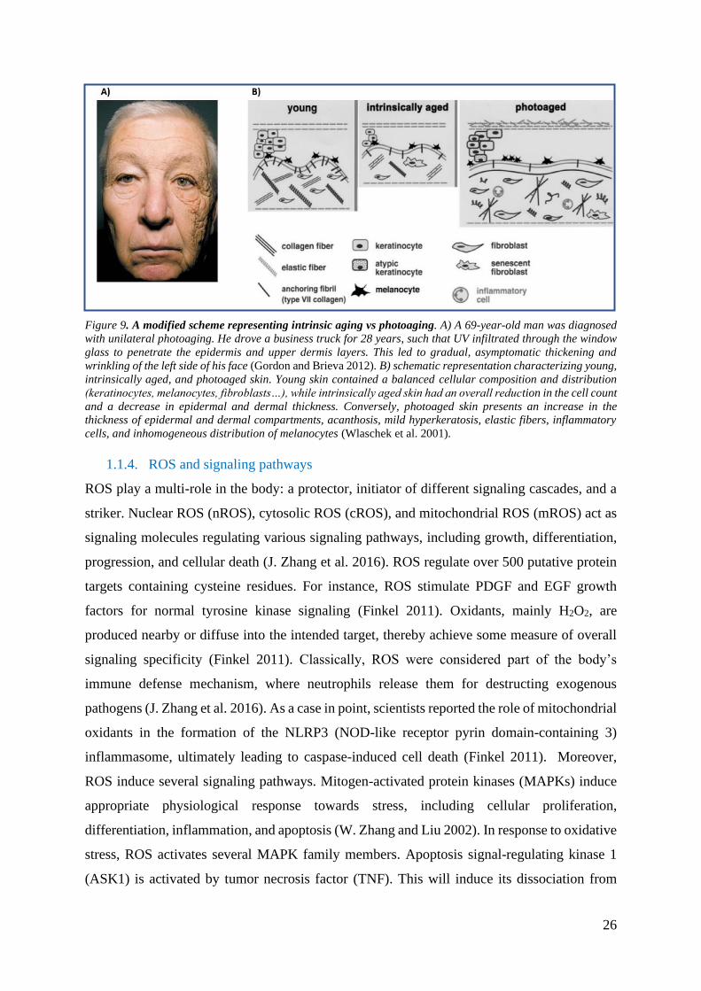
26
Figure 9. A modified scheme representing intrinsic aging vs photoaging. A) A 69-year-old man was diagnosed
with unilateral photoaging. He drove a business truck for 28 years, such that UV infiltrated through the window
glass to penetrate the epidermis and upper dermis layers. This led to gradual, asymptomatic thickening and
wrinkling of the left side of his face (Gordon and Brieva 2012). B) schematic representation characterizing young,
intrinsically aged, and photoaged skin. Young skin contained a balanced cellular composition and distribution
(keratinocytes, melanocytes, fibroblasts…), while intrinsically aged skin had an overall reduction in the cell count
and a decrease in epidermal and dermal thickness. Conversely, photoaged skin presents an increase in the
thickness of epidermal and dermal compartments, acanthosis, mild hyperkeratosis, elastic fibers, inflammatory
cells, and inhomogeneous distribution of melanocytes (Wlaschek et al. 2001).
1.1.4. ROS and signaling pathways
ROS play a multi-role in the body: a protector, initiator of different signaling cascades, and a
striker. Nuclear ROS (nROS), cytosolic ROS (cROS), and mitochondrial ROS (mROS) act as
signaling molecules regulating various signaling pathways, including growth, differentiation,
progression, and cellular death (J. Zhang et al. 2016). ROS regulate over 500 putative protein
targets containing cysteine residues. For instance, ROS stimulate PDGF and EGF growth
factors for normal tyrosine kinase signaling (Finkel 2011). Oxidants, mainly H2O2, are
produced nearby or diffuse into the intended target, thereby achieve some measure of overall
signaling specificity (Finkel 2011). Classically, ROS were considered part of the body’s
immune defense mechanism, where neutrophils release them for destructing exogenous
pathogens (J. Zhang et al. 2016). As a case in point, scientists reported the role of mitochondrial
oxidants in the formation of the NLRP3 (NOD-like receptor pyrin domain-containing 3)
inflammasome, ultimately leading to caspase-induced cell death (Finkel 2011). Moreover,
ROS induce several signaling pathways. Mitogen-activated protein kinases (MAPKs) induce
appropriate physiological response towards stress, including cellular proliferation,
differentiation, inflammation, and apoptosis (W. Zhang and Liu 2002). In response to oxidative
stress, ROS activates several MAPK family members. Apoptosis signal-regulating kinase 1
(ASK1) is activated by tumor necrosis factor (TNF). This will induce its dissociation from

27
thioredoxin (TRX) to activate downstream effectors, including the c-Jun N-terminal kinase
(JNK) and the p38 MAPK pathway required for cell death. PKG, cAMP dependent-PKA, and
JNK are activated by intermolecular disulfide bond formation, intramolecular disulfide bond,
and phosphatase inhibition, respectively (Finkel 2011; P. D. Ray, Huang, and Tsuji 2012).
Other activated cellular pathways engaged in cellular apoptosis and proliferation involve
NFκB, phosphoinositide-3-kinase- (PI3K-) Akt, and ataxia-telangiectasia mutated (ATM)
pathways (J. Zhang et al. 2016; Finkel 2011). In the presence of ROS, Akt can stimulate the
Nrf2/KEAP1/ARE pathway. This pathway is well-known as a major model of cellular defense
against oxidative stress where Nuclear factor E2-related factor 2 (Nrf2) heterodimerize with
transcription factors of the Maf family to bind upstream the cis-regulatory antioxidant response
element (ARE) sequence in the promoter region of cytoprotective genes to stimulate various
antioxidant enzymes (SOD, GST…) and to reduce oxidative DNA damage and mediate redox
homeostasis. In the absence of stress, Kelch-like ECH-associated protein 1 (KEAP1)
negatively regulate Nrf2 in the cytoplasm by tagging it for proteasomal degradation (David et
al. 2017; Ming et al. 2011). A recent study showed that KEAP1 inhibition and Nrf2 pathway
activation could be done via Nardochinoid C. Such a product could be a safer substitute to the
few available Nrf2 activators with some side effects (Luo et al. 2018). However, other
experiments (human cell culture, animal models, clinical trials, etc.…) are needed in the future
to confirm its therapeutic efficacy and efficiency (Luo et al. 2018).
Also, ATM protein kinase is involved in the homeostatic feedback loop regulated by and
regulating ROS. It can modulate NADPH synthesis through pentose phosphate shunt and
regulate mitochondrial biogenesis. In parallel, H2O2 can oxidize its C-terminal cysteine residue
for activation, thereby preventing ataxia, immunodeficiency, premature aging, and cancer
predisposition due to activating a response to DNA double-strand breaks (Finkel 2011).
P53’s cysteine residues act as targets for ROS, but it can also regulate ROS in turn. In response
to ROS, P53 is known to upregulate transcription of NADPH and several antioxidant genes
(SODs, Gpx1…) (Sies 2019). Once the delicate balance between expressed ROS and anti-
oxidants is disturbed, ROS-induced tumor-promoting events will begin (Liou and Storz 2010).
This is done by upregulating the expression of cell cycle cyclins (cyclin B2, cyclin D3, cyclin
E1, and cyclin E2..) to expedite G1 to S phase transition and promote tumor cell metastasis and
invasiveness (Liou and Storz 2010). Surprisingly, at high ROS levels, P53 will step out to
reduce the antioxidants’ and their transcription factors’ expression tipping the cellular fate
towards death (Srinivas et al. 2019).

28
1.2. Defense mechanisms against oxidative stress
Several protective mechanisms were studied in aerobic organisms to detoxify or prevent ROS.
It could be by natural metabolic processes, including series of antioxidant proteins [SOD,
glutathione peroxidase (GPx), catalase, Nrf2] or synthetic/natural products (J. Zhang et al.
2016). Such products could act as ROS scavengers or antioxidants such as alpha-tocopherol,
selenium, ferulic acid, flavonoids (strawberry, pomegranate….), lipid-soluble carotenoids
(lycopene, beta-carotene), and vitamins (A and C) (Amaro-Ortiz, Yan, and D’Orazio 2014).
It is worth mentioning that the antioxidants state differs amongst cell types. For example,
although keratinocytes show less sensitivity to UV-DNA damage than lung or skin fibroblasts,
they have triple the level of GSH compared to the fibroblasts (Morley et al. 2003).
1.2.1. Enzymatic defense mechanism
• Superoxide dismutases (SODs)
In mammals, SODs are categorized based on their location and the metal ions they require for
their function into three variants: (i) Cu/Zn superoxide dismutase (SOD1), (ii) mitochondrial
Mn superoxide dismutase (SOD2), and (iii) Cu/Zn extracellular superoxide dismutase (SOD3)
(D. Wu and Cederbaum 2003). They are responsible for O2−•, the most catastrophic free radical,
dismutation into H2O2 (reactions I&II). Astoundingly, researchers found a link between
increased ROS, disturbed antioxidants activities, and tumors. It was hypothesized that high
expression of SOD2 leads to an accumulation of H2O2 that will unlock the gates in front of
tumor cell lines to gain invasive and metastatic properties, as shown in advanced stages of
gastrointestinal (GI) cancer (Valko et al. 2006).
It should be noted that SOD enzymes work in conjunction with H2O2-removing enzymes,
catalases, and glutathione peroxidases (Valko et al. 2006).
• Glutathione peroxidase
Glutathione peroxidases are primarily categorized into two forms: (i) selenium-independent
glutathione-S-transferases (GST) and (ii) selenium-dependent glutathione-S-transferases
(GPx). They use GSH as a cofactor that will be oxidized simultaneously to reduce peroxides
(H2O2, ROOH) to water or alcohol (reaction III):
2 GSH + H2O2 → GSSG + 2H2O (I)
2 GSH + ROOH → GSSG + ROH + H2O (II) Reaction III.

29
o Glutathione (GSH)
L-γ-glutamyl-L-cysteinyl glycine is a tripeptide that is abundant in eukaryotic cells (0.1-10
mM concentrations). More than 90 percent are present in the thiol reduced form, GSH. It is
catalyzed by the consecutive action of γ-glutamylcysteine synthetase (glutamate-cysteine
ligase) and GSH synthetase through the γ-glutamyl cycle or by oxidized glutathione (GSSG)
reduction by glutathione disulfide reductase (GSSG reductase) in the presence of NADPH.
GSH has multiple functions, including metabolism, catalysis, transport of cysteine moieties,
and an antioxidant that reduces cellular components such as free radicals (Meister 1995). By
donating an electron to reduce products, GSH becomes oxidized. Then NADPH, as an electron
donor, will regulate GSH’s turnover. Therefore, any misbalance in the ratio between oxidized
and reduced GSH could indicate oxidative stress (D’Orazio et al. 2013).
• Glutathione metabolism methods: inhibition vs induction
In general, L-buthionine-sulfoximine (BSO) is used to reduce cysteine and irreversibly inhibit
the first steps of GSH synthesis (γ-glutamylcysteine synthetase) and therefore selectively
decreasing cellular levels of GSH. This will induce oxidative stress associated with elevated
levels of 8-oxoGua, DNA SSBs, and gene deletions in vitro and in vivo (Ghetti et al. 2018;
Fernandez et al. 2014). Several reports have indicated the effectiveness of BSO in inhibiting
growth and inducing apoptosis of cancer cell lines when used in combination with irradiation
and with/without other drugs, mainly dimethyl fumarate. Dimethyl fumarate (DMF) depletes
pre-existing intracellular GSH rapidly by conjugating with it to be exported and/or metabolized
by the cell. In addition, DMF has been shown to treat psoriasis and enhance the cytotoxicity of
antitumor agents. Therefore, BSO/DMF combined treatment can completely deplete GSH and
trigger intrinsic cell death in various cell lines, including CD44+ cancer stem-like cells and
head and neck squamous cell carcinoma cells (HNSCC). Excitingly, BSO/DMSO showed
efficiency at in vivo level too. Ninety-five percent of mean tumor volume decreased in mice
after pretreatment and irradiating for five consecutive days (4Gy/day). This shows the potential
of using such a combination with irradiation as a therapy to eliminate resistant tumor cells
(Boivin et al. 2011).
On the contrary, the administration of compounds such as N-acetyl-cysteine (NAC), which
increases cellular cysteine levels, GSH synthetase substrates (γ-glutamylcysteine ), or
glutathione esters can increase cellular GSH levels in vivo and in vitro (Meister 1995).
Specifically, NAC acts as a GSH precursor and/or free radicals’ scavenger. Thus, it prevents
and/or reduces the formation of 8-oxoGua lesions and photoproducts in embryonic cells, mice,

30
Atm deficient mice, UV-irradiated lung fibroblasts, and others (Reliene and Schiestl 2006). For
instance, incubating skin fibroblasts for 4 hours with 5mM or 1 hour with 6mM NAC
maximized GSH production and reduced ROS-induced DNA lesions at UVA physiological
doses. Such a ROS downregulation is due to an inhibition in HMGB1 (High-mobility group
box 1) release. This photoprotective effect is reduced with time. Nevertheless, it is ineffective
in preventing UVB-induced-pyrimidine dimers formation. Therefore, NAC is postulated to be
involved directly in repairing UV damages caused by ROS, and its supplementation is
inexpensive, safe, FDA approved, and effective as dermatological therapy (Srinivas et al. 2019;
Morley et al. 2003).
Additionally, it has been used to preserve lung function in idiopathic pulmonary fibrosis
patients (Goodson et al. 2009).
• Catalase
Catalase is an iron-containing enzyme found in the peroxisome and competes with glutathione
peroxidase on removing H2O2, resulting in water and O2 (D. Wu and Cederbaum 2003). UVB
is known to induce ROS at two distinct chronologic stages: immediately and few hours post-
irradiation. Catalase overexpression could be involved in the latter stage. It was recorded to
have a protective effect against UVB-induced apoptosis in cultured keratinocytes and
reconstructed epidermis. Up to 50 percent of catalase’s activity was found to be downregulated
in XP patients due to a reduction in NADPH (H. R. Rezvani, Ged, et al. 2008). So, what is the
link between catalase and NADPH?
Glutathione reductases and peroxidases need NADPH to eliminate H2O2. Also, it was
discovered that each NADPH molecule tightly binds to each of the catalase’s four subunits to
protect and enhance the efficiency of the latter’s activity. NADPH oxidation inhibits the
formation of compound II, an inactive catalase form (Kirkman, Galiano, and Gaetani 1987)
(Reaction IV):
Where Compound II is an oxidative inactive catalase, AH and A. are groups within the active center of catalase
Herein, we can conclude that the catalase and glutathione reductase/ peroxidase pathway act
on H2O2 NADPH-dependent reduction.
Compound I-AH → Compound II-A. (I)
Compound II-A. + NADPH→ catalase-AH + NADP+ (II)
Reaction IV.

31
1.2.2. Non-enzymatic defense mechanism
• Nicotinamide (NIC)
NIC, also known as vitamin B3 or niacinamide, has been used to treat different diseases
(dermatitis, diarrhea, dementia, actinic keratosis, and melanoma) due to its effect on the intra-
cellular functioning and metabolism. It produces nicotinamide adenine dinucleotide (NAD+),
a pro-survival cofactor acting in the mitochondrial electron transport chain machinery to
regulate cell survival and oxidative homeostasis. It is worth addressing that NIC has a dose-
specific dual role, acting as an antioxidant at low concentrations and as an oxidative stressor
after exceeding a certain threshold. At high concentrations (30 mM), NIC was shown to induce
fibroblasts’ apoptosis by elevating ROS, reducing GSH levels, inhibiting glucose-6-phosphate
dehydrogenase (G6PD) enzyme (i.e., starving cells), and modulating the expression of
antioxidant, anti-apoptotic, and pro-apoptotic genes (Hassan, Luo, and Jiang 2020).
Meanwhile, at low concentrations, NIC has a photoprotective effect. Recently, scientists have
proved that 50µM NIC protects melanocytes from UV-induced DNA damage (CPDs and 8-
oxoGua). This may be by upregulating the expression of critical genes involved in NER and
BER (SIRT1, P53, DDB1, DDB2, OGG1, ERCC1, ERCC2, Nrf2, and CDK7) in response to
UVB-irradiation. Furthermore, a recent phase III clinical trial indicated that oral nicotinamide
is non-cytotoxic and effective in reducing non-melanoma skin cancers and actinic keratosis
incidences in high-risk patients (Chhabra et al. 2019).
• Crocin
Crocin is an active constituent present in saffron and traditional Chinese medicine that has been
used to treat various diseases (neurodegenerative disorders, coronary artery diseases,
respiratory diseases, and gastrointestinal diseases). Crocin is effective as an anti-carcinogenic,
anti-inflammatory, and antioxidative agent. In addition, it could protect dermal fibroblasts from
UVB damage by reducing ROS (directly or by increasing GPx expression), enhancing
proliferation (by increasing the anti-apoptotic Bcl-2 gene’s expression), and upregulating ECM
production (i.e., preventing aging) (Deng et al. 2018).
• Vitamin C
Vitamin C (L-ascorbic acid) is the most copious naturally occurring hydrophilic antioxidant
incorporated in multiple dermatological cosmetics to protect and rejuvenate photoaged skin.
Unfortunately, it cannot be synthesized by the body instead obtained from dietary sources
(fruits, vegetables) or oral/topical supplementations. Topical vitamin C protects the skin from
UVB-induced induced erythema, sunburns, photoproducts, and ROS. In other words, it

32
prevents photoaging and rejuvenates the skin. This occurs upon directly neutralizing ROS
through donating it with electrons, inducing collagen synthesis (collagen I and III), and
enhancing the activation of lipid-soluble vitamin E (Z. Liu et al. 2018; Farris 2005). This will
reduce the membrane-bound α-tocopherol from its oxidized states and ROS-mediated signaling
cascades (JNK, NFκB). Several clinical studies proved such an enhancement of photoaged
skin. A 3-month double-blind and randomized study showed that applying 10 percent of
vitamin C improves the patients’ skin wrinkles, tiredness, and hyperpigmentation brightness
compared to control (Farris 2005). Also, vitamin C can prevent ROS-induced inflammation by
suppressing transcription factor NFκB, thereby inhibiting the production of proinflammatory
cytokines (TNF, Interleukin-1/6/8) (Z. Liu et al. 2018; Farris 2005).
• Vitamin A (retinoids, carotenoids)
Similar to vitamin E, vitamin A is a natural lipid-soluble antioxidant. It can be found as one of
the two forms: (i) retinol and retinyl esters or (ii) as pro-vitamin carotenoids (β-carotene), and
it is usually stored and metabolized in the liver. Vitamin A has many biological roles including,
gene transcription, and immune responses, etc.… However, we will exclusively focus on its
role in oxidative stress defense. Remarkably, several studies have shown that vitamin A
supplementation could act as double hatted depending on its dose, administration mechanism,
and the characteristics of the stressors. At low concentrations, Vitamin A acts as an anti-
inflammatory and ROS scavenger that targets mainly peroxyl and superoxide radicals. At
higher concentrations, or upon strenuous exercises, it increases ROS production (superoxide)
in parallel to decreasing the activity of the antioxidants, SOD and CAT. Due to this and vitamin
A’s lipophilic properties, lipid peroxidation and protein damages will occur (Petiz et al. 2017).
For example, high concentrations of β-carotene act as prooxidants that boost the oxidative
environmental stress’s carcinogenic effect. β-carotene enhances lung cancer by increasing the
partial pressure of oxygen in patients’ lungs, increasing its oxidative metabolites
(apocarotenoids) that will alter the retinoic acid level and signaling, and reducing the
expression of tumor suppressor genes. Such events escalate further in smokers where β-
carotene may interact with the carcinogenic components of cigarette smoke (Russell 2002).
More experiments should be conducted to study deeper the role of vitamin A during stress.
• Vitamin E (α-tocopherol)
Vitamin E acts as a ROS scavenger, protector against oxidative and UV-DNA damage (8-
oxoGua, CPDs), and inhibitor of CPDs’ production in melanocytes, HaCaT keratinocytes, and
in vivo mouse skin. This could show a link between CPDs’ production and ROS level. Vitamin

33
E may enhance CPDs’ repair by inhibiting ROS levels and preventing the latter’s drastic effect
on different DNA repair enzymes (Delinasios et al. 2018).
1.3. Pathologies linked to oxidative stress
Excessive ROS has been implicated in developing various diseases, including respiratory,
digestive, cardiovascular, and neurodegenerative diseases. For example, increased
mitochondrial H2O2 was detected in insulin resistance and subsequent type 2 diabetes mellitus
(T2DM) animal and human models. Similarly, high ROS contribute to the development of
neurodegenerative diseases such as Alzheimer’s disease (AD), Huntington’s disease (HD),
amyotrophic lateral sclerosis (ALS), Parkinson’s disease (PD), and spinocerebellar ataxia
(SCA). Premature aging was found as a progressive step in AD. As Denham Harman suggested
in the 1950s, aging is another field in which ROS is involved. Such an increase in the steady-
state level of ROS in parallel to a decrease in antioxidants can oxidatively modify cellular
proteins, lipids, and DNA; consequently, it contributes to cellular aging (Z. Liu et al. 2018;
Finkel 2011). More importantly, oxidative stress plays a role in most carcinogenesis steps: (i)
initiation by accumulating DNA mutations and altering the cellular energy and metabolism,
(ii) promotion of cancerous cells’ expansion by altering the cellular signaling cascades (i.e.,
enhances oncogenes and inhibit pro-apoptotic transcription factors), and (iii) progression
(metastasis) by upregulating matrix metalloproteinases and downregulating the action of anti-
proteases and angiogenesis (Z. Liu et al. 2018). These cancers may be internal (breast, lung,
liver, colon, prostate, ovary, and brain) or skin cancers (NMSC and MSC) (S. K. Saha et al.
2017).
Hence, why ROS has such a dramatic effect on cells? How can it target the cellular DNA,
accumulate mutations, and trigger their escape from cellular repair mechanisms?
ROS is involved in the regulation of different DNA repair pathways. For instance, it can inhibit
OGG1’s glycosylase activity, a factor involved in BER. This will lead to the accumulation of
oxidative DNA lesions and single-strand breaks that may be converted into double-strand
breaks (Srinivas et al. 2019). Such a link will be further discussed in detail throughout the
manuscript.

34
1.4. Summary for ROS
Figure 10. ROS: mechanisms of actions and alterations. Either endogenous or exogenous processes can produce
ROS. It will affect several signaling cascades (MAPK, NFKB, etc..) that could alter proliferation and cell survival
and alter cell cycle cyclins to induce cancer initiation, promotion, and metastasis. In parallel, antioxidant
signaling mechanisms, such as Nrf2, will be triggered as a defense mechanism. This will induce antioxidants
activation. These antioxidants can be either enzymatic or non-enzymatic. They can be manipulated by
pharmacological drugs (upregulated by NAC and downregulated by BSO or BSO/DMF).
35
2. Chapter Two: The Base Excision Repair Pathway (BER)
2.1.Overview of BER pathway
BER is a frontline repair pathway responsible for maintaining genome integrity, preventing
premature aging, malignancy, and many other diseases assumingly occurring at the G1 phase
of the cell cycle (Dianov and Hübscher 2013). As presented in figure 11, it is initiated by one
of at least 11 distinct DNA glycosylases, depending on the type of base lesion (deaminated,
methylated, or oxidized) (Krokan and Bjørås 2013). OGG1 and MYH are the most common
nuclear glycosylases that we will focus on in this manuscript due to their role in recognizing
and excising the most frequent oxidative DNA lesion, 8-oxoGua. The former recognizes the
lesion directly; meanwhile, the latter recognizes the substrate A opposing 8-oxoGua (Krokan
and Bjørås 2013). The arising baseless site (also called abasic site, apurinic/apyrimidinic site,
or AP site) is further processed by an AP endonuclease (APE1) that carries both an AP-
endonuclease activity and a redox function required for activation of several transcription
factors. It also protects against oxidative stress. APE1 cleaves the phosphodiester bond 5’ to
the AP site, generating a single-strand break containing a hydroxyl residue at the 3’-end and
deoxyribose phosphate at the 5’-end (Krokan and Bjørås 2013; Wallace, Murphy, and Sweasy
2012). A DNA polymerase then fills single-strand breaks in, either through single nucleotide
(short) or long-repair patch sub-pathways. The one nucleotide gap is filled in by the polymerase
β (POLβ), followed by strand sealing by ligase 3-XRCC1 complex. Meanwhile, long-patch
BER occurs when DNA polymerase δ/ε initiates polymerization from the free 3′-OH adjacent
to the deoxyribose phosphate (dRP) group by incorporating between 2 to 15 nucleotides
displacing the flap strand, a strand containing the 5′-dRP. This occurs with the help of FEN1
nuclease and PCNA (figure 11) (Krokan and Bjørås 2013).

36
Figure 11. A simplified schematic representation of BER pathway. Exogenous or endogenous stresses induce a
damaged base in the DNA (here, we took oxidative DNA damage as an example). Base damage is recognized by
a lesion-dependent DNA glycosylase (OGG1) that will use its glycosylase activity for base excision leading to the
formation of an abasic site. This abasic site will be recognized by 5’APE1 endonuclease as a rate-limiting
enzymatic step. APE1 will cleave the site leaving behind a single-strand DNA break (SSB) with 3′ OH and 5′
deoxyribose phosphate (5′dRP) termini. This SSB will be recognized and repaired by either short-patch or long-
patch sub-pathways: For the short-patch sub-pathway, POLB will synthesize a new nucleotide base for the LIG3-
XRCC1 heterodimer to ligate the strand. However, for the long-patch sub-pathway, FEN1-PCNA will remove the
generated 5’flap post-polymerization by POLδ/ε-PCNA-RFC. Then the strand will be sealed by LIGI and its
accessory protein PCNA. Note: XRCC1 acts as an accessory protein and coordinator for LIG3, PARP1, and
POLβ.

37
2.2.BER factors roles and post-translational modifications
Different post-translational modifications (PTMs) have emerged as regulators of BER’s
localization, activity, and interactions (figure 12). These include acetylation, methylation,
phosphorylation, SUMOylation, and ubiquitination (Carter and Parsons 2016).
OGG1 consists of several isoforms, mainly OGG1-α and OGG1-β, in the nucleus and
mitochondria, respectively. It is a multifunctional glycosylase protein that is involved in DNA
repair (cleavage of the glycosidic bond of a DNA base lesion and form DNA strand breaks)
and in transcription regulation [interaction with NFκB and specificity protein 1 (Sp1)] (R.
Wang et al. 2018). Following oxidative stress, p300 enzyme acetylates OGG1’s lysines 338
and 341 (K338 and K341, respectively) to increase OGG1’s turnover on AP sites that’ll
increase BER’s activity (Carter and Parsons 2016). Also, tyrosine kinase c-Abl, PKC, and
cyclin-dependent serine/threonine kinase 4 (Cdk4) phosphorylate OGG1’s serine residues to
stimulate OGG1’s activity (Carter and Parsons 2016). On the other hand, ubiquitination by
chaperone-dependent E3 ubiquitin-ligase triggers proteasomal degradation (Carter and Parsons
2016).
MYH encodes for at least 10 different protein glycosylases isoforms (three primary transcripts,
α, β, and γ) that are involved in DNA repair (excision of A bases opposing 8-oxoGua) at nuclear
and mitochondrial levels (Markkanen, Dorn, and Hübscher 2013). In vitro study revealed that
colorectal cancer was proposed due to MYH dephosphorylation (Carter and Parsons 2016;
Markkanen, Dorn, and Hübscher 2013). Accordingly, PKC phosphorylates MYH’s serine 524
(S524) to increase its DNA glycosylase activity and regulates repair by altering its binding to
PCNA (Carter and Parsons 2016; Kundu et al. 2010). On the contrary to OGG1, MYH’s
ubiquitination by Mcl-1 ubiquitin ligase E3/ARF binding protein 1 (Mule/ARF-BP1) on at least
one of five C-terminal lysine residues, between amino acids 475 and 535, stabilizes it, prevents
its degradation, and influences subcellular localization and/or DNA binding (Carter and
Parsons 2016).
APE1 is a multifunctional protein that plays a role in BER and gene regulation (redox-
dependent transcription activator and a co-repressor responding to intracellular calcium influx).
Therefore, PTMs are needed to regulate and equilibrate the different roles of APE1 for cellular
stability (Busso, Lake, and Izumi 2010). While the effect of acetylation appears to be minimal
to APE1’s DNA repair activity, it plays a role in APE1’s transcriptional regulation and calcium-

38
dependent parathyroid hormone repression activities (Busso, Lake, and Izumi 2010). K6 and
K7 lysine residues’ acetylation by histone acetyltransferase (HAT) p300 enhances the gene
repressor function of APE1 (Busso, Lake, and Izumi 2010). In vivo acetylation of k27, K31,
K32, and K35 enhance APE1’s interaction with RNA to modulate the latter’s metabolism
(Carter and Parsons 2016). SIRT1, a member of the HDAC deacetylase family, was shown to
increase the APE1-XRCC1 interaction and regulate APE1’s gene regulatory function (Busso,
Lake, and Izumi 2010; Carter and Parsons 2016; Yamamori et al. 2010). Upon oxidative stress,
APE1 deacetylation allows its de-attachment from genes’ promoters for their expression
inhibition, including parathyroid hormone gene and Y-box-binding protein 1 (YB-1);
subsequently, it trigger its BER functional activity (Busso, Lake, and Izumi 2010; Yamamori
et al. 2010). Another major modification is phosphorylation by serine/threonine casein kinases
and protein kinase C, altering its endonuclease activity but enhancing its redox activity (Busso,
Lake, and Izumi 2010; Choi, Joo, and Jeon 2016). Also, APE1’s phosphorylation by cyclin-
dependent kinase 5 (Cdk5)/p35 at threonine 233 (T233) inhibits its endonuclease activity and
enhances its ubiquitination (Carter and Parsons 2016). APE1’subiquitination by E3 ubiquitin
ligase at multiple lysine residues near its N-terminus (K6, K7, K25, K25, K27, K31, K32, and
K35). Upon stress, MDM2 is activated to mono-ubiquitinate APE1 post-T233 phosphorylation
by CDK5. This will alter APE1’s DNA repair and gene regulation functions which will trigger
polyubiquitination, consequently degradation in the presence of oligomerized MDM2 (Carter
and Parsons 2016; Busso, Wedgeworth, and Izumi 2011).
POLβ is a primary DNA polymerase that catalyzes the synthesis of new DNA nucleotides into
the DNA strand. It is regulated at stability and activity levels by different modifications. For
instance, POLβ’s lysine 72 (K72) is acetylated by p300 acetyltransferase as a regulatory step
to inactivate its activity when not needed or upon shifting from short-patch to long-patch BER
(Carter and Parsons 2016). Meanwhile, its arginine 137 (R137) is methylated by
methyltransferases to inhibit its interaction with PCNA. This prevents POLβ’s involvement in
PCNA-dependent processes, including long-patch BER. In contrast, an in vitro study showed
that methylation of POLβ on arginines 83 and 152 (R83 and R152) by PRMT6 enhances the
binding of the enzyme to DNA and increased processivity (Carter and Parsons 2016). However,
methylation at Arg137 prevents its interaction with PCNA, thereby inhibiting its role (M. Bai
et al. 2020). Ubiquitin proteasome pathway (UPP) regulates POLβ’s protein levels at steady
and oxidative states. This is due to polyubiquitination or monoubiquitination within the 8-kDa
N-terminal domain (K41, K61, and K8) containing the β lyase activity by the E3 ubiquitin

39
ligase CHIP and E3 ubiquitin ligase Mule/ARF-BP, respectively. It occurs when POLβ is free
in the cytoplasm in the absence of DNA lesions. On the contrary, upon DNA damage, the
interaction between XRCC1-LIG3 and POLβ on DNA stabilizes the polymerase to repair
efficiently (Carter and Parsons 2016).
POLε and POLδ are long-patch-DNA polymerases whose PTMs are rarely discussed in
publications. However, phosphorylation S458 of POL δ’s p68 subunit by protein kinase A
(PKA) leads to a decreased binding affinity to PCNA; henceforth, decreased polymerase
processivity (Carter and Parsons 2016).
LIG3 is a DNA ligase that joins DNA strands together by catalyzing the formation of a
phosphodiester bond. It has two isoforms that differ at their c-termini, 103-kDa ligase 3-alpha
polypeptide expressed in all tissues and cells, and 96-kDa DNA ligase 3-beta polypeptide that
is expressed only in the testis (Mackey et al. 1997). We will focus on the former’s post-
translational modifications since it is the most abundant form and due to the lack of information
about the latter’s modifications. LIG3-alpha is constitutively phosphorylated by casein kinase
II (Cdk2) on its Ser123, in a cell cycle-dependent manner, from early S-phase into M-phase
(Dong and Tomkinson 2006). Upon oxidative stress, it will be dephosphorylated in an ATM-
dependent manner to repair DNA at S-phase (Dong and Tomkinson 2006). Notably,
phosphorylated XRCC1 stabilizes nuclear LIG3 by forming a dimeric complex (Parsons et al.
2010). LIG3’s activity in the mitochondria is independent of XRCC1 (Akbari et al. 2014).
Lastly, rarely LIG3’s ubiquitination is discussed; however, few had shown that CHIP and
Iduna/RNF146 could polyubiquitylate and degrade it (Carter and Parsons 2016; Parsons et al.
2008).
LIGI’s posttranslational modifications are not identified to date. It would be interesting to
identify the different modifications it needs to be activated, inhibited, or degraded since it is
the major DNA ligase involved in long-patch BER.
XRCC1 is a scaffold protein that interacts with several enzymes involved in DNA single-strand
break repair. XRCC1’s phosphorylation on serine 518, threonine 519, and threonine 523 (S518,
T519, and T523) by CK2 induces its interaction with the forkhead-associated domain (FHA)
of aprataxin, a protein involved in DNA damage signaling and repair, to prevent genotoxicity
promoting XRCC1’s turnover and stability. Additional phosphorylation within its C-terminal

40
region promotes XRCC1-PNKP to repair DNA single-strand breaks efficiently. Post-stress
(alkylation, oxidation, irradiation), XRCC1 has been reported to be phosphorylated on
threonine 284 (T284) and serine 371 (S371) for single-strand and double-strand breaks repair,
respectively. Like other proteins, polyubiquitination at the C-terminal by CHIP or E3 ubiquitin
ligase Iduna/RNF146 leads to degradation, unless bound to POLβ or heat shock protein 90
(HSP90) (Carter and Parsons 2016).
FEN1 is a long-patch BER endonuclease. Post-stress, its lysine residues are acetylated (K354,
K355, K377, and K380) by p300, displaying reduced endonuclease activity and DNA binding.
This regulates its enzymatic activity and prevents premature processing of Okazaki fragments.
Reduction in its endonuclease activity was also observed upon phosphorylation by the Cdk1-
cyclin A complex, particularly during the end of the S phase of the cell cycle. Intriguingly, a
cross-talk between phosphorylation and other modifications has been reported. Upon oxidative
stress, phosphorylation is inhibited while methylation is activated by methylating arginine 192
(R192) for efficient DNA damage repair. It also initiates a cascade of events leading to
ubiquitination and degradation of FEN1. Phosphorylation of FEN1 at S187 promotes
SUMOylation on lysine 168 (K168) that will trigger polyubiquitination mediated by the E3
ubiquitin ligase pre-mRNA processing factor 19 (PRP19) (Carter and Parsons 2016).
PCNA is a scaffold protein involved in DNA repair and replication. Multi-studies have
analyzed PCNA’s PTMs due to its role in DNA lesion repair during replication. During S-
phase, SUMOylation at its Lys164 by Srs2 inhibits homologues recombination and prevents
double-strand breaks (DSBs). Meanwhile, ubiquitination at K164 residue by RAD6–RAD18
complex activates the error-prone DNA damage tolerance pathway (TLS). Further
ubiquitination, by RAD5 and the UBC13-MMS2 complex, at K63 triggers an alternative
template switching mechanism from replicative to translesion synthesis DNA polymerases
(ZHU et al. 2014, Spivak 2015).
PARP1 is an active intermediate member of BER with a high affinity towards DNA SSBs and
plays a role in cell cycle regulation. Hence, it is interesting to study its PTMs to target it. This
will inhibit SSB repair, by which cancer cells’ apoptosis will be triggered. PARP1’s acetylation
on different lysine residues (K498, K505, K508, K521, and K524) by p300 induces its
interaction with the P50 subunit of NFκB to induce its transcriptional activity (Carter and
Parsons 2016). However, phosphorylation on its serine 372 (S372) and threonine 373 (T373)

41
by extracellular signal-regulated kinases 1 and 2 (ERK1/2) are required for its accumulation
on damaged DNA (Carter and Parsons 2016). It has been shown that SUMOylation on its
lysines 203 and 486 (K203 and K486), in response to heat shock, induces its ubiquitinated
clearance and controls its binding to intact and damaged DNA. In contrast, SUMOylating
PARP1’s lysine 482 (K482) controls its poly(ADP-ribosyl)ation of chromatin-associated
proteins. Once lysine 486 (K486) is SUMOylated, acetylation is inhibited, thereby reducing its
coactivator activity and regulating gene expression (Carter and Parsons 2016). As mentioned
previously, ubiquitination occurs post-SUMOylation by E3 ubiquitin ligase RNF4.
Iduna/RNF146 and RING finger domain protein (CHFR) are other proteins that induce
poly(ADP-ribosyl)ated PARP1’s ubiquitination and degradation due to elevated alkylated
DNA damage and mitotic stress, respectively (Carter and Parsons 2016). This may allow the
dissociation of activated PARP1 from damaged DNA sites to promote continuity of DNA
repair cascade or prevent cancerous cells' survival.
Further studies should be done to fully understand the importance of post-translational
modifications on the different BER factors, their interactions, roles. This could pave the way
in front of personalized treatments against cancerous cells. Indeed, other types of modifications
may be present in the BER enzymes. Therefore, their cellular consequences and importance
should be studied too.
Figure 12. Post-translational modifications (PTMs) of BER factors. Several BER factors (main and accessory
proteins) are subjected to different PTMs that interfere with their functions, including those involved in BER.
These modifications can act as inhibitory (Red color) or excitatory (green color).

42
2.3.BER and human disorders: aging, oxidative stress, and ROS
BER pathway’s dysregulation leads to DNA instability in several diseases associated with
elevated oxidative stress, aging, and age-related neurodegenerations. Sliwinska et al. showed
that APE1, OGG1, MYH, PARP1, and NEIL1 genes are significantly downregulated in
Alzheimer's disease patients (Sliwinska et al. 2017). This may explain why oxidative stress and
elevated 8-oxoGua in brain cells are the main factors for its pathogenesis (Perry, Cash, and
Smith 2002). High 8-oxoGua level and OGG1’s loss contribute to Huntington’s disease
(Krokan and Bjørås 2013). Similarly, defective BER and excessive oxidative stress were
reported in Parkinson’s disease (Ciccone et al. 2013).
Interestingly, BER is linked to diabetes. Studies have demonstrated that chronic high glucose
decreases OGG1’s expression via Akt redox-dependent activation (Pang et al. 2012). In
parallel, other experimental studies had shown that Ogg1−/− mice exhibit altered insulin levels,
glucose tolerance, adiposity, hepatic steatosis (German et al. 2017). Besides, asthma, a complex
chronic inflammatory lung disease, is mediated by multiple inflammatory mediators and ROS-
induced oxidative stress. ROS elevates 8-oxoGua in Asthma patients’ genome and body fluids
(Ba et al. 2015). Therefore, ROS, 8-oxoGua, and BER are directly linked.
ROS are formed as by-products of proficient cellular metabolism and exposure to endogenous
or exogenous chemical or physical stresses. Its main target in the DNA is guanine due to its
lowest redox potential compared to other nucleic acid bases leading to around 105 8-oxoGua
DNA lesions daily per cell (Pang et al. 2012; Ba et al. 2015). Although 8-oxoGua does not
induce local DNA structural change, ATP-dependent remodeling of the nucleosomal DNA
facilitates lesion accessibility by OGG1. In addition, its Nrf2 binding site at the promoter region
regulates the expression upon oxidative stress. This is done via modulating its PTMs (refer to
part 2.2.). For example, phosphorylation mediates relocalization, and acetylation enhances its
turnover (Boiteux, Coste, and Castaing 2017).
Another DNA glycosylase involved in oxidative stress is MYH. It excises adenine incorporated
opposite to 8-oxoGua during replication. This restores G:C base pairs to maintain DNA
replication integrity and fidelity (Jingwen Chen et al. 2019). Loss of MYH leads to the
accumulation of oxidative DNA damage, including 8-oxoGua, in the liver, heart, kidney, etc.…
In parallel, superoxide dismutase (SOD), an antioxidant associated with ROS activity, was
downregulated in mouse Myh-/- kidney, lung, and hippocampus. This leads to high oxidative
stress and numerous pathologies such as colorectal cancer, MYH-associated polyposis (MAP),
and aging (Jingwen Chen et al. 2019). Chen et al. indicated that MYH deficiency induces

43
oxidative damage-response that is not efficient in the presence of excessive damage burden
(Jingwen Chen et al. 2019).
2.4.BER variants/mutations and cancer (skin and internal)
Studies showed a relationship between BER genes’ alterations and cancer progression (H. Chen
et al. 2019). 8-oxoGua, the most abundant consequence of ROS, is the substrate for both MYH
and OGG1; consequently, a mutation in one or both glycosylases leads to dramatic
consequences, especially when the mutation is at their C-terminal binding site. This mutation
leads to protein’s structural deformation and dysfunctionality (Rizzolo et al. 2018).
Studies have shown at least 30 mutations in the MYH gene predicted to truncate the protein,
including nonsense, small insertions and deletions, and splice site variants (Wallace, Murphy,
and Sweasy 2012). G:C to T:A transversions are considered a genetic signature of defective
MYH protein activity in patients, including colorectal cancer patients (Ali et al. 2008). MYH
single nucleotide polymorphism (SNP; rs3219487) with G/A and A/A genotypes (OR=9.27,
95% CI=2.39−32.1) was related to increased risk for liver and hepatocellular carcinoma
compared to G/G genotype (Sakurada et al. 2018). G/A MYH genotype was also detected in
breast and endometrial cancer patients (Out et al. 2012; Barnetson et al. 2007). Having the
minor allele A in MYH decreases its transcription and activity in the carriers (Sakurada et al.
2018). Ty165Cys and Gly382Asp MYH variants lead to MYH-associated polyposis (MAP), an
autosomal disorder with risks to develop colorectal, thyroid, and duodenal cancer. You et al.
had shown that patients with a homozygous variant MYH genotype for rs3219472 have a high
risk of developing bile duct cancer (You et al. 2013). Also, MYH Gln324His polymorphism
seemed to increase the risk of lung (His/His genotype, OR= 3.03, 95% CI= 1.31–7.00) and
colorectal (Gln/His and His/His genotypes, OR=4.08, 95% CI= 1.22–13.58) cancer incidences
in the Japanese population (Miyaishi et al. 2009). Additionally, an Italian study had identified
p.Arg245His variant in male breast, colorectal and gastric cancers, p.Tyr179Cys, and
p.Gly396Asp in breast and colorectal cancers, p.Gln338His in women breast cancer and
p.Gly264Trpfs*7 variant in male breast cancer (Rizzolo et al. 2018).
OGG1 mutations also contribute to different cancer types, where studies have identified OGG1
missense mutations in 3 out of 40 lung and kidney tumors (Chevillard et al. 1998). OGG1
Gly308Glu acts as a low-penetrance allele that contributes to colorectal cancer, while SNP
rs2304277 increases cancer risk (ovarian..) in BRCA1 carriers due to lower OGG1
gene expression levels, consequently, higher DNA damage, genomic instability, and telomeres
shortening (Benitez-Buelga et al. 2016; Smith et al. 2013). This polymorphism was also linked

44
to urothelial bladder carcinoma (OR=3.55, 95% CI= 1.79-7.06) (Ahmed et al. 2018). OGG1
Ser326Cys polymorphism (rs1052133) correlates with lower activity and risk of several
cancers, including acute lymphoblastic leukemia, esophageal, bladder lung, prostate, gastric,
and hepatocellular cancers (H. Chen et al. 2019; Peng et al. 2014; Smal et al. 2018). For
example, a meta-analysis study showed that OGG1 Ser326Cys polymorphism (Cys/Cys vs
Ser/Ser homozygous genotype model) increased breast cancer risk (OR=1.14, 95% CI= 1.01-
1.29) in the studied population (Moghaddam et al. 2018). Scientists also showed that OGG1
Ser326Cys polymorphism plays a critical role in the pathogenicity of cervical carcinoma or
precancerous cervical intraepithelial neoplasia (CIN III) lesions and participates in the infection
process of high-risk human papillomavirus (HR-HPV) infection (H. Chen et al. 2019).
A double mutation in both 8-oxoGua glycosylases, OGG1 and MYH, induces additional
tumorigenesis. Ogg1-/-Myh-/- double knockout mice exhibit a high frequency (65.7 percent)
of lung and ovarian tumors and lymphomas (Wallace, Murphy, and Sweasy 2012; T.-H. Lee
and Kang 2019; Xie et al. 2004; Tahara et al. 2018).
Other BER factors’ SNP genetic variants were associated with increased cancer occurrence.
For example, PARP-1 Ala762Ala and P53 Arg72Pro, are significantly associated with cervical
cancer and HR-HPV infection, where P53 is considered part of the BER activation cascade (H.
Chen et al. 2019). Wild-type P53 stimulates BER by interacting with APE1 and POLβ and
stabilizing the latter's interaction with abasic DNA sites (J. Zhou et al. 2001). Additionally,
XRCC1 R399Q and APE1 Asp148Glu were associated with an increased risk of breast cancer
(T. Wang et al. 2018; Busso, Lake, and Izumi 2010). Also, the SNP combination of APE1
D148 and XRCC1 R194W was linked to pancreatic cancer risk (Busso, Lake, and Izumi 2010).
This position may be vital for APE1 and XRCC1 interaction. Polymorphisms of XRCC1 at
codons 194, 280, and 399 are associated with a risk of several types of gastrointestinal cancers,
breast cancer, and lung cancer. Also, T to C homozygous point mutation at nucleotide 889
(T889C) resulting in the substitution of Leucine by Serine amino acid 259 in POLβ have been
identified to be involved in primary gastric cancer (Tan et al. 2015; Y. Seo and Kinsella 2009).
This may be due to some of POLβ’s mutations that lead to DNA synthesis in low fidelity
(Simonelli et al. 2016).
Hence, could BER’s role has a drastic effect on the acidic tumor microenvironment?
This was well-illustrated by researchers who had found that inhibiting APE1/Ref1 and/or
XRCC1 (BER’s factors) through drugs could inhibit cellular survival upon an accumulation of
mutations that trigger apoptosis (Y. Seo and Kinsella 2009).

45
2.5.BER and cell cycle
Upon transcriptomic and proteomic data analysis of differently synchronized cell lines,
researchers were able to elucidate the cell cycle regulation of different BER factors (figure 13).
Short-patch BER expresses its genes equally in all the cell cycle phases (POLβ, APE1…), with
higher APE1’s activity at G1-phase in the presence of ionizing radiation (measured by
oligonucleotide incision assay); meanwhile, long-patch BER genes are extensively expressed
in S-phase (Mjelle et al. 2015; Chaudhry 2007). 4 of the 11 genes encoding DNA glycosylases
were cell cycle regulated. For example, NTHL1and uracil-DNA glycosylase (UNG) were
shown to be overexpressed in the G1/S-phase. UNG showed upregulation at protein level too.
In contrast, thymine-DNA glycosylase (TDG) peaks solely at G1-phase. NEIL3 is upregulated
at the S/G2-phase. During S-phase, NEIL1 showed an upregulation only upon prolonged serum
starvation of fibroblasts (for synchronization). Such upregulation of some glycosylases at the
S-phase may be due to their defined role pre-/post-/ and during the replicative process. Other
glycosylases (OGG1, MYH…) showed no evidence of cell cycle regulation. Downstream long-
patch BER factors (PARP1, PARP2, PCNA, FEN1, POLD1, POLD3, POLE, and POLE2)
were detected steadily regulated at S or G1/S-phase due to their additional roles as replication
proteins. Common short and long-patch BER downstream factors, including XRCC1 and LIG1
were upregulated at S/G2 and S-phases, respectively. All the regulations mentioned above
show that long-patch BER is implicated in proliferating cells, and arrest in G1 prevents the
replication of damaged DNA while arrest in G2 prevents segregation of defective
chromosomes (Mjelle et al. 2015; Chaudhry 2007).
Additionally, P53 can activate BER factors as APE1 and POLβ due to direct protein-protein
interactions (Fitch 2003).
On a side note, BRG1, a chromatin remodeling factor, has also been proposed to regulate
BER’s polymerase activity (L. Zhang et al. 2009).

46
Figure 13. Cell cycle regulated BER genes (Mjelle et al. 2015). The genes are color-coded based on their level
of expression in the different cell cycle phases. Only gene products shown to be cell cycle regulated in at least
two transcriptome or translational studies are colored. Gray color-coded genes show that they were not found to
be cell cycle regulated.
2.6.BER targeted treatment: The debased side of BER
Numerous in vitro and in vivo studies have been trying to find therapeutic drugs targeting or
enhancing the different DNA repair pathways for treatments against carcinogenesis and
metastasis. Despite the challenges and complications, DNA repair inhibitors that block a
specific protein or multi-proteins’ activities in a particular DNA repair pathway paved the way
for personalized medicine that could help target chemo-resistant and sensitive cancer cells
leading to their apoptosis (Kelley, Logsdon, and Fishel 2014).
BER pathway is a double-edged sword where its inhibition or overexpression may be
cancerogenic. For example, studies found POLβ overexpression at mRNA and protein levels
in uterus, ovary, prostate, and stomach cancers due to genetic instability by facilitating cell
survival to augment DNA repair capability and bypass various DNA damages causing
apoptosis by commonly used chemotherapeutic drugs (Chan et al. 2007). Hence, POLβ exhibits

47
dichotomous roles depending on its expression, either as an oncogene or tumor suppressor
(Chan et al. 2007). Similarly, some reported OGG1’s overexpression in ulcerative colitis-
associated carcinogenesis and esophageal squamous cell carcinoma (Kumagae et al. 2018).
This may reflect the persistence of cancer cells against stress, such as oxidative stress.
PARP1 has been the interest of scientists due to its dual role in regulating oncogenes and tumor
suppressors' expression and DNA repair pathways, including BER. It has a high expression in
some cancers as it activates OGG1’s expression at the G1-phase. Some PARP1 inhibitors have
already been in use (Olaparib) in BRCA-deficient cancers. In addition, iCDK4/6 (PD0332991,
LEE011) was shown to be an efficient anti-cancer treatment for lung cancer cells since it
impairs OGG1-dependent BER and sensitizes cancer cells to oxidative imbalance-induced
death by decreasing PARP1 transcriptional expression. However, this dual treatment has its
weak points. Tempka et al. suggested that for this treatment to be efficient, a functional link
should exist between RB1 (retinoblastoma protein), PARP1, and OGG1. Unfortunately, RB1
is mutated in most retinoblastomas, osteosarcomas, small cell lung cancers, and other cancer
types at lower frequencies. This may prevent efficient PARP1 targeting. Therefore, genomic
and transcriptome profiles should be checked before administering PARP1 suppressive agents
such as CDK4/6 inhibitors (Tempka et al. 2017).
8-oxoGua’s repair is indispensable for cell survival. Inhibiting OGG1 by monotherapy or in
combination with DNA damaging agents could also be interesting to target cancer cells where
the loss of OGG1’s function has been shown to sensitize cells to multiple chemotherapies and
irradiation (IR) (Tahara et al. 2018). A study had suggested hydrazine and hydrazone as
specific inhibitors of Schiff base formation during OGG1-mediated catalysis after screening a
~50 000-molecule Chembridge DIVERset library (Donley et al. 2015). In 2018, Tahara et al.
screened almost 25975 potential compounds that could inhibit OGG1’s activity. They found a
new compound, SU0268, which has an acyl tetrahydroquinoline sulfonamide skeleton and is
more potent than the previous inhibitors. The prior compounds exhibited delayed kinetics of
inhibition, but SU0268 is a non-cytotoxic and specific compound that binds directly to the
OGG1 enzyme in HEK293T and HeLa cells to inhibit its base excision activity (Tahara et al.
2018). Further studies are being applied in cellular and animal models of various disorders.
TH5487 is another OGG1’s DNA glycosylase incision activity inhibitor as it prevents OGG1
from binding to 8-oxoGua in DNA (Visnes et al. 2018). It also decreases proinflammatory gene
expression, which may be a potentially helpful strategy for treating inflammation (Visnes et al.
2018). OGG1’s downregulation has been shown to elevate the efficacy of bleomycin, an
anticancer drug, to human lung adenocarcinoma cells (S. Liu, Wu, and Zhang 2010).

48
Unfortunately, OGG1 inhibitors have not reached clinical trials; however, different chemical
screenings of pharmacological libraries are being done producing potential hits (Mechetin et
al. 2020).
Scientists are focusing on a combinational treatment of OGG1 and MTH1 inhibitors to block
the whole GO system triggering excessive oxidative stress triggering cancer cells’ death. They
had already identified an MTH1 inhibitor, crizotinib. Crizotinib is a chiral compound that can
be: (R)-enantiomer, which blocks kinases (c-MET, ALK…) and (S)-enantiomer, which binds
to and blocks MTH1. In addition, another MTH1 inhibitor, karonudib (TH1579), has been
recently registered in two-phase one-clinical trials, and a possible phase two clinical trial was
proposed (Mechetin et al. 2020).
Finally, POLβ inhibition has been shown to enhance the efficacy of some alkylating agents
used in chemotherapies as mustard compounds, oxaliplatin, and temozolomide (S. Liu, Wu,
and Zhang 2010). This is due to the DNA repair blockage. For instance, in vitro and in vivo
studies showed that NSC666715 POLβ inhibitor enhances temozolomide sensitivity in colon
cancer cells (S. Liu, Wu, and Zhang 2010).
2.7.BER and Drugs: Acetohexamide (ACETO)
(experiments were done on ACETO but were not included in the manuscript)
Interestingly, in 2014, Alli et al. published for the first time acetohexamide as a potential
chemo-preventative and DNA repair enhancer agent against mutated BRCA1-associated
malignancies. Due to the known role of BRCA1 in BER of oxidative DNA damage, its
mutation limits BER’s activity which will induce excessive oxidative DNA damage leading to
genomic instability and high cancer risks. Such cancers are aggressive and prevalent.
Therefore, they conducted high-throughput chemical screening, then transfected cells with
GFP plasmid containing oxidized bases and identified ACETO (20μM) as a potential enhancer
of BER’s activity and inhibitor of basal and induced 8-oxoGua levels in BRCA1-mutated cells,
with minimal cytotoxicity. Another DNA repair-activating agent showing similar results was
benserazide. According to the U.S. National Library of Medicine TOXNET database, ACETO
and benserazide are FDA-approved drugs lacking chemical, toxicological, and environmental
threats. They may target directly or indirectly BER enzyme(s)/protein(s), inhibit BER’s
negative regulators, or they may activate BER’s positive regulators. Hence, further studies must
be investigated in addition to studying the structure-activity relationship deeply to avoid any
unneeded adverse effects (Alli et al. 2014). For example, ACETO is used as an antidiabetic
drug, i.e., it lowers blood glucose level by regulating insulin through targeting ATP-sensitive

49
potassium channels (Alli et al. 2014; Mazouzi et al. 2017). This may lead to hypoglycemia in
cancer patients if left unmodified. These drugs may be effective for other DNA repair-deficient
diseases or malignancies associated with oxidative stress or cancers (Alli et al. 2014). Another
chemical screening study proved that ACETO plays a role other than quenching ROS and its
effects; it can act as pyrimidine dimers repair enhancer (i.e., NER enhancer). It enhances the
removal of pyrimidine dimers in NER-deficient cells. This may be through antagonizing MYH
DNA glycosylase, thereby decreasing its binding to UV-DNA damage, such as CPDs. This
synthetic viability concept could enhance pyrimidine photoproducts repair in an unknown
NER-independent mechanism (Mazouzi et al. 2017). Therefore ACETO seems like an
interesting drug to study as a potential treatment for DNA repair-deficient patients.
2.8.BER and other repair pathways
The body has developed a direct/indirect interaction network amongst the different DNA repair
systems to provide the ultimate protection against the various stresses a human body could
encounter. For example, during the repair process, BER factors are known to dynamically
orchestrate with other DNA repair proteins to modulate the efficiency of their activity and to
prevent accumulation of their DNA damage intermediates, i.e., apurinic/apyrimidinic (AP)
sites, single-strand, and double-strand breaks (table 1).
Non-homologous End Joining (NHEJ), Homologous Recombination (HR), and BER:
NHEJ and HR repair double-strand breaks at G0-G1 and S-G2 phases, respectively. These
double-strand breaks can occur as BER intermediates if close lesions are being repaired
simultaneously. A lot of proteins from diverse DNA repair pathways are required for NHEJ,
including NTHL1 and UNG. However, RAD52, HR factor, and OGG1 have reciprocal
interactions. RAD52 enhances OGG1 turnover and 8-oxoGua incision. Conversely, OGG1
inhibits RAD52 catalytic activity. This may be because BER inhibits HR to induce NHEJ
(Limpose, Corbett, and Doetsch 2017).
Mismatch repair (MMR) and BER: both repair pathways protect against oxidized DNA
damage (8-oxoGua). MMR can recognize these lesions by MSH2/MSH6. Researchers
demonstrated that MSH2/MSH6 heterodimer interacts with MYH to induce the latter’s binding
and glycosylase activity. This may be through PCNA acting as a coordinator between both
repair systems (Cheadle and Sampson 2003).

50
NER and BER: several NER proteins have shown an interactive regulatory role towards BER.
XPC, a GG-NER initiator, interacts with TDG (G:T mismatch glycosylase) directly and
indirectly with OGG1 to enhance their turnover and activity. Probably, this is done upon an
explicitly direct interaction between XPC and APE1 that will enhance the glycosylases
excision from DNA (Limpose, Corbett, and Doetsch 2017; de Melo et al. 2016). Such a vital
role of XPC in BER started after researchers found that fluorescently labeled XPC localizes to
the nucleolus upon inducing oxidative DNA damage, 8-oxoGua (Limpose, Corbett, and
Doetsch 2017). It may be because 8-oxoGua subverts the DNA helix's major groove cation
binding and disrupts the bases’ hydrophilicity (Menoni, Hoeijmakers, and Vermeulen 2012).
This distortion is directly recognized by XPC that will activate both NER and BER pathways
(Menoni, Hoeijmakers, and Vermeulen 2012). Similarly, such stress induces fluorescently
labeled CSB, TC-NER factor, localization to the nucleolus and nucleoplasm where CSB-/- cells
were found to be hypersensitive to ROS-inducing agents. CSB interacts with OGG1 indirectly
to induce its turnover. Also, it interacts with NEIL1 and NEIL2 glycosylases. Such
glycosylases excise oxidized DNA damage: FapyA and FapyG. Both share 8-oxoGua as an
intermediate lesion. These lesions were found to increase in CSB-/- mice compared to control.
This is due to the capacity of CSB in modulating the incision and APlyase activities and
turnover of NEIL1 and NEIL2. In addition, it binds to APE1 directly to increase its
endonuclease activity more than 4 folds. PARP1 and FEN1 are other BER proteins that bind
with CSB.
XPG is another NER factor that stimulates NTHL1 glycosylase to recognize oxidized bases
and thymine glycols during replication (Limpose, Corbett, and Doetsch 2017).
Understanding the nuances of regulation and interaction between the different DNA repair
systems at the proteomic level could act as an effective targeted therapeutic and preventive
approach against several disorders and carcinogenesis, including xeroderma pigmentosum
(XP).

51
Table 1. A brief possible interaction of BER factors with other DNA repair proteins/enzymes. The green color
identifies a positive interaction that could be direct or indirect. BER: Base excision repair, NER: Nucleotide
excision repair, HR: Homologous recombination, and MMR: Mismatch repair.

52
3. Chapter Three: Nucleotide Excision Repair (NER)
3.1. Overview of NER pathway
NER is a highly conserved nuclear repair pathway that recognizes a wide range of DNA
mutations altering the DNA double-helical structure. They are bulky lesions including UV-
induced photoproducts [CPDs and (6-4) PPs], environmental mutagens (benzo[a]pyrene,
aromatic amines), some endogenous oxidative DNA lesions (cyclopurines), and intra-strand
crosslinks. Their mode of recognition divides NER into two pathways. (i) Global-genome NER
(GG-NER) recognizes the lesions across the entire genome (active, silent, and non-transcribed
genes) directly by XPC-hRAD23B-centrin 2 (CENT2) complex post-kinking DNA duplex by
DDB2 (XPE)-DDB1 complex (figure 14). In comparison, (ii) transcription-coupled NER (TC-
NER) recognizes the lesions by CSB/CSA upon RNA polymerase II stalling in actively
transcribed genes and ensures a rapid repair (figure 14). Prolonged transcription stalling
induces severe cellular damages, including P53-dependent apoptosis (Arima et al. 2005).
After that, in both sub-pathways, TFIIH complex (XPB-XPD, p8, p52, p44…) will be recruited
to unwind the DNA creating a ~20 to 30 nucleotide bubble. 5’-3’ lesion incision follows this
via XPA, XPF/ERCC1 (5’endonuclease), and XPG (3’ endonuclease). Then the gap will be re-
filled by synthesizing a new DNA sequence via polymerase, mainly POLβ, and its accessory
proteins (PCNA, RCF, and RPA1). Finally, LIG3-XRCC1 or LIGI ligates the sequence
forming an intact DNA (figure 14) (Budden et al. 2016; Schärer 2013; Spivak 2015).
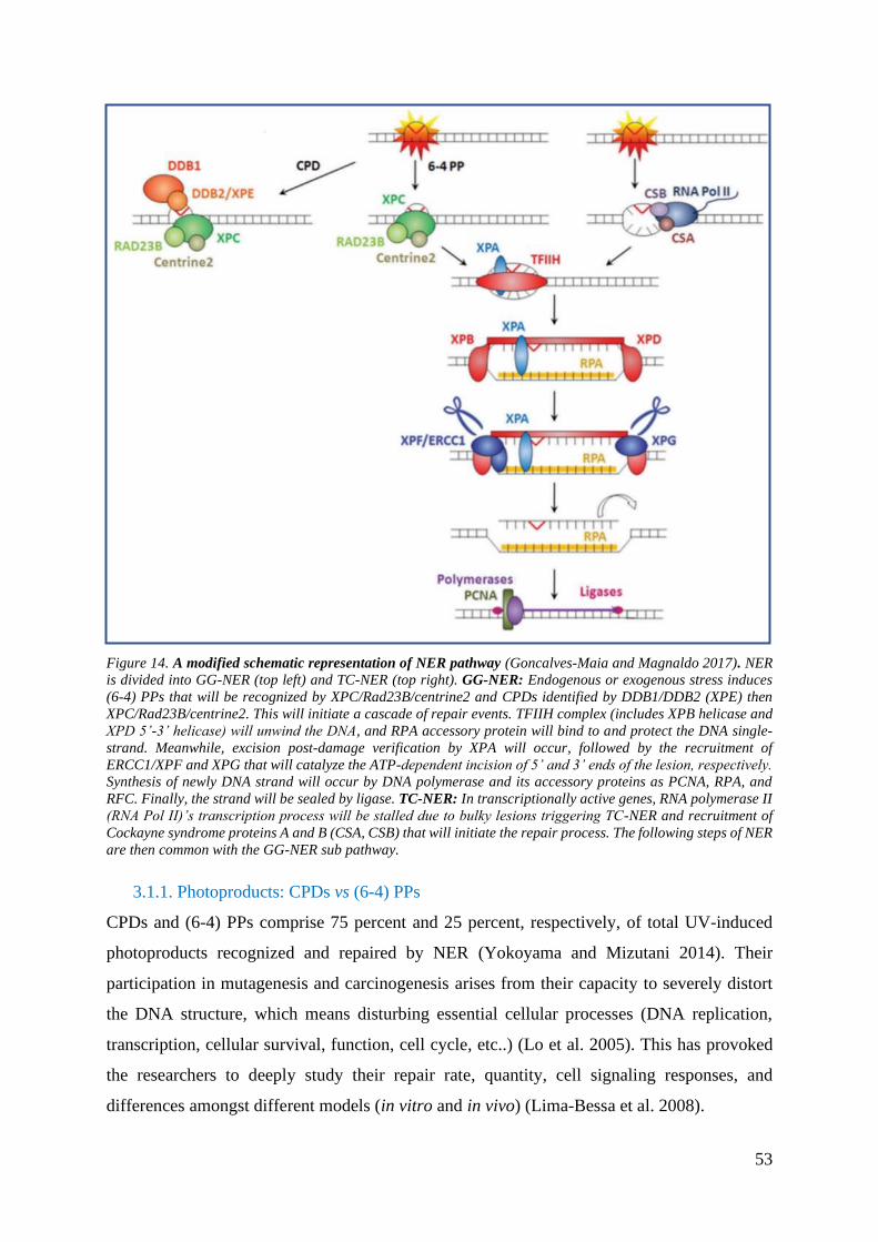
53
Figure 14. A modified schematic representation of NER pathway (Goncalves-Maia and Magnaldo 2017). NER
is divided into GG-NER (top left) and TC-NER (top right). GG-NER: Endogenous or exogenous stress induces
(6-4) PPs that will be recognized by XPC/Rad23B/centrine2 and CPDs identified by DDB1/DDB2 (XPE) then
XPC/Rad23B/centrine2. This will initiate a cascade of repair events. TFIIH complex (includes XPB helicase and
XPD 5’-3’ helicase) will unwind the DNA, and RPA accessory protein will bind to and protect the DNA single-
strand. Meanwhile, excision post-damage verification by XPA will occur, followed by the recruitment of
ERCC1/XPF and XPG that will catalyze the ATP-dependent incision of 5’ and 3’ ends of the lesion, respectively.
Synthesis of newly DNA strand will occur by DNA polymerase and its accessory proteins as PCNA, RPA, and
RFC. Finally, the strand will be sealed by ligase. TC-NER: In transcriptionally active genes, RNA polymerase II
(RNA Pol II)’s transcription process will be stalled due to bulky lesions triggering TC-NER and recruitment of
Cockayne syndrome proteins A and B (CSA, CSB) that will initiate the repair process. The following steps of NER
are then common with the GG-NER sub pathway.
3.1.1. Photoproducts: CPDs vs (6-4) PPs
CPDs and (6-4) PPs comprise 75 percent and 25 percent, respectively, of total UV-induced
photoproducts recognized and repaired by NER (Yokoyama and Mizutani 2014). Their
participation in mutagenesis and carcinogenesis arises from their capacity to severely distort
the DNA structure, which means disturbing essential cellular processes (DNA replication,
transcription, cellular survival, function, cell cycle, etc..) (Lo et al. 2005). This has provoked
the researchers to deeply study their repair rate, quantity, cell signaling responses, and
differences amongst different models (in vitro and in vivo) (Lima-Bessa et al. 2008).

54
CPDs are highly mutagenic and are produced in substantial quantities by UVA and UVB-
irradiations. They correspond to a dimerization between two adjacent pyrimidines (thymine,
cytosine, 5-methyl cytosine, or 5-hydroxymethylcytosine) at carbons 5 and 6, forming a four-
membered ring structure (S. Kim, Jin, and Pfeifer 2013; Lo et al. 2005). Once their cytosine/5-
methylcytosine is deaminated, CPDs bypass repair triggering mutagenic events, initiating
cytokines, inducing photo immunosuppression, and initiating skin cancer. Indeed, the most
common mutation post-UV is G:C to T:A transversion that has been described in a lot of genes,
including P53 in melanoma and non-melanoma skin cancers (Delinasios et al. 2018; S. Kim,
Jin, and Pfeifer 2013).
Experiments showed that CPDs’ repair reduces UV-induced apoptosis, erythema, and
hyperplasia in vivo and in vitro. Contradictory, an in vitro experiment treating cells with UVB,
CPD/(6-4) PP photolyases, and Annexin V showed that the repair of minor DNA lesions, (6-
4) PPs, reduces apoptosis by 70 percent. In comparison, CPDs repair reduces it by 40 percent.
Such findings indicate that (6-4) PPs are more apoptotic inducers than CPDs and that the latter
are more involved in cell cycle arrest in the absence of increased apoptosis. In other words,
CPDs affect repair by halting the cell cycle, which may increase their mutation accumulation,
while (6-4) PPs induce apoptosis to eliminate damaged cells, which reduces their mutation
accumulation. Such a contradiction amongst publications may depend on P53’s studied
cells/animals status due to its vital role in triggering cellular death. In this study, P53 mutated
XP cells were used. This may affect the contribution of CPDs in inducing apoptosis. Other
studies showed that CPDs decrease UV-induced apoptosis by inducing P53 mutations and
provoking skin carcinogenesis in mice models. However, when P53 is wild-type, CPD lesions
induce apoptosis and cell cycle arrest.
It is worth mentioning that (6-4) PPs are P53-independent and less mutagenic due to their rapid
repair and 5 to 10 folds less frequency than CPDs (Budden et al. 2016; S. Kim, Jin, and Pfeifer
2013). While CPDs arise in nucleosome cores, (6-4) PPs are formed in linker DNA consisting
of a non-cyclic bond between two adjacent pyrimidines at carbons 6 and 4 through Paterno-
Büchi reaction (Lo et al. 2005; Puumalainen et al. 2016). (6-4) PPs’ strong absorption to a 320
nm UV-wavelength leads to an electrocyclization and the formation of dewar isomers (Douki,
Koschembahr, and Cadet 2017).

55
3.1.2. NER factors roles and post-translational modifications
Like BER, NER is regulated by various post-translational modifications that could act
simultaneously in a coordinated manner (Figure 15).
XPC is the initiator of GG-NER; hence, its PTM regulation plays a vital role in protecting cells
against DNA damage. For instance, ubiquitination plays an essential role in XPC’s turnover.
After irradiation, ubiquitination by CRL4DDB2 E3 ligase increases the DNA binding affinity of
XPC, while ubiquitination by RNF111 and its cognate E2-UBC13 releases XPC from the DNA
damage site. Through co-immunoprecipitation, researchers found OTUD4 deubiquitinase
responsible for removing the ubiquitin moiety from XPC for its turnover (Lubin et al. 2014).
Ubiquitination by RNF111 relies on DDB2- and XPA-dependent SUMOylation for XPC’s
stability (van Cuijk et al. 2015). Such a SUMOylation (Small ubiquitin-related modification)
has an opposite effect once created on Lysine (Lys 655) (Park and Kang 2016). Another critical
PTM is phosphorylation at various sites, including serine (Ser 61, 94, 397, 399, 883, 884, and
892) and threonine (T169) (Shah et al. 2018). The function of such different phosphorylated
sites is still inconclusive. However, Shah et al. identified that phosphorylation at Ser94 and
Ser892 regulate ubiquitylated XPC’s recruitment to the damage site (Shah et al. 2018). Also,
XPC can be PARylated to be more effective in recognizing and binging to DNA lesions
(Rechkunova, Maltseva, and Lavrik 2019).
XPE (DNA Damage Binding Factor 2, DDB2) recognizes DNA damage sites and acts as a
ubiquitination inducing-protein ligase (Krzeszinski et al. 2014). It is PARylated and regulated
by XPC for stabilization while ubiquitinated for degradation (Park and Kang 2016, Matsumoto
et al. 2015). UVR-induced SUMOylation of DDB2 enhances NER at recognition and
processing steps (C. Han et al. 2017).
CSB (ERCC6) and CSA initiate TC-NER post-RNA stalling. CSB is degraded by CSA-
dependent ubiquitination (Spivak 2015). However, for an efficient function in TC-NER
initiation, several PTMs are needed: Phosphorylation is required for CSB nuclear localization,
then SUMOylation and PARylation are required for efficient bulky and oxidative DNA damage
repair, respectively (Park and Kang 2016).
TFIIH is a hetero decameric protein consisting of XPB, XPD, p62, p52, p44, p34, p8, Cdk7,
Cyclin H, and Mat1. In addition to its role in NER, it is involved in transcription, chromatin

56
remodeling, and ubiquitination (Krzeszinski et al. 2014; Sandoz et al. 2019). Phosphorylation
of XPB and ubiquitination of XPD inhibits NER by inactivating the former proteins (Park and
Kang 2016; Rechkunova, Maltseva, and Lavrik 2019).
XPG (ERCC5) nuclease plays a role in assembling the preincision NER complex and 3’
incision to the damaged site in the DNA. It is acetylated to be stabilized at the DNA damage
site while ubiquitylated for degradation (Park and Kang 2016).
XPA is recruited to the DNA damage site by TFIIH complex to verify the lesion and the
assembly of NER incision complex. It is stabilized and activated by phosphorylation stabilizes
it. Meanwhile, acetylation and ubiquitination degrade XPA and inhibit its activity in lesion
incision with ERCC1 (Rechkunova, Maltseva, and Lavrik 2019).
ERCC1 is involved in the incision step of NER. It is polyubiquitinated at its C-terminal to
form a complex with XPF (Borsos, Majoros, and Pankotai 2020).
DNA Polymerase β and δ are previously mentioned in section 2.2.
XRCC1’s post-translational modifications are previously mentioned in section 2.2.
PCNA’s post-translational modifications are previously mentioned in section 2.2.

57
Figure 15. Post-translational modifications (PTMs) of NER factors. Several NER factors (main and accessory
proteins) are subjected to different forms of posttranslational modifications that interfere with their function,
including that involved in NER. These modifications can act as inhibitory (Red color) or excitatory (green color).
3.2. NER and cell cycle
In general, NER factors are linked to cell cycle checkpoints for regulating apoptosis, cell cycle
arrest, and DNA repair and augmenting genomic stability and cell survival (Lu Liu et al. 2016).
Although downstream genes (polymerases, ligases…) are upregulated at transcriptional and/or
translational level in the G1/S-phase, the genes involved in the initial steps (XPC, CSA, CSB,
XPA, XPB, XPD, XPE, XPG…) are expressed independently of the cell cycle (Mjelle et al.
2015). For example, XPF is not regulated at the transcriptional level rather upregulated at the
translational level in G1/S/M phases (Mjelle et al. 2015). Although XPA’s expression is
independent of the cell cycle, it is exported to the nucleus to induce P53-independent NER due
to UV in the S-phase (Mjelle et al. 2015).

58
What about the regulation of the cell cycle by NER?
DNA damage response (DDR) is a signaling pathway that is driven mainly by protein
phosphorylation and contains sensors, transducers, and effectors that will ameliorate the
outcome of genotoxic stresses (intrinsic and extrinsic) to maintain genomic stability (Maréchal
and Zou 2013). Two primary transducers of the DDR signaling pathway and cell cycle are
ATM (ataxia-telangiectasia mutated) and ATR (ATM- and Rad3- related) kinases. Even
though both kinases belong to PIKK (phosphoinositide-3-kinase-related-protein kinase) family
and are activated post-DNA damage to initiate DNA repair and checkpoint arrest, they differ
in their damage selectivity (Maréchal and Zou 2013; A. Ray et al. 2016).
Briefly, ATR is activated by UV-induced single-strand DNA gaps in the G1-phase and by
replication stalling due to bulky lesions [CPDs and (6-4) PPs] in the S-phase. This will trigger
the phosphorylation of CHK1 and Cdc25 phosphatase causing cell cycle arrest and the
activation of the DDR pathway, which includes a complex of proteins as ATRIP-ATR
complex, TopBP1, MRE11, Rad50, and Rad17.
If replication halting persists, DNA double-strand gaps will be formed, inducing the activation
and recruitment of ATM. ATM will phosphorylate ChK2 phosphatase that will phosphorylate
Cdc25 for cell cycle arrest. In the presence of DNA double-strand breaks, ATR and ATM
phosphorylate histone H2AX (γH2AX) and BRCA1, double-strand breaks, and DNA repair
biomarkers, respectively (A. Ray et al. 2016). Interestingly, Ray et al. proved by single-cell
analysis that ATR/ATM are phosphorylated and recruited to the UV-induced DNA damage
sites in DDB2-XPC- and XPA- dependent manners during the G1-phase and not S-phase
(Budden et al. 2016; A. Ray et al. 2016). For instance, no defect in S-phase was detected in
XP-C fibroblasts despite the accumulation of mutations and single-strand DNA breaks due to
the dysregulated DNA damage repair (A. Ray et al. 2016). Besides, in the presence of cisplatin
(alkylating agent), XP-C cells were identified lacking caspase-2 and caspase-3 activation,
thereby delayed or diminished apoptosis (Budden et al. 2016). The relation between XPC and
apoptosis was also resembled in DDIT3-RPS3A-XPC regulated apoptotic pathway (Lubin et
al. 2014).
These may be some of the reasons explaining the 10000-fold increase in skin cancer incidences
in XP- patients (A. Ray et al. 2016). In conclusion, NER, GG-NER particularly, is tightly linked
to the cell cycle and checkpoint pathway by regulating the upstream proteins (ATR/ATM).
Additionally, Krzeszinski et al. showed that XPC triggers post-ubiquitination MDM2-
dependent P53 proteolysis via Rad23 to reset cells back to their quiescent state post-repair
(Krzeszinski et al. 2014). In parallel, P53 upregulates UV-induced DDB2 (p48) and XPC

59
expressions, stimulates nuclear import of XPA, and promotes histone modification to facilitate
XPB binding to DNA lesions (Krzeszinski et al. 2014; Fitch 2003).
3.3. NER and human disorders
Cockayne Syndrome (CS) is mainly due to mutations either in the CSA (ERCC8)
or CSB (ERCC6) genes (figure 16). Recently, it has been found that mutations in XPF
(ERCC4) or ERCC1 may also play a role in such a disorder. This leads to TC-NER deficiency.
Some of its main characteristics are photosensitivity, neurological disorders, microcephaly,
hypertension, and progeria with shortened life span. Surprisingly, there have been no reports
of carcinogenesis in CS-diagnosed patients (Spivak and Hanawalt 2015).
Cerebro oculofacial-skeletal syndrome (COFS) is due to mutations in CSB, ERCC1, XPD,
or XPG. It shares some characteristics with CS, including photosensitivity. Some of its
additional traits are hypotonia, poor vision, and abnormal kidneys and heart. The severity of its
symptoms leads to a short life span or lethality during infancy (Spivak and Hanawalt 2015).
De Sanctis-Cacchione (DSC) syndrome was initially known as “xerodermic idiocy” due to
similar symptoms to xeroderma pigmentosum patients. It is due to mutations in XP, mainly
XPA and CSB genes. DSC’s clinical symptoms are mental deficiency, progressive neurologic
deterioration, dwarfism, and gonadal hypoplasia (Spivak and Hanawalt 2015).
Tricothiodystrophy (TTD) is the result of mutations in some of TFIIH genes: XPB
(ERCC3), XPD (ERCC2), and TTDA (figure 16). Its main characteristics are sun sensitivity,
brittle and abnormal hair and nails, reduced fertility, and premature aging (Spivak and
Hanawalt 2015).
UV-sensitive syndrome (UVSS) is due to mutations in TC-NER CSA, CSB, or UVSSA genes.
UVSS patients suffer from photosensitivity and hyperpigmentation. Due to its mild symptoms
and the fully functional GG-NER and MMR, they do not develop cancer and are rarely
diagnosed with a genetic disorder (Spivak and Hanawalt 2015).
Xeroderma Pigmentosum (XP) is a rare skin genodermatosis that is characterized by skin
pigmentation, photosensitivity, high cancer incidences in photo-exposed areas (eyes, ears, skin,
tip of the tongue), and internal cancer (glioma, lung, breast, leukemia, uterus, prostate). XP
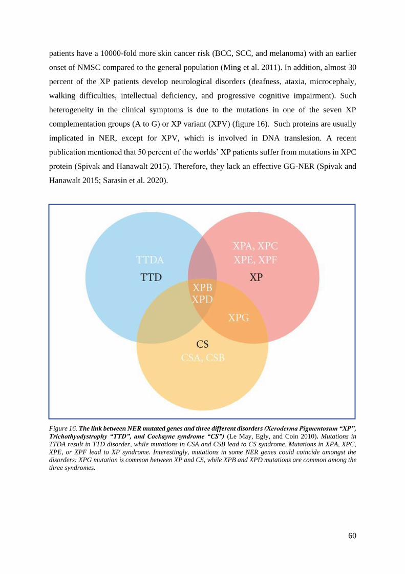
60
patients have a 10000-fold more skin cancer risk (BCC, SCC, and melanoma) with an earlier
onset of NMSC compared to the general population (Ming et al. 2011). In addition, almost 30
percent of the XP patients develop neurological disorders (deafness, ataxia, microcephaly,
walking difficulties, intellectual deficiency, and progressive cognitive impairment). Such
heterogeneity in the clinical symptoms is due to the mutations in one of the seven XP
complementation groups (A to G) or XP variant (XPV) (figure 16). Such proteins are usually
implicated in NER, except for XPV, which is involved in DNA translesion. A recent
publication mentioned that 50 percent of the worlds’ XP patients suffer from mutations in XPC
protein (Spivak and Hanawalt 2015). Therefore, they lack an effective GG-NER (Spivak and
Hanawalt 2015; Sarasin et al. 2020).
Figure 16. The link between NER mutated genes and three different disorders (Xeroderma Pigmentosum “XP”,
Trichothyodystrophy “TTD”, and Cockayne syndrome “CS”) (Le May, Egly, and Coin 2010). Mutations in
TTDA result in TTD disorder, while mutations in CSA and CSB lead to CS syndrome. Mutations in XPA, XPC,
XPE, or XPF lead to XP syndrome. Interestingly, mutations in some NER genes could coincide amongst the
disorders: XPG mutation is common between XP and CS, while XPB and XPD mutations are common among the
three syndromes.

61
4. Chapter Four: XPC the Bridge Between BER and NER
XPC has been identified to interact with BER (discussed previously) and play a significant role
in GG-NER. XPC’s impairment leads to XPC disorder which is the most common XP
genodermatosis. In the absence of any current treatment, studying this protein thoroughly could
provide better insight for further understanding of the disease at mechanistic standpoint. This
could pave the way for potential treatments.
4.1. XPC expression and interactions
XPC gene contains around 16 exons and 15 introns located on the short arm of chromosome 3
at position 25 (3p25) (Zebian et al. 2019; Bensenouci et al. 2016). It is translated into 940
amino acid protein (125 KDa) which usually forms a heterotrimeric complex with RAD23B
and CETN2 for stabilization and proper folding (Puumalainen et al. 2016). XPC harbors
different domains for binding to both the DNA and various protein partners (figure 17): (i) a
C-terminal segment (492-940 residues): the 847-863 residues form α-helix with EF-hand
calcium-binding protein centrin-2 (CETN2) while the 816-940 residues and part of the amino-
terminal (334 residues) interact with TFIIH (P62 and XPB) (Puumalainen et al. 2016; Bunick
et al. 2006), (ii) N-terminal that binds with XPA, P62, and glycosylases (OGG1) at the TGD
domain (154-331 residues) (Puumalainen et al. 2016). Other identified interacting domains are
TGD (496-637 residues) that interacts with DNA, RAD23B, and DDB2 and the three β-hairpin
domains (BHD1-3) (Puumalainen et al. 2016). XPC interacts with several other proteins
involved in DNA synthesis, transcription [DNA topoisomerase 2-beta (TOP2B)], proteolysis,
posttranslational modifications [the OTU deubiquitinase 4 (OTUD4), USP7,11 deubiquitinase
(for Ubiquitin-Specific-processing Protease 7,11)], signal transduction (SMAD1),
pluripotency ( Oct4-Sox2 activator), and metabolism (Puumalainen et al. 2016, Lubin et al.
2014).
Post-stress, XPC scans through its BHD1-BHD2 domain the DNA double helix for unpaired
bases. Once it senses damage, it increases the efficiency of its binding to DNA through BHD3,
which inserts its β-hairpin finger onto the DNA lesion site. In parallel, it flips out the unpaired
bases opposing the bulky lesion. Then RAD23B is released to initiate the cascade of GG-NER
events (Puumalainen et al. 2016).

62
Figure 17. A modified schematic representation of the human XPC protein (Puumalainen et al. 2016).
Transglutaminase homology domain (TGD) in blue, BHD1, BHD2, and BHD3 are β- hairpin domains in green,
yellow, and red. The left side represents N-terminus, while the right side represents C-terminus.
4.2. XPC’s regulation
XPC is regulated positively through the MAPK signaling pathway by interacting with mitogen-
activated protein kinase kinase kinase 5 (MAP3K5) and PTEN while regulated negatively by
protein tyrosine phosphatase type IVA, member 2 (PRL2) (Lubin et al. 2014).
In the presence of AKT, PTEN tumor suppressor and p38 have been reported to positively
regulate XPC in UVB-induced keratinocytes where their loss impair XPC, triggering the
predisposition for human skin cancer initiation and progression (papilloma, squamous cell
carcinoma, actinic keratosis) (Budden et al. 2016; Ming et al. 2011).
At steady-state, E2F4-p130 downregulates XPC gene expression. Such repression is inverted
by SIRT1 (sirtuin-1 NAD-dependent deacetylase), ARF (alternative reading frame), and
BRCA1 (Puumalainen et al. 2016). Another study showed that P53 positively regulates the
basal level of XPC and DDB to maintain a critical cellular level that is ready for action and to
prevent excessive loss of XPC and DDB. P53 also triggers the latter proteins post-UV response,
reaching the highest levels post-24 hours (Fitch 2003). Of note, XPF is suggested to stimulate
the UV-induced XPC expression (Q.-E. Wang et al. 2007). P38 also promotes GG-NER by
stabilizing DDB2 (Ming et al. 2011). Furthermore, XPC was shown to be regulated during the
cell cycle. Retinoblastoma protein (RB) stabilizes XPC for DNA repair during the G1-phase.
Phosphorylation of RB inhibits RB-stabilized XPC and DNA repair activity for G1/S transition
and DNA replication to proceed (Nemzow et al. 2015).
Lastly, several NER factors are transcriptionally regulated upon attenuated HIF-1α, such as
XPC, XPD, XPB, and XPG (Mahfouf et al. 2019). This upregulates UVB-induced NER and
regulates the cell cycle (Mahfouf et al. 2019).

63
4.3. XPC’s role
4.3.1. Canonical role
At the basal level, XPC protein shuttles between the cytoplasm and the nucleus via nuclear
export signals (Puumalainen et al. 2016).
In response to UV stress, CPDs and (6-4) PPs bulky lesions are formed in a ratio of 3:1
(Puumalainen et al. 2016). Phosphorylated p300 acetylates histones (heterochromatin regions)
for DNA recognition by XPC (Lubin et al. 2014). DDB2 (p48) and DDB2 dependent-BRG1,
INO80, PRKACB, PRKACA, and Snf5 (chromatin remodeling factors) recognize CPDs and
relax the chromatin triggering XPC’s stabilization, activation, higher recruitment from the
cytoplasm into the nucleus, and binding to the chromatin. P53 can also assist in double-
checking whether CPDs were repaired effectively (Fitch 2003). However, XPC recognizes (6-
4) PPs independent of DDB (L. Zhang et al. 2009; Puumalainen et al. 2016; Lubin et al. 2014;
A. Ray et al. 2013).
Afterward, XPC binds to the ssDNA opposing the DNA damage (A. Ray et al. 2013). This
initiates GG-NER, where the TFIIH complex is recruited to open the DNA strand and trigger
XPA’s recruitment that will be stabilized by XPG (3’endonuclease). However, XPC and XPG
cannot exist simultaneously at the CPD site. So, XPA-RPA will disengage XPC to be degraded
for the progression of the cascade of repair (Koch et al. 2016). Meanwhile, (6-4) PPs induce
pronounced helical DNA distortion accommodating the congregation of all the repair
machinery, including XPC and XPG (Q.-E. Wang et al. 2007). Succinctly, a set of
chronological events will be activated after recognizing lesions by XPC and its protein-protein
interactions with some NER factors leading to the complete repair.
The medical relevance and phenotypes of mutated XPC extend beyond its GG-NER function
as several studies implicated, suggesting XPC as a multifunctional protein.
4.3.2. Other roles
The prominence of XPC is elucidated in its recognition of a wide range of substrates, including
multiple repair factors other than NER that result in pleiotropy. More than 49 potential
interactors are involved in DNA synthesis, DNA repair, transcriptional regulation, protein
degradation, signal transduction, redox homeostasis, cellular metabolism, etc.… (figure 18).
Therefore, there is a complex heterogeneity in the clinical phenotype of XP-C patients.
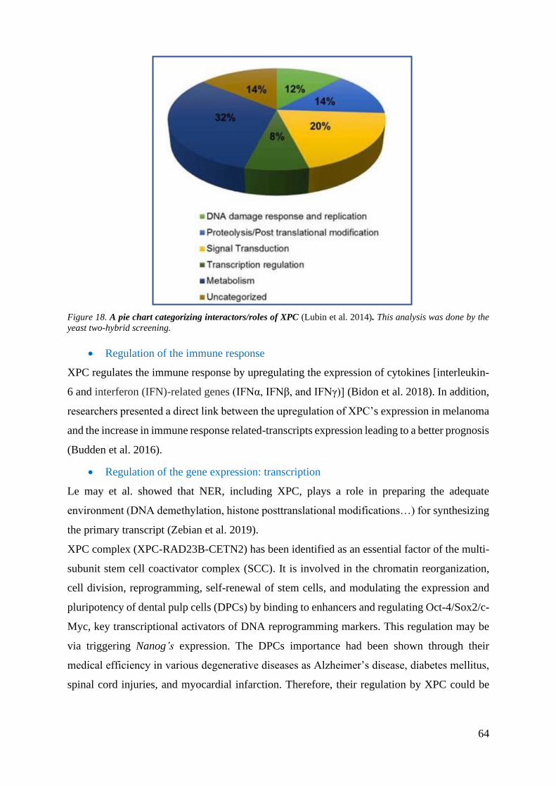
64
Figure 18. A pie chart categorizing interactors/roles of XPC (Lubin et al. 2014). This analysis was done by the
yeast two-hybrid screening.
• Regulation of the immune response
XPC regulates the immune response by upregulating the expression of cytokines [interleukin-
6 and interferon (IFN)-related genes (IFNα, IFNβ, and IFNγ)] (Bidon et al. 2018). In addition,
researchers presented a direct link between the upregulation of XPC’s expression in melanoma
and the increase in immune response related-transcripts expression leading to a better prognosis
(Budden et al. 2016).
• Regulation of the gene expression: transcription
Le may et al. showed that NER, including XPC, plays a role in preparing the adequate
environment (DNA demethylation, histone posttranslational modifications…) for synthesizing
the primary transcript (Zebian et al. 2019).
XPC complex (XPC-RAD23B-CETN2) has been identified as an essential factor of the multi-
subunit stem cell coactivator complex (SCC). It is involved in the chromatin reorganization,
cell division, reprogramming, self-renewal of stem cells, and modulating the expression and
pluripotency of dental pulp cells (DPCs) by binding to enhancers and regulating Oct-4/Sox2/c-
Myc, key transcriptional activators of DNA reprogramming markers. This regulation may be
via triggering Nanog’s expression. The DPCs importance had been shown through their
medical efficiency in various degenerative diseases as Alzheimer’s disease, diabetes mellitus,
spinal cord injuries, and myocardial infarction. Therefore, their regulation by XPC could be

65
the key for the potential usage of DPCs in therapies where their limited progenitors,
pluripotency, and cell division could be manipulated (Lu Liu et al. 2016).
Additionally, it interacts with hSNF5, SWI/SNF ATP-dependent chromatin remodeling
complex. XPC acts as a coactivator and epigenetics remodeler of RNA polymerase II-mediated
transcription (Puumalainen et al. 2016).
XPC and E2F1 interact with and activate histone acetyltransferase (HAT) lysine
acetyltransferase 2A (KAT2A) to induce gene promoters positively. Furthermore, XPC acts as
a positive linker between E2F1 transcription factor and ATAC coactivator and as an activator
of DNA damage-inducible transcript 3 (DDIT3) and SET1 methyltransferase that will
methylate histone H3 at lysine 4 (H3K4) for chromatin remodeling (Bidon et al. 2018, 1; Lubin
et al. 2014). This stimulates transcription. Therefore, XP-C cells show a reduced transcript
level of coactivator genes (Puumalainen et al. 2016).
Another evidence is that XPC is known to recruit itself and other NER factors (CSB,
XPA/RPA, XPG/XPF/Gadd45α) to the retinoic acid receptor beta2 (RARβ2) in the presence
and absence of UV to induce the promoter’s transactivation and chromatin remodeling (Zebian
et al. 2019; Nemzow et al. 2015).
• Regulation of cell cycle
In the absence of adequate DNA repair, XPC triggers damage-induced apoptosis via
downregulating anti-apoptotic casp-2S to avoid the division and progression of mutation-prone
cells. Accordingly, XP-C cells exhibit a caspase-3 (apoptotic) inhibition, caspase-2 (anti-
apoptotic) upregulation, hypersensitivity to genotoxic stress (UVB…), and faster stress
induced-tumor growth rate (Budden et al. 2016; Nemzow et al. 2015). For instance, some
researchers reported a more apoptotic resistance in xpc-/- mouse skin compared to control post-
UVB (H. R. Rezvani, Ged, et al. 2008).
• Regulation of proteolysis
XPC is implicated in the ubiquitination and degradation of some proteins. For example, XPC
degrades hUfd2 through the ubiquitin fusion degradation (UFD) pathway. It also induces P53
degradation via MDM2 (Nemzow et al. 2015).
XPC is expected to have proteolytic substrates other than P53. Identifying these substrates
could contribute to better understanding the cellular signaling processes during disease
progression (Zebian et al. 2019). An interesting project (How a sun protection complex
moonlights in proteolysis, UT Health San Antonio, funded by NIH-General Medical
Sciences) is in progress to detect such substrates.

66
• Regulation of other repair pathways
For activity regulation, XPC interacts with: (i) mismatch repair protein (MSH2), (ii)
homologues recombination repair proteins (ATM, ATR, and Rad51) (discussed in section 3.2.,
NER and cell cycle), (iii) Non-homologues recombination repair proteins (PKCs, XRCC4,
XRCC6, and LIG IV), and (iv) BER proteins [glycosylases (OGG1, MPG, TDG, SMUG1) and
APE1) (Zebian et al. 2019). In our studies, we will focus only on the link between XPC and
BER. In this respect, scientists had already identified OGG1’s interaction domain in XPC (in
the region of codon 334) and that XPC mutations delay activation of APE1 and inhibit OGG1’s
and MYH’s activation and expression (Qiao, Ansari, et al. 2011).
• Regulation of redox homeostasis
Regulation of redox homeostasis has been linked to XPC due to the high sensitivity and
detrimental effects of the oxidizing agents on XP-C cells. For example, D’Errico et al. and
Kassam et al. demonstrated XP-C fibroblasts and keratinocytes sensitivity towards KBrO3 and
methylene blue, thereby accumulating mutations including oxidized lesions (Kumar et al.
2020). Also, Melis et al. reported slow but higher somatic mutagenesis in xpc-/- mice compared
to control when exposed to oxidative stressors [diethylhexyl phthalate (DEHP) or paraquat]
(Melis et al. 2013). This suggests a slow accumulation of oxidative stress and oxidative DNA
damage in the absence of XPC. Another example is the increase in NER factors’ level
(including XPC) in the presence of oxidative stress in parallel to the glutathione-dependent
regulation of NER and XPC-dependent regulation of glutathione in the presence of prooxidant
(discussed in enzymatic defense mechanism section 1.2.1) (Melis et al. 2011). Liu et al. found
that UV and arsenic trioxide trigger XPC that will suppress superoxide production, induce
glutathione production, regulate the cell cycle, and modulate ROS scavenging systems (GSH,
catalase, and SOD) to induce redox balance (S.-Y. Liu et al. 2010). For instance, 35 and 40
percent lower catalase activity were detected in XP-C keratinocytes and fibroblasts,
respectively, compared to control (H. R. Rezvani, Ged, et al. 2008).
In parallel, Melis et al. reported that, in the presence of DEHP or paraquat oxidative stressors,
xpc-/- mice showed an increase in mutations and liver lipofuscin, a granular pigment acting as
in vivo oxidative stress biomarker (Zebian et al. 2019). Another oxidative stress biomarker is
hyperoxidized peroxiredoxin (Prx-SO2). XP-C cells exhibit sensitivity towards these stresses
and others such as IR, UV, and H2O2 (S.-Y. Liu et al. 2010).
In the absence of XPC, DNA damage will trigger DNA-dependent protein kinase (DNA-PK),
which subsequently activates AKT1 and NOX1, inducing ROS and further DNA mutations

67
(Melis et al. 2011). This NOX-mediated ROS has been detected to contribute to altered
bioenergetics (hyperpigmentation…) and metabolic alterations in XP-C keratinocytes leading
to photosensitivity and carcinogenesis (Kasraian et al. 2019).
4.4. XPC disorder
Briefly, as discussed throughout the manuscript, a mutation in XPC at gene/protein level leads
to functional dysregulation, consequently, XPC disease (figure 19). This genodermatotic
disease differs in severity amongst patients based on the type of mutation and genetic
background (Kasraian et al. 2019). For instance, patients with homozygous XPC mutations
exhibit different pigmentation abnormalities (extensive, moderate, and none) and different
repair capacities (between 13 and 40 percent) (Kasraian et al. 2019). Furthermore, K655A XPC
mutation affects its degradation, not recruitment nor activity. This prevents XPG recruitment
and compromises NER efficiency (Q.-E. Wang et al. 2007).
A study revealed that 49 and 59 percent of invasive immunocompetent and immunosuppressive
cutaneous squamous cell carcinoma, respectively, in non-XP patients had absent XPC protein.
Therefore, the absence of XPC plays a significant role in promoting malignancy and
carcinogenesis (J. W. Lee et al. 2020).
Figure 19. Clinical features of a Mahori XP-C patient at 6- and 8-years-old (Cartault et al. 2011). When he was
6-years-old, skin damage and pigmentations were detected. As the patient got older, the symptoms became more
severe. After two years (8-years-old), he had a running nose, tumors at different body sites, including the tip of
the tongue, and pigmentation and skin damage.
4.4.1. Clinical features
Due to the multiple roles of XPC, patients suffer from inter-individual heterogeneity in the
clinical symptoms. Nevertheless, some of the main clinical features are (Krzeszinski et al.
2014; Lehmann, McGibbon, and Stefanini 2011; Yurchenko et al. 2020):
• Photosensitivity
• Dry skin

68
• Pigmentation, poikiloderma, and lentiginosis
• Premature skin aging
• Skin cancer in photo-exposed areas (SCC, BCC, Melanoma)
• Early incidences of internal cancers (leukemia, lung, thyroid, sarcoma, etc..) in photo-
protected areas (median age of 24 years)
• Other symptoms (autism, hypoglycemia…)
4.4.2. Clinical treatments
Unfortunately, no treatment has been found targeting mutated XPC directly; instead,
precautions must be considered. For instance, protective suits along with eye protection [figure
20 (A)], photoprotection (sunscreens, antioxidant creams, serums, food, and supplements), and
therapies are used to limit the severeness of the clinical symptoms (H. R. Rezvani, Ged, et al.
2008; Delinasios et al. 2018). Therefore, an early diagnosis is essential to limit the severity of
the clinical symptoms.
Some of the available/in progress palliative treatments of symptoms include:
• Surgical tumor removal
• Retinoids (vitamin A) (discussed in section 1.2.2.)
• Enzymatic therapy with liposomes containing bacteriophage T4 endonuclease V
(T4N5) reduces skin cancer and actinic keratoses and increases the rate of repair of UV-
induced DNA damage in XP cells (Yarosh et al. 1996; Gache et al. 2013).
• XPC gene therapy [under clinical trials, figure 20 (B)]: engineered nucleases
(meganucleases and TALE nucleases) correct mutations in XP-C cells (as TG deletion
of exon 9, the most common XPC mutation), generating double-strand DNA break to
promote XPC locus correction and XPC protein efficiency by homologues
recombination. For activity optimization, it should be used in parallel with
demethylation treatment (5aza-dC) (Dupuy and Sarasin 2015). However, global
genome demethylation induces cytotoxicity. Henceforth, TALENTM (Transcription
Activator Like Effector) modified nuclease is a better selection as gene therapy. Such
an approach is considered better than retroviral vector transfection as it showed its
efficiency in XP-C fibroblasts by reversing the genotype without under- or over-
expressing the gene uncontrollably. Researchers are always trying to design other new
vectors with better efficiency and safety (Dupuy and Sarasin 2015). For example, two
of the most recent and deliberated strategies are CRISPR/cas9, CRISPR/pass (Dupuy
and Sarasin 2015). CRISPR/pass has been shown to convert A to G in XP-C patients

69
successfully (nonsense XPC mutated, GM14867), which restores XPC’s function
independently of HR (C. Lee et al. 2019). CRISPR/pass restores genes by reading
through premature termination codons (nonsense mutations) (figure 21) (C. Lee et al.
2019).
Other new therapeutic strategies are expected to be developed to repair mutated XPC
selectively. This could be the solution for most monogenic pathologies, especially XPC,
as monoallelic corrections of few keratinocytes might be clinically efficient.
Figure 20. XP-C patients’ precautions and proposed treatments (Dupuy and Sarasin 2015). A) XP-C patients
must wear protective suits during their whole life. Hence, scientists are trying to provide complete protection suits
(as shown between 2003 and 2014) for better UV protection, ventilation, and suitability for everyday life as much
as possible. B) XPC skin gene therapy. A skin biopsy is extracted from XP-C patients to culture epidermal cells
ex-vivo. This is followed by gene correction in keratinocytes and fibroblasts (including stem cells) via the modified
virus vector or by repairing via nucleases and homologues recombination repair activation. After modifications,
cells are cultured in vitro to form reconstructed skins that will be autografted.

70
Figure 21. CRISPR/pass mechanism of action (C. Lee et al. 2019). CRISPR/pass is an Adenine base editor that
bypasses premature termination codons (PTCs) by converting adenine (A) to guanine (G) or thymine (T) to
cytosine (C). Such a process will convert a non-functional protein into a full-length protein with partial or
complete function.
4.5. XPC mutations/polymorphic variants and cancer
Researchers identified altered XPC gene expression (mutated or deleted) in NMSC patients
without variations in photosensitivity and in invasive SCCs patients (I. Kim and He 2014;
Feraudy et al. 2010). This loss of expression is P53-independent and could be due to (i)
mutagenesis, (ii) promoter methylation, or (iii) XPC region vulnerability for inactivation during
UV-induced carcinogenesis (Feraudy et al. 2010). Kgokolo et al. presented in a study that South
African XP-C patients shared SCC/BCC as a clinical profile (Kgokolo et al. 2019). However,
BBC occurs in some XP-C patients (17 years) at a higher median age compared to other XP
patients (9 years) (Ben Rekaya et al. 2013). As a result, mutations (C > T or CC > TT transitions
at dipyrimidine sites) will accumulate, leading to the double-strand breaks and upregulation of
their repair pathway (HR) (Budden et al. 2016). Other studies revealed that some XPC
polymorphisms and mutations increase the risk of melanoma (Budden et al. 2016; Zebian et al.
2019). For example, high UV-induced-mutational loads and XPC mutations were detected in
the melanoma genome promoters compared to normal melanocytes (Budden et al. 2016).
Conclusively, XPC plays a significant role as a tumor initiation- and progression- suppressor.

71
Since SNPs can lead to protein variations, studying XPC SNPs (point mutations) could
genotypically and phenotypically explain some clinical outcomes such as an increased
predisposition to internal (bladder, ovarian, breast, colorectal, and lung) and skin cancers. Such
cancer types result from an accumulation of oxidative DNA damage and bulky DNA damage,
respectively.
Until the moment, between 46 and 60 mutations [deletions (~37 percent), substitutions (~35
percent), splicing (19.5 percent), and insertions (8.6 percent)] have been described in XPC (Ben
Rekaya et al. 2013; Doubaj et al. 2017). 25 percent of the XPC mutations were shown to be
located in the exon 9 as: c.1211delG, c.1103_1104delAA, c.1292_1293delAA, c.1421dupA,
c.1627_1872del246, c.1243C>T, c.658C>T (Doubaj et al. 2017; Soufir et al. 2010). Other
identified mutations are c.652delT (p. Phe218SerfsX41), c.2092_2093insGTG (p.
Val696_Val697insVal), and c.2287delC (p. Leu763CysfsX4) at exons 6, 9, and 13,
respectively (Soufir et al. 2010). Mutations at splice sites were also identified in introns 2 and
5. Furthermore, c.1643_1644delTG (p. Val548AlafsX25) is the most common mutation
amongst patients (1/250 estimated frequency in Moroccan patients), leading to a premature
stop codon. Another identified frameshift mutation was c.1644_1645insCATG (p.
G550Afs*25). It induces a stop codon, thereby affecting the domain of interaction with
RAD23B (Ben Rekaya et al. 2013; Doubaj et al. 2017).
C/A polymorphism in intron 11 has been shown to increase the susceptibility to sporadic
colorectal and bladder cancer, while CG polymorphism has been associated with gastric cancer
(Zebian et al. 2019; Melis et al. 2011; Ben Rekaya et al. 2013). Ben Rekaya et al. identified
homozygous G to T substitution mutation (g.18810G>T ; c.850G>T) in a Tunisien patient. It
leads to a premature stop codon (p284X) and carcinogenesis (BCC, melanoma, and thyroid
cancers) (Ben Rekaya et al. 2013). C.905T>C (rs121965091) increases bladder cancer risk.
A>C transversion (Lys939Gln, rs2228001) in exon 15 increases the risk of melanoma, prostate,
lung, bladder, digestive, thyroid, and colorectal cancers despite having a proficient NER
activity. C/T polymorphism (Ala499Val, rs2228000) increases the risk of melanoma, leukemia,
breast, bladder, and head and neck cancers; while the poly (AT) insertion/deletion
polymorphism (PAT-/+, PAT+/+) in intron 9 is associated with SCC, melanoma, head and neck,
gastric, urinary, bladder, prostate, and lung cancer (Zebian et al. 2019; Melis et al. 2011;
Yoshino et al. 2016). C/A polymorphism (rs2733533) is significantly associated with sporadic
colorectal, bladder, prostate, and lung cancers, while 499CT/TT genotype increases the risk of
lung cancer (Ben Rekaya et al. 2013; Qiu et al. 2008; Yoshino et al. 2016). Finally, XPC 499
Val/Val genotype is associated with head and neck cancer (Qiu et al. 2008).
72
Remarkably, as shown above, most of the distinct listed and unlisted XPC mutations and
variant polymorphisms are the hallmarks of skin and internal cancers. Moreover, the
latter are classified as common clinical symptoms of BER deficiency and oxidative stress
(segment 2.4). This shows the profound correlation between both DNA repair-inducing
players (XPC and BER).

73
5. Representative Summary
Figure 22. Schematic summary of the role of XPC post-UV irradiation. UV irradiation triggers different types
of DNA lesions. Once activated, XPC will be present exclusively in the nucleus to recognize bulky lesions initiating
GG-NER and boosting the efficiency of other repair systems in mending their respective target lesions. In parallel,
XPC activates the cell cycle regulatory pathway by activating the ATM/ATR pathway to induce cell cycle arrest
and apoptosis when needed.

74
Materials and Methods

75
Materials and Methods
Since our project is divided into two parts, both share similar experimental strategies but differ in UVB
dosage (J/cm²) and the cell types.
Moving forward in the materials and methods, we identified the part of the project when we wanted to
mention specific details presented in one part but not the other as follows:
Part One “Deciphering the Role of XPC in BER and Oxidative Stress” or Part Two “Ameliorating
the DNA repair of XP-C cells by modulating their redox state via pharmacological treatments”.
Not specifying the part of the project means that both share the same strategy and information.
1. Cell culture and treatments
1.1. Cell culture
(i) Part One “Deciphering the Role of XPC in BER and Oxidative Stress”
For the first objective, we studied three different XP-C and three different normal (N=3)
primary fibroblasts.
Our collaborators in Bordeaux, France, extracted the XP-C fibroblasts (XP-C1, XP-C2, and
XP-C3) from punch biopsies obtained from photo-protected body sites in three unrelated young
patients whose parents gave the consent for the procedure. Afterward, they sequenced the XP-
C mutations (table 2, figure 23).
Table 2. Main characteristics of the XP-C patients involved in the study. After having the parents ' consent, the
three selected patients were diagnosed and followed up regularly at the Dermatology Department, Bordeaux
hospital, to examine precancerous lesions. The three different XPC mutations (XP-C1, XP-C2, and XP-C3)
derived from different patients were identified by Sanger sequencing: XP-C1: homozygous deletion
(c.1643_1644del) resulting in premature stop codons in both alleles, XP-C2: deletion at the splice site of allele 1
(c.413_3delC) and deletion at allele 2 (c.1086del) resulting in a premature stop codon, and XP-C3: non-sense
mutation in allele 1 (c.1243C>T) and deletion mutation resulting in a premature stop codon in allele 2
(c.2287del).

76
These XP-C patients were clinically diagnosed with classical XPC disorder lacking neurologic
and extracutaneous clinical symptoms. This is compatible with our aim in explaining the reason
for the development of malignancy features in photo-protected areas in some XP-C patients.
They were compared to normal primary fibroblasts extracted by our team, CIBEST, from skin
biopsies obtained from young mammary hypertrophy patients at CHU (Centre Hospitalier
Universitaire), Grenoble.
Figure 23. Morphological images of normal and XP-C primary fibroblasts. Both fibroblasts share a similar
morphological structure.
Primary fibroblasts were cultured in DMEM medium (DMEM, high glucose, GlutaMAX™
Supplement, + Pyruvate, ThermoFisher Scientific) with 10% fetal bovine serum (FBS) and 1%
penicillin/streptomycin in falcon flasks (75 cm2) at 37°C in 5% CO2 incubator.
Cells were collected at 80 percent of confluency post-trypsinization with trypsin-
ethylenediaminetetraacetic acid (EDTA) (11590626, Thermo Fisher Scientific) for passage and
further experiments.

77
(i) Part Two “Ameliorating the DNA repair of XP-C cells by modulating their redox
state via pharmacological treatments”
SV40-transformed normal (AG10076, Normal) and XPC mutated (GM15983, XP4PA-SV-EB,
XP-C) fibroblast cell lines were purchased from Coriell Institute for Medical Research, USA
(figure 24, table 3).
Table 3. Main characteristics of the XP-C transformed cell line involved in the study. It was received from
Coriell Institute as an immortalized fibroblast by Simian Virus 40 (SV40) (Qiao, Scott, et al. 2011).
Similar to the previously used primary fibroblasts (part one), both cell lines were cultured in
DMEM medium. They were harvested at 80 percent of confluency by trypsinization for
passage and experiments.
Figure 24. Morphological images of normal and XP-C SV40 transformed fibroblasts. Both fibroblasts do not
share similar morphology.

78
Workflow
Figure 25. A summary of the thesis workflow. After a biopsy had been punched from patients/controls, fibroblasts
were isolated and sequenced, followed by their culture in incubators. After reaching 80% of confluency, cells
were collected and characterized at expression and activity levels. Afterward, cells were cultured and collected
at 80 percent of confluency to study the effect of XPC on BER’s expression and activity levels with/without
irradiation and treatments.
1.2. Cell treatments
1.2.1. Drugs’ pretreatments
Part Two “Ameliorating the DNA repair of XP-C cells by modulating their redox state
via pharmacological treatments”: As shown in figure 26, cell lines were seeded for 24 hours
then treated with the appropriate concentrations of one of the following treatments:
✓ for 24 hours: Nicotinamide (NIC, N0636-100G, Sigma Aldrich) and N-acetylcysteine
(NAC, A9165-5G, Sigma Aldrich)
✓ or for 4 hours with L-buthionine sulfoximine/ dimethyl fumarate (BSO/DMF, B2515-
1G/ 242926, Sigma Aldrich). This was followed by UVB-irradiation.
Selected controls were cell lines without treatments and irradiation.
Figure 26. Cellular treatments and collection. Cells were cultured in 100mm petri dishes until 80 percent of
confluency, where they will be treated with selected drugs or not. Then, after a chosen incubation time, cells were
treated with UVB (0.01 J/cm²) (untreated cells were considered as control) to be then collected after a specific
time (based on the experiment type).

79
The drugs’ concentrations were selected based on the following criteria:
(i) Literature: several articles worked at the same concentrations: for example NIC
50µM for 24 hours (Thompson et al. 2015, Malesu et al. 2019), NAC 1mM for 24
hours (Zhang et al. 2011), BSO/DMF 100µM for 4 hours (Boivin et al. 2011)
(ii) Dose response curves (some of the tested concentrations are shown later in the
results and discussion section)
(iii) We did not want to choose a high dose that could have side effects or force any
pathway or role. Therefore, concentrations that could be reached in vivo have been
selected.
1.2.2. UVB-irradiation
For each experiment, we measured the mean of one-minute irradiation’s doses (J/cm²) at 5
different zones at the surface via a dosimeter before irradiating our cells. This allowed us to
calculate the needed time to obtain the targeted UVB dose (figure 27).
Figure 27. Bioblock Scientific with UVB lamp (312nm, 15W) used by our laboratoty, CIBEST/CEA. Before
irradiation, we measured 5 zones at the surface with a dosimeter and calculated the mean. This mean allowed us
to know the incubated time corresponding to the needed UVB dose. After that, the plate/dish was located at the
center of the surface each time for homogeneity.
After that, we irradiated our cells with the UVB lamp (312nm, 15W) as follows:
We treated our studied cells with different doses of UVB lamp (312nm, 15W) (figure 27) to
check their photosensitivity (24 hours post-UVB) and to select the most suitable dose for the
rest of our experiments.

80
As discussed later, we used the following UVB doses:
(i) Part One “Deciphering the Role of XPC in BER and Oxidative Stress”: 0.05
J/cm2.It resembles more than 50 percent of cellular survival. Untreated cells were used
as control.
(ii) Part Two “Ameliorating the DNA repair of XP-C cells by modulating their redox
state via pharmacological treatments”: 0.01 J/cm². It resembles the most suitable
dose to be selected with more than 50 percent of cellular survival.
1.2.3. Solar simulation
Done only for one experiment: Part One “Deciphering the Role of XPC in BER and
Oxidative Stress”
We treated our primary fibroblasts with LS1000 Solar Simulator (Solar Light Company,
Glenside, PA) (figure 28), which emits UVA+UVB radiations at 290–400 nm range, at
different doses to check the cellular photosensitivity 24 hours post-UV by MTT.
Figure 28. LS1000 Solar Simulator used by our laboratory, CIBEST/CEA. The photo was taken from the
original website for purchasing this model. (“6" (15.25 Cm) 1000W PV Cell Testing Solar Simulator Kit Model
LS1000-6R-002 - Solarlight” n.d.).
1.3. Immunocytochemistry (Immunofluorescence) and associated microscopy
Part One “Deciphering the Role of XPC in BER and Oxidative Stress”: (6-4) PPs repair
efficiency was detected by immunocytochemistry. Briefly, primary fibroblasts were exposed
to 0.03 J/cm² UVB-irradiation. Afterward, they were fixed at 0- and 24-hours post-UVB
irradiation with 4 percent paraformaldehyde (15 minutes, room temperature) and were

81
permeabilized with 0.2 percent triton X-100 (5 minutes, room temperature). Then, cells were
washed with PBS-1X (12037539, Thermo Fisher Scientific) and their DNA were denatured
with 2M HCL (30 minutes, room temperature) to then be blocked with 3 percent fetal bovine
serum (FBS) in PBS-1X. Next, cells were incubated with primary anti-pyrimidine (6-4)
pyrimidone photoproducts (64M-2, Cosmo Bio) diluted in 1 percent FBS. After washing three
times with PBS-1X, cells were incubated with secondary antibodies (Alexa Fluor 488 goat anti-
mouse, Invitrogen) diluted in 1 percent FBS. Finally, nuclear DNA was counter-stained with
Hoechst (Sigma-Aldrich). Cell images were captured by the Cell-insight NXT high content
screening platform at 10X magnification. Data were normalized against non-irradiated
samples.
1.4. Short-term cytotoxicity (MTT)
In both project parts, 3-(4,5- dimethylthiazol-2-yl)-2,5- diphenyltetrazolium bromide (MTT,
M5655-1G, Sigma Aldrich) colorimetric assay was selected for drug’s concentration selection
(dose response curve) and to evaluate cell viability 24 hours post-UVB irradiation.
It is based on the measurement of the cellular metabolic activity that is directly linked to cellular
viability. This is due to reducing the yellow MTT into formazan, the insoluble purple
pigmentation product, by NADPH-dependent mitochondrial oxidoreductase.
5mg/mL of MTT were added to each well of cells followed by a 2 hours-incubation at 37ºC.
Then the supernatant was discarded, and DMSO was added to solubilize formazan crystals.
The intensity of the purple color was measured at 560 nm spectrophotometrically (Spectramax
M2; from Molecular Devices) (figure 29).
Figure 29. Cell viability versus different UVB doses (J/cm²). We exposed each 3 columns of the plate to different
UVB doses (0, 0.01, 0.02, 0.04 J/cm²) and quantified the cellular viability based on the darkness of the purple
pigmentation. The darker the pigmentation color, the higher is the viability.
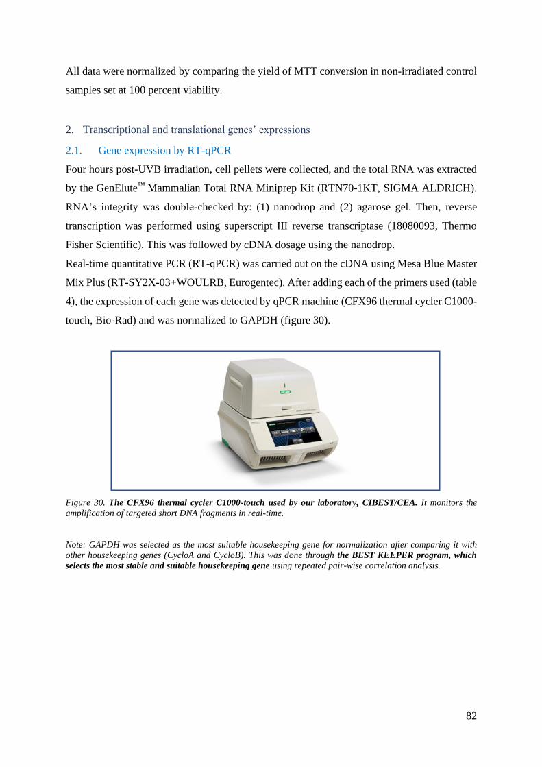
82
All data were normalized by comparing the yield of MTT conversion in non-irradiated control
samples set at 100 percent viability.
2. Transcriptional and translational genes’ expressions
2.1. Gene expression by RT-qPCR
Four hours post-UVB irradiation, cell pellets were collected, and the total RNA was extracted
by the GenElute™ Mammalian Total RNA Miniprep Kit (RTN70-1KT, SIGMA ALDRICH).
RNA’s integrity was double-checked by: (1) nanodrop and (2) agarose gel. Then, reverse
transcription was performed using superscript III reverse transcriptase (18080093, Thermo
Fisher Scientific). This was followed by cDNA dosage using the nanodrop.
Real-time quantitative PCR (RT-qPCR) was carried out on the cDNA using Mesa Blue Master
Mix Plus (RT-SY2X-03+WOULRB, Eurogentec). After adding each of the primers used (table
4), the expression of each gene was detected by qPCR machine (CFX96 thermal cycler C1000-
touch, Bio-Rad) and was normalized to GAPDH (figure 30).
Figure 30. The CFX96 thermal cycler C1000-touch used by our laboratory, CIBEST/CEA. It monitors the
amplification of targeted short DNA fragments in real-time.
Note: GAPDH was selected as the most suitable housekeeping gene for normalization after comparing it with
other housekeeping genes (CycloA and CycloB). This was done through the BEST KEEPER program, which
selects the most stable and suitable housekeeping gene using repeated pair-wise correlation analysis.
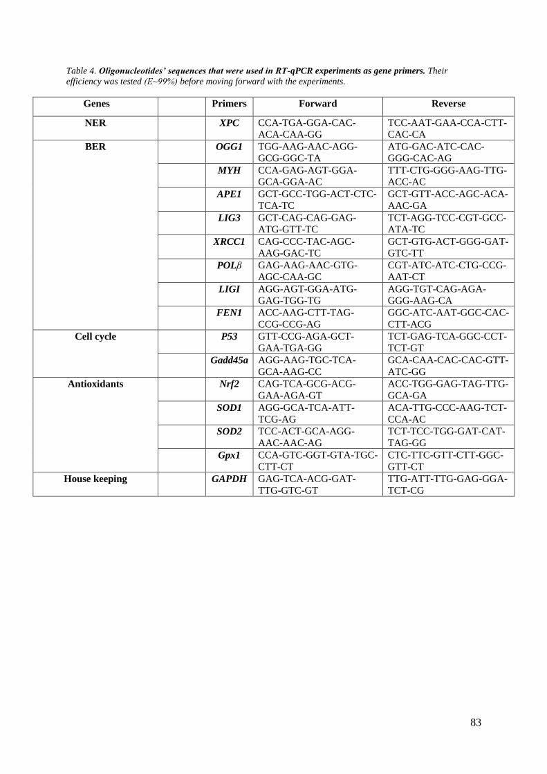
83
Table 4. Oligonucleotides’ sequences that were used in RT-qPCR experiments as gene primers. Their
efficiency was tested (E⁓99%) before moving forward with the experiments.
Genes Primers Forward Reverse
NER XPC CCA-TGA-GGA-CAC-
ACA-CAA-GG
TCC-AAT-GAA-CCA-CTT-
CAC-CA
BER OGG1 TGG-AAG-AAC-AGG-
GCG-GGC-TA
ATG-GAC-ATC-CAC-
GGG-CAC-AG
MYH CCA-GAG-AGT-GGA-
GCA-GGA-AC
TTT-CTG-GGG-AAG-TTG-
ACC-AC
APE1 GCT-GCC-TGG-ACT-CTC-
TCA-TC
GCT-GTT-ACC-AGC-ACA-
AAC-GA
LIG3 GCT-CAG-CAG-GAG-
ATG-GTT-TC
TCT-AGG-TCC-CGT-GCC-
ATA-TC
XRCC1 CAG-CCC-TAC-AGC-
AAG-GAC-TC
GCT-GTG-ACT-GGG-GAT-
GTC-TT
POLβ GAG-AAG-AAC-GTG-
AGC-CAA-GC
CGT-ATC-ATC-CTG-CCG-
AAT-CT
LIGI AGG-AGT-GGA-ATG-
GAG-TGG-TG
AGG-TGT-CAG-AGA-
GGG-AAG-CA
FEN1 ACC-AAG-CTT-TAG-
CCG-CCG-AG
GGC-ATC-AAT-GGC-CAC-
CTT-ACG
Cell cycle P53 GTT-CCG-AGA-GCT-
GAA-TGA-GG
TCT-GAG-TCA-GGC-CCT-
TCT-GT
Gadd45a AGG-AAG-TGC-TCA-
GCA-AAG-CC
GCA-CAA-CAC-CAC-GTT-
ATC-GG
Antioxidants Nrf2 CAG-TCA-GCG-ACG-
GAA-AGA-GT
ACC-TGG-GAG-TAG-TTG-
GCA-GA
SOD1 AGG-GCA-TCA-ATT-
TCG-AG
ACA-TTG-CCC-AAG-TCT-
CCA-AC
SOD2 TCC-ACT-GCA-AGG-
AAC-AAC-AG
TCT-TCC-TGG-GAT-CAT-
TAG-GG
Gpx1 CCA-GTC-GGT-GTA-TGC-
CTT-CT
CTC-TTC-GTT-CTT-GGC-
GTT-CT
House keeping GAPDH GAG-TCA-ACG-GAT-
TTG-GTC-GT
TTG-ATT-TTG-GAG-GGA-
TCT-CG

84
2.2. Western blot
Four hours post-UVB irradiation, cell pellets were collected, and the total proteins were
extracted and solubilized effectively upon cell lysis by RIPA lysis buffer (R0278-50mL, Sigma
Aldrich). This was followed by total protein dosage (MicroBC Assay Protein Quantification
kit, UP75860A, Interchim) and storage in -80°C. On the experiment day, samples were
prepared by adding 1X lamellae blue and ribonuclease-free water, heated at 90°C for 10
minutes, and equally loaded in Bio-Rad 4-20% mini-protein gels (2553103). Then, blotting and
blockage of Bio-Rad membrane (2553047) by 5 percent lyophilized milk were done (figure
31). After that, membranes were incubated with primary antibodies at 4°C overnight (table 5).
The next day, membranes were washed several times and incubated for one hour with a
secondary antibody (table 5). This was followed by washing and revealing. The membranes
were visualized through Bio-Rad Molecular Imager® Chemi DocTM XRS+ using Image LabTM
software (figure 31). The analysis was done by normalizing the target protein’s expression to
the stain-free total protein extract via Image Lab program
Figure 31 shows the AmershamTM ECLTM RainbowTM Marker full range ladder used to
determine the molecular weights of the targeted bands on the membrane.
Figure 31. Western blot instruments used by our laboratory, CIBEST/CEA. Panel A) AmershamTM ECLTM
RainbowTM Marker-Full Range ladder (RPN800E, Sigma Aldrich). Panel B) the BIO-RAD Transfer-Blot
Turbo™ transfer system used to blot our gels into membranes. Panel C) BIO-RAD ChemiDOc™ XRS
(HELA commercial nuclear protein extract was used as positive control)

85
Table 5. Antibodies that were used in western blot experiments.
Antibodies Type Dilution Supplier Reference
NER XPC 1/1000, 1/500 Santa Cruz sc-74410
BER OGG1 1/50000 Abcam ab124741
MYH 1/250 Novus Biologicals NB600-1032
APE1 1/1000 Sigma HPA002564
LIG3 1/3000 Novus Biologicals NBP1 41190
XRCC1 1/500 Abcam ab1838
FEN1 1/10000 Abcam ab109132
PARP1 1/3000 Cell Signaling
Technology
9542
Cell Cycle P53 1/5000 Sigma p6874
Cytoprotection
against Oxidative
Stress
Nrf2 1/1000 Abcam ab62352
Positive control GAPDH 1/5000 Santa Cruz sc-137179
Secondary Mouse 1/10000 Amersham
Biosciences
NA931
Rabbit 1/10000 Amersham
Biosciences
NA934
3. Detection of DNA lesions
3.1. HPLC-MS/MS: detection of bulky lesions
For this experiment, after cell collection at different time points, we needed to do DNA
extraction and enzymatic digestion as follows:
Similar to Mouret et al., cell pellet was lysed by triton X-100 then the nuclei were isolated by
centrifugation and solubilized by SDS. To precipitate DNA solely, successive treatments with
RNases and proteinase were done, followed by the addition of sodium iodide and 2-
isopropanol.Finally, thee resulting DNA pellet was solubilized with 0.1 mM deferoxamine
mesylate solution ready for digestion (Mouret et al. 2006).
Two successive digestions were done as follows: (i) phosphodiesterase II (0.1 U/µL), DNase
II (10 U/µL), nuclease P1 (0.2 U/µL), and MNSPDE buffer (200 mM succinic acid, 100 mM
CaCl2, pH 6) were added to the DNA for 2 hours at 37°C. Then, (ii) phosphodiesterase I and
alkaline phosphatase (2 units, pH 8) were added for another 2 hours at 37°C. Finally, HCL

86
(0.1M) was added, followed by sample centrifugation (5000g, 5 minutes), transfer in HPLC
vial, and storage at -20°c (Mouret et al. 2006).
For injection in the HPLC-MS/MS, samples were freeze-dried overnight then the remaining
residues were solubilized in triethylammonium acetate solution (20 mM). We used the
transitions mentioned in table 6 to detect the different bipyrimidine photoproducts (Mouret et
al. 2006):
Table 6. The transitions used for our analysis were based on specific chromatographic conditions and mass
spectrometry features (Mouret et al. 2006). Standard nucleosides were quantified at 270 nm. For identified CPDs:
T<>T (TT cyclobutane dimer), C<>C (CC cyclobutane dimer), T<>C (TC cyclobutane dimer), C<>T (CT
cyclobutane dimer). For identified 6-4 PPs : TT (TT (6-4) photoproduct), TC (TC (6-4) photoproduct).
Photoproducts Transitions
T<>T 545→447
C<>C 517→195
T<>C 531→195
C<>T 531→195
TT 545→432
TC 530→195
3.2. Comet Assay ± Fpg: detection of oxidized purines (8-oxoGua)
In our experiments, we did both: the classical and modified Fpg-comet assay. It is usually done
to measure the induced DNA lesions of cellular extracts and their repair at various kinetic
points.
After cell collection (as shown in part 1.2), cell pellets were suspended in freezing buffer
[Sucrose (85.5 g/L), sodium citrate (11.76 g/L), DMSO (50 mL/L), pH 7.6 (adjusted via citric
acid 0.1 M)] (2*105cells/100µL) and stored at -80°C until use.
Before the day of the experiment, slides were also prepared by covering them with normal
agarose (A9539-250G, Sigma Aldrich). On the experimental day, cells were diluted with 0.6
percent of low-melting agarose (A9414-5G, Sigma Aldrich), deposited on the previously
prepared slides, and covered by coverslips. This was followed by incubating slides with lysis
buffer for one hour and washing 3 times with neutralizing buffer. After that, Fpg (5U/slide and
2.5U/slide for parts one and two, respectively) + Fpg buffer (+FPG) and Fpg buffer (-FPG)
were prepared and added to the slides for a humidified incubation at 37°C for 40 minutes. The
reaction was stopped by incubating slides on ice for 10 minutes. Afterward, slides were
incubated in cold buffer for 30 minutes followed by DNA electrophoresis (25 V, 530 mA) at
4°C for further 30 minutes. Then, slides were washed by neutralizing buffer, dried, and 50µL

87
1X-Gel Red was added to each one for the next day's reading. Slides were read using a 10X
objective microscope and Comet Assay IV software (Perceptive Instruments, Suffolk, UK) by
randomly selecting 50 nuclei (DNA). The extent of damage was evaluated by the mean tail
intensity percentage of DNA, and normalization was done as a ratio of irradiated/non-irradiated
at each condition. Each experiment was repeated 3 different times and contained triplicate for
each condition.
Two positive controls were used in the experiments: Hydrogen peroxide (H2O2, 400µM) and
UVA + Riboflavin (10218153, AlfaAesar) (10 J/cm²+0.1mM)
Note: The positive control concentration was selected after checking different concentrations and selecting the
best one for the experiment.
4. Studying oxidative stress
Part Two “Ameliorating the DNA repair of XP-C cells by modulating their redox state
via pharmacological treatments”:
4.1. ROS assay
Intracellular ROS was quantified using DHR123 (Dihydrorhodamine 123, Sigma Aldrich).
First, cells were plated in dark 96-well plates for 24 hours, then treated with different drugs, as
shown in part 1.2. After that, cells were rinsed with 1X-PBS and incubated with diluted
DHR123 (1/100 PBS) for 30 minutes. This was followed by irradiation with UVB (0.02 J/cm²),
adding cell medium, and measuring the fluorescence intensity (λexc/λem 480/530 nm) at
different kinetic points (0-, 0.5-, 1-, 2-, 24-, and 48-hours post-irradiation).
H2O2 (500µM) was used as a positive control.
4.2. Glutathione assay
We collected cells post-UVB irradiation (0.01 J/cm²) using a rubber policeman and
experimented as suggested by the supplier (703002, Cayman). Cell pellets were suspended in
1X 2-(N-morpholino) ethanesulfonic acid (MES, 703010, Sigma Aldrich) cold buffer then
lysed using liquid nitrogen (freezing-thawing 5 times). This was followed by centrifugation at
10,000 g for 15 minutes at 4°C. The supernatant was then deproteinated as follows:
1mL of metaphosphoric acid (MPA) (5g/50mL) was added to the sample, followed by
vortexing and 5 minutes incubation at room temperature. Then, samples were centrifuged
(2000 g, 2 minutes), and the supernatant was carefully collected and stored at -20°C. On the
experiment day, triethanolamine (TEAM, 4M, T58300, Sigma Aldrich) was prepared, and
50µL was added to the samples to increase the pH. Next, we designed a plate of standard and

88
sample wells and added freshly prepared cocktail solution (MES buffer, reconstituted Cofactor
Mixture, reconstituted Enzyme Mixture, water, and reconstituted DTNB). Then GSH
concentration was quantified by the endpoint method (405-414 nm) after 25 minutes.

89
Results & Discussion

90
Part One “Deciphering the Role of XPC in BER and Oxidative Stress”
PC has been linked to BER and redox homeostasis by various researchers to explain
the clinical heterogeneity amongst XP-C patients (high skin and internal cancers).
XP-C cell lines showed reduced excision of oxidized DNA damage (8-oxoGua, 8-
oxoA, TG) and sensitivity to oxidants (Kumar et al. 2020).
In our study, we tried to scan such a link in a detailed manner to better understand the biological
role of XPC in UVB-induced-oxidized DNA repair. For that, we used three distinct XP-C
primary fibroblasts. We studied the effect of such mutations on the stimulated expression (gene,
protein) and activity of the BER pathway, the main pathway responsible for repairing oxidized
bases, compared to three normal primary fibroblasts. However, before going deeper in the
research, we devoted the first part of our study to characterize these primary fibroblasts and to
optimize their experimental conditions.
This work was published in Frontiers in Genetics Journal (https://doi.org/10.3389/fgene.2020.561687) (refer to
Annex).
X

91
1. Chapter One: Characterization of Primary Fibroblasts
1.1. Impaired XPC gene and protein expression in XP-C fibroblasts
To verify that the three different mutated primary fibroblasts (materials and methods, table 2)
lack XPC, we analyzed the gene expression at mRNA and protein basal levels through RT-
qPCR and western blot, respectively.
As expected, XP-C1, XP-C2, and XP-C3 cells showed a significant 8, 4, and 3-fold
downregulation (p<0.0001, ****), respectively, in XPC’s mRNA level compared to control
cells (figure 32). Similarly, Khan el al. and Kuschal et al. showed that XP-C cells extracted
from patients had less than 25 percent of the XPC mRNA level present in normal control (Khan
et al. 2006; Kuschal et al. 2013).
Patients with low XPC mRNA level have a poorer cancer prognosis compared to those with
higher expression level. Notably, this was shown in adenocarcinoma patients and mice with
lung cancer (Y.-H. Wu et al. 2010; Hollander et al. 2005). Hence, it may not be a surprise for
XP-C patients to develop cancer frequently where reduced XPC mRNA level may act as a
prognostic to carcinogenesis (Y.-H. Wu et al. 2010).
Figure 32. Lower XPC mRNA level in XP-C fibroblasts compared to normal control, at basal level. RT-qPCR
showed that XP-C1, XP-C2, and XP-C3 had a significantly lower XPC mRNA expression (p<0.0001, ****)
compared to the normal control (N=3). XP-C2 and XP-C3 showed significantly higher XPC mRNA expression
than XP-C1 (p<0.01, ¤¤). The data were normalized relative to the GAPDH mRNA levels, where GAPDH was
used as an endogenous control. Unpaired t-test was used to compare XPC mRNA level between normal and each

92
XP-C fibroblast (p<0.05, *), while paired t-test was used to compare XPC mRNA level between XP-C fibroblasts
(p<0.05, ¤). The results are the mean ± SEM from three independent experiments, n=3.
Of note, XP-C2 and XP-C3 had significantly higher XPC mRNA level compared to XP-C1
(p<0.01, ¤¤). This could be due to the difference in mutations amongst the studied fibroblasts.
Their premature termination codons’ positions could affect the susceptibility to nonsense-
mediated decay in degrading transcripts (table 2).
Nevertheless, as shown in figure 33, all three low mRNA levels failed to be translated into full
or truncated detectable XPC proteins. XP-C1’s frameshift mutation (c. 1643-1644del) on exon
9 is most prevalent in Mediterranean countries such as Morocco, Algeria, Tunisia, Egypt, and
Italy (Senhaji et al. 2013; N et al. 2018). It leads to mRNA harboring premature termination
codon (PTC). This will trigger nonsense-mediated RNA decay (NMD) pathway that leads to
translation termination (N et al. 2018; H. R. Rezvani, C. Ged, et al. 2008). The other studied
mutations [(c.413-3delC, c.1086del) in XP-C2 and (c.1243C>T, c.2287delC) in XP-C3] also
lead to PTC and proteins’ absence. C.1243C>T and c.2287delC had been already identified on
exons 9 and 13, respectively, in French families (Soufir et al. 2010).
Figure 33. Absence of XPC protein in XP-C primary fibroblasts compared to normal control, at basal level. A)
XPC protein was absent in the three XP-C fibroblasts (XP-C1, XP-C2, XP-C3) while detectable in the normal
control, at MW=125KDa. B) represents the western blot membrane images (i) of the XPC band in normal vs XP-
C cells upon hybridization with anti-XPC, and (ii) shows the total protein membrane that was used for
normalization. The results correspond to the mean ± SEM from three independent experiments, n=3.
1.2.Impaired NER capacities in XP-C primary fibroblasts compared to control cells
We wanted to check further whether the three XP-C primary fibroblasts used in our study have
a moderate, mild, or deficient GG-NER activity. Therefore, we selectively studied the repair
of (6-4) PPs recognized more specifically than CPDs by XPC (refer to canonical roles, 4.3.1).

93
As shown in figure 34, the UVB-induced (6-4) PPs persisted after 24 hours in the three XP-C
mutated fibroblasts where they were significantly higher compared to the normal control (XP-
C1: p<0.001, ***; XP-C2: p<0.001, ***; and XP-C3: p<0.05, *). Thus, after 24 hours, the
repair was almost 20 percent in the XP-C cells compared to 70 percent in the normal cells. This
agrees with other studies, which showed that (6-4) PPs are repaired efficiently after 24 hours
in normal primary fibroblasts but are less repaired (10-20 percent) in XP-C fibroblasts
(Chavanne et al. 2000).
Figure 34. Deficient (6-4) PPs repair in XP-C primary fibroblasts compared to normal control, post-UVB
irradiation. Panel A) XP-C1, XP-C2, and XP-C3 showed a significantly slower repair and more persistence of
lesion 24 hours after irradiation (p<0.001, ***; p<0.001, ***; p<0.05, * respectively). We used
immunocytochemistry to monitor the repair of (6-4) PPs at 0 and 24 hours post-UVB (0.03 J/cm²). For that, we
stained the nuclei with Hoechst and the (6-4) PPs with green fluorescently labeled primary antibody, then we
merged both fluorescence and quantified the fluorescence signal. Normalization was done relative to non-
irradiated values. An absence of primary antibody was used as the negative control. Unpaired t-test was used to
compare the normalized lesion ratio between normal and each XP-C fibroblast at each UVB dose (0 and 24 hours)
(p<0.05, *). The results are the mean ± SEM from two independent experiments, n=2 (each experiment was done
as a triplicate). IR= irradiated, NIR= non-irradiated. Panels B and C) represent the immunochemistry images of

94
normal and XP-C1 primary fibroblasts, respectively. The green color represents the presence of (6-4) PPs in the
nuclei. Nuclei were visualized by Hoechst staining.
0.03 J/cm² was used to avoid over-expressed fluorescence and cellular overstaining.
Note: As we proceeded with the characterization and results validation, we compared normal and XP-C1
fibroblasts. As presented in table 2, XP-C1 harbors homozygous frameshift mutation (c. 1643-1644del), classified
as the most common and studied mutation.
To confirm the results obtained by immunofluorescence, we did HPLC-MS/MS to monitor the
kinetics of repair in XP-C1 versus normal fibroblast between 0 and 48 hours post-UVB
irradiation (0.05 J/cm²). As shown in figure 35, (6-4) PPs were completely repaired at 24 hours
in the normal fibroblasts but persisted, almost unrepaired, in XP-C1 fibroblasts for more than
48 hours. Similarly, more than 20 percent of CPDs were repaired at 48 hours in the normal
fibroblasts but persisted, almost unrepaired, in XP-C1 fibroblasts.
Figure 35. Kinetics of bulky lesions’ [CPDs and (6-4) PPs] repair in XP-C1 versus normal fibroblast post-UVB
irradiation. We collected cells at different kinetic points (0, 4, 24, and 48 hours) post-UVB (0.05 J/cm²) and
monitored the repair of both types of lesions: CPDs and (6-4) PPs by HPLC-MS/MS. (preliminary result, n=1)
HPLC-MS/MS provided us the privilege to monitor how each type of (6-4) PPs and CPDs were
repaired (supplementary figures 1 and 2). We showed that, in normal fibroblast, the (6-4) PPs
and CPDs were repaired at 24 and 48 hours post-UVB, respectively, while they persisted in
XP-C cells.
Therefore, XP-C fibroblasts failed to repair the studied bulky lesions.

95
1.3. Similar UVB-induced photosensitivity between XP-C and normal primary
fibroblasts
Photosensitivity represents the exaggerated skin response to UVA/UVB light. This response is
present in numerous diseases like xeroderma pigmentosum and is translated clinically as skin
burning, redness, freckles, and carcinogenesis. Hence, do our studied XP-C primary fibroblasts
have photosensitivity?
As shown in figure 36, 24 hours post-UVB irradiation, the MTT cytotoxicity test revealed that
normal and XP-C primary fibroblasts share similar photosensitivity. This may be due to the
fully functional TC-NER in XP-C cells that prevents P53-dependent apoptotic pathways in the
absence of RNA synthesis arrest (Qiao, Scott, et al. 2011). For example, Queille et al. showed
that XP-D and XP-A cells are more sensitive to UV-induced apoptosis compared to XP-C and
normal cells due to their TC-NER deficiency where bulky lesions at DNA transcribed strands
trigger apoptosis (Queille et al. 2001). Also, it was shown that xpc-/- mice and mouse
embryonic fibroblasts (MEF) are moderately UVB-photosensitive (Boonstra et al. 2001; de
Waard et al. 2008). In parallel, Khan et al. and Sethi et al. showed that XP-C patients do not
have acute photosensitivity and severe sunburns at low UV doses (Khan et al. 2009; Sethi et
al. 2013). Interestingly, XP-C fibroblasts did not show any variation in viability compared to
control cells when exposed to other stress types, including oxidative agents (H2O2, methylene
blue..) (de Melo et al. 2016). Therefore, XPC could be considered as a non-vital protein for cell
viability.
Figure 36. Similar photosensitivity between normal and XP-C primary fibroblasts. We measured the percentage
of cellular viability 24 hours post-UVB irradiation by a short-term cytotoxicity test (MTT). Then we normalized
each sample by its untreated value (100% viability). Unpaired t-test was used to compare photosensitivity between
normal and each XP-C fibroblast at each UVB dose. The results are the mean ± SEM from three independent
experiments, n=3.

96
This experiment allowed us to choose a UVB dose for our experiments selectively. Based on
the regression analysis (table 7), we should select a UVB dose as follows: <0.15 J/cm² for
normal and XP-C3 cells, <0.18 J/cm² for XP-C1 cell, and <0.06 J/cm² for XP-C2 cell.
Therefore, we decided to use 0.05 J/cm², a dose that kills less than 40 percent of normal, XP-
C1, and XP-C3 fibroblasts and almost 50 percent of XP-C2 primary fibroblast. Such a dose
seems adequate for our aims as it will not kill more than 50 percent of the cells but could induce
a detectable and monitored effect.
Table 7. Measurement of LD50 for normal and XP-C primary fibroblasts post-UVB irradiation. LD50 represents
the UVB dose that will reduce 50% of the in vitro cell survival. It is calculated after the short-cytotoxicity test that
represents cellular viability vs UVB doses. The results represent the average of three different experiments, n=3.

97
1.4.Higher ROS level in XP-C1 fibroblasts
To show whether UVB affects XP-C cells at levels other than cellular viability, we selected
XP-C1 and normal fibroblasts and treated them with DHR123 followed by UVB-irradiation
(0.02 J/cm²) and monitored the kinetic of ROS fluorescence at 0, 0.5, 1, 2, 3, 24, and 48 hours.
Higher ROS level was observed in XP-C1 compared to normal, particularly 48 hours post-
UVB (p<0.05, *) where ROS was decreasing slower in the absence of XPC compared to its
presence (figure 37). Furthermore, high ROS levels were also detected in keratinocytes upon
XPC silencing (Hamid Reza Rezvani et al. 2011). This could be one of the main reasons behind
XP-C patients’ physiopathology.
Figure 37. Kinetics of ROS level in XP-C1 and normal primary fibroblasts. We treated cells with DHR123
(1/1000) followed by UVB and monitored fluorescence kinetics at different kinetic time points. Then we
normalized each sample by its untreated value (100% viability). H2O2 was used as a positive control. Unpaired
t-test was used to compare the ROS fluorescence between normal and XP-C1 fibroblast at each kinetic time point
(hours) (p<0.05, *). The results are the mean ± SEM from three independent experiments, n=3.
Therefore, we suggest that the higher persistent ROS level in XP-C1 in parallel to the acute
UVB photosensitivity could play a major role in the accumulation of mutations and the slow
initiation and progression of multiple of tumorigenesis in absence of detectable cell death.

98
1.5.Higher photoresistance in XP-C1 fibroblasts to solar simulation
After checking the effect of UVB on XP-C cells’ viability, we wanted to check whether it was
the case with UVA+UVB. This will allow us to detect any synergistic, antagonistic, or
independent effect of UV-induced lesions and oxidative stress on the cells. Furthermore, it will
support the link between XPC mutations, cell viability, and oxidative stress.
When we monitored the effect of the solar simulator, which mimics the sun’s radiation, on XP-
C1 and normal primary fibroblasts, we detected more photoresistance from the former cells
than the normal control (figure 38). XP-C1 fibroblast had significantly higher viability
compared to control at 1 (p<0.05, *) and 2 (p<0.01, **) MED. Since solar simulator contains
UVA+UVB and UVA is known to produce more oxidative stress than UVB, this may further
confirm our hypothesis and show resistance of XP-C fibroblasts to high oxidative stress as
suggested by de Melo et al. (de Melo et al. 2016).
XP-C cells accumulate mutations and macromolecular damages, which provokes them to
transform later and induce metastasis; meanwhile, once the damage exceeds the capacities of
DNA repair pathways, it triggers death in normal cells. In addition, XP-C cells have high
NOX1 levels, which play a role in P53 dysregulation via SIRT1 and induce apoptosis
suppression (Puca et al. 2010; Hamid Reza Rezvani et al. 2011). On the other hand, Naik et al.
had shown that UV induces programmed cell death in primary keratinocytes and fibroblasts
via neutralizing the anti-apoptotic Bcl-2 family that regulates P53-dependent and P53-
independent pathways against UV (Naik et al. 2007).
Figure 38. Higher photo-resistance in XP-C1 primary fibroblast compared to normal control. We measured the
percentage of cellular viability 24 hours post-solar simulator (UVA+UVB) irradiation by short-term cytotoxicity
test (MTT). Then we normalized each sample by its untreated value (100% viability). Unpaired t-test was used to
compare normal and each XP-C fibroblast photosensitivity at each UV dose (p<0.05, *). The results are the mean
± SEM. ssUV= solar simulator UV; MED=minimal erythema dose.
Therefore, our studied XP-C cells show persistence to high UV-induced oxidative stress (ssUV,
UVB), which may trigger mutagenesis and tumorigenesis.

99
Briefing of the characterization
The three different XP-C fibroblasts had impaired XPC gene expression compared to the
normal fibroblasts. This led to:
• Similar UVB-photosensitivity
• High ROS level and resistance to oxidative stress
• Deficiency in bulky lesions’ [CPDs, (6-4) PPs] repair
Such cellular profiling could explain why the absence of XPC triggers a variety of cancers. For
instance, Hollander et al. showed that one hundred percent of the studied xpc-/- mice developed
lung cancer (Hollander et al. 2005). This paved the way for checking whether the XPC-BER
interplay is the primary key in such an event and whether BER could endure the consequences
of high ROS levels in XP-C cells.

100
2. Chapter Two: Effect of XP-C Mutations on BER’s Expression
and Excision Activity
Studies have shown a complexity in the NER and BER pathways due to their interplay. One of
the critical players in such crosstalk is XPC: it interacts and/or stimulates SMUG1, TDG,
OGG1, and APE1, but does it affect the rest of the BER factors?
2.1. Effect of XP-C on BER’s mRNA expression post-UVB
For that, we explored the UVB-stimulated gene expression ratio [irradiated (IR)/non-irradiated
(NIR)] of the main BER factors starting with the incision/excision step (OGG1, MYH, APE1),
then the synthesis step (POLβ), and ending with the ligase step (LIG3). BER is known to repair
bases (oxidized, alkylated, deaminated) and single-strand DNA breaks. This could show us
how cells adapt to UVB stress. We mainly focused on the short-patch BER pathway because
8-oxoGua is primarily repaired; nevertheless, this does not exclude its repair by the long-patch
BER pathway (Marsin et al. 2003).
As discussed earlier in section 2.1. (overview of BER pathway), OGG1, MYH, and APE1 are
common BER initiation factors in both sub-pathways (short-patch and long-patch). OGG1 and
MYH are responsible for the excision of oxidized purines (8-oxoGua) and adenine opposing
the oxidized purines, respectively, while APE1 plays a role as an endonuclease and redox
regulator. POLβ and LIG3 had also been shown to be vital for oxidative DNA damage repair
(Akbari et al. 2014). POLβ adds newly synthesized nucleotide to 3’end of the previously arising
abasic site and removes the 5’ sugar-phosphate due to its polymerase and 5'-deoxyribose-5-
phosphate lyase (dRP lyase) activities, respectively. On the other hand, LIG3 seals the nick of
the DNA strand. Furthermore, we were inquisitive about considering other genes that could
affect the expression and/or efficiency of previously mentioned factors. For that, we checked
the gene expression of the accessory genes: XRCC1, FEN1, PARP1, and stress-inducible genes:
P53 and GADD45a.
2.1.1. BER factors
We showed a significant global inhibition of BER mRNAs’ stimulated expression post-UVB
in the presence of the three different XP-C mutations compared to the normal control (figure
39).
OGG1, MYH, APE1, and LIG3 showed a significant downregulation (p<0.01) in all the three
XP-C primary fibroblasts, while POLβ showed a downregulation (p<0.01) in XP-C1 and XP-
C3 but not XP-C2. This allows us to hypothesize that XPC deficiency hinders BER at the

101
transcriptional level. De Melo et al. had already suggested an inhibition of OGG1 and APE1’s
mRNA expression following H2O2 treatment in SV-40 immortalized XP-C fibroblasts (de
Melo et al. 2016). To our knowledge, there is no other research discussing the effect of XPC
mutation on the rest of the BER transcripts. However, several studies reported a link between
a downregulation in their expression level and cancer, which may explain the cancer risk
heterogeneity in XP-C patients. For example, MYH mRNA and protein expressions were
downregulated in gastric cancer (Shinmura et al. 2011). Similarly, POLβ mRNA and protein
expressions were downregulated in breast cancer. Likewise, LIG3 mRNA was shown to be
downregulated in dermal fibroblasts isolated from nevoid basal cell carcinoma patients (Abdel-
Fatah et al. 2014; Charazac et al. 2020).
Besides the direct interactions between XPC and BER factors, could such an impaired BER
factors’ mRNA expression be due to the role of XPC in inducing BER’s expression through
specific signaling mechanisms or promoters directly and/or indirectly?
Apart from XPC’s role in DNA repair, it plays a role in transcription regulation. For example,
XPC deficiency has been shown to reduce BRCA1 expression via upregulating its repressor,
Pit-1 (H. Wang et al. 2019). In its turn, BRCA1 was reported to stimulate the expression of
some BER factors and enhance their activity, including OGG1, XRCC1, and APE1 (T. Saha et
al. 2010, 1).
Therefore, we propose a direct and indirect role of XPC in stimulating BER’s expression post-
stress (UVB).

102
Figure 39. Downregulated BER-associated gene transcription in XP-C fibroblasts compared to normal control,
post-UVB irradiation. After total RNA extraction and reverse transcription, we did RT-qPCR to study the BER
gene transcription 4 hours post-UVB (0.05 J/cm²). Panels A), B), and C) show that XP-C fibroblasts had a
significant downregulation of normalized IR/NIR mRNA expression (p<0.0001, ****) of OGG1, MYH, and APE1,
respectively. Panel D) shows a significant downregulation of normalized IR/NIR POLB mRNA expression in XP-
C1 (p<0.0001, ****) and XP-C3 (p<0.01, **) compared to control. Panel E) shows a significant downregulation
of normalized IR/NIR LIG3 mRNA expression in XP-C1 (p<0.0001, ****), XP-C2 (p<0.0001, ****), and XP-C3
(p<0.01, **) compared to control. Normalization was done relative to non-irradiated control. Unpaired t-test
was used to compare the normalized gene expression ratio between normal and each XP-C fibroblast (p<0.05,
*). The results are the mean ± SEM from three independent experiments, n=3. Normal (N=3). IR= irradiated,
NIR= non-irradiated

103
2.1.2. BER accessory factors
Studying the gene expression at the transcriptional level is vital for the characterization and
understanding of the molecular basis underlying the phenotypic changes. Hence, we further
screened BER’s stimulated expression (IR/NIR) and studied XRCC1, FEN1, and PARP1
(figure 40). Although XRCC1 lacks an enzymatic activity, it is vital to coordinate an efficient
DNA repair by interacting with several BER factors (Marsin et al. 2003). It interacts with
OGG1 and APE1 to stimulate their excision and glycosylase activities, respectively (Marsin et
al. 2003). It also interacts with LIG3 for the latter’s stabilization and with POLβ (Marsin et al.
2003). FEN1 is an endonuclease involved in long-patch BER of oxidized abasic sites as 2-
deoxyribonolactone (P. Liu et al. 2008). It cleaves within the apurinic/apyrimidinic (AP) site-
terminated flap that will be then sealed by LIGI (Ranalli, Tom, and Bambara 2002). FEN1
interacts with APE1 (Ranalli, Tom, and Bambara 2002).
Furthermore, PARP1 is involved in several mechanisms, including DNA repair. XRCC1’s
recruitment to DNA damage is PARP1-dependent (Ray Chaudhuri and Nussenzweig 2017).
PARP1 also interacts with POLB, APE1, and XPC (Abdel-Fatah et al. 2014; Lili Liu et al.
2017, 1; Robu et al. 2017). Such proteins have a direct impact on the activity and efficacy of
BER proteins. However, does XPC has the same impact on their mRNA expression as it did
on the previously studied BER factors? Answering this question could contribute to
understanding the disease’s molecular mechanism and regulatory networks for mRNA and
targeted therapies, especially RNA-based therapy.
As shown in figure 40, we found that XRCC1 and FEN1 were significantly inhibited in all XP-
C fibroblasts compared to normal cells (p<0.05). Additionally, PARP1 was significantly
inhibited in XP-C1 (p<0.05, *) but not in XP-C2 and XP-C3. It was demonstrated that XRCC1
and FEN1 deficiencies induce genetic instability and cellular transformation post-genotoxic
stress (Brem and Hall 2005; H. Sun et al. 2017). As PARP1 plays a role in both, NER and BER,
its downregulation could augment the accumulation of CPDs and oxidative DNA lesions post-
UVB irradiation, thereby triggering photocarcinogenesis (Hegedűs et al. 2019). It is also
possible that the downregulation of LIG3, APE1, and POLB in the absence of XPC could
influence the activation and expression of XRCC1, FEN1, and PARP1. This confirms the direct
interaction between the BER factors and their accessory proteins. For instance, PARP1 needs
APE1 to become activated and recruit downstream BER factors at the AP site (Lili Liu et al.
2017).

104
In addition, it was reported that XP-C cells show low apoptosis due to low PARP1 levels post-
methylene blue treatment (de Melo et al. 2016). This is coherent with our photosensitivity study
(figure 36).
It is important to address that it would have been interesting to check the efficacy of the BER
factors at the translational level and posttranslational modifications as the latter play a major
role in their efficiency and activity.
Figure 40. Downregulated BER-associated accessory gene transcription in XP-C fibroblasts compared to
normal control, post-UVB irradiation. After total RNA extraction and reverse transcription, we did RT-qPCR to
study the gene transcription 4 hours post-UVB (0.05 J/cm²). Panel A) shows a downregulation in normalized
IR/NIR mRNA expression of XRCC1 in XP-C1 (p<0.01, **), XP-C2 (p<0.0001, ****), and XP-C3 (p<0.001, ***)
compared to control. Panel B) shows a downregulation in normalized IR/NIR mRNA expression of FEN1 in XP-
C1 (p<0.05, *), XP-C2 (p<0.0001, ****), and XP-C3 (p<0.05, *) compared to control. Panel C) shows a
significant downregulation in normalized IR/NIR mRNA expression of PARP1 in XP-C1 (p<0.05, *) but not XP-
C2 and XP-C3. Normalization was done relative to non-irradiated control. Unpaired t-test was used to compare
normalized gene expression ratio between normal and each XP-C fibroblast (p<0.05, *). The results are the mean
± SEM from three independent experiments, n=3. Normal (N=3). IR= irradiated, NIR= non-irradiated

105
2.2. Effect of XPC mutations on BER’s protein expression post-UVB
After screening the fibroblasts at the transcriptional level, we wanted to selectively study the
stimulated translational level of OGG1, MYH, and APE1 post-UVB. This may further
elucidate the regulations of the molecular interaction network of the disease, which in turn can
pave the way in front of methods for prevention, diagnosis, and treatment. Therefore, OGG1,
MYH, and APE1 were selected for western blot analysis due to their essential role in initiating
oxidative DNA lesions’ repair.
As presented in figure 41, OGG1 stimulated protein expression (IR/NIR) was downregulated
in XP-C1 (p<0.001, ***), XP-C2 (p<0.05, *), and XP-C3 (p<0.001, ***) compared to normal.
Several studies support our finding. This indicates the involvement of XPC in stimulating the
initiation of BER post-stress.
De Melo et al. showed that OGG1 protein level decreases in the absence of XPC. Once cells
are complemented with XPC, the level of OGG1 protein augments (de Melo et al. 2016). A
downregulated OGG1 protein in the absence of XPC could lead to a lower concentration than
the UV-induced lesion substrates. This induces steady phase activity and lower efficiency in
lesions’ excision, particularly, in the presence of high ROS level and oxidative DNA damage
(as discussed before).
This contributes to a higher oxidative DNA repair efficacy and elucidates the role of XPC in
BER.
Figure 41. Downregulated BER-associated OGG1 protein level in XP-C fibroblasts compared to normal
control, post-UVB irradiation. After total protein extraction and quantification, a western blot was done to check
the expression of OGG1 protein in XP-C vs normal fibroblasts 4 hours post-UVB (0.05 J/cm²). Panel A) shows a
downregulation of normalized OGG1 IR/NIR protein level in XP-C1 (p<0.001, ***), XP-C2 (p<0.05, *), and XP-
C3 (p<0.001, ***) compared to control. Panel B) represents the western blot membrane image of the OGG1

106
protein band and total protein. Normalization was done by the total protein then ratio was done relative to non-
irradiated samples. Unpaired t-test was used to compare the normalized protein expression ratio between normal
and each XP-C fibroblast (p<0.05, *). Values shown are the mean ± SEM from three independent experiments,
n=3. Normal (N=3). IR= irradiated, NIR= Non-Irradiated.
Furthermore, MYH IR/NIR protein expression was downregulated in XP-C1 (p<0.01, **), XP-
C2 (p<0.01, **), and XP-C3 (p<0.05, *) (figure 42). Like OGG1, a low MYH protein level
could affect its ability to repair high DNA lesions, contributing to XP-C pathogenesis. If both
OGG1 and MYH fail to repair all the lesions, then G:C to T:A transversions will increase,
inducing carcinogenesis (figure 2).
Figure 42. Downregulated BER-associated MYH protein level in XP-C fibroblasts compared to normal control,
post-UVB irradiation. After total protein extraction and quantification, a western blot was done to check the
expression of MYH protein in XP-C vs normal fibroblasts 4 hours post-UVB (0.05 J/cm²). Panel A) shows a
downregulation of normalized MYH IR/NIR protein level in XP-C1 (p<0.01, **), XP-C2 (p<0.01, **), and XP-
C3 (p<0.05, *) compared to control. Panel B) represents the western blot membrane image of the OGG1 protein
band and total protein. Normalization was done by the total protein then the ratio was done relative to non-
irradiated samples. Unpaired t-test was used to compare the normalized protein expression ratio between normal
and each XP-C fibroblast (p<0.05, *). Values shown are the mean ± SEM from three independent experiments,
n=3. Normal (N=3). IR= irradiated, NIR= Non-Irradiated.
However, APE1 IR/NIR protein expression was only significantly downregulated in XP-C2
(p<0.0001, ****) compared to normal control (figure 43). These results show downregulation
in stimulation and initiation of BER in response to UVB stress. De Melo et al. showed that
APE1 protein level also decreased in the absence of XPC but not significantly (de Melo et al.
2016). Moreover, APE1 is a multifunctional protein that plays a role in DNA repair and redox
regulation. Therefore, its protein downregulation could explain the high oxidative stress in XP-
C cells (Hamid Reza Rezvani et al. 2011). Another reason could be OGG1 and/or MYH
downregulation. The downregulated OGG1 and MYH glycosylases may be insufficient to

107
repair 8-oxoGua efficiently and A:8-oxoG mispair, respectively. This might contribute to the
high oxidized purines in a time-dependent manner and BER’s dysregulation. Therefore, the
downregulation in OGG1, MYH, and APE1 expression could propose the interconnection
between them and the activation coordination to stimulate BER cascade.
Figure 43. Downregulated BER-associated APE1 protein level in XP-C fibroblasts compared to normal control,
post-UVB irradiation. After total protein extraction and quantification, a western blot was done to check the
expression of APE1 protein in XP-C vs normal fibroblasts 4 hours post-UVB (0.05 J/cm²). Panel A) shows
downregulation of normalized APE1 IR/NIR protein level in XP-C1 (p<0.001, ***) and XP-C2 (p<0.0001, ****)
but not in XP-C3 compared to control. Panel B) represents the western blot membrane image of the OGG1 protein
band and total protein. Normalization was done by the total protein then ratio was done relative to non-irradiated
samples. Unpaired t-test was used to compare the normalized protein expression ratio between normal and each
XP-C fibroblast (p<0.05, *). Values shown are the mean ± SEM from three independent experiments, n=3. Normal
(N=3). IR= irradiated, NIR= Non-Irradiated.
2.3. Effect of XP-C on BER’s activity post-UVB
Undeniably, XP-C is an interesting model due to its intricacy, and studying the gene expression
of BER gave us an idea about the tight connection and crosslink between the two. This
connection is presented at the phenotypic level where XP-C patients have a high risk of internal
cancer linked to oxidative stress and dysfunctional BER.
However, how profound is the effect of dysregulated XPC on BER’s activity?
Comet ± FPG assay
For that, we selected comet assay due to its advantages as accuracy, sensitivity, reliability, and
the need for a small number of cells per sample. The latter was one of the main reasons for
selecting comet assay over HPLC-MS/MS, which needs more than 2 million cells per sample.
Collecting a high cell number is not practical when working with primary fibroblasts,
especially when we studied six different primary fibroblasts with/without irradiation at
different kinetic points.

108
We used modified comet assay (±FPG) where FPG glycosylase (DNA-formamidopyrimidine
glycosylase) excises FPG-sensitive sites (oxidized purines: 8-oxoGua, Fapy) into abasic sites
then DNA single-strands. We can quantify the oxidized purines by subtracting the value
without FPG (-FPG) from values with FPG (+FPG) for each sample at each time point.
Figure 44 represents an example of the intact DNA (head of the comet) and damaged DNA
(tail of the comet) ± UVB ± FPG in normal and XP-C1 fibroblasts.
Figure 44. The undamaged (comet head) and damaged (comet tail) DNA ± FPG in normal and XP-C1
fibroblasts and positive control H2O2. The tail intensity and its length are proportional to the DNA damage.
Figure 45 shows a significantly higher DNA damage (± FPG) in XP-C fibroblasts compared to
control, post-UVB. It persisted until 24 hours. This indicates that UVB induces more DNA
lesions instantaneously in the absence of XPC compared to control. These lesions include
oxidized purines.
In the absence of FPG (-FPG)
DNA strand breaks (alkali-sensitive and abasic sites) were significantly higher in XP-C1 and
XP-C2 fibroblasts (p<0.05, *) compared to normal control at 0, 2, and 24 hours post-UVB
(figure 45). Similarly, Berra et al. showed that photosensitized methylene blue induced higher
DNA damage in XP-C cells than in control (Berra et al. 2013). Also, Zhou et al. indicated
through measuring alkaline comet assay that XPC protected primary murine type II
pneumocytes from cigarette smoke-induced DNA damage, including bulky lesions and
oxidative purines (H. Zhou et al. 2019).
These induced lesions represent a crucial initial event in carcinogenesis.
This indicates that, in addition to dysregulated GG-NER, XP-C fibroblasts may have other
impaired DNA repair pathways that fail to repair UVB-induced genomic lesions. Therefore,
we used a modified comet assay (+FPG) to selectively study the link between XPC and BER.

109
In the presence of FPG
As presented in figure 45, we detected a significantly increased % mean tail intensity (p<0.05,
$) in all fibroblasts in the presence of FPG compared to its absence, at 0 hours post-UVB. After
2 hours, the high level persisted in all fibroblasts except for XP-C2. This indicates that UVB
induces oxidized purines in all the fibroblasts. But is it more potent in XP-C cells?
At 0, 2, and 24 hours, XP-C1 and XP-C2 had significantly higher % mean tail intensity (p<0.05,
µ) compared to normal control. However, XP-C3 showed a significantly higher % mean tail
intensity (p<0.05, µ) 0 hours post-UVB (figure 45). This indicates that oxidized purines are
more prominent and persistent in XP-C cells.
Figure 45. Downregulated BER excision repair capacities in XP-C primary fibroblast compared to normal
fibroblasts, post-UVB irradiation. To follow-up the kinetics of repair of UV-induced lesions [single-strand breaks
(SSB), alkali-labile sites (ALS) and oxidative purines (including 8-oxoGua)] in XP-C vs normal fibroblasts, we
did Comet ± FPG 0, 2, and 24 hours post-UVB (0.05 J/cm²). This graph represents the % mean tail intensity for
each IR/NIR sample with FPG (oxidized purines, dark-colored) and without FPG (single-strand breaks, double-
strand breaks, and alkali-labile sites). Although all primary fibroblasts could repair the DNA damage (± FPG)
over time, XP-C cells showed a downregulated and slower repair than the normal control. In addition, more
oxidized purines were induced at time= 0 hours in the absence of XPC. Paired t-test was done to compare each
sample with 2 conditions (FPG-ve or FPG+ve), while an unpaired t-test was done to compare different samples
within the same condition. The results are the mean ± SEM from three independent experiments, n=3. Normal
(N=3).
$ Sample significantly (p<0.05) higher in its tail intensity with the presence of FPG (+FPG) compared to its
absence (-FPG)
* XP-C fibroblast significantly (p<0.05) higher in its tail intensity compared to normal fibroblast, FPG-ve
condition.

110
µ XP-C fibroblast significantly (p<0.05) higher in its tail intensity compared to normal fibroblast, at FPG+ve
condition.
-FPG= FPG alkaline buffer without the enzyme; +FPG= FPG alkaline buffer and FPG enzyme; IR= irradiated;
NIR= Non-Irradiated
This is well-illustrated in figure 46. It represents oxidized purines kinetic repair in each sample
post-UVB. At 0 and 2 hours, significantly higher oxidized purines (p<0.05, *) were detected
in XP-C cells compared to normal fibroblasts, while at 24 hours, oxidized purines were
significantly higher in XP-C1 and XP-C2 but not XP-C3 compared to normal control.
Figure 46. A simple graphical representation of BER excision repair in XP-C primary fibroblasts compared to
normal fibroblasts, post-UVB irradiation. It displays the oxidized purines (8-oxoGua and Fapy) repaired over
time in XP-C vs normal fibroblasts (N=3). Such DNA damage can be quantified by subtracting the values without
FPG (-FPG) from values with FPG (+FPG) for each sample at each time point. Oxidized purines were
significantly higher at each time point in all the XP-C fibroblasts than normal, except for XP-C3 24 hours post-
UVB (p<0.05). Unpaired t-test was done to compare different samples within the same condition (p<0.05, *).
Shown values correspond to the mean ± SEM from three independent experiments (n=3). Normal (N=3).
* XP-C fibroblast significantly (p<0.05) higher in its oxidized purines compared to normal fibroblast.
Therefore, XP-C cells have higher induced oxidized purines. This may be due to the higher
oxidative stress and ROS in XP-C cells compared to normal control. Several studies showed
that UVB generates excessive quantities of ROS that could induce DNA oxidation in vivo and
in vitro (H. R. Rezvani, C. Ged, et al. 2008). In parallel, Menoni et al. detected through live
cellular imaging that XPC is recruited to laser micro-irradiation induced 8-oxoGua
(D’Augustin et al. 2020). Furthermore, XP-C cells are sensitive to oxidants as photoactivated
methylene blue and KBrO3 (Berra et al. 2013; Mariarosaria D’Errico et al. 2006).

111
As shown in figure 46, oxidized purines were repaired slower in XP-C cells than normal cells.
This is discordant with Berra et al. Their results showed that FPG-sensitive sites were slightly
higher in XP-C cells but repaired efficiently at 24 hours (Berra et al. 2013). Our results may
differ due to different treatments and cell types (primary fibroblasts vs SV40-transformed skin
fibroblasts) with different genetic backgrounds. Meanwhile, D’Errico et al. showed results that
agree with ours. Their results showed that XP-C primary fibroblasts and keratinocytes have a
downregulated KBrO3-induced 8-oxoGua repair (Mariarosaria D’Errico et al. 2006). A
supporting result showed, by HPLC-ED, that xpc-/- mouse embryo fibroblasts displayed a
reduced 8-oxoGua removal compared to control following treatment with an oxidizing agent,
KBrO3 (Kumar et al. 2020).
Accumulation of oxidized purines contributes to carcinogenesis (skin and internal) in XP-C
patients. Multiple studies emphasized an interplay and interaction between NER and BER in
oxidative DNA damage and bulky lesions repair (Kumar et al. 2020). High ROS levels and
lack of XPC can inactivate OGG1’s enzymatic activity by oxidizing its cysteine residue and
halting its turnover, respectively. This results in G:C to T:A transversions mutations and
carcinogenesis if intermediates are not repaired by MYH (Mariarosaria D’Errico et al. 2006;
Hao et al. 2018). MYH low mRNA and protein levels were also linked to impaired repair of
elevated A:8-oxoGua (Markkanen, Dorn, and Hübscher 2013). A low MYH activity or gene
expression has been linked to gastric cancers, while reduced OGG1 activity has been linked to
high breast cancer risk incidences (Shinmura et al. 2011; Yuzefovych et al. 2016). Mentioned
internal cancers are also present in XP-C patients, revealing the impact of XPC mutations on
BER and the consequences (Giglia et al. 1998; X. Bai et al. 2012). Indeed, other factors may
play a role in such events. This may include the protein oxidation, in the presence of high ROS
level, that will directly affect BER factors, thereby reducing their ability to repair oxidized
purines efficiently and rapidly (Kumar et al. 2020).
Henceforth, does lack of XPC affect OGG1 activity or ROS level? We would say both based
on our experiments (gene expression and activity, ROS level quantification) and other
experiments.
Other severe clinical symptoms induced by oxidation, including neurological disorder, are
rarely found in these patients due to the capacity of BER to repair oxidized lesions, although
at a slower pace compared to normal control. For instance, 24 hours post-UVB, more than half
of the oxidized purines were repaired in XP-C cells. This could be explained by the following

112
scenarios: (i) XPC deficiency downregulates but does not inhibit BER’s activity and efficiency.
OGG1’s turnover is slower, so it will take more time to recognize consecutive lesions, and/or
(ii) other backup-glycosylases and repair systems (lower kinetics) will step up once
BER/OGG1 is incompetent.

113
3. Chapter Three: Effect of XP-C on Some Genes Linked to Cell Cycle
Regulation
As mentioned previously (NER and cell cycle, section 3.2), XPC is involved in cell cycle
regulation. Therefore, it would be interesting to check whether P53 and GADD45a’s gene
expressions are cluttered in the absence of XPC. This could provide us with an idea about the
cellular behavior post-UVB.
Figure 47 shows that P53’s mRNA level was downregulated significantly in XP-C1 (p<0.01,
**) and XP-C3 (p<0.05, *).
Figure 47. Different P53 mRNA level between XP-C and normal fibroblasts. After total RNA extraction and
reverse transcription, we did RT-qPCR to study the gene transcription 4 hours post-UVB (0.05 J/cm²). Normalized
IR/NIR P53’s mRNA expression was downregulated in XP-C1 (p<0.01, **) and XP-C3 (p<0.05, *) but not XP-
C2 compared to control cells (N=3). Normalization was done relative to non-irradiated control. Unpaired t-test
was used to compare normalized gene expression ratio between normal and each XP-C fibroblast (p<0.05, *).
The results are the mean ± SEM (n=3). Normal (N=3). IR= irradiated, NIR= non-irradiated
However, unlike some articles, figure 48 proposes a potentially higher P53 UV-induced-
expression in XP-C1 and XP-C2 than normal cells (preliminary result). Under normal
conditions, P53 level is low and increases upon genotoxic stress, including UV, for a short time
to trigger cell cycle arrest for DNA repair or apoptosis. Nevertheless, it was addressed that once
the P53 is mutated, it may function differently. Mutated P53 is more stable and triggers
transcriptional dysregulation and uncontrolled tumor growth (HUANG et al. 2014). For
instance, a high P53 protein level was detected in various cancer types, including colorectal,
breast, endometrial, and esophageal squamous cell carcinomas (HUANG et al. 2014). In

114
parallel, P53 is known to be mutated and dysregulated in XP patients, mainly due to G:C to
T:A transversions and C to T and CC to TT UV signature mutations, which could play a
significant role in carcinogenesis (Khan et al. 2006). Such mutations might have been induced
in our cells due to ROS accumulation and oxidative DNA damage triggering mutagenesis. This
anticipates in the dysregulation of P53’s gene expression. Krzeszinski et al. showed in their
study that P53 regulates UV-induced XPC expression. Once the XPC level becomes sufficient
for an efficient DNA repair, it downregulates P53 for cells’ return to their resting state. This
may be through regulating P53’s turnover via facilitating MDM2-Rad23-dependent
degradation of polyubiquitylated P53 (Krzeszinski et al. 2014). Our preliminary result supports
this scenario (figure 48). This protein accumulation in XP-C cells may indicate a failure in
ubiquitylated-P53 proteolysis that negatively affect its mRNA expression.
Therefore, we suggest that the observed high P53 level could be a mutated and dysfunctional
P53. Indeed, further experiments are required to confirm such a hypothesis such as repeating
the western blot and checking the activity and the regulations post-translationally of P53.
Figure 48. Different BER-associated P53 protein level in XP-C fibroblasts compared to normal control, post-
UVB irradiation. After total protein extraction and quantification, a western blot was done to check the expression
of P53 in XP-C vs normal fibroblasts 4 hours post-UVB (0.05 J/cm²). Panel A) shows a non-significant
upregulation of normalized P53 IR/NIR protein level in XP-C1 and XP-C2 but not XP-C3 compared to control.
Panel B) represents the P53 band and the total protein of normal and XP-C fibroblasts at basal level. Panel C)
represents the P53 band and the total protein of normal and XP-C fibroblasts at UVB level. Normalization was
done by the total protein then ratio was done relative to non-irradiated samples. Values shown are from one
experiment, n=1. Normal (N=3). IR= irradiated, NIR= Non-Irradiated. (preliminary result)

115
Such a dysregulated P53 affects its interactions and regulations of several signaling cascades
post-genotoxic stress (such as UV). GADD45a (growth arrest and DNA damage-inducible45
alpha) is known to protect cells from UV-induced carcinogenesis in a P53-dependent and P53-
independent manner to induce positive feedback on the P53 signaling pathway (Hildesheim et
al. 2002). Our transcriptional analysis (figure 49) showed that GADD45a mRNA level was
downregulated significantly (p<0.05, *) in XP-C2 and XP-C3 compared to normal cells.
Hildesheim et al. showed that GADD45a knockout reduces UV-induced MAPK/P53 dependent
apoptosis of keratinocytes. Herein, GADD45a prolongates the activation of apoptosis and/or
cell cycle arrest pathways (p38/JNK MAPK and P53 pathways, G1/G2 arrest) and orchestrates
DNA repair by interacting with NER (XPC, XPE) and BER (PCNA, APE1) to prevent genomic
instability. Inhibition of P53 and GADD45a contributes to carcinoma progression and
metastasis, especially in lung adenocarcinoma patients (Hildesheim et al. 2002, 53; Jung et al.
2013; Y.-H. Wu et al. 2010).
Figure 49. Different GADD45a mRNA level between XP-C and normal fibroblasts. After total RNA extraction
and reverse transcription, we did RT-qPCR to study the gene transcription 4 hours post-UVB (0.05 J/cm²).
Normalized IR/NIR gene expression of GADD45a was downregulated in XP-C2 and XP-C3 (p<0.05, *) but not
XP-C1 compared to control cells. Normalization was done relative to non-irradiated control. Unpaired t-test was
used to compare normalized gene expression ratio between normal and each XP-C fibroblast (p<0.05, *). The
results are the mean ± SEM. Normal (N=3). IR= irradiated, NIR= non-irradiated

116
The dysregulation of P53 and GADD45a might explain the absence of extreme photosensitivity
for the studied XP-C fibroblasts and the high cancer risk due to mutagenesis in XP-C patients.
Nevertheless, we should consider other GADD45 genes that are usually highly expressed in
dermal fibroblasts (like GADD45g) and could compensate for the downregulation in
GADD45a to activate G1 and G2 checkpoints (Hildesheim et al. 2002).
It should be noted that studying GADD45a protein’s expression level and cell cycle (P53 and
GADD45a’s activities) could have been interesting primarily due to the high mutated P53
recorded level in XP patients. Therefore, these studies may be considered as part of this
project’s perspectives.

117
Conclusion and Perspective
The three studied XP-C primary fibroblasts:
❖ Had low XPC mRNA and no XPC protein
❖ Inhibited NER activity
❖ Downregulated BER at gene level (mRNA and protein) and at
activity (excision) level
XP-C1 had higher ROS level compared to normal control.
In summary, the synergic effects of amassed oxidative DNA damage
and impaired BER could explain the heterogeneity in the clinical
spectrum of XP-C patients.
Could such a DNA repair impairment be adjusted by drug
treatments?
To answer this question, we studied the effect of different
antioxidants/oxidants on NER and BER pathways in the presence and
absence of XPC mutation by ameliorating their redox state (PART
TWO).
Indeed, it would be interesting to try later the same experiments on
keratinocytes and to use UVA or solar simulator to study the same
objectives on fibroblasts, keratinocytes, and 3D-reconstructed skin
models. Another future perspective is to check the protein expression of
the different tested factors at 24 hours post-UVB. Perhaps, a better
variation could be detected (at level of p53 for example).

118
Schematic Summary
Figure 50. The difference between normal and XP-C in response to UVB stress. UVB induces direct DNA
damage (bulky lesions) and indirect ROS-induced DNA damage (oxidative DNA damage) that are usually
recognized by XPC and repaired by NER and BER, respectively. XPC also plays a role in maintaining redox
homeostasis and preventing the increase of free ROS. However, in the absence of XPC, bulky lesions will
accumulate, BER will be less efficient, and unbalanced redox homeostasis will occur. All these events participate
in XP-C patients’ clinical phenotype—red rectangle=oxidized purines.

119
Part Two “Ameliorating the DNA repair of XP-C cells by modulating their
redox state via pharmacological treatments”
tudying the effect of XPC mutations on BER’s gene expression and activity in XP-C
cells derived from XP-C patients (part one) imposes a critical question: Is this effect
reversible? Could treatments with antioxidants manipulate the deficient NER and
impaired BER in XP-C cells? Could this be done mainly through balancing the cellular
redox state with glutathione?
Such a question is fascinating, mainly because XPC was previously linked to glutathione
production (S.-Y. Liu et al. 2010).
Xeroderma pigmentosum C disorder (XPC) is a multisystem disease with a wide range of
heterogeneous clinical symptoms. Unfortunately, diagnosis is usually made after the disease
has already been initiated. Hence, finding a treatment has been challenging and focuses on
numerous researchers, oncologists, and dermatologists. Succeeding in developing a preventive
and/or protective therapy could be a major turning point for it and other DNA repair-deficient
and skin diseases.
As mentioned previously in the introduction (XPC disorder, section 4.2.2.), several drugs have
arisen to limit XPC disorder’s symptoms, including retinoids, vismodegib, and surgery for
skin tumors. Potential treatments are also in progress, such as gene therapy. Gene therapy is
based on transferring specific genetic material into the cells via viral vectors or nonviral
administrations to reverse or halt disorders. Multiple clinical trials targeting T-cells to reduce
solid tumors such as melanoma have been conducted (Gorell et al. 2014). Researchers had also
tried to correct the DNA repair in XP-C keratinocytes. Gene therapy still has many challenges
to overcome, such as the effectiveness, safety, and durability of delivered vectors (Gorell et al.
2014). Hence, trying to scan for therapy for xeroderma pigmentosum from a different aspect
could also be interesting. Activating or inhibiting different cellular signaling pathways could
interfere with halting the clinical phenotypes of xeroderma pigmentosum disorder. As XPC has
been directly linked to BER and redox homeostasis (proven in part one and by other
researchers), studying the efficacy of antioxidant drugs on this disease (part two) brings us a
step closer towards exploring new treatments and profiling the disease further at genotypic and
phenotypic sides.
S

120
We divided part two of the project into three chapters. Each chapter discusses the role and
effect of one of the three different types of drugs used:
• Nicotinamide (NIC)
• N-acetylcysteine (NAC)
• Buthionine sulfoximine/Dimethylfumurate (BSO/DMF)
Studies were done at basal and UVB levels to monitor the detailed effect of each treatment as
a preventive, protective, and therapeutic (stimulator).
Part of this project will be the subject of an article in preparation.
For this part of the project, we used SV40 immortalized fibroblasts instead of primary cells for
multiple reasons: (i) we have many conditions. For each condition, we need a high cell number
for most of the experiments. This is not practical when using primary fibroblasts as they have
a slower growth rate and limited self-renewal, (ii) it is challenging to have primary fibroblasts
from the same source that could be sufficient for our objectives. However, it is essential to
address that repeating the experiments on primary fibroblasts is one of the main perspectives
for the future since they are more relevant in representing in vivo models and patients.

121
1. Chapter One: Characterization of Cell lines
Similarly to part one, we characterized the two SV40 transformed fibroblasts [normal
(AG10076) and XP-C (GM15983)] at gene and activity levels before launching the
experiments.
1.1.Impaired XPC gene and protein expression in XP-C cell line
As expected, XPC mRNA level was significantly downregulated in XP-C cells (p<0.01, **) at
basal level compared to normal control (figure 51). This shows that the XPC mutation inhibited
XPC’s expression at mRNA level. In normal cases, UVB triggers the upregulation of XPC’s
expression and efficacy to neutralize the formation of DNA lesions. Once the XPC gene is
mutated, the expression will be faltered. The low XPC mutated mRNA level in XP-C cell line
was not stable to be translated into protein. This was similar to the observations of part one. In
agreement with Qiao et al., there was a complete absence of XPC protein compared to normal
(Qiao, Scott, et al. 2011).
Figure 51. Impaired XPC mRNA and protein expressions in XP-C cells compared to normal control, at basal
level. Panel A) RT-qPCR showed that XP-C had a significantly lower XPC mRNA expression (p<0.01, **) than
the control. The data were normalized relative to the GAPDH mRNA levels, where GAPDH was used as an
endogenous control. Unpaired t-test was used to compare XPC mRNA level between the normal and XP-C
fibroblasts (p<0.05, *). The results are the mean ± SEM from two independent experiments, n=2. Panel B) shows
the XPC protein band (125 KDa, 1/500) in normal but not XP-C cells. GAPDH (37KDa, 1/5000) was used as the
loading control.

122
1.2.Higher UVB-induced photosensitivity in XP-C cells compared to control
On the contrary to part one, XP-C cells are significantly (p<0.05) more photosensitive
compared to control (figure 52).
Due to the high photosensitivity of XP-C cells, a lower UVB dose (0.01 J/cm²) was selected
for the experiments compared to that used in part one (0.05 J/cm²). Thus, it provides the
necessary damage to monitor an effect but is not cytotoxic nor lethal (52 and 81.4 percent of
viability in XP-C and normal cells, respectively).
Figure 52. Higher photosensitivity in XP-C cells compared to normal control. We measured the percentage of
cellular viability 24 hours post-UVB irradiation panel A) by short-term cytotoxicity test (MTT) were XP-C had
lower % viability compared to normal control at 0.01 (p<0.05, *), 0.02 (p<0.01, **), 0.1 (p<0.01, **), and 0.2
(p>0.0001, ****) J/cm² of UVB doses and panel B) by trypan blue were XP-C had lower % viability compared
to normal control (p<0.05, *) at 0.02 J/cm² of UVB. We normalized each sample by its untreated value (100%
viability). Unpaired t-test was used to compare normal and each XP-C fibroblast photosensitivity at each UVB
dose (p<0.05, *). The results are the mean ± SEM from three independent experiments, n=3, in MTT and two
independent experiments, n=2, in trypan blue manipulations.

123
In the beginning, we were worried that such a low dose could be lower than the effects’ limit
of detection (LOD). So, we did HPLC-MS/MS and verified that it is sufficient to induce a
high amount of DNA damage (table 8).
Table 8. Level of dimeric photoproducts [CPDs and (6-4) PPs] per million normal DNA bases. Dewar isomers
were not detected due to the low UVB dose used (0.01 J/cm²). The low value of the standard deviation represents
the reproducibility of both the experimental and analytical protocols.
1.3.Impaired NER capacities in XP-C cells compared to control
We did HPLC-MS/MS to examine the repair of different bulky photoproducts by XP-C and
normal cells. As expected, XP-C cells had lower photoproducts’ repair efficiency compared to
normal control (figures 53 and 54). Studying the repair capacities by HPLC-MS/MS instead of
immunocytochemistry provided us the privilege to monitor each type of CPD and (6-4) PP
lesions.
Figure 53 shows that UVB-induced TT and TC (6-4) PPs were repaired slower and more
persistent in XP-C cells. Almost 60 percent of the (6-4) PPs were detected at 48 hours. On the
contrary, both (6-4) PPs were efficiently repaired 24 hours post-UVB in normal cells. Thus,
independently of the UV dose, normal fibroblasts can repair efficiently (6-4) PPs within 24
hours (Courdavault et al. 2004).
Perhaps we did not detect CT and CC (6-4) PPs because they are less frequent. The frequency
of the (6-4) PPs with the different dinucleotides is as follows: TC>TT>CC and CT (Khoe,
Chung, and Murray 2018).

124
Figure 53. Deficient (6-4) PPs lesions repair in XP-C cells compared to normal control. We monitored repair
kinetics for (6-4) PPs bulky photoproducts at different hours post-UVB by HPLC-MS/MS. TT and TC (6-4) PPs
were significantly higher (p<0.05) from 0 until 48 hours in XP-C cells compared to control. Normalization was
done relative to irradiated values at 0 hours. Unpaired t-test was used to compare the normalized lesion ratio
between normal and each XP-C fibroblast at each time point (p<0.05, *). The results are the mean ± SEM from
two independent experiments, n=2.
Figure 54 shows the kinetics of repair for TT (the hallmark of UV-induced DNA damage), TC,
CT, and CC CPDs (Douki, Koschembahr, and Cadet 2017).
Higher CPDs with higher persistence and slower repair were detected in the XP-C cells
compared to normal control. At 48 hours, more than 60 percent of CPDs were repaired in
normal control, while less than 25 percent were repaired in XP-C cells. The high level was
significant in CT dinucleotides [figure 54 (D)], while the other types lack significance due to a
high SEM. It is worth mentioning that the increase in CPDs at 2 hours post-UVB may be due
to dark CPDs’ formation. This phenomenon has been detected in human keratinocytes and
melanocytes (Delinasios et al. 2018). In vivo studies revealed that dark CPDs have faster repair
than the other CPDs (light CPDs, ~72 hours). The higher cytosine-containing nucleobases in
dark CPDs could contribute to more rapid repair (Delinasios et al. 2018).
We would like to address that if the experiment was done as a triplicate, significantly higher
CPDs lesions at different kinetic points in XP-C cells could be detected compared to the normal
control.
Figure 54. Deficient CPDs lesions repair in XP-C cells compared to normal control. We monitored repair
kinetics for the different types of CPDs bulky photoproducts at different hours post-UVB by HPLC-MS/MS. Panel
A) shows no significant difference in CC CPDs repair between XP-C and normal cells. Panel B) shows no
significant difference in TT CPDs repair between XP-C and normal cells. Panel C) shows no significant difference

125
in TC CPDs repair between XP-C and normal cells. Panel D) shows a significantly higher CT CPDs at 2 (p<0.01,
**), 24 (p<0.01, **), and 48 (p<0.001, ***) hours post-UVB irradiation. Normalization was done relative to
irradiated values at 0 hours. Unpaired t-test was used to compare the normalized lesion ratio between normal
and each XP-C fibroblast at each time point (p<0.05, *). The results are the mean ± SEM from two independent
experiments, n=2.
Briefing of the characterization
The XP-C cell line had:
• Low XPC mRNA level and no XPC protein at basal level
• High UVB-photosensitivity
• Deficiency in bulky lesions’ [CPDs, (6-4) PPs] repair
The XP-C cells used have GG-NER deficiency due to the absence of XPC protein, the inducer
of GG-NER. However, the slow-paced detected repair could be due to the active TC-NER
usually not being affected in XP-C patients.

126
2. Chapter Two: The Effect of Nicotinamide (NIC) on UVB-
Induced Oxidative Stress and DNA Repair in XP-C and Normal
Cells
NAD-dependent pathways were suggested to play a vital role in impeding the initiation and
promotion of skin carcinogenesis, the principal clinical phenotype in XP-C patients (Benavente
and Jacobson 2008). In parallel, NIC is known as NAD+ source. Studies had demonstrated that
NIC can enhance the repair of CPDs and 8-oxoGua in vitro (normal melanocytes, lymphocytes,
HaCat keratinocytes) and ex vivo human skin (Chhabra et al. 2019; Thompson, Halliday, and
Damian 2015; Malesu et al. 2020). Herein, we were interested in validating whether such a
treatment could enhance the redox state and deficient DNA repair (NER and BER) in XP-C
cells.
2.1.Characterization of NIC-treated normal and XP-C cells
2.1.1. NIC dose selection
We did a dose-response curve to select the optimal dose of NIC for our experiments. Figure 55
shows that 50µM of NIC treatment for 24 hours is the most suitable dose to work within normal
and XP-C cells. It is the optimal dose with the highest concentration to be used before
cytotoxicity. Other reasons for such a dose selection: (i) it is achievable in vivo, as a dose
representing supplementation with 500mg tablet, and used in vitro in several articles (Malesu
et al. 2020; Thompson, Halliday, and Damian 2015) and (ii) higher concentrations were shown
to act as proapoptotic by inactivating SIRT1 and inducing P53. This helps in inhibiting the
progression of tumors (Malesu et al. 2020). Nevertheless, this also means that they are
cytotoxic and may alter the natural cellular behavior. This is not our primary concern in this
project.

127
Figure 55. NIC dose-response curve. Normal and XP-C cells were tested with different doses of NIC for 24 hours,
followed by MTT test to monitor the cellular viability. 50µM is the most suitable dose with the least cytotoxicity.
The results are the mean ± SD.
2.1.2. Effect of NIC on XP-C and normal cells’ UVB-induced photosensitivity
We treated XP-C and normal cells with/without NIC for 24 hours, then we UVB-induced cells
at different doses and checked their viability after 24 hours. This will allow us to monitor
whether NIC could alter the cell response (viability, cell death) against UVB.
As shown in figure 56, each cell line showed no difference with and without NIC at different
UVB doses. This indicates that NIC does not affect the cell viability. Similarly, several results
showed no effect of 50µM NIC on keratinocytes, melanoma, and melanocytes’ viability and
proliferation (Thompson, Halliday, and Damian 2015; Malesu et al. 2020). It should be
addressed that depletion of NAD+ due to NIC restriction could affect cell viability (Benavente
and Jacobson 2008). Both cell lines were not deprived of NIC instead supplemented with a
non-cytotoxic dose. Also, our cell culture medium (DMEM Glutamax) contains 4mg/L of NIC,
and we did not allow our cells to have high population doublings to prevent any stress other
than the ones we induce. Therefore, 50 µM NIC is safe to be used mainly in high cancer risk
XP-C patients.
Figure 56. Similar UVB-induced photosensitivity in each cell line (normal and XP-C), with and without NIC.
We treated cells with 50 µM NIC for 24 hours, then we measured the percentage of cellular viability 24 hours
post-UVB irradiation by a short-term cytotoxicity test (MTT). For each condition, the sample was normalized by
its unirradiated value (100% viability). Paired t-test was used to compare photosensitivity for each condition at
each UVB dose (p<0.05, *). The results are the mean ± SEM from three independent experiments, n=3.

128
2.2.Effect of NIC on XP-C and normal cells’ redox state
2.2.1. Effect of NIC on XP-C and normal cells’ ROS and glutathione levels
• Effect on UVB-induced ROS
In part one, we showed that XPC mutation upregulates ROS level that persisted 48 hours post-
UVB in XP-C1 compared to normal primary fibroblast (p<0.05, *). However, NIC had already
been addressed to exert an antioxidant effect by downregulating ROS level in primary
fibroblasts (Kwak et al. 2015). Therefore, we wanted to check whether it was the case with our
cells.
As shown in figure 57 (A), NIC did not affect ROS level in normal cells. However, ROS level
dramatically decreased at 24 hours (p<0.001, ***) and 48 hours (p<0.05, *) post-UVB
irradiation in XP-C cells in presence of NIC pretreatment [figure 57 (B)]. This shows that NIC
was able to protect XP-C cells from ROS accumulation and inhibited their level. This could be
mediated via enhancing the antioxidant defense system in the cells. For example, NIC
decreases NOX-mediated ROS via increasing antioxidants and NAD+ levels, thereby halts
premature skin aging and neoplastic transformation in skin cells (Fania et al. 2019).
The absence of such an effect on normal cells could be due to the rapid response of normal
cells in adapting to the induced stress. Besides, we used DHR123 to detect the ROS level.
Therefore, it may not be sensitive to detect the small changes as accurately as flow cytometry.

129
Figure 57. Effect of NIC on ROS level in normal and XP-C cells, post-UVB irradiation. Panel A) No effect of
NIC on ROS level in normal cells. Panel B) NIC downregulated ROS level in XP-C cells significantly at 24 and
48 hours (p<0.001, ***; p<0.05, *, respectively). We treated cells with 1/1000 DHR123 followed by UVB (0.02
J/cm²) and monitored the kinetics of fluorescence at different kinetic time points. Then we normalized each sample
by its untreated value (100% viability). H2O2 was used as a positive control. Paired t-test was used to compare
the ROS fluorescence between NIC treated and untreated samples in normal and XP-C1 fibroblast, respectively,
at each kinetic time point (hours) (p<0.05, *). The results are the mean ± SEM from three independent
experiments, n=3.
Note: 0.02 J/cm² was used to detect the ROS fluorescence more accurately as DHR123 is not a very sensitive
experiment.
• Effect on glutathione level 24 hours post-UVB
Briefly, glutathione (γ-L-glutamyl-L-cysteinyl-glycine, GSH) is the cells’ guardian against
oxidative damage. It maintains redox homeostasis by: (i) reducing free or ROS conjugated to
DNA as well as to other biomolecules (methylglyoxal and 4-hydroxynonenal) and (ii) acting
as the electron donor and cofactor of glutathione peroxidase in the reduction of peroxides into
water (reaction III) (Gaucher et al. 2018).
So, how could NIC influence such an interesting tripeptide?
It was shown that NIC increases NADPH (NAD+→NADH→NADPH) and GSH levels in
mice (Ghosh et al. 2012). NADPH is usually used for GSH turnover (figure 58). Its low cellular
level was detected in XP cells associated with diminished catalase activity (Parlanti et al. 2015).
In parallel, GSH depletion leads to oxidative stress, oxidative damage, and tumorigenesis
(figure 58).

130
Figure 58. The turnover of glutathione (GSH). GSSG and NAPH+H+ produce NADP+ (side product) and
2GSH. These GSH reduce radicals like hydrogen peroxide through glutathione peroxidase to produce oxidized
GSSG and water. A misbalance in GSH:GSSG ratio towards GSSG leads to oxidative stress and damage
contributing to aging, diseases, and carcinogenesis.
So, it could be interesting to have an upregulation of GSH (directly or via NADPH
upregulation) in our studied cells in the presence of NIC. This could play a role in reversing
XP-C patients’ phenotype. Figure 59 shows that NIC increased the UVB-induced GSH level
at 24 hours post-UVB in both cell lines (normal and XP-C). A high GSH level upregulates
GSH-dependent antioxidants and their role as ROS scavengers to protect oxidant-sensitive
cells like our XP-C cells.
Figure 59. Effect of NIC on the UVB-induced IR/NIR glutathione (GSH) level in normal and XP-C cells. After
NIC treatment and UVB-irradiation for 24 hours, we extracted cells and measured glutathione level
calorimetrically using a kit from CAYMAN. This treatment showed a significant upregulation (p<0.05, *) in
glutathione level in each cell compared to its untreated sample. For each condition, the sample was normalized
by its untreated value. Paired t-test was used to compare the expression for each condition (p<0.05, *). The results
are the mean ± SD.
Therefore, NIC influences several pathways to protect the cells against UV stress, including
antioxidant/oxidant defense mechanisms.

131
2.2.2. Effect of NIC on XP-C and normal cells’ oxidative stress-linked genes’
expression
To screen the effect of NIC as a protective pretreatment in XP-C cells against oxidative stress,
we studied the mRNA expression of some detoxificant genes. The studied antioxidants could
be upregulated to protect XP-C cells from UVB-induced ROS and DNA damage in GSH
dependent or independent manner.
As previously mentioned, an antioxidant defense system is present to protect cells against ROS
and oxidative damage. Superoxide dismutases (SOD1, SOD2) and glutathione peroxidase
(GPx1) are vital components of such a protective system.
At basal level, NIC upregulated SOD2 (p<0.05, *) in normal cells but had no significant effect
on XP-C cells [figure 60 (A) and (B)]. Meanwhile, post-UVB, NIC upregulated SOD1 and
SOD2 (p<0.05, *) in normal cells and SOD1 (p<0.01, **) in XP-C cells [figure 60 (C) and
(D)].
This shows that NIC influences the antioxidant defense, particularly post-UVB irradiation.
Harlan et al. showed that NIC enhances oxidative stress resistance via redox reactions and
NAD+ dependent pathways in SOD1 mutated astrocytes (Harlan et al. 2016). Therefore, the
effect of NIC could be via multiple pathways to protect against oxidative stress, including the
suggested one.

132
Figure 60. Effect of NIC on detoxificants’ mRNA expression at basal and UVB levels in normal and XP-C
cells. Panel A) shows an upregulation in SOD2 expression upon NIC treatment (p<0.05, *) in normal cells at
basal level. Panel B) shows no difference in expression upon NIC treatment (p<0.05, *) in XP-C cells at the basal
level. Panel C) shows an upregulation of SOD1 and SOD2 upon NIC treatment (p<0.05, *) in normal cells post-
UVB irradiation. Panel D) shows an upregulation in SOD1 (p<0.01, **) in XP-C cells post-UVB irradiation. For
each condition, the sample was normalized by its untreated value (100% viability). Paired t-test was used to
compare the expression for each condition (p<0.05, *). The results are the mean ± SEM from three independent
experiments, n=3. IR=irradiated with UVB (0.01 J/cm²).
We did not detect a specific enhancement of certain detoxification genes rather a general
upregulation of the antioxidant defense system. Therefore, we propose Nrf2 as a suggested
pathway for an interplay with NIC to protect cells.
Nrf2 can mediate protection from oxidative stress by enhancing the expression of antioxidants,
as GPx1, and increasing the synthesis of GSH (Schäfer et al. 2010). It is also involved in
cellular signaling against oxidative stress.
We did not detect any significant variation in Nrf2’s expression in both cell lines upon adding
NIC (figure 61).
Figure 61. Effect of NIC on Nrf2 mRNA expression at basal and UVB levels in normal and XP-C cells. The
sample was normalized by its untreated value (100% viability). Paired t-test was used to compare the expression
for each condition. The results are the mean ± SEM from three independent experiments, n=3. IR=irradiated
with UVB (0.01 J/cm²).
Surprisingly, figure 62 shows that NIC downregulated Nrf2 protein in XP-C cells at the basal
level. No further changes were detected in other conditions. Thus, our result may indicate that
NIC prevented any genotoxicity at the basal level in XP-C cells. This leads to a downregulation
in Nrf2 protein expression as Nrf2 is usually stimulated post-oxidative stress.

133
Since the Nrf2 gene expression was not enhanced in the presence of NIC, we suspect that the
effect of NIC is via other pathways, by enhancing Nrf2’s activity, or by downstream factors.
Perhaps upregulating Nrf2’s gene expression requires NIC treatment pre- and post-UVB.
Figure 62. Effect of NIC on Nrf2 protein expression at basal and UVB levels in normal and XP-C cells. Panel
A) shows that NIC downregulates Ntf2 in XP-C cells at the basal level. Panel B) shows no significant effect of
NIC on Nrf2 in both cells at UVB level. Panels C) and D) show the Nrf2 bands in normal and XP-C cells,
respectively, treated with different conditions (treatments and irradiation). The total protein was used for
normalization. Paired t-test was used to compare the expression for each condition (p<0.05, *). The results are
the mean ± SD from three independent experiments, n=3. IR=irradiated with UVB (0.01 J/cm²).
2.3.Effect of NIC on XP-C and normal cells’ PARP1 protein expression
PARP1
Moving forward, PARP1 regulates around 60 to 70 percent of genes involved in the cell cycle,
cell metabolism, and transcription (Chaitanya, Alexander, and Babu 2010). A dysregulation in
PARP1 increased the risk of skin diseases, indicating a role of PARP1 in UV-induced DNA
damage repair (Chaitanya, Alexander, and Babu 2010). P53 interacts with PARP1, where the
former uses NAD+ to PARylate proteins, including those involved in DNA repair and DNA
damage response (Pfister, Yoh, and Prives 2014).
Our preliminary results show that NIC may enhance PARP1 protein expression at basal and
UVB levels in both cells, normal and XP-C [figure 63 (A) and (B) and figure 64]. PARP1 is

134
involved in single-strand break repair, an intermediate part of the BER pathway. Herein, NIC
could enhance PARP1 and ATP levels to augment BER’s efficacy (Ray Chaudhuri and
Nussenzweig 2017). In addition, an in vivo study showed that PARP1-deficient cells lack
efficient oxidative DNA damage repair. This suggests that PARP1 may play a role in some of
the BER steps to enhance the excision of oxidative purines (Marsin et al. 2003). However,
several studies showed that NIC could inhibit PARP1’s activity to prevent ATP and NAD+
depletion at high DNA damage (Salech et al. 2020). This may not be the case in our study as
the DNA damage caused by UVB at 0.01 J/cm² is not severe.
Figure 63. Effect of NIC on PARP1 protein expression at basal and UVB levels in normal and XP-C cells.
Panels A) shows no significant effect of NIC on PARP1 in normal cells at basal and UVB levels. Panel B) shows
no significant effect of NIC on PARP1 in XP-C cells at basal and UVB levels. The total protein was used for
normalization. Paired t-test was used to compare the expression for each condition (p<0.05, *). The results are
the mean ± SD from two independent experiments, n=2. IR=irradiated with UVB (0.01 J/cm²). (preliminary
result)

135
Figure 64. Band images showing the effect of NIC on PARP1 protein expression at basal and UVB levels in
normal and XP-C cells. Panels A) and B) show the cleaved (89 KDa) and complete (116 KDa) PARP1 bands in
normal and XP-C cells, respectively, treated with different conditions (treatments and irradiation). The total
protein was used for normalization. The results are the mean ± SD from two independent experiments, n=2.
IR=irradiated with UVB (0.01 J/cm²).
As previously mentioned, the bands referring to ACETO are due to experiments done on this pretreatment in
normal and XP-C cells.
Cleaved PARP1 “apoptosis hallmark”
On the other hand, monitoring PARP1’s cleavage provides us with some knowledge about the
apoptotic state of our cells in the presence and absence of NIC and UVB stress.
As shown in figures 64 and 65 [(A) and (B)], no cleaved PARP1 was present in cells at the
basal level due to the lack of cytotoxic stress at the resting state.
UVB increased the level of cleaved PARP1 as a cellular defense mechanism against
mutagenesis to prevent carcinogenesis.
NIC significantly downregulated cleaved PARP1’s expression in XP-C cells (p<0.05, *). In
the presence of high-stress levels, suicidal proteases (caspases, granzymes, etc..) cleave PARP1
to induce cell death (Chaitanya, Alexander, and Babu 2010). This shows that NIC protects XP-
C cells from cytotoxic damage.
Notably, cleaved PARP1 had been implicated in several diseases as Alzheimer and Parkinson
and internal cancers as brain tumor. Such pathologies had been linked to a deficiency in BER’s
expression and/or function due to the accumulation of high oxidative DNA damage (Chaitanya,
Alexander, and Babu 2010). Therefore, we can speculate that lowering cleaved PARP1’s
expression might indicate enhanced oxidative DNA damage repair in the presence of NIC.
Further experiments were done later to prove such a hypothesis.
Figure 65. Effect of NIC on cleaved PARP1 protein expression at basal and UVB levels in normal and XP-C
cells. Panel A) shows, in normal cells, the absence of cleaved PARP1 in the presence and absence of NIC at basal
level while no significant effect was detected in the presence and absence of NIC at UVB level. Panel B) shows,
in XP-C cells, an absence of cleaved PARP1 in the presence and absence of NIC at basal level while NIC

136
downregulated the cleaved PARP1 significantly (p<0.05, *) at UVB level. Paired t-test was used to compare the
expression for each condition (p<0.05, *). The results are the mean ± SD from two independent experiments,
n=2. IR=irradiated with UVB (0.01 J/cm²). (preliminary result)
2.4.Effect of NIC on XP-C and normal cells’ P53 gene expression
After checking cleaved PARP1’s status, we were interested in P53’s gene expression. In
addition to its role in cell cycle regulation, P53 has been linked to NER and BER to protect
cells from genotoxicity. Therefore, it is speculated that P53’s downregulation could affect the
efficiency of DNA repair activity (Y. R. Seo and Jung 2004). Consequently, we wanted to
check whether NIC could enhance P53’s expression as a substitute method to enhance the
background DNA repair pathway, particularly BER, and whether it can downregulate apoptosis
in cells.
NIC did not affect P53 in both cell lines at basal and UVB levels (figure 66).
Figure 66. Effect of NIC on P53 mRNA expression at basal and UVB levels in normal and XP-C cells. Panel
A) shows no effect of NIC on P53 in normal and XP-C cells, at the basal level. Panel B) shows no effect of NIC
on P53 in normal and XP-C cells, at UVB level. For each condition, the sample was normalized by its untreated
value (100% viability). Paired t-test was used to compare the expression for each condition (p<0.05, *). The
results are the mean ± SEM from three independent experiments, n=3. IR=irradiated with UVB (0.01 J/cm²).
Similarly, no effect of NIC was detected on P53 protein in both cell lines at basal and UVB
levels [figure 67 (A) and (B)].
However, NIC was shown to enhance P53’s protein expression post-stress (chemotherapy) to
induce P53-dependent pathways, including apoptosis (Audrito et al. 2011). Despite that we
could not see such an increase in our result, it cannot be excluded. Perhaps the selected time
was too early to detect variations, and we should have studied P53’s expression 24 hours post-
UVB. P53’s upregulation might also be an indication for P53-dependent DNA repair
enhancement (section 2.6.).
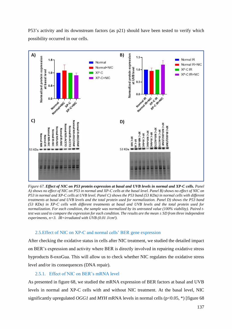
137
P53’s activity and its downstream factors (as p21) should have been tested to verify which
possibility occurred in our cells.
Figure 67. Effect of NIC on P53 protein expression at basal and UVB levels in normal and XP-C cells. Panel
A) shows no effect of NIC on P53 in normal and XP-C cells at the basal level. Panel B) shows no effect of NIC on
P53 in normal and XP-C cells at UVB level. Panel C) shows the P53 band (53 KDa) in normal cells with different
treatments at basal and UVB levels and the total protein used for normalization. Panel D) shows the P53 band
(53 KDa) in XP-C cells with different treatments at basal and UVB levels and the total protein used for
normalization. For each condition, the sample was normalized by its untreated value (100% viability). Paired t-
test was used to compare the expression for each condition. The results are the mean ± SD from three independent
experiments, n=3. IR=irradiated with UVB (0.01 J/cm²).
2.5.Effect of NIC on XP-C and normal cells’ BER gene expression
After checking the oxidative status in cells after NIC treatment, we studied the detailed impact
on BER’s expression and activity where BER is directly involved in repairing oxidative stress
byproducts 8-oxoGua. This will allow us to check whether NIC regulates the oxidative stress
level and/or its consequences (DNA repair).
2.5.1. Effect of NIC on BER’s mRNA level
As presented in figure 68, we studied the mRNA expression of BER factors at basal and UVB
levels in normal and XP-C cells with and without NIC treatment. At the basal level, NIC
significantly upregulated OGG1 and MYH mRNA levels in normal cells (p<0.05, *) [figure 68

138
(A)] and APE1 and LIG3 mRNA levels in XP-C cells (p<0.05, *) [figure 68 (B)] compared to
their untreated controls. This may indicate a preventative and protective role of NIC by
increasing the expression of oxidative DNA repair genes pre-stress. Benavente et al. showed
that NAD+ depletion increases ROS and oxidative DNA damage in keratinocytes and NIC
supplementation reverses the genotoxicity (Benavente and Jacobson 2008). This could be the
case in figure 68 (A) and (B), where NIC supplementation prevented any spontaneous DNA
damage at the basal level by enhancing some BER factors’ expression.
Figure 68. Effect of NIC on BER mRNA expression at basal and UVB levels in normal and XP-C cells. Panel
A) shows an upregulation in OGG1 and LIG3 expression upon NIC treatment (p<0.05, *) in normal cells at the
basal level. Panel B) shows an upregulation in APE1 and LIG3 expression upon NIC treatment (p<0.05, *) in
XP-C cells at the basal level. Panel C) shows an upregulation of MYH and POLB upon NIC treatment (p<0.05,
*) in normal cells post-UVB irradiation. Panel D) shows an upregulation in OGG1 (p<0.05, *), MYH (p<0.05,
*), APE1 (p<0.05, *), and LIG3 (p<0.05, *) in XP-C cells post-UVB irradiation. For each condition, the sample
was normalized by its untreated value (100% viability). Paired t-test was used to compare the expression for each
condition (p<0.05, *). The results are the mean ± SEM from three independent experiments, n=3. IR=irradiated
with UVB (0.01 J/cm²).
Post-UVB, MYH and POLB were significantly upregulated (p<0.05, *) in normal cells in
presence of NIC compared to its absence [figure 68 (C)] while an upregulation of OGG1
(p<0.05, *), MYH (p<0.05, *), APE1 (p<0.05, *), and LIG3 (p<0.01, **) were observed in XP-
C cells in the presence of NIC compared to its absence [figure 68 (D)]. Chhabra et al. showed
that NIC protects the melanocytes from UV-induced DNA damage by upregulating the mRNA
expression of genes involved in NER and BER pathways. Their results showed that damage-
specific DNA binding subunits (DDB1 and DDB2), excision repair cross-complementation
groups (ERCC1 and ERCC2) , and cyclin-dependent kinase 7 (CDK7; TFIIH subunit)
mRNA expressions were enhanced in the presence of NIC+UVB compared to UVB (Chhabra

139
et al. 2019). This confirms that NIC could enhance DNA repair by inducing the expression of
NER factors. Their results also indicated that NIC enhanced OGG1 mRNA expression in the
presence of UVB (Chhabra et al. 2019). Such an interesting study provided a new insight
towards protection against DNA damage and melanoma.
Therefore, NIC protects the cells from UV-induced DNA damage by stimulating the expression
of BER genes before and after stress.
Interestingly, BER’s gene expression stimulation was induced more prominently in XP-C cells
compared to normal control. This may be due to the higher sensitivity of XP-C cells to UVB-
induced ROS and the low BER activity and expression, as proven in part one. Therefore, NIC’s
protective effect was more prominent due to increasing NAD+ level and more BER gene
expression induction to compensate and protect from oxidative DNA damage. Furthermore,
we suggest that such a detected enhancement of BER’s mRNA expression is XPC-independent.
To check our hypothesis and whether NIC solely affects BER’s transcript level or also its
protein level, activity, and cellular ROS level, further experiments were done.
2.5.2. Effect of NIC on XP-C and normal cells’ UVB-induced BER protein expression
We did a western blot to examine whether NIC influenced the translational level of BER factors
similar to the transcriptional level. However, we were selective in choosing those we already
studied in part one and are known to be at the initiation step of BER (OGG1, MYH, and APE1)
as they were proven to be downregulated in XP-C primary fibroblasts (part one). This could
reveal how NIC may act as a potential preventative therapy in XP-C patients.
As presented in figures 69 and 70, NIC increased MYH’s protein expression in normal cells at
the basal level (p<0.05, *). However, no effect of NIC was observed post-UVB irradiation
except for a downregulation of OGG1’s protein expression (p<0.05, *) in XP-C cells.
Interestingly, OGG1 is known to be at the early steps of BER. Therefore, its downregulation
could reveal that cells had already started their DNA repair and recovery from UVB-induced
stress, which negatively affected the studied protein. In fact, this could prevent the excessive
expression of BER proteins that are usually indicative of carcinogenesis.
Therefore, the preventative effect of NIC may be through (i) enhancing BER’s turnover
(OGG1), (ii) increasing the stimulation and activity of BER via ATP upregulation and/or (iii)
reducing the amount of induced oxidative stress. (section 2.2.).
Another explanation for the observed protein expressions’ level could be the selected time.
Perhaps, we should have taken another kinetic point to give proteins more time to be stimulated
and expressed after stress.

140
Therefore, post-UVB, NIC may have succeeded in boosting cellular defense against UVB and
its damages which is convenient with its suggestive role as a preventative method against stress.
Figure 69. Effect of NIC on BER protein expression at basal and UVB levels in normal and XP-C cells. Panel
A) shows that NIC upregulated MYH (p<0.05, *) in normal cells at the basal level. Panel B) that NIC has no
effect on BER proteins in XP-C cells at the basal level. Panel C) shows that NIC has no impact on BER factors in
normal cells post-UVB irradiation. Panel D) shows that NIC downregulated OGG1 (p<0.05, *) in XP-C cells
post-UVB irradiation. For each condition, the sample was normalized by its untreated value (100% viability).
Paired t-test was used to compare the expression for each condition (p<0.05, *). The results are the mean ± SD
from three independent experiments, n=3. IR=irradiated with UVB (0.01 J/cm²).

141
Figure 70. Effect of different treatments (NIC, NAC, ACETO, BSO/DMF) on BER protein expression at basal
and UVB levels in normal and XP-C cells. Panels A), B), and C) show OGG1, MYH, and APE1 bands in normal
cells, respectively, at different conditions and their respective total protein membranes used for normalization.
Panels D), E), and F) show OGG1, MYH, and APE1 bands in XP-C cells, respectively, at different conditions and
their respective total protein membranes used for normalization. NIC=Nicotinamide, NAC=N-acetylcysteine,
ACETO=Acetohexamide (done but not included in manuscript), BSO/DMF=Buthionine sulfoximine/
Dimethylfumurate, NIR=Non-irradiated, IR=irradiated with UVB (0.01 J/cm²)
2.6.Effect of NIC on XP-C and normal cells’ UVB-induced BER and NER activity
It has been demonstrated that NIC protects against CPDs and oxidative damage at the DNA,
proteins, and lipids levels (Kamat and Devasagayam 1999; Thompson et al. 2014). We also
showed some effect on BER’s expression. Therefore, we intended to check whether NIC
pretreatment could protect our studied cells from induced bulky lesions [(6-4) PPs and CPDs]
and oxidative DNA damage and improve their repair.
2.6.1. Monitoring NER activity
Using HPLC-MS/MS we examined the kinetic repair of (6-4) PPs (figure 71) and CPDs (figure
72) in XP-C and normal cells with/without NIC (50µM).
NIC showed no effect on the repair of (6-4) PPs in normal cells [figure 71 (A)]. This may
indicate that DNA repair is fully active in the normal cells and repaired these photoproducts
rapidly with no need to use the extra ATP that is usually provided in the presence of NIC via
the following reaction: NAD++ADP→NADH+ATP to improve DNA repair. However, we
were able to detect an improvement in the repair of TT (6-4) PPs in the presence of NIC in GG-
NER deficient XP-C cells [figure 71 (B)].
The high TT compared to TC in XP-C cells was consistent with what was previously reported
about UVB (figure 6). Hu et al. showed that TC (6-4) PPs are repaired more rapidly than TT
(6-4) PPs in human fibroblasts (Hu et al. 2017).
It should be stressed that we tried to quantify the Dewar isomers as they are mutagenic
photoisomerization products of excited (6-4) PPs where the former induce mutations at the 3’
end of TT sequences while the latter induce mutations at the end of 3’ TC doublets. This could
strongly impact the DNA structure. We did not detect these lesions at 0.01 J/cm². Perhaps UVA
and UVB are needed to induce such a lesion. First, UVB will lead to the (6-4) PPs formation;
then UVA photons will be absorbed by them, leading to their excitation (Douki 2016).

142
Figure 71. Effect of NIC on UVB-induced-(6-4) PPs kinetic repair in normal and XP-C cells. Panel A) shows
no significant effect of NIC in repairing TT and TC (6-4) PPs in normal cells. Panel B) shows no significant effect
of NIC in TC (6-4) PPs in XP-C cells. However, significantly higher TT (6-4) PPs at 0.5 (p<0.001, ***), 24
(p<0.01, **), and 48 (p<0.05, *) hours in XP-C cells in presence of NIC compared to its absence. For each
condition, the sample was normalized by its irradiated value at 0 hours (100% viability). Paired t-test was used
to compare the expression for each condition (p<0.05, *). The results are the mean ± SD from two independent
experiments, n=2.
Moving forward, TT and TC CPDs are the most dominant UVB-induced CPD lesions. Figure
72 shows that NIC did not significantly affect the repair of CPDs in normal cells or TC CPDs
in XP-C cells. Interestingly, this treatment successfully improved the repair of TT CPDs in the
absence of XPC. Such a conclusion was similar to that shown in (6-4) PPs repair. However, it
should be emphasized that NIC reduced the induced lesions promptly post-UVB in normal
cells. Similarly, Surjana et al. showed that NIC enhanced CPDs repair and reduced their level
post-solar simulation in keratinocytes (Thompson, Halliday, and Damian 2015).
These results are consistent with the proposed role of NIC as a non-toxic chemopreventive
agent against skin cancer.

143
Figure 72. Effect of NIC on UVB-induced-CPDs kinetic repair in normal and XP-C cells. Panels A) shows no
effect of NIC on TT and TC CPDs repair in normal cells. Panel B) shows no effect of NIC on TC CPDs repair but
was able to downregulate TT CPDs at 2, 24, and 48 hours significantly (p<0.05, *) in XP-C cells. Panel C) shows
no effect of NIC on the repair of CT and CC CPDs in normal cells. Panel D) shows no effect of NIC on CT and
CC CPDs repair in XP-C cells. For each condition, the sample was normalized by its irradiated value at 0 hours
(100% viability). Paired t-test was used to compare the expression for each condition (p<0.05, *). The results are
the mean ± SD from two independent experiments, n=2.
2.6.2. Monitoring BER activity
As previously mentioned, studies showed that NIC enhances the repair of 8-oxoGua. Therefore,
we sought to check whether it is the case in our study. It was shown that UVB induces 8-
oxoGua in murine and human epidermal keratinocytes (Thompson, Halliday, and Damian
2015). Hence, we used the modified comet ±FPG assay to check the effect of NIC on UVB-
induced oxidized lesions’ excision in normal and XP-C cells at 0 and 24 hours.
We already verified a lower kinetic repair of oxidized purines, including 8-oxoGua, in XP-C
primary fibroblasts compared to normal (part one). This seems to be the case in the current
studied XP- C cells (figure 73).
NIC pretreatment reduced the quantity of initiated oxidized purines in XP-C and normal cell
lines (P<0.001, µµµ). It also enhanced their repair at 24 hours compared to their amount
promptly post-UVB (p<0.05, *). This indicates that NIC pretreatment could protect against
oxidative DNA damage and could enhance their repair. This is done via enhancing BER’s
mRNA expression (as shown previously), preventing UV-induced ATP depletion, improving
cellular energy for better DNA repair, and/or reducing produced ROS levels (section 2.8).

144
Similarly, Surjana et al. showed that ex vivo skin pretreated with NIC (50µM for 24 hours)
before solar-simulated UV (4 J/cm²) had lower induced epidermal 8-oxoGua and higher repair
compared to untreated control (Thompson, Halliday, and Damian 2015).
Figure 73. Effect of NIC on alkaline and oxidized purines repair in normal and XP-C cells, post-UVB
irradiation. Panel A) shows that NIC decreased the UVB-induced oxidized purines at 0 hours and 24 hours
(+FPG, p<0.001, µµµ and p<0.01, $, respectively) in normal cells. Panel B) shows that NIC decreased the UVB-
induced oxidized purines at 0 hours (+FPG, p<0.001, µµµ). For each condition, the sample was normalized by
its unirradiated value (100% viability). Paired t-test was used to compare the expression for each condition. The
results are the mean ± SEM from three independent experiments, n=3. * p<0.05 between -FPG and +FPG for
each treatment at 0 and 24 hours. $ p<0.05 between the NIC and untreated samples at 24 hours +FPG. µ p<0.05
between the NIC and untreated samples at 0 hours +FPG. ¤ p<0.05 between -FPG and + FPG for NIC and
untreated samples at 0 hours.
Therefore, NIC pretreatment could decrease incidences of non-melanoma skin cancer and basal
cell carcinoma in XP-C patients by reducing the DNA damage (Thompson, Halliday, and
Damian 2015). More importantly, it could also play a role in halting internal cancer and
preventing macromolecular cellular damage.
Mo

145
Conclusion and Perspective
Nicotinamide (50µM):
❖ Upregulated some BER gene expressions:
✓ mRNA level:
Basal: OGG1 and LIG3 in normal cells while APE1 and LIG3
in XP-C cells
UVB: MYH and POLB in normal cells while OGG1, MYH,
APE1 and LIG3 in XP-C cells
✓ Protein level:
Basal: MYH in normal cells
❖ Downregulated some BER protein expressions:
✓ OGG1 at UVB level in XP-C cells
❖ Enhanced TT (6-4) PP and TT CPDs NER repair in XP-C cells
❖ Enhanced oxidized purines repair by BER in XP-C and normal
cells
❖ Upregulated Glutathione level
❖ Downregulated ROS level in XP-C cells
In summary, NIC is a non-toxic therapeutic agent. It could act as a
preventive treatment to protect XP-C cells against UV-induced DNA
lesions and oxidative stress. This could prevent the initiation of dramatic
cascade of events leading to the severe clinical symptoms in XP-C
patients.
It would be interesting to try later the same experiments on keratinocytes and
3D skin models. Perhaps, checking the detailed signaling mechanism will be
ideal in understanding furthermore the efficacy of not only the therapeutic
effect per se rather also the protective effect of NIC.

146
3. Chapter Three: The effect of N-acetylcysteine (NAC) on UVB-
Induced Oxidative Stress and DNA Repair in XP-C and Normal
Cells
After checking NIC pretreatment, we were interested in studying a more specific antioxidant
treatment, NAC. NAC has been used a lot in research due to its significant protective and
detoxifying role, particularly at the level of oxidative stress.
3.1.Characterization of NAC-treated normal and XP-C cells
3.1.1. NAC dose selection
We did a dose-response curve to select the most suitable NAC dose for our experiments.
Figure 74 shows that 1mM of NAC treatment for 24 hours is the most suitable dose for the
experiments. It is the highest concentration to use before cytotoxicity. Such a dose had already
been used in research (Zhang et al. 2011).
Figure 74.NAC dose-response curve. Normal and XP-C cells were tested with different doses of NAC for 24 hours
followed by MTT test to monitor the cellular viability. 1mM is the most suitable dose with least cytotoxicity. The
results are the mean ± SD.

147
3.1.2. Effect of NAC on XP-C and normal cells’ UVB-induced photosensitivity
Before studying NAC's effect on DNA repair, we wanted to check whether it affects cellular
viability post-UVB. As shown in figure 75, no difference in photosensitivity between treated
and untreated samples in both cell lines, normal and XP-C. Perhaps higher NAC doses or a
pre- and post-UVB treatment are needed to obtain adequate protection. Then, NAC
pretreatment does not interfere with cellular viability as they are not cytotoxic at low doses. A
similar observation was illustrated by Mitsopoulos et al. They showed that NAC pretreatment
did not affect cell viability post-oxidative stress (Paraquat) (Mitsopoulos and Suntres 2011).
However, high NAC doses were recorded as cytotoxic at variable concentrations depending on
the cell type. For instance, NAC concentrations below 10 mM did not affect A547 cells’
viability, while higher doses (50 mM) decreased cell viability after 24 hours. Such a high dose
was detected as non-toxic for aortic endothelial cells (Mitsopoulos and Suntres 2011).
Figure 75. Similar UVB-induced photosensitivity in each cell line (normal and XP-C), with and without NAC.
We treated cells with 1 mM NAC for 24 hours then we measured the percentage of cellular viability 24 hours
post-UVB irradiation by short-term cytotoxicity test (MTT). For each condition, the sample was normalized by its
unirradiated value (100% viability). Paired t-test was used to compare photosensitivity for each condition at each
UVB dose (p<0.05, *). The results are the mean ± SEM from three independent experiments, n=3.

148
3.2.Effect of NAC on XP-C and normal cells’ redox state
3.2.1. Effect of NAC on XP-C and normal cells’ ROS and glutathione levels
• Effect on UVB-induced ROS
As mentioned earlier, NAC is an efficient thiol-containing ROS scavenger. The dose and
treatment duration used in our experiment proved efficient in reducing ROS in normal and XP-
C cells (figure 76).
Figure 76 (A) shows that NAC protected normal cells from the UVB-induced ROS at early
stages where ROS fluorescence was significantly lower at 0.5, 1, and 2 hours post-UVB
(p<0.05, *; p<0.001, ***; p<0.05, *; and p<0.01, **, respectively). Figure 76 (B) showed that
ROS level was downregulated in presence of NAC at 0.5, 2, 3, and 24 hours post-UVB
irradiation (p<0.01, **; p<0.05, *; p<0.05, *; and p<0.05, *, respectively). NAC was able to
downregulate the UVB-induced ROS levels in XP-C cells and decrease their persistence and
accumulation throughout time. Therefore, NAC pretreatment protected both cell types from
oxidative stress. Similarly, NAC scavenged UVB-induced and H2O2-induced ROS in HaCaT
and H9c2 cells, respectively (X. Liu et al. 2019; Zhu et al. 2017).
Figure 76. Effect of NAC on ROS level in normal and XP-C cells, post-UVB irradiation. Panel A) NAC
downregulated ROS level at 0.5, 1, and 2 hours post-UVB (p<0.05, *; p<0.001, ***; p<0.05, *; and p<0.01, **,
respectively) in normal cells. Panel B) NAC downregulated ROS level at 0.5, 2, 3, and 24 hours post-UVB
irradiation (p<0.01, **; p<0.05, *; p<0.05, *; and p<0.05, *, respectively) in XP-C cells. We treated cells with

149
1/1000 DHR123 followed by UVB (0.02 J/cm²) and monitored the kinetics of fluorescence at different kinetic time
points. Then we normalized each sample by its untreated value (100% viability). H2O2 was used as a positive
control. Paired t-test was used to compare the ROS fluorescence between NAC treated and untreated samples in
normal and XP-C1 fibroblast, respectively, at each kinetic time point (hours) (p<0.05, *). The results are the
mean ± SEM from three independent experiments, n=3.
• Effect on glutathione level 24 hours post-UVB
To show whether NAC pretreatment protected cells by halting ROS levels, neutralizing them,
and/or improving oxidative DNA repair, we measured GSH concentration in cells 24 hours
post-UVB.
Our studied XP-C cells had lower GSH level compared to normal (figure 77). A reduced XPC
mRNA expression was detected in keratinocytes upon GSH downregulation, demonstrating
that XPC’s transcription requires redox homeostasis (W. Han et al. 2012). Additionally to PTC,
this could contribute to the lower mRNA XPC levels in XP-C cells compared to control (figure
51). Further, silencing XPC using siRNA decreased GSH and increased catalase and
superoxide dismutase activities in glioma cells post-oxidant treatment (arsenic trioxide) (S.-Y.
Liu et al. 2010). This shows a direct link between glutathione and XPC.
Figure 77. The induced IR/NIR glutathione (GSH) level in normal and XP-C cells. After UVB-irradiation for
24 hours, we extracted cells and measured glutathione levels calorimetrically using a CAYMAN kit. This treatment
showed significant downregulation in glutathione level in XP-C (p<0.05, *) compared to the normal cells. XP-C
and normal samples were normalized by normal IR/NIR value. Paired t-test was used to compare the expression
for each condition (p<0.05, *). The results are the mean ± SD.

150
So, will supplementation with NAC improve GSH downregulation?
As expected, NAC pretreatment significantly enhanced the induced GSH level in normal
(p<0.001, ***) and XP-C (p<0.01, **) cells (figure 78). This could explain the results
previously stated where NAC was reported to efficiently downregulate UV-induced ROS and
prevent GSH depletion in human skin (Goodson et al. 2009).
A clinical study done in 2009 showed that melanocytes extracted from melanoma patients had
reduced antioxidants and increased ROS. So then, melanoma and UV-induced oxidative stress
are tightly linked. In 1989, almost 16 percent of xeroderma pigmentosum Japanese patients
were recorded to have melanoma (Takebe, Nishigori, and Tatsumi 1989). Therefore,
pretreatment with NAC orally can prevent GSH depletion and reduce triggered oxidative stress
in patients’ nevi in the presence of acute UV exposure. This may suggest NAC as a preventive
and protective treatment against melanoma risk (Goodson et al. 2009).
Figure 78. Effect of NAC on the UVB-induced IR/NIR glutathione (GSH) level in normal and XP-C cells, with
and without NAC. After NAC pretreatment and UVB-irradiation for 24 hours, we extracted cells and measured
glutathione levels calorimetrically using a CAYMAN kit. This treatment showed a significant upregulation in
glutathione level in normal (p<0.001, ***) and XP-C (p<0.01, **) cells compared to their untreated sample. For
each condition, the sample was normalized by its untreated value. Paired t-test was used to compare the
expression for each condition (p<0.05, *). The results are the mean ± SD.

151
3.2.2. Effect of NAC on XP-C and normal cells’ oxidative stress-linked genes’
expression
One way to start monitoring the antioxidant defense status of cells is by checking their gene
expression. Similarly to NIC, we studied the effect of NAC pretreatment on several
antioxidants involved in halting oxidative stress in the nucleus and mitochondria.
NAC pretreatment abolished GPx1 and SOD2 expressions in normal and XP-C cells,
respectively, at basal level [figure 79 (A) and (B)]. Perhaps this is the cellular response to a
resting state.
After UVB-irradiation, SOD2 was significantly upregulated by NAC in normal cells [figure 79
(C)]. This may indicate the presence of superoxide byproducts that need to be neutralized by
the enzyme. Meanwhile, GPx1 was significantly upregulated by NAC in XP-C cells which may
indicate the presence of hydrogen peroxide that needs to be neutralized by this enzyme and the
efficiency of NAC’s role in boosting GSH level for GPx1’s role enhancement. Furthermore,
our result may suggest that SOD and GPx1 are implicated in preventing UVB-induced damage.
Previously, NAC pretreatment had shown an augmentation of SOD2 gene expression in
chicken embryo post-oxidative stress (cadmium) (Doi et al. 2011). Thus, despite that NAC acts
as a ROS scavenger and GSH precursor, it could improve the antioxidant defense system. Of
course, we are well aware that most of the changes could be post-translational and could have
been interesting to be monitored.

152
Figure 79. Effect of NAC on the detoxificants’ mRNA expression at basal and UVB levels in normal and XP-
C cells. Panel A) shows a downregulation in GPX1 expression (p<0.01, **) upon NAC treatment in normal cells
at basal level. Panel B) shows downregulation in SOD2 (p<0.05, *) but an upregulation in GPx1 (p<0.05, *)
expression upon NAC treatment in XP-C cells, at basal level. Panel C) shows an upregulation of SOD2 upon NAC
treatment (p<0.05, *) in normal cells post-UVB irradiation. Panel D) shows an upregulation in GPx1 (p<0.05,
*) in XP-C cells, post-UVB irradiation. For each condition, the sample was normalized by its untreated value
(100% viability). Paired t-test was used to compare the expression for each condition (p<0.05, *). The results are
the mean ± SEM from three independent experiments, n=3. IR=irradiated with UVB (0.01 J/cm²).
As both, SOD and GPx1, were shown to be affected by NAC, we suspected that it might
influence an upstream gene, Nrf2. However, figure 80 shows that NAC could only enhance
Nrf2’s expression in XP-C at UVB level and not at the basal level. This indicates that Nrf2 is
triggered post-stress efficiently by NAC pretreatment.
Figure 80. Effect of NAC on the Nrf2 mRNA expression at basal and UVB levels in normal and XP-C cells.
NAC upregulated Nrf2 significantly (p<0.05, *) in XP-C cells at UVB level. The sample was normalized by its
untreated value (100% viability). Paired t-test was used to compare the expression for each condition (p<0.05,
*). The results are the mean ± SEM from three independent experiments, n=3. IR=irradiated with UVB (0.01
J/cm²).
Nrf2 showed a downregulation at the basal level in normal cells but an upregulation in XP-C
cells [figure 81 (A) and figure 62 (C) and (D)]. The attenuated oxidative stress could explain
the downregulated expression. Nevertheless, the upregulation in XP-C cells is spectacular.
Perhaps this is due to the different cell lines used, and using the same cell line with/without
XPC’s expression could have more relevant results. Jannatifar et al. previously demonstrated
that NAC pretreatment increases Nrf2 gene expression in Asthenoteratozoospermia Men
(Jannatifar et al. 2020). Another study showed that NAC supplementation induces Nrf2 and
OGG1 as Nrf2 regulates the latter’s expression (K. C. Kim et al. 2017).
This demonstrates the role of NAC not only in increasing GSH level per se rather also in
regulating signaling pathways, including the Nrf2-dependent antioxidant signaling pathway.

153
However, no effect was detected at the UVB level. Maybe 4 hours post-UVB irradiation is
more than the time needed to detect such an early response to stress, or 0.01 J/cm² is not enough
dose to detect notable changes.
Therefore, our previous and following results may indicate that NAC rapidly increases repair
and defense rates against oxidative stress and damage.
Figure 81. Effect of NAC on the Nrf2 protein expression at basal and UVB levels in normal and XP-C cells. In
the presence of NAC, panel A) shows a significant downregulation of Nrf2 (p<0.05, *) in normal cells but an
upregulation of Nrf2 (p<0.05, *) in XP-C cells, at the basal level. On the other hand, Panel B) shows no effect of
NAC on Nrf2 in normal and XP-C cells at UVB level. For each condition, the sample was normalized by its
untreated value (100% viability). Paired t-test was used to compare the expression for each condition (p<0.05,
*). The results are the mean ± SD from two independent experiments, n=2. IR=irradiated with UVB (0.01 J/cm²).
(preliminary result)
3.3.Effect of NAC on XP-C and normal cells’ PARP1 protein expression
PARP1
Due to the importance of PARP1’s role in DNA repair, cell survival, and signaling, we
monitored its gene expression (similarly to above, section 2.3.). No significant change in
PARP1 was detected in both cell lines in the presence of NAC at basal and UVB levels [figure
82 (A) and (B) and figure 64 (C) and (D)]. An in vivo study showed that NAC supplementation
attenuated PARP1 gene expression in mice (G. Wang et al. 2019). This is proposed due to
lower oxidative DNA damage and single-strand DNA damage; thereby, PARP1 activation is
redundant.

154
Figure 82. Effect of NAC on the PARP1 protein expression at basal and UVB levels in normal and XP-C cells.
Panels A) shows no significant effect of NAC on PARP1 in normal cells at basal and UVB levels. Panel B) shows
no significant effect of NAC on PARP1 in XP-C cells at basal and UVB levels. The total protein was used for
normalization. Paired t-test was used to compare the expression for each condition. The results are the mean ±
SD from two independent experiments, n=2. IR=irradiated with UVB (0.01 J/cm²). (preliminary result)
Cleaved PARP1 “apoptosis hallmark”
NAC showed a similar role to NIC in downregulating cleaved PARP1 in XP-C cells post-UVB
(p<0.01, **) compared to the untreated control (figure 83). No change was detected at the level
of normal cells (figure 83). Therefore, the downregulated cleaved PARP1 level in XP-C cells
may indicate lower cell death due to the successful protection of NAC to cells from UVB-
induced cytotoxicity and lethality. NAC had already shown protection against apoptosis in
brain cells by reducing oxidative stress and enhancing cell signaling as the modulating group I
metabotropic glutamate receptor with a neuroprotective role (L. Sun et al. 2012).
Figure 83. Effect of NAC on cleaved PARP1 protein expression at basal and UVB levels in normal and XP-C
cells. Panel A) shows, in normal cells, the absence of cleaved PARP1 in the presence and absence of NAC at the
basal level, while no significant effect was detected in the presence and absence of NAC at UVB level. Panel B)

155
shows, in XP-C cells, the lack of cleaved PARP1 in the presence and absence of NAC at basal level while NAC
downregulated the cleaved PARP1 significantly (p<0.01, **) at UVB level. Paired t-test was used to compare the
expression for each condition (p<0.05, *). The results are the mean ± SD from two independent experiments,
n=2. IR=irradiated with UVB (0.01 J/cm²). (preliminary result)
3.4.Effect of NAC on XP-C and normal cells’ P53 gene expression
P53 is a highly complex protein involved in several cell signaling pathways other than
apoptosis, including DNA repair. Therefore, it is interesting to check whether NAC treatment
interferes with P53’s expression.
Figure 84 showed no significant effect of NAC on P53 in both cells, normal and XP-C, at basal
and UVB levels.
Figure 84. Effect of NAC on the P53 mRNA expression at basal and UVB levels in normal and XP-C cells.
Panel A) shows no effect of NAC on P53 in normal and XP-C cells, at the basal level. Panel B) shows no effect of
NAC on P53 in normal and XP-C cells at UVB level. For each condition, the sample was normalized by its
untreated value (100% viability). Paired t-test was used to compare the expression for each condition. The results
are the mean ± SEM from three independent experiments, n=3. IR=irradiated with UVB (0.01 J/cm²).
As it is speculated that P53 is mainly regulated at translational and post-translational levels, we
checked its protein expression as P53 is known to be UVB-induced to regulate genes through
the stress-signaling pathway (Amundson et al. 2005).
Similar to NIC pretreatment, the only detected variation was in XP-C cells at the basal level
where P53 was downregulated significantly (p<0.01, **) (figure 85). This could show that cells
at rest lack stress where P53 is not needed to trigger apoptosis or stabilize POLB for DNA
repair (J. Zhou et al. 2001). Notably, APE1 was shown to be downregulated in XP-C cells at
the basal level [figure 87 (B)]. APE1 usually interacts with and activates P53 in vitro and in
vivo (J. Zhou et al. 2001). Therefore, this could be the case in our experiment.
Another interesting proposed theory is that in XP-C cells, P53 usually is trapped by MDM2
and not degraded, disrupting its turnover and efficacy (Krzeszinski et al. 2014). This was

156
observed in figure 48 due to a high P53 level in XP-C fibroblasts compared to control at basal
level. Therefore, NAC may have enhanced P53’s turnover and degradation at a resting state
independently of XPC.
Such proposed theories may contradict with the role of P53 in apoptosis to prevent
carcinogenesis. However, we should consider that we are discussing the cellular resting state
where P53 maybe not be needed, the complexity of such a protein and that our result needs
further validations, particularly at the interactome level.
Figure 85. Effect of NAC on the P53 protein expression at basal and UVB levels in normal and XP-C cells.
Panel A) shows a downregulation in P53 (p<0.01, **) in the presence of NAC in XP-C cells, at the basal level.
Panel B) shows no effect of NAC on P53 in normal and XP-C cells at UVB level. For each condition, the sample
was normalized by its untreated value (100% viability). Paired t-test was used to compare the expression for each
condition (p<0.05, *). The results are the mean ± SD from three independent experiments, n=3. IR=irradiated
with UVB (0.01 J/cm²).
3.5.Effect of NAC on XP-C and normal cells’ BER gene expression
Several studies showed that NAC downregulates oxidative DNA damage, particularly 8-
oxoGua. To establish whether this occurs via stimulating BER or reducing ROS, we started by
examining the BER mRNA and protein expression.
3.5.1. Effect of NAC on XP-C and normal cells’ BER mRNA level
At the basal level, NAC induced an upregulation in MYH and APE1’s mRNA expressions
(p<0.05, *) in normal and XP-C cells, respectively [figure 86 (A) and (B)]. Post-UVB
irradiation, figure 86 (C) and (D) shows that NAC induced an upregulation of LIG3 (p<0.05,
*) in normal cells and an upregulation of APE1 and LIG3 (p<0.01, **; p<0.05, *, respectively)
in XP-C cells. This may indicate a positive effect of NAC on BER’s transcriptional expression,
particularly in steps following the initiation of the pathway.

157
However, to know further about the effect of NAC on BER’s gene expression, we selected
OGG1, MYH, and APE1 and checked their expression at the translational level.
Figure 86. Effect of NAC on BER mRNA expression at basal and UVB levels in normal and XP-C cells. Panel
A) shows that NAC increased MYH mRNA expressions at the basal level (p<0.05, *) in normal cells. Panel B)
shows that NAC increased APE1 mRNA expression (p<0.05, *) at the basal level in XP-C cells. Panel C) shows
that NAC increased LIG3 mRNA expressions at UVB level (p<0.05, *) in normal cells. Panel D) shows that NAC
increased APE1 (p<0.01, **) and LIG3 (p<0.05, *) mRNA expressions at UVB level in XP-C cells. For each
condition, the sample was normalized by its untreated value (100% viability). Paired t-test was used to compare
the expression for each condition (p<0.05, *). The results are the mean ± SEM from three independent
experiments, n=3. IR=irradiated with UVB (0.01 J/cm²).
3.5.2. Effect of NAC on XP-C and normal cells’ BER protein expression
UVB produces ROS and oxidative damages [lipid, protein, and DNA (8-oxoGua)]. Such DNA
damage requires the activation of BER factors. Figure 87 shows that some of these BER factors
were downregulated at basal and UVB levels. Figure 87 [(A) and (C)] shows that OGG1 is
downregulated at basal (p<0.01, **) and UVB (p<0.05, *) levels in normal cells, respectively.
Also, NAC downregulated OGG1 (p<0.001, ***) and APE1 (p<0.01, **) at basal level and
OGG1 and MYH (p<0.01, **) at UVB level in XP-C cells.
On the other hand, it was reported that NAC enhances OGG1’s expression (K. C. Kim et al.
2017). This may be due to the different stress, treatment conditions, and cells between our
experiments. They pretreated HaCat keratinocytes with 1mM of NAC for 30 minutes before
exposing them to non-thermal dielectric barrier discharge (DBD) plasma (K. C. Kim et al.
2017). Perhaps our result indicates that pretreatment of cells with NAC enhanced BER for a
rapid repair before cell collection (4 hours). It may have also reduced ROS and the oxidative

158
DNA damage efficiently, thereby inhibiting the initiation and stimulation of BER. However, a
background expression (IR/NIR) is still active.
Figure 87. Effect of NAC on BER protein expression at basal and UVB levels in normal and XP-C cells. Panel
A) shows that NAC downregulated OGG1 (p<0.01, **) at basal level in normal cells. Panel B) shows that NAC
downregulates OGG1 (p<0.001, ***) and APE1 (p<0.01, **) at basal level in XP-C cells. Panel C) shows that
NAC downregulates OGG1 (p<0.05, *) in normal cells at UVB level. Panel D) shows that NAC downregulates
OGG1 and MYH (p<0.01, **) in XP-C cells at UVB level. For each condition, the sample was normalized by its
untreated value (100% viability). Paired t-test was used to compare the expression for each condition (p<0.05,
*). The results are the mean ± SD from three independent experiments, n=3. IR=irradiated with UVB (0.01
J/cm²).
3.6.Effect of NAC on XP-C and normal cells’ UVB-induced BER and NER activity
NAC is an exciting drug that has an impressive array of cellular regulatory and protective
effects. This is due to its nucleophilicity and multiple roles as ROS scavenging, cell signaling
and DNA repair modulation, and inhibition of genotoxicity and carcinogenesis (De Flora et al.
2001). Herein, it is highly expected to enhance repair in normal and XP-C cells.
3.6.1. Monitoring NER activity
Figures 88 and 89 represent the kinetic repair of UVB-induced (6-4) PPs and CPDs,
respectively, in the presence and absence of NAC pretreatment.
No effect of NAC on (6-4) PPs in normal and XP-C cells [figure 88 (A) and (B)].

159
Figure 88. Effect of NAC on UVB-induced (6-4) PPs kinetic repair in normal and XP-C cells. Panels A) shows
no difference in (6-4) PPs repair in the presence and absence of NAC in normal cells. Panel B) shows no difference
in (6-4) PPs repair in the presence and absence of NAC in XP-C cells. For each condition, the sample was
normalized by its irradiated value at 0 hours (100% viability). Paired t-test was used to compare the expression
for each condition (p<0.05, *). The results are the mean ± SD from two independent experiments, n=2.
However, a potential of enhanced CPDs repair was observed in both cells (figure 89). For
instance, TU and UT CPD lesions were significantly lower (p<0.05, *) in normal cells in the
presence of NAC at 24 and 2 hours post-UVB, respectively. Also, TT, UT, and UU CPDs were
significantly lower (p<0.05, *) at 0.5 and 2 hours, respectively, in XP-C cells. Unfortunately,
such an improvement was moderate due to the high SEMs and/or that pretreatment with such
a dose could not be efficient to induce such a detected shift in repair.
It should be stated that the observed U residues result from the deamination of C in CPD
lesions. Therefore, they are highly mutagenic and mimic T during replication (Douki,
Koschembahr, and Cadet 2017).

160
Figure 89. Effect of NAC on UVB-induced CPDs kinetic repair in normal and XP-C cells. Panels A) shows that
NAC improved TU CPDs (p<0.05, *) repair at 24 hours in normal cells. Panel B) shows that NAC improved
repair of TT CPDs (p<0.01, **) at 0.5 hours in XP-C cells. Panel C) shows that NAC improved repair of UT
CPDs (p<0.01, **) at 2 hours in normal cells. Panel D) shows that NAC downregulated UT and UU CPDs at 0.5
hours (p<0.05, *), respectively, in XP-C cells. For each condition, the sample was normalized by its irradiated
value at 0 hours (100% viability). Paired t-test was used to compare the expression for each condition (p<0.05,
*). The results are the mean ± SEM from two independent experiments, n=2.
Maybe the enhanced repair detected in CPDs but not (6-4) PPs in the presence of NAC could
not be linked to the distorted DNA duplex rather due to the quantity of lesions induced
subsequently to UVB. Another reason could be the need to repeat the experiment as a triplicate
due to the high SEM and variations between the samples in experiments. NAC could have
enhanced (6-4) PPs repair in normal cells. Still, it was not detected due to its efficiency in
rapidly repairing such a bulky lesion with/without treatment (24 hours).
3.6.2. Monitoring BER activity
NAC pretreatment seemed to protect the cells from induced DNA damage, mainly oxidized
purines (p<0.05, µ), and enhance the repair. XP-C cells had significantly lower single-strand
breaks and oxidized purines 24 hours post-UVB in the presence of NAC compared to its
absence [figure 90 (A) and (B)].
Such a result is due to the role of NAC as a ROS scavenger. As previously shown, NAC
enhances GSH level, thereby balancing the ratio of oxidants/antioxidants to the latter's favor.
This prevents UVB-induced ROS from targeting the DNA-forming lesions. Thereby, less BER
is required to remove UVB-induced DNA damage.

161
Figure 90. Effect of NAC on alkaline and oxidized purines repair in normal and XP-C cells, post-UVB
irradiation. Panel A) shows that NAC decreased the UVB-induced oxidized purines at 0 hours (+FPG, p<0.01,
µµ) in normal cells. Panel B) shows that NAC decreased the UVB-induced oxidized purines at 0 and 24 hours
(+FPG, p<0.0001, µµµµ; p<0.05, $, respectively). For each condition, the sample was normalized by its
unirradiated value (100% viability). Paired t-test was used to compare the expression for each condition (p<0.05,
*). The results are the mean ± SEM from three independent experiments, n=3. * p<0.05 between -FPG and +FPG
for each treatment at 0 and 24 hours. $ p<0.05 between the NAC and untreated samples at 24 hours +FPG. µ
p<0.05 between the NAC and untreated samples at 0 hours +FPG. ¤ p<0.05 between -FPG and + FPG for NAC
and untreated samples at 0 hours.

162
Conclusion and Perspective
N-acetylcysteine (1mM):
❖ Effect on some BER gene expressions:
✓ Upregulated mRNA level:
Basal: MYH and APE1 in normal and XP-C cells
✓ Upregulated Protein level:
UVB: APE1 and LIG3 in XP-C cells
✓ Downregulated Protein level:
Basal: OGG1 in normal and XP-C cells, APE1 in XP-C cells
UVB: OGG1 and MYH in XP-C cells
❖ Enhanced moderately CPDs NER repair in XP-C cells
❖ Enhanced oxidized purines repair by BER and reduced their
induction in XP-C and normal cells
❖ Upregulated Glutathione level
❖ Downregulated ROS level
In summary, NAC is non-toxic therapeutic agent. It could act as a
preventative treatment to protect XP-C cells against UV-induced DNA
lesions, oxidized purines, and oxidative stress. This could prevent the
initiation of dramatic cascade of events leading to the severe clinical
symptoms in XP-C patients.
To strengthen further our conclusion about NAC, we repeated the
same experiments with BSO/DMF (chapter four) which should
trigger opposite effects.
It would be interesting to try later the same experiments on keratinocytes
and 3D-skin models. Perhaps, checking the detailed signaling mechanism
will be ideal in understanding furthermore the efficacy of not only the
therapeutic effect per se rather also the protective effect of NAC.

163
4. Chapter Four: The Effect of Buthionine
sulfoximine/Dimethylfumurate (BSO/DMF) on UVB-Induced
Oxidative Stress and DNA Repair in XP-C and Normal Cells
In this chapter, we used BSO (100µM) and DMF (100µM) to support the results of NAC
treatment.
4.1.Characterization of BSO/DMF-treated normal and XP-C cells
4.1.1. BSO/DMF dose selection
Dose selection was based on a dose-response curve to check the % viability and choose the
most suitable and maximum concentration of BSO/DMF (1:1) before inducing cellular
cytotoxicity. As shown in figure 91, 100 µM was the best dose for pretreatment 4 hours before
UVB-irradiation. This dose had also been used by different researchers (Boivin et al. 2011).
Figure 91. BSO/DMF dose-response curve. Normal cells were tested with different doses of BSO/DMF for 4
hours followed by MTT test to monitor the cellular viability. 100µM is the most suitable dose with the least
cytotoxicity. The results are the mean ± SD.
BSO acts as a GSH biosynthesis inhibitor, while DMF acts as GSH depleting agent. Using a
combination of both drugs guaranteed the total depletion of GSH to study such an effect on
normal and XP-C cells. The results could be dramatic due to the prominent ROS level, thereby
confirming the antioxidant role of NAC in enhancing repair and neutralizing oxidative stress
as shown in previously illustrated experiments.

164
4.1.2. Effect of BSO/DMF on XP-C and normal cells’ UVB-induced photosensitivity
Similar to previously tested drugs, we wanted to check whether BSO/DMF could affect the
photosensitivity of cells to UVB. Figure 92 shows that BSO/DMF significantly decreased the
cellular viability of normal cells at 0.01 and 0.1 J/cm² (p<0.01, **; p<0.05, *, respectively) and
of XP-C cells at 0.02, 0.1, and 0.2 J/cm² (p<0.05, *; p<0.01, **, respectively). Similarly, Boivin
et al. showed that the combination of BSO/DMF decreases cell survival more than each drug
solely due to a depletion of GSH for more than 4 hours (Boivin et al. 2011).
Briefly, GSH has been proposed as essential for cell viability and a photoprotective mediator
in the skin. Thereby, its depletion increases the UV-induced photosensitivity of normal and
XP-C cells.
Figure 92. Higher photosensitivity in each cell line (normal and XP-C) in presence of BSO/DMF compared to
its untreated control. We treated cells with BSO (100µM) and DMF (100µM) for 4 hours then we measured the
percentage of cellular viability 24 hours post-UVB irradiation by short-term cytotoxicity test (MTT). For each
condition, the sample was normalized by its unirradiated value (100% viability). Paired t-test was used to
compare photosensitivity for each condition at each UVB dose (p<0.05, *). The results are the mean ± SEM from
three independent experiments, n=3.

165
4.2.Effect of BSO/DMF on XP-C and normal cells’ redox state
4.2.1. Effect of BSO/DMF on XP-C and normal cells’ ROS and glutathione levels
• Effect on UVB-induced ROS
On the contrary to NAC, BSO/DMF increased the level of ROS dramatically in normal and
XP-C cells. Figure 93 (A) shows that BSO/DMF upregulated ROS level at 0.5, 1, 3, 24, and 48
hours post-UVB irradiation in normal cells. In parallel, figure 93 (B) shows that BSO/DMF
upregulated ROS levels at 2,3 and 48 hours post-UVB irradiation in XP-C cells. This indicates
that GSH is vital for protection against ROS and that the dramatic effects of XPC mutations
could be partly linked to a downregulated GSH level. Such a result was well demonstrated in
several studies. For instance, Bohl et al. used BSO to induce severe ROS in breast cancer cells
to induce apoptosis (Bohl et al. 2012).
Thereby, supplementing cells with GSH could enhance and reverse some of the XP-C’s
phenotype, including those linked to oxidative DNA damage repair and antioxidant defense
pathways.
Figure 93. Effect of BSO/DMF on ROS level in normal and XP-C cells, post-UVB irradiation. Panel A)
BSO/DMF upregulated ROS level at 0.5, 1, 3, 24 and 48 hours post-UVB (p<0.05, *; p<0.01, **, respectively) in
normal cells. Panel B) BSO/DMF upregulated ROS level at, 2, 3, and 48 hours post-UVB irradiation (p<0.05) in
XP-C cells. We treated cells with 1/1000 DHR123 followed by UVB (0.02 J/cm²) and monitored fluorescence
kinetics at different kinetic time points. Then we normalized each sample by its untreated value (100% viability).
H2O2 was used as a positive control. Paired t-test was used to compare the ROS fluorescence between NAC treated

166
and untreated samples in normal and XP-C1 fibroblast, respectively, at each kinetic time point (hours) (p<0.05,
*). The results are the mean ± SEM from three independent experiments, n=3.
• Effect on glutathione level 24 hours post-UVB
Studying the level of cellular glutathione in the presence of BSO/DMF confirmed the efficiency
of the treatment. As shown in figure 94, BSO/DMF significantly abolished the GSH level in
normal (p<0.01, **) and XP-C (p<0.001, ***) cells. This complements our previously
illustrated results in the presence of BSO/DMF treatment.
Figure 94. Effect of BSO/DMF on UVB-induced IR/NIR glutathione (GSH) level in normal and XP-C cells.
After UVB-irradiation for 24 hours, we extracted cells and measured glutathione levels calorimetrically using a
CAYMAN kit. This treatment showed significant downregulation in glutathione level in normal (p<0.01, **) and
XP-C (p<0.001, **) cells. XP-C and normal samples were normalized by normal IR/NIR value. Paired t-test was
used to compare the expression for each condition (p<0.05, *). The results are the mean ± SD.
4.2.2. Effect of BSO/DMF on XP-C and normal cells’ oxidative stress-linked genes’
expression
Checking the mRNA expression of some genes involved in the oxidative stress defense
pathway showed that GPx1 was downregulated significantly (p<0.05, *) in normal cells at basal
level in the presence of BSO/DMF pretreatment [figure 95 (A)]. However, post-UVB,
BSO/DMF induced higher expression of GPx1 and SOD1 (p<0.05, *) compared to untreated
control [figure 95 (C)]. This may show that BSO/DMF triggers oxidative stress that will
stimulate the antioxidants defense mechanism. A depletion in GSH may not inhibit antioxidants
expression rather activity. This could lead to depletion of cellular resources as ATP and NAD+,
which are necessary for normal cellular activity and protection. XP-C cells did not show any

167
effect significant effect except for a downregulation in SOD2 (p<0.01, **) after BSO/DMF
treatment at UVB level [figure 95 (D)].
Similarly, Krifka et al. showed that the gene expression of GPx1 increased in the presence of
BSO in macrophages due to high H2O2 induced levels due to GSH depletion (Krifka et al.
2012). Treating cells with an additional oxidative stress source (2-hydroxyethyl methacrylate)
leads to ROS levels that exceed the capacity of the antioxidant defense system. To compensate,
catalase was overexpressed (Krifka et al. 2012). Unfortunately, our XP-C cells are known to
lack catalase activity (Vuillaume et al. 1986). This could cause extreme ROS levels that may
have caused negative feedback on SOD enzymes’ expression in XP-C but not normal cells.
Figure 95. Effect of BSO/DMF on the detoxificants’ mRNA expression at basal and UVB levels in normal and
XP-C cells. Panel A) shows downregulation in GPX1 expression (p<0.05, *) upon BSO/DMF treatment in normal
cells at the basal level. Panel B) shows no significant effect upon BSO/DMF treatment in XP-C cells at the basal
level. Panel C) shows an upregulation of SOD1 and GPX1(p<0.05, *) upon BSO/DMF treatment in normal cells
post-UVB irradiation. Panel D) shows a downregulation in SOD2 (p<0.01, **) in XP-C cells post-UVB
irradiation. For each condition, the sample was normalized by its untreated value (100% viability). Paired t-test
was used to compare the expression for each condition (p<0.05, *). The results are the mean ± SEM from three
independent experiments, n=3. IR=irradiated with UVB (0.01 J/cm²).
BSO/DMF seems to significantly impact different pathways linked to cell survival, signaling,
and DNA repair. One of these pathways is the antioxidant defense pathway. For that, we studied
the expression of an upstream factor, Nrf2, at the mRNA and protein levels. As a result, Nrf2
increased in the presence of BSO/DMF at UVB level in normal cells (p<0.05, *) (figure 96).

168
Figure 96. Effect of BSO/DMF on the Nrf2 mRNA expression at basal and UVB levels in normal and XP-C
cells. NAC upregulated Nrf2 significantly (p<0.05, *) in normal cells, at UVB level. The sample was normalized
by its untreated value (100% viability). Paired t-test was used to compare the expression for each condition
(p<0.05, *). The results are the mean ± SEM from three independent experiments, n=3. IR=irradiated with UVB
(0.01 J/cm²).
Our preliminary results in figure 97 suggest that BSO/DMF triggers Nrf2 at basal and UVB
levels in normal cells. However, Nrf2 is almost inhibited in XP-C cells at the UVB level (figure
97). Similar to our studied normal cells, BSO had been shown to activate the Nrf2 pathway in
murine embryonic fibroblasts to upregulate the expression of antioxidant genes as a protection
mechanism against oxidative stress (H.-R. Lee et al. 2008). Maybe this was not the case in XP-
C cells because they are susceptible to stress and were not BSO-resistant. Therefore, it is
interesting to check whether Nrf2 is deficient in the studied XP-C cells. Maybe BSO/DMF and
the absence of XPC leading to oxidative stress and high NOX1 contribute to such a dramatic
inhibition in Nrf2. This could be via the P53-dependent pathway (SIRT1…).
It should be noted that high levels of NOX were detected in Nrf2 deficient cells and XP-C cells
are known to have high NOX1 level (Kovac et al. 2015).

169
Figure 97. Effect of BSO/DMF on the Nrf2 protein expression at basal and UVB levels in normal and XP-C
cells. Panel A) shows a difference in PARP1 in the presence and absence of BSO/DMF in normal and XP-C cells,
at the basal level. Panel B) shows a difference in PARP1 in the presence and absence of BSO/DMF in normal
and XP-C cells, at UVB level. For each condition, the sample was normalized by its untreated value (100%
viability). Paired t-test was used to compare the expression for each condition (p<0.05, *). The results are the
mean ±SD from two independent experiments, n=3. IR=irradiated with UVB (0.01 J/cm²) (preliminary result).
4.3.Effect of BSO/DMF on XP-C and normal cells’ PARP1 protein expression
PARP1
PARP1 induces cell survival via the HIF1α-dependent pathway in the presence of oxidative
stress (Pietrzak et al. 2018). It is also involved in DNA repair and other signaling pathways,
as previously mentioned.
Our preliminary results in figure 98 suggest that BSO/DMF enhances PARP1’s expression at
UVB level in normal cells and at basal and UVB levels in XP-C cells. Answering whether this
is linked to cell viability or other signaling pathways is tricky and needs further experiments.
Nevertheless, a recent publication showed that BSO treatment enhanced PARP1’s expression
in the presence of oxidative stress (Yıldızhan and Nazıroğlu 2020). Excessive activated PARP1
could deplete cells from ATP in an attempt to repair damaged DNA. This could trigger cell
death.
Figure 98. Effect of BSO/DMF on the PARP1 protein expression at basal and UVB levels in normal and XP-
C cells. Panels A) shows no significant effect of BSO/DMF on PARP1 in normal cells at basal and UVB levels.
Panel B) shows no significant effect of BSO/DMF on PARP1 in XP-C cells at basal and UVB levels. The total
protein was used for normalization. Paired t-test was used to compare the expression for each condition (p<0.05,
*). The results are the mean ± SD from two independent experiments, n=2. IR=irradiated with UVB (0.01 J/cm²).
(preliminary result)

170
Cleaved PARP1 “apoptosis hallmark”
Normal cells showed an upregulated cleaved PARP1 shown in figure 99 (A) and (B) post-UVB
in the presence of BSO/DMF (p<0.05, *). This could indicate the presence of cell apoptosis.
Similarly, the cleaved PARP1 in XP-C cells at basal level post-BSO/DMF implies how
dramatic the cellular changes were against the highly induced oxidative stress. Consistently,
BSO pretreatment had been shown to trigger late apoptosis and necrosis in macrophages
(Krifka et al. 2012).
Figure 99. Effect of BSO/DMF on cleaved PARP1 protein expression at basal and UVB levels in normal and
XP-C cells. Panel A) shows, in normal cells, the absence of cleaved PARP1 in the presence and absence of
BSO/DMF, at the basal level while BSO/DMF pretreatment significantly upregulated the cleaved PARP1 (p<0.05,
*) at UVB level. Panel B) shows, in XP-C cells, absence of cleaved PARP1 in presence and absence of BSO/DMF
at basal level while no effect of BSO/DMF was shown on the cleaved PARP1 at UVB level. Paired t-test was used
to compare the expression for each condition (p<0.05, *). The results are the mean ± SD from two independent
experiments, n=2. IR=irradiated with UVB (0.01 J/cm²). (preliminary result)
4.4.Effect of BSO/DMF on XP-C and normal cells’ P53 gene expression
Another factor linked to cell death is P53. GSH depletion has been shown to play a critical role
in triggering apoptosis. Furthermore, an activation and translocation of NFκB and cytochrome
C release were detected in cells 3 hours after BSO treatment as hallmarks of apoptosis induction
(Armstrong et al. 2002). Therefore, to check whether we had a stimulation of apoptosis in our
cells treated with BSO/DMF we monitored the gene expression of P53.
As presented in figure 100, P53 was significantly reduced at basal level in normal cells (p<0.01,
**) and UVB level in XP-C cells (p<0.05, *).

171
Figure 100. Effect of BSO/DMF on the P53 mRNA expression at basal and UVB levels in normal and XP-C
cells. Panel A) shows that BSO/DMF downregulated P53 (p<0.01; **) in normal cells, at the basal level. Panel
B) shows that BSO/DMF downregulated P53 (p<0.05, *) in XP-C cells, at UVB level. For each condition, the
sample was normalized by its untreated value (100% viability). Paired t-test was used to compare the expression
for each condition (p<0.05, *). The results are the mean ± SEM from three independent experiments, n=3.
IR=irradiated with UVB (0.01 J/cm²).
Such a result reflected P53’s protein expression in normal cells [figure 101 (A)]. It was
significantly downregulated in the presence of BSO/DMF compared to its absence at the basal
level (p<0.05, *). However, no change was detected at the UVB level. This was consistent with
Armstrong et al. They showed that P53’s expression did not vary post-redox modulation in the
presence of BSO (Armstrong et al. 2002). However, irreversible apoptosis will be induced after
48-72 hours of BSO treatment (Armstrong et al. 2002). This was not the case in our study, as
we incubated the cells only for 4 hours in BSO/DMF.
Interestingly, P53 was upregulated (p<0.05, *) in XP-C cells at UVB level [figures 101 (B) and
67 (C) and (D)]. This proposes a different possible signaling pathway induced in normal versus
XP-C cells to decrease cell viability and induce apoptosis. Remarkably, we previously
mentioned that XPC plays a role in P53’s turnover. Herein, in the absence of XPC and high
stress (UVB+BSO/DMF), cells accumulate P53 that could be inactive despite its high
expression level.

172
Figure 101. Effect of BSO/DMF on the P53 protein expression at basal and UVB levels in normal and XP-C
cells. Panel A) shows a downregulation in P53 (p<0.05, *) in the presence of BSO/DMF in normal cells at the
basal level. Panel B) shows that BSO/DMF upregulated P53 in XP-C cells at UVB level. For each condition, the
sample was normalized by its untreated value (100% viability). Paired t-test was used to compare the expression
for each condition (p<0.05, *). The results are the mean ± SD from three independent experiments, n=3.
IR=irradiated with UVB (0.01 J/cm²).
4.5.Effect of BSO/DMF on XP-C and normal cells’ BER’s gene expression
4.5.1. Effect of BSO/DMF on XP-C and normal cells’ BER mRNA expression
Figure 102 shows a global downregulation in BER factors at transcriptional level after
pretreatment with BSO/DMF at basal and UVB levels. In normal cells, figure 102 (A) and (C)
shows that BSO/DMF downregulated APE1 (p<0.05, *), LIG3 (p<0.05, *), and XRCC1
(p<0.01, **) at basal level while APE1 (p<0.01, **), POLB (p<0.01, **), and LIG3 (p<0.05,
*) at UVB level. Similarly, figure 101 (B) and (D) shows a significant downregulation of MYH
(p<0.05, *), POLB (p<0.01, **), LIG3 (p<0.01, **), and XRCC1 (p<0.05, *) at basal level and
MYH (p<0.05, *) at UVB level in XP-C cells post-BSO/DMF treatment.
Indeed, the reason for such a downregulation should oppose that found in the presence of NAC.
BSO/DMF depletes cells from GSH. This will induce high oxidative stress triggering signaling
cascades’ dysregulation and cellular molecules’ oxidation. Maybe the induction of BER
factors’ promoters was inhibited. In fact, such a hypothesis was proposed for NER where GSH
depletion downregulations its expression and capacity. XPC’s expression has been identified
as GSH-dependent (W. Han et al. 2012). Therefore XPC’s expression may have been altered
in normal cells too. Of notice, variations in the redox homeostasis may result in alterations in
gene expression profile. A downregulation in BER mRNA expression had been recorded in
neurological diseases with high oxidative stress, such as Alzheimer’s disease (AD). Forestier
et al. showed that an overall downregulation of BER-associated genes (OGG1, MYH, APE1,
XRCC1, etc.…) were detected in neuroblastoma cell line secreting high amyloid-β protein,
AD’s pathological hallmark that leads to elevated oxidative stress (Forestier et al. 2012).

173
Therefore, the downregulation of BER mRNA expression in both cells could be due to (i) the
highly induced total oxidative stress and (ii) downregulated XPC expression that had been
addressed previously to interfere in BER’s expression (part one).
Figure 102. Effect of BSO/DMF on BER mRNA expression at basal and UVB levels in normal and XP-C cells.
Panel A) shows that BSO/DMF downregulated APE1 (p<0.05, *), LIG3 (p<0.05, *), and XRCC1 (p<0.01, **) at
basal level in normal cells. Panel B) shows that BSO/DMF downregulated MYH (p<0.05, *), POLB (p<0.01, **),
LIG3 (p<0.01, **) and XRCC1 (p<0.05, *) at the basal level in XP-C cells. Panel C) shows that BSO/DMF
downregulated APE1 (p<0.01, **), POLB (p<0.01, *) and LIG3 (p<0.05, *) at UVB level in normal cells. Panel
D) shows that BSO/DMF downregulated MYH (p<0.05, *) at UVB level in XP-C cells. For each condition, the
sample was normalized by its untreated value (100% viability). Paired t-test was used to compare the expression
for each condition (p<0.05, *). The results are the mean ± SEM from three independent experiments, n=3.
IR=irradiated with UVB (0.01 J/cm²).
4.5.2. Effect of BSO/DMF on XP-C and normal cells’ BER protein expression
We sought to check the effect of BSO/DMF on the translation of BER factors’ transcripts. So
we did western blot on the three main key proteins in initiating BER and repairing oxidative
DNA damage: OGG1, MYH, and APE1.
Figure 103 [(A) and (C)] shows that BSO/DMF downregulated OGG1’s expression (p<0.05,
*) at basal and UVB levels in normal cells. Similarly, OGG1 was downregulated (p<0.001,
***) upon BSO/DMF in XP-C cells at basal level [figure 103 (B)].
Previously, Dusinka et al. had proposed that glutathione s-transferases (GSTs) might be
involved in DNA damage signaling (MAPK kinase). Their activity is induced by ROS, which
can influence DNA stability and oxidative DNA damage repair. As they require GSH to reduce
substrates, a deficiency in this tripeptide downregulates GSTs activity. Such activity had been
correlated with BER capacity (Dusinska et al. 2012). Hence, their level could have been
abolished, leading to lower activation of BER.

174
Interestingly, APE1 at UVB level was significantly upregulated (p<0.01, **) at the protein
level in XP-C cells in the presence of BSO/DMF pretreatment [figure 103 (D)]. APE1 has been
identified as a DNA repair protein and plays a role in transcription and redox regulations. In
part one, we showed that XP-C cells have higher oxidative DNA damage and oxidative stress
than control. BSO/DMF worsens the scenario dramatically. It was suggested that APE1’s DNA
repair and redox regulatory functions are independent. In vivo and in vitro studies demonstrated
that acute and chronic ROS levels induce APE1 protein expression rapidly. Hence, we suggest
that APE1 was upregulated based on its dual role to protect cells as much as possible from
genome instability by (i) activating some transcription factors (P53, p21, NFκB…) for cell
cycle and proliferation regulation, (ii) antioxidant response, and (iii) other DNA repair
pathways regulation where a crosslink between APE1 and DNA repair genes from other
pathways had already been demonstrated (GADD45a, BRCA1…) (Tell et al. 2009).
Figure 103. Effect of BSO/DMF on BER protein expression at basal and UVB levels in normal and XP-C cells.
Panels A) and C) show that BSO/DMF downregulated OGG1 at basal (p<0.05, *) and UVB (p<0.01, **) levels
in normal cells. Panel B) shows that BSO/DMF downregulated OGG1 (p<0.001, ***) at the basal level in XP-C
cells. Panel D) shows that BSO/DMF upregulated APE1 (p<0.01, **) at UVB level in XP-C cells. For each
condition, the sample was normalized by its untreated value (100% viability). Paired t-test was used to compare
the expression for each condition (p<0.05, *). The results are the mean ± SD from three independent experiments,
n=3. IR=irradiated with UVB (0.01 J/cm²).

175
4.6.Effect of BSO/DMF on XP-C and normal cells’ UVB-induced BER and NER activity
4.6.1. Monitoring NER activity
Treatment of keratinocytes with BSO and UVA leads to increased oxidative stress triggering
protein oxidation, consequently inhibiting NER (Karran and Brem 2016). Similarly,
BSO/DMF treatment affected the NER repair in normal and XP-C cells (figure 104). However,
it should be stressed that a moderate effect was visualized due to the high SD.
It is highly recommended to repeat the experiment due to the high potentials of the results.
Figure 104 [(A) and (C)] shows that in normal cells: BSO/DMF significantly increased TT and
TC (6-4) PPs (p<0.05, *) at 2 hours. In parallel, figure 105 shows that BSO/DMF upregulated
TC CPDs (p<0.01, **) at 24 hours, CT CPDs at 0.5 and 24 hours (p<0.05, *; p<0.01, **,
respectively) and CC CPDs were significantly higher at 24 and 48 hours (p<0.05, *).
Meanwhile, in XP-C cells, BSO/DMF upregulated TT (6-4) PPs at 2 and 24 hours (p<0.05, *)
[Figure 104 (B)]. TC and CC CPDs were also significantly higher at 24 hours (p<0.05, *)
compared to control [figure 105 (B) and (D)].
Therefore, BSO/DMF increases the UVB-induced bulky lesions and dysregulates their repair
by boosting their persistence. This is due to the high ROS level in the absence of GSH that
could enhance proteins’ oxidation and their inactivation in normal cells. On the other hand,
NER is already diminished in XP-C cells, and adding BSO/DMF worsens the situation.
Figure 104. Effect of BSO/DMF on UVB-induced (6-4) PPs kinetic repair in normal and XP-C cells. Panels
A) and C) show that normal cells had a downregulated TT and TC (6-4) PPs (p<0.05, *) in the presence of
BSO/DMF, respectively, at 2 hours post-UVB irradiation. Panel B) shows that BSO/DMF induced downregulated
TT (6-4) PPs at 2 (p<0.05, *) and 24 (p<0.05, *) hours post-UVB irradiation in XP-C cells. Panel D) shows No
significant effect of BSO/DMF on TC (6-4) PPs’ repair in XP-C cells. For each condition, the sample was

176
normalized by its irradiated value at 0 hours (100% viability). Paired t-test was used to compare the expression
for each condition (p<0.05, *). The results are the mean ± SD from two independent experiments, n=2.
Figure 105. Effect of BSO/DMF on UVB-induced CPDs kinetic repair in normal and XP-C cells. Panels A)
shows that BSO/DMF downregulated TC CPDs (p<0.01, **) repair at 24 hours post-UVB irradiation in normal
cells. Panel B) shows that BDO/DMF downregulated TT and TC CPDs repair (p<0.05, *) at 2 and 24 hours,
respectively, post-UVB irradiation in XP-C cells. Panel C) shows that BSO/DMF downregulated CC CPDs at 24
and 48 hours (p<0.05, *) and CT at 0.5 (p<0.05, *) and 24 hours (p<0.01, **) post-UVB irradiation in normal
cells. Panel D) shows that BSO/DMF downregulated CC CPDs repair (p<0.05, *) at 24 hours post-UVB
irradiation in XP-C cells. For each condition, the sample was normalized by its irradiated value at 0 hours (100%
viability). Paired t-test was used to compare the expression for each condition (p<0.05, *). The results are the
mean ± SD from two independent experiments, n=2.
4.6.2. Monitoring BER activity
Figure 106 illustrates that BSO/DMF increased the oxidized purines and single-strand breaks
significantly in normal cell-lines compared to its absence (p<0.01, µµ; p<0.001, µµµ,
respectively). Such treatment also impacted the repair efficacy in normal and XP-C cells. At
24 hours post-UVB, normal cells failed to repair the damage where significantly higher
oxidized lesions (p<0.01, $$) and single-strand DNA damage (p<0.05, *) persisted. Similarly,
oxidized purines persisted at a higher level at 24 hours in XP-C cells (p<0.05, $).
This may show that GSH depletion impacted the efficacy of BER activity in both cell lines,
where it induced further oxidized purines reaching a plateau that is not repaired. Therefore
GSH plays an essential role in protecting cells against oxidative DNA damage independently
of XPC protein.

177
Figure 106. Effect of BSO/DMF on alkaline and oxidized purines repair in normal and XP-C cells, post-UVB
irradiation. Panel A) shows that BSO/DMF increased the UVB-induced single-strand DNA damages and oxidized
purines at 0 hours (p<0.001, µµµ; p<0.01, µµ, respectively) in normal cells. Both DNA damages persisted after
24 hours significantly (p<0.05, $). Panel B) shows that BSO/DMF stimulated oxidized purines persistence at 24
hours post-UVB in XP-C cells (p<0.05, $). For each condition, the sample was normalized by its unirradiated
value (100% viability). Paired t-test was used to compare the expression for each condition. The results are the
mean ± SEM from three independent experiments, n=3. * p<0.05 between -FPG and +FPG for each treatment
at 0 and 24 hours. $ p<0.05 between the BSO/DMF and untreated samples at 24 hours +FPG. µ p<0.05 between
the BSO/DMF and untreated samples at 0 hours +FPG. ¤ p<0.05 between -FPG and + FPG for each of
BSO/DMF and untreated samples at 0 hours.

178
Conclusion and Perspective
BSO/DMF (100µM/100µM):
❖ Increased photosensitivity
❖ Effect on some BER gene expressions:
✓ downregulated mRNA level:
Basal: APE1, LIG3, and XRCC1 in normal cells and MYH,
POLB, LIG3, and XRCC1 in XP-C cells
UVB: APE1, POLB, and LIG3 in normal cells and MYH in XP-
C cells
✓ downregulated protein level:
Basal: OGG1 in normal and XP-C cells
UVB: OGG1 in normal cells
✓ Upregulated Protein level:
UVB: APE1 in XP-C cells
❖ Dysregulated NER and BER activity
❖ Depleted Glutathione level
❖ Upregulated ROS level
In summary, BSO/DMF is cytotoxic. It increases the UV-induced
oxidative DNA damage in cells. This proves the importance of GSH in
DNA repair and other signaling pathways including cell survival.

179
Schematic Summary
Figure 107. Effect of NIC, NAC, and BSO/DMF on cells post-UVB. UVB induces bulky lesions (CPDs, (6-4)
PPs) that are usually repaired by NER in the absence of any drug. Such a repair is impaired in XP-C patients
triggering mutagenesis. Furthermore, UVB initiates ROS-induced oxidative damage, including oxidized purines
(8-oxoGua) that BER usually repairs. As illustrated before, BER is downregulated in XP-C patients triggering
mutagenesis and oxidative stress. So, NIC has been shown to decrease ROS, 8-oxoguanine, and bulky lesions. In
parallel, it enhances NER activity and BER’s expression and activity. NAC decreased ROS, 8-oxoguanine and
bulky lesions while enhancing NER.
Interestingly, it inhibited BER’s expression. That is probably due to the dramatic downregulation in oxidative
stress as a ROS scavenger. Lastly, BSO/DMF induced ROS, 8-oxoGua and bulky lesions. In parallel, it inhibited
BER and NER. This could enhance senescence and apoptosis. Therefore, the drugs used are potentially protective
measurements pre-UVB (NIC, NAC) and potential use in cancer treatment (BSO/DMF). Nevertheless, more
studies are needed at proteomic and posttranslational levels, in addition to studies in vivo, before confirming such
encouraging results.

180
General Discussion
eroderma pigmentosum C (XPC) protein plays a major role in initiating GG-NER
pathway to excise DNA helix distorting lesions. More than forty-five XPC
inactivating mutations were detected, leading to the impairment of the protein
product and the accum
ulation of the bulky DNA lesions (Ben Rekaya et al. 2013). This could increase skin sensitivity
to sun radiation leading to mutagenesis, morbidity, skin carcinogenesis, and early mortality.
Nevertheless, XP-C patients’ phenotype spectrum is heterogeneous and broad including
internal tumorigenesis (thyroid, hematological, gynecological, spinal cord, and brain cancers)
and rarely neurological abnormalities (Uribe-Bojanini, Hernandez-Quiceno, and Cock-Rada
2017; Yurchenko et al. 2020; Zebian et al. 2019). Herein, researchers revealed other versatile
roles in other DNA repair pathways, cell cycle regulation, and redox homeostasis (Melis et al.
2011).
As oxidative stress induces oxidative DNA damage that BER usually repairs, we thought to
selectively check the link between XPC, base excision repair, and oxidative stress from a
mechanistic standpoint. This could show whether XPC acts directly on BER and/or indirectly
via regulating the cellular redox state. For that, we split the project into two parts: part one
discusses the effect of XPC protein on BER and ROS. Meanwhile, part two checks whether
modulating the redox state in XP-C cells by different treatments could reverse their phenotype
and ameliorate the background DNA repair.
• Part One “Deciphering the Role of XPC in BER and Oxidative
Stress” Evidence had already discussed a potential role of XPC in BER due to its interactions with
various DNA glycosylases and regulation of their activities. Nevertheless, we were further
interested in checking the effect of XPC on stimulating the global BER pathway at both gene
and activity levels. For that, we treated the cells with ultraviolet radiation B (UVB) before the
experimental setup. UVB allows us to induce both (i) NER substrates [bulky lesions, CPDs
and (6-4) PPs] and (ii) BER substrates (oxidative DNA damage, 8-oxoGua) due to an elevated
ROS level (Budden and Bowden 2013; Deng et al. 2018).
First, we showed that the different XP-C cells, derived from different XP-C patients, had the
standard characteristics: impaired XPC basal gene expression and reduced GG-NER of UVB-
X

181
induced bulky lesions. TC-NER is known to be faster in repair rate than GG-NER (Soufir et
al. 2010). Hence, the lesions repaired by TC-NER could not have been detected. Therefore,
what we observe in our experiments could be solely due to the deficiency of GG-NER or NER
inactivation in the presence of high oxidative stress.
Langie et al. showed that exposure of epithelial cells to oxidant (H2O2) impeded an effective
NER activity (Langie et al. 2007). Therefore, we may link the results of the ensuing
experiments with the lack of XPC and NER deficiency.
Our RT-qPCR experiments revealed that the absence of XPC contributed to the downregulation
of UVB-stimulated BER factors, including OGG1, MYH, APE1, LIG3, XRCC1, and POLβ.
Inhibited OGG1 and delayed APE1 mRNA expression were also shown in XP-C deficient cells
following oxidant treatment (H2O2) (de Melo et al. 2016). This indicates that XPC may play a
direct/indirect role in stimulating the mRNA expression of BER genes due to its role as a
transcriptional regulator. For example, XPC has been shown to regulate P53, and our results
indicated a dysregulation in its expression (Krzeszinski et al. 2014). In parallel, P53 is one of
the transcription factors regulating APE1 and MYH’s expressions (de Melo et al. 2016; Oka et
al. 2014). Therefore, XPC may also be involved in their regulation. Further investigations are
required to confirm such a hypothesis.
Moving forward, OGG1 and MYH DNA glycosylases excise 8-oxoGua and adenine facing 8-
oxoGua, respectively, and APE1 is known as an endonuclease and redox regulator. Therefore,
we studied these factors at the protein level and showed that they are downregulated in the
absence of XPC. This shows that the lack of XPC affects the BER’s expression in the presence
of stress. Such a decrease in the BER initiation factors’ expression delayed the excision efficacy
of UVB-induced oxidized purines. Using modified comet assay (±FPG), we monitored the
excision capacity of XP-C cells compared to normal at different kinetic points, which could
reflect the cellular repair efficiency of the UVB-induced oxidized purines. Oxidized purines
persisted for more than 24 hours in the XP-C cells compared to normal control. At standard
conditions, half of the oxidized purines were shown to be repaired in 2 hours. Such oxidized
purines involve 8-oxoGua and Fapy, where 8-oxoGua is the most common type of oxidative
DNA damage (Fortini et al. 2003). Accordantly, D’Errico et al. showed that 8-oxoGua is fully
repaired 7 hours post-oxidative stress (KBrO3) in keratinocytes and fibroblasts (M. D’Errico
et al. 2007). This could be due to the rapid recruitment of XPC to the site of oxidative DNA
damage, 8-oxoGua. In parallel, XP-C cells showed an impairment in 8-oxoGua repair
(D’Augustin et al. 2020). However, such a drastic effect was also demonstrated in cells
deficient with other NER factors, as XPA and CSB. This could be due to the recognition of 8-

182
oxoGua by XPC, XPA, CSB, DDB1 etc (D’Augustin et al. 2020). Also, several oxidative DNA
lesions were reported, leading to bulky lesions that NER could recognize and repair. Hence,
could our observed results be a direct impact of XPC dysregulation or due to a wonky
crosslink between NER and BER?
Melis et al. compared the effect of oxidative stress on XP-C and XP-A mice and fibroblasts.
They detected a higher mutational load, more sensitivity towards oxidants, and higher internal
tumor spectrum in the absence of XPC compared to XPA deficiency or normal control (Melis
et al. 2008). Furthermore, xpc+/- mice showed a higher predisposition to UV-induced skin
cancer than xpa+/- mice (Melis et al. 2011). This may be due to the role of XPC as a rate-
limiting protein in NER or due to other possible roles. Even though the accumulation of bulky
lesions majorly triggers skin cancer, oxidative stress has been suggested to contribute to
photocarcinogenesis. For instance, Melanoma and nonmelanoma skin cancer exhibit high
oxidative stress and inadequacy in the antioxidant defense system (Sander et al. 2003).
Additionally, some non-bulky lesions, including 8-oxoGua, were reported to be repaired
primarily by TC-NER due to a possible partial stalling of RNA polymerase II (Melis et al.
2011). This may indicate a competition between BER and NER. As XPA and XPC are involved
in NER, the difference in response towards oxidants suggests a role of XPC in oxidative DNA
damage removal by stimulating BER’s functionality and/or redox homeostasis, thereby halting
internal tumor development. For example, XP-C fibroblasts showed a deficient BER repair of
oxidative DNA damage post-visible light excited methylene blue, a ROS stimulator (Berra et
al. 2013).
XPC was postulated as a co-factor stimulating and interacting with several BER factors to
induce the repair of 8-oxoGua. For instance, XPC interacts with different BER repair factors,
including glycosylases [thymine DNA glycosylase (TDG) and SMUG1] and APE1. For
example, it interacts with OGG1, a rate-limiting glycosylase in BER, to regulate its turnover
and expression (Melis et al. 2011; de Melo et al. 2016).
Similar to XPC, CSB, TC-NER factor is known to interact with different BER proteins as
PARP1 and APE1 and regulate the repair of 8-oxoGua and 8-oxoA post-oxidative stress (Melis
et al. 2011).
In conclusion, XPC has a direct but not essential role in BER’s expression and activity. For
example, we showed that oxidized purines were repaired in XP-C cells but slower than the
normal control. Therefore, XPC’s role in BER affects the efficacy but not the efficiency of
repair. A study showed XP-C patients developing only basal and squamous cell carcinomas
but not internal cancer, implying an effective BER activity. It also showed two siblings with

183
the same type of XPC mutation (c.1643-1644delTG), but one developed B-cell lymphoma
while the other did not develop internal cancers (Oetjen et al. 2020). This could indicate the
need for XPC impairment and an additional stimulant to induce internal cancers. In our case,
such a stimulus could be UVB, where we showed a higher and more persistent ROS level in
XP-C cells after stress than normal control.
Therefore, could the halted BER’s expression and efficacy be due to a higher redox
imbalance and disturbed stimulation of BER in the absence of XPC? Is it possible to reverse
such an impact to enhance the background DNA repair?
Part two of the project allowed us to answer such a question.
• Part Two “Ameliorating the DNA repair of XP-C cells by
modulating their redox state via pharmacological treatments” To focus on the redox state in XP-C versus normal cells, we pretreated the cells with different
treatments that act differently but work towards the same goal.
Nicotinamide (NIC)’s central role is to increase the nicotinamide adenine dinucleotide
(NAD+) level in cells. A diminution in NAD+ leads to its precursor’s depletion, ATP. This
induces cellular energy depletion and dysregulated DNA repair triggering genomic instability,
skin aging, and carcinogenesis (Fania et al. 2019). As BER is known to be ATP-dependent,
NIC might enhance BER’s background status shown in part one via preventing its depletion.
NER is also ATP-dependent. For instance, XPB and XPD have ATPase activities (Schärer
2013). Therefore, monitoring NER could also be interesting.
After selecting the most suitable NIC dose (50 µM), we checked the effect of such a treatment
on UVB-photosensitivity, BER’s gene expression and activity, and NER’s activity. No impact
of NIC on the photosensitivity of XP-C and normal cells was detected. This could be
considered a good indicator. If NIC enhanced an exaggerated cellular viability of XP-C cells
carrying mutations, carcinogenesis might be ultimately triggered. Similarly, at low NIC doses
(<2 mg/mL), no difference in cellular viability was detected in cervical cancer-associated
fibroblasts. Still, a reduction was observed at higher doses (Hassan, Luo, and Jiang 2020). Also,
NIC (50µM) showed no effect on HaCaT keratinocytes and primary melanocytes viability in
the presence and absence of UV (Thompson, Halliday, and Damian 2015; Thompson et al.
2014). Therefore, at low levels, NIC plays a role other than affecting cellular viability, which
may indirectly show no effect of NIC on apoptosis and cell cycle. For instance, treatment with
NIC led to similar melanocytes entering S-phase compared to control (Thompson et al. 2014).

184
To check the other possible protective roles of NIC, we studied the mRNA and protein
expression of different BER factors in normal and XP-C cells.
NIC increased the mRNA expression of several BER factors at basal and UVB levels in normal
and XP-C cells. It also increased the basal expression of MYH protein in normal cells. This
shows that NIC triggered the BER’s expression. NIC also induced BER’s excision activity
post-UVB. Both cell lines had lower initiated an enhanced repair of the oxidized purines. This
may show that NIC may play a role in ameliorating BER’s background in DNA repair-deficient
cells such as XP-C cells. Similar results were observed by Thompson et al., who showed that
NIC reduced UV-induced 8-oxoGua in HaCaT keratinocytes and in ex vivo human skin. They
proposed that NIC does not prevent DNA lesions' formation instead enhances their repair by
increasing cellular ATP level (Thompson, Halliday, and Damian 2015).
On the contrary, NIC significantly downregulated oxidized purines instantaneously post-UVB
in normal and XP-C cells. This could be due to the different parameters of our experiments.
We used a comet±FPG assay that detects 8-oxoGua and other oxidized purines in the cells
while they used immunofluorescence that detects 8-oxoGua solely. Also, we used UVB lamp
post-incubating cell lines with NIC (50 µM) for 24 hours while they treated cells with NIC (50
µM) before and after solar simulation. Thompson et al. showed that solar simulation-induced
8-oxoGua is repaired better in NIC presence in keratinocytes and human skin. Their comet
assay revealed lower 8-oxoGua in the presence of NIC compared to its absence in keratinocytes
instantly post-solar simulation (the starting time point of lesion detection, 15 minutes)
(Thompson, Halliday, and Damian 2015).
Moving forward, several studies showed that NIC reduces precancerous skin lesions (actinic
keratosis) and skin cancers (MSCs and NMSCs) (Snaidr, Damian, and Halliday 2019; Malesu
et al. 2020). These cancer incidences are usually triggered by the accumulation of unrepaired
bulky lesions [CPDs and (6-4) PPs], and NIC could enhance their repair or prevent their
induction. It was demonstrated that NIC does not reduce or prevent the initial UV-induced CPD
level in keratinocytes but rather enhances their repair by NER (Thompson, Halliday, and
Damian 2015; Snaidr, Damian, and Halliday 2019). We showed that NIC pretreatment
enhanced the repair of TT CPDs and TT (6-4) PPs in XP-C cells but not their initiation. On the
other hand, NIC did not induce a difference in effect in normal cells. This could indicate that
NIC triggers multiple pathways to protect the cells against stress. As XP-C cells lack efficient
GG-NER, NIC might be stimulating other alternative excision pathways as a compensation.
Interestingly, Topoisomerase I (TOP1), an essential enzyme in resolving torsional stress in the
genome, dependent-BER was suggested to substitute a deficient-NER pathway in repairing (6-

185
4) PPs in vivo and in vitro. During DNA replication and transcription, TOP1 forms a transient
complex with the single-strand DNA (3’-TOP1 DNA adduct). Such complex becomes trapped
next to unrepaired (6-4) PPs leading to single-strand breaks due to TDP1/TDP2 enzymes that
activate BER. Higher UV-induced bulky lesions were detected in XP-A cells when BER is
deficient compared to being proficient (L. K. Saha et al. 2020). NIC was also shown to inhibit
protein and lipid oxidation (L. K. Saha et al. 2020).
To check whether other factors participated in the effect of NIC on cells, we monitored the
redox status by checking different antioxidants’ expression levels and the GSH level. This will
allow us to check whether NIC downregulates the initiation of DNA damage in cells and/or
enhance their repair. Our results showed that SOD1 and SOD2 were significantly higher in
NIC-treated normal cells post-UVB, while only SOD1 was upregulated in the presence of NIC
post-UVB in XP-C cells. The difference between both superoxide dismutases is that SOD1 is
usually located in the cytoplasm while SOD2 is in the mitochondria. Perhaps, no significant
change in SOD2 in XP-C cells was due to this enzyme's known high expression and activity in
the absence of XPC (S.-Y. Liu et al. 2010). Phosphorylated Nrf2 plays a role in the
transcriptional activation of antioxidants, including the SODs, catalases, etc (Jing Chen, Zhang,
and Cai 2014). Studying the gene expression of Nrf2 could provide a more general conclusion
about the effect of NIC on the antioxidant defense pathway in the presence and absence of
XPC. NIC significantly only downregulated the Nrf2 protein basal level in XP-C cells. This
could indicate that NIC enhanced the XP-C cell’s resting redox status, which negatively affects
Nrf2. This prevents the overactivation of Nrf2 that could lead to abnormal proliferation due to
the overexpression of downstream target genes (Schäfer et al. 2014). Furthermore, NIC may
act downstream Nrf2 pathway or on Nrf2’s posttranslational activity. For example, NAD+
induces ERK activation to translocate Nrf2 into the nucleus to bind to antioxidants’ promoters.
Therefore, NAD+ pretreatment enhances the resistance of cells to UV-induced oxidative stress
in an XPC-independent manner by increasing the capacity of antioxidants (J.-K. Kim and Jang
2014). This is well-illustrated where NIC was able to reduce ROS level by time in XP-C cells.
Previously, NIC had been shown to alleviate ROS levels in primary human fibroblasts and
reduce oxidative cellular damage in vivo (Kwak et al. 2015). In addition to neutralizing UV-
induced ROS, NIC reduces cells' mitochondrial activity, contributing to lower respiration rate
and hydrogen peroxide and superoxide anion levels (Kang, Lee, and Hwang 2006). This could
be interesting in XP-C cells that are sensitive to mitochondrially-induced ROS under stressed
conditions due to altered H2O2 production/clearance upon impaired mitochondrial complex I
and upregulated complex II (Mori et al. 2017).

186
Additionally, ROS-induced DNA damage triggers PARP1 involved in several cellular
functions, including DNA repair, transcription, and cell death. Overactivation of PARP1 in
DNA repair-deficient cells leads to ATP and NAD+ depletion, which can be compensated by
NIC treatment (Kwak et al. 2015). A cleavage of PARP1 by caspase-3 also indicates the
presence of cell death and genotoxic stress (Touat et al. 2019). Therefore, exploiting the status
of PARP1 in our cells could unravel the interaction between NIC and PARP1 in the presence
and absence of XPC at basal and UVB levels and could provide an insight into the general
effect of NIC on cellular health. We showed that NIC significantly inhibited the cleavage of
PARP1 in XP-C cells but not in normal cells. This indicates that NIC could neutralize the
severe genotoxicity in XP-C cells which could decrease cell death.
On the other hand, we detected an upregulated PARP1, though not significant, which could be
due to the increase in NAD+ availability and the role of PARP1 in localizing to the DNA
lesions to recruit factors as CSB to induce repair. In addition to CSB’s role in transcription and
TC-NER, it regulates UV-induced oxidative DNA damage and interacts with OGG1 and APE1
(Boetefuer et al. 2018). PARP1 also recruits other DNA repair proteins as XRCC1. This could
participate in the enhancement of DNA repair presented in the cells post-NIC treatment.
Therefore, we propose that NIC enhances DNA repair by increasing ATP level and stimulating
BER’s gene expression. In parallel, it increases the antioxidant defense system (gene
expression and GSH) to inhibit ROS and prevent further dramatic initiated DNA damages,
particularly in XP-C cells.
N-acetylcysteine (NAC) is a potent ROS scavenger, while BSO/DMF depletes cellular GSH,
triggering higher ROS levels. Thus, pretreatment of cells with these paradoxical drugs could
provide an insight into the importance of GSH in normal and XP-C cells at DNA repair level
and antioxidants defense level.
NAC pretreatment increased the mRNA expression of some BER factors while BSO/DMF
inhibited the mRNA expression of some BER factors in normal and XP-C cells at basal and
UVB levels. Surprisingly, NAC and BSO/DMF inhibited some BER protein factors in the
studied cells. Nevertheless, the reason for downregulation is different between the two
conditions. We suggest that NAC inhibited the oxidative stress and DNA damage which could
act as a negative feedback on BER’s expression. In contrast, BSO/DMF inhibits BER’s
expression and activity, worsening the scenario. To prove this hypothesis, we checked the DNA
repair activity in cells. NAC enhanced CPD repair and oxidized purines repair in both cell lines,
suggesting that NAC protects cells via an XPC-independent mechanism of action while

187
BSO/DMF had an opposite function. GSH and DNA have a negative charge, so no direct
interaction occurs amongst them; however, the former is suggested to either prevent DNA
damage or participate in their repair. GSH’s concentration had previously been inversely
correlated with higher DNA damage due to its role in redox homeostasis directly or indirectly
through enzymatic interactions (Chatterjee 2013). As discussed earlier, XPC deficiency could
induce internal cancers such as leukemia, and XP-C cells have lower GSH level compared to
control. In parallel, defective GSH had been linked to leukemia and the high sensitivity of cells
to radiation. This shows that GSH deficiency plays a major role in XP-C patients’ phenotype.
The observed enhancement of CPD and 8-oxoGua repair in cell lines could be due to lower
ROS levels in the presence of GSH. NAC also triggered the upregulation of some antioxidants’
mRNA expression post-UVB in normal and XP-C cells; however, depletion of GSH by
BSO/DMF triggered an upregulation of their expression in normal cells only. XP-C cells had
a downregulated level of some genes. This could indicate a high ROS level that is oxidizing
proteins responsible for activating such genes or due to mitochondrial disturbance. SOD2 is
localized in the mitochondria, and its impairment post-stress may indicate high oxidative
damage and mitochondrial dysfunction. In 2019, Düzenli et al. suggested a combination of
NAC and acetyl-L-carnitine, an antioxidant and anti-apoptotic agent, to stimulate DNA repair
genes as synergic protection against UV damage (Düzenli et al. 2019). Such treatment seems
promising to be tested in XP-C cells.
GSH also plays a role in suppressing apoptosis (Chatterjee 2013). In view of this, we checked
the effect of NAC and BSO/DMF on PARP1 due to its previously described essential roles in
cell cycle and DNA repair. NAC dramatically decreased cleaved PARP1 in XP-C cells while
BSO/DMF increased it only in normal cells. This may be due to the high level of cleaved
PARP1 in XP-C cells before treatment with BSO/DMF. Therefore, GSH could enhance the
cell status via buffering ROS levels leading to more minor macromolecular damages.

188
Conclusion and Perspectives
As a conclusion, XP-C cells have a weakened BER gene expression and activity due to the
high ROS imbalance that could play a significant role in XP-C patients’ phenotypes.
NIC and NAC represent potential preventive methods to protect and treat XP-C patients per se
and other DNA-repair deficient patients. This is mainly by boosting the antioxidant defense
mechanism and buffering ROS level via GSH, which could participate in BER’s higher
capacity in accommodating daily life threats and oxidative stressors. Based on the observed
results (gene expression, activity..) NAC could be proposed to act more as a preventive
pretreatment rather than therapeutic, while NIC could be proposed as preventive and
therapeutic pretreatment.
To further validate our hypothesis, these results should be confirmed in primary cells
(fibroblasts and keratinocytes), in vivo models (mice), and 3D-reconstructed skin models. This
will provide further realistic and robust conclusions about the efficacy of these treatments in
the absence of DNA repair. Additionally, checking the treatments’ different interactions more
detailed via proteomic analysis could enhance our understanding of their mechanisms and
provide insights for different targeted therapies. Another interesting study could be to
investigate the efficiency of a combination treatment and whether it could have a synergetic
protective effect. Perhaps the combination of NIC and NAC could provide synergic protection
and damage prevention on cells that could help XP patients have a better/healthier life.

189
Proposed schematic summary
Based on our results and bibliography, we propose the following mechanism of action of NIC
in cells (figure 107).
Since NAC has a similar goal and mechanism of action (enhancing GSH..), we propose a
similar action mechanism to this treatment. BSO/DMF acts the opposite to both drugs by
depleting GSH, increasing DNA damage and apoptosis, and inhibiting DNA repair.
Figure 108. Suggested mechanism of action of NIC on XP-C cells. It increases NAD+, ATP, and antioxidants
(SOD, GSH..) levels. This leads to lower ROS and cleaved PARP1 (apoptosis hallmark) levels, higher PARP1,
and upregulated DNA repair’s (NER and BER) expression and activity to remove DNA oxidized and bulky lesions.
The enhancement in antioxidants inhibits NOX1, which is highly present in XP-C cells. This will reduce ROS
level, thereby preventing BER’s inhibition, oxidative DNA damage, and an increase of SIRT1, which usually halts
P53. This will allow P53 to increase and function normally in initiating DNA repair (NER and BER). The red
cross= inhibition of path, green arrow=activates, red arrow=inhibits.

190
References
“6" (15.25 Cm) 1000W PV Cell Testing Solar Simulator Kit Model LS1000-6R-002 -
Solarlight.” n.d. Accessed May 1, 2021. https://solarlight.com/product/6-15-25-cm-
1000w-pv-cell-testing-solar-simulator-kit-model-ls1000-6r-002/.
“2020-Campaign-Report-GC-Version-MPA_1.Pdf.” n.d. Accessed April 27, 2021.
https://melanomapatients.org.au/wp-content/uploads/2020/04/2020-campaign-report-
GC-version-MPA_1.pdf.
A Abdel-Fatah, Tarek M. A., Roslin Russell, Devika Agarwal, Paul Moseley, Michael Ayotunde
Abayomi, Christina Perry, Nada Albarakati, et al. 2014. “DNA Polymerase β
Deficiency Is Linked to Aggressive Breast Cancer: A Comprehensive Analysis of Gene
Copy Number, MRNA and Protein Expression in Multiple Cohorts.” Molecular
Oncology 8 (3): 520–32. https://doi.org/10.1016/j.molonc.2014.01.001.
Aguiar, Pedro H. N., Carolina Furtado, Bruno M. Repolês, Grazielle A. Ribeiro, Isabela C.
Mendes, Eduardo F. Peloso, Fernanda R. Gadelha, et al. 2013. “Oxidative Stress and
DNA Lesions: The Role of 8-Oxoguanine Lesions in Trypanosoma Cruzi Cell
Viability.” PLoS Neglected Tropical Diseases 7 (6).
https://doi.org/10.1371/journal.pntd.0002279.
Ahmad, Gulfam, Mazen Almasry, Amolak S. Dhillon, Muna M. Abuayyash, Narasimhan
Kothandaraman, and Zeynep Cakar. 2017. “Overview and Sources of Reactive Oxygen
Species (ROS) in the Reproductive System.” In Oxidative Stress in Human
Reproduction: Shedding Light on a Complicated Phenomenon, edited by Ashok
Agarwal, Rakesh Sharma, Sajal Gupta, Avi Harlev, Gulfam Ahmad, Stefan S. du
Plessis, Sandro C. Esteves, Siew May Wang, and Damayanthi Durairajanayagam, 1–
16. Cham: Springer International Publishing. https://doi.org/10.1007/978-3-319-
48427-3_1.
Ahmed, Tayyaba, Saira Nawaz, Rabia Noreen, Kashif Sardar Bangash, Abdur Rauf,
Muhammad Younis, Khursheed Anwar, et al. 2018. “A 3’ Untranslated Region
Polymorphism Rs2304277 in the DNA Repair Pathway Gene OGG1 Is a Novel Risk
Modulator for Urothelial Bladder Carcinoma.” Annals of Human Genetics 82 (2): 74–
87. https://doi.org/10.1111/ahg.12225.
Akbari, Mansour, Guido Keijzers, Scott Maynard, Morten Scheibye-Knudsen, Claus Desler,
Ian D. Hickson, and Vilhelm A. Bohr. 2014. “Overexpression of DNA Ligase III in
Mitochondria Protects Cells against Oxidative Stress and Improves Mitochondrial
DNA Base Excision Repair.” DNA Repair 16 (April): 44–53.
https://doi.org/10.1016/j.dnarep.2014.01.015.
Ali, Mohsin, Hyeja Kim, Sean Cleary, Claire Cupples, Steven Gallinger, and Robert Bristow.
2008. “Characterization of Mutant MUTYH Proteins Associated with Familial
Colorectal Cancer.” Gastroenterology 135 (2): 499–507.
https://doi.org/10.1053/j.gastro.2008.04.035.
Alli, Elizabeth, David Solow-Cordero, Stephanie C. Casey, and James M. Ford. 2014.
“Therapeutic Targeting of BRCA1-Mutated Breast Cancers with Agents That Activate
DNA Repair.” Cancer Research 74 (21): 6205–15. https://doi.org/10.1158/0008-
5472.CAN-14-1716.

191
Alnajjar, Khadijeh S., and Joann B. Sweasy. 2019. “A New Perspective on Oxidation of DNA
Repair Proteins and Cancer.” DNA Repair 76 (April): 60–69.
https://doi.org/10.1016/j.dnarep.2019.02.006.
Amaro-Ortiz, Alexandra, Betty Yan, and John A. D’Orazio. 2014. “Ultraviolet Radiation,
Aging and the Skin: Prevention of Damage by Topical CAMP Manipulation.”
Molecules 19 (5): 6202–19. https://doi.org/10.3390/molecules19056202.
Amundson, Sally A., Khanh T. Do, Lisa Vinikoor, Christine A. Koch-Paiz, Michael L. Bittner,
Jeffrey M. Trent, Paul Meltzer, and Albert J. Fornace. 2005. “Stress-Specific
Signatures: Expression Profiling of P53 Wild-Type and -Null Human Cells.” Oncogene
24 (28): 4572–79. https://doi.org/10.1038/sj.onc.1208653.
Arima, Yoshimi, Masayuki Nitta, Shinji Kuninaka, Dongwei Zhang, Toshiyoshi Fujiwara,
Yoichi Taya, Mitsuyoshi Nakao, and Hideyuki Saya. 2005. “Transcriptional Blockade
Induces P53-Dependent Apoptosis Associated with Translocation of P53 to
Mitochondria*.” Journal of Biological Chemistry 280 (19): 19166–76.
https://doi.org/10.1074/jbc.M410691200.
Armstrong, J. S., K. K. Steinauer, B. Hornung, J. M. Irish, P. Lecane, G. W. Birrell, D. M.
Peehl, and S. J. Knox. 2002. “Role of Glutathione Depletion and Reactive Oxygen
Species Generation in Apoptotic Signaling in a Human B Lymphoma Cell Line.” Cell
Death and Differentiation 9 (3): 252–63. https://doi.org/10.1038/sj.cdd.4400959.
Audrito, Valentina, Tiziana Vaisitti, Davide Rossi, Daniela Gottardi, Giovanni D’Arena, Luca
Laurenti, Gianluca Gaidano, Fabio Malavasi, and Silvia Deaglio. 2011. “Nicotinamide
Blocks Proliferation and Induces Apoptosis of Chronic Lymphocytic Leukemia Cells
through Activation of the P53/MiR-34a/SIRT1 Tumor Suppressor Network.” Cancer
Research 71 (13): 4473–83. https://doi.org/10.1158/0008-5472.CAN-10-4452.
B Ba, Xueqing, Leopoldo Aguilera Aguirre, Sanjiv Sur, and Istvan Boldogh. 2015. “8-
Oxoguanine DNA Glycosylase-1 Driven DNA Base Excision Repair: Role in Asthma
Pathogenesis.” Current Opinion in Allergy and Clinical Immunology 15 (1): 89–97.
https://doi.org/10.1097/ACI.0000000000000135.
Bae, Yun Soo, Hyunjin Oh, Sue Goo Rhee, and Young Do Yoo. 2011. “Regulation of Reactive
Oxygen Species Generation in Cell Signaling.” Molecules and Cells 32 (6): 491–509.
https://doi.org/10.1007/s10059-011-0276-3.
Bai, Miaomiao, Dongdong Ti, Qian Mei, Jiejie Liu, Xin Yan, Deyun Chen, Xiang Li, Zhiqiang
Wu, and Weidong Han. 2020. “The Role of Posttranslational Modifications in DNA
Repair.” Review Article. BioMed Research International. Hindawi. November 9, 2020.
https://doi.org/10.1155/2020/7493902.
Bai, Xuefeng, Feng Jin, Yingzi Fu, Zhaojin Yu, Lin Zhao, Jie Ren, Yanlin Li, et al. 2012.
“Clinicopathological Significance and Prognostic Value of Xeroderma Pigmentosum
Complementary Group C (XPC) Expression in Sporadic Breast Cancer Patients.”
Medical Oncology (Northwood, London, England) 29 (3): 1543–53.
https://doi.org/10.1007/s12032-011-0086-7.
Barnetson, R. A., L. Devlin, J. Miller, S. M. Farrington, S. Slater, A. C. Drake, H. Campbell,
M. G. Dunlop, and M. E. Porteous. 2007. “Germline Mutation Prevalence in the Base
Excision Repair Gene, MYH, in Patients with Endometrial Cancer.” Clinical Genetics
72 (6): 551–55. https://doi.org/10.1111/j.1399-0004.2007.00900.x.
Barrera, Giuseppina. 2012. “Oxidative Stress and Lipid Peroxidation Products in Cancer
Progression and Therapy.” ISRN Oncology 2012 (October).
https://doi.org/10.5402/2012/137289.
Ben Rekaya, Mariem, Manel Jerbi, Olfa Messaoud, Ahlem Sabrine Ben Brick, Mohamed
Zghal, Chiraz Mbarek, Ashraf Chadli-Debbiche, et al. 2013. “Further Evidence of
192
Mutational Heterogeneity of the XPC Gene in Tunisian Families: A Spectrum of
Private and Ethnic Specific Mutations.” Research Article. BioMed Research
International. Hindawi. July 25, 2013. https://doi.org/10.1155/2013/316286.
Benavente, Claudia A., and Elaine L. Jacobson. 2008. “Niacin Restriction Upregulates
NADPH Oxidase and ROS in Human Keratinocytes.” Free Radical Biology &
Medicine 44 (4): 527–37. https://doi.org/10.1016/j.freeradbiomed.2007.10.006.
Benitez-Buelga, Carlos, Tereza Vaclová, Sofia Ferreira, Miguel Urioste, Lucia Inglada-Perez,
Nora Soberón, Maria A. Blasco, Ana Osorio, and Javier Benitez. 2016. “Molecular
Insights into the OGG1 Gene, a Cancer Risk Modifier in BRCA1 and BRCA2
Mutations Carriers.” Oncotarget 7 (18): 25815–25.
https://doi.org/10.18632/oncotarget.8272.
Bensenouci, Salima, Lotfi Louhibi, Hubert De Verneuil, Khadidja Mahmoudi, and Nadhira
Saidi-Mehtar. 2016. “Diagnosis of Xeroderma Pigmentosum Groups A and C by
Detection of Two Prevalent Mutations in West Algerian Population: A Rapid
Genotyping Tool for the Frequent XPC Mutation c.1643_1644delTG.” BioMed
Research International 2016. https://doi.org/10.1155/2016/2180946.
Bernerd, Françoise, Claire Marionnet, and Christine Duval. 2012. “Solar Ultraviolet Radiation
Induces Biological Alterations in Human Skin in Vitro: Relevance of a Well-Balanced
UVA/UVB Protection.” Indian Journal of Dermatology, Venereology, and Leprology
78 (7): 15. https://doi.org/10.4103/0378-6323.97351.
Berra, Carolina Maria, Carla Santos de Oliveira, Camila Carrião Machado Garcia, Clarissa
Ribeiro Reily Rocha, Letícia Koch Lerner, Leonardo Carmo de Andrade Lima,
Maurício da Silva Baptista, and Carlos Frederico Martins Menck. 2013. “Nucleotide
Excision Repair Activity on DNA Damage Induced by Photoactivated Methylene
Blue.” Free Radical Biology and Medicine 61 (August): 343–56.
https://doi.org/10.1016/j.freeradbiomed.2013.03.026.
Bidon, B., I. Iltis, M. Semer, Z. Nagy, A. Larnicol, A. Cribier, M. Benkirane, F. Coin, J.-M.
Egly, and N. Le May. 2018. “XPC Is an RNA Polymerase II Cofactor Recruiting ATAC
to Promoters by Interacting with E2F1.” Nature Communications 9 (1): 2610.
https://doi.org/10.1038/s41467-018-05010-0.
Biniek, Krysta, Kemal Levi, and Reinhold H. Dauskardt. 2012. “Solar UV Radiation Reduces
the Barrier Function of Human Skin.” Proceedings of the National Academy of Sciences
of the United States of America 109 (42): 17111–16.
https://doi.org/10.1073/pnas.1206851109.
Birben, Esra, Umit Murat Sahiner, Cansin Sackesen, Serpil Erzurum, and Omer Kalayci. 2012.
“Oxidative Stress and Antioxidant Defense.” The World Allergy Organization Journal
5 (1): 9–19. https://doi.org/10.1097/WOX.0b013e3182439613.
Boetefuer, Erica L., Robert J. Lake, Kostiantyn Dreval, and Hua-Ying Fan. 2018. “Poly(ADP-
Ribose) Polymerase 1 (PARP1) Promotes Oxidative Stress–Induced Association of
Cockayne Syndrome Group B Protein with Chromatin.” Journal of Biological
Chemistry 293 (46): 17863–74. https://doi.org/10.1074/jbc.RA118.004548.
Bohl, Luciana P., Ana C. Liaudat, Gabriela Picotto, Ana M. Marchionatti, Carmen J. Narvaez,
JoEllen Welsh, Valeria A. Rodriguez, and Nori G. Tolosa de Talamoni. 2012.
“Buthionine Sulfoximine and 1,25-Dihydroxyvitamin D Induce Apoptosis in Breast
Cancer Cells via Induction of Reactive Oxygen Species.” Cancer Investigation 30 (8):
560–70. https://doi.org/10.3109/07357907.2012.700985.
Boiteux, Serge, Franck Coste, and Bertrand Castaing. 2017. “Repair of 8-Oxo-7,8-
Dihydroguanine in Prokaryotic and Eukaryotic Cells: Properties and Biological Roles
of the Fpg and OGG1 DNA N-Glycosylases.” Free Radical Biology and Medicine,

193
Oxidative DNA Damage & Repair, 107 (June): 179–201.
https://doi.org/10.1016/j.freeradbiomed.2016.11.042.
Boivin, Anthony, Maité Hanot, Céline Malesys, Mira Maalouf, Robert Rousson, Claire
Rodriguez-Lafrasse, and Dominique Ardail. 2011a. “Transient Alteration of Cellular
Redox Buffering before Irradiation Triggers Apoptosis in Head and Neck Carcinoma
Stem and Non-Stem Cells.” PloS One 6 (1): e14558.
https://doi.org/10.1371/journal.pone.0014558.
Boonstra, André, Adri van Oudenaren, Miranda Baert, Pieter J.M. Leenen, Huub F.J.
Savelkoul, Harry van Steeg, Gijsbertus T.J. van der Horst, Jan H.J. Hoeijmakers, and
Johan Garssen. 2001. “Differential Ultraviolet-B-Induced Immunomodulation in XPA,
XPC, and CSB DNA Repair-Deficient Mice.” Journal of Investigative Dermatology
117 (1): 141–46. https://doi.org/10.1046/j.0022-202x.2001.01390.x.
Borsos, Barbara N., Hajnalka Majoros, and Tibor Pankotai. 2020. “Emerging Roles of Post-
Translational Modifications in Nucleotide Excision Repair.” Cells 9 (6).
https://doi.org/10.3390/cells9061466.
Brem, Reto, and Janet Hall. 2005. “XRCC1 Is Required for DNA Single-Strand Break Repair
in Human Cells.” Nucleic Acids Research 33 (8): 2512–20.
https://doi.org/10.1093/nar/gki543.
Brem, Reto, Peter Macpherson, Melisa Guven, and Peter Karran. 2017. “Oxidative Stress
Induced by UVA Photoactivation of the Tryptophan UVB Photoproduct 6-
Formylindolo[3,2-b]Carbazole (FICZ) Inhibits Nucleotide Excision Repair in Human
Cells.” Scientific Reports 7 (June). https://doi.org/10.1038/s41598-017-04614-8.
Budden, Timothy, and Nikola A. Bowden. 2013. “The Role of Altered Nucleotide Excision
Repair and UVB-Induced DNA Damage in Melanomagenesis.” International Journal
of Molecular Sciences 14 (1): 1132–51. https://doi.org/10.3390/ijms14011132.
Budden, Timothy, Ryan J. Davey, Ricardo E. Vilain, Katie A. Ashton, Stephen G. Braye,
Natalie J. Beveridge, and Nikola A. Bowden. 2016. “Repair of UVB-Induced DNA
Damage Is Reduced in Melanoma Due to Low XPC and Global Genome Repair.”
Oncotarget 7 (38): 60940–53. https://doi.org/10.18632/oncotarget.10902.
Bunick, Christopher G., Michael R. Miller, Brian E. Fuller, Ellen Fanning, and Walter J.
Chazin. 2006. “Biochemical and Structural Domain-Analysis of XPC.” Biochemistry
45 (50): 14965–79. https://doi.org/10.1021/bi061370o.
Busso, Carlos S., Michael W. Lake, and Tadahide Izumi. 2010. “Posttranslational Modification
of Mammalian AP Endonuclease (APE1).” Cellular and Molecular Life Sciences :
CMLS 67 (21): 3609–20. https://doi.org/10.1007/s00018-010-0487-3.
Busso, Carlos S., Courtney M. Wedgeworth, and Tadahide Izumi. 2011. “Ubiquitination of
Human AP-Endonuclease 1 (APE1) Enhanced by T233E Substitution and by CDK5.”
Nucleic Acids Research 39 (18): 8017–28. https://doi.org/10.1093/nar/gkr401.
C Cadet, Jean, and Thierry Douki. 2018. “Formation of UV-Induced DNA Damage Contributing
to Skin Cancer Development.” Photochemical & Photobiological Sciences 17 (12):
1816–41. https://doi.org/10.1039/C7PP00395A.
Cartault, François, Caroline Nava, Anne-Claire Malbrunot, Patrick Munier, Jean-Christophe
Hebert, Patrick N’guyen, Nadia Djeridi, et al. 2011. “A New XPC Gene Splicing
Mutation Has Lead to the Highest Worldwide Prevalence of Xeroderma Pigmentosum
in Black Mahori Patients.” DNA Repair 10 (6): 577–85.
https://doi.org/10.1016/j.dnarep.2011.03.005.
Carter, Rachel J., and Jason L. Parsons. 2016. “Base Excision Repair, a Pathway Regulated by
Posttranslational Modifications.” Molecular and Cellular Biology 36 (10): 1426–37.
https://doi.org/10.1128/MCB.00030-16.

194
Cecarini, Valentina, Jillian Gee, Evandro Fioretti, Manila Amici, Mauro Angeletti, Anna Maria
Eleuteri, and Jeffrey N. Keller. 2007. “Protein Oxidation and Cellular Homeostasis:
Emphasis on Metabolism.” Biochimica et Biophysica Acta (BBA) - Molecular Cell
Research 1773 (2): 93–104. https://doi.org/10.1016/j.bbamcr.2006.08.039.
Chaitanya, Ganta Vijay, Jonathan S Alexander, and Phanithi Prakash Babu. 2010. “PARP-1
Cleavage Fragments: Signatures of Cell-Death Proteases in Neurodegeneration.” Cell
Communication and Signaling : CCS 8 (December): 31. https://doi.org/10.1186/1478-
811X-8-31.
Chan, Katie, Sue Houlbrook, Qiu-Mei Zhang, Mark Harrison, Ian D. Hickson, and Grigory L.
Dianov. 2007. “Overexpression of DNA Polymerase β Results in an Increased Rate of
Frameshift Mutations during Base Excision Repair.” Mutagenesis 22 (3): 183–88.
https://doi.org/10.1093/mutage/gel070.
Charazac, Aurélie, Nour Fayyad, David Beal, Sandrine Bourgoin-Voillard, Michel Seve,
Sylvie Sauvaigo, Jérôme Lamartine, et al. 2020. “Impairment of Base Excision Repair
in Dermal Fibroblasts Isolated From Nevoid Basal Cell Carcinoma Patients.” Frontiers
in Oncology 10: 1551. https://doi.org/10.3389/fonc.2020.01551.
Chatterjee, Anupam. 2013. “Reduced Glutathione: A Radioprotector or a Modulator of DNA-
Repair Activity?” Nutrients 5 (2): 525–42. https://doi.org/10.3390/nu5020525.
Chaudhry, M Ahmad. 2007. “Base Excision Repair of Ionizing Radiation-Induced DNA
Damage in G1 and G2 Cell Cycle Phases.” Cancer Cell International 7 (September):
15. https://doi.org/10.1186/1475-2867-7-15.
Chavanne, F., B. C. Broughton, D. Pietra, T. Nardo, A. Browitt, A. R. Lehmann, and M.
Stefanini. 2000. “Mutations in the XPC Gene in Families with Xeroderma
Pigmentosum and Consequences at the Cell, Protein, and Transcript Levels.” Cancer
Research 60 (7): 1974–82.
Cheadle, Jeremy P., and Julian R. Sampson. 2003. “Exposing the MYtH about Base Excision
Repair and Human Inherited Disease.” Human Molecular Genetics 12 (suppl_2):
R159–65. https://doi.org/10.1093/hmg/ddg259.
Chen, Huaizeng, Hanzhi Wang, Jia Liu, Qi Cheng, Xiaojing Chen, and Feng Ye. 2019.
“Association of Base Excision Repair Gene HOGG1 Ser326Cys Polymorphism with
Susceptibility to Cervical Squamous Cell Carcinoma and High-Risk Human Papilloma
Virus Infection in a Chinese Population.” Genetic Testing and Molecular Biomarkers
23 (2): 138–44. https://doi.org/10.1089/gtmb.2018.0150.
Chen, Jing, Zhiguo Zhang, and Lu Cai. 2014. “Diabetic Cardiomyopathy and Its Prevention by
Nrf2: Current Status.” Diabetes & Metabolism Journal 38 (5): 337–45.
https://doi.org/10.4093/dmj.2014.38.5.337.
Chen, Jingwen, Zhenqian Huang, Xin Wu, Jiaqi Kang, Yan Ren, Wei Gao, Xiang Lu, et al.
2019. “Oxidative Stress Induces Different Tissue Dependent Effects on Mutyh-
Deficient Mice.” Free Radical Biology and Medicine 143 (November): 482–93.
https://doi.org/10.1016/j.freeradbiomed.2019.09.005.
Chen, Junyin, Dandan Jiao, Meng Zhang, Shihong Zhong, Tai Zhang, Xiangyu Ren, and
Guiyun Ren. 2019. “Concentrated Growth Factors Can Inhibit Photoaging Damage
Induced by Ultraviolet A (UVA) on the Human Dermal Fibroblasts In Vitro.” Medical
Science Monitor : International Medical Journal of Experimental and Clinical
Research 25 (May): 3739–49. https://doi.org/10.12659/MSM.913967.
Chevillard, Sylvie, J Pablo Radicella, Céline Levalois, Jérôme Lebeau, Marie-France Poupon,
Stéphane Oudard, Bernard Dutrillaux, and Serge Boiteux. 1998. “Mutations in OGG1,
a Gene Involved in the Repair of Oxidative DNA Damage, Are Found in Human Lung
and Kidney Tumours.” Oncogene 16 (23): 3083–86.
https://doi.org/10.1038/sj.onc.1202096.

195
Chhabra, Gagan, Debra R. Garvey, Chandra K. Singh, Charlotte A. Mintie, and Nihal Ahmad*.
2019. “Effects and Mechanism of Nicotinamide Against UVA- and/or UVB-Mediated
DNA Damages in Normal Melanocytes.” Photochemistry and Photobiology 95 (1):
331–37. https://doi.org/10.1111/php.12994.
Choi, Sunga, Hee Kyoung Joo, and Byeong Hwa Jeon. 2016. “Dynamic Regulation of
APE1/Ref-1 as a Therapeutic Target Protein.” Chonnam Medical Journal 52 (2): 75–
80. https://doi.org/10.4068/cmj.2016.52.2.75.
Ciążyńska, Magdalena, Grażyna Kamińska-Winciorek, Dariusz Lange, Bogumił
Lewandowski, Adam Reich, Martyna Sławińska, Marta Pabianek, et al. 2021. “The
Incidence and Clinical Analysis of Non-Melanoma Skin Cancer.” Scientific Reports 11
(1): 4337. https://doi.org/10.1038/s41598-021-83502-8.
Ciccone, Sarah, Emiliano Maiani, Giovanna Bellusci, Marc Diederich, and Stefania Gonfloni.
2013. “Parkinson’s Disease: A Complex Interplay of Mitochondrial DNA Alterations
and Oxidative Stress.” International Journal of Molecular Sciences 14 (2): 2388–2409.
https://doi.org/10.3390/ijms14022388.
Courdavault, Sophie, Caroline Baudouin, Sylvie Sauvaigo, Stéphane Mouret, Serge Candéias,
Marie Charveron, Alain Favier, Jean Cadet, and Thierry Douki. 2004. “Unrepaired
Cyclobutane Pyrimidine Dimers Do Not Prevent Proliferation of UV-B-Irradiated
Cultured Human Fibroblasts.” Photochemistry and Photobiology 79 (2): 145–51.
https://doi.org/10.1111/j.1751-1097.2004.tb00004.x.
Cuijk, Loes van, Gijsbert J. van Belle, Yasemin Turkyilmaz, Sara L. Poulsen, Roel C. Janssens,
Arjan F. Theil, Mariangela Sabatella, et al. 2015. “SUMO and Ubiquitin-Dependent
XPC Exchange Drives Nucleotide Excision Repair.” Nature Communications 6 (1):
7499. https://doi.org/10.1038/ncomms8499.
D Dai, Tianhong, Mark S Vrahas, Clinton K Murray, and Michael R Hamblin. 2012. “Ultraviolet
C Irradiation: An Alternative Antimicrobial Approach to Localized Infections?” Expert
Review of Anti-Infective Therapy 10 (2): 185–95. https://doi.org/10.1586/eri.11.166.
D’Augustin, Ostiane, Sébastien Huet, Anna Campalans, and Juan Pablo Radicella. 2020. “Lost
in the Crowd: How Does Human 8-Oxoguanine DNA Glycosylase 1 (OGG1) Find 8-
Oxoguanine in the Genome?” International Journal of Molecular Sciences 21 (21).
https://doi.org/10.3390/ijms21218360.
David, Joshua A., William J. Rifkin, Piul S. Rabbani, and Daniel J. Ceradini. 2017. “The
Nrf2/Keap1/ARE Pathway and Oxidative Stress as a Therapeutic Target in Type II
Diabetes Mellitus.” Review Article. Journal of Diabetes Research. Hindawi. August 20,
2017. https://doi.org/10.1155/2017/4826724.
Davis, Lauren E., Sara C. Shalin, and Alan J. Tackett. 2019. “Current State of Melanoma
Diagnosis and Treatment.” Cancer Biology & Therapy 20 (11): 1366–79.
https://doi.org/10.1080/15384047.2019.1640032.
De Flora, Silvio, Alberto Izzotti, Francesco D’Agostini, and Roumen M. Balansky. 2001.
“Mechanisms of N-Acetylcysteine in the Prevention of DNA Damage and Cancer, with
Special Reference to Smoking-Related End-Points.” Carcinogenesis 22 (7): 999–1013.
https://doi.org/10.1093/carcin/22.7.999.
Del Río, Luis A., and Eduardo López-Huertas. 2016. “ROS Generation in Peroxisomes and Its
Role in Cell Signaling.” Plant & Cell Physiology 57 (7): 1364–76.
https://doi.org/10.1093/pcp/pcw076.
Delinasios, George J., Mahsa Karbaschi, Marcus S. Cooke, and Antony R. Young. 2018.
“Vitamin E Inhibits the UVAI Induction of ‘Light’ and ‘Dark’ Cyclobutane Pyrimidine
Dimers, and Oxidatively Generated DNA Damage, in Keratinocytes.” Scientific
Reports 8 (1): 423. https://doi.org/10.1038/s41598-017-18924-4.

196
Deng, Mingwu, Dong Li, Yichen Zhang, Guangdong Zhou, Wei Liu, Yilin Cao, and Wenjie
Zhang. 2018. “Protective Effect of Crocin on Ultraviolet B‑induced Dermal Fibroblast
Photoaging.” Molecular Medicine Reports 18 (2): 1439–46.
https://doi.org/10.3892/mmr.2018.9150.
D’Errico, M., E. Parlanti, M. Teson, P. Degan, T. Lemma, A. Calcagnile, I. Iavarone, et al.
2007. “The Role of CSA in the Response to Oxidative DNA Damage in Human Cells.”
Oncogene 26 (30): 4336–43. https://doi.org/10.1038/sj.onc.1210232.
D’Errico, Mariarosaria, Eleonora Parlanti, Massimo Teson, Bruno M. Bernardes de Jesus,
Paolo Degan, Angelo Calcagnile, Pawel Jaruga, et al. 2006. “New Functions of XPC in
the Protection of Human Skin Cells from Oxidative Damage.” The EMBO Journal 25
(18): 4305–15. https://doi.org/10.1038/sj.emboj.7601277.
Di Meo, Sergio, Tanea T. Reed, Paola Venditti, and Victor Manuel Victor. 2016. “Role of ROS
and RNS Sources in Physiological and Pathological Conditions.” Oxidative Medicine
and Cellular Longevity 2016: 1245049. https://doi.org/10.1155/2016/1245049.
Dianov, Grigory L., and Ulrich Hübscher. 2013. “Mammalian Base Excision Repair: The
Forgotten Archangel.” Nucleic Acids Research 41 (6): 3483–90.
https://doi.org/10.1093/nar/gkt076.
Doi, Takashi, Prem Puri, John Bannigan, and Jennifer Thompson. 2011. “Pre-Treatment with
N-Acetylcysteine Upregulates Superoxide Dismutase 2 and Catalase Genes in
Cadmium-Induced Oxidative Stress in the Chick Omphalocele Model.” Pediatric
Surgery International 27 (2): 131–36. https://doi.org/10.1007/s00383-010-2794-z.
Dong, Zhiwan, and Alan E. Tomkinson. 2006. “ATM Mediates Oxidative Stress-Induced
Dephosphorylation of DNA Ligase IIIalpha.” Nucleic Acids Research 34 (20): 5721–
5279. https://doi.org/10.1093/nar/gkl705.
Donley, Nathan, Pawel Jaruga, Erdem Coskun, Miral Dizdaroglu, Amanda K. McCullough,
and R. Stephen Lloyd. 2015. “Small Molecule Inhibitors of 8-Oxoguanine DNA
Glycosylase-1 (OGG1).” ACS Chemical Biology 10 (10): 2334–43.
https://doi.org/10.1021/acschembio.5b00452.
D’Orazio, John, Stuart Jarrett, Alexandra Amaro-Ortiz, and Timothy Scott. 2013. “UV
Radiation and the Skin.” International Journal of Molecular Sciences 14 (6): 12222–
48. https://doi.org/10.3390/ijms140612222.
Doubaj, Yassamine, Wiam Smaili, Fatima-Zahra Laarabi, and Abdelaziz Sefiani. 2017. “A
Novel Frameshift Mutation in the XPC Gene in a Moroccan Patient: A Case Report.”
Journal of Medical Case Reports 11 (1): 158. https://doi.org/10.1186/s13256-017-
1311-6.
Douki, Thierry. 2016. “Relative Contributions of UVB and UVA to the Photoconversion of (6-
4) Photoproducts into Their Dewar Valence Isomers.” Photochemistry and
Photobiology 92 (4): 587–94. https://doi.org/10.1111/php.12605.
Douki, Thierry, Anne von Koschembahr, and Jean Cadet. 2017. “Insight in DNA Repair of
UV-Induced Pyrimidine Dimers by Chromatographic Methods.” Photochemistry and
Photobiology 93 (1): 207–15. https://doi.org/10.1111/php.12685.
Douki, Thierry, Anne Reynaud-Angelin, Jean Cadet, and Evelyne Sage. 2003. “Bipyrimidine
Photoproducts Rather than Oxidative Lesions Are the Main Type of DNA Damage
Involved in the Genotoxic Effect of Solar UVA Radiation.” Biochemistry 42 (30):
9221–26. https://doi.org/10.1021/bi034593c.
Dupuy, Aurélie, and Alain Sarasin. 2015. “DNA Damage and Gene Therapy of Xeroderma
Pigmentosum, a Human DNA Repair-Deficient Disease.” Mutation
Research/Fundamental and Molecular Mechanisms of Mutagenesis, DNA damage in
chronic diseases and aging, 776 (June): 2–8.
https://doi.org/10.1016/j.mrfmmm.2014.08.007.

197
Dusinska, Maria, Marta Staruchova, Alexandra Horska, Bozena Smolkova, Andrew Collins,
Stefano Bonassi, and Katarina Volkovova. 2012. “Are Glutathione S Transferases
Involved in DNA Damage Signalling? Interactions with DNA Damage and Repair
Revealed from Molecular Epidemiology Studies.” Mutation Research/Fundamental
and Molecular Mechanisms of Mutagenesis, DNA Repair as a biomarker, 736 (1): 130–
37. https://doi.org/10.1016/j.mrfmmm.2012.03.003.
Düzenli, Ufuk, Zekiye Altun, Yüksel Olgun, Safiye Aktaş, Ayça Pamukoğlu, Hasan Oğuz
Çetinayak, Asuman Feda Bayrak, and Levent Olgun. 2019. “Role of N-Acetyl Cysteine
and Acetyl-l-Carnitine Combination Treatment on DNA-Damage-Related Genes
Induced by Radiation in HEI-OC1 Cells.” International Journal of Radiation Biology
95 (3): 298–306. https://doi.org/10.1080/09553002.2019.1547847.
F Fania, Luca, Cinzia Mazzanti, Elena Campione, Eleonora Candi, Damiano Abeni, and Elena
Dellambra. 2019. “Role of Nicotinamide in Genomic Stability and Skin Cancer
Chemoprevention.” International Journal of Molecular Sciences 20 (23): 5946.
https://doi.org/10.3390/ijms20235946.
Farris, Patricia K. 2005. “Topical Vitamin C: A Useful Agent for Treating Photoaging and
Other Dermatologic Conditions.” Dermatologic Surgery: Official Publication for
American Society for Dermatologic Surgery [et Al.] 31 (7 Pt 2): 814–17; discussion
818. https://doi.org/10.1111/j.1524-4725.2005.31725.
Feller, L., R. A. G. Khammissa, B. Kramer, M. Altini, and J. Lemmer. 2016. “Basal Cell
Carcinoma, Squamous Cell Carcinoma and Melanoma of the Head and Face.” Head &
Face Medicine 12 (February). https://doi.org/10.1186/s13005-016-0106-0.
Feraudy, Sebastien de, Katie Ridd, Lauren M. Richards, Pui-Yan Kwok, Ingrid Revet, Dennis
Oh, Luzviminda Feeney, and James E. Cleaver. 2010. “The DNA Damage-Binding
Protein XPC Is a Frequent Target for Inactivation in Squamous Cell Carcinomas.” The
American Journal of Pathology 177 (2): 555–62.
https://doi.org/10.2353/ajpath.2010.090925.
Fernandez, Tara L., Derek R. Van Lonkhuyzen, Rebecca A. Dawson, Michael G. Kimlin, and
Zee Upton. 2014. “In Vitro Investigations on the Effect of Dermal Fibroblasts on
Keratinocyte Responses to Ultraviolet B Radiation.” Photochemistry and Photobiology
90 (6): 1332–39. https://doi.org/10.1111/php.12317.
Finkel, Toren. 2011. “Signal Transduction by Reactive Oxygen Species.” The Journal of Cell
Biology 194 (1): 7–15. https://doi.org/10.1083/jcb.201102095.
Fitch, M. E. 2003. “P53 Responsive Nucleotide Excision Repair Gene Products P48 and XPC,
but Not P53, Localize to Sites of UV-Irradiation-Induced DNA Damage, in Vivo.”
Carcinogenesis 24 (5): 843–50. https://doi.org/10.1093/carcin/bgg031.
Forestier, Anne, Thierry Douki, Sylvie Sauvaigo, Viviana De Rosa, Christine Demeilliers, and
Walid Rachidi. 2012. “Alzheimer’s Disease-Associated Neurotoxic Peptide Amyloid-
β Impairs Base Excision Repair in Human Neuroblastoma Cells.” International Journal
of Molecular Sciences 13 (11): 14766–87. https://doi.org/10.3390/ijms131114766.
Fortini, P., B. Pascucci, E. Parlanti, M. D’Errico, V. Simonelli, and E. Dogliotti. 2003. “8-
Oxoguanine DNA Damage: At the Crossroad of Alternative Repair Pathways.”
Mutation Research 531 (1–2): 127–39.
https://doi.org/10.1016/j.mrfmmm.2003.07.004.
G Gache, Yannick, Emilie Warrick, Sophie Rouanet, Sabine Scarzello, and Thierry Magnaldo.
2013. “Recent Advances in Ex Vivo Gene Therapy for Xeroderma Pigmentosum
Patients.” Expert Review of Dermatology 8 (3): 249–56.
https://doi.org/10.1586/edm.13.30.

198
Gaucher, Caroline, Ariane Boudier, Justine Bonetti, Igor Clarot, Pierre Leroy, and Marianne
Parent. 2018. “Glutathione: Antioxidant Properties Dedicated to Nanotechnologies.”
Antioxidants 7 (5). https://doi.org/10.3390/antiox7050062.
German, Peter, David Saenz, Peter Szaniszlo, Leopoldo Aguilera-Aguirre, Lang Pan,
Muralidhar L. Hegde, Attila Bacsi, et al. 2017. “8-Oxoguanine DNA Glycosylase1–
Driven DNA Repair --a Paradoxical Role in Lung Aging.” Mechanisms of Ageing and
Development 161 (Pt A): 51–65. https://doi.org/10.1016/j.mad.2016.06.009.
Ghetti, M., H. Topouzi, G. Theocharidis, V. Papa, G. Williams, E. Bondioli, G. Cenacchi, J.T.
Connelly, and C.A. Higgins. 2018. “Subpopulations of Dermal Skin Fibroblasts Secrete
Distinct Extracellular Matrix: Implications for Using Skin Substitutes in the Clinic.”
The British Journal of Dermatology 179 (2): 381–93.
https://doi.org/10.1111/bjd.16255.
Ghosh, Debolina, Kelsey R. LeVault, Aaron J. Barnett, and Gregory J. Brewer. 2012. “A
Reversible Early Oxidized Redox State That Precedes Macromolecular ROS Damage
in Aging Nontransgenic and 3xTg-AD Mouse Neurons.” Journal of Neuroscience 32
(17): 5821–32. https://doi.org/10.1523/JNEUROSCI.6192-11.2012.
Giglia, G., N. Dumaz, C. Drougard, M. F. Avril, L. Daya-Grosjean, and A. Sarasin. 1998. “P53
Mutations in Skin and Internal Tumors of Xeroderma Pigmentosum Patients Belonging
to the Complementation Group C.” Cancer Research 58 (19): 4402–9.
Go, Young-Mi, and Dean P. Jones. 2017. “Redox Theory of Aging: Implications for Health
and Disease.” Clinical Science (London, England: 1979) 131 (14): 1669–88.
https://doi.org/10.1042/CS20160897.
Gomes, A. A., A. C. T. Silva-Júnior, E. B. Oliveira, L. M. B. O. Asad, N. C. S. C. Reis, I.
Felzenszwalb, K. Kovary, and N. R. Asad. 2005. “Reactive Oxygen Species Mediate
Lethality Induced by Far-UV in Escherichia Coli Cells.” Redox Report:
Communications in Free Radical Research 10 (2): 91–95.
https://doi.org/10.1179/135100005X38833.
Goncalves-Maia, Maria, and Thierry Magnaldo. 2017. “Genetic Therapy of Xeroderma
Pigmentosum: Analysis of Strategies and Translation.” Expert Opinion on Orphan
Drugs 5 (1): 5–17. https://doi.org/10.1080/21678707.2017.1256770.
Goodson, Agnessa Gadeliya, Murray A. Cotter, Pamela Cassidy, Mark Wade, Scott R. Florell,
Tong Liu, Kenneth M. Boucher, and Douglas Grossman. 2009. “Use of Oral N-
Acetylcysteine for Protection of Melanocytic Nevi against UV-Induced Oxidative
Stress: Towards a Novel Paradigm for Melanoma Chemoprevention.” Clinical Cancer
Research : An Official Journal of the American Association for Cancer Research 15
(23): 7434–40. https://doi.org/10.1158/1078-0432.CCR-09-1890.
Gordon, Jennifer R. S., and Joaquin C. Brieva. 2012. “Images in Clinical Medicine. Unilateral
Dermatoheliosis.” The New England Journal of Medicine 366 (16): e25.
https://doi.org/10.1056/NEJMicm1104059.
Gorell, Emily, Ngon Nguyen, Alfred Lane, and Zurab Siprashvili. 2014. “Gene Therapy for
Skin Diseases.” Cold Spring Harbor Perspectives in Medicine 4 (4).
https://doi.org/10.1101/cshperspect.a015149.
H Han, Chunhua, Ran Zhao, John Kroger, Jinshan He, Gulzar Wani, Qi-En Wang, and Altaf A
Wani. 2017. “UV Radiation-Induced SUMOylation of DDB2 Regulates Nucleotide
Excision Repair.” Carcinogenesis 38 (10): 976–85.
https://doi.org/10.1093/carcin/bgx076.
Han, Weinong, Mei Ming, Rui Zhao, Jingbo Pi, Chunli Wu, and Yu-Ying He. 2012. “Nrf1
CNC-BZIP Protein Promotes Cell Survival and Nucleotide Excision Repair through

199
Maintaining Glutathione Homeostasis.” The Journal of Biological Chemistry 287 (22):
18788–95. https://doi.org/10.1074/jbc.M112.363614.
Hao, Wenjing, Tianyang Qi, Lang Pan, Ruoxi Wang, Bing Zhu, Leopoldo Aguilera-Aguirre,
Zsolt Radak, et al. 2018. “Effects of the Stimuli-Dependent Enrichment of 8-
Oxoguanine DNA Glycosylase1 on Chromatinized DNA.” Redox Biology 18: 43–53.
https://doi.org/10.1016/j.redox.2018.06.002.
Harlan, Benjamin A., Mariana Pehar, Deep R. Sharma, Gyda Beeson, Craig C. Beeson, and
Marcelo R. Vargas. 2016. “Enhancing NAD+ Salvage Pathway Reverts the Toxicity of
Primary Astrocytes Expressing Amyotrophic Lateral Sclerosis-Linked Mutant
Superoxide Dismutase 1 (SOD1).” The Journal of Biological Chemistry 291 (20):
10836–46. https://doi.org/10.1074/jbc.M115.698779.
Hassan, Reem N, Hualei Luo, and Weiying Jiang. 2020. “Effects of Nicotinamide on Cervical
Cancer-Derived Fibroblasts: Evidence for Therapeutic Potential.” Cancer Management
and Research 12 (February): 1089–1100. https://doi.org/10.2147/CMAR.S229395.
Heck, Diane E., Anna M. Vetrano, Thomas M. Mariano, and Jeffrey D. Laskin. 2003. “UVB
Light Stimulates Production of Reactive Oxygen Species UNEXPECTED ROLE FOR
CATALASE.” Journal of Biological Chemistry 278 (25): 22432–36.
https://doi.org/10.1074/jbc.C300048200.
Hegedűs, Csaba, Gábor Boros, Eszter Fidrus, Gréta Nikoletta Kis, Miklós Antal, Tamás Juhász,
Eszter Anna Janka, et al. 2019. “PARP1 Inhibition Augments UVB-Mediated
Mitochondrial Changes—Implications for UV-Induced DNA Repair and
Photocarcinogenesis.” Cancers 12 (1). https://doi.org/10.3390/cancers12010005.
Hildesheim, Jeffrey, Dmitry V. Bulavin, Miriam R. Anver, W. Gregory Alvord, M. Christine
Hollander, Lilit Vardanian, and Albert J. Fornace. 2002. “Gadd45a Protects against UV
Irradiation-Induced Skin Tumors, and Promotes Apoptosis and Stress Signaling via
MAPK and P53.” Cancer Research 62 (24): 7305–15.
Hirobe, Tomohisa. 2014a. “Keratinocytes Regulate the Function of Melanocytes.”
Dermatologica Sinica, Special Issue: Pigmentary Disorders-Bringing Colors to Our
Specialty, 32 (4): 200–204. https://doi.org/10.1016/j.dsi.2014.05.002.
Hoeijmakers, Jan H. J. 2009. “DNA Damage, Aging, and Cancer.” Review-article.
Http://Dx.Doi.Org/10.1056/NEJMra0804615. Massachusetts Medical Society. World.
December 10, 2009. https://doi.org/10.1056/NEJMra0804615.
Hollander, M. Christine, Robyn T. Philburn, Andrew D. Patterson, Susana Velasco-Miguel,
Errol C. Friedberg, R. Ilona Linnoila, and Albert J. Fornace. 2005. “Deletion of XPC
Leads to Lung Tumors in Mice and Is Associated with Early Events in Human Lung
Carcinogenesis.” Proceedings of the National Academy of Sciences of the United States
of America 102 (37): 13200–205. https://doi.org/10.1073/pnas.0503133102.
Hosseini, Mohsen, Léa Dousset, Walid Mahfouf, Martin Serrano-Sanchez, Isabelle Redonnet-
Vernhet, Samir Mesli, Zeinab Kasraian, et al. 2018. “Energy Metabolism Rewiring
Precedes UVB-Induced Primary Skin Tumor Formation.” Cell Reports 23 (12): 3621–
34. https://doi.org/10.1016/j.celrep.2018.05.060.
Hosseini, Mohsen, Walid Mahfouf, Martin Serrano-Sanchez, Houssam Raad, Ghida
Harfouche, Marc Bonneu, Stephane Claverol, et al. 2015. “Premature Skin Aging
Features Rescued by Inhibition of NADPH Oxidase Activity in XPC-Deficient Mice.”
The Journal of Investigative Dermatology 135 (4): 1108–18.
https://doi.org/10.1038/jid.2014.511.
Hu, Jinchuan, Ogun Adebali, Sheera Adar, and Aziz Sancar. 2017. “Dynamic Maps of UV
Damage Formation and Repair for the Human Genome.” Proceedings of the National
Academy of Sciences of the United States of America 114 (26): 6758–63.
https://doi.org/10.1073/pnas.1706522114.

200
Huang, Kate, Lin Chen, Jiliang Zhang, Zhi Wu, Linhua Lan, Lu Wang, Bin Lu, and Yongzhang
Liu. 2014. “Elevated P53 Expression Levels Correlate with Tumor Progression and
Poor Prognosis in Patients Exhibiting Esophageal Squamous Cell Carcinoma.”
Oncology Letters 8 (4): 1441–46. https://doi.org/10.3892/ol.2014.2343.
Humans, IARC Working Group on the Evaluation of Carcinogenic Risk to. 2012. SOLAR AND
ULTRAVIOLET RADIATION. Radiation. International Agency for Research on
Cancer. https://www.ncbi.nlm.nih.gov/books/NBK304366/.
J Jannatifar, Rahil, Kazem Parivar, Nasim Hayati Roodbari, and Mohammad Hossein Nasr-
Esfahani. 2020. “The Effect of N-Acetyl-Cysteine on NRF2 Antioxidant Gene
Expression in Asthenoteratozoospermia Men: A Clinical Trial Study.” International
Journal of Fertility & Sterility 14 (3): 171–75.
https://doi.org/10.22074/ijfs.2020.44411.
Jung, Hwa Jin, Hye Lim Kim, Yeo Jin Kim, Jong-Il Weon, and Young Rok Seo. 2013. “A
Novel Chemopreventive Mechanism of Selenomethionine: Enhancement of APE1
Enzyme Activity via a Gadd45a, PCNA and APE1 Protein Complex That Regulates
P53-Mediated Base Excision Repair.” Oncology Reports 30 (4): 1581–86.
https://doi.org/10.3892/or.2013.2613.
Juzeniene, Asta, and Johan Moan. 2012. “Beneficial Effects of UV Radiation Other than via
Vitamin D Production.” Dermato-Endocrinology 4 (2): 109–17.
https://doi.org/10.4161/derm.20013.
K Kamat, J. P., and T. P. A. Devasagayam. 1999. “Nicotinamide (Vitamin B3) as an Effective
Antioxidant against Oxidative Damage in Rat Brain Mitochondria.” Redox Report 4
(4): 179–84. https://doi.org/10.1179/135100099101534882.
Kang, Hyun Tae, Hyung Il Lee, and Eun Seong Hwang. 2006. “Nicotinamide Extends
Replicative Lifespan of Human Cells.” Aging Cell 5 (5): 423–36.
https://doi.org/10.1111/j.1474-9726.2006.00234.x.
Karran, Peter, and Reto Brem. 2016. “Protein Oxidation, UVA and Human DNA Repair.” DNA
Repair 44 (August): 178–85. https://doi.org/10.1016/j.dnarep.2016.05.024.
Kasraian, Zeinab, Sandra Trompezinski, Muriel Cario‐André, Fanny Morice‐Picard, Cécile
Ged, Marie-Laure Jullie, Alain Taieb, and Hamid Reza Rezvani. 2019. “Pigmentation
Abnormalities in Nucleotide Excision Repair Disorders: Evidence and Hypotheses.”
Pigment Cell & Melanoma Research 32 (1): 25–40.
https://doi.org/10.1111/pcmr.12720.
Kelley, Mark R, Derek Logsdon, and Melissa L Fishel. 2014. “Targeting DNA Repair
Pathways for Cancer Treatment: What’s New?” Future Oncology (London, England)
10 (7): 1215–37. https://doi.org/10.2217/fon.14.60.
Kgokolo, M., F. Morice‐Picard, H. R. Rezvani, F. Austerlitz, F. Cartault, A. Sarasin, M.
Sathekge, A. Taieb, and C. Ged. 2019. “Xeroderma Pigmentosum in South Africa:
Evidence for a Prevalent Founder Effect.” British Journal of Dermatology 181 (5):
1070–72. https://doi.org/10.1111/bjd.18030.
Khan, Sikandar G., Kyu-Seon Oh, Steffen Emmert, Kyoko Imoto, Deborah Tamura, John J.
DiGiovanna, Tala Shahlavi, et al. 2009. “XPC INITIATION CODON MUTATION IN
XERODERMA PIGMENTOSUM PATIENTS WITH AND WITHOUT
NEUROLOGICAL SYMPTOMS.” DNA Repair 8 (1): 114–25.
https://doi.org/10.1016/j.dnarep.2008.09.007.
Khan, Sikandar G., Kyu-Seon Oh, Tala Shahlavi, Takahiro Ueda, David B. Busch, Hiroki Inui,
Steffen Emmert, et al. 2006. “Reduced XPC DNA Repair Gene MRNA Levels in

201
Clinically Normal Parents of Xeroderma Pigmentosum Patients.” Carcinogenesis 27
(1): 84–94. https://doi.org/10.1093/carcin/bgi204.
Khoe, Clairine V., Long H. Chung, and Vincent Murray. 2018. “The Sequence Specificity of
UV-Induced DNA Damage in a Systematically Altered DNA Sequence.” Journal of
Photochemistry and Photobiology B: Biology 183 (June): 88–100.
https://doi.org/10.1016/j.jphotobiol.2018.04.023.
Kim, InYoung, and Yu-Ying He. 2014. “Ultraviolet Radiation-Induced Non-Melanoma Skin
Cancer: Regulation of DNA Damage Repair and Inflammation.” Genes & Diseases 1
(2): 188–98. https://doi.org/10.1016/j.gendis.2014.08.005.
Kim, Jin-Kyoung, and Hae-Dong Jang. 2014. “Nrf2-Mediated HO-1 Induction Coupled with
the ERK Signaling Pathway Contributes to Indirect Antioxidant Capacity of Caffeic
Acid Phenethyl Ester in HepG2 Cells.” International Journal of Molecular Sciences 15
(7): 12149–65. https://doi.org/10.3390/ijms150712149.
Kim, Ki Cheon, Madduma Hewage Susara Ruwan Kumara, Kyoung Ah Kang, Mei Jing Piao,
Min Chang Oh, Yea Seong Ryu, Jin Oh Jo, et al. 2017. “Exposure of Keratinocytes to
Non‑thermal Dielectric Barrier Discharge Plasma Increases the Level of 8‑oxoguanine
via Inhibition of Its Repair Enzyme.” Molecular Medicine Reports 16 (5): 6870–75.
https://doi.org/10.3892/mmr.2017.7454.
Kim, Sang-in, Seung-Gi Jin, and Gerd P. Pfeifer. 2013. “Formation of Cyclobutane Pyrimidine
Dimers at Dipyrimidines Containing 5-Hydroxymethylcytosine.” Photochemical &
Photobiological Sciences : Official Journal of the European Photochemistry
Association and the European Society for Photobiology 12 (8): 1409–15.
https://doi.org/10.1039/c3pp50037c.
Kirkman, H. N., S. Galiano, and G. F. Gaetani. 1987. “The Function of Catalase-Bound
NADPH.” The Journal of Biological Chemistry 262 (2): 660–66.
Koch, Sandra C., Nina Simon, Charlotte Ebert, and Thomas Carell. 2016. “Molecular
Mechanisms of Xeroderma Pigmentosum (XP) Proteins.” Quarterly Reviews of
Biophysics 49. https://doi.org/10.1017/S0033583515000268.
Koňaříková, Eliška, and Holger Prokisch. 2015. “MITOCHONDRIA: Much Ado About
Nothing? How Dangerous Is Reactive Oxygen Species Production?” The International
Journal of Biochemistry & Cell Biology 63 (February).
https://doi.org/10.1016/j.biocel.2015.01.021.
Kovac, Stjepana, Plamena R. Angelova, Kira M. Holmström, Ying Zhang, Albena T. Dinkova-
Kostova, and Andrey Y. Abramov. 2015. “Nrf2 Regulates ROS Production by
Mitochondria and NADPH Oxidase.” Biochimica et Biophysica Acta 1850 (4): 794–
801. https://doi.org/10.1016/j.bbagen.2014.11.021.
Krifka, Stephanie, Karl-Anton Hiller, Gianrico Spagnuolo, Anahid Jewett, Gottfried Schmalz,
and Helmut Schweikl. 2012. “The Influence of Glutathione on Redox Regulation by
Antioxidant Proteins and Apoptosis in Macrophages Exposed to 2-Hydroxyethyl
Methacrylate (HEMA).” Biomaterials 33 (21): 5177–86.
https://doi.org/10.1016/j.biomaterials.2012.04.013.
Krokan, Hans E., and Magnar Bjørås. 2013. “Base Excision Repair.” Cold Spring Harbor
Perspectives in Biology 5 (4). https://doi.org/10.1101/cshperspect.a012583.
Krzeszinski, Jing Yan, Vitnary Choe, Jia Shao, Xin Bao, Haili Cheng, Shiwen Luo, Keke Huo,
and Hai Rao. 2014. “XPC Promotes MDM2-Mediated Degradation of the P53 Tumor
Suppressor.” Molecular Biology of the Cell 25 (2): 213–21.
https://doi.org/10.1091/mbc.E13-05-0293.
Kumagae, Yoshiteru, Minako Hirahashi, Katsumi Takizawa, Hidetaka Yamamoto, Masaki
Gushima, Motohiro Esaki, Takayuki Matsumoto, Masafumi Nakamura, Takanari
Kitazono, and Yoshinao Oda. 2018. “Overexpression of MTH1 and OGG1 Proteins in

202
Ulcerative Colitis-Associated Carcinogenesis.” Oncology Letters 16 (2): 1765–76.
https://doi.org/10.3892/ol.2018.8812.
Kumar, Namrata, Natália C. Moreno, Bruno C. Feltes, Carlos FM Menck, and Bennett Van
Houten. 2020. “Cooperation and Interplay between Base and Nucleotide Excision
Repair Pathways: From DNA Lesions to Proteins.” Genetics and Molecular Biology 43
(1 Suppl 1). https://doi.org/10.1590/1678-4685-GMB-2019-0104.
Kundu, Sucharita, Megan K. Brinkmeyer, Richard A. Eigenheer, and Sheila S. David. 2010.
“Ser 524 Is a Phosphorylation Site in MUTYH and Ser 524 Mutations Alter 8-
Oxoguanine (OG):A Mismatch Recognition.” DNA Repair 9 (10): 1026–37.
https://doi.org/10.1016/j.dnarep.2010.07.002.
Kunisada, Makoto, Kunihiko Sakumi, Yohei Tominaga, Arief Budiyanto, Masato Ueda,
Masamitsu Ichihashi, Yusaku Nakabeppu, and Chikako Nishigori. 2005. “8-
Oxoguanine Formation Induced by Chronic UVB Exposure Makes Ogg1 Knockout
Mice Susceptible to Skin Carcinogenesis.” Cancer Research 65 (14): 6006–10.
https://doi.org/10.1158/0008-5472.CAN-05-0724.
Kuschal, Christiane, John J. DiGiovanna, Sikandar G. Khan, Richard A. Gatti, and Kenneth H.
Kraemer. 2013. “Repair of UV Photolesions in Xeroderma Pigmentosum Group C
Cells Induced by Translational Readthrough of Premature Termination Codons.”
Proceedings of the National Academy of Sciences 110 (48): 19483–88.
https://doi.org/10.1073/pnas.1312088110.
Kwak, Ju Yeon, Hyun Joo Ham, Cheol Min Kim, and Eun Seong Hwang. 2015. “Nicotinamide
Exerts Antioxidative Effects on Senescent Cells.” Molecules and Cells 38 (3): 229–35.
https://doi.org/10.14348/molcells.2015.2253.
L
Langie, Sabine A. S., Ad M. Knaapen, Joyce M. J. Houben, Frederik C. van Kempen, Joep P.
J. de Hoon, Ralph W. H. Gottschalk, Roger W. L. Godschalk, and Frederik J. van
Schooten. 2007. “The Role of Glutathione in the Regulation of Nucleotide Excision
Repair during Oxidative Stress.” Toxicology Letters, Highlights of EUROTOX 2006/6
CTDC Congress - 43rd Congress of the European Societies of Toxicology & 6th
Congress of Toxicology in Developing Countries, 168 (3): 302–9.
https://doi.org/10.1016/j.toxlet.2006.10.027.
Le May, Nicolas, Jean-Marc Egly, and Frédéric Coin. 2010. “True Lies: The Double Life of
the Nucleotide Excision Repair Factors in Transcription and DNA Repair.” Review
Article. Journal of Nucleic Acids. Hindawi. July 25, 2010.
https://doi.org/10.4061/2010/616342.
Lee, Choongil, Dong Hyun Jo, Gue-Ho Hwang, Jihyeon Yu, Jin Hyoung Kim, Se-eun Park,
Jin-Soo Kim, Jeong Hun Kim, and Sangsu Bae. 2019. “CRISPR-Pass: Gene Rescue of
Nonsense Mutations Using Adenine Base Editors.” Molecular Therapy 27 (8): 1364–
71. https://doi.org/10.1016/j.ymthe.2019.05.013.
Lee, Hyang-Rim, Jeong-Min Cho, Dong-ha Shin, Chul Soon Yong, Han-Gon Choi, Nobunao
Wakabayashi, and Mi-Kyoung Kwak. 2008. “Adaptive Response to GSH Depletion
and Resistance to L-Buthionine-(S,R)-Sulfoximine: Involvement of Nrf2 Activation.”
Molecular and Cellular Biochemistry 318 (1): 23–31. https://doi.org/10.1007/s11010-
008-9853-y.
Lee, Jihoon W., Kajan Ratnakumar, Kai-Feng Hung, Daiki Rokunohe, and Masaoki
Kawasumi. 2020. “Deciphering UV-Induced DNA Damage Responses to Prevent and
Treat Skin Cancer.” Photochemistry and Photobiology 96 (3): 478–99.
https://doi.org/10.1111/php.13245.

203
Lee, Tae-Hee, and Tae-Hong Kang. 2019. “DNA Oxidation and Excision Repair Pathways.”
International Journal of Molecular Sciences 20 (23).
https://doi.org/10.3390/ijms20236092.
Lehmann, Alan R., David McGibbon, and Miria Stefanini. 2011. “Xeroderma Pigmentosum.”
Orphanet Journal of Rare Diseases 6 (November): 70. https://doi.org/10.1186/1750-
1172-6-70.
Lewis, Davina A., Jeffrey B. Travers, Ally-Khan Somani, and Dan F Spandau. 2010. “The
IGF-1/IGF-1R Signaling Axis in the Skin: A New Role for the Dermis in Aging-
Associated Skin Cancer.” Oncogene 29 (10): 1475–85.
https://doi.org/10.1038/onc.2009.440.
Li, Robert, Zhenquan Jia, and Michael A. Trush. 2016. “Defining ROS in Biology and
Medicine.” Reactive Oxygen Species (Apex, N.C.) 1 (1): 9–21.
https://doi.org/10.20455/ros.2016.803.
Lima-Bessa, Keronninn Moreno de, Melissa Gava Armelini, Vanessa Chiganças, Jacqueline
F. Jacysyn, Gustavo P. Amarante-Mendes, Alain Sarasin, and Carlos Frederico Martins
Menck. 2008. “CPDs and 6-4PPs Play Different Roles in UV-Induced Cell Death in
Normal and NER-Deficient Human Cells.” DNA Repair 7 (2): 303–12.
https://doi.org/10.1016/j.dnarep.2007.11.003.
Limpose, Kristin, Anita H. Corbett, and Paul W. Doetsch. 2017. “BERing the Burden of
Damage: Pathway Crosstalk and Posttranslational Modification of Base Excision
Repair Proteins Regulate DNA Damage Management.” DNA Repair 56 (August): 51–
64. https://doi.org/10.1016/j.dnarep.2017.06.007.
Liou, Geou-Yarh, and Peter Storz. 2010. “Reactive Oxygen Species in Cancer.” Free Radical
Research 44 (5). https://doi.org/10.3109/10715761003667554.
Liu, Lili, Muwen Kong, Natalie R. Gassman, Bret D. Freudenthal, Rajendra Prasad, Stephanie
Zhen, Simon C. Watkins, Samuel H. Wilson, and Bennett Van Houten. 2017. “PARP1
Changes from Three-Dimensional DNA Damage Searching to One-Dimensional
Diffusion after Auto-PARylation or in the Presence of APE1.” Nucleic Acids Research
45 (22): 12834–47. https://doi.org/10.1093/nar/gkx1047.
Liu, Lu, Zhengjun Peng, Zhezhen Xu, and Xi Wei. 2016. “XPC Promotes Pluripotency of
Human Dental Pulp Cells through Regulation of Oct-4/Sox2/c-Myc.” Stem Cells
International 2016. https://doi.org/10.1155/2016/3454876.
Liu, Pingfang, Limin Qian, Jung-Suk Sung, Nadja C. de Souza-Pinto, Li Zheng, Daniel F.
Bogenhagen, Vilhelm A. Bohr, David M. Wilson, Binghui Shen, and Bruce Demple.
2008. “Removal of Oxidative DNA Damage via FEN1-Dependent Long-patch Base
Excision Repair in Human Cell Mitochondria.” Molecular and Cellular Biology 28
(16): 4975–87. https://doi.org/10.1128/MCB.00457-08.
Liu, Shin-Yi, Ching-Ya Wen, Yi-Jang Lee, and Te-Chang Lee. 2010. “XPC Silencing
Sensitizes Glioma Cells to Arsenic Trioxide via Increased Oxidative Damage.”
Toxicological Sciences: An Official Journal of the Society of Toxicology 116 (1): 183–
93. https://doi.org/10.1093/toxsci/kfq113.
Liu, Shukun, Mei Wu, and Zunzhen Zhang. 2010. “Involvement of DNA Polymerase Beta in
Repairing Oxidative Damages Induced by Antitumor Drug Adriamycin.” Toxicology
and Applied Pharmacology 246 (3): 163–70.
https://doi.org/10.1016/j.taap.2010.05.011.
Liu, Xiehong, Li Wang, Jiaodi Cai, Ke Liu, Meidong Liu, Hao Wang, and Huali Zhang. 2019.
“N-Acetylcysteine Alleviates H2O2-Induced Damage via Regulating the Redox Status
of Intracellular Antioxidants in H9c2 Cells.” International Journal of Molecular
Medicine 43 (1): 199–208. https://doi.org/10.3892/ijmm.2018.3962.

204
Liu, Zewen, Zhangpin Ren, Jun Zhang, Chia-Chen Chuang, Eswar Kandaswamy, Tingyang
Zhou, and Li Zuo. 2018. “Role of ROS and Nutritional Antioxidants in Human
Diseases.” Frontiers in Physiology 9 (May). https://doi.org/10.3389/fphys.2018.00477.
Lo, Hsin-Lung, Satoshi Nakajima, Lisa Ma, Barbara Walter, Akira Yasui, Douglas W Ethell,
and Laurie B Owen. 2005. “Differential Biologic Effects of CPD and 6-4PP UV-
Induced DNA Damage on the Induction of Apoptosis and Cell-Cycle Arrest.” BMC
Cancer 5 (October): 135. https://doi.org/10.1186/1471-2407-5-135.
Lodovici, Maura, and Elisabetta Bigagli. 2011. “Oxidative Stress and Air Pollution Exposure.”
Review Article. Journal of Toxicology. Hindawi. 2011.
https://doi.org/10.1155/2011/487074.
Lomas, A., J. Leonardi‐Bee, and F. Bath‐Hextall. 2012. “A Systematic Review of Worldwide
Incidence of Nonmelanoma Skin Cancer.” British Journal of Dermatology 166 (5):
1069–80. https://doi.org/10.1111/j.1365-2133.2012.10830.x.
Lubin, Abigail, Ling Zhang, Hua Chen, Victoria M. White, and Feng Gong. 2014. “A Human
XPC Protein Interactome—A Resource.” International Journal of Molecular Sciences
15 (1): 141–58. https://doi.org/10.3390/ijms15010141.
Luo, Jin-Fang, Xiu-Yu Shen, Chon Kit Lio, Yi Dai, Chun-Song Cheng, Jian-Xin Liu, Yun-Da
Yao, et al. 2018. “Activation of Nrf2/HO-1 Pathway by Nardochinoid C Inhibits
Inflammation and Oxidative Stress in Lipopolysaccharide-Stimulated Macrophages.”
Frontiers in Pharmacology 9 (September). https://doi.org/10.3389/fphar.2018.00911.
M Ma, Yongsheng, Lin Zhang, Shengzhong Rong, Hongyan Qu, Yannan Zhang, Dong Chang,
Hongzhi Pan, and Wenbo Wang. 2013. “Relation between Gastric Cancer and Protein
Oxidation, DNA Damage, and Lipid Peroxidation.” Oxidative Medicine and Cellular
Longevity 2013. https://doi.org/10.1155/2013/543760.
Mackey, Z. B., W. Ramos, D. S. Levin, C. A. Walter, J. R. McCarrey, and A. E. Tomkinson.
1997. “An Alternative Splicing Event Which Occurs in Mouse Pachytene
Spermatocytes Generates a Form of DNA Ligase III with Distinct Biochemical
Properties That May Function in Meiotic Recombination.” Molecular and Cellular
Biology 17 (2): 989–98. https://doi.org/10.1128/mcb.17.2.989.
Mahfouf, Walid, Mohsen Hosseini, Elodie Muzotte, Martin Serrano-Sanchez, Lea Dousset,
François Moisan, Walid Rachidi, Alain Taieb, Jana Rudolf, and Hamid Reza Rezvani.
2019. “Loss of Epidermal HIF-1α Blocks UVB-Induced Tumorigenesis by Affecting
DNA Repair Capacity and Oxidative Stress.” The Journal of Investigative Dermatology
139 (9): 2016-2028.e7. https://doi.org/10.1016/j.jid.2019.01.035.
Malesu, Rashi, Andrew J. Martin, J. Guy Lyons, Richard A. Scolyer, Andrew C. Chen,
Catriona A. McKenzie, Jason Madore, Gary M. Halliday, and Diona L. Damian. 2020.
“Nicotinamide for Skin Cancer Chemoprevention: Effects of Nicotinamide on
Melanoma in Vitro and in Vivo.” Photochemical & Photobiological Sciences 19 (2):
171–79. https://doi.org/10.1039/C9PP00388F.
Maréchal, Alexandre, and Lee Zou. 2013. “DNA Damage Sensing by the ATM and ATR
Kinases.” Cold Spring Harbor Perspectives in Biology 5 (9).
https://doi.org/10.1101/cshperspect.a012716.
Markkanen, Enni, Julia Dorn, and Ulrich Hübscher. 2013. “MUTYH DNA Glycosylase: The
Rationale for Removing Undamaged Bases from the DNA.” Frontiers in Genetics 4.
https://doi.org/10.3389/fgene.2013.00018.
Marsin, Stéphanie, Antonio E. Vidal, Marguerite Sossou, Josiane Ménissier-de Murcia,
Florence Le Page, Serge Boiteux, Gilbert de Murcia, and J. Pablo Radicella. 2003.
“Role of XRCC1 in the Coordination and Stimulation of Oxidative DNA Damage

205
Repair Initiated by the DNA Glycosylase HOGG1 *.” Journal of Biological Chemistry
278 (45): 44068–74. https://doi.org/10.1074/jbc.M306160200.
Matsumoto, Syota, Eric S. Fischer, Takeshi Yasuda, Naoshi Dohmae, Shigenori Iwai, Toshio
Mori, Ryotaro Nishi, et al. 2015. “Functional Regulation of the DNA Damage-
Recognition Factor DDB2 by Ubiquitination and Interaction with Xeroderma
Pigmentosum Group C Protein.” Nucleic Acids Research 43 (3): 1700–1713.
https://doi.org/10.1093/nar/gkv038.
Mazouzi, Abdelghani, Federica Battistini, Sarah C. Moser, Joana Ferreira da Silva, Marc
Wiedner, Michel Owusu, Charles-Hugues Lardeau, et al. 2017. “Repair of UV-Induced
DNA Damage Independent of Nucleotide Excision Repair Is Masked by MUTYH.”
Molecular Cell 68 (4): 797-807.e7. https://doi.org/10.1016/j.molcel.2017.10.021.
Mead, M. Nathaniel. 2008. “Benefits of Sunlight: A Bright Spot for Human Health.”
Environmental Health Perspectives 116 (4): A160–67.
Mechetin, Grigory V., Anton V. Endutkin, Evgeniia A. Diatlova, and Dmitry O. Zharkov.
2020. “Inhibitors of DNA Glycosylases as Prospective Drugs.” International Journal
of Molecular Sciences 21 (9): 3118. https://doi.org/10.3390/ijms21093118.
Meister, Alton. 1995. “[1] Glutathione Metabolism.” In Methods in Enzymology, 251:3–7.
Biothiols Part A Monothiols and Dithiols, Protein Thiols, and Thiyl Radicals.
Academic Press. https://doi.org/10.1016/0076-6879(95)51106-7.
Melis, Joost P. M., Mirjam Luijten, Leon H. F. Mullenders, and Harry van Steeg. 2011. “The
Role of XPC: Implications in Cancer and Oxidative DNA Damage.” Mutation Research
728 (3): 107–17. https://doi.org/10.1016/j.mrrev.2011.07.001.
Melis, Joost P. M., Susan W. P. Wijnhoven, Rudolf B. Beems, Marianne Roodbergen, Jolanda
van den Berg, Hojin Moon, Errol Friedberg, et al. 2008. “Mouse Models for Xeroderma
Pigmentosum Group A and Group C Show Divergent Cancer Phenotypes.” Cancer
Research 68 (5): 1347–53. https://doi.org/10.1158/0008-5472.CAN-07-6067.
Melis, Joost P.M., Raoul V. Kuiper, Edwin Zwart, Joke Robinson, Jeroen L.A. Pennings,
Conny T.M. van Oostrom, Mirjam Luijten, and Harry van Steeg. 2013. “Slow
Accumulation of Mutations in Xpc−/− Mice upon Induction of Oxidative Stress.” DNA
Repair 12 (12). https://doi.org/10.1016/j.dnarep.2013.08.019.
Melo, Julliane Tamara Araújo de, Ana Rafaela de Souza Timoteo, Tirzah Braz Petta Lajus,
Juliana Alves Brandão, Nadja Cristhina de Souza-Pinto, Carlos Frederico Martins
Menck, Anna Campalans, et al. 2016a. “XPC Deficiency Is Related to APE1 and OGG1
Expression and Function.” Mutation Research 784–785 (March): 25–33.
https://doi.org/10.1016/j.mrfmmm.2016.01.004.
Menoni, Hervé, Jan H. J. Hoeijmakers, and Wim Vermeulen. 2012. “Nucleotide Excision
Repair-Initiating Proteins Bind to Oxidative DNA Lesions in Vivo.” The Journal of
Cell Biology 199 (7): 1037–46. https://doi.org/10.1083/jcb.201205149.
Ming, Mei, Li Feng, Christopher R. Shea, Keyoumars Soltani, Baozhong Zhao, Weinong Han,
Robert C. Smart, Carol S. Trempus, and Yu-Ying He. 2011. “PTEN Positively
Regulates UVB-Induced DNA Damage Repair.” Cancer Research 71 (15): 5287–95.
https://doi.org/10.1158/0008-5472.CAN-10-4614.
Mitsopoulos, Panagiotis, and Zacharias E. Suntres. 2011. “Protective Effects of Liposomal N-
Acetylcysteine against Paraquat-Induced Cytotoxicity and Gene Expression.” Research
Article. Journal of Toxicology. Hindawi. April 4, 2011.
https://doi.org/10.1155/2011/808967.
Miyaishi, Aiko, Kayo Osawa, Yasunori Osawa, Natsuko Inoue, Kana Yoshida, Mayumi
Kasahara, Akimitsu Tsutou, et al. 2009. “MUTYH Gln324His Gene Polymorphism and
Genetic Susceptibility for Lung Cancer in a Japanese Population.” Journal of

206
Experimental & Clinical Cancer Research : CR 28 (1): 10.
https://doi.org/10.1186/1756-9966-28-10.
Mjelle, Robin, Siv Anita Hegre, Per Arne Aas, Geir Slupphaug, Finn Drabløs, Pål Sætrom, and
Hans E. Krokan. 2015. “Cell Cycle Regulation of Human DNA Repair and Chromatin
Remodeling Genes.” DNA Repair 30 (June): 53–67.
https://doi.org/10.1016/j.dnarep.2015.03.007.
Moghaddam, Ali Sanjari, Milad Nazarzadeh, Zeinab Bidel, Aliasghar Karamatinia, Hossein
Darvish, and Alireza Mosavi Jarrahi. 2018. “HOGG1 Gene Polymorphism and Breast
Cancer Risk: A Systematic Review and Meta-Analysis Study.” The Breast Journal 24
(1): 70–73. https://doi.org/10.1111/tbj.12842.
Mori, Mateus P., Rute A. P. Costa, Daniela T. Soltys, Thiago de S. Freire, Franco A. Rossato,
Ignácio Amigo, Alicia J. Kowaltowski, Aníbal E. Vercesi, and Nadja C. de Souza-
Pinto. 2017. “Lack of XPC Leads to a Shift between Respiratory Complexes I and II
but Sensitizes Cells to Mitochondrial Stress.” Scientific Reports 7 (1): 155.
https://doi.org/10.1038/s41598-017-00130-x.
Morley, N., A. Curnow, L. Salter, S. Campbell, and D. Gould. 2003. “N-Acetyl-l-Cysteine
Prevents DNA Damage Induced by UVA, UVB and Visible Radiation in Human
Fibroblasts.” Journal of Photochemistry and Photobiology B: Biology 72 (1): 55–60.
https://doi.org/10.1016/j.jphotobiol.2003.06.004.
Mouret, S., C. Baudouin, M. Charveron, A. Favier, J. Cadet, and T. Douki. 2006. “Cyclobutane
Pyrimidine Dimers Are Predominant DNA Lesions in Whole Human Skin Exposed to
UVA Radiation.” Proceedings of the National Academy of Sciences of the United States
of America 103 (37): 13765–70.
Mundt, Janna M., Sang Soo Hah, Rhoda A. Sumbad, Vern Schramm, and Paul T. Henderson.
2008. “Incorporation of Extracellular 8-OxodG into DNA and RNA Requires Purine
Nucleoside Phosphorylase in MCF-7 Cells.” Nucleic Acids Research 36 (1): 228–36.
https://doi.org/10.1093/nar/gkm1032.
N
N, Le May, Calmels N, Abiayad Y, Boukli L, Semer M, Serradj A, Egly Jm, and Laugel V.
2018. “Xeroderma Pigmentosum Groups C and A in Algerian Patients with
Deregulation of Both Transcription and DNA Repair.” Journal of Case Reports and
Studies 6 (4). https://doi.org/10.15744/2348-9820.6.401.
Naik, Edwina, Ewa M. Michalak, Andreas Villunger, Jerry M. Adams, and Andreas Strasser.
2007. “Ultraviolet Radiation Triggers Apoptosis of Fibroblasts and Skin Keratinocytes
Mainly via the BH3-Only Protein Noxa.” The Journal of Cell Biology 176 (4): 415–24.
https://doi.org/10.1083/jcb.200608070.
Nakabeppu, Yusaku. 2014. “Cellular Levels of 8-Oxoguanine in Either DNA or the Nucleotide
Pool Play Pivotal Roles in Carcinogenesis and Survival of Cancer Cells.” International
Journal of Molecular Sciences 15 (7): 12543–57.
https://doi.org/10.3390/ijms150712543.
Narayanan, Deevya L., Rao N. Saladi, and Joshua L. Fox. 2010. “Review: Ultraviolet Radiation
and Skin Cancer.” International Journal of Dermatology 49 (9): 978–86.
https://doi.org/10.1111/j.1365-4632.2010.04474.x.
Narita, K., K. Asano, K. Naito, H. Ohashi, M. Sasaki, Y. Morimoto, T. Igarashi, and A. Nakane.
2020. “Ultraviolet C Light with Wavelength of 222 Nm Inactivates a Wide Spectrum
of Microbial Pathogens.” Journal of Hospital Infection 105 (3): 459–67.
https://doi.org/10.1016/j.jhin.2020.03.030.
Nemzow, Leah, Abigail Lubin, Ling Zhang, and Feng Gong. 2015. “XPC: Going Where No
DNA Damage Sensor Has Gone Before.” DNA Repair 36 (December): 19–27.
https://doi.org/10.1016/j.dnarep.2015.09.004.

207
O Oetjen, Karolyn A., Melissa A. Levoska, Deborah Tamura, Sawa Ito, Dorothea Douglas,
Sikandar G. Khan, Katherine R. Calvo, Kenneth H. Kraemer, and John J. DiGiovanna.
2020. “Predisposition to Hematologic Malignancies in Patients with Xeroderma
Pigmentosum.” Haematologica 105 (4): e144–46.
https://doi.org/10.3324/haematol.2019.223370.
Oka, S., J. Leon, D. Tsuchimoto, K. Sakumi, and Y. Nakabeppu. 2014. “MUTYH, an Adenine
DNA Glycosylase, Mediates P53 Tumor Suppression via PARP-Dependent Cell
Death.” Oncogenesis 3 (10): e121–e121. https://doi.org/10.1038/oncsis.2014.35.
Olsen, Catherine M, Louise F Wilson, Adele C Green, Christopher J Bain, Lin Fritschi, Rachel
E Neale, and David C Whiteman. 2015. “Cancers in Australia Attributable to Exposure
to Solar Ultraviolet Radiation and Prevented by Regular Sunscreen Use.” Australian
and New Zealand Journal of Public Health 39 (5): 471–76.
https://doi.org/10.1111/1753-6405.12470.
Out, Astrid A., Marijke Wasielewski, Petra E. A. Huijts, Ivonne J. H. M. van Minderhout,
Jeanine J. Houwing-Duistermaat, Carli M. J. Tops, Maartje Nielsen, et al. 2012.
“MUTYH Gene Variants and Breast Cancer in a Dutch Case–Control Study.” Breast
Cancer Research and Treatment 134 (1): 219–27. https://doi.org/10.1007/s10549-012-
1965-0.
P Pang, Jing, Chao Xi, Yang Dai, Huan Gong, and Tie-mei Zhang. 2012. “Altered Expression of
Base Excision Repair Genes in Response to High Glucose-Induced Oxidative Stress in
HepG2 Hepatocytes.” Medical Science Monitor : International Medical Journal of
Experimental and Clinical Research 18 (7): BR281–85.
https://doi.org/10.12659/MSM.883206.
Park, Jeong-Min, and Tae-Hong Kang. 2016. “Transcriptional and Posttranslational Regulation
of Nucleotide Excision Repair: The Guardian of the Genome against Ultraviolet
Radiation.” International Journal of Molecular Sciences 17 (11).
https://doi.org/10.3390/ijms17111840.
Parlanti, Eleonora, Donatella Pietraforte, Egidio Iorio, Sergio Visentin, Chiara De Nuccio,
Andrea Zijno, Mariarosaria D’Errico, et al. 2015. “An Altered Redox Balance and
Increased Genetic Instability Characterize Primary Fibroblasts Derived from
Xeroderma Pigmentosum Group A Patients.” Mutation Research/Fundamental and
Molecular Mechanisms of Mutagenesis 782 (December): 34–43.
https://doi.org/10.1016/j.mrfmmm.2015.10.002.
Parsons, Jason L., Irina I. Dianova, David Finch, Phillip S. Tait, Cecilia E. Ström, Thomas
Helleday, and Grigory L. Dianov. 2010. “XRCC1 Phosphorylation by CK2 Is Required
for Its Stability and Efficient DNA Repair.” DNA Repair 9 (7): 835–41.
https://doi.org/10.1016/j.dnarep.2010.04.008.
Parsons, Jason L., Phillip S. Tait, David Finch, Irina I. Dianova, Sarah L. Allinson, and Grigory
L. Dianov. 2008. “CHIP-Mediated Degradation and DNA Damage-Dependent
Stabilization Regulate Base Excision Repair Proteins.” Molecular Cell 29 (4): 477–87.
https://doi.org/10.1016/j.molcel.2007.12.027.
Peng, Qiliu, Yu Lu, Xianjun Lao, Zhiping Chen, Ruolin Li, Jingzhe Sui, Xue Qin, and Shan
Li. 2014. “Association between OGG1 Ser326Cys and APEX1 Asp148Glu
Polymorphisms and Breast Cancer Risk: A Meta-Analysis.” Diagnostic Pathology 9
(1): 108. https://doi.org/10.1186/1746-1596-9-108.
Perry, George, Adam D. Cash, and Mark A. Smith. 2002. “Alzheimer Disease and Oxidative
Stress.” Journal of Biomedicine and Biotechnology 2 (3): 120–23.
https://doi.org/10.1155/S1110724302203010.

208
Petiz, Lyvia Lintzmaier, Alice Kunzler, Rafael Calixto Bortolin, Juciano Gasparotto, Cristiane
Matté, José Claudio Fonseca Moreira, and Daniel Pens Gelain. 2017. “Role of Vitamin
A Oral Supplementation on Oxidative Stress and Inflammatory Response in the Liver
of Trained Rats.” Applied Physiology, Nutrition, and Metabolism = Physiologie
Appliquee, Nutrition Et Metabolisme 42 (11): 1192–1200.
https://doi.org/10.1139/apnm-2017-0193.
Pfeifer, Gerd P., and Ahmad Besaratinia. 2012. “UV Wavelength-Dependent DNA Damage
and Human Non-Melanoma and Melanoma Skin Cancer.” Photochemical &
Photobiological Sciences : Official Journal of the European Photochemistry
Association and the European Society for Photobiology 11 (1): 90–97.
https://doi.org/10.1039/c1pp05144j.
Pfister, Neil T, Kathryn E Yoh, and Carol Prives. 2014. “P53, DNA Damage, and NAD+
Homeostasis.” Cell Cycle 13 (11): 1661–62. https://doi.org/10.4161/cc.29151.
Pietrzak, Julita, Corinne M. Spickett, Tomasz Płoszaj, László Virág, and Agnieszka
Robaszkiewicz. 2018. “PARP1 Promoter Links Cell Cycle Progression with
Adaptation to Oxidative Environment.” Redox Biology 18 (September): 1–5.
https://doi.org/10.1016/j.redox.2018.05.017.
Pisoschi, Aurelia Magdalena, and Aneta Pop. 2015. “The Role of Antioxidants in the
Chemistry of Oxidative Stress: A Review.” European Journal of Medicinal Chemistry
97 (June): 55–74. https://doi.org/10.1016/j.ejmech.2015.04.040.
Powers, Julia Montelin, and James Edward John Murphy. 2019. “Sunlight Radiation as a
Villain and Hero: 60 Years of Illuminating Research.” International Journal of
Radiation Biology 95 (7): 1043–49. https://doi.org/10.1080/09553002.2019.1627440.
Puca, Rosa, Lavinia Nardinocchi, Giuseppe Starace, Gideon Rechavi, Ada Sacchi, David
Givol, and Gabriella D’Orazi. 2010. “Nox1 Is Involved in P53 Deacetylation and
Suppression of Its Transcriptional Activity and Apoptosis.” Free Radical Biology and
Medicine 48 (10): 1338–46. https://doi.org/10.1016/j.freeradbiomed.2010.02.015.
Puumalainen, Marjo-Riitta, Peter Rüthemann, Jun-Hyun Min, and Hanspeter Naegeli. 2016.
“Xeroderma Pigmentosum Group C Sensor: Unprecedented Recognition Strategy and
Tight Spatiotemporal Regulation.” Cellular and Molecular Life Sciences 73 (3): 547–
66. https://doi.org/10.1007/s00018-015-2075-z.
Q
Qiao, Boling, Abdul-Haq Ansari, Gina B. Scott, Sei C. Sak, Philip A. Chambers, Faye Elliott,
Mark T.W. Teo, et al. 2011. “In Vitro Functional Effects of XPC Gene Rare Variants
from Bladder Cancer Patients.” Carcinogenesis 32 (4): 516–21.
https://doi.org/10.1093/carcin/bgr005.
Qiao, Boling, Gina B Scott, Faye Elliott, Laurence Vaslin, Johanne Bentley, Janet Hall, D
Timothy Bishop, Margaret A Knowles, and Anne E Kiltie. 2011. “Functional Assays
to Determine the Significance of Two Common XPC 3’UTR Variants Found in Bladder
Cancer Patients.” BMC Medical Genetics 12 (1): 84. https://doi.org/10.1186/1471-
2350-12-84.
Qiu, Li, Zhongxu Wang, Xiuquan Shi, and Zengzhen Wang. 2008. “Associations between XPC
Polymorphisms and Risk of Cancers: A Meta-Analysis.” European Journal of Cancer
44 (15): 2241–53. https://doi.org/10.1016/j.ejca.2008.06.024.
Queille, Sophie, Christiane Drougard, Alain Sarasin, and Leela Daya-Grosjean. 2001. “Effects
of XPD Mutations on Ultraviolet-Induced Apoptosis in Relation to Skin Cancer-
Proneness in Repair-Deficient Syndromes.” Journal of Investigative Dermatology 117
(5): 1162–70. https://doi.org/10.1046/j.0022-202x.2001.01533.x.

209
R Ranalli, Tamara A., Samson Tom, and Robert A. Bambara. 2002. “AP Endonuclease 1
Coordinates Flap Endonuclease 1 and DNA Ligase I Activity in Long-patch Base
Excision Repair *.” Journal of Biological Chemistry 277 (44): 41715–24.
https://doi.org/10.1074/jbc.M207207200.
Ray, Alo, Chessica Blevins, Gulzar Wani, and Altaf A. Wani. 2016. “ATR- and ATM-
Mediated DNA Damage Response Is Dependent on Excision Repair Assembly during
G1 but Not in S Phase of Cell Cycle.” PLoS ONE 11 (7).
https://doi.org/10.1371/journal.pone.0159344.
Ray, Alo, Keisha Milum, Aruna Battu, Gulzar Wani, and Altaf A. Wani. 2013. “NER Initiation
Factors, DDB2 and XPC, Regulate UV Radiation Response by Recruiting ATR and
ATM Kinases to DNA Damage Sites.” DNA Repair 12 (4): 273–83.
https://doi.org/10.1016/j.dnarep.2013.01.003.
Ray Chaudhuri, Arnab, and André Nussenzweig. 2017. “The Multifaceted Roles of PARP1 in
DNA Repair and Chromatin Remodelling.” Nature Reviews Molecular Cell Biology 18
(10): 610–21. https://doi.org/10.1038/nrm.2017.53.
Ray, Paul D., Bo-Wen Huang, and Yoshiaki Tsuji. 2012. “Reactive Oxygen Species (ROS)
Homeostasis and Redox Regulation in Cellular Signaling.” Cellular Signalling 24 (5):
981–90. https://doi.org/10.1016/j.cellsig.2012.01.008.
Rechkunova, N. I., E. A. Maltseva, and O. I. Lavrik. 2019. “Post-Translational Modifications
of Nucleotide Excision Repair Proteins and Their Role in the DNA Repair.”
Biochemistry (Moscow) 84 (9): 1008–20.
https://doi.org/10.1134/S0006297919090037.
Reliene, Ramune, and Robert H. Schiestl. 2006. “Glutathione Depletion by Buthionine
Sulfoximine Induces DNA Deletions in Mice.” Carcinogenesis 27 (2): 240–44.
https://doi.org/10.1093/carcin/bgi222.
Rezvani, H. R., C. Ged, B. Bouadjar, H. de Verneuil, and A. Taïeb. 2008. “Catalase
Overexpression Reduces UVB-Induced Apoptosis in a Human Xeroderma
Pigmentosum Reconstructed Epidermis.” Cancer Gene Therapy 15 (4): 241–51.
https://doi.org/10.1038/sj.cgt.7701102.
Rezvani, H. R., C. Ged, B. Bouadjar, H. de Verneuil, and A. Taïeb. 2008. “Catalase
Overexpression Reduces UVB-Induced Apoptosis in a Human Xeroderma
Pigmentosum Reconstructed Epidermis.” Cancer Gene Therapy 15 (4): 241–51.
https://doi.org/10.1038/sj.cgt.7701102.
Rezvani, Hamid Reza, Rodrigue Rossignol, Nsrein Ali, Giovanni Benard, Xiuwei Tang, Hee
Seung Yang, Thomas Jouary, et al. 2011. “XPC Silencing in Normal Human
Keratinocytes Triggers Metabolic Alterations through NOX-1 Activation-Mediated
Reactive Oxygen Species.” Biochimica et Biophysica Acta 1807 (6): 609–19.
https://doi.org/10.1016/j.bbabio.2010.12.006.
Rizzolo, Piera, Valentina Silvestri, Agostino Bucalo, Veronica Zelli, Virginia Valentini, Irene
Catucci, Ines Zanna, et al. 2018. “Contribution of MUTYH Variants to Male Breast
Cancer Risk: Results From a Multicenter Study in Italy.” Frontiers in Oncology 8.
https://doi.org/10.3389/fonc.2018.00583.
Robu, Mihaela, Rashmi G. Shah, Nupur K. Purohit, Pengbo Zhou, Hanspeter Naegeli, and
Girish M. Shah. 2017. “Poly(ADP-Ribose) Polymerase 1 Escorts XPC to UV-Induced
DNA Lesions during Nucleotide Excision Repair.” Proceedings of the National
Academy of Sciences 114 (33): E6847–56. https://doi.org/10.1073/pnas.1706981114.
Roy, Jérôme, Jean-Marie Galano, Thierry Durand, Jean-Yves Le Guennec, and Jetty Chung-
Yung Lee. 2017. “Physiological Role of Reactive Oxygen Species as Promoters of

210
Natural Defenses.” The FASEB Journal 31 (9): 3729–45.
https://doi.org/10.1096/fj.201700170R.
Russell, Robert M. 2002. “Beta-carotene and lung cancer.” Pure and Applied Chemistry 74 (8):
1461–67. https://doi.org/10.1351/pac200274081461.
Ryu, Ho-Cheol, Cheolmin Kim, Joo-Young Kim, Jin-Ho Chung, and Jae-Hong Kim. 2010.
“UVB Radiation Induces Apoptosis in Keratinocytes by Activating a Pathway Linked
to ‘BLT2-Reactive Oxygen Species.’” Journal of Investigative Dermatology 130 (4):
1095–1106. https://doi.org/10.1038/jid.2009.436.
S Saha, Liton Kumar, Mitsuo Wakasugi, Salma Akter, Rajendra Prasad, Samuel H. Wilson,
Naoto Shimizu, Hiroyuki Sasanuma, et al. 2020. “Topoisomerase I-Driven Repair of
UV-Induced Damage in NER-Deficient Cells.” Proceedings of the National Academy
of Sciences of the United States of America 117 (25): 14412–20.
https://doi.org/10.1073/pnas.1920165117.
Saha, Subbroto Kumar, Soo Bin Lee, Jihye Won, Hye Yeon Choi, Kyeongseok Kim, Gwang-
Mo Yang, Ahmed Abdal Dayem, and Ssang-goo Cho. 2017. “Correlation between
Oxidative Stress, Nutrition, and Cancer Initiation.” International Journal of Molecular
Sciences 18 (7). https://doi.org/10.3390/ijms18071544.
Saha, Tapas, Jeong Keun Rih, Rabindra Roy, Rahul Ballal, and Eliot M. Rosen. 2010.
“Transcriptional Regulation of the Base Excision Repair Pathway by BRCA1.” The
Journal of Biological Chemistry 285 (25): 19092–105.
https://doi.org/10.1074/jbc.M110.104430.
Sakurada, Akira, Koji Miyanishi, Shingo Tanaka, Masanori Sato, Hiroki Sakamoto, Yutaka
Kawano, Kohichi Takada, Yusaku Nakabeppu, Masayoshi Kobune, and Junji Kato.
2018. “An Intronic Single Nucleotide Polymorphism in the MUTYH Gene Is
Associated with Increased Risk for HCV-Induced Hepatocellular Carcinoma.” Free
Radical Biology and Medicine 129 (September).
https://doi.org/10.1016/j.freeradbiomed.2018.09.010.
Salech, Felipe, Daniela P. Ponce, Andrea C. Paula-Lima, Carol D. SanMartin, and María I.
Behrens. 2020. “Nicotinamide, a Poly [ADP-Ribose] Polymerase 1 (PARP-1) Inhibitor,
as an Adjunctive Therapy for the Treatment of Alzheimer’s Disease.” Frontiers in
Aging Neuroscience 12. https://doi.org/10.3389/fnagi.2020.00255.
Sander, C. S., F. Hamm, P. Elsner, and J. J. Thiele. 2003. “Oxidative Stress in Malignant
Melanoma and Non-Melanoma Skin Cancer.” British Journal of Dermatology 148 (5):
913–22. https://doi.org/10.1046/j.1365-2133.2003.05303.x.
Sandoz, Jérémy, Zita Nagy, Philippe Catez, Gizem Caliskan, Sylvain Geny, Jean-Baptiste
Renaud, Jean-Paul Concordet, et al. 2019. “Functional Interplay between TFIIH and
KAT2A Regulates Higher-Order Chromatin Structure and Class II Gene Expression.”
Nature Communications 10 (1): 1288. https://doi.org/10.1038/s41467-019-09270-2.
Sarasin, Alain, Patrick Munier, François Cartault, Alain Sarasin, Patrick Munier, and François
Cartault. 2020. “How History and Geography May Explain the Distribution in the
Comorian Archipelago of a Novel Mutation in DNA Repair-Deficient Xeroderma
Pigmentosum Patients.” Genetics and Molecular Biology 43 (1).
https://doi.org/10.1590/1678-4685-gmb-2019-0046.
Schäfer, Matthias, Sabine Dütsch, Ulrich auf dem Keller, Fatemeh Navid, Agatha Schwarz,
Delinda A. Johnson, Jeffrey A. Johnson, and Sabine Werner. 2010. “Nrf2 Establishes
a Glutathione-Mediated Gradient of UVB Cytoprotection in the Epidermis.” Genes &
Development 24 (10): 1045–58. https://doi.org/10.1101/gad.568810.
Schäfer, Matthias, Ann-Helen Willrodt, Svitlana Kurinna, Andrea S Link, Hany Farwanah,
Alexandra Geusau, Florian Gruber, et al. 2014. “Activation of Nrf2 in Keratinocytes

211
Causes Chloracne (MADISH)-like Skin Disease in Mice.” EMBO Molecular Medicine
6 (4): 442–57. https://doi.org/10.1002/emmm.201303281.
Schärer, Orlando D. 2013. “Nucleotide Excision Repair in Eukaryotes.” Cold Spring Harbor
Perspectives in Biology 5 (10). https://doi.org/10.1101/cshperspect.a012609.
Senhaji, Mohamed Amine, Omar Abidi, Sellama Nadifi, Hakima Benchikhi, Khadija Khadir,
Mariem Ben Rekaya, Abdelmajid Eloualid, Olfa Messaoud, Sonia Abdelhak, and
Abdelhamid Barakat. 2013. “C.1643_1644delTG XPC Mutation Is More Frequent in
Moroccan Patients with Xeroderma Pigmentosum.” Archives of Dermatological
Research 305 (1): 53–57. https://doi.org/10.1007/s00403-012-1299-0.
Seo, Young Rok, and Hwa Jin Jung. 2004. “The Potential Roles of P53 Tumor Suppressor in
Nucleotide Excision Repair (NER) and Base Excision Repair (BER).” Experimental &
Molecular Medicine 36 (6): 505–9. https://doi.org/10.1038/emm.2004.64.
Seo, Yuji, and Timothy J. Kinsella. 2009. “Essential Role of DNA Base Excision Repair on
Survival in an Acidic Tumor Microenvironment.” Cancer Research 69 (18): 7285–93.
https://doi.org/10.1158/0008-5472.CAN-09-0624.
Sethi, M., A. R. Lehmann, H. Fawcett, M. Stefanini, N. Jaspers, K. Mullard, S. Turner, et al.
2013. “Patients with Xeroderma Pigmentosum Complementation Groups C, E and V
Do Not Have Abnormal Sunburn Reactions.” The British Journal of Dermatology 169
(6): 1279–87. https://doi.org/10.1111/bjd.12523.
Shah, Palak, Baozhong Zhao, Lei Qiang, and Yu-Ying He. 2018. “Phosphorylation of
Xeroderma Pigmentosum Group C Regulates Ultraviolet-Induced DNA Damage
Repair.” Nucleic Acids Research 46 (10): 5050–60.
https://doi.org/10.1093/nar/gky239.
Shen, Cenchao, Terence W. Turney, Terrence J. Piva, Bryce N. Feltis, and Paul F. A. Wright.
2014. “Comparison of UVA-Induced ROS and Sunscreen Nanoparticle-Generated
ROS in Human Immune Cells.” Photochemical & Photobiological Sciences 13 (5):
781–88. https://doi.org/10.1039/C3PP50428J.
Shin, Jung-Won, Soon-Hyo Kwon, Ji-Young Choi, Jung-Im Na, Chang-Hun Huh, Hye-Ryung
Choi, and Kyung-Chan Park. 2019. “Molecular Mechanisms of Dermal Aging and
Antiaging Approaches.” International Journal of Molecular Sciences 20 (9).
https://doi.org/10.3390/ijms20092126.
Shinmura, Kazuya, Masanori Goto, Masaya Suzuki, Hong Tao, Hidetaka Yamada, Hisaki
Igarashi, Shun Matsuura, et al. 2011. “Reduced Expression of MUTYH with
Suppressive Activity against Mutations Caused by 8-Hydroxyguanine Is a Novel
Predictor of a Poor Prognosis in Human Gastric Cancer.” The Journal of Pathology 225
(3): 414–23. https://doi.org/10.1002/path.2953.
Siegel, Rebecca L., Kimberly D. Miller, and Ahmedin Jemal. 2020. “Cancer Statistics, 2020.”
CA: A Cancer Journal for Clinicians 70 (1): 7–30. https://doi.org/10.3322/caac.21590.
Sies, Helmut. 2019. “Chapter 13 - Oxidative Stress: Eustress and Distress in Redox
Homeostasis.” In Stress: Physiology, Biochemistry, and Pathology, edited by George
Fink, 153–63. Academic Press. https://doi.org/10.1016/B978-0-12-813146-6.00013-8.
Simonelli, Valeria, Giuseppe Leuzzi, Giorgia Basile, Mariarosaria D’Errico, Paola Fortini,
Annapaola Franchitto, Valentina Viti, et al. 2016. “Crosstalk between Mismatch Repair
and Base Excision Repair in Human Gastric Cancer.” Oncotarget 8 (49): 84827–40.
https://doi.org/10.18632/oncotarget.10185.
“Skin Cancer.” 2018. World Cancer Research Fund. April 27, 2018.
https://www.wcrf.org/dietandcancer/skin-cancer.
Sliwinska, Agnieszka, Przemysław Sitarek, Monika Toma, Piotr Czarny, Ewelina Synowiec,
Renata Krupa, Paulina Wigner, et al. 2017. “Decreased Expression Level of BER Genes
in Alzheimer’s Disease Patients Is Not Derivative of Their DNA Methylation Status.”

212
Progress in Neuro-Psychopharmacology and Biological Psychiatry 79 (October): 311–
16. https://doi.org/10.1016/j.pnpbp.2017.07.010.
Smal, M. P., T. D. Kuzhir, N. V. Savina, N. V. Nikitchenko, A. I. Rolevich, S. A. Krasny, and
R. I. Goncharova. 2018. “BER Gene Polymorphisms Associated with Key Molecular
Events in Bladder Cancer.” Experimental Oncology 40 (4): 288–98.
Smith, Christopher G., Hannah West, Rebecca Harris, Shelley Idziaszczyk, Timothy S.
Maughan, Richard Kaplan, Susan Richman, et al. 2013. “Role of the Oxidative DNA
Damage Repair Gene OGG1 in Colorectal Tumorigenesis.” JNCI: Journal of the
National Cancer Institute 105 (16): 1249–53. https://doi.org/10.1093/jnci/djt183.
Snaidr, Victoria A., Diona L. Damian, and Gary M. Halliday. 2019. “Nicotinamide for
Photoprotection and Skin Cancer Chemoprevention: A Review of Efficacy and Safety.”
Experimental Dermatology 28 (S1): 15–22. https://doi.org/10.1111/exd.13819.
Snezhkina, Anastasiya V., Anna V. Kudryavtseva, Olga L. Kardymon, Maria V. Savvateeva,
Nataliya V. Melnikova, George S. Krasnov, and Alexey A. Dmitriev. 2019. “ROS
Generation and Antioxidant Defense Systems in Normal and Malignant Cells.” Review
Article. Oxidative Medicine and Cellular Longevity. Hindawi. 2019.
https://doi.org/10.1155/2019/6175804.
Soufir, Nadem, Cecile Ged, Agnes Bourillon, Frederic Austerlitz, Cécile Chemin, Anne Stary,
Jacques Armier, et al. 2010. “A Prevalent Mutation with Founder Effect in Xeroderma
Pigmentosum Group C from North Africa.” The Journal of Investigative Dermatology
130 (6): 1537–42. https://doi.org/10.1038/jid.2009.409.
Sova, H, A Jukkola-Vuorinen, U Puistola, S Kauppila, and P Karihtala. 2010a. “8-
Hydroxydeoxyguanosine: A New Potential Independent Prognostic Factor in Breast
Cancer.” British Journal of Cancer 102 (6): 1018–23.
https://doi.org/10.1038/sj.bjc.6605565.
Spivak, Graciela. 2015. “Nucleotide Excision Repair in Humans.” DNA Repair 36 (December):
13–18. https://doi.org/10.1016/j.dnarep.2015.09.003.
Spivak, Graciela, and Philip C. Hanawalt. 2015. “Photosensitive Human Syndromes.”
Mutation Research 776 (June): 24–30. https://doi.org/10.1016/j.mrfmmm.2014.11.003.
Srinivas, Upadhyayula Sai, Bryce W. Q. Tan, Balamurugan A. Vellayappan, and Anand D.
Jeyasekharan. 2019. “ROS and the DNA Damage Response in Cancer.” Redox Biology,
Redox Regulation of Cell State and Fate, 25 (July): 101084.
https://doi.org/10.1016/j.redox.2018.101084.
Sun, H., L. He, H. Wu, F. Pan, X. Wu, J. Zhao, Z. Hu, et al. 2017. “The FEN1 L209P Mutation
Interferes with Long-patch Base Excision Repair and Induces Cellular
Transformation.” Oncogene 36 (2): 194–207. https://doi.org/10.1038/onc.2016.188.
Sun, Lili, Li Gu, Shuting Wang, Jifang Yuan, Huimin Yang, Jiawei Zhu, and Hong Zhang.
2012. “N-Acetylcysteine Protects against Apoptosis through Modulation of Group I
Metabotropic Glutamate Receptor Activity.” PLOS ONE 7 (3): e32503.
https://doi.org/10.1371/journal.pone.0032503.
T Tahara, Yu-Ki, Douglas Auld, Debin Ji, Andrew A. Beharry, Anna M. Kietrys, David L.
Wilson, Marta Jimenez, Daniel King, Zachary Nguyen, and Eric T. Kool. 2018. “Potent
and Selective Inhibitors of 8-Oxoguanine DNA Glycosylase.” Journal of the American
Chemical Society 140 (6): 2105–14. https://doi.org/10.1021/jacs.7b09316.
Takebe, H., C. Nishigori, and K. Tatsumi. 1989. “Melanoma and Other Skin Cancers in
Xeroderma Pigmentosum Patients and Mutation in Their Cells.” The Journal of
Investigative Dermatology 92 (5 Suppl): 236S-238S. https://doi.org/10.1111/1523-
1747.ep13075720.

213
Tan, Xiaohui, Hongyi Wang, Guangbin Luo, Shuyang Ren, Wenmei Li, Jiantao Cui,
Harindarpal S. Gill, Sidney W. Fu, and Youyong Lu. 2015. “Clinical Significance of a
Point Mutation in DNA Polymerase Beta (POLB) Gene in Gastric Cancer.”
International Journal of Biological Sciences 11 (2): 144–55.
https://doi.org/10.7150/ijbs.10692.
Tell, Gianluca, Franco Quadrifoglio, Claudio Tiribelli, and Mark R. Kelley. 2009. “The Many
Functions of APE1/Ref-1: Not Only a DNA Repair Enzyme.” Antioxidants & Redox
Signaling 11 (3): 601–19. https://doi.org/10.1089/ars.2008.2194.
Tempka, Dominika, Paulina Tokarz, Kinga Chmielewska, Magdalena Kluska, Julita Pietrzak,
Żaneta Rygielska, László Virág, and Agnieszka Robaszkiewicz. 2017.
“Downregulation of PARP1 Transcription by CDK4/6 Inhibitors Sensitizes Human
Lung Cancer Cells to Anticancer Drug-Induced Death by Impairing OGG1-Dependent
Base Excision Repair.” Redox Biology 15 (December): 316–26.
https://doi.org/10.1016/j.redox.2017.12.017.
ThingLink. n.d. “Melanoma, Basal Cell Carcinoma, Squamous Cell Carcinoma by Anju
Maharjan.” Accessed April 30, 2020.
https://www.thinglink.com/scene/777678348770869250.
Thompson, Benjamin C., Gary M. Halliday, and Diona L. Damian. 2015. “Nicotinamide
Enhances Repair of Arsenic and Ultraviolet Radiation-Induced DNA Damage in
HaCaT Keratinocytes and Ex Vivo Human Skin.” PloS One 10 (2): e0117491.
https://doi.org/10.1371/journal.pone.0117491.
Thompson, Benjamin C., Devita Surjana, Gary M. Halliday, and Diona L. Damian. 2014.
“Nicotinamide Enhances Repair of Ultraviolet Radiation-Induced DNA Damage in
Primary Melanocytes.” Experimental Dermatology 23 (7): 509–11.
https://doi.org/10.1111/exd.12430.
Touat, Mehdi, Tony Sourisseau, Nicolas Dorvault, Roman M. Chabanon, Marlène Garrido,
Daphné Morel, Dragomir B. Krastev, et al. 2019. “DNA Repair Deficiency Sensitizes
Lung Cancer Cells to NAD+ Biosynthesis Blockade.” The Journal of Clinical
Investigation 128 (4): 1671–87. https://doi.org/10.1172/JCI90277.
Tracy, Lauren E., Raquel A. Minasian, and E.J. Caterson. 2016. “Extracellular Matrix and
Dermal Fibroblast Function in the Healing Wound.” Advances in Wound Care 5 (3):
119–36. https://doi.org/10.1089/wound.2014.0561.
“Types of Skin Cancer: Can You Spot Them?” n.d. EverydayHealth.Com. Accessed April 30,
2020. https://www.everydayhealth.com/skin-cancer/types/.
U Uribe-Bojanini, Esteban, Sara Hernandez-Quiceno, and Alicia María Cock-Rada. 2017.
“Xeroderma Pigmentosum with Severe Neurological Manifestations/De Sanctis–
Cacchione Syndrome and a Novel XPC Mutation.” Case Reports in Medicine 2017
(February): e7162737. https://doi.org/10.1155/2017/7162737.
V Valencia, Antonio, and Irene E. Kochevar. 2008. “Nox1-Based NADPH Oxidase Is the Major
Source of UVA-Induced Reactive Oxygen Species in Human Keratinocytes.” Journal
of Investigative Dermatology 128 (1): 214–22. https://doi.org/10.1038/sj.jid.5700960.
Valko, M., C. J. Rhodes, J. Moncol, M. Izakovic, and M. Mazur. 2006. “Free Radicals, Metals
and Antioxidants in Oxidative Stress-Induced Cancer.” Chemico-Biological
Interactions 160 (1): 1–40. https://doi.org/10.1016/j.cbi.2005.12.009.
Visnes, Torkild, Armando Cázares-Körner, Wenjing Hao, Olov Wallner, Geoffrey Masuyer,
Olga Loseva, Oliver Mortusewicz, et al. 2018. “Small-Molecule Inhibitor of OGG1
Suppresses Proinflammatory Gene Expression and Inflammation.” Science (New York,
N.Y.) 362 (6416): 834–39. https://doi.org/10.1126/science.aar8048.

214
Vodicka, Pavel, Marketa Urbanova, Pavol Makovicky, Kristyna Tomasova, Michal Kroupa,
Rudolf Stetina, Alena Opattova, et al. 2020. “Oxidative Damage in Sporadic Colorectal
Cancer: Molecular Mapping of Base Excision Repair Glycosylases in Colorectal
Cancer Patients.” International Journal of Molecular Sciences 21 (7).
https://doi.org/10.3390/ijms21072473.
Voter, K. Z., J. C. Whitin, A. Torres, P. E. Morrow, C. Cox, Y. Tsai, M. J. Utell, and M. W.
Frampton. 2001. “Ozone Exposure and the Production of Reactive Oxygen Species by
Bronchoalveolar Cells in Humans.” Inhalation Toxicology 13 (6): 465–83.
https://doi.org/10.1080/08958370151131837.
Vuillaume, M., R. Calvayrac, M. Best-Belpomme, P. Tarroux, M. Hubert, Y. Decroix, and A.
Sarasin. 1986. “Deficiency in the Catalase Activity of Xeroderma Pigmentosum Cell
and Simian Virus 40-Transformed Human Cell Extracts.” Cancer Research 46 (2):
538–44.
W Waard, Harm de, Edwin Sonneveld, Jan de Wit, Rebecca Esveldt-van Lange, Jan H. J.
Hoeijmakers, Harry Vrieling, and Gijsbertus T. J. van der Horst. 2008. “Cell-Type-
Specific Consequences of Nucleotide Excision Repair Deficiencies: Embryonic Stem
Cells versus Fibroblasts.” DNA Repair 7 (10): 1659–69.
https://doi.org/10.1016/j.dnarep.2008.06.009.
Wallace, Susan S. 2014. “Base Excision Repair: A Critical Player in Many Games.” DNA
Repair 19 (July): 14–26. https://doi.org/10.1016/j.dnarep.2014.03.030.
Wallace, Susan S., Drew L. Murphy, and Joann B. Sweasy. 2012. “Base Excision Repair and
Cancer.” Cancer Letters 327 (1–2): 73–89.
https://doi.org/10.1016/j.canlet.2011.12.038.
Wang, Gangduo, Huaxian Ma, Jianling Wang, and M. Firoze Khan. 2019. “Contribution of
Poly(ADP-Ribose)Polymerase-1 Activation and Apoptosis in Trichloroethene-
Mediated Autoimmunity.” Toxicology and Applied Pharmacology 362 (January): 28–
34. https://doi.org/10.1016/j.taap.2018.10.012.
Wang, Huanhuan, Yaqin Huang, Jiazhong Shi, Yi Zhi, Fang Yuan, Jin Yu, Zhiwen Chen, and
Jin Yang. 2019. “XPC Deficiency Leads to Centrosome Amplification by Inhibiting
BRCA1 Expression upon Cisplatin-Mediated DNA Damage in Human Bladder
Cancer.” Cancer Letters 444 (March): 136–46.
https://doi.org/10.1016/j.canlet.2018.12.004.
Wang, Qi-En, Mette Prætorius-Ibba, Qianzheng Zhu, Mohamed A. El-Mahdy, Gulzar Wani,
Qun Zhao, Song Qin, Srinivas Patnaik, and Altaf A. Wani. 2007. “Ubiquitylation-
Independent Degradation of Xeroderma Pigmentosum Group C Protein Is Required for
Efficient Nucleotide Excision Repair.” Nucleic Acids Research 35 (16): 5338–50.
https://doi.org/10.1093/nar/gkm550.
Wang, Ruoxi, Wenjing Hao, Lang Pan, Istvan Boldogh, and Xueqing Ba. 2018. “The Roles of
Base Excision Repair Enzyme OGG1 in Gene Expression.” Cellular and Molecular
Life Sciences 75 (20): 3741–50. https://doi.org/10.1007/s00018-018-2887-8.
Wang, Tao, Haitao Wang, Suisheng Yang, Hongyun Guo, Binming Zhang, Huan Guo, Lan
Wang, et al. 2018. “Association of APEX1 and OGG1 Gene Polymorphisms with
Breast Cancer Risk among Han Women in the Gansu Province of China.” BMC
Medical Genetics 19 (May). https://doi.org/10.1186/s12881-018-0578-9.
Watson, Meg, Dawn M. Holman, and Maryellen Maguire-Eisen. 2016. “Ultraviolet Radiation
Exposure and Its Impact on Skin Cancer Risk.” Seminars in Oncology Nursing 32 (3):
241–54. https://doi.org/10.1016/j.soncn.2016.05.005.
“WHO | Ultraviolet Radiation and Health.” n.d. WHO. World Health Organization. Accessed
April 12, 2020. http://www.who.int/uv/uv_and_health/en/.

215
Wlaschek, M., I. Tantcheva-Poór, L. Naderi, W. Ma, L. A. Schneider, Z. Razi-Wolf, J.
Schüller, and K. Scharffetter-Kochanek. 2001. “Solar UV Irradiation and Dermal
Photoaging.” Journal of Photochemistry and Photobiology. B, Biology 63 (1–3): 41–
51. https://doi.org/10.1016/s1011-1344(01)00201-9.
Wölfle, Ute, Philipp R. Esser, Birgit Simon-Haarhaus, Stefan F. Martin, Jürgen Lademann, and
Christoph M. Schempp. 2011. “UVB-Induced DNA Damage, Generation of Reactive
Oxygen Species, and Inflammation Are Effectively Attenuated by the Flavonoid
Luteolin in Vitro and in Vivo.” Free Radical Biology and Medicine 50 (9): 1081–93.
https://doi.org/10.1016/j.freeradbiomed.2011.01.027.
Wu, Defeng, and Arthur I. Cederbaum. 2003. “Alcohol, Oxidative Stress, and Free Radical
Damage.” Alcohol Research & Health: The Journal of the National Institute on Alcohol
Abuse and Alcoholism 27 (4): 277–84.
Wu, Yi-Hui, Tzu-Chin Wu, Jiunn-Wang Liao, Kun-Tu Yeh, Chih-Yi Chen, and Huei Lee.
2010. “P53 Dysfunction by Xeroderma Pigmentosum Group C Defects Enhance Lung
Adenocarcinoma Metastasis via Increased Mmp1 Expression.” Cancer Research 70
(24): 10422–32. https://doi.org/10.1158/0008-5472.CAN-10-2615.
X Xie, Yali, Hanjing Yang, Cristina Cunanan, Kimberly Okamoto, Darryl Shibata, Janet Pan,
Deborah E. Barnes, et al. 2004. “Deficiencies in Mouse Myh and Ogg1 Result in Tumor
Predisposition and G to T Mutations in Codon 12 of the K-Ras Oncogene in Lung
Tumors.” Cancer Research 64 (9): 3096–3102. https://doi.org/10.1158/0008-
5472.CAN-03-3834.
Y Yamamori, Tohru, Jeremy DeRicco, Asma Naqvi, Timothy A. Hoffman, Ilwola
Mattagajasingh, Kenji Kasuno, Saet-Byel Jung, Cuk-Seong Kim, and Kaikobad Irani.
2010. “SIRT1 Deacetylates APE1 and Regulates Cellular Base Excision Repair.”
Nucleic Acids Research 38 (3): 832–45. https://doi.org/10.1093/nar/gkp1039.
Yamamori, Tohru, Hironobu Yasui, Masayuki Yamazumi, Yusuke Wada, Yoshinari
Nakamura, Hideo Nakamura, and Osamu Inanami. 2012. “Ionizing Radiation Induces
Mitochondrial Reactive Oxygen Species Production Accompanied by Upregulation of
Mitochondrial Electron Transport Chain Function and Mitochondrial Content under
Control of the Cell Cycle Checkpoint.” Free Radical Biology & Medicine 53 (2): 260–
70. https://doi.org/10.1016/j.freeradbiomed.2012.04.033.
Yarosh, D., J. Klein, J. Kibitel, L. Alas, A. O’Connor, B. Cummings, D. Grob, et al. 1996.
“Enzyme Therapy of Xeroderma Pigmentosum: Safety and Efficacy Testing of T4N5
Liposome Lotion Containing a Prokaryotic DNA Repair Enzyme.” Photodermatology,
Photoimmunology & Photomedicine 12 (3): 122–30. https://doi.org/10.1111/j.1600-
0781.1996.tb00188.x.
Yıldızhan, Kenan, and Mustafa Nazıroğlu. 2020. “Glutathione Depletion and Parkinsonian
Neurotoxin MPP+-Induced TRPM2 Channel Activation Play Central Roles in
Oxidative Cytotoxicity and Inflammation in Microglia.” Molecular Neurobiology 57
(8): 3508–25. https://doi.org/10.1007/s12035-020-01974-7.
Yokoyama, Hideshi, and Ryuta Mizutani. 2014. “Structural Biology of DNA (6-4)
Photoproducts Formed by Ultraviolet Radiation and Interactions with Their Binding
Proteins.” International Journal of Molecular Sciences 15 (11): 20321–38.
https://doi.org/10.3390/ijms151120321.
Yoshihara, Minako, Li Jiang, Shinya Akatsuka, Mikita Suyama, and Shinya Toyokuni. 2014.
“Genome-Wide Profiling of 8-Oxoguanine Reveals Its Association with Spatial
Positioning in Nucleus.” DNA Research 21 (6): 603–12.
https://doi.org/10.1093/dnares/dsu023.

216
Yoshino, Yoshihiro, Shouhei Takeuchi, Takahiko Katoh, and Yoshiki Kuroda. 2016. “XPC
Intron11 C/A Polymorphism as a Risk Factor for Prostate Cancer.” Environmental
Health and Preventive Medicine 21 (2): 100–104. https://doi.org/10.1007/s12199-015-
0505-z.
You, Si-Hong, Xiang Wang, Shu Huang, Min Wang, Guo-Zhong Ji, Jin-Rong Xia, and Zhi-
Ning Fan. 2013. “MYH Rs3219476 and Rs3219472 Polymorphisms and Risk of
Cholangiocarcinoma.” Molecular Medicine Reports 7 (1): 347–51.
https://doi.org/10.3892/mmr.2012.1175.
Yurchenko, Andrey A., Ismael Padioleau, Bakhyt T. Matkarimov, Jean Soulier, Alain Sarasin,
and Sergey Nikolaev. 2020. “XPC Deficiency Increases Risk of Hematologic
Malignancies through Mutator Phenotype and Characteristic Mutational Signature.”
Nature Communications 11 (1): 5834. https://doi.org/10.1038/s41467-020-19633-9.
Yuzefovych, Larysa V., Andrea G. Kahn, Michele A. Schuler, Lars Eide, Ritu Arora, Glenn L.
Wilson, Ming Tan, and Lyudmila I. Rachek. 2016. “Mitochondrial DNA Repair
through OGG1 Activity Attenuates Breast Cancer Progression and Metastasis.” Cancer
Research 76 (1): 30–34. https://doi.org/10.1158/0008-5472.CAN-15-0692.
Z Zabłocka-Słowińska, Katarzyna, Sylwia Płaczkowska, Katarzyna Skórska, Anna Prescha,
Konrad Pawełczyk, Irena Porębska, Monika Kosacka, and Halina Grajeta. 2019.
“Oxidative Stress in Lung Cancer Patients Is Associated with Altered Serum Markers
of Lipid Metabolism.” PLoS ONE 14 (4).
https://doi.org/10.1371/journal.pone.0215246.
Zebian, Abir, Abdullah Shaito, Frédéric Mazurier, Hamid Reza Rezvani, and Kazem Zibara.
2019. “XPC beyond Nucleotide Excision Repair and Skin Cancers.” Mutation
Research/Reviews in Mutation Research 782 (October): 108286.
https://doi.org/10.1016/j.mrrev.2019.108286.
Zhang, Jixiang, Xiaoli Wang, Vikash Vikash, Qing Ye, Dandan Wu, Yulan Liu, and Weiguo
Dong. 2016. “ROS and ROS-Mediated Cellular Signaling.” Oxidative Medicine and
Cellular Longevity 2016: 4350965. https://doi.org/10.1155/2016/4350965.
Zhang, Ling, Qian Zhang, Kristi Jones, Mausam Patel, and Feng Gong. 2009. “The Chromatin
Remodeling Factor BRG1 Stimulates Nucleotide Excision Repair by Facilitating
Recruitment of XPC to Sites of DNA Damage.” Cell Cycle 8 (23): 3953–59.
https://doi.org/10.4161/cc.8.23.10115.
Zhang, Wei, and Hui Tu Liu. 2002. “MAPK Signal Pathways in the Regulation of Cell
Proliferation in Mammalian Cells.” Cell Research 12 (1): 9–18.
https://doi.org/10.1038/sj.cr.7290105.
Zhao, Jiayuan, and Philip K. Hopke. 2012. “Concentration of Reactive Oxygen Species (ROS)
in Mainstream and Sidestream Cigarette Smoke.” Aerosol Science and Technology 46
(2): 191–97. https://doi.org/10.1080/02786826.2011.617795.
Zhao, Ru-Zhou, Shuai Jiang, Lin Zhang, and Zhi-Bin Yu. 2019. “Mitochondrial Electron
Transport Chain, ROS Generation and Uncoupling (Review).” International Journal of
Molecular Medicine 44 (1): 3–15. https://doi.org/10.3892/ijmm.2019.4188.
Zhou, Huaxin, Jacob Saliba, George E Sandusky, and Catherine R Sears. 2019. “XPC Protects
against Smoking- and Carcinogen-Induced Lung Adenocarcinoma.” Carcinogenesis 40
(3): 403–11. https://doi.org/10.1093/carcin/bgz003.
Zhou, Jianmin, Jinwoo Ahn, Samuel H. Wilson, and Carol Prives. 2001. “A Role for P53 in
Base Excision Repair.” The EMBO Journal 20 (4): 914–23.
https://doi.org/10.1093/emboj/20.4.914.
ZHU, QIONG, YUXIAO CHANG, JIN YANG, and QUANFANG WEI. 2014. “Post-
Translational Modifications of Proliferating Cell Nuclear Antigen: A Key Signal

217
Integrator for DNA Damage Response (Review).” Oncology Letters 7 (5): 1363–69.
https://doi.org/10.3892/ol.2014.1943.
Zhu, Xianbing, Ning Li, Yiling Wang, Li Ding, Houjie Chen, Yehui Yu, and Xiaojun Shi.
2017. “Protective Effects of Quercetin on UVB Irradiation‑induced Cytotoxicity
through ROS Clearance in Keratinocyte Cells.” Oncology Reports 37 (1): 209–18.
https://doi.org/10.3892/or.2016.5217.

218
Annex 1-Preliminary Results
• Part One “Deciphering the Role of XPC in BER and Oxidative
Stress”
• Bulky lesions repair in normal and XP-C1 primary fibroblasts
(HPLC-MS/MS)
Supplementary figure 1. TT and TC (6-4) PPs kinetic repair in normal vs XP-C1 fibroblasts.
Supplementary figure 2. TT, TC, CC, CT CPDs kinetic repair in normal vs XP-C1 fibroblasts.

219
Note: We tested another drug (Acetohexamide) but we did not add it to have a
coherence in our manuscript where we focused on drugs that have similar
mechanism of action.

220
Annex 2-Research Article

221

222

223

224

225

226

227

228

229

230

231

232

233

234
Annex 3 -Review Article

235

236

237

238
239

240

241

242

243

244

245

246

247

248
249

250
251

252

253
Annex 4 -Other Activities
• Other published articles:
Isoconazole and Clemizole Hydrochloride Partially Reverse the Xeroderma
Pigmentosum C Phenotype. International Journal of Molecular Sciences| 2021 Farah Kobaisi, Eric Sulpice, Caroline Barette, Nour FAYYAD, Marie-Odile Fauvarque, Bassam Badran,
Mohammad Fayyad-Kazan, Hussein Fayyad-kazan, Xavier Gidrol, Walid Rachidi
Impairment of Base Excision Repair in Dermal Fibroblasts Isolated from Nevoid Basal
Cell Carcinoma Patients. Frontiers in Oncology| 2020 Aurélie Charazac, Nour Fayyad, David Beal, Sandrine Bourgoin-Voillard, Michel Sève, Sylvie Sauvaigo, Jérôme
Lamartine, Pascal Soularue, Sandra Moratille, Michèle T. Martin, Jean-Luc Ravanat, Thierry Douki, Walid
Rachidi
Xeroderma Pigmentosum C a valuable tool to decipher the signaling pathways in skin
cancers. Oxidative Medicine and Cellular Longevity| 2021 Ali Nasrallah, Nour Fayyad, Farah Kobaisi, Bassam Badran, Hussein Fayyad-Kazan, Mohammad Fayyad-Kazan,
Michel Sève, Walid Rachidi
High-throughput synthetic rescue for exhaustive characterization of suppressor
mutations in human genes. Cellular and Molecular Life sciences| 2020 Farah Kobaisi, Nour Fayyad, Eric Sulpice, Bassam Badran, Hussein Fayyad-Kazan, Walid Rachidi, Xavier Gidrol
Signaling Pathways, Chemical and Biological Modulators of Nucleotide Excision Repair:
The Faithful Shield against UV Genotoxicity. Oxidative Medicine and Cellular Longevity|
2019 Farah Kobaisi, Nour Fayyad, Hamid.R. Rezvani, Mohammad Fayyad-Kazan, Eric Sulpice, Bassam Badran,
Hussein Fayyad-Kazan, Xavier Gidrol, Walid Rachidi
• Oral and poster communications:
European Society for Dermatological Research (ESDR)| September 18th-21st, 2019 -
Bordeaux, France: Oral presentation and poster
International Workshop on Radiation Damage to DNA (IWRDD)| May27th-June 1st, 2018
-Aussois, France: Oral presentation and poster
• Trainings and Workshops:
Elected member of the “Chemistry, Biology, Health” research council| 2018-2020
Grenoble Alpes University-Grenoble
Member of the “Doctorants Engagés” doctoral students’ group| 2018-2020 Grenoble Alpes
University-Grenoble
Biological risk formation| 2017 CEA-Grenoble
Chemical risk formation | 2017 CEA-Grenoble

254
Abstract
eroderma pigmentosum C (XPC) protein initiates global genome-nucleotide excision
repair (GG-NER) pathway to remove UV-induced DNA lesions such as pyrimidine
(6-4) pyrimidone photoproducts [(6-4) PPs] and cyclobutane pyrimidine dimers
(CPDs). XPC deficient (XP-C) patients show a persistence of such lesions triggering high skin
cancer incidences. They also suffer from internal cancers that could be due to the accumulation
of oxidative DNA damage. Such base lesions, including 8-oxoguanine (8-oxoGua), are usually
repaired by the base excision repair (BER) pathway. Despite growing evidence about how XPC
enhances the activity of several BER DNA glycosylases, the effect of XPC mutations on other
BER factors and their activities is still elusive. Herein, we seek to answer this open question
by characterizing normal and XP-C fibroblasts derived from patients, optimizing the
conditions, and dividing our project into two parts.
In part one, we showed a global downregulation of BER’s genes in XP-C cells post-UVB
compared to normal controls. Furthermore, the major proteins linked to oxidative DNA damage
repair (OGG1, MYH, and APE1) were downregulated. This led to an ineffectiveness of BER
in excising UVB-induced oxidative DNA damage. In part two, we investigated whether
balancing the cellular redox state by treating XP-C cells with different drugs could boost their
BER’s activity post-UVB. We showed that nicotinamide (NIC) and N-acetyl cysteine (NAC)
pretreatments increase glutathione level, decrease ROS level, and enhance BER’s gene
expression and activity. Meanwhile, buthionine sulfoximine/dimethylfumuate (BSO/DMF)
pretreatment depletes glutathione level, increases ROS level, and impairs BER’s gene
expression and activity.
Based on these results, we propose that pretreatment with drugs that could enhance
glutathione’s level may protect XP-C cells from an imbalanced redox state that affects the DNA
repair. This could pave the way for therapeutic strategies for XP-patients and other DNA repair-
deficient patients.
Future work is required to check the efficiency of such treatments on 3D reconstructed skin
and in vivo models. Additionally, studying the interactome linking XPC and glutathione
signaling could be interesting.
Keywords: Ultraviolet irradiation-B (UVB), xeroderma pigmentosum C (XPC), nucleotide excision repair (NER), bulky
lesions [CPDs and (6-4) PPs)], base excision repair (BER), oxidative DNA lesions (8-oxoguanine), reactive oxygen species
(ROS), oxidative stress, glutathione (GSH), nicotinamide (NIC), N-acetylcysteine (NAC), buthionine sulfoximine/dimethyl
fumarate (BSO/DMF)
X

255
Résumé
La protéine Xeroderma pigmentosum C (XPC) initie la réparation globale du génome par
excision de nucléotides (GG-NER) pour éliminer les lésions de l'ADN induites par les
rayonnements UV, telles que les photoproduits de pyrimidine (6-4) [(6-4) PPs] et les dimères
de cyclobutane de pyrimidine (CPDs). Les patients déficients en XPC (XP-C) présentent une
persistance de ces lésions, déclenchant ainsi une forte incidence de cancers cutanés. Ces
patients souffrent également de cancers internes qui pourraient être dus à l'accumulation de
lésions d’oxydation de l'ADN. Ces dernières, dont la 8-oxoguanine (8-oxoGua), sont
généralement réparées par excision de bases (BER). Malgré les preuves, de plus en plus
tangibles, concernant l’implication de la protéine XPC dans l'activité de plusieurs glycosylases
clés de la voie BER, l'effet des mutations de XPC sur les autres facteurs de cette voie reste
encore peu connu. Le but de ce travail de thèse est de répondre à cette question ouverte en
caractérisant la modulation de la voie BER dans les cellules normales et les cellules XP-C
issues de patients.
Dans un premier temps, nous avons montré un effondrement global de l’expression de plusieurs
gènes importants de la voie BER dans les cellules XP-C par rapport aux cellules témoins après
irradiation aux UVB. En outre, les principales protéines liées à la réparation des dommages
d’oxydation de l'ADN (OGG1, MYH, et APE1) ont été déréglées. Cela a conduit à une
inefficacité du BER dans l'excision des purines oxydées induites par les UVB. Dans un
deuxième temps, nous avons cherché à savoir si la modulation de l'état redox en traitant les
cellules avec différents médicaments pharmacologiques pouvait restaurer l'activité de BER
après irradiation aux UVB. Nous avons montré que les prétraitements par le nicotinamide
(NIC) et le N-acétyl cystéine (NAC) augmentent le niveau de glutathion, diminuent la
génération des espèces réactives de l'oxygène (ROS), et augmentent l'activité du BER après
irradiation aux UVB. Cependant, le prétraitement à la buthionine sulfoximine/diméthylfumate
(BSO/DMF) inhibe le glutathion, augmente la production des ROS, et diminue l'activité du
BER.
Sur la base de ces résultats, nous pourrions proposer que le prétraitement avec des médicaments
qui pourraient augmenter le niveau de glutathion puisse protéger les cellules XP-C d'un état
redox déséquilibré qui affecte la réparation de l'ADN. Cela pourrait ouvrir la voie à des
stratégies thérapeutiques pour les patients XP et d'autres patients souffrant des maladies
génétiques de réparation de l'ADN.
Des travaux futurs sont nécessaires pour vérifier l'efficacité de ces traitements au niveau de la
peau reconstruite en 3D et sur des modèles pré-cliniques in vivo. En outre, l'étude de
l'interactome reliant XPC et la signalisation du glutathion pourrait être intéressante.
Mots-clés : Rayonnement ultraviolet B (UVB), xeroderma pigmentosum C (XPC), réparation par excision de nucléotides
(NER), lésions de l'ADN [CPD et (6-4) PP)], réparation par excision de bases (BER), lésions d’oxydation de l'ADN (8-
oxoguanine), espèces réactives de l'oxygène (ROS), stress oxydatif, glutathion (GSH), nicotinamide (NIC), N-acétylcystéine
(NAC), buthionine sulfoximine/fumarate de diméthyle (BSO/DMF)