Analyse du rôle des chercheurs en sciences sociales dans la gestion des déchets radioactifs
Upload
khangminh22Category
view
0download
0
HAL Id: tel-01591483https://tel.archives-ouvertes.fr/tel-01591483
Submitted on 21 Sep 2017
HAL is a multi-disciplinary open accessarchive for the deposit and dissemination of sci-entific research documents, whether they are pub-lished or not. The documents may come fromteaching and research institutions in France orabroad, or from public or private research centers.
L’archive ouverte pluridisciplinaire HAL, estdestinée au dépôt et à la diffusion de documentsscientifiques de niveau recherche, publiés ou non,émanant des établissements d’enseignement et derecherche français ou étrangers, des laboratoirespublics ou privés.
Rôle du récepteur 5-HT4 et de la protéine beta-arrestine1 dans la modulation des processus émotionnels et
cognitifs dans un modèle d’anxiété-dépressionFlavie Darcet
To cite this version:Flavie Darcet. Rôle du récepteur 5-HT4 et de la protéine beta-arrestine 1 dans la modulation desprocessus émotionnels et cognitifs dans un modèle d’anxiété-dépression. Neurosciences. UniversitéParis Saclay (COmUE), 2016. Français. �NNT : 2016SACLS126�. �tel-01591483�
NNT : 2016SACLS126
Thèse de Doctorat de l’Université Paris-Saclay
préparée à l’Université PARIS SUD
ÉCOLE DOCTORALE N°569 INNOVATION THERAPEUTIQUE : DU FONDAMENTAL A L’APPLIQUE
Spécialité de doctorat : Sciences pharmacologiques
Par
Mme Flavie DARCET
Rôle du récepteur 5-HT4 et de la protéine β-arrestine 1 dans la
modulation des processus émotionnels et cognitifs dans un
modèle d’anxiété/dépression
Thèse présentée et soutenue à Châtenay-Malabry, le 18 mai 2016 Composition du Jury :
M. Jean-Martin Beaulieu, Professeur, Université de Laval, Québec, Canada, Président
M. Tangui Maurice, Directeur de Recherche, INSERM U.1198, Université de Montpellier, Rapporteur
M. Michel Boulouard, Professeur, Université de Caen Basse Normandie, UCBN, Rapporteur
M. Bruno Giros, Professeur, UMR 8246, Neurosciences, Université Pierre et Marie Curie, Examinateur
M. Denis David, Professeur, Univ. Paris Saclay, Univ. Paris Sud, INSERM UMRS 1178, Directeur de thèse
M. Jean-Philippe Guilloux, Maître de Conférences, Univ. Paris Saclay, Univ. Paris Sud, INSERM UMRS 1178,
Directeur de thèse

2
Remerciements
x A Messieurs les membres du jury,
Je remercie Monsieur le Professeur Michel Boulouard, Doyen de l'U.F.R des Sciences Pharmaceutiques à
l’Université de Caen Basse Normandie pour m’avoir fait l’honneur d’accepter d’évaluer et de juger mon travail de thèse en qualité de rapporteur.
Je remercie Monsieur le Docteur Tangui Maurice, Directeur de recherche (CNRS U1198) à l’Université de Montpellier pour m’avoir suivie depuis le comité de mi-thèse et pour avoir accepté de juger mon travail final en tant que
rapporteur.
Je remercie Monsieur le Professeur Jean-Martin Beaulieu, Directeur de recherche à l’Université de Laval (I.U.S.M.Q) pour m’avoir donnée l’opportunité de travailler sur les souris génétiquement modifiées pour la β-arrestine 1 et
pour notre fructueuse collaboration autour de cette thématique. Merci également d’avoir accepté de faire partie de mon
jury de thèse.
Je remercie Monsieur le Professeur Bruno Giros, Directeur de recherche à l’Université Pierre et Marie Curie (UMRS113- INSERM - UMR8246-CNRS) d’avoir accepté de siéger à ce jury de thèse.
x A Monsieur le Professeur Alain Gardier,
Je tiens à vous exprimer toute ma reconnaissance pour m’avoir accueillie au sein de votre laboratoire et pour
m’avoir soutenue dès le master 2 et tout au long de ces 3 années de thèse. Je vous adresse mes remerciements les plus sincères pour m’avoir donné l’envie d’explorer le domaine de la pharmacologie et des neurosciences. Merci enfin pour tous vos conseils avisés et votre implication dans mon travail de thèse.
x A Monsieur le Professeur Denis David,
Je peux affirmer sans nul doute que c’est votre passion et votre enthousiasme pour la recherche qui m’ont poussés à
m’engager dans ce travail de thèse. Je tiens donc à vous remercier sincèrement pour m’avoir permis de vivre cette expérience humaine et professionnelle très formatrice et pour m’avoir confié des projets de recherche très intéressants et
ambitieux. Merci également pour votre soutien et votre grande disponibilité tout au long de mon parcours. Souvenez-vous,
vous m’aviez dit en début de thèse que vous accepteriez que je vous tutoie lorsque je deviendrai Docteur en Sciences. A
l’heure où vous lisez ces quelques lignes, c’est chose faite (ou presque !), alors aujourd’hui Denis, officiellement, je TE remercie pour ces 3 années de collaboration !
x A Monsieur le Docteur Jean-Philippe Guilloux,
Un merci tout aussi grand te revient JP. Je tiens à te remercier chaleureusement pour ta présence, ta disponibilité et
ton énorme investissement pour l’ensemble de mon travail de thèse. Merci beaucoup également pour ton aide précieuse
autant dans les nombreuses manipulations de qPCR, d’hybridation ou de prélèvements de structures que dans les tâches
plus … informatiques ! Et même si nos univers musicaux se confrontent (« c’était mieux avant ! »), je trouve qu’on a formé
une plutôt bonne équipe ;-) !
x A Monsieur le Docteur Laurent Tritschler,
J’ai beaucoup apprécié travailler avec toi à l’occasion de notre brève mais agréable collaboration sur le Frontiers. Merci
pour ta bonne humeur communicative et pour tous tes coups de main qui m’ont été bien utiles, Mac Giver du labo ! Tu
resteras pour moi l’homme au chapeau, qui raconte comme personne d’autre l’histoire du parapluie, et quelle histoire !
x Aux chercheurs et docteurs avec lesquels j’ai pu collaborer durant la thèse,
Je tiens à remercier les Docteurs Alexandra Barbelivien du Laboratoire de Neurosciences Cognitives et
Adaptatives de Strasbourg (UMR7364) ainsi que Marianne Leger de l’Université de Caen Normandie pour les conseils constructifs qu’elles m’ont apportés sur la mise au point des tests de cognition.
3
Je remercie l’équipe du Docteur Sandrine Humbert (Institut des Neurosciences, Grenoble) pour avoir participé à
mon travail de thèse et pour m’avoir permis de découvrir une nouvelle technique in vitro. Je tiens à remercier tout
particulièrement Fabienne Agasse et Caroline Benstaali pour leur aide précieuse et leur enthousiasme dans ce projet de
recherche.
Je remercie l’équipe de Fabrice Chrétien de l’Unité d'Histopathologie humaine et modèles animaux de l’Institut Pasteur à Paris pour avoir pris le temps de scanner les lames d’hybridation in situ dans le cadre du projet sur le protéine β-
arrestine 1 et pour m’avoir conseillée sur la méthode de comptage.
Je remercie Monsieur le Professeur René Hen pour m’avoir permis de travailler sur des souris Nestine-CreERT2
afin
de créer les animaux tissus-spécifiques. Je remercie également les Docteurs Mazen Kheirbek, Ben Samuels et Christine
Denny pour tous leurs conseils constructifs en réponse à mes problématiques de thèse.
x Aux doctorants, post-doctorants ou stagiaires, d’hier et d’aujourd’hui, qui ont croisé mon chemin, Merci à Maryam, Daniela, Ha, Charlène, Christelle, Yannick, Indira, Gaël, Sophie, Guillaume, Lin, Hai, Asmaa,
Clarisse, Délia, Flavien pour votre dynamisme et votre bonne humeur. Pour ceux qui commencent ou sont en cours, COURAGE ! Pour ceux qui ont fini … BRAVO ! Merci à Louise, Nicole et Françoise pour leur aide.
x A tous les membres de l’ADIT, Romain, Benjamin, Hélène, Stéfania, Marie L., Marie D., Géraldine, Marie-Eliane, Rami, Chloé. Continuez à faire
vivre cette association ;-)
x Au personnel de la plateforme Animalerie,
Je tiens à remercier particulièrement Valérie Dupont-Domergue, Ayma Galland et Pauline Robert pour leur aide,
leur gentillesse et leur professionnalisme. Merci également à Oliver, Julie, Aurélie qui ont également permis que mes
études de comportement se passent dans les meilleures conditions.
x Au personnel de la plateforme Trans-Prot,
Je tiens à remercier Claudine Delomenie et Céline Boursier pour leur formation et leurs conseils.
x A l’équipe du Palais de la découverte,
Je tiens à remercier Tanguy Schindler et Aurélie Massaux pour m’avoir donné l’opportunité de vivre ma première expérience de vulgarisation scientifique à l’occasion de la Semaine du Cerveau 2014.
x A mes amis,
Merci pour vos encouragements pendant ces 3 années et votre préoccupation sans faille pour le sort de mes
petits compagnons de travail : « comment vont tes souris ? » ! A l’heure qu’il est, je vous laisse deviner la réponse …
x A ma petite famille,
Merci à mes parents, mon frère Thomas et ma sœur Cécile pour m’avoir écoutée parler de mon sujet de thèse en
boucle, pour m’avoir encouragée sans cesse et reboostée devant les difficultés pendant ces 3 longues années. Votre intérêt
pour la « Arestide Beta Plus » comme vous le disiez m’a beaucoup touché ! Enfin, un merci tout particulier aux deux
hommes qui partagent ma vie. Pierre, merci d’avoir partagé avec moi cette folle période et d’avoir été compréhensif devant TOUS les aspects de cette thèse, de l’excitation de la publication à la morosité générée par la perspective d’un week-end de
manips, sans oublier l’agréable odeur de ta femme à la fin d’une journée avec les Souris ! Tu as été mon repère, qui m’a permis de garder les pieds sur terre et de prendre du recul sur cette incroyable expérience, merci pour tout. Valentin, défi à
atteindre pour tes 18 ans, lire la thèse de Maman et m’en faire un résumé en Anglais ;-). Votre soutien à tous a été le
moteur de ma réussite.
4
Publications scientifiques
Revue
Darcet F, Gardier AM, Gaillard R, David JD, Guilloux JP. Cognitive dysfunction in major depressive
disorder: A translational review in animal models of the disease (Pharmaceuticals, February 2016)
Articles de recherche
Darcet F, Mendez-David I, Tritschler L, Gardier AM, Guilloux JP, David DJ. Learning and memory
impairments in a neuroendocrine mouse model of anxiety/depression. Frontiers in Behavioral
Neurosciences (2014) (Article 1).
Mendez-David I, David DJ, Darcet F, Wu MV, Kerdine-Römer S, Gardier AM, Hen R. Rapid anxiolytic
effects of a 5-HT4 receptor agonist are mediated by a neurogenesis-independent mechanism.
Neuropsychopharmacology, Nov 28. doi: 10.1038/npp.2013.332 (2014) (Article 2).
Darcet F, Gardier AM, Guilloux JP, David DJ Chronic 5-HT4 receptor agonist treatment restores
learning and memory deficits in a neuroendocrine mouse model of anxiety/depression
(Neurosciences Letters, February 2016) (Article 3).
Darcet F, Beaulieu JM, Hen R, Agasse F, Benstaali C, Mendez-David I Gardier AM, Guilloux JP, David
DJ. Conditional β-arrestin 1 deletion in stem cells of the dentate gyrus alters emotional state and
dampens antidepressant effects in adult mice (in preparation) (Article 4).
Darcet F, Beaulieu JM, Hen R, Gardier AM, Guilloux JP, David DJ. Selective β-arrestin 1 deletion in
stem cells of the dentate gyrus alters cognitive phenotype and prevents pro-cognitive-like effects of
chronic fluoxetine and 5-HT4 receptor agonist treatments in adult mice (in preparation) (Résultats
complémentaires).
5
Communications affichées
Darcet F, Gardier AM, Guilloux JP, David DJ. Chronic 5-HT4 receptor agonist treatment restores
learning and memory deficits in a neuroendocrine mouse model of anxiety/depression. XVème
Journées de l’école doctorale, Châtenay-Malabry, France (Juin 2015)
Darcet F, Beaulieu JM Hen R, Gardier AM, Guilloux JP, David DJ. “Selective β-arrestin 1 deletion in
stem cells of the dentate gyrus alters emotional state and dampens antidepressant effects in adult
mice”, Society for Neuroscience, Washington DC, USA (Novembre 2014)
Darcet F, Mendez-David I, Tritschler L, Gardier AM, Guilloux JP, David DJ. “Learning and memory
impairments in a neuroendocrine mouse model of anxiety/depression” Young Researchers in Life
Sciences, Institut Pasteur, Paris, France (Mai 2014)
Darcet F, Mendez-David I, Tritschler L, Gardier AM, Guilloux JP, David DJ. “Learning and memory
impairments in a neuroendocrine mouse model of anxiety/depression” XIVème Journées de l’école
doctorale, Châtenay-Malabry, France (Juin 2014)
Mendez-David I, David JD, El Ali Z, Darcet F, Wu M, Kerdine-Romer S, Hen R, Gardier A. “Fast onset
and neurogenesis independency of a 5HT4 receptor agonist in an animal model of
anxiety/depression”, Society for Neuroscience, San Diego, USA (Novembre 2013)
Darcet F, Gardier AM, Guilloux JP, David DJ. “Cognitive performances in a neuroendocrine mouse
model of anxiety/depression” ULLA Summer School, Londres, Angleterre (Juillet 2013)
6
Sommaire
Remerciements …………………..……………….. 2 Publications scientifiques……………………… 4 Communications affichées…………….……… 5 Sommaire……………………………………………… 6 Abréviations…………...…………………………… 10 Liste des figures…………….………………..…… 12 Liste des tableaux……….………………..……… 13 Résumé………………………….…………….……… 14
Introduction 18
1 Les épisodes dépressifs majeurs et leurs traitements 20
La pathologie de la dépression majeure ........................................................................................ 20 1.1
Les traitements antidépresseurs & leurs limitations ..................................................................... 23 1.2
1.2.1 Prise en charge clinique des épisodes dépressifs caractérisés 23
1.2.2 Traitements historiques 25
1.2.3 Limites actuels des traitements 27
1.2.4 Nouveaux traitements innovants 28
2 Les troubles cognitifs dans la dépression majeure 30
3 Le récepteur 5-HT4 et son implication dans la dépression et les troubles cognitifs 73
Généralités sur le récepteur 5-HT4 ................................................................................................ 73 3.1
3.1.1 Découverte du récepteur sérotoninergique de type 4 73
3.1.2 Structure du récepteur sérotoninergique de type 4 74
Localisations et fonctionnalités du récepteur 5-HT4 ...................................................................... 75 3.2
3.2.1 Localisations et fonctionnalités du récepteur 5-HT4 au niveau périphérique 75
3.2.2 Localisations et fonctionnalités du récepteur 5-HT4 dans le SNC 76
Implication du récepteur 5-HT4 et de ses ligands dans l’anxiété /dépression .............................. 78 3.1
3.1.1 Implication du récepteur 5-HT4 chez l’Homme dans la dépression 78
3.1.2 Implication du récepteur 5-HT4 chez l’animal dans l’anxiété/dépression 79
Implications des ligands du récepteur 5-HT4 dans la cognition ..................................................... 86 3.2
4 La protéine β-arrestine 1 93
Généralités sur les protéines β-arrestines ..................................................................................... 93 4.1
Répartition cérébrale de la β-arrestine 1....................................................................................... 94 4.2
Structure et mécanismes d’action de la protéine β-arrestine 1 .................................................... 95 4.3
7
Implication de la protéine β-arrestine 1 dans les troubles de l’humeur ....................................... 97 4.4
Les interactions entre le récepteur 5-HT4 et la protéine β-arrestine 1 ......................................... 99 4.5
Matériel et Méthodes 106
1 Animaux 108
Choix du modèle animal d’anxiété/dépression ........................................................................... 108 1.1
1.1.1 Modèle d’administration chronique à la corticostérone 110
Souris tissus-spécifiques Nestine-CreERT2/FLOX ARRB1 ................................................................ 112 1.2
1.2.1 Génération de la lignée 112
1.2.2 Induction de la délétion spécifique de la β-arrestine 1 115
1.2.3 Génotypage des animaux 117
2 Manipulations in vivo 119
Etudes des performances mnésiques .......................................................................................... 119 2.1
Etudes des performances liées à l’émotion ................................................................................. 184 2.2
2.2.1 Tests prédictifs d’un phénotype ou d’une activité de type anxiolytique 184
2.2.2 Test prédictif d’un phénotype ou d’une activité anxiolytique et antidépressive : le test
d’alimentation supprimée par la nouveauté ou « novelty suppressed feeding » 186
2.2.3 Tests prédictifs d’un phénotype ou d’une activité de type antidépressive 187
3 Manipulations ex vivo 189
Culture de cellules souches neurales à partir de gyrus dentelé d’hippocampe adulte de Souris189 3.1
Mesure de l’expression protéique par Western Blot .................................................................. 189 3.2
3.2.1 Dosage des protéines 189
3.2.2 Technique du Western Blot 190
Cartographie de l’expression d’ARN messager par technique d’hybridation in situ ................... 190 3.3
Etude de la neurogenèse hippocampique adulte ........................................................................ 192 3.4
3.4.1 Préparation des tissus 192
3.4.2 Etude de la prolifération des cellules progénitrices 193
3.4.3 Etude de la survie des cellules progénitrices 193
3.4.4 Etude de la maturation neuronale 195
Résultats expérimentaux 198
ARTICLE 1: Learning and memory impairments in a neuroendocrine mouse model of
anxiety/depression ................................................................................................................................. 200
8
ARTICLE 2: Rapid anxiolytic effects of a 5-HT4 receptor agonist are mediated by a neurogenesis-
independent mechanism ........................................................................................................................ 230
ARTICLE 3: Chronic 5-HT4 receptor agonist treatment restores learning and memory deficits in a
neuroendocrine mouse model of anxiety/depression ........................................................................... 268
ARTICLE 4: Conditional β-arrestin 1 deletion in adult stem cells of the dentate gyrus alters emotional
state and dampens antidepressant effects in adult mice ...................................................................... 298
Résultats complémentaires .................................................................................................................... 318
Discussion générale 336
Références bibliographiques 360

9
10
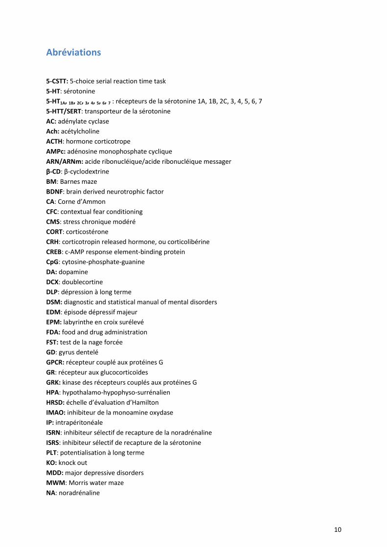
Abréviations
5-CSTT: 5-choice serial reaction time task
5-HT: sérotonine
5-HT1A, 1B, 2C, 3, 4, 5, 6, 7 : récepteurs de la sérotonine 1A, 1B, 2C, 3, 4, 5, 6, 7
5-HTT/SERT: transporteur de la sérotonine
AC: adénylate cyclase
Ach: acétylcholine
ACTH: hormone corticotrope
AMPc: adénosine monophosphate cyclique
ARN/ARNm: acide ribonucléique/acide ribonucléique messager
β-CD: β-cyclodextrine
BM: Barnes maze
BDNF: brain derived neurotrophic factor
CA: Corne d’Ammon
CFC: contextual fear conditioning
CMS: stress chronique modéré
CORT: corticostérone
CRH: corticotropin released hormone, ou corticolibérine
CREB: c-AMP response element-binding protein
CpG: cytosine-phosphate-guanine
DA: dopamine
DCX: doublecortine
DLP: dépression à long terme
DSM: diagnostic and statistical manual of mental disorders
EDM: épisode dépressif majeur
EPM: labyrinthe en croix surélevé
FDA: food and drug administration
FST: test de la nage forcée
GD: gyrus dentelé
GPCR: récepteur couplé aux protéines G
GR: récepteur aux glucocorticoïdes
GRK: kinase des récepteurs couplés aux protéines G
HPA: hypothalamo-hypophyso-surrénalien
HRSD: échelle d’évaluation d’Hamilton
IMAO: inhibiteur de la monoamine oxydase
IP: intrapéritonéale
ISRN: inhibiteur sélectif de recapture de la noradrénaline
ISRS: inhibiteur sélectif de recapture de la sérotonine
PLT: potentialisation à long terme
KO: knock out
MDD: major depressive disorders
MWM: Morris water maze
NA: noradrénaline

11
NORL: novel object recognition location
NORT: novel object recognition test
NRI: inhibiteur de recapture de la noradrénaline
NSF: novelty suppressed feeding
OF: test du champ ouvert
OMS: organisation mondiale de la santé
PCR: réaction en chaîne par polymérase
PFC: cortex préfrontal
SNC: système nerveux central
SRE: promoteur sélectif de la recapture de la sérotonine
TCA: antidépresseur tricyclique / tricyclic antidepressant
12
Liste des figures
Figure 1: Réponse, rémission, rechute et récidive pendant les phases de traitement antidépresseur …………… 23
Figure 2: Proposition d’un algorithme universel des décisions thérapeutiques utilisées par les médecins dans la prise en charge des patients dépressifs ………………………………………………………………………………………………. 24
Figure 3: Représentation bidimensionnelle du récepteur 5-HT4 ……………………………………………………………………. 74
Figure 4: Activation du récepteur 5-HT4 menant à de nombreux effets cellulaires et moléculaires ………………. 75
Figure 5 : Localisation et effets principaux du récepteur 5-HT4 au niveau des organes périphériques ………….. 76
Figure 6: Localisation cérébrale du récepteur 5-HT4 chez l’Humain (A) et chez le rongeur (B) ……………………… 77
Figure 7 : Expression de la β-arrestine 1 dans le système nerveux central par une technique d'hybridation in
situ. …………………………………………………………………………………………………………………………………………………….. 94
Figure 8 : Structure cristallographique de la protéine β-arrestine 1 …………………………………………………………….. 95
Figure 9 : Mécanisme d'action de la protéine β-arrestine ……………………………………………………………………………. 96
Figure 10 : Expression de la β-arrestine 1 réduite dans les leucocytes de patients déprimés et corrélation entre
les concentrations de β-arrestine 1 et la sévérité des symptômes dépressifs …………………………………….. 97
Figure 11: Expression de la protéine ß-arrestine 1 dans les leucocytes de patients déprimés ou de souris anxio-
déprimées non traité(e)s et après traitements antidépresseurs. (A) ………………………………………………….. 98
Figure 12: Interactions entre la protéine ß-arrestine 1 et le récepteur 5-HT4 …………………………………………….. 100
Figure 13: Modèle de désensibilisation par GRK5 du récepteur 5-HT4 opéré par la signalisation ERK ………… 101
Figure 14 : Système de réponse au stress via l'axe hypothalamo-hypophyso-surrénalien ………………………….. 110
Figure 15: Schématisation du gène β-arrestine 1 de la souris ……………………………………………………………………. 113
Figure 16: Insertion génétique du gène CreERT2
pour confirmer la spécificité de la construction génétique 114
Figure 17: Schéma d’obtention des souris β-Arrestine 1FLOX/FLOX
/Nestine-CreERT2-/+…………………………………… 115
Figure 18: Système Cre-ERT2
-loxP inductible au tamoxifène (Gunschmann et al., 2014) ……………………………. 116
Figure 19: Génotypage des souris Nestine-CreERT2/FLOX ARRB1 ………………………………………………………………. 119
Figure 20: Test de Champ Ouvert chez la Souris ……………………………………………………………………………………….. 184
Figure 21: Test du labyrinthe en croix surélevé …………………………………………………………………………………………. 185
Figure 22 : Test des 4 plaques …………………………………………………………………………………………………………………… 186
Figure 23 : Test du Novelty Suppressed Feeding ……………………………………………………………………………………….. 186
Figure 24 : Dégradation de l’état du pelage ………………………………………………………………………………………………. 187
Figure 25: Dispositif d’enregistrement du test de suspension par la queue ………………………………………………. 188
Figure 26: Exemple de marquage β-arrestine 1 par hybridation in situ sur une coupe coronale complète au
niveau du gyrus dentelé après traitement par scanner de lames (A) ou par microscopie optique (B) … 191
Figure 27: Photographies de coupes histologiques du gyrus dentelé de l’hippocampe adulte après marquage au BrdU à différents grossissements ……………………………………………………………………………………………………… 194
Figure 28: Caractérisation des neurones immatures marqués à la doublecortine en fonction de leur
morphologie dendritique ………………………………………………………………………………………………………………….. 195
Figure 29: Représentation schématique de la trajectoire possible d’un patient dépressif …………………………. 338
Figure 30: Représentation schématique du rôle facilitateur ou limitant de l’activation des récepteurs sérotoninergiques sur l’activité pro-cognitive …………………………………………………………………………………… 347
Figure 31: Proposition de score de cognition global (A) et répartition des capacités de mémoire selon les types
de mémoire (B) …………………………………………………………………………………………………………………………………. 355
Figure 32: Profil des capacités cognitives et émotionnelles dans les différents groupes expérimentaux
étudiés……………………………………………………………………………………………………………………………………………….. 356
Figure 33: Différentes voies activées par le RS67333 et médiées par la protéine β-arrestine 1 dans les réponses
comportementales anxiolytiques, antidépresseurs et cognitives ………………………………………………………… 357
13
Liste des tableaux Tableau 1: Critères diagnostics d’un épisode dépressif majeur d’après le DSM-5 ………………………………………….. 21
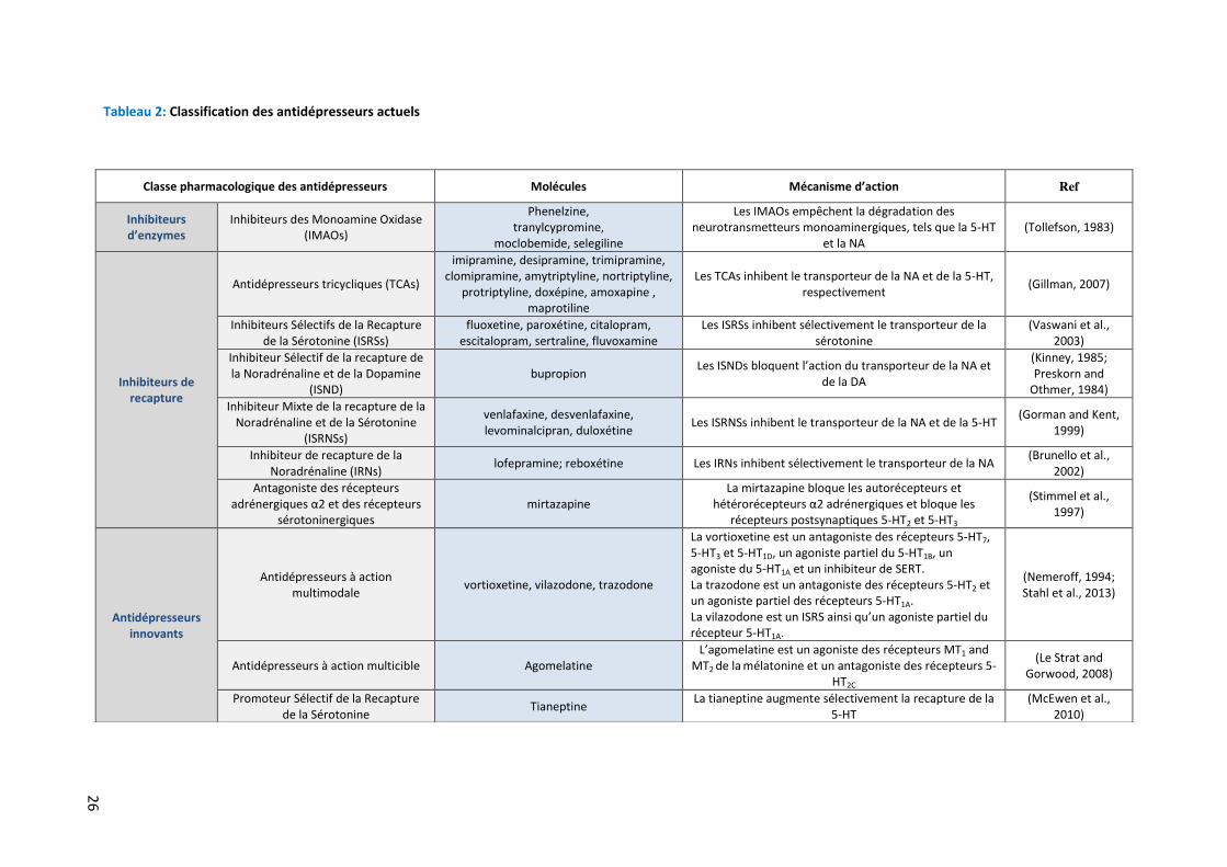
Tableau 2: Classification des antidépresseurs actuels …………………………………………………………………………………… 26
Tableau 3: Effets des ligands du récepteur 5-HT4 sur l'anxiété/dépression – Partie 1 ………………………………….. 82
Tableau 4: Effets des ligands du récepteur 5-HT4 sur l'anxiété/dépression – Partie 2 ………………………………….. 83
Tableau 5: Effets des ligands du récepteur 5-HT4 sur les performances cognitives - Partie 1 ………………………. 88
Tableau 6: Effets des ligands du récepteur 5-HT4 sur les performances cognitives - Partie 2 ………………………. 89
Tableau 7: Effets des ligands du récepteur 5-HT4 sur les performances cognitives - Partie 3 ………………………. 90
Tableau 8: Effets des ligands du récepteur 5-HT4 sur les performances cognitives - Partie 4 ………………………. 91
Tableau 9: Modèles chroniques d'anxiété/dépression chez le rongeur ……………………….……………………………… 109
Tableau 10: Similarités des conséquences de l'administration de CORT et manifestation humaines de la
dépression ……………………….……………………….……………………….……………………….…………………………………….. 110
Tableau 11 : Descriptif des amorces utilisées en PCR pour le génotypage des souris NestineCre ERT2
/FLOX
ARRB1-/-
……………………….……………………….……………………….……………………….…………………………………………. 118
Tableau 12: Programmes PCR des gènes à amplifier ……………………….……………………….………………………………… 118
Tableau 13 : Récapitulatif des anticorps utilisés en Western Blot ……………………….……………………………………… 190
Tableau 14: Récapitulatif des anticorps utilisés en immunohistochimie ……………………….…………………………… 196
Tableau 15: Effets d'une administration chronique de corticostérone sur les performances cognitives chez le
Rongeur - Partie 1 ……………………….……………………….……………………….……………………….………………………….. 340
Tableau 16: Effets d'une administration chronique de corticostérone sur les performances cognitives chez le
Rongeur - Partie 2 ……………………….……………………….……………………….……………………….………………………….. 341
Tableau 17 : Récapitulatif des effets d'une administration chronique de corticostérone sur les différents types
de mémoires. ……………………….……………………….……………………….……………………….……………………………….. 342
Tableau 18: Récapitulatif des effets d’un traitement chronique de fluoxetine ou d'agoniste du récepteur 5-HT4
(RS67333) sur les performances cognitives chez les animaux anxio-déprimés …………………………………… 344
Tableau 19: Conséquences comportementales et neurogéniques de la délétion conditionnelle de la protéine β-
arrestine 1 au niveau des cellules souches du gyrus dentelé …………………………………………………………….. 349
Tableau 20: Conséquences comportementales et neurogéniques de la délétion conditionnelle de la protéine β-
arrestine 1 au niveau des cellules souches du gyrus dentelé après un traitement chronique de fluoxetine.
…………………….……………………….……………………….……………………….…………………………………………………………. 349
Tableau 21: Conséquences de la délétion conditionnelle de la protéine β-arrestine 1 au niveau des cellules
souches du gyrus dentelé sur les performances cognitives. ……………………………………………………………….. 351
Tableau 22: Conséquences de la délétion conditionnelle de la protéine β-arrestine 1 au niveau des cellules
souches du gyrus dentelé après un traitement chronique de fluoxetine ou de RS67333 sur les
performances cognitives . …………………………………….……………………….……………………….………………………… 351
Tableau 23: Modifications des protocoles de tests de cognition en fonction de la souche de souris utilisée. 353
14
Résumé
Souvent précurseurs d’épisodes dépressifs caractérisés, les troubles cognitifs (difficultés à se
concentrer, raisonnement déformé face aux stimuli normalement agréables, indécision, diminution
du temps de réaction et/ou pertes de mémoire) constituent des symptômes quasi constants,
retrouvés parmi les patients souffrant de troubles de l’humeur. La persistance de certains
symptômes cognitifs, même après rémission complète des symptômes dépressifs et pendant les
épisodes récurrents de dépression, souligne l’importance d’une évolution dans la prise en charge
thérapeutique de ces patients. Une réelle évaluation aussi bien préclinique que clinique est
nécessaire afin de déterminer quel traitement pourrait le mieux bénéficier aux symptômes de la
dépression et autre signes de co-morbidité associés. L'efficacité modeste des antidépresseurs
conventionnels, tels que les Inhibiteurs Sélectifs de Recapture de la Sérotonine (ISRS), quant à la
correction du déficit cognitif appelle à de nouvelles approches thérapeutiques. De récentes études
indiquent que les troubles mentaux tels que l'anxiété et/ou la dépression pourraient bénéficier de la
modulation de la signalisation du récepteur sérotoninergique 5-HT4. Si les études précliniques
montrent un certain attrait pour l’utilisation des agonistes du récepteur 5-HT4 dans le traitement des
épisodes dépressifs, la quasi majorité de ces travaux ont été réalisées chez des animaux naïfs. Il est
donc nécessaire de procéder à une caractérisation complète des conséquences d’un traitement
chronique par un agoniste du récepteur 5-HT4 aussi bien sur le plan émotionnel que cognitif pas
seulement chez des animaux naïfs comme la plupart des études le font, mais bel et bien dans un
modèle d’anxiété/dépression. Au laboratoire, nous avons développé un modèle animal
d’anxiété/dépression basé sur l’élévation des concentrations en corticostérone et mimant les effets
d’un stress chronique (modèle CORT)
Les premières questions posées dans ce travail sont donc les suivantes : L’application d’un
stress chronique à la corticostérone (CORT) peut-il induire-il des troubles cognitifs chez les animaux ?
Si oui, quelles stratégies thérapeutiques sont efficaces pour améliorer ces altérations cognitives ?
Dans ce but, après induction du phénotype anxio/dépressif chez la souris, nous avons caractérisé de
façon complète les performances d’apprentissage et de mémoire (mémoire de type épisodique,
mémoire spatiale, flexibilité mentale, mémoire associative) chez les animaux et tenté de les corriger
à l’aide de composés de différentes classes thérapeutiques. Nous avons démontré que
l’administration chronique de corticostérone induit un déficit global de toutes les fonctions
cognitives testées en plus des symptômes d’anxiété/dépression classiquement générés dans ce
modèle (Article 1).
15
J’ai ensuite participé à l’évaluation des propriétés antidépressives et anxiolytiques suite à une
administration infra-chronique ou chronique d'un agoniste du récepteur 5-HT4 dans notre modèle
d'anxiété / dépression, le modèle CORT. Notre étude a montré que dans ce modèle, un agoniste du
récepteur 5-HT4, le RS67333 (1,5 mg/kg/jour pendant 4 semaines), possède des propriétés
anxiolytiques d’action rapide (Article 2).
Ensuite, toujours en utilisant ce modèle, nous avons évalué si la correction du phénotype
anxio/dépressif par un traitement chronique de RS67333 (1,5 mg/kg/jour pendant 4 semaines)
comparé à un traitement à la fluoxetine (18 mg/kg/jour), s’accompagne aussi d’une amélioration des
fonctions cognitives. Alors que le traitement chronique avec le RS67333 restaure l’intégralité des
déficits cognitifs induits par la corticostérone, le traitement avec la fluoxetine ne permet qu’une
amélioration partielle, dépendante du type de mémoire étudié (Article 3).
Des données de la littérature indiquent que la cascade de signalisation de β-arrestine 1
(impliquée également dans la désensibilisation et l’internalisation du récepteur 5-HT4) serait un
biomarqueur potentiel préclinique/clinique des états dépressifs et de la réponse au traitement
antidépresseur. Dans la mesure où la neurogenèse hippocampique adulte est un processus en partie
nécessaire à la réponse aux antidépresseurs, nous avons cherché à caractériser le phénotype
anxio/dépressif des souris tissus-spécifiques conditionnelles, dont l’expression de la protéine β-
arrestine 1 dans les cellules souches du gyrus dentelé a été supprimée (ARRB1). Puis, de façon à
affiner plus précisément le rôle de la β-arrestine 1 dans les effets comportementaux et
neurogéniques des antidépresseurs ISRSs, nous avons administré un traitement chronique de
fluoxetine (18 mg/kg/jour) aux animaux dépourvus de β-arrestine 1 et procédé à une batterie
complète de tests comportementaux anxiolytiques/antidépresseurs.
Si le phénotype anxio/dépressif semble dans l’ensemble peu altéré par la délétion spécifique et
conditionnelle de la β-arrestine 1, la réponse à la fluoxetine a été bloquée dans certains tests
comportementaux tels que le test des 4 plaques et le Novelty Suppressed Feeding. La protéine β-
arrestine 1 apparait donc comme un acteur clé dans certains effets antidépresseurs de la fluoxetine.
D’autre part, l’étude du phénomène de neurogenèse hippocampique révèle que l’expression de la
protéine β-arrestine 1 dans les cellules souches du gyrus dentelé de l’hippocampe est nécessaire à la
survie des jeunes neurones et essentielle pour obtenir des effets bénéfiques de la fluoxétine sur la
prolifération et la survie de ces jeunes neurones. En revanche, la protéine β-arrestine 1 ne semble
pas impliquée dans la réponse de la fluoxetine sur l’étape de maturation des jeunes neurones.
Enfin, afin de déterminer l’importance de la protéine β-arrestine 1 dans le phénotype cognitif
et dans la réponse aux traitements classiques et innovants, nous avons évalué les performances
16
cognitives chez les animaux ARRB1-/- dont la β-arrestine 1 a été supprimée sélectivement dans les
cellules souches du gyrus dentelé. Nous avons ensuite examiné à nouveau les conséquences d’un
traitement chronique avec un agoniste du récepteur 5-HT4 (RS67333) comparé à un traitement à la
fluoxetine. Contrairement au phénotype anxio/dépressif, le phénotype cognitif est altéré chez les
animaux déficitaires en β-arrestine 1. De plus, les traitements chroniques au RS67333 et à la
fluoxetine ne sont pas capables de corriger ces déficits cognitifs chez les souris ARRB1-/-. Ces résultats
suggèrent non seulement que la protéine β-arrestine 1 est nécessaire aux différents mécanismes de
mémoire, mais surtout que son expression dans les cellules souches du gyrus dentelé est nécessaire
pour que l’agoniste du récepteur 5-HT4 puisse induire ses effets pro-cognitifs.
Ce travail de thèse a mis en avant le rôle prépondérant du récepteur 5-HT4 dans la réponse aux
antidépresseurs non seulement dans sa capacité à corriger les troubles d’anxiété/dépression, mais
aussi les troubles cognitifs associés à la dépression dans un modèle d’anxiété/dépression. D’un point
de vue mécanistique, ces données confirment l’implication de la protéine β-arrestine 1 dans la
réponse à la fluoxetine au niveau comportemental et directement au sein du processus de
neurogenèse hippocampique chez la souris adulte. Enfin, la protéine β-arrestine 1 se révèle être un
acteur déterminant dans les mécanismes cognitifs et indispensable dans la réponse aux traitements
étudiés.

17

18
Introduction

Introduction
20
1 Les épisodes dépressifs majeurs et leurs traitements
La pathologie de la dépression majeure 1.1
L'Organisation Mondiale de la Santé (OMS) prévoit que d'ici 2030 la dépression majeure sera
la deuxième cause d'invalidité dans le monde. Les troubles de l'humeur touchent 7% (prévalence
annuelle) de la population mondiale, tandis que les formes sévères de dépression ont une incidence
sur 2-5% de la population américaine (Kessler et al., 2005). En outre, environ 32 à 35.000.000
d’adultes dans la population américaine (prévalence vie entière : 16%) connaitront un épisode
dépressif majeur au cours de leur vie. En Europe, une méta-analyse fondée sur 27 études cliniques,
comprenant plus de 150.000 sujets de 16 pays européens, a estimé la prévalence de la dépression
entre 3 à 10 % au cours des 12 derniers mois (Kessler et al., 2005). Quant à la France, selon un
rapport de l’Institut de Veille Sanitaire, les épisodes dépressifs majeurs affecteraient 7,8% de la
population (Sapinho et al., 2008).
La dépression majeure se caractérise par plusieurs symptômes biologiques et psychologiques qui
affectent de nombreux aspects du quotidien. Selon le manuel diagnostique et statistique des
troubles mentaux (American Psychiatric Association. and American Psychiatric Association. DSM-5
Task Force., 2013), le trouble dépressif caractérisé se définit par une modification persistante de
l’humeur pendant une longue période accompagnée d’une souffrance morale et d’un ralentissement
psychomoteur
Plus précisément, un épisode dépressif majeur (EDM) est caractérisé par une humeur ou une
perte d’intérêt ou de plaisir généralisé pendant au moins deux semaines consécutives, et ce
pratiquement toute la journée et presque chaque jour. L’EDM est avéré si, durant cette période,
apparaissent au moins 4 des symptômes suivants : fatigue, ralentissement psychomoteur, perte de
poids ou d’appétit, sommeil altéré, difficultés à se concentrer ou à prendre des décisions, idées de
dévalorisation ou de culpabilité et idées de mort récurrentes ou tentatives de suicide, et qu’ils
entrainent une perturbation des activités habituelles (Tableau 1). Sa classification est en perpétuelle
évolution de façon à catégoriser au mieux chaque cas clinique et améliorer ensuite la prise en
charge.
Introduction
21
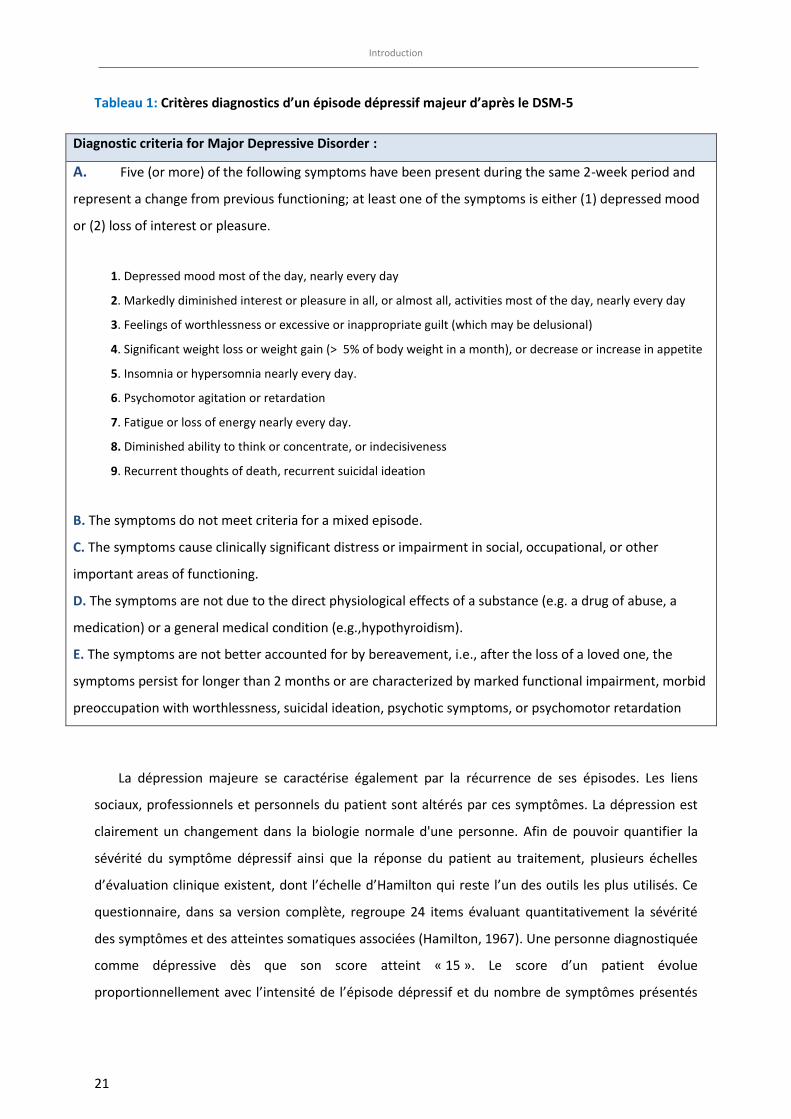
Tableau 1: Critères diagnostics d’un épisode dépressif majeur d’après le DSM-5
Diagnostic criteria for Major Depressive Disorder :
A. Five (or more) of the following symptoms have been present during the same 2-week period and
represent a change from previous functioning; at least one of the symptoms is either (1) depressed mood
or (2) loss of interest or pleasure.
1. Depressed mood most of the day, nearly every day
2. Markedly diminished interest or pleasure in all, or almost all, activities most of the day, nearly every day
3. Feelings of worthlessness or excessive or inappropriate guilt (which may be delusional)
4. Significant weight loss or weight gain (> 5% of body weight in a month), or decrease or increase in appetite
5. Insomnia or hypersomnia nearly every day.
6. Psychomotor agitation or retardation
7. Fatigue or loss of energy nearly every day.
8. Diminished ability to think or concentrate, or indecisiveness
9. Recurrent thoughts of death, recurrent suicidal ideation
B. The symptoms do not meet criteria for a mixed episode.
C. The symptoms cause clinically significant distress or impairment in social, occupational, or other
important areas of functioning.
D. The symptoms are not due to the direct physiological effects of a substance (e.g. a drug of abuse, a
medication) or a general medical condition (e.g.,hypothyroidism).
E. The symptoms are not better accounted for by bereavement, i.e., after the loss of a loved one, the
symptoms persist for longer than 2 months or are characterized by marked functional impairment, morbid
preoccupation with worthlessness, suicidal ideation, psychotic symptoms, or psychomotor retardation
La dépression majeure se caractérise également par la récurrence de ses épisodes. Les liens
sociaux, professionnels et personnels du patient sont altérés par ces symptômes. La dépression est
clairement un changement dans la biologie normale d'une personne. Afin de pouvoir quantifier la
sévérité du symptôme dépressif ainsi que la réponse du patient au traitement, plusieurs échelles
d’évaluation clinique existent, dont l’échelle d’Hamilton qui reste l’un des outils les plus utilisés. Ce
questionnaire, dans sa version complète, regroupe 24 items évaluant quantitativement la sévérité
des symptômes et des atteintes somatiques associées (Hamilton, 1967). Une personne diagnostiquée
comme dépressive dès que son score atteint « 15 ». Le score d’un patient évolue
proportionnellement avec l’intensité de l’épisode dépressif et du nombre de symptômes présentés
Introduction
22
par le patient. Cet outil permet également l’évaluation de la réponse au traitement. Classiquement,
la réponse au traitement est définie par une diminution d’au moins 50% du score obtenu lors de la
première évaluation du patient. L’échelle de Montgomery et Asberg (MADRS) (Montgomery and
Asberg, 1979) est un outil également fréquemment utilisés en pratique par les psychiatres ainsi que
dans les études cliniques incluant des patients déprimés.
En complément des critères diagnostics fondamentaux décrits dans le Tableau 1, le DSM-5 définit
au sein des épisodes dépressifs majeurs différents sous-types de dépression : la dépression
mélancolique, la dépression atypique. Celles-ci différent selon plusieurs paramètres cliniques et
biologiques identifiées grâce aux études cliniques et permet une meilleure classification dès la
première prise en charge des patients. Ainsi, la dépression mélancolique sera caractérisée par une
perte permanente d’intérêt et de réactivité à des activités qui procuraient en temps normal du plaisir
et est accompagnée d’épisodes dépressifs plus intenses le matin, de réveils matinaux, de retards
moteurs et une prévalence accrue des idées suicidaires (DSM-5 ;(Caldieraro et al., 2013)). De plus, les
données neurochimiques abondent dans le sens d’une hyperactivité de l’axe hypothalamo-
hypophysaire adrénalien (HPA) dû à un défaut de rétrocontrôle inhibiteur puisque 40 à 55 % des
patients seraient non-suppresseurs dans le test à la dexaméthasone suivi d’une injection de CRH
(Coryell, 2007). Cette hyperactivité de l’axe génère alors une augmentation des concentrations
d’hormone corticotrope (ACTH), de corticolibérine (CRH) et de cortisol et induit in fine une
hypercortisolémie (Antonijevic, 2008; Carroll et al., 2007; O'Keane et al., 2012). A l’inverse, la
dépression atypique est caractérisée par une hypofonctionnalité de l’axe HPA, avec une sécrétion de
CRH et de cortisol pouvant être diminuées (Antonijevic, 2008). De plus, elle s’accompagne des
symptômes suivants : une meilleure réactivité émotionnelle aux évènements positifs, une
hypersomnie et une augmentation du poids et de l’appétit (DSM-5).
Introduction
23
Les traitements antidépresseurs & leurs limitations 1.2
1.2.1 Prise en charge clinique des épisodes dépressifs caractérisés
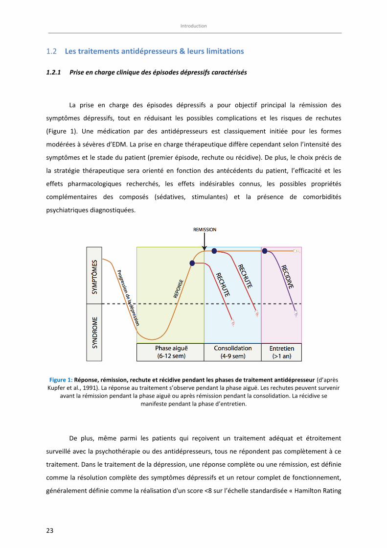
La prise en charge des épisodes dépressifs a pour objectif principal la rémission des
symptômes dépressifs, tout en réduisant les possibles complications et les risques de rechutes
(Figure 1). Une médication par des antidépresseurs est classiquement initiée pour les formes
modérées à sévères d’EDM. La prise en charge thérapeutique diffère cependant selon l’intensité des
symptômes et le stade du patient (premier épisode, rechute ou récidive). De plus, le choix précis de
la stratégie thérapeutique sera orienté en fonction des antécédents du patient, l’efficacité et les
effets pharmacologiques recherchés, les effets indésirables connus, les possibles propriétés
complémentaires des composés (sédatives, stimulantes) et la présence de comorbidités
psychiatriques diagnostiquées.
Figure 1: Réponse, rémission, rechute et récidive pendant les phases de traitement antidépresseur (d’après Kupfer et al., 1991). La réponse au traitement s’observe pendant la phase aiguë. Les rechutes peuvent survenir
avant la rémission pendant la phase aiguë ou après rémission pendant la consolidation. La récidive se manifeste pendant la phase d’entretien.
De plus, même parmi les patients qui reçoivent un traitement adéquat et étroitement
surveillé avec la psychothérapie ou des antidépresseurs, tous ne répondent pas complètement à ce
traitement. Dans le traitement de la dépression, une réponse complète ou une rémission, est définie
comme la résolution complète des symptômes dépressifs et un retour complet de fonctionnement,
généralement définie comme la réalisation d'un score <8 sur l’échelle standardisée « Hamilton Rating
Introduction
24
Scale for Depression » (HRSD). Cependant, certains inconvénients sont encore observés : une
réponse thérapeutique se développant lentement (2 à 3 semaines) et un pourcentage significatif
(30%) de patients non répondeurs, résistants au traitement ou qui récidivent (Wong and Licinio,
2001).
Dans la dépression caractérisée, le traitement de consolidation à privilégier est le
médicament qui a permis d’obtenir la rémission symptomatique tout en maintenant les mêmes
posologies. Le traitement de maintien a pour but d’éviter les récidives (ou rechutes). Lors d’un
épisode de rechute, il est recommandé de choisir un antidépresseur qui s’est avéré efficace et bien
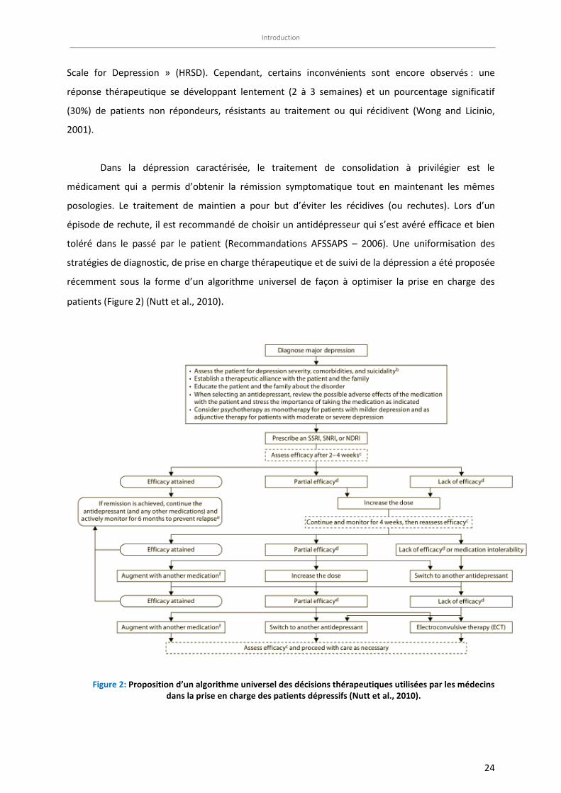
toléré dans le passé par le patient (Recommandations AFSSAPS – 2006). Une uniformisation des
stratégies de diagnostic, de prise en charge thérapeutique et de suivi de la dépression a été proposée
récemment sous la forme d’un algorithme universel de façon à optimiser la prise en charge des
patients (Figure 2) (Nutt et al., 2010).
Figure 2: Proposition d’un algorithme universel des décisions thérapeutiques utilisées par les médecins dans la prise en charge des patients dépressifs (Nutt et al., 2010).
Introduction
25
1.2.2 Traitements historiques
Différentes classes d’antidépresseurs ont été développées selon leur sélectivité vis-à-vis des
transporteurs de recapture monoaminergiques. Les antidépresseurs tricycliques (par exemple,
l'imipramine), qui inhibent la recapture de la sérotonine et de la noradrénaline, et les Inhibiteurs de
la MonoAmine Oxydase (exemple l’iproniazide, IMAOA/B non sélectif irréversible), apparaissent dans
les années 1950 à 1960 et sont les premiers médicaments utilisés en clinique pour traiter la
dépression (Nestler et al., 2002) (Tableau 2). Ces médicaments ont été depuis remplacés par de
nouveaux antidépresseurs ayant une efficacité comparable, mais une tolérance supérieure et un
meilleur profil de sécurité.
La découverte de ces effets antidépresseurs et de leurs cibles moléculaires a conduit à la
conception de médicaments de deuxième et troisième génération, tels que les Inhibiteurs Sélectifs
du Recapture de la Sérotonine (ISRS) et les Inhibiteurs Sélectif du Recapture de la noradrénaline
(ISRN). Depuis cette découverte, l‘hypothèse monoaminergique de la dépression a été émise. La
dépression serait une atteinte de la neurotransmission sérotoninergique, noradrénergique et, dans
une moindre mesure, dopaminergique centrale. Aujourd'hui, la plupart des médicaments utilisés
dans le traitement de la dépression, et notamment les ISRS (comme par exemple, la fluoxetine), sont
efficaces pour traiter à la fois l'anxiété et la dépression (Samuels et al., 2011; Schatzberg and
Nemeroff, 2009). Néanmoins, les derniers éléments de recherche montrent que cette hypothèse
monoaminergique ne peut expliquer à elle seule la physiopathologie de la dépression (Chaudhury et
al., 2015). C’est pourquoi l’hypothèse neuroendrocrinienne avec une perturbation de l’axe
hypothalamo-hypophysaire (HPA) et la théorie de la plasticité ont été énoncées.
26
Tableau 2: Classification des antidépresseurs actuels
Classe pharmacologique des antidépresseurs Molécules Mécanisme d’action Ref
Inhibiteurs
d’enzymes
Inhibiteurs des Monoamine Oxidase (IMAOs)
Phenelzine, tranylcypromine,
moclobemide, selegiline
Les IMAOs empêchent la dégradation des neurotransmetteurs monoaminergiques, tels que la 5-HT
et la NA (Tollefson, 1983)
Inhibiteurs de
recapture
Antidépresseurs tricycliques (TCAs)
imipramine, desipramine, trimipramine, clomipramine, amytriptyline, nortriptyline,
protriptyline, doxépine, amoxapine , maprotiline
Les TCAs inhibent le transporteur de la NA et de la 5-HT, respectivement
(Gillman, 2007)
Inhibiteurs Sélectifs de la Recapture de la Sérotonine (ISRSs)
fluoxetine, paroxétine, citalopram, escitalopram, sertraline, fluvoxamine
Les ISRSs inhibent sélectivement le transporteur de la sérotonine
(Vaswani et al., 2003)
Inhibiteur Sélectif de la recapture de la Noradrénaline et de la Dopamine
(ISND) bupropion
Les ISNDs bloquent l’action du transporteur de la NA et de la DA
(Kinney, 1985; Preskorn and
Othmer, 1984)
Inhibiteur Mixte de la recapture de la Noradrénaline et de la Sérotonine
(ISRNSs)
venlafaxine, desvenlafaxine, levominalcipran, duloxétine
Les ISRNSs inhibent le transporteur de la NA et de la 5-HT (Gorman and Kent,
1999)
Inhibiteur de recapture de la Noradrénaline (IRNs)
lofepramine; reboxétine Les IRNs inhibent sélectivement le transporteur de la NA (Brunello et al.,
2002)
Antagoniste des récepteurs adrénergiques α2 et des récepteurs
sérotoninergiques mirtazapine
La mirtazapine bloque les autorécepteurs et hétérorécepteurs α2 adrénergiques et bloque les
récepteurs postsynaptiques 5-HT2 et 5-HT3
(Stimmel et al., 1997)
Antidépresseurs
innovants
Antidépresseurs à action multimodale
vortioxetine, vilazodone, trazodone
La vortioxetine est un antagoniste des récepteurs 5-HT7, 5-HT3 et 5-HT1D, un agoniste partiel du 5-HT1B, un agoniste du 5-HT1A et un inhibiteur de SERT. La trazodone est un antagoniste des récepteurs 5-HT2 et un agoniste partiel des récepteurs 5-HT1A. La vilazodone est un ISRS ainsi qu’un agoniste partiel du récepteur 5-HT1A.
(Nemeroff, 1994; Stahl et al., 2013)
Antidépresseurs à action multicible Agomelatine L’agomelatine est un agoniste des récepteurs MT1 and
MT2 de la mélatonine et un antagoniste des récepteurs 5-HT2C
(Le Strat and Gorwood, 2008)
Promoteur Sélectif de la Recapture de la Sérotonine
Tianeptine La tianeptine augmente sélectivement la recapture de la
5-HT (McEwen et al.,
2010)
Introduction
27
En plus de l’approche pharmacologique, d’autres stratégies thérapeutiques qui n’impliquent
pas les antidépresseurs sont disponibles et ont prouvés leur efficacité dans le traitement de la
dépression. Par exemple, la thérapie par électrochocs (électro-convulsivo thérapie ou ECT), la
stimulation électrique du nerf vague, la stimulation cérébrale profonde et certains types de
psychothérapie ciblée comme la thérapie cognitivo-comportementale et interpersonnelle (Guidi et
al., 2016) peuvent être mises en place pour soulager les symptômes de la dépression mais sont
encore en cours d’évaluation clinique. D’autres interventions telles que la stimulation chronique de
la région cingulaire subgénuale (aire 25 de Brodmann) ont également montré des effets prometteurs
lors d'essais cliniques (Mayberg et al., 2005). Ces stratégies non pharmacologiques sont
généralement envisagées en cas de dépressions sévères résistantes à toutes autres stratégies
thérapeutiques.
1.2.3 Limites actuels des traitements
Malgré les progrès évidents apportés par les antidépresseurs actuellement disponibles
(sélectivité d’effet, rapport bénéfices/risques), 30 à 60 % des patients ne répondent toujours pas de
manière adéquate et souffrent toujours de symptômes résiduels incapacitants (Trivedi et al., 2006).
Au cours des dernières décennies, des milliards de dollars ont été dépensés pour cibler de nouveaux
médicaments plus sélectifs que les récepteurs de la sérotonine ou de la noradrénaline, agonistes ou
antagonistes ayant des effets semblables aux médicaments antidépresseurs déjà sur le marché, mais
avec une réponse plus rapide et présentant moins d’effets indésirables. Malheureusement, en dépit
de la recherche et des connaissances acquises sur le mécanisme d'action des antidépresseurs
classiques et de leurs propriétés thérapeutiques, le traitement pharmacologique de la dépression
reste peu satisfaisant. En effet, bien que les antidépresseurs actuellement disponibles aient une
réelle efficacité curative (démontrée par des études cliniques « contre placebo » ou en comparaison
à la thérapie de référence, l’ECT) dans le traitement de cette pathologie, il faut i- attendre plusieurs
semaines pour qu’ils démontrent une pleine efficacité, ii- de nombreux patients répondent peu au
traitement, iii- les symptômes concomitants sont souvent peu contrôlés et iv- certains
antidépresseurs peuvent engendrer des problèmes de tolérance ou de dépendance (pour
revue,(Martinowich et al., 2013)). Environ 60% des patients souffrant de dépression ne répondent
pas de façon adéquate aux antidépresseurs ou sont résistantes à ces médicaments. Moins de 50 %
des patients souffrant de dépression montrent une complète amélioration (Trivedi et al., 2006). Les
effets indésirables des ISRS sont fréquemment décrits lors d’un traitement chronique, notamment
l'insomnie, somnolence, sensation vertigineuse, akathisie et dysfonction sexuelle à long terme (par
Introduction
28
exemple, diminution de la libido, l'éjaculation retardée, etc…). Comme les antidépresseurs de
première génération, les ISRS nécessitent au moins 2 à 4 semaines d'administration avant l'obtention
de bénéfices thérapeutiques (Wong and Licinio, 2001). Le fait que la réponse au traitement
antidépresseur soit si imprévisible chez un individu et qu’il soit souvent nécessaire d'essayer
plusieurs antidépresseurs pour obtenir un effet optimal, peut causer de la frustration chez le patient,
favoriser une mauvaise observance de ce traitement, ce qui en limitera son efficacité et
involontairement servira à renforcer les symptômes de la dépression (Fava, 2000; Trivedi et al.,
2006). L'écart entre les effets aigus des ISRS (in vitro, le blocage des transporteurs SERT et/ou NET est
rapide) et l'apparition tardive de leurs effets in vivo après administration chronique chez l’animal et
chez l’Homme suggèrent un mécanisme d'action plus complexe que prévu. Cette distinction
apparente entre les effets aigus et chroniques des ISRS a fait émerger l'hypothèse de la nécessaire
activation de plusieurs mécanismes pharmacologiques dans le cerveau lors d'un traitement
chronique avec des ISRS. L'efficacité modeste des antidépresseurs conventionnels, et notamment des
ISRSs, appelle à de nouvelles approches pour traiter les différentes formes légères, modérée ou
sévères des épisodes dépressifs associés ou non à des troubles anxieux. Des études cliniques mettant
en œuvre des thérapies pharmacologiques combinées, telles que le blocage de certains récepteurs
monoaminergiques centraux (antagoniste de l’autorécepteurs 5-HT1A, par exemple) en plus de
l'inhibition d’un des transporteurs des monoamines, ont déjà été proposées afin de raccourcir le
délai d’apparition de l'effet antidépresseur et/ou d'augmenter l'efficacité de ces médicaments.
1.2.4 Nouveaux traitements innovants
Les efforts actuels de développement de médicaments visent à découvrir de nouvelles cibles
et de nouvelles classes d'antidépresseurs dans l'espoir d’identifier de nouveaux composés ayant une
efficacité plus large et / ou d’apparition des effets plus rapide avec un meilleur profil d'effets
indésirables (Tableau 2). Pour exemples, les inhibiteurs de recapture doubles ou triples des
monoamines sont des molécules nouvelles capables d'agir sur plusieurs systèmes de
neurotransmetteurs à la fois (Roose et al., 1994). Autre exemple, la vortioxetine (Lu AA21004; 1-[2-
(2,4-diméthyl-phénylsulfanyl)-phényl]- pipérazine) est un nouvel antidépresseur ayant une activité
antidépressive « multimodale » développé par des Laboratoires Lundbeck. Fin Septembre 2013, la «
Food and Drug Administration » (FDA) américaine et fin octobre 2013, l’Agence Européenne du
Médicament (EMA) ont approuvé la mise sur le marché de la vortioxetine (BRINTELLIX®) avec pour
indication le traitement des adultes souffrant de troubles dépressifs majeurs. En 2015, l’EMA et la
Introduction
29
FDA ont reconnu les propriétés bénéfiques de la vortioxetine sur la correction des déficits cognitifs
lors d‘EDM.
Autre exemple, le développement de l’agomelatine (S20098, N-[2 - (7- méthoxynaphthalén-
1-yl) éthyl] acétamide), médicament antidépresseur ayant à la fois des propriétés agoniste
mélatoninergique et antagoniste du récepteur 5-HT2C, est prometteur car les troubles affectifs sont
caractérisés par des rythmes circadiens anormaux (pour revue, voir (de Bodinat et al., 2010)).
L’agomelatine (Valdoxan®/ Thymanax® développé par les Laboratoires IRIS) a obtenu l'autorisation
de commercialisation en 2009 pour le traitement des épisodes dépressifs majeurs en Europe,
devenant ainsi un des premiers antidépresseurs approuvés avec un mécanisme d’action non
exclusivement monoaminergique.
Enfin, les antagonistes du récepteur ionotropique glutamatergique NMDA, en particulier la
(±)-kétamine sont comme des candidats à la prochaine génération d'antidépresseurs d’action rapide
(pour revue, voir (Maeng et al., 2008; Martinowich et al., 2013). De façon étonnante, il semble que
les effets d’un anesthésique, la (±)-kétamine sur le comportement reposent sur l'activation du
récepteur AMPA, un autre type de récepteur canal ionique du L-glutamate (l’isomère S- est en cours
d’évaluation car il aurait moins d’effets indésirables « psychomimétiques ») et, par conséquent, des
médicaments capables d'activer directement le récepteur AMPA ou un de ses sites modulateurs
allostériques (Ex : essais cliniques en cours du GLYX-13 ou rapastinel, un agoniste partiel du site
glycine du récepteur NMDA) pourraient produire des actions antidépressives rapides et de longue
durée.
Enfin, malgré les énormes progrès réalisés en recherche avec l'utilisation de traitements
physiques ou somatiques comme la psychothérapie, la thérapie par électrochocs (ECT) ou la
pharmacologie des antidépresseurs visant à élucider la physiopathologie des troubles
anxio/dépressifs, de nombreuses questions restent en suspens concernant le mécanisme d’action
des antidépresseurs et leur lien avec la dépression (Nestler et al., 2002).
Introduction
30
2 Les troubles cognitifs dans la dépression majeure
Bien que seule la « difficulté de concentration » apparaisse clairement dans la liste des
critères de diagnostic de la dépression majeure définie par le DSM-5, de nombreux autres troubles
cognitifs associés aux symptômes dépressifs sont reportés par les patients souffrant de troubles de
l’humeur. Parmi eux, des altérations dans l’attention, la mémoire de travail, les performances
d’apprentissage et de mémoire et les fonctions exécutives alourdissent significativement le poids de
la maladie chez ces patients.
La Revue “Cognitive dysfunction in major depressive disorder : A translational review in
Animal models of the disease » qui suit, présente dans un premier temps les déficits cognitifs
observés chez les patients déprimés, en fonction de leur âge et la nature de dépression. Les
différents outils cliniques permettant de déceler ces troubles cognitifs sont également évoqués. Dans
un second temps, les troubles d’apprentissage et de mémoire associés à différents modèles animaux
d’anxiété/dépression sont détaillés. Pour chaque domaine cognitif étudié, nous avons déterminé si
les troubles cognitifs observés sont communs aux différents modèles animaux ou si d’autres facteurs
non-spécifiques (tels que l’espèce animale, le sexe ou l’âge) pouvaient dessiner des altérations
communes à travers les différents modèles. Enfin, le rôle de la neurogenèse hippocampique chez les
Rongeurs adulte dans les déficits cognitifs a également été développé dans cet article.
pharmaceuticals
Review
Cognitive Dysfunction in Major DepressiveDisorder. A Translational Review in AnimalModels of the DiseaseFlavie Darcet 1, Alain M. Gardier 1, Raphael Gaillard 2,3,4, Denis J. David 1,* andJean-Philippe Guilloux 1
1 Université Paris-Saclay, University Paris-Sud, Faculté de Pharmacie, CESP, INSERM UMRS1178,Chatenay-Malabry 92296, France; [email protected] (F.D.); [email protected] (A.M.G.);[email protected] (J.-P.G.)
2 Laboratoire de “Physiopathologie des maladies Psychiatriques”, Centre de Psychiatrie etNeurosciences U894, INSERM, Université Paris Descartes, Sorbonne Paris Cité, Paris 75014, France;[email protected]
3 Service de Psychiatrie, Centre Hospitalier Sainte-Anne, Faculté de Médecine Paris Descartes,Université Paris Descartes, Sorbonne Paris Cité, Paris 75014, France
4 Human Histopathology and Animal Models, Infection and Epidemiology Department, Institut Pasteur,Paris 75015, France
* Correspondence: [email protected]; Tel.: +331-468-359-68; Fax: +331-468-353-55
Academic Editor: Guy GriebelReceived: 4 December 2015; Accepted: 1 February 2016; Published: 17 February 2016
Abstract: Major Depressive Disorder (MDD) is the most common psychiatric disease, affectingmillions of people worldwide. In addition to the well-defined depressive symptoms, patientssuffering from MDD consistently complain about cognitive disturbances, significantly exacerbatingthe burden of this illness. Among cognitive symptoms, impairments in attention, working memory,learning and memory or executive functions are often reported. However, available data about theheterogeneity of MDD patients and magnitude of cognitive symptoms through the different phasesof MDD remain difficult to summarize. Thus, the first part of this review briefly overviewed clinicalstudies, focusing on the cognitive dysfunctions depending on the MDD type. As animal modelsare essential translational tools for underpinning the mechanisms of cognitive deficits in MDD, thesecond part of this review synthetized preclinical studies observing cognitive deficits in differentrodent models of anxiety/depression. For each cognitive domain, we determined whether deficitscould be shared across models. Particularly, we established whether specific stress-related proceduresor unspecific criteria (such as species, sex or age) could segregate common cognitive alteration acrossmodels. Finally, the role of adult hippocampal neurogenesis in rodents in cognitive dysfunctionsduring MDD state was also discussed.
Keywords: major depressive disorder; cognitive dysfunctions; animal models of anxiety/depression;neurogenesis
1. Introduction
Cognitive dysfunction is a common feature of major depressive disorder (MDD), contributingto the serious decline in patients’ quality of life. Described in the 5th edition of the Diagnosticand Statistical Manual of Mental Disorders (DSM-V, [1]) as “significantly affecting the individual’scapacity to function”, these cognitive changes are associated with the set of emotional and behavioralalterations (including persistent depressed mood and loss of pleasure) that characterizes MDDpathology. Many clinical studies have focused their work on the nature and the magnitude of cognitive
Pharmaceuticals 2016, 9, 9; doi:10.3390/ph9010009 www.mdpi.com/journal/pharmaceuticals
Pharmaceuticals 2016, 9, 9 2 of 42
alterations during the clinical course of MDD [2–6]. During clinical observation, patients mostlyreport their difficulty to concentrate, to make decisions, the feeling that their brain is slowed downor the fact that “they forget everything” (tasks, meetings, etc.) [7]. These subjective complaints relateto a broad range of cognitive impairments reported during depressive episodes, from executivefunctions (attention, processing speed, cognitive flexibility) to working and visual learning andmemory. However, contradictory findings from neuropsychological tests have encouraged clinicians toexamine whether the heterogeneity of the MDD population would prevent from identifying a specificneurocognitive profile in depressed individuals.
The DSM-V clearly defines subtypes in major depression such as melancholic and atypicalsubtypes of MDD. Comparing different cognitive functions across MDD subtypes may help in theidentification of neurocognitive patterns according to the specificity of MDD markers. Additionally,few studies aimed at delineating between trait- and state-like cognitive alterations, i.e., the deficitsobserved exclusively during depressed episodes and those occurring prior, between and after MDDepisodes [7]. One meta-analysis performed across first-episode MDD subjects, and including 13 studies,segregated state-dependent cognitive alterations in MDD subjects (psychomotor speed and memoryfunctioning) from trait-markers (attention and executive functioning). However, the large clinicalheterogeneity of MDD subjects may dampen this report.
In order for basic research to provide potential advances in this field, it is essential to use animalmodels that present behavioral, neurochemical and brain morphological phenotype reminiscent ofsome symptoms of MDD. Indeed, animal models exhibiting deficits in one or more of the relevantdomains of cognition are useful to investigate mechanisms underlying impaired cognitive processesobserved in MDD and their dependence on mood pathology. In rodents, many anxiety-depressionmodels, including chronic early-life stress and adulthood models, have been validated for the study ofanhedonic behaviors, modeling the negative mood symptomatology of MDD. It has been reported thatadverse experiences during pregnancy or early stress life events in childhood could lead to an increasedsensitivity to the effects of stress in adulthood life, directly enhancing vulnerability to depression [8,9].Rodent models such as prenatal stress (chronic stress exposure during gestational period), earlypost-natal handling and maternal separation (chronic pups-dams separation during weaning period),have been validated to produce depression-like behavior in adulthood [10,11]. However, in mostclinical cases, the apparition of MDD pathology occurs during adulthood and is typically causedby a succession of adverse stress episodes in life, leading progressively to the core of the pathology.Several adult animal models have been validated as anxiety-depression models such as social defeatmodel (SD) [12], learned helplessness (LH) [13], unpredictable chronic mild stress (UCMS) [14] andchronic corticosterone administration (CORT) [15]. Among these models, UCMS procedure, basedon a chronic exposure of unpredictable stressors, has been reported as one of the most robust animalmodels of depression thanks to good predictive, face and construct validities [16,17].
In this review, the first part will be focus on clinical cognitive dysfunctions depending on the typeand stage of the MDD illness. The second part of this work will gather preclinical studies observingcognitive deficits in different rodent models of anxiety/depression. For each cognitive domain, we willhighlight which deficits can be shared, across models, and, whether specific stress-related proceduresor non-specific criteria (species, sex, type of cognitive parameter measured) can segregate commoncognitive alteration. Finally, the role of adult hippocampal neurogenesis in rodents in cognitivedysfunctions during MDD state will be discussed.
2. Cognition in Patients Suffering from MDD
2.1. Cognitive Performances in MDD through Different Ages
Cognitive abnormalities in many various cognitive domains have been reported among patientssuffering from MDD [4,18–22]. Specifically, cognitive alterations in attentional processes [23–25],executive functioning [6,26–28], working memory [27,29,30], verbal or visual learning and
Pharmaceuticals 2016, 9, 9 3 of 42
memory [29,31] and emotional processing [31,32] were noted in MDD patients. While early-onsetdepression is associated with higher disease severity and with higher levels of recurrence [33], limiteddata are available regarding children, adolescents or young adults cognitive performances duringMDD episodes. Deficit in attention, memory and problem solving could have a serious impact in thesepopulations on daily activities, especially when individuals are involved in education or academicsprograms, during which their achievement depends on these skills [34]. Among the few studiesexamining cognitive performances in pediatric, adolescent or young adults depressed subjects, all ofthem agreed on a general cognitive degradation but none managed to extract specific impairmentsrelated to the early-onset of depression [33,35,36]. It remains currently unclear whether or not cognitiveimpairments should be considered as vulnerability markers of depression, potentially preceding thedevelopment of depressive symptoms or, whether cognitive symptoms develop only after the onset ofa major depressive episode [33].
Further studies involving larger, homogenous cohorts of patients are needed to provide newunderstanding regarding this issue. Cognitive dysfunctions in elderly depressed people havebeen widely investigated, but no specific cognitive impairments have been observed due to thephysiological decline of cognitive process with age and to potential neurodegenerative disordersappearing in late-life. However, late-life depression has been particularly associated with a slowerspeed in information processing, executive functions difficulties and working memory deficits [37–39].Alternative treatments strategies including cognitive training, psychotherapy, assistive devices,interventional procedures, physical/speed therapy and others emerging therapies are employedto treat cognitive dysfunctions in elderly depressed patients, in addition to a classical pharmacologicaltherapy [40].
2.2. Cognitive Neuropsychological Assessments Instruments Used for MDD Patients
The Hamilton Depression Rating Scale (HAM-D) and the Montgomery-Asberg Depression RatingScale (MADRS) are clinician-administered assessments of depressive symptoms that are the mostfrequently used methods in depression clinical trials. However, neither of these scales evaluatescognition in any depth and both rely on a clinician’s subjective opinion based upon a patient’s report.In the HAM-D, a single item assesses psychomotor functioning, whereas a single item on the 10-itemMADRS assesses concentration [41].
The number of studies investigating cognition performances in depressive disorder has grownduring the last decade, reflecting the interest in cognition as a therapeutic target [42]. Given the extentand the magnitude of cognitive dysfunctions in MDD, a greater assessment of cognitive performancesmay help in MDD evaluation. However, little is known about current clinical routine practice andspecific available assessment tools to assess cognitive symptoms in a depression context. A recentcross-sectional survey interrogating psychiatrists from different countries investigated the strategyand routine methods facing cognitive evaluation in MDD patients [43]. When psychiatrists were askedto share their assessment method to explore cognitive function in MDD, 61% of them exclusively reliedon patient history interview, 32% of them used solely cognitive instruments and only 7% of them usedboth methods. Most of the psychiatrists who reported using instruments specifically cited the MiniMental Status Examination (MMSE, preferentially used in dementia disorder such as Alzheimer’sdisease) or instruments assessing depression severity rather than cognitive assessments tools (HAM-D,MADRS, Beck Depression Inventory or Geriatric Depression Scale). Only six appropriate cognitiveassessment tools were mentioned, including the Trail Making Test, the Stroop test or the Digit spantask. While this study showed psychiatrists’ awareness of cognitive dysfunction in MDD patients, fewwere actually using appropriate instruments and most of instruments cited were inappropriate for theintended population and disease state, evoking a general misuse and confusion regarding instrumentsfor assessing cognitive dysfunction in MDD.
Through a review highlighting the nature of cognitive assessment instruments used in MDD trial,the California Verbal Learning task (CVLT), the Trail Making Test (TMT-A), the Wechsler Memory-Scale
Pharmaceuticals 2016, 9, 9 4 of 42
(WMS) and the Wechsler Adult Intelligence Scale (WAIS) were listed as the most frequently used inclinical studies [44]. Among cognitive existing batteries that assess several cognitive domains ratherthat one domain represented by a single task, the CANTAB battery (Cambridge NeuropsychologicalTest Automated Battery) is the most widely used in MDD trials.
Classified by cognitive domains, some of the most familiar used tests in the MDD context are asfollows [41,45]:
‚ Attention processing monitoring: the Digit Span test and the Continuous Performance test,‚ Processing speed: the Trail Making Test-Part A, the Digit symbol test and the Finger Tapping test,‚ Executive functions and verbal memory: the Stroop Color Word test, the Trail Making Test-Part B
and the Wisconsin Card Sorting Test,‚ Memory functions: the Rey Auditory Verbal Learning Test, the Wechsler Memory Scale and the
California Verbal Learning task
Despite the variety of accessible methods, there is still a lack of harmony regarding thepractical use of appropriate instruments or batteries to assess cognitive functions in major depression.The development of a validated and standardized cognitive battery to use in a specific manner inMDD clinical trials is necessary to improve both assessments and treatments of MDD patients.
2.3. Progression of Cognitive Symptoms along the Course of MMD
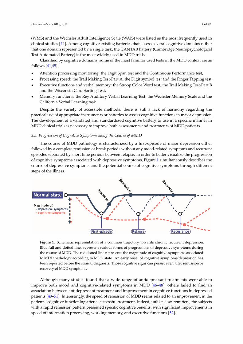
The course of MDD pathology is characterized by a first-episode of major depression eitherfollowed by a complete remission or break periods without any mood-related symptoms and recurrentepisodes separated by short time periods between relapse. In order to better visualize the progressionof cognitive symptoms associated with depressive symptoms, Figure 1 simultaneously describes thecourse of depressive symptoms and the potential course of cognitive symptoms through differentsteps of the illness.
Pharmaceuticals 2016, 9, 9 4 of 32
cognitive domains rather that one domain represented by a single task, the CANTAB battery
(Cambridge Neuropsychological Test Automated Battery) is the most widely used in MDD trials.
Classified by cognitive domains, some of the most familiar used tests in the MDD context are as
follows [41,45]:
x Attention processing monitoring: the Digit Span test and the Continuous Performance test,
x Processing speed: the Trail Making Test-Part A, the Digit symbol test and the Finger
Tapping test,
x Executive functions and verbal memory: the Stroop Color Word test, the Trail Making
Test-Part B and the Wisconsin Card Sorting Test,
x Memory functions: the Rey Auditory Verbal Learning Test, the Wechsler Memory Scale and
the California Verbal Learning task
Despite the variety of accessible methods, there is still a lack of harmony regarding the practical
use of appropriate instruments or batteries to assess cognitive functions in major depression. The
development of a validated and standardized cognitive battery to use in a specific manner in MDD
clinical trials is necessary to improve both assessments and treatments of MDD patients.
2.3. Progression of Cognitive Symptoms along the Course of MMD
The course of MDD pathology is characterized by a first-episode of major depression either
followed by a complete remission or break periods without any mood-related symptoms and
recurrent episodes separated by short time periods between relapse. In order to better visualize the
progression of cognitive symptoms associated with depressive symptoms, Figure 1 simultaneously
describes the course of depressive symptoms and the potential course of cognitive symptoms
through different steps of the illness.
Figure 1. Schematic representation of a common trajectory towards chronic recurrent depression.
Blue full and dotted lines represent various forms of progressions of depressive symptoms during
the course of MDD. The red dotted line represents the magnitude of cognitive symptoms associated
to MDD pathology according to MDD state. An early onset of cognitive symptoms depression has
been reported before the clinical diagnosis. Those cognitive signs can persist even after remission or
recovery of MDD symptoms.
Although many studies found that a wide range of antidepressant treatments were able to
improve both mood and cognitive-related symptoms in MDD [46–48], others failed to find an
association between antidepressant treatment and improvement in cognitive functions in depressed
patients [49–51]. Interestingly, the speed of remission of MDD seems related to an improvement in
the patients’ cognitive functioning after a successful treatment. Indeed, unlike slow-remitters, the
subjects with a rapid remission pattern presented specific cognitive benefits, with significant
improvements in speed of information processing, working memory, and executive functions [52].
Although description of cognitive alterations in the context of mood disorders has been
extensively analyzed in the literature [6,18,19,24,28,31,53], understanding why some cognitive
dysfunctions are still present in remitted or recurrent state of MDD and how specifically progress
Figure 1. Schematic representation of a common trajectory towards chronic recurrent depression.Blue full and dotted lines represent various forms of progressions of depressive symptoms duringthe course of MDD. The red dotted line represents the magnitude of cognitive symptoms associatedto MDD pathology according to MDD state. An early onset of cognitive symptoms depression hasbeen reported before the clinical diagnosis. Those cognitive signs can persist even after remission orrecovery of MDD symptoms.
Although many studies found that a wide range of antidepressant treatments were able toimprove both mood and cognitive-related symptoms in MDD [46–48], others failed to find anassociation between antidepressant treatment and improvement in cognitive functions in depressedpatients [49–51]. Interestingly, the speed of remission of MDD seems related to an improvement in thepatients’ cognitive functioning after a successful treatment. Indeed, unlike slow-remitters, the subjectswith a rapid remission pattern presented specific cognitive benefits, with significant improvements inspeed of information processing, working memory, and executive functions [52].
Pharmaceuticals 2016, 9, 9 5 of 42
Although description of cognitive alterations in the context of mood disorders has been extensivelyanalyzed in the literature [6,18,19,24,28,31,53], understanding why some cognitive dysfunctions arestill present in remitted or recurrent state of MDD and how specifically progress these deficits remainsunanswered. Indeed, recent reviews, focused on cognitive functioning in the remitted state ofdepression, confirmed that cognitive deficits persist in depressed patients despite the stabilizationof mood-related symptoms [5,54]. These deficits seem to reside more within the cognitive domainsof attention and executive functions (mental flexibility, decision-speed) than within other domains,although studies in additional patient cohorts are needed to strengthen this statement. The involvementof many factors associated to the progression of MDD pathology (number, duration and severityof MDD episodes) and other clinical features (age of onset, time elapsed since the last episode ofdepression, treatment interventions, sex or co-morbid psychiatric disorders) make the identification ofa specific neurocognitive profile in remitted patients even more complex [5].
Although many studies found that a wide range of antidepressant treatments were able toimprove both mood and cognitive-related symptoms in MDD [46–48], others failed to found anassociation between antidepressant treatment and improvement in cognitive functions in depressedpatients [49–51].
The proportion of MDD patients that encountered recurrence episodes is estimated at50%–60% [55]. Cognitive symptoms in recurrent depressive disorder (rDD) represent the largest rangeof cognitive impairments during the MDD pathology with decrements in memory, learning, attention,spatial visualization, visual-motor coordination, verbal fluency and memory, psychomotor retardationand most of the executive functions (planning, problem solving, behavioral inhibition, mentalflexibility) [56–58]. A way to examine the degree of cognitive deficits in recurrent depressive disordersis to gather studies that compare first-MDD episode cognitive symptoms to recurrent depressivecognitive symptoms. A recent study evaluated the following cognitive functions: informationprocessing speed (Digit Symbol from WAIS-R), executive functions and working memory (TMT, Strooptest), verbal memory (immediate and delayed memory) and learning ability (CVLT), and verbal fluency(VFT) in first-MDD episode and in recurrent episode patients [58]. Although no difference was foundin the severity of the depressive symptoms between first-episode and rDD individuals, the patientsfrom the latter group recorded significantly lower scores in all the cognitive tests. These findingsare in line with a previous report, in which recurrent patients exhibited worse performances inall the tests (executive functions and perseverative tendency) compared to controls subjects [59].Moreover, the number of recurrent episodes seems correlated with the severity of perseverativebehavior. The cumulative duration of depressive episodes and their repetition have a detrimentaleffect on the severity of the associated-cognitive deficits.
Otherwise, evidence suggesting that rDD was associated with negative biases and increasedsensitivity to negative events. While first-episode MDD patients can show an impairment in emotionalcognitive function [31,32], rDD subjects displayed a deficit in identifying facial expression at a moresevere degree, even demonstrating excitation and exaggeration for sad emotions [60]. Thus, neuralmechanisms involved in the perception of negative or happy/neutral faces may differ, confirmingthe abnormal neural processing of emotional stimuli in depressed patients all along the course ofthe disease.
Taken together, rDD are characterized by a proportional aggravation of cognitive disturbancescompared to first-episode MDD (Figure 1). This progressive decline of cognitive functioning throughMDD illness may contribute to the failure to reach a full recovery in patients and considerably widenstheir risks of developing over time dementia and neurodegenerative disorders.
Many questions about cognitive deficits associated with MDD pathology remain unanswered:could cognitive impairments be a premature behavioral marker of susceptibility to depression and beused as a prevention tool? Are residual cognitive dysfunctions in remitted patients a risk factor forrelapse? New discoveries in this field could significantly improve therapeutic care in MDD patients.Relieving cognitive dysfunctions may be a necessarily step to treat major depression.
Pharmaceuticals 2016, 9, 9 6 of 42
2.4. Cognitive Dysfunctions According to the MDD Subtypes
Most of clinical studies investigating cognitive dysfunctions in MDD patients did not detailthe heterogeneous condition of depression symptomatology. Melancholia and atypical disordersare the main subtypes in depression. Specifically, melancholic depression can be distinguish fromnon-melancholia by a set of physical symptoms occurring during clinical depressive episodes suchas anhedonia and/or lack of reactivity to usually pleasurable stimuli, observable psychomotorretardation, weight loss, worsening of symptoms in the morning hours and early morning awaking(DSM-V, [1,61,62]). On the other hand, atypical subtype is rather characterized by mood reactivityand positivity, a younger onset of age, less severe and fewer depressive episodes, longer duration ofepisodes, hypersomnia, weight gain and more co-morbidity with anxiety and substance abuse [1,63].In addition to the DSM-V definition, distinct biological correlates differentiate melancholic subtypefrom others subtypes of depression. Melancholic depression is associated with HPA-axis hyperactivity(hypercortisolemia) and specific sleep patterns (reduced REM latency, increased REM time and reduceddeep sleep) [64,65].
After two decades of research, increasing evidence suggests that melancholic patients exhibitgreater cognitive dysfunctions than non-melancholic patients but no qualitative review focusing oncognitive impairments in melancholic subtype has been written yet. Table 1 summarizes clinical studiesevaluating cognitive functions within melancholic patients through various neuropsychological tests.Classical clinical assessments to identify melancholic patients in these studies were determined byDSM-IV or DSM-V criteria for melancholic features Checklist (DSM-IV) and the CORE index formelancholia by behavioral observation. The CORE index was developed in 2007 when observablepsychomotor disturbance was judged as the most relevant clinical markers of melancholic depressionand psychomotor retardation was announced as the “core” behavioral pattern defining melancholicMDD [61,66]. This assessment was preceded by a 17-item HAM-D evaluation and, occasionally, theMini Mental State Examination (MMSE) was employed as a screening tool to exclude potential subjectswith early onset dementia.
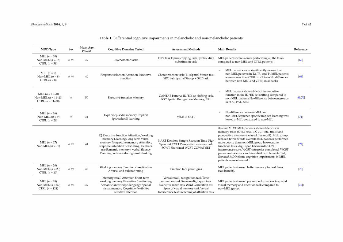
Pharmaceuticals 2016, 9, 9 7 of 42
Table 1. Differential cognitive impairments in melancholic and non-melancholic patients.
MDD Type Sex Mean Age(Years) Cognitive Domains Tested Assessment Methods Main Results Reference
MEL (n = 20)Non-MEL (n = 18)
CTRL (n = 38)⇢/⇡ 39 Psychomotor tasks Fitt’s task Figure-copying task Symbol digit
substitution taskMEL patients were slower performing all the taskscompared to non-MEL and CTRL patients. [67]
MEL (n = 7)Non-MEL (n = 8)
CTRL (n = 8)⇢/⇡ 40 Response selection Attention Executive
functionChoice reaction task (T1) Spatial Stroop task
SRC task Spatial Stroop + SRC task
- MEL patients were significantly slower thannon-MEL patients in T2, T3, and T4.MEL patientswere slower than CTRL in all tasksNo differencebetween non-MEL and CTRL in all tasks
[68]
MEL (n = 11-20)Non-MEL (n = 11–20)
CTRL (n = 11–20)⇡ 50 Executive function Memory CANTAB battery: ID/ED set shifting task,
SOC Spatial Recognition Memory, PAL
- MEL patients showed deficit in executivefunction in the ID/ED set shifting compared tonon-MEL patients;No difference between groupsin SOC, PAL, SRC
[69,70]
MEL (n = 26)Non-MEL (n = 9)
CTRL (n = 26)⇡ 34 Explicit episodic memory Implicit
(procedural) learning WMS-R SRTT- No difference between MEL and
non-MELSequence-specific implicit learning waslower in MEL compared to non-MEL
[71]
MEL (n = 17)Non-MEL (n = 17) ⇢/⇡ 41
IQ Executive function Attention/workingmemory Learning/long-term verbal
memory Prospective memory Attention,response inhibition Set shifting, feedback
use Semantic memory/ verbal fluencyPlanning, self-monitoring, multi-tasking
NART Donders Simple Reaction Time DigitSpan test CVLT Prospective memory task
SCWT Shortened WCST COWAT SET
Baseline MDD: MEL patients showed deficits inmemory tasks (CVLT trial 1, CVLT total trials) andprospective memory (delayed free recall). MEL grouprecalled fewer words overall; MEL patients performedmore poorly than non-MEL group in executivefunctions tests: digit span backwards, SCWTinterference score, WCST categories completed, WCSTperseverative errors and modified Six Elements Test;Remitted MDD: Same cognitive impairments in MELpatients were observed.
[72]
MEL (n = 20)Non-MEL (n = 20)
CTRL (n = 20)⇢/⇡ 47 Working memory Emotion classification
Arousal and valence rating Emotion face paradigms MEL patients showed better memory for sad faces(sad benefit). [73]
MEL (n = 65)Non-MEL (n = 59)
CTRL (n = 124)⇢/⇡ 39
Memory recall Attention Short-termworking memory Executive functioningSemantic knowledge, language Spatial
visual memory Cognitive flexibility,selective attention
Verbal recall, recognition task Timeestimation task Reverse digit span task
Executive maze task Word Generation testSpan of visual memory task Verbal
Interference test Switching of attention task
MEL patients showed poorer performances in spatialvisual memory and attention task compared tonon-MEL group.
[74])
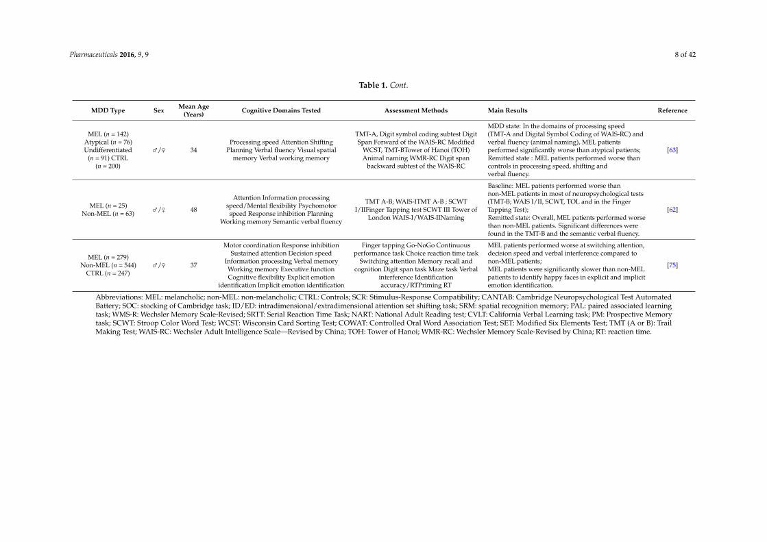
Pharmaceuticals 2016, 9, 9 8 of 42
Table 1. Cont.
MDD Type Sex Mean Age(Years) Cognitive Domains Tested Assessment Methods Main Results Reference
MEL (n = 142)Atypical (n = 76)Undifferentiated
(n = 91) CTRL(n = 200)
⇢/⇡ 34Processing speed Attention Shifting
Planning Verbal fluency Visual spatialmemory Verbal working memory
TMT-A, Digit symbol coding subtest DigitSpan Forward of the WAIS-RC Modified
WCST, TMT-BTower of Hanoi (TOH)Animal naming WMR-RC Digit span
backward subtest of the WAIS-RC
MDD state: In the domains of processing speed(TMT-A and Digital Symbol Coding of WAIS-RC) andverbal fluency (animal naming), MEL patientsperformed significantly worse than atypical patients;Remitted state : MEL patients performed worse thancontrols in processing speed, shifting andverbal fluency.
[63]
MEL (n = 25)Non-MEL (n = 63) ⇢/⇡ 48
Attention Information processingspeed/Mental flexibility Psychomotorspeed Response inhibition Planning
Working memory Semantic verbal fluency
TMT A-B; WAIS-ITMT A-B ; SCWTI/IIFinger Tapping test SCWT III Tower of
London WAIS-I/WAIS-IINaming
Baseline: MEL patients performed worse thannon-MEL patients in most of neuropsychological tests(TMT-B; WAIS I/II, SCWT, TOL and in the FingerTapping Test);Remitted state: Overall, MEL patients performed worsethan non-MEL patients. Significant differences werefound in the TMT-B and the semantic verbal fluency.
[62]
MEL (n = 279)Non-MEL (n = 544)
CTRL (n = 247)⇢/⇡ 37
Motor coordination Response inhibitionSustained attention Decision speed
Information processing Verbal memoryWorking memory Executive functionCognitive flexibility Explicit emotion
identification Implicit emotion identification
Finger tapping Go-NoGo Continuousperformance task Choice reaction time task
Switching attention Memory recall andcognition Digit span task Maze task Verbal
interference Identificationaccuracy/RTPriming RT
MEL patients performed worse at switching attention,decision speed and verbal interference compared tonon-MEL patients;MEL patients were significantly slower than non-MELpatients to identify happy faces in explicit and implicitemotion identification.
[75]
Abbreviations: MEL: melancholic; non-MEL: non-melancholic; CTRL: Controls; SCR: Stimulus-Response Compatibility; CANTAB: Cambridge Neuropsychological Test AutomatedBattery; SOC: stocking of Cambridge task; ID/ED: intradimensional/extradimensional attention set shifting task; SRM: spatial recognition memory; PAL: paired associated learningtask; WMS-R: Wechsler Memory Scale-Revised; SRTT: Serial Reaction Time Task; NART: National Adult Reading test; CVLT: California Verbal Learning task; PM: Prospective Memorytask; SCWT: Stroop Color Word Test; WCST: Wisconsin Card Sorting Test; COWAT: Controlled Oral Word Association Test; SET: Modified Six Elements Test; TMT (A or B): TrailMaking Test; WAIS-RC: Wechsler Adult Intelligence Scale—Revised by China; TOH: Tower of Hanoi; WMR-RC: Wechsler Memory Scale-Revised by China; RT: reaction time.
Pharmaceuticals 2016, 9, 9 9 of 42
Conducted in small cohorts of patients, the oldest studies mainly focused on psychomotor andmotor coordination performances such as Finger tapping task, fast walk, finger-thumb apposition,hands movements, foot tapping, finger-copying task or reaction time tasks. In accordance to theCORE concept, psychomotor performances were diminished in melancholic patients compared tonon-melancholic patients [67,68,76].
The major finding emerging from the variety of cognitive domains tested is that allstudies assessing attentional processes and more globally executive functions invariably showeddeficits in one or several associated tasks in melancholic patients compared to non-melancholicpatients [62,63,68,70–72,74,75]. These specific impairments are already a key component in melancholicprofile. It also appears that the differentiation between melancholic and non-melancholic groups relieson increased task difficulty. Indeed, simple reaction task [68], spatial recognition memory [69,70],explicit episodic memory [71], semantic memory [72], memory recall or working memory [74,75] weresimilar across depression subtypes. In contrast, complex tasks that required set-shifting [62,69,70],attentional processes [62,68,71,72,74], cognitive flexibility [72,75], planning [62], decision-speed [63,75]distinguish melancholic and non-melancholic MDD patients.
As mentioned previously, cognitive abnormalities in MDD patients can persist even afterclinical remission [5,54,77,78]. So far, only two studies compared cognitive performances of MDDpatients with or without melancholic features longitudinally, from admission to recovery [62,72].At baseline, both investigations agreed that cognitive performances in melancholic were impaired inmost of the cognitive domains tested (attentional processes, information processing, psychomotorspeed, mental flexibility, learning verbal memory, planning) compared to non-melancholic subtype.Interestingly, these memory and executive dysfunctions in melancholic patients were maintaineddespite the remitted state of patients. In contrast, some of cognitive disturbances observed innon-melancholic patients were improved after remission [62,72]. The persistence of some cognitivedysfunctions, mostly reported in melancholic patients, could represent a marker of a distinct depressivesubtype instead of being secondary to the severity of depression. Further investigations are needed todetermine why melancholic patients require a longer period of recovery in order for their cognitivefunctions to be restored to control levels. Only one study investigated cognitive deficit in the threedifferent MDD subtypes defined by DSM-V: melancholic, atypical and undifferentiated patients.Major depressive subtypes displayed similar cognitive deficits in most of the tested domains butdiffered in some cognitive tasks. Melancholic patients performed significantly worse in domainssuch as processing speed (TMT-A, Digit symbol coding subtest) and verbal fluency (animal naming)compared to atypical and undifferentiated patients [63]. After remission from depression, while all thesubtypes could recover their visual spatial memory and verbal working memory, clinically remittedmelancholic patients exhibited specific deficits in processing speed, set-shifting measures and verbalfluency, in line with previously discussed results [62,72], suggesting once again a distinct profile inneurocognitive performance in baseline and remitted MDD, depending on the subtype.
There is substantial evidence showing that perception in patients with MDD is characterizedby blunted responsiveness to emotionally positive information as well as an increased tendency toperceive emotionally neutral visual information as negative [79]. However, relatively few studieshave used neurobehavioral measures to examine emotional disturbances and loss of positive affectin melancholic MDD population. When melancholic subjects were asked to classify emotional facespreviously encountered, they showed either a better memory for sad faces [73] or significantly moretime at explicitly identifying happy faces [75] compared to non-melancholic patients. These findingssupport that melancholic patients display a hypersensitivity to sad stimuli, reflected in a greatertendency to recall or identify sad expressions. Perceptual emotion biases appear to be specific inmelancholic patients and could open up the debate about a possible primary neurocognitive featureof melancholic depression instead of a consequence of symptomatic mood. Further longitudinalneuroimaging studies combined with cognitive measures in melancholic patients could provide newinsights in this field.
Pharmaceuticals 2016, 9, 9 10 of 42
Taken together, the majority of studies agreed on both generalized and specific cognitiveimpairments in the melancholic subtype, characterized by a greater degree of these alterationscompared to non-melancholic patients. Specifically, melancholic patients were distinguished byspecific impairments in decision speed and positive emotion processing [63,73–75]. Moreover, giventhat most of cognitive deficits persist in remitted state in melancholic patients, the debate about therehabilitation process of this type of depressive patients warrants an important focus.
Although subtyping depressive patients can be a useful tool in identifying specific profiles indepressive symptomatology, its clinical utility regarding predictive antidepressant drug efficacy mustbe hampered by recent exploratory [80] or translational research [81] investigations.
Even though, anhedonia and dysfunctional reward processing are unspecific to MDD, andmay be observable in other psychiatric disorders [82], this is one of two required symptoms fora diagnosis of MDD [1]. This decrease in engagement in rewarding activities may be related tothe complex disturbances in the emotional, motivational and cognitive domains that are associatedwith depression. Changes in reward-motivated learning in depression may exacerbate the clinicalsymptoms, or delay recovery. MDD and especially MDD-anhedonic subjects can display deficitsin motivational processing and dysregulation of the brain's reward system [83]. Studies in MDDsubjects showed a deficit in establishing a reward bias [84], and in reward-related decision-making [85].If the assessment of “motivational anhedonia” is improved, it could help patient making behavioralchoices that are likely to increase exposure to positively reinforcing experiences. Indeed, alterations inreward-related learning failed to appropriately guide or motivate subsequent behavior. These formsof incentive learning depend on several factors, including reward processing, motivation and theneuroplasticity underlying formation of new or strengthened memories. However, anhedonia is aparticularly difficult symptom to treat, especially with current first-line pharmacotherapies (e.g., SSRIs),that do not adequately address motivational anhedonia in depression ([86] for review). If little isknown about reward-motivated learning in animal models, a recent study using chronic exposure tocorticosterone-induced depression-like behavior in Rat produced lasting deficits in the acquisitionof reward-related learning tested on a food-motivated instrumental task [87]. Interestingly, inthe same study, authors showed that amitriptyline increased instrumental performance comparedin corticosterone-exposed rats. This may suggest that chronic exposure to amitriptyline per sefacilitates this form of reward-related learning. The interrelationship between specific components ofmotivational dysfunction and specific cognitive systems in the context of depression remains to bestudied in order to allow for a greater understanding of mood disorders [88].
3. Cognitive Behavioral Paradigms Used to Assess Learning and Memory Performances inRodent Models of MDD
Cognition is a complex brain function that involves processes including attention, processingspeed, learning and memory, working memory, verbal fluency and executive functions. However, someof these cognitive domains cannot be modeled in Rodents because of the higher cognitivedemands specific to Humans, particularly in executive functions such as planning, problem solving,multi-tasking or decision-making and verbal fluency.
Collective efforts from preclinical researchers and clinicians are currently in progress to improvethe translation of fundamental research tools into clinical application in Psychiatry. For example,the Cognitive Neuroscience Treatment Research to Improve Cognition in Schizophrenia (CNTRICS)initiative was introduced to identify tasks with construct validity across species to improve the“translational-ity” of behavioral tests from rodents to non-human primates and humans [89,90].More recently, the Research Domain Criteria (RDoC) project, established by the National Instituteof Mental Health, propose a biologically-valid framework for the understanding mental disorders.This latter project relies on functional dimension of behavior based on genes, molecules, cells, circuits,physiology, behavior and self-reports, like the “Cognitive Systems” domain, and will allow a bettercrosstalk between clinical and preclinical studies [81,91]. In preclinical research, the development of
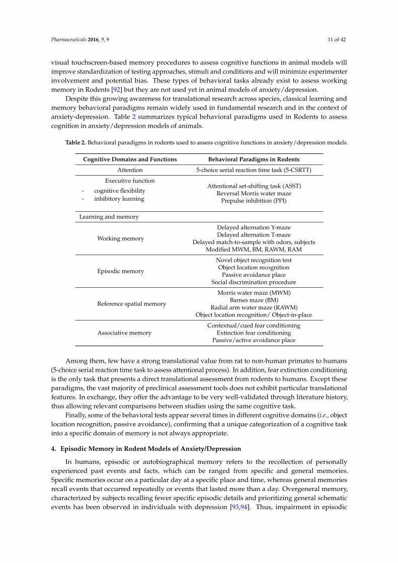
Pharmaceuticals 2016, 9, 9 11 of 42
visual touchscreen-based memory procedures to assess cognitive functions in animal models willimprove standardization of testing approaches, stimuli and conditions and will minimize experimenterinvolvement and potential bias. These types of behavioral tasks already exist to assess workingmemory in Rodents [92] but they are not used yet in animal models of anxiety/depression.
Despite this growing awareness for translational research across species, classical learning andmemory behavioral paradigms remain widely used in fundamental research and in the context ofanxiety-depression. Table 2 summarizes typical behavioral paradigms used in Rodents to assesscognition in anxiety/depression models of animals.
Table 2. Behavioral paradigms in rodents used to assess cognitive functions in anxiety/depression models.
Cognitive Domains and Functions Behavioral Paradigms in Rodents
Attention 5-choice serial reaction time task (5-CSRTT)
Executive function
- cognitive flexibility- inhibitory learning
Attentional set-shifting task (ASST)Reversal Morris water maze
Prepulse inhibition (PPI)
Learning and memory
Working memory
Delayed alternation Y-mazeDelayed alternation T-maze
Delayed match-to-sample with odors, subjectsModified MWM, BM, RAWM, RAM
Episodic memory
Novel object recognition testObject location recognition
Passive avoidance placeSocial discrimination procedure
Reference spatial memory
Morris water maze (MWM)Barnes maze (BM)
Radial arm water maze (RAWM)Object location recognition/ Object-in-place
Associative memoryContextual/cued fear conditioning
Extinction fear conditioningPassive/active avoidance place
Among them, few have a strong translational value from rat to non-human primates to humans(5-choice serial reaction time task to assess attentional process). In addition, fear extinction conditioningis the only task that presents a direct translational assessment from rodents to humans. Except theseparadigms, the vast majority of preclinical assessment tools does not exhibit particular translationalfeatures. In exchange, they offer the advantage to be very well-validated through literature history,thus allowing relevant comparisons between studies using the same cognitive task.
Finally, some of the behavioral tests appear several times in different cognitive domains (i.e., objectlocation recognition, passive avoidance), confirming that a unique categorization of a cognitive taskinto a specific domain of memory is not always appropriate.
4. Episodic Memory in Rodent Models of Anxiety/Depression
In humans, episodic or autobiographical memory refers to the recollection of personallyexperienced past events and facts, which can be ranged from specific and general memories.Specific memories occur on a particular day at a specific place and time, whereas general memoriesrecall events that occurred repeatedly or events that lasted more than a day. Overgeneral memory,characterized by subjects recalling fewer specific episodic details and prioritizing general schematicevents has been observed in individuals with depression [93,94]. Thus, impairment in episodic
Pharmaceuticals 2016, 9, 9 12 of 42
autobiographical memory has become a consistent feature in MDD patients. In addition, overgeneralautobiographical memory was found to be a predictor of the course of depression in a meta-analysis [95]and to be persistent in remitted or recurrent phases of MDD [96,97]. Finally, a recent work found thatchildren of mothers with a history of MDD during their child’s life, recalled less-specific memories,pointed out overgeneral memory as a cognitive vulnerability for depression [98]. However, the majorityof neuropsychological tests used to evaluate cognitive functions in depressed patients do not includeepisodic memory assessment.
Considering the conscious approach of episodic memory assessment in Humans, translationalresearch in Rodents required an adaptation in terminology, finally using “episodic-like” memoryto designate animal’s performances. Recognition memory tasks are widely used tools to assessepisodic-like memory in rodents. These behavioral paradigms rely on animal’s ability to determinewhether or not a stimulus has been previously encountered. The type of stimulus (nature of objects,location of objects, order of presentation) can vary to evaluate the various components of episodic-likememory (What? Where? When?). The spontaneous nature of this task, exploiting the curiosityof rodents and their propensity for novelty seeking, confers to recognition tests many advantages,especially in an anxiety-depression context. Unlike many others learning or memory paradigms, nopositive (food reward) or negative (punishment, water) reinforcements are needed, minimizing otherpsychological parameters that could influence performances during the task [99]. However, modelingMDD conditions in rodents induces some modifications in parameters such as anxiety, motivationand motor activity [15,100,101] that cannot be excluded for the interpretation of the performance inrecognition tasks. The involvement of these parameters is systematically corrected with a measure ofmotor activity either during the test itself (total ambulatory distance, number of crossings in the arena)or in other behavioral paradigms applied before memory assessment (rotarod, Open Field) [99,102].When the anxious state overly restricts exploration behavior, adaptations in the protocol test mustbe set up to re-establish a sufficient amount of exploration. For example, introducing one or severalhabituation periods (animals exploring the arena empty without objects) preceding the acquisitionand the retention period of the test can fix this issue [99].
4.1. WHAT?
Many preclinical studies have investigated the performances of anxiety-depression models onobject recognition memory (Table 3). The novel object recognition test (NORT), initially described byEnnaceur and Delacour [103] usually comprises an acquisition session with two identical objects anda retention session during which animals have to discriminate between a familiar object previouslyfaced and a recently introduced new object (different in size, shape, texture, colors). Overall, findingsdiffer strongly across and inside animal models [104]. Antenatal and early post-natal chronic stressprocedures (PNS, MS, MD) present the most conflicting findings when compared to chronic adultstress procedure (SD, UCMS and CORT models). Independently of the model, the main source ofheterogeneity in discrimination tasks arises from protocol variations across laboratories, such as thenature of objects (size, shape, color, texture), the discriminability of the stimulus (low, moderate orhigh degree), the time and episodes of familiarization, sessions/inter-trial interval durations andenvironmental conditions (light, noise, cage size, sawdust). Additional inevitable variables like species,strain or sex add even more variability across findings and differences inside each depression models,especially in prenatal stress and maternal separation/deprivation procedures.
Studies using prenatal stress or early-life stress procedures showed either impairing [105–113], orimproving [114,115] or no effects [105,106,108–110,114,116–118] on object recognition test. Despite athorough analysis of all the parameters characterizing studies presented in Table 3, no common featuresin pre/perinatal manipulations were observed.
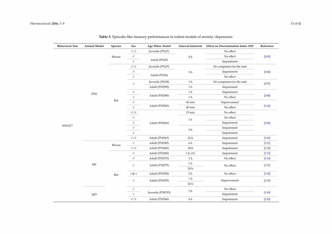
Pharmaceuticals 2016, 9, 9 13 of 42
Table 3. Episodic-like memory performances in rodent models of anxiety/depression.
Behavioral Test Animal Model Species Sex Age When Tested Interval Intertrial Effect on Discrimination Index (DI) Reference
WHAT?
PNS
Mouse
⇢/⇡ Juvenile (PN23)
4 h
No effect
[105]⇢Adult (PN45)
No effect
⇡ Impairment
Rat
⇢/⇡ Juvenile (PN23)
2 h
No competent for the task
[106]⇢Adult (PN56)
Impairment
⇡ No effect
⇡ Juvenile (PN28) 1 h No competent for the task [107]Adult (PND90) 1 h Impairment
⇢Adult (PND80)
1 h Impairment[108]⇡ 1 h No effect
⇢Adult (PND60)
40 min Improvement[114]⇡ 40 min No effect
⇢/⇡
Adult (PND63)
15 min No effect
[109]
⇢1 h
No effect
⇡ Impairment
⇢3 h
Impairment
⇡ Impairment
⇢/⇡ Adult (PND63) 24 h Impairment [110]
MS
Mouse⇡ Adult (PND85) 6 h Impairment [111]
⇢/⇡ Adult (PND60) 24 h Impairment [112]
Rat
⇢ Adult (PND60) 1 h, 4 h Impairment [113]
⇢ Adult (PND70) 1 h No effect [116]
⇢ Adult (PND75) 1 hNo effect [117]
24 h
⇢& ⇡ Adult (PND90) 2 h No effect [118]
⇡ Adult (PND55) 1 h Improvement [115]24 h
MD
⇢Juvenile (PND35) 1 h
No effect[119]⇡ Impairment
⇢/⇡ Adult (PND60) 4 h Impairment [120]
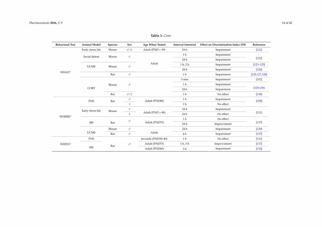
Pharmaceuticals 2016, 9, 9 14 of 42
Table 3. Cont.
Behavioral Test Animal Model Species Sex Age When Tested Interval Intertrial Effect on Discrimination Index (DI) Reference
WHAT?
Early stress life Mouse ⇢/⇡ Adult (PND > 90) 24 h Impairment [121]
Social defeat Mouse ⇢
Adult
1 h Impairment[122]24 h Impairment
UCMS Mouse ⇢ 1 h, 2 h Impairment [123–125]
24 h Impairment [126]
Rat ⇢ 1 h Impairment [125,127,128]
CORTMouse ⇢
5 min Impairment [102]
1 h Impairment[129,130]24 h Impairment
Rat ⇢/⇡ 1 h No effect [118]
WHERE?
PNS Rat⇢ Adult (PND80) 1 h Impairment [108]⇡ 1 h No effect
Early stress life Mouse⇢
Adult (PND > 90)24 h Impairment
[121]⇡ 24 h No effect
MS Rat ⇢ Adult (PND75)1 h No effect
[117]24 h Improvement
UCMSMouse ⇢
Adult24 h Impairment [126]
Rat ⇢ 4 h Impairment [127]
WHEN?
PNS
Rat ⇢Juvenile (PND30–40) 1 h No effect [131]
MSAdult (PND75) 1 h, 3 h Improvement [117]
Adult (PND60) 3 h Impairment [132]
Pharmaceuticals 2016, 9, 9 15 of 42
On the contrary, object recognition memory is predominantly impaired in chronic stressprocedures applied during adulthood, such as social defeat, UCMS or the CORT model. This alterationis indicated by a diminution of the discrimination index (DI) [102,122–125,129,130], regardless ofthe duration of the inter-trial delay. One of these studies assessing long-lasting effects of UCMSprocedure showed that anxio-depressed animals still displayed a discrimination deficit one monthafter the end of the stress treatment [125], suggesting that episodic-like alterations persist even after thecessation of the stress. These findings are in line with episodic memory deficits remaining in remittedor recurrent MDD patients [96,97]. A single study performed in rats disproves this impairment in theCORT model observing no effect in anxio-depressed animals [118]. Besides a likely species-dependentperformance, the 2-week corticosterone protocol used in this study compared to a 4-week one in othersstudies [102,129,130] could explain in this case the lack of effect on recognition memory.
4.2. Where and When?
Although the novel object recognition test is widely used in laboratories, place recognition(the “Where” component) and temporal ordering (the “When” component) tasks can help to refinethe degree of evaluation of episodic-like memory. The place recognition task examines the animal’sability to discriminate between an object that was moved during the delay between the acquisitionand the retention phase and an object that remained stationary, adding a spatial component to the task.On the other hand, temporal ordering task consists in presenting a set of two identical objects during afirst acquisition session. After a defined interval, another set of two new identical objects is presentedto animals for exploration. During the retention session, animals have to discriminate between the“old object” from the first acquisition session and the “recent” object from the 2nd session. An intacttemporal order memory is observed when an animal has spent more time with the “old” object [99].
The same pattern emerges from place recognition memory compared to object recognition withconflicting pre/peri-natal findings (impaired: [108,121]; improved: [117]; no effect: [108,117,121]) versusadult chronic stress procedures (impaired: [126,127]). In this type of recognition, female animals acrossmodels seem to be less vulnerable than male animals [108,121].
Very few studies evaluated the “when?” component of episodic-like memory via the temporalordering task. Only works in maternal separation were performed, once again presenting discrepanciesin the results. One study showed no effect of MS procedure on temporal order memory [133], whileanother one found an impairment [132]. At this time, there is not enough information available in thestudy of place recognition and temporal ordering tasks to clarify the impact of anxiety-depressionmodels on these episodic-related tests.
Independently of the nature of task, antenatal and early post-natal procedures were theonly models in which an improvement in episodic-like performances was observed [114,115,117].For example, Makena et al. [117] showed that object location and temporal ordering task performanceswere enhanced in maternally separated rats, suggesting that perinatal manipulations can positivelymodulate brain development and neuro-adaptative responses to a stimulus, influencing the animalbehavior reactivity as adults. Furthermore, post-natal manipulations have been reported to increasethe intensity of maternal care (during reunion after a given period of dams-pups separation) ofmaternally-separated pups after a brief separation [134,135], and to provide greater skills to adapt topsychological and physiological stressors in adulthood [115,136].
Taken together, except an overall impairment in all discrimination tasks observed in adult chronicstress procedures, results are too inconsistent in pre/perinatal manipulations to extract relevantadvances in the area of episodic-like memory. Further investigations focusing on place and temporalrecognition may contribute to progress in the discovery of fundamental mechanisms underlyingepisodic-like memory in rodents in a pathological context. Finally, efforts should be intended tointegrate episodic memory in clinical neuropsychological tests performed in MDD patients to fullyassess cognitive functions.
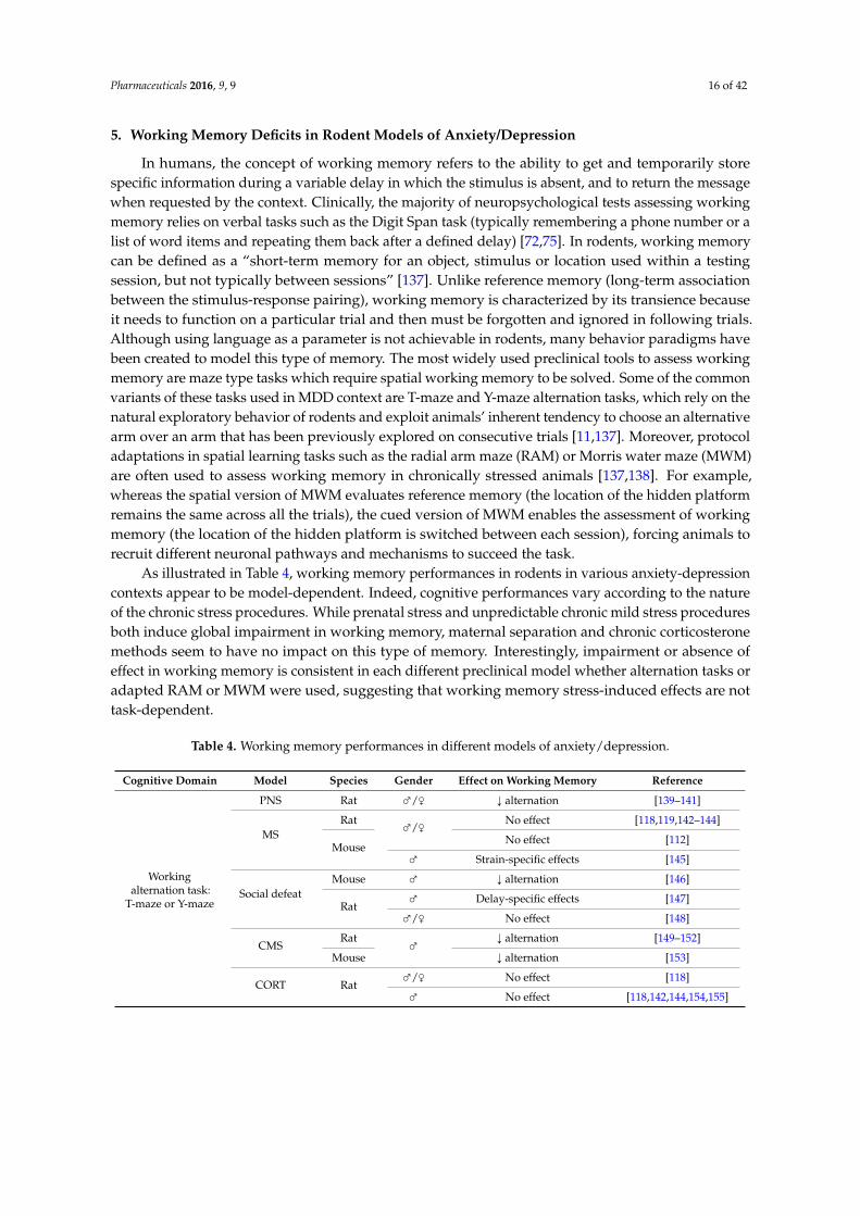
Pharmaceuticals 2016, 9, 9 16 of 42
5. Working Memory Deficits in Rodent Models of Anxiety/Depression
In humans, the concept of working memory refers to the ability to get and temporarily storespecific information during a variable delay in which the stimulus is absent, and to return the messagewhen requested by the context. Clinically, the majority of neuropsychological tests assessing workingmemory relies on verbal tasks such as the Digit Span task (typically remembering a phone number or alist of word items and repeating them back after a defined delay) [72,75]. In rodents, working memorycan be defined as a “short-term memory for an object, stimulus or location used within a testingsession, but not typically between sessions” [137]. Unlike reference memory (long-term associationbetween the stimulus-response pairing), working memory is characterized by its transience becauseit needs to function on a particular trial and then must be forgotten and ignored in following trials.Although using language as a parameter is not achievable in rodents, many behavior paradigms havebeen created to model this type of memory. The most widely used preclinical tools to assess workingmemory are maze type tasks which require spatial working memory to be solved. Some of the commonvariants of these tasks used in MDD context are T-maze and Y-maze alternation tasks, which rely on thenatural exploratory behavior of rodents and exploit animals’ inherent tendency to choose an alternativearm over an arm that has been previously explored on consecutive trials [11,137]. Moreover, protocoladaptations in spatial learning tasks such as the radial arm maze (RAM) or Morris water maze (MWM)are often used to assess working memory in chronically stressed animals [137,138]. For example,whereas the spatial version of MWM evaluates reference memory (the location of the hidden platformremains the same across all the trials), the cued version of MWM enables the assessment of workingmemory (the location of the hidden platform is switched between each session), forcing animals torecruit different neuronal pathways and mechanisms to succeed the task.
As illustrated in Table 4, working memory performances in rodents in various anxiety-depressioncontexts appear to be model-dependent. Indeed, cognitive performances vary according to the natureof the chronic stress procedures. While prenatal stress and unpredictable chronic mild stress proceduresboth induce global impairment in working memory, maternal separation and chronic corticosteronemethods seem to have no impact on this type of memory. Interestingly, impairment or absence ofeffect in working memory is consistent in each different preclinical model whether alternation tasks oradapted RAM or MWM were used, suggesting that working memory stress-induced effects are nottask-dependent.
Table 4. Working memory performances in different models of anxiety/depression.
Cognitive Domain Model Species Gender Effect on Working Memory Reference
Workingalternation task:
T-maze or Y-maze
PNS Rat ⇢/⇡ Ó alternation [139–141]
MSRat ⇢/⇡ No effect [118,119,142–144]
MouseNo effect [112]
⇢ Strain-specific effects [145]
Social defeatMouse ⇢ Ó alternation [146]
Rat⇢ Delay-specific effects [147]
⇢/⇡ No effect [148]
CMSRat ⇢ Ó alternation [149–152]
Mouse Ó alternation [153]
CORT Rat⇢/⇡ No effect [118]
⇢ No effect [118,142,144,154,155]
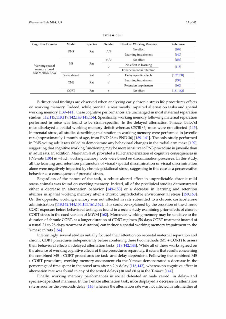
Pharmaceuticals 2016, 9, 9 17 of 42
Table 4. Cont.
Cognitive Domain Model Species Gender Effect on Working Memory Reference
Working spatialmemory: cued
MWM/BM/RAW
PNS Rat ⇢/⇡ No effect [109]
Learning impairment [140]
MS Rat⇢/⇡ No effect [156]
⇡ No effect in learning [115]Enhancement in retention
Social defeat Rat ⇢ Delay-specific effects [157,158]
CMS Rat ⇢ Learning impairment [159]
Retention impairment [160]
CORT Rat ⇢ No effect [161,162]
Bidirectional findings are observed when analyzing early chronic stress life procedures effectson working memory. Indeed, while prenatal stress mostly impaired alternation tasks and spatialworking memory [139–141], these cognitive performances are unchanged in most maternal separationstudies [112,115,118,119,142,143,145,156]. Specifically, working memory following maternal separationperformed in mice was found to be strain-specific. In the delayed alternation T-maze, Balb/cJmice displayed a spatial working memory deficit whereas C57Bl/6J mice were not affected [145].In prenatal stress, all studies describing an alteration in working memory were performed in juvenilerats (approximately 1 month of age, from PND 26 to PND 36) [139–141]. The only study performedin PNS-young adult rats failed to demonstrate any behavioral changes in the radial-arm maze [109],suggesting that cognitive working functioning may be more sensitive to PNS procedure in juvenile thanin adult rats. In addition, Markham et al. provided a full characterization of cognitive consequences inPNS-rats [106] in which working memory tools were based on discrimination processes. In this study,all the learning and retention parameters of visual/spatial discrimination or visual discriminationalone were negatively impacted by chronic gestational stress, suggesting in this case as a perseverativebehavior as a consequence of prenatal stress.
Regardless of the nature of the task, a robust altered effect in unpredictable chronic mildstress animals was found on working memory. Indeed, all of the preclinical studies demonstratedeither a decrease in alternation behavior [149–153] or a decrease in learning and retentionabilities in spatial working memory after a chronic unpredictable environmental stress [159,160].On the opposite, working memory was not affected in rats submitted to a chronic corticosteroneadministration [118,142,144,154,155,161,162]. This could be explained by the cessation of the chronicCORT exposure before behavioral testing, as found in a recent study examining prior effects of chronicCORT stress in the cued version of MWM [162]. Moreover, working memory may be sensitive to theduration of chronic CORT, as a longer duration of CORT regimen (56-days CORT treatment instead ofa usual 21 to 28 days treatment duration) can induce a spatial working memory impairment in theY-maze in rats [154].
Interestingly, several studies initially focused their attention on neonatal maternal separation andchronic CORT procedures independently before combining these two methods (MS + CORT) to assesstheir behavioral effects in delayed alternation tasks [118,142,144]. While all of these works agreed onthe absence of working cognitive effects of these procedures separately, it seems that results concerningthe combined MS + CORT procedures are task- and delay-dependent. Following the combined MS+ CORT procedure, working memory assessment via the Y-maze demonstrated a decrease in thepercentage of time spent in the novel arm after a 2 h-delay [118,142], whereas no cognitive effect inalternation rate was found in any of the tested delays (30 and 60 s) in the T-maze [144].
Finally, working memory performances in social defeated animals varied, in delay- andspecies-dependent manners. In the T-maze alternation task, mice displayed a decrease in alternationrate as soon as the 5-seconds delay [146] whereas the alternation rate was not affected in rats, neither at
Pharmaceuticals 2016, 9, 9 18 of 42
10 or 30 or 60 s delays [147,148]. The delay must rise to 90 s to induce a deficit in working memory inadult defeated-rats compared to controls [147]. This observation supports that: (1) the longer the delaybetween sessions, the better the probability to forget the information; and (2) chronic social stressedmice are more vulnerable than defeated-rats in working memory tasks.
Taken together, chronic environmental stress procedures lead to a decline in working memorywhether it was applied during gestational or adulthood periods. On the contrary, early maternalseparation or chronic corticosterone regimen do not cause any damaging effects separately,but demonstrated conflicting delay-dependent effects when associated. Socially-defeated animalsdisplayed species and delayed-dependent behaviors in working memory. Other non-spatial behavioralparadigms evaluating working memory with operant tools are currently in development such asDelayed Match to Sample, Delayed non-Match to Sample Delayed [137], but so far, no reports werefound in an anxiety-depression context.
6. Attention and Executive Functions in Rodent Models of Anxiety/Depression
Attention deficits are referenced as a diagnostic criterion in DSM-V [1] and are oftenreported in patients suffering from MDD, increasing the need to better understand mechanismsunderlying this cognitive process through rodent models. Attention processes can be dividedin three sub-domains including: the ability to sustain attention on a specific task over a longperiod of time (sustained attention), the ability to respond simultaneously to multiple tasks(divided attention) and the ability to focus on selective environmental information while ignoringdistractions (selective attention) [163]. However, a small amount of studies examined attentionalabilities in animal models of anxiety/depression (Table 5). In preclinical studies, one of the mostwidely used tasks measuring attention is the 5-choice serial reaction time task (5-CSRTT). Variations inthis protocol can be arranged in order to assess the three subtypes of attention in a single behaviorparadigm [163]. Briefly, this task consists in presenting five spatial different apertures to animals in anoperant chamber, with a food cup located on the opposite wall of the five location holes. The animalinitiates a trial by opening the food cup door. After a short delay, a visual signal consisting of a briefillumination of one of the five apertures is presented. If the animal nose pokes in the correspondinghole, a food pellet is delivered into the food cup and this action is counted as a correct response tothe test. Conducted through several trials, attention performance is usually measured by examiningwhen the animal responds to a correct or incorrect hole where the light appears (accuracy versusomissions) and the speed with which the animal responds (time reaction). Complexity in the taskcan be increased by varying the stimulus light or inter-trial interval duration for example, assessingin this case parameters such as premature or perseverative responses (nose poke that occurs priorto light presentation or repeated nose pokes in a previously lit aperture), giving further informationon impulsive behavior (see [164–166] for details in methodology). Among few available reportspresented in Table 5, all of them agreed about some deleterious effects of anxiety-depression onattention performances in the 5-CSRTT [167–170], increasing accordingly as cognitive challenges inthe task are getting more complex. For example, under standard 5-CSRTT conditions (fixed inter-trialinterval (ITI) and stimulus durations (SD)), there were few differences in performances between ratssubmitted to prenatal stress procedure and controls. However, when ITI and SD vary across trials,there was a decrease in accuracy and an increase in number of premature and timeout responses,respectively, indicating an impairment in sustained attention, increased impulsivity and cognitiveinflexibility in prenatal stressed rats [168,171].
Accumulating evidence suggests that unrestrained secretion of corticotropin-releasinghormone (CRH) can contribute to the apparition of depressive symptoms as well as cognitivedeficits ([172] for review). Interestingly, attentional abilities have been evaluated with the 5-CSRTT in agenetic mouse model based on overexpression of CRH throughout development. Disrupted attentionprocesses were observed once the task was acquired, as CRH transgenic mice showed a decrease in thepercentage of correct responses, a longer correct response latency and an increase in omitted responses
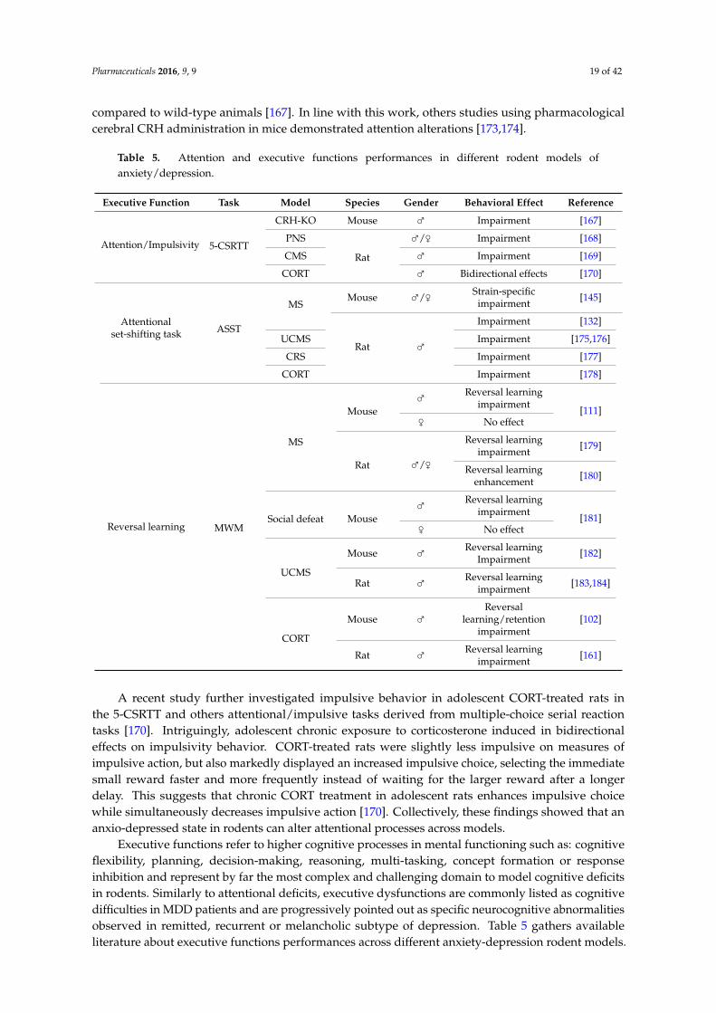
Pharmaceuticals 2016, 9, 9 19 of 42
compared to wild-type animals [167]. In line with this work, others studies using pharmacologicalcerebral CRH administration in mice demonstrated attention alterations [173,174].
Table 5. Attention and executive functions performances in different rodent models ofanxiety/depression.
Executive Function Task Model Species Gender Behavioral Effect Reference
Attention/Impulsivity 5-CSRTT
CRH-KO Mouse ⇢ Impairment [167]
PNS
Rat
⇢/⇡ Impairment [168]
CMS ⇢ Impairment [169]
CORT ⇢ Bidirectional effects [170]
Attentionalset-shifting task ASST
MSMouse ⇢/⇡ Strain-specific
impairment [145]
Rat ⇢
Impairment [132]
UCMS Impairment [175,176]
CRS Impairment [177]
CORT Impairment [178]
Reversal learning MWM
MS
Mouse⇢ Reversal learning
impairment [111]⇡ No effect
Rat ⇢/⇡
Reversal learningimpairment [179]
Reversal learningenhancement [180]
Social defeat Mouse⇢ Reversal learning
impairment [181]⇡ No effect
UCMS
Mouse ⇢ Reversal learningImpairment [182]
Rat ⇢ Reversal learningimpairment [183,184]
CORT
Mouse ⇢Reversal
learning/retentionimpairment
[102]
Rat ⇢ Reversal learningimpairment [161]
A recent study further investigated impulsive behavior in adolescent CORT-treated rats inthe 5-CSRTT and others attentional/impulsive tasks derived from multiple-choice serial reactiontasks [170]. Intriguingly, adolescent chronic exposure to corticosterone induced in bidirectionaleffects on impulsivity behavior. CORT-treated rats were slightly less impulsive on measures ofimpulsive action, but also markedly displayed an increased impulsive choice, selecting the immediatesmall reward faster and more frequently instead of waiting for the larger reward after a longerdelay. This suggests that chronic CORT treatment in adolescent rats enhances impulsive choicewhile simultaneously decreases impulsive action [170]. Collectively, these findings showed that ananxio-depressed state in rodents can alter attentional processes across models.
Executive functions refer to higher cognitive processes in mental functioning such as: cognitiveflexibility, planning, decision-making, reasoning, multi-tasking, concept formation or responseinhibition and represent by far the most complex and challenging domain to model cognitive deficitsin rodents. Similarly to attentional deficits, executive dysfunctions are commonly listed as cognitivedifficulties in MDD patients and are progressively pointed out as specific neurocognitive abnormalitiesobserved in remitted, recurrent or melancholic subtype of depression. Table 5 gathers availableliterature about executive functions performances across different anxiety-depression rodent models.
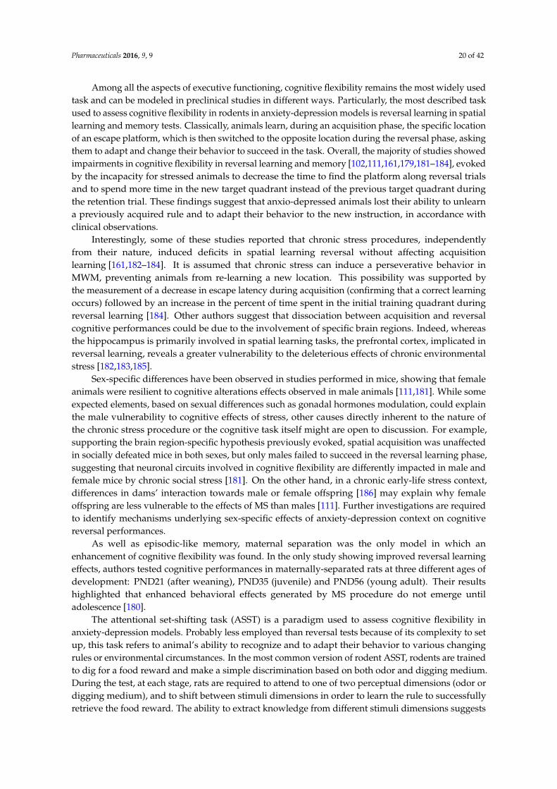
Pharmaceuticals 2016, 9, 9 20 of 42
Among all the aspects of executive functioning, cognitive flexibility remains the most widely usedtask and can be modeled in preclinical studies in different ways. Particularly, the most described taskused to assess cognitive flexibility in rodents in anxiety-depression models is reversal learning in spatiallearning and memory tests. Classically, animals learn, during an acquisition phase, the specific locationof an escape platform, which is then switched to the opposite location during the reversal phase, askingthem to adapt and change their behavior to succeed in the task. Overall, the majority of studies showedimpairments in cognitive flexibility in reversal learning and memory [102,111,161,179,181–184], evokedby the incapacity for stressed animals to decrease the time to find the platform along reversal trialsand to spend more time in the new target quadrant instead of the previous target quadrant duringthe retention trial. These findings suggest that anxio-depressed animals lost their ability to unlearna previously acquired rule and to adapt their behavior to the new instruction, in accordance withclinical observations.
Interestingly, some of these studies reported that chronic stress procedures, independentlyfrom their nature, induced deficits in spatial learning reversal without affecting acquisitionlearning [161,182–184]. It is assumed that chronic stress can induce a perseverative behavior inMWM, preventing animals from re-learning a new location. This possibility was supported bythe measurement of a decrease in escape latency during acquisition (confirming that a correct learningoccurs) followed by an increase in the percent of time spent in the initial training quadrant duringreversal learning [184]. Other authors suggest that dissociation between acquisition and reversalcognitive performances could be due to the involvement of specific brain regions. Indeed, whereasthe hippocampus is primarily involved in spatial learning tasks, the prefrontal cortex, implicated inreversal learning, reveals a greater vulnerability to the deleterious effects of chronic environmentalstress [182,183,185].
Sex-specific differences have been observed in studies performed in mice, showing that femaleanimals were resilient to cognitive alterations effects observed in male animals [111,181]. While someexpected elements, based on sexual differences such as gonadal hormones modulation, could explainthe male vulnerability to cognitive effects of stress, other causes directly inherent to the nature ofthe chronic stress procedure or the cognitive task itself might are open to discussion. For example,supporting the brain region-specific hypothesis previously evoked, spatial acquisition was unaffectedin socially defeated mice in both sexes, but only males failed to succeed in the reversal learning phase,suggesting that neuronal circuits involved in cognitive flexibility are differently impacted in male andfemale mice by chronic social stress [181]. On the other hand, in a chronic early-life stress context,differences in dams’ interaction towards male or female offspring [186] may explain why femaleoffspring are less vulnerable to the effects of MS than males [111]. Further investigations are requiredto identify mechanisms underlying sex-specific effects of anxiety-depression context on cognitivereversal performances.
As well as episodic-like memory, maternal separation was the only model in which anenhancement of cognitive flexibility was found. In the only study showing improved reversal learningeffects, authors tested cognitive performances in maternally-separated rats at three different ages ofdevelopment: PND21 (after weaning), PND35 (juvenile) and PND56 (young adult). Their resultshighlighted that enhanced behavioral effects generated by MS procedure do not emerge untiladolescence [180].
The attentional set-shifting task (ASST) is a paradigm used to assess cognitive flexibility inanxiety-depression models. Probably less employed than reversal tests because of its complexity to setup, this task refers to animal’s ability to recognize and to adapt their behavior to various changingrules or environmental circumstances. In the most common version of rodent ASST, rodents are trainedto dig for a food reward and make a simple discrimination based on both odor and digging medium.During the test, at each stage, rats are required to attend to one of two perceptual dimensions (odor ordigging medium), and to shift between stimuli dimensions in order to learn the rule to successfullyretrieve the food reward. The ability to extract knowledge from different stimuli dimensions suggests
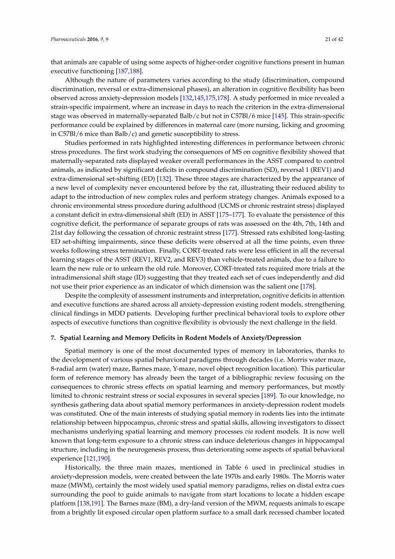
Pharmaceuticals 2016, 9, 9 21 of 42
that animals are capable of using some aspects of higher-order cognitive functions present in humanexecutive functioning [187,188].
Although the nature of parameters varies according to the study (discrimination, compounddiscrimination, reversal or extra-dimensional phases), an alteration in cognitive flexibility has beenobserved across anxiety-depression models [132,145,175,178]. A study performed in mice revealed astrain-specific impairment, where an increase in days to reach the criterion in the extra-dimensionalstage was observed in maternally-separated Balb/c but not in C57Bl/6 mice [145]. This strain-specificperformance could be explained by differences in maternal care (more nursing, licking and groomingin C57Bl/6 mice than Balb/c) and genetic susceptibility to stress.
Studies performed in rats highlighted interesting differences in performance between chronicstress procedures. The first work studying the consequences of MS on cognitive flexibility showed thatmaternally-separated rats displayed weaker overall performances in the ASST compared to controlanimals, as indicated by significant deficits in compound discrimination (SD), reversal 1 (REV1) andextra-dimensional set-shifting (ED) [132]. These three stages are characterized by the appearance ofa new level of complexity never encountered before by the rat, illustrating their reduced ability toadapt to the introduction of new complex rules and perform strategy changes. Animals exposed to achronic environmental stress procedure during adulthood (UCMS or chronic restraint stress) displayeda constant deficit in extra-dimensional shift (ED) in ASST [175–177]. To evaluate the persistence of thiscognitive deficit, the performance of separate groups of rats was assessed on the 4th, 7th, 14th and21st day following the cessation of chronic restraint stress [177]. Stressed rats exhibited long-lastingED set-shifting impairments, since these deficits were observed at all the time points, even threeweeks following stress termination. Finally, CORT-treated rats were less efficient in all the reversallearning stages of the ASST (REV1, REV2, and REV3) than vehicle-treated animals, due to a failure tolearn the new rule or to unlearn the old rule. Moreover, CORT-treated rats required more trials at theintradimensional shift stage (ID) suggesting that they treated each set of cues independently and didnot use their prior experience as an indicator of which dimension was the salient one [178].
Despite the complexity of assessment instruments and interpretation, cognitive deficits in attentionand executive functions are shared across all anxiety-depression existing rodent models, strengtheningclinical findings in MDD patients. Developing further preclinical behavioral tools to explore otheraspects of executive functions than cognitive flexibility is obviously the next challenge in the field.
7. Spatial Learning and Memory Deficits in Rodent Models of Anxiety/Depression
Spatial memory is one of the most documented types of memory in laboratories, thanks tothe development of various spatial behavioral paradigms through decades (i.e. Morris water maze,8-radial arm (water) maze, Barnes maze, Y-maze, novel object recognition location). This particularform of reference memory has already been the target of a bibliographic review focusing on theconsequences to chronic stress effects on spatial learning and memory performances, but mostlylimited to chronic restraint stress or social exposures in several species [189]. To our knowledge, nosynthesis gathering data about spatial memory performances in anxiety-depression rodent modelswas constituted. One of the main interests of studying spatial memory in rodents lies into the intimaterelationship between hippocampus, chronic stress and spatial skills, allowing investigators to dissectmechanisms underlying spatial learning and memory processes via rodent models. It is now wellknown that long-term exposure to a chronic stress can induce deleterious changes in hippocampalstructure, including in the neurogenesis process, thus deteriorating some aspects of spatial behavioralexperience [121,190].
Historically, the three main mazes, mentioned in Table 6 used in preclinical studies inanxiety-depression models, were created between the late 1970s and early 1980s. The Morris watermaze (MWM), certainly the most widely used spatial memory paradigms, relies on distal extra cuessurrounding the pool to guide animals to navigate from start locations to locate a hidden escapeplatform [138,191]. The Barnes maze (BM), a dry-land version of the MWM, requests animals to escapefrom a brightly lit exposed circular open platform surface to a small dark recessed chamber located
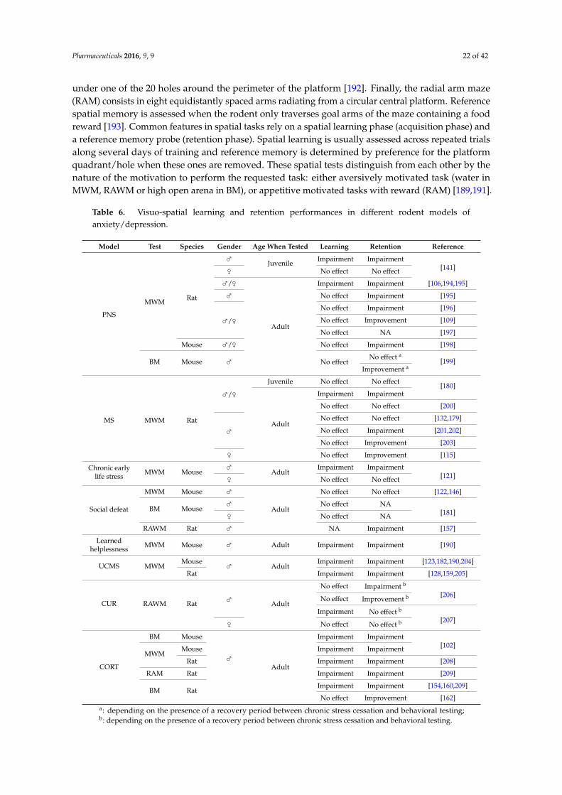
Pharmaceuticals 2016, 9, 9 22 of 42
under one of the 20 holes around the perimeter of the platform [192]. Finally, the radial arm maze(RAM) consists in eight equidistantly spaced arms radiating from a circular central platform. Referencespatial memory is assessed when the rodent only traverses goal arms of the maze containing a foodreward [193]. Common features in spatial tasks rely on a spatial learning phase (acquisition phase) anda reference memory probe (retention phase). Spatial learning is usually assessed across repeated trialsalong several days of training and reference memory is determined by preference for the platformquadrant/hole when these ones are removed. These spatial tests distinguish from each other by thenature of the motivation to perform the requested task: either aversively motivated task (water inMWM, RAWM or high open arena in BM), or appetitive motivated tasks with reward (RAM) [189,191].
Table 6. Visuo-spatial learning and retention performances in different rodent models ofanxiety/depression.
Model Test Species Gender Age When Tested Learning Retention Reference
PNS
MWM Rat
⇢ Juvenile Impairment Impairment[141]⇡ No effect No effect
⇢/⇡
Adult
Impairment Impairment [106,194,195]
⇢ No effect Impairment [195]
⇢/⇡No effect Impairment [196]
No effect Improvement [109]
No effect NA [197]
Mouse ⇢/⇡ No effect Impairment [198]
BM Mouse ⇢ No effectNo effect a
[199]Improvement a
MS MWM Rat
⇢/⇡Juvenile No effect No effect [180]
Adult
Impairment Impairment
No effect No effect [200]
⇢No effect No effect [132,179]
No effect Impairment [201,202]
No effect Improvement [203]
⇡ No effect Improvement [115]
Chronic earlylife stress MWM Mouse
⇢Adult
Impairment Impairment[121]⇡ No effect No effect
Social defeat
MWM Mouse ⇢
Adult
No effect No effect [122,146]
BM Mouse⇢ No effect NA
[181]⇡ No effect NA
RAWM Rat ⇢ NA Impairment [157]
Learnedhelplessness MWM Mouse ⇢ Adult Impairment Impairment [190]
UCMS MWMMouse ⇢ Adult
Impairment Impairment [123,182,190,204]
Rat Impairment Impairment [128,159,205]
CUR RAWM Rat ⇢ Adult
No effect Impairment b
[206]No effect Improvement b
Impairment No effect b
[207]⇡ No effect No effect b
CORT
BM Mouse
⇢Adult
Impairment Impairment[102]
MWMMouse Impairment Impairment
Rat Impairment Impairment [208]
RAM Rat Impairment Impairment [209]
BM RatImpairment Impairment [154,160,209]
No effect Improvement [162]a: depending on the presence of a recovery period between chronic stress cessation and behavioral testing;b: depending on the presence of a recovery period between chronic stress cessation and behavioral testing.
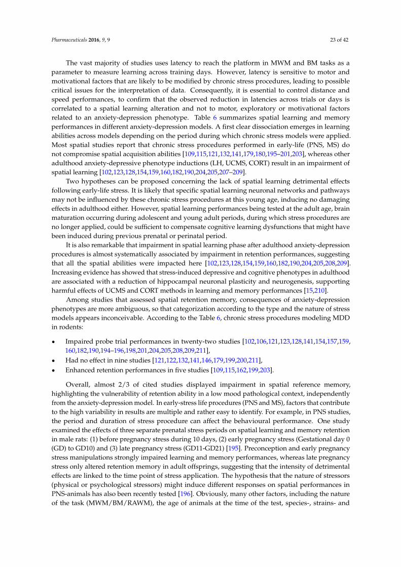
Pharmaceuticals 2016, 9, 9 23 of 42
The vast majority of studies uses latency to reach the platform in MWM and BM tasks as aparameter to measure learning across training days. However, latency is sensitive to motor andmotivational factors that are likely to be modified by chronic stress procedures, leading to possiblecritical issues for the interpretation of data. Consequently, it is essential to control distance andspeed performances, to confirm that the observed reduction in latencies across trials or days iscorrelated to a spatial learning alteration and not to motor, exploratory or motivational factorsrelated to an anxiety-depression phenotype. Table 6 summarizes spatial learning and memoryperformances in different anxiety-depression models. A first clear dissociation emerges in learningabilities across models depending on the period during which chronic stress models were applied.Most spatial studies report that chronic stress procedures performed in early-life (PNS, MS) donot compromise spatial acquisition abilities [109,115,121,132,141,179,180,195–201,203], whereas otheradulthood anxiety-depressive phenotype inductions (LH, UCMS, CORT) result in an impairment ofspatial learning [102,123,128,154,159,160,182,190,204,205,207–209].
Two hypotheses can be proposed concerning the lack of spatial learning detrimental effectsfollowing early-life stress. It is likely that specific spatial learning neuronal networks and pathwaysmay not be influenced by these chronic stress procedures at this young age, inducing no damagingeffects in adulthood either. However, spatial learning performances being tested at the adult age, brainmaturation occurring during adolescent and young adult periods, during which stress procedures areno longer applied, could be sufficient to compensate cognitive learning dysfunctions that might havebeen induced during previous prenatal or perinatal period.
It is also remarkable that impairment in spatial learning phase after adulthood anxiety-depressionprocedures is almost systematically associated by impairment in retention performances, suggestingthat all the spatial abilities were impacted here [102,123,128,154,159,160,182,190,204,205,208,209].Increasing evidence has showed that stress-induced depressive and cognitive phenotypes in adulthoodare associated with a reduction of hippocampal neuronal plasticity and neurogenesis, supportingharmful effects of UCMS and CORT methods in learning and memory performances [15,210].
Among studies that assessed spatial retention memory, consequences of anxiety-depressionphenotypes are more ambiguous, so that categorization according to the type and the nature of stressmodels appears inconceivable. According to the Table 6, chronic stress procedures modeling MDDin rodents:
‚ Impaired probe trial performances in twenty-two studies [102,106,121,123,128,141,154,157,159,160,182,190,194–196,198,201,204,205,208,209,211],
‚ Had no effect in nine studies [121,122,132,141,146,179,199,200,211],‚ Enhanced retention performances in five studies [109,115,162,199,203].
Overall, almost 2/3 of cited studies displayed impairment in spatial reference memory,highlighting the vulnerability of retention ability in a low mood pathological context, independentlyfrom the anxiety-depression model. In early-stress life procedures (PNS and MS), factors that contributeto the high variability in results are multiple and rather easy to identify. For example, in PNS studies,the period and duration of stress procedure can affect the behavioural performance. One studyexamined the effects of three separate prenatal stress periods on spatial learning and memory retentionin male rats: (1) before pregnancy stress during 10 days, (2) early pregnancy stress (Gestational day 0(GD) to GD10) and (3) late pregnancy stress (GD11-GD21) [195]. Preconception and early pregnancystress manipulations strongly impaired learning and memory performances, whereas late pregnancystress only altered retention memory in adult offsprings, suggesting that the intensity of detrimentaleffects are linked to the time point of stress application. The hypothesis that the nature of stressors(physical or psychological stressors) might induce different responses on spatial performances inPNS-animals has also been recently tested [196]. Obviously, many other factors, including the natureof the task (MWM/BM/RAWM), the age of animals at the time of the test, species-, strains- and
Pharmaceuticals 2016, 9, 9 24 of 42
sex-specificity, contribute to the difficulty to find a distinct profile in cognitive spatial deficits inearly-life stress protocols.
Interestingly, some of the studies in which spatial learning was not affected in anxio-depressedanimals revealed impairments in retention trial [195,196,198,201]. Even though retention performancesare intimately linked to learning processes, two distinct functioning circuits involving different brainstructures and neuronal pathways may exist [212,213]. In these particular cases, it appears that learningprocess is more resistant to pre/perinatal chronic procedures than retention functioning.
Studies using chronic unpredictable restraint (CUR) stress provide other pieces of information.A recent study investigated whether a recovery period between chronic stress application and spatialtests may impact behavioral responses. For that purpose, one group of rats received behavioral testingimmediately after a 21 days-CUR procedure, whereas another group of animals was given 21 days torecover from chronic stress before assessing spatial performances. Immediately-stressed rats displayeda learning spatial impairment, while there was no impact on spatial learning in rats that benefitedfrom a recovery period [207]. Focusing on retention parameters, the same pattern of results with animprovement in spatial reference memory after a recovery period was found in rats [206], confirmingthe reversibility of CUR-induced deficits in this hippocampal-dependent spatial learning and referencememory task.
Interestingly, administration of CUR in female rodents failed to alter spatial learning and memoryperformances, whether they had a recovery period or not. This comment is in line with all the otherstudies that have been conducted in female mouse or rat, because they did not report any changes inspatial learning and retention behaviors following different chronic stress procedures [115,121,141,207].The female resilience to cognitive spatial alterations observed in males in an anxiety-depression contextis not an isolated phenomenon and was also apparent in other types of memory such as episodic-likememory [106,108,121] and reversal learning tasks [111,181]. Further studies are required to clearlydifferentiate sex-specificities of cognitive performances in a MDD context.
8. Associative Memory in Rodent Models of Anxiety/Depression
Up to now, the vast majority of clinical studies using neuropsychological tests in MDD focusedon cognitive deficits such as executive functions, attention, processing speed, working memory, verbaland visual learning and memory or psychomotor performances. Clinical emotional assessmentremains paradoxically one of the less documented type of memory studied in MDD. Only fewreports concentrate their research specifically on cognitive biases associated with mood disorders [73].However, anhedonia, one of the core symptoms in the DSM-V, is defined as the loss of interest inoriginally rewarding and enjoyable activities. Precisely, cognitive biases in depressed patients referto dysfunctions in emotional memory including distorted information processing and cognitive biasfavoring sad information and lower responsiveness to positive outcomes [20,214,215].
Emotional memory (associative memory in rodents) refers to a non-declarative type of memory,implicit memory, characterized by the impossibility to reach the memory through a conscious process.Emotional memory is necessary for individuals to learn to predict a danger and adapt their behavioraccordingly. In their natural environment, rodents respond to danger in a species-specific manner by atypical freezing behavior triggered to avoid detection from possible predators. Freezing is defined as acessation of all movements, except breathing. This instinctive behavioral response is used in preclinicalstudies to assess associative learning and memory. In anxiety-depression models, this type of memoryis evaluated using classical conditioning tasks (contextual or cued fear conditioning; passive avoidancetest), based on negative reinforcers, and being dependent on the hippocampal brain structure [216,217].Basically, animals learn to associate a conditioned stimulus (context, light, tone) with an aversiveand inescapable unconditioned stimulus (mild foot shock applied through the grid floor) during anacquisition session. During the test session, animals are re-introduced in the conditioned chamber andare submitted to either a cued fear conditioning (tone or light presentation in a modified context) or
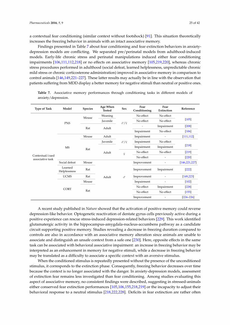
Pharmaceuticals 2016, 9, 9 25 of 42
a contextual fear conditioning (similar context without footshock) [91]. This situation theoreticallyincreases the freezing behavior in animals with an intact associative memory.
Findings presented in Table 7 about fear conditioning and fear extinction behaviors in anxiety-depression models are conflicting. We separated pre/perinatal models from adulthood-inducedmodels. Early-life chronic stress and perinatal manipulations induced either fear conditioningimpairments [106,111,112,218] or no effects on associative memory [105,219,220], whereas chronicstress procedures performed in adulthood (social defeat, learned helplessness, unpredictable chronicmild stress or chronic corticosterone administration) improved in associative memory in comparison tocontrol animals [146,149,221–227]. These latter results may actually be in line with the observation thatpatients suffering from MDD display a better memory for negative stimuli than neutral or positive ones.
Table 7. Associative memory performances through conditioning tasks in different models ofanxiety/depression.
Type of Task Model Species Age WhenTested Sex Fear
ConditioningFear
Extinction Reference
Contextual/cuedassociative task
PNS
MouseWeaning
⇢/⇡
No effect No effect[105]Juvenile No effect No effect
Rat Adult- Impairment [208]
Impairment No effect [106]
MS
Mouse Adult
⇢/⇡Impairment - [111,112]
Rat
Juvenile Impairment No effect[218]
Adult
Impairment Impairment
⇡ No effect No effect [219]
No effect - [220]
Social defeat Mouse
Adult ⇢
Improvement - [146,221,227]
LearnedHelplessness Rat Improvement Impairment [222]
UCMS Rat Improvement - [149,223]
CORT
Mouse Impairment - [102]
RatNo effect Impairment [228]
No effect No effect [155]
Improvement - [224–226]
A recent study published in Nature showed that the activation of positive memory could reversedepression-like behavior. Optogenetic reactivation of dentate gyrus cells previously active during apositive experience can rescue stress-induced depression-related behaviors [229]. This work identifiedglutamatergic activity in the hippocampus-amygdala-nucleus-accumbens pathway as a candidatecircuit supporting positive memory. Studies revealing a decrease in freezing duration compared tocontrols are also in accordance with an associative memory alteration since animals are unable toassociate and distinguish an unsafe context from a safe one [230]. Here, opposite effects in the sametask can be associated with behavioral associative impairment: an increase in freezing behavior may beinterpreted as an enhancement in memory for negative stimuli, while a decrease in freezing behaviormay be translated as a difficulty to associate a specific context with an aversive stimulus.
When the conditioned stimulus is repeatedly presented without the presence of the unconditionedstimulus, it corresponds to the extinction phase. Consequently, freezing behavior decreases over timebecause the context is no longer associated with the danger. In anxiety-depression models, assessmentof extinction fear remains less investigated than fear conditioning. Among studies evaluating thisaspect of associative memory, no consistent findings were described, suggesting in stressed-animalseither conserved fear extinction performances [105,106,155,218,219] or the incapacity to adjust theirbehavioral response to a neutral stimulus [218,222,228]. Deficits in fear extinction are rather often
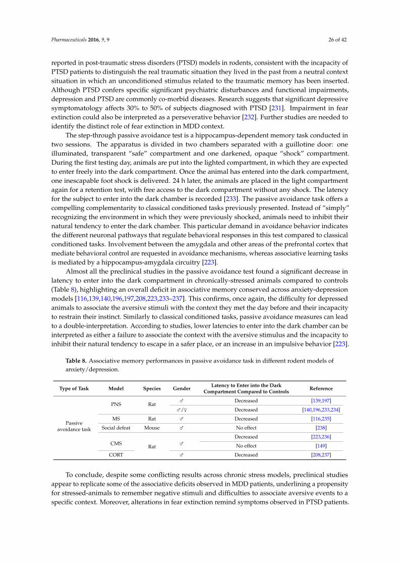
Pharmaceuticals 2016, 9, 9 26 of 42
reported in post-traumatic stress disorders (PTSD) models in rodents, consistent with the incapacity ofPTSD patients to distinguish the real traumatic situation they lived in the past from a neutral contextsituation in which an unconditioned stimulus related to the traumatic memory has been inserted.Although PTSD confers specific significant psychiatric disturbances and functional impairments,depression and PTSD are commonly co-morbid diseases. Research suggests that significant depressivesymptomatology affects 30% to 50% of subjects diagnosed with PTSD [231]. Impairment in fearextinction could also be interpreted as a perseverative behavior [232]. Further studies are needed toidentify the distinct role of fear extinction in MDD context.
The step-through passive avoidance test is a hippocampus-dependent memory task conducted intwo sessions. The apparatus is divided in two chambers separated with a guillotine door: oneilluminated, transparent “safe” compartment and one darkened, opaque “shock” compartment.During the first testing day, animals are put into the lighted compartment, in which they are expectedto enter freely into the dark compartment. Once the animal has entered into the dark compartment,one inescapable foot shock is delivered. 24 h later, the animals are placed in the light compartmentagain for a retention test, with free access to the dark compartment without any shock. The latencyfor the subject to enter into the dark chamber is recorded [233]. The passive avoidance task offers acompelling complementarity to classical conditioned tasks previously presented. Instead of “simply”recognizing the environment in which they were previously shocked, animals need to inhibit theirnatural tendency to enter the dark chamber. This particular demand in avoidance behavior indicatesthe different neuronal pathways that regulate behavioral responses in this test compared to classicalconditioned tasks. Involvement between the amygdala and other areas of the prefrontal cortex thatmediate behavioral control are requested in avoidance mechanisms, whereas associative learning tasksis mediated by a hippocampus-amygdala circuitry [223].
Almost all the preclinical studies in the passive avoidance test found a significant decrease inlatency to enter into the dark compartment in chronically-stressed animals compared to controls(Table 8), highlighting an overall deficit in associative memory conserved across anxiety-depressionmodels [116,139,140,196,197,208,223,233–237]. This confirms, once again, the difficulty for depressedanimals to associate the aversive stimuli with the context they met the day before and their incapacityto restrain their instinct. Similarly to classical conditioned tasks, passive avoidance measures can leadto a double-interpretation. According to studies, lower latencies to enter into the dark chamber can beinterpreted as either a failure to associate the context with the aversive stimulus and the incapacity toinhibit their natural tendency to escape in a safer place, or an increase in an impulsive behavior [223].
Table 8. Associative memory performances in passive avoidance task in different rodent models ofanxiety/depression.
Type of Task Model Species Gender Latency to Enter into the DarkCompartment Compared to Controls Reference
Passiveavoidance task
PNS Rat⇢ Decreased [139,197]
⇢/⇡ Decreased [140,196,233,234]
MS Rat ⇢ Decreased [116,235]
Social defeat Mouse ⇢ No effect [238]
CMS Rat ⇢Decreased [223,236]
No effect [149]
CORT ⇢ Decreased [208,237]
To conclude, despite some conflicting results across chronic stress models, preclinical studiesappear to replicate some of the associative deficits observed in MDD patients, underlining a propensityfor stressed-animals to remember negative stimuli and difficulties to associate aversive events to aspecific context. Moreover, alterations in fear extinction remind symptoms observed in PTSD patients.
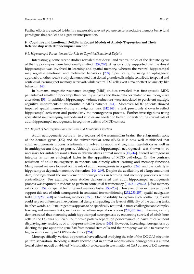
Pharmaceuticals 2016, 9, 9 27 of 42
Further efforts are needed to identify measurable relevant parameters in associative memory behavioralparadigms that can lead to a greater interpretation.
9. Cognitive and Emotional Deficits in Rodent Models of Anxiety/Depression and TheirRelationship with Hippocampus Function
9.1. Hippocampal Formation and Its Role in Cognitive/Emotional Deficits
Interestingly, some recent studies revealed that dorsal and ventral poles of the dentate gyrusof the hippocampus were functionally distinct [239,240]. A lesion study supported that the dorsalhippocampus was involved in learning and spatial memory, whereas the ventral hippocampalmay regulate emotional and motivated behaviors [239]. Specifically, by using an optogeneticapproach, another recent study demonstrated that dorsal granule cells might contribute to spatial andcontextual learning (not memory retrieval), while ventral DG cells exert a major effect on anxiety-likebehavior [240].
In humans, magnetic resonance imaging (MRI) studies revealed that first-episode MDDpatients had smaller hippocampi than healthy subjects and these data correlated to neurocognitivealterations [53]. In addition, hippocampal volume reductions were associated to persistent multiplecognitive impairments at six months in MDD patients [241]. Moreover, MDD patients showedimpaired spatial memory during a navigation task [242,243], a task previously shown to reflecthippocampal activation and particularly the neurogenesis process. Further investigations usingspecialized neuroimaging methods and studies are needed to better understand the crucial role ofadult hippocampal neurogenesis in cognitive deficits of MDD context.
9.2. Impact of Neurogenesis on Cognitive and Emotional Function
Adult neurogenesis occurs in two regions of the mammalian brain: the subgranular zoneof the dentate gyrus (DG) and the subventricular zone (SVZ). It is now well established thatadult neurogenesis process is intimately involved in mood and cognition regulations as well asin antidepressant drug response. Although adult hippocampal neurogenesis was shown to benecessary for antidepressant action in chronic-stress animal models [15,244], altered neurogenesisintegrity is not an etiological factor in the apparition of MDD pathology. On the contrary,reduction of adult neurogenesis in rodents can directly affect learning and memory functions.Many recent reviews focused on the role of adult neurogenesis in cognition [245] and particularly inhippocampus-dependent memory formation [246–249]. Despite the availability of a large amount ofdata, findings about the involvement of neurogenesis in learning and memory processes remaincontradictory. For example, some studies demonstrated that adult hippocampal neurogenesisprocess was required in rodents to perform contextual fear memory [216,217,250,251], fear memoryextinction [252] or spatial learning and memory tasks [253–256]. However, other evidences do notsupport this role of adult neurogenesis in contextual fear conditioning [252,253,257], spatial navigationtasks [216,258–260] or working memory [250]. One possibility to explain such conflicting resultscould rely on differences in experimental designs impacting the level of difficulty of the training tasks.In other words, adult neurogenesis appears to be specifically required in more challenging and complexlearning and memory tasks, such as in the pattern separation process [257,261,262]. Likewise, a studydemonstrated that increasing adult hippocampal neurogenesis by enhancing survival of adult-borncells in the DG was sufficient to improve pattern separation performances in naïve mice withoutdisplaying any anxiolytic or antidepressant-like effects [263]. However, increasing neurogenesis bydeleting the pro-apoptotic gene Bax from neural stem cells and their progeny was able to rescue thehigher emotionality in CORT-treated mice [264].
More specifically, various approaches have allowed studying the role of the DG-CA3 circuit inpattern separation. Recently, a study showed that in animal models where neurogenesis is altered(social defeat model) or ablated (x-irradiation), a decrease in reactivation of CA3 but not of DG neurons
Pharmaceuticals 2016, 9, 9 28 of 42
priory activated by fear exposure was observed in a time dependent manner [265]. These results areconsistent with the proposal that adult-born dentate granule cells are responsible for reactivation ofmemory traces in CA3, and how mal-adaptive neurogenesis state may participate in fear-generalizationand stress response [266]. Overall, these preclinical observation correlates with the impaired patternseparation observed in MDD subjects [267]. Hippocampal neurogenesis inducement in adulthood,by modifying DG-CA3 circuits, may also participate in forgetting and memory clearance, dependingon the strength of the initial memory [268], potentially participating to the clinical remission ofMDD subjects.
The presence of cognitive alterations in mood disorders, including MDD, strongly suggests thatbrain structures implicated in emotional and cognitive functions are linked. However, questions of howcognitive and depressive symptoms are related to each other and how to distinguish specific biologicalmechanisms attributable to cognitive or emotional functions in a MDD context remain opened.Moreover, the fact that most of antidepressant drug treatments failed to treat the cognitive dysfunctionsobserved in MDD, despite the remission of mood-related symptoms, confirms that independent neuralcircuitry underlying cognitive functions and depressive symptoms may be engaged.
10. Conclusions
It is now well accepted that patients with depression suffer from associated cognitive dysfunctions,such as negative or distorted thinking, difficulty concentrating, indecisiveness, reduced reaction timeor memory loss. However, whether these cognitive deficits are specific to a clinical subtype of MDDin Humans (melancholic or atypical) or to a specific chronic stress procedure in animal modelsof the disease (pre/peri natal, adulthood or social chronic stress methods) remains to be furtherdetermined. The use of translational anxiety/depression animal models will substantially improveour understanding about these cognitive alterations.
In this review, we provided an overview of clinical findings regarding the nature of cognitivedysfunctions in depressed patients from the first episode to recurrence according to the subtype ofMDD. Then, we have summarized available data indicating the type of cognitive functions (includingattention, executive functions, working memory, spatial memory, episodic-like memory and associativememory) affected in different anxiety/depression animal models.
Clinical evidence indicates an overall cognitive decline in MDD patients persisting even afterremission and during recurrent episodes. Among MDD subtypes, melancholic patients wereidentified to experience more pronounced cognitive deficits, suggesting a different pattern of cognitiveimpairment from those found in non-melancholic patients. Among all the cognitive alterations,executive and attentional functions deficits seem to be a constant hallmark in MDD patients, sinceit is present in all stages of the MDD course and in melancholic patients. Through the study ofdifferent anxiety/depression animal models, it appears that these executive and attentional deficitsare conserved in rodents regardless of the chronic stress procedure. About the other types of memory,results are more heterogeneous according to the chronic stress methods used. This review points out thelimit of rodent models, since heterogeneity in the results was observable within models and/or withina same type of memory. Numerous factors such as sex, protocol details, species, strain, environmentalconditions, and the nature of the test promote this variability. Besides, animal models cannot fullyreplicate all of the cognitive deficits observed in human MDD, but rather only reflects differentetiologies or aspects of the disease. Overall, among all the suggested models, adulthood-inducedchronic stress models (the UCMS and the CORT model for example) seems to show greater similaritieswith the cognitive symptoms observed in MDD patients, key advantages compared to pre or perinatalchronic stress procedures.
Unfortunately, cognitive condition in MDD patients tends to receive less attention than otherdepressive-related symptoms, particularly when it comes to consider the therapeutic strategy.Indeed, finding therapeutic strategies that treat both depressive and cognitive-related symptomsremains one of the main challenges in this field. Up to now, clinical reports about the efficacy of
Pharmaceuticals 2016, 9, 9 29 of 42
classical antidepressant drugs are relatively inconsistent [46,47,50,51,185]. Recent preclinical resultsare in line with clinical disparate findings using antidepressant drug therapies, specifically withselective serotonin reuptake inhibitors (see [192] for review). Further investigations, includingnon-antidepressant and innovative drugs strategies, are needed to determine which treatment couldbenefit to depression and comorbidities-associated signs.
Acknowledgments: This work was supported by the Brain & Behavior Research Foundation (formerly NARSAD)and the Pierre Deniker Foundation (D.J.D.), by the Ministère de l’Education Nationale, de l’EnseignementSupérieur et de la Recherche (MENESR, Paris, France) Fellowship (F.D.).
Author Contributions: All authors participated to the writing of the manuscript.
Conflicts of Interest: D.J.D. serves as a consultant for Lundbeck, Roche, and Servier. R.G. has receivedcompensation as a member of the scientific advisory board of Janssen, Lundbeck, Roche, Takeda. He hasserved as consultant and/or speaker for Astra Zeneca, Pierre Fabre, Lilly, Otsuka, Sanofi, Servier and receivedcompensation, and he has received research support from Servier. The remaining authors declare no conflictof interest.
References
1. American Psychiatric Association. Diagnostic and Statistical Manual of Mental Disorders, 5th ed.; AmericanPsychiatric Publishing: Arlington, VA, USA, 2013.
2. Porter, R.J.; Gallagher, P.; Thompson, J.M.; Young, A.H. Neurocognitive impairment in drug-free patientswith major depressive disorder. Br. J. Psychiatry 2003, 182, 214–220. [CrossRef] [PubMed]
3. Hammar, A.; Ardal, G. Cognitive functioning in major depression—A summary. Front. Hum. Neurosci. 2009,3, 26. [CrossRef] [PubMed]
4. Trivedi, M.H.; Greer, T.L. Cognitive dysfunction in unipolar depression: Implications for treatment.J. Affect. Disord. 2014, 152–154, 19–27. [CrossRef] [PubMed]
5. Hasselbalch, B.J.; Knorr, U.; Kessing, L.V. Cognitive impairment in the remitted state of unipolar depressivedisorder: A systematic review. J. Affect. Disord. 2011, 134, 20–31. [CrossRef] [PubMed]
6. Snyder, H.R. Major depressive disorder is associated with broad impairments on neuropsychologicalmeasures of executive function: A meta-analysis and review. Psychol. Bull. 2013, 139, 81–132. [CrossRef][PubMed]
7. Gonda, X.; Pompili, M.; Serafini, G.; Carvalho, A.F.; Rihmer, Z.; Dome, P. The role of cognitive dysfunction inthe symptoms and remission from depression. Ann. Gen. Psychiatry 2015, 14, 27. [CrossRef] [PubMed]
8. Gershon, A.; Hayward, C.; Schraedley-Desmond, P.; Rudolph, K.D.; Booster, G.D.; Gotlib, I.H. Life stress andfirst onset of psychiatric disorders in daughters of depressed mothers. J. Psychiatr. Res. 2011, 45, 855–862.[CrossRef] [PubMed]
9. Hammen, C.; Hazel, N.A.; Brennan, P.A.; Najman, J. Intergenerational transmission and continuity of stressand depression: Depressed women and their offspring in 20 years of follow-up. Psychol. Med. 2012, 42,931–942. [CrossRef] [PubMed]
10. Nishi, M.; Horii-Hayashi, N.; Sasagawa, T. Effects of early life adverse experiences on the brain: Implicationsfrom maternal separation models in rodents. Front. Neurosci. 2014, 8, 166. [CrossRef] [PubMed]
11. Richman, C.L.; Dember, W.N.; Kim, P. Spontaneous alternation behavior in animals: A review. Curr. Psychol.1986, 5, 358–391. [CrossRef]
12. Blanchard, R.J.; McKittrick, C.R.; Blanchard, D.C. Animal models of social stress: Effects on behavior andbrain neurochemical systems. Physiol. Behav. 2001, 73, 261–271. [CrossRef]
13. Chourbaji, S.; Zacher, C.; Sanchis-Segura, C.; Dormann, C.; Vollmayr, B.; Gass, P. Learned helplessness:Validity and reliability of depressive-like states in mice. Brain Res. Brain Res. Protoc. 2005, 16, 70–78.[CrossRef] [PubMed]
14. Willner, P. Chronic mild stress (CMS) revisited: Consistency and behavioural-neurobiological concordancein the effects of CMS. Neuropsychobiology 2005, 52, 90–110. [CrossRef] [PubMed]
15. David, D.J.; Samuels, B.A.; Rainer, Q.; Wang, J.W.; Marsteller, D.; Mendez, I.; Drew, M.; Craig, D.A.;Guiard, B.P.; Guilloux, J.P.; et al. Neurogenesis-dependent and -independent effects of fluoxetine in an animalmodel of anxiety/depression. Neuron 2009, 62, 479–493. [CrossRef] [PubMed]
Pharmaceuticals 2016, 9, 9 30 of 42
16. Belzung, C.; Lemoine, M. Criteria of validity for animal models of psychiatric disorders: Focus on anxietydisorders and depression. Biol. Mood Anxiety Disord. 2011, 1, 9. [CrossRef] [PubMed]
17. Willner, P. The validity of animal models of depression. Psychopharmacology 1984, 83, 1–16. [CrossRef][PubMed]
18. Marazziti, D.; Consoli, G.; Picchetti, M.; Carlini, M.; Faravelli, L. Cognitive impairment in major depression.Eur. J. Pharmacol. 2010, 626, 83–86. [CrossRef] [PubMed]
19. Lee, R.S.; Hermens, D.F.; Porter, M.A.; Redoblado-Hodge, M.A. A meta-analysis of cognitive deficits infirst-episode major depressive disorder. J. Affect. Disord. 2012, 140, 113–124. [CrossRef] [PubMed]
20. Murrough, J.W.; Iacoviello, B.; Neumeister, A.; Charney, D.S.; Iosifescu, D.V. Cognitive dysfunction indepression: Neurocircuitry and new therapeutic strategies. Neurobiol. Learn. Mem. 2011, 96, 553–563.[CrossRef] [PubMed]
21. Millan, M.J.; Agid, Y.; Brune, M.; Bullmore, E.T.; Carter, C.S.; Clayton, N.S.; Connor, R.; Davis, S.; Deakin, B.;DeRubeis, R.J.; et al. Cognitive dysfunction in psychiatric disorders: Characteristics, causes and the quest forimproved therapy. Nat. Rev. Drug Discov. 2012, 11, 141–168. [CrossRef] [PubMed]
22. Papakostas, G.I. Cognitive symptoms in patients with major depressive disorder and their implications forclinical practice. J. Clin. Psychiatry 2014, 75, 8–14. [CrossRef] [PubMed]
23. Pardo, J.V.; Pardo, P.J.; Humes, S.W.; I Posner, M. Neurocognitive dysfunction in antidepressant-free,non-elderly patients with unipolar depression: Alerting and covert orienting of visuospatial attention.J. Affect. Disord. 2006, 92, 71–78. [CrossRef] [PubMed]
24. Godard, J.; Grondin, S.; Baruch, P.; Lafleur, M.F. Psychosocial and neurocognitive profiles in depressedpatients with major depressive disorder and bipolar disorder. Psychiatry Res. 2011, 190, 244–252. [CrossRef][PubMed]
25. Gualtieri, C.T. A practical approach to objective attention deficit/hyperactivity disorder diagnosis andmanagement. Psychiatry 2005, 2, 16–25. [PubMed]
26. Fossati, P.; Ergis, A.M.; Allilaire, J.F. Executive functioning in unipolar depression: A review. L'Encephale2002, 28, 97–107. [PubMed]
27. Gohier, B.; Ferracci, L.; Surguladze, S.A.; Lawrence, E.; El Hage, W.; Kefi, M.Z.; Allain, P.; Garre, J.B.;Le Gall, D. Cognitive inhibition and working memory in unipolar depression. J. Affect. Disord. 2009, 116,100–105. [CrossRef] [PubMed]
28. Cella, M.; Dymond, S.; Cooper, A. Impaired flexible decision-making in major depressive disorder.J. Affect. Disord. 2010, 124, 207–210. [CrossRef] [PubMed]
29. Gruber, O.; Zilles, D.; Kennel, J.; Gruber, E.; Falkai, P. A systematic experimental neuropsychologicalinvestigation of the functional integrity of working memory circuits in major depression. Eur. Arch. PsychiatryClin. Neurosci. 2011, 261, 179–184. [CrossRef] [PubMed]
30. Borbely-Ipkovich, E.; Janacsek, K.; Nemeth, D.; Gonda, X. The effect of negative mood and major depressiveepisode on working memory and implicit learning. Neuropsychopharmacol. Hung. 2014, 16, 29–42. [PubMed]
31. Schmid, M.; Hammar, A. A follow-up study of first episode major depressive disorder. Impairment ininhibition and semantic fluency-potential predictors for relapse? Front. Psychol. 2013, 4, 633. [CrossRef][PubMed]
32. Foland-Ross, L.C.; Gotlib, I.H. Cognitive and neural aspects of information processing in major depressivedisorder: An integrative perspective. Front. Psychol. 2012, 3, 489. [CrossRef] [PubMed]
33. Castaneda, A.E.; Tuulio-Henriksson, A.; Marttunen, M.; Suvisaari, J.; Lonnqvist, J. A review on cognitiveimpairments in depressive and anxiety disorders with a focus on young adults. J. Affect. Disord. 2008, 106,1–27. [CrossRef] [PubMed]
34. Best, J.R.; Miller, P.H.; Naglieri, J.A. Relations between executive function and academic achievement fromages 5 to 17 in a large, representative national sample. Learn. Individ. Differ. 2011, 21, 327–336. [CrossRef][PubMed]
35. Vilgis, V.; Silk, T.J.; Vance, A. Executive function and attention in children and adolescents with depressivedisorders: A systematic review. Eur. Child Adolesc. Psychiatry 2015, 24, 365–384. [CrossRef] [PubMed]
36. Baune, B.T.; Fuhr, M.; Air, T.; Hering, C. Neuropsychological functioning in adolescents and young adultswith major depressive disorder—a review. Psychiatry Res. 2014, 218, 261–271. [CrossRef] [PubMed]
Pharmaceuticals 2016, 9, 9 31 of 42
37. Nebes, R.D.; Butters, M.A.; Mulsant, B.H.; Pollock, B.G.; Zmuda, M.D.; Houck, P.R.; Reynolds, C.F., 3rd.Decreased working memory and processing speed mediate cognitive impairment in geriatric depression.Psychol. Med. 2000, 30, 679–691. [CrossRef] [PubMed]
38. Baudic, S.; Tzortzis, C.; Barba, G.D.; Traykov, L. Executive deficits in elderly patients with major unipolardepression. J. Geriatr. Psychiatry Neurol. 2004, 17, 195–201. [CrossRef] [PubMed]
39. Sheline, Y.I.; Barch, D.M.; Garcia, K.; Gersing, K.; Pieper, C.; Welsh-Bohmer, K.; Steffens, D.C.;Doraiswamy, P.M. Cognitive function in late life depression: Relationships to depression severity,cerebrovascular risk factors and processing speed. Biol. Psychiatry 2006, 60, 58–65. [CrossRef] [PubMed]
40. Koenig, A.M.; Butters, M.A. Cognition in late life depression: Treatment considerations. Curr. Treat.Options Psychiatry 2014, 1, 1–14. [CrossRef] [PubMed]
41. Russo, M.; Mahon, K.; Burdick, K.E. Measuring cognitive function in MDD: Emerging assessment tools.Depression Anxiety 2015, 32, 262–269. [CrossRef] [PubMed]
42. Papakostas, G.I. Antidepressants and their effect on cognition in major depressive disorder. J. Clin. Psychiatry2015, 76, e1046. [CrossRef] [PubMed]
43. El Hammi, E.; Samp, J.; Remuzat, C.; Auray, J.P.; Lamure, M.; Aballea, S.; Kooli, A.; Akhras, K.; Toumi, M.Difference of perceptions and evaluation of cognitive dysfunction in major depressive disorder patientsacross psychiatrists internationally. Ther. Adv. Psychopharmacol. 2014, 4, 22–29. [CrossRef] [PubMed]
44. Bakkour, N.; Samp, J.; Akhras, K.; El Hammi, E.; Soussi, I.; Zahra, F.; Duru, G.; Kooli, A.; Toumi, M.Systematic review of appropriate cognitive assessment instruments used in clinical trials of schizophrenia,major depressive disorder and bipolar disorder. Psychiatry Res. 2014, 216, 291–302. [CrossRef] [PubMed]
45. Lim, J.; Oh, I.K.; Han, C.; Huh, Y.J.; Jung, I.K.; Patkar, A.A.; Steffens, D.C.; Jang, B.H. Sensitivity of cognitivetests in four cognitive domains in discriminating mdd patients from healthy controls: A meta-analysis.Int. Psychogeriatr. 2013, 25, 1543–1557. [CrossRef] [PubMed]
46. Gallassi, R.; Di Sarro, R.; Morreale, A.; Amore, M. Memory impairment in patients with late-onset majordepression: The effect of antidepressant therapy. J. Affect. Disord. 2006, 91, 243–250. [CrossRef] [PubMed]
47. Herrera-Guzman, I.; Gudayol-Ferre, E.; Herrera-Abarca, J.E.; Herrera-Guzman, D.; Montelongo-Pedraza, P.;Padros Blazquez, F.; Pero-Cebollero, M.; Guardia-Olmos, J. Major depressive disorder in recovery andneuropsychological functioning: Effects of selective serotonin reuptake inhibitor and dual inhibitordepression treatments on residual cognitive deficits in patients with major depressive disorder in recovery.J. Affect. Disord. 2010, 123, 341–350. [CrossRef] [PubMed]
48. Diaconescu, A.O.; Kramer, E.; Hermann, C.; Ma, Y.; Dhawan, V.; Chaly, T.; Eidelberg, D.; McIntosh, A.R.;Smith, G.S. Distinct functional networks associated with improvement of affective symptoms and cognitivefunction during citalopram treatment in geriatric depression. Hum. Brain Mapp. 2011, 32, 1677–1691.[CrossRef] [PubMed]
49. Culang, M.E.; Sneed, J.R.; Keilp, J.G.; Rutherford, B.R.; Pelton, G.H.; Devanand, D.P.; Roose, S.P.Change in cognitive functioning following acute antidepressant treatment in late-life depression. Am. J.Geriatr. Psychiatry 2009, 17, 881–888. [CrossRef] [PubMed]
50. Ferguson, J.M.; Wesnes, K.A.; Schwartz, G.E. Reboxetine versus paroxetine versus placebo: Effects on cognitivefunctioning in depressed patients. Int. Clin. Psychopharmacol. 2003, 18, 9–14. [CrossRef] [PubMed]
51. Bastos, A.G.; Guimaraes, L.S.; Trentini, C.M. Neurocognitive changes in depressed patients in psychodynamicpsychotherapy, therapy with fluoxetine and combination therapy. J. Affect. Disord. 2013, 151, 1066–1075.[CrossRef] [PubMed]
52. Gudayol-Ferre, E.; Guardia-Olmos, J.; Pero-Cebollero, M. Effects of remission speed and improvement ofcognitive functions of depressed patients. Psychiatry Res. 2015, 226, 103–112. [CrossRef] [PubMed]
53. Kaymak, S.U.; Demir, B.; Senturk, S.; Tatar, I.; Aldur, M.M.; Ulug, B. Hippocampus, glucocorticoids andneurocognitive functions in patients with first-episode major depressive disorders. Eur. Arch. PsychiatryClin. Neurosci. 2010, 260, 217–223. [CrossRef] [PubMed]
54. Hasselbalch, B.J. Cognition and neuroplasticity in the remitted state of unipolar depressive disorder.Dan. Med. J. 2015, 62, B5080. [PubMed]
55. Burcusa, S.L.; Iacono, W.G. Risk for recurrence in depression. Clin. Psychol. Rev. 2007, 27, 959–985. [CrossRef][PubMed]
56. Galecki, P.; Talarowska, M.; Anderson, G.; Berk, M.; Maes, M. Mechanisms underlying neurocognitivedysfunctions in recurrent major depression. Med. Sci. Monit. 2015, 21, 1535–1547. [PubMed]
Pharmaceuticals 2016, 9, 9 32 of 42
57. Lampe, I.K.; Sitskoorn, M.M.; Heeren, T.J. Effects of recurrent major depressive disorder on behavior andcognitive function in female depressed patients. Psychiatry Res. 2004, 125, 73–79. [CrossRef] [PubMed]
58. Talarowska, M.; Zajaczkowska, M.; Galecki, P. Cognitive functions in first-episode depression and recurrentdepressive disorder. Psychiatria Danubina 2015, 27, 38–43. [PubMed]
59. Karabekiroglu, A.; Topcuoglu, V.; Gimzal Gonentur, A.; Karabekiroglu, K. Executive function differencesbetween first episode and recurrent major depression patients. Turk Psikiyatri Derg. 2010, 21, 280–288.[PubMed]
60. Chen, J.; Ma, W.; Zhang, Y.; Wu, X.; Wei, D.; Liu, G.; Deng, Z.; Zhang, Z.; Yang, L. Distinct facial processingrelated negative cognitive bias in first-episode and recurrent major depression: Evidence from the n170 erpcomponent. PLoS ONE 2014, 9, e109176. [CrossRef] [PubMed]
61. Caldieraro, M.A.; Baeza, F.L.; Pinheiro, D.O.; Ribeiro, M.R.; Parker, G.; Fleck, M.P. Clinical differencesbetween melancholic and nonmelancholic depression as defined by the core system. Compr. Psychiatry 2013,54, 11–15. [CrossRef] [PubMed]
62. Roca, M.; Monzon, S.; Vives, M.; Lopez-Navarro, E.; Garcia-Toro, M.; Vicens, C.; Garcia-Campayo, J.;Harrison, J.; Gili, M. Cognitive function after clinical remission in patients with melancholic andnon-melancholic depression: A 6 month follow-up study. J. Affect. Disord. 2015, 171, 85–92. [CrossRef][PubMed]
63. Lin, K.; Xu, G.; Lu, W.; Ouyang, H.; Dang, Y.; Lorenzo-Seva, U.; Guo, Y.; Bessonov, D.; Akiskal, H.S.; So, K.F.;et al. Neuropsychological performance in melancholic, atypical and undifferentiated major depressionduring depressed and remitted states: A prospective longitudinal study. J. Affect. Disord. 2014, 168, 184–191.[CrossRef] [PubMed]
64. Lamers, F.; Vogelzangs, N.; Merikangas, K.R.; de Jonge, P.; Beekman, A.T.; Penninx, B.W. Evidence for adifferential role of HPA-axis function, inflammation and metabolic syndrome in melancholic versus atypicaldepression. Mol. Psychiatry 2013, 18, 692–699. [CrossRef] [PubMed]
65. Antonijevic, I. HPA axis and sleep: Identifying subtypes of major depression. Stress 2008, 11, 15–27.[CrossRef] [PubMed]
66. Parker, G. Defining melancholia: The primacy of psychomotor disturbance. Acta Psychiatr. Scand. Suppl.2007, 115, 21–30. [CrossRef] [PubMed]
67. Pier, M.P.; Hulstijn, W.; Sabbe, B.G. Differential patterns of psychomotor functioning in unmedicatedmelancholic and nonmelancholic depressed patients. J. Psychiatr. Res. 2004, 38, 425–435. [CrossRef][PubMed]
68. Rogers, M.A.; Bellgrove, M.A.; Chiu, E.; Mileshkin, C.; Bradshaw, J.L. Response selection deficits inmelancholic but not nonmelancholic unipolar major depression. J. Clin. Exp. Neuropsychol. 2004, 26,169–179. [CrossRef] [PubMed]
69. Michopoulos, I.; Zervas, I.M.; Papakosta, V.M.; Tsaltas, E.; Papageorgiou, C.; Manessi, T.; Papakostas, Y.G.;Lykouras, L.; Soldatos, C.R. Set shifting deficits in melancholic vs. Non-melancholic depression: Preliminaryfindings. Eur. Psychiatry 2006, 21, 361–363. [CrossRef] [PubMed]
70. Michopoulos, I.; Zervas, I.M.; Pantelis, C.; Tsaltas, E.; Papakosta, V.M.; Boufidou, F.; Nikolaou, C.;Papageorgiou, C.; Soldatos, C.R.; Lykouras, L. Neuropsychological and hypothalamic-pituitary-axis functionin female patients with melancholic and non-melancholic depression. Eur. Arch. Psychiatry Clin. Neurosci.2008, 258, 217–225. [CrossRef] [PubMed]
71. Exner, C.; Lange, C.; Irle, E. Impaired implicit learning and reduced pre-supplementary motor cortex sizein early-onset major depression with melancholic features. J. Affect. Disord. 2009, 119, 156–162. [CrossRef][PubMed]
72. Withall, A.; Harris, L.M.; Cumming, S.R. A longitudinal study of cognitive function in melancholic andnon-melancholic subtypes of major depressive disorder. J. Affect. Disord. 2010, 123, 150–157. [CrossRef][PubMed]
73. Linden, S.C.; Jackson, M.C.; Subramanian, L.; Healy, D.; Linden, D.E. Sad benefit in face working memory:An emotional bias of melancholic depression. J. Affect. Disord. 2011, 135, 251–257. [CrossRef] [PubMed]
74. Quinn, C.R.; Harris, A.; Felmingham, K.; Boyce, P.; Kemp, A. The impact of depression heterogeneity oncognitive control in major depressive disorder. Aust. N. Z. J. Psychiatry 2012, 46, 1079–1088. [CrossRef][PubMed]
Pharmaceuticals 2016, 9, 9 33 of 42
75. Day, C.V.; Gatt, J.M.; Etkin, A.; DeBattista, C.; Schatzberg, A.F.; Williams, L.M. Cognitive and emotionalbiomarkers of melancholic depression: An ispot-d report. J. Affect. Disord. 2015, 176, 141–150. [CrossRef][PubMed]
76. Austin, M.P.; Mitchell, P.; Hadzi-Pavlovic, D.; Hickie, I.; Parker, G.; Chan, J.; Eyers, K. Effect of apomorphineon motor and cognitive function in melancholic patients: A preliminary report. Psychiatry Res. 2000, 97,207–215. [CrossRef]
77. Romero, N.; Sanchez, A.; Vazquez, C. Memory biases in remitted depression: The role of negative cognitionsat explicit and automatic processing levels. J. Behav. Ther. Exp. Psychiatry 2014, 45, 128–135. [CrossRef][PubMed]
78. Behnken, A.; Schoning, S.; Gerss, J.; Konrad, C.; de Jong-Meyer, R.; Zwanzger, P.; Arolt, V. Persistentnon-verbal memory impairment in remitted major depression—Caused by encoding deficits? J. Affect. Disord.2010, 122, 144–148. [CrossRef] [PubMed]
79. Munkler, P.; Rothkirch, M.; Dalati, Y.; Schmack, K.; Sterzer, P. Biased recognition of facial affect in patientswith major depressive disorder reflects clinical state. PLoS ONE 2015, 10, e0129863. [CrossRef] [PubMed]
80. Arnow, B.A.; Blasey, C.; Williams, L.M.; Palmer, D.M.; Rekshan, W.; Schatzberg, A.F.; Etkin, A.; Kulkarni, J.;Luther, J.F.; Rush, A.J. Depression subtypes in predicting antidepressant response: A report from the iSPOT-Dtrial. Am. J. Psychiatry 2015, 172, 743–750. [CrossRef] [PubMed]
81. Hofmann, S.G. Toward a cognitive-behavioral classification system for mental disorders. Behav. Ther. 2014,45, 576–587. [CrossRef] [PubMed]
82. Whitton, A.E.; Treadway, M.T.; Pizzagalli, D.A. Reward processing dysfunction in major depression, bipolardisorder and schizophrenia. Curr. Opin. Psychiatry 2015, 28, 7–12. [CrossRef] [PubMed]
83. Eshel, N.; Roiser, J.P. Reward and punishment processing in depression. Biol. Psychiatry 2010, 68, 118–124.[CrossRef] [PubMed]
84. Liu, W.H.; Wang, L.Z.; Shang, H.R.; Shen, Y.; Li, Z.; Cheung, E.F.; Chan, R.C. The influence of anhedonia onfeedback negativity in major depressive disorder. Neuropsychologia 2014, 53, 213–220. [CrossRef] [PubMed]
85. Vrieze, E.; Pizzagalli, D.A.; Demyttenaere, K.; Hompes, T.; Sienaert, P.; de Boer, P.; Schmidt, M.; Claes, S.Reduced reward learning predicts outcome in major depressive disorder. Biol. Psychiatry 2013, 73, 639–645.[CrossRef] [PubMed]
86. Treadway, M.T.; Zald, D.H. Reconsidering anhedonia in depression: Lessons from translational neuroscience.Neurosci. Biobehav. Rev. 2011, 35, 537–555. [CrossRef] [PubMed]
87. Olausson, P.; Kiraly, D.D.; Gourley, S.L.; Taylor, J.R. Persistent effects of prior chronic exposure tocorticosterone on reward-related learning and motivation in rodents. Psychopharmacology 2013, 225, 569–577.[CrossRef] [PubMed]
88. Insel, T.; Cuthbert, B.; Garvey, M.; Heinssen, R.; Pine, D.S.; Quinn, K.; Sanislow, C.; Wang, P. Research domaincriteria (RDOC): Toward a new classification framework for research on mental disorders. Am. J. Psychiatry2010, 167, 748–751. [CrossRef] [PubMed]
89. Moore, H.; Geyer, M.A.; Carter, C.S.; Barch, D.M. Harnessing cognitive neuroscience to develop newtreatments for improving cognition in schizophrenia: Cntrics selected cognitive paradigms for animalmodels. Neurosci. Biobehav. Rev. 2013, 37, 2087–2091. [CrossRef] [PubMed]
90. Young, J.W.; Powell, S.B.; Risbrough, V.; Marston, H.M.; Geyer, M.A. Using the matrics to guide developmentof a preclinical cognitive test battery for research in schizophrenia. Pharmacol. Ther. 2009, 122, 150–202.[CrossRef] [PubMed]
91. Wallace, T.L.; Ballard, T.M.; Glavis-Bloom, C. Animal paradigms to assess cognition with translation tohumans. Handb. Exp. Pharmacol. 2015, 228, 27–57. [PubMed]
92. Oomen, C.A.; Hvoslef-Eide, M.; Heath, C.J.; Mar, A.C.; Horner, A.E.; Bussey, T.J.; Saksida, L.M.The touchscreen operant platform for testing working memory and pattern separation in rats and mice.Nat. Protoc. 2013, 8, 2006–2021. [CrossRef] [PubMed]
93. Williams, J.M.; Barnhofer, T.; Crane, C.; Herman, D.; Raes, F.; Watkins, E.; Dalgleish, T. Autobiographicalmemory specificity and emotional disorder. Psychol. Bull. 2007, 133, 122–148. [CrossRef] [PubMed]
94. Soderlund, H.; Moscovitch, M.; Kumar, N.; Daskalakis, Z.J.; Flint, A.; Herrmann, N.; Levine, B.Autobiographical episodic memory in major depressive disorder. J. Abnorm. Psychol. 2014, 123, 51–60.[CrossRef] [PubMed]
Pharmaceuticals 2016, 9, 9 34 of 42
95. Sumner, J.A.; Griffith, J.W.; Mineka, S. Overgeneral autobiographical memory as a predictor of the course ofdepression: A meta-analysis. Behav. Res. Ther. 2010, 48, 614–625. [CrossRef] [PubMed]
96. Nandrino, J.L.; Pezard, L.; Poste, A.; Reveillere, C.; Beaune, D. Autobiographical memory in major depression:A comparison between first-episode and recurrent patients. Psychopathology 2002, 35, 335–340. [CrossRef][PubMed]
97. Young, K.D.; Bellgowan, P.S.; Bodurka, J.; Drevets, W.C. Neurophysiological correlates of autobiographicalmemory deficits in currently and formerly depressed subjects. Psychol. Med. 2014, 44, 2951–2963. [CrossRef][PubMed]
98. Woody, M.L.; Burkhouse, K.L.; Gibb, B.E. Overgeneral autobiographical memory in children of depressedmothers. Cogn. Emot. 2015, 29, 130–137. [CrossRef] [PubMed]
99. Leger, M.; Quiedeville, A.; Bouet, V.; Haelewyn, B.; Boulouard, M.; Schumann-Bard, P.; Freret, T. Objectrecognition test in mice. Nat. Protoc. 2013, 8, 2531–2537. [CrossRef] [PubMed]
100. Gronli, J.; Murison, R.; Fiske, E.; Bjorvatn, B.; Sorensen, E.; Portas, C.M.; Ursin, R. Effects of chronic mild stresson sexual behavior, locomotor activity and consumption of sucrose and saccharine solutions. Physiol. Behav.2005, 84, 571–577. [CrossRef] [PubMed]
101. Kleen, J.K.; Sitomer, M.T.; Killeen, P.R.; Conrad, C.D. Chronic stress impairs spatial memory and motivationfor reward without disrupting motor ability and motivation to explore. Behav. Neurosci. 2006, 120, 842–851.[CrossRef] [PubMed]
102. Darcet, F.; Mendez-David, I.; Tritschler, L.; Gardier, A.M.; Guilloux, J.P.; David, D.J. Learning and memoryimpairments in a neuroendocrine mouse model of anxiety/depression. Front. Behav. Neurosci. 2014, 8, 136.[CrossRef] [PubMed]
103. Ennaceur, A.; Delacour, J. A new one-trial test for neurobiological studies of memory in rats. 1: Behavioraldata. Behav. Brain Res. 1988, 31, 47–59. [CrossRef]
104. Antunes, M.; Biala, G. The novel object recognition memory: Neurobiology, test procedure, and itsmodifications. Cogn. Process 2012, 13, 93–110. [CrossRef] [PubMed]
105. Abdul Aziz, N.H.; Kendall, D.A.; Pardon, M.C. Prenatal exposure to chronic mild stress increasescorticosterone levels in the amniotic fluid and induces cognitive deficits in female offspring, improvedby treatment with the antidepressant drug amitriptyline. Behav. Brain Res. 2012, 231, 29–39. [CrossRef][PubMed]
106. Markham, J.A.; Taylor, A.R.; Taylor, S.B.; Bell, D.B.; Koenig, J.I. Characterization of the cognitive impairmentsinduced by prenatal exposure to stress in the rat. Front. Behav. Neurosci. 2010, 4, 173. [CrossRef] [PubMed]
107. Ehrlich, D.E.; Neigh, G.N.; Bourke, C.H.; Nemeth, C.L.; Hazra, R.; Ryan, S.J.; Rowson, S.; Jairam, N.;Sholar, C.A.; Rainnie, D.G.; et al. Prenatal stress, regardless of concurrent escitalopram treatment, altersbehavior and amygdala gene expression of adolescent female rats. Neuropharmacology 2015, 97, 251–258.[CrossRef] [PubMed]
108. Schulz, K.M.; Pearson, J.N.; Neeley, E.W.; Berger, R.; Leonard, S.; Adams, C.E.; Stevens, K.E. Maternalstress during pregnancy causes sex-specific alterations in offspring memory performance, social interactions,indices of anxiety, and body mass. Physiol. Behav. 2011, 104, 340–347. [CrossRef] [PubMed]
109. Wilson, C.A.; Terry, A.V., Jr. Variable maternal stress in rats alters locomotor activity, social behavior, andrecognition memory in the adult offspring. Pharmacol. Biochem. Behav. 2013, 104, 47–61. [CrossRef] [PubMed]
110. Said, N.; Lakehayli, S.; El Khachibi, M.; El Ouahli, M.; Nadifi, S.; Hakkou, F.; Tazi, A. Effect of prenatal stresson memory, nicotine withdrawal and 5ht1a expression in raphe nuclei of adult rats. Int. J. Dev. Neurosci.2015, 43, 92–98. [CrossRef] [PubMed]
111. Wang, L.; Jiao, J.; Dulawa, S.C. Infant maternal separation impairs adult cognitive performance in BALB/cJmice. Psychopharmacology 2011, 216, 207–218. [CrossRef] [PubMed]
112. Niwa, M.; Matsumoto, Y.; Mouri, A.; Ozaki, N.; Nabeshima, T. Vulnerability in early life tochanges in the rearing environment plays a crucial role in the aetiopathology of psychiatric disorders.Int. J. Neuropsychopharmacol. 2011, 14, 459–477. [CrossRef] [PubMed]
113. Aisa, B.; Tordera, R.; Lasheras, B.; Del Rio, J.; Ramirez, M.J. Cognitive impairment associated to hpa axishyperactivity after maternal separation in rats. Psychoneuroendocrinology 2007, 32, 256–266. [CrossRef][PubMed]
Pharmaceuticals 2016, 9, 9 35 of 42
114. Salomon, S.; Bejar, C.; Schorer-Apelbaum, D.; Weinstock, M. Corticosterone mediates some but not otherbehavioural changes induced by prenatal stress in rats. J. Neuroendocrinol. 2011, 23, 118–128. [CrossRef][PubMed]
115. Plescia, F.; Marino, R.A.; Navarra, M.; Gambino, G.; Brancato, A.; Sardo, P.; Cannizzaro, C. Early handlingeffect on female rat spatial and non-spatial learning and memory. Behav. Process. 2014, 103, 9–16. [CrossRef][PubMed]
116. Vivinetto, A.L.; Suarez, M.M.; Rivarola, M.A. Neurobiological effects of neonatal maternal separation andpost-weaning environmental enrichment. Behav. Brain Res. 2013, 240, 110–118. [CrossRef] [PubMed]
117. Makena, N.; Bugarith, K.; Russell, V.A. Maternal separation enhances object location memory and preventsexercise-induced mapk/erk signalling in adult sprague-dawley rats. Metab. Brain Dis. 2012, 27, 377–385.[CrossRef] [PubMed]
118. Hill, R.A.; Klug, M.; Kiss Von Soly, S.; Binder, M.D.; Hannan, A.J.; van den Buuse, M. Sex-specific disruptionsin spatial memory and anhedonia in a “two hit” rat model correspond with alterations in hippocampalbrain-derived neurotrophic factor expression and signaling. Hippocampus 2014, 24, 1197–1211. [CrossRef][PubMed]
119. Marco, E.M.; Valero, M.; de la Serna, O.; Aisa, B.; Borcel, E.; Ramirez, M.J.; Viveros, M.P. Maternal deprivationeffects on brain plasticity and recognition memory in adolescent male and female rats. Neuropharmacology2013, 68, 223–231. [CrossRef] [PubMed]
120. Llorente-Berzal, A.; Mela, V.; Borcel, E.; Valero, M.; Lopez-Gallardo, M.; Viveros, M.P.; Marco, E.M.Neurobehavioral and metabolic long-term consequences of neonatal maternal deprivation stress andadolescent olanzapine treatment in male and female rats. Neuropharmacology 2012, 62, 1332–1341. [CrossRef][PubMed]
121. Naninck, E.F.; Hoeijmakers, L.; Kakava-Georgiadou, N.; Meesters, A.; Lazic, S.E.; Lucassen, P.J.; Korosi, A.Chronic early life stress alters developmental and adult neurogenesis and impairs cognitive function in mice.Hippocampus 2015, 25, 309–328. [CrossRef] [PubMed]
122. Jin, H.M.; Shrestha Muna, S.; Bagalkot, T.R.; Cui, Y.; Yadav, B.K.; Chung, Y.C. The effects of social defeat onbehavior and dopaminergic markers in mice. Neuroscience 2015, 288, 167–177. [CrossRef] [PubMed]
123. Gumuslu, E.; Mutlu, O.; Sunnetci, D.; Ulak, G.; Celikyurt, I.K.; Cine, N.; Akar, F.; Savli, H.; Erden, F.The antidepressant agomelatine improves memory deterioration and upregulates creb and bdnf geneexpression levels in unpredictable chronic mild stress (UCMS)-exposed mice. Drug Target Insights 2014, 8,11–21. [PubMed]
124. Haridas, S.; Kumar, M.; Manda, K. Melatonin ameliorates chronic mild stress induced behavioraldysfunctions in mice. Physiol. Behav. 2013, 119, 201–207. [CrossRef] [PubMed]
125. Elizalde, N.; Gil-Bea, F.J.; Ramirez, M.J.; Aisa, B.; Lasheras, B.; Del Rio, J.; Tordera, R.M. Long-lastingbehavioral effects and recognition memory deficit induced by chronic mild stress in mice: Effect ofantidepressant treatment. Psychopharmacology 2008, 199, 1–14. [CrossRef] [PubMed]
126. Li, S.; Wang, C.; Wang, W.; Dong, H.; Hou, P.; Tang, Y. Chronic mild stress impairs cognition in mice: Frombrain homeostasis to behavior. Life Sci. 2008, 82, 934–942. [CrossRef] [PubMed]
127. Orsetti, M.; Colella, L.; Dellarole, A.; Canonico, P.L.; Ghi, P. Modification of spatial recognition memoryand object discrimination after chronic administration of haloperidol, amitriptyline, sodium valproate orolanzapine in normal and anhedonic rats. Int. J. Neuropsychopharmacol. 2007, 10, 345–357. [PubMed]
128. Liu, D.; Wang, Z.; Gao, Z.; Xie, K.; Zhang, Q.; Jiang, H.; Pang, Q. Effects of curcumin on learning and memorydeficits, bdnf, and erk protein expression in rats exposed to chronic unpredictable stress. Behav. Brain Res.2014, 271, 116–121. [CrossRef] [PubMed]
129. Dobarro, M.; Orejana, L.; Aguirre, N.; Ramirez, M.J. Propranolol reduces cognitive deficits, amyloid betalevels, tau phosphorylation and insulin resistance in response to chronic corticosterone administration.Int. J. Neuropsychopharmacol. 2013, 16, 1351–1360. [CrossRef] [PubMed]
130. Solas, M.; Gerenu, G.; Gil-Bea, F.J.; Ramirez, M.J. Mineralocorticoid receptor activation induces insulinresistance through c-Jun n-terminal kinases in response to chronic corticosterone: Cognitive implications.J. Neuroendocrinol. 2013, 25, 350–356. [CrossRef] [PubMed]
131. Muhammad, A.; Kolb, B. Mild prenatal stress-modulated behavior and neuronal spine density withoutaffecting amphetamine sensitization. Dev. Neurosci. 2011, 33, 85–98. [CrossRef] [PubMed]
Pharmaceuticals 2016, 9, 9 36 of 42
132. Baudin, A.; Blot, K.; Verney, C.; Estevez, L.; Santamaria, J.; Gressens, P.; Giros, B.; Otani, S.; Dauge, V.;Naudon, L. Maternal deprivation induces deficits in temporal memory and cognitive flexibility andexaggerates synaptic plasticity in the rat medial prefrontal cortex. Neurobiol. Learn. Mem. 2012, 98, 207–214.[CrossRef] [PubMed]
133. Grace, L.; Hescham, S.; Kellaway, L.A.; Bugarith, K.; Russell, V.A. Effect of exercise on learning and memoryin a rat model of developmental stress. Metab. Brain Dis. 2009, 24, 643–657. [CrossRef] [PubMed]
134. Frankola, K.A.; Flora, A.L.; Torres, A.K.; Grissom, E.M.; Overstreet, S.; Dohanich, G.P. Effects of early rearingconditions on cognitive performance in prepubescent male and female rats. Neurobiol. Learn. Mem. 2010, 94,91–99. [CrossRef] [PubMed]
135. Macri, S.; Chiarotti, F.; Wurbel, H. Maternal separation and maternal care act independently on thedevelopment of hpa responses in male rats. Behav. Brain Res. 2008, 191, 227–234. [CrossRef] [PubMed]
136. Russo, S.J.; Murrough, J.W.; Han, M.H.; Charney, D.S.; Nestler, E.J. Neurobiology of resilience. Nat. Neurosci.2012, 15, 1475–1484. [CrossRef] [PubMed]
137. Dudchenko, P.A. An overview of the tasks used to test working memory in rodents. Neurosci. Biobehav. Rev.2004, 28, 699–709. [CrossRef] [PubMed]
138. Morris, R. Developments of a water-maze procedure for studying spatial learning in the rat. J. Neurosci.Methods 1984, 11, 47–60. [CrossRef]
139. Gue, M.; Bravard, A.; Meunier, J.; Veyrier, R.; Gaillet, S.; Recasens, M.; Maurice, T. Sex differences in learningdeficits induced by prenatal stress in juvenile rats. Behav. Brain Res. 2004, 150, 149–157. [CrossRef]
140. Meunier, J.; Gue, M.; Recasens, M.; Maurice, T. Attenuation by a sigma1 (�1) receptor agonist of the learningand memory deficits induced by a prenatal restraint stress in juvenile rats. Br. J. Pharmacol. 2004, 142,689–700. [CrossRef] [PubMed]
141. Zheng, A.; Li, H.; Cao, K.; Xu, J.; Zou, X.; Li, Y.; Chen, C.; Liu, J.; Feng, Z. Maternal hydroxytyrosoladministration improves neurogenesis and cognitive function in prenatally stressed offspring. J. Nutr.Biochem. 2015, 26, 190–199. [CrossRef] [PubMed]
142. Choy, K.H.; de Visser, Y.; Nichols, N.R.; van den Buuse, M. Combined neonatal stress and young-adultglucocorticoid stimulation in rats reduce bdnf expression in hippocampus: Effects on learning and memory.Hippocampus 2008, 18, 655–667. [CrossRef] [PubMed]
143. Klug, M.; van den Buuse, M. Chronic cannabinoid treatment during young adulthood induces sex-specificbehavioural deficits in maternally separated rats. Behav. Brain Res. 2012, 233, 305–313. [CrossRef] [PubMed]
144. Garner, B.; Wood, S.J.; Pantelis, C.; van den Buuse, M. Early maternal deprivation reduces prepulse inhibitionand impairs spatial learning ability in adulthood: No further effect of post-pubertal chronic corticosteronetreatment. Behav. Brain Res. 2007, 176, 323–332. [CrossRef] [PubMed]
145. Mehta, M.; Schmauss, C. Strain-specific cognitive deficits in adult mice exposed to early life stress.Behav. Neurosci. 2011, 125, 29–36. [CrossRef] [PubMed]
146. Yu, T.; Guo, M.; Garza, J.; Rendon, S.; Sun, X.L.; Zhang, W.; Lu, X.Y. Cognitive and neural correlatesof depression-like behaviour in socially defeated mice: An animal model of depression with cognitivedysfunction. Int. J. Neuropsychopharmacol. 2011, 14, 303–317. [CrossRef] [PubMed]
147. Novick, A.M.; Miiller, L.C.; Forster, G.L.; Watt, M.J. Adolescent social defeat decreases spatial workingmemory performance in adulthood. Behav. Brain Funct. BBF 2013, 9, 39. [CrossRef] [PubMed]
148. Furuta, M.; Ninomiya-Baba, M.; Chiba, S.; Funabashi, T.; Akema, T.; Kunugi, H. Exposure to social defeatstress in adolescence improves the working memory and anxiety-like behavior of adult female rats withintrauterine growth restriction, independently of hippocampal neurogenesis. Horm. Behav. 2015, 70, 30–37.[CrossRef] [PubMed]
149. Henningsen, K.; Andreasen, J.T.; Bouzinova, E.V.; Jayatissa, M.N.; Jensen, M.S.; Redrobe, J.P.; Wiborg, O.Cognitive deficits in the rat chronic mild stress model for depression: Relation to anhedonic-like responses.Behav. Brain Res. 2009, 198, 136–141. [CrossRef] [PubMed]
150. Henningsen, K.; Woldbye, D.P.; Wiborg, O. Electroconvulsive stimulation reverses anhedonia and cognitiveimpairments in rats exposed to chronic mild stress. Eur. Neuropsychopharmacol. 2013, 23, 1789–1794.[CrossRef] [PubMed]
151. Andreasen, J.T.; Henningsen, K.; Bate, S.; Christiansen, S.; Wiborg, O. Nicotine reverses anhedonic-likeresponse and cognitive impairment in the rat chronic mild stress model of depression: Comparison withsertraline. J. Psychopharmacol. 2011, 25, 1134–1141. [CrossRef] [PubMed]
Pharmaceuticals 2016, 9, 9 37 of 42
152. Mizoguchi, K.; Yuzurihara, M.; Ishige, A.; Sasaki, H.; Chui, D.H.; Tabira, T. Chronic stress induces impairmentof spatial working memory because of prefrontal dopaminergic dysfunction. J. Neurosci. 2000, 20, 1568–1574.[PubMed]
153. Palumbo, M.L.; Canzobre, M.C.; Pascuan, C.G.; Rios, H.; Wald, M.; Genaro, A.M. Stress induced cognitivedeficit is differentially modulated in BALB/c and C57Bl/6 mice: Correlation with Th1/Th2 balance afterstress exposure. J. Neuroimmunol. 2010, 218, 12–20. [CrossRef] [PubMed]
154. Coburn-Litvak, P.S.; Pothakos, K.; Tata, D.A.; McCloskey, D.P.; Anderson, B.J. Chronic administration ofcorticosterone impairs spatial reference memory before spatial working memory in rats. Neurobiol. Learn.Mem. 2003, 80, 11–23. [CrossRef]
155. Gururajan, A.; Hill, R.A.; van den Buuse, M. Brain-derived neurotrophic factor heterozygous mutant ratsshow selective cognitive changes and vulnerability to chronic corticosterone treatment. Neuroscience 2015,284, 297–310. [CrossRef] [PubMed]
156. Tata, D.A.; Markostamou, I.; Ioannidis, A.; Gkioka, M.; Simeonidou, C.; Anogianakis, G.; Spandou, E. Effectsof maternal separation on behavior and brain damage in adult rats exposed to neonatal hypoxia-ischemia.Behav. Brain Res. 2015, 280, 51–61. [CrossRef] [PubMed]
157. Patki, G.; Solanki, N.; Atrooz, F.; Allam, F.; Salim, S. Depression, anxiety-like behavior and memoryimpairment are associated with increased oxidative stress and inflammation in a rat model of social stress.Brain Res. 2013, 1539, 73–86. [CrossRef] [PubMed]
158. Patki, G.; Solanki, N.; Atrooz, F.; Ansari, A.; Allam, F.; Jannise, B.; Maturi, J.; Salim, S. Novel mechanisticinsights into treadmill exercise based rescue of social defeat-induced anxiety-like behavior and memoryimpairment in rats. Physiol. Behav. 2014, 130, 135–144. [CrossRef] [PubMed]
159. Tagliari, B.; Scherer, E.B.; Machado, F.R.; Ferreira, A.G.; Dalmaz, C.; Wyse, A.T. Antioxidants prevent memorydeficits provoked by chronic variable stress in rats. Neurochem. Res. 2011, 36, 2373–2380. [CrossRef] [PubMed]
160. Trofimiuk, E.; Braszko, J.J. Ciproxifan differentially modifies cognitive impairment evoked by chronic stressand chronic corticosterone administration in rats. Behav. Brain Res. 2015, 283, 145–153. [CrossRef] [PubMed]
161. Cerqueira, J.J.; Pego, J.M.; Taipa, R.; Bessa, J.M.; Almeida, O.F.; Sousa, N. Morphological correlates ofcorticosteroid-induced changes in prefrontal cortex-dependent behaviors. J. Neurosci. 2005, 25, 7792–7800.[CrossRef] [PubMed]
162. Workman, J.L.; Chan, M.Y.; Galea, L.A. Prior high corticosterone exposure reduces activation of immatureneurons in the ventral hippocampus in response to spatial and nonspatial memory. Hippocampus 2015, 25,329–344. [CrossRef] [PubMed]
163. Bushnell, P.J.; Strupp, B.J. Assessing attention in rodents. In Methods of Behavior Analysis in Neuroscience,2nd ed.; Buccafusco, J.J., Ed.; CRC Press: Boca Raton, FL, USA, 2009.
164. De Bruin, N.M.; Fransen, F.; Duytschaever, H.; Grantham, C.; Megens, A.A. Attentional performance of(C57Bl/6J ˆ 129Sv)F2 mice in the five-choice serial reaction time task. Physiol. Behav. 2006, 89, 692–703.[CrossRef] [PubMed]
165. Sanchez-Roige, S.; Pena-Oliver, Y.; Stephens, D.N. Measuring impulsivity in mice: The five-choice serialreaction time task. Psychopharmacology 2012, 219, 253–270. [CrossRef] [PubMed]
166. Bari, A.; Dalley, J.W.; Robbins, T.W. The application of the 5-choice serial reaction time task for the assessmentof visual attentional processes and impulse control in rats. Nat. Protoc. 2008, 3, 759–767. [CrossRef] [PubMed]
167. Van Gaalen, M.M.; Stenzel-Poore, M.; Holsboer, F.; Steckler, T. Reduced attention in mice overproducingcorticotropin-releasing hormone. Behav. Brain Res. 2003, 142, 69–79. [CrossRef]
168. Wilson, C.A.; Schade, R.; Terry, A.V., Jr. Variable prenatal stress results in impairments of sustained attentionand inhibitory response control in a 5-choice serial reaction time task in rats. Neuroscience 2012, 218, 126–137.[CrossRef] [PubMed]
169. Comeau, W.L.; Winstanley, C.A.; Weinberg, J. Prenatal alcohol exposure and adolescent stress—Unmaskingpersistent attentional deficits in rats. Eur. J. Neurosci. 2014, 40, 3078–3095. [CrossRef] [PubMed]
170. Torregrossa, M.M.; Xie, M.; Taylor, J.R. Chronic corticosterone exposure during adolescence reducesimpulsive action but increases impulsive choice and sensitivity to yohimbine in male sprague-dawleyrats. Neuropsychopharmacology 2012, 37, 1656–1670. [CrossRef] [PubMed]
171. Amitai, N.; Markou, A. Comparative effects of different test day challenges on performance in the 5-choiceserial reaction time task. Behav. Neurosci. 2011, 125, 764–774. [CrossRef] [PubMed]
Pharmaceuticals 2016, 9, 9 38 of 42
172. Bangasser, D.A.; Kawasumi, Y. Cognitive disruptions in stress-related psychiatric disorders: A role forcorticotropin releasing factor (CRF). Horm. Behav. 2015, 76, 125–135. [CrossRef] [PubMed]
173. Ohmura, Y.; Yamaguchi, T.; Futami, Y.; Togashi, H.; Izumi, T.; Matsumoto, M.; Yoshida, T.; Yoshioka, M.Corticotropin releasing factor enhances attentional function as assessed by the five-choice serial reactiontime task in rats. Behav. Brain Res. 2009, 198, 429–433. [CrossRef] [PubMed]
174. Van’t Veer, A.; Yano, J.M.; Carroll, F.I.; Cohen, B.M.; Carlezon, W.A., Jr. Corticotropin-releasing factor(CRF)-induced disruption of attention in rats is blocked by the kappa-opioid receptor antagonist jdtic.Neuropsychopharmacology 2012, 37, 2809–2816. [CrossRef] [PubMed]
175. Bondi, C.O.; Rodriguez, G.; Gould, G.G.; Frazer, A.; Morilak, D.A. Chronic unpredictable stress induces acognitive deficit and anxiety-like behavior in rats that is prevented by chronic antidepressant drug treatment.Neuropsychopharmacology 2008, 33, 320–331. [CrossRef] [PubMed]
176. Naegeli, K.J.; O’Connor, J.A.; Banerjee, P.; Morilak, D.A. Effects of milnacipran on cognitive flexibilityfollowing chronic stress in rats. Eur. J. Pharmacol. 2013, 703, 62–66. [CrossRef] [PubMed]
177. Nikiforuk, A.; Popik, P. Long-lasting cognitive deficit induced by stress is alleviated by acute administrationof antidepressants. Psychoneuroendocrinology 2011, 36, 28–39. [CrossRef] [PubMed]
178. Wallace, J.; Marston, H.M.; McQuade, R.; Gartside, S.E. Evidence that aetiological risk factors for psychiatricdisorders cause distinct patterns of cognitive deficits. Eur. Neuropsychopharmacol. 2014, 24, 879–889.[CrossRef] [PubMed]
179. Xue, X.; Shao, S.; Wang, W.; Shao, F. Maternal separation induces alterations in reversal learning andbrain-derived neurotrophic factor expression in adult rats. Neuropsychobiology 2013, 68, 243–249. [CrossRef][PubMed]
180. Wang, Q.; Li, M.; Du, W.; Shao, F.; Wang, W. The different effects of maternal separation on spatial learningand reversal learning in rats. Behav. Brain Res. 2015, 280, 16–23. [CrossRef] [PubMed]
181. Laredo, S.A.; Steinman, M.Q.; Robles, C.F.; Ferrer, E.; Ragen, B.J.; Trainor, B.C. Effects of defeat stress onbehavioral flexibility in males and females: Modulation by the mu-opioid receptor. Eur. J. Neurosci. 2015, 41,434–441. [CrossRef] [PubMed]
182. Bisaz, R.; Schachner, M.; Sandi, C. Causal evidence for the involvement of the neural cell adhesion molecule,NCAM, in chronic stress-induced cognitive impairments. Hippocampus 2011, 21, 56–71. [CrossRef] [PubMed]
183. Bessa, J.M.; Mesquita, A.R.; Oliveira, M.; Pego, J.M.; Cerqueira, J.J.; Palha, J.A.; Almeida, O.F.; Sousa, N.A trans-dimensional approach to the behavioral aspects of depression. Front. Behav. Neurosci. 2009, 3, 1.[CrossRef] [PubMed]
184. Hill, M.N.; Patel, S.; Carrier, E.J.; Rademacher, D.J.; Ormerod, B.K.; Hillard, C.J.; Gorzalka, B.B.Downregulation of endocannabinoid signaling in the hippocampus following chronic unpredictable stress.Neuropsychopharmacology 2005, 30, 508–515. [CrossRef] [PubMed]
185. Cerqueira, J.J.; Mailliet, F.; Almeida, O.F.; Jay, T.M.; Sousa, N. The prefrontal cortex as a key target of themaladaptive response to stress. J. Neurosci. 2007, 27, 2781–2787. [CrossRef] [PubMed]
186. Llorente, R.; Miguel-Blanco, C.; Aisa, B.; Lachize, S.; Borcel, E.; Meijer, O.C.; Ramirez, M.J.; De Kloet, E.R.;Viveros, M.P. Long term sex-dependent psychoneuroendocrine effects of maternal deprivation and juvenileunpredictable stress in rats. J. Neuroendocrinol. 2011, 23, 329–344. [CrossRef] [PubMed]
187. Heisler, J.M.; Morales, J.; Donegan, J.J.; Jett, J.D.; Redus, L.; O’Connor, J.C. The attentional set shifting task:A measure of cognitive flexibility in mice. J. Vis. Exp. JoVE 2015. [CrossRef] [PubMed]
188. Tait, D.S.; Chase, E.A.; Brown, V.J. Attentional set-shifting in rodents: A review of behavioural methods andpharmacological results. Curr. Pharm. Des. 2014, 20, 5046–5059. [CrossRef] [PubMed]
189. Conrad, C.D. A critical review of chronic stress effects on spatial learning and memory. Prog. Neuro-Psychopharmacol. Biol. Psychiatry 2010, 34, 742–755. [CrossRef] [PubMed]
190. Song, L.; Che, W.; Min-Wei, W.; Murakami, Y.; Matsumoto, K. Impairment of the spatial learning and memoryinduced by learned helplessness and chronic mild stress. Pharmacol. Biochem. Behav. 2006, 83, 186–193.[CrossRef] [PubMed]
191. Vorhees, C.V.; Williams, M.T. Morris water maze: Procedures for assessing spatial and related forms oflearning and memory. Nat. Protoc. 2006, 1, 848–858. [CrossRef] [PubMed]
192. Sunyer, B.; Patil, S.; Höger, H.; Luber, G. Barnes maze, a useful task to assess spatial reference memory in themice. Protocol Exch. 2007, 390, 10–38. [CrossRef]
193. Olton, D.S. Mazes, maps, and memory. Am. Psychol. 1979, 34, 583–596. [CrossRef] [PubMed]
Pharmaceuticals 2016, 9, 9 39 of 42
194. Huang, Y.; Shi, X.; Xu, H.; Yang, H.; Chen, T.; Chen, S.; Chen, X. Chronic unpredictable stress beforepregnancy reduce the expression of brain-derived neurotrophic factor and n-methyl-d-aspartate receptor inhippocampus of offspring rats associated with impairment of memory. Neurochem. Res. 2010, 35, 1038–1049.[CrossRef] [PubMed]
195. Modir, F.; Elahdadi Salmani, M.; Goudarzi, I.; Lashkarboluki, T.; Abrari, K. Prenatal stress decreases spatiallearning and memory retrieval of the adult male offspring of rats. Physiol. Behav. 2014, 129, 104–109.[CrossRef] [PubMed]
196. Nazeri, M.; Shabani, M.; Ghotbi Ravandi, S.; Aghaei, I.; Nozari, M.; Mazhari, S. Psychological or physicalprenatal stress differentially affects cognition behaviors. Physiol. Behav. 2015, 142, 155–160. [CrossRef][PubMed]
197. Palacios-Garcia, I.; Lara-Vasquez, A.; Montiel, J.F.; Diaz-Veliz, G.F.; Sepulveda, H.; Utreras, E.; Montecino, M.;Gonzalez-Billault, C.; Aboitiz, F. Prenatal stress down-regulates reelin expression by methylation of itspromoter and induces adult behavioral impairments in rats. PLoS ONE 2015, 10, e0117680.
198. Benoit, J.D.; Rakic, P.; Frick, K.M. Prenatal stress induces spatial memory deficits and epigenetic changes inthe hippocampus indicative of heterochromatin formation and reduced gene expression. Behav. Brain Res.2015, 281, 1–8. [CrossRef] [PubMed]
199. Negron-Oyarzo, I.; Neira, D.; Espinosa, N.; Fuentealba, P.; Aboitiz, F. Prenatal stress produces persistence ofremote memory and disrupts functional connectivity in the hippocampal-prefrontal cortex axis. Cereb. Cortex2015, 25, 3132–3143. [CrossRef] [PubMed]
200. Sun, X.M.; Tu, W.Q.; Shi, Y.W.; Xue, L.; Zhao, H. Female-dependent impaired fear memory of adultrats induced by maternal separation, and screening of possible related genes in the hippocampal ca1.Behav. Brain Res. 2014, 267, 111–118. [CrossRef] [PubMed]
201. Couto, F.S.; Batalha, V.L.; Valadas, J.S.; Data-Franca, J.; Ribeiro, J.A.; Lopes, L.V. Escitalopram improvesmemory deficits induced by maternal separation in the rat. Eur. J. Pharmacol. 2012, 695, 71–75. [CrossRef][PubMed]
202. Mello, P.B.; Benetti, F.; Cammarota, M.; Izquierdo, I. Physical exercise can reverse the deficit in fear memoryinduced by maternal deprivation. Neurobiol. Learn. Mem. 2009, 92, 364–369. [CrossRef] [PubMed]
203. Zhang, X.; Wang, B.; Jin, J.; An, S.; Zeng, Q.; Duan, Y.; Yang, L.; Ma, J.; Cao, X. Early deprivation reducedanxiety and enhanced memory in adult male rats. Brain Res. Bull. 2014, 108, 44–50. [CrossRef] [PubMed]
204. Rinwa, P.; Kumar, A. Piperine potentiates the protective effects of curcumin against chronic unpredictablestress-induced cognitive impairment and oxidative damage in mice. Brain Res. 2012, 1488, 38–50. [CrossRef][PubMed]
205. Liu, D.; Zhang, Q.; Gu, J.; Wang, X.; Xie, K.; Xian, X.; Wang, J.; Jiang, H.; Wang, Z. Resveratrol preventsimpaired cognition induced by chronic unpredictable mild stress in rats. Prog. Neuro-Psychopharmacol.Biol. Psychiatry 2014, 49, 21–29. [CrossRef] [PubMed]
206. Hoffman, A.N.; Krigbaum, A.; Ortiz, J.B.; Mika, A.; Hutchinson, K.M.; Bimonte-Nelson, H.A.; Conrad, C.D.Recovery after chronic stress within spatial reference and working memory domains: Correspondence withhippocampal morphology. Eur. J. Neurosci. 2011, 34, 1023–1030. [CrossRef] [PubMed]
207. Ortiz, J.B.; Taylor, S.B.; Hoffman, A.N.; Campbell, A.N.; Lucas, L.R.; Conrad, C.D. Sex-specific impairmentand recovery of spatial learning following the end of chronic unpredictable restraint stress: Potentialrelevance of limbic gad. Behav. Brain Res. 2015, 282, 176–184. [CrossRef] [PubMed]
208. Lee, B.; Sur, B.; Shim, I.; Lee, H.; Hahm, D.H. Baicalin improves chronic corticosterone-induced learning andmemory deficits via the enhancement of impaired hippocampal brain-derived neurotrophic factor and campresponse element-binding protein expression in the rat. J. Nat. Med. 2014, 68, 132–143. [CrossRef] [PubMed]
209. McLay, R.N.; Freeman, S.M.; Zadina, J.E. Chronic corticosterone impairs memory performance in the barnesmaze. Physiol. Behav. 1998, 63, 933–937. [CrossRef]
210. Mineur, Y.S.; Belzung, C.; Crusio, W.E. Functional implications of decreases in neurogenesis followingchronic mild stress in mice. Neuroscience 2007, 150, 251–259. [CrossRef] [PubMed]
211. Wang, Y.; Ma, Y.; Hu, J.; Cheng, W.; Jiang, H.; Zhang, X.; Li, M.; Ren, J.; Li, X. Prenatal chronic mild stressinduces depression-like behavior and sex-specific changes in regional glutamate receptor expression patternsin adult rats. Neuroscience 2015, 301, 363–374. [CrossRef] [PubMed]
212. McDonald, R.J.; Hong, N.S. How does a specific learning and memory system in the mammalian brain gaincontrol of behavior? Hippocampus 2013, 23, 1084–1102. [CrossRef] [PubMed]
Pharmaceuticals 2016, 9, 9 40 of 42
213. Anderson, E.B.; Grossrubatscher, I.; Frank, L. Dynamic hippocampal circuits support learning- andmemory-guided behaviors. Cold Spring Harb. Symp. Quant. Biol. 2014, 79, 51–58. [CrossRef] [PubMed]
214. Mathews, A.; MacLeod, C. Cognitive vulnerability to emotional disorders. Ann. Rev. Clin. Psychol. 2005, 1,167–195. [CrossRef] [PubMed]
215. Gotlib, I.H.; Joormann, J. Cognition and depression: Current status and future directions. Ann. Rev.Clin. Psychol. 2010, 6, 285–312. [CrossRef] [PubMed]
216. Saxe, M.D.; Battaglia, F.; Wang, J.W.; Malleret, G.; David, D.J.; Monckton, J.E.; Garcia, A.D.; Sofroniew, M.V.;Kandel, E.R.; Santarelli, L.; et al. Ablation of hippocampal neurogenesis impairs contextual fear conditioningand synaptic plasticity in the dentate gyrus. Proc. Natl. Acad. Sci. USA 2006, 103, 17501–17506. [CrossRef][PubMed]
217. Imayoshi, I.; Sakamoto, M.; Ohtsuka, T.; Takao, K.; Miyakawa, T.; Yamaguchi, M.; Mori, K.; Ikeda, T.;Itohara, S.; Kageyama, R. Roles of continuous neurogenesis in the structural and functional integrity of theadult forebrain. Nat. Neurosci. 2008, 11, 1153–1161. [CrossRef] [PubMed]
218. Chocyk, A.; Przyborowska, A.; Makuch, W.; Majcher-Maslanka, I.; Dudys, D.; Wedzony, K. The effects ofearly-life adversity on fear memories in adolescent rats and their persistence into adulthood. Behav. Brain Res.2014, 264, 161–172. [CrossRef] [PubMed]
219. Xiong, G.J.; Yang, Y.; Wang, L.P.; Xu, L.; Mao, R.R. Maternal separation exaggerates spontaneous recovery ofextinguished contextual fear in adult female rats. Behav. Brain Res. 2014, 269, 75–80. [CrossRef] [PubMed]
220. Oomen, C.A.; Soeters, H.; Audureau, N.; Vermunt, L.; van Hasselt, F.N.; Manders, E.M.; Joels, M.; Krugers, H.;Lucassen, P.J. Early maternal deprivation affects dentate gyrus structure and emotional learning in adultfemale rats. Psychopharmacology 2011, 214, 249–260. [CrossRef] [PubMed]
221. Dulka, B.N.; Lynch, J.F., 3rd; Latsko, M.S.; Mulvany, J.L.; Jasnow, A.M. Phenotypic responses to social defeatare associated with differences in cued and contextual fear discrimination. Behav. Process. 2015, 118, 115–122.[CrossRef] [PubMed]
222. Shumake, J.; Barrett, D.; Gonzalez-Lima, F. Behavioral characteristics of rats predisposed to learnedhelplessness: Reduced reward sensitivity, increased novelty seeking, and persistent fear memories.Behav. Brain Res. 2005, 164, 222–230. [CrossRef] [PubMed]
223. Camp, R.M.; Johnson, J.D. Repeated stressor exposure enhances contextual fear memory in a beta-adrenergicreceptor-dependent process and increases impulsivity in a non-beta receptor-dependent fashion.Physiol. Behav. 2015, 150, 64–68. [CrossRef] [PubMed]
224. Liao, Y.; Shi, Y.W.; Liu, Q.L.; Zhao, H. Glucocorticoid-induced enhancement of contextual fear memoryconsolidation in rats: Involvement of D1 receptor activity of hippocampal area CA1. Brain Res. 2013, 1524,26–33. [CrossRef] [PubMed]
225. Marks, W.N.; Fenton, E.Y.; Guskjolen, A.J.; Kalynchuk, L.E. The effect of chronic corticosterone on fearlearning and memory depends on dose and the testing protocol. Neuroscience 2015, 289, 324–333. [CrossRef][PubMed]
226. Monsey, M.S.; Boyle, L.M.; Zhang, M.L.; Nguyen, C.P.; Kronman, H.G.; Ota, K.T.; Duman, R.S.; Taylor, J.R.;Schafe, G.E. Chronic corticosterone exposure persistently elevates the expression of memory-related genes inthe lateral amygdala and enhances the consolidation of a pavlovian fear memory. PLoS ONE 2014, 9, e91530.[CrossRef] [PubMed]
227. Duclot, F.; Hollis, F.; Darcy, M.J.; Kabbaj, M. Individual differences in novelty-seeking behavior in rats asa model for psychosocial stress-related mood disorders. Physiol. Behav. 2011, 104, 296–305. [CrossRef][PubMed]
228. Gourley, S.L.; Kedves, A.T.; Olausson, P.; Taylor, J.R. A history of corticosterone exposure regulates fearextinction and cortical NR2B, GluR2/3, and BDNF. Neuropsychopharmacology 2009, 34, 707–716. [CrossRef][PubMed]
229. Ramirez, S.; Liu, X.; MacDonald, C.J.; Moffa, A.; Zhou, J.; Redondo, R.L.; Tonegawa, S. Activating positivememory engrams suppresses depression-like behaviour. Nature 2015, 522, 335–339. [CrossRef] [PubMed]
230. Denny, C.A.; Burghardt, N.S.; Schachter, D.M.; Hen, R.; Drew, M.R. 4- to 6-week-old adult-born hippocampalneurons influence novelty-evoked exploration and contextual fear conditioning. Hippocampus 2012, 22,1188–1201. [CrossRef] [PubMed]
Pharmaceuticals 2016, 9, 9 41 of 42
231. Campbell, D.G.; Felker, B.L.; Liu, C.F.; Yano, E.M.; Kirchner, J.E.; Chan, D.; Rubenstein, L.V.; Chaney, E.F.Prevalence of depression-ptsd comorbidity: Implications for clinical practice guidelines and primarycare-based interventions. J. General Intern. Med. 2007, 22, 711–718. [CrossRef] [PubMed]
232. Sotres-Bayon, F.; Cain, C.K.; LeDoux, J.E. Brain mechanisms of fear extinction: Historical perspectives on thecontribution of prefrontal cortex. Biol. Psychiatry 2006, 60, 329–336. [CrossRef] [PubMed]
233. Cherian, S.B.; Bairy, K.L.; Rao, M.S. Chronic prenatal restraint stress induced memory impairment in passiveavoidance task in post weaned male and female wistar rats. Indian J. Exp. Biol. 2009, 47, 893–899. [PubMed]
234. Wu, J.; Song, T.B.; Li, Y.J.; He, K.S.; Ge, L.; Wang, L.R. Prenatal restraint stress impairs learning and memoryand hippocampal pkcbeta1 expression and translocation in offspring rats. Brain Res. 2007, 1141, 205–213.[CrossRef] [PubMed]
235. Sung, Y.H.; Shin, M.S.; Cho, S.; Baik, H.H.; Jin, B.K.; Chang, H.K.; Lee, E.K.; Kim, C.J. Depression-like state inmaternal rats induced by repeated separation of pups is accompanied by a decrease of cell proliferation andan increase of apoptosis in the hippocampus. Neuroscience Lett. 2010, 470, 86–90. [CrossRef] [PubMed]
236. Radahmadi, M.; Alaei, H.; Sharifi, M.R.; Hosseini, N. Effects of different timing of stress on corticosterone,bdnf and memory in male rats. Physiol. Behav. 2015, 139, 459–467. [CrossRef] [PubMed]
237. Bisagno, V.; Ferrini, M.; Rios, H.; Zieher, L.M.; Wikinski, S.I. Chronic corticosterone impairs inhibitoryavoidance in rats: Possible link with atrophy of hippocampal CA3 neurons. Pharmacol. Biochem. Behav. 2000,66, 235–240. [CrossRef]
238. Costa-Nunes, J.; Zubareva, O.; Araujo-Correia, M.; Valenca, A.; Schroeter, C.A.; Pawluski, J.L.; Vignisse, J.;Steinbusch, H.; Hermes, D.; Phillipines, M.; et al. Altered emotionality, hippocampus-dependent performanceand expression of nmda receptor subunit mrnas in chronically stressed mice. Stress 2014, 17, 108–116.[CrossRef] [PubMed]
239. Fanselow, M.S.; Dong, H.W. Are the dorsal and ventral hippocampus functionally distinct structures? Neuron2010, 65, 7–19. [CrossRef] [PubMed]
240. Kheirbek, M.A.; Drew, L.J.; Burghardt, N.S.; Costantini, D.O.; Tannenholz, L.; Ahmari, S.E.; Zeng, H.;Fenton, A.A.; Hen, R. Differential control of learning and anxiety along the dorsoventral axis of the dentategyrus. Neuron 2013, 77, 955–968. [CrossRef] [PubMed]
241. O'Brien, J.T.; Lloyd, A.; McKeith, I.; Gholkar, A.; Ferrier, N. A longitudinal study of hippocampal volume,cortisol levels, and cognition in older depressed subjects. Am. J. Psychiatry 2004, 161, 2081–2090. [CrossRef][PubMed]
242. Cornwell, B.R.; Salvadore, G.; Colon-Rosario, V.; Latov, D.R.; Holroyd, T.; Carver, F.W.; Coppola, R.;Manji, H.K.; Zarate, C.A., Jr.; Grillon, C. Abnormal hippocampal functioning and impaired spatial navigationin depressed individuals: Evidence from whole-head magnetoencephalography. Am. J. Psychiatry 2010, 167,836–844. [CrossRef] [PubMed]
243. Gould, N.F.; Holmes, M.K.; Fantie, B.D.; Luckenbaugh, D.A.; Pine, D.S.; Gould, T.D.; Burgess, N.; Manji, H.K.;Zarate, C.A., Jr. Performance on a virtual reality spatial memory navigation task in depressed patients.Am. J. Psychiatry 2007, 164, 516–519. [CrossRef] [PubMed]
244. Surget, A.; Tanti, A.; Leonardo, E.D.; Laugeray, A.; Rainer, Q.; Touma, C.; Palme, R.; Griebel, G.;Ibarguen-Vargas, Y.; Hen, R.; et al. Antidepressants recruit new neurons to improve stress response regulation.Mol. Psychiatry 2011, 16, 1177–1188. [CrossRef] [PubMed]
245. Oomen, C.A.; Bekinschtein, P.; Kent, B.A.; Saksida, L.M.; Bussey, T.J. Adult hippocampal neurogenesis andits role in cognition. Wiley Interdiscip. Rev. Cogn. Sci. 2014, 5, 573–587. [CrossRef] [PubMed]
246. Deng, W.; Aimone, J.B.; Gage, F.H. New neurons and new memories: How does adult hippocampalneurogenesis affect learning and memory? Nat. Rev. Neurosci. 2010, 11, 339–350. [CrossRef] [PubMed]
247. Kim, W.R.; Christian, K.; Ming, G.L.; Song, H. Time-dependent involvement of adult-born dentate granulecells in behavior. Behav. Brain Res. 2012, 227, 470–479. [CrossRef] [PubMed]
248. Marin-Burgin, A.; Schinder, A.F. Requirement of adult-born neurons for hippocampus-dependent learning.Behav. Brain Res. 2012, 227, 391–399. [CrossRef] [PubMed]
249. Ming, G.L.; Song, H. Adult neurogenesis in the mammalian brain: Significant answers and significantquestions. Neuron 2011, 70, 687–702. [CrossRef] [PubMed]
Pharmaceuticals 2016, 9, 9 42 of 42
250. Hernandez-Rabaza, V.; Llorens-Martin, M.; Velazquez-Sanchez, C.; Ferragud, A.; Arcusa, A.; Gumus, H.G.;Gomez-Pinedo, U.; Perez-Villalba, A.; Rosello, J.; Trejo, J.L.; et al. Inhibition of adult hippocampalneurogenesis disrupts contextual learning but spares spatial working memory, long-term conditional ruleretention and spatial reversal. Neuroscience 2009, 159, 59–68. [CrossRef] [PubMed]
251. Winocur, G.; Wojtowicz, J.M.; Sekeres, M.; Snyder, J.S.; Wang, S. Inhibition of neurogenesis interferes withhippocampus-dependent memory function. Hippocampus 2006, 16, 296–304. [CrossRef] [PubMed]
252. Deng, W.; Saxe, M.D.; Gallina, I.S.; Gage, F.H. Adult-born hippocampal dentate granule cells undergoingmaturation modulate learning and memory in the brain. J. Neurosci. 2009, 29, 13532–13542. [CrossRef][PubMed]
253. Dupret, D.; Revest, J.M.; Koehl, M.; Ichas, F.; De Giorgi, F.; Costet, P.; Abrous, D.N.; Piazza, P.V. Spatialrelational memory requires hippocampal adult neurogenesis. PLoS ONE 2008, 3, e1959. [CrossRef] [PubMed]
254. Garthe, A.; Kempermann, G. An old test for new neurons: Refining the morris water maze to study thefunctional relevance of adult hippocampal neurogenesis. Front. Neurosci. 2013, 7, 63. [CrossRef] [PubMed]
255. Jessberger, S.; Clark, R.E.; Broadbent, N.J.; Clemenson, G.D., Jr.; Consiglio, A.; Lie, D.C.; Squire, L.R.;Gage, F.H. Dentate gyrus-specific knockdown of adult neurogenesis impairs spatial and object recognitionmemory in adult rats. Learn. Mem. 2009, 16, 147–154. [CrossRef] [PubMed]
256. Clelland, C.D.; Choi, M.; Romberg, C.; Clemenson, G.D., Jr.; Fragniere, A.; Tyers, P.; Jessberger, S.;Saksida, L.M.; Barker, R.A.; Gage, F.H.; et al. A functional role for adult hippocampal neurogenesis inspatial pattern separation. Science 2009, 325, 210–213. [CrossRef] [PubMed]
257. Shors, T.J.; Townsend, D.A.; Zhao, M.; Kozorovitskiy, Y.; Gould, E. Neurogenesis may relate to some but notall types of hippocampal-dependent learning. Hippocampus 2002, 12, 578–584. [CrossRef] [PubMed]
258. Meshi, D.; Drew, M.R.; Saxe, M.; Ansorge, M.S.; David, D.; Santarelli, L.; Malapani, C.; Moore, H.; Hen, R.Hippocampal neurogenesis is not required for behavioral effects of environmental enrichment. Nat. Neurosci.2006, 9, 729–731. [CrossRef] [PubMed]
259. Groves, J.O.; Leslie, I.; Huang, G.J.; McHugh, S.B.; Taylor, A.; Mott, R.; Munafo, M.; Bannerman, D.M.;Flint, J. Ablating adult neurogenesis in the rat has no effect on spatial processing: Evidence from a novelpharmacogenetic model. PLoS Genet. 2013, 9, e1003718. [CrossRef] [PubMed]
260. Saxe, M.D.; Malleret, G.; Vronskaya, S.; Mendez, I.; Garcia, A.D.; Sofroniew, M.V.; Kandel, E.R.; Hen, R.Paradoxical influence of hippocampal neurogenesis on working memory. Proc. Natl. Acad. Sci. USA 2007,104, 4642–4646. [CrossRef] [PubMed]
261. Schmidt, B.; Marrone, D.F.; Markus, E.J. Disambiguating the similar: The dentate gyrus and patternseparation. Behav. Brain Res. 2012, 226, 56–65. [CrossRef] [PubMed]
262. Aimone, J.B.; Deng, W.; Gage, F.H. Resolving new memories: A critical look at the dentate gyrus, adultneurogenesis, and pattern separation. Neuron 2011, 70, 589–596. [CrossRef] [PubMed]
263. Sahay, A.; Scobie, K.N.; Hill, A.S.; O’Carroll, C.M.; Kheirbek, M.A.; Burghardt, N.S.; Fenton, A.A.;Dranovsky, A.; Hen, R. Increasing adult hippocampal neurogenesis is sufficient to improve pattern separation.Nature 2011, 472, 466–470. [CrossRef] [PubMed]
264. Hill, A.S.; Sahay, A.; Hen, R. Increasing adult hippocampal neurogenesis is sufficient to reduce anxiety anddepression-like behaviors. Neuropsychopharmacology 2015, 40, 2368–2378. [CrossRef] [PubMed]
265. Denny, C.A.; Kheirbek, M.A.; Alba, E.L.; Tanaka, K.F.; Brachman, R.A.; Laughman, K.B.; Tomm, N.K.;Turi, G.F.; Losonczy, A.; Hen, R. Hippocampal memory traces are differentially modulated by experience,time, and adult neurogenesis. Neuron 2014, 83, 189–201. [CrossRef] [PubMed]
266. Besnard, A.; Sahay, A. Adult hippocampal neurogenesis, fear generalization, and stress.Neuropsychopharmacology 2016, 41, 24–44. [CrossRef] [PubMed]
267. Fujii, T.; Saito, D.N.; Yanaka, H.T.; Kosaka, H.; Okazawa, H. Depressive mood modulates the anterior lateralca1 and dg/ca3 during a pattern separation task in cognitively intact individuals: A functional mri study.Hippocampus 2014, 24, 214–224. [CrossRef] [PubMed]
268. Akers, K.G.; Martinez-Canabal, A.; Restivo, L.; Yiu, A.P.; De Cristofaro, A.; Hsiang, H.L.; Wheeler, A.L.;Guskjolen, A.; Niibori, Y.; Shoji, H.; et al. Hippocampal neurogenesis regulates forgetting during adulthoodand infancy. Science 2014, 344, 598–602. [CrossRef] [PubMed]
© 2016 by the authors; licensee MDPI, Basel, Switzerland. This article is an open accessarticle distributed under the terms and conditions of the Creative Commons by Attribution(CC-BY) license (http://creativecommons.org/licenses/by/4.0/).
Introduction
73
3 Le récepteur 5-HT4 et son implication dans la dépression et les troubles
cognitifs
L’action physiologique de la sérotonine est portée par une quinzaine de récepteurs
sérotoninergiques regroupés en 7 familles (famille des récepteurs 5-HT1, 5-HT2, 5-HT3, 5-HT4, 5-HT5,
5-HT6, 5-HT7) (Barnes and Sharp, 1999) Ce travail de thèse se concentre sur le récepteur
sérotoninergique de type 4 (5-HT4). Avec les récepteurs 5-HT6 et 5-HT7, ce sont les seuls récepteurs
métabotropiques couplés positivement à l’adénylate cyclase et stimulant donc la production de
second messager, l’AMPc (Gerald et al., 1995).
Généralités sur le récepteur 5-HT4 3.1
3.1.1 Découverte du récepteur sérotoninergique de type 4
Découvert en 1985 parmi tous les sous-types de récepteurs sérotoninergiques connus, le
récepteur sérotoninergique de type 4 (5-HT4) a été initialement apparenté au récepteur
sérotoninergique de type 3 (5-HT3). En effet, ces deux récepteurs présentaient un profil
pharmacologique similaire, avec une forte affinité pour des ligands communs, notamment pour le
tropisetron (Bockaert et al., 2004). En outre, les premiers ligands découverts pour le récepteur 5-HT4
présentent une forte affinité pour le récepteur 5-HT3 (Bockaert et al., 2004; Dumuis et al., 1988;
Eglen et al., 1995). En 1992, le récepteur 5-HT4 a été finalement classé comme un nouveau sous-type
de récepteur sérotoninergique puisqu’insensible aux antagonistes connus des récepteurs
sérotoninergiques de la famille des 5-HT1, 5-HT2 et 5-HT3 (Dumuis et al., 1988), mais aussi grâce à la
découverte de puissants agonistes sélectifs pour le récepteur 5-HT4, comme le cisapride et le
metoclopramide (Ki= 29,0 et 1080 nM, respectivement ; (Yoshikawa et al., 1998)). Ces molécules
prokinétiques ont également permis de mettre en évidence chez la souris l’expression du récepteur
5-HT4 à la périphérie, insensible aux antagonistes du récepteur 5-HT3.
Par la suite, la caractérisation du gène codant pour le récepteur 5-HT4 humain,
exceptionnellement long (700 kb, 38 exons) a permis de confirmer que le récepteur 5-HT4 pouvait
être classé comme un nouveau type de récepteur sérotoninergique (Claeysen et al., 1999; Van den
Wyngaert et al., 1997). Ce gène complexe, cloné dans deux espèces différentes, le Rat et la Souris
(Claeysen et al., 1996; Gerald et al., 1995) conduit après épissage alternatif à plusieurs isoformes
différentes aux propriétés fonctionnelles bien distinctes (Bockaert et al., 2004). Dans le cerveau et les
tissus périphériques, les Humains expriment six variants du récepteur 5-HT4 (a, b, c, g, i et n), contre
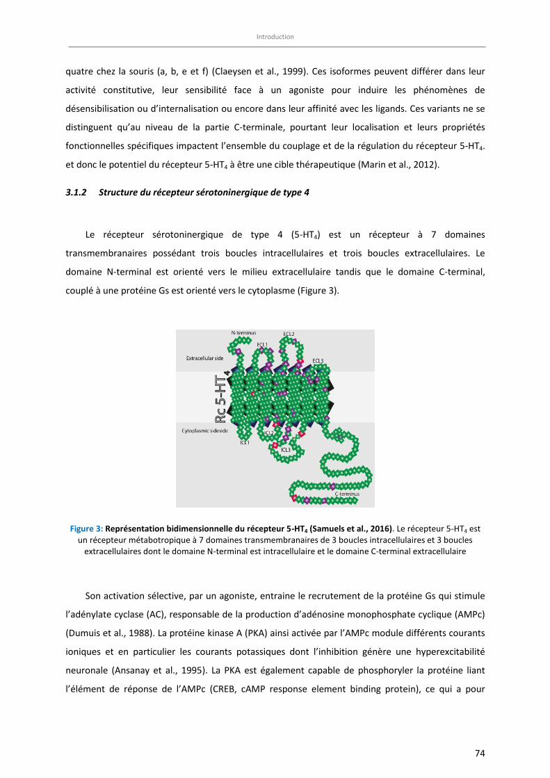
Introduction
74
quatre chez la souris (a, b, e et f) (Claeysen et al., 1999). Ces isoformes peuvent différer dans leur
activité constitutive, leur sensibilité face à un agoniste pour induire les phénomènes de
désensibilisation ou d’internalisation ou encore dans leur affinité avec les ligands. Ces variants ne se
distinguent qu’au niveau de la partie C-terminale, pourtant leur localisation et leurs propriétés
fonctionnelles spécifiques impactent l’ensemble du couplage et de la régulation du récepteur 5-HT4.
et donc le potentiel du récepteur 5-HT4 à être une cible thérapeutique (Marin et al., 2012).
3.1.2 Structure du récepteur sérotoninergique de type 4
Le récepteur sérotoninergique de type 4 (5-HT4) est un récepteur à 7 domaines
transmembranaires possédant trois boucles intracellulaires et trois boucles extracellulaires. Le
domaine N-terminal est orienté vers le milieu extracellulaire tandis que le domaine C-terminal,
couplé à une protéine Gs est orienté vers le cytoplasme (Figure 3).
Figure 3: Représentation bidimensionnelle du récepteur 5-HT4 (Samuels et al., 2016). Le récepteur 5-HT4 est un récepteur métabotropique à 7 domaines transmembranaires de 3 boucles intracellulaires et 3 boucles
extracellulaires dont le domaine N-terminal est intracellulaire et le domaine C-terminal extracellulaire
Son activation sélective, par un agoniste, entraine le recrutement de la protéine Gs qui stimule
l’adénylate cyclase (AC), responsable de la production d’adénosine monophosphate cyclique (AMPc)
(Dumuis et al., 1988). La protéine kinase A (PKA) ainsi activée par l’AMPc module différents courants
ioniques et en particulier les courants potassiques dont l’inhibition génère une hyperexcitabilité
neuronale (Ansanay et al., 1995). La PKA est également capable de phosphoryler la protéine liant
l’élément de réponse de l’AMPc (CREB, cAMP response element binding protein), ce qui a pour
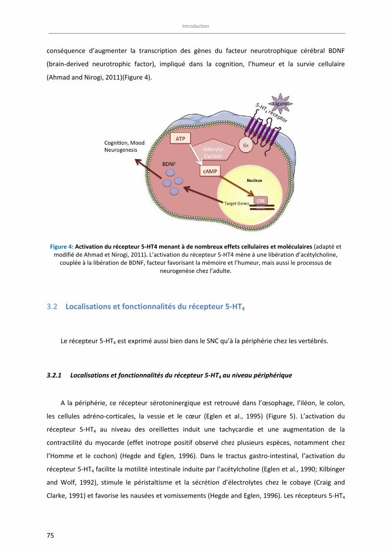
Introduction
75
conséquence d’augmenter la transcription des gènes du facteur neurotrophique cérébral BDNF
(brain-derived neurotrophic factor), impliqué dans la cognition, l’humeur et la survie cellulaire
(Ahmad and Nirogi, 2011)(Figure 4).
Figure 4: Activation du récepteur 5-HT4 menant à de nombreux effets cellulaires et moléculaires (adapté et modifié de Ahmad et Nirogi, 2011). L’activation du récepteur 5-HT4 mène à une libération d’acétylcholine,
couplée à la libération de BDNF, facteur favorisant la mémoire et l’humeur, mais aussi le processus de neurogenèse chez l’adulte.
Localisations et fonctionnalités du récepteur 5-HT4 3.2
Le récepteur 5-HT4 est exprimé aussi bien dans le SNC qu’à la périphérie chez les vertébrés.
3.2.1 Localisations et fonctionnalités du récepteur 5-HT4 au niveau périphérique
A la périphérie, ce récepteur sérotoninergique est retrouvé dans l’œsophage, l’iléon, le colon,
les cellules adréno-corticales, la vessie et le cœur (Eglen et al., 1995) (Figure 5). L’activation du
récepteur 5-HT4 au niveau des oreillettes induit une tachycardie et une augmentation de la
contractilité du myocarde (effet inotrope positif observé chez plusieurs espèces, notamment chez
l’Homme et le cochon) (Hegde and Eglen, 1996). Dans le tractus gastro-intestinal, l’activation du
récepteur 5-HT4 facilite la motilité intestinale induite par l’acétylcholine (Eglen et al., 1990; Kilbinger
and Wolf, 1992), stimule le péristaltisme et la sécrétion d’électrolytes chez le cobaye (Craig and
Clarke, 1991) et favorise les nausées et vomissements (Hegde and Eglen, 1996). Les récepteurs 5-HT4
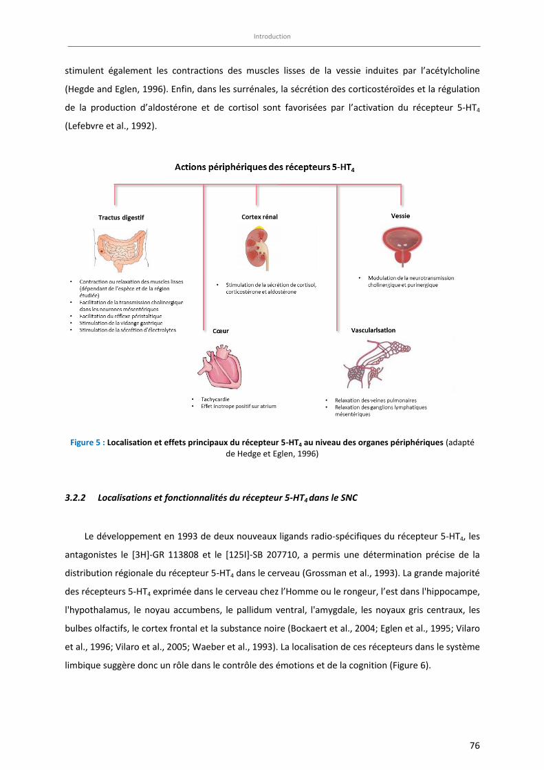
Introduction
76
stimulent également les contractions des muscles lisses de la vessie induites par l’acétylcholine
(Hegde and Eglen, 1996). Enfin, dans les surrénales, la sécrétion des corticostéroïdes et la régulation
de la production d’aldostérone et de cortisol sont favorisées par l’activation du récepteur 5-HT4
(Lefebvre et al., 1992).
Figure 5 : Localisation et effets principaux du récepteur 5-HT4 au niveau des organes périphériques (adapté de Hedge et Eglen, 1996)
3.2.2 Localisations et fonctionnalités du récepteur 5-HT4 dans le SNC
Le développement en 1993 de deux nouveaux ligands radio-spécifiques du récepteur 5-HT4, les
antagonistes le [3H]-GR 113808 et le [125I]-SB 207710, a permis une détermination précise de la
distribution régionale du récepteur 5-HT4 dans le cerveau (Grossman et al., 1993). La grande majorité
des récepteurs 5-HT4 exprimée dans le cerveau chez l’Homme ou le rongeur, l’est dans l'hippocampe,
l'hypothalamus, le noyau accumbens, le pallidum ventral, l'amygdale, les noyaux gris centraux, les
bulbes olfactifs, le cortex frontal et la substance noire (Bockaert et al., 2004; Eglen et al., 1995; Vilaro
et al., 1996; Vilaro et al., 2005; Waeber et al., 1993). La localisation de ces récepteurs dans le système
limbique suggère donc un rôle dans le contrôle des émotions et de la cognition (Figure 6).
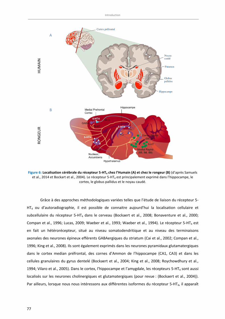
Introduction
77
Figure 6: Localisation cérébrale du récepteur 5-HT4 chez l’Humain (A) et chez le rongeur (B) (d’après Samuels et al., 2014 et Bockart et al., 2004). Le récepteur 5-HT4 est principalement exprimé dans l’hippocampe, le
cortex, le globus pallidus et le noyau caudé.
Grâce à des approches méthodologiques variées telles que l’étude de liaison du récepteur 5-
HT4 ou d’autoradiographie, il est possible de connaitre aujourd’hui la localisation cellulaire et
subcellulaire du récepteur 5-HT4 dans le cerveau (Bockaert et al., 2008; Bonaventure et al., 2000;
Compan et al., 1996; Lucas, 2009; Waeber et al., 1993; Waeber et al., 1994). Le récepteur 5-HT4 est
en fait un hétérorécepteur, situé au niveau somatodendritique et au niveau des terminaisons
axonales des neurones épineux efférents GABAergiques du striatum (Cai et al., 2002; Compan et al.,
1996; King et al., 2008). Ils sont également exprimés dans les neurones pyramidaux glutamatergiques
dans le cortex median préfrontal, des cornes d’Ammon de l’hippocampe (CA1, CA3) et dans les
cellules granulaires du gyrus dentelé (Bockaert et al., 2004; King et al., 2008; Roychowdhury et al.,
1994; Vilaro et al., 2005). Dans le cortex, l’hippocampe et l’amygdale, les récepteurs 5-HT4 sont aussi
localisés sur les neurones cholinergiques et glutamatergiques (pour revue : (Bockaert et al., 2004)).
Par ailleurs, lorsque nous nous intéressons aux différentes isoformes du récepteur 5-HT4, il apparaît
HU
MA
IN
RO
NG
EUR
A
B
Introduction
78
que les isoformes a et b sont exprimées de façon importante dans les régions du système limbique et
notamment des structures qui sont responsables du contrôle des réponses de motivation et de
plaisir et sont affectées dans la dépression comme le noyau accumbens, l’hippocampe et le striatum,
(Bockaert et al., 2008; Marin et al., 2012; Ponimaskin et al., 2002; Qanbar and Bouvier, 2003; Vilaro
et al., 2002). Ces isoformes ont fait l’objet de recherches comme cibles potentielles pour le
développement d’un nouveau traitement antidépresseur d’action rapide (Lucas et al., 2007; Pascual-
Brazo et al., 2012). Cependant, en raison du manque d’anticorps et de ligands spécifiques des
isoformes, la distinction et la caractérisation des propriétés de chacune dans les fonctions de
signalisation et de régulation restent difficiles.
Une revue de la littérature montre que le récepteur 5-HT4 est une cible de choix dans la
régulation des fonctions cérébrales telles que la mémoire et la cognition, les troubles de l’humeur et
les désordres de la prise alimentaire (Ahmad and Nirogi, 2011; Bockaert et al., 2004, 2008; King et al.,
2008; Lucas, 2009; Lucas et al., 2007; Pascual-Brazo et al., 2012). Le rôle du récepteur 5-HT4 dans le
contrôle de la prise alimentaire ne sera pas développé dans le cadre de ce travail. Une attention plus
particulière sera donnée aux implications du récepteur 5-HT4 dans les fonctions émotionnelles et
cognitives.
Implication du récepteur 5-HT4 et de ses ligands dans l’anxiété /dépression 3.1
3.1.1 Implication du récepteur 5-HT4 chez l’Homme dans la dépression
Chez l’Homme, quatre études cliniques supportent le rôle du récepteur 5-HT4 dans la dépression.
L’une d’entre elle mentionne des polymorphismes sur la région variant d’épissage du gène codant
pour le récepteur 5-HT4, associés à la dépression unipolaire (Ohtsuki et al., 2002). Cette étude
d’association menée sur un panel d’hommes et de femmes japonais âgés de 27 à 77 ans, souffrant de
dépression majeure récurrente, a rapporté l’existence d’un lien entre les polymorphismes des
variants issus de l’épissage alternatif du gène codant pour le récepteur 5-HT4 et les dépressions
unipolaires (83097C/T, 83159G/A, 83164(T)9-10 et 83198A/G) (Ohtsuki et al., 2002). En revanche,
l’influence du polymorphisme du récepteur 5-HT4 sur la réponse au traitement antidépresseur ou
anxiolytique n’a pas enocore été étudiée à ce jour. D’autre part, l’association entre les génotypes du
5-HTTLPR (allèle S et/ou allèle L) et la liaison du récepteur 5-HT4 a été mesurée en utilisant un
radioligand ([11C]SB207145) et une technique de tomographie par émission de positron in vivo (Fisher
Introduction
79
et al., 2012). Il a ainsi été montré que la liaison de ce radioligand au récepteur 5-HT4 dans le
néocortex était plus faible chez les homozygotes pour l’allèle S de 5-HTT ou SERT ou les
hétérozygotes que chez les homozygotes pour l’allèle L. Cette donnée est cohérente avec le fait que
l’allèle S conduit à une diminution de la transcription du transporteur de la sérotonine (SERT) menant
à une diminution de l’expression du récepteur 5-HT4. Une autre étude récente, utilisant la même
technique d’imagerie cérébrale, s’est quant à elle intéressée au lien entre le risque familial de
développer un épisode dépressif majeur et l’expression du récepteur 5-HT4 (Madsen et al., 2015). Ce
travail montre qu’un individu dont un ou plusieurs parents au premier degré ont été traités pour un
épisode dépressif présente une diminution de la liaison du radioligand ([11C]SB207145) au récepteur
5-HT4 dans le striatum ainsi que dans d’autres régions limbiques. Ces données confirment ainsi
l’implication du récepteur 5-HT4 dans le mécanisme neurobiologique qui sous-tend le risque familial
de dépression et suggèrent qu’une densité plus faible du récepteur 5-HT4 dans le striatum est
associée à un risque accru de développer un épisode dépressif majeur. Enfin, une autre étude révèle
des altérations des sites de liaison du récepteur 5-HT4 et des concentrations d’AMPc dans plusieurs
régions cérébrales (cortex frontal, hippocampe, noyau caudé et amygdale) chez des patients
déprimés ayant commis un acte de suicide violent (Rosel et al., 2004). Enfin, le nombre de récepteurs
5-HT4 et les taux d’AMPc sont augmentés dans le cortex frontal et le noyau caudé de patients suicidés
sans changement d’affinité de liaison pour le récepteur 5-HT4 (Haahr et al., 2013).
L’expression importante du récepteur 5-HT4 dans le système limbique suggère un rôle
prépondérant de ce récepteur dans la régulation de l’humeur. Dans le domaine préclinique, de
récents travaux indiquent que les désordres psychologiques tels que l’anxiété et/ou la dépression
majeure pourraient bénéficier d’une modulation de la signalisation du récepteur 5-HT4 (Lucas, 2009;
Lucas et al., 2007; Pascual-Brazo et al., 2012).
3.1.2 Implication du récepteur 5-HT4 chez l’animal dans l’anxiété/dépression
Les premières observations impliquant le récepteur 5-HT4 dans la régulation de la réponse
anxiolytique/antidépressive reposent sur des données d’électrophysiologie. En effet, il semble
qu’une administration unique par certains agonistes de ce récepteur (cisapride, prucalopride) chez le
Rat augmente la fréquence de décharge des neurones sérotoninergiques des noyaux du raphé dorsal
(Lucas and Debonnel, 2002). En revanche, lorsque les souris sont privées du récepteur 5-HT4 (souris
KO constitutive R.5-HT4), cette fréquence moyenne de décharge est diminuée de moitié par rapport
aux souris sauvages (Conductier et al., 2006). Par ailleurs, une étude de microdialyse intracérébrale a
montré que l’activation du récepteur 5-HT4 (RS67333, 1,5 mg/kg, i.v.) potentialisait les effets d’une
Introduction
80
administration aiguë de paroxetine (0,5 mg/kg, i.v.), un ISRS, sur les taux de sérotonine
extracellulaire dans l’hippocampe ventral de rat (Licht et al., 2010).
3.1.2.1 Implication du récepteur 5-HT4 et de ses ligands au niveau phénotypique
La caractérisation phénotypique comportementale suite à l’administration unique ou
chronique d’un ligand du récepteur 5-HT4 a permis d’en préciser son rôle. Par exemple, dans
le test de la double enceinte éclairée, un paradigme permettant de mesurer l’activité
anxiolytique d’un ligand, les effets de type anxiolytiques induits par une administration
unique de diazépam (0,16 mg/kg) chez la souris naïve « non-stressée » sont bloqués par des
antagonistes du récepteur 5-HT4 (GR 113808, SB 204070 et SDZ 205-557) et cela de façon
dose-dépendante (Costall and Naylor, 1997). Ces données suggèrent de façon très
surprenante que le récepteur 5-HT4 est impliqué dans l’effet anxiolytique du diazépam.
D’autres études montrent en revanche un effet anxiolytique propre de certains antagonistes
du récepteur 5-HT4 tels que le SB 204070, GR 113808 (Silvestre et al., 1996) et SB 207266A
(Kennett et al., 1997; Silvestre et al., 1996) chez le rat dans le labyrinthe en croix surélevée
(Tableau 3). Les rats traités avec le SB 204070 et le GR 113808 ont montré une augmentation
du pourcentage de temps total passé dans les bras ouverts du labyrinthe. Cependant, il est
difficile de conclure formellement que les antagonistes du récepteur 5-HT4 présentent des
effets de type anxiolytiques, puisque ces exemples issus de la littérature qui indiquent soit
une absence d’effet dans la double enceinte éclairée, soit une réduction des effets anxieux
dans le labyrinthe en croix surélevée, ont été réalisés chez des animaux naïfs.
De manière plus convaincante, il a été montré que chez le rat, l’administration d’agoniste
du récepteur 5-HT4 (RS67333) induit des effets similaires à ceux des ISRSs dans certains tests
prédictifs d’une activité antidépressive, notamment dans le test de la nage forcée chez le Rat
après 3 jours de traitement (Lucas et al., 2007). Ainsi, des agonistes des récepteur du 5-HT4,
tel que le RS67333 (1,5 mg/kg, i.p.) ou le prucalopride (2,5 mg/kg , i.p.) réduisent
significativement le temps d’immobilité d’environ 50% comparé aux animaux contrôles,
tandis que le citalopram (10 mg/kg , i.p.) ne le réduit que de 23% (Lucas et al., 2007)
(Tableau 3 et 4). Dans cette même étude, Lucas et al., ont montré que les réponses
Introduction
81
cellulaires et comportementales qui nécessitent 2 à 3 semaines de traitement
antidépresseur chez le rongeur pourraient apparaitre après seulement 1 à 3 jours de
traitement avec un agoniste sélectif des récepteur du 5-HT4 comme le RS67333. Les mêmes
auteurs ont montré en 2010, à nouveau grâce à des techniques d’électrophysiologie, que la
co-administration d’un ISRSs avec un agoniste du récepteur 5-HT4 potentialise l’effet de type
antidépresseur rapide chez le Rat (Lucas et al., 2010). Très récemment, il a été mis en
évidence qu’un traitement de 7 jours par le RS67333 induit un effet de type antidépresseur
et augmente l’étape de prolifération cellulaire de la neurogenèse hippocampique ainsi que
certains facteurs liés à la neuroplasticité (BDNF, CREB) (Pascual-Brazo et al., 2012). Ces effets
sont d’autant plus remarquables qu’ils sont de même intensité qu’un traitement de 2 à 3
semaines avec des antidépresseurs classiques.
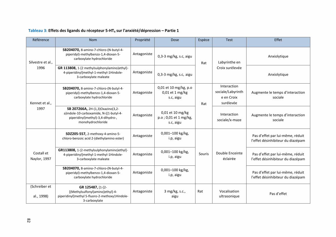
82
Tableau 3: Effets des ligands du récepteur 5-HT4 sur l'anxiété/dépression – Partie 1
Référence Nom Propriété Dose Espèce Test Effet
Silvestre et al.,
1996
SB204070, 8-amino-7-chloro-(N-butyl-4-piperidyl)-methylbenzo-1,4-dioxan-5-
carboxylate hydrochloride Antagoniste
0,3-3 mg/kg, s.c, aigu
Rat Labyrinthe en
Croix surélevée
Anxiolytique
GR 113808, 1-[2 methylsulphonylamino)ethyl]- 4-piperidinyl]methyl-1-methyl-1Hindole-
3-carboxylate maleate Antagoniste
0,3-3 mg/kg, s.c, aigu Anxiolytique
Kennet et al.,
1997
SB204070, 8-amino-7-chloro-(N-butyl-4-
piperidyl)-methylbenzo-1,4-dioxan-5- carboxylate hydrochloride
Antagoniste 0,01 et 10 mg/kg, p.o
0,01 et 1 mg/kg s.c, aigu
Rat
Interaction
sociale/Labyrinth
e en Croix
surélevée
Augmente le temps d’interaction sociale
SB 207266A, 2H-(1,3)Oxazino(3,2- a)indole-10-carboxamide, N-((1-butyl-4-
piperidinyl)methyl)-3,4-dihydro-, monohydrochloride
Antagoniste 0,01 et 10 mg/kg p.o ; 0,01 et 1 mg/kg,
s.c, aigu
Interaction
sociale/x-maze Augmente le temps d’interaction
sociale
Costall et
Naylor, 1997
SDZ205-557, 2-methoxy-4-amino-5-
chloro-benzoic acid 2-(diethylamino ester) Antagoniste
0,001–100 kg/kg, i.p, aigu
Souris Double Enceinte
éclairée
Pas d’effet par lui-même, réduit l’effet désinhibiteur du diazépam
GR113808, 1-[2-methylsulphonylamino)ethyl]- 4-piperidinyl]methyl-1-methyl-1Hindole-
3-carboxylate maleate Antagoniste 0,001–100 kg/kg,
i.p, aigu Pas d’effet par lui-même, réduit l’effet désinhibiteur du diazépam
SB204070, 8-amino-7-chloro-(N-butyl-4- piperidyl)-methylbenzo-1,4-dioxan-5-
carboxylate hydrochloride Antagoniste 0,001–100 kg/kg,
i.p, aigu Pas d’effet par lui-même, réduit l’effet désinhibiteur du diazépam
(Schreiber et
al., 1998)
GR 125487, [1-[2-
[(Methylsulfonyl)amino]ethyl]-4- piperidinyl]methyl 5-fluoro-2-methoxy1Hindole-
3-carboxylate
Antagoniste 3 mg/kg, s.c., aigu
Rat Vocalisation ultrasonique
Pas d’effet
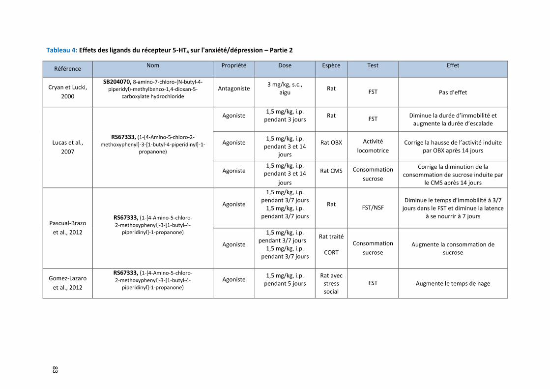
83
Tableau 4: Effets des ligands du récepteur 5-HT4 sur l'anxiété/dépression – Partie 2
Référence Nom Propriété Dose Espèce Test Effet
Cryan et Lucki,
2000
SB204070, 8-amino-7-chloro-(N-butyl-4- piperidyl)-methylbenzo-1,4-dioxan-5-
carboxylate hydrochloride Antagoniste
3 mg/kg, s.c., aigu
Rat FST Pas d’effet
Lucas et al.,
2007
RS67333, (1-[4-Amino-5-chloro-2-
methoxyphenyl]-3-[1-butyl-4-piperidinyl]-1- propanone)
Agoniste 1,5 mg/kg, i.p.
pendant 3 jours Rat
FST Diminue la durée d’immobilité et
augmente la durée d’escalade
Agoniste 1,5 mg/kg, i.p. pendant 3 et 14
jours
Rat OBX Activité
locomotrice Corrige la hausse de l’activité induite
par OBX après 14 jours
Agoniste 1,5 mg/kg, i.p.
pendant 3 et 14
jours
Rat CMS Consommation
sucrose
Corrige la diminution de la consommation de sucrose induite par
le CMS après 14 jours
Pascual-Brazo
et al., 2012
RS67333, (1-[4-Amino-5-chloro-
2-methoxyphenyl]-3-[1-butyl-4- piperidinyl]-1-propanone)
Agoniste
1,5 mg/kg, i.p. pendant 3/7 jours
1,5 mg/kg, i.p. pendant 3/7 jours
Rat FST/NSF
Diminue le temps d’immobilité à 3/7 jours dans le FST et diminue la latence
à se nourrir à 7 jours
Agoniste
1,5 mg/kg, i.p. pendant 3/7 jours
1,5 mg/kg, i.p. pendant 3/7 jours
Rat traité
CORT Consommation
sucrose Augmente la consommation de
sucrose
Gomez-Lazaro
et al., 2012
RS67333, (1-[4-Amino-5-chloro-
2-methoxyphenyl]-3-[1-butyl-4- piperidinyl]-1-propanone)
Agoniste 1,5 mg/kg, i.p.
pendant 5 jours Rat avec
stress social
FST Augmente le temps de nage
Introduction
84
De plus, des molécules de signalisation qui interagissent avec le récepteur 5-HT4 comme la
protéine p11 (S100A10) (Egeland et al., 2011; Warner-Schmidt et al., 2009) pourraient représenter
de nouvelles cibles pour des traitements anxiolytique et/ou antidépresseur à action rapide. De
nombreux éléments récents montrent que les neurones corticaux qui co-expriment p11 et les
récepteurs du 5-HT4 impliqués dans les effets comportementaux des IRSSs (Schmidt et al., 2012) et
qu’un traitement chronique par la fluoxetine induit une augmentation de l’expression des récepteur
du 5-HT4 dans les neurones corticaux. Enfin, dans des tests comportementaux tels que le FST ou le
test de suspension caudale, l’activité antidépressive du RS67333 est absente chez les souris privées
du gène codant pour p11 comparées à leur contrôles WT (Warner-Schmidt et al., 2009).
3.1.2.2 Implication des ligands du récepteur 5-HT4 sur la plasticité cérébrale et la neurogenèse
hippocampique chez l’adulte
En plus des données comportementales, de nombreux changements au niveau de la
plasticité neuronale et notamment au niveau de la neurogenèse hippocampique interviennent avec
l’administration chronique d’antidépresseur monoaminergique (Santarelli et al., 2003; Wang et al.,
2008). Il en est de même avec certains agonistes du récepteur 5-HT4 puisqu’une administration
chronique de RS67333 chez le rongeur favorise l’étape de survie de la neurogenèse hippocampique
(Lucas et al.,2007 ;2009). D’autre part, la stimulation in vitro des récepteurs 5-HT4 par deux autres
agonistes du récepteur 5-HT4 (RS67506 et tegaserod) favorise également la survie neuronale
entérique (Liu et al., 2009). En complément des résultats comportementaux, une étude récente
réalisée chez des rats naïfs a confirmé que 3 jours de traitement avec le RS67333, augmentent de
façon significative la neurogenèse dans la zone sous granulaire du gyrus dentelé de l’hippocampe, un
effet connu pour apparaitre après minimum 2 semaines de traitement aux antidépresseurs
classiques comme les IRSSs (Pascual-Brazo et al., 2012). Ces résultats sont déterminants dans la
compréhension du lien entre les données comportementales et les changements dans la
neurogenèse hippocampique adulte en réponse à un traitement par un antidépresseur. La
neurogenèse hippocampique adulte est impliquée dans certains des effets comportementaux des
antidépresseurs chez le rongeur adulte (David et al., 2009; Santarelli et al., 2003). Cependant, aucune
preuve directe n’a été trouvée pour relier les effets de type antidépresseur de la stimulation du
récepteur 5-HT4 et ses effets neurogéniques.
D’autres données basées sur l’analyse du signal de transduction suite à l’activation du
récepteur 5-HT4 pourraient expliquer l’effet de type antidépresseur des agonistes 5-HT4. Les
Introduction
85
récepteurs 5-HT4 sont couplés à une protéine G(s) qui active l’adénylate cyclase, augmentant alors la
production d’AMPc (Dumuis et al., 1989; Torres et al., 1995) qui à son tour active la protéine kinase A
(PKA) qui phosphoryle le facteur de transcription CREB. De manière intéressante, il a été démontré
qu’un traitement chronique par un antidépresseur activait cette même voie de signalisation (Nibuya
et al., 1995). Il semblerait donc que l’activation de CREB en CREB phosphorylé constitue une étape
clé de la facilitation de la neurogenèse hippocampique, une des autres propriétés caractéristiques
des antidépresseurs (Duman et al., 2001; Malberg et al., 2000).
Le facteur neurotrophique BDNF a également un rôle dans la croissance neuronale et la
plasticité synaptique (McAllister, 1999). Plusieurs études ont montré que les antidépresseurs
régulaient à la hausse la production de BDNF hippocampique (Duman and Monteggia, 2006). Alors
que certains auteurs ont reporté une induction de l’expression de BDNF après un traitement sub-
chronique aux antidépresseurs (Larsen et al., 2008; Musazzi et al., 2009), d’autres études ne
montrent une augmentation du BDNF qu’après un traitement chronique aux antidépresseurs (De
Foubert et al., 2004; Nibuya et al., 1995). L’effet à long-terme des antidépresseurs modulant le
comportement et la survie des nouveaux neurones est médié au moins en partie par le BDNF. De
plus, dans la mesure où l’activation du récepteur 5-HT4 peut favoriser l’expression de BDNF, son
activation pourrait mener à des effets de type antidépresseurs BDNF-dépendants, sur le
comportement et la neuroplasticité. Une étude récente a démontré une augmentation de
l’expression du BDNF après 3 jours de traitement avec le RS67333 et une accumulation significative
de la protéine du BDNF dans l’ensemble de la structure de l’hippocampe après un traitement de 7
jours (Pascual-Brazo et al., 2012).
D’autre part, il a été fortement suggéré que le second messager AMPc était impliqué dans le
mécanisme d’action des antidépresseurs (Donati and Rasenick, 2003). Une diminution significative de
la production d’APMc dépendante du récepteur 5-HT4 a été rapporté après un traitement chronique
de 3 semaines par de la fluoxetine, de la venlafaxine (Vidal et al., 2010) ou de l’imipramine (Reierson
et al., 2009), probablement reflétant le processus de désensibilisation, dû à un couplage aux
protéines moins efficaces. Une étude a également reporté qu’un traitement de 7 jours par une
stimulation du récepteur 5-HT4 est nécessaire pour désensibiliser entièrement la voie de signalisation
post-récepteur associée aux récepteurs 5-HT4, en accord avec les résultats obtenus dans les tests
comportementaux prédictifs d’une réponse chronique (Pascual-Brazo et al., 2012).
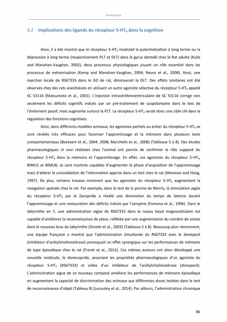
Introduction
86
Implications des ligands du récepteur 5-HT4 dans la cognition 3.2
Ainsi, il a été montré que le récepteur 5-HT4 modulait la potentialisation à long terme ou la
dépression à long terme (respectivement PLT et DLT) dans le gyrus dentelé chez le Rat adulte (Kulla
and Manahan-Vaughan, 2002), deux processus physiologiques jouant un rôle essentiel dans les
processus de mémorisation (Kemp and Manahan-Vaughan, 2004; Neves et al., 2008). Ainsi, une
injection locale de RS67333 dans le GD de rat, diminuerait la DLT. Des effets similaires ont été
observés chez des rats anesthésiés en utilisant un autre agoniste sélective du récepteur 5-HT4 appelé
SC 53116 (Matsumoto et al., 2001). L’injection intracérébroventriculaire de SC 53116 corrige non
seulement les déficits cognitifs induits par un pré-traitement de scopolamaine dans le test de
l’évitement passif, mais augmente surtout la PLT. Le récepteur 5-HT4 serait donc une cible clé dans la
régulation des fonctions cognitives.
Ainsi, dans différents modèles animaux, les agonistes partiels ou entier du récepteur 5-HT4 se
sont révélés très efficaces pour favoriser l’apprentissage et la mémoire dans plusieurs tests
comportementaux (Bockaert et al., 2004, 2008; Marchetti et al., 2008) (Tableaux 5 à 8). Des études
pharmacologiques in vivo réalisées chez l’animal ont permis de confirmer le rôle supposé du
récepteur 5-HT4 dans la mémoire et l’apprentissage. En effet, ces agonistes du récepteur 5-HT4,
BIMU1 et BIMU8, se sont montrés capables d’augmenter la phase d’acquisition de l’apprentissage
mais d’altérer la consolidation de l’information apprise dans un test chez le rat (Meneses and Hong,
1997). De plus, certains travaux montrent que les agonistes du récepteur 5-HT4 augmentent la
navigation spatiale chez le rat. Par exemple, dans le test de la piscine de Morris, la stimulation aigüe
du récepteur 5-HT4 par le Zacopride a révélé une diminution du temps de latence durant
l’apprentissage et une restauration des déficits induits par l’atropine (Fontana et al., 1996). Dans le
labyrinthe en Y, une administration aigüe de RS67333 dans le noyau basal magnocellulaire est
capable d’améliorer la reconnaissance de place, reflétée par une augmentation du nombre de visites
dans le nouveau bras du labyrinthe (Orsetti et al., 2003) (Tableaux 5 à 8). Beaucoup plus récemment,
une équipe française a montré que l’administration simultanée du RS67333 avec le donepezil
(inhibiteur d’acétylcholinestérase) provoquait un effet synergique sur les performances de mémoire
de type épisodique chez le rat (Freret et al., 2012). Ces mêmes auteurs ont alors développé une
nouvelle molécule, le donecopride, associant les propriétés pharmacologiques d’un agoniste du
récepteur 5-HT4 (RS67333) et celles d’un inhibiteur de l’acétylcholinestérase (donepezil).
L’administration aigue de ce nouveau composé améliore les performances de mémoire épisodique
en augmentant la capacité de discrimination des animaux aux différentes doses testées dans le test
de reconnaissance d’objet (Tableau 8) (Lecoutey et al., 2014). Par ailleurs, l’administration chronique

Introduction
87
de RS67333 aux doses de 0,3 et de 1 mg/kg pendant 14 jours, dans le test de reconnaissance d’objet
a mis en évidence une amélioration de l’indice de discrimination chez la souris (Quiedeville et al.,
2015).
Enfin, chez des macaques jeunes et âgés, l’agoniste RS17107 a induit des améliorations de la
mémoire associative dans un tâche cognitive « delayed-matching-to-sample task » dans lequel les
singes devaient apprendre à associer correctement des couleurs pour obtenir une récompense
alimentaire (Terry et al., 1998). L’efficacité des agonistes du récepteur 5-HT4 chez les singes âgés
supporte l’utilisation potentielle de ces composés dans le traitement des désordres cognitifs.
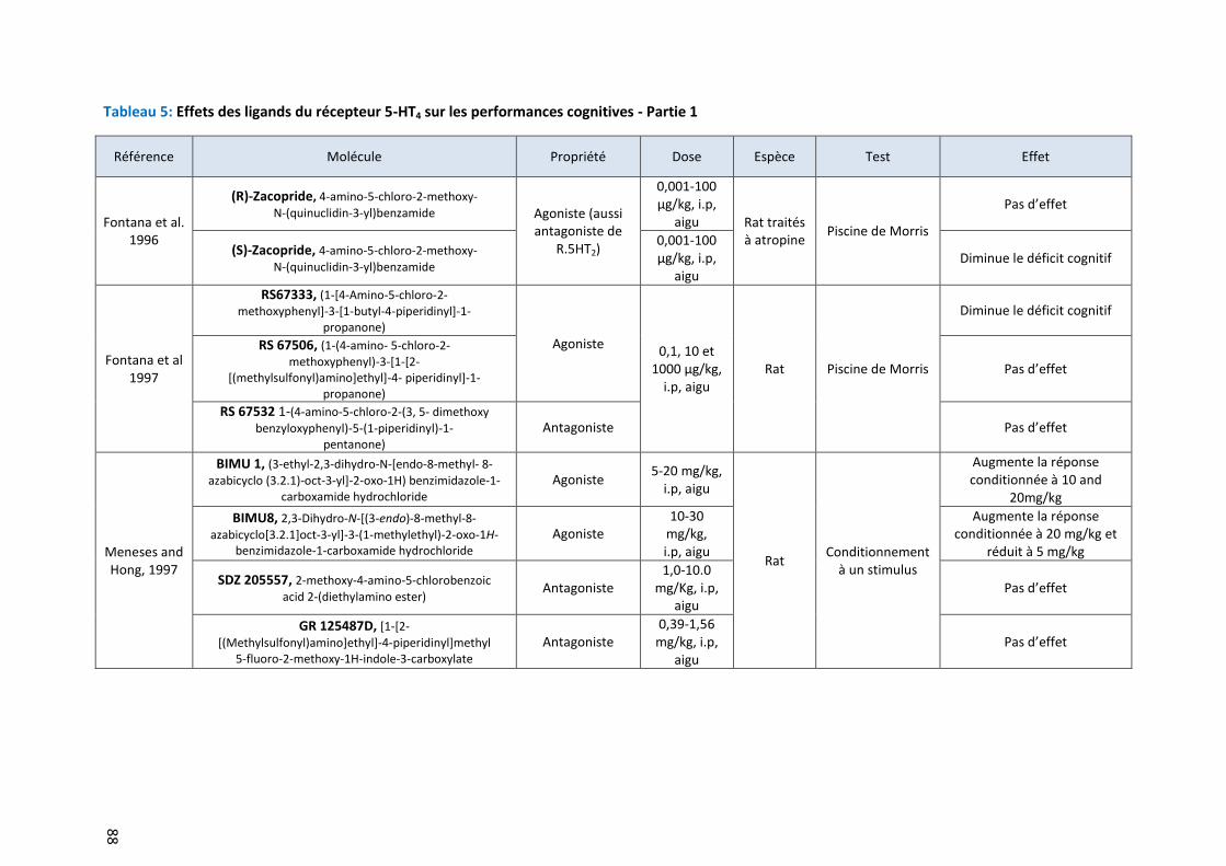
88
Tableau 5: Effets des ligands du récepteur 5-HT4 sur les performances cognitives - Partie 1
Référence Molécule Propriété Dose Espèce Test Effet
Fontana et al. 1996
(R)-Zacopride, 4-amino-5-chloro-2-methoxy-
N-(quinuclidin-3-yl)benzamide Agoniste (aussi antagoniste de
R.5HT2)
0,001-100 μg/kg, i.p,
aigu Rat traités à atropine
Piscine de Morris
Pas d’effet
(S)-Zacopride, 4-amino-5-chloro-2-methoxy-
N-(quinuclidin-3-yl)benzamide
0,001-100 μg/kg, i.p,
aigu Diminue le déficit cognitif
Fontana et al 1997
RS67333, (1-[4-Amino-5-chloro-2- methoxyphenyl]-3-[1-butyl-4-piperidinyl]-1-
propanone) Agoniste
0,1, 10 et 1000 μg/kg,
i.p, aigu Rat Piscine de Morris
Diminue le déficit cognitif
RS 67506, (1-(4-amino- 5-chloro-2- methoxyphenyl)-3-[1-[2-
[(methylsulfonyl)amino]ethyl]-4- piperidinyl]-1- propanone)
Pas d’effet
RS 67532 1-(4-amino-5-chloro-2-(3, 5- dimethoxy
benzyloxyphenyl)-5-(1-piperidinyl)-1- pentanone)
Antagoniste Pas d’effet
Meneses and Hong, 1997
BIMU 1, (3-ethyl-2,3-dihydro-N-[endo-8-methyl- 8-
azabicyclo (3.2.1)-oct-3-yl]-2-oxo-1H) benzimidazole-1-carboxamide hydrochloride
Agoniste 5-20 mg/kg,
i.p, aigu
Rat Conditionnement
à un stimulus
Augmente la réponse conditionnée à 10 and
20mg/kg
BIMU8, 2,3-Dihydro-N-[(3-endo)-8-methyl-8-
azabicyclo[3.2.1]oct-3-yl]-3-(1-methylethyl)-2-oxo-1H-benzimidazole-1-carboxamide hydrochloride
Agoniste 10-30
mg/kg, i.p, aigu
Augmente la réponse conditionnée à 20 mg/kg et
réduit à 5 mg/kg
SDZ 205557, 2-methoxy-4-amino-5-chlorobenzoic
acid 2-(diethylamino ester) Antagoniste
1,0-10.0 mg/Kg, i.p,
aigu Pas d’effet
GR 125487D, [1-[2-
[(Methylsulfonyl)amino]ethyl]-4-piperidinyl]methyl 5-fluoro-2-methoxy-1H-indole-3-carboxylate
Antagoniste 0,39-1,56
mg/kg, i.p, aigu
Pas d’effet
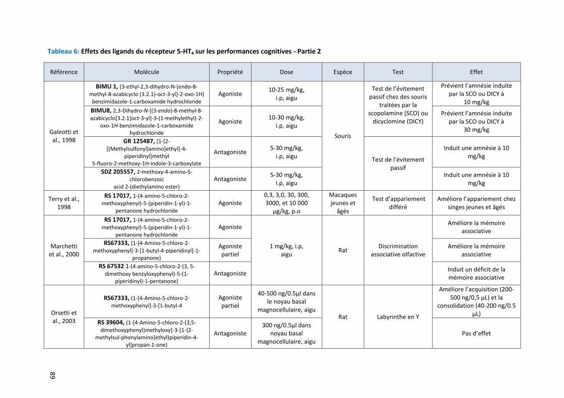
89
Tableau 6: Effets des ligands du récepteur 5-HT4 sur les performances cognitives - Partie 2
Référence Molécule Propriété Dose Espèce Test Effet
Galeotti et al., 1998
BIMU 1, (3-ethyl-2,3-dihydro-N-[endo-8-
methyl-8-azabicyclo (3.2.1)-oct-3-yl]-2-oxo-1H) benzimidazole-1-carboxamide hydrochloride
Agoniste 10-25 mg/kg,
i.p, aigu
Souris
Test de l’évitement passif chez des souris
traitées par la scopolamine (SCO) ou
dicyclomine (DICY)
Prévient l’amnésie induite par la SCO ou DICY à
10 mg/kg
BIMU8, 2,3-Dihydro-N-[(3-endo)-8-methyl-8-
azabicyclo[3.2.1]oct-3-yl]-3-(1-methylethyl)-2-oxo-1H-benzimidazole-1-carboxamide
hydrochloride
Agoniste 10-30 mg/kg,
i.p, aigu
Prévient l’amnésie induite par la SCO ou DICY à
30 mg/kg
GR 125487, [1-[2-
[(Methylsulfonyl)amino]ethyl]-4-piperidinyl]methyl
5-fluoro-2-methoxy-1H-indole-3-carboxylate
Antagoniste 5-30 mg/kg,
i.p, aigu Test de l’évitement
passif
Induit une amnésie à 10 mg/kg
SDZ 205557, 2-methoxy-4-amino-5-chlorobenzoic
acid 2-(diethylamino ester)
Antagoniste 5-30 mg/kg,
i.p, aigu Induit une amnésie à 10
mg/kg
Terry et al., 1998
RS 17017, 1-(4-amino-5-chloro-2- methoxyphenyl)-5-(piperidin-1-yl)-1-
pentanone hydrochloride
Agoniste 0,3, 3,0, 30, 300, 3000, et 10 000
μg/kg, p.o
Macaques jeunes et
âgés
Test d’appariement différé
Améliore l’appariement chez singes jeunes et âgés
Marchetti et al., 2000
RS 17017, 1-(4-amino-5-chloro-2-
methoxyphenyl)-5-(piperidin-1-yl)-1- pentanone hydrochloride
Agoniste
1 mg/kg, i.p, aigu
Rat Discrimination
associative olfactive
Améliore la mémoire associative
RS67333, (1-[4-Amino-5-chloro-2-
methoxyphenyl]-3-[1-butyl-4-piperidinyl]-1- propanone)
Agoniste partiel
Améliore la mémoire associative
RS 67532 1-(4-amino-5-chloro-2-(3, 5-
dimethoxy benzyloxyphenyl)-5-(1- piperidinyl)-1-pentanone)
Antagoniste Induit un déficit de la mémoire associative
Orsetti et al., 2003
RS67333, (1-[4-Amino-5-chloro-2- methoxyphenyl]-3-[1-butyl-4
Agoniste partiel
40-500 ng/0.5μl dans le noyau basal
magnocellulaire, aigu Rat Labyrinthe en Y
Améliore l’acquisition (200-500 ng/0,5 µL) et la
consolidation (40-200 ng/0.5 μL)
RS 39604, (1-[4-Amino-5-chloro-2-(3,5-
dimethoxyphenyl}methyloxy]-3-[1-{2-methylsul-phonylamino]ethyl)piperidin-4-
yl]propan-1-one)
Antagoniste 300 ng/0.5μl dans
noyau basal magnocellulaire, aigu
Pas d’effet
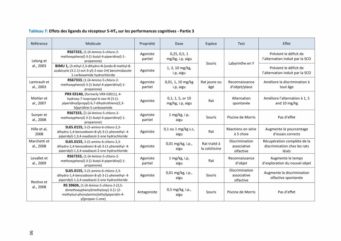
90
Tableau 7: Effets des ligands du récepteur 5-HT4 sur les performances cognitives - Partie 3
Référence Molécule Propriété Dose Espèce Test Effet
Lelong et al., 2003
RS67333, (1-[4-Amino-5-chloro-2-
methoxyphenyl]-3-[1-butyl-4-piperidinyl]-1-propanone)
Agoniste partiel
0,25, 0,5, 1 mg/kg, i.p, aigu
Souris Labyrinthe en Y
Prévient le déficit de l’alternation induit par la SCO
BIMU 1, (3-ethyl-2,3-dihydro-N-[endo-8-methyl-8-
azabicyclo (3.2.1)-oct-3-yl]-2-oxo-1H) benzimidazole-1-carboxamide hydrochloride
Agoniste 1, 3, 10 mg/kg,
i.p, aigu Prévient le déficit de
l’alternation induit par la SCO
Lamirault et al., 2003
RS67333, (1-[4-Amino-5-chloro-2-
methoxyphenyl]-3-[1-butyl-4-piperidinyl]-1- propanone)
Agoniste partiel
0,01, 1, 10 mg/kg i.p, aigu
Rat jeune ou âgé
Reconnaissance d’objet/place
Améliore la discrimination à tout âge
Mohler et al., 2007
PRX 03140, (formerly VRX-03011), 4-
hydroxy-7-isopropyl-6-oxo-N-[3-(1- piperidinyl)propyl]-6,7-dihydrothieno[2,3-
b]pyridine-5-carboxamide
Agoniste 0,1, 1, 5, or 10 mg/kg, i.p, aigu
Rat Alternation spontanée
Améliore l’alternation à 1, 5 and 10 mg/kg
Sunyer et al., 2008
RS67333, (1-[4-Amino-5-chloro-2-
methoxyphenyl]-3-[1-butyl-4-piperidinyl]-1- propanone)
Agoniste partiel
1 mg/kg, i.p, aigu
Souris Piscine de Morris Pas d’effet
Hille et al, 2008
SL65.0155, 5-(5-amino-6-chloro-2,3-
dihydro-1,4-benzodioxin-8-yl)-3-(1-phenethyl- 4-piperidyl)-1,3,4-oxadiazol-2-one hydrochloride
Agoniste 0,1 ou 1 mg/kg s.c,
aigu Rat
Réactions en série à 5 choix
Augmente le pourcentage d’essais corrects
Marchetti et al., 2008
SL65.0155, 5-(5-amino-6-chloro-2,3-
dihydro-1,4-benzodioxin-8-yl)-3-(1-phenethyl- 4-piperidyl)-1,3,4-oxadiazol-2-one hydrochloride
Agoniste 0,01 mg/kg, i.p.,
aigu Rat traité à
la colchicine
Discrimination associative
olfactive
Récupération complète de la discrimination chez les rats
lésés
Levallet et al., 2009
RS67333, (1-[4-Amino-5-chloro-2-
methoxyphenyl]-3-[1-butyl-4-piperidinyl]-1- propanone)
Agoniste partiel
1 mg/kg, i.p, aigu
Rat Reconnaissance
d’objet Augmente le temps
d’exploration du nouvel objet
Restivo et al., 2008
SL65.0155, 5-(5-amino-6-chloro-2,3-
dihydro-1,4-benzodioxin-8-yl)-3-(1-phenethyl- 4-piperidyl)-1,3,4-oxadiazol-2-one hydrochloride
Agoniste 0,01 mg/kg, i.p.,
aigu Souris
Discrimination associative
olfactive
Augmente la discrimination olfactive spontanée
RS 39604, (1-[4-Amino-5-chloro-2-(3,5-
dimethoxyphenyl}methyloxy]-3-[1-{2- methylsul-phonylamino]ethyl)piperidin-4-
yl]propan-1-one)
Antagoniste 0,5 mg/kg, i.p.,
aigu Souris Piscine de Morris Pas d’effet
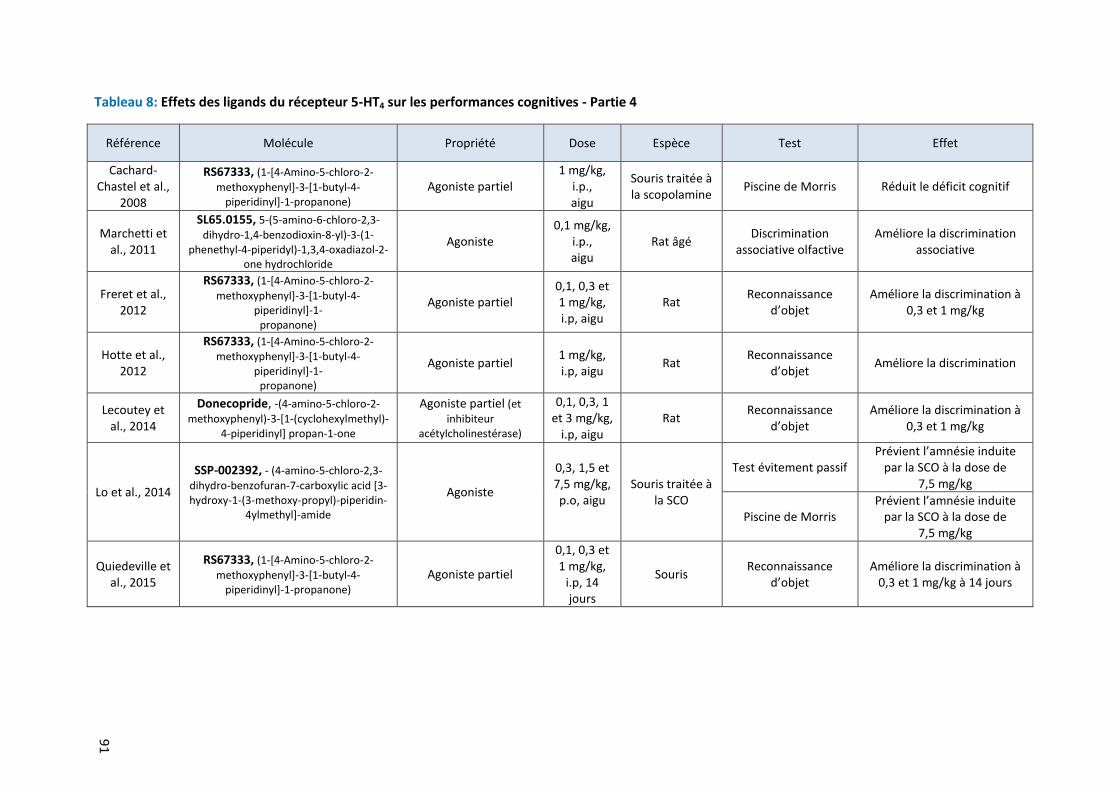
91
Tableau 8: Effets des ligands du récepteur 5-HT4 sur les performances cognitives - Partie 4
Référence Molécule Propriété Dose Espèce Test Effet
Cachard-Chastel et al.,
2008
RS67333, (1-[4-Amino-5-chloro-2-
methoxyphenyl]-3-[1-butyl-4-piperidinyl]-1-propanone)
Agoniste partiel 1 mg/kg,
i.p., aigu
Souris traitée à la scopolamine
Piscine de Morris Réduit le déficit cognitif
Marchetti et al., 2011
SL65.0155, 5-(5-amino-6-chloro-2,3-
dihydro-1,4-benzodioxin-8-yl)-3-(1-phenethyl-4-piperidyl)-1,3,4-oxadiazol-2-
one hydrochloride
Agoniste 0,1 mg/kg,
i.p., aigu
Rat âgé Discrimination
associative olfactive Améliore la discrimination
associative
Freret et al., 2012
RS67333, (1-[4-Amino-5-chloro-2-
methoxyphenyl]-3-[1-butyl-4-piperidinyl]-1-
propanone)
Agoniste partiel 0,1, 0,3 et 1 mg/kg, i.p, aigu
Rat Reconnaissance
d’objet Améliore la discrimination à
0,3 et 1 mg/kg
Hotte et al., 2012
RS67333, (1-[4-Amino-5-chloro-2- methoxyphenyl]-3-[1-butyl-4-
piperidinyl]-1- propanone)
Agoniste partiel 1 mg/kg, i.p, aigu
Rat Reconnaissance
d’objet Améliore la discrimination
Lecoutey et al., 2014
Donecopride, -(4-amino-5-chloro-2-
methoxyphenyl)-3-[1-(cyclohexylmethyl)-4-piperidinyl] propan-1-one
Agoniste partiel (et
inhibiteur acétylcholinestérase)
0,1, 0,3, 1 et 3 mg/kg,
i.p, aigu Rat
Reconnaissance d’objet
Améliore la discrimination à 0,3 et 1 mg/kg
Lo et al., 2014
SSP-002392, - (4-amino-5-chloro-2,3-
dihydro-benzofuran-7-carboxylic acid [3-hydroxy-1-(3-methoxy-propyl)-piperidin-
4ylmethyl]-amide
Agoniste
0,3, 1,5 et 7,5 mg/kg, p.o, aigu
Souris traitée à la SCO
Test évitement passif Prévient l’amnésie induite
par la SCO à la dose de 7,5 mg/kg
Piscine de Morris Prévient l’amnésie induite
par la SCO à la dose de 7,5 mg/kg
Quiedeville et al., 2015
RS67333, (1-[4-Amino-5-chloro-2-
methoxyphenyl]-3-[1-butyl-4-piperidinyl]-1-propanone)
Agoniste partiel
0,1, 0,3 et 1 mg/kg,
i.p, 14 jours
Souris Reconnaissance
d’objet Améliore la discrimination à
0,3 et 1 mg/kg à 14 jours
Introduction
92
Par ailleurs, il a été montré que l’activation de la protéine CREB, cible de la voie de
signalisation du récepteur 5-HT4, est un important médiateur de la formation de la mémoire et des
effets pro-cognitifs (Bockaert et al., 2004, 2008). L’activation du récepteur 5-HT4 améliore favorise la
libération d’acétylcholine. Ainsi, l’équipe de Consolo a été la première à démontrer par des études de
microdialyse in vivo chez le rat, que les agonistes du récepteur 5-HT4, BIMU1 et BIMU8, étaient
capable d’augmenter les concentrations d’acétylcholine dans les dialysats (Consolo et al., 1994). Par
la suite, d’autres études ont confirmé ces résultats, et précisèrent que les agonistes du récepteur 5-
HT4 facilitaient la libération d’acétylcholine (Letty et al., 1997; Orsetti et al., 2007; Yamaguchi et al.,
1997). Ces travaux montrent que les récepteurs 5-HT4 peuvent donc améliorer la formation de
l’apprentissage et de la mémoire en favorisant la libération synaptique d’acétylcholine dans le
cerveau (Bockaert et al., 2008; King et al., 2008; Lucas, 2009).
Enfin, les agonistes du récepteur 5-HT4 se sont révélés être de très intéressantes cibles dans
la recherche de stratégies visant à traiter la maladie d’Alzheimer, caractérisée par la présence de
troubles cognitifs variés. Des études in vitro évaluant le potentiel du RS67333 montrent que
l’activation sélective du récepteur 5-HT4 inhibe la sécrétion des peptides β-amyloïdes et augmente la
survie neuronale dans des cultures de neurones corticaux de souris transgéniques Tg2576 (Cho and
Hu, 2007). D’autre part, les récepteurs 5-HT4 activent également le facteur d’échange Epac, qui
stimule l’activité de l’α-sécrétase et la libération de la protéine précurseur amyloide soluble (sAPP),
ayant des propriétés neuroprotectrices et pro-cognitives (King et al., 2008; Marin et al., 2012).
Récemment, l’évaluation préclinique in vivo du donécopride, une molécule inhibitrice de
l’acétylcholinestérase (donépézil) et agoniste du récepteur 5-HT4 (RS67333) a démontré son grand
potentiel dans le traitement de la maladie d’Alzheimer, révélé par une amélioration significative des
performances cognitives dans le test de reconnaissance d’objets aux deux doses étudiées (Lecoutey
et al., 2014). De nouvelles molécules dérivées des agonistes du récepteur 5-HT4 sont en cours de
synthèse et d’évaluation et pourraient représenter une grande avancée dans la mise en place de
nouvelles thérapies dans la maladie d’Alzheimer (Brodney et al., 2012; Nirogi et al., 2015; Rochais et
al., 2015).
Ces résultats obtenus par des études précliniques sont à relier à une étude clinique réalisée
en postmortem chez des patients atteints de la maladie d’Alzheimer (Tsang et al., 2010), démontrant
une diminution de la densité de récepteurs 5-HT4 dans les régions corticales du cerveau. Ces données
chez l’Homme confirment le rôle prometteur des agonistes du récepteur 5-HT4 dans le traitement de
la pathologie d’Alzheimer.
Introduction
93
Au final, si les études précliniques montrent un certain attrait pour l’utilisation des agonistes
du récepteur 5-HT4 dans le traitement des épisodes dépressifs, la quasi majorité de ces travaux a été
réalisée chez des animaux naïfs. Il est donc nécessaire de procéder à une caractérisation complète
des conséquences d’un traitement chronique par un agoniste du récepteur 5-HT4 dans un modèle
d’anxiété/dépression. Par ailleurs, en plus des données de comportement, il a été proposé qu'une
courte période de traitement avec des agonistes du récepteur 5-HT4 augmentait l'une des étapes de
la neurogenèse hippocampique adulte, la prolifération de cellules progénitrices dans GD (Pascual-
Brazo et al., 2012). Si certains des effets comportementaux des antidépresseurs monoaminergiques
ont été associés à l’augmentation de la neurogenèse hippocampique, aucune donnée à ce jour ne
relie les effets antidépresseurs induits par l'activation du récepteur 5-HT4 et ses effets
neurogéniques.
4 La protéine β-arrestine 1
Conformément à l'idée que les antidépresseurs exercent des effets dans de multiples régions
cérébrales, il a été démontré qu'un traitement par de la fluoxetine chez des animaux sous un régime
de corticostérone, provoquait des effets sur l'expression de gènes, non seulement dans
l’hippocampe, mais aussi dans l’hypothalamus et l’amygdale chez la souris adulte (David et al., 2009).
Parmi les gènes étudiés, ceux codant pour les β-arrestines 1 et 2 montraient des changements
d’expression dans ces régions cérébrales. Les protéines β-arrestines 1 et 2 pourraient donc être des
déterminants moléculaires importants impliqués dans la réponse thérapeutique dans le cadre d’un
épisode de dépression majeure. Dans cette thèse, seule la β-arrestine 1 sera développée.
Généralités sur les protéines β-arrestines 4.1
Les arrestines constituent une famille de protéines qui sont capables d’interagir avec les
récepteurs couplés aux protéines G (RCPG) suite à l’activation de ceux-ci par un agoniste (DeWire et
al., 2007). Quatre membres de la famille des arrestines ont été identifiés :
- Arrestine 1 ou arrestine visuelle codée par le gène SAG
- Arrestine 2 ou β-arrestine 1 codée par le gène ARRB1
- Arrestine 3 ou β-arrestine 2 codée par le gène ARRB2
- Arrestine 4 ou arrestine conale codée par le gène ARR3
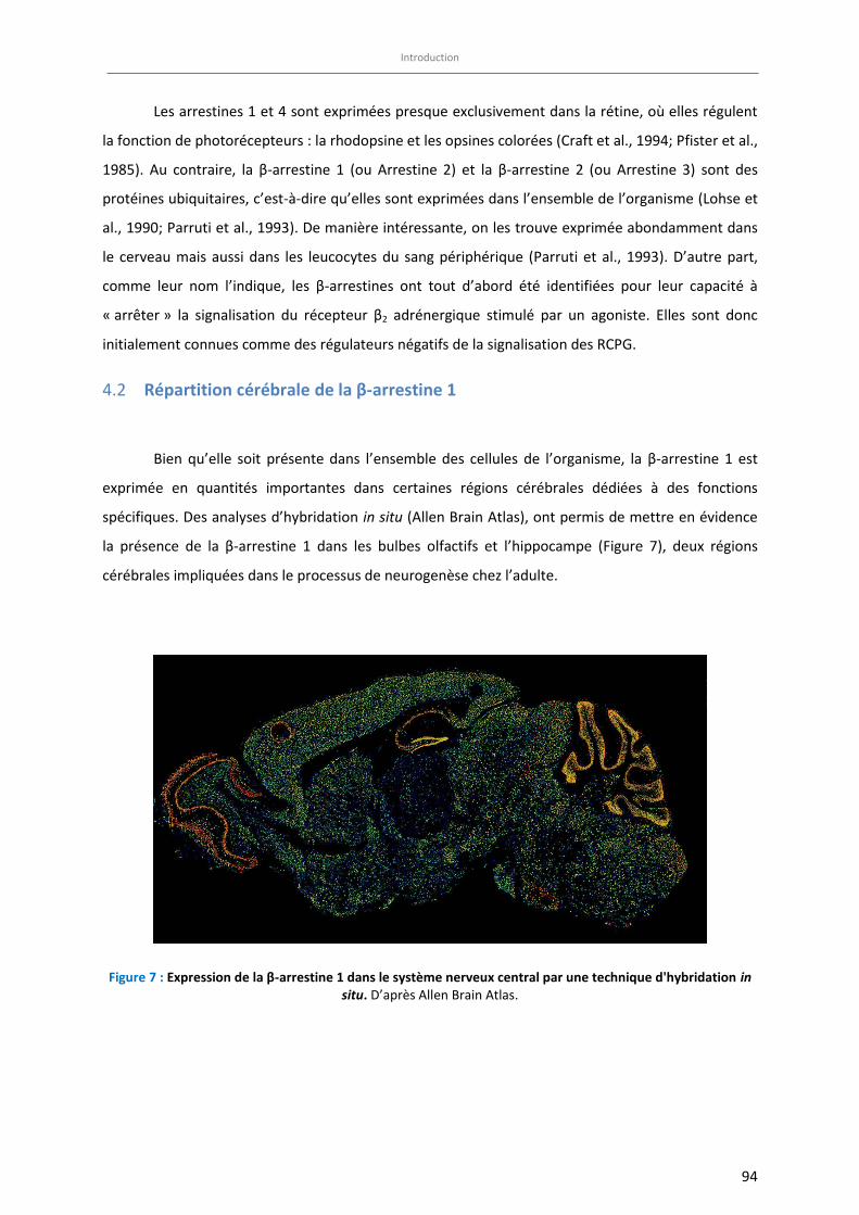
Introduction
94
Les arrestines 1 et 4 sont exprimées presque exclusivement dans la rétine, où elles régulent
la fonction de photorécepteurs : la rhodopsine et les opsines colorées (Craft et al., 1994; Pfister et al.,
1985). Au contraire, la β-arrestine 1 (ou Arrestine 2) et la β-arrestine 2 (ou Arrestine 3) sont des
protéines ubiquitaires, c’est-à-dire qu’elles sont exprimées dans l’ensemble de l’organisme (Lohse et
al., 1990; Parruti et al., 1993). De manière intéressante, on les trouve exprimée abondamment dans
le cerveau mais aussi dans les leucocytes du sang périphérique (Parruti et al., 1993). D’autre part,
comme leur nom l’indique, les β-arrestines ont tout d’abord été identifiées pour leur capacité à
« arrêter » la signalisation du récepteur β2 adrénergique stimulé par un agoniste. Elles sont donc
initialement connues comme des régulateurs négatifs de la signalisation des RCPG.
Répartition cérébrale de la β-arrestine 1 4.2
Bien qu’elle soit présente dans l’ensemble des cellules de l’organisme, la β-arrestine 1 est
exprimée en quantités importantes dans certaines régions cérébrales dédiées à des fonctions
spécifiques. Des analyses d’hybridation in situ (Allen Brain Atlas), ont permis de mettre en évidence
la présence de la β-arrestine 1 dans les bulbes olfactifs et l’hippocampe (Figure 7), deux régions
cérébrales impliquées dans le processus de neurogenèse chez l’adulte.
Figure 7 : Expression de la β-arrestine 1 dans le système nerveux central par une technique d'hybridation in situ. D’après Allen Brain Atlas.
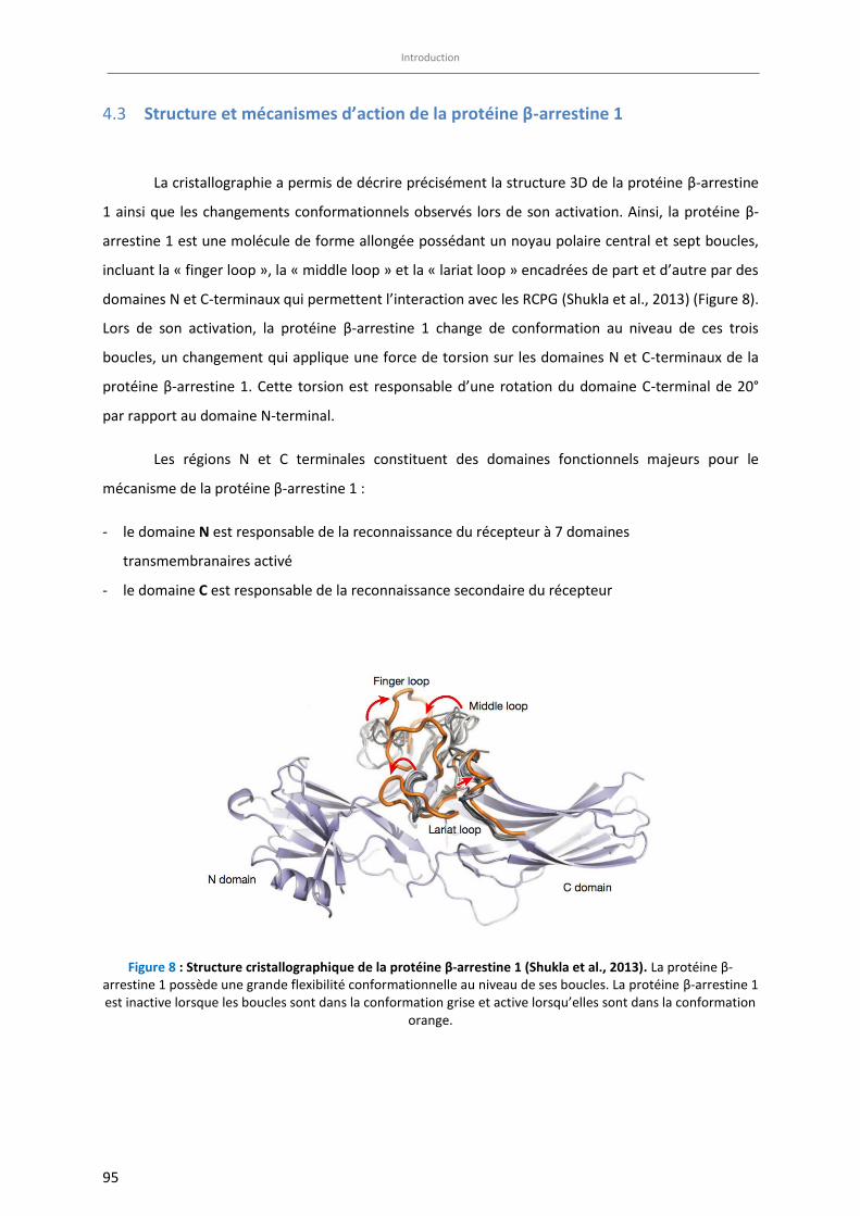
Introduction
95
Structure et mécanismes d’action de la protéine β-arrestine 1 4.3
La cristallographie a permis de décrire précisément la structure 3D de la protéine β-arrestine
1 ainsi que les changements conformationnels observés lors de son activation. Ainsi, la protéine β-
arrestine 1 est une molécule de forme allongée possédant un noyau polaire central et sept boucles,
incluant la « finger loop », la « middle loop » et la « lariat loop » encadrées de part et d’autre par des
domaines N et C-terminaux qui permettent l’interaction avec les RCPG (Shukla et al., 2013) (Figure 8).
Lors de son activation, la protéine β-arrestine 1 change de conformation au niveau de ces trois
boucles, un changement qui applique une force de torsion sur les domaines N et C-terminaux de la
protéine β-arrestine 1. Cette torsion est responsable d’une rotation du domaine C-terminal de 20°
par rapport au domaine N-terminal.
Les régions N et C terminales constituent des domaines fonctionnels majeurs pour le
mécanisme de la protéine β-arrestine 1 :
- le domaine N est responsable de la reconnaissance du récepteur à 7 domaines
transmembranaires activé
- le domaine C est responsable de la reconnaissance secondaire du récepteur
Figure 8 : Structure cristallographique de la protéine β-arrestine 1 (Shukla et al., 2013). La protéine β-arrestine 1 possède une grande flexibilité conformationnelle au niveau de ses boucles. La protéine β-arrestine 1 est inactive lorsque les boucles sont dans la conformation grise et active lorsqu’elles sont dans la conformation
orange.
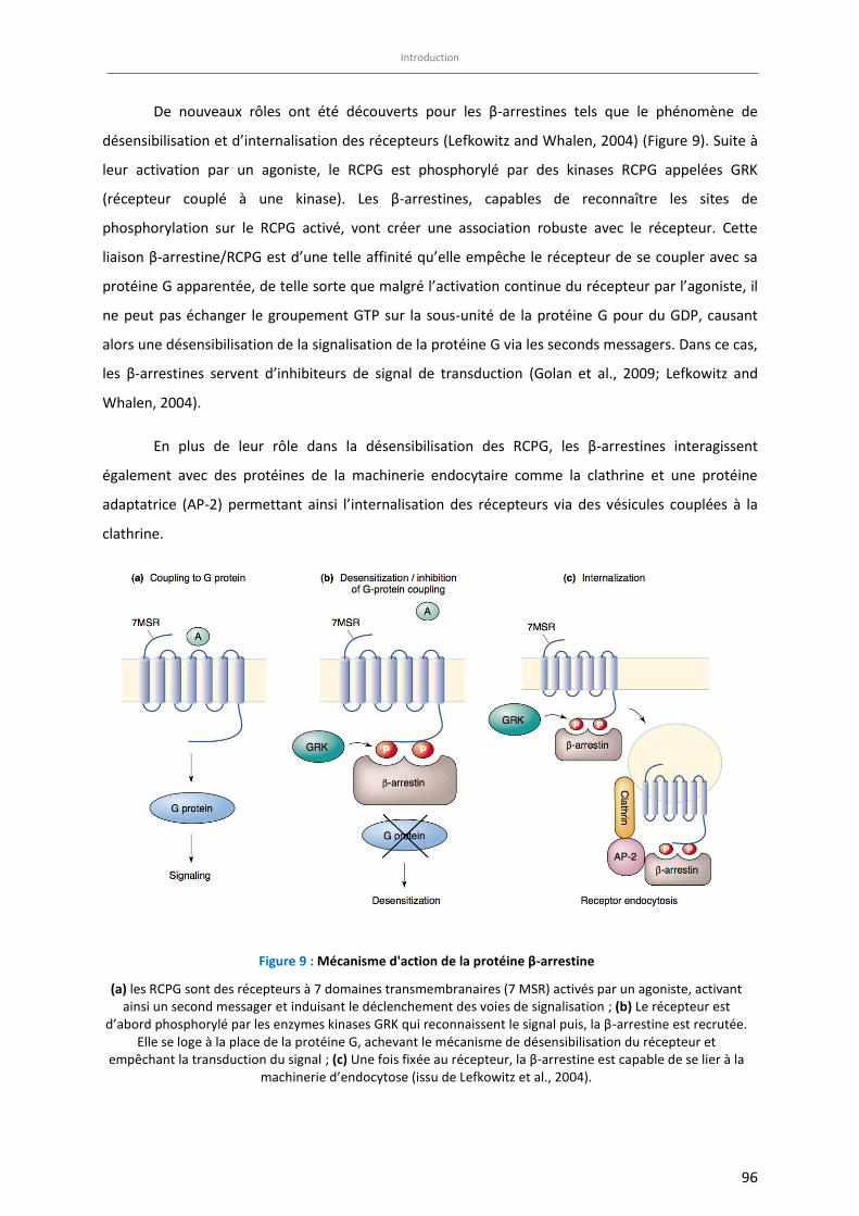
Introduction
96
De nouveaux rôles ont été découverts pour les β-arrestines tels que le phénomène de
désensibilisation et d’internalisation des récepteurs (Lefkowitz and Whalen, 2004) (Figure 9). Suite à
leur activation par un agoniste, le RCPG est phosphorylé par des kinases RCPG appelées GRK
(récepteur couplé à une kinase). Les β-arrestines, capables de reconnaître les sites de
phosphorylation sur le RCPG activé, vont créer une association robuste avec le récepteur. Cette
liaison β-arrestine/RCPG est d’une telle affinité qu’elle empêche le récepteur de se coupler avec sa
protéine G apparentée, de telle sorte que malgré l’activation continue du récepteur par l’agoniste, il
ne peut pas échanger le groupement GTP sur la sous-unité de la protéine G pour du GDP, causant
alors une désensibilisation de la signalisation de la protéine G via les seconds messagers. Dans ce cas,
les β-arrestines servent d’inhibiteurs de signal de transduction (Golan et al., 2009; Lefkowitz and
Whalen, 2004).
En plus de leur rôle dans la désensibilisation des RCPG, les β-arrestines interagissent
également avec des protéines de la machinerie endocytaire comme la clathrine et une protéine
adaptatrice (AP-2) permettant ainsi l’internalisation des récepteurs via des vésicules couplées à la
clathrine.
Figure 9 : Mécanisme d'action de la protéine β-arrestine
(a) les RCPG sont des récepteurs à 7 domaines transmembranaires (7 MSR) activés par un agoniste, activant ainsi un second messager et induisant le déclenchement des voies de signalisation ; (b) Le récepteur est
d’abord phosphorylé par les enzymes kinases GRK qui reconnaissent le signal puis, la β-arrestine est recrutée. Elle se loge à la place de la protéine G, achevant le mécanisme de désensibilisation du récepteur et
empêchant la transduction du signal ; (c) Une fois fixée au récepteur, la β-arrestine est capable de se lier à la machinerie d’endocytose (issu de Lefkowitz et al., 2004).
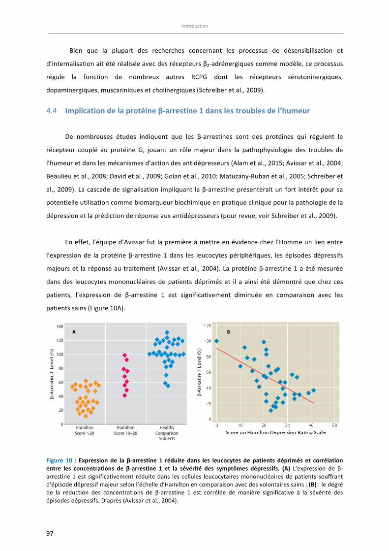
Introduction
97
Bien que la plupart des recherches concernant les processus de désensibilisation et
d’internalisationaitétéréaliséeavecdesrécepteursβ2-adrénergiquescommemodèle,ceprocessus
régule la fonction de nombreux autres RCPG dont les récepteurs sérotoninergiques,
dopaminergiques,muscariniquesetcholinergiques(Schreiberetal.,2009).
Implicationdelaprotéineβ-arrestine1danslestroublesdel’humeur4.4
De nombreuses études indiquent que les β-arrestines sont des protéines qui régulent le
récepteur couplé au protéine G, jouant un rôle majeur dans la pathophysiologie des troubles de
l’humeuretdanslesmécanismesd’actiondesantidépresseurs(Alametal.,2015;Avissaretal.,2004;
Beaulieuetal.,2008;Davidetal.,2009;Golanetal.,2010;Matuzany-Rubanetal.,2005;Schreiberet
al.,2009).Lacascadedesignalisation impliquant laβ-arrestineprésenteraitun fort intérêtpoursa
potentielleutilisationcommebiomarqueurbiochimiqueenpratiquecliniquepourlapathologiedela
dépressionetlaprédictionderéponseauxantidépresseurs(pourrevue,voirSchreiberetal.,2009).
Eneffet, l’équiped’Avissarfut lapremièreàmettreenévidencechez l’Hommeunlienentre
l’expressionde laprotéineβ-arrestine1dans les leucocytespériphériques, les épisodesdépressifs
majeurset la réponseau traitement (Avissaretal.,2004).Laprotéineβ-arrestine1aétémesurée
dansdes leucocytesmononucléaires depatients déprimés et il a ainsi été démontréque chez ces
patients, l’expression de β-arrestine 1 est significativement diminuée en comparaison avec les
patientssains(Figure10A).
Figure 10 : Expression de la β-arrestine 1 réduite dans les leucocytes de patients déprimés et corrélationentre les concentrations de β-arrestine 1 et la sévérité des symptômes dépressifs. (A) L’expression de β-arrestine1est significativement réduitedans les cellules leucocytairesmononucléairesdepatients souffrant
d’épisodedépressifmajeurselonl’échelled’Hamiltonencomparaisonavecdesvolontairessains;(B):ledegréde la réduction des concentrations de β-arrestine 1 est corrélée de manière significative à la sévérité des
épisodesdépressifs.D’après(Avissaretal.,2004).
A B
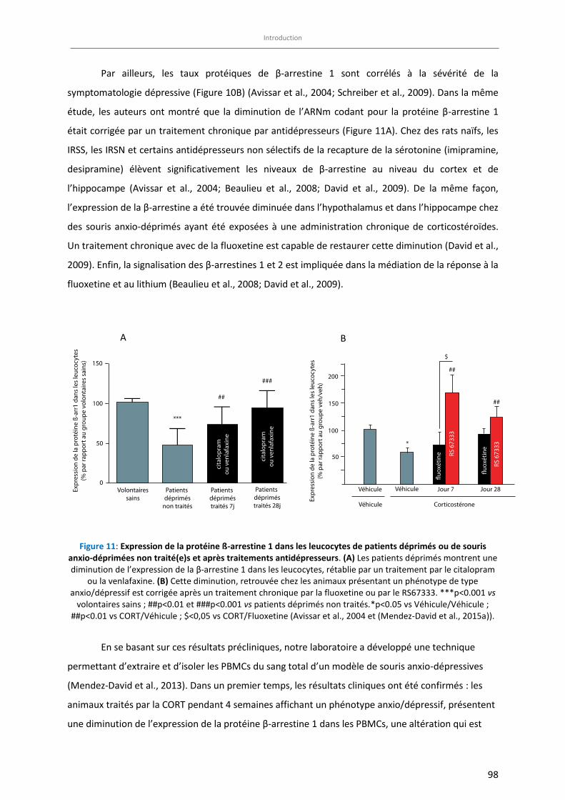
Introduction
98
Par ailleurs, les taux protéiques de β-arrestine 1 sont corrélés à la sévérité de la
symptomatologie dépressive (Figure 10B) (Avissar et al., 2004; Schreiber et al., 2009). Dans la même
étude, les auteurs ont montré que la diminution de l’ARNm codant pour la protéine β-arrestine 1
était corrigée par un traitement chronique par antidépresseurs (Figure 11A). Chez des rats naïfs, les
IRSS, les IRSN et certains antidépresseurs non sélectifs de la recapture de la sérotonine (imipramine,
desipramine) élèvent significativement les niveaux de β-arrestine au niveau du cortex et de
l’hippocampe (Avissar et al., 2004; Beaulieu et al., 2008; David et al., 2009). De la même façon,
l’expression de la β-arrestine a été trouvée diminuée dans l’hypothalamus et dans l’hippocampe chez
des souris anxio-déprimés ayant été exposées à une administration chronique de corticostéroïdes.
Un traitement chronique avec de la fluoxetine est capable de restaurer cette diminution (David et al.,
2009). Enfin, la signalisation des β-arrestines 1 et 2 est impliquée dans la médiation de la réponse à la
fluoxetine et au lithium (Beaulieu et al., 2008; David et al., 2009).
Figure 11: Expression de la protéine ß-arrestine 1 dans les leucocytes de patients déprimés ou de souris anxio-déprimées non traité(e)s et après traitements antidépresseurs. (A) Les patients déprimés montrent une diminution de l’expression de la β-arrestine 1 dans les leucocytes, rétablie par un traitement par le citalopram
ou la venlafaxine. (B) Cette diminution, retrouvée chez les animaux présentant un phénotype de type anxio/dépressif est corrigée après un traitement chronique par la fluoxetine ou par le RS67333. ***p<0.001 vs
volontaires sains ; ##p<0.01 et ###p<0.001 vs patients déprimés non traités.*p<0.05 vs Véhicule/Véhicule ; ##p<0.01 vs CORT/Véhicule ; $<0,05 vs CORT/Fluoxetine (Avissar et al., 2004 et (Mendez-David et al., 2015a)).
En se basant sur ces résultats précliniques, notre laboratoire a développé une technique
permettant d’extraire et d’isoler les PBMCs du sang total d’un modèle de souris anxio-dépressives
(Mendez-David et al., 2013). Dans un premier temps, les résultats cliniques ont été confirmés : les
animaux traités par la CORT pendant 4 semaines affichant un phénotype anxio/dépressif, présentent
une diminution de l’expression de la protéine β-arrestine 1 dans les PBMCs, une altération qui est
A B
Introduction
99
corrigée après 28 jours de traitement par la fluoxetine (18mg/kg/j) (Mendez-David et al., 2013).
D’autrepart,cetteexpérienceaétéréitéréeencomparantl’effetdelafluoxetineparrapportàcelui
du RS67333 sur l’expression de la protéine β-arrestine 1 dans les PBMCs. Il a été montré que la
fluoxetine (18 mg/kg/j) et le RS67333 (1,5 mg/kg/j) corrigeaient tous les deux la diminution de
l’expressionde laprotéineβ-arrestine1périphérique induitepar lacorticostérone,maisquecette
correctionétaitobservableaprèsseulement7joursdetraitementparl’agonistedurécepteur5-HT4
contre 28 jours pour la fluoxetine (Figure 11B). L’expression de la protéine β-arrestine 1 dans les
PBMCsestdoncunbiomarqueurprédictifdelaréponsethérapeutiqueutilisablechezlerongeuret
chezl’Homme.
Bien que des interrogations subsistent encore sur l’identité du (des) récepteur(s)
sérotoninergique(s)impliquésdansl’interactionaveclaprotéineβ-arrestine1,cesderniersrésultats
précliniques semblent indiquer que le récepteur 5-HT4 serait un bon candidat pour participer aux
effetscomportementauxetneurogéniques induitspar lafluoxetine,en interactionavec laprotéine
β-arrestine1.
Lesinteractionsentrelerécepteur5-HT4etlaprotéineβ-arrestine14.5
Lesmembresdelafamilledesarrestinessontconnuspourparticiperàlatransmissiondusignal
intracellulairemaisaussiàlarégulationdel’activitédesrécepteurscouplésauxprotéinesG(RCPG),
dont fait partie le récepteur sérotoninergique de type 4 (5-HT4) (Barthet et al., 2009). Plus
précisément, la protéine β-arrestine 1 est une protéine cytosolique interagissant avec les RCPGs
entrainantleurdésensibilisationetleurinternalisationsuiteàl’interactionavecleursligands(Vilaro
etal.,2005)(Figure12).
Ladésensibilisationdurécepteur5-HT4peutsefairedetroisfaçonsdifférentes(Mnie-Filaliand
Piñeyro,2012):
• Ladésensibilisationhétérologueaffectantpeulesrécepteurssérotoninergiques
• La désensibilisation homologue, phosphorylation-indépendante ou phosphorylation-
dépendantemédiéeparuneprotéinedelafamilledessérine/thréoninekinases,laGRK
• Ladésensibilisationligand-spécifique
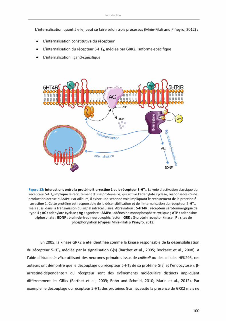
Introduction
100
L’internalisation quant à elle, peut se faire selon trois processus (Mnie-Filali and Piñeyro, 2012) :
x L’internalisation constitutive du récepteur
x L’internalisation du récepteur 5-HT4, médiée par GRK2, isoforme-spécifique
x L’internalisation ligand-spécifique
Figure 12: Interactions entre la protéine ß-arrestine 1 et le récepteur 5-HT4. La voie d’activation classique du récepteur 5-HT4 implique le recrutement d’une protéine Gs, qui active l’adénylate cyclase, responsable d’une production accrue d’AMPc. Par ailleurs, il existe une seconde voie impliquant le recrutement de la protéine ß-
arrestine 1. Cette protéine est responsable de la désensibilisation et de l’internalisation du récepteur 5-HT4,
mais aussi dans la transmission du signal intracellulaire. Abréviation : 5-HT4R : récepteur sérotoninergique de
type 4 ; AC : adénylate cyclase ; Ag : agoniste ; AMPc : adénosine monophosphate cyclique ; ATP : adénosine
triphosphate ; BDNF : brain-derived neurotrophic factor ; GRK : G-protein receptor kinase ; P : sites de
phosphorylation (d’après Mnie-Filali & Piñeyro, 2012)
En 2005, la kinase GRK2 a été identifiée comme la kinase responsable de la désensibilisation
du récepteur 5-HT4 médiée par la signalisation G(s) (Barthet et al., 2005; Bockaert et al., 2008). A
l’aide d’études in vitro utilisant des neurones primaires issus de colliculi ou des cellules HEK293, ces
auteurs ont démontré que le découplage du récepteur 5-HT4 de sa protéine G(s) et l’endocytose « β-
arrestine-dépendante » du récepteur sont des évènements moléculaire distincts impliquant
différemment les GRKs (Barthet et al., 2009; Bohn and Schmid, 2010; Marin et al., 2012). Par
exemple, le découplage du récepteur 5-HT4 des protéines Gαs nécessite la présence de GRK2 mais ne
ATP
AMPc
Gαβ
γ
AC5HT4R 5HT4R
GRK
P
P
ß -a r
rest
ine
Ag Ag
Akt
Signalisation intracellulaire
BDNF
Internalisation
Désensibilisation
ß-arre
stineP
P
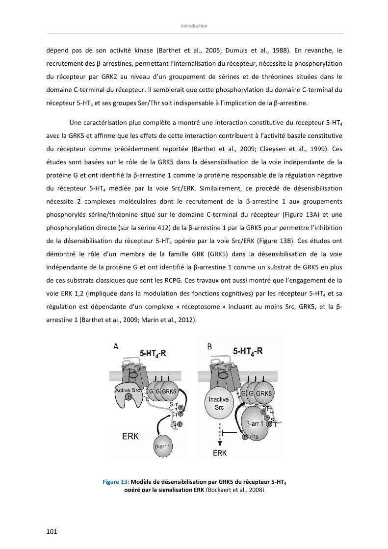
Introduction
101
dépend pas de son activité kinase (Barthet et al., 2005; Dumuis et al., 1988). En revanche, le
recrutement des β-arrestines, permettant l’internalisation du récepteur, nécessite la phosphorylation
du récepteur par GRK2 au niveau d’un groupement de sérines et de thréonines situées dans le
domaine C-terminal du récepteur. Il semblerait que cette phosphorylation du domaine C-terminal du
récepteur 5-HT4 et ses groupes Ser/Thr soit indispensable à l’implication de la β-arrestine.
Une caractérisation plus complète a montré une interaction constitutive du récepteur 5-HT4
avec la GRK5 et affirme que les effets de cette interaction contribuent à l’activité basale constitutive
du récepteur comme précédemment reportée (Barthet et al., 2009; Claeysen et al., 1999). Ces
études sont basées sur le rôle de la GRK5 dans la désensibilisation de la voie indépendante de la
protéine G et ont identifié la β-arrestine 1 comme la protéine responsable de la régulation négative
du récepteur 5-HT4 médiée par la voie Src/ERK. Similairement, ce procédé de désensibilisation
nécessite 2 complexes moléculaires dont le recrutement de la β-arrestine 1 aux groupements
phosphorylés sérine/thréonine situé sur le domaine C-terminal du récepteur (Figure 13A) et une
phosphorylation directe (sur la sérine 412) de la β-arrestine 1 par la GRK5 pour permettre l’inhibition
de la désensibilisation du récepteur 5-HT4 opérée par la voie Src/ERK (Figure 13B). Ces études ont
démontré le rôle d’un membre de la famille GRK (GRK5) dans la désensibilisation de la voie
indépendante de la protéine G et ont identifié la β-arrestine 1 comme un substrat de GRK5 en plus
de ces substrats classiques que sont les RCPG. Ces travaux ont aussi montré que l’engagement de la
voie ERK 1,2 (impliquée dans la modulation des fonctions cognitives) par les récepteur 5-HT4 et sa
régulation est dépendante d’un complexe « réceptosome » incluant au moins Src, GRK5, et la β-
arrestine 1 (Barthet et al., 2009; Marin et al., 2012).
Figure 13: Modèle de désensibilisation par GRK5 du récepteur 5-HT4 opéré par la signalisation ERK (Bockaert et al., 2008)
Introduction
102
Puisque l’expression de GRK5 et de la β-arrestine 1 augmente considérablement pendant le
développement (Gurevich et al., 2004), il est probable que la régulation négative du récepteur 5-HT4
médiée par l’activation de la voie Src/ERK indépendante de la protéine G soit plus forte chez les
adultes. De plus, GRK5 et la β-arrestine 1 jouent un rôle critique dans la détermination de la voie de
signalisation (Gs ou ERK) recrutée via l’activation du récepteur 5-HT4. Ce rôle pourrait être
déterminant dans les phénomènes de plasticité synaptique dépendante du 5-HT4 et de consolidation
de la mémoire (Kemp and Manahan-Vaughan, 2005; Marchetti et al., 2004; Micale et al., 2006).
Deux études précliniques menées chez le rat ont montré que l’administration chronique de
fluoxetine (Vidal et al., 2009) ou de venlafaxine (Vidal et al., 2010) étaient responsables d’une
désensibilisation du récepteur 5-HT4 et/ou de l’activation du rétrocontrôle négatif induit par le
récepteur 5-HT4. En effet, ces deux études ont montré une diminution de la densité de récepteurs 5-
HT4, notamment dans les ganglions de la base et dans l’hippocampe, une diminution de l’activité de
l’AC induite par l’activation du récepteur 5-HT4 et une diminution de l’excitabilité neuronale 5-HT4-
dépendante dans la zone CA1 de l’hippocampe. Ces altérations conduisent à une diminution de
l’intensité de la réponse cellulaire malgré l’administration continue d’antidépresseur. Fait
surprenant, le RS67333 à la dose de 1,5 mg/kg/j n’induit pas de tolérance puisque ses effets de type
anxio/dépressif persistent plusieurs jours après l’administration (Lucas et al., 2007). Cette
observation est pour le moins inattendue puisque les récepteurs 5-HT4 sont connus pour se
désensibiliser très rapidement après exposition à un agoniste (Ansanay et al., 1992).

Introduction
103
Introduction
104
Objectifs de la thèse
Différents troubles cognitifs (difficulté à se concentrer, perception négative de stimuli normalement
perçus comme positifs) figurent parmi les critères de diagnostic d’un épisode de dépression majeure
selon le DSM-5. Malheureusement, la prise en charge actuelle des patients dépressifs ne prend pas
systématiquement en compte l’altération des fonctions cognitives dans la mise en place du
traitement de première attention. En outre, l’efficacité d’une prise en charge spécifique des troubles
de l’apprentissage et de la mémoire dans le contexte de la dépression par des antidépresseurs reste
à ce jour non résolue.
Le premier objectif de ce travail a été de caractériser les troubles cognitifs suspectés dans un
modèle d’anxiété/dépression basé sur l’administration chronique de corticostérone : le modèle
CORT (Article 1). Ces travaux s’inscrivent dans la caractérisation phénotypique complète de ce
modèle animal d’anxiété/dépression faite au sein de l’UMRS 1178 via la description des troubles
cognitifs en tant que facteurs de co-morbidité des troubles anxio/dépressifs.
Les antidépresseurs sont les molécules indiquées en première intention dans le traitement de la
dépression majeure, mais la plupart de ces traitements sont aujourd’hui limités par plusieurs
facteurs, tels que l’important nombre de patients non-répondeurs au traitement, le long délai
d’action avant l’apparition des premiers effets thérapeutiques, les nombreux effets indésirables et
l’absence d’efficacité contre les symptômes cognitifs étroitement liés aux symptômes dépressifs. De
ce fait, le développement de nouvelles molécules plus efficaces, ayant des propriétés antidépressives
rapides et traitant simultanément les altérations cognitives associées à la dépression, devient
indispensable. Le récepteur 5-HT4 semble être une cible à privilégier car si son implication dans le
traitement des troubles cognitifs chez des animaux naïfs est à ce jour bien décrit. L’efficacité
thérapeutique des ligands du récepteur 5-HT4 dans le traitement des troubles de l’humeur reste à
confirmer.
De nombreuses études se sont intéressées aux propriétés antidépresseurs des agonistes du
récepteur 5-HT4 (Lucas et al., 2007; Pascual-Brazo et al., 2012). Cependant, peu d’entre elles
conduisent leurs expérimentations sur des animaux anxio-déprimés (Article 2) et moins encore se
sont concentrées sur les propriétés pro-cognitives des agonistes du récepteur 5-HT4 dans un contexte
d’anxiété/dépression.
Le second objectif de ce travail a donc été de corriger les troubles cognitifs observés chez les
animaux anxio/dépressifs (modèle CORT) à l’aide de différentes stratégies thérapeutiques. Ainsi,
les effets d’une administration chronique d’un antidépresseur monoaminergique classique
Introduction
105
(fluoxetine) ou d’un agoniste du récepteur 5-HT4 (RS67333) chez les animaux anxio-déprimés ont été
mesurés dans plusieurs tests d’apprentissage et de la mémoire, reflétant les différents types de
mémoire existantes (Article 3).
Récemment, la protéine β-arrestine 1 a été signalée comme étant un acteur important de la
physiopathologie de la dépression ainsi que dans la prédiction de la réponse aux antidépresseurs
(Avissar et al., 2004; Matuzany-Ruban et al., 2005). Des données précliniques ont validé son
implication dans un modèle d’anxiété/dépression basé sur l’administration chronique de
corticostérone dans différentes régions cérébrales (David et al., 2009) ainsi qu’au niveau de
marqueurs périphériques (Mendez-David et al., 2013; Mendez-David et al., 2015b). Ainsi, ces travaux
rapportent une diminution de l’expression de la β-arrestine 1 dans les régions de l’hippocampe et de
l’hypothalamus chez des animaux anxio-déprimés (restaurée après un traitement chronique par de la
fluoxetine ou un traitement sub-chronique de RS67333) et ont confirmé l’utilisation de la protéine β-
arrestine 1 comme un biomarqueur potentiel de l’état dépressif et de la réponse aux
antidépresseurs. Ces résultats évoquent la possibilité que l’expression de la β-arrestine 1 dans le
cerveau pourrait être un substrat potentiel pour les effets comportementaux et neurogéniques des
antidépresseurs.
Le 3ème objectif de cette thèse a été de caractériser les conséquences comportementales d’une
délétion sélective de la protéine β-arrestine 1 dans les cellules souches du gyrus dentelé de
l’hippocampe dans plusieurs tests prédictifs d’une activité anxiolytique et/ou antidépressive en
présence ou non d’un traitement chronique à la fluoxetine. Les conséquences neurogéniques de
cette délétion sélective ont également été étudiées (Article 4).
Enfin, les conséquences comportementales d’une délétion sélective de la protéine β-arrestine 1 dans
les cellules souches du gyrus dentelé de l’hippocampe dans différents tests de cognition (mémoire de
type épisodique, spatiale et associative) ont été évaluées. Au vue de la relation fonctionnelle entre le
récepteur 5-HT4 et la protéine β-arrestine 1, le dernier objectif de ce travail a été d’étudier les effets
d’un traitement chronique par un agoniste du récepteur 5-HT4 sur les performances cognitives
d’animaux dont la β-arrestine 1 a été spécifiquement supprimée, en comparaison avec un
traitement antidépresseur classique de référence (fluoxetine) (Résultats complémentaires).
Après une partie consacrée aux méthodes utilisées pour réaliser ces travaux de thèse, les articles
expérimentaux seront insérés en anglais dans leur format de publication, précédés d’un paragraphe
en français précisant la question posée et introduisant l’étude et suivis de commentaires résumant
les principaux effets découverts.

Matériel et Méthodes
106
Matériel et Méthodes

107
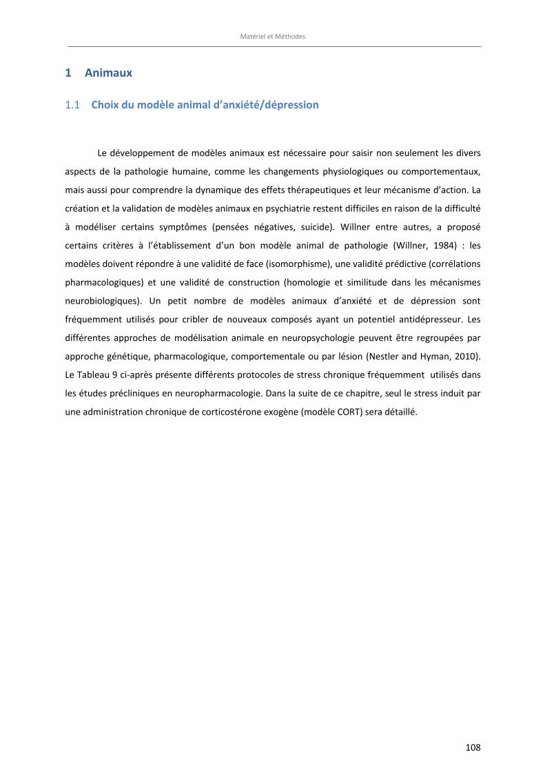
Matériel et Méthodes
108
1 Animaux
Choix du modèle animal d’anxiété/dépression 1.1
Le développement de modèles animaux est nécessaire pour saisir non seulement les divers
aspects de la pathologie humaine, comme les changements physiologiques ou comportementaux,
mais aussi pour comprendre la dynamique des effets thérapeutiques et leur mécanisme d’action. La
création et la validation de modèles animaux en psychiatrie restent difficiles en raison de la difficulté
à modéliser certains symptômes (pensées négatives, suicide). Willner entre autres, a proposé
certains critères à l’établissement d’un bon modèle animal de pathologie (Willner, 1984) : les
modèles doivent répondre à une validité de face (isomorphisme), une validité prédictive (corrélations
pharmacologiques) et une validité de construction (homologie et similitude dans les mécanismes
neurobiologiques). Un petit nombre de modèles animaux d’anxiété et de dépression sont
fréquemment utilisés pour cribler de nouveaux composés ayant un potentiel antidépresseur. Les
différentes approches de modélisation animale en neuropsychologie peuvent être regroupées par
approche génétique, pharmacologique, comportementale ou par lésion (Nestler and Hyman, 2010).
Le Tableau 9 ci-après présente différents protocoles de stress chronique fréquemment utilisés dans
les études précliniques en neuropharmacologie. Dans la suite de ce chapitre, seul le stress induit par
une administration chronique de corticostérone exogène (modèle CORT) sera détaillé.
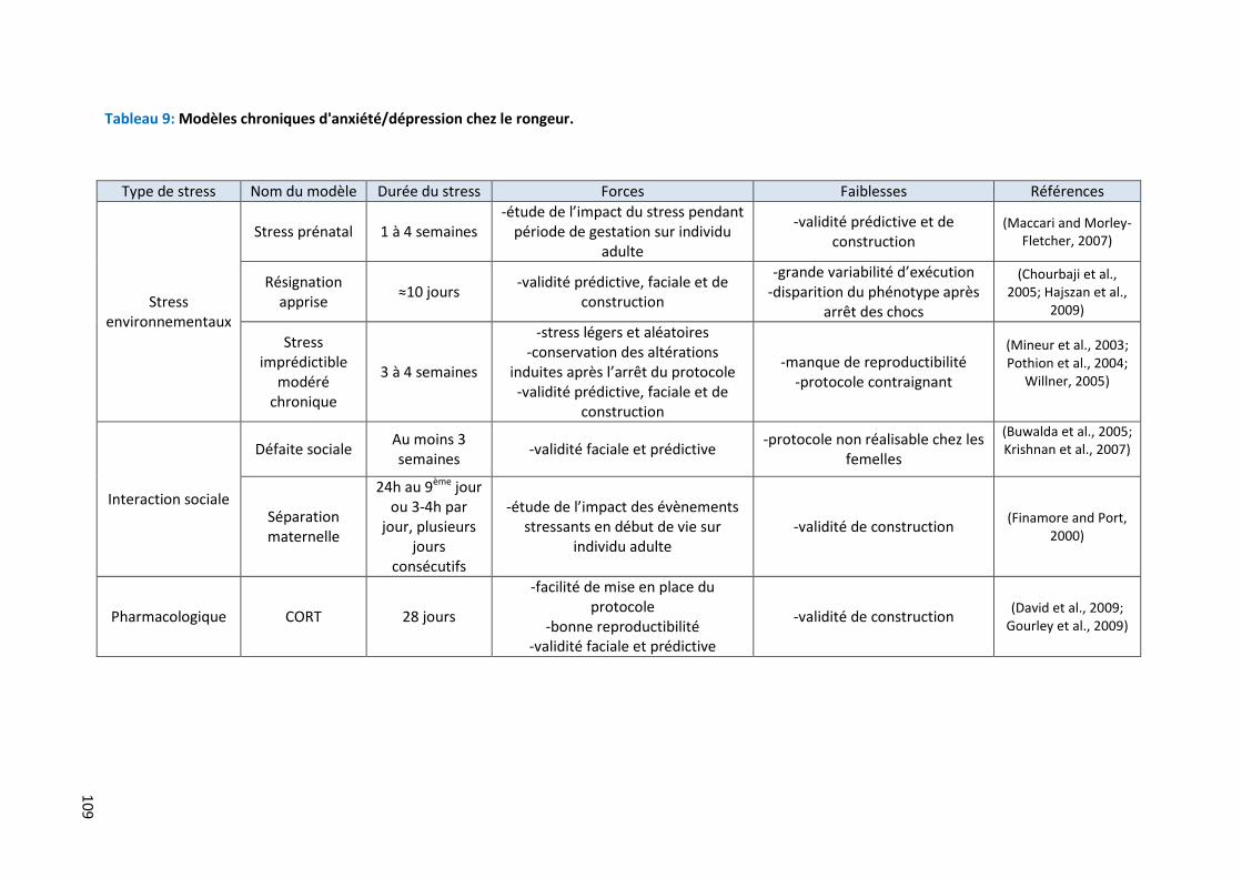
109
Tableau 9: Modèles chroniques d'anxiété/dépression chez le rongeur.
Type de stress Nom du modèle Durée du stress Forces Faiblesses Références
Stress environnementaux
Stress prénatal 1 à 4 semaines -étude de l’impact du stress pendant
période de gestation sur individu adulte
-validité prédictive et de construction
(Maccari and Morley-Fletcher, 2007)
Résignation apprise
≈10 jours -validité prédictive, faciale et de
construction
-grande variabilité d’exécution -disparition du phénotype après
arrêt des chocs
(Chourbaji et al., 2005; Hajszan et al.,
2009)
Stress imprédictible
modéré chronique
3 à 4 semaines
-stress légers et aléatoires -conservation des altérations
induites après l’arrêt du protocole -validité prédictive, faciale et de
construction
-manque de reproductibilité -protocole contraignant
(Mineur et al., 2003; Pothion et al., 2004;
Willner, 2005)
Interaction sociale
Défaite sociale Au moins 3 semaines
-validité faciale et prédictive -protocole non réalisable chez les
femelles
(Buwalda et al., 2005; Krishnan et al., 2007)
Séparation maternelle
24h au 9ème jour ou 3-4h par
jour, plusieurs jours
consécutifs
-étude de l’impact des évènements stressants en début de vie sur
individu adulte -validité de construction
(Finamore and Port, 2000)
Pharmacologique CORT 28 jours
-facilité de mise en place du protocole
-bonne reproductibilité -validité faciale et prédictive
-validité de construction (David et al., 2009;
Gourley et al., 2009)
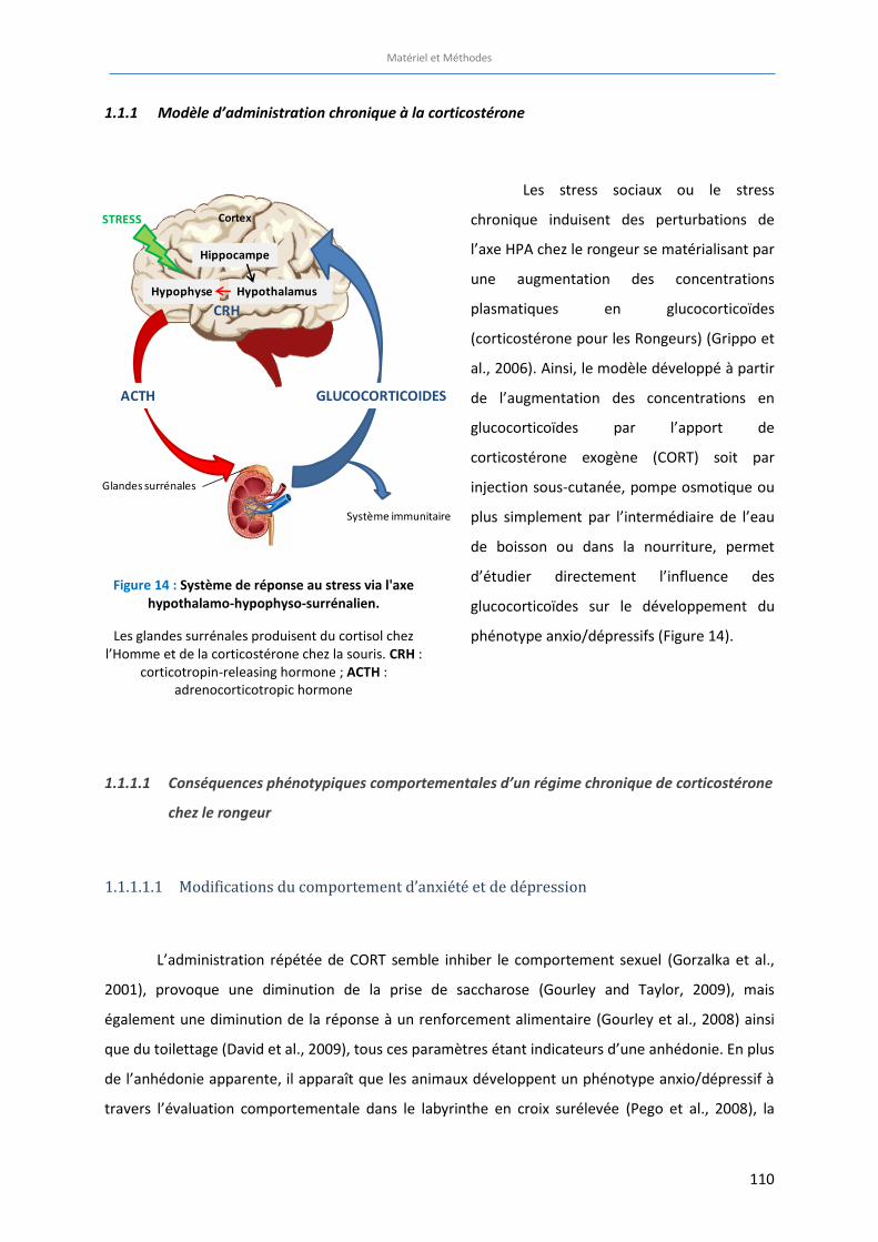
Matériel et Méthodes
110
1.1.1 Modèle d’administration chronique à la corticostérone
Les stress sociaux ou le stress
chronique induisent des perturbations de
l’axe HPA chez le rongeur se matérialisant par
une augmentation des concentrations
plasmatiques en glucocorticoïdes
(corticostérone pour les Rongeurs) (Grippo et
al., 2006). Ainsi, le modèle développé à partir
de l’augmentation des concentrations en
glucocorticoïdes par l’apport de
corticostérone exogène (CORT) soit par
injection sous-cutanée, pompe osmotique ou
plus simplement par l’intermédiaire de l’eau
de boisson ou dans la nourriture, permet
d’étudier directement l’influence des
glucocorticoïdes sur le développement du
phénotype anxio/dépressifs (Figure 14).
1.1.1.1 Conséquences phénotypiques comportementales d’un régime chronique de corticostérone
chez le rongeur
1.1.1.1.1 Modifications du comportement d’anxiété et de dépression
L’administration répétée de CORT semble inhiber le comportement sexuel (Gorzalka et al.,
2001), provoque une diminution de la prise de saccharose (Gourley and Taylor, 2009), mais
également une diminution de la réponse à un renforcement alimentaire (Gourley et al., 2008) ainsi
que du toilettage (David et al., 2009), tous ces paramètres étant indicateurs d’une anhédonie. En plus
de l’anhédonie apparente, il apparaît que les animaux développent un phénotype anxio/dépressif à
travers l’évaluation comportementale dans le labyrinthe en croix surélevée (Pego et al., 2008), la
Tableau 10: Similarités des conséquences de l'administration de CORT et manifestation humaines de la dépression (Extrait de la thèse du Dr. Quentin Rainer, d’après Sterner et Kalynchuk, 2010)
Figure 14 : Système de réponse au stress via l'axe
hypothalamo-hypophyso-surrénalien.
Les glandes surrénales produisent du cortisol chez l’Homme et de la corticostérone chez la souris. CRH :
corticotropin-releasing hormone ; ACTH : adrenocorticotropic hormone
Glandes surrénales
ACTH
Système immunitaire
HypothalamusHypophyse
Hippocampe
CortexSTRESS
GLUCOCORTICOIDES
CRH
Matériel et Méthodes
111
double enceinte éclairée (Murray et al., 2008), le test d’odeur de prédateur (Kalynchuk et al., 2004),
ou encore dans le test d’hypophagie induite par la faim, et le test du champ ouvert(David et al., 2009;
Gregus et al., 2005). L’apparition d’un phénotype de dépression sans phénotype anxieux est
rarement référencée chez les patients (Mineka et al., 1998), ce qui renforce les résultats obtenus par
ce modèle. D’autres marqueurs physiologiques d’anxiété/dépression sont également altérés,
notamment la dérégulation des fonctions et des concentrations des acteurs de l’axe HPA, la prise de
poids et l’atrophie des glandes surrénales (Gourley et al., 2008; Murray et al., 2008). Cependant, de
réelles données relatives aux constituants neurobiochimiques de l’axe HPA durant le protocole de
stress restent à ce jour peu renseignées. Par ailleurs, la majorité des comportements induits par ce
modèle est corrigée par l’administration chronique d’antidépresseurs (Ago et al., 2008; David et al.,
2009; Rainer et al., 2012), supportant la validité prédictive de modèle préclinique de la dépression
humaine.
1.1.1.1.2 Modifications comportementales des capacités cognitives
En plus des modifications comportementales modélisant les symptômes de la pathologie de
la dépression, l’administration de CORT ainsi que de nombreux modèles d’anxiété/dépression
provoquent également des changements au niveau de la plasticité synaptique, souvent révélés par
des déficits comportementaux d’apprentissage et de mémoire. Les modifications comportementales
induites par le modèle CORT sont présentées dans la Revue “Cognitive dysfunction in major
depressive disorder : A translational review in Animal models of the disease » disponible dans la
partie introductive de cette thèse et seront développées dans la partie Discussion.
1.1.1.2 Conséquences structurelles et moléculaires d’un régime chronique par de la
corticostérone
Au-delà des modifications comportementales induites par l’administration chronique de
CORT, ce modèle provoque également des modifications structurales liées à la plasticité et
notamment la diminution du processus de prolifération cellulaire et de la neurogenèse dans
l’hippocampe (David et al., 2009; Malberg et al., 2000). Plusieurs études ont pu mettre en évidence
que les atteintes de la plasticité se localisent dans les régions cérébrales clefs impliquées dans la
dépression. En effet, il a été observé un remodelage dendritique induit par la CORT dans
Matériel et Méthodes
112
l’hippocampe (Magarinos et al., 1999; Southwick et al., 2005; Watanabe et al., 1992), l’amygdale
(Mitra and Sapolsky, 2008), et le cortex préfrontal (Seib and Wellman, 2003). Ces résultats sont
également retrouvés après des études post mortem de patients souffrant de dépression (Konarski et
al., 2008). En particulier, il est connu que l’exposition prolongée à la CORT induit une atrophie
dendritique des cellules pyramidales de l’hippocampe, et une perte de volume des dendrites apicaux
des régions CA3 et CA1 (Magarinos et al., 1999). D’autre part, bien qu’étant également le siège de la
neurogenèse chez le rongeur adulte, les effets des glucocorticoïdes dans le bulbe olfactif adulte
restent moins étudiés. Néanmoins, une étude récente a mis en évidence la présence de déficits
olfactifs chez les animaux traités chroniquement par de la corticostérone, partiellement restaurés par
un traitement chronique par de la fluoxetine (Siopi et al., 2016).
L’administration de CORT chez l’Animal et chez l’Homme est associée à une réduction du
facteur de transcription de l’élément de réponse liant l’AMPc appelé CREB (Pittenger and Duman,
2008). Fait intéressant, l’activation de CREB induit la neurogenèse alors que son inhibition la réduit
(Nakagawa et al., 2002), faisant de CREB un acteur important de la stabilité neuronale associée au
stress et à la dépression. Entre autres, CREB contrôle des gènes effecteurs, eux aussi impliqués dans
la stabilité de la plasticité synaptique. L’un de ces gènes est celui codant pour le « brain derived
neurotrophic factor » (BDNF), qui joue un rôle critique sur la plasticité dans le développement, dans
la survie et la fonction des neurones (Pittenger and Duman, 2008). Chez l’animal, l’exposition aux
glucocorticoïdes altère aussi bien CREB que le BDNF dans les différentes régions limbiques (Gourley
et al., 2008; Jacobsen and Mork, 2006), également chez les patients atteints de dépression (Dwivedi
et al., 2003; Sen et al., 2008).
Souris tissus-spécifiques Nestine-CreERT2
/FLOX ARRB1 1.2
1.2.1 Génération de la lignée
1.2.1.1 Caractéristique de la lignée FLOX ARRB1
Les souris FLOX β-Arrestine 1 (ARRB1) ont été générées initialement par le laboratoire du
Professeur Jean-Martin BEAULIEU à l’Université de Laval, Québec, Canada. La lignée a ensuite été
transférée à l’animalerie Centrale de la Faculté de Pharmacie. J’ai donc prise en charge la gestion de
cette lignée au cours de ma thèse.
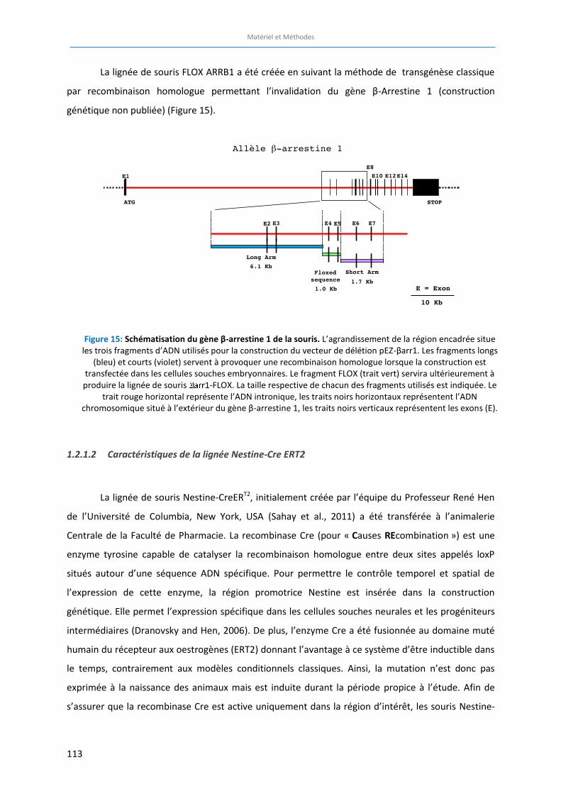
Matériel et Méthodes
113
La lignée de souris FLOX ARRB1 a été créée en suivant la méthode de transgénèse classique
par recombinaison homologue permettant l’invalidation du gène β-Arrestine 1 (construction
génétique non publiée) (Figure 15).
1.2.1.2 Caractéristiques de la lignée Nestine-Cre ERT2
La lignée de souris Nestine-CreERT2, initialement créée par l’équipe du Professeur René Hen
de l’Université de Columbia, New York, USA (Sahay et al., 2011) a été transférée à l’animalerie
Centrale de la Faculté de Pharmacie. La recombinase Cre (pour « Causes REcombination ») est une
enzyme tyrosine capable de catalyser la recombinaison homologue entre deux sites appelés loxP
situés autour d’une séquence ADN spécifique. Pour permettre le contrôle temporel et spatial de
l’expression de cette enzyme, la région promotrice Nestine est insérée dans la construction
génétique. Elle permet l’expression spécifique dans les cellules souches neurales et les progéniteurs
intermédiaires (Dranovsky and Hen, 2006). De plus, l’enzyme Cre a été fusionnée au domaine muté
humain du récepteur aux oestrogènes (ERT2) donnant l’avantage à ce système d’être inductible dans
le temps, contrairement aux modèles conditionnels classiques. Ainsi, la mutation n’est donc pas
exprimée à la naissance des animaux mais est induite durant la période propice à l’étude. Afin de
s’assurer que la recombinase Cre est active uniquement dans la région d’intérêt, les souris Nestine-
Figure 15: Schématisation du gène β-arrestine 1 de la souris. L’agrandissement de la région encadrée situe les trois fragments d’ADN utilisés pour la construction du vecteur de délétion pEZ-βarr1. Les fragments longs
(bleu) et courts (violet) servent à provoquer une recombinaison homologue lorsque la construction est transfectée dans les cellules souches embryonnaires. Le fragment FLOX (trait vert) servira ultérieurement à
produire la lignée de souris -FLOX. La taille respective de chacun des fragments utilisés est indiquée. Le trait rouge horizontal représente l’ADN intronique, les traits noirs horizontaux représentent l’ADN
chromosomique situé à l’extérieur du gène β-arrestine 1, les traits noirs verticaux représentent les exons (E).
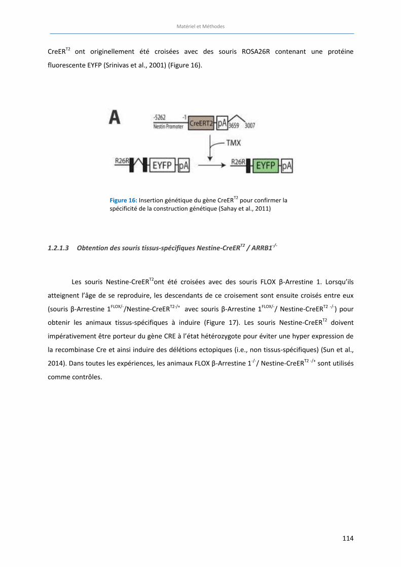
Matériel et Méthodes
114
CreERT2 ont originellement été croisées avec des souris ROSA26R contenant une protéine
fluorescente EYFP (Srinivas et al., 2001) (Figure 16).
1.2.1.3 Obtention des souris tissus-spécifiques Nestine-CreERT2 / ARRB1-/-
Les souris Nestine-CreERT2ont été croisées avec des souris FLOX β-Arrestine 1. Lorsqu’ils
atteignent l’âge de se reproduire, les descendants de ce croisement sont ensuite croisés entre eux
(souris β-Arrestine 1FLOX/-/Nestine-CreERT2-/+ avec souris β-Arrestine 1FLOX/-/ Nestine-CreERT2 -/-) pour
obtenir les animaux tissus-spécifiques à induire (Figure 17). Les souris Nestine-CreERT2 doivent
impérativement être porteur du gène CRE à l’état hétérozygote pour éviter une hyper expression de
la recombinase Cre et ainsi induire des délétions ectopiques (i.e., non tissus-spécifiques) (Sun et al.,
2014). Dans toutes les expériences, les animaux FLOX β-Arrestine 1-/-/ Nestine-CreERT2 -/+ sont utilisés
comme contrôles.
Figure 16: Insertion génétique du gène CreERT2
pour confirmer la spécificité de la construction génétique (Sahay et al., 2011)
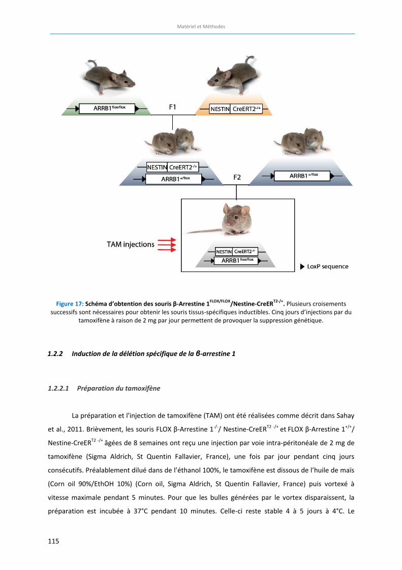
Matériel et Méthodes
115
Figure 17: Schéma d’obtention des souris β-Arrestine 1FLOX/FLOX
/Nestine-CreERT2-/+
. Plusieurs croisements successifs sont nécessaires pour obtenir les souris tissus-spécifiques inductibles. Cinq jours d’injections par du
tamoxifène à raison de 2 mg par jour permettent de provoquer la suppression génétique.
1.2.2 Induction de la délétion spécifique de la β-arrestine 1
1.2.2.1 Préparation du tamoxifène
La préparation et l'injection de tamoxifène (TAM) ont été réalisées comme décrit dans Sahay
et al., 2011. Brièvement, les souris FLOX β-Arrestine 1-/-/ Nestine-CreERT2 -/+ et FLOX β-Arrestine 1+/+/
Nestine-CreERT2 -/+ âgées de 8 semaines ont reçu une injection par voie intra-péritonéale de 2 mg de
tamoxifène (Sigma Aldrich, St Quentin Fallavier, France), une fois par jour pendant cinq jours
consécutifs. Préalablement dilué dans de l’éthanol 100%, le tamoxifène est dissous de l’huile de maïs
(Corn oil 90%/EthOH 10%) (Corn oil, Sigma Aldrich, St Quentin Fallavier, France) puis vortexé à
vitesse maximale pendant 5 minutes. Pour que les bulles générées par le vortex disparaissent, la
préparation est incubée à 37°C pendant 10 minutes. Celle-ci reste stable 4 à 5 jours à 4°C. Le
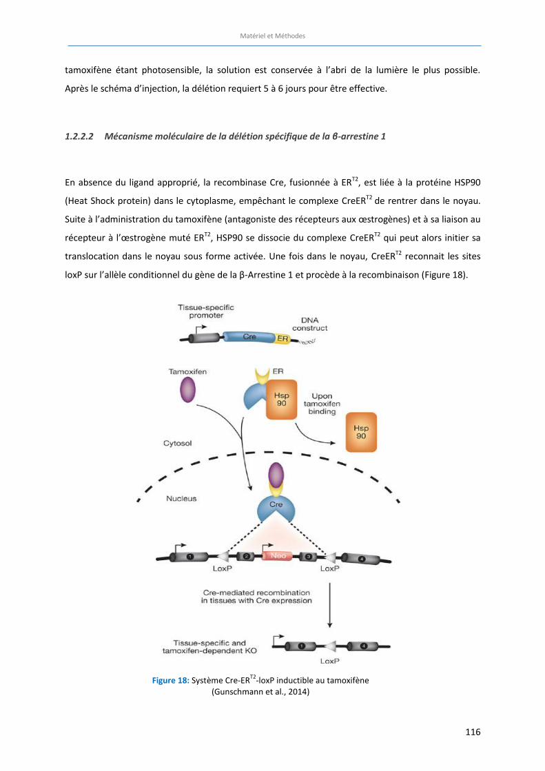
Matériel et Méthodes
116
tamoxifène étant photosensible, la solution est conservée à l’abri de la lumière le plus possible.
Après le schéma d’injection, la délétion requiert 5 à 6 jours pour être effective.
1.2.2.2 Mécanisme moléculaire de la délétion spécifique de la β-arrestine 1
En absence du ligand approprié, la recombinase Cre, fusionnée à ERT2, est liée à la protéine HSP90
(Heat Shock protein) dans le cytoplasme, empêchant le complexe CreERT2 de rentrer dans le noyau.
Suite à l’administration du tamoxifène (antagoniste des récepteurs aux œstrogènes) et à sa liaison au
récepteur à l’œstrogène muté ERT2, HSP90 se dissocie du complexe CreERT2 qui peut alors initier sa
translocation dans le noyau sous forme activée. Une fois dans le noyau, CreERT2 reconnait les sites
loxP sur l’allèle conditionnel du gène de la β-Arrestine 1 et procède à la recombinaison (Figure 18).
Figure 18: Système Cre-ERT2
-loxP inductible au tamoxifène (Gunschmann et al., 2014)
Matériel et Méthodes
117
Ce mécanisme n’est possible que dans les cellules qui expriment Cre sous le promoteur Nestine,
permettant ainsi une délétion tissu-spécifique de la protéine β-Arrestine 1 au niveau des cellules
souches neurales du GD de l’hippocampe. Dans les autres types cellulaires dans lesquels le gène Cre
n’est pas exprimé, le gène cible β-Arrestine 1 sera toujours présent dans le génome et fonctionnera
donc normalement.
1.2.3 Génotypage des animaux
Le génotypage des différentes souris mutées a été réalisé grâce à la technique
d’Amplification en Chaîne par Polymérase (PCR). Ce procédé permet d’amplifier in vitro une région
spécifique de l’ADN afin d’en produire une quantité détectable. L’ADN génomique est extrait de
fragments de queues des souris. Les animaux sont génotypés à partir du 18ème jour de vie et
numérotés aux oreilles à l’aide d’un poinçon adapté. Les souriceaux ARRB1+/+ et ARRB1-/- sont sevrés
à 21 jours après leur naissance et sont répartis dans des cages selon leur sexe avant le début des
expériences.
1.2.3.1 Extraction de l’ADN
Les queues sont d’abord digérées dans un tampon de lyse pendant 2 à 3 heures à 50°C. Le
tampon de lyse est constitué de Tris pH8 à 1M, Sodium Dodécyl Sulfate 10% (SDS), d’acide éthylène
diamine tétra-acétique (EDTA) 0,5M, Chlorure de Sodium (NaCl) 5M, de Protéinase K complété avec
de l’eau distillée. Pour une digestion optimale, les fragments de queues doivent être recouverts de
tampon. Une fois dans la cuve à 55°C, les tubes seront vortexés toutes les demi-heures jusqu’à
digestion complète du tissu. L’ADN est précipité grâce à l’ajout de phénol (volume identique au
tampon de lyse) qui permet de séparer l’ADN du reste des éléments de la queue. Les digestats sont
ensuite centrifugés pendant 10 minutes à 15000 tours/min. Le surnageant contient l’ADN. Les tubes
seront conservés à -20°C pour une longue conservation.
1.2.3.2 Amplification par Polymerase Chain Reaction (PCR)
Le mélange DNTP’s et ADN polymérase est utilisé sous forme de petites billes lyophilisées
appelées « Ready-to-Go PCR beads » (General Electric, USA). Il suffit alors d’ajouter 1,5 µL de l’ADN
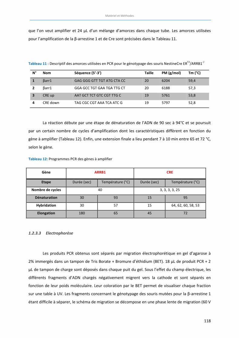
Matériel et Méthodes
118
que l’on veut amplifier et 24 µL d’un mélange d’amorces dans chaque tube. Les amorces utilisées
pour l’amplification de la β-arrestine 1 et de Cre sont précisées dans le Tableau 11.
Tableau 11 : Descriptif des amorces utilisées en PCR pour le génotypage des souris NestineCre ERT2
/ARRB1-/-
N° Nom Séquence (5’-3’) Taille PM (g/mol) Tm (°C)
1 βarr1 GAG GGG GTT TGT ATG CTA CC 20 6204 59,4
2 βarr1 GGA GCC TGT GAA TGA TTG CT 20 6188 57,3
3 CRE up AAT GCT TCT GTC CGT TTG C 19 5761 53,8
4 CRE down TAG CGC CGT AAA TCA ATC G 19 5797 52,8
La réaction débute par une étape de dénaturation de l’ADN de 90 sec à 94°C et se poursuit
par un certain nombre de cycles d’amplification dont les caractéristiques diffèrent en fonction du
gène à amplifier (Tableau 12). Enfin, une extension finale a lieu pendant 7 à 10 min entre 65 et 72 °C,
selon le gène.
Tableau 12: Programmes PCR des gènes à amplifier
Gène ARRB1 CRE
Etape Durée (sec) Température (°C) Durée (sec) Température (°C)
Nombre de cycles 40 3, 3, 3, 3, 25
Dénaturation 30 93 15 95
Hybridation 30 57 15 64, 62, 60, 58, 53
Elongation 180 65 45 72
1.2.3.3 Electrophorèse
Les produits PCR obtenus sont séparés par migration électrophorétique en gel d’agarose à
2% immergés dans un tampon de Tris Borate + Bromure d’éthidium (BET). 18 µL de produit PCR + 2
µL de tampon de charge sont déposés dans chaque puit du gel. Sous l’effet du champ électrique, les
différents fragments d’ADN chargés négativement migrent vers la cathode et sont séparés en
fonction de leur poids moléculaire. Leur coloration par le BET permet de visualiser chaque fraction
sur une table à UV. Les fragments concernant le génotypage des souris mutées pour la β-arrestine 1
étant difficile à séparer, le schéma de migration se décompose en une phase lente de migration (60 V
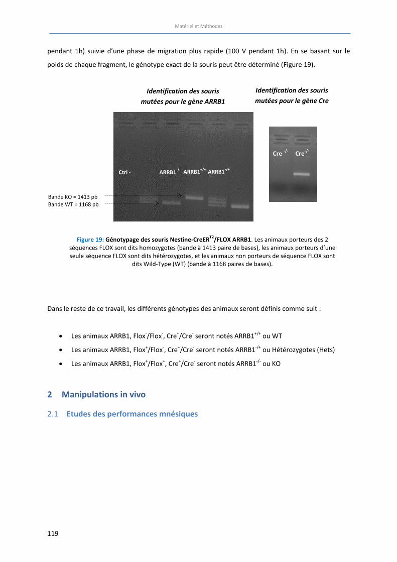
Matériel et Méthodes
119
pendant 1h) suivie d’une phase de migration plus rapide (100 V pendant 1h). En se basant sur le
poids de chaque fragment, le génotype exact de la souris peut être déterminé (Figure 19).
Dans le reste de ce travail, les différents génotypes des animaux seront définis comme suit :
x Les animaux ARRB1, Flox-/Flox-, Cre+/Cre- seront notés ARRB1+/+ ou WT
x Les animaux ARRB1, Flox+/Flox-, Cre+/Cre- seront notés ARRB1-/+ ou Hétérozygotes (Hets)
x Les animaux ARRB1, Flox+/Flox+, Cre+/Cre- seront notés ARRB1-/- ou KO
2 Manipulations in vivo
Etudes des performances mnésiques 2.1
Figure 19: Génotypage des souris Nestine-CreERT2
/FLOX ARRB1. Les animaux porteurs des 2 séquences FLOX sont dits homozygotes (bande à 1413 paire de bases), les animaux porteurs d’une seule séquence FLOX sont dits hétérozygotes, et les animaux non porteurs de séquence FLOX sont
dits Wild-Type (WT) (bande à 1168 paires de bases).
Identification des souris mutées pour le gène Cre
Cre -/-
Cre-/+
Identification des souris mutées pour le gène ARRB1
Ctrl - ARRB1-/+
ARRB1-/-
ARRB1+/+
Bande KO = 1413 pb
Bande WT = 1168 pb
1. Fiche méthode : le test de reconnaissance d’objet

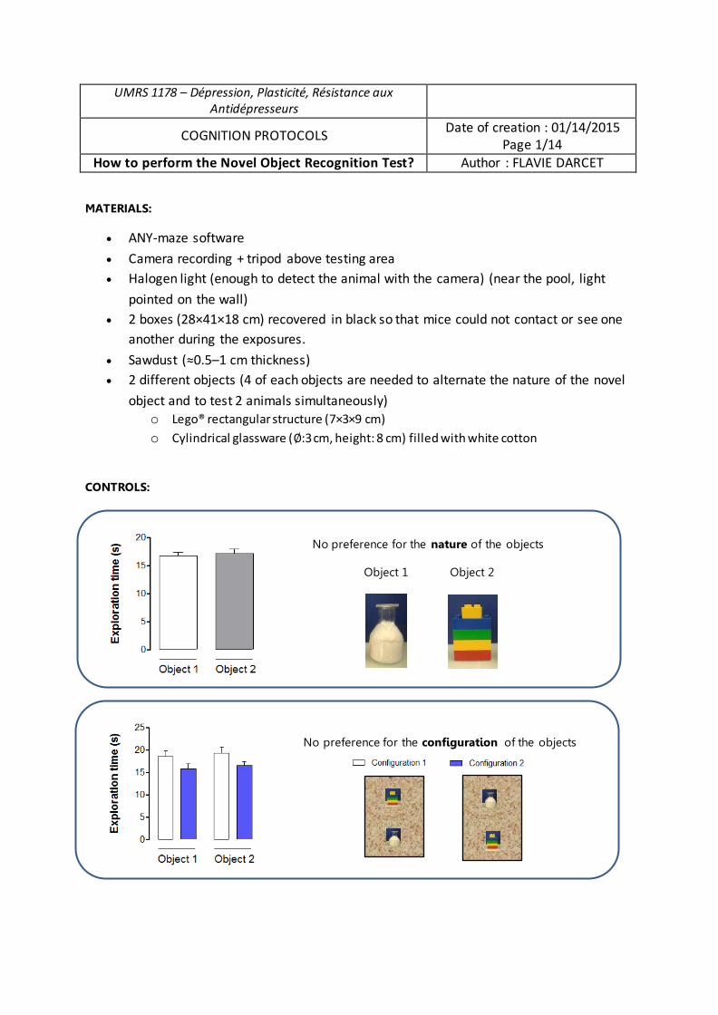
UMRS 1178 – Dépression, Plasticité, Résistance aux Antidépresseurs
COGNITION PROTOCOLS Date of creation : 01/14/2015
Page 1/14 How to perform the Novel Object Recognition Test? Author : FLAVIE DARCET
MATERIALS:
x ANY-maze software
x Camera recording + tripod above testing area x Halogen light (enough to detect the animal with the camera) (near the pool, light
pointed on the wall) x 2 boxes (28×41×18 cm) recovered in black so that mice could not contact or see one
another during the exposures.
x Sawdust (≈0.5–1 cm thickness) x 2 different objects (4 of each objects are needed to alternate the nature of the novel
object and to test 2 animals simultaneously) o Lego® rectangular structure (7×3×9 cm) o Cylindrical glassware (∅:3 cm, height: 8 cm) filled with white cotton
CONTROLS:
No preference for the nature of the objects
Object 1 Object 2
No preference for the configuration of the objects
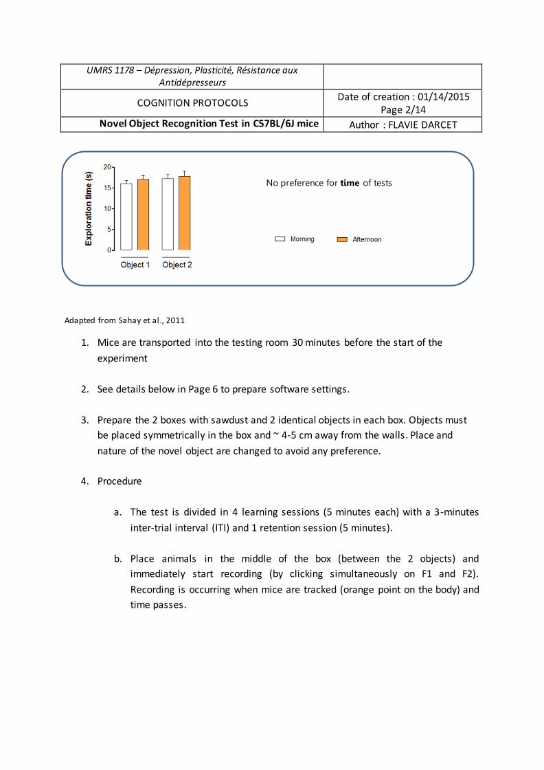
UMRS 1178 – Dépression, Plasticité, Résistance aux Antidépresseurs
COGNITION PROTOCOLS Date of creation : 01/14/2015 Page 2/14
Novel Object Recognition Test in C57BL/6J mice Author : FLAVIE DARCET
Adapted from Sahay et al., 2011
1. Mice are transported into the testing room 30 minutes before the start of the experiment
2. See details below in Page 6 to prepare software settings.
3. Prepare the 2 boxes with sawdust and 2 identical objects in each box. Objects must be placed symmetrically in the box and ~ 4-5 cm away from the walls. Place and nature of the novel object are changed to avoid any preference.
4. Procedure
a. The test is divided in 4 learning sessions (5 minutes each) with a 3-minutes
inter-trial interval (ITI) and 1 retention session (5 minutes).
b. Place animals in the middle of the box (between the 2 objects) and immediately start recording (by clicking simultaneously on F1 and F2).
Recording is occurring when mice are tracked (orange point on the body) and time passes.
No preference for time of tests

c. When the session is finished, take mice back in their home cage during ITI. Between each session, change sawdust in each cage, clean boxes and objects with 70% ethanol.
d. Repeat Steps b and c to perform 4 learning sessions.
e. For the retention session, remove one of the familiar objects from boxes and
replace it with a novel object. Assess memory by scoring novel object exploration time during 5 minutes.
UMRS 1178 – Dépression, Plasticité, Résistance aux Antidépresseurs
COGNITION PROTOCOLS Date of creation : 01/14/2015 Page 3/14
Novel Object Recognition Test in C57BL/6J mice Author : FLAVIE DARCET
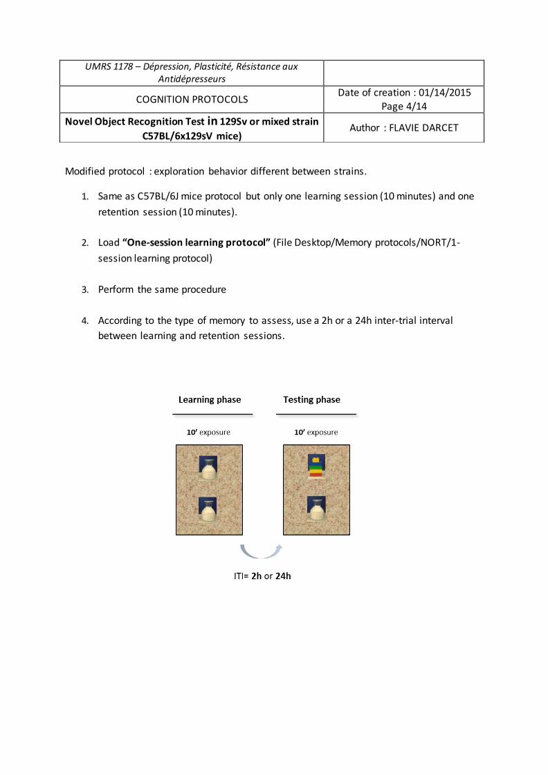
Modified protocol : exploration behavior different between strains.
1. Same as C57BL/6J mice protocol but only one learning session (10 minutes) and one retention session (10 minutes).
2. Load “One-session learning protocol” (File Desktop/Memory protocols/NORT/1-session learning protocol)
3. Perform the same procedure
4. According to the type of memory to assess, use a 2h or a 24h inter-trial interval between learning and retention sessions.
UMRS 1178 – Dépression, Plasticité, Résistance aux Antidépresseurs
COGNITION PROTOCOLS Date of creation : 01/14/2015 Page 4/14
Novel Object Recognition Test in 129Sv or mixed strain C57BL/6x129sV mice)
Author : FLAVIE DARCET
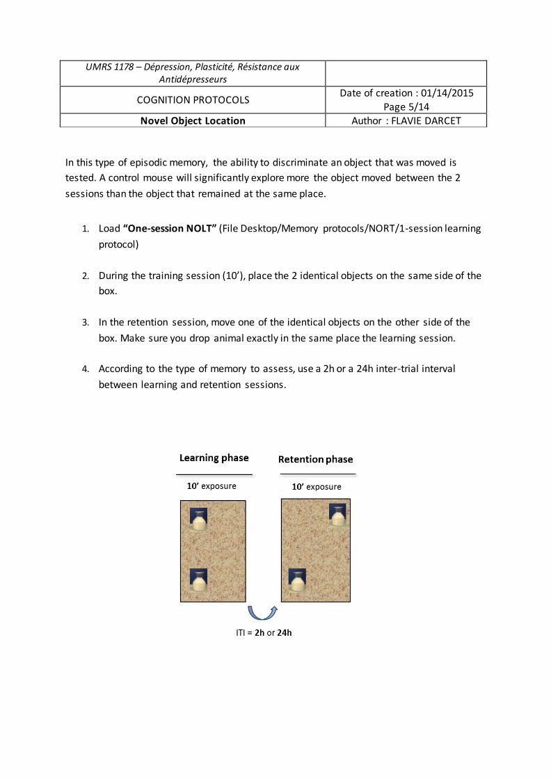
In this type of episodic memory, the ability to discriminate an object that was moved is tested. A control mouse will significantly explore more the object moved between the 2 sessions than the object that remained at the same place.
1. Load “One-session NOLT” (File Desktop/Memory protocols/NORT/1-session learning
protocol)
2. During the training session (10’), place the 2 identical objects on the same side of the box.
3. In the retention session, move one of the identical objects on the other side of the box. Make sure you drop animal exactly in the same place the learning session.
4. According to the type of memory to assess, use a 2h or a 24h inter-trial interval between learning and retention sessions.
UMRS 1178 – Dépression, Plasticité, Résistance aux Antidépresseurs
COGNITION PROTOCOLS Date of creation : 01/14/2015 Page 5/14
Novel Object Location Author : FLAVIE DARCET
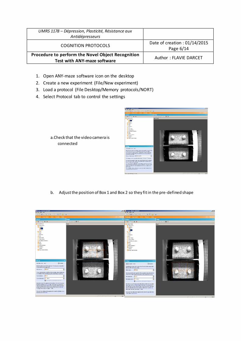
UMRS 1178 – Dépression, Plasticité, Résistance aux Antidépresseurs
COGNITION PROTOCOLS Date of creation : 01/14/2015
Page 6/14
Procedure to perform the Novel Object Recognition Test with ANY-maze software Author : FLAVIE DARCET
1. Open ANY-maze software icon on the desktop
2. Create a new experiment (File/New experiment)
3. Load a protocol (File Desktop/Memory protocols/NORT)
4. Select Protocol tab to control the settings
a.Check that the video camera is
connected
b. Adjust the position of Box 1 and Box 2 so they fit in the pre-defined shape
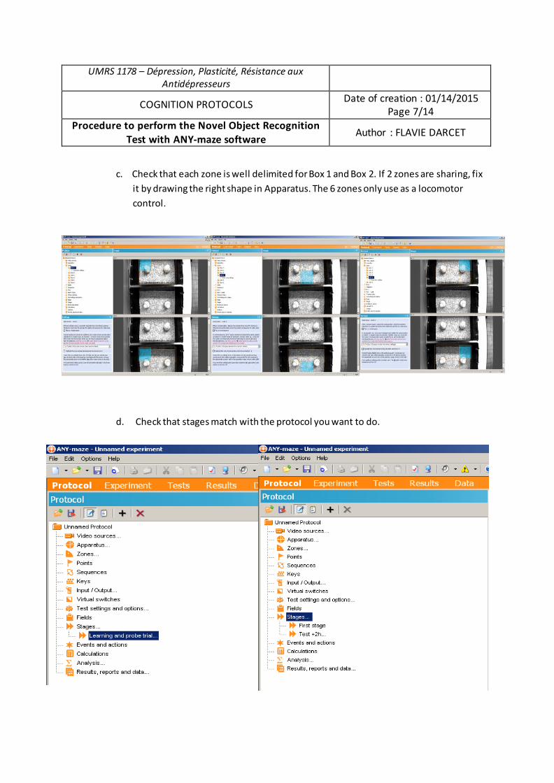
UMRS 1178 – Dépression, Plasticité, Résistance aux Antidépresseurs
COGNITION PROTOCOLS Date of creation : 01/14/2015
Page 7/14
Procedure to perform the Novel Object Recognition Test with ANY-maze software Author : FLAVIE DARCET
c. Check that each zone is well delimited for Box 1 and Box 2. If 2 zones are sharing, fix
it by drawing the right shape in Apparatus. The 6 zones only use as a locomotor
control.
d. Check that stages match with the protocol you want to do.
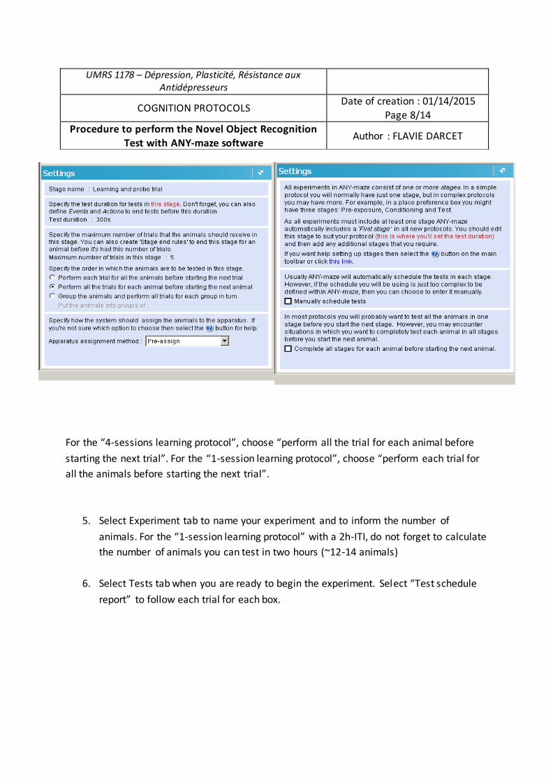
For the “4-sessions learning protocol”, choose “perform all the trial for each animal before starting the next trial”. For the “1-session learning protocol”, choose “perform each trial for all the animals before starting the next trial”.
5. Select Experiment tab to name your experiment and to inform the number of animals. For the “1-session learning protocol” with a 2h-ITI, do not forget to calculate the number of animals you can test in two hours (~12-14 animals)
6. Select Tests tab when you are ready to begin the experiment. Select “Test schedule report” to follow each trial for each box.
UMRS 1178 – Dépression, Plasticité, Résistance aux Antidépresseurs
COGNITION PROTOCOLS Date of creation : 01/14/2015 Page 8/14
Procedure to perform the Novel Object Recognition Test with ANY-maze software Author : FLAVIE DARCET
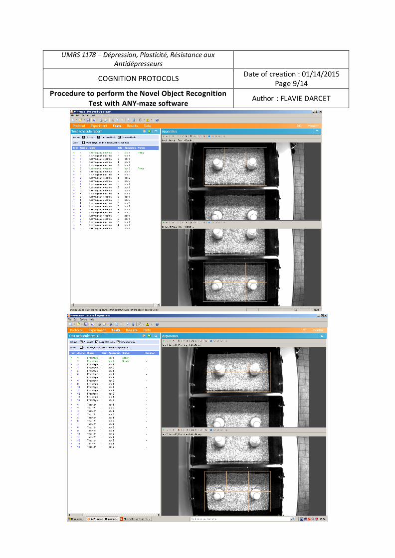
UMRS 1178 – Dépression, Plasticité, Résistance aux Antidépresseurs
COGNITION PROTOCOLS Date of creation : 01/14/2015 Page 9/14
Procedure to perform the Novel Object Recognition Test with ANY-maze software Author : FLAVIE DARCET
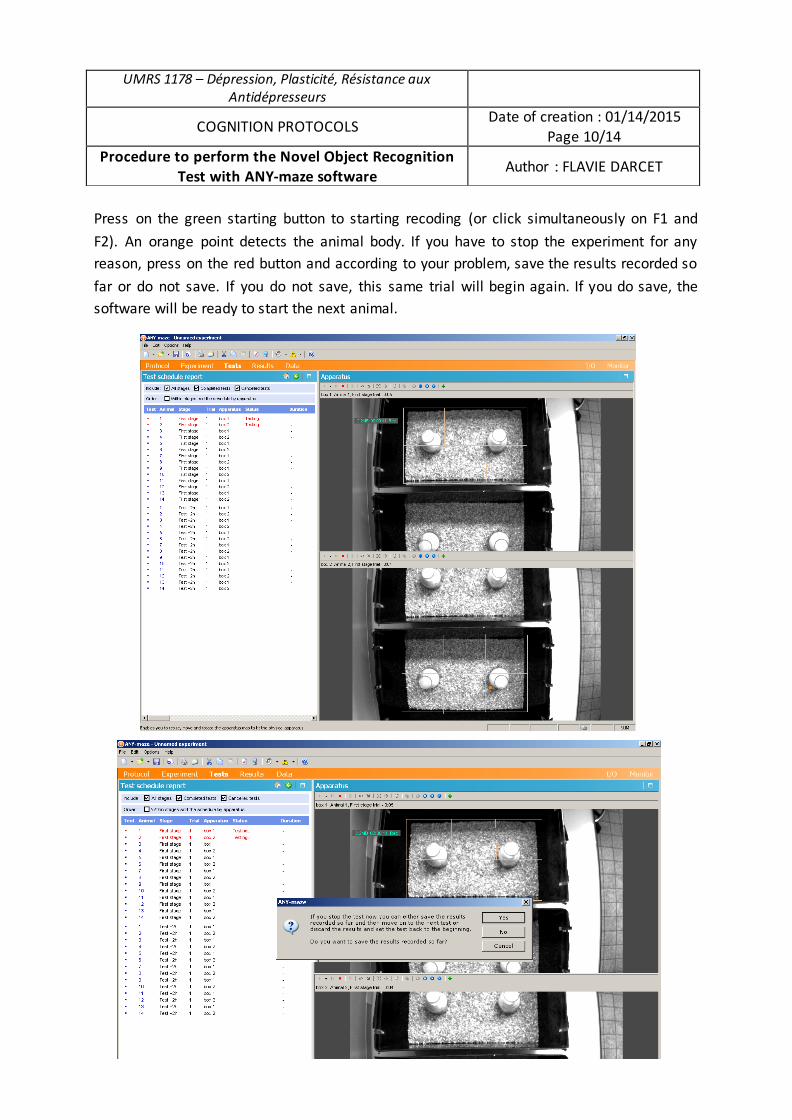
Press on the green starting button to starting recoding (or click simultaneously on F1 and F2). An orange point detects the animal body. If you have to stop the experiment for any reason, press on the red button and according to your problem, save the results recorded so far or do not save. If you do not save, this same trial will begin again. If you do save, the software will be ready to start the next animal.
UMRS 1178 – Dépression, Plasticité, Résistance aux Antidépresseurs
COGNITION PROTOCOLS Date of creation : 01/14/2015 Page 10/14
Procedure to perform the Novel Object Recognition Test with ANY-maze software Author : FLAVIE DARCET
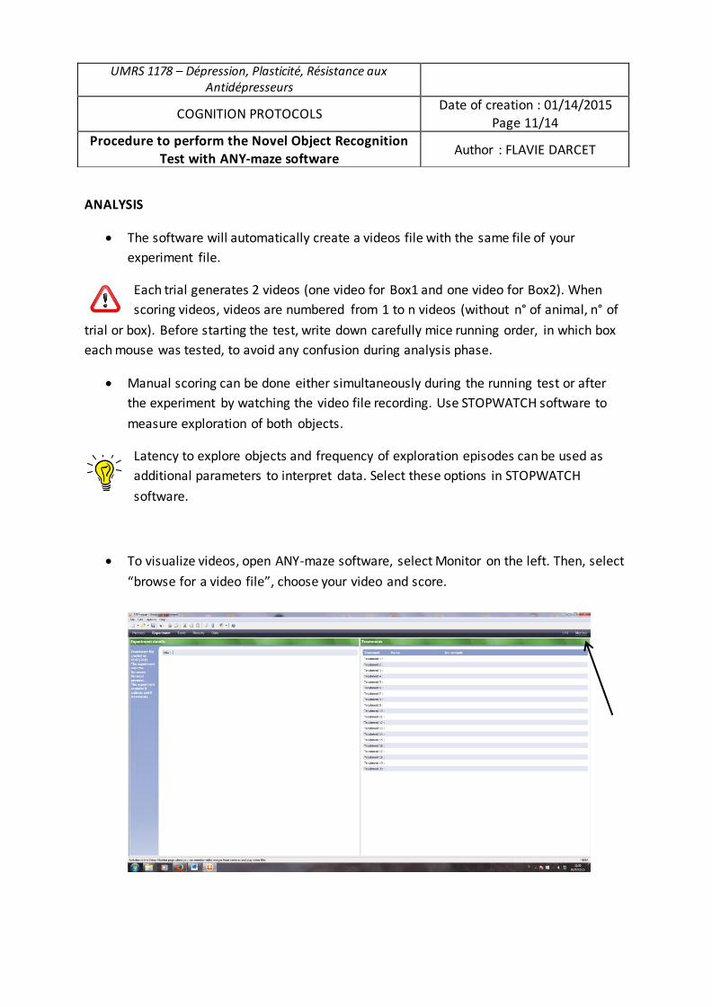
ANALYSIS
x The software will automatically create a videos file with the same file of your experiment file.
Each trial generates 2 videos (one video for Box1 and one video for Box2). When scoring videos, videos are numbered from 1 to n videos (without n° of animal, n° of
trial or box). Before starting the test, write down carefully mice running order, in which box each mouse was tested, to avoid any confusion during analysis phase.
x Manual scoring can be done either simultaneously during the running test or after the experiment by watching the video file recording. Use STOPWATCH software to measure exploration of both objects.
Latency to explore objects and frequency of exploration episodes can be used as additional parameters to interpret data. Select these options in STOPWATCH software.
x To visualize videos, open ANY-maze software, select Monitor on the left. Then, select “browse for a video file”, choose your video and score.
UMRS 1178 – Dépression, Plasticité, Résistance aux Antidépresseurs
COGNITION PROTOCOLS Date of creation : 01/14/2015 Page 11/14
Procedure to perform the Novel Object Recognition Test with ANY-maze software Author : FLAVIE DARCET
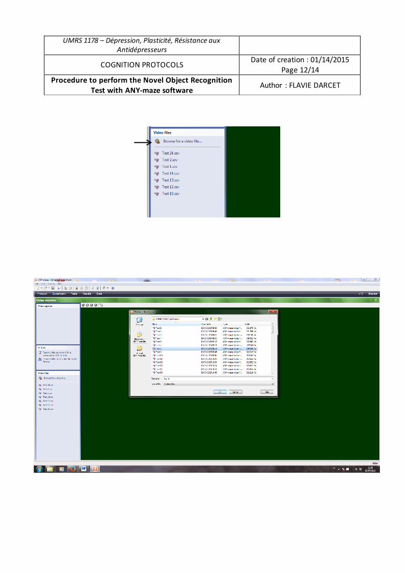
UMRS 1178 – Dépression, Plasticité, Résistance aux Antidépresseurs
COGNITION PROTOCOLS Date of creation : 01/14/2015 Page 12/14
Procedure to perform the Novel Object Recognition Test with ANY-maze software Author : FLAVIE DARCET
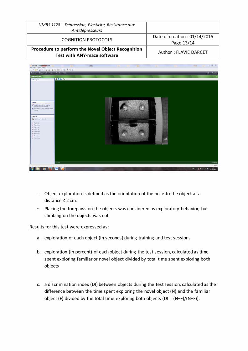
- Object exploration is defined as the orientation of the nose to the object at a
distance ≤ 2 cm.
- Placing the forepaws on the objects was considered as exploratory behavior, but
climbing on the objects was not.
Results for this test were expressed as:
a. exploration of each object (in seconds) during training and test sessions
b. exploration (in percent) of each object during the test session, calculated as time
spent exploring familiar or novel object divided by total time spent exploring both
objects
c. a discrimination index (DI) between objects during the test session, calculated as the
difference between the time spent exploring the novel object (N) and the familiar
object (F) divided by the total time exploring both objects (DI = (N–F)/(N+F)).
UMRS 1178 – Dépression, Plasticité, Résistance aux Antidépresseurs
COGNITION PROTOCOLS Date of creation : 01/14/2015
Page 13/14 Procedure to perform the Novel Object Recognition
Test with ANY-maze software Author : FLAVIE DARCET
LOCOMOTOR ANALYSIS
Locomotor activity was controlled during the entire experiment (parameter: ambulatory distance) using a videotracking procedure (ANY-maze Software, Bioseb, France).
Check with the following parameters that motor activity remains unchanged for all conditions:
- time in each zone (1 to 6) - crossing in each zone (1 to 6) - distance travelled in each zone (1 to 6)
Collect data from ANY-maze software and export them in an Excel file.
UMRS 1178 – Dépression, Plasticité, Résistance aux Antidépresseurs
COGNITION PROTOCOLS Date of creation : 01/14/2015 Page 14/14
Procedure to perform the Novel Object Recognition Test with ANY-maze software Author : FLAVIE DARCET
2. FicheméthodedutestdelapiscinedeMorris

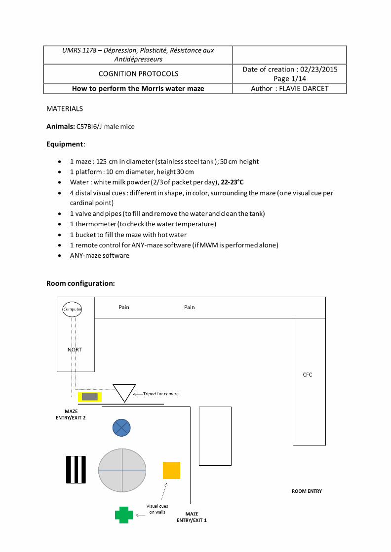
UMRS 1178 – Dépression, Plasticité, Résistance aux Antidépresseurs
COGNITION PROTOCOLS Date of creation : 02/23/2015 Page 1/14
How to perform the Morris water maze Author : FLAVIE DARCET
MATERIALS
Animals: C57Bl6/J male mice
Equipment:
x 1 maze : 125 cm in diameter (stainless steel tank ); 50 cm height x 1 platform : 10 cm diameter, height 30 cm x Water : white milk powder (2/3 of packet per day), 22-23°C
x 4 distal visual cues : different in shape, in color, surrounding the maze (one visual cue per cardinal point)
x 1 valve and pipes (to fill and remove the water and clean the tank) x 1 thermometer (to check the water temperature)
x 1 bucket to fill the maze with hot water x 1 remote control for ANY-maze software (if MWM is performed alone) x ANY-maze software
Room configuration:
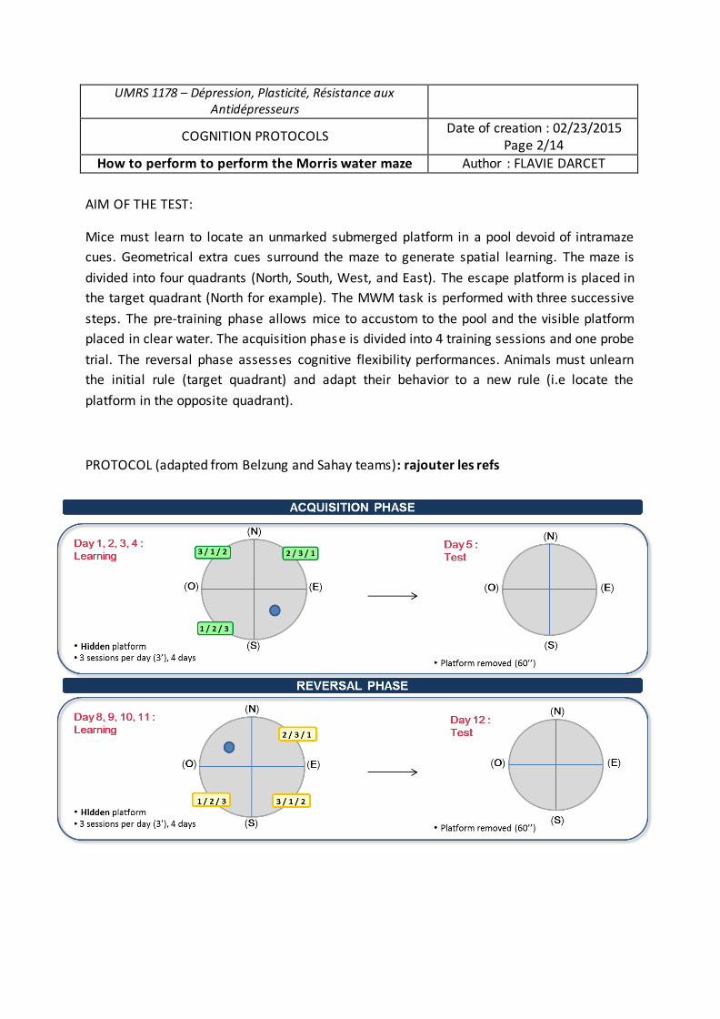
UMRS 1178 – Dépression, Plasticité, Résistance aux Antidépresseurs
COGNITION PROTOCOLS Date of creation : 02/23/2015
Page 2/14
How to perform to perform the Morris water maze Author : FLAVIE DARCET
AIM OF THE TEST:
Mice must learn to locate an unmarked submerged platform in a pool devoid of intramaze
cues. Geometrical extra cues surround the maze to generate spatial learning. The maze is
divided into four quadrants (North, South, West, and East). The escape platform is placed in
the target quadrant (North for example). The MWM task is performed with three successive
steps. The pre-training phase allows mice to accustom to the pool and the visible platform
placed in clear water. The acquisition phase is divided into 4 training sessions and one probe
trial. The reversal phase assesses cognitive flexibility performances. Animals must unlearn
the initial rule (target quadrant) and adapt their behavior to a new rule (i.e locate the
platform in the opposite quadrant).
PROTOCOL (adapted from Belzung and Sahay teams): rajouter les refs
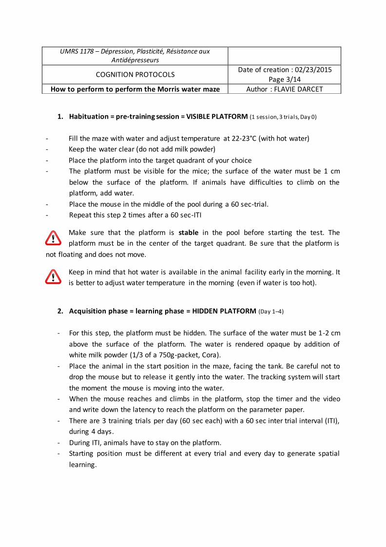
1. Habituation = pre-training session = VISIBLE PLATFORM (1 session, 3 trials, Day 0)
- Fill the maze with water and adjust temperature at 22-23°C (with hot water) - Keep the water clear (do not add milk powder) - Place the platform into the target quadrant of your choice - The platform must be visible for the mice; the surface of the water must be 1 cm
below the surface of the platform. If animals have difficulties to climb on the platform, add water.
- Place the mouse in the middle of the pool during a 60 sec-trial. - Repeat this step 2 times after a 60 sec-ITI
Make sure that the platform is stable in the pool before starting the test. The platform must be in the center of the target quadrant. Be sure that the platform is
not floating and does not move.
Keep in mind that hot water is available in the animal facility early in the morning. It is better to adjust water temperature in the morning (even if water is too hot).
2. Acquisition phase = learning phase = HIDDEN PLATFORM (Day 1–4)
- For this step, the platform must be hidden. The surface of the water must be 1-2 cm above the surface of the platform. The water is rendered opaque by addition of white milk powder (1/3 of a 750g-packet, Cora).
- Place the animal in the start position in the maze, facing the tank. Be careful not to drop the mouse but to release it gently into the water. The tracking system will start the moment the mouse is moving into the water.
- When the mouse reaches and climbs in the platform, stop the timer and the video and write down the latency to reach the platform on the parameter paper.
- There are 3 training trials per day (60 sec each) with a 60 sec inter trial interval (ITI), during 4 days.
- During ITI, animals have to stay on the platform. - Starting position must be different at every trial and every day to generate spatial
learning.
UMRS 1178 – Dépression, Plasticité, Résistance aux Antidépresseurs
COGNITION PROTOCOLS Date of creation : 02/23/2015 Page 3/14
How to perform to perform the Morris water maze Author : FLAVIE DARCET
Mice that exceed 60 sec to find the platform will be gently guided to the platform by the experimenter. In this case, note 60 sec as escape latency for this animal. If the mouse jumps into the water before the end of the 60-sec ITI, replace it on the platform until it stays 60 sec.
Examples for starting positions:
� Day 1: SE - NW – SW � Day 2: SW – SE – NW � Day 3: NW – SW – SE � Day 4: SE - SW – NW
3. Probe trial = retention phase (Day 5)
- Remove the platform from the opaque water. - Place the animal in the opposite quadrant, facing the tank wall . - Probe trial lasts 60 sec during which animals are free to explore the maze.
4. Reversal phase (Day 8–11)
If the aim of the study includes assessing cognitive flexibility, spatial reversal learning and retention starts 2 days after the acquisition probe trial in Day 5.
- Change the location of the escape platform from the target quadrant used during acquisition to the opposite target quadrant (which becomes the new target quadrant).
- Repeat the exact same procedure as acquisition phase : 4 training days, 3 trials per days, 60 sec per trial and ITI
5. Probe trial = retention phase for cognitive flexibility (Day 12)
Repeat the same procedure as Probe trial in Day 5.
UMRS 1178 – Dépression, Plasticité, Résistance aux Antidépresseurs
COGNITION PROTOCOLS Date of creation : 02/23/2015 Page 4/14
How to perform to perform the Morris water maze Author : FLAVIE DARCET
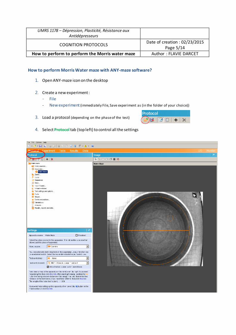
UMRS 1178 – Dépression, Plasticité, Résistance aux Antidépresseurs
COGNITION PROTOCOLS Date of creation : 02/23/2015 Page 5/14
How to perform to perform the Morris water maze Author : FLAVIE DARCET
How to perform Morris Water maze with ANY-maze software?
1. Open ANY-maze icon on the desktop
2. Create a new experiment : - File - New experiment (immediately File, Save experiment as (in the folder of your choice))
3. Load a protocol (depending on the phase of the test)
4. Select Protocol tab (top left) to control all the settings
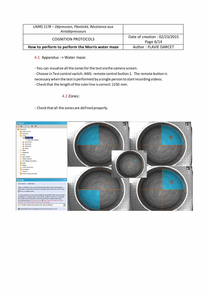
UMRS 1178 – Dépression, Plasticité, Résistance aux Antidépresseurs
COGNITION PROTOCOLS Date of creation : 02/23/2015
Page 6/14
How to perform to perform the Morris water maze Author : FLAVIE DARCET
4.1 Apparatus -> Water maze:
- You can visualize all the zones for the test via the camera screen.
- Choose in Test control switch: AM1- remote control button 1. The remote button is
necessary when the test is performed by a single person to start recording videos.
- Check that the length of the ruler line is correct: 1250 mm.
4.2 Zones:
- Check that all the zones are defined properly.
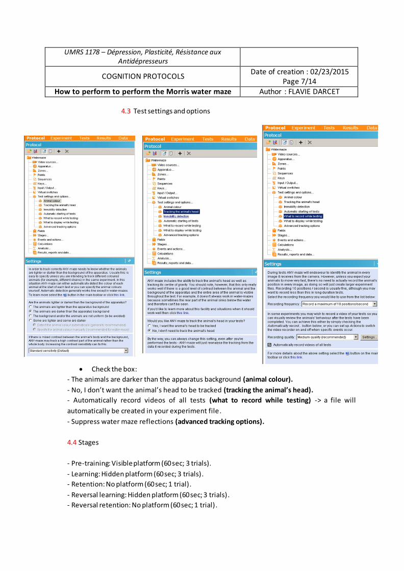
UMRS 1178 – Dépression, Plasticité, Résistance aux Antidépresseurs
COGNITION PROTOCOLS Date of creation : 02/23/2015 Page 7/14
How to perform to perform the Morris water maze Author : FLAVIE DARCET
4.3 Test settings and options
x Check the box: - The animals are darker than the apparatus background (animal colour). - No, I don’t want the animal’s head to be tracked (tracking the animal’s head). - Automatically record videos of all tests (what to record while testing) -> a file will automatically be created in your experiment file. - Suppress water maze reflections (advanced tracking options). 4.4 Stages - Pre-training: Visible platform (60 sec; 3 trials). - Learning: Hidden platform (60 sec; 3 trials). - Retention: No platform (60 sec; 1 trial). - Reversal learning: Hidden platform (60 sec; 3 trials). - Reversal retention: No platform (60 sec; 1 trial).
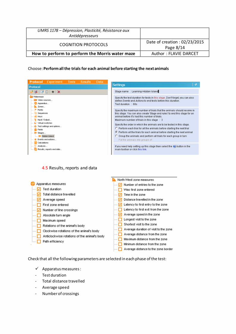
UMRS 1178 – Dépression, Plasticité, Résistance aux Antidépresseurs
COGNITION PROTOCOLS Date of creation : 02/23/2015
Page 8/14
How to perform to perform the Morris water maze Author : FLAVIE DARCET
Choose: Perform all the trials for each animal before starting the next animals
4.5 Results, reports and data
Check that all the following parameters are selected in each phase of the test:
9 Apparatus measures :
- Test duration
- Total distance travelled
- Average speed
- Number of crossings
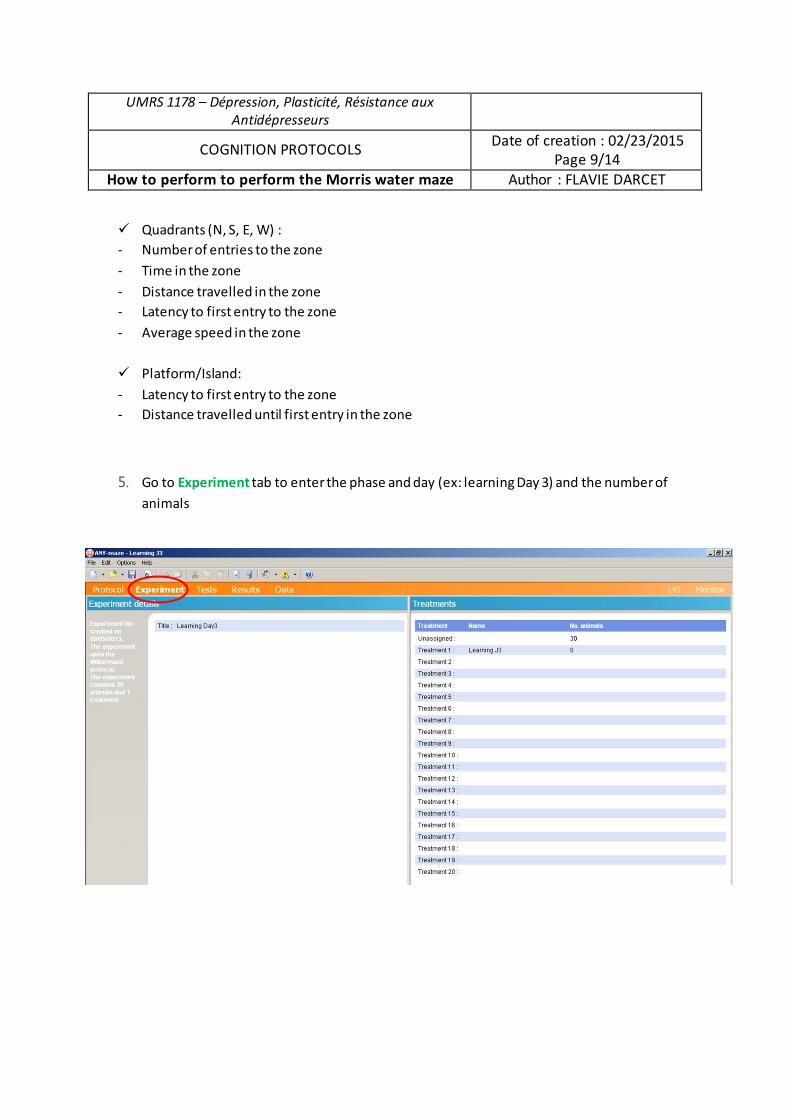
UMRS 1178 – Dépression, Plasticité, Résistance aux Antidépresseurs
COGNITION PROTOCOLS Date of creation : 02/23/2015 Page 9/14
How to perform to perform the Morris water maze Author : FLAVIE DARCET
9 Quadrants (N, S, E, W) : - Number of entries to the zone - Time in the zone - Distance travelled in the zone - Latency to first entry to the zone - Average speed in the zone
9 Platform/Island: - Latency to first entry to the zone - Distance travelled until first entry in the zone
5. Go to Experiment tab to enter the phase and day (ex: learning Day 3) and the number of animals
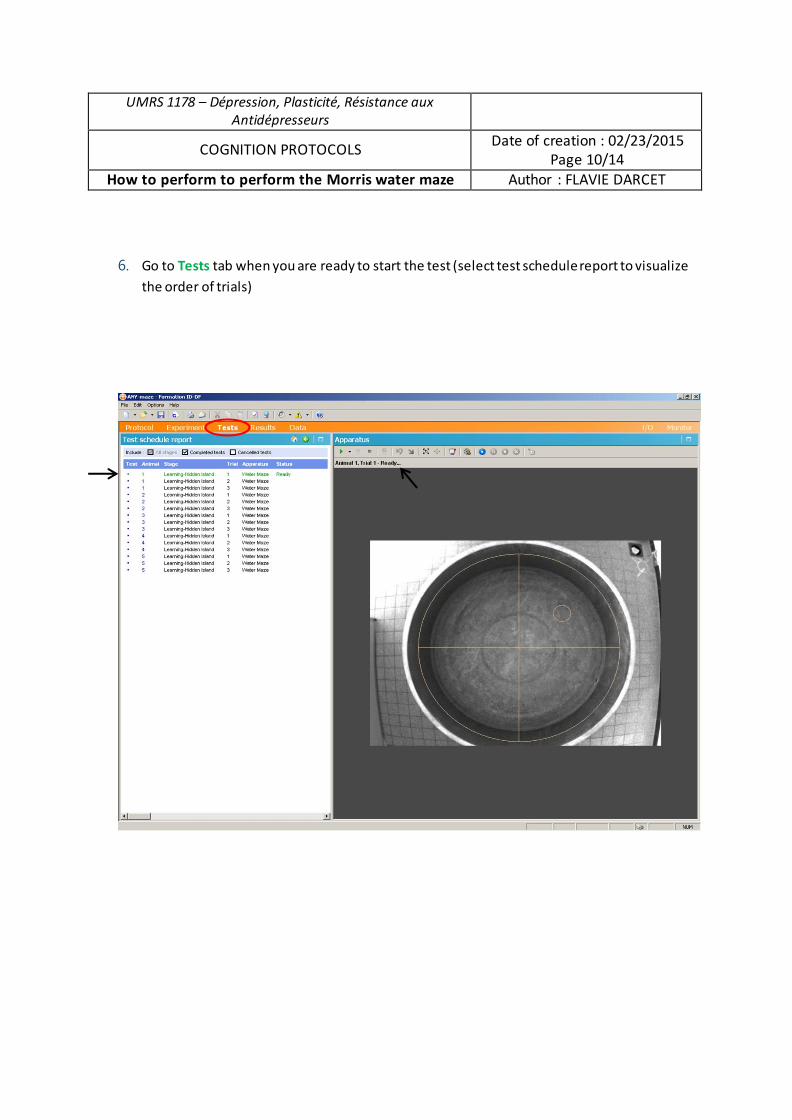
UMRS 1178 – Dépression, Plasticité, Résistance aux Antidépresseurs
COGNITION PROTOCOLS Date of creation : 02/23/2015
Page 10/14
How to perform to perform the Morris water maze Author : FLAVIE DARCET
6. Go to Tests tab when you are ready to start the test (select test schedule report to visualize
the order of trials)
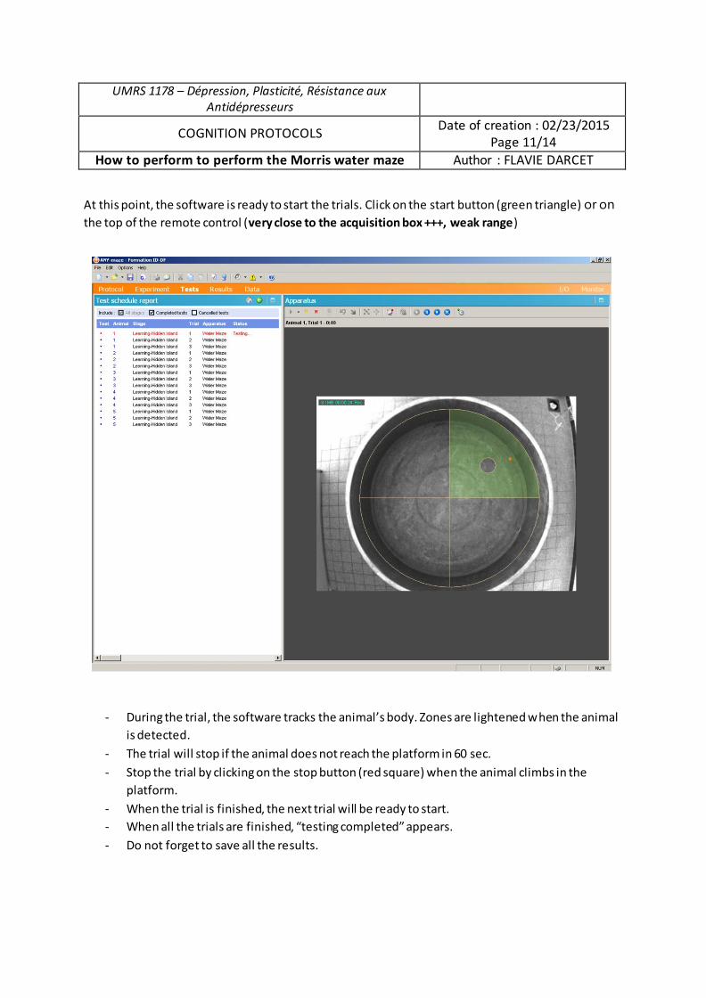
UMRS 1178 – Dépression, Plasticité, Résistance aux Antidépresseurs
COGNITION PROTOCOLS Date of creation : 02/23/2015 Page 11/14
How to perform to perform the Morris water maze Author : FLAVIE DARCET
At this point, the software is ready to start the trials. Click on the start button (green triangle) or on the top of the remote control (very close to the acquisition box +++, weak range)
- During the trial, the software tracks the animal’s body. Zones are lightened when the animal is detected.
- The trial will stop if the animal does not reach the platform in 60 sec. - Stop the trial by clicking on the stop button (red square) when the animal climbs in the
platform. - When the trial is finished, the next trial will be ready to start. - When all the trials are finished, “testing completed” appears. - Do not forget to save all the results.
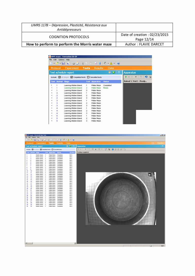
UMRS 1178 – Dépression, Plasticité, Résistance aux Antidépresseurs
COGNITION PROTOCOLS Date of creation : 02/23/2015 Page 12/14
How to perform to perform the Morris water maze Author : FLAVIE DARCET
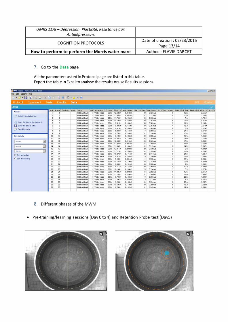
UMRS 1178 – Dépression, Plasticité, Résistance aux Antidépresseurs
COGNITION PROTOCOLS Date of creation : 02/23/2015
Page 13/14 How to perform to perform the Morris water maze Author : FLAVIE DARCET
7. Go to the Data page
All the parameters asked in Protocol page are listed in this table. Export the table in Excel to analyse the results or use Results sessions.
8. Different phases of the MWM
x Pre-training/learning sessions (Day 0 to 4) and Retention Probe test (Day5)
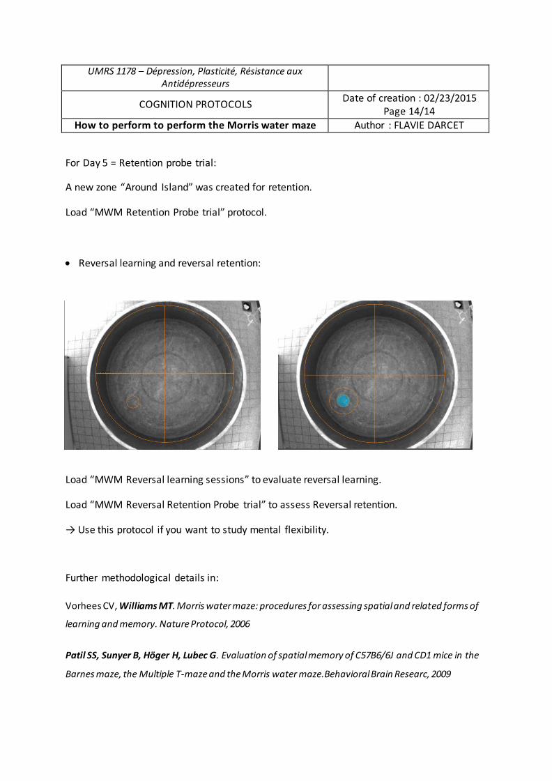
UMRS 1178 – Dépression, Plasticité, Résistance aux Antidépresseurs
COGNITION PROTOCOLS Date of creation : 02/23/2015 Page 14/14
How to perform to perform the Morris water maze Author : FLAVIE DARCET
For Day 5 = Retention probe trial:
A new zone “Around Island” was created for retention.
Load “MWM Retention Probe trial” protocol.
x Reversal learning and reversal retention:
Load “MWM Reversal learning sessions” to evaluate reversal learning.
Load “MWM Reversal Retention Probe trial” to assess Reversal retention.
→ Use this protocol if you want to study mental flexibility.
Further methodological details in:
Vorhees CV, Williams MT. Morris water maze: procedures for assessing spatial and related forms of
learning and memory. Nature Protocol, 2006
Patil SS, Sunyer B, Höger H, Lubec G. Evaluation of spatial memory of C57B6/6J and CD1 mice in the
Barnes maze, the Multiple T-maze and the Morris water maze.Behavioral Brain Researc, 2009
3. Fiche méthode du test du labyrinthe de Barnes

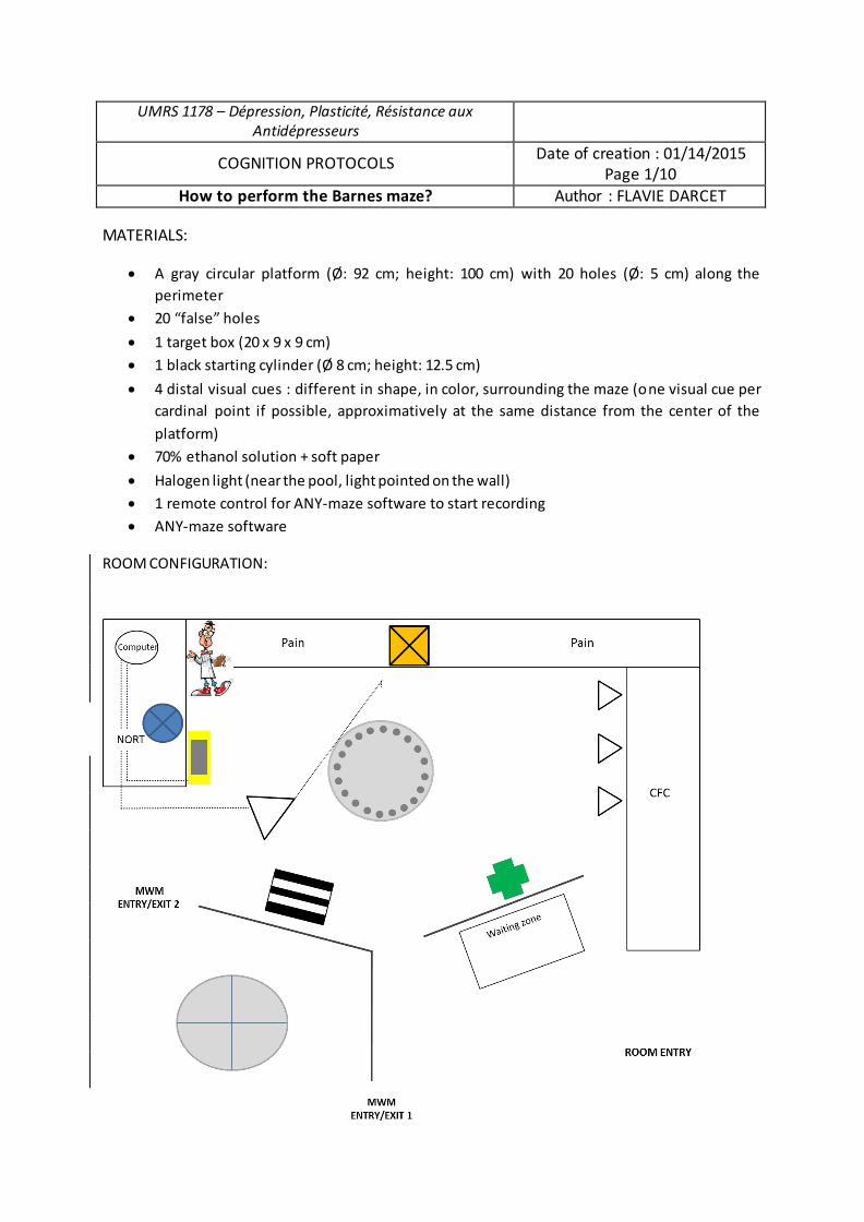
UMRS 1178 – Dépression, Plasticité, Résistance aux Antidépresseurs
COGNITION PROTOCOLS Date of creation : 01/14/2015
Page 1/10
How to perform the Barnes maze? Author : FLAVIE DARCET
MATERIALS:
x A gray circular platform (Ø: 92 cm; height: 100 cm) with 20 holes (Ø: 5 cm) along the
perimeter
x 20 “false” holes
x 1 target box (20 x 9 x 9 cm)
x 1 black starting cylinder (Ø 8 cm; height: 12.5 cm)
x 4 distal visual cues : different in shape, in color, surrounding the maze (one visual cue per
cardinal point if possible, approximatively at the same distance from the center of the
platform)
x 70% ethanol solution + soft paper
x Halogen light (near the pool, light pointed on the wall)
x 1 remote control for ANY-maze software to start recording
x ANY-maze software
ROOM CONFIGURATION:
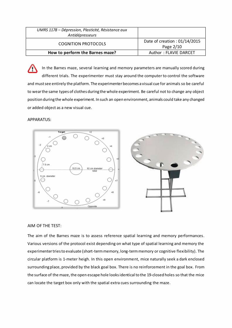
UMRS 1178 – Dépression, Plasticité, Résistance aux Antidépresseurs
COGNITION PROTOCOLS Date of creation : 01/14/2015
Page 2/10
How to perform the Barnes maze? Author : FLAVIE DARCET
In the Barnes maze, several learning and memory parameters are manually scored during
different trials. The experimenter must stay around the computer to control the software
and must see entirely the platform. The experimenter becomes a visual cue for animals so be careful
to wear the same types of clothes during the whole experiment. Be careful not to change any object
position during the whole experiment. In such an open environment, animals could take any changed
or added object as a new visual cue.
APPARATUS:
AIM OF THE TEST:
The aim of the Barnes maze is to assess reference spatial learning and memory performances.
Various versions of the protocol exist depending on what type of spatial learning and memory the
experimenter tries to evaluate (short-term memory, long-term memory or cognitive flexibility). The
circular platform is 1-meter heigh. In this open environment, mice naturally seek a dark enclosed
surrounding place, provided by the black goal box. There is no reinforcement in the goal box. From
the surface of the maze, the open escape hole looks identical to the 19-closed holes so that the mice
can locate the target box only with the spatial extra cues surrounding the maze.
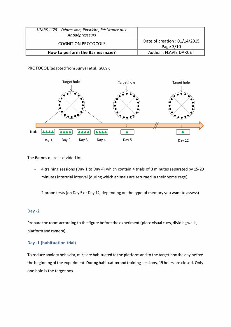
UMRS 1178 – Dépression, Plasticité, Résistance aux Antidépresseurs
COGNITION PROTOCOLS Date of creation : 01/14/2015
Page 3/10 How to perform the Barnes maze? Author : FLAVIE DARCET
PROTOCOL (adapted from Sunyer et al., 2009):
The Barnes maze is divided in:
- 4 training sessions (Day 1 to Day 4) which contain 4 trials of 3 minutes separated by 15-20
minutes intertrial interval (during which animals are returned in their home cage)
- 2 probe tests (on Day 5 or Day 12, depending on the type of memory you want to assess)
Day -2
Prepare the room according to the figure before the experiment (place visual cues, dividing walls,
platform and camera).
Day -1 (habituation trial)
To reduce anxiety behavior, mice are habituated to the platform and to the target box the day before
the beginning of the experiment. During habituation and training sessions, 19 holes are closed. Only
one hole is the target box.
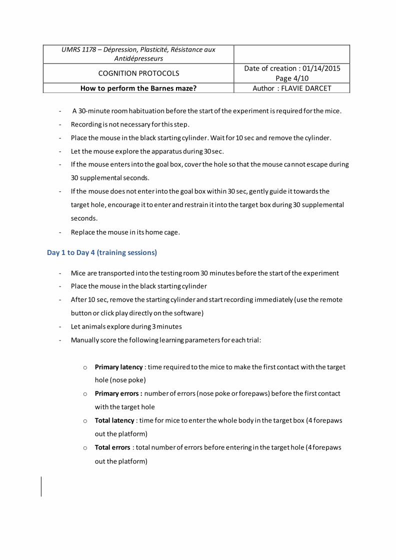
- A 30-minute room habituation before the start of the experiment is required for the mice.
- Recording is not necessary for this step.
- Place the mouse in the black starting cylinder. Wait for 10 sec and remove the cylinder.
- Let the mouse explore the apparatus during 30 sec.
- If the mouse enters into the goal box, cover the hole so that the mouse cannot escape during
30 supplemental seconds.
- If the mouse does not enter into the goal box within 30 sec, gently guide it towards the
target hole, encourage it to enter and restrain it into the target box during 30 supplemental
seconds.
- Replace the mouse in its home cage.
Day 1 to Day 4 (training sessions)
- Mice are transported into the testing room 30 minutes before the start of the experiment
- Place the mouse in the black starting cylinder
- After 10 sec, remove the starting cylinder and start recording immediately (use the remote
button or click play directly on the software)
- Let animals explore during 3 minutes
- Manually score the following learning parameters for each trial:
o Primary latency : time required to the mice to make the first contact with the target
hole (nose poke)
o Primary errors : number of errors (nose poke or forepaws) before the first contact
with the target hole
o Total latency : time for mice to enter the whole body in the target box (4 forepaws
out the platform)
o Total errors : total number of errors before entering in the target hole (4 forepaws
out the platform)
UMRS 1178 – Dépression, Plasticité, Résistance aux Antidépresseurs
COGNITION PROTOCOLS Date of creation : 01/14/2015 Page 4/10
How to perform the Barnes maze? Author : FLAVIE DARCET
- Ambulatory parameters are recorded by the software (see details above in “How to perform
Barnes maze with ANY-maze software”).
- Once animals have entered into the target box, let them stay in during 60 supplemental
seconds.
- If they failed to find or enter into the target box, gently guide animals towards the target box
entry. For those mice, 180 seconds are recorded for escape latency.
- Return the animals in their home cage during the 15-20 minutes intertrial interval.
- Clean the apparatus, the target box and false boxes between each trial and each animal with
a 70% ethanol solution to avoid any olfactory cues.
- Repeat the same protocol for trial 2, 3 and 4.
- Repeat the same procedure for Day 2, Day 3 and Day 4.
A hole was considered visited when mice tilted their head over it (nose poke) or introduced
theirs paws in the hole.
You can count errors and measure latencies either facing the maze (directly looking at the
mouse on the platform) either looking on the computer screen (back to the maze, indirectly
looking at the mouse). Both options are possible, but once you decided, keep it that way for the rest
of the experiment.
Always control on the screen that the mouse is correctly tracked. If you rapidly realize that
the tracking is not working, you can stop the recording (do not save the trial) and start again
without stopping the timer for latency to identify and enter into the target hole . Tracking measures
are mostly dedicated to distance and speed parameters used to verify that there is no differences in
motor activity or motivational behavior between groups.
If you feel that one of your manual scoring is uncertain, you will be able to score again with
videos of each trial.
UMRS 1178 – Dépression, Plasticité, Résistance aux Antidépresseurs
COGNITION PROTOCOLS Date of creation : 01/14/2015 Page 5/10
How to perform the Barnes maze? Author : FLAVIE DARCET
Day 5 and Day 12 (retention)
On Day 5, reference memory was evaluated by a probe/retention trial (90 s) during which the target
box was removed and the target hole was closed. Mice were allowed to explore the maze and to visit
the target and the adjacent holes.
- Mice are transported into the testing room 30 minutes before the start of the experiment.
- Remove the target box and replace it with a false hole.
- Place the mouse in the starting cylinder, wait for 10 seconds and remove the cylinder.
- Let the mouse explore the apparatus and all the holes during 90 sec.
- Manually score retention parameters:
o Primary latency
o Primary errors
o Total errors
o Errors distribution (which holes were visited and how many times)
- Return the mouse in the home cage.
Others ambulatory and memory parameters are automatically recorded by the software (time spent
in quadrants, distance and speed in quadrants).
On Day 12, mice were once again submitted to a probe trial (recall) in the same conditions as Day 5
to evaluate long-term retention. No training occurred between Days 5 and 12.
Make sure that no objects have moved between Day 5 and Day 12. Inform other
experimenters who could use the room, not to move objects or to replace them correctly.
Before starting the experiment, identify with attention how many animals you can test per
experiment. Count around 1h15 to test 4 animals. Testing 30 animals will take you the day (from 7
a.m to 17 p.m)
UMRS 1178 – Dépression, Plasticité, Résistance aux Antidépresseurs
COGNITION PROTOCOLS Date of creation : 01/14/2015 Page 6/10
How to perform the Barnes maze? Author : FLAVIE DARCET
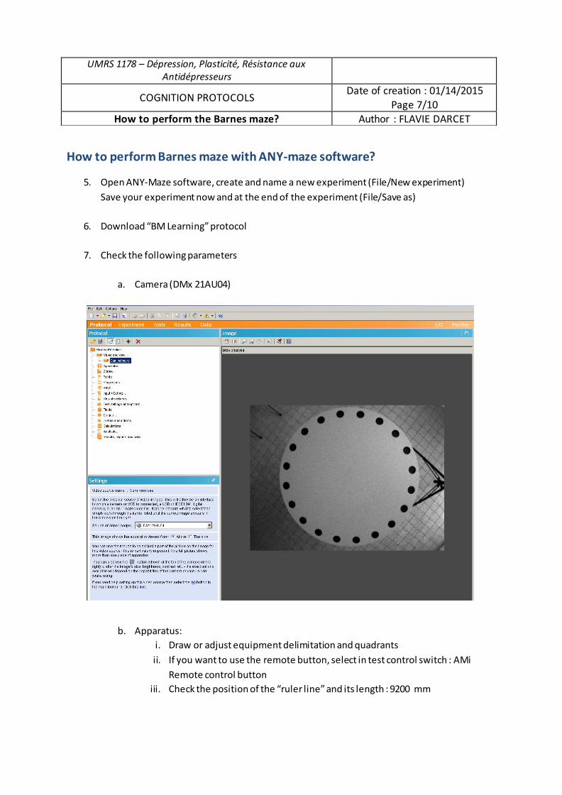
How to perform Barnes maze with ANY-maze software?
5. Open ANY-Maze software, create and name a new experiment (File/New experiment) Save your experiment now and at the end of the experiment (File/Save as)
6. Download “BM Learning” protocol
7. Check the following parameters
a. Camera (DMx 21AU04)
b. Apparatus: i. Draw or adjust equipment delimitation and quadrants ii. If you want to use the remote button, select in test control switch : AMi
Remote control button iii. Check the position of the “ruler line” and its length : 9200 mm
UMRS 1178 – Dépression, Plasticité, Résistance aux Antidépresseurs
COGNITION PROTOCOLS Date of creation : 01/14/2015 Page 7/10
How to perform the Barnes maze? Author : FLAVIE DARCET
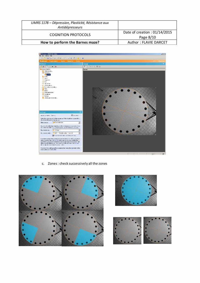
c. Zones : check successively all the zones
UMRS 1178 – Dépression, Plasticité, Résistance aux Antidépresseurs
COGNITION PROTOCOLS Date of creation : 01/14/2015 Page 8/10
How to perform the Barnes maze? Author : FLAVIE DARCET
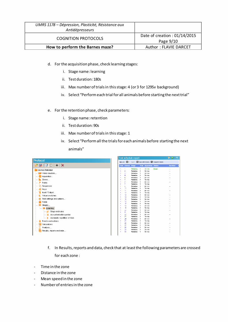
d. For the acquisition phase, check learning stages:
i. Stage name: learning
ii. Test duration: 180s
iii. Max number of trials in this stage: 4 (or 3 for 129Sv background)
iv. Select “Perform each trial for all animals before starting the next trial”
e. For the retention phase, check parameters:
i. Stage name: retention
ii. Test duration: 90s
iii. Max number of trials in this stage: 1
iv. Select “Perform all the trials for each animals before starting the next
animals”
f. In Results, reports and data, check that at least the following parameters are crossed
for each zone :
- Time in the zone - Distance in the zone - Mean speed in the zone - Number of entries in the zone
UMRS 1178 – Dépression, Plasticité, Résistance aux Antidépresseurs
COGNITION PROTOCOLS Date of creation : 01/14/2015 Page 9/10
How to perform the Barnes maze? Author : FLAVIE DARCET
DATA ANALYSIS
Click on the “data” page
- All the parameters you requested in Results, reports and data section or Protocol page are
listed in the table
- Export the table in an Excel file to analyze the results with the statistical software of your
choice
UMRS 1178 – Dépression, Plasticité, Résistance aux Antidépresseurs
COGNITION PROTOCOLS Date of creation : 01/14/2015 Page 10/10
How to perform the Barnes maze? Author : FLAVIE DARCET

4. Fiche méthode du test de conditionnement par la peur

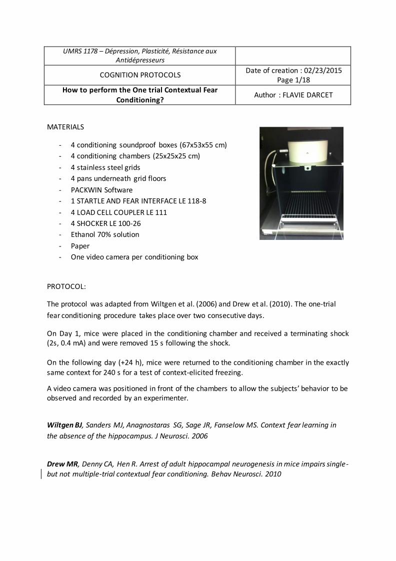
UMRS 1178 – Dépression, Plasticité, Résistance aux Antidépresseurs
COGNITION PROTOCOLS Date of creation : 02/23/2015 Page 1/18
How to perform the One trial Contextual Fear Conditioning? Author : FLAVIE DARCET
MATERIALS
- 4 conditioning soundproof boxes (67x53x55 cm) - 4 conditioning chambers (25x25x25 cm) - 4 stainless steel grids - 4 pans underneath grid floors - PACKWIN Software - 1 STARTLE AND FEAR INTERFACE LE 118-8 - 4 LOAD CELL COUPLER LE 111 - 4 SHOCKER LE 100-26 - Ethanol 70% solution - Paper - One video camera per conditioning box
PROTOCOL:
The protocol was adapted from Wiltgen et al. (2006) and Drew et al. (2010). The one-trial fear conditioning procedure takes place over two consecutive days.
On Day 1, mice were placed in the conditioning chamber and received a terminating shock (2s, 0.4 mA) and were removed 15 s following the shock. On the following day (+24 h), mice were returned to the conditioning chamber in the exactly same context for 240 s for a test of context-elicited freezing.
A video camera was positioned in front of the chambers to allow the subjects’ behavior to be observed and recorded by an experimenter.
Wiltgen BJ, Sanders MJ, Anagnostaras SG, Sage JR, Fanselow MS. Context fear learning in the absence of the hippocampus. J Neurosci. 2006
Drew MR, Denny CA, Hen R. Arrest of adult hippocampal neurogenesis in mice impairs single- but not multiple-trial contextual fear conditioning. Behav Neurosci. 2010
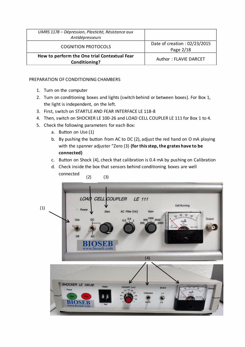
PREPARATION OF CONDITIONING CHAMBERS
1. Turn on the computer
2. Turn on conditioning boxes and lights (switch behind or between boxes). For Box 1,
the light is independent, on the left.
3. First, switch on STARTLE AND FEAR INTERFACE LE 118-8
4. Then, switch on SHOCKER LE 100-26 and LOAD CELL COUPLER LE 111 for Box 1 to 4.
5. Check the following parameters for each Box:
a. Button on Use (1)
b. By pushing the button from AC to DC (2), adjust the red hand on O mA playing
with the spanner adjuster “Zero (3) (for this step, the grates have to be connected)
c. Button on Shock (4), check that calibration is 0.4 mA by pushing on Calibration
d. Check inside the box that sensors behind conditioning boxes are well
connected
UMRS 1178 – Dépression, Plasticité, Résistance aux Antidépresseurs
COGNITION PROTOCOLS Date of creation : 02/23/2015
Page 2/18
How to perform the One trial Contextual Fear Conditioning? Author : FLAVIE DARCET
(3)
(1)
(2)
(4)
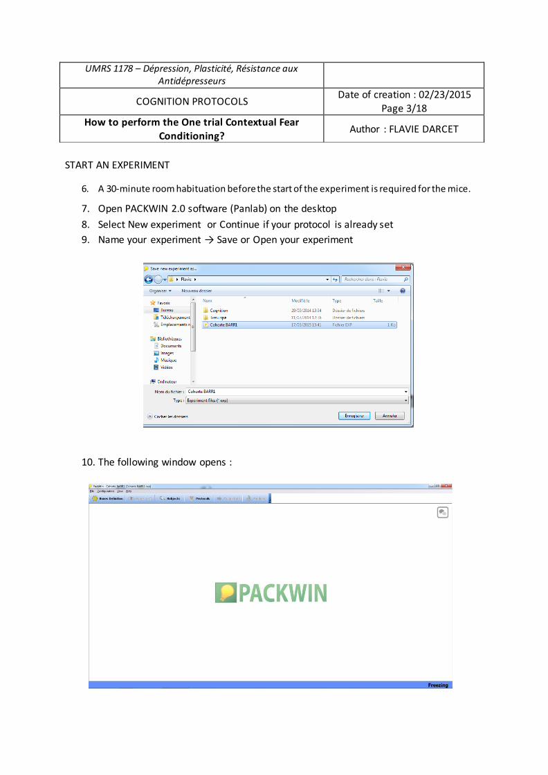
START AN EXPERIMENT
6. A 30-minute room habituation before the start of the experiment is required for the mice.
7. Open PACKWIN 2.0 software (Panlab) on the desktop 8. Select New experiment or Continue if your protocol is already set 9. Name your experiment → Save or Open your experiment
10. The following window opens :
UMRS 1178 – Dépression, Plasticité, Résistance aux Antidépresseurs
COGNITION PROTOCOLS Date of creation : 02/23/2015 Page 3/18
How to perform the One trial Contextual Fear Conditioning? Author : FLAVIE DARCET
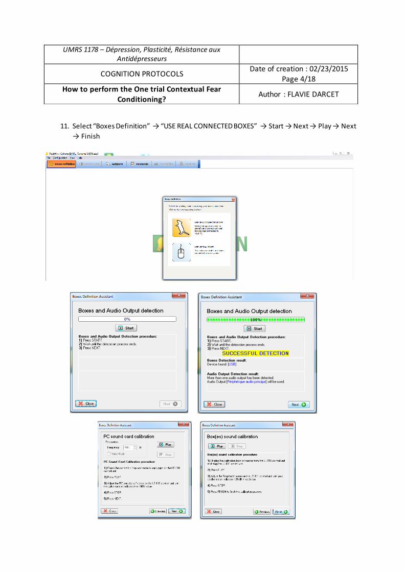
11. Select “Boxes Definition” → “USE REAL CONNECTED BOXES” → Start → Next → Play → Next → Finish
UMRS 1178 – Dépression, Plasticité, Résistance aux Antidépresseurs
COGNITION PROTOCOLS Date of creation : 02/23/2015 Page 4/18
How to perform the One trial Contextual Fear Conditioning? Author : FLAVIE DARCET
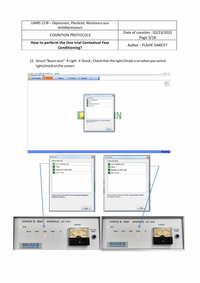
12. Select “Boxes test” → Light → Shock ; Check that the light/shock is on when you select light/shock on the screen
UMRS 1178 – Dépression, Plasticité, Résistance aux Antidépresseurs
COGNITION PROTOCOLS Date of creation : 02/23/2015 Page 5/18
How to perform the One trial Contextual Fear Conditioning? Author : FLAVIE DARCET
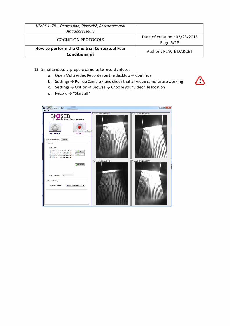
13. Simultaneously, prepare cameras to record videos. a. Open Multi Video Recorder on the desktop → Continue b. Settings → Pull up Camera 4 and check that all video cameras are working c. Settings → Option → Browse → Choose your video file location d. Record → “Start all”
UMRS 1178 – Dépression, Plasticité, Résistance aux Antidépresseurs
COGNITION PROTOCOLS Date of creation : 02/23/2015 Page 6/18
How to perform the One trial Contextual Fear Conditioning? Author : FLAVIE DARCET
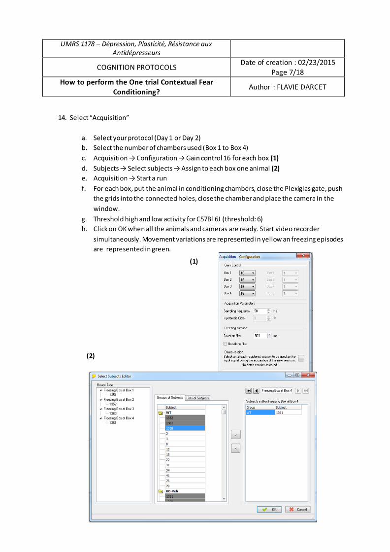
14. Select “Acquisition”
a. Select your protocol (Day 1 or Day 2) b. Select the number of chambers used (Box 1 to Box 4)
c. Acquisition → Configuration → Gain control 16 for each box (1) d. Subjects → Select subjects → Assign to each box one animal (2) e. Acquisition → Start a run f. For each box, put the animal in conditioning chambers, close the Plexiglas gate, push
the grids into the connected holes, close the chamber and place the camera in the
window.
g. Threshold high and low activity for C57Bl 6J (threshold: 6) h. Click on OK when all the animals and cameras are ready. Start video recorder
simultaneously. Movement variations are represented in yellow an freezing episodes are represented in green.
UMRS 1178 – Dépression, Plasticité, Résistance aux Antidépresseurs
COGNITION PROTOCOLS Date of creation : 02/23/2015
Page 7/18 How to perform the One trial Contextual Fear
Conditioning? Author : FLAVIE DARCET
(1)
(2)
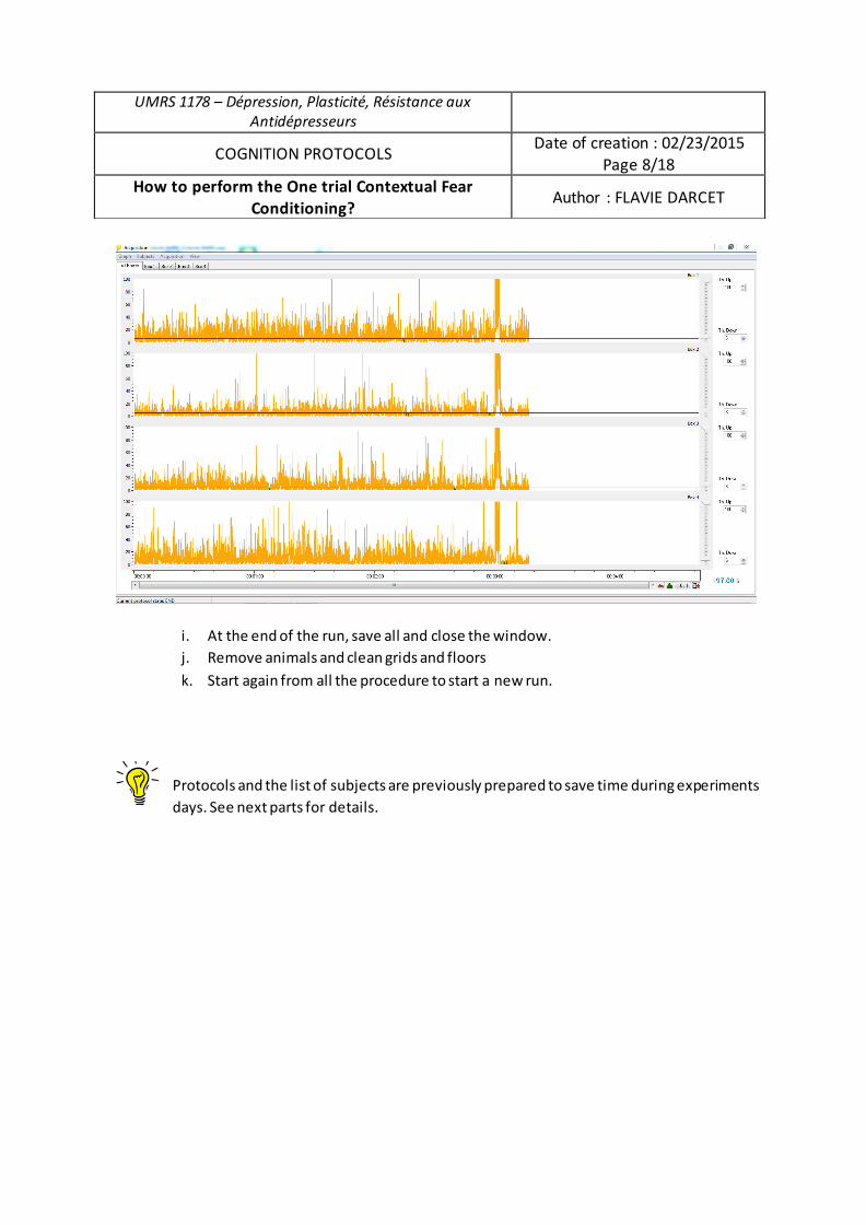
i. At the end of the run, save all and close the window. j. Remove animals and clean grids and floors
k. Start again from all the procedure to start a new run.
Protocols and the list of subjects are previously prepared to save time during experiments days. See next parts for details.
UMRS 1178 – Dépression, Plasticité, Résistance aux Antidépresseurs
COGNITION PROTOCOLS Date of creation : 02/23/2015
Page 8/18 How to perform the One trial Contextual Fear
Conditioning? Author : FLAVIE DARCET
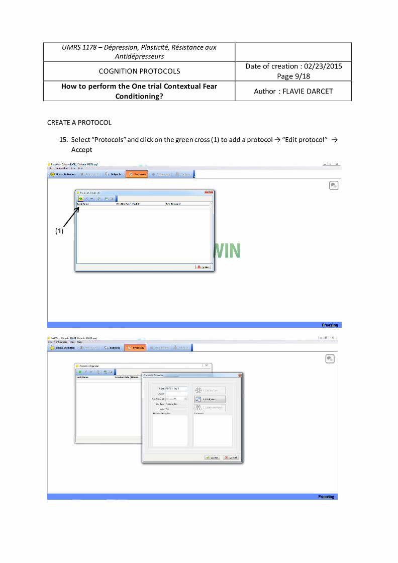
CREATE A PROTOCOL
15. Select “Protocols” and click on the green cross (1) to add a protocol → “Edit protocol” → Accept
UMRS 1178 – Dépression, Plasticité, Résistance aux Antidépresseurs
COGNITION PROTOCOLS Date of creation : 02/23/2015 Page 9/18
How to perform the One trial Contextual Fear Conditioning? Author : FLAVIE DARCET
(1)
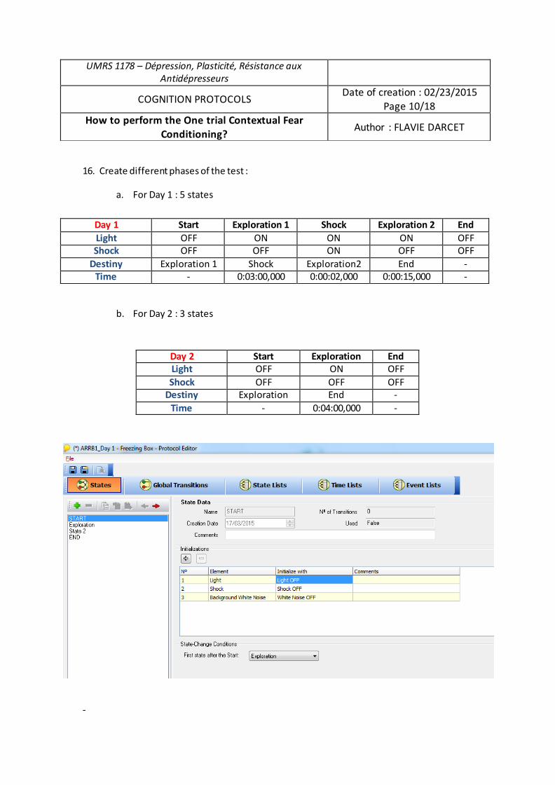
16. Create different phases of the test :
a. For Day 1 : 5 states
b. For Day 2 : 3 states
-
UMRS 1178 – Dépression, Plasticité, Résistance aux Antidépresseurs
COGNITION PROTOCOLS Date of creation : 02/23/2015 Page 10/18
How to perform the One trial Contextual Fear Conditioning? Author : FLAVIE DARCET
Day 1 Start Exploration 1 Shock Exploration 2 End Light OFF ON ON ON OFF Shock OFF OFF ON OFF OFF
Destiny Exploration 1 Shock Exploration2 End - Time - 0:03:00,000 0:00:02,000 0:00:15,000 -
Day 2 Start Exploration End Light OFF ON OFF Shock OFF OFF OFF
Destiny Exploration End - Time - 0:04:00,000 -
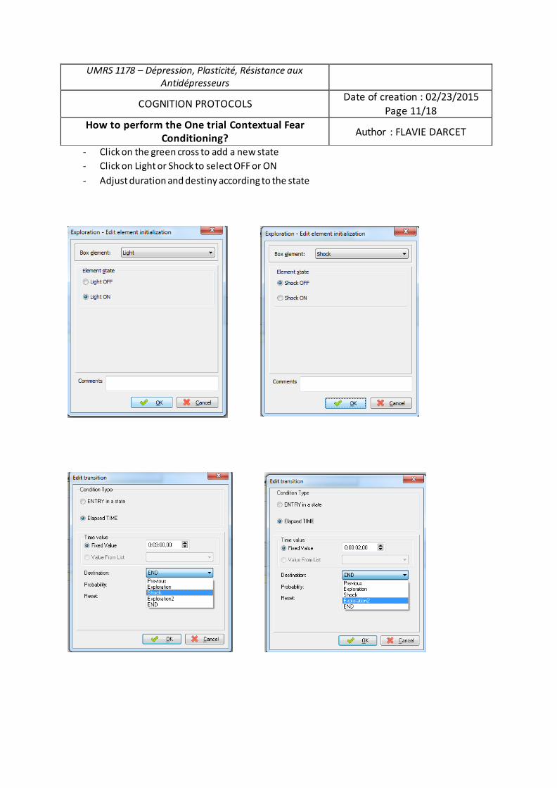
- Click on the green cross to add a new state - Click on Light or Shock to select OFF or ON - Adjust duration and destiny according to the state
UMRS 1178 – Dépression, Plasticité, Résistance aux Antidépresseurs
COGNITION PROTOCOLS Date of creation : 02/23/2015 Page 11/18
How to perform the One trial Contextual Fear Conditioning? Author : FLAVIE DARCET
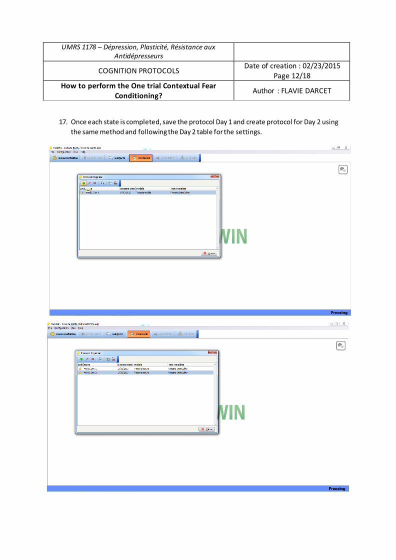
17. Once each state is completed, save the protocol Day 1 and create protocol for Day 2 using the same method and following the Day 2 table for the settings.
UMRS 1178 – Dépression, Plasticité, Résistance aux Antidépresseurs
COGNITION PROTOCOLS Date of creation : 02/23/2015 Page 12/18
How to perform the One trial Contextual Fear Conditioning? Author : FLAVIE DARCET
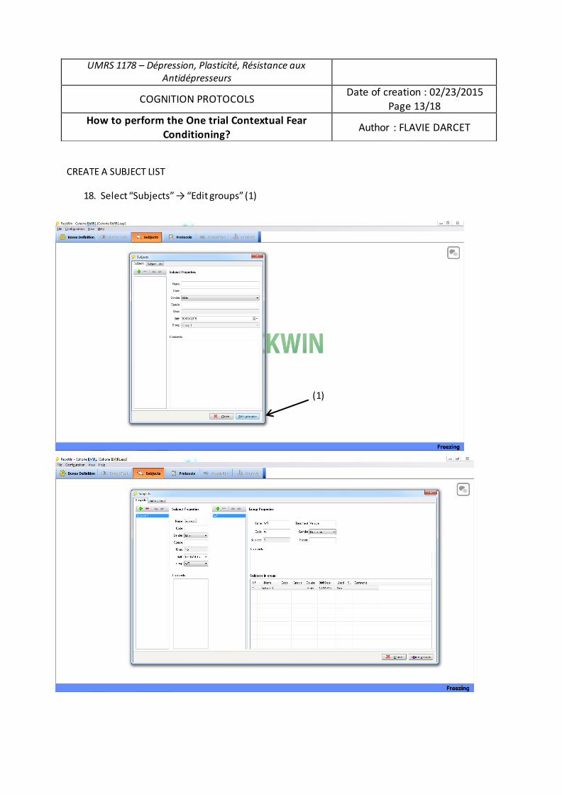
CREATE A SUBJECT LIST
18. Select “Subjects” → “Edit groups” (1)
UMRS 1178 – Dépression, Plasticité, Résistance aux Antidépresseurs
COGNITION PROTOCOLS Date of creation : 02/23/2015 Page 13/18
How to perform the One trial Contextual Fear Conditioning? Author : FLAVIE DARCET
(1)
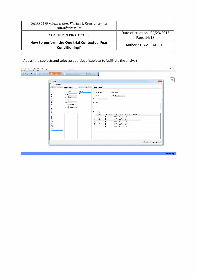
Add all the subjects and select properties of subjects to facilitate the analysis.
UMRS 1178 – Dépression, Plasticité, Résistance aux Antidépresseurs
COGNITION PROTOCOLS Date of creation : 02/23/2015 Page 14/18
How to perform the One trial Contextual Fear Conditioning? Author : FLAVIE DARCET
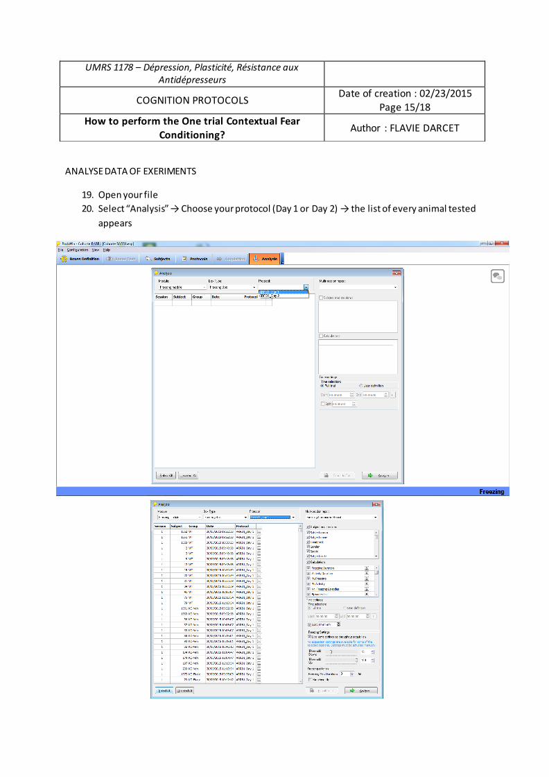
ANALYSE DATA OF EXERIMENTS
19. Open your file 20. Select “Analysis” → Choose your protocol (Day 1 or Day 2) → the list of every animal tested
appears
UMRS 1178 – Dépression, Plasticité, Résistance aux Antidépresseurs
COGNITION PROTOCOLS Date of creation : 02/23/2015 Page 15/18
How to perform the One trial Contextual Fear Conditioning? Author : FLAVIE DARCET
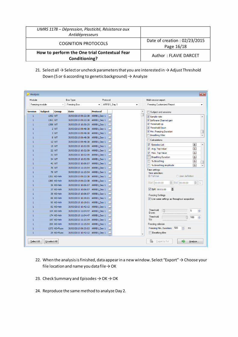
21. Select all → Select or uncheck parameters that you are interested in → Adjust Threshold
Down (5 or 6 according to genetic background) → Analyze
22. When the analysis is finished, data appear in a new window. Select “Export” → Choose your file location and name you data file→ OK
23. Check Summary and Episodes → OK → OK
24. Reproduce the same method to analyze Day 2.
UMRS 1178 – Dépression, Plasticité, Résistance aux Antidépresseurs
COGNITION PROTOCOLS Date of creation : 02/23/2015 Page 16/18
How to perform the One trial Contextual Fear Conditioning? Author : FLAVIE DARCET
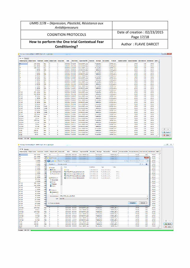
UMRS 1178 – Dépression, Plasticité, Résistance aux Antidépresseurs
COGNITION PROTOCOLS Date of creation : 02/23/2015 Page 17/18
How to perform the One trial Contextual Fear Conditioning? Author : FLAVIE DARCET
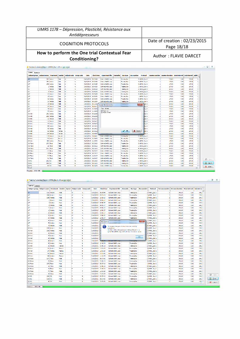
UMRS 1178 – Dépression, Plasticité, Résistance aux Antidépresseurs
COGNITION PROTOCOLS Date of creation : 02/23/2015 Page 18/18
How to perform the One trial Contextual Fear Conditioning? Author : FLAVIE DARCET
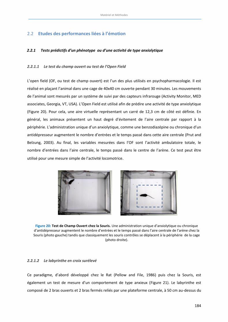
Matériel et Méthodes
184
Etudes des performances liées à l’émotion 2.2
2.2.1 Tests prédictifs d’un phénotype ou d’une activité de type anxiolytique
2.2.1.1 Le test du champ ouvert ou test de l’Open Field
L’open field (OF, ou test de champ ouvert) est l’un des plus utilisés en psychopharmacologie. Il est
réalisé en plaçant l’animal dans une cage de 40x40 cm ouverte pendant 30 minutes. Les mouvements
de l’animal sont mesurés par un système de suivi par des capteurs infrarouge (Activity Monitor, MED
associates, Georgia, VT, USA). L’Open Field est utilisé afin de prédire une activité de type anxiolytique
(Figure 20). Pour cela, une aire virtuelle représentant un carré de 12,3 cm de côté est définie. En
général, les animaux présentent un haut degré d’évitement de l’aire centrale par rapport à la
périphérie. L’administration unique d’un anxiolytique, comme une benzodiazépine ou chronique d’un
antidépresseur augmentent le nombre d’entrées et le temps passé dans cette aire centrale (Prut and
Belzung, 2003). Au final, les variables mesurées dans l’OF sont l’activité ambulatoire totale, le
nombre d’entrées dans l’aire centrale, le temps passé dans le centre de l’arène. Ce test peut être
utilisé pour une mesure simple de l’activité locomotrice.
2.2.1.2 Le labyrinthe en croix surélevé
Ce paradigme, d’abord développé chez le Rat (Pellow and File, 1986) puis chez la Souris, est
également un test de mesure d’un comportement de type anxieux (Figure 21). Le labyrinthe est
composé de 2 bras ouverts et 2 bras fermés reliés par une plateforme centrale, à 50 cm au-dessus du
Figure 20: Test de Champ Ouvert chez la Souris. Une administration unique d’anxiolytique ou chronique d’antidépresseur augmentent le nombre d’entrées et le temps passé dans l’aire centrale de l’arène chez la Souris (photo gauche) tandis que classiquement les souris contrôles se déplacent à la périphérie de la cage
(photo droite).
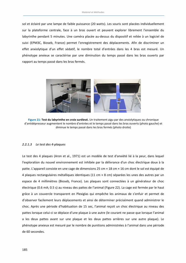
Matériel et Méthodes
185
sol et éclairé par une lampe de faible puissance (20 watts). Les souris sont placées individuellement
sur la plateforme centrale, face à un bras ouvert et peuvent explorer librement l’ensemble du
labyrinthe pendant 5 minutes. Une caméra placée au-dessus du dispositif et reliée à un logiciel de
suivi (EPM3C, Bioseb, France) permet l’enregistrement des déplacements. Afin de discriminer un
effet anxiolytique d’un effet sédatif, le nombre total d’entrées dans les 4 bras est mesuré. Un
phénotype anxieux se caractérise par une diminution du temps passé dans les bras ouverts par
rapport au temps passé dans les bras fermés.
2.2.1.3 Le test des 4-plaques
Le test des 4 plaques (Aron et al., 1971) est un modèle de test d'anxiété lié à la peur, dans lequel
l'exploration du nouvel environnement est inhibée par la délivrance d'un choc électrique doux à la
patte. L’appareil consiste en une cage de dimensions 25 cm × 18 cm × 16 cm dont le sol est équipé de
4 plaques rectangulaires métalliques identiques (11 cm × 8 cm) séparées les unes des autres par un
espace de 4 millimètres (Bioseb, France). Les plaques sont connectées à un générateur de choc
électrique (0.6 mA; 0.5 s) au niveau des pattes de l’animal (Figure 22). La cage est fermée par le haut
grâce à un couvercle transparent en Plexiglas qui empêche les animaux de s’enfuir et permet de
d’observer facilement leurs déplacements et ainsi de déterminer précisément quand administrer le
choc. Après une période d’habituation de 15 sec, l’animal reçoit un choc électrique au niveau des
pattes lorsque celui-ci se déplace d’une plaque à une autre (le courant ne passe que lorsque l’animal
a les deux pattes avant sur une plaque et les deux pattes arrières sur une autre plaque). Le
phénotype anxieux est mesuré par le nombre de punitions administrées à l’animal dans une période
de 60 secondes.
Figure 21: Test du labyrinthe en croix surélevé. Un traitement aigu par des anxiolytiques ou chronique d’antidépresseur augmentent le nombre d’entrées et le temps passé dans les bras ouverts (photo gauche) et
diminue le temps passé dans les bras fermés (photo droite)
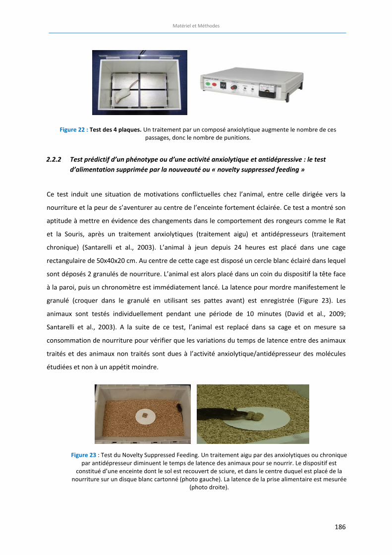
Matériel et Méthodes
186
2.2.2 Test prédictif d’un phénotype ou d’une activité anxiolytique et antidépressive : le test d’alimentation supprimée par la nouveauté ou « novelty suppressed feeding »
Ce test induit une situation de motivations conflictuelles chez l’animal, entre celle dirigée vers la
nourriture et la peur de s’aventurer au centre de l’enceinte fortement éclairée. Ce test a montré son
aptitude à mettre en évidence des changements dans le comportement des rongeurs comme le Rat
et la Souris, après un traitement anxiolytiques (traitement aigu) et antidépresseurs (traitement
chronique) (Santarelli et al., 2003). L’animal à jeun depuis 24 heures est placé dans une cage
rectangulaire de 50x40x20 cm. Au centre de cette cage est disposé un cercle blanc éclairé dans lequel
sont déposés 2 granulés de nourriture. L’animal est alors placé dans un coin du dispositif la tête face
à la paroi, puis un chronomètre est immédiatement lancé. La latence pour mordre manifestement le
granulé (croquer dans le granulé en utilisant ses pattes avant) est enregistrée (Figure 23). Les
animaux sont testés individuellement pendant une période de 10 minutes (David et al., 2009;
Santarelli et al., 2003). A la suite de ce test, l’animal est replacé dans sa cage et on mesure sa
consommation de nourriture pour vérifier que les variations du temps de latence entre des animaux
traités et des animaux non traités sont dues à l’activité anxiolytique/antidépresseur des molécules
étudiées et non à un appétit moindre.
Figure 22 : Test des 4 plaques. Un traitement par un composé anxiolytique augmente le nombre de ces passages, donc le nombre de punitions.
Figure 23 : Test du Novelty Suppressed Feeding. Un traitement aigu par des anxiolytiques ou chronique par antidépresseur diminuent le temps de latence des animaux pour se nourrir. Le dispositif est
constitué d’une enceinte dont le sol est recouvert de sciure, et dans le centre duquel est placé de la nourriture sur un disque blanc cartonné (photo gauche). La latence de la prise alimentaire est mesurée
(photo droite).
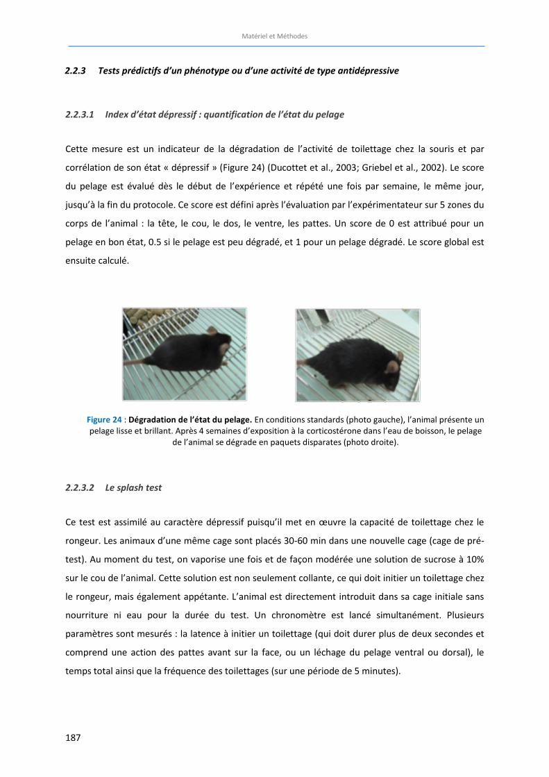
Matériel et Méthodes
187
2.2.3 Tests prédictifs d’un phénotype ou d’une activité de type antidépressive
2.2.3.1 Index d’état dépressif : quantification de l’état du pelage
Cette mesure est un indicateur de la dégradation de l’activité de toilettage chez la souris et par
corrélation de son état « dépressif » (Figure 24) (Ducottet et al., 2003; Griebel et al., 2002). Le score
du pelage est évalué dès le début de l’expérience et répété une fois par semaine, le même jour,
jusqu’à la fin du protocole. Ce score est défini après l’évaluation par l’expérimentateur sur 5 zones du
corps de l’animal : la tête, le cou, le dos, le ventre, les pattes. Un score de 0 est attribué pour un
pelage en bon état, 0.5 si le pelage est peu dégradé, et 1 pour un pelage dégradé. Le score global est
ensuite calculé.
2.2.3.2 Le splash test
Ce test est assimilé au caractère dépressif puisqu’il met en œuvre la capacité de toilettage chez le
rongeur. Les animaux d’une même cage sont placés 30-60 min dans une nouvelle cage (cage de pré-
test). Au moment du test, on vaporise une fois et de façon modérée une solution de sucrose à 10%
sur le cou de l’animal. Cette solution est non seulement collante, ce qui doit initier un toilettage chez
le rongeur, mais également appétante. L’animal est directement introduit dans sa cage initiale sans
nourriture ni eau pour la durée du test. Un chronomètre est lancé simultanément. Plusieurs
paramètres sont mesurés : la latence à initier un toilettage (qui doit durer plus de deux secondes et
comprend une action des pattes avant sur la face, ou un léchage du pelage ventral ou dorsal), le
temps total ainsi que la fréquence des toilettages (sur une période de 5 minutes).
Figure 24 : Dégradation de l’état du pelage. En conditions standards (photo gauche), l’animal présente un pelage lisse et brillant. Après 4 semaines d’exposition à la corticostérone dans l’eau de boisson, le pelage
de l’animal se dégrade en paquets disparates (photo droite).

Matériel et Méthodes
188
2.2.3.3 Le test de préférence à la saccharine
Ce test repose sur la préférence naturelle des souris pour une boisson sucrée (à la saccharine par
exemple) par rapport à de l’eau. Il permet ainsi de mesurer le niveau d’anhédonie des animaux,
considérée comme un des principaux symptômes de la dépression. D’une façon pratique, le test est
réalisé directement dans la salle d’élevage. Les souris sont individualisées la veille du début de
l’expérience et sont habituées à boire de l’eau provenant de 2 tubes plastiques de 15 mL (troués au
culot) pendant 3 jours consécutifs. Le 4ème jour, les souris ont un libre accès à un tube plastique
contenant de l’eau et un autre tube plastique contenant une solution de saccharine à 0,1% pendant
une période de 48h. La quantité consommée d’eau et de saccharine est mesurée toutes les 12h à 8h
et à 20h chaque jour. De plus, les tubes plastiques sont interchangés toutes les 12h pour éviter le
développement d’une préférence de place (droite/gauche). La préférence à la saccharine (en %) est
calculée comme suit : Préférence = [consommation de saccharine (mL)/consommation totale (mL)
(eau + saccharine)]*100.
2.2.3.4 Le test de suspension caudale
Le TST est un modèle animal (Steru et al., 1985), dérivé du FST où la suspension de la souris ou du rat
par la queue induit une immobilité. Il mesure la durée de l’immobilité (pendant 6 minutes) ainsi que
la puissance des mouvements de l’animal (Porsolt et al., 1987; Steru et al., 1985) permettant de
distinguer différentes classes de psychotropes. Le TST résoudrait plusieurs problèmes rencontrés
avec le FST : l’immobilité est mesurée objectivement (automatique) et aucune hypothermie n’est
induite après immersion dans l’eau froide (Thierry et al., 1986). Les études montrent que le TST peut
être prédictif de l’activité de nombreux antidépresseurs. Un scotch doux est placé au bout de la
partie caudale de l’animal, ce qui permet de le suspendre par la queue à un crochet. Le crochet est
relié à un capteur qui enregistre les variations de mouvements (Bio-TST2, Bioseb, Vitrolles, France,
Figure 25). L’enregistrement des données est effectué sur une période de 6 minutes.
Figure 25: Dispositif d’enregistrement du test de suspension caudale. Une souris traitée par un antidépresseur voit son temps d’immobilité diminué.
Matériel et Méthodes
189
3 Manipulations ex vivo
Culture de cellules souches neurales à partir de gyrus dentelé d’hippocampe 3.1
adulte de Souris
De façon à vérifier la délétion spécifique de la protéine β-arrestine 1 dans les cellules souches
du gyrus dentelé de l’hippocampe, des cultures de cellules souches provenant du gyrus dentelé de
souris ARRB1-/- ont été réalisées en collaboration avec le laboratoire de Sandrine Humbert de
l’Institut des Neurosciences de Grenoble. La culture des cellules souches neurales à partir du gyrus
dentelé de l’hippocampe est effectuée comme décrit dans Babu et al., 2011 (Babu et al., 2011). La
culture de cellules souches de la zone sous ventriculaire des mêmes souris est réalisée en suivant le
même protocole. Brièvement, des souris adultes (de 2 à 4 mois) mâles ARRB1+/+ et ARRB1-/- sont
tuées par dislocation cervicale puis les tissus sont rapidement prélevés dans une solution de PBS
stérile. Après digestion enzymatique, les cellules viables sont séparées des débris cellulaires et de la
myéline par centrifugation sur gradient de Percoll. Les cellules sont mises en culture dans du
DMEM/F12/Glutamax complémenté avec 1% de Pénicilline/Streptomycine, 1% de B27, 20 ng/ml
d’EGF et 20 ng/ml de FGF-2 dans des boîtes de pétri préalablement traitées à la poly-D-lysine (20
µg/ml) et à la laminine (20 µg/ml) pour promouvoir l’adhérence des cellules souches et leur
croissance en monocouche. Les cultures se développent dans l’incubateur pendant 7 jours (zone
sous-ventriculaire) et 14 jours (gyrus dentelé) puis les protéines sont récoltées dans du RIPA
classique supplémenté en inhibiteurs de protéases et phosphatases en vue d’un dosage de la
protéine β-arrestine 1 par Western Blot. Les cultures ont été réalisées par Fabienne Agasse et
Caroline Benstaali de l’Institut des Neurosciences de Grenoble.
Mesure de l’expression protéique par Western Blot 3.2
3.2.1 Dosage des protéines
La quantité de protéines présentes dans les différents échantillons est préalablement
déterminée afin de déposer la même quantité de protéines dans chaque puit. La quantification des
protéines a été effectuée en utilisant une technique combinant la réaction de Biuret (Gornall et al.,
1949) avec une détection colorimétrique hautement sensible et sélective des ions cuivre, par un
réactif contenant l’acide bicinchoninique (Thermo Scientific Pierce BCA Protein Assay Kit, Pierce
Biotechnology).
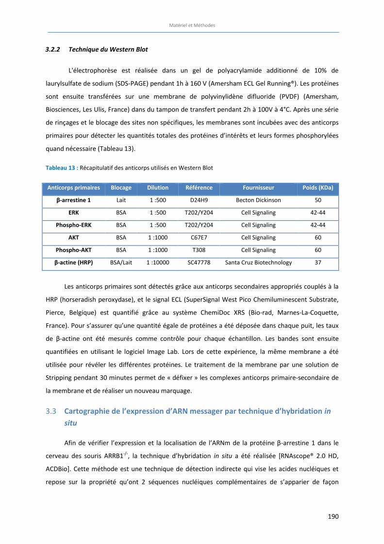
Matériel et Méthodes
190
3.2.2 Technique du Western Blot
L'électrophorèse est réalisée dans un gel de polyacrylamide additionné de 10% de
laurylsulfate de sodium (SDS-PAGE) pendant 1h à 160 V (Amersham ECL Gel Running®). Les protéines
sont ensuite transférées sur une membrane de polyvinylidène difluoride (PVDF) (Amersham,
Biosciences, Les Ulis, France) dans du tampon de transfert pendant 2h à 100V à 4°C. Après une série
de rinçages et le blocage des sites non spécifiques, les membranes sont incubées avec des anticorps
primaires pour détecter les quantités totales des protéines d’intérêts et leurs formes phosphorylées
quand nécessaire (Tableau 13).
Tableau 13 : Récapitulatif des anticorps utilisés en Western Blot
Les anticorps primaires sont détectés grâce aux anticorps secondaires appropriés couplés à la
HRP (horseradish peroxydase), et le signal ECL (SuperSignal West Pico Chemiluminescent Substrate,
Pierce, Belgique) est quantifié grâce au système ChemiDoc XRS (Bio-rad, Marnes-La-Coquette,
France). Pour s’assurer qu’une quantité égale de protéines a été déposée dans chaque puit, les taux
de β-actine ont été mesurés comme contrôle pour chaque échantillon. Les bandes sont ensuite
quantifiées en utilisant le logiciel Image Lab. Lors de cette expérience, la même membrane a été
utilisée pour révéler les différentes protéines. Le traitement de la membrane par une solution de
Stripping pendant 30 minutes permet de « défixer » les complexes anticorps primaire-secondaire de
la membrane et de réaliser un nouveau marquage.
Cartographie de l’expression d’ARN messager par technique d’hybridation in 3.3
situ
Afin de vérifier l’expression et la localisation de l’ARNm de la protéine β-arrestine 1 dans le
cerveau des souris ARRB1-/-, la technique d’hybridation in situ a été réalisée [RNAscope® 2.0 HD,
ACDBio]. Cette méthode est une technique de détection indirecte qui vise les acides nucléiques et
repose sur la propriété qu’ont 2 séquences nucléiques complémentaires de s’apparier de façon
Anticorps primaires Blocage Dilution Référence Fournisseur Poids (KDa)
β-arrestine 1 Lait 1 :500 D24H9 Becton Dickinson 50
ERK BSA 1 :500 T202/Y204 Cell Signaling 42-44
Phospho-ERK BSA 1 :500 T202/Y204 Cell Signaling 42-44
AKT BSA 1 :1000 C67E7 Cell Signaling 60
Phospho-AKT BSA 1 :1000 T308 Cell Signaling 60
β-actine (HRP) BSA/Lait 1 :10000 SC47778 Santa Cruz Biotechnology 37
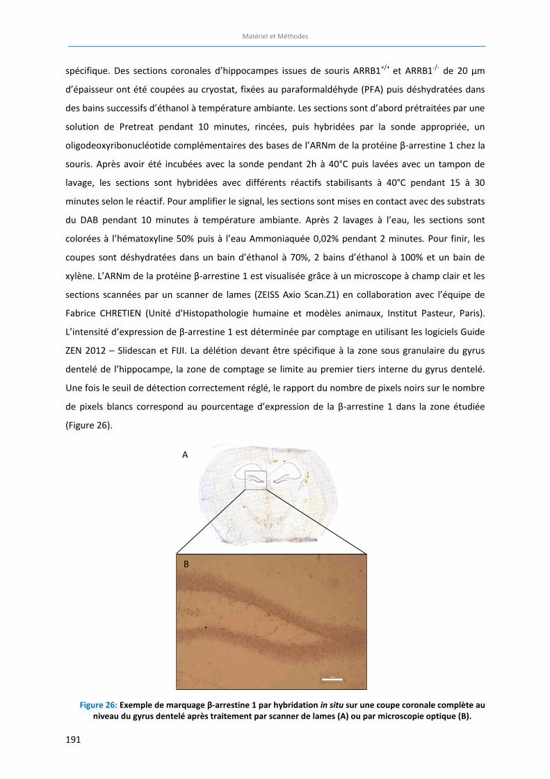
Matériel et Méthodes
191
spécifique. Des sections coronales d’hippocampes issues de souris ARRB1+/+ et ARRB1-/- de 20 µm
d’épaisseur ont été coupées au cryostat, fixées au paraformaldéhyde (PFA) puis déshydratées dans
des bains successifs d’éthanol à température ambiante. Les sections sont d’abord prétraitées par une
solution de Pretreat pendant 10 minutes, rincées, puis hybridées par la sonde appropriée, un
oligodeoxyribonucléotide complémentaires des bases de l’ARNm de la protéine β-arrestine 1 chez la
souris. Après avoir été incubées avec la sonde pendant 2h à 40°C puis lavées avec un tampon de
lavage, les sections sont hybridées avec différents réactifs stabilisants à 40°C pendant 15 à 30
minutes selon le réactif. Pour amplifier le signal, les sections sont mises en contact avec des substrats
du DAB pendant 10 minutes à température ambiante. Après 2 lavages à l’eau, les sections sont
colorées à l’hématoxyline 50% puis à l’eau Ammoniaquée 0,02% pendant 2 minutes. Pour finir, les
coupes sont déshydratées dans un bain d’éthanol à 70%, 2 bains d’éthanol à 100% et un bain de
xylène. L’ARNm de la protéine β-arrestine 1 est visualisée grâce à un microscope à champ clair et les
sections scannées par un scanner de lames (ZEISS Axio Scan.Z1) en collaboration avec l’équipe de
Fabrice CHRETIEN (Unité d'Histopathologie humaine et modèles animaux, Institut Pasteur, Paris).
L’intensité d’expression de β-arrestine 1 est déterminée par comptage en utilisant les logiciels Guide
ZEN 2012 – Slidescan et FIJI. La délétion devant être spécifique à la zone sous granulaire du gyrus
dentelé de l’hippocampe, la zone de comptage se limite au premier tiers interne du gyrus dentelé.
Une fois le seuil de détection correctement réglé, le rapport du nombre de pixels noirs sur le nombre
de pixels blancs correspond au pourcentage d’expression de la β-arrestine 1 dans la zone étudiée
(Figure 26).
Figure 26: Exemple de marquage β-arrestine 1 par hybridation in situ sur une coupe coronale complète au
niveau du gyrus dentelé après traitement par scanner de lames (A) ou par microscopie optique (B).
B
A
Matériel et Méthodes
192
Etude de la neurogenèse hippocampique adulte 3.4
3.4.1 Préparation des tissus
3.4.1.1 Perfusions intra-cardiaque
La souris est anesthésiée à l’aide d’un mélange kétamine/xylazine (120 mg/kg et 10 mg/kg,
respectivement). On contrôle l’anesthésie en effectuant une pression sur la patte arrière de l’animal.
L’absence de réaction permet d’entreprendre la chirurgie. L’animal est fixé sur une planche en liège,
sur le dos. Une incision au niveau médian de l’abdomen permet d’introduire les ciseaux de chirurgie
et de remonter jusqu’au diaphragme par les flancs de l’animal. Le diaphragme sectionné laisse
entrevoir le cœur. La peau qui recouvrait l’abdomen est rabattue en arrière puis fixée, permettant un
accès dégagé jusqu’au cœur. Il est primordial d’opérer rapidement l’animal pour conserver la
propriété pulsatile du cœur. Cette capacité va permettre de rincer et fixer les tissus cérébraux, ce qui
est nécessaire pour obtenir des marquages sans pollution par des cellules sanguines résiduelles. Un
cathéter relié à une pompe péristaltique (pompe péristaltique, W40578, Fisher Scientific SAS, Illkirch,
France) est inséré dans le ventricule gauche du cœur. Cette pompe a pour fonction de perfuser en
premier lieu une solution de NaCl 0.9% conservée dans la glace. Une fois le cathéter en place, une
incision de l’oreillette droite va permettre au cœur et à la pompe d’évacuer le sang du corps de
l’animal. Après 5 minutes, on peut constater une décoloration de certaines zones du corps de
l’animal tels que : le foie, les pattes avant, la langue, les oreilles etc. Ces indicateurs permettent de
poursuivre le processus de perfusion en changeant le NaCl 0.9% par une solution de
paraformaldéhyde 4% dans 0,2M de solution de Sørensen pendant 10 minutes. Cette solution va
permettre de fixer les tissus et de stopper toute activité cellulaire. Le cerveau est ensuite prélevé et
conservé pendant la première journée dans une solution de paraformaldéhyde 4% à 4°C. Elle sera
ensuite substituée par une solution de tampon phosphate (PBS) + sucrose 30% + azide 0.1% à 4° ce
qui permettra de protéger et conserver les tissus jusqu’à leur prochaine utilisation.
3.4.1.2 Préparation des cerveaux
Les cerveaux sont placés à l’envers dans des blocs en plastique individuels préalablement
remplis avec de l’OCT (Tissue-Tek, Optimal Cutting Temperature). Ainsi préparés, les blocs sont
plongés dans une boite en polystyrène contenant de la carboglace (-60 °C). La propriété de l’OCT à se
solidifier à basse température va contenir le cerveau dans un bloc. Ils sont ensuite démoulés puis
conservés au congélateur à -80°C. Le bloc d’OCT est fixé sur une platine amovible encore une fois à
Matériel et Méthodes
193
l’aide d’OCT, à l’intérieur d’un dispositif réfrigéré à -20°C (Leica CM 3000). Cet appareil va permettre
de faire des sections du bloc, ici définies à 35 μm d’épaisseur. On équilibre ensuite le cerveau par
rapport à la lame du couteau du cryostat de façon à obtenir une morphologie symétrique des
structures cérébrales. Après l’identification du début de l’hippocampe à l’aide de l’atlas pour Souris
(Bregma -1.20 to -3.80 mm; Franklin et Paxinos, 2007), chaque section de l’hippocampe est collectée.
Le stockage des coupes de cerveaux se fait à l’aide d’une boîte de 24 puits. Chaque ligne correspond
à un cerveau. Les puits sont remplis par une solution de PBS + sodium azide (0.1%) qui préservera les
tissus d’une éventuelle dégradation par les bactéries ou champignons. L’hippocampe se subdivisera
par 13 séries de 6 coupes. Dans chaque puit, nous retrouverons 1/6 de l’hippocampe et chaque
section est espacée de la suivante par 210 μm. Cette méthode permet d’économiser le nombre
d’animaux en utilisant différents marqueurs d’immunohistochimie pour le même cerveau.
3.4.2 Etude de la prolifération des cellules progénitrices
L’étude de la prolifération cellulaire est réalisée grâce à une technique d’immunohistochimie
utilisant le marquage au Ki67. La protéine Ki67 est une protéine nucléaire exprimée par la cellule
lorsqu’elle est entrée dans le cycle cellulaire. Son expression augmente progressivement au cours des
phases de synthèse, G2 puis M du cycle (Cattoretti et al., 1992).
Après avoir été rincées 3 fois 15 minutes dans une solution de tampon phosphate contenant du
triton PBST (PBS+Triton 0,3%), les coupes de cerveaux sont plongées pendant deux heures dans une
solution de 10% de sérum d’âne afin de bloquer les sites non spécifiques. Les coupes sont ensuite
immédiatement incubées dans l’anticorps primaire anti-Ki67 fabriqué chez le lapin (Eurobio, France)
le temps d’une nuit à 4°C sous agitation. Le lendemain, après 3 rinçages de 15 minutes dans un
tampon phosphate (PBS), les tissus reposent deux heures dans l’anticorps secondaire d’âne anti-lapin
couplé à un fluorochrome Cy2 (Jackson, dilution 1 :500) (Sahay et al., 2011). Le marquage est
visualisé grâce à un fluorochrome couplé à l’anticorps secondaire. La fluorescence permet
d’apprécier la proportion de cellules en cycle et donc la prolifération cellulaire.
3.4.3 Etude de la survie des cellules progénitrices
L’étude de la prolifération cellulaire dans l’hippocampe de la souris adulte nécessite un
schéma spécial d’injection de 5-bromo-2-deoxyuridine (BrdU), 2 fois par jour pendant 3 jours 3
semaines avant le sacrifice de l’animal (100 mg/kg) (Cameron and McKay, 2001; Wang et al., 2015).
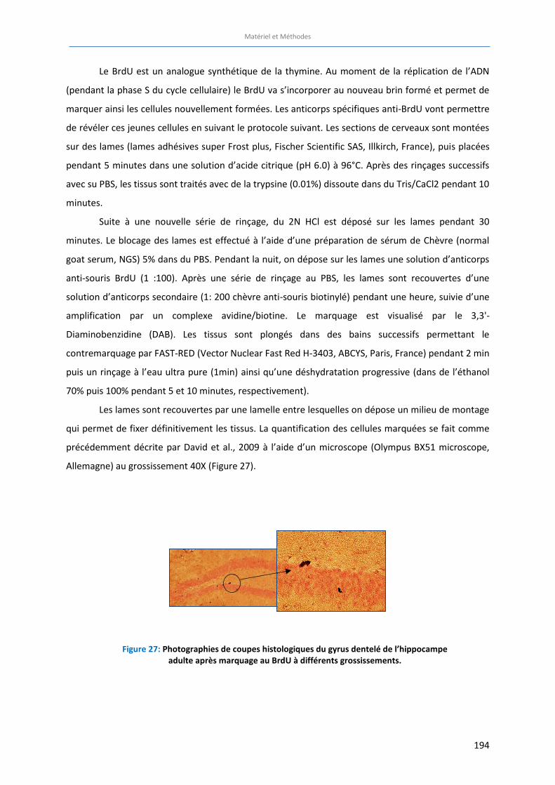
Matériel et Méthodes
194
Le BrdU est un analogue synthétique de la thymine. Au moment de la réplication de l’ADN
(pendant la phase S du cycle cellulaire) le BrdU va s’incorporer au nouveau brin formé et permet de
marquer ainsi les cellules nouvellement formées. Les anticorps spécifiques anti-BrdU vont permettre
de révéler ces jeunes cellules en suivant le protocole suivant. Les sections de cerveaux sont montées
sur des lames (lames adhésives super Frost plus, Fischer Scientific SAS, Illkirch, France), puis placées
pendant 5 minutes dans une solution d’acide citrique (pH 6.0) à 96°C. Après des rinçages successifs
avec su PBS, les tissus sont traités avec de la trypsine (0.01%) dissoute dans du Tris/CaCl2 pendant 10
minutes.
Suite à une nouvelle série de rinçage, du 2N HCl est déposé sur les lames pendant 30
minutes. Le blocage des lames est effectué à l’aide d’une préparation de sérum de Chèvre (normal
goat serum, NGS) 5% dans du PBS. Pendant la nuit, on dépose sur les lames une solution d’anticorps
anti-souris BrdU (1 :100). Après une série de rinçage au PBS, les lames sont recouvertes d’une
solution d’anticorps secondaire (1: 200 chèvre anti-souris biotinylé) pendant une heure, suivie d’une
amplification par un complexe avidine/biotine. Le marquage est visualisé par le 3,3'-
Diaminobenzidine (DAB). Les tissus sont plongés dans des bains successifs permettant le
contremarquage par FAST-RED (Vector Nuclear Fast Red H-3403, ABCYS, Paris, France) pendant 2 min
puis un rinçage à l’eau ultra pure (1min) ainsi qu’une déshydratation progressive (dans de l’éthanol
70% puis 100% pendant 5 et 10 minutes, respectivement).
Les lames sont recouvertes par une lamelle entre lesquelles on dépose un milieu de montage
qui permet de fixer définitivement les tissus. La quantification des cellules marquées se fait comme
précédemment décrite par David et al., 2009 à l’aide d’un microscope (Olympus BX51 microscope,
Allemagne) au grossissement 40X (Figure 27).
Figure 27: Photographies de coupes histologiques du gyrus dentelé de l’hippocampe adulte après marquage au BrdU à différents grossissements.
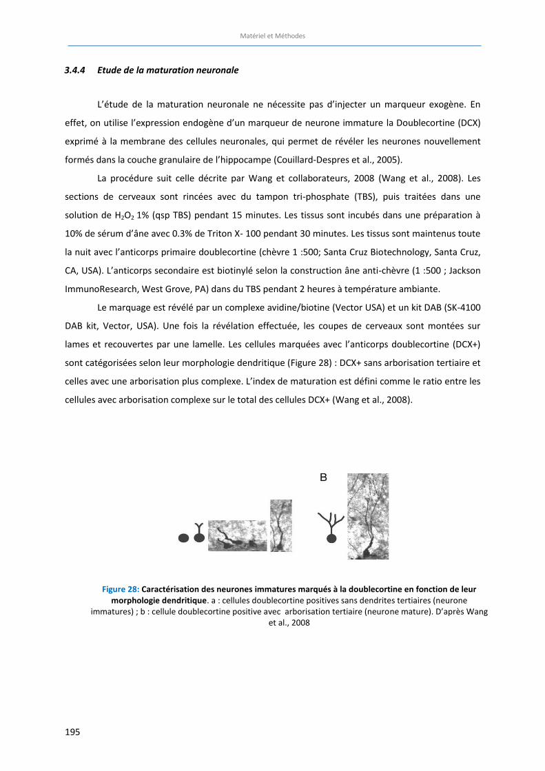
Matériel et Méthodes
195
3.4.4 Etude de la maturation neuronale
L’étude de la maturation neuronale ne nécessite pas d’injecter un marqueur exogène. En
effet, on utilise l’expression endogène d’un marqueur de neurone immature la Doublecortine (DCX)
exprimé à la membrane des cellules neuronales, qui permet de révéler les neurones nouvellement
formés dans la couche granulaire de l’hippocampe (Couillard-Despres et al., 2005).
La procédure suit celle décrite par Wang et collaborateurs, 2008 (Wang et al., 2008). Les
sections de cerveaux sont rincées avec du tampon tri-phosphate (TBS), puis traitées dans une
solution de H2O2 1% (qsp TBS) pendant 15 minutes. Les tissus sont incubés dans une préparation à
10% de sérum d’âne avec 0.3% de Triton X- 100 pendant 30 minutes. Les tissus sont maintenus toute
la nuit avec l’anticorps primaire doublecortine (chèvre 1 :500; Santa Cruz Biotechnology, Santa Cruz,
CA, USA). L’anticorps secondaire est biotinylé selon la construction âne anti-chèvre (1 :500 ; Jackson
ImmunoResearch, West Grove, PA) dans du TBS pendant 2 heures à température ambiante.
Le marquage est révélé par un complexe avidine/biotine (Vector USA) et un kit DAB (SK-4100
DAB kit, Vector, USA). Une fois la révélation effectuée, les coupes de cerveaux sont montées sur
lames et recouvertes par une lamelle. Les cellules marquées avec l’anticorps doublecortine (DCX+)
sont catégorisées selon leur morphologie dendritique (Figure 28) : DCX+ sans arborisation tertiaire et
celles avec une arborisation plus complexe. L’index de maturation est défini comme le ratio entre les
cellules avec arborisation complexe sur le total des cellules DCX+ (Wang et al., 2008).
Figure 28: Caractérisation des neurones immatures marqués à la doublecortine en fonction de leur
morphologie dendritique. a : cellules doublecortine positives sans dendrites tertiaires (neurone immatures) ; b : cellule doublecortine positive avec arborisation tertiaire (neurone mature). D’après Wang
et al., 2008
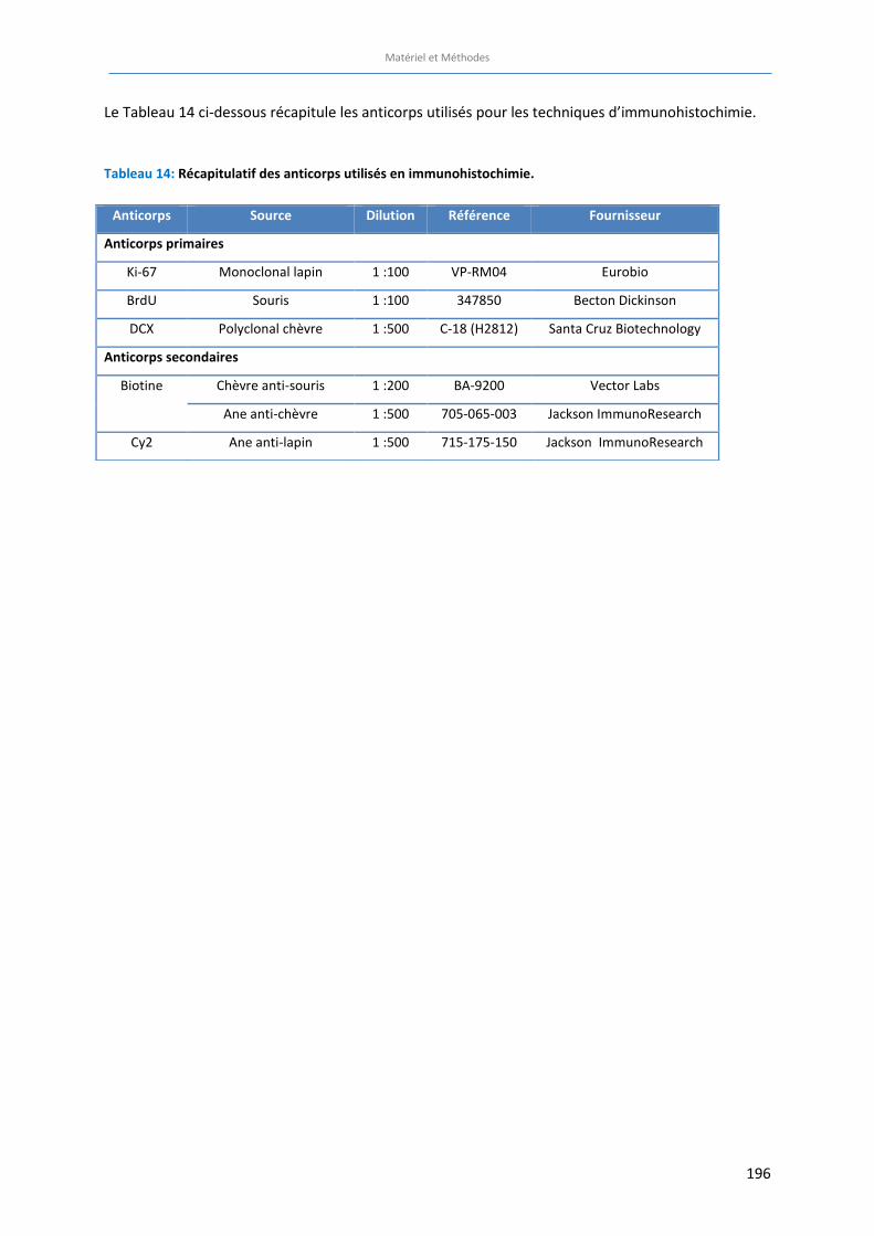
Matériel et Méthodes
196
Le Tableau 14 ci-dessous récapitule les anticorps utilisés pour les techniques d’immunohistochimie.
Tableau 14: Récapitulatif des anticorps utilisés en immunohistochimie.
Anticorps Source Dilution Référence Fournisseur
Anticorps primaires
Ki-67 Monoclonal lapin 1 :100 VP-RM04 Eurobio
BrdU Souris 1 :100 347850 Becton Dickinson
DCX Polyclonal chèvre 1 :500 C-18 (H2812) Santa Cruz Biotechnology
Anticorps secondaires
Biotine Chèvre anti-souris 1 :200 BA-9200 Vector Labs
Ane anti-chèvre 1 :500 705-065-003 Jackson ImmunoResearch
Cy2 Ane anti-lapin 1 :500 715-175-150 Jackson ImmunoResearch

197
198
Résultats expérimentaux
Article 1:
Learning and memory impairments in a neuroendocrine mouse model of anxiety/depression.
Darcet et al., Front Behav Neurosci. 2014 May 1;8:136. doi: 10.3389/fnbeh.2014.00136.
Article 2:
Rapid anxiolytic effects of a 5-HT4 receptor agonist are mediated by a neurogenesis-independent
mechanism.
Mendez-David et al., Neuropsychopharmacology. 2014 May;39(6):1366-78. doi: 10.1038/npp.2013.332.
Article 3:
Chronic 5-HT4 receptor agonist treatment restores learning and memory deficits in a neuroendocrine
mouse model of anxiety/depression.
Darcet et al., Neurosci Lett. 2016 Feb 2. pii: S0304-3940(16)30054-4.
Article 4:
Selective β-arrestin 1 deletion in stem cells of the dentate gyrus alters emotional state and dampens
antidepressant-like effects in adult mice.
En préparation
Résultats complémentaires:
Selective β-arrestin 1 deletion in stem cells of the dentate gyrus alters cognitive phenotype and
prevents pro-cognitive-like effects of chronic fluoxetine and 5-HT4 receptor agonist treatments in
adult mice
En préparation

199
Résultats expérimentaux
200
ARTICLE 1: Learning and memory impairments in a neuroendocrine mouse
model of anxiety/depression
Flavie Darcet, Indira Mendez-David, Laurent Tritschler, Alain M Gardier, Jean-Philippe Guilloux, Denis
J David
Publié dans Frontiers in Behavioral Neuroscience, doi :10.3389/fnbeh.2014.00136
Questions posées :
La modélisation d’un phénotype d’anxiété/dépression chez la souris par l’administration chronique
de corticostérone (modèle CORT) induit-elle des déficits d’apprentissage et de mémoire
comparables à ceux observés chez l’Homme présentant une dépression caractérisée ? Si oui, quels
sont les types de mémoire affectés chez ces souris « anxio/dépressives » ?
Résumé de l’étude
Les troubles cognitifs sont souvent reportés comme des symptômes invalidants chez les patients
souffrant de dépression caractérisée. L’incapacité à se concentrer fait d’ailleurs partie des critères de
diagnostic de la dépression majeure définis par le DSM-5. Dans de nombreuses études précliniques,
des altérations cognitives ont été observées dans différents modèles d’anxiété/dépression (voir
Revue Pharmaceuticals). Cependant, peu d’études se sont intéressées aux effets d’un traitement
chronique par la corticostérone sur les performances d’apprentissage et de mémoire. Dans cette
étude, nous avons mené une caractérisation complète des fonctions cognitives (dont la mémoire de
type épisodique, la mémoire spatiale et la mémoire associative) chez des animaux ayant été soumis
pendant 4 semaines à un régime de corticostérone (modèle CORT) (David et al., 2009).
Afin d’éviter que les différents tests cognitifs n’influent les uns sur les autres, l’étude a été menée
avec 4 cohortes différentes de souris C57BL/6Rj, chaque cohorte étant testée pour une tâche
cognitive précise après 4 semaines de traitement à la corticostérone (35 µg/mL) dans l’eau de
boisson. Le phénotype anxio/dépressif est vérifié grâce à un test de l’Open Field au début de la
période de comportement et un Splash test après le test cognitif pour les cohortes n°1 et 2. La
mémoire de type épisodique est étudiée grâce au test de reconnaissance d’objet, la mémoire
spatiale est estimée à l’aide tests de navigations spatiales dans un environnement aquatique ou sec
(la piscine de Morris et le Barnes maze, respectivement) et la mémoire à composante associative est
mesurée grâce à un test de conditionnement par la peur.
Résultats expérimentaux
201
Tout d’abord, nous avons observé une diminution de la capacité de discrimination des objets chez les
animaux présentant un phénotype « anxio/dépressif » par rapport aux contrôles, suggérant un déficit
de la mémoire de type épisodique. D’autre part, les tests spatiaux mettent en évidence une
altération globale des capacités d’apprentissage et de mémoire chez les animaux traités par la
corticostérone. La flexibilité mentale - un des aspects des fonctions exécutives de la mémoire - ainsi
que la mémoire à long-terme semblent également affectées par le phénotype anxio/dépressif. Enfin,
l’absence d’augmentation du comportement d’immobilité ou « freezing » observée dans le test de
conditionnement par la peur chez les animaux anxio/dépressifs indique un trouble de la mémoire
associative.
Ces résultats suggèrent que le phénotype anxio/dépressif induit par le traitement chronique de
corticostérone est associé à un déficit cognitif global, affectant tous les aspects de la mémoire ayant
été testés.
Contribution personnelle :
Au cours de ce travail :
x J’ai mis au point et validé 3 tests comportementaux de cognition chez la Souris : le test de
reconnaissance d’objet, la piscine de Morris et le Barnes maze.
x J’ai réalisé le suivi des animaux incluant la préparation et l’administration du régime
chronique de corticosterone.
x J’ai mené l’ensemble des tests comportementaux émotionnels et cognitifs (excepté le test de
conditionnement par la peur) sur les 3 cohortes différentes.
x J’ai analysé les résultats et rédigé l’intégralité de l’article sous la supervision du Pr. Denis
David et du Dr Jean-Philippe Guilloux
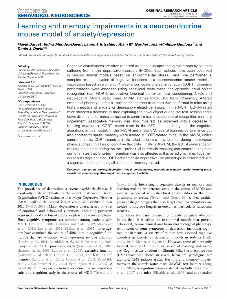
ORIGINAL RESEARCH ARTICLEpublished: 01 May 2014
doi: 10.3389/fnbeh.2014.00136
Learning and memory impairments in a neuroendocrinemouse model of anxiety/depressionFlavie Darcet , Indira Mendez-David , Laurent Tritschler , Alain M. Gardier , Jean-Philippe Guilloux† andDenis J. David*†
EA3544, Neuropharmacologie des troubles anxio-dépressifs et neurogenèse, Faculté de Pharmacie, Université Paris-Sud, Châtenay-Malabry, France
Edited by:Benjamin Adam Samuels, ColumbiaUniversity/Research Foundation forMental Hygiene, USA
Reviewed by:Michael Drew, University of Texas atAustin, USAChristine Ann Denny, ColumbiaUniversity, USA
*Correspondence:Denis J. David, EA3544“Pharmacologie des TroublesAnxio-Dépressifs et Neurogenèse,”Faculté de Pharmacie, UniversitéParis-Sud, 5 rue J-B Clément,Tour D1, 2e etage, F-92296Chatenay-Malabry, Francee-mail: [email protected]
†Last co-authorship.
Cognitive disturbances are often reported as serious incapacitating symptoms by patientssuffering from major depressive disorders (MDDs). Such deficits have been observedin various animal models based on environmental stress. Here, we performed acomplete characterization of cognitive functions in a neuroendocrine mouse model ofdepression based on a chronic (4 weeks) corticosterone administration (CORT). Cognitiveperformances were assessed using behavioral tests measuring episodic (novel objectrecognition test, NORT), associative (one-trial contextual fear conditioning, CFC), andvisuo-spatial (Morris water maze, MWM; Barnes maze, BM) learning/memory. Alteredemotional phenotype after chronic corticosterone treatment was confirmed in mice usingtests predictive of anxiety or depression-related behaviors. In the NORT, CORT-treatedmice showed a decrease in time exploring the novel object during the test session and alower discrimination index compared to control mice, characteristic of recognition memoryimpairment. Associative memory was also impaired, as observed with a decrease infreezing duration in CORT-treated mice in the CFC, thus pointing out the cognitivealterations in this model. In the MWM and in the BM, spatial learning performance butalso short-term spatial memory were altered in CORT-treated mice. In the MWM, unlikecontrol animals, CORT-treated animals failed to learn a new location during the reversalphase, suggesting a loss of cognitive flexibility. Finally, in the BM, the lack of preference forthe target quadrant during the recall probe trial in animals receiving corticosterone regimendemonstrates that long-term retention was also affected in this paradigm. Taken together,our results highlight that CORT-induced anxio-depressive-like phenotype is associated witha cognitive deficit affecting all aspects of memory tested.
Keywords: depression, anxiety/depression model, corticosterone, recognition memory, spatial learning maze,associative memory, cognitive impairments, cognitive flexibility
INTRODUCTIONThe prevalence of depression, a severe psychiatric disease, isconstantly high worldwide to the extent that World HealthOrganization (WHO) estimates that Major Depressive Disorder(MDD) will be the second largest cause of disability in year2020 (WHO, 2008). Major depression is characterized by a setof emotional and behavioral alterations, including persistentdepressed mood and loss of interest or pleasure as core symptoms.Since cognitive symptoms are common among patients withMDD (Fava et al., 2006; Hammar and Ardal, 2009; Murroughet al., 2011; Lee et al., 2012; Millan et al., 2012), investiga-tors have examined the nature of difficulties in cognitive func-tioning that are associated with depression such as attention(Landro et al., 2001; Ravnkilde et al., 2002; Porter et al., 2003;Lampe et al., 2004), processing speed (Ravnkilde et al., 2002;Hammar et al., 2003; Lampe et al., 2004), executive function(Naismith et al., 2003; Lampe et al., 2004) and learning andmemory (Landro et al., 2001; Fossati et al., 2002; Ravnkildeet al., 2002; Porter et al., 2003; Vythilingam et al., 2004). Arecent literature review e assessed abnormalities in neural cir-cuits and cognition early in the course of MDD (Trivedi and
Greer, 2014). Interestingly, cognitive deficits in memory anddecision-making are detected early in the course of MDD andmay be associated with structural abnormalities in the hip-pocampus or cortex (Trivedi and Greer, 2014). New antide-pressant drug strategies that also target cognitive symptoms areneeded to improve long-term outcomes, particularly functionalrecovery.
In order for basic research to provide potential advancesin the field, it is critical to use animal models that presentbehavioral, neurochemical and brain morphological phenotypereminiscent of some symptoms of depression including cogni-tive impairments. A variety of studies have assessed cognitivedisorders in anxiety or depression models in rodents (Patkiet al., 2013; Richter et al., 2013). However, some of these onlyfocused their work on a single aspect of learning and mem-ory. Cognitive dysfunctions in Chronic Mild Stress-exposed rats(CMS) have been shown in several behavioral paradigms. Forexample, CMS induces spatial learning and memory impair-ments in the Morris water maze (MWM) test in mice (Songet al., 2006), recognition memory deficits in both rats (Orsettiet al., 2007) and mice (Elizalde et al., 2008) and suppression
Frontiers in Behavioral Neuroscience www.frontiersin.org May 2014 | Volume 8 | Article 136 | 1
BEHAVIORAL NEUROSCIENCE
Darcet et al. Cognition and anxiety/depression
of fear extinction (Garcia et al., 2008). Similarly, maternal sep-aration and social defeat models were employed to highlightspatial reference memory deficits (Couto et al., 2012; Patki et al.,2013).
Here, we performed a thorough characterization of cognitiveperformances in a neuroendocrine mouse model of depressionbased on a chronic CORT (David et al., 2009; Mendez-Davidet al., 2014). Given the multiple forms of learning and mem-ory, we selected a range of cognitive behavioral paradigms thatallows investigation of different memory systems including short-term episodic memory, associative memory and spatial referencelearning and memory. Additional parameters such as cognitiveflexibility and long-term memory were also evaluated in spatialmemory tests.
MATERIALS AND METHODSANIMALSEight to 10-weeks old male C57BL/6J mice (Janvier Labs,France) were maintained on a 12L:12D schedule and werehoused 5 per cage. Food and water were provided ad libi-tum. All behavioral testing occurred during the light phasebetween 7 am and 7 pm and were conducted in com-pliance with animal cares guidelines and with protocolsapproved by the Institutional Animal Care and Use Committee(Council Directive #87-848, October 19, 1987, Ministère del’Agriculture et de la Forêt, Service Vétérinaire de la santé etde la Protection Animale, permission #92-256B to Denis J.David).
DRUGSCorticosterone (4-pregnen-11b-diol-3 20-dione 21 hemisuc-cinate, CORT from Sigma-Aldrich, France) was dissolvedin vehicle (0.45% hydroxypropyl-β-cyclodextrin, β-CD fromSigma-Aldrich, France). Corticosterone (35 µg/ml equivalent to5 mg/kg/day) was delivered for 28 days in drinking water and con-tinued when the behavioral tests were performed (David et al.,2009). Control animals received vehicle (β-CD) in drinking waterduring the entire experiment (Figure 1).
BEHAVIORAL TESTINGFour different cohorts of mice were used to assess learn-ing and memory performances in anxio-depressive animals.To prevent any confounding effects between cognitive tasks,each animal was subjected to only one type of learning andmemory test. Fur coat state of the animals was scored weeklyduring the whole treatment period. The anxio-depressive-like phenotype induced by chronic corticosterone among thedifferent cohorts was evaluated using the Open Field andthe Splash tests, before and after the cognitive task, respec-tively. Details of emotional assessment are mentioned inFigure 1.
Cognitive performances were evaluated using behavioral testsmeasuring episodic (novel object recognition test, NORT), asso-ciative (one-trial contextual fear conditioning, CFC) and visuo-spatial (MWM; Barnes maze, BM) learning/memory. Animalswere placed in the experimental room 30 min before the start ofthe behavioral experiments.
ANXIETY AND DEPRESSION BEHAVIOR PARADIGMSFur coat stateThe score corresponding to the state of the coat resulted fromthe sum of the score of five different body parts: head, neck,dorsal/ventral coat, tail and fore-/hind paws. For each body area, ascore of 0 was given for a well-groomed coat and 1 for an unkemptcoat (Surget et al., 2008; David et al., 2009).
Open fieldMotor activity was quantified in 43 × 43 cm plexiglas open fieldboxes (MED Associates, Georgia, VT). Two sets of 16 pulse-modulated infrared photobeams were placed on opposite walls2–5 cm apart to record x-y ambulatory movements. Activitychambers were computer interfaced for data sampling at 100 msresolution. The computer defined grid lines that divided eachopen field into center and surround regions, with each of fourlines being 11 cm from each wall. Time in the center was recordedfor 30 min to evaluate anxiety-related behavior. The locomotoractivity was quantified as total ambulatory distance.
Splash testSplash test was performed as previously described (David et al.,2009). This test consisted in squirting a 10% sucrose solution onthe mouse’s snout. The grooming duration was then recordedover a 5 min period.
COGNITION BEHAVIORAL PARADIGMSEpisodic short-term memory: novel object recognition testThe procedure was adapted from the Sahay study (Sahay et al.,2011). The apparatus consisted in black plastic boxes (28 × 41×18 cm) slightly filled with sawdust (≈0.5–1 cm thickness) in aroom with a low level of light. Locomotor activity was con-trolled during the entire experiment (parameter: ambulatorydistance) using a videotracking procedure (ANY-maze Software,Bioseb, France). Objects exploration was hand-scored by anexperimenter.
The NORT was divided into 4 training sessions and one testsession. Each exposure lasted 5 min with a 3-min inter-trial inter-val. Between each trial, mice returned to their home cage, beddingof apparatus was changed and boxes were cleaned with 70%ethanol solution. During training sessions, two identical objects[cylindrical glassware (∅:3 cm, height: 8 cm) filled with white cot-ton, (Figure 2D)] were present in the box. The mouse was placedin the middle of the box facing the wall and was allowed tofreely explore the apparatus and the objects. During the test ses-sion, one of the familiar objects was removed from the cage andreplaced by a novel object [Lego® rectangular structure (7 × 3×9 cm), (Figure 2D)]. The objects had been previously validatedto ensure there was no inherent preference for either object (datanot shown). The nature (Lego® vs. glass) and the position of thenovel object (left vs. right) were chosen randomly.
Object exploration was defined as the orientation of the noseto the object at a distance ≤ 2 cm. Placing the forepaws on theobjects was considered as exploratory behavior, but climbing onthe objects was not. Objects were cleaned with 70% ethanolbetween trials to avoid olfactory cues. Results for this test wereexpressed as: (1) exploration of each object (in seconds) during
Frontiers in Behavioral Neuroscience www.frontiersin.org May 2014 | Volume 8 | Article 136 | 2
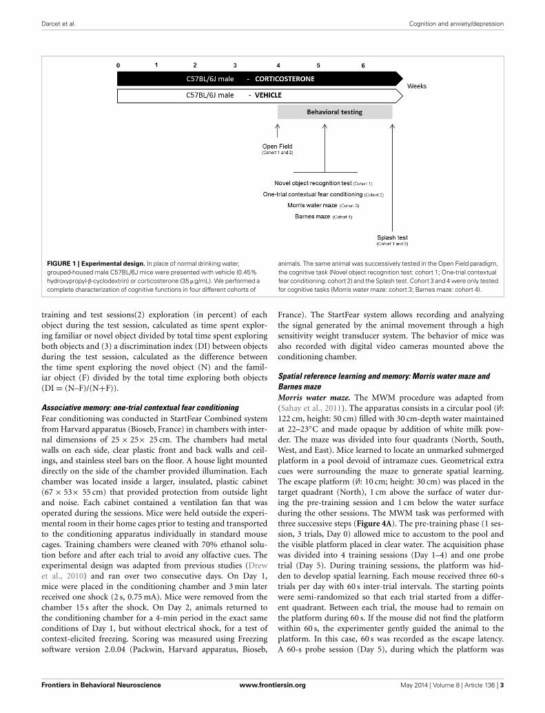
Darcet et al. Cognition and anxiety/depression
FIGURE 1 | Experimental design. In place of normal drinking water,grouped-housed male C57BL/6J mice were presented with vehicle (0.45%hydroxypropyl-β-cyclodextrin) or corticosterone (35 µg/mL). We performed acomplete characterization of cognitive functions in four different cohorts of
animals. The same animal was successively tested in the Open Field paradigm,the cognitive task (Novel object recognition test: cohort 1; One-trial contextualfear conditioning: cohort 2) and the Splash test. Cohort 3 and 4 were only testedfor cognitive tasks (Morris water maze: cohort 3; Barnes maze: cohort 4).
training and test sessions(2) exploration (in percent) of eachobject during the test session, calculated as time spent explor-ing familiar or novel object divided by total time spent exploringboth objects and (3) a discrimination index (DI) between objectsduring the test session, calculated as the difference betweenthe time spent exploring the novel object (N) and the famil-iar object (F) divided by the total time exploring both objects(DI = (N–F)/(N+F)).
Associative memory: one-trial contextual fear conditioningFear conditioning was conducted in StartFear Combined systemfrom Harvard apparatus (Bioseb, France) in chambers with inter-nal dimensions of 25 × 25× 25 cm. The chambers had metalwalls on each side, clear plastic front and back walls and ceil-ings, and stainless steel bars on the floor. A house light mounteddirectly on the side of the chamber provided illumination. Eachchamber was located inside a larger, insulated, plastic cabinet(67 × 53× 55 cm) that provided protection from outside lightand noise. Each cabinet contained a ventilation fan that wasoperated during the sessions. Mice were held outside the experi-mental room in their home cages prior to testing and transportedto the conditioning apparatus individually in standard mousecages. Training chambers were cleaned with 70% ethanol solu-tion before and after each trial to avoid any olfactive cues. Theexperimental design was adapted from previous studies (Drewet al., 2010) and ran over two consecutive days. On Day 1,mice were placed in the conditioning chamber and 3 min laterreceived one shock (2 s, 0.75 mA). Mice were removed from thechamber 15 s after the shock. On Day 2, animals returned tothe conditioning chamber for a 4-min period in the exact sameconditions of Day 1, but without electrical shock, for a test ofcontext-elicited freezing. Scoring was measured using Freezingsoftware version 2.0.04 (Packwin, Harvard apparatus, Bioseb,
France). The StartFear system allows recording and analyzingthe signal generated by the animal movement through a highsensitivity weight transducer system. The behavior of mice wasalso recorded with digital video cameras mounted above theconditioning chamber.
Spatial reference learning and memory: Morris water maze andBarnes mazeMorris water maze. The MWM procedure was adapted from(Sahay et al., 2011). The apparatus consists in a circular pool (∅:122 cm, height: 50 cm) filled with 30 cm-depth water maintainedat 22–23◦C and made opaque by addition of white milk pow-der. The maze was divided into four quadrants (North, South,West, and East). Mice learned to locate an unmarked submergedplatform in a pool devoid of intramaze cues. Geometrical extracues were surrounding the maze to generate spatial learning.The escape platform (∅: 10 cm; height: 30 cm) was placed in thetarget quadrant (North), 1 cm above the surface of water dur-ing the pre-training session and 1 cm below the water surfaceduring the other sessions. The MWM task was performed withthree successive steps (Figure 4A). The pre-training phase (1 ses-sion, 3 trials, Day 0) allowed mice to accustom to the pool andthe visible platform placed in clear water. The acquisition phasewas divided into 4 training sessions (Day 1–4) and one probetrial (Day 5). During training sessions, the platform was hid-den to develop spatial learning. Each mouse received three 60-strials per day with 60 s inter-trial intervals. The starting pointswere semi-randomized so that each trial started from a differ-ent quadrant. Between each trial, the mouse had to remain onthe platform during 60 s. If the mouse did not find the platformwithin 60 s, the experimenter gently guided the animal to theplatform. In this case, 60 s was recorded as the escape latency.A 60-s probe session (Day 5), during which the platform was
Frontiers in Behavioral Neuroscience www.frontiersin.org May 2014 | Volume 8 | Article 136 | 3
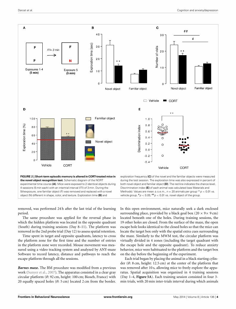
Darcet et al. Cognition and anxiety/depression
FIGURE 2 | Short-term episodic memory is altered in CORT-treated mice inthe novel object recognition test. Schematic diagram of the NORTexperimental time course (A). Mice were exposed to 2 identical objects during4 sessions (5 min each) with an intertrial interval (ITI) of 3 min. During the5thexposure, one familiar object (F) was removed and replaced with a novelobject (N) different in shape, color, and texture. Exploration time (B) and
exploration frequency (C) of the novel and the familiar objects were measuredduring the test session. The exploration time was also expressed in percent ofboth novel object and familiar object (D). The red line indicates the chance level.Discrimination index (E) of each animal was calculated (see Materials andMethods). Values are mean ± s.e.m., n = 20 animals per group ∗∗p < 0.01 vs.vehicle group. #p < 0.05; ##p < 0.01 vs. novel object of the group.
removed, was performed 24 h after the last trial of the learningperiod.
The same procedure was applied for the reversal phase inwhich the hidden platform was located in the opposite quadrant(South) during training sessions (Day 8–11). The platform wasremoved in the 2nd probe trial (Day 12) to assess spatial retention.
Time spent in target and opposite quadrants, latency to crossthe platform zone for the first time and the number of entriesin the platform zone were recorded. Mouse movement was mea-sured using a video tracking system and analyzed by ANY-mazeSoftware to record latency, distance and pathways to reach theescape platform through all the sessions.
Barnes maze. The BM procedure was modified from a previouswork (Sunyer et al., 2007). The apparatus consisted in a clear graycircular platform (∅: 92 cm, height: 100 cm; Bioseb, France) with20 equally spaced holes (∅: 5 cm) located 2 cm from the border.
In this open environment, mice naturally seek a dark enclosedsurrounding place, provided by a black goal box (20 × 9× 9 cm)located beneath one of the holes. During training sessions, the19 other holes are closed. From the surface of the maze, the openescape hole looks identical to the closed holes so that the mice canlocate the target box only with the spatial extra cues surroundingthe maze. Similarly to the MWM test, the circular platform wasvirtually divided in 4 zones (including the target quadrant withthe escape hole and the opposite quadrant). To reduce anxietybehavior, mice were habituated to the platform and the target boxon the day before the beginning of the experiment.
Each trial began by placing the animal in a black starting cylin-der (∅: 8 cm, height: 12.5 cm) at the center of the platform thatwas removed after 10 s, allowing mice to freely explore the appa-ratus. Spatial acquisition was organized in 4 training sessions(Day 1–4, Figure 5A). Each training session consisted in four 3-min trials, with 20 min inter-trials interval during which animals
Frontiers in Behavioral Neuroscience www.frontiersin.org May 2014 | Volume 8 | Article 136 | 4
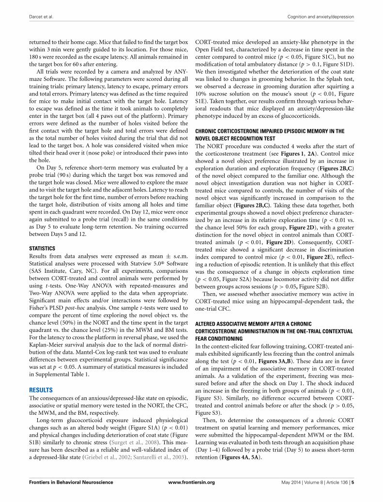
Darcet et al. Cognition and anxiety/depression
returned to their home cage. Mice that failed to find the target boxwithin 3 min were gently guided to its location. For those mice,180 s were recorded as the escape latency. All animals remained inthe target box for 60 s after entering.
All trials were recorded by a camera and analyzed by ANY-maze Software. The following parameters were scored during alltraining trials: primary latency, latency to escape, primary errorsand total errors. Primary latency was defined as the time requiredfor mice to make initial contact with the target hole. Latencyto escape was defined as the time it took animals to completelyenter in the target box (all 4 paws out of the platform). Primaryerrors were defined as the number of holes visited before thefirst contact with the target hole and total errors were definedas the total number of holes visited during the trial that did notlead to the target box. A hole was considered visited when micetilted their head over it (nose poke) or introduced their paws intothe hole.
On Day 5, reference short-term memory was evaluated by aprobe trial (90 s) during which the target box was removed andthe target hole was closed. Mice were allowed to explore the mazeand to visit the target hole and the adjacent holes. Latency to reachthe target hole for the first time, number of errors before reachingthe target hole, distribution of visits among all holes and timespent in each quadrant were recorded. On Day 12, mice were onceagain submitted to a probe trial (recall) in the same conditionsas Day 5 to evaluate long-term retention. No training occurredbetween Days 5 and 12.
STATISTICSResults from data analyses were expressed as mean ± s.e.m.Statistical analyses were processed with Statview 5.0® Software(SAS Institute, Cary, NC). For all experiments, comparisonsbetween CORT-treated and control animals were performed byusing t-tests. One-Way ANOVA with repeated-measures andTwo-Way ANOVA were applied to the data when appropriate.Significant main effects and/or interactions were followed byFisher’s PLSD post-hoc analysis. One sample t-tests were used tocompare the percent of time exploring the novel object vs. thechance level (50%) in the NORT and the time spent in the targetquadrant vs. the chance level (25%) in the MWM and BM tests.For the latency to cross the platform in reversal phase, we used theKaplan-Meier survival analysis due to the lack of normal distri-bution of the data. Mantel-Cox log-rank test was used to evaluatedifferences between experimental groups. Statistical significancewas set at p < 0.05. A summary of statistical measures is includedin Supplemental Table 1.
RESULTSThe consequences of an anxious/depressed-like state on episodic,associative or spatial memory were tested in the NORT, the CFC,the MWM, and the BM, respectively.
Long-term glucocorticoid exposure induced physiologicalchanges such as an altered body weight (Figure S1A) (p < 0.01)and physical changes including deterioration of coat state (FigureS1B) similarly to chronic stress (Surget et al., 2008). This mea-sure has been described as a reliable and well-validated index ofa depressed-like state (Griebel et al., 2002; Santarelli et al., 2003).
CORT-treated mice developed an anxiety-like phenotype in theOpen Field test, characterized by a decrease in time spent in thecenter compared to control mice (p < 0.05, Figure S1C), but nomodification of total ambulatory distance (p > 0.1, Figure S1D).We then investigated whether the deterioration of the coat statewas linked to changes in grooming behavior. In the Splash test,we observed a decrease in grooming duration after squirting a10% sucrose solution on the mouse’s snout (p < 0.01, FigureS1E). Taken together, our results confirm through various behav-ioral readouts that mice displayed an anxiety/depression-likephenotype induced by an excess of glucocorticoids.
CHRONIC CORTICOSTERONE IMPAIRED EPISODIC MEMORY IN THENOVEL OBJECT RECOGNITION TESTThe NORT procedure was conducted 4 weeks after the start ofthe corticosterone treatment (see Figures 1, 2A). Control miceshowed a novel object preference illustrated by an increase inexploration duration and exploration frequency (Figures 2B,C)of the novel object compared to the familiar one. Although thenovel object investigation duration was not higher in CORT-treated mice compared to controls, the number of visits of thenovel object was significantly increased in comparison to thefamiliar object (Figures 2B,C). Taking these data together, bothexperimental groups showed a novel object preference character-ized by an increase in its relative exploration time (p < 0.01 vs.the chance level 50% for each group, Figure 2D), with a greaterdistinction for the novel object in control animals than CORT-treated animals (p < 0.01, Figure 2D). Consequently, CORT-treated mice showed a significant decrease in discriminationindex compared to control mice (p < 0.01, Figure 2E), reflect-ing a reduction of episodic retention. It is unlikely that this effectwas the consequence of a change in objects exploration time(p < 0.05, Figure S2A) because locomotor activity did not differbetween groups across sessions (p > 0.05, Figure S2B).
Then, we assessed whether associative memory was active inCORT-treated mice using an hippocampal-dependent task, theone-trial CFC.
ALTERED ASSOCIATIVE MEMORY AFTER A CHRONICCORTICOSTERONE ADMINISTRATION IN THE ONE-TRIAL CONTEXTUALFEAR CONDITIONINGIn the context-elicited fear following training, CORT-treated ani-mals exhibited significantly less freezing than the control animalsalong the test (p < 0.01, Figures 3A,B). These data are in favorof an impairment of the associative memory in CORT-treatedanimals. As a validation of the experiment, freezing was mea-sured before and after the shock on Day 1. The shock inducedan increase in the freezing in both groups of animals (p < 0.01,Figure S3). Similarly, no difference occurred between CORT-treated and control animals before or after the shock (p > 0.05,Figure S3).
Then, to determine the consequences of a chronic CORTtreatment on spatial learning and memory performances, micewere submitted the hippocampal-dependent MWM or the BM.Learning was evaluated in both tests through an acquisition phase(Day 1–4) followed by a probe trial (Day 5) to assess short-termretention (Figures 4A, 5A).
Frontiers in Behavioral Neuroscience www.frontiersin.org May 2014 | Volume 8 | Article 136 | 5
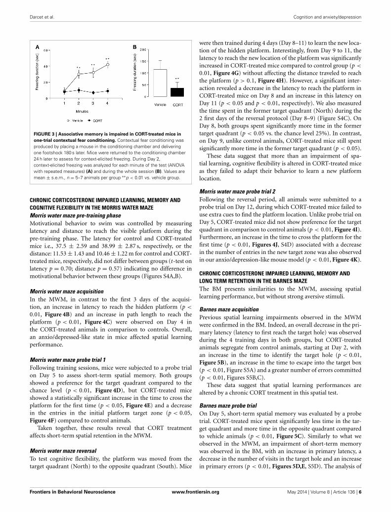
Darcet et al. Cognition and anxiety/depression
FIGURE 3 | Associative memory is impaired in CORT-treated mice inone-trial contextual fear conditioning. Contextual fear conditioning wasproduced by placing a mouse in the conditioning chamber and deliveringone footshock 180 s later. Mice were returned to the conditioning chamber24 h later to assess for context-elicited freezing. During Day 2,context-elicited freezing was analyzed for each minute of the test (ANOVAwith repeated measures) (A) and during the whole session (B). Values aremean ± s.e.m., n = 5–7 animals per group ∗∗p < 0.01 vs. vehicle group.
CHRONIC CORTICOSTERONE IMPAIRED LEARNING, MEMORY ANDCOGNITIVE FLEXIBILITY IN THE MORRIS WATER MAZEMorris water maze pre-training phaseMotivational behavior to swim was controlled by measuringlatency and distance to reach the visible platform during thepre-training phase. The latency for control and CORT-treatedmice i.e., 37.5 ± 2.59 and 38.99 ± 2.87 s, respectively, or thedistance: 11.53 ± 1.43 and 10.46 ± 1.22 m for control and CORT-treated mice, respectively, did not differ between groups (t-test onlatency p = 0.70; distance p = 0.57) indicating no difference inmotivational behavior between these groups (Figures S4A,B).
Morris water maze acquisitionIn the MWM, in contrast to the first 3 days of the acquisi-tion, an increase in latency to reach the hidden platform (p <
0.01, Figure 4B) and an increase in path length to reach theplatform (p < 0.01, Figure 4C) were observed on Day 4 inthe CORT-treated animals in comparison to controls. Overall,an anxio/depressed-like state in mice affected spatial learningperformance.
Morris water maze probe trial 1Following training sessions, mice were subjected to a probe trialon Day 5 to assess short-term spatial memory. Both groupsshowed a preference for the target quadrant compared to thechance level (p < 0.01, Figure 4D), but CORT-treated miceshowed a statistically significant increase in the time to cross theplatform for the first time (p < 0.05, Figure 4E) and a decreasein the entries in the initial platform target zone (p < 0.05,Figure 4F) compared to control animals.
Taken together, these results reveal that CORT treatmentaffects short-term spatial retention in the MWM.
Morris water maze reversalTo test cognitive flexibility, the platform was moved from thetarget quadrant (North) to the opposite quadrant (South). Mice
were then trained during 4 days (Day 8–11) to learn the new loca-tion of the hidden platform. Interestingly, from Day 9 to 11, thelatency to reach the new location of the platform was significantlyincreased in CORT-treated mice compared to control group (p <
0.01, Figure 4G) without affecting the distance traveled to reachthe platform (p > 0.1, Figure 4H). However, a significant inter-action revealed a decrease in the latency to reach the platform inCORT-treated mice on Day 8 and an increase in this latency onDay 11 (p < 0.05 and p < 0.01, respectively). We also measuredthe time spent in the former target quadrant (North) during the2 first days of the reversal protocol (Day 8–9) (Figure S4C). OnDay 8, both groups spent significantly more time in the formertarget quadrant (p < 0.05 vs. the chance level 25%). In contrast,on Day 9, unlike control animals, CORT-treated mice still spentsignificantly more time in the former target quadrant (p < 0.05).
These data suggest that more than an impairment of spa-tial learning, cognitive flexibility is altered in CORT-treated miceas they failed to adapt their behavior to learn a new platformlocation.
Morris water maze probe trial 2Following the reversal period, all animals were submitted to aprobe trial on Day 12, during which CORT-treated mice failed touse extra cues to find the platform location. Unlike probe trial onDay 5, CORT-treated mice did not show preference for the targetquadrant in comparison to control animals (p < 0.01, Figure 4I).Furthermore, an increase in the time to cross the platform for thefirst time (p < 0.01, Figures 4J, S4D) associated with a decreasein the number of entries in the new target zone was also observedin our anxio/depression-like mouse model (p < 0.01, Figure 4K).
CHRONIC CORTICOSTERONE IMPAIRED LEARNING, MEMORY ANDLONG TERM RETENTION IN THE BARNES MAZEThe BM presents similarities to the MWM, assessing spatiallearning performance, but without strong aversive stimuli.
Barnes maze acquisitionPrevious spatial learning impairments observed in the MWMwere confirmed in the BM. Indeed, an overall decrease in the pri-mary latency (latency to first reach the target hole) was observedduring the 4 training days in both groups, but CORT-treatedanimals segregate from control animals, starting at Day 2, withan increase in the time to identify the target hole (p < 0.01,Figure 5B), an increase in the time to escape into the target box(p < 0.01, Figure S5A) and a greater number of errors committed(p < 0.01, Figures S5B,C).
These data suggest that spatial learning performances arealtered by a chronic CORT treatment in this spatial test.
Barnes maze probe trialOn Day 5, short-term spatial memory was evaluated by a probetrial. CORT-treated mice spent significantly less time in the tar-get quadrant and more time in the opposite quadrant comparedto vehicle animals (p < 0.01, Figure 5C). Similarly to what weobserved in the MWM, an impairment of short-term memorywas observed in the BM, with an increase in primary latency, adecrease in the number of visits in the target hole and an increasein primary errors (p < 0.01, Figures 5D,E, S5D). The analysis of
Frontiers in Behavioral Neuroscience www.frontiersin.org May 2014 | Volume 8 | Article 136 | 6
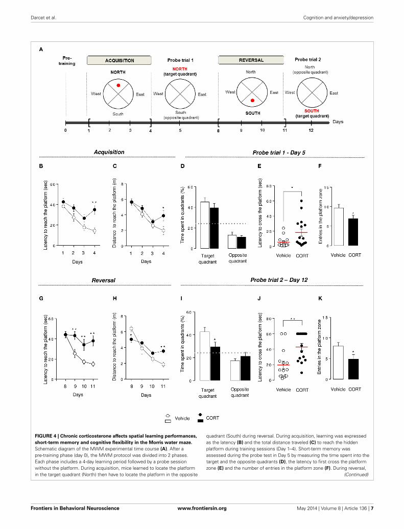
Darcet et al. Cognition and anxiety/depression
FIGURE 4 | Chronic corticosterone affects spatial learning performances,short-term memory and cognitive flexibility in the Morris water maze.Schematic diagram of the MWM experimental time course (A). After apre-training phase (day 0), the MWM protocol was divided into 2 phases.Each phase includes a 4-day learning period followed by a probe sessionwithout the platform. During acquisition, mice learned to locate the platformin the target quadrant (North) then have to locate the platform in the opposite
quadrant (South) during reversal. During acquisition, learning was expressedas the latency (B) and the total distance traveled (C) to reach the hiddenplatform during training sessions (Day 1–4). Short-term memory wasassessed during the probe test in Day 5 by measuring the time spent into thetarget and the opposite quadrants (D), the latency to first cross the platformzone (E) and the number of entries in the platform zone (F). During reversal,
(Continued)
Frontiers in Behavioral Neuroscience www.frontiersin.org May 2014 | Volume 8 | Article 136 | 7
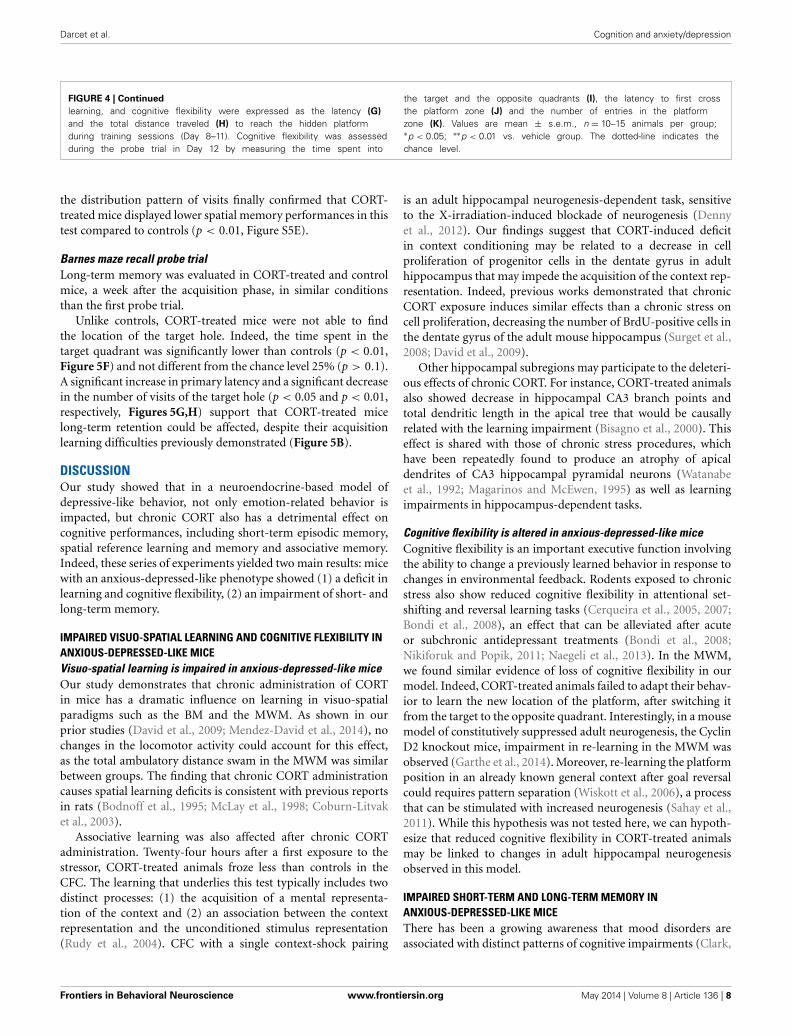
Darcet et al. Cognition and anxiety/depression
FIGURE 4 | Continuedlearning, and cognitive flexibility were expressed as the latency (G)and the total distance traveled (H) to reach the hidden platformduring training sessions (Day 8–11). Cognitive flexibility was assessedduring the probe trial in Day 12 by measuring the time spent into
the target and the opposite quadrants (I), the latency to first crossthe platform zone (J) and the number of entries in the platformzone (K). Values are mean ± s.e.m., n = 10–15 animals per group;∗p < 0.05; ∗∗p < 0.01 vs. vehicle group. The dotted-line indicates thechance level.
the distribution pattern of visits finally confirmed that CORT-treated mice displayed lower spatial memory performances in thistest compared to controls (p < 0.01, Figure S5E).
Barnes maze recall probe trialLong-term memory was evaluated in CORT-treated and controlmice, a week after the acquisition phase, in similar conditionsthan the first probe trial.
Unlike controls, CORT-treated mice were not able to findthe location of the target hole. Indeed, the time spent in thetarget quadrant was significantly lower than controls (p < 0.01,Figure 5F) and not different from the chance level 25% (p > 0.1).A significant increase in primary latency and a significant decreasein the number of visits of the target hole (p < 0.05 and p < 0.01,respectively, Figures 5G,H) support that CORT-treated micelong-term retention could be affected, despite their acquisitionlearning difficulties previously demonstrated (Figure 5B).
DISCUSSIONOur study showed that in a neuroendocrine-based model ofdepressive-like behavior, not only emotion-related behavior isimpacted, but chronic CORT also has a detrimental effect oncognitive performances, including short-term episodic memory,spatial reference learning and memory and associative memory.Indeed, these series of experiments yielded two main results: micewith an anxious-depressed-like phenotype showed (1) a deficit inlearning and cognitive flexibility, (2) an impairment of short- andlong-term memory.
IMPAIRED VISUO-SPATIAL LEARNING AND COGNITIVE FLEXIBILITY INANXIOUS-DEPRESSED-LIKE MICEVisuo-spatial learning is impaired in anxious-depressed-like miceOur study demonstrates that chronic administration of CORTin mice has a dramatic influence on learning in visuo-spatialparadigms such as the BM and the MWM. As shown in ourprior studies (David et al., 2009; Mendez-David et al., 2014), nochanges in the locomotor activity could account for this effect,as the total ambulatory distance swam in the MWM was similarbetween groups. The finding that chronic CORT administrationcauses spatial learning deficits is consistent with previous reportsin rats (Bodnoff et al., 1995; McLay et al., 1998; Coburn-Litvaket al., 2003).
Associative learning was also affected after chronic CORTadministration. Twenty-four hours after a first exposure to thestressor, CORT-treated animals froze less than controls in theCFC. The learning that underlies this test typically includes twodistinct processes: (1) the acquisition of a mental representa-tion of the context and (2) an association between the contextrepresentation and the unconditioned stimulus representation(Rudy et al., 2004). CFC with a single context-shock pairing
is an adult hippocampal neurogenesis-dependent task, sensitiveto the X-irradiation-induced blockade of neurogenesis (Dennyet al., 2012). Our findings suggest that CORT-induced deficitin context conditioning may be related to a decrease in cellproliferation of progenitor cells in the dentate gyrus in adulthippocampus that may impede the acquisition of the context rep-resentation. Indeed, previous works demonstrated that chronicCORT exposure induces similar effects than a chronic stress oncell proliferation, decreasing the number of BrdU-positive cells inthe dentate gyrus of the adult mouse hippocampus (Surget et al.,2008; David et al., 2009).
Other hippocampal subregions may participate to the deleteri-ous effects of chronic CORT. For instance, CORT-treated animalsalso showed decrease in hippocampal CA3 branch points andtotal dendritic length in the apical tree that would be causallyrelated with the learning impairment (Bisagno et al., 2000). Thiseffect is shared with those of chronic stress procedures, whichhave been repeatedly found to produce an atrophy of apicaldendrites of CA3 hippocampal pyramidal neurons (Watanabeet al., 1992; Magarinos and McEwen, 1995) as well as learningimpairments in hippocampus-dependent tasks.
Cognitive flexibility is altered in anxious-depressed-like miceCognitive flexibility is an important executive function involvingthe ability to change a previously learned behavior in response tochanges in environmental feedback. Rodents exposed to chronicstress also show reduced cognitive flexibility in attentional set-shifting and reversal learning tasks (Cerqueira et al., 2005, 2007;Bondi et al., 2008), an effect that can be alleviated after acuteor subchronic antidepressant treatments (Bondi et al., 2008;Nikiforuk and Popik, 2011; Naegeli et al., 2013). In the MWM,we found similar evidence of loss of cognitive flexibility in ourmodel. Indeed, CORT-treated animals failed to adapt their behav-ior to learn the new location of the platform, after switching itfrom the target to the opposite quadrant. Interestingly, in a mousemodel of constitutively suppressed adult neurogenesis, the CyclinD2 knockout mice, impairment in re-learning in the MWM wasobserved (Garthe et al., 2014). Moreover, re-learning the platformposition in an already known general context after goal reversalcould requires pattern separation (Wiskott et al., 2006), a processthat can be stimulated with increased neurogenesis (Sahay et al.,2011). While this hypothesis was not tested here, we can hypoth-esize that reduced cognitive flexibility in CORT-treated animalsmay be linked to changes in adult hippocampal neurogenesisobserved in this model.
IMPAIRED SHORT-TERM AND LONG-TERM MEMORY INANXIOUS-DEPRESSED-LIKE MICEThere has been a growing awareness that mood disorders areassociated with distinct patterns of cognitive impairments (Clark,
Frontiers in Behavioral Neuroscience www.frontiersin.org May 2014 | Volume 8 | Article 136 | 8
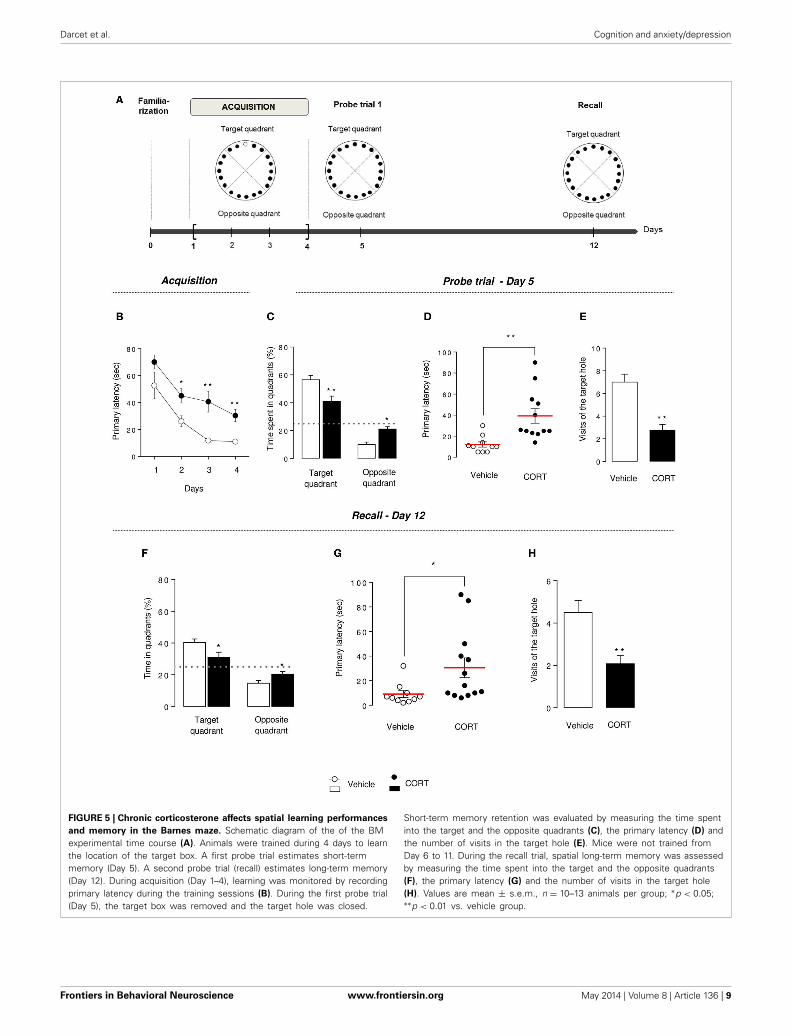
Darcet et al. Cognition and anxiety/depression
FIGURE 5 | Chronic corticosterone affects spatial learning performancesand memory in the Barnes maze. Schematic diagram of the of the BMexperimental time course (A). Animals were trained during 4 days to learnthe location of the target box. A first probe trial estimates short-termmemory (Day 5). A second probe trial (recall) estimates long-term memory(Day 12). During acquisition (Day 1–4), learning was monitored by recordingprimary latency during the training sessions (B). During the first probe trial(Day 5), the target box was removed and the target hole was closed.
Short-term memory retention was evaluated by measuring the time spentinto the target and the opposite quadrants (C), the primary latency (D) andthe number of visits in the target hole (E). Mice were not trained fromDay 6 to 11. During the recall trial, spatial long-term memory was assessedby measuring the time spent into the target and the opposite quadrants(F), the primary latency (G) and the number of visits in the target hole(H). Values are mean ± s.e.m., n = 10–13 animals per group; ∗p < 0.05;∗∗p < 0.01 vs. vehicle group.
Frontiers in Behavioral Neuroscience www.frontiersin.org May 2014 | Volume 8 | Article 136 | 9
Darcet et al. Cognition and anxiety/depression
2009; Gotlib and Joormann, 2010). In this study, episodic(NORT), associative (CFC) and visuo-spatial (MWM and BM)memory were assessed in our model of anxiety-depressionin mice.
HPA axis hyperactivation leads to negative effects on mem-ory processes (Song et al., 2006; Aisa et al., 2007; Maccari andMorley-Fletcher, 2007). Specifically, a study demonstrated thatmice submitted to learned helplessness paradigm or chronic mildstress procedures showed poor water maze performances (Songet al., 2006). Here, we observed that chronic CORT admin-istration impaired spatial cognitive retention in both mazes.Moreover, because the BM evaluates cognitive function withminimal stress to the animal (vs. the MWM), we confirmthat poor performances after chronic glucocorticoid treatmentin both mazes are the result of cognitive impairment, ratherthan a possible effect of stress. Memory retention was alsoinvestigated in CORT-treated mice using the NORT. This teststrongly relies on visual recognition memory and is based onrodent’s exploratory behavior and spontaneous preference fornovel objects (Ennaceur and Delacour, 1988). Notably, this taskhas been a model of short-term episodic memory as mice areable to recognize the familiar object up to a few hours post-training (Bertaina-Anglade et al., 2006). In our study, CORT-treated mice had no difficulties to distinguish between the twoobjects, but showed an important alteration on short-term mem-ory characterized by a lower discrimination index comparedto control mice. Non-spatial NORT is commonly describedas a hippocampal-independent task. However, a recent reportproposed that the hippocampus was required in this episodicshort-term memory test (Cohen et al., 2013), thus raising thehypothesis that lower discrimination index of CORT-treated micecould be attributable to morphologic molecular and cellularchanges in brain processes, especially in neurogenesis adap-tation. Current studies investigating the relationship betweennon-spatial NORT and adult hippocampal neurogenesis led toconflicting findings: adult hippocampal neurogenesis alterationis linked either negatively (Jessberger et al., 2009; Cohen et al.,2013) or positively (Denny et al., 2012; Oury et al., 2013)to the novel object discrimination. It seems, at least that thefunctional integrity of the dentate gyrus is involved in thenon-spatial NORT.
We also investigated long-term memory in the BM 1 weekafter the first probe trial, in similar conditions. In this spatial test,learning behavioral profile in CORT-treated mice makes difficultto interpret the long-term memory assessment. Although acqui-sition learning and memory were affected in CORT-treated mice,they still succeeded to learn the task. Comparing performanceson Day 5 vs. 12 in CORT-treated mice, we showed that theseanimals lost their ability to locate the target hole a week afterthe last trial. This finding is in a favor of a change in long-termretention induced by the chronic CORT treatment. Similaritiesin long-term behavioral changes were observed in a study per-formed by El Hage et al. (2006), where mice exposed to a uniquetraumatic stress showed spatial disabilities in the radial maze thatpersisted beyond a long period, leading to long-term impairedmemory.
TRANSLATIONAL APPLICABILITY OF OUR RESULTSIn humans, common physiological mechanisms involvinghypothalamo-pituitary-adrenal (HPA) dysfunctions linkstress-induced mood disorders and cognitive impairments. It hasbeen shown that enhanced HPA axis activity induced adverseeffects on cognitive performances in MDD subjects (Gomezet al., 2009; Hinkelmann et al., 2009). Substantial evidence showsthat cognitive symptoms affect a large subset of patients withunipolar depression (for review Marazziti et al., 2010; Trivediand Greer, 2014). Specifically, the characteristic cognitive profileincludes impairments in pattern recognition memory, processingspeed, visuo-spatial memory or executive function (McDermottand Ebmeier, 2009). Visuo-spatial learning deficits are well-documented through various automated neuropsychological testbattery (Austin et al., 2001; Porter et al., 2003; Egerhazi et al.,2013). In a recent meta-analysis in early MDD, Lee et al. (2012)employed data from 644 patients from 13 different studies, andshowed that patients with a first major depressive episode hadsignificant visual learning impairments compared with healthycontrols. Similarly, a clinical study assessing neuropsychologicalfunctioning showed that major depressive episode patientsdisplayed spatial working memory alterations (Bourke et al.,2012). Few studies aimed at evaluate the cognitive flexibility inunipolar subjects. However, several studies showed that majordepression can impair cognitive flexibility (Degl’innocenti et al.,1998; Deveney and Deldin, 2006; Murphy et al., 2012), an effectthat could persist even after MDD remission (Hasselbalch et al.,2012).
STUDY LIMITATIONSThe mouse CORT model is a chronic exposure method opti-mized for use in modeling the persistent anxiety/depression-likestate in rodents. Allowing multiple behavioral tests in the sameanimals, the CORT procedure is an etiologically relevant modelof depression that is easily replicable between and within labo-ratories (Gourley and Taylor, 2009; Mendez-David et al., 2014).However, it does not fully replicate the core pathology of MDD,as animals in this model are not facing environmental stres-sors, or the greater female susceptibility observed in the disease(Guilloux et al., 2011). Although, it benefits from its reliabilityand repeatability compared to standard models of depression.Indeed, learned helplessness or chronic mild stress (CMS) pro-cedures are hampered by protocol variability in rodents (Nestleret al., 2002), probably leading to the low reports of co-occurrenceof anxiety and depression-like behaviors as well as learning-memory impairments in such a model (Gomez et al., 2013;Haridas et al., 2013).
CONCLUSIONOur results highlight that altered emotional phenotype after 4weeks of chronic CORT treatment induced a cognitive deficit thataffects all aspects of learning and memory, especially episodic,associative and visuo-spatial systems in mice. Because cogni-tive symptoms have a substantial impact on functional recoveryand disability associated with depression, therapies are needed toimprove or preserve cognition. Future research in this area should
Frontiers in Behavioral Neuroscience www.frontiersin.org May 2014 | Volume 8 | Article 136 | 10
Darcet et al. Cognition and anxiety/depression
evaluate potential cognitive properties of antidepressant in miceunder stressful conditions. Considering the controversial resultsin the literature about beneficial (Song et al., 2006; Couto et al.,2012) or detrimental (Gumuslu et al., 2013) effects of chronicmonoaminergic antidepressant drug treatment in cognition, itwill be worth to investigate whether chronic antidepressantadministration ameliorates cognitive performance in the chronicCORT model. The strong reliability of this animal model ofanxiety/depression (see Mendez-David et al., 2013b, for review)will certainly allow the dissection of the mechanisms linkingdepression, cognitive impairments and antidepressant-treatmentresponse.
AUTHOR CONTRIBUTIONSFlavie Darcet, Jean-Philippe Guilloux, and Denis J. Daviddesigned research; Flavie Darcet, Indira Mendez-David per-formed research. Flavie Darcet, Indira Mendez-David, LaurentTritschler, Alain M. Gardier, Jean-Philippe Guilloux and DenisJ. David analyzed data and wrote the manuscript. Flavie Darcet,Indira Mendez-David, Laurent Tritschler, Alain M. Gardier,Jean-Philippe Guilloux and Denis J. David contributed to thepreparation of the manuscript.
ACKNOWLEDGMENTSWe greatly acknowledge Valérie Domergue-Dupont and herstaff from the animal care facility of the Institut Paris-Sudd’Innovation Therapeutique at the Université Paris Sud and DrMarianne Léger (EA4259, Caen Basse-Normandie University,France) for advices regarding the experimental protocol.
SUPPLEMENTARY MATERIALThe Supplementary Material for this article can be foundonline at: http://www.frontiersin.org/journal/10.3389/fnbeh.2014.00136/abstract
REFERENCESAisa, B., Tordera, R., Lasheras, B., Del Rio, J., and Ramirez, M. J. (2007).
Cognitive impairment associated to HPA axis hyperactivity after mater-nal separation in rats. Psychoneuroendocrinology 32, 256–266. doi:10.1016/j.psyneuen.2006.12.013
Austin, M. P., Mitchell, P., and Goodwin, G. M. (2001). Cognitive deficits in depres-sion: possible implications for functional neuropathology. Br. J. Psychiatry 178,200–206. doi: 10.1192/bjp.178.3.200
Bertaina-Anglade, V., Enjuanes, E., Morillon, D., and Drieu La Rochelle, C.(2006). The object recognition task in rats and mice: a simple and rapidmodel in safety pharmacology to detect amnesic properties of a new chemi-cal entity. J. Pharmacol. Toxicol. Methods 54, 99–105. doi: 10.1016/j.vascn.2006.04.001
Bisagno, V., Ferrini, M., Rios, H., Zieher, L. M., and Wikinski, S. I. (2000). Chroniccorticosterone impairs inhibitory avoidance in rats: possible link with atrophyof hippocampal CA3 neurons. Pharmacol. Biochem. Behav. 66, 235–240. doi:10.1016/S0091-3057(00)00265-3
Bodnoff, S. R., Humphreys, A. G., Lehman, J. C., Diamond, D. M., Rose, G. M., andMeaney, M. J. (1995). Enduring effects of chronic corticosterone treatment onspatial learning, synaptic plasticity, and hippocampal neuropathology in youngand mid-aged rats. J. Neurosci. 15, 61–69.
Bondi, C. O., Rodriguez, G., Gould, G. G., Frazer, A., and Morilak, D. A.(2008). Chronic unpredictable stress induces a cognitive deficit and anxiety-like behavior in rats that is prevented by chronic antidepressant drug treatment.Neuropsychopharmacology 33, 320–331. doi: 10.1038/sj.npp.1301410
Bourke, C., Porter, R. J., Carter, J. D., McIntosh, V. V., Jordan, J., Bell, C.,et al. (2012). Comparison of neuropsychological functioning and emo-tional processing in major depression and social anxiety disorder subjects,and matched healthy controls. Aust. N.Z. J. Psychiatry 46, 972–981. doi:10.1177/0004867412451502
Cerqueira, J. J., Mailliet, F., Almeida, O. F., Jay, T. M., and Sousa, N. (2007). Theprefrontal cortex as a key target of the maladaptive response to stress. J. Neurosci.27, 2781–2787. doi: 10.1523/JNEUROSCI.4372-06.2007
Cerqueira, J. J., Pego, J. M., Taipa, R., Bessa, J. M., Almeida, O. F., andSousa, N. (2005). Morphological correlates of corticosteroid-induced changesin prefrontal cortex-dependent behaviors. J. Neurosci. 25, 7792–7800. doi:10.1523/JNEUROSCI.1598-05.2005
Clark, D. A. (2009). Cognitive behavioral therapy for anxiety and depression: pos-sibilities and limitations of a transdiagnostic perspective. Cogn. Behav. Ther.38(Suppl. 1), 29–34. doi: 10.1080/16506070902980745
Coburn-Litvak, P. S., Pothakos, K., Tata, D. A., McCloskey, D. P., and Anderson,B. J. (2003). Chronic administration of corticosterone impairs spatial referencememory before spatial working memory in rats. Neurobiol. Learn. Mem. 80,11–23. doi: 10.1016/S1074-7427(03)00019-4
Cohen, S. J., Munchow, A. H., Rios, L. M., Zhang, G., Asgeirsdottir, H. N., andStackman, R. W. Jr. (2013). The rodent hippocampus is essential for nonspatialobject memory. Curr. Biol. 23, 1685–1690. doi: 10.1016/j.cub.2013.07.002
Couto, F. S., Batalha, V. L., Valadas, J. S., Data-Franca, J., Ribeiro, J. A., and Lopes,L. V. (2012). Escitalopram improves memory deficits induced by maternal sep-aration in the rat. Eur. J. Pharmacol. 695, 71–75. doi: 10.1016/j.ejphar.2012.08.020
David, D. J., Samuels, B. A., Rainer, Q., Wang, J. W., Marsteller, D., Mendez,I., et al. (2009). Neurogenesis-dependent and -independent effects of fluox-etine in an animal model of anxiety/depression. Neuron 62, 479–493. doi:10.1016/j.neuron.2009.04.017
Degl’innocenti, A., Agren, H., and Backman, L. (1998). Executive deficits inmajor depression. Acta Psychiatr. Scand. 97, 182–188. doi: 10.1111/j.1600-0447.1998.tb09985.x
Denny, C. A., Burghardt, N. S., Schachter, D. M., Hen, R., and Drew, M. R. (2012).4- to 6-week-old adult-born hippocampal neurons influence novelty-evokedexploration and contextual fear conditioning. Hippocampus 22, 1188–1201. doi:10.1002/hipo.20964
Deveney, C. M., and Deldin, P. J. (2006). A preliminary investigation of cog-nitive flexibility for emotional information in major depressive disorder andnon-psychiatric controls. Emotion 6, 429–437. doi: 10.1037/1528-3542.6.3.429
Drew, M. R., Denny, C. A., and Hen, R. (2010). Arrest of adult hippocampalneurogenesis in mice impairs single- but not multiple-trial contextual fearconditioning. Behav. Neurosci. 124, 446–454. doi: 10.1037/a0020081
Egerhazi, A., Balla, P., Ritzl, A., Varga, Z., Frecska, E., and Berecz, R. (2013).Automated neuropsychological test battery in depression – preliminary data.Neuropsychopharmacol. Hung. 15, 5–11.
El Hage, W., Griebel, G., and Belzung, C. (2006). Long-term impaired mem-ory following predatory stress in mice. Physiol. Behav. 87, 45–50. doi:10.1016/j.physbeh.2005.08.039
Elizalde, N., Gil-Bea, F. J., Ramirez, M. J., Aisa, B., Lasheras, B., Del Rio, J.,et al. (2008). Long-lasting behavioral effects and recognition memory deficitinduced by chronic mild stress in mice: effect of antidepressant treatment.Psychopharmacology (Berl.) 199, 1–14. doi: 10.1007/s00213-007-1035-1
Ennaceur, A., and Delacour, J. (1988). A new one-trial test for neurobiologicalstudies of memory in rats. 1: behavioral data. Behav. Brain Res. 31, 47–59. doi:10.1016/0166-4328(88)90157-X
Fava, M., Graves, L. M., Benazzi, F., Scalia, M. J., Iosifescu, D. V., Alpert, J. E.,et al. (2006). A cross-sectional study of the prevalence of cognitive and phys-ical symptoms during long-term antidepressant treatment. J. Clin. Psychiatry67, 1754–1759. doi: 10.4088/JCP.v67n1113
Fossati, P., Coyette, F., Ergis, A. M., and Allilaire, J. F. (2002). Influence of age andexecutive functioning on verbal memory of inpatients with depression. J. Affect.Disord. 68, 261–271. doi: 10.1016/S0165-0327(00)00362-1
Garcia, R., Spennato, G., Nilsson-Todd, L., Moreau, J. L., and Deschaux, O. (2008).Hippocampal low-frequency stimulation and chronic mild stress similarly dis-rupt fear extinction memory in rats. Neurobiol. Learn. Mem. 89, 560–566. doi:10.1016/j.nlm.2007.10.005
Garthe, A., Huang, Z., Kaczmarek, L., Filipkowski, R. K., and Kempermann, G.(2014). Not all water mazes are created equal: cyclin D2 knockout mice with
Frontiers in Behavioral Neuroscience www.frontiersin.org May 2014 | Volume 8 | Article 136 | 11
Darcet et al. Cognition and anxiety/depression
constitutively suppressed adult hippocampal neurogenesis do show specificspatial learning deficits. Genes Brain Behav. 13, 357–364. doi: 10.1111/gbb.12130
Gomez, J. L., Lewis, M. J., Sebastian, V., Serrano, P., and Luine, V. N. (2013). Alcoholadministration blocks stress-induced impairments in memory and anxiety, andalters hippocampal neurotransmitter receptor expression in male rats. Horm.Behav. 63, 659–666. doi: 10.1016/j.yhbeh.2013.01.007
Gomez, R. G., Posener, J. A., Keller, J., Debattista, C., Solvason, B., and Schatzberg,A. F. (2009). Effects of major depression diagnosis and cortisol levels onindices of neurocognitive function. Psychoneuroendocrinology 34, 1012–1018.doi: 10.1016/j.psyneuen.2009.01.017
Gotlib, I. H., and Joormann, J. (2010). Cognition and depression: currentstatus and future directions. Annu. Rev. Clin. Psychol. 6, 285–312. doi:10.1146/annurev.clinpsy.121208.131305
Gourley, S. L., and Taylor, J. R. (2009). Recapitulation and reversal of a persistentdepression-like syndrome in rodents. Curr. Protoc. Neurosci. Chapter 9, Unit 932. doi: 10.1002/0471142301.ns0932s49
Griebel, G., Simiand, J., Serradeil-Le Gal, C., Wagnon, J., Pascal, M., Scatton,B., et al. (2002). Anxiolytic- and antidepressant-like effects of the non-peptide vasopressin V1b receptor antagonist, SSR149415, suggest an innovativeapproach for the treatment of stress-related disorders. Proc. Natl. Acad. Sci.U.S.A. 99, 6370–6375. doi: 10.1073/pnas.092012099
Guilloux, J. P., Seney, M., Edgar, N., and Sibille, E. (2011). Integrated behavioralz-scoring increases the sensitivity and reliability of behavioral phenotyping inmice: relevance to emotionality and sex. J. Neurosci. Methods 197, 21–31. doi:10.1016/j.jneumeth.2011.01.019
Gumuslu, E., Mutlu, O., Sunnetci, D., Ulak, G., Celikyurt, I. K., Cine, N., et al.(2013). The effects of tianeptine, olanzapine and fluoxetine on the cognitivebehaviors of unpredictable chronic mild stress-exposed mice. Drug Res. (Stuttg).63, 532–539. doi: 10.1055/s-0033-1347237
Hammar, A., and Ardal, G. (2009). Cognitive functioning in major depression–asummary. Front. Hum. Neurosci. 3:26. doi: 10.3389/neuro.09.026.2009
Hammar, A., Lund, A., and Hugdahl, K. (2003). Long-lasting cognitive impairmentin unipolar major depression: a 6-month follow-up study. Psychiatry Res. 118,189–196. doi: 10.1016/S0165-1781(03)00075-1
Haridas, S., Kumar, M., and Manda, K. (2013). Melatonin ameliorates chronic mildstress induced behavioral dysfunctions in mice. Physiol. Behav. 119, 201–207.doi: 10.1016/j.physbeh.2013.06.015
Hasselbalch, B. J., Knorr, U., Hasselbalch, S. G., Gade, A., and Kessing, L. V.(2012). Cognitive deficits in the remitted state of unipolar depressive disorder.Neuropsychology 26, 642–651. doi: 10.1037/a0029301
Hinkelmann, K., Moritz, S., Botzenhardt, J., Riedesel, K., Wiedemann,K., Kellner, M., et al. (2009). Cognitive impairment in major depres-sion: association with salivary cortisol. Biol. Psychiatry 66, 879–885. doi:10.1016/j.biopsych.2009.06.023
Jessberger, S., Clark, R. E., Broadbent, N. J., Clemenson, G. D. Jr., Consiglio, A., Lie,D. C., et al. (2009). Dentate gyrus-specific knockdown of adult neurogenesisimpairs spatial and object recognition memory in adult rats. Learn. Mem. 16,147–154. doi: 10.1101/lm.1172609
Lampe, I. K., Sitskoorn, M. M., and Heeren, T. J. (2004). Effects of recur-rent major depressive disorder on behavior and cognitive function in femaledepressed patients. Psychiatry Res. 125, 73–79. doi: 10.1016/j.psychres.2003.12.004
Landro, N. I., Stiles, T. C., and Sletvold, H. (2001). Neuropsychological function innonpsychotic unipolar major depression. Neuropsychiatry Neuropsychol. Behav.Neurol. 14, 233–240.
Lee, R. S., Hermens, D. F., Porter, M. A., and Redoblado-Hodge, M. A. (2012).A meta-analysis of cognitive deficits in first-episode major depressive disorder.J. Affect. Disord. 140, 113–124. doi: 10.1016/j.jad.2011.10.023
Maccari, S., and Morley-Fletcher, S. (2007). Effects of prenatal restraint stresson the hypothalamus-pituitary-adrenal axis and related behavioural and neu-robiological alterations. Psychoneuroendocrinology 32(Suppl. 1), S10–S15. doi:10.1016/j.psyneuen.2007.06.005
Magarinos, A. M., and McEwen, B. S. (1995). Stress-induced atrophy of api-cal dendrites of hippocampal CA3c neurons: involvement of glucocorticoidsecretion and excitatory amino acid receptors. Neuroscience 69, 89–98. doi:10.1016/0306-4522(95)00259-L
Marazziti, D., Consoli, G., Picchetti, M., Carlini, M., and Faravelli, L. (2010).Cognitive impairment in major depression. Eur. J. Pharmacol. 626, 83–86. doi:10.1016/j.ejphar.2009.08.046
McDermott, L. M., and Ebmeier, K. P. (2009). A meta-analysis of depres-sion severity and cognitive function. J. Affect. Disord. 119, 1–8. doi:10.1016/j.jad.2009.04.022
McLay, R. N., Freeman, S. M., and Zadina, J. E. (1998). Chronic corticosteroneimpairs memory performance in the Barnes maze. Physiol. Behav. 63, 933–937.doi: 10.1016/S0031-9384(97)00529-5
Mendez-David, I., David, D. J., Darcet, F., Wu, M. V., Kerdine-Romer, S., Gardier,A. M., et al. (2014). Rapid anxiolytic effects of a 5-HT receptor agonist are medi-ated by a neurogenesis-independent mechanism. Neuropsychopharmacology. 39,1366–1378. doi: 10.1038/npp.2013.332
Mendez-David, I., Hen, R., Gardier, A. M., and David, D. J. (2013b). Adult hip-pocampal neurogenesis: an actor in the antidepressant-like action. Ann. Pharm.Fr. 71, 143–149. doi: 10.1016/j.pharma.2013.02.006
Millan, M. J., Agid, Y., Brune, M., Bullmore, E. T., Carter, C. S., Clayton, N. S., et al.(2012). Cognitive dysfunction in psychiatric disorders: characteristics, causesand the quest for improved therapy. Nat. Rev. Drug Discov. 11, 141–168. doi:10.1038/nrd3628
Murphy, F. C., Michael, A., and Sahakian, B. J. (2012). Emotion modulates cognitiveflexibility in patients with major depression. Psychol. Med. 42, 1373–1382. doi:10.1017/S0033291711002418
Murrough, J. W., Iacoviello, B., Neumeister, A., Charney, D. S., and Iosifescu,D. V. (2011). Cognitive dysfunction in depression: neurocircuitry andnew therapeutic strategies. Neurobiol. Learn. Mem. 96, 553–563. doi:10.1016/j.nlm.2011.06.006
Naegeli, K. J., O’connor, J. A., Banerjee, P., and Morilak, D. A. (2013). Effectsof milnacipran on cognitive flexibility following chronic stress in rats. Eur. J.Pharmacol. 703, 62–66. doi: 10.1016/j.ejphar.2013.02.006
Naismith, S. L., Hickie, I. B., Turner, K., Little, C. L., Winter, V., Ward, P. B., et al.(2003). Neuropsychological performance in patients with depression is associ-ated with clinical, etiological and genetic risk factors. J. Clin. Exp. Neuropsychol.25, 866–877. doi: 10.1076/jcen.25.6.866.16472
Nestler, E. J., Barrot, M., Dileone, R. J., Eisch, A. J., Gold, S. J., and Monteggia, L.M. (2002). Neurobiology of depression. Neuron 34, 13–25. doi: 10.1016/S0896-6273(02)00653-0
Nikiforuk, A., and Popik, P. (2011). Long-lasting cognitive deficit inducedby stress is alleviated by acute administration of antidepressants.Psychoneuroendocrinology 36, 28–39. doi: 10.1016/j.psyneuen.2010.06.001
Orsetti, M., Colella, L., Dellarole, A., Canonico, P. L., and Ghi, P. (2007).Modification of spatial recognition memory and object discrimination afterchronic administration of haloperidol, amitriptyline, sodium valproate orolanzapine in normal and anhedonic rats. Int. J. Neuropsychopharmacol. 10,345–357. doi: 10.1017/S1461145706006705
Oury, F., Khrimian, L., Denny, C. A., Gardin, A., Chamouni, A., Goeden,N., et al. (2013). Maternal and offspring pools of osteocalcin influencebrain development and functions. Cell 155, 228–241. doi: 10.1016/j.cell.2013.08.042
Patki, G., Solanki, N., Atrooz, F., Allam, F., and Salim, S. (2013). Depression,anxiety-like behavior and memory impairment are associated with increasedoxidative stress and inflammation in a rat model of social stress. Brain Res. 1539,73–86. doi: 10.1016/j.brainres.2013.09.033
Porter, R. J., Gallagher, P., Thompson, J. M., and Young, A. H. (2003).Neurocognitive impairment in drug-free patients with major depressive disor-der. Br. J. Psychiatry 182, 214–220. doi: 10.1192/bjp.182.3.214
Ravnkilde, B., Videbech, P., Clemmensen, K., Egander, A., Rasmussen, N. A., andRosenberg, R. (2002). Cognitive deficits in major depression. Scand. J. Psychol.43, 239–251. doi: 10.1111/1467-9450.00292
Richter, S. H., Zeuch, B., Lankisch, K., Gass, P., Durstewitz, D., and Vollmayr,B. (2013). Where have I been? Where should I go? Spatial working memoryon a radial arm maze in a rat model of depression. PLoS ONE 8:e62458. doi:10.1371/journal.pone.0062458
Rudy, J. W., Huff, N. C., and Matus-Amat, P. (2004). Understanding contextualfear conditioning: insights from a two-process model. Neurosci. Biobehav. Rev.28, 675–685. doi: 10.1016/j.neubiorev.2004.09.004
Sahay, A., Scobie, K. N., Hill, A. S., O’carroll, C. M., Kheirbek, M. A., Burghardt,N. S., et al. (2011). Increasing adult hippocampal neurogenesis is sufficient toimprove pattern separation. Nature 472, 466–470. doi: 10.1038/nature09817
Santarelli, L., Saxe, M., Gross, C., Surget, A., Battaglia, F., Dulawa, S., et al.(2003). Requirement of hippocampal neurogenesis for the behavioral effects ofantidepressants. Science 301, 805–809. doi: 10.1126/science.1083328
Frontiers in Behavioral Neuroscience www.frontiersin.org May 2014 | Volume 8 | Article 136 | 12
Darcet et al. Cognition and anxiety/depression
Song, L., Che, W., Min-Wei, W., Murakami, Y., and Matsumoto, K. (2006).Impairment of the spatial learning and memory induced by learned helpless-ness and chronic mild stress. Pharmacol. Biochem. Behav. 83, 186–193. doi:10.1016/j.pbb.2006.01.004
Sunyer, B., Patil, S., Höger, H., and Lubec, G. (2007). Barnes maze, a usefultask to assess spatial reference memory in the mice. Protocol Exchange doi:10.1038/nprot.2007.390
Surget, A., Saxe, M., Leman, S., Ibarguen-Vargas, Y., Chalon, S., Griebel, G., et al.(2008). Drug-dependent requirement of hippocampal neurogenesis in a modelof depression and of antidepressant reversal. Biol. Psychiatry 64, 293–301. doi:10.1016/j.biopsych.2008.02.022
Trivedi, M. H., and Greer, T. L. (2014). Cognitive dysfunction in unipolardepression: implications for treatment. J. Affect. Disord. 152–154, 19–27. doi:10.1016/j.jad.2013.09.012
Vythilingam, M., Vermetten, E., Anderson, G. M., Luckenbaugh, D.,Anderson, E. R., Snow, J., et al. (2004). Hippocampal volume, mem-ory, and cortisol status in major depressive disorder: effects oftreatment. Biol. Psychiatry 56, 101–112. doi: 10.1016/j.biopsych.2004.04.002
Watanabe, Y., Gould, E., Cameron, H. A., Daniels, D. C., and McEwen, B. S.(1992). Phenytoin prevents stress- and corticosterone-induced atrophy of CA3pyramidal neurons. Hippocampus 2, 431–435. doi: 10.1002/hipo.450020410
WHO. (2008). World Health Organization - The Global Burden of Disease - 2004Update. Geneva: WHO Library.
Wiskott, L., Rasch, M. J., and Kempermann, G. (2006). A functional hypothesis foradult hippocampal neurogenesis: avoidance of catastrophic interference in thedentate gyrus. Hippocampus 16, 329–343. doi: 10.1002/hipo.20167
Conflict of Interest Statement: The authors declare that the research was con-ducted in the absence of any commercial or financial relationships that could beconstrued as a potential conflict of interest.
Received: 31 January 2014; accepted: 03 April 2014; published online: 01 May 2014.Citation: Darcet F, Mendez-David I, Tritschler L, Gardier AM, Guilloux J-P andDavid DJ (2014) Learning and memory impairments in a neuroendocrine mousemodel of anxiety/depression. Front. Behav. Neurosci. 8:136. doi: 10.3389/fnbeh.2014.00136This article was submitted to the journal Frontiers in Behavioral Neuroscience.Copyright © 2014 Darcet, Mendez-David, Tritschler, Gardier, Guilloux and David.This is an open-access article distributed under the terms of the Creative CommonsAttribution License (CC BY). The use, distribution or reproduction in other forums ispermitted, provided the original author(s) or licensor are credited and that the originalpublication in this journal is cited, in accordance with accepted academic practice. Nouse, distribution or reproduction is permitted which does not comply with these terms.
Frontiers in Behavioral Neuroscience www.frontiersin.org May 2014 | Volume 8 | Article 136 | 13
Learning and memory impairments in a neuroendocrine
mouse model of anxiety/depression
Flavie Darcet, Indira Mendez-David, Laurent Tritschler, Alain M. Gardier, Jean-Philippe Guilloux*, Denis J. David*,§
Université Paris-Sud, EA3544, Faculté de Pharmacie,
5 rue Jean-Baptiste Clément, Châtenay-Malabry, France *Last co-authorship. § To whom correspondence should be addressed.
Dr Denis DAVID Univ Paris-Sud Fac Pharmacie 5, rue J-B Clément, Tour D1, 2e etage EA 3544 "Pharmacologie des troubles anxio-dépressifs et Neurogenèse" F-92296 Chatenay-Malabry cedex tel: (33) 1 46 83 59 68 Fax: (33) 1 46 83 53 55 E-mail: [email protected]
SUPPLEMENTAL FIGURES
Supplemental Figure 1: Effects of chronic corticosterone treatment on the
anxious/depressed-like phenotype
Effects of a chronic CORT treatment on the anxious/depressed- like phenotype (A) Weekly
measures of mouse body weight during all the protocol showed a greater gain weight in CORT-
treated mice compared to control mice. (B) Chronic CORT treatment induced a significant
alteration of the fur coat state, reflected by an increasing coat state score from week 2 to week 6.
Anxiety, evaluated in the Open Field test, was expressed as (C-D) time spent in the center, in
seconds, for the entire session (C) and also as ambulatory distance in the center, in meters, for the
entire session (D). Depressive- like phenotype assessed in the Splash test was expressed as mean
duration of grooming after receiving a 10% sucrose solution on the snout. (E) Values are mean ±
SEM (n=20 animals per group, Cohort 1) *p<0.05; **p<0.01 versus vehicle group.
Supplemental Figure 2: Effects of chronic corticosterone treatment on exploration duration
and locomotor activity in the novel object recognition test.
Effects of chronic CORT treatment on both objects exploration time (A) and ambulatory distance
(B) during training and test sessions. Values are mean ± SEM (n=20 animals per group)
**p<0.01 versus vehicle group.
Supplemental Figure 3: Effects of a chronic corticosterone treatment on freezing duration
during shock exposure in the one-trial contextual fear conditioning.
The percentage of freezing was measured before and after the shock during Day 1. A one-way
ANOVA with repeated-measures was applied. Values are mean ± SEM (n=5-7 animals per
group) **p<0.01 versus before the shock.
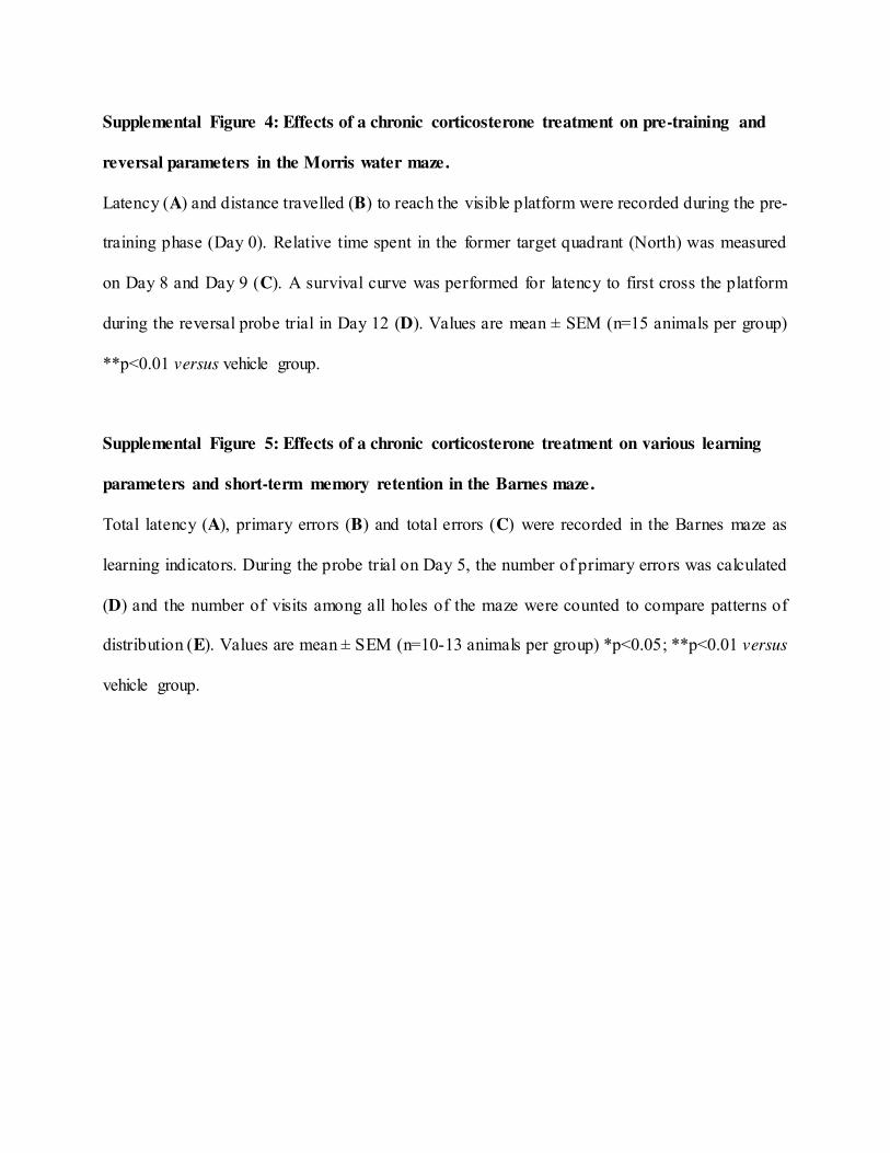
Supplemental Figure 4: Effects of a chronic corticosterone treatment on pre-training and
reversal parameters in the Morris water maze.
Latency (A) and distance travelled (B) to reach the visible platform were recorded during the pre-
training phase (Day 0). Relative time spent in the former target quadrant (North) was measured
on Day 8 and Day 9 (C). A survival curve was performed for latency to first cross the platform
during the reversal probe trial in Day 12 (D). Values are mean ± SEM (n=15 animals per group)
**p<0.01 versus vehicle group.
Supplemental Figure 5: Effects of a chronic corticosterone treatment on various learning
parameters and short-term memory retention in the Barnes maze.
Total latency (A), primary errors (B) and total errors (C) were recorded in the Barnes maze as
learning indicators. During the probe trial on Day 5, the number of primary errors was calculated
(D) and the number of visits among all holes of the maze were counted to compare patterns of
distribution (E). Values are mean ± SEM (n=10-13 animals per group) *p<0.05; **p<0.01 versus
vehicle group.
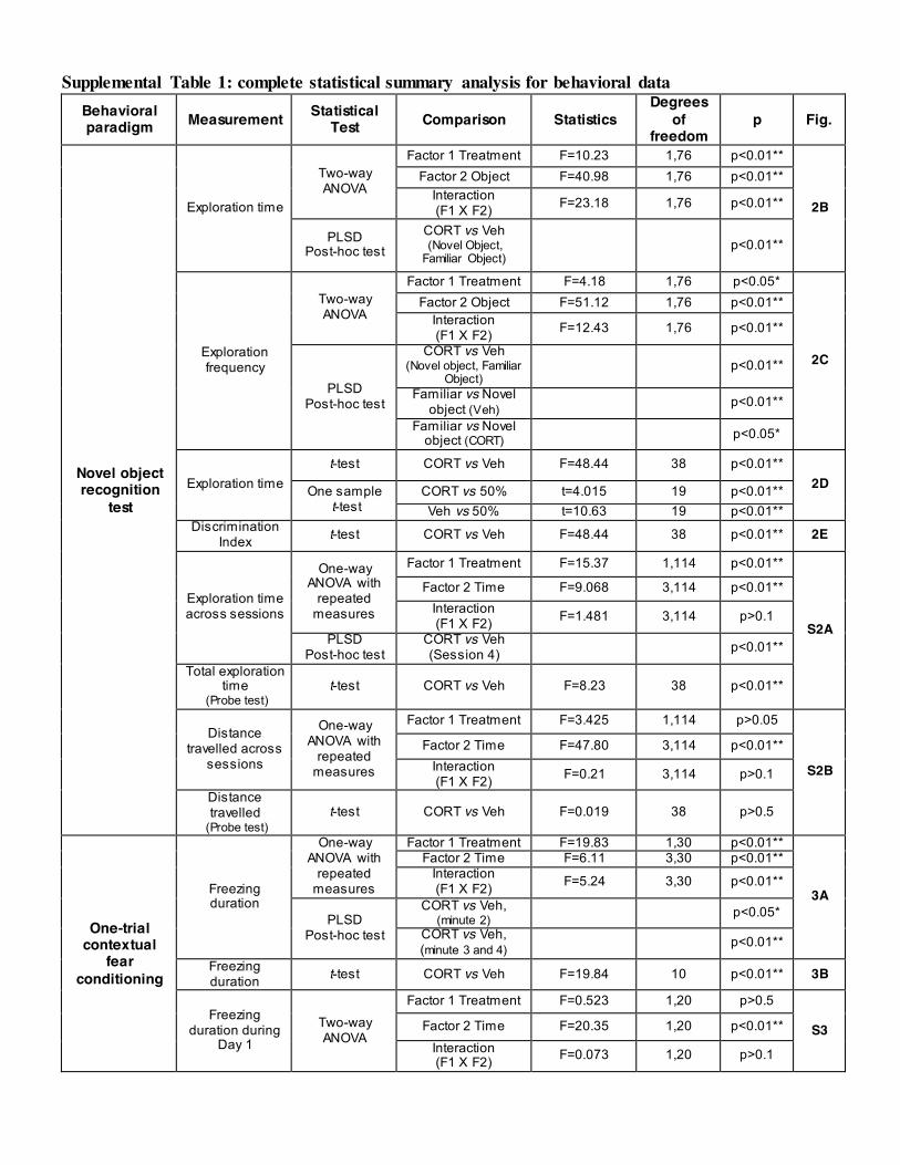
Supplemental Table 1: complete statistical summary analysis for behavioral data Behavioral paradigm Measurement Statistical
Test Comparison Statistics Degrees
of freedom
p Fig.
Novel object recognition
test
Exploration time
Two-way ANOVA
Factor 1 Treatment F=10.23 1,76 p<0.01**
2B
Factor 2 Object F=40.98 1,76 p<0.01** Interaction (F1 X F2) F=23.18 1,76 p<0.01**
PLSD Post-hoc test
CORT vs Veh (Novel Object, Familiar Object)
p<0.01**
Exploration frequency
Two-way ANOVA
Factor 1 Treatment F=4.18 1,76 p<0.05*
2C
Factor 2 Object F=51.12 1,76 p<0.01** Interaction (F1 X F2) F=12.43 1,76 p<0.01**
PLSD Post-hoc test
CORT vs Veh (Novel object, Familiar
Object) p<0.01**
Familiar vs Novel object (Veh) p<0.01**
Familiar vs Novel object (CORT) p<0.05*
Exploration time t-test CORT vs Veh F=48.44 38 p<0.01**
2D One sample t-test
CORT vs 50% t=4.015 19 p<0.01** Veh vs 50% t=10.63 19 p<0.01**
Discrimination Index t-test CORT vs Veh F=48.44 38 p<0.01** 2E
Exploration time across sessions
One-way ANOVA with
repeated measures
Factor 1 Treatment F=15.37 1,114 p<0.01**
S2A
Factor 2 Time F=9.068 3,114 p<0.01** Interaction (F1 X F2) F=1.481 3,114 p>0.1
PLSD Post-hoc test
CORT vs Veh (Session 4) p<0.01**
Total exploration time
(Probe test) t-test CORT vs Veh F=8.23 38 p<0.01**
Distance travelled across
sessions
One-way ANOVA with
repeated measures
Factor 1 Treatment F=3.425 1,114 p>0.05
S2B
Factor 2 Time F=47.80 3,114 p<0.01** Interaction (F1 X F2) F=0.21 3,114 p>0.1
Distance travelled
(Probe test) t-test CORT vs Veh F=0.019 38 p>0.5
One-trial contextual
fear conditioning
Freezing duration
One-way ANOVA with
repeated measures
Factor 1 Treatment F=19.83 1,30 p<0.01**
3A
Factor 2 Time F=6.11 3,30 p<0.01** Interaction (F1 X F2) F=5.24 3,30 p<0.01**
PLSD Post-hoc test
CORT vs Veh, (minute 2) p<0.05*
CORT vs Veh, (minute 3 and 4) p<0.01**
Freezing duration t-test CORT vs Veh F=19.84 10 p<0.01** 3B
Freezing duration during
Day 1
Two-way ANOVA
Factor 1 Treatment F=0.523 1,20 p>0.5
S3 Factor 2 Time F=20.35 1,20 p<0.01**
Interaction (F1 X F2) F=0.073 1,20 p>0.1
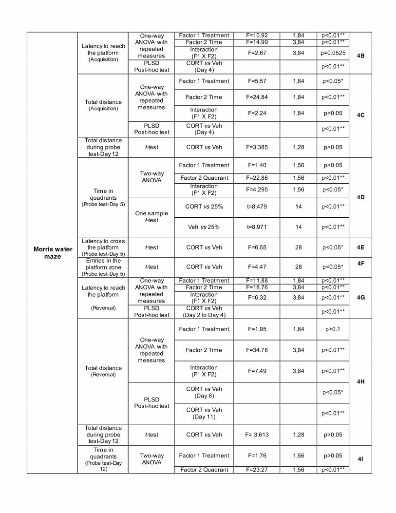
Morris water maze
Latency to reach the platform (Acquisition)
One-way ANOVA with
repeated measures
Factor 1 Treatment F=10.92 1,84 p<0.01**
4B
Factor 2 Time F=14.99 3,84 p<0.01** Interaction (F1 X F2) F=2.67 3,84 p=0.0525
PLSD Post-hoc test
CORT vs Veh (Day 4) p<0.01**
Total distance (Acquisition)
One-way ANOVA with
repeated measures
Factor 1 Treatment F=5.57 1,84 p<0.05*
4C
Factor 2 Time F=24.84 1,84 p<0.01**
Interaction (F1 X F2) F=2.24 1,84 p>0.05
PLSD Post-hoc test
CORT vs Veh (Day 4) p<0.01**
Total distance during probe test-Day 12
t-test CORT vs Veh F=3.385 1,28 p>0.05
Time in quadrants
(Probe test-Day 5)
Two-way ANOVA
Factor 1 Treatment F=1.40 1,56 p>0.05
4D
Factor 2 Quadrant F=22.86 1,56 p<0.01** Interaction (F1 X F2) F=4.295 1,56 p<0.05*
One sample t-test
CORT vs 25%
t=8.479 14 p<0.01**
Veh vs 25%
t=8.971 14 p<0.01**
Latency to cross the platform
(Probe test-Day 5) t-test CORT vs Veh F=6.55 28 p<0.05* 4E
Entries in the platform zone
(Probe test-Day 5) t-test CORT vs Veh F=4.47 28 p<0.05* 4F
Latency to reach the platform
(Reversal)
One-way ANOVA with
repeated measures
Factor 1 Treatment F=11.88 1,84 p<0.01**
4G Factor 2 Time F=18.76 3,84 p<0.01**
Interaction (F1 X F2) F=6.32 3,84 p<0.01**
PLSD Post-hoc test
CORT vs Veh (Day 2 to Day 4) p<0.01**
Total distance (Reversal)
One-way ANOVA with
repeated measures
Factor 1 Treatment F=1.95 1,84 p>0.1
4H
Factor 2 Time F=34.78 3,84 p<0.01**
Interaction (F1 X F2) F=7.49 3,84 p<0.01**
PLSD Post-hoc test
CORT vs Veh (Day 8) p<0.05*
CORT vs Veh (Day 11) p<0.01**
Total distance during probe test-Day 12
t-test CORT vs Veh F= 3.613 1,28 p>0.05
Time in quadrants
(Probe test-Day 12)
Two-way ANOVA
Factor 1 Treatment F=1.76 1,56 p>0.05 4I
Factor 2 Quadrant F=23.27 1,56 p<0.01**
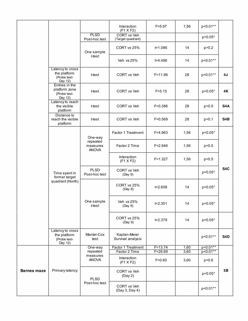
Interaction (F1 X F2)
F=5.97 1,56 p<0.01**
PLSD Post-hoc test
CORT vs Veh (Target quadrant) p<0.05*
One sample t-test
CORT vs 25%
t=1.086 14 p>0.2
Veh vs 25%
t=4.496 14 p<0.01**
Latency to cross the platform (Probe test-
Day 12)
t-test CORT vs Veh F=11.96 28 p<0.01** 4J
Entries in the platform zone
(Probe test- Day 12)
t-test CORT vs Veh F=5.15 28 p<0.05* 4K
Latency to reach the visible platform
t-test CORT vs Veh F=0.386 28 p>0.5 S4A
Distance to reach the visible
platform t-test CORT vs Veh F=0.569 28 p>0.1 S4B
Time spent in former target
quadrant (North)
One-way repeated
measures ANOVA
Factor 1 Treatment F=4.963 1,56 p<0.05*
S4C
Factor 2 Time F=2.946 1,56 p>0.5
Interaction (F1 X F2) F=1.327 1,56 p>0.5
PLSD Post-hoc test
CORT vs Veh (Day 9) p<0.05*
One sample t-test
CORT vs 25%
(Day 8)
t=2.658 14 p<0.05*
Veh vs 25%
(Day 8)
t=2.301 14 p<0.05*
CORT vs 25%
(Day 9)
t=2.379 14 p<0.05*
Latency to cross the platform (Probe test-
Day 12)
Mantel-Cox test
Kaplan-Meier Survival analysis p<0.01** S4D
Barnes maze Primary latency
One-way repeated
measures ANOVA
Factor 1 Treatment F=13.74 1,60 p<0.01**
5B
Factor 2 Time F=26.69 3,60 p<0.01**
Interaction (F1 X F2) F=0.60 3,60 p>0.6
PLSD Post-hoc test
CORT vs Veh (Day 2) p<0.05*
CORT vs Veh (Day 3, Day 4) p<0.01**
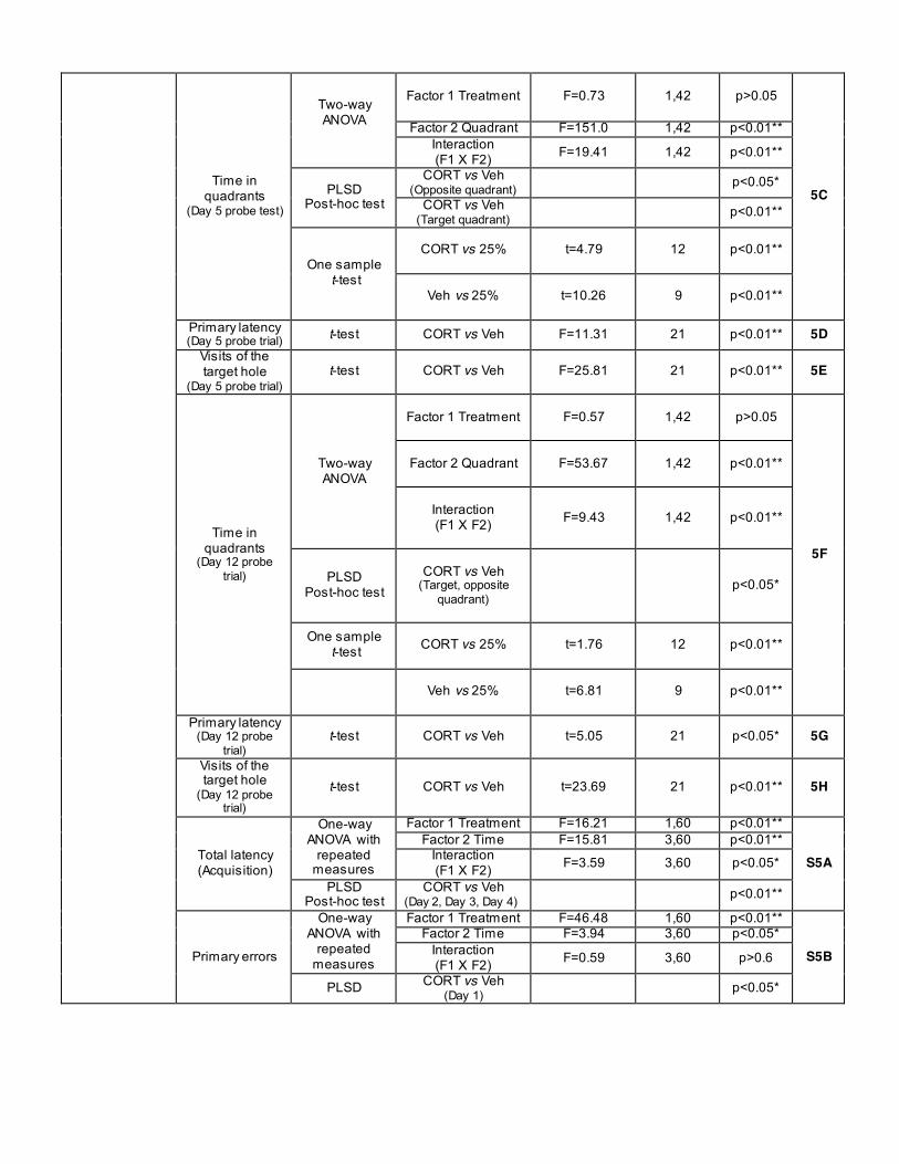
Time in quadrants
(Day 5 probe test)
Two-way ANOVA
Factor 1 Treatment F=0.73 1,42 p>0.05
5C
Factor 2 Quadrant F=151.0 1,42 p<0.01** Interaction (F1 X F2) F=19.41 1,42 p<0.01**
PLSD Post-hoc test
CORT vs Veh (Opposite quadrant) p<0.05*
CORT vs Veh (Target quadrant) p<0.01**
One sample t-test
CORT vs 25%
t=4.79 12 p<0.01**
Veh vs 25%
t=10.26 9 p<0.01**
Primary latency (Day 5 probe trial) t-test CORT vs Veh F=11.31 21 p<0.01** 5D
Visits of the target hole
(Day 5 probe trial) t-test CORT vs Veh F=25.81 21 p<0.01** 5E
Time in quadrants
(Day 12 probe trial)
Two-way ANOVA
Factor 1 Treatment
F=0.57 1,42 p>0.05
5F
Factor 2 Quadrant
F=53.67 1,42 p<0.01**
Interaction (F1 X F2)
F=9.43 1,42 p<0.01**
PLSD Post-hoc test
CORT vs Veh
(Target, opposite quadrant)
p<0.05*
One sample t-test
CORT vs 25%
t=1.76 12 p<0.01**
Veh vs 25%
t=6.81 9 p<0.01**
Primary latency (Day 12 probe
trial) t-test CORT vs Veh t=5.05 21 p<0.05* 5G
Visits of the target hole
(Day 12 probe trial)
t-test CORT vs Veh t=23.69 21 p<0.01** 5H
Total latency (Acquisition)
One-way ANOVA with
repeated measures
Factor 1 Treatment F=16.21 1,60 p<0.01**
S5A Factor 2 Time F=15.81 3,60 p<0.01**
Interaction (F1 X F2) F=3.59 3,60 p<0.05*
PLSD Post-hoc test
CORT vs Veh (Day 2, Day 3, Day 4) p<0.01**
Primary errors
One-way ANOVA with
repeated measures
Factor 1 Treatment F=46.48 1,60 p<0.01**
S5B Factor 2 Time F=3.94 3,60 p<0.05*
Interaction (F1 X F2) F=0.59 3,60 p>0.6
PLSD CORT vs Veh (Day 1) p<0.05*
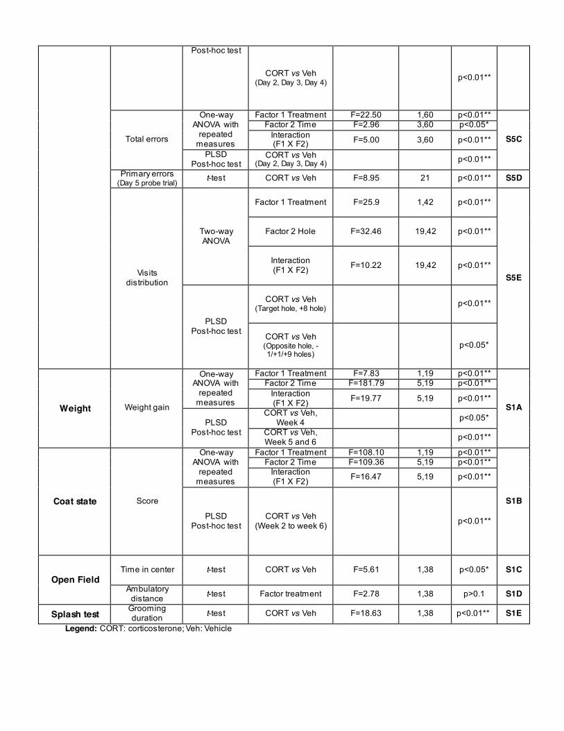
Post-hoc test
CORT vs Veh (Day 2, Day 3, Day 4) p<0.01**
Total errors
One-way ANOVA with
repeated measures
Factor 1 Treatment F=22.50 1,60 p<0.01**
S5C Factor 2 Time F=2.96 3,60 p<0.05*
Interaction (F1 X F2) F=5.00 3,60 p<0.01**
PLSD Post-hoc test
CORT vs Veh (Day 2, Day 3, Day 4) p<0.01**
Primary errors (Day 5 probe trial) t-test CORT vs Veh F=8.95 21 p<0.01** S5D
Visits distribution
Two-way ANOVA
Factor 1 Treatment
F=25.9 1,42 p<0.01**
S5E
Factor 2 Hole
F=32.46 19,42 p<0.01**
Interaction (F1 X F2)
F=10.22 19,42 p<0.01**
PLSD Post-hoc test
CORT vs Veh
(Target hole, +8 hole)
p<0.01**
CORT vs Veh (Opposite hole, -1/+1/+9 holes)
p<0.05*
Weight Weight gain
One-way ANOVA with
repeated measures
Factor 1 Treatment F=7.83 1,19 p<0.01**
S1A
Factor 2 Time F=181.79 5,19 p<0.01** Interaction (F1 X F2) F=19.77 5,19 p<0.01**
PLSD Post-hoc test
CORT vs Veh, Week 4 p<0.05*
CORT vs Veh, Week 5 and 6 p<0.01**
Coat state Score
One-way ANOVA with
repeated measures
Factor 1 Treatment F=108.10 1,19 p<0.01**
S1B
Factor 2 Time F=109.36 5,19 p<0.01** Interaction (F1 X F2) F=16.47 5,19 p<0.01**
PLSD Post-hoc test
CORT vs Veh (Week 2 to week 6) p<0.01**
Open Field Time in center t-test
CORT vs Veh
F=5.61 1,38 p<0.05* S1C
Ambulatory distance t-test Factor treatment F=2.78 1,38 p>0.1 S1D
Splash test Grooming duration t-test CORT vs Veh F=18.63 1,38 p<0.01** S1E
Legend: CORT: corticosterone; Veh: Vehicle
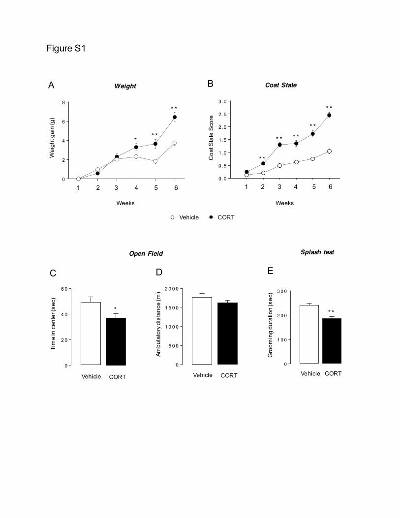
Figure S1
C
Open Field
D
Splash test
Tim
e in
cen
ter (
sec)
Gro
omin
g du
ratio
n (s
ec)
Vehicle CORT Vehicle CORT 0
2 0
4 0
6 0
*
0
2 0
4 0
6 0
*
0
5 0 0
1 0 0 0
1 5 0 0
2 0 0 0
*
0
5 0 0
1 0 0 0
1 5 0 0
2 0 0 0
*
Vehicle CORT
Ambu
lato
ry d
ista
nce
(m)
E
0
1 0 0
2 0 0
3 0 0
* *
0
1 0 0
2 0 0
3 0 0
* *
A Weight
Wei
ght g
ain
(g)
Weeks
1 2 3 4 5 6 0
2
4
6
8* *
* **
B Coat State
Coa
t Sta
te S
core
1 2 3 4 5 6
Weeks
0 .0
0 .5
1 .0
1 .5
2 .0
2 .5
3 .0
* *
* * * ** *
* *
Vehicle CORT
0 .0
0 .5
1 .0
1 .5
2 .0
2 .5
3 .0
* *
* * * ** *
* *V e h ic le C O R T
0 .0
0 .5
1 .0
1 .5
2 .0
2 .5
3 .0
* *
* * * ** *
* *V e h ic le C O R T
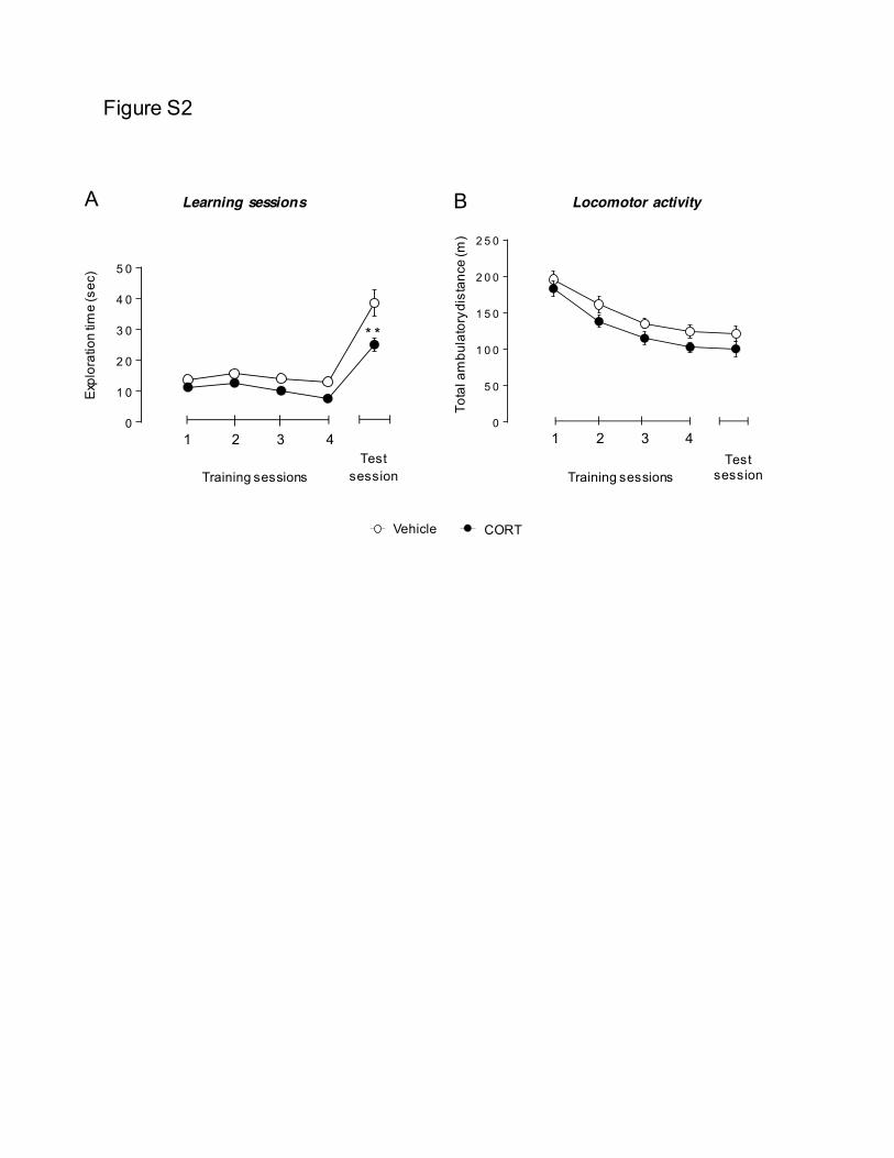
Figure S2
C
Novel Object
B Locomotor activity
1 2 3 4
Training sessions Test
session
0
5 0
1 0 0
1 5 0
2 0 0
2 5 0
Tota
l am
bula
tory
dis
tanc
e (m
)
A Learning sessions
0
1 0
2 0
3 0
4 0
5 0
* *
1 2 3 4
Training sessions Test
session
Exp
lora
tion
time
(sec
)
0 .0
0 .5
1 .0
1 .5
2 .0
2 .5
3 .0
* *
* * * ** *
* *V e h ic le C O R T
0 .0
0 .5
1 .0
1 .5
2 .0
2 .5
3 .0
* *
* * * ** *
* *V e h ic le C O R TVehicle CORT
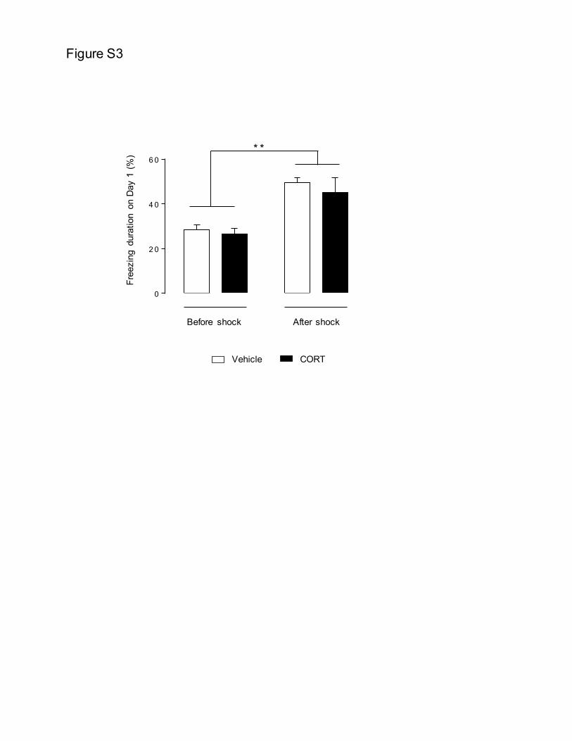
Figure S3
0
2 0
4 0
6 0* *
Before shock After shock
Free
zing
dur
atio
n on
Day
1 (
%)
0
1 0
2 0
3 0
4 0
5 0
6 0
*
V e h ic le C O R TVehicle CORT
0
1 0
2 0
3 0
4 0
5 0
6 0
*
V e h ic le C O R T
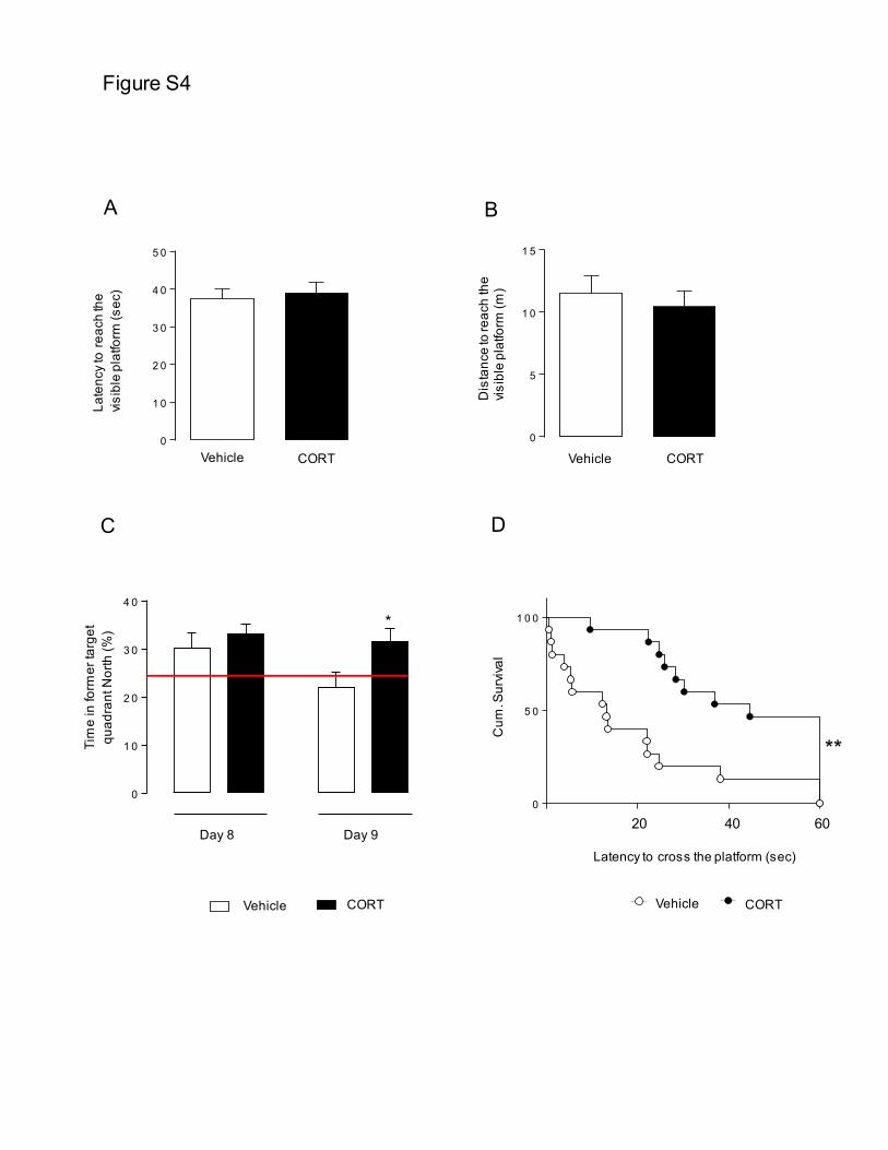
Figure S4
B A
Late
ncy
to r
each
the
visib
le p
latfo
rm (s
ec)
0
1 0
2 0
3 0
4 0
5 0
0
5
1 0
1 5
Dis
tanc
e to
reac
h th
e vi
sibl
e pl
atfo
rm (m
)
Vehicle CORT Vehicle CORT
**
0
5 0
1 0 0
D
20 40 60
Latency to cross the platform (sec)
Cum
. Sur
vival
0 .0
0 .5
1 .0
1 .5
2 .0
2 .5
3 .0
* *
* * * ** *
* *V e h ic le C O R T
0 .0
0 .5
1 .0
1 .5
2 .0
2 .5
3 .0
* *
* * * ** *
* *V e h ic le C O R TVehicle CORT
0
1 0
2 0
3 0
4 0
*
C
Tim
e in
form
er ta
rget
qu
adra
nt N
orth
(%)
Day 8 Day 9
0
1 0
2 0
3 0
4 0
5 0
6 0
*
V e h ic le C O R TVehicle CORT
0
1 0
2 0
3 0
4 0
5 0
6 0
*
V e h ic le C O R T
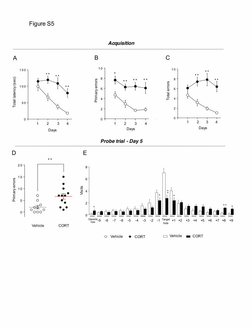
Figure S5
1 2 3 4
Target hole
A
Tota
l lat
ency
(sec
)
Prim
ary
erro
rs
Tota
l err
ors
B C
Acquisition
0
2
4
6
8
1 0
* ** *
* *
0
5 0
1 0 0
1 5 0* * * *
* *
0
2
4
6
8
1 0
*
* * * * * *
0
1 0
2 0
3 0
4 0
5 0
6 0
*
V e h ic le C O R T
Prim
ary
erro
rs
D
0
5
1 0
1 5
2 0* *
E
Visi
ts
Probe trial - Day 5
Vehicle CORT -9 -8 -7 -6 -5 -4 -3 -2 -1 +1 +2 +3 +4 +5 +6 +7 +8 +9 Opposite
hole
0
2
4
6
8
**
**
** **
0
1 0
2 0
3 0
4 0
5 0
6 0
*
V e h ic le C O R T
0 .0
0 .5
1 .0
1 .5
2 .0
2 .5
3 .0
* *
* * * ** *
* *V e h ic le C O R T
0 .0
0 .5
1 .0
1 .5
2 .0
2 .5
3 .0
* *
* * * ** *
* *V e h ic le C O R TVehicle CORT Vehicle CORT
1 2 3 4 1 2 3 4
Days Days Days
Résultats expérimentaux
228
Commentaires sur Article 1 :
L’ensemble de ces résultats suggère qu’une souris développant un phénotype anxio/dépressif, suite
à l’exposition prolongée à une faible dose de corticostérone exogène, présente un déficit cognitif
global, incluant une altération de la mémoire de type épisodique, une dégradation des performances
spatiales dont la capacité de flexibilité mentale et une diminution de la mémoire associative.
D’autre part, si l’efficacité d’un traitement chronique par la fluoxetine pour restaurer le phénotype
anxio/dépressif n’est plus à démontrer, ses activités sur les performances cognitives n’ont à ce jour
pas encore évaluées dans ce modèle animal d’anxiété/dépression.
A l’inverse, l’efficacité des agonistes du récepteur 5-HT4 dans les troubles cognitifs n’est plus à
prouver. Mais si de nombreuses études ont évalué l’activité pro-cognitive et de type antidépressive
des agonistes du récepteur 5-HT4 chez des animaux naïfs, aucune n’a à ce jour examiné leur activité
dans un modèle d’anxiété/dépression.

229
Résultats expérimentaux
230
ARTICLE 2: Rapid anxiolytic effects of a 5-HT4 receptor agonist are mediated
by a neurogenesis-independent mechanism
Indira Mendez-David, Denis J David, Flavie Darcet, Melody V Wu, Saadia Kerdine-Römer, Alain M
Gardier, René Hen
Publié dans Neuropsychopharmacology, Nov 28. doi: 10.1038/npp.2013.332 (2014)
Question posée :
Une administration subchronique et chronique d’un agoniste du récepteur 5-HT4, le RS67333, est-
elle capable de restaurer le phénotype anxio/dépressif induit par l’exposition prolongée de
corticostérone ?
Résumé de l’étude :
Les inhibiteurs sélectifs de recapture de sérotonine (ISRSs) sont les médicaments les plus prescrits
dans le traitement de la dépression caractérisée. Malheureusement, l’apparition des effets
thérapeutiques des ISRSs est souvent retardée de 3 à 6 semaines après le début du traitement.
D’autre part, seulement un tiers des patients ne répondent pas au traitement initialement prescrit.
Ces problématiques accentuent la nécessité d’identifier des antidépresseurs agissant plus
rapidement et qui soient plus efficace (Samuels et al., 2011). Il a été proposé que les agonistes du
récepteurs 5-HT4 tel que le RS67333 pourraient être un élément intéressant dans le traitement de la
dépression (Lucas, 2009; Lucas et al., 2005; Lucas and Debonnel, 2002; Lucas et al., 2010; Lucas et al.,
2007). Alors qu’un grand nombre d’études ont évalué l’activité de type antidépressive des agonistes
des récepteurs 5-HT4 chez des souris naïves, aucune n’a à ce jour examiné leur activité dans un
modèle d’anxiété/dépression. Dans cette étude, nous avons donc examiné si l’activation du
récepteur 5-HT4 par un agoniste, le RS67333, pouvait corriger le phénotype « anxio/dépressif » induit
par le modèle CORT chez les souris. Enfin, en utilisant ce même modèle d’anxiété/dépression
combiné à une ablation de la neurogenèse hippocampique par irradiation, nous avons déterminé si
l’action de type anxiolytique et antidépressive du RS67333 après 7 et 28 jours de traitement recrutait
un mécanisme dépendant de la neurogenèse hippocampique.
Résultats expérimentaux
231
Contribution personnelle :
Au cours de ce travail :
x J’ai participé au suivi des animaux incluant la préparation et administration des différents
traitements pharmacologiques (traitement par la CORT et par la fluoxetine, préparation et
implantation des mini-pompes pour RS67333 et GR125487).
x J’ai participé à l’ensemble des tests comportementaux pour la cohorte des animaux traités
par la corticostérone.
x J’ai participé aux analyses immunohistochimiques des étapes de la neurogenèse
hippocampique depuis la perfusion, le prélèvement et la coupe des cerveaux jusqu’aux
marquages des cellules.
x J’ai participé à la rédaction des données statistiques de l’article.
Rapid Anxiolytic Effects of a 5-HT4 Receptor Agonist AreMediated by a Neurogenesis-Independent Mechanism
Indira Mendez-David1,5, Denis J David1,5, Flavie Darcet1, Melody V Wu2,3, Saadia Kerdine-Romer4,Alain M Gardier1 and Rene Hen*,2,3
1Univ Paris Sud, EA3544, Faculte de Pharmacie, Chatenay-Malabry, France; 2Departments of Neuroscience and Psychiatry, Columbia University,New York, NY, USA; 3Department of Integrative Neuroscience, New York State Psychiatric Institute, New York, NY, USA; 4Univ Paris Sud,INSERM UMR 996, Faculte de Pharmacie, Chatenay-Malabry, France
Selective serotonin reuptake inhibitors (SSRIs) display a delayed onset of action of several weeks. Past work in naive rats showed that 5-HT4 receptor agonists had rapid effects on depression-related behaviors and on hippocampal neurogenesis. We decided to investigatewhether 5-HT4 receptor stimulation was necessary for the effects of SSRIs in a mouse model of anxiety/depression, and whetherhippocampal neurogenesis contributed to these effects. Using the mouse corticosterone model of anxiety/depression, we assessedwhether chronic treatment with a 5-HT4 receptor agonist (RS67333, 1.5 mg/kg/day) had effects on anxiety- and depression-relatedbehaviors, as well as on hippocampal neurogenesis in comparison with chronic fluoxetine treatment (18 mg/kg/day). Then, using ouranxiety/depression model combined with ablation of hippocampal neurogenesis, we investigated whether neurogenesis was necessaryfor the behavioral effects of subchronic (7 days) or chronic (28 days) RS67333 treatment. We also assessed whether a 5-HT4 receptorantagonist (GR125487, 1 mg/kg/day) could prevent the behavioral and neurogenic effects of fluoxetine. Chronic treatment withRS67333, similar to fluoxetine, induced anxiolytic/antidepressant-like activity and stimulated adult hippocampal neurogenesis, specificallyfacilitating maturation of newborn neurons. However, unlike fluoxetine, anxiolytic effects of RS67333 were already present after 7 daysand did not require hippocampal neurogenesis. Chronic treatment with GR125487 prevented both anxiolytic/antidepressant-like andneurogenic effects of fluoxetine, indicating that 5-HT4 receptor activation is necessary for these effects of SSRIs. 5-HT4 receptorstimulation could represent an innovative and rapid onset therapeutic approach to treat depression with comorbid anxiety.Neuropsychopharmacology advance online publication, 22 January 2014; doi:10.1038/npp.2013.332
Keywords: 5-HT4 receptor; anxiolytic; neurogenesis; fast onset
!!!!!!!!!!!!!!!!!!!!!!!!!!!!!!!!!!!!!!!!!!!!!!!!!!!!!!
INTRODUCTION
Selective serotonin reuptake inhibitors (SSRIs) are the mostcommonly prescribed drugs for the treatment of depressionand several anxiety disorders. Unfortunately, the onset ofaction of SSRIs is often delayed by 3–6 weeks (Artigas,2013). The existence of this delayed action combined withthe fact that one-third of patients do not respond totreatment emphasizes the need for faster acting and moreeffective antidepressants (Samuels et al, 2011).
It has been proposed that 5-HT4 receptor agonists such asRS67333 may bring new hope for treating depression(Lucas, 2009; Lucas et al, 2005; Lucas and Debonnel, 2002;Lucas et al, 2010; Lucas et al, 2007). Indeed, administrationof 5-HT4 agonists induced similar molecular and behavioralchanges as common antidepressants in rodents (Bockaert
et al, 2008; Lucas et al, 2007; Pascual-Brazo et al, 2012).Depressed-like state in the olfactory bulbectomy or chronicmild stress model was completely abolished after 10–14days of RS67333 treatment in rats, suggesting a more rapidresponse mechanism in comparison with classical anti-depressants (Lucas et al, 2007). A positive behavioralresponse in the Novelty-Suppressed Feeding (NSF) test inrat, a complete reversion of anhedonic-like state (sucroseconsumption), and an increase in swimming behavior indefeated mice in the forced swim test were also observedafter a short period of RS67333 treatment (Gomez-Lazaroet al, 2012; Pascual-Brazo et al, 2012). In addition tobehavioral data, and in agreement with a previous reportfrom Lucas et al (2007), a recent study performed in naiverats confirmed that a short period of treatment withRS67333 increased the number of newborn cells in thedentate gyrus (DG) (Pascual-Brazo et al, 2012). Theseresults are interesting because hippocampal neurogenesishas been implicated in some of the behavioral effects ofantidepressants in adult rodents (David et al, 2009;Santarelli et al, 2003). However, no direct evidence has yetlinked the antidepressant-like effects of 5-HT4 receptoractivation and its neurogenic effects. Finally, it has been
*Correspondence: Professor Rene Hen, Departments of Neuroscienceand Psychiatry, Columbia University, New York, NY 10032, USA, Tel:+1 212 543 5328, Fax: +1 212 543 5074, E-mail: [email protected] authors contributed equally to this work.Received 6 September 2013; revised 16 November 2013; accepted 20November 2013; accepted article preview online 28 November 2013
Neuropsychopharmacology (2014), 1–13
& 2014 American College of Neuropsychopharmacology. All rights reserved 0893-133X/14
www.neuropsychopharmacology.org
suggested that SSRIs and 5-HT4 receptor agonists sharecommon mechanisms of action. Indeed, the 5-HT4 receptoragonist, RS67333, augmented the acute effect of paroxetineon extracellular 5-HT levels in rat ventral hippocampus, andafter only 3 days of administration it increased basalhippocampal 5-HT levels (Licht et al, 2010). The coadmi-nistration of the SSRI citalopram and RS67333 stronglypotentiated the antidepressant-like properties of the latterin several electrophysiological, molecular, and behavioralparadigms (Lucas et al, 2010).
Although a number of studies have assessed the anti-depressant-like activity of 5-HT4 receptor agonists, nonehave so far evaluated their anxiolytic-like profile. It isnoteworthy that some SSRIs are often prescribed for thetreatment of anxiety disorders (Burghardt and Bauer, 2013).Anxiety disorders have a lifetime prevalence of over 25%,thus making them the most common psychiatric disorders(Kheirbek et al, 2012). Moreover, a comorbidity betweendepression and anxiety disorders is commonly observed.Thus, this study aimed to investigate both antidepressantand anxiolytic-like effects of either subchronic or chronicadministration of a 5-HT4 receptor agonist in a model ofanxiety/depression based on the elevation of glucocorticoidsin mice (CORT model) (David et al, 2009). Standard modelsof depression that rely on environmental stress manipula-tions such as learned helplessness or the chronic mild stressare hampered by protocol variability and reported difficul-ties in replication, thus highlighted the need for a reliable,easily replicable depression model (Nestler et al, 2002). Thecorticosterone model is a chronic exposure method opti-mized for use in modeling the persistent anxiety/depression-like state in rodents, allowing for multiple behavioral tests inthe same animals using an etiologically relevant model ofdepression that is easily replicable between and withinlaboratories (David et al, 2009; David et al, 2010; Gould,2011; Mendez-David et al, 2013).
We also assessed whether chronic 5-HT4 receptorstimulation can affect proliferation of newborn cells andmaturation of newborn neurons. Finally, using our mousemodel of anxiety/depression combined with ablation ofhippocampal neurogenesis by X-irradiation, we assessedwhether the anxiolytic/antidepressant action of RS67333after 7 and 28 days of treatment recruits a neurogenesis-dependent mechanism.
MATERIALS AND METHODS
Subjects
Adult male C57BL/6Ntac mice were purchased fromTaconic Farms (Lille Skensved, Denmark and Germantown,NY, USA for the pharmacological and the X-irradiationstudies, respectively). All mice were 7–8 weeks old, weighed23–25 g at the beginning of the treatment, and weremaintained on a 12 L:12 D schedule (lights on at 0600hours). They were housed in groups of five. Food and waterwere provided ad libitum. All testing was conducted incompliance with the laboratory animal care guidelines andwith protocols approved by the Institutional Animal Careand Use Committee (Council directive no. 87-848, October19, 1987, Ministere de l’Agriculture et de la Foret, Service
Veterinaire de la Sante et de la Protection Animale,permissions no. 92-256B to DJD).
Drugs
Corticosterone [4-pregnen-11b-DIOL-3 20-DIONE 21-hemi-succinate (35mg/ml)] purchased from Sigma-Aldrich (Saint-Quentin Fallavier, France) was dissolved in a vehicle (0.45%hydroxypropyl-b-cyclodextrin (b-CD); Sigma-Aldrich). Fluo-xetine hydrochloride (160mg/ml, equivalent to 18 mg/kg/day)was purchased from Anawa Trading, (Zurich, Switzerland)and dissolved in 0.45% b-CD/corticosterone solution. 1-(4-amino-5-chloro-2-methoxyphenyl)-3-(1-butyl-4piperidinyl)-1-propanone hydrochloride (RS67333), and 5-Fluoro-2-methoxy-[1-[2-[(methylsulfonyl) amino]ethyl]-4-piperidi-nyl]-1H-indole-3-methylcarboxylate sulfamate (GR125487)were purchased from Tocris Bioscience (Bristol, UK) anddissolved in 0.9% saline solution. RS67333 and GR125487were chosen based on previous work (Cachard-Chastel et al,2007; Lucas et al, 2007).
RS67333 shows high-binding affinity for the 5-HT4
receptor with a pKi of 8.7 (Bockaert et al, 2004; Eglenet al, 1995). Except for the sigma receptors, which arebound at affinities comparable to 5-HT4 (sigma 1: pKi¼ 8.9and sigma 2: pKi¼ 8.0), RS67333 has a pKi of o6.7 forother neurotransmitter receptors including 5-HT1A, 5-HT1D,5-HT2A, 5-HT2C, dopamine D1, D2, and muscarinic M1–M3
receptors. However, little is known about the function ofsigma receptors’.
GR125487 is the most selective 5-HT4 receptor antagonistwith a pKi¼ 10.6 (Schiavi et al, 1994), presenting aselectivity more than 1000-fold over other 5-HT receptor(Gale et al, 1994). The dose of RS67333 and GR125487 usedin this study were chosen based on previous works(Cachard-Chastel et al, 2007; Lucas et al, 2007).
Corticosterone Model and Treatment
Our model of elevated glucocorticoids (also named CORTmodel) is able to blunt the response of the hypothalamic–pituitary–adrenal axis as shown by the markedly attenuatedstress-induced corticosterone levels observed in these mice(David et al, 2009). This is probably a consequence of thenegative feedback exerted by corticosterone on thehypothalamic–pituitary–adrenal axis. This model displayshallmark characteristics of anxiety and depression.
The dose and duration of corticosterone treatment wasselected based on previous studies (David et al, 2009; Raineret al, 2011). Corticosterone (35 mg/ml, equivalent to about5 mg/kg/day) or vehicle (0.45% b-CD) was available adlibitum in the drinking water in opaque bottles to protect itfrom light. Corticosterone-treated water was changed every3 days in order to prevent any possible degradation.Thereafter, while administration with b-CD or corticoster-one continued, mice were treated with vehicle (0.45% b-CD), fluoxetine, RS67333, GR125487 alone, or GR125487 inthe presence of fluoxetine (Supplementary Figure 1). BothRS67333 and GR125487 were delivered by osmotic mini-pumps at a dose of 1.5 mg/kg/day and 1 mg/kg/day,respectively (Lucas et al, 2005). Fluoxetine (18 mg/kg/day)was delivered in the drinking water as previously described(David et al, 2009). Osmotic minipumps (42 days minipumps,
5-HT4 receptor contribution to antidepressant responseI Mendez-David et al
2
Neuropsychopharmacology
2006 model, Alzet, Cupertino, CA) were implanted sub-cutaneously under light anesthesia (ketamine/xylazine;(75/20 mg/kg) from Sigma-Aldrich. Control animals (vehi-cle/vehicle or corticosterone/vehicle groups) were alsoimplanted with a minipump containing 0.9% saline (2006model, Alzet). Treatment was always maintained until theend of the experiments. Corticosterone and fluoxetinedosages were calculated assuming an average fluid intakeof about 5 ml/day (David et al, 2009).
Behavioral Tests
The same cohort of animals was tested in five differentbehavioral models of anxiety and depression. Each animal,over a week, was successively tested in the Open Field (OF),Elevated Plus Maze (EPM), NSF, Splash Test (ST), and TailSuspension Test (Supplementary Material). Behavioral test-ing occurred during the light phase between 0700 and 1900hours. Behavioral paradigms occurred after 7 or 28 days ofdrug treatment depending on the study (SupplementaryFigure S1A and S1B).
Immunohistochemistry
The effects of chronic RS67333 treatment on cell prolifera-tion or maturation of newborn neurons was assessed incorticosterone-treated animals. After anesthesia with keta-mine and xylazine (100 mg/ml ketamine and 20 mg/mlxylazine), mice were perfused transcardially (cold saline for2 min, followed by 4% cold paraformaldehyde at 4 1C). Thebrains were then removed and cryoprotected in 30%sucrose and stored at 4 1C. Serial sections (35 mm) werecryosectioned through the entire hippocampus and storedin PBS with 0.1% NaN3.
Proliferation of newborn cells. We first looked atproliferation of newborn cells using Ki-67 immunohisto-chemistry as described previously (Xia et al, 2012). Sectionswere washed in PBS, blocked (PBS containing 0.3% TritonX-100 and 10% normal donkey serum (NDS)), and incubatedwith primary antibody overnight at 4 1C (Ki67 rabbit, 1 : 100,Vector, Burlingame, CA). Following washes in PBS, sectionswere incubated with fluorescence-coupled rabbit secondaryantibody (Jackson ImmunoResearch, Beckman, France).Stereological quantification of Ki-67 labeling was performedusing an Olympus BX51 microscope (Germany).
Maturation of newborn neurons. For doublecortin(DCX) staining, the procedure consisted of the followingsteps: 1 h incubation in 0.1 M TBS with 0.5% Triton X-100and 10% NDS, followed by goat anti-DCX primary antibody(1 : 100) in TBS/Tx/NDS for 24 h at 4 1C. Biotinylatedsecondary donkey anti-goat antibody (1 : 500) in TBS/NDSfor 1 h at room temperature was used, followed by a 1 hamplification step using an avidin–biotin complex (Vector).The immunohistochemistry protocol was adapted fromDavid et al (2009). DCX-positive (DCXþ ) cells weresubcategorized according to their dendritic morphology:DCXþ cells without and DCXþ cells with tertiary (or higherorder) dendrites. The maturation index was defined as theratio of DCXþ cells possessing tertiary dendrites to the totalnumber of DCXþ cells.
Sholl analysis. Sholl analysis was performed as describedelsewhere (Guilloux et al, 2013). DCXþ cells with tertiary orhigher order dendrites were traced using Neurolucidasoftware (MicroBrightField, Williston, VT) on an OlympusBX51 microscope equipped with a motorized stage deviceand " 100 immersion oil objective. Sholl analysis fordendritic complexity was performed using the accompany-ing software (NeuroExplorer; MicroBrightField, version 10)to determine dendritic length and number of intersections(branch points). One DCXþ cell was traced for each 35-mmhippocampal slice; n¼ 6 cells/brain for DAB-stainedsections).
X-irradiation
A separate batch of mice were anesthetized with ketamineand xylazine (75/20 mg/kg), placed in a stereotaxic frame,and exposed to cranial irradiation using a PXI X-RAD 320X-ray system operated at 300 kV and 12 mA with a 2-mm Alfilter. Animals were protected with a lead shield thatcovered the entire body, but left unshielded a 3.22" 11-mmtreatment field above the hippocampus (interaural 3.00 mmto 0.00 mm) exposed to X-ray, thus effectively preventingirradiation from targeting the rest of the brain (Santarelliet al, 2003). The corrected dose rate was B0.95 Gy/min at asource to skin distance of 36 cm. The procedure lasted for2 min and 39 s, delivering a total of 2.5 Gy. Three 2.5 Gydoses were delivered on days 1, 4, and 7 as previouslydescribed (Quesseveur et al, 2013). This 7.5 Gy cumulativedose was determined from prior pilot experiments to be theminimum dosage necessary to result in permanent ablationof adult-born neurons in the DG as assessed by expressionof the immature neuronal marker DCX. The reason forusing a fractionated paradigm rather than a single high doseof 7.5 Gy is that the ablation is not permanent after asingle high dose. Histological staining for CD68 as a markerof inflammation throughout the brain revealed thatirradiated mice were indistinguishable from shamanimals 8 weeks post irradiation, indicating minimalnonspecific side effects of irradiation at time of behavioraltesting (Meshi et al, 2006). Immunohistochemistry con-firmed the ablation of adult hippocampal neurogenesis(Supplementary Figure 4).
Data Analysis and Statistics
Results from data analyses were expressed as mean±SEM.Data were analyzed using StatView 5.0 software (SASInstitute, Cary, NC). For all experiments, one-way or two-way ANOVAs with repeated measures were applied to thedata as appropriate. Significant main effects and/or inter-actions were followed by Fisher’s PLSD post hoc analysis,unpaired t-tests. In the NSF test, we used the Kaplan–Meiersurvival analysis owing to the lack of normal distribution ofthe data. Mantel–Cox log rank test was used to evaluatedifferences between experimental groups. Statistical sig-nificance was set at po0.05. A summary of statisticalmeasures is included in Supplementary Tables S1–S6,available online.
5-HT4 receptor contribution to antidepressant responseI Mendez-David et al
3
Neuropsychopharmacology
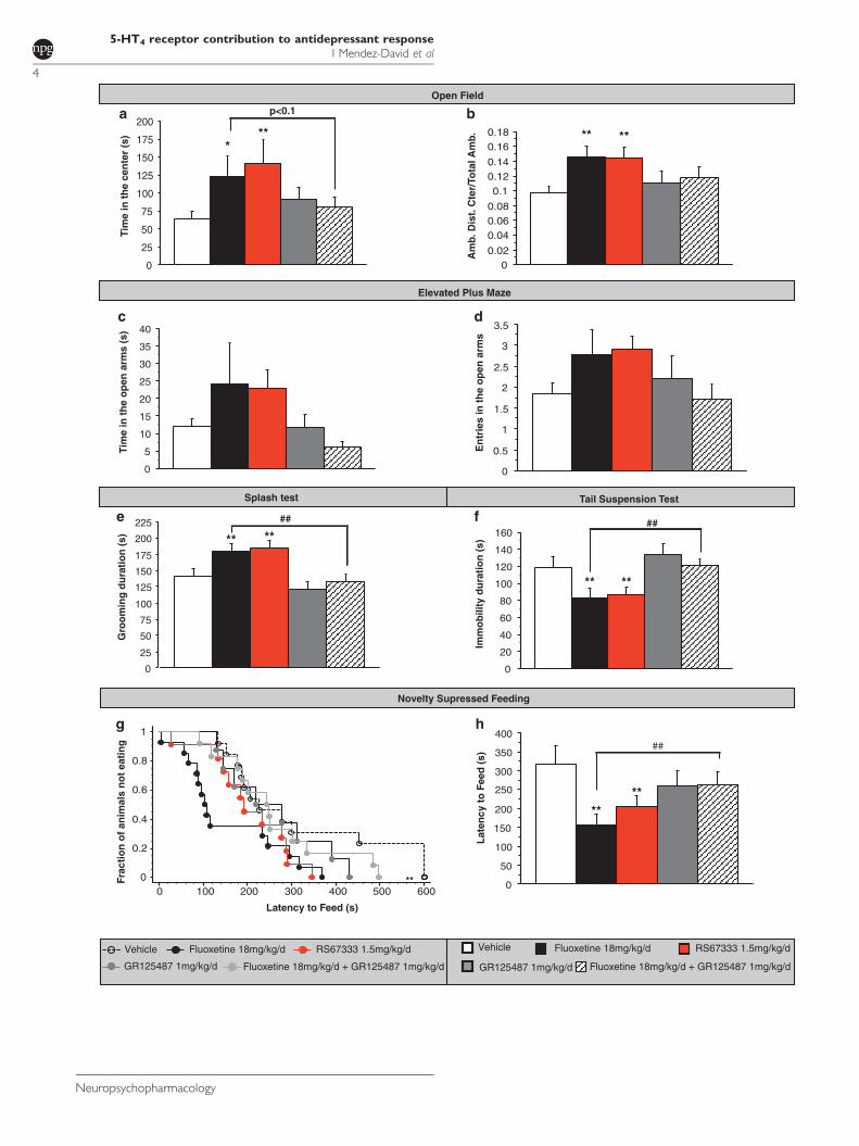
Am
b. D
ist.
Cte
r/To
tal A
mb.
g
e f
c d
a b
h
Vehicle Fluoxetine 18mg/kg/d RS67333 1.5mg/kg/d
Fluoxetine 18mg/kg/d + GR125487 1mg/kg/dGR125487 1mg/kg/d
00.020.040.060.080.1
0.120.140.160.18
0
5
10
15
20
25
30
35
40
0
0.2
0.4
0.6
0.8
1
0 100 200 300 400 500 600
*** ** **
**
** **
** **
**
p<0.1
##
Open Field
Elevated Plus Maze
Splash test Tail Suspension Test
Novelty Supressed Feeding
**
Fluoxetine 18mg/kg/d + GR125487 1mg/kg/dGR125487 1mg/kg/d
Vehicle Fluoxetine 18mg/kg/d RS67333 1.5mg/kg/d
**
Tim
e in
the
cent
er (s
)
0
25
50
75
100
125
150
175
200Ti
me
in th
e op
en a
rms
(s)
0
0.5
1
1.5
2
2.5
3
3.5
Ent
ries
in th
e op
en a
rms
0
50
100
150
200
300
350
400
Late
ncy
to F
eed
(s)
Frac
tion
of a
nim
als
not e
atin
g
Latency to Feed (s)
0
25
50
75
100
125
150
175
200
225
Gro
omin
g du
ratio
n (s
)
0
20
40
60
80
100
120
140
160
Imm
obili
ty d
urat
ion
(s)
## ##
250
5-HT4 receptor contribution to antidepressant responseI Mendez-David et al
4
Neuropsychopharmacology
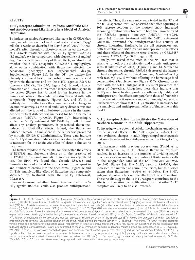
RESULTS
5-HT4 Receptor Stimulation Produces Anxiolytic-Likeand Antidepressant-Like Effects in a Model of Anxiety/Depression
To induce an anxious/depressed-like state in C57BL/6Ntacmice, we administered a low dose of corticosterone (35 mg/ml) for 4 weeks as described in David et al (2009) (‘CORTmodel’). After chronic corticosterone, we tested the effectsof a 4-week treatment with the 5-HT4 agonist RS67333(1.5 mg/kg/day) in comparison with fluoxetine (18 mg/kg/day). To assess the selectivity of these effects, we also testedwhether the 5-HT4 antagonist GR125487 (1 mg/kg/day),alone or in combination with fluoxetine, affected thebehavioral phenotype (see experimental design,Supplementary Figure S1). In the OF, the anxiety-likephenotype induced by chronic corticosterone was reversedby chronic fluoxetine and by the 5-HT4 agonist RS67333(one-way ANOVA, *po0.05, Figure 1a). Indeed, chronicfluoxetine and RS67333 treatment increased time spent inthe center (Figure 1a). A trend for an increase in thenumber of entries in the center was also observed with bothcompounds (Supplementary Figures S2A and B). It isunlikely that this effect was the consequence of a change inlocomotor activity, as the total ambulatory distance was notaffected and the ratio of ambulatory distance in the centerdivided by total distance was increased for both treatments(one-way ANOVA, *po0.05, Figure 1b). Interestingly,while the 5-HT4 antagonist GR125487 by itself did notaffect any anxiety parameters, it prevented fluoxetine-induced anxiolytic-like effects. Indeed, the fluoxetine-induced increase in time spent in the center was preventedby chronic GR125487 administration. These data indicatethat 5-HT4 stimulation induces an anxiolytic-like effect andis necessary for the anxiolytic effect of chronic fluoxetinetreatment.
To further validate these results, we next tested the effectsof RS67333 and fluoxetine alone or in the presence ofGR125487 in the same animals in another anxiety-relatedtest, the EPM. We found that chronic RS67333 andfluoxetine induced a trend for an increase in time spent inand number of entries into the open arms, (Figure 1c andd). This anxiolytic-like effect of fluoxetine was completelyabolished by treatment with the 5-HT4 antagonist,GR125487.
We next assessed whether chronic treatment with the 5-HT4 agonist RS67333 could also produce antidepressant-
like effects. Thus, the same mice were tested in the ST andthe tail suspension test. We observed that after squirting a10% sucrose solution on the mouse’s snout, increasedgrooming duration was observed in both the fluoxetine andthe RS67333 groups (one-way ANOVA, **po0.01,Figure 1e). Chronic treatment with the 5-HT4 antagonistGR125487 prevented the antidepressant-like activity ofchronic fluoxetine. Similarly, in the tail suspension test,both fluoxetine and RS67333 had antidepressant-like effectsand these effects of fluoxetine were blocked by GR125487(one-way ANOVA, **po0.01; Figure 1f).
Finally, we tested these mice in the NSF test that issensitive to both acute anxiolytics and chronic antidepres-sants (Guilloux et al, 2013) (Figure 1g and h). Similar tochronic fluoxetine, chronic RS67333 decreased the latencyto feed (Kaplan–Meier survival analysis, Mantel–Cox logrank test, **po0.01) without affecting the home-cage foodconsumption (Supplementary Figure S2C). Chronic treat-ment with the 5-HT4 antagonist (GR125487) prevented theeffect of fluoxetine. Altogether, these data indicate that5-HT4 receptor activation produces both anxiolytic-like andantidepressant-like effects comparable to those of fluoxetinein the chronic corticosterone model of anxiety/depression.Furthermore, we show that 5-HT4 activation is necessary forthe anxiolytic and antidepressant effects of fluoxetine in thismodel.
5-HT4 Receptor Activation Facilitates the Maturation ofNewborn Neurons in the Adult Hippocampus
To investigate the potential cellular mechanisms underlyingthe behavioral effects of the 5-HT4 agonist RS67333, wenext evaluated changes in adult hippocampal neurogenesisthat may be relevant to antidepressant action (Surget et al,2011).
In agreement with previous observations (David et al,2009; Rainer et al, 2011), chronic fluoxetine exposureresulted in an increase in the number of dividing neuralprecursors as assessed by the number of Ki67-positive cellsin the subgranular zone of the DG (one-way ANOVA,*po0.05, Figure 2a). The 5-HT4 agonist, RS67333, alsoincreased the number of neural precursors, but to a lesserextent than fluoxetine (þ 51% vs þ 170%). The 5-HT4
antagonist partially blocked the effect of chronic fluoxetine.These results suggest that 5-HT4 receptors contribute to theeffects of fluoxetine on proliferation, but that other 5-HTreceptors are likely to be also involved.
Figure 1 Effects of chronic 5-HT4 receptor stimulation (28 days) on the anxious/depressed-like phenotype induced by chronic corticosterone exposure.(a and b) Effects of chronic treatment with 5-HT4 ligands or fluoxetine, starting after 4 weeks of corticosterone (35 mg/ml), on anxiety behaviors in the openfield (OF) test. Anxiety is measured as mean time spent in the center in seconds (a) or the ratio of ambulatory distance in the center/total ambulatorydistance (b). Values plotted are mean±SEM (n¼ 10–15/group). *po0.05, **po0.01 vs corticosterone/vehicle group. (c and d) Effects of chronic treatmentwith 5-HT4 ligands or fluoxetine, starting after 4 weeks of corticosterone (35 mg/ml), on anxiety behaviors in the elevated plus maze (EPM). Anxiety isexpressed as mean time in (c) or entries into (d) the open arms. Values plotted are mean±SEM (n¼ 10–15/group). (e) Effect of chronic treatment with 5-HT4 ligands or fluoxetine on corticosterone-induced depression-related behaviors in the splash test (ST). Results are expressed as mean duration ofgrooming after receiving a 10% sucrose solution on the snout. Values plotted are mean±SEM (n¼ 10–15/group). **po0.01, ##po0.01 vs corticosterone/vehicle group and corticosterone/fluoxetine group, respectively. (f) Effect of chronic treatment with 5-HT4 ligands or fluoxetine in the tail suspension testfollowing chronic corticosterone. Results are expressed as mean of immobility duration in seconds. Values plotted are mean±SEM (n¼ 10–15/group).**po0.01, ##po0.01 vs corticosterone/vehicle group and corticosterone/fluoxetine group, respectively. (g and h) Effects of chronic treatment with 5-HT4
ligands or fluoxetine on anxiety- and depression-like behaviors in the novelty-suppressed feeding (NSF) paradigm after chronic corticosterone. Valuesplotted are cumulative survival of animals that have not eaten over 10 min (n¼ 10–15/group) (g) or mean of latency to feed in seconds±SEM (h).**po0.01, ##po 0.01 vs corticosterone/vehicle group and corticosterone/fluoxetine group, respectively.
5-HT4 receptor contribution to antidepressant responseI Mendez-David et al
5
Neuropsychopharmacology
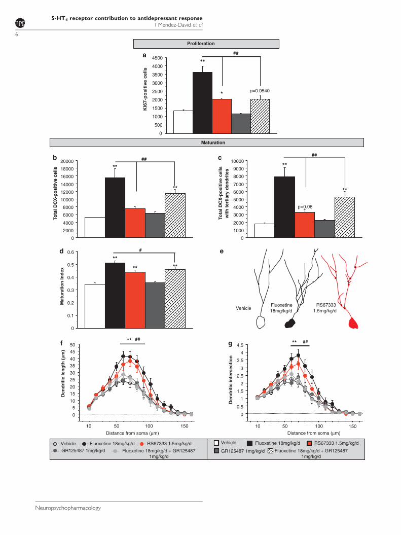
Maturation
0
2000
4000
6000
8000
10000
12000
14000
16000
18000
20000
0
1000
2000
3000
4000
5000
6000
7000
8000
9000
10000
0
0.1
0.2
0.3
0.4
0.5
0.6
Tota
l DC
X-p
ositi
ve c
ells
Tota
l DC
X-p
ositi
ve c
ells
with
tert
iary
den
drite
s
Mat
urat
ion
Inde
x
p<0.08
**
**
** **
####
#
cb
d
Den
driti
c in
ters
ectio
n
Distance from soma (µm)Distance from soma (µm)
Den
driti
c le
ngth
(!m
)
f g **
e
Proliferation
0
500
1000
1500
2000
2500
3000
3500
4000
4500
KI6
7-p
osi
tive
cel
ls
**
*
##a
VehicleFluoxetine 18mg/kg/d
RS673331.5mg/kg/d
** **
**
p=0.0540
10 50 100 150
0
0,5
1
1,5
2
2,5
3
3,5
4
4,5
1501005010
##
0
510
1520
2530
3540
4550
Vehicle Fluoxetine 18mg/kg/d RS67333 1.5mg/kg/d
Fluoxetine 18mg/kg/d + GR1254871mg/kg/d
GR125487 1mg/kg/dFluoxetine 18mg/kg/d + GR1254871mg/kg/d
GR125487 1mg/kg/dVehicle Fluoxetine 18mg/kg/d RS67333 1.5mg/kg/d
** ##
5-HT4 receptor contribution to antidepressant responseI Mendez-David et al
6
Neuropsychopharmacology
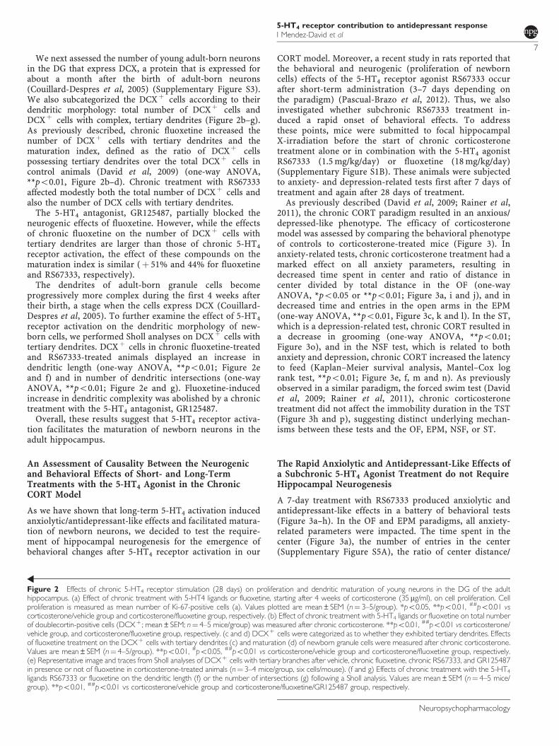
We next assessed the number of young adult-born neuronsin the DG that express DCX, a protein that is expressed forabout a month after the birth of adult-born neurons(Couillard-Despres et al, 2005) (Supplementary Figure S3).We also subcategorized the DCXþ cells according to theirdendritic morphology: total number of DCXþ cells andDCXþ cells with complex, tertiary dendrites (Figure 2b–g).As previously described, chronic fluoxetine increased thenumber of DCXþ cells with tertiary dendrites and thematuration index, defined as the ratio of DCXþ cellspossessing tertiary dendrites over the total DCXþ cells incontrol animals (David et al, 2009) (one-way ANOVA,**po0.01, Figure 2b–d). Chronic treatment with RS67333affected modestly both the total number of DCXþ cells andalso the number of DCX cells with tertiary dendrites.
The 5-HT4 antagonist, GR125487, partially blocked theneurogenic effects of fluoxetine. However, while the effectsof chronic fluoxetine on the number of DCXþ cells withtertiary dendrites are larger than those of chronic 5-HT4
receptor activation, the effect of these compounds on thematuration index is similar (þ 51% and 44% for fluoxetineand RS67333, respectively).
The dendrites of adult-born granule cells becomeprogressively more complex during the first 4 weeks aftertheir birth, a stage when the cells express DCX (Couillard-Despres et al, 2005). To further examine the effect of 5-HT4
receptor activation on the dendritic morphology of new-born cells, we performed Sholl analyses on DCXþ cells withtertiary dendrites. DCXþ cells in chronic fluoxetine-treatedand RS67333-treated animals displayed an increase indendritic length (one-way ANOVA, **po0.01; Figure 2eand f) and in number of dendritic intersections (one-wayANOVA, **po0.01; Figure 2e and g). Fluoxetine-inducedincrease in dendritic complexity was abolished by a chronictreatment with the 5-HT4 antagonist, GR125487.
Overall, these results suggest that 5-HT4 receptor activa-tion facilitates the maturation of newborn neurons in theadult hippocampus.
An Assessment of Causality Between the Neurogenicand Behavioral Effects of Short- and Long-TermTreatments with the 5-HT4 Agonist in the ChronicCORT Model
As we have shown that long-term 5-HT4 activation inducedanxiolytic/antidepressant-like effects and facilitated matura-tion of newborn neurons, we decided to test the require-ment of hippocampal neurogenesis for the emergence ofbehavioral changes after 5-HT4 receptor activation in our
CORT model. Moreover, a recent study in rats reported thatthe behavioral and neurogenic (proliferation of newborncells) effects of the 5-HT4 receptor agonist RS67333 occurafter short-term administration (3–7 days depending onthe paradigm) (Pascual-Brazo et al, 2012). Thus, we alsoinvestigated whether subchronic RS67333 treatment in-duced a rapid onset of behavioral effects. To addressthese points, mice were submitted to focal hippocampalX-irradiation before the start of chronic corticosteronetreatment alone or in combination with the 5-HT4 agonistRS67333 (1.5 mg/kg/day) or fluoxetine (18 mg/kg/day)(Supplementary Figure S1B). These animals were subjectedto anxiety- and depression-related tests first after 7 days oftreatment and again after 28 days of treatment.
As previously described (David et al, 2009; Rainer et al,2011), the chronic CORT paradigm resulted in an anxious/depressed-like phenotype. The efficacy of corticosteronemodel was assessed by comparing the behavioral phenotypeof controls to corticosterone-treated mice (Figure 3). Inanxiety-related tests, chronic corticosterone treatment had amarked effect on all anxiety parameters, resulting indecreased time spent in center and ratio of distance incenter divided by total distance in the OF (one-wayANOVA, *po0.05 or **po0.01; Figure 3a, i and j), and indecreased time and entries in the open arms in the EPM(one-way ANOVA, **po0.01, Figure 3c, k and l). In the ST,which is a depression-related test, chronic CORT resulted ina decrease in grooming (one-way ANOVA, **po0.01;Figure 3o), and in the NSF test, which is related to bothanxiety and depression, chronic CORT increased the latencyto feed (Kaplan–Meier survival analysis, Mantel–Cox logrank test, **po0.01; Figure 3e, f, m and n). As previouslyobserved in a similar paradigm, the forced swim test (Davidet al, 2009; Rainer et al, 2011), chronic corticosteronetreatment did not affect the immobility duration in the TST(Figure 3h and p), suggesting distinct underlying mechan-isms between these tests and the OF, EPM, NSF, or ST.
The Rapid Anxiolytic and Antidepressant-Like Effects ofa Subchronic 5-HT4 Agonist Treatment do not RequireHippocampal Neurogenesis
A 7-day treatment with RS67333 produced anxiolytic andantidepressant-like effects in a battery of behavioral tests(Figure 3a–h). In the OF and EPM paradigms, all anxiety-related parameters were impacted. The time spent in thecenter (Figure 3a), the number of entries in the center(Supplementary Figure S5A), the ratio of center distance/
Figure 2 Effects of chronic 5-HT4 receptor stimulation (28 days) on proliferation and dendritic maturation of young neurons in the DG of the adulthippocampus. (a) Effect of chronic treatment with 5-HT4 ligands or fluoxetine, starting after 4 weeks of corticosterone (35mg/ml), on cell proliferation. Cellproliferation is measured as mean number of Ki-67-positive cells (a). Values plotted are mean±SEM (n¼ 3–5/group). *po0.05, **po0.01, ##po0.01 vscorticosterone/vehicle group and corticosterone/fluoxetine group, respectively. (b) Effect of chronic treatment with 5-HT4 ligands or fluoxetine on total numberof doublecortin-positive cells (DCXþ ; mean±SEM; n¼ 4–5 mice/group) was measured after chronic corticosterone. **po0.01, ##po0.01 vs corticosterone/vehicle group, and corticosterone/fluoxetine group, respectively. (c and d) DCXþ cells were categorized as to whether they exhibited tertiary dendrites. Effectsof fluoxetine treatment on the DCXþ cells with tertiary dendrites (c) and maturation (d) of newborn granule cells were measured after chronic corticosterone.Values are mean±SEM (n¼ 4–5/group). **po0.01, #po0.05, ##po0.01 vs corticosterone/vehicle group and corticosterone/fluoxetine group, respectively.(e) Representative image and traces from Sholl analyses of DCXþ cells with tertiary branches after vehicle, chronic fluoxetine, chronic RS67333, and GR125487in presence or not of fluoxetine in corticosterone-treated animals (n¼ 3–4 mice/group, six cells/mouse). (f and g) Effects of chronic treatment with the 5-HT4
ligands RS67333 or fluoxetine on the dendritic length (f) or the number of intersections (g) following a Sholl analysis. Values are mean±SEM (n¼ 4–5 mice/group). **po0.01, ##po0.01 vs corticosterone/vehicle group and corticosterone/fluoxetine/GR125487 group, respectively.
5-HT4 receptor contribution to antidepressant responseI Mendez-David et al
7
Neuropsychopharmacology
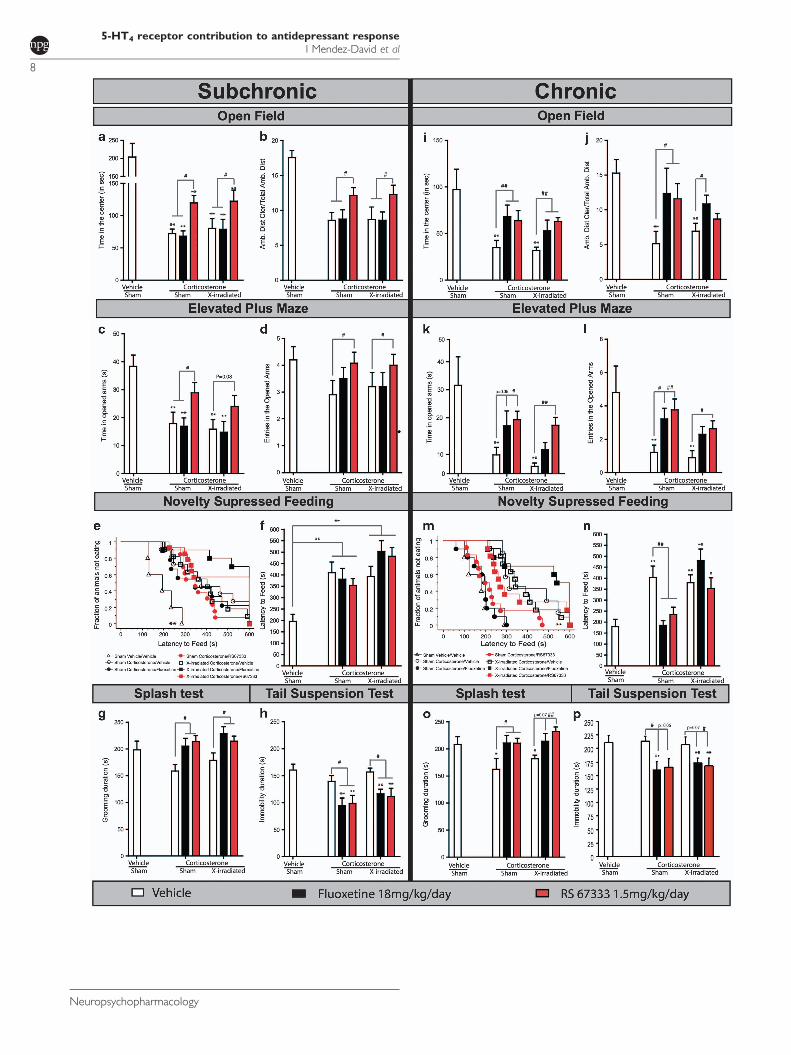
5-HT4 receptor contribution to antidepressant responseI Mendez-David et al
8
Neuropsychopharmacology
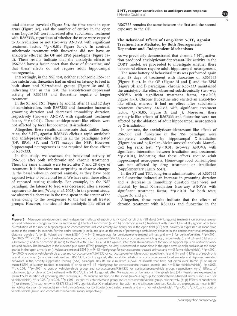
total distance traveled (Figure 3b), the time spent in openarms (Figure 3c), and the number of entries in the openarms (Figure 3d) were increased after subchronic treatmentwith RS67333, regardless of whether the mice were exposedto X-irradiation or not (two-way ANOVA with significanttreatment factor, **po0.01; Figure 3a–c). In contrast,subchronic treatment with fluoxetine did not have ananxiolytic effect in the OF and EPM paradigms (Figure 3a–d). These results indicate that the anxiolytic effects ofRS67333 have a faster onset than those of fluoxetine, andthat these effects do not require adult hippocampalneurogenesis.
Interestingly, in the NSF test, neither subchronic RS67333nor subchronic fluoxetine had an effect on latency to feed inboth sham and X-irradiated groups (Figure 3e and f),indicating that in this test, the anxiolytic/antidepressantactivity of RS67333 and fluoxetine require a longertreatment.
In the ST and TST (Figure 3g and h), after 11 and 12 daysof administration, both RS67333 and fluoxetine increasedgrooming duration and decreased immobility duration,respectively (two-way ANOVA with significant treatmentfactor, **po0.01). These antidepressant-like effects werenot affected by focal hippocampal X-irradiation.
Altogether, these results demonstrate that, unlike fluox-etine, the 5-HT4 agonist RS67333 elicits a rapid anxiolyticand antidepressant-like effect in all the paradigms tested(OF, EPM, ST, and TST) except the NSF. However,hippocampal neurogenesis is not required for these effectsof RS67333.
In this study, we assessed the behavioral activity ofRS67333 after both subchronic and chronic treatments.Thus, the same animals were tested after 7 and 28 days oftreatment. It is therefore not surprising to observe changesin the basal values in control animals, as they have beenexposed twice to behavioral tests. We have seen these effectsof repeated testing routinely. For example, in the NSFparadigm, the latency to feed was decreased after a secondexposure to the test (Wang et al, 2008). In the present study,we observed a decrease in the time spent in the center of thearena owing to the re-exposure to the test in all treatedgroups. However, the size of the anxiolytic-like effect of
RS67333 remains the same between the first and the secondexposure to the OF.
The Behavioral Effects of Long-Term 5-HT4 AgonistTreatment are Mediated by Both Neurogenesis-Dependent and -Independent Mechanisms
As we previously demonstrated that chronic 5-HT4 activa-tion produced anxiolytic/antidepressant-like activity in theCORT model, we proceeded to investigate whether thesebehavioral effects require adult hippocampal neurogenesis.
The same battery of behavioral tests was performed againafter 28 days of treatment with fluoxetine or RS67333(Figure 3i–p). In the OF (Figure 3i and j) and the EPM(Figure 3k and l) paradigms, chronic RS67333 maintainedthe anxiolytic-like effect observed subchronically (two-wayANOVA with significant treatment factor, *po0.05;Figure 3i–l). Chronic fluoxetine also elicited an anxiolytic-like effect, whereas it had no effect after subchronictreatment (two-way ANOVA with significant treatmentfactor, *po0.05; Figure 3i and j). Moreover, theseanxiolytic-like effects of RS67333 and fluoxetine were notaffected by the ablation of adult hippocampal neurogenesisby X-irradiation.
In contrast, the anxiolytic/antidepressant-like effects ofRS67333 and fluoxetine in the NSF paradigm werecompletely abolished by hippocampal X-irradiation(Figure 3m and n; Kaplan–Meier survival analysis, Mantel–Cox log rank test, **po0.01, two-way ANOVA withsignificant interaction between irradiation and treatment,**po0.01), indicating that these effects require adulthippocampal neurogenesis. Home-cage food consumptionwas not affected by drug treatment or irradiation(Supplementary Figure S5D).
In the ST and TST, long-term administration of RS67333and fluoxetine induced an increase in grooming durationand a decrease in immobility duration that were notaffected by focal X-irradiation (two-way ANOVA withsignificant treatment factor, **po0.01 for both tests;Figure 3o and p).
Altogether, these results indicate that the effects ofchronic treatment with RS67333 and fluoxetine in the
Figure 3 Neurogenesis-dependent and -independent effects of subchronic (7 days) or chronic (28 days) 5-HT4 agonist treatment on corticosterone-induced behavioral changes in mice. (a and b/i and j) Effects of subchronic (a and b) or chronic (i and j) treatment with RS67333, a 5-HT4 agonist, after focalX-irradiation of the mouse hippocampus on corticosterone-induced anxiety-like behaviors in the open field (OF) test. Anxiety is expressed as mean timespent in the center, in seconds, for the entire session (a or i), and also as the mean of percentage ambulatory distance in the center over total ambulatorydistance traveled (b or j). Values are mean±SEM (n¼ 9–15 mice/group for corticosterone-treated animals and n¼ 5 for vehicle/vehicle). **po0.01,#po0.05, ##po0.01 vs control vehicle/vehicle group and corticosterone/RS67333 or corticosterone/vehicle group, respectively. (c and d/k and l) Effects ofsubchronic (c and d) or chronic (k and l) treatment with RS67333, a 5-HT4 agonist, after focal X-irradiation of the mouse hippocampus on corticosterone-induced anxiety-like behaviors in the elevated plus maze (EPM) paradigm. Anxiety is expressed as mean time in the open arms (c or k) and also as the meanentries in the open arms (d or l). Values are mean±SEM (n¼ 9–15 mice/group for corticosterone-treated animals and n¼ 5 for vehicle/vehicle). **po0.01,#po0.05 vs control vehicle/vehicle group and corticosterone/RS67333 or corticosterone/vehicle group, respectively. (e and f/m and n) Effects of subchronic(e and f) or chronic (m and n) treatment with RS67333, a 5-HT4 agonist, after focal X-irradiation on corticosterone-induced anxiety- and depression-relatedbehaviors in the novelty-suppressed feeding (NSF) paradigm. Results are cumulative survival of animals that have not eaten over 10 min (e or m) ormean±SEM of latency to feed in seconds (f or n) (n¼ 9–15 mice/group for corticosterone-treated animals and n¼ 5 for vehicle/vehicle). *po0.05,**po0.01, ##po0.01 vs control vehicle/vehicle group and corticosterone/RS67333 or corticosterone/vehicle group, respectively. (g–o) Effects ofsubchronic (g) or chronic (o) treatment with RS67333, a 5-HT4 agonist, after X-irradiation on behavior in the splash test (ST). Results are expressed asmean±SEM duration of grooming after receiving a 10% sucrose solution on the snout (n¼ 9–15/group for corticosterone-treated animals and n¼ 5 forvehicle/vehicle). *po0.05, #po0.05, ##po0.01, vs control vehicle/vehicle group and corticosterone/vehicle group, respectively. (h–p) Effects of subchronic(h) or chronic (p) treatment with RS67333, a 5-HT4 agonist, after X-irradiation on behavior in the tail suspension test. Results are expressed as mean±SEMimmobility duration (in seconds) (n¼ 9–15 mice/group for corticosterone-treated animals and n¼ 5 for vehicle/vehicle). **po0.01, #po0.05 vs controlvehicle/vehicle group and corticosterone/vehicle group, respectively.
5-HT4 receptor contribution to antidepressant responseI Mendez-David et al
9
Neuropsychopharmacology
OF, EPM, ST, and TST are independent of hippocampalneurogenesis. In contrast, the anxiolytic/antidepressant-likeeffects of these compounds in the NSF test requireneurogenesis.
DISCUSSION
Fast Anxiolytic Action of a 5-HT4 Agonist
Most current antidepressant treatments are limited by asignificant degree of nonresponsiveness among patients(Trivedi et al, 2006), delayed onset of therapeutic efficacy,and a number of side effects (Kato and Serretti, 2010). Thedevelopment of new antidepressants is therefore ofconsiderable importance (Wong et al, 2010), and under-standing the mechanisms underlying the delayed onsetshould offer insights into new approaches. Recent studies aswell as our present results indicate that 5-HT4 receptoragonists are faster acting than SSRIs (Lucas et al, 2007;Pascual-Brazo et al, 2012; Tamburella et al, 2009).
Although a 7-day treatment with fluoxetine or RS67333induced antidepressant-like activity in the TST and ST, onlythe 5-HT4 agonist RS67333 resulted in anxiolytic-likeactivity in the OF and the EPM. A longer duration oftreatment (28 days) is required for fluoxetine to exertanxiolytic-like effects comparable to 7 days of RS67333treatment. Although recent evidence indicates that 5-HT4
receptors may represent a new target for antidepressantdrugs (Bockaert et al, 2004; Lucas et al, 2007; Pascual-Brazoet al, 2012; Tamburella et al, 2009), the role of 5-HT4
receptor ligands in anxiety is unclear. Discrepancies inresults observed with 5-HT4 receptor antagonists have beenobserved. In one study, the 5-HT4 receptor antagonists,SDZ205-557, GR113808, and SB204070, administeredacutely, failed to induce anxiolytic-like behavior in thelight/dark choice test in mice (Costall and Naylor, 1997),whereas in two other studies, acute administration of the5-HT4 receptor antagonists, SB204070, GR113808 (Silvestreet al, 1996), and SB207266A (Kennett et al, 1997; Silvestreet al, 1996) had an anxiolytic-like effect in rats in the EPM.5-HT4 receptor knockout (KO) mice do not display ananxious or depressed-like phenotype, although an attenu-ated response to novelty may be relevant to mood disorders(Compan et al, 2004). In our hands, chronic treatment withGR125487 did not affect the anxiety-like phenotype inducedby chronic corticosterone treatment. 5-HT4 receptoragonists have mainly been tested in behavioral tests ofantidepressant-like activity (see (Lucas, 2009) for review).Only one study investigated the effects of RS67333 in the OFparadigm over a 5-min period (Lucas et al, 2007). Theauthors showed that hyperlocomotion induced by olfactorybulbectomy was totally abolished after 14 days of RS67333treatment in rats. To our knowledge, the present study is thefirst to clearly demonstrate fast anxiolytic-like activity of a5-HT4 receptor agonist in a mouse model of anxiety/depression. There is considerable evidence that RS67333 is aspecific agonist of the 5-HT4 receptor. Indeed, three studieshave evaluated the effects of RS67333 in the presence of theselective 5-HT4 antagonist GR125487, and shown that theeffects of RS67333 are blocked by GR125487. Lucas et al(2005) showed that the increase in 5-HT firing inducedby RS67333 (1.5 mg/kg, acutely, during 3 or 21 days)
is prevented by GR125487 administration. This effect ofRS67333 on the firing of 5-HT neurons is likely tocontribute to the phenotype we observe in the currentstudy. Enhanced cognition induced by RS67333 was blockedby the 5-HT4 receptor antagonist GR 125487 (1 mg/kg,intravenous) (Freret et al, 2012; Lamirault and Simon,2001). Both studies tested the specificity of the effects ofRS67333 in the Novel Object Recognition test by using thehighly potent and selective 5-HT4 receptor antagonist,GR125487. They found that 1 mg/kg of GR125487, whichhad no effect per se on the discrimination index, totallyreversed the beneficial effects of RS67333, arguing thus thatthese effects of RS67333 are mediated by 5-HT4 receptor.
Interestingly, in the NSF test, both fluoxetine andRS67333 had an anxiolytic/antidepressant-like effect onlyafter chronic treatment, suggesting that the neurobiologicalmechanisms involved in this paradigm are different fromthose underlying the other tests (OF, EPM, TST, and ST).Indeed, we show that the effects of RS67333 and fluoxetinein the OF, EPM, TST, and ST are independent ofhippocampal neurogenesis, whereas the effects of thesecompounds in the NSF test require neurogenesis. This is inagreement with our previous study showing both neurogen-esis-dependent (NSF) and independent (OF, forced swimtest) effects of fluoxetine in the CORT model (David et al,2009). It is also noteworthy that the only test (NSF) thatrequires neurogenesis is also the one that requires a chronicadministration. This observation is likely related to the factthat young adult-born neurons take several weeks to matureand that the critical period during which adult-bornneurons contribute to behavior extends from 4 to 6 weeksafter their birth (Denny et al, 2012). Interestingly, the effectof the 5-HT4 agonist RS67333 on proliferation of neuralprecursors are weaker than those of fluoxetine, whereas theeffects of RS67333 on the maturation of young neurons aresimilar to those of fluoxetine. Newborn neurons undergo anaccelerated maturation after chronic fluoxetine treatment(Wang et al, 2008) and possibly also after 5-HT4 receptoractivation. These results suggest that the neurogenesis-dependent effect of RS67333 and fluoxetine in the NSF testis more likely to result from increased maturation than fromincreased proliferation.
The fast onset of action of the 5-HT4 receptor agonistcould be a consequence of an increase in serotonergicoutput to projection areas (Lucas et al, 2005; Lucas andDebonnel, 2002). Indeed, by measuring spontaneouselectrical activity in mice lacking 5-HT4 receptors,Conductier et al (2006) demonstrated that 5-HT4 receptorsexert a tonic positive influence on the firing activity ofdorsal raphe nucleus 5-HT neurons, and previous studieshave shown that 5-HT4 receptor activation by selectiveagonists modulates central 5-HT neurotransmission, in-creasing the firing of dorsal raphe nucleus 5-HT neurons(Lucas and Debonnel, 2002). There is also accumulatingevidence that cortical regions are involved in 5-HT4-induced anxiolytic/antidepressant-like activities (Lucaset al, 2005) (for review see also (Lucas, 2009)). 5-HT4
receptors in the prefrontal cortex control the firing rate ofmidbrain serotonergic neurons via descending inputs(Lucas et al, 2005), and their activation leads to increasesin serotonin release in projection sites including thehippocampus (Ge and Barnes, 1996).
5-HT4 receptor contribution to antidepressant responseI Mendez-David et al
10
Neuropsychopharmacology
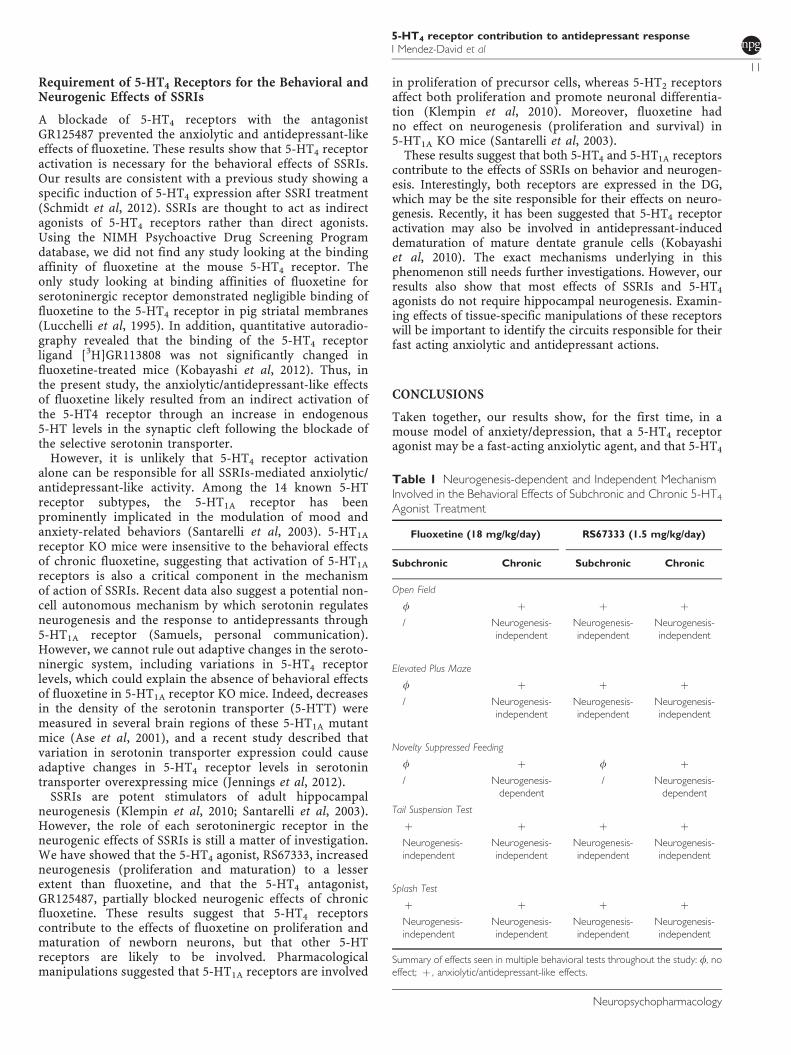
Requirement of 5-HT4 Receptors for the Behavioral andNeurogenic Effects of SSRIs
A blockade of 5-HT4 receptors with the antagonistGR125487 prevented the anxiolytic and antidepressant-likeeffects of fluoxetine. These results show that 5-HT4 receptoractivation is necessary for the behavioral effects of SSRIs.Our results are consistent with a previous study showing aspecific induction of 5-HT4 expression after SSRI treatment(Schmidt et al, 2012). SSRIs are thought to act as indirectagonists of 5-HT4 receptors rather than direct agonists.Using the NIMH Psychoactive Drug Screening Programdatabase, we did not find any study looking at the bindingaffinity of fluoxetine at the mouse 5-HT4 receptor. Theonly study looking at binding affinities of fluoxetine forserotoninergic receptor demonstrated negligible binding offluoxetine to the 5-HT4 receptor in pig striatal membranes(Lucchelli et al, 1995). In addition, quantitative autoradio-graphy revealed that the binding of the 5-HT4 receptorligand [3H]GR113808 was not significantly changed influoxetine-treated mice (Kobayashi et al, 2012). Thus, inthe present study, the anxiolytic/antidepressant-like effectsof fluoxetine likely resulted from an indirect activation ofthe 5-HT4 receptor through an increase in endogenous5-HT levels in the synaptic cleft following the blockade ofthe selective serotonin transporter.
However, it is unlikely that 5-HT4 receptor activationalone can be responsible for all SSRIs-mediated anxiolytic/antidepressant-like activity. Among the 14 known 5-HTreceptor subtypes, the 5-HT1A receptor has beenprominently implicated in the modulation of mood andanxiety-related behaviors (Santarelli et al, 2003). 5-HT1A
receptor KO mice were insensitive to the behavioral effectsof chronic fluoxetine, suggesting that activation of 5-HT1A
receptors is also a critical component in the mechanismof action of SSRIs. Recent data also suggest a potential non-cell autonomous mechanism by which serotonin regulatesneurogenesis and the response to antidepressants through5-HT1A receptor (Samuels, personal communication).However, we cannot rule out adaptive changes in the seroto-ninergic system, including variations in 5-HT4 receptorlevels, which could explain the absence of behavioral effectsof fluoxetine in 5-HT1A receptor KO mice. Indeed, decreasesin the density of the serotonin transporter (5-HTT) weremeasured in several brain regions of these 5-HT1A mutantmice (Ase et al, 2001), and a recent study described thatvariation in serotonin transporter expression could causeadaptive changes in 5-HT4 receptor levels in serotonintransporter overexpressing mice (Jennings et al, 2012).
SSRIs are potent stimulators of adult hippocampalneurogenesis (Klempin et al, 2010; Santarelli et al, 2003).However, the role of each serotoninergic receptor in theneurogenic effects of SSRIs is still a matter of investigation.We have showed that the 5-HT4 agonist, RS67333, increasedneurogenesis (proliferation and maturation) to a lesserextent than fluoxetine, and that the 5-HT4 antagonist,GR125487, partially blocked neurogenic effects of chronicfluoxetine. These results suggest that 5-HT4 receptorscontribute to the effects of fluoxetine on proliferation andmaturation of newborn neurons, but that other 5-HTreceptors are likely to be involved. Pharmacologicalmanipulations suggested that 5-HT1A receptors are involved
in proliferation of precursor cells, whereas 5-HT2 receptorsaffect both proliferation and promote neuronal differentia-tion (Klempin et al, 2010). Moreover, fluoxetine hadno effect on neurogenesis (proliferation and survival) in5-HT1A KO mice (Santarelli et al, 2003).
These results suggest that both 5-HT4 and 5-HT1A receptorscontribute to the effects of SSRIs on behavior and neurogen-esis. Interestingly, both receptors are expressed in the DG,which may be the site responsible for their effects on neuro-genesis. Recently, it has been suggested that 5-HT4 receptoractivation may also be involved in antidepressant-induceddematuration of mature dentate granule cells (Kobayashiet al, 2010). The exact mechanisms underlying in thisphenomenon still needs further investigations. However, ourresults also show that most effects of SSRIs and 5-HT4
agonists do not require hippocampal neurogenesis. Examin-ing effects of tissue-specific manipulations of these receptorswill be important to identify the circuits responsible for theirfast acting anxiolytic and antidepressant actions.
CONCLUSIONS
Taken together, our results show, for the first time, in amouse model of anxiety/depression, that a 5-HT4 receptoragonist may be a fast-acting anxiolytic agent, and that 5-HT4
Table 1 Neurogenesis-dependent and Independent MechanismInvolved in the Behavioral Effects of Subchronic and Chronic 5-HT4
Agonist Treatment
Fluoxetine (18 mg/kg/day) RS67333 (1.5 mg/kg/day)
Subchronic Chronic Subchronic Chronic
Open Field
f þ þ þ/ Neurogenesis-
independentNeurogenesis-independent
Neurogenesis-independent
Elevated Plus Maze
f þ þ þ/ Neurogenesis-
independentNeurogenesis-independent
Neurogenesis-independent
Novelty Suppressed Feeding
f þ f þ/ Neurogenesis-
dependent/ Neurogenesis-
dependent
Tail Suspension Test
þ þ þ þNeurogenesis-independent
Neurogenesis-independent
Neurogenesis-independent
Neurogenesis-independent
Splash Test
þ þ þ þNeurogenesis-independent
Neurogenesis-independent
Neurogenesis-independent
Neurogenesis-independent
Summary of effects seen in multiple behavioral tests throughout the study: f, noeffect; þ , anxiolytic/antidepressant-like effects.
5-HT4 receptor contribution to antidepressant responseI Mendez-David et al
11
Neuropsychopharmacology
stimulation is necessary for the behavioral and neurogeniceffects (proliferation and maturation) of fluoxetine, a classicSSRI antidepressant. Furthermore, we showed that, with theexception of the NSF test, the anxiolytic and antidepressant-like effects of the 5-HT4 agonist were independent ofhippocampal neurogenesis (Table 1).
The present study is encouraging for the development ofRS-67333 as an anxiolytic/antidepressant compound for usein patients. However, the use of the 5-HT4 receptor as anovel antidepressant target may be hampered by the factthat it also has important roles outside the central nervoussystem, for example, in the heart, gastrointestinal tract,adrenal gland, and urinary bladder (Tonini and Pace, 2006),which may prevent its development as an effectiveanxiolytic/antidepressant drug (Bockaert et al, 2004,2008). Thus, signaling molecules that interact with the 5-HT4 receptor such as P11 (Egeland et al, 2011; Warner-Schmidt et al, 2009) may represent novel targets for fast-acting anxiolytic/antidepressant treatments. There is indeedrecent evidence that cortical neurons that express both P11and 5-HT4 receptors are involved in the behavioral effects ofSSRIs (Schmidt et al, 2012), and that chronic treatment withfluoxetine results in an increase in 5-HT4 receptor expres-sions in cortical neurons (Schmidt et al, 2012).
FUNDING AND DISCLOSURE
This work was also supported by Young Investigator Awardto DJD (The Brain & Behavior Research Foundation). DrDJD currently received investigator-initiated research sup-port from Lundbeck and served as a consultant in the areasof target identification and validation and new compounddevelopment to Lundbeck, Roche, and Servier. Dr RHreceives compensation as a consultant for Roche, Lundbeckand Servier in relation to the generation of novelantidepressants.
ACKNOWLEDGEMENTS
This work was supported by the technical assistance ofValerie Dupont-Domergue and staff from the animal carefacility of the Institut Federatif de Recherche-IFR141 of theParis-Sud University. We also thank Dr Jean-PhilippeGuilloux for his help designing Figure 2.
REFERENCES
Artigas F (2013). Developments in the field of antidepressants,where do we go now? Eur Neuropsychopharmacol (e-pub aheadof print; doi:10.1016/j.euroneuro.2013.04.013).
Ase AR, Reader TA, Hen R, Riad M, Descarries L (2001). Regionalchanges in density of serotonin transporter in the brain of 5-HT1A and 5-HT1B knockout mice, and of serotonin innervationin the 5-HT1B knockout. J Neurochem 78: 619–630.
Bockaert J, Claeysen S, Compan V, Dumuis A (2004). 5-HT4receptors. Curr Drug Targets CNS Neurol Disord 3: 39–51.
Bockaert J, Claeysen S, Compan V, Dumuis A (2008). 5-HT(4)receptors: history, molecular pharmacology and brain functions.Neuropharmacology 55: 922–931.
Burghardt NS, Bauer EP (2013). Acute and chronic effects ofselective serotonin reuptake inhibitor treatment on fear con-ditioning: Implications for underlying fear circuits. Neuroscience247: 253–272.
Cachard-Chastel M, Lezoualc’h F, Dewachter I, Delomenie C, CroesS, Devijver H et al (2007). 5-HT4 receptor agonists increasesAPPalpha levels in the cortex and hippocampus of male C57BL/6j mice. Br J Pharmacol 150: 883–892.
Compan V, Zhou M, Grailhe R, Gazzara RA, Martin R, Gingrich Jet al (2004). Attenuated response to stress and noveltyand hypersensitivity to seizures in 5-HT4 receptor knock-outmice. J Neurosci 24: 412–419.
Conductier G, Dusticier N, Lucas G, Cote F, Debonnel G, Daszuta Aet al (2006). Adaptive changes in serotonin neurons of the raphenuclei in 5-HT(4) receptor knock-out mouse. Eur J Neurosci 24:1053–1062.
Costall B, Naylor RJ (1997). The influence of 5-HT2 and5-HT4 receptor antagonists to modify drug induced disinhibi-tory effects in the mouse light/dark test. Br J Pharmacol 122:1105–1118.
Couillard-Despres S, Winner B, Schaubeck S, Aigner R, VroemenM, Weidner N et al (2005). Doublecortin expression levels inadult brain reflect neurogenesis. Eur J Neurosci 21: 1–14.
David DJ, Samuels BA, Rainer Q, Wang JW, Marsteller D, Mendez Iet al (2009). Neurogenesis-dependent and -independent effectsof fluoxetine in an animal model of anxiety/depression. Neuron62: 479–493.
David DJ, Wang J, Samuels BA, Rainer Q, David I, Gardier AM et al(2010). Implications of the functional integration of adult-bornhippocampal neurons in anxiety-depression disorders. Neuro-scientist 16: 578–591.
Denny CA, Burghardt NS, Schachter DM, Hen R, Drew MR (2012).4- to 6-week-old adult-born hippocampal neurons influencenovelty-evoked exploration and contextual fear conditioning.Hippocampus 22: 1188–1201.
Egeland M, Warner-Schmidt J, Greengard P, Svenningsson P(2011). Co-expression of serotonin 5-HT(1B) and 5-HT(4)receptors in p11 containing cells in cerebral cortex, hippocam-pus, caudate-putamen and cerebellum. Neuropharmacology 61:442–450.
Eglen RM, Bonhaus DW, Johnson LG, Leung E, Clark RD (1995).Pharmacological characterization of two novel and potent 5-HT4receptor agonists, RS 67333 and RS 67506, in vitro and in vivo.Br J Pharmacol 115: 1387–1392.
Freret T, Bouet V, Quiedeville A, Nee G, Dallemagne P, Rochais Cet al (2012). Synergistic effect of acetylcholinesterase inhibition(donepezil) and 5-HT(4) receptor activation (RS67333) on objectrecognition in mice. Behav Brain Res 230: 304–308.
Gale JD, Grossman CJ, Whitehead JW, Oxford AW, Bunce KT,Humphrey PP (1994). GR113808: a novel, selective antagonistwith high affinity at the 5-HT4 receptor. Br J Pharmacol 111:332–338.
Ge J, Barnes NM (1996). 5-HT4 receptor-mediated modulation of5-HT release in the rat hippocampus in vivo. Br J Pharmacol 117:1475–1480.
Gomez-Lazaro E, Garmendia L, Beitia G, Perez-Tejada J, Azpiroz A,Arregi A (2012). Effects of a putative antidepressant with a rapidonset of action in defeated mice with different coping strategies.Prog Neuropsychopharmacol Biol Psychiatry 38: 317–327.
Gould TD (2011). Mood and Anxiety Related Phenotypes inMice Characterization Using Behavioral TestsVolume II. Neuro-methods Ser. Humana Press Springer Distributor, Secaucus, NewYork, NY, USA.
Guilloux JP, David I, Pehrson A, Guiard BP, Reperant C, Orvoen Set al (2013). Antidepressant and anxiolytic potential of themultimodal antidepressant vortioxetine (Lu AA21004) assessedby behavioral and neurogenesis outcomes in mice. Neurophar-macology 73: 147–159.
Jennings KA, Licht CL, Bruce A, Lesch KP, Knudsen GM, Sharp T(2012). Genetic variation in 5-hydroxytryptamine transporterexpression causes adaptive changes in 5-HT(4) receptor levels.Int J Neuropsychopharmacol 15: 1099–1107.
5-HT4 receptor contribution to antidepressant responseI Mendez-David et al
12
Neuropsychopharmacology
Kato M, Serretti A (2010). Review and meta-analysis of anti-depressant pharmacogenetic findings in major depressivedisorder. Mol Psychiatry 15: 473–500.
Kennett GA, Wood MD, Bright F, Trail B, Riley G, Holland V et al(1997). SB 242084, a selective and brain penetrant 5-HT2Creceptor antagonist. Neuropharmacology 36: 609–620.
Kheirbek MA, Klemenhagen KC, Sahay A, Hen R (2012).Neurogenesis and generalization: a new approach to stratifyand treat anxiety disorders. Nat Neurosci 15: 1613–1620.
Klempin F, Babu H, De Pietri Tonelli D, Alarcon E, Fabel K,Kempermann G (2010). Oppositional effects of serotoninreceptors 5-HT1a, 2, and 2c in the regulation of adulthippocampal neurogenesis. Front Mol Neurosci 3: pii: 14.
Kobayashi K, Haneda E, Higuchi M, Suhara T, Suzuki H (2012).Chronic fluoxetine selectively upregulates dopamine D(1)-likereceptors in the hippocampus. Neuropsychopharmacology 37:1500–1508.
Kobayashi K, Ikeda Y, Sakai A, Yamasaki N, Haneda E, Miyakawa Tet al (2010). Reversal of hippocampal neuronal maturation by sero-tonergic antidepressants. Proc Natl Acad Sci USA 107: 8434–8439.
Lamirault L, Simon H (2001). Enhancement of place andobject recognition memory in young adult and old rats by RS67333, a partial agonist of 5-HT4 receptors. Neuropharmacology41: 844–853.
Licht CL, Knudsen GM, Sharp T (2010). Effects of the 5-HT(4)receptor agonist RS67333 and paroxetine on hippocampalextracellular 5-HT levels. Neurosci Lett 476: 58–61.
Lucas G (2009). Serotonin receptors, type 4: a new hope? Curr DrugTargets 10: 1085–1095.
Lucas G, Compan V, Charnay Y, Neve RL, Nestler EJ, Bockaert Jet al (2005). Frontocortical 5-HT4 receptors exert positivefeedback on serotonergic activity: viral transfections, subacuteand chronic treatments with 5-HT4 agonists. Biol Psychiatry 57:918–925.
Lucas G, Debonnel G (2002). 5-HT4 receptors exert a frequency-related facilitatory control on dorsal raphe nucleus 5-HTneuronal activity. Eur J Neurosci 16: 817–822.
Lucas G, Du J, Romeas T, Mnie-Filali O, Haddjeri N, Pineyro Get al (2010). Selective serotonin reuptake inhibitors potentiatethe rapid antidepressant-like effects of serotonin4 receptoragonists in the rat. PLoS One 5: e9253.
Lucas G, Rymar VV, Du J, Mnie-Filali O, Bisgaard C, Manta S et al(2007). Serotonin(4) (5-HT(4)) receptor agonists are putativeantidepressants with a rapid onset of action. Neuron 55: 712–725.
Lucchelli A, Santagostino-Barbone MG, Barbieri A, Candura SM,Tonini M (1995). The interaction of antidepressant drugs withcentral and peripheral (enteric) 5-HT3 and 5-HT4 receptors. BrJ Pharmacol 114: 1017–1025.
Mendez-David I, Hen R, Gardier AM, David DJ (2013). Adulthippocampal neurogenesis: an actor in the antidepressant-likeaction. Ann Pharm Fr 71: 143–149.
Meshi D, Drew MR, Saxe M, Ansorge MS, David D, Santarelli Let al (2006). Hippocampal neurogenesis is not required forbehavioral effects of environmental enrichment. Nature Neuro-science 9: 729–731.
Nestler EJ, Gould E, Manji H, Buncan M, Duman RS, GreshenfeldHK et al (2002). Preclinical models: status of basic research indepression. Biol Psychiatry 52: 503–528.
Pascual-Brazo J, Castro E, Diaz A, Valdizan EM, Pilar-Cuellar F,Vidal R et al (2012). Modulation of neuroplasticity pathways andantidepressant-like behavioural responses following the short-term (3 and 7 days) administration of the 5-HT(4) receptoragonist RS67333. Int J Neuropsychopharmacol 15: 631–643.
Quesseveur G, David DJ, Gaillard MC, Pla P, Wu MV, Nguyen HTet al (2013). BDNF overexpression in mouse hippocampalastrocytes promotes local neurogenesis and elicits anxiolytic-likeactivities. Trans Psychiatry 3: e253.
Rainer Q, Xia L, Guilloux JP, Gabriel C, Mocaer E, Hen R et al(2011). Beneficial behavioural and neurogenic effects of agome-latine in a model of depression/anxiety. Int J Neuropsychophar-macol 1–15 (e-pub ahead of print).
Samuels BA, Leonardo ED, Gadient R, Williams A, Zhou J, DavidDJ et al (2011). Modeling treatment-resistant depression.Neuropharmacology 61: 408–413.
Santarelli L, Saxe M, Gross C, Surget A, Battaglia F, Dulawa S et al(2003). Requirement of hippocampal neurogenesis for thebehavioral effects of antidepressants. Science 301: 805–809.
Schiavi GB, Brunet S, Rizzi CA, Ladinsky H (1994). Identificationof serotonin 5-HT4 recognition sites in the porcine caudatenucleus by radioligand binding. Neuropharmacology 33:543–549.
Schmidt EF, Warner-Schmidt JL, Otopalik BG, Pickett SB, Green-gard P, Heintz N (2012). Identification of the cortical neuronsthat mediate antidepressant responses. Cell 149: 1152–1163.
Silvestre JS, Fernandez AG, Palacios JM (1996). Effects of 5-HT4receptor antagonists on rat behaviour in the elevated plus-mazetest. Eur J Pharmacol 309: 219–222.
Surget A, Tanti A, Leonardo ED, Laugeray A, Rainer Q, Touma Cet al (2011). Antidepressants recruit new neurons to improvestress response regulation. Mol Psychiatry 16: 1177–1188.
Tamburella A, Micale V, Navarria A, Drago F (2009). Antidepres-sant properties of the 5-HT4 receptor partial agonist, SL65.0155:behavioral and neurochemical studies in rats. Prog Neuropsy-chopharmacol Biol Psychiatry 33: 1205–1210.
Tonini M, Pace F (2006). Drugs acting on serotonin receptors forthe treatment of functional GI disorders. Dig Dis 24: 59–69.
Trivedi MH, Rush AJ, Wisniewski SR, Nierenberg AA, Warden D,Ritz L et al (2006). Evaluation of outcomes with citalopram fordepression using measurement-based care in STAR*D: implica-tions for clinical practice. Am J Psychiatry 163: 28–40.
Wang JW, David DJ, Monckton JE, Battaglia F, Hen R (2008).Chronic fluoxetine stimulates maturation and synapticplasticity of adult-born hippocampal granule cells. J Neurosci 28:1374–1384.
Warner-Schmidt JL, Flajolet M, Maller A, Chen EY, Qi H,Svenningsson P et al (2009). Role of p11 in cellular andbehavioral effects of 5-HT4 receptor stimulation. J Neurosci 29:1937–1946.
Wong EH, Yocca F, Smith MA, Lee CM (2010). Challengesand opportunities for drug discovery in psychiatric disorders:the drug hunters’ perspective. Int J Neuropsychopharmacol 13:1269–1284.
Xia L, Delomenie C, David I, Rainer Q, Marouard M, Delacroix Het al (2012). Ventral hippocampal molecular pathways andimpaired neurogenesis associated with 5-HT(1)A and 5-HT(1)Breceptors disruption in mice. Neurosci Lett 521: 20–25.
Supplementary Information accompanies the paper on the Neuropsychopharmacology website (http://www.nature.com/npp)
5-HT4 receptor contribution to antidepressant responseI Mendez-David et al
13
Neuropsychopharmacology
SUPPLEMENTAL DATA
Rapid anxiolytic effects of a 5-HT4 receptor agonist are mediated by a neurogenesis-
independent mechanism
Authors: Indira Mendez-David1*, Denis J. David1*, Flavie Darcet1, Melody V. Wu3, Saadia
Kerdine-Romer2, Alain M. Gardier1, René Hen3,§
SUPPLEMENTAL EXPERIMENTAL PROCEDURES Behavioral tests
Open Field (OF)
This test was performed as described by David and colleagues (David et al, 2009). Motor activity
was quantified in four 43 x 43 cm2 Plexiglas open field boxes (MED Associates, Georgia, VT).
Two sets of 16 pulse-modulated infrared photobeams were placed on opposite walls 2.5-cm apart
to record x–y ambulatory movements. Activity chambers were computer interfaced for data
sampling at 100-ms resolution. The computer defined grid lines that divided each open field into
center and surround regions, with each of four lines being 11 cm from each wall. Dependent
measures in the center were the total time and the number of entries over a 30-min test period.
The activity in the center was quantified as distance traveled in the center divided by total
distance traveled.
!
Elevated Plus Maze (EPM)
This test was performed as described by David and colleagues (David et al, 2009). The maze is a
plus-cross-shaped apparatus, with two open arms and two arms closed by walls linked by a
central platform 50 cm above the floor. Mice were individually put in the center of the maze
facing an open arm and were allowed to explore the maze during 5 min. The time spent in and the
number of entries into the open arms were used as an anxiety index. All parameters were
measured using a videotracker (EPM3C, Bioseb, Vitrolles, France).
!
Novelty-Suppressed-Feeding (NSF)
The NSF is a conflict test that elicits competing motivations: the drive to eat and the fear of
venturing into the center of a brightly lit arena. The latency to begin eating is used as an index of
anxiety/depression-like behavior, because classical anxiolytic drugs as well as chronic
antidepressants decrease this measure. The NSF test was carried out during a 10-min period as
previously described (David et al, 2009). Briefly, the testing apparatus consisted of a plastic box
(50x50x20 cm), the floor of which was covered with approximately 2 cm of wooden bedding.
Twenty-four hours prior to behavioral testing, all food was removed from the home cage. At the
time of testing, a single pellet of food (regular chow) was placed on a white paper platform
positioned in the center of the box. Each animal was placed in a corner of the box, and a
stopwatch was immediately started. The latency to eat (defined as the mouse sitting on its
haunches and biting the pellet with the use of forepaws) was timed. Immediately afterwards, the
animal was transferred to its home cage, and the amount of food consumed by the mouse in the
subsequent 5 min was measured, serving as a control for change in appetite as a possible
confounding factor.
!
Tail Suspension Test (TST)
The TST is an antidepressant activity screening test (Steru et al, 1985) often used to test
compounds that are expected to affect depression related behaviors. Mice are suspended by their
tails with tape, in such a position that they cannot escape or hold on to nearby surfaces. During
this test, typically 6 minutes in duration, the resulting escape oriented behaviors are quantified
using an automated tail suspension test apparatus (Bioseb, Vitrolles, France). A specific strain
gauge linked to a computer quantifies the time spent by the animal trying to escape.
!
Splash Test
This test consisted of squirting 200 µl of a 10% sucrose solution on the mouse’s snout. The
grooming duration was assessed at the end of the corticosterone regimen in the presence or
absence of 4-weeks of drug treatment according to a protocol used elsewhere (David et al, 2009).
!
Immunohistochemistry
Doublecortin-immunostaining consisted of the following steps: slices were washed with 3 times
with PBST (PBS 1X + 0.3% triton) for 15 min, blocked with 10%NDS (normal donkey serum) +
PBST during 2hrs and incubated with by goat anti-doublecortin primary antibody (1:100) in
PBS/Triton/NDS for 24 hrs at 4°C. After secondary antibody incubation, sections were developed
using CY2 (Donkey anti-rabbit) 1/250 diluted (dilute in PBS) for 2hrs.
!
References
David DJ, Samuels BA, Rainer Q, Wang JW, Marsteller D, Mendez I, et al (2009). Neurogenesis-dependent and -independent effects of fluoxetine in an animal model of anxiety/depression. Neuron 62(4): 479-493. Steru L, Chermat R, Thierry B, Simon P (1985). The tail suspension test: a new method for screening antidepressants in mice. Psychopharmacology 85(3): 367-370. !SUPPLEMENTAL FIGURES Supplemental figure 1: Experimental timeline.
(A) In a first set of experiments, in place of normal drinking water, grouped-housed male
C57BL/6Ntac mice were presented with vehicle (0.45% hydroxypropyl-β-cyclodextrin) or
corticosterone (35 µg/ml) in the presence or absence of a 5-HT4 agonist (RS67333, 1.5
mg/kg/day, Alzet® mini pump model 2006 implanted subcutaneously), fluoxetine, 18 mg/kg/day,
or a 5-HT4 antagonist alone (GR125487, 1 mg/kg/day, Alzet® mini pump model 2006 implanted
subcutaneously) or in combination with fluoxetine. We investigated whether the behavioral
changes induced by chronic corticosterone were reversed by chronic 5-HT4 ligands, fluoxetine
alone, or fluoxetine in combination with 5-HT4 receptor antagonist treatment. The same animal
was successively tested in the OF paradigm, the EPM, the NSF, the ST, and the TST and then
sacrificed for neurogenesis
(B) In another set of experiments, focal X-irradiation of the hippocampus was employed to assess
whether the mechanisms underlying RS67333 mediated rescue of corticosterone-induced
anxiety/depressive-like behavior were neurogenesis-dependent. X-irradiation (2.5 Gy) was
delivered five weeks before the start of corticosterone treatment. All animals (Sham or X-
irradiated) received corticosterone (35 µg /ml) in the presence of vehicle, RS67333 (1.5
mg/kg/day), or fluoxetine (18 mg/kg/day). The anxiety/depressive-like phenotype of chronic
corticosterone was assessed by comparing a chronic corticosterone/vehicle group versus a
vehicle/vehicle group. The same animals were successively tested in the OF paradigm, the EPM,
the NSF, the ST, and the TST after subchronic (days 7 to 11) or chronic (days 28 to 33) drug
treatment.
!
Supplemental figure 2: Effects of chronic 5-HT4 receptor stimulation on entries in the
center in the open field paradigm and home food consumption in the NSF test in
anxious/depressive mice.
(A-B) Effect of chronic 5-HT4 ligands or fluoxetine treatment, started after 4 weeks of
corticosterone (35 µg/ml), on the number of entries in the center (A) and on total ambulatory
distance in the open field test (B). Values plotted are mean ± SEM (n= 10–15 per group).
(C) Effect of chronic 5-HT4 ligands or fluoxetine treatment, started after 4 weeks of
corticosterone (35 µg/ml), on home food consumption in the NSF test. Values plotted are mean ±
SEM (n= 10–15 per group).!!
Supplemental figure 3: Images of doublecortin staining following corticosterone for 8 weeks
± chronic RS67333 (1.5 mg/kg/day), GR125487 (1 mg/kg/day), or fluoxetine (18 mg/kg/day)
alone or in combination with GR125487 treatment.
Images of coronal sections of mouse hippocampal dentate gyrus stained for doublecortin (10x
magnification).
!
Supplemental figure 4: Images of doublecortin staining following corticosterone for 8 weeks
in Sham/vehicle or X-irradiated/vehicle animals.
Images of coronal sections of mouse hippocampal dentate gyrus stained for doublecortin (10x
magnification) in sham/vehicle (A) or in Xray/vehicle–treated animals (B).
Supplemental figure 5: Effects of subchronic or chronic 5-HT4 receptor stimulation on
entries in the center in the open field paradigm and home food consumption in the NSF test
in anxious/depressive X-irradiated mice.
(A) Effect of subchronic 5-HT4 agonist or fluoxetine treatment, started after 4 weeks of
corticosterone (35 µg/ml), on entries in the center in the open field test in X-irradiated mice.
Values plotted are mean ± SEM (n = 9–15 per group for corticosterone-treated animals and n=5
for vehicle/vehicle). **p<0.01, #p<0.05, versus control vehicle/vehicle group and
corticosterone/vehicle group, respectively.
(B) Effect of chronic 5-HT4 agonist or fluoxetine treatment, started after 4 weeks of
corticosterone (35 µg/ml), on home food consumption in the NSF test in X-irradiated mice.
Values plotted are mean ± SEM (n = 9–15 per group for corticosterone-treated animals and n=5
for vehicle/vehicle). **p<0.01, #p<0.05, versus control vehicle/vehicle group and
corticosterone/vehicle group, respectively.
(C) Effect of subchronic 5-HT4 agonist or fluoxetine treatment, started after 4 weeks of
corticosterone (35 µg/ml), on food consumption in the novelty suppressed feeding test in X-
irradiated mice. Values plotted are mean ± SEM (n = 9–15 per group for corticosterone-treated
animals and n=5 for vehicle/vehicle).
(D) Effect of chronic 5-HT4 agonist or fluoxetine treatment, started after 4 weeks of
corticosterone (35 µg/ml), on food consumption in the novelty suppressed feeding test in X-
irradiated mice. Values plotted are mean ± SEM (n = 9–15 per group for corticosterone-treated
animals and n=5 for vehicle/vehicle).
!
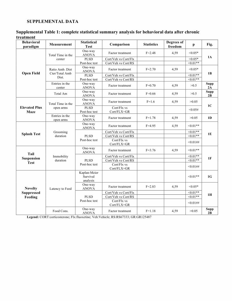
!SUPPLEMENTAL DATA
Supplemental Table 1: complete statistical summary analysis for behavioral data after chronic treatment
Behavioral paradigm Measurement Statistical
Test Comparison Statistics Degrees of freedom p Fig.
Open Field
Total Time in the center
One-way ANOVA Factor treatment F=2.48 4,59 <0.05*
1A PLSD Post-hoc test
Cort/Veh vs Cort/Flx <0.05* Cort/Veh vs Cort/RS <0.01**
Ratio Amb. Dist Cter/Total Amb
Dist.
One-way ANOVA Factor treatment F=2.70 4,59 <0.05*
1B PLSD Post-hoc test
Cort/Veh vs Cort/Flx <0.01** Cort/Veh vs Cort/RS <0.01**
Entries in the center
One-way ANOVA Factor treatment F=0.70 4,59 >0.5 Supp
2A
Total Am One-way ANOVA Factor treatment F=0.66 4,59 >0.5 Supp
2B
Elevated Plus Maze
Total Time in the open arms
One-way ANOVA Factor treatment F=1.6 4,59 >0.05
1C PLSD Post-hoc test
Cort/Flx vs Cort/FLX+GR <0.05#
Entries in the open arms
One-way ANOVA Factor treatment F=1.78 4,59 >0.05 1D
Splash Test Grooming duration
One-way ANOVA Factor treatment F=4.95 4,59 <0.01**
1E PLSD Post-hoc test
Cort/Veh vs Cort/Flx <0.01** Cort/Veh vs Cort/RS <0.01**
Cort/Flx vs Cort/FLX+GR <0.01##
Tail Suspension
Test
Immobility duration
One-way ANOVA Factor treatment F=3.76 4,59 <0.01**
1F PLSD Post-hoc test
Cort/Veh vs Cort/Flx <0.01** Cort/Veh vs Cort/RS <0.01**
Cort/Flx vs Cort/FLX+GR <0.01##
Novelty Suppressed
Feeding
Latency to Feed
Kaplan-Meier Survival analysis
<0.01** 1G
One-way ANOVA Factor treatment F=2.83 4,59 <0.05*
1H PLSD Post-hoc test
Cort/Veh vs Cort/Flx <0.01** Cort/Veh vs Cort/RS <0.01**
Cort/Flx vs Cort/FLX+GR <0.01##
Food Cons. One-way ANOVA Factor treatment F=1.18 4,59 >0.05 Supp
2B Legend: CORT:corticosterone; Flx:fluoxetine; Veh:Vehicle; RS:RS67333; GR:GR125487
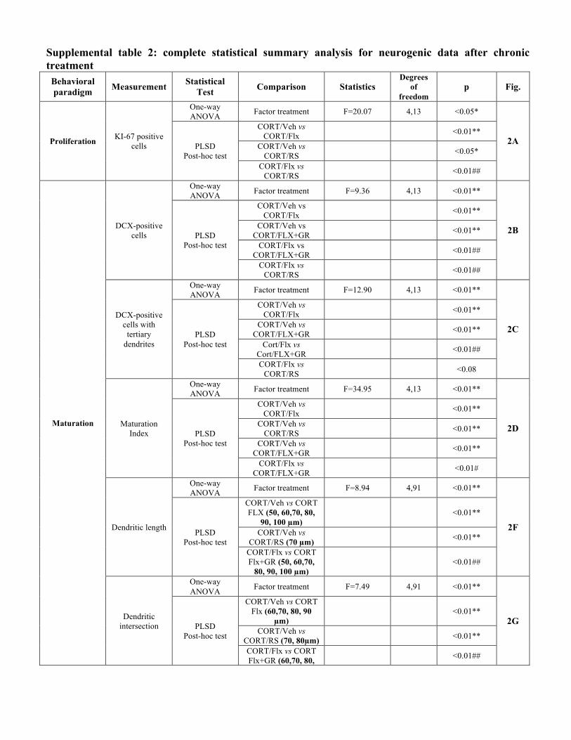
Supplemental table 2: complete statistical summary analysis for neurogenic data after chronic treatment
Behavioral paradigm Measurement Statistical
Test Comparison Statistics Degrees
of freedom
p Fig.
Proliferation KI-67 positive cells
One-way ANOVA Factor treatment F=20.07 4,13 <0.05*
2A PLSD Post-hoc test
CORT/Veh vs CORT/Flx <0.01**
CORT/Veh vs CORT/RS <0.05*
CORT/Flx vs CORT/RS <0.01##
Maturation
DCX-positive cells
One-way ANOVA Factor treatment F=9.36 4,13 <0.01**
2B PLSD Post-hoc test
CORT/Veh vs CORT/Flx <0.01**
CORT/Veh vs CORT/FLX+GR <0.01**
CORT/Flx vs CORT/FLX+GR <0.01##
CORT/Flx vs CORT/RS <0.01##
DCX-positive cells with tertiary
dendrites
One-way ANOVA Factor treatment F=12.90 4,13 <0.01**
2C PLSD Post-hoc test
CORT/Veh vs CORT/Flx <0.01**
CORT/Veh vs CORT/FLX+GR <0.01**
Cort/Flx vs Cort/FLX+GR <0.01##
CORT/Flx vs CORT/RS <0.08
Maturation Index
One-way ANOVA Factor treatment F=34.95 4,13 <0.01**
2D PLSD Post-hoc test
CORT/Veh vs CORT/Flx <0.01**
CORT/Veh vs CORT/RS <0.01**
CORT/Veh vs CORT/FLX+GR <0.01**
CORT/Flx vs CORT/FLX+GR <0.01#
Dendritic length
One-way ANOVA Factor treatment F=8.94 4,91 <0.01**
2F PLSD Post-hoc test
CORT/Veh vs CORT FLX (50, 60,70, 80,
90, 100 µm) <0.01**
CORT/Veh vs CORT/RS (70 µm) <0.01**
CORT/Flx vs CORT Flx+GR (50, 60,70,
80, 90, 100 µm) <0.01##
Dendritic intersection
One-way ANOVA Factor treatment F=7.49 4,91 <0.01**
2G PLSD Post-hoc test
CORT/Veh vs CORT Flx (60,70, 80, 90
µm) <0.01**
CORT/Veh vs CORT/RS (70, 80µm) <0.01**
CORT/Flx vs CORT Flx+GR (60,70, 80, <0.01##

90, 100 µm) Legend: CORT:corticosterone; Flx:fluoxetine; Veh:Vehicle; RS:RS67333; GR:GR125487
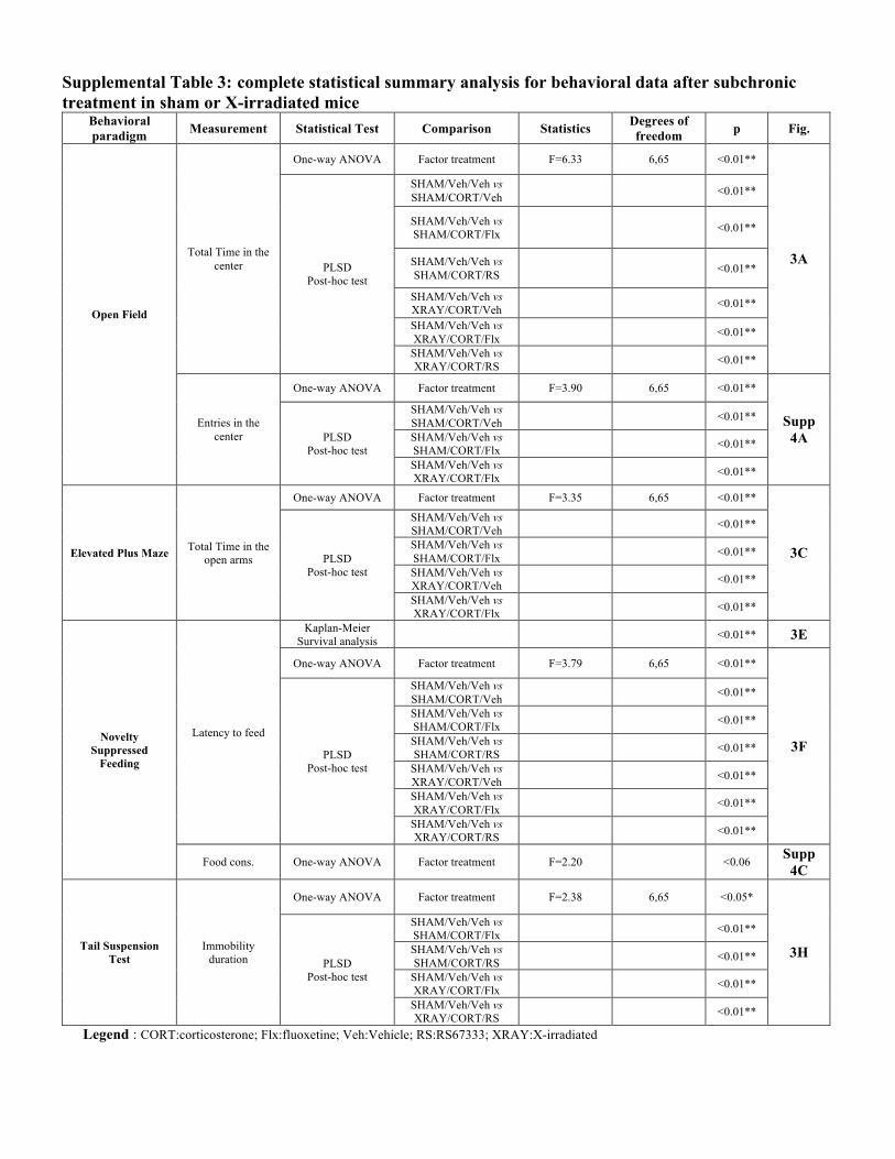
Supplemental Table 3: complete statistical summary analysis for behavioral data after subchronic treatment in sham or X-irradiated mice
Behavioral paradigm Measurement Statistical Test Comparison Statistics Degrees of
freedom p Fig.
Open Field
Total Time in the center
One-way ANOVA Factor treatment F=6.33 6,65 <0.01**
3A PLSD
Post-hoc test
SHAM/Veh/Veh vs SHAM/CORT/Veh <0.01**
SHAM/Veh/Veh vs SHAM/CORT/Flx <0.01**
SHAM/Veh/Veh vs SHAM/CORT/RS <0.01**
SHAM/Veh/Veh vs XRAY/CORT/Veh <0.01**
SHAM/Veh/Veh vs XRAY/CORT/Flx <0.01**
SHAM/Veh/Veh vs XRAY/CORT/RS <0.01**
Entries in the center
One-way ANOVA Factor treatment F=3.90 6,65 <0.01**
Supp 4A PLSD
Post-hoc test
SHAM/Veh/Veh vs SHAM/CORT/Veh <0.01**
SHAM/Veh/Veh vs SHAM/CORT/Flx <0.01**
SHAM/Veh/Veh vs XRAY/CORT/Flx <0.01**
Elevated Plus Maze Total Time in the open arms
One-way ANOVA Factor treatment F=3.35 6,65 <0.01**
3C PLSD Post-hoc test
SHAM/Veh/Veh vs SHAM/CORT/Veh <0.01**
SHAM/Veh/Veh vs SHAM/CORT/Flx <0.01**
SHAM/Veh/Veh vs XRAY/CORT/Veh <0.01**
SHAM/Veh/Veh vs XRAY/CORT/Flx <0.01**
Novelty Suppressed
Feeding
Latency to feed
Kaplan-Meier Survival analysis <0.01** 3E
One-way ANOVA Factor treatment F=3.79 6,65 <0.01**
3F PLSD Post-hoc test
SHAM/Veh/Veh vs SHAM/CORT/Veh <0.01**
SHAM/Veh/Veh vs SHAM/CORT/Flx <0.01**
SHAM/Veh/Veh vs SHAM/CORT/RS <0.01**
SHAM/Veh/Veh vs XRAY/CORT/Veh <0.01**
SHAM/Veh/Veh vs XRAY/CORT/Flx <0.01**
SHAM/Veh/Veh vs XRAY/CORT/RS <0.01**
Food cons. One-way ANOVA Factor treatment F=2.20 <0.06 Supp 4C
Tail Suspension Test
Immobility duration
One-way ANOVA Factor treatment F=2.38 6,65 <0.05*
3H PLSD
Post-hoc test
SHAM/Veh/Veh vs SHAM/CORT/Flx
<0.01**
SHAM/Veh/Veh vs SHAM/CORT/RS
<0.01**
SHAM/Veh/Veh vs XRAY/CORT/Flx
<0.01**
SHAM/Veh/Veh vs XRAY/CORT/RS
<0.01**
Legend : CORT:corticosterone; Flx:fluoxetine; Veh:Vehicle; RS:RS67333; XRAY:X-irradiated
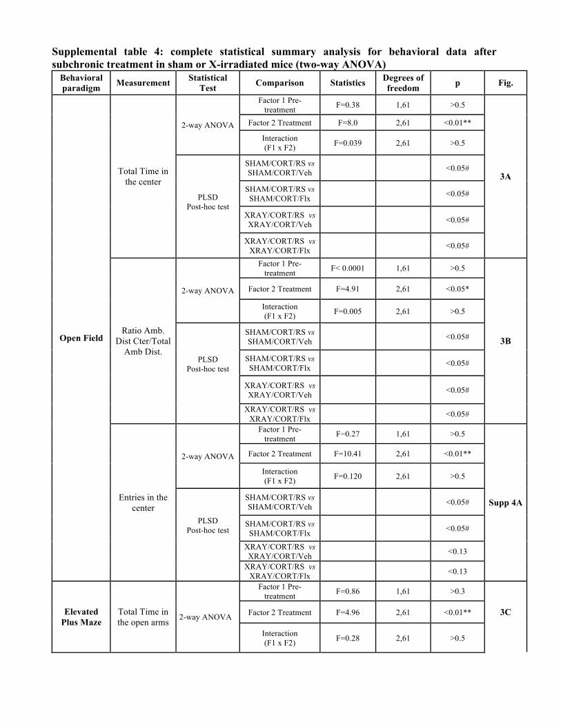
Supplemental table 4: complete statistical summary analysis for behavioral data after subchronic treatment in sham or X-irradiated mice (two-way ANOVA)
Behavioral paradigm Measurement Statistical
Test Comparison Statistics Degrees of freedom p Fig.
Open Field
Total Time in the center
2-way ANOVA
Factor 1 Pre-treatment F=0.38 1,61 >0.5
3A
Factor 2 Treatment F=8.0 2,61 <0.01**
Interaction (F1 x F2) F=0.039 2,61 >0.5
PLSD Post-hoc test
SHAM/CORT/RS vs SHAM/CORT/Veh <0.05#
SHAM/CORT/RS vs SHAM/CORT/Flx <0.05#
XRAY/CORT/RS vs XRAY/CORT/Veh <0.05#
XRAY/CORT/RS vs XRAY/CORT/Flx <0.05#
Ratio Amb. Dist Cter/Total
Amb Dist.
2-way ANOVA
Factor 1 Pre-treatment F< 0.0001 1,61 >0.5
3B
Factor 2 Treatment F=4.91 2,61 <0.05*
Interaction (F1 x F2) F=0.005 2,61 >0.5
PLSD Post-hoc test
SHAM/CORT/RS vs SHAM/CORT/Veh <0.05#
SHAM/CORT/RS vs SHAM/CORT/Flx <0.05#
XRAY/CORT/RS vs XRAY/CORT/Veh <0.05#
XRAY/CORT/RS vs XRAY/CORT/Flx <0.05#
Entries in the center
2-way ANOVA
Factor 1 Pre-treatment F=0.27 1,61 >0.5
Supp 4A
Factor 2 Treatment F=10.41 2,61 <0.01**
Interaction (F1 x F2) F=0.120 2,61 >0.5
PLSD Post-hoc test
SHAM/CORT/RS vs SHAM/CORT/Veh <0.05#
SHAM/CORT/RS vs SHAM/CORT/Flx <0.05#
XRAY/CORT/RS vs XRAY/CORT/Veh <0.13
XRAY/CORT/RS vs XRAY/CORT/Flx <0.13
Elevated Plus Maze
Total Time in the open arms
2-way ANOVA
Factor 1 Pre-treatment F=0.86 1,61 >0.3
3C
Factor 2 Treatment F=4.96 2,61 <0.01**
Interaction (F1 x F2) F=0.28 2,61 >0.5
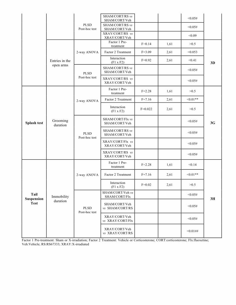
PLSD Post-hoc test
SHAM/CORT/RS vs SHAM/CORT/Veh <0.05#
SHAM/CORT/RS vs SHAM/CORT/Veh <0.05#
XRAY/CORT/RS vs XRAY/CORT/Veh <0.09
Entries in the open arms
2-way ANOVA
Factor 1 Pre-treatment F=0.14 1,61 >0.5
3D
Factor 2 Treatment F=3.09 2,61 <0.053
Interaction (F1 x F2) F=0.92 2,61 <0.41
PLSD Post-hoc test
SHAM/CORT/RS vs SHAM/CORT/Veh <0.05#
XRAY/CORT/RS vs XRAY/CORT/Veh <0.05#
Splash test Grooming duration
2-way ANOVA
Factor 1 Pre-treatment F=2.28 1,61 >0.5
3G
Factor 2 Treatment F=7.16 2,61 <0.01**
Interaction (F1 x F2) F=0.022 2,61 >0.5
PLSD Post-hoc test
SHAM/CORT/Flx vs SHAM/CORT/Veh <0.05#
SHAM/CORT/RS vs SHAM/CORT/Veh <0.05#
XRAY/CORT/Flx vs XRAY/CORT/Veh <0.05#
XRAY/CORT/RS vs XRAY/CORT/Veh <0.05#
Tail Suspension
Test
Immobility duration
2-way ANOVA
Factor 1 Pre-treatment F=2.28 1,61 <0.14
3H
Factor 2 Treatment F=7.16 2,61 <0.01**
Interaction (F1 x F2) F=0.02 2,61 >0.5
PLSD Post-hoc test
SHAM/CORT/Veh vs SHAM/CORT/Flx <0.05#
SHAM/CORT/Veh vs SHAM/CORT/RS <0.05#
XRAY/CORT/Veh vs XRAY/CORT/Flx <0.05#
XRAY/CORT/Veh vs XRAY/CORT/RS <0.01##
Factor 1 Pre-treatment: Sham or X-irradiation; Factor 2 Treatment: Vehicle or Corticosterone; CORT:corticosterone; Flx:fluoxetine; Veh:Vehicle; RS:RS67333; XRAY:X-irradiated
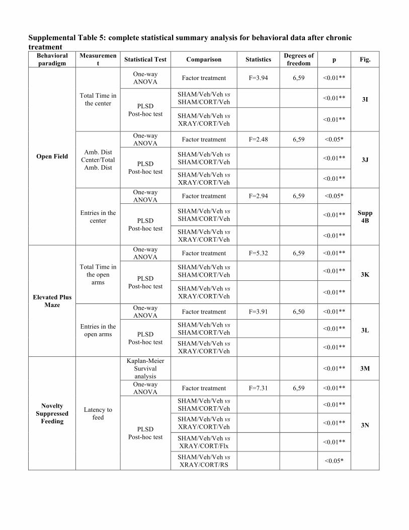
Supplemental Table 5: complete statistical summary analysis for behavioral data after chronic treatment
Behavioral paradigm
Measurement Statistical Test Comparison Statistics Degrees of
freedom p Fig.
Open Field
Total Time in the center
One-way ANOVA Factor treatment F=3.94 6,59 <0.01**
3I PLSD
Post-hoc test
SHAM/Veh/Veh vs SHAM/CORT/Veh <0.01**
SHAM/Veh/Veh vs XRAY/CORT/Veh <0.01**
Amb. Dist Center/Total Amb. Dist
One-way ANOVA Factor treatment F=2.48 6,59 <0.05*
3J PLSD Post-hoc test
SHAM/Veh/Veh vs SHAM/CORT/Veh <0.01**
SHAM/Veh/Veh vs XRAY/CORT/Veh <0.01**
Entries in the center
One-way ANOVA Factor treatment F=2.94 6,59 <0.05*
Supp 4B PLSD
Post-hoc test
SHAM/Veh/Veh vs SHAM/CORT/Veh <0.01**
SHAM/Veh/Veh vs XRAY/CORT/Veh <0.01**
Elevated Plus Maze
Total Time in the open
arms
One-way ANOVA Factor treatment F=5.32 6,59 <0.01**
3K PLSD Post-hoc test
SHAM/Veh/Veh vs SHAM/CORT/Veh <0.01**
SHAM/Veh/Veh vs XRAY/CORT/Veh <0.01**
Entries in the open arms
One-way ANOVA Factor treatment F=3.91 6,50 <0.01**
3L PLSD Post-hoc test
SHAM/Veh/Veh vs SHAM/CORT/Veh <0.01**
SHAM/Veh/Veh vs XRAY/CORT/Veh <0.01**
Novelty Suppressed
Feeding
Latency to feed
Kaplan-Meier Survival analysis
<0.01** 3M
One-way ANOVA Factor treatment F=7.31 6,59 <0.01**
3N PLSD Post-hoc test
SHAM/Veh/Veh vs SHAM/CORT/Veh <0.01**
SHAM/Veh/Veh vs XRAY/CORT/Veh <0.01**
SHAM/Veh/Veh vs XRAY/CORT/Flx <0.01**
SHAM/Veh/Veh vs XRAY/CORT/RS <0.05*
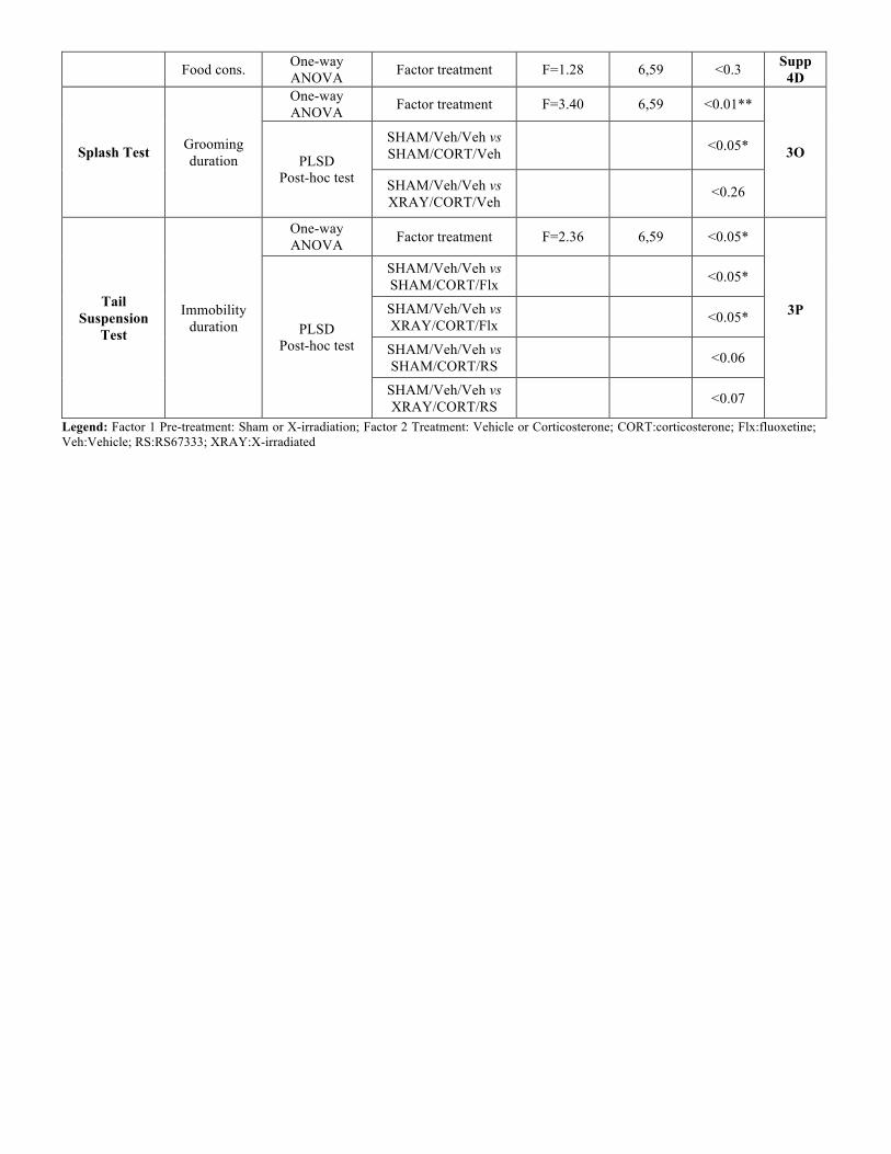
Food cons. One-way ANOVA Factor treatment F=1.28 6,59 <0.3 Supp
4D
Splash Test Grooming duration
One-way ANOVA Factor treatment F=3.40 6,59 <0.01**
3O PLSD Post-hoc test
SHAM/Veh/Veh vs SHAM/CORT/Veh <0.05*
SHAM/Veh/Veh vs XRAY/CORT/Veh
<0.26
Tail Suspension
Test
Immobility duration
One-way ANOVA Factor treatment F=2.36 6,59 <0.05*
3P PLSD
Post-hoc test
SHAM/Veh/Veh vs SHAM/CORT/Flx <0.05*
SHAM/Veh/Veh vs XRAY/CORT/Flx <0.05*
SHAM/Veh/Veh vs SHAM/CORT/RS <0.06
SHAM/Veh/Veh vs XRAY/CORT/RS <0.07
Legend: Factor 1 Pre-treatment: Sham or X-irradiation; Factor 2 Treatment: Vehicle or Corticosterone; CORT:corticosterone; Flx:fluoxetine; Veh:Vehicle; RS:RS67333; XRAY:X-irradiated
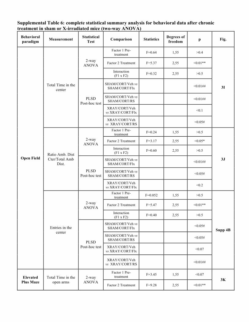
Supplemental Table 6: complete statistical summary analysis for behavioral data after chronic treatment in sham or X-irradiated mice (two-way ANOVA)
Behavioral paradigm Measurement Statistical
Test Comparison Statistics Degrees of freedom p Fig.
Open Field
Total Time in the center
2-way ANOVA
Factor 1 Pre-treatment F=0.64 1,55 >0.4
3I
Factor 2 Treatment F=5.37 2,55 <0.01**
Interaction (F1 x F2) F=0.32 2,55 >0.5
PLSD Post-hoc test
SHAM/CORT/Veh vs SHAM/CORT/Flx <0.01##
SHAM/CORT/Veh vs SHAM/CORT/RS <0.01##
XRAY/CORT/Veh vs XRAY/CORT/Flx <0.1
XRAY/CORT/Veh vs XRAY/CORT/RS <0.05#
Ratio Amb. Dist Cter/Total Amb
Dist.
2-way ANOVA
Factor 1 Pre-treatment F=0.24 1,55 >0.5
3J
Factor 2 Treatment F=3.17 2,55 <0.05*
Interaction (F1 x F2) F=0.60 2,55 >0.5
PLSD Post-hoc test
SHAM/CORT/Veh vs SHAM/CORT/Flx <0.01##
SHAM/CORT/Veh vs SHAM/CORT/RS <0.05#
XRAY/CORT/Veh vs XRAY/CORT/Flx <0.2
Entries in the center
2-way ANOVA
Factor 1 Pre-treatment F=0.052 1,55 >0.5
Supp 4B
Factor 2 Treatment F=5.47 2,55 <0.01**
Interaction (F1 x F2) F=0.40 2,55 >0.5
PLSD Post-hoc test
SHAM/CORT/Veh vs SHAM/CORT/Flx <0.05#
SHAM/CORT/Veh vs SHAM/CORT/RS <0.05#
XRAY/CORT/Veh vs XRAY/CORT/Flx <0.07
XRAY/CORT/Veh vs XRAY/CORT/RS <0.01##
Elevated Plus Maze
Total Time in the open arms
2-way ANOVA
Factor 1 Pre-treatment F=3.45 1,55 <0.07
3K Factor 2 Treatment F=9.28 2,55 <0.01**
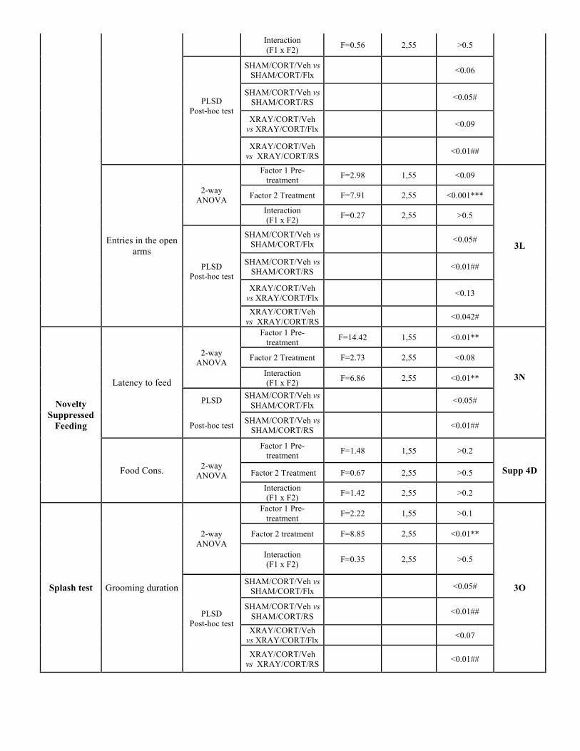
Interaction (F1 x F2) F=0.56 2,55 >0.5
PLSD Post-hoc test
SHAM/CORT/Veh vs SHAM/CORT/Flx <0.06
SHAM/CORT/Veh vs SHAM/CORT/RS <0.05#
XRAY/CORT/Veh vs XRAY/CORT/Flx <0.09
XRAY/CORT/Veh vs XRAY/CORT/RS <0.01##
Entries in the open arms
2-way ANOVA
Factor 1 Pre-treatment F=2.98 1,55 <0.09
3L
Factor 2 Treatment F=7.91 2,55 <0.001***
Interaction (F1 x F2) F=0.27 2,55 >0.5
PLSD Post-hoc test
SHAM/CORT/Veh vs SHAM/CORT/Flx <0.05#
SHAM/CORT/Veh vs SHAM/CORT/RS <0.01##
XRAY/CORT/Veh vs XRAY/CORT/Flx <0.13
XRAY/CORT/Veh vs XRAY/CORT/RS <0.042#
Novelty Suppressed
Feeding
Latency to feed
2-way ANOVA
Factor 1 Pre-treatment F=14.42 1,55 <0.01**
3N
Factor 2 Treatment F=2.73 2,55 <0.08
Interaction (F1 x F2) F=6.86 2,55 <0.01**
PLSD SHAM/CORT/Veh vs SHAM/CORT/Flx <0.05#
Post-hoc test SHAM/CORT/Veh vs SHAM/CORT/RS <0.01##
Food Cons. 2-way ANOVA
Factor 1 Pre-treatment F=1.48 1,55 >0.2
Supp 4D Factor 2 Treatment F=0.67 2,55 >0.5
Interaction (F1 x F2) F=1.42 2,55 >0.2
Splash test Grooming duration
2-way ANOVA
Factor 1 Pre-treatment F=2.22 1,55 >0.1
3O
Factor 2 treatment F=8.85 2,55 <0.01**
Interaction (F1 x F2) F=0.35 2,55 >0.5
PLSD Post-hoc test
SHAM/CORT/Veh vs SHAM/CORT/Flx <0.05#
SHAM/CORT/Veh vs SHAM/CORT/RS <0.01##
XRAY/CORT/Veh vs XRAY/CORT/Flx <0.07
XRAY/CORT/Veh vs XRAY/CORT/RS <0.01##
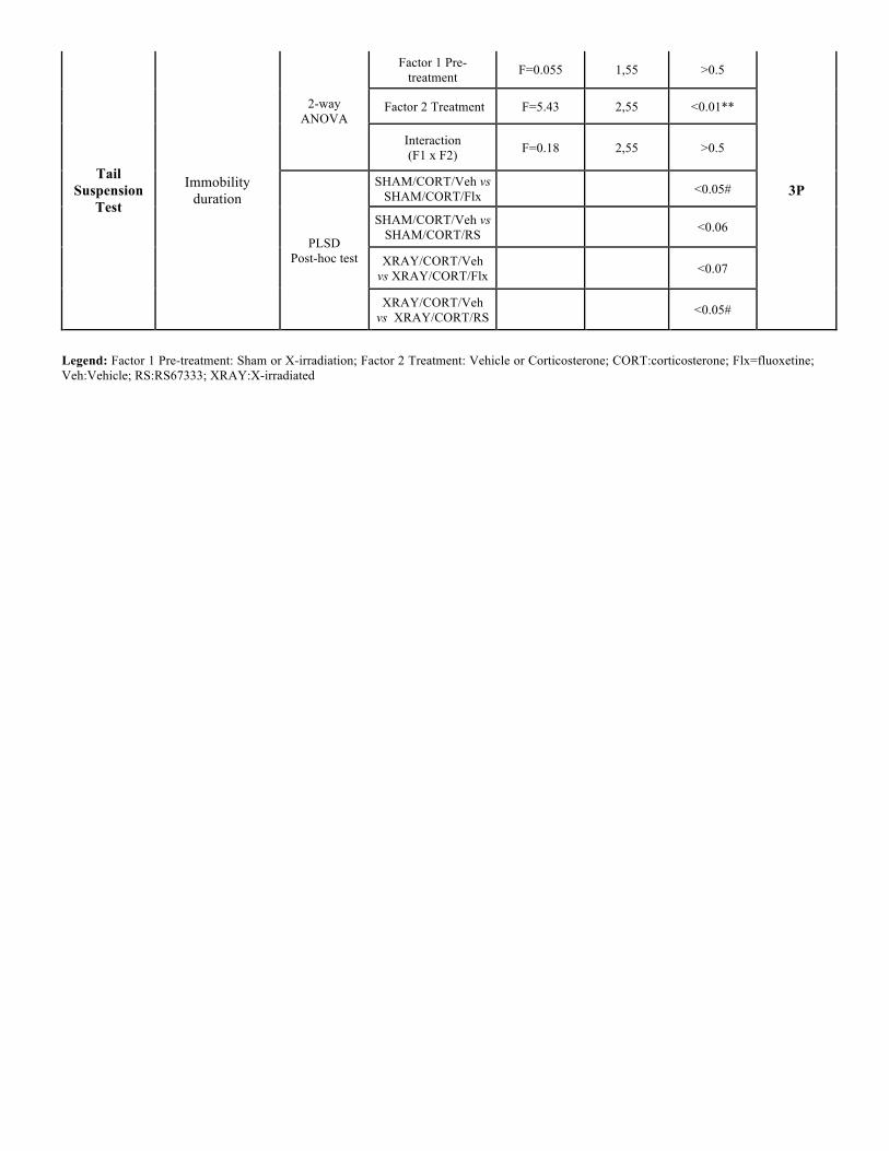
Tail Suspension
Test
Immobility duration
2-way ANOVA
Factor 1 Pre-treatment F=0.055 1,55 >0.5
3P
Factor 2 Treatment F=5.43 2,55 <0.01**
Interaction (F1 x F2) F=0.18 2,55 >0.5
PLSD Post-hoc test
SHAM/CORT/Veh vs SHAM/CORT/Flx <0.05#
SHAM/CORT/Veh vs SHAM/CORT/RS <0.06
XRAY/CORT/Veh vs XRAY/CORT/Flx <0.07
XRAY/CORT/Veh vs XRAY/CORT/RS <0.05#
Legend: Factor 1 Pre-treatment: Sham or X-irradiation; Factor 2 Treatment: Vehicle or Corticosterone; CORT:corticosterone; Flx=fluoxetine; Veh:Vehicle; RS:RS67333; XRAY:X-irradiated
Résultats expérimentaux
267
Commentaires sur Article 2 :
Cette étude montre pour la première fois, dans un modèle d’anxiété/dépression, qu’un agoniste du
récepteur 5-HT4 peut agir comme un composé anxiolytique et antidépresseur de façon rapide et que
la stimulation du récepteur 5-HT4 est nécessaire pour les effets comportementaux et neurogéniques
(prolifération et maturation) de la fluoxetine. De plus, nous avons démontré qu’à l’exception du test
du NSF, les effets de type anxiolytiques et antidépresseurs de l’agoniste 5-HT4 sont indépendants de
la neurogenèse hippocampique.
Ce travail est encourageant pour le développement des agonistes du récepteur 5-HT4 tel que le
RS67333 comme un médicament anxiolytique et antidépresseur pour les patients atteints de
dépression majeure avec une co-morbidité anxieuse. Toutefois, d’autres propriétés
pharmacologiques importantes de ces molécules tant au niveau du système nerveux central
(cognition, satiété) qu’au niveau périphérique (cœur, intestin, vessie) doivent être prises en compte
pour une éventuelle utilisation de cette famille d’agonistes du récepteur 5-HT4 en clinique.
Devant l’efficacité du composé RS67333 dans la correction du phénotype anxio/dépressif induit par
le modèle CORT et connaissant les propriétés pro-cognitives des agonistes du récepteur 5-HT4 chez
des animaux naïfs, nous nous sommes interrogés sur la capacité du RS67333 à restaurer les troubles
cognitifs induits par l’administration de corticostérone à long-terme.

Résultats expérimentaux
268
ARTICLE 3: Chronic 5-HT4 receptor agonist treatment restores learning and
memory deficits in a neuroendocrine mouse model of
anxiety/depression
Flavie Darcet, Alain M Gardier, Denis J David, Jean-Philippe Guilloux
Publié dans le journal Neuroscience Letters (Février 2016)
Question posée :
Les différents troubles de l’apprentissage et de la mémoire induits par le modèle CORT sont-ils
corrigés par une administration chronique d’un antidépresseur monoaminergique classique
(fluoxetine) ou d’un agoniste des récepteurs 5-HT4 (RS67333)?
Résumé de l’étude
Les troubles cognitifs sont souvent reportés comme des symptômes invalidants chez les patients
souffrant de dépression majeure. De plus, plusieurs études ont montré que certaines des difficultés
cognitives observées pendant des épisodes dépressifs persistaient malgré la rémission des patients. Il
est donc nécessaire d’identifier de nouvelles stratégies thérapeutiques qui permettent de prendre en
charge les symptômes émotionnels liés à la pathologie de la dépression au même titre que les
troubles cognitifs associés.
Récemment, de nombreuses études se sont intéressées aux propriétés antidépressives des agonistes
des récepteurs 5-HT4 (Lucas et al 2007 ; Pascual-Brazo et al 2012). Cependant, peu d’entre elles
conduisent leurs tests sur des animaux anxio/dépressifs (Mendez-David et al 2014) et moins encore
se sont concentrées sur les propriétés pro-cognitives des agonistes des récepteurs 5-HT4 dans un
contexte d’anxiété/dépression.
C’est pourquoi nous avons testé l’efficacité du RS67333, un agoniste du récepteur 5-HT4, dans notre
modèle d’anxiété/dépression basé sur une exposition chronique à la corticostérone sur les
performances cognitives des animaux, en comparaison à un traitement chronique par un
antidépresseur monoaminergique classique, la fluoxetine.
Comme nous l’avons précédemment démontré, (Article 1), un trouble cognitif global chez les
animaux traités à la CORT est observé. D’un point de vue pharmacologique, les traitements
chroniques par la fluoxetine et par le RS67333 ont permis de restaurer le déficit de mémoire de type
épisodique observé chez les animaux anxio/dépressifs, en rétablissant l’index de
Résultats expérimentaux
269
discrimination au niveau des animaux contrôles. Dans le test de conditionnement par la peur, alors
que l’agoniste du récepteur 5-HT4 améliore l’altération de la mémoire associative induite par la
CORT, le traitement par la fluoxetine ne modifie pas le déficit observé chez les animaux CORT. En
outre, le nombre d’épisodes de freezing est même significativement inférieur à celui des animaux
traités par la CORT. Enfin, dans le Barnes maze, le traitement par le RS67333 améliore aussi bien le
déficit d’apprentissage que de mémoire chez les animaux CORT. Le traitement chronique par la
fluoxetine, pour sa part, a permis de corriger uniquement le déficit d’apprentissage.
Dans l’ensemble, un traitement chronique par le RS67333 est capable de corriger entièrement le
déficit cognitif induit par la corticostérone. En revanche, le traitement chronique par la fluoxetine ne
permet d’obtenir qu’une restauration partielle des fonctions cognitives, dépendante du type de
mémoire testé et des paramètres d’apprentissage et de mémoire eux-mêmes. Ces résultats
suggèrent que ces deux stratégies thérapeutiques pourraient mettre en jeu des mécanismes d’action
différents (mémoire associative et spatiale) ou parfois similaires (mémoire type épisodique) en
fonction du type de mémoire engagé.
Contribution personnelle :
Au cours de ce travail :
x J’ai réalisé le suivi des animaux incluant la préparation et l’administration des traitements
pharmacologiques (traitement par la CORT et par la fluoxetine, préparation et implantation
des mini-pompes pour RS67333).
x J’ai mené l’ensemble des tests comportementaux émotionnels et cognitifs.
x J’ai analysé les résultats et rédigé l’intégralité de l’article sous la supervision du Pr. Denis
David et du Dr Jean-Philippe Guilloux
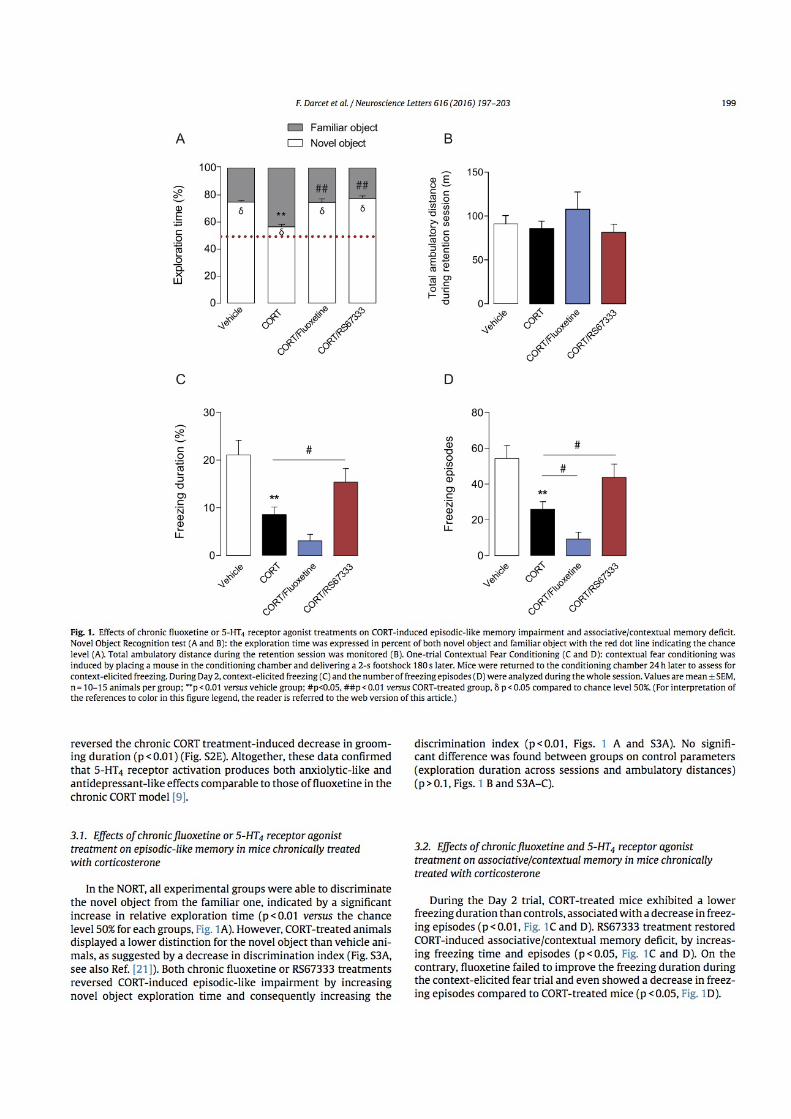
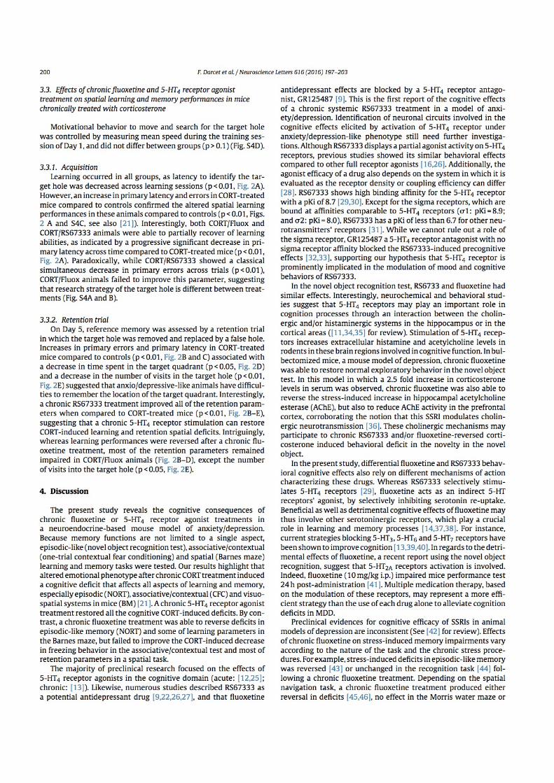
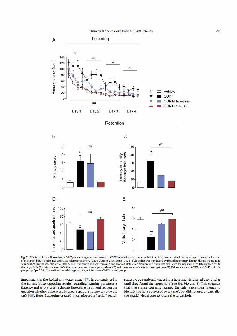
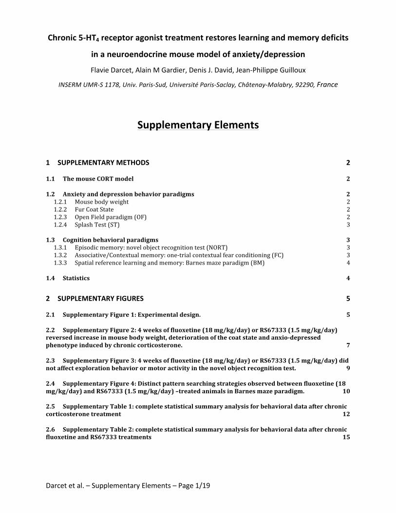
Darcetetal.–SupplementaryElements–Page1/19
Chronic5-HT4receptoragonisttreatmentrestoreslearningandmemorydeficits
inaneuroendocrinemousemodelofanxiety/depression
FlavieDarcet,AlainMGardier,DenisJ.David,Jean-PhilippeGuilloux
INSERMUMR-S1178,Univ.Paris-Sud,UniversitéParis-Saclay,Châtenay-Malabry,92290,France
SupplementaryElements
1 SUPPLEMENTARYMETHODS 2
1.1 ThemouseCORTmodel 2
1.2 Anxietyanddepressionbehaviorparadigms 21.2.1 Mousebodyweight 21.2.2 FurCoatState 21.2.3 OpenFieldparadigm(OF) 21.2.4 SplashTest(ST) 3
1.3 Cognitionbehavioralparadigms 31.3.1 Episodicmemory:novelobjectrecognitiontest(NORT) 31.3.2 Associative/Contextualmemory:one-trialcontextualfearconditioning(FC) 31.3.3 Spatialreferencelearningandmemory:Barnesmazeparadigm(BM) 4
1.4 Statistics 4
2 SUPPLEMENTARYFIGURES 5
2.1 SupplementaryFigure1:Experimentaldesign. 5
2.2 SupplementaryFigure2:4weeksoffluoxetine(18mg/kg/day)orRS67333(1.5mg/kg/day)reversedincreaseinmousebodyweight,deteriorationofthecoatstateandanxio-depressedphenotypeinducedbychroniccorticosterone. 7
2.3 SupplementaryFigure3:4weeksoffluoxetine(18mg/kg/day)orRS67333(1.5mg/kg/day)didnotaffectexplorationbehaviorormotoractivityinthenovelobjectrecognitiontest. 9
2.4 SupplementaryFigure4:Distinctpatternsearchingstrategiesobservedbetweenfluoxetine(18mg/kg/day)andRS67333(1.5mg/kg/day)–treatedanimalsinBarnesmazeparadigm. 10
2.5 SupplementaryTable1:completestatisticalsummaryanalysisforbehavioraldataafterchroniccorticosteronetreatment 12
2.6 SupplementaryTable2:completestatisticalsummaryanalysisforbehavioraldataafterchronicfluoxetineandRS67333treatments 15
Darcetetal.–SupplementaryElements–Page2/19
1 SupplementaryMethods
1.1 ThemouseCORTmodel
Themodelofelevatedglucocorticoids(alsonamed“CORTmodel”)isabletoblunttheresponse
of thehypothalamic-pituitary-adrenalaxis (HPA)asshownbythemarkedlyattenuatedstress-
induced corticosterone levels observed in these mice [1]. This model displays hallmark
characteristics of anxiety anddepression. Thedose anddurationof corticosterone treatment
wasselectedbasedonpreviousstudies[1,2].
1.2 Anxietyanddepressionbehaviorparadigms
1.2.1 Mousebodyweight
Mousebodyweightforeachanimalwasfollowedonceaweekduringalltheprocedure.
1.2.2 FurCoatState
The score corresponding to the state of the coat resulted from the sum of the score of five
differentbodyparts:head,neck,dorsal/ventral coat, tail and fore-/hindpaws. Foreachbody
area,ascoreof0wasgivenforawell-groomedcoatand1foranunkemptcoat[1,3].Furcoat
stateandmousebodyweightwasfollowedweeklyduringthewholetreatmentperiod.
1.2.3 OpenFieldparadigm(OF)
Motor activity was quantified in Plexiglas OF boxes 43x43 cm (MED associates, Georgia, VT,
USA)duringa10-minsession [4].Twosetsof16pulse-modulated infraredphotobeamswere
placedonoppositewalls2.5cmaparttorecordx-yambulatorymovements.Activitychambers
werecomputerinterfacedfordatasamplingat100msresolution.Thecenterwasdefinedasa
32x32-cmcentralarena.Dependentmeasureswere:totaltimespentinthecenter,thenumbers
Darcetetal.–SupplementaryElements–Page3/19
ofentriesintothecenteranddistancetravelledinthecenterdividedbytotaldistancetravelled.
Overallmotoractivitywasquantifiedasthetotaldistancetravelled(cm).
1.2.4 SplashTest(ST)
This test consisted in squirting a 10% sucrose solution on themouse’s snout. The grooming
durationwasthenrecordedovera5minperiodinthehomecageoftheanimal[5].
1.3 Cognitionbehavioralparadigms
1.3.1 Episodicmemory:novelobjectrecognitiontest(NORT)
TheNORTwasdividedinto4trainingsessionsandonetestsessionasdescribedpreviously[6].
Eachexposure lasted5minuteswitha3-min inter-trial interval.During training sessions, two
identical objects were present in the box and the mouse was allowed to freely explore the
apparatus and the objects. During the test session, one of the familiar objectswas removed
fromthecageandreplacedbyanovelobject.Objectexplorationwasdefinedastheorientation
of thenose to theobjectatadistance≤2cm.Ambulatoryactivitywascontrolledduring the
entireexperimentandobjectsexplorationwashand-scoredbyanexperimenter.Objectswere
cleanedwith 70% ethanol between trials to avoid olfactory cues. Results for this test were
expressed as: 1) exploration (in percent) of each object during the test session and 2) a
discrimination index (DI)betweenobjectsduringthetestsession,calculatedas thedifference
betweenthetimespentexploringthenovelobject(N)andthefamiliarobject(F)dividedbythe
totaltimeexploringbothobjects(DI=(N-F)/(N+F)).
1.3.2 Associative/Contextualmemory:one-trialcontextualfearconditioning(FC)
Darcetetal.–SupplementaryElements–Page4/19
The experimental design ran over two consecutive days. On Day 1,micewere placed in the
conditioning chamber and, 3 min later, received one shock (2 sec., 0.75 mA). Mice were
removed15secaftertheshock.OnDay2,animalsreturnedtotheconditioningchamberfora
4-minperiod intheexactsameconditionsofDay1,butwithoutelectricalshock, foratestof
context-elicitedfreezing.Trainingchamberswerecleanedwith70%ethanolsolutionbeforeand
aftereachtrial.ScoringwasmeasuredusingFreezingsoftwareversion2.0.04(Packwin,Harvard
apparatus,Bioseb,France).
1.3.3 Spatialreferencelearningandmemory:Barnesmazeparadigm(BM)
Spatialacquisitionwasorganized in4trainingsessions (Day1toDay4).Eachtrainingsession
consisted in four 3-min trials, with 15-20 min intertrial intervals during which animals were
returnedtotheirhomecage.Primarylatencyandprimaryerrorstoidentifythetargetholewere
manuallyscoredduringall learningtrials.Aholewasconsideredvisitedwhenmicetiltedtheir
headover it(nosepoke)or introducedtheirpawsintothehole.OnDay5,referencememory
wasevaluatedbyaretentionprobetrial(90sec)duringwhichthetargetboxwasremovedand
thetargetholewasclosed.Micewereallowedtoexplorethemazeandtovisitthetargetand
theadjacentholes.Latencytoreachthetargetholeforthefirsttime,numberoferrorsbefore
reaching the target hole, number of visits in the target hole and time spent in the target
quadrantwererecorded(ANY-mazeSoftware)
1.4 Statistics
Resultsfromdataanalyseswereexpressedasmean±SEM.Statisticalanalyseswereprocessed
withStatview5.0®Software(SASInstitute,Cary,NC).Forallexperiments,comparisonsbetween
CORT-treated and control animals were performed by using t-tests or one-way ANOVAwith
Darcetetal.–SupplementaryElements–Page5/19
repeated-measureswhenappropriate.One-wayANOVAaloneorwithrepeated-measureswere
appliedtothedataasappropriate.Significantmaineffectsand/orinteractionswerefollowedby
Fisher’sPLSDposthocanalysis.Onesamplet-testswereusedtocomparethepercentoftime
exploringthenovelobjectversusthechancelevel(50%)inthenovelobjectrecognitiontestand
the time spent in the target quadrant versus the chance level (22.5 sec) in the BM test.
Statistical significance was set at p<0.05. A summary of statistical measures is included in
SupplementaryTables1and2.
2 SupplementaryFigures
2.1 SupplementaryFigure1:Experimentaldesign.
C57BL/6JRjmicereceiveda4-weekscorticosteronetreatment indrinkingwater to inducethe
anxio-depressed phenotype.Mice were then given a 4-week chronic fluoxetine treatment in
drinkingwaterorthe5-HT4receptoragonist(RS67333)treatmentviaosmoticmini-pumps.Mice
were then subjected to a complete characterization of cognitive functions in episodic-like,
spatialandassociativedomains.Eachanimalwassuccessivelytestedintheopenfieldparadigm,
thenovel object recognition test, theBarnesmaze, theone-trial contextual fear conditioning
andtheSplashtest.
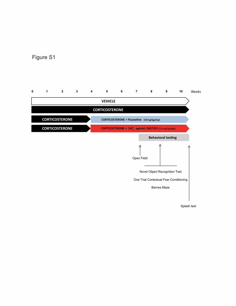
0 41 2 6
Open Field
Splash test
Behavioral testing
Novel Object Recognition Test
One Trial Contextual Fear Conditioning
Barnes Maze
CORTICOSTERONE
VEHICLE
Weeks3 5 7 8
CORTICOSTERONE + Fluoxetine (18 mg/kg/day)
CORTICOSTERONE + 5HT4 agonist, RS67333 (1.5 mg/kg/day)
CORTICOSTERONE
CORTICOSTERONE
9 10
Figure S1
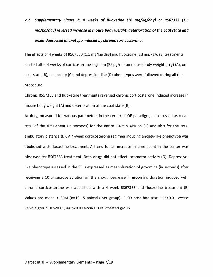
Darcetetal.–SupplementaryElements–Page7/19
2.2 Supplementary Figure 2: 4 weeks of fluoxetine (18 mg/kg/day) or RS67333 (1.5
mg/kg/day)reversedincreaseinmousebodyweight,deteriorationofthecoatstateand
anxio-depressedphenotypeinducedbychroniccorticosterone.
Theeffectsof4weeksofRS67333(1.5mg/kg/day)andfluoxetine(18mg/kg/day)treatments
startedafter4weeksofcorticosteroneregimen(35µg/ml)onmousebodyweight(ing)(A),on
coatstate(B),onanxiety(C)anddepression-like(D)phenotypeswerefollowedduringallthe
procedure.
ChronicRS67333andfluoxetinetreatmentsreversedchroniccorticosteroneinducedincreasein
mousebodyweight(A)anddeteriorationofthecoatstate(B).
Anxiety,measuredforvariousparametersinthecenterofOFparadigm,isexpressedasmean
total of the time-spent (in seconds) for the entire 10-min session (C) and also for the total
ambulatorydistance(D).A4-weekcorticosteroneregimeninducinganxiety-likephenotypewas
abolishedwith fluoxetine treatment. A trend for an increase in time spent in the centerwas
observedforRS67333treatment.Bothdrugsdidnotaffect locomotoractivity(D).Depressive-
likephenotypeassessedintheSTisexpressedasmeandurationofgrooming(inseconds)after
receiving a 10% sucrose solutionon the snout.Decrease in groomingduration inducedwith
chronic corticosterone was abolished with a 4 week RS67333 and fluoxetine treatment (E)
Values are mean ± SEM (n=10-15 animals per group). PLSD post hoc test: **p<0.01 versus
vehiclegroup;#p<0.05,##p<0.01versusCORT-treatedgroup.
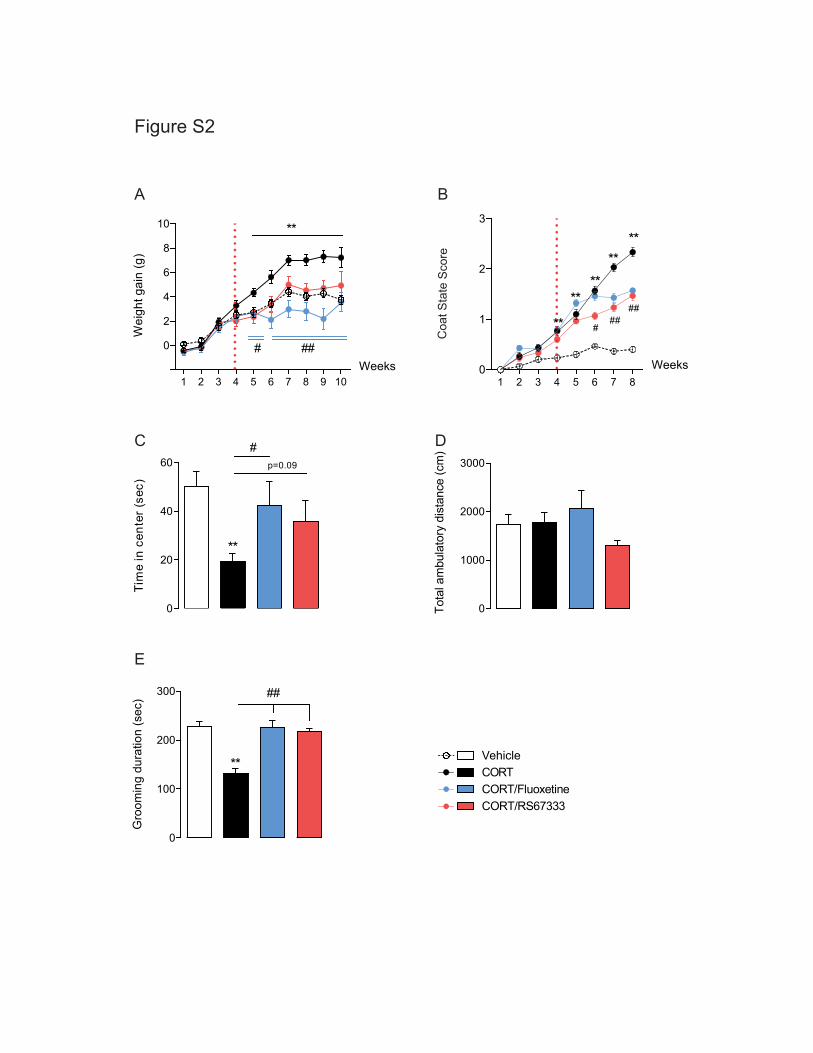
Figure S2Ti
me
in c
ente
r (se
c)
0
20
40
60
**
#p=0.09
Gro
omin
g du
ratio
n (s
ec)
0
100
200
300
**
##
A B
C
E
DTo
tal a
mbu
lato
ry d
ista
nce
(cm
)
0
1000
2000
3000
Weeks
VehicleCORTCORT/FluoxetineCORT/RS67333
Wei
ght g
ain
(g)
1 2 3 4 5 6 7 8 9 10
0
2
4
6
8
10 **
###
1 2 3 4 5 6 7 80
1
2
3
****
****
** ###
##
Coa
t Sta
te S
core
Weeks
Darcetetal.–SupplementaryElements–Page9/19
2.3 Supplementary Figure 3: 4 weeks of fluoxetine (18 mg/kg/day) or RS67333 (1.5
mg/kg/day) did not affect exploration behavior or motor activity in the novel object
recognitiontest.
Theeffectsof4weeksofRS67333(1.5mg/kg/day)andfluoxetine(18mg/kg/day)treatments
started after 4weeks of corticosterone regimen (35µg/ml) onbothobjects exploration time
along learning sessions (A) and ambulatory distance during training (B) and test (C) sessions
were followed during the NORT. None of these parameters was affected by either chronic
corticosterone or drugs (RS67333, fluoxetine) treatments. Values are mean ± SEM (n=10-15
animalspergroup).
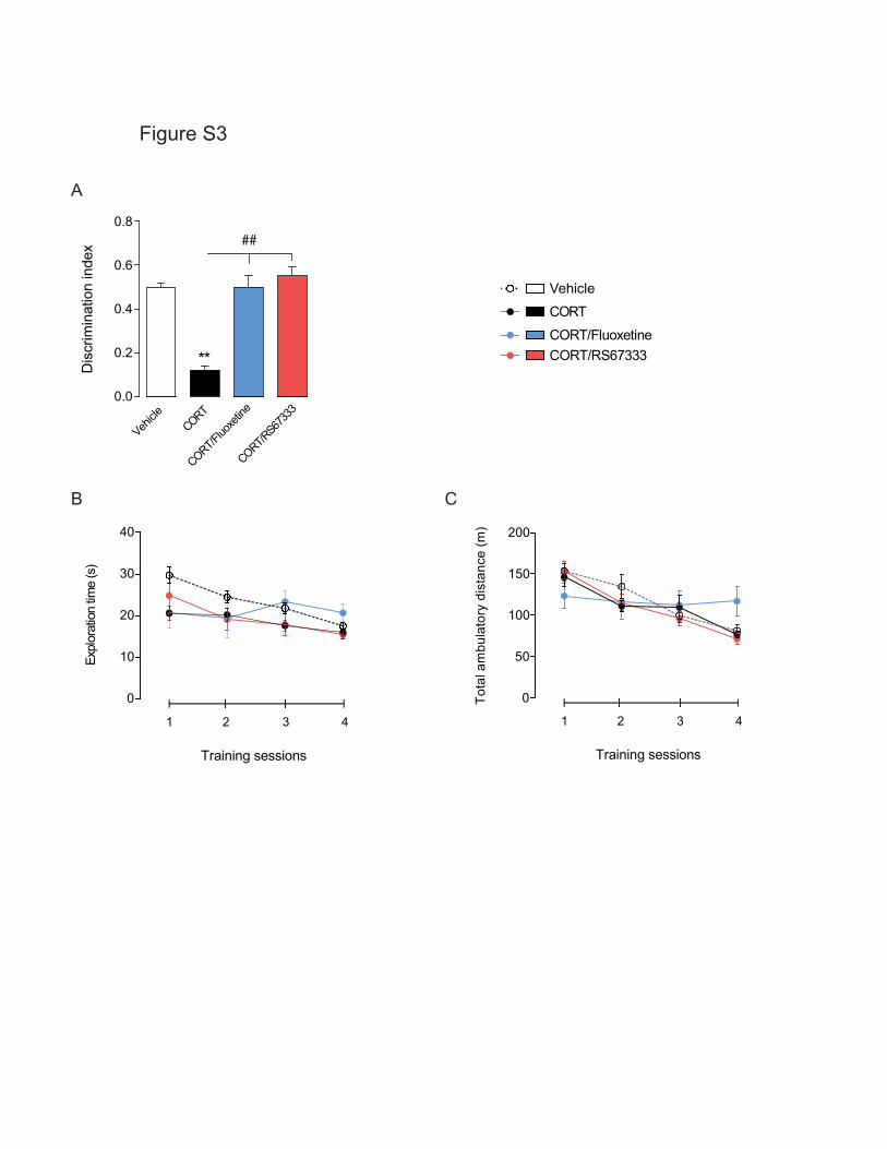
Figure S3
A
Vehicle
CORT
CORT/FluoxetineCORT/RS67333
BTo
tal a
mbu
lato
ry d
ista
nce
(m)
0
50
100
150
200
Training sessions
1 2 3 4
Expl
orat
ion
time
(s)
0
10
20
30
40
Training sessions
1 2 3 4
C
Dis
crim
inat
ion
inde
x
0.0
0.2
0.4
0.6
0.8
**
##
Vehicle
CORT
CORT/Fluo
xetin
e
CORT/RS67
333
Darcetetal.–SupplementaryElements–Page10/19
2.4 Supplementary Figure 4: Distinct pattern searching strategies observed between
fluoxetine (18mg/kg/day) and RS67333 (1.5 mg/kg/day) –treated animals in Barnes
mazeparadigm.
Contradictory learning parameters revealed distinct pattern of searching strategies between
CORT/Fluoxetine and CORT/RS67333 groups as shown with representative cartoons of 4
differentanimalspergroup(A).Percentageofanimalsdisplayingeithera“non-serial”or“serial”
searchstrategyduringatleast2outof4trialsduringthelearningperiod(B).Numberoferrors
beforevisitingthetargethole(primaryerrors)scoredalongtrainingsessions(C).Averagespeed
during the first four trials of Day 1 training was measured to detect motivational variations
betweenexperimentalgroups(D).
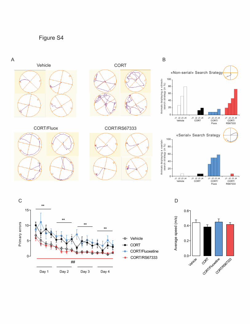
Figure S4
D
Ave
rage
spe
ed (m
/s)
0.0
0.2
0.4
0.6
Vehicle
CORT
CORT/Fluo
xetine
CORT/RS6
7333
C
Prim
ary
erro
rs
0
5
10
15
Day 1 Day 2 Day 3 Day 4
****
**
**
##
Vehicle
CORTCORT/FluoxetineCORT/RS67333
A B
CORT/Fluox CORT/RS67333
Vehicle CORT
0
20
40
60
80
100
Vehicle CORT CORT/Fluox
CORT/RS67333
J1 J2 J3 J4 J1 J2 J3 J4J1 J2 J3 J4J1 J2 J3 J4
Vehicle CORT CORT/Fluox
CORT/RS67333
J1 J2 J3 J4 J1 J2 J3 J4J1 J2 J3 J4J1 J2 J3 J40
20
40
60
80
100 «Serial» Search Srategy
Ani
mal
s ds
ipla
ying
a «
seria
l»se
arch
str
ateg
y (in
%)
Ani
mal
s ds
ipla
ying
a «
dire
ct»
sear
ch s
trat
egy
(in %
)
«Non-serial» Search Srategy
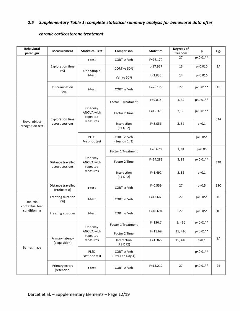
Darcetetal.–SupplementaryElements–Page12/19
2.5 SupplementaryTable1:completestatisticalsummaryanalysisforbehavioraldataafter
chroniccorticosteronetreatment
Behavioralparadigm Measurement StatisticalTest Comparison Statistics Degreesof
freedom p Fig.
Novelobjectrecognitiontest
Explorationtime(%)
t-test CORTvsVeh F=76.179 27 p<0.01**
1AOnesample
t-test
CORTvs50% t=17.967 13 p<0.01δ
Vehvs50% t=3.835 14 p<0.01δ
DiscriminationIndex t-test CORTvsVeh F=76.179 27 p<0.01** 1B
Explorationtimeacrosssessions
One-wayANOVAwithrepeatedmeasures
Factor1Treatment F=9.814 1,39 p<0.01**
S3A
Factor2Time F=15.376 3,39 p<0.01**
Interaction(F1XF2)
F=3.056 3,39 p>0.1
PLSDPost-hoctest
CORTvsVeh(Session1,3)
p<0.05*
Distancetravelledacrosssessions
One-wayANOVAwithrepeatedmeasures
Factor1Treatment F=0.670 1,81 p>0.05
S3BFactor2Time F=24.289 3,81 p<0.01**
Interaction(F1XF2)
F=1.492 3,81 p>0.1
Distancetravelled(Probetest) t-test CORTvsVeh F=0.559 27 p>0.5 S3C
One-trialcontextualfearconditioning
Freezingduration(%) t-test CORTvsVeh F=12.669 27 p<0.05* 1C
Freezingepisodes t-test CORTvsVeh F=10.694 27 p<0.05* 1D
Barnesmaze
Primarylatency(acquisition)
One-wayANOVAwithrepeatedmeasures
Factor1Treatment F=136.7 1,416 p<0.01**
2AFactor2Time F=11.69 15,416 p<0.01**
Interaction(F1XF2)
F=1.366 15,416 p>0.1
PLSDPost-hoctest
CORTvsVeh(Day1toDay4)
p<0.01**
Primaryerrors(retention) t-test CORTvsVeh F=13.210 27 p<0.01** 2B
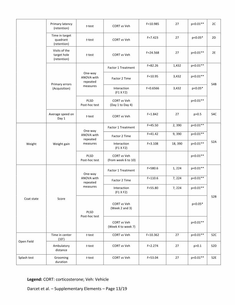
Darcetetal.–SupplementaryElements–Page13/19
Primarylatency(retention) t-test CORTvsVeh F=10.985 27 p<0.01** 2C
Timeintargetquadrant(retention)
t-test CORTvsVeh F=7.423 27 p<0.05* 2D
Visitsofthetargethole(retention)
t-test CORTvsVeh F=24.568 27 p<0.01** 2E
Primaryerrors(Acquisition)
One-wayANOVAwithrepeatedmeasures
Factor1Treatment F=82.26 1,432 p<0.01**
S4B
Factor2Time F=10.95 3,432 p<0.01**
Interaction(F1XF2)
F=0.6566 3,432 p<0.05*
PLSDPost-hoctest
CORTvsVeh(Day1toDay4)
p<0.01**
AveragespeedonDay1 t-test CORTvsVeh F=1.842 27 p>0.5 S4C
Weight Weightgain
One-wayANOVAwithrepeatedmeasures
Factor1Treatment F=45.50 2,390 p<0.01**
S2A
Factor2Time F=41.42 9,390 p<0.01**
Interaction(F1XF2)
F=3.108 18,390 p<0.01**
PLSDPost-hoctest
CORTvsVeh(fromweek6to10)
p<0.01**
Coatstate Score
One-wayANOVAwithrepeatedmeasures
Factor1Treatment F=580.6 1,224 p<0.01**
S2B
Factor2Time F=110.6 7,224 p<0.01**
Interaction(F1XF2)
F=55.80 7,224 p<0.01**
PLSDPost-hoctest
CORTvsVeh(Week2and3)
p<0.05*
CORTvsVeh(Week4toweek7)
p<0.01**
OpenField
Timeincenter(10’)
t-test CORTvsVeh F=10.362 27 p<0.01** S2C
Ambulatorydistance
t-test CORTvsVeh F=2.274 27 p>0.1 S2D
Splashtest Groomingduration
t-test CORTvsVeh F=53.04 27 p<0.01** S2E
Legend:CORT:corticosterone;Veh:Vehicle

Darcetetal.–SupplementaryElements–Page14/19
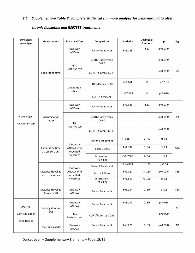
Darcetetal.–SupplementaryElements–Page15/19
2.6 SupplementaryTable2:completestatisticalsummaryanalysisforbehavioraldataafter
chronicfluoxetineandRS67333treatments
Behavioralparadigm Measurement StatisticalTest Comparison Statistics Degreesof
freedom p Fig.
Novelobject
recognitiontest
Explorationtime
One-wayANOVA FactorTreatment F=32.28 2.37 p<0.01##
1A
PLSDPost-hoctest
CORT/FluoxversusCORT p<0.01##
CORT/RSversusCORT p<0.01##
Onesamplet-test
CORT/Fluoxvs50% t=8.161 11 p<0.01δ
CORT/RSvs50%
t=17.069 14 p<0.01δ
DiscriminationIndex
One-wayANOVA FactorTreatment F=32.28 2,37 p<0.01##
1B
PLSDPost-hoctest
CORT/FluoxversusCORT
p<0.01##
CORT/RSversusCORT p<0.01##
Explorationtimeacrosssessions
One-wayANOVAwithrepeatedmeasures
Factor1Treatment F=0.8210 2,56 p>0.1
S3AFactor2Time F=1.384 3,56 p>0.1
Interaction(F1XF2)
F=0.7885 6,56 p>0.1
Distancetravelledacrosssessions
One-wayANOVAwithrepeatedmeasures
Factor1Treatment F=0.4758 2,160 p>0.05
S3BFactor2Time F=9.027 3,160 p<0.01##
Interaction(F1XF2)
F=1.840 6,160 p>0.1
Distancetravelled(Probetest)
One-wayANOVA FactorTreatment F=1.184 2,39 p>0.5 S3C
One-trial
contextualfear
conditioning
Freezingduration(%)
One-wayANOVA FactorTreatment F=8.131 2,39 p<0.05#
1C
PLSDPost-hoctest CORT/RSversusCORT
p<0.05#
Freezingepisodes One-wayANOVA
FactorTreatment F=8.826 2,39 p<0.01## 1D
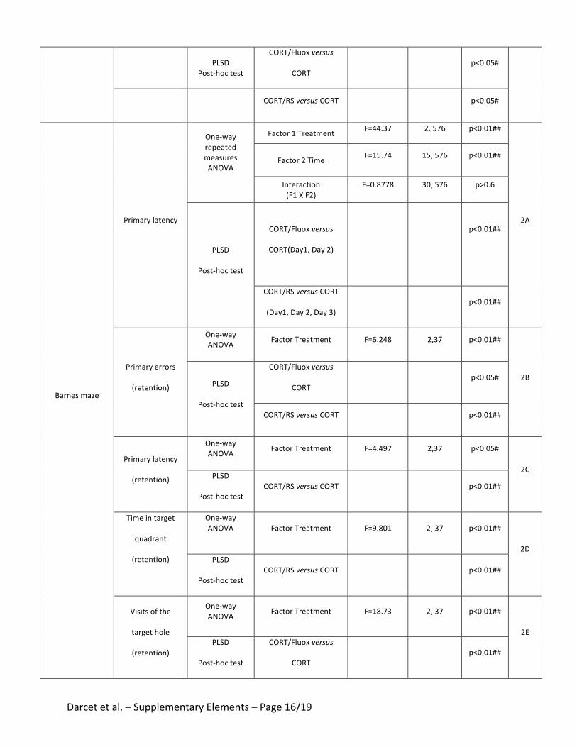
Darcetetal.–SupplementaryElements–Page16/19
PLSD
Post-hoctest
CORT/Fluoxversus
CORT
p<0.05#
CORT/RSversusCORT p<0.05#
Barnesmaze
Primarylatency
One-way
repeated
measures
ANOVA
Factor1TreatmentF=44.37 2,576 p<0.01##
2A
Factor2TimeF=15.74 15,576 p<0.01##
Interaction
(F1XF2)
F=0.8778 30,576 p>0.6
PLSD
Post-hoctest
CORT/Fluoxversus
CORT(Day1,Day2)
p<0.01##
CORT/RSversusCORT
(Day1,Day2,Day3)
p<0.01##
Primaryerrors
(retention)
One-way
ANOVA FactorTreatment F=6.248 2,37 p<0.01##
2BPLSD
Post-hoctest
CORT/Fluoxversus
CORT
p<0.05#
CORT/RSversusCORT p<0.01##
Primarylatency
(retention)
One-way
ANOVA FactorTreatment F=4.497 2,37 p<0.05#
2CPLSD
Post-hoctestCORT/RSversusCORT p<0.01##
Timeintarget
quadrant
(retention)
One-way
ANOVA
FactorTreatment F=9.801 2,37 p<0.01##
2D
PLSD
Post-hoctestCORT/RSversusCORT p<0.01##
Visitsofthe
targethole
(retention)
One-way
ANOVAFactorTreatment F=18.73 2,37 p<0.01##
2E
PLSD
Post-hoctest
CORT/Fluoxversus
CORT
p<0.01##
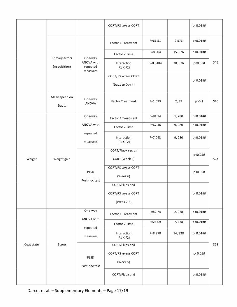
Darcetetal.–SupplementaryElements–Page17/19
CORT/RSversusCORT p<0.01##
Primaryerrors
(Acquisition)
One-wayANOVAwithrepeatedmeasures
Factor1Treatment F=61.51 2,576 p<0.01##
S4B
Factor2Time F=8.904 15,576 p<0.01##
Interaction(F1XF2)
F=0.8484 30,576 p<0.05#
CORT/RSversusCORT
(Day1toDay4) p<0.01##
Meanspeedon
Day1
One-wayANOVA FactorTreatment F=1.073 2,37 p>0.1 S4C
Weight Weightgain
One-way
ANOVAwith
repeated
measures
Factor1Treatment F=81.74 1,280 p<0.01##
S2A
Factor2Time F=67.46 9,280 p<0.01##
Interaction(F1XF2)
F=7.043 9,280 p<0.01##
PLSD
Post-hoctest
CORT/Fluoxversus
CORT(Week5) p<0.05#
CORT/RSversusCORT
(Week6) p<0.05#
CORT/Fluoxand
CORT/RSversusCORT
(Week7-8)
p<0.01##
Coatstate Score
One-way
ANOVAwith
repeated
measures
Factor1Treatment F=42.74 2,328 p<0.01##
S2B
Factor2Time F=252.9 7,328 p<0.01##
Interaction(F1XF2)
F=8.870 14,328 p<0.01##
PLSD
Post-hoctest
CORT/Fluoxand
CORT/RSversusCORT
(Week5)
p<0.05#
CORT/Fluoxand p<0.01##
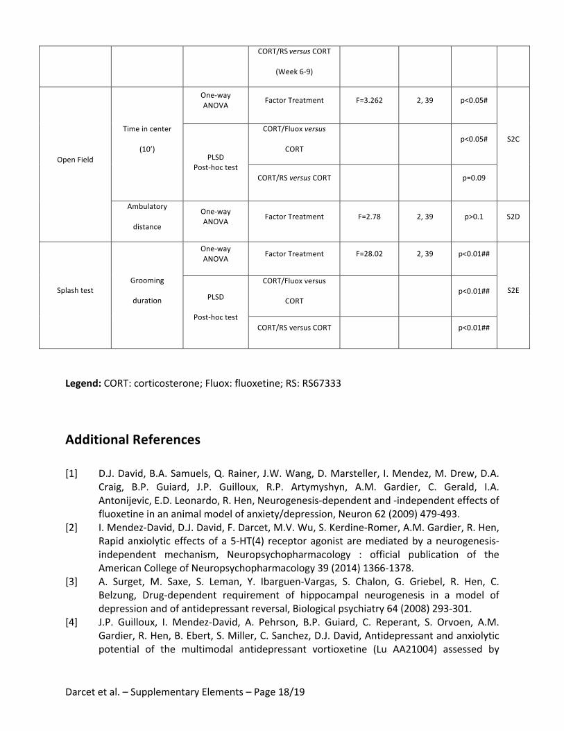
Darcetetal.–SupplementaryElements–Page18/19
CORT/RSversusCORT
(Week6-9)
OpenField
Timeincenter
(10’)
One-wayANOVA
FactorTreatment F=3.262 2,39 p<0.05#
S2C
PLSDPost-hoctest
CORT/Fluoxversus
CORT p<0.05#
CORT/RSversusCORT p=0.09
Ambulatory
distance
One-wayANOVA
FactorTreatment F=2.78 2,39 p>0.1 S2D
SplashtestGrooming
duration
One-wayANOVA
FactorTreatment F=28.02 2,39 p<0.01##
S2EPLSD
Post-hoctest
CORT/Fluoxversus
CORT p<0.01##
CORT/RSversusCORT p<0.01##
Legend:CORT:corticosterone;Fluox:fluoxetine;RS:RS67333
AdditionalReferences
[1] D.J.David,B.A.Samuels,Q.Rainer,J.W.Wang,D.Marsteller,I.Mendez,M.Drew,D.A.Craig, B.P. Guiard, J.P. Guilloux, R.P. Artymyshyn, A.M. Gardier, C. Gerald, I.A.Antonijevic,E.D.Leonardo,R.Hen,Neurogenesis-dependentand-independenteffectsoffluoxetineinananimalmodelofanxiety/depression,Neuron62(2009)479-493.
[2] I.Mendez-David,D.J.David,F.Darcet,M.V.Wu,S.Kerdine-Romer,A.M.Gardier,R.Hen,Rapidanxiolyticeffectsofa5-HT(4) receptoragonistaremediatedbyaneurogenesis-independent mechanism, Neuropsychopharmacology : official publication of theAmericanCollegeofNeuropsychopharmacology39(2014)1366-1378.
[3] A. Surget, M. Saxe, S. Leman, Y. Ibarguen-Vargas, S. Chalon, G. Griebel, R. Hen, C.Belzung, Drug-dependent requirement of hippocampal neurogenesis in a model ofdepressionandofantidepressantreversal,Biologicalpsychiatry64(2008)293-301.
[4] J.P. Guilloux, I. Mendez-David, A. Pehrson, B.P. Guiard, C. Reperant, S. Orvoen, A.M.Gardier,R.Hen,B.Ebert,S.Miller,C.Sanchez,D.J.David,Antidepressantandanxiolyticpotential of the multimodal antidepressant vortioxetine (Lu AA21004) assessed by
Darcetetal.–SupplementaryElements–Page19/19
behavioural and neurogenesis outcomes in mice, Neuropharmacology 73 (2013) 147-159.
[5] C. Ducottet, C. Belzung, Behaviour in the elevated plus-maze predicts coping aftersubchronicmildstressinmice,Physiology&behavior81(2004)417-426.
[6] F.Darcet,I.Mendez-David,L.Tritschler,A.M.Gardier,J.P.Guilloux,D.J.David,Learningand memory impairments in a neuroendocrine mouse model of anxiety/depression,Frontiersinbehavioralneuroscience8(2014)136.

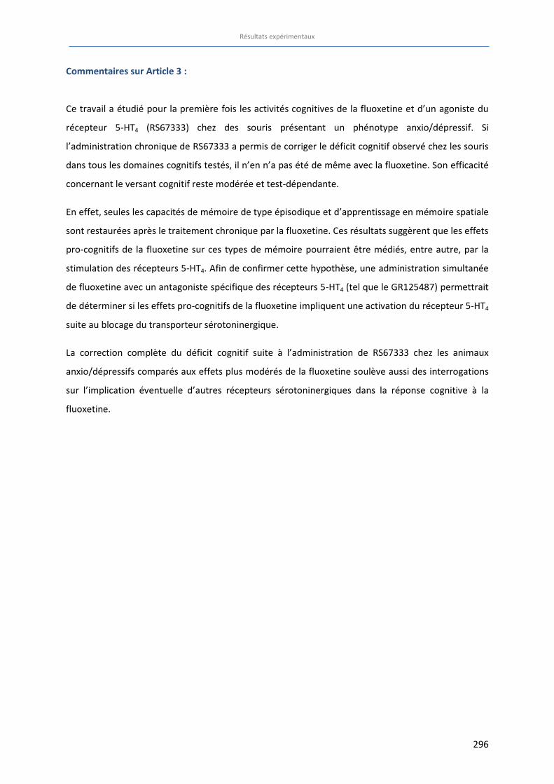
Résultats expérimentaux
296
Commentaires sur Article 3 :
Ce travail a étudié pour la première fois les activités cognitives de la fluoxetine et d’un agoniste du
récepteur 5-HT4 (RS67333) chez des souris présentant un phénotype anxio/dépressif. Si
l’administration chronique de RS67333 a permis de corriger le déficit cognitif observé chez les souris
dans tous les domaines cognitifs testés, il n’en n’a pas été de même avec la fluoxetine. Son efficacité
concernant le versant cognitif reste modérée et test-dépendante.
En effet, seules les capacités de mémoire de type épisodique et d’apprentissage en mémoire spatiale
sont restaurées après le traitement chronique par la fluoxetine. Ces résultats suggèrent que les effets
pro-cognitifs de la fluoxetine sur ces types de mémoire pourraient être médiés, entre autre, par la
stimulation des récepteurs 5-HT4. Afin de confirmer cette hypothèse, une administration simultanée
de fluoxetine avec un antagoniste spécifique des récepteurs 5-HT4 (tel que le GR125487) permettrait
de déterminer si les effets pro-cognitifs de la fluoxetine impliquent une activation du récepteur 5-HT4
suite au blocage du transporteur sérotoninergique.
La correction complète du déficit cognitif suite à l’administration de RS67333 chez les animaux
anxio/dépressifs comparés aux effets plus modérés de la fluoxetine soulève aussi des interrogations
sur l’implication éventuelle d’autres récepteurs sérotoninergiques dans la réponse cognitive à la
fluoxetine.

297
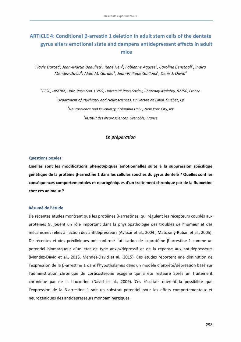
Résultats expérimentaux
298
ARTICLE 4: Conditional β-arrestin 1 deletion in adult stem cells of the dentate
gyrus alters emotional state and dampens antidepressant effects in adult
mice
Flavie Darcet1, Jean-Martin Beaulieu2, René Hen3, Fabienne Agasse4, Caroline Benstaali4, Indira
Mendez-David1, Alain M. Gardier1, Jean-Philippe Guilloux1, Denis J. David1
1CESP, INSERM, Univ. Paris-Sud, UVSQ, Université Paris-Saclay, Châtenay-Malabry, 92290, France
2Department of Psychiatry and Neurosciences, Université de Laval, Québec, QC
3Neuroscience and Psychiatry, Columbia Univ., New York City, NY
4Institut des Neurosciences, Grenoble, France
En préparation
Questions posées :
Quelles sont les modifications phénotypiques émotionnelles suite à la suppression spécifique
génétique de la protéine β-arrestine 1 dans les cellules souches du gyrus dentelé ? Quelles sont les
conséquences comportementales et neurogéniques d’un traitement chronique par de la fluoxetine
chez ces animaux ?
Résumé de l’étude
De récentes études montrent que les protéines β-arrestines, qui régulent les récepteurs couplés aux
protéines G, jouent un rôle important dans la physiopathologie des troubles de l’humeur et des
mécanismes reliés à l’action des antidépresseurs (Avissar et al., 2004 ; Matuzany-Ruban et al., 2005).
De récentes études précliniques ont confirmé l’utilisation de la protéine β-arrestine 1 comme un
potentiel biomarqueur d’un état de type anxio/dépressif et de la réponse aux antidépresseurs
(Mendez-David et al., 2013, Mendez-David et al., 2015). Ces études reportent une diminution de
l’expression de la β-arrestine 1 dans l’hypothalamus dans un modèle d’anxiété/dépression basé sur
l’administration chronique de corticosterone exogène qui a été restauré après un traitement
chronique par de la fluoxetine (David et al., 2009). Ces résultats ouvrent la possibilité que
l’expression de la β-arrestine 1 soit un substrat potentiel pour les effets comportementaux et
neurogéniques des antidépresseurs monoaminergiques.
Résultats expérimentaux
299
Puisque d’une part la neurogenèse hippocampique et notamment les jeunes neurones du GD
âgés de 4-6 semaines semblent jouer un rôle prépondérant dans l’activité cognitive ou antidépressive
(Sahay et al., 2011, Denny et al., 2014),nous avons souhaité étudier les conséquences de l’absence
d’expression de la protéine β-arrestine 1 dans les jeunes neurones âgées de 4/7 semaines. Dans ce
travail, nous avons caractérisé les conséquences comportementales d’une délétion sélective de la
protéine β-arrestine 1 dans les cellules souches du gyrus dentelé de l’hippocampe dans plusieurs
tests prédictifs d’une activité anxiolytique et/ou antidépressive en présence ou non d’un traitement
chronique à la fluoxetine. Les conséquences neurogéniques de cette délétion sélective ont
également été étudiées.
Contribution personnelle :
Au cours de ce travail :
x J’ai mis en place les cohortes d’animaux tissus-spécifiques et leurs contrôles avec la méthode
de génotypage et induit la suppression génétique spécifique avec les injections de
tamoxifène.
x J’ai réalisé le suivi des animaux incluant la préparation et l’administration des traitements
pharmacologiques (traitement par la fluoxetine).
x J’ai mené l’ensemble des tests comportementaux émotionnels.
x J’ai réalisé l’ensemble des analyses immunohistochimiques des étapes de la neurogenèse
hippocampique depuis la perfusion, le prélèvement et la coupe des cerveaux jusqu’au
comptage des cellules.
x J’ai analysé les résultats et rédigé l’article sous la supervision du Pr. Denis David et du Dr.
Jean-Philippe Guilloux.
Résultats expérimentaux
300
Conditional β-arrestin 1 deletion in stem cells of the dentate gyrus alters
emotional state and dampens antidepressant effects in adult mice
Flavie Darcet1, Jean-Martin Beaulieu2, René Hen3, Fabienne Agasse4, Caroline Benstaali4, Indira Mendez-
David1Alain M. Gardier1, Jean-Philippe Guilloux1, Denis J. David1
1CESP, INSERM, Univ. Paris-Sud, UVSQ, Université Paris-Saclay, Châtenay-Malabry, 92290, France
2Department of Psychiatry and Neurosciences,Université Laval, Québec, QC
3Neuroscience and Psychiatry, Columbia Univ., New York City, NY
4Institut des Neurosciences, Grenoble, France
En préparation
INTRODUCTION OF THE STUDY:
Elucidating the neurobiological basis of depression and anxiety are one of the foremost challenges
for today’s society. Severe forms of depression affect 2-5% of the U.S. population and mood
disorders impact 7% of the world’s population and rank among the top ten causes of disability.
Selective Serotonin Reuptake Inhibitors (SSRIs) are the most commonly prescribed drugs for the
treatment of depression and several anxiety disorders. However, key questions remain regarding the
molecular and cellular mechanisms underlying the effects of SSRIs and other antidepressants (ADs).
In accordance with the idea that ADs exert essential effects in multiple brain regions, we previously
demonstrated that chronic corticosterone regimen induces anxiety/depression-like phenotype and
subsequent fluoxetine treatment elicits effects on genes’ expression not only in the hippocampus,
but also in the hypothalamus in adult mice. Among the genes investigated, only 3, including β-
arrestins 1 and 2, displayed a change in mRNA levels in the chronic corticosterone group that was
reversed by fluoxetine treatment. Molecular functions of β-arrestins have placed them in pathways
associated with mood and responsiveness to lithium, a drug currently used to treat bipolar disorders.
This includes regulation of an Akt/GSK3 complex and classical functions such as desensitization of G-
protein coupled receptors including serotonin and dopamine receptors. Finally, there is also evidence
in humans implicating β-arrestins in depression and in response to stress, and that these changes are
Résultats expérimentaux
301
reversible by various AD treatments. Thus, β-arrestins could be an important molecular determinant
of the effects of fluoxetine and more generally to ADs. Recently, clinical data from Avissar’s group
(Avissar et al., 2004) suggest that β-arrestin-1 mRNA and protein levels are highest in peripheral
blood leukocytes of MDD patients. Therefore, β-arrestin-1 may be a putative candidate biochemical
marker in clinical practice for depressive pathophysiology and the response to antidepressants (for
review see (Schreiber et al., 2009). β-arrestin mRNA levels and β-arrestin-1 protein levels in
mononuclear leukocytes of untreated patients with MDD are lower than the levels found in healthy
subjects. Furthermore, reduced levels of β- arrestin-1 protein and mRNA are significantly correlated
with the severity of depressive symptoms (Avissar et al., 2004; Schreiber et al., 2009). However, the
low β-arrestin-1 protein and mRNA levels are alleviated by antidepressant treatment. Therefore,
these low levels may predict clinical improvement (Avissar et al., 2004; Golan et al., 2010). Finally,
SSRIs are potent stimulators of adult hippocampal neurogenesis (David et al., 2009; Santarelli et al,
2003) and some of these effects are required for the anxiolytic/antidepressant-like effects of SSRIs
(David et al., 2009). These results raise the possibility that β-arrestin 1 expression is a potential
substrate for the behavioral antidepressant effects, including the neurogenic effects of
antidepressant drugs. Here, we characterized the behavioral consequences of β-arrestin 1 (ARRB1)
conditional deletion in stem cells in the dentate gyrus of the hippocampus in various behavioral
paradigms predictive of anxiolytic/antidepressant therapy in presence or not of a chronic fluoxetine
treatment.
Résultats expérimentaux
302
MATERIALS AND METHODS:
Animals
Inducible tissue-specific male mice were created by interbreeding Nestin-CreERT2 (René Hen,
Columbia University, USA) mice and ARRB1flox/flox mice (Jean-Martin Beaulieu, Université Laval,
Canada). Nestin-CreERT2; ARRB1+/flox and ARRB1+/flox mice from F1 were then crossed to generate
NestinCreERT2-/+; ARRB1flox/flox mice (ARRB1-/- mice). NestinCreERT2-/+; ARRB1+/+ mice (ARRB1+/+) were
used as controls. ARRB1 mice were maintained on a mixed (C57BL/6-129/Sv) genetic background. To
induce CreERT2 mediated recombination in neural stem cells in the adult brain, ARRB1-/- mice were
given 2 mg of tamoxifen (TAM) (Sigma, France), once a day, intraperitoneally for 5 consecutive days.
TAM solution (10 mg/mL) was prepared in corn oil containing 10% ethanol. Control mice were
injected with an identical volume of corn oil with 10% ethanol. Animals were maintained on a
12L:12D schedule (Lights ON at 7AM) and were housed 5 per cage. Food and water were provided ad
libitum. All testing were conducted in compliance with the laboratory animal care guidelines, with
protocols approved by the Institutional Animal Care and Use Committee (CEE26 authorization 2012-
099) and with the European directive 2010/63/EU. Experiments were conducted on male transgenic
mice between 12 and 14 weeks of age.
Two cohorts of male animals were generated to assess the behavioral and the neurogenic
consequences of the conditional deletion of β-arrestin 1 in adult hippocampal stem cell in baseline
and under chronic antidepressant treatment.
Control of selective deletion
In order to control the conditional and selective deletion of β-arrestin 1 protein in stem cells of the
dentate gyrus, in situ hybridization and western blot from stem cells cultures were applied in ARRB1-
/- mice and their littermates.
Résultats expérimentaux
303
In situ hybridization
Coronal hippocampal sections (20 µm thickness) from ARRB1+/+ and ARRB1-/- mice were collected
fixed with paraformaldehyde and dehydrate in ethanol at room temperature. Slices were first
treated with Pretreat solution for 10 minutes, rinsed and hybridize for 2h at 40°C with a
complementary oligodeoxyribonucleotide probe of β-arrestine 1 protein in mice. Sections were then
hybridized with different stabilizer reagents at 40°C from 15 to 30 minutes. To amplify the signal,
sections were incubated in DAB during 10 minutes at room temperature then colored with 50%
hematoxylin and a 0.02% ammoniac solution for 2 minutes. Finally, slices were dehydrated in
successive baths: 70% ethanol, 100% ethanol and xylene. β-arrestin 1 mRNA was visualized with a
microscope and scanned by ZEISS Axio Scan.Z1 apparatus. β-arrestin 1 expression was counted using
Guide ZEN 2012 – Slidescan and FIJI softwares.
Stem cells culture
Neural stem cells were grown in monolayers according to the protocol of Babu et al, 2011 with
modifications. Adult mice (2 to 4 month-old) were killed by cervical dislocation; dentate gyrus and
subventricular zone were rapidly dissected and placed in sterile saline solution. The tissues were
digested for 20 min at 37˚C in PBS containing papain (2,5 U/ml), DNase I250 U/ml (both from Sigma)
and dispase (8 U/ml) (Roche) and viable cells were separated from myelin and cell debris by
centrifugation on a percoll gradient (see Babu et al., 2011). Then, cells were diluted in serum-free
culture medium (SFM) composed of Dulbecco's modified Eagle's medium/Ham's (DMEM) F-12
medium GlutaMAX-I supplemented with 100 U/ml penicillin, 100 μg/ml streptomycin, 1% B27
supplement, 20 ng/ml epidermal growth factor (EGF) and 20 ng/ml basic fibroblast growth factor
(FGF-2) (all from Life Technologies) and grown as monolayers for 7 days (SVZ stem cells) or 14 days
(dentate gyrus stem cells) in petri dishes coated with 20 µg/ml poly-D-Lysine and 20 µg/ml laminin (
both from Sigma) in a 95% air–5% CO2 humidified atmosphere at 37°C.
Résultats expérimentaux
304
Western Blot
For Western Blot analysis, protein concentration from stem cells cultures was quantified using BCA
Protein Assay Kit (Pierce Biotechnology). Equal amount of proteins were separated by 10% sodium
dodecyl sulfate–polyacrylamide electrophoresis (SDS-PAGE) and transferred to polyvinylidene
difluoride membranes (PVDF), (Amersham Biosciences, Les Ulis, France). After blocking in 10 Mm
Tris-HCl, pH:7,6, 200 mM NaCl, 0,1% Tween-20 and 5% BSA, membranes were incubated with
primary monoclonal antibody Mouse Anti- β-Arrestin 1 (BD Bioscience Pharmigen) and β-Actin (Santa
Cruz Biotechnology, CA).Primary antibodies were detected with appropriate HRP-coupled secondary
antibodies and ECL signals (SuperSignal West Pico Chemiluminescent Substrate (Pierce,
Erembodegem, Belgium) were quantitated with Image lab software (Bio-Rad).
Drugs and administration
Fluoxetine hydrochloride (160 mg/mL, equivalent to 18mg/kg/day) was purchased from Anawa
Trading, (Zurich, Switzerland) and administered in the drinking water in opaque bottles for 4 weeks.
The dose of fluoxetine was based on previous data showing the ability of fluoxetine to produce
antidepressant-like activity (David et al., 2009).
Behavioral testing
For each cohort, the same animal was tested in five different behavioral paradigms of anxiety and
depression. Each animal, over a week, was successively tested in: Open Field (1), Elevated Plus maze
(2), 4-plate test (3), Novelty Suppressed Feeding (4), Splash test (5) and Tail Suspension test (6). Z-
score methodology was used to investigate the potential of combining results within and across the
different behavior tests for depressive/anxious-like behaviors and investigate the
anxiolytic/antidepressant-like response of fluoxetine treatment in ARRB1-/- mice and controls
(Guilloux et al., 2011).
Résultats expérimentaux
305
Open Field (OF)
This test was performed as described by David and colleagues (David et al, 2009). Motor activity was
quantified in four 43 x 43 cm2 Plexiglas open field boxes (MED Associates, Georgia, VT). Two sets of
16 pulse-modulated infrared photobeams were placed on opposite walls 2.5-cm apart to record x–y
ambulatory movements. Activity chambers were computer interfaced for data sampling at 100-ms
resolution. The computer defined grid lines that divided each open field into center and surround
regions, with each of four lines being 11 cm from each wall. Dependent measures in the center were
the total time and the number of entries over a 30-min test period. The activity in the center was
quantified as distance traveled in the center divided by total distance traveled.
Elevated Plus Maze (EPM)
This test was performed as described by David and colleagues (David et al, 2009). The maze is a plus-
cross-shaped apparatus, with two open arms and two arms closed by walls linked by a central
platform 50 cm above the floor. Mice were individually put in the center of the maze facing an open
arm and were allowed to explore the maze during 5 min. The time spent and the number of entries
into the open arms were used as an anxiety index. All parameters were measured using a
videotracker (EPM3C, Bioseb, Vitrolles, France).
4-plates test (4-PT)
The 4-plates test (Aron et al., 1971) is a fear-related anxiety paradigm in which the natural
exploration of a novel environment is inhibited by the delivery of a footshock. The equipment
consists in a small cage (25×18×16 cm) in which the floor is composed of 4 similar rectangular metal
plates (11×8 cm) separated from each other with a 4-mm space (Bioseb, France). Each plate is
connected to a foot shock generator (0.6 mA; 0.5 s). After a 15-sec habituation period, a foot shock is
administrated to animals when they move from one plate to another. Anxiety parameter is measured
by the number of punitions administered to animals over a 60-sec period.
Résultats expérimentaux
306
Novelty-Suppressed-Feeding (NSF)
The NSF is a conflict test that elicits competing motivations: the drive to eat and the fear of venturing
into the center of a brightly lit arena. The latency to begin eating is used as an index of
anxiety/depression-like behavior, because classical anxiolytic drugs as well as chronic antidepressants
decrease this measure. The NSF test was carried out during a 10-min period as previously described
(David et al, 2009). Briefly, the testing apparatus consisted of a plastic box (50x40x20 cm), the floor
of which was covered with approximately 2 cm of wooden bedding. Twenty-four hours prior to
behavioral testing, all food was removed from the home cage. At the time of testing, a single pellet of
food (regular chow) was placed on a white paper platform positioned in the center of the box. Each
animal was placed in a corner of the box, and a stopwatch was immediately started. The latency to
eat (defined as the mouse sitting on its haunches and biting the pellet with the use of forepaws) was
timed. Immediately afterwards, the animal was transferred to its home cage, and the amount of food
consumed by the mouse in the subsequent 5 min was measured, serving as a control for change in
appetite as a possible confounding factor.
Tail Suspension Test (TST)
The TST is an antidepressant activity-screening test (Steru et al, 1985) used to test compounds that
are expected to affect depression related behaviors. Mice are suspended by their tails with tape, in a
position that they cannot escape or hold on to nearby surfaces. During this 6 minutes test, the
resulting escape oriented behaviors are quantified using an automated tail suspension test apparatus
(Bioseb, Vitrolles, France). A specific strain gauge linked to a computer quantifies the time spent by
the animal trying to escape.
Splash Test
This test consisted of squirting 200 μl of a 10% sucrose solution on the mouse’s snout. The grooming
duration was assessed at the end of the corticosterone regimen in the presence or absence of 4-
weeks of drug treatment according to a protocol previously described (David et al, 2009).
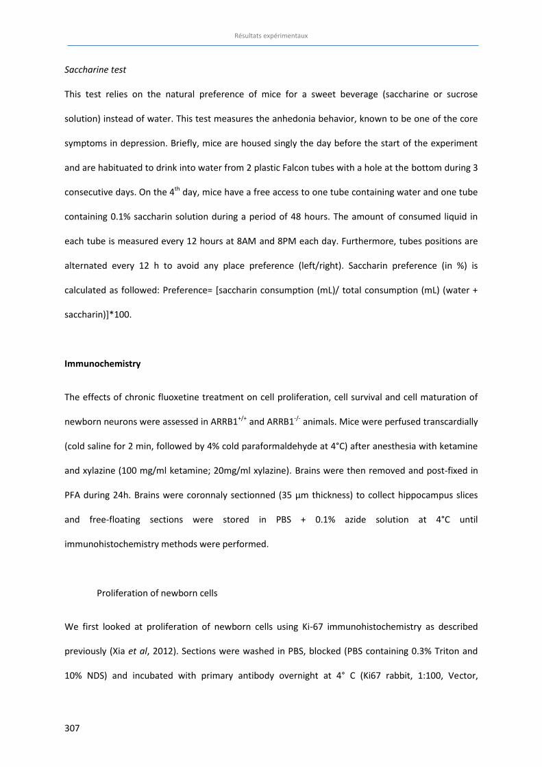
Résultats expérimentaux
307
Saccharine test
This test relies on the natural preference of mice for a sweet beverage (saccharine or sucrose
solution) instead of water. This test measures the anhedonia behavior, known to be one of the core
symptoms in depression. Briefly, mice are housed singly the day before the start of the experiment
and are habituated to drink into water from 2 plastic Falcon tubes with a hole at the bottom during 3
consecutive days. On the 4th day, mice have a free access to one tube containing water and one tube
containing 0.1% saccharin solution during a period of 48 hours. The amount of consumed liquid in
each tube is measured every 12 hours at 8AM and 8PM each day. Furthermore, tubes positions are
alternated every 12 h to avoid any place preference (left/right). Saccharin preference (in %) is
calculated as followed: Preference= [saccharin consumption (mL)/ total consumption (mL) (water +
saccharin)]*100.
Immunochemistry
The effects of chronic fluoxetine treatment on cell proliferation, cell survival and cell maturation of
newborn neurons were assessed in ARRB1+/+ and ARRB1-/- animals. Mice were perfused transcardially
(cold saline for 2 min, followed by 4% cold paraformaldehyde at 4°C) after anesthesia with ketamine
and xylazine (100 mg/ml ketamine; 20mg/ml xylazine). Brains were then removed and post-fixed in
PFA during 24h. Brains were coronnaly sectionned (35 µm thickness) to collect hippocampus slices
and free-floating sections were stored in PBS + 0.1% azide solution at 4°C until
immunohistochemistry methods were performed.
Proliferation of newborn cells
We first looked at proliferation of newborn cells using Ki-67 immunohistochemistry as described
previously (Xia et al, 2012). Sections were washed in PBS, blocked (PBS containing 0.3% Triton and
10% NDS) and incubated with primary antibody overnight at 4° C (Ki67 rabbit, 1:100, Vector,
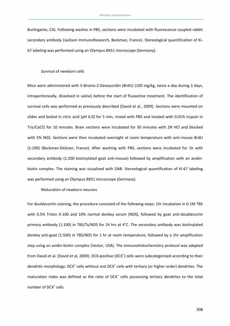
Résultats expérimentaux
308
Burlingame, CA). Following washes in PBS, sections were incubated with fluorescence coupled rabbit
secondary antibody (Jackson ImmunoResearch, Beckman, France). Stereological quantification of Ki-
67 labeling was performed using an Olympus BX51 microscope (Germany).
Survival of newborn cells
Mice were administered with 5-Bromo-2-Deoxyuridin (BrdU) (100 mg/kg, twice a day during 3 days,
intraperitoneally, dissolved in saline) before the start of fluoxetine treatment. The identification of
survival cells was performed as previously described (David et al., 2009). Sections were mounted on
slides and boiled in citric acid (pH 6.0) for 5 min, rinsed with PBS and treated with 0.01% trypsin in
Tris/CaCl2 for 10 minutes. Brain sections were incubated for 30 minutes with 2N HCl and blocked
with 5% NGS. Sections were then incubated overnight at room temperature with anti-mouse BrdU
(1:100) (Beckman-Dickson, France). After washing with PBS, sections were incubated for 1h with
secondary antibody (1:200 biotinylated goat anti-mouse) followed by amplification with an avidin-
biotin complex. The staining was visualized with DAB. Stereological quantification of Ki-67 labeling
was performed using an Olympus BX51 microscope (Germany).
Maturation of newborn neurons
For doublecortin staining, the procedure consisted of the following steps: 1hr incubation in 0.1M TBS
with 0.5% Triton X-100 and 10% normal donkey serum (NDS), followed by goat anti-doublecortin
primary antibody (1:100) in TBS/Tx/NDS for 24 hrs at 4°C. The secondary antibody was biotinylated
donkey anti-goat (1:500) in TBS/NDS for 1 hr at room temperature, followed by a 1hr amplification
step using an avidin-biotin complex (Vector, USA). The immunohistochemistry protocol was adapted
from David et al. (David et al, 2009). DCX-positive (DCX+) cells were subcategorized according to their
dendritic morphology: DCX+ cells without and DCX+ cells with tertiary (or higher order) dendrites. The
maturation index was defined as the ratio of DCX+ cells possessing tertiary dendrites to the total
number of DCX+ cells.
Résultats expérimentaux
309
Data analysis:
Results were expressed as mean ± SEM for all experiments. T-tests, one-way or two-way ANOVA
(genotype and treatment factors) were applied when appropriate. Significant main effects and/or
interactions were followed by a Fisher's PLSD post hoc analysis. In the NSF test, we used the Kaplan-
Meier survival analysis due to the lack of normal distribution of the data. Mantel-Cox log-rank test
was used to evaluate differences between experimental groups. Statistical significance was set at
p<0.05.
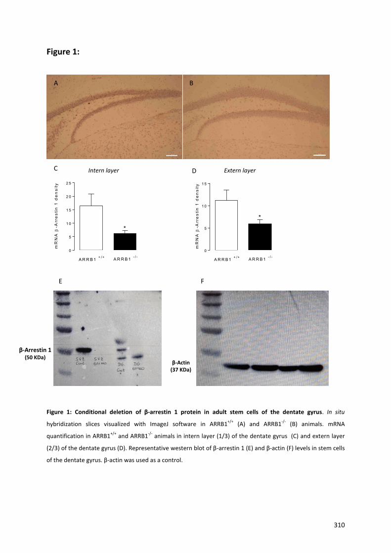
310
Figure 1:
Figure 1: Conditional deletion of β-arrestin 1 protein in adult stem cells of the dentate gyrus. In situ
hybridization slices visualized with ImageJ software in ARRB1+/+
(A) and ARRB1-/-
(B) animals. mRNA
quantification in ARRB1+/+
and ARRB1-/-
animals in intern layer (1/3) of the dentate gyrus (C) and extern layer
(2/3) of the dentate gyrus (D). Representative western blot of β-arrestin 1 (E) and β-actin (F) levels in stem cells
of the dentate gyrus. β-actin was used as a control.
A B
β-Arrestin 1 (50 KDa)
β-Actin (37 KDa)
E F
C
mR
NA
E-A
rre
stin
1 d
en
sity
0
5
1 0
1 5
2 0
2 5
*
A R R B 1 - / -A R R B 1 + /+
D
mR
NA
E-A
rre
sti
n 1
de
ns
ity
0
5
1 0
1 5
A R R B 1 - / -A R R B 1 + /+
*
Intern layer Extern layer
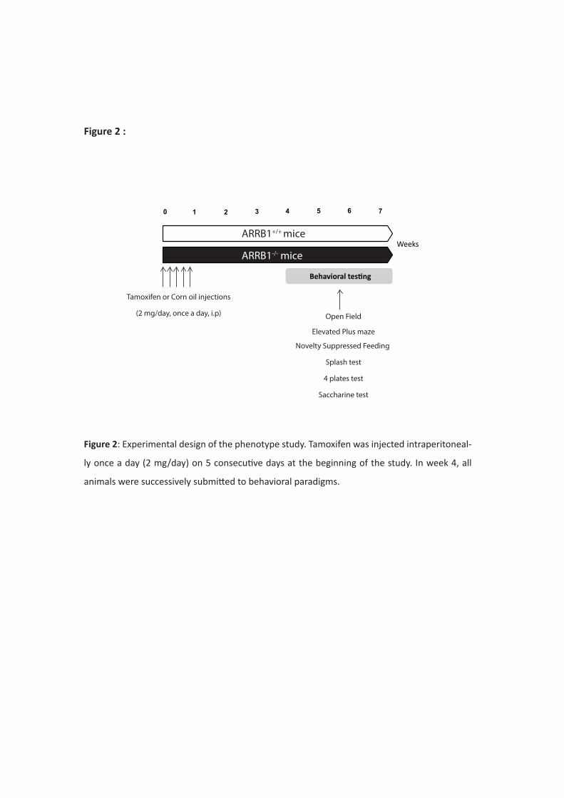
0 41 2 6
Behavioral testing
Weeks
3 5 7
Tamoxifen or Corn oil injections
(2 mg/day, once a day, i.p) Open Field
Elevated Plus maze
Novelty Suppressed Feeding
Splash test
4 plates test
Saccharine test
ARRB1+/+ mice
ARRB1-/- mice
Figure 2 :
Figure 2: Experimental design of the phenotype study. Tamoxifen was injected intraperitoneal-
ly once a day (2 mg/day) on 5 consecutive days at the beginning of the study. In week 4, all
animals were successively submitted to behavioral paradigms.
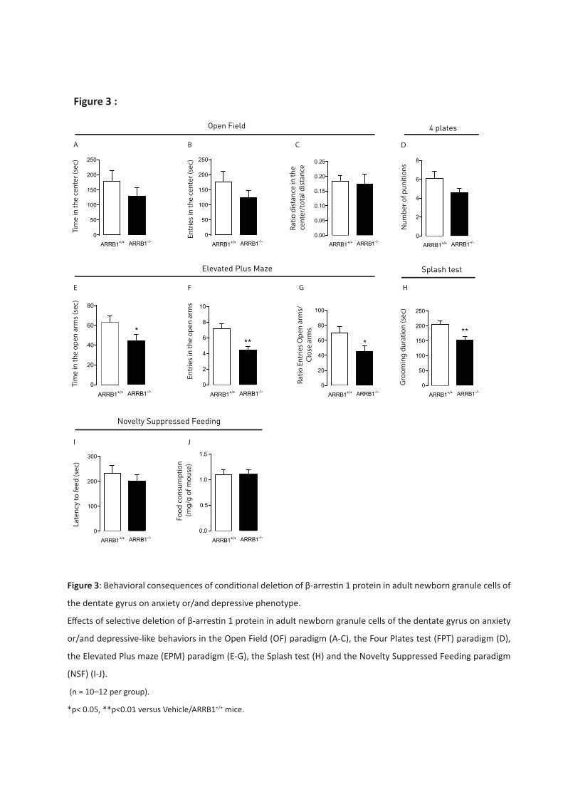
Figure 3 :
0
50
100
150
200
250
ARRB1+/+ ARRB1-/-
Tim
e in
the
cent
er (s
ec)
0.0
0.5
1.0
1.5
ARRB1+/+ ARRB1-/-
Food
con
sum
ptio
n (m
g/g
of m
ouse
)
0
100
200
300
ARRB1+/+ ARRB1-/-
Late
ncy
to fe
ed (s
ec)
0
50
100
150
200
250
ARRB1+/+ ARRB1-/-
Entr
ies i
n th
e ce
nter
(sec
)
0.00
0.05
0.10
0.15
0.20
0.25
ARRB1+/+ ARRB1-/-
Ratio
dist
ance
in th
e c
ente
r/to
tal d
istan
ce
0
2
4
6
8
10
**
ARRB1+/+ ARRB1-/-
Entr
ies i
n th
e op
en a
rms
0
20
40
60
80
*
ARRB1+/+ ARRB1-/-
Tim
e in
the
open
arm
s (se
c)
0
20
40
60
80
100
*
ARRB1+/+ ARRB1-/-
Ratio
Ent
ries O
pen
arm
s/Cl
ose
arm
s
A B C
E F G
I J
Open Field
Elevated Plus Maze
Novelty Suppressed Feeding
0
50
100
150
200
250
**
ARRB1+/+ ARRB1-/-
Gro
omin
g du
ratio
n (s
ec)
H
Splash test
0
2
4
6
8
ARRB1+/+ ARRB1-/-
Num
ber o
f pun
ition
s
D
4 plates
Figure 3: Behavioral consequences of conditional deletion of β-arrestin 1 protein in adult newborn granule cells of
the dentate gyrus on anxiety or/and depressive phenotype.
Effects of selective deletion of β-arrestin 1 protein in adult newborn granule cells of the dentate gyrus on anxiety
or/and depressive-like behaviors in the Open Field (OF) paradigm (A-C), the Four Plates test (FPT) paradigm (D),
the Elevated Plus maze (EPM) paradigm (E-G), the Splash test (H) and the Novelty Suppressed Feeding paradigm
(NSF) (I-J).
(n = 10–12 per group).
*p< 0.05, **p<0.01 versus Vehicle/ARRB1+/+ mice.
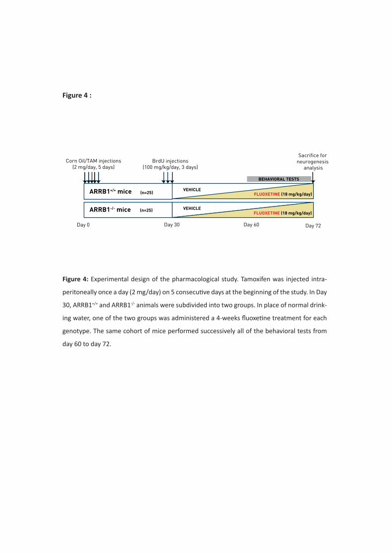
Corn Oil/TAM injections(2 mg/day, 5 days)
Day 0 Day 30
BrdU injections(100 mg/kg/day, 3 days)
Day 60 Day 72
ARRB1+/+ mice
ARRB1-/- mice
(n=25)
(n=25)
Sacrifice for neurogenesis
analysis
BEHAVIORAL TESTS
FLUOXETINE (18 mg/kg/day)
FLUOXETINE (18 mg/kg/day)VEHICLE
VEHICLE
Figure 4 :
Figure 4: Experimental design of the pharmacological study. Tamoxifen was injected intra-
peritoneally once a day (2 mg/day) on 5 consecutive days at the beginning of the study. In Day
30, ARRB1+/+ and ARRB1-/- animals were subdivided into two groups. In place of normal drink-
ing water, one of the two groups was administered a 4-weeks fluoxetine treatment for each
genotype. The same cohort of mice performed successively all of the behavioral tests from
day 60 to day 72.
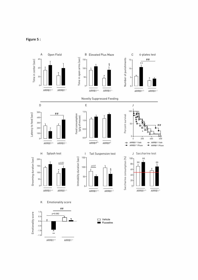
VehicleFluoxetine
0
20
40
60
Open Field
Tim
e in
cen
ter
(sec
)
ARRB1+/+ ARRB1-/-
0
50
100
150
*
§
ARRB1+/+ ARRB1-/-
Tim
e in
ope
n ar
ms
(sec
)
Elevated Plus Maze 4-plates test
0
5
10
15
**
*
##
ARRB1+/+ ARRB1-/-
Num
ber
of p
unis
hmen
ts
Novelty Suppressed Feeding
A B C
0
50
100
150
200
*
Gro
omin
g du
ratio
n (s
ec)
ARRB1+/+ ARRB1-/-
Splash test
0
50
100
150
§p=0.07
Imm
obili
ty d
urat
ion
(sec
)
ARRB1+/+ ARRB1-/-
Tail Suspension testH I
p=0.053
J
0
100
200
300
400
500
*
##
ARRB1+/+ ARRB1-/-
Late
ncy
to fe
ed (s
ec)
D E
0.0
0.5
1.0
1.5
ARRB1+/+ ARRB1-/-
Food
con
sum
ptio
n (g
/g o
f mou
se)
J
0
20
40
60
80
100
δδ
δδ
δδ
ARRB1+/+ ARRB1-/-Sacc
hari
ne c
onsu
mpt
ion
(%)
Saccharine test
0 200 400 6000
50
100
ARRB1+/+/Veh
Per
cent
sur
viva
l
*
##
ARRB1+/+/FluoxARRB1-/-/VehARRB1-/-/Fluox
Figure 5 :
Emot
iona
lity
scor
e
-1.5
-1.0
-0.5
0.0
0.5
1.0
##
**
p=0.082
ARRB1+/+ ARRB1-/-
K Emotionality score
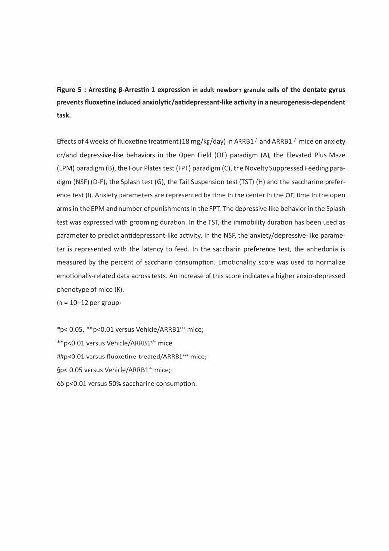
Figure 5 : Arresting β-Arrestin 1 expression in adult newborn granule cells of the dentate gyrus
prevents fluoxetine induced anxiolytic/antidepressant-like activity in a neurogenesis-dependent
task.
Effects of 4 weeks of fluoxetine treatment (18 mg/kg/day) in ARRB1-/- and ARRB1+/+ mice on anxiety
or/and depressive-like behaviors in the Open Field (OF) paradigm (A), the Elevated Plus Maze
(EPM) paradigm (B), the Four Plates test (FPT) paradigm (C), the Novelty Suppressed Feeding para-
digm (NSF) (D-F), the Splash test (G), the Tail Suspension test (TST) (H) and the saccharine prefer-
ence test (I). Anxiety parameters are represented by time in the center in the OF, time in the open
arms in the EPM and number of punishments in the FPT. The depressive-like behavior in the Splash
test was expressed with grooming duration. In the TST, the immobility duration has been used as
parameter to predict antidepressant-like activity. In the NSF, the anxiety/depressive-like parame-
ter is represented with the latency to feed. In the saccharin preference test, the anhedonia is
measured by the percent of saccharin consumption. Emotionality score was used to normalize
emotionally-related data across tests. An increase of this score indicates a higher anxio-depressed
phenotype of mice (K).
(n = 10–12 per group)
*p< 0.05, **p<0.01 versus Vehicle/ARRB1+/+ mice;
**p<0.01 versus Vehicle/ARRB1+/+ mice
##p<0.01 versus fluoxetine-treated/ARRB1+/+ mice;
§p< 0.05 versus Vehicle/ARRB1-/- mice;
δδ p<0.01 versus 50% saccharine consumption.
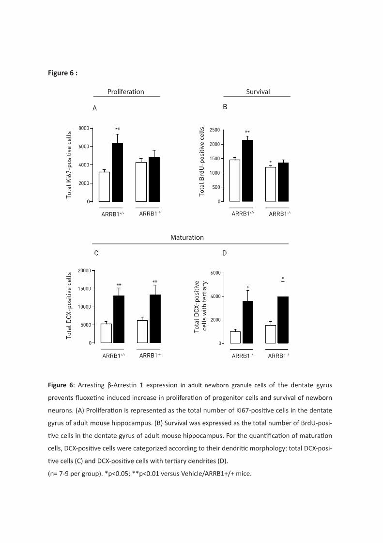
**
0
2000
4000
6000
8000
ARRB1+/+ ARRB1-/-
Tota
l Ki6
7-po
sitiv
e ce
lls
A
**
*
Tota
l Brd
U-p
ositi
ve c
ells
0
500
1000
1500
2000
2500
ARRB1-/-ARRB1+/+
B
C D
** **
Tota
l DC
X-po
sitiv
e ce
lls
0
5000
10000
15000
20000
ARRB1+/+ ARRB1-/-
0
2000
4000
6000
**
Tota
l DC
X-po
sitiv
e c
ells
with
tert
iary
ARRB1+/+ ARRB1-/-
Maturation
Proliferation Survival
Figure 6 :
Figure 6: Arresting β-Arrestin 1 expression in adult newborn granule cells of the dentate gyrus
prevents fluoxetine induced increase in proliferation of progenitor cells and survival of newborn
neurons. (A) Proliferation is represented as the total number of Ki67-positive cells in the dentate
gyrus of adult mouse hippocampus. (B) Survival was expressed as the total number of BrdU-posi-
tive cells in the dentate gyrus of adult mouse hippocampus. For the quantification of maturation
cells, DCX-positive cells were categorized according to their dendritic morphology: total DCX-posi-
tive cells (C) and DCX-positive cells with tertiary dendrites (D).
(n= 7-9 per group). *p<0.05; **p<0.01 versus Vehicle/ARRB1+/+ mice.
Résultats expérimentaux
317
DISCUSSION OF THE STUDY:
In this study, arresting β-arrestin 1 expression in adult stem cells of the dentate gyrus did not
significantly change overall the emotionality in mice but altered behavior in specific tests such as
Elevated Plus Maze, 4-plates test and Splash test. Interestingly arresting β-arrestin 1 expression
dampened anxiolytic/antidepressant-like effects of chronic fluoxetine treatment, as revealed by an
absence of decrease of the emotionality score analysis, and prevents fluoxetine induced
anxiolytic/antidepressant-like activity in a neurogenesis-dependent task, the Novelty Suppressed
Feeding.
On another hand, neurogenesis study revealed that β-arrestin 1 protein expression in stem cells of
the dentate gyrus is necessary for survival of newborn cells. Interestingly, β-arrestin 1 protein
expression in stem cells of the dentate gyrus necessary for fluoxetine-induced proliferation and
survival of newborn cells. Finally, β-arrestin 1 protein expression in stem cells of the dentate gyrus is
not involved in fluoxetine-induced maturation of newborn granule cells
Further experiments using death cells staining (caspase-3) or triple staining (BrdU, NeuN and DCX)
could help to better determine neurogenesis modifications after the specific deletion of β-arrestin 1
protein in stem cells of the dentate gyrus.
Résultats complémentaires
318
Résultats complémentaires:
Conditional β-arrestin 1 deletion in adult stem cells of the dentate gyrus
alters cognitive phenotype and prevents pro-cognitive effects of chronic
fluoxetine and 5-HT4 receptor agonist treatments in mice
Flavie Darcet1, Jean-Martin Beaulieu2, René Hen3, Alain M. Gardier1, Jean-Philippe Guilloux1, Denis J.
David1
1CESP, INSERM, Univ. Paris-Sud, UVSQ, Université Paris-Saclay, Châtenay-Malabry, 92290, France 2Department of Psychiatry and Neurosciences, Université de Laval, Québec, QC
3Neuroscience and Psychiatry, Columbia Univ., New York City, NY
Questions posées :
La délétion conditionelle de la protéine β-arrestine 1 dans les cellules souches du gyrus
dentelé induit-elle des modifications dans les performances d’apprentissage et de mémoire ? Si
oui, quelles sont les conséquences comportementales d’un traitement chronique par de la
fluoxetine ou par le RS67333, un agoniste du récepteur 5-HT4 chez ces animaux adultes ?
Résumé de l’étude
De récentes études montrent que les protéines β-arrestines, qui régulent l’activité des récepteurs
couplés aux protéines G, jouent un rôle important dans la physiopathologie des troubles de l’humeur
et des mécanismes reliés à l’action des antidépresseurs (Avissar et al., 2004 ; Matuzany-Ruban et al.,
2005). En plus d’être impliqués dans les mécanismes émotionnels et de réponse aux antidépresseurs,
l’hippocampe d’une manière générale est également étroitement lié aux mécanismes de cognition.
De plus, la relation fonctionnelle entre la protéine β-arrestine 1 et le récepteur 5-HT4 est bien décrite
dans la littérature et pourrait constituer une nouvelle piste dans l’étude des effets des ligands du
récepteur 5-HT4, tel que le RS67333 (Marin et al., 2012).
Dans cette étude, nous avons dans un premier temps caractérisé les conséquences d’une délétion
conditionnelle de la protéine β-arrestine 1 dans les cellules souches du gyrus dentelé de
l’hippocampe de souris adulte dans plusieurs tests d’apprentissage et de mémoire. Dans un second
temps, les conséquences d’un traitement chronique à la fluoxetine ou d’un traitement chronique par
un agoniste des récepteurs 5-HT4, le RS67333 sur les performances cognitives de ces animaux ont été
examinées dans le test de reconnaissance d’objet, le Barnes maze et le test de conditionnement par
la peur.
Résultats complémentaires
319
Les résultats montrent que la délétion conditionnelle de la protéine β-arrestine 1 dans les jeunes
neurones issus de la neurogenèse provoque une altération globale des performances cognitives des
animaux, surtout au niveau de la mémoire de type épisodique et de la mémoire associative. De plus,
aucun des traitements pharmacologiques appliqués aux animaux génétiquement modifiés n’a permis
de corriger ces déficits d’apprentissage et de mémoire. Ces données suggèrent donc que l’expression
de la protéine β-arrestine 1 dans les neurones nouvellement formés est nécessaire aux effets
pharmacologiques pro-cognitifs de la fluoxetine et du RS67333. Ce travail souligne le rôle clé de la
protéine β-arrestine 1 dans le processus de neurogenèse pour la réponse aux différentes stratégies
thérapeutiques.
Contribution personnelle :
Au cours de ce travail :
x J’ai mis en place les cohortes d’animaux tissus-spécifiques et leurs contrôles avec la méthode
de génotypage et induit la suppression génétique spécifique avec les injections de
tamoxifène.
x J’ai réalisé le suivi des animaux incluant la préparation et l’administration des traitements
pharmacologiques (traitement par la fluoxetine).
x J’ai mené l’ensemble des tests comportementaux d’apprentissage et de mémoire
x J’ai analysé les résultats et rédigé l’article sous la supervision du Pr. Denis David et du Dr.
Jean-Philippe Guilloux
Résultats complémentaires
320
Conditional β-arrestin 1 deletion in stem cells of the dentate gyrus
alters cognitive phenotype and prevents pro-cognitive effects of
chronic fluoxetine and 5-HT4 receptor agonist treatments in adult
mice
Flavie Darcet1, Jean-Martin Beaulieu2, René Hen3, Indira Mendez-David1, Alain M. Gardier1,
Jean-Philippe Guilloux1, Denis J. David1
1CESP, INSERM, Univ. Paris-Sud, UVSQ, Université Paris-Saclay, Châtenay-Malabry, 92290, France
2Department of Psychiatry and Neurosciences, Université de Laval, Québec, QC
3Neuroscience and Psychiatry, Columbia Univ., New York City, NY
Résultats complémentaires
321
MATERIALS AND METHODS:
Animals
Inducible tissue-specific male mice were created by interbreeding Nestin-CreERT2 (René Hen,
Columbia University, USA) mice and ARRB1flox/flox mice (Jean-Martin Beaulieu, Université Laval,
Canada). Nestin-CreERT2-/+; ARRB1+/flox and ARRB1+/flox mice from F1 were then crossed to generate
NestinCreERT2-/+; ARRB1flox/flox mice (ARRB1-/- mice). NestinCreERT2-/+; ARRB1+/+ mice (ARRB1+/+) were
used as controls. To induce CreERT2 mediated recombination in neural stem cells in the adult brain,
ARRB1-/- mice were given 2 mg of tamoxifen (TAM) (Sigma, France), once a day, intraperitoneally for
5 consecutive days. TAM solution (10 mg/mL) was prepared in corn oil containing 10% ethanol.
Control mice were injected with an identical volume of corn oil with 10% ethanol. Animals were
maintained on a 12L:12D schedule (Lights ON at 7AM) and were housed 5 per cage. Food and water
were provided ad libitum. All testing were conducted in compliance with the laboratory animal care
guidelines, with protocols approved by the Institutional Animal Care and Use Committee (CEE26
authorization 2012-099) and with the European directive 2010/63/EU. Experiments were conducted
on male transgenic mice between 12 and 14 weeks of age.
Drugs and administration
Fluoxetine hydrochloride (160 mg/mL, equivalent to 18mg/kg/day) was purchased from Anawa
Trading, (Zurich, Switzerland) and administered in the drinking water in opaque bottles for 4 weeks.
The dose of fluoxetine was based on previous data showing the ability of fluoxetine to produce
antidepressant-like activity (David et al., 2009). Hydrochloride (1-(4-amino-5-chloro-2-
methoxyphenyl)-3-(1-butyl-4piperidinyl)-1-propanone hydrochloride) (RS67333), was purchased
from Tocris Bioscience (Bristol, United Kingdom) and dissolved in 0.9% saline solution. RS67333 was
chosen based on previous work (Lucas et al, 2007). RS67333 shows high binding affinity for the 5-HT4
receptor with a pKi of 8.7 (Bockaert et al, 2004; Eglen et al, 1995). Except for the sigma receptors,
which are bound at affinities comparable to 5-HT4 (sigma 1: pKi = 8.9; and sigma 2: pKi = 8.0),
Résultats complémentaires
322
RS67333 has a pKi of less than 6.7 for other neurotransmitter receptors including 5-HT1A, 5-HT1D, 5-
HT2A, 5-HT2C, dopamine D1, D2 and muscarinic M1-3 receptors. However little is known about the
function of sigma receptors. However little is known about the function of sigma receptors.
Behavioral testing
In the cognitive phenotype study, different cohorts of mice were used to assess learning and memory
performances in ARRB1-/- animals. To prevent any confounding effects between cognitive tasks, each
animal was subjected to only one type of learning and memory test. Cognitive performances were
evaluated using behavioral tests measuring episodic (novel object recognition test, NORT and novel
object location test, NOLT), visuo- spatial (MWM; Barnes maze, BM) and associative (one-trial
contextual fear conditioning, CFC) learning/memory. Animals were placed in the experimental room
30 min before the start of the behavioral experiments.
In the pharmacological study, the same cohort of animals was tested in three different learning and
memory behavioral paradigms. Each animal, over 2 weeks, was successively tested in: the novel
object recognition test (NORT), the Barnes maze (BM) and the one-trial contextual fear conditioning
(CFC). To determine the effects of a chronic treatment of fluoxetine and RS67333 in episodic-like
memory, the novel object recognition test with a 2h-ITI was chosen because the cognitive alteration
in ARRB1-/- mice was the most pronounced with these conditions. The visuo-spatial Barnes maze was
added in the list of behavioral tests in order to assess another type of memory.
Episodic-like memory
Novel object recognition test (NORT)
Because of a decrease in exploration behavior in this particular strain (C57BL6/Jx129Sv), the
procedure was adapted from previous studies. The apparatus consisted in black plastic boxes
(28x41x18 cm) slightly filled with sawdust (≈ 0.5-1 cm thickness) in a room with a low level of light.
Locomotor activity was controlled during the entire experiment (parameter: ambulatory distance)
Résultats complémentaires
323
using a videotracking procedure (ANY-maze Software, Bioseb, France). Objects exploration was hand-
scored by an experimenter.
The NORT was divided into one training session and one retention test session. Each exposure lasted
10 minutes separated with a 2 hours or 24 hours inter-trial interval. Between training and retention
session, mice returned to their home cage, bedding of apparatus was changed and boxes were
cleaned with 70 % ethanol solution. During the training session, two identical objects [cylindrical
glassware (�:3 cm, height: 8 cm) filled with white cotton) were present in the box. The mouse was
placed in the middle of the box facing the wall and was allowed to freely explore the apparatus and
the objects. During the test session, one of the familiar objects was removed from the cage and
replaced by a novel object [Lego® rectangular structure (7x3x9 cm). The objects had been previously
validated to ensure there was no inherent preference for either object (data not shown). The nature
(Lego® versus glass) and the position of the novel object (left versus right) were chosen randomly.
Object exploration was defined as the orientation of the nose to the object at a distance ≤ 2 cm.
Placing the forepaws on the objects was considered as exploratory behavior, but climbing on the
objects was not. Objects were cleaned with 70 % ethanol between trials to avoid olfactory cues.
Results for this test were expressed as: 1) exploration of each object (in seconds) during training and
test sessions 2) exploration (in percent) of each object during the test session, calculated as time
spent exploring familiar or novel object divided by total time spent exploring both objects and 3) a
discrimination index (DI) between objects during the test session, calculated as the difference
between the time spent exploring the novel object (N) and the familiar object (F) divided by the total
time exploring both objects (DI=(N-F)/(N+F)).
Novel object location test (NOLT)
The place recognition task examines the animal’s ability to discriminate between an object that was
moved during the delay between the acquisition and the retention phase and an object that
remained stationary, adding a spatial component to the task. Briefly, the procedure was divided in
one training session in which 2 identical objects are placed at the left side of the box, and one
Résultats complémentaires
324
retention session, in which one of the object was moved to the right side of the box. The initial
position of objects (left/right) alternated between animals to avoid any side preference. Similarly as
the NORT, training and retention session lasted 10 min with a 2 hours delay. Parameters to analyse
were the same as in the NORT.
Spatial learning and memory: Barnes maze (BM)
The BM procedure was modified from a previous work (Sunyer et al., 2007). The apparatus consisted
in a clear gray circular platform (∅: 92cm, height: 100 cm; Bioseb, France) with 20 equally spaced
holes (∅: 5cm) located 2 cm from the border. In this open environment, mice naturally seek a dark
enclosed surrounding place, provided by a black goal box (20x9x9 cm) located beneath one of the
holes. During training sessions, the 19 other holes are closed. From the surface of the maze, the open
escape hole looks identical to the closed holes so that mice can locate the target box only with the
spatial extra cues surrounding the maze. The circular platform was virtually divided in 4 zones
(including the target quadrant with the escape hole). To reduce anxiety behavior, mice were
habituated to the platform and the target box on the day before the beginning of the experiment.
Each trial began by placing the animal in a black starting cylinder (Ø: 8 cm, height: 12,5 cm) at the
center of the platform that was removed after 10s, allowing mice to freely explore the apparatus.
Spatial acquisition was organized in 4 training session (Day 1-4). Each training session consisted in
three 3-min trials, with 20 min inter-trials interval during which animals returned to their home cage.
Mice that failed to find the target box within 3 min were gently guided to its location. For those mice,
180 sec were recorded as the escape latency. All animals remained in the target box for 60 sec after
entering.
All trials were recorded by a camera and analyzed by ANY-maze Software version 4.99 (Stoelting,
Bioseb, France). The following parameters were scored during all training trials : primary latency,
latency to escape (total latency), primary errors and total errors. Primary latency was defined as the
time required for mice to make initial contact with the target hole. Latency to escape was defined as
Résultats complémentaires
325
the time it took animals to completely enter in the target box (all 4 paws out of the platform).
Primary errors were defined as the number of holes visited before the first contact with the target
hole and total errors were defined as the total number of holes visited during the trial that did not
lead to the target box. A hole was considered visited when mice tilted their head over it (nose poke)
or introduced their paws into the hole.
On Day 5, reference memory was evaluated by a retention probe trial (90 sec) during which the
target box was removed and the target hole was closed. Mice were allowed to explore the maze and
to visit the target hole and the adjacent holes. Latency to reach the target hole for the first time,
number of errors before reaching the target hole and time spent in target quadrant were recorded.
Associative memory: one-trial contextual fear conditioning (CFC)
Fear conditioning was conducted in Start Fear Combined system from Harvard apparatus (Bioseb,
France) in chambers with internal dimensions of 25 × 25× 25 cm. The chambers had metal walls on
each side, clear plastic front and back walls and ceilings, and stainless steel bars on the floor. A house
light mounted directly on the side of the chamber provided illumination. Each chamber was located
inside a larger insulated plastic cabinet (67 × 53× 55 cm) that provided protection from outside light
and noise. Each cabinet contained a ventilation fan that was operated during the sessions. Mice were
held outside the experimental room in their home cages prior to testing and transported to the
conditioning apparatus individually in standard mouse cages. Training chambers were cleaned with
70% ethanol solution before and after each trial to avoid any olfactive cues. The experimental design
was adapted from previous studies (Drew et al., 2010) and ran over two consecutive days. On Day 1,
mice were placed in the conditioning chamber and 3 min later received one shock (2 s, 0.75 mA).
Mice were removed from the chamber 15 s after the shock. On Day 2, animals returned to the
conditioning chamber for a 4-min period in the exact same conditions of Day 1, but without electrical
shock, for a test of context-elicited freezing. Scoring was measured using Freezing software version
2.0.04 (Packwin, Harvard apparatus, Bioseb, France). The StartFear system allows recording and
Résultats complémentaires
326
analyzing the signal generated by the animal movement through a high sensitivity weight transducer
system. The behavior of mice was also recorded with digital video cameras mounted above the
conditioning chamber.
Data analysis:
Results were expressed as mean ± SEM for all experiments. T-tests, one-way or two-way ANOVA
(genotype and treatment factors) were applied when appropriate. Significant main effects and/or
interactions were followed by a Fisher's PLSD post hoc analysis. In the NSF test, we used the Kaplan-
Meier survival analysis due to the lack of normal distribution of the data. Mantel-Cox log-rank test
was used to evaluate differences between experimental groups. Statistical significance was set at
p<0.05.
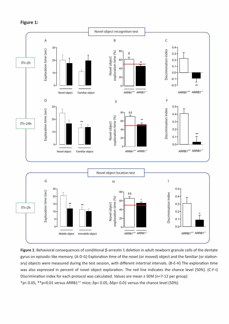
0
10
20
30
Novel object Familiar object
Expl
orat
ion
time
(sec
)
0
5
10
15
20
25
Mobile object Immobile object
** **
Expl
orat
ion
time
(sec
)
0
10
20
30
**
Expl
orat
ion
time
(sec
)
Novel object Familiar object
0
20
40
60
80
**
ARRB1+/+ ARRB1-/-
δδN
ovel
obj
ect
expl
orat
ion
time
(%)
0
20
40
60
80
*
ARRB1+/+ ARRB1-/-
δδ
Nov
el o
bjec
t ex
plor
atio
n tim
e (%
)N
ovel
obj
ect
expl
orat
ion
time
(%)
0
20
40
60
80
ARRB1+/+ ARRB1-/-
**
δ
-0.2
-0.1
0.0
0.1
0.2
0.3
0.4
ARRB1+/+ ARRB1-/-**
Disc
rimin
atio
n in
dex
0.0
0.1
0.2
0.3
0.4
0.5
**
ARRB1+/+ ARRB1-/-
Disc
rimin
atio
n in
dex
0.0
0.1
0.2
0.3
0.4
0.5
ARRB1+/+ ARRB1-/-
*
Disc
rimin
atio
n in
dex
Novel object recognition test
Novel object location test
A B C
D E F
G H I
ITI=2h
ITI=24h
ITI=2h
Figure 1:
Figure 1: Behavioral consequences of conditional β-arrestin 1 deletion in adult newborn granule cells of the dentate gyrus on episodic-like memory. (A-D-G) Exploration time of the novel (or moved) object and the familiar (or station-ary) objects were measured during the test session, with different intertrial intervals. (B-E-H) The exploration time was also expressed in percent of novel object exploration. The red line indicates the chance level (50%). (C-F-I) Discrimination index for each protocol was calculated. Values are mean ± SEM (n=7-12 per group) *p< 0.05, **p<0.01 versus ARRB1+/+ mice; δp< 0.05, δδp< 0.01 versus the chance level (50%)
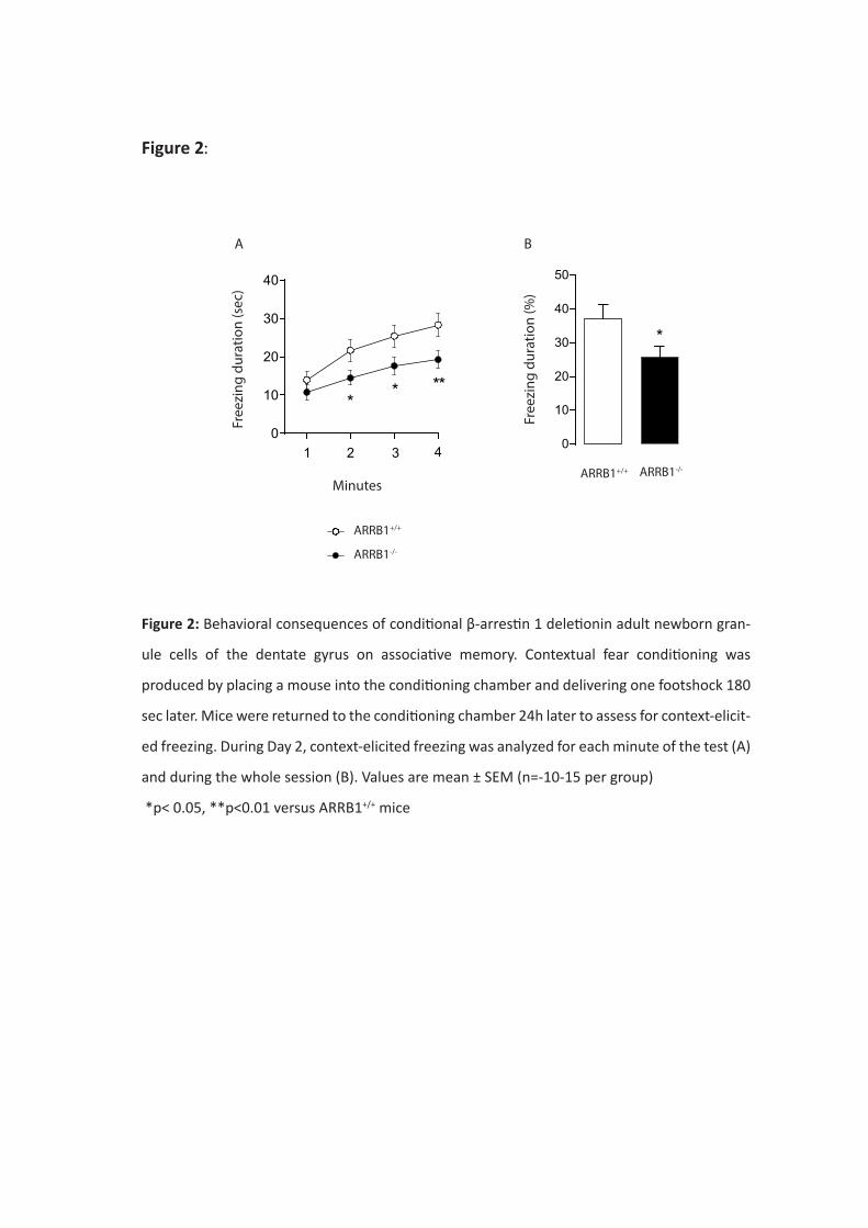
ARRB1+/+
ARRB1-/-
0
10
20
30
40
50
*
Free
zing
dur
atio
n (%
)
B
ARRB1+/+ ARRB1-/-
0
10
20
30
40
1 2 3 4
* * **
Free
zing
dur
atio
n (s
ec)
A
Minutes
Figure 2:
Figure 2: Behavioral consequences of conditional β-arrestin 1 deletionin adult newborn gran-
ule cells of the dentate gyrus on associative memory. Contextual fear conditioning was
produced by placing a mouse into the conditioning chamber and delivering one footshock 180
sec later. Mice were returned to the conditioning chamber 24h later to assess for context-elicit-
ed freezing. During Day 2, context-elicited freezing was analyzed for each minute of the test (A)
and during the whole session (B). Values are mean ± SEM (n=-10-15 per group)
*p< 0.05, **p<0.01 versus ARRB1+/+ mice
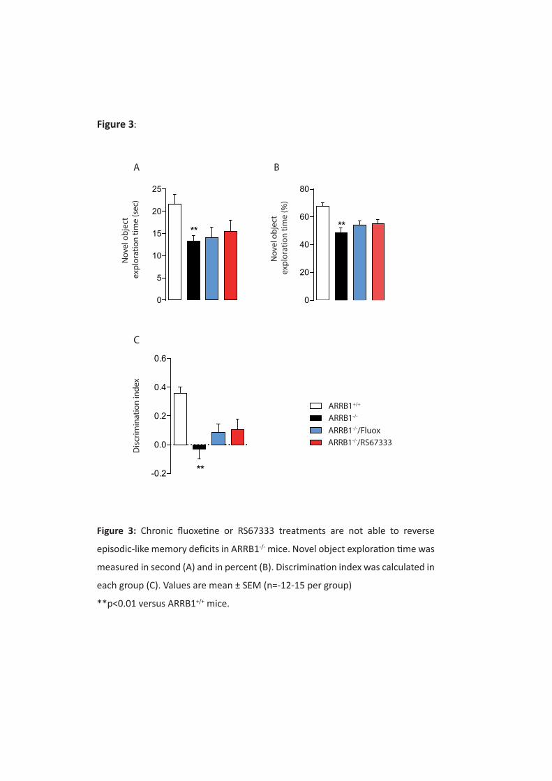
0
5
10
15
20
25
**
Nov
el o
bjec
t ex
plor
atio
n tim
e (s
ec)
0
20
40
60
80
**
Nov
el o
bjec
t ex
plor
atio
n tim
e (%
)
-0.2
0.0
0.2
0.4
0.6
**
Disc
rimin
atio
n in
dex
A B
C
ARRB1+/+
ARRB1-/-
ARRB1-/-/FluoxARRB1-/-/RS67333
Figure 3:
Figure 3: Chronic fluoxetine or RS67333 treatments are not able to reverse
episodic-like memory deficits in ARRB1-/- mice. Novel object exploration time was
measured in second (A) and in percent (B). Discrimination index was calculated in
each group (C). Values are mean ± SEM (n=-12-15 per group)
**p<0.01 versus ARRB1+/+ mice.
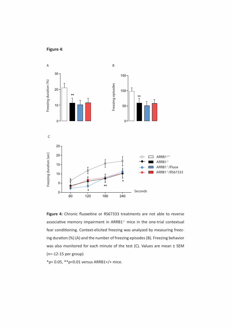
ARRB1+/+
ARRB1-/-
ARRB1-/-/FluoxARRB1-/-/RS67333
Free
zing
dur
atio
n (%
)
0
10
20
30
**
0
50
100
150
**
Free
zing
epi
sode
s
60 120 180 2400
5
10
15
20
25
*
***Fr
eezi
ng d
urat
ion
(sec
)
A B
C
Seconds
Figure 4:
Figure 4: Chronic fluoxetine or RS67333 treatments are not able to reverse
associative memory impairment in ARRB1-/- mice in the one-trial contextual
fear conditioning. Context-elicited freezing was analyzed by measuring freez-
ing duration (%) (A) and the number of freezing episodes (B). Freezing behavior
was also monitored for each minute of the test (C). Values are mean ± SEM
(n=-12-15 per group)
*p< 0.05, **p<0.01 versus ARRB1+/+ mice.
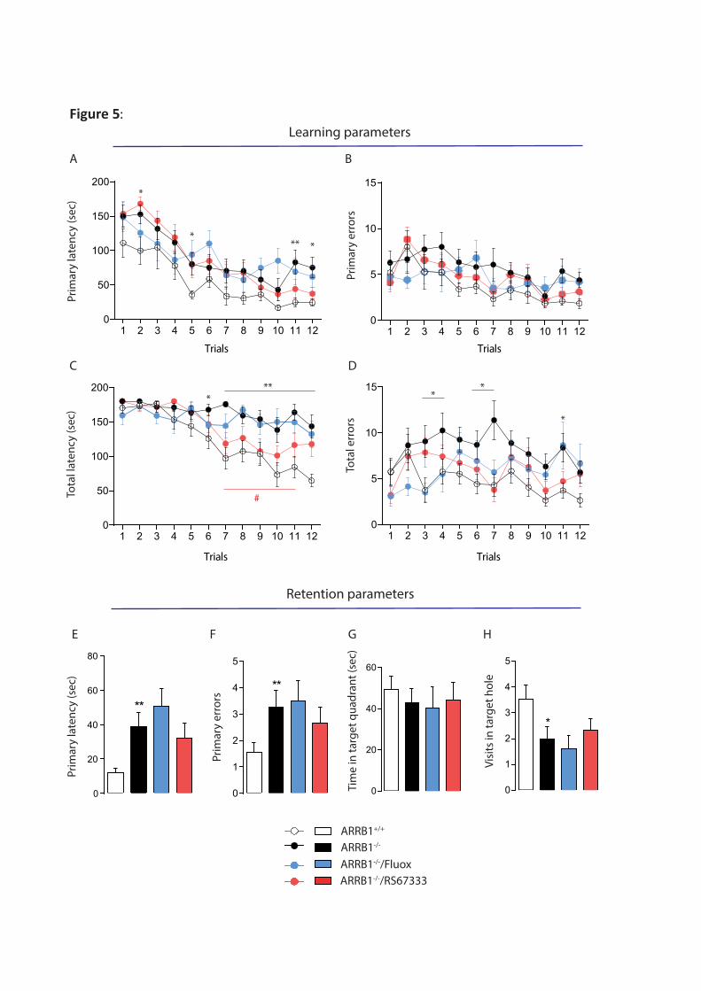
ARRB1+/+
ARRB1-/-
ARRB1-/-/FluoxARRB1-/-/RS67333
Learning parameters
Prim
ary
late
ncy
(sec
)
Trials
1 2 3 4 5 6 7 8 9 10 11 120
50
100
150
200
1 2 3 4 5 6 7 8 9 10 11 120
5
10
15
Prim
ary
erro
rsTrials
1 2 3 4 5 6 7 8 9 10 11 120
50
100
150
200
Tota
l lat
ency
(sec
)
Trials
1 2 3 4 5 6 7 8 9 10 11 120
5
10
15
Tota
l err
ors
Trials
A B
C D
Retention parameters
0
20
40
60
80
**
Prim
ary
late
ncy
(sec
)
**
0
1
2
3
4
5
Prim
ary
erro
rs
0
1
2
3
4
5
*
Visit
s in
targ
et h
ole
Tim
e in
targ
et q
uadr
ant (
sec)
0
20
40
60
E F G H
****
*
*** *
*
*
#
Figure 5:
Figure 5: Chronic fluoxetine or RS67333 treatments are not able to reverse spatial memory deficits
in ARRB1-/- mice.
Animals were trained during 4 days to learn the location of the target box. During this acquisition
period, learning was monitored by recording primary latency (A), total latency (B), primary errors
(C) and total errors (D). At Day 5, a retention probe trial estimates reference spatial memory, by
removing the target box and closing the target hole. Spatial retention was evaluated by measuring
primary latency (E), primary errors (F), time in target quadrant (G) and the number of visits in the
target hole (H). Values are mean ± SEM (n=-10-15 per group)
*p< 0.05, **p<0.01 versus ARRB1+/+ mice;
#p<0.01 versus ARRB1-/- mice.
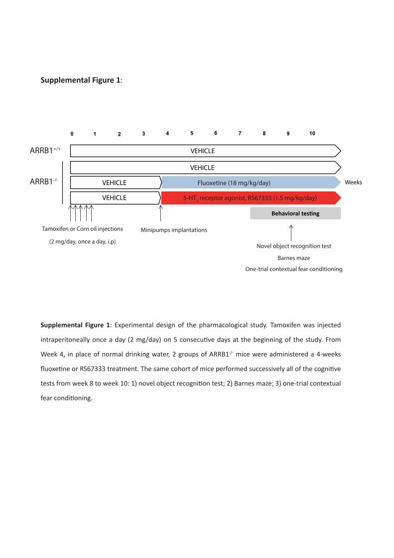
0 41 2 6
Behavioral testing
VEHICLE
Weeks
3 5 7 8 9 10
VEHICLE
VEHICLE
VEHICLE
Fluoxetine (18 mg/kg/day)
5-HT4 receptor agonist, RS67333 (1.5 mg/kg/day)
Tamoxifen or Corn oil injections
(2 mg/day, once a day, i.p)
Minipumps implantations
Novel object recognition test
Barnes maze
One-trial contextual fear conditioning
ARRB1+/+
ARRB1-/-
Supplemental Figure 1:
Supplemental Figure 1: Experimental design of the pharmacological study. Tamoxifen was injected
intraperitoneally once a day (2 mg/day) on 5 consecutive days at the beginning of the study. From
Week 4, in place of normal drinking water, 2 groups of ARRB1-/- mice were administered a 4-weeks
fluoxetine or RS67333 treatment. The same cohort of mice performed successively all of the cognitive
tests from week 8 to week 10: 1) novel object recognition test; 2) Barnes maze; 3) one-trial contextual
fear conditioning.
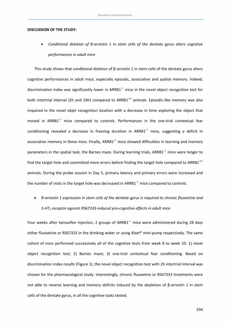
Résultats complémentaires
334
DISCUSSION OF THE STUDY:
x Conditional deletion of β-arrestin 1 in stem cells of the dentate gyrus alters cognitive
performances in adult mice
This study shows that conditional deletion of β-arrestin 1 in stem cells of the dentate gyrus alters
cognitive performances in adult mice, especially episodic, associative and spatial memory. Indeed,
discrimination index was significantly lower in ARRB1-/- mice in the novel object recognition test for
both intertrial interval (2h and 24h) compared to ARRB1+/+ animals. Episodic-like memory was also
impaired in the novel objet recognition location with a decrease in time exploring the object that
moved in ARRB1-/- mice compared to controls. Performances in the one-trial contextual fear
conditioning revealed a decrease in freezing duration in ARRB1-/- mice, suggesting a deficit in
associative memory in these mice. Finally, ARRB1-/- mice showed difficulties in learning and memory
parameters in the spatial task, the Barnes maze. During learning trials, ARRB1-/- mice were longer to
find the target hole and committed more errors before finding the target hole compared to ARRB1+/+
animals. During the probe session in Day 5, primary latency and primary errors were increased and
the number of visits in the target hole was decreased in ARRB1-/- mice compared to controls.
x β-arrestin 1 expression in stem cells of the dentate gyrus is required to chronic fluoxetine and
5-HT4 receptor agonist RS67333-induced pro-cognitive effects in adult mice
Four weeks after tamoxifen injection, 2 groups of ARRB1-/- mice were administered during 28 days
either fluoxetine or RS67333 in the drinking water or using Alzet® mini-pump respectively. The same
cohort of mice performed successively all of the cognitive tests from week 8 to week 10: 1) novel
object recognition test; 2) Barnes maze; 3) one-trial contextual fear conditioning. Based on
discrimination index results (Figure 1), the novel object recognition test with 2h intertrial interval was
chosen for the pharmacological study. Interestingly, chronic fluoxetine or RS67333 treatments were
not able to reverse learning and memory deficits induced by the depletion of β-arrestin 1 in stem
cells of the dentate gyrus, in all the cognitive tasks tested.
Résultats complémentaires
335
The conditional deletion of β-arrestin 1 prevents fluoxetine and RS67333-induce improvement of
episodic-like, associative and spatial memory. In the novel object recognition test, chronic fluoxetine
and RS67333 treatments were not able to correct the discrimination deficit observed in ARRB1-/-
mice. In the one-trial contextual fear conditioning, freezing duration was still decreased in ARRB1-/-
/Fluox and ARRB1-/-/RS67333-treated mice compared to ARRB1+/+ animals, indicating that the
alteration of associative memory is maintained in ARRB1-/- animals despite pharmacological
treatments. Finally, ARRB1-/-/Fluox and ARRB1-/-/RS67333-treated mice failed to restore spatial
performances, in both learning and memory parameters.
These data suggest that β-arrestin 1 expression in stem cells of the dentate gyrus is required to pro-
cognitive effects of chronic fluoxetine and 5-HT4 receptor agonist in adult mice and confirm that β-
arrestin 1 protein is a key element in cognitive mechanisms and pharmacological cognitive
responses.

336
Discussion générale

337
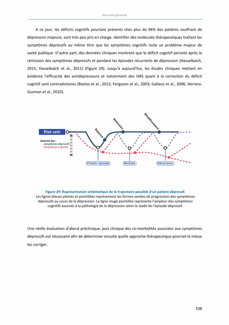
Discussion générale
338
A ce jour, les déficits cognitifs pourtant présents chez plus de 94% des patients souffrant de
dépression majeure, sont très peu pris en charge. Identifier des molécules thérapeutiques traitant les
symptômes dépressifs au même titre que les symptômes cognitifs reste un problème majeur de
santé publique. D’autre part, des données cliniques montrent que le déficit cognitif persiste après la
rémission des symptômes dépressifs et pendant les épisodes récurrents de dépression (Hasselbalch,
2015; Hasselbalch et al., 2011) (Figure 29). Jusqu’à aujourd’hui, les études cliniques mettant en
évidence l’efficacité des antidépresseurs et notamment des ISRS quant à la correction du déficit
cognitif sont contradictoires (Bastos et al., 2013; Ferguson et al., 2003; Gallassi et al., 2006; Herrera-
Guzman et al., 2010).
Une réelle évaluation d’abord préclinique, puis clinique des co-morbidités associées aux symptômes
dépressifs est nécessaire afin de déterminer ensuite quelle approche thérapeutique pourrait le mieux
les corriger.
Figure 29: Représentation schématique de la trajectoire possible d’un patient dépressif. Les lignes bleues pleines et pointillées représentent les formes variées de progression des symptômes
dépressifs au cours de la dépression. La ligne rouge pointillée représente l’ampleur des symptômes cognitifs associés à la pathologie de la dépression selon le stade de l’épisode dépressif.
Discussion générale
339
Ce travail de recherche préclinique, décliné en 3 objectifs a permis d’approfondir la compréhension
du rôle du récepteur 5-HT4 et de la protéine β-arrestine 1 dans la modulation des processus
émotionnels et cognitifs dans un modèle d’anxiété/dépression chez la Souris. Pour cela, nous avons
caractérisé les troubles de la mémoire et de l’apprentissage dans un modèle animal murin
d’anxiété/dépression : le modèle CORT. Nous avons ensuite évalué l’efficacité pro-cognitive de 2
stratégies thérapeutiques à l’aide d’un antidépresseur ISRS de référence (la fluoxetine) et d’une
molécule à visée antidépressive possédant des propriétés agonistes du récepteur 5-HT4 (le RS67333).
Enfin, nous avons mis en évidence le rôle joué par la protéine β-arrestine 1 au niveau des jeunes
neurones du GD, dans la physiopathologie des troubles de l’humeur et de la mémoire et dans la
réponse aux traitements et notamment celle de l’agoniste du récepteur 5-HT4.
1.1 Le phénotype anxio/dépressif induit par un traitement chronique de
corticostérone chez la Souris est associé à des déficits cognitifs.
Pour mieux comprendre les mécanismes responsables des déficits cognitifs dans les épisodes
dépressifs majeurs, la modélisation chez l’animal est essentielle. De nombreuses études se sont
d’ailleurs intéressées aux troubles d’apprentissage et de mémoire dans différents modèles
d’anxiété/dépression chez le rongeur (cf. Revue). Malgré des résultats très hétérogènes, il semble
néanmoins que les troubles des fonctions exécutives et de l’attention soient constants, quelle que
soit la procédure de stress chronique utilisée. D’autre part, les modèles de stress chronique induits
pendant l’âge adulte (comme le UCMS et le modèle CORT) présentent de plus fortes similarités avec
les symptômes cognitifs observés chez les patients déprimés, par rapport aux procédures de stress
pré- ou périnatal.
Dans ce travail, le phénotype anxio/dépressif a été induit suite à l’administration chronique
de corticostérone à faible dose chez la souris C57BL/6J (le modèle CORT) (David et al., 2009). Une
revue de la littérature, reprenant différents travaux ayant étudiés l’impact d’une exposition
chronique de corticostérone chez le Rongeur dans différentes tâches cognitives montre que la
majorité des différents types de mémoires est affectée par ce protocole de stress chronique. Seule la
mémoire de travail (alternation ou spatiale) semble insensible au stress généré par la CORT (Tableau
16).
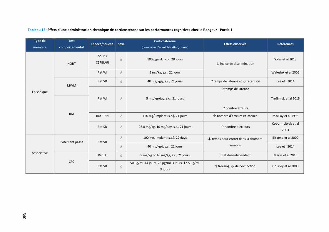
340
Tableau 15: Effets d'une administration chronique de corticostérone sur les performances cognitives chez le Rongeur - Partie 1
Type de
mémoire
Test
comportemental Espèce/Souche Sexe
Corticostérone
(dose, voie d’administration, durée) Effets observés Références
Episodique
NORT
Souris
C57BL/6J ♂ 100 µg/mL, v.o., 28 jours
↓ indice de discrimination
Solas et al 2013
Rat Wi ♂ 5 mg/kg, s.c., 21 jours Walesiuk et al 2005
MWM Rat SD ♂ 40 mg/kg/j, s.c., 21 jours ↑temps de latence et ↓ rétention Lee et l 2014
Rat Wi ♂ 5 mg/kg/day, s.c., 21 jours
↑temps de latence
↑nombre erreurs
Trofimiuk et al 2015
BM Rat F-BN ♂ 150 mg/ Implant (s.c.), 21 jours ↑ nombre d’erreurs et latence MacLay et al 1998
Rat SD ♂ 26.8 mg/kg, 10 mg/day, s.c., 21 jours ↑ nombre d’erreurs Coburn-Litvak et al
2003
Associative
Evitement passif Rat SD ♂ 100 mg, Implant (s.c.), 22 days ↓ temps pour entrer dans la chambre
sombre
Bisagno et al 2000
♂ 40 mg/kg/j, s.c., 21 jours Lee et l 2014
CFC
Rat LE ♂ 5 mg/kg or 40 mg/kg, s.c., 21 jours Effet dose-dépendant Marks et al 2015
Rat SD ♂ 50 µg/mL 14 jours, 25 µg/mL 3 jours, 12.5 µg/mL
3 jours ↑freezing, ↓ de l’extinction Gourley et al 2009
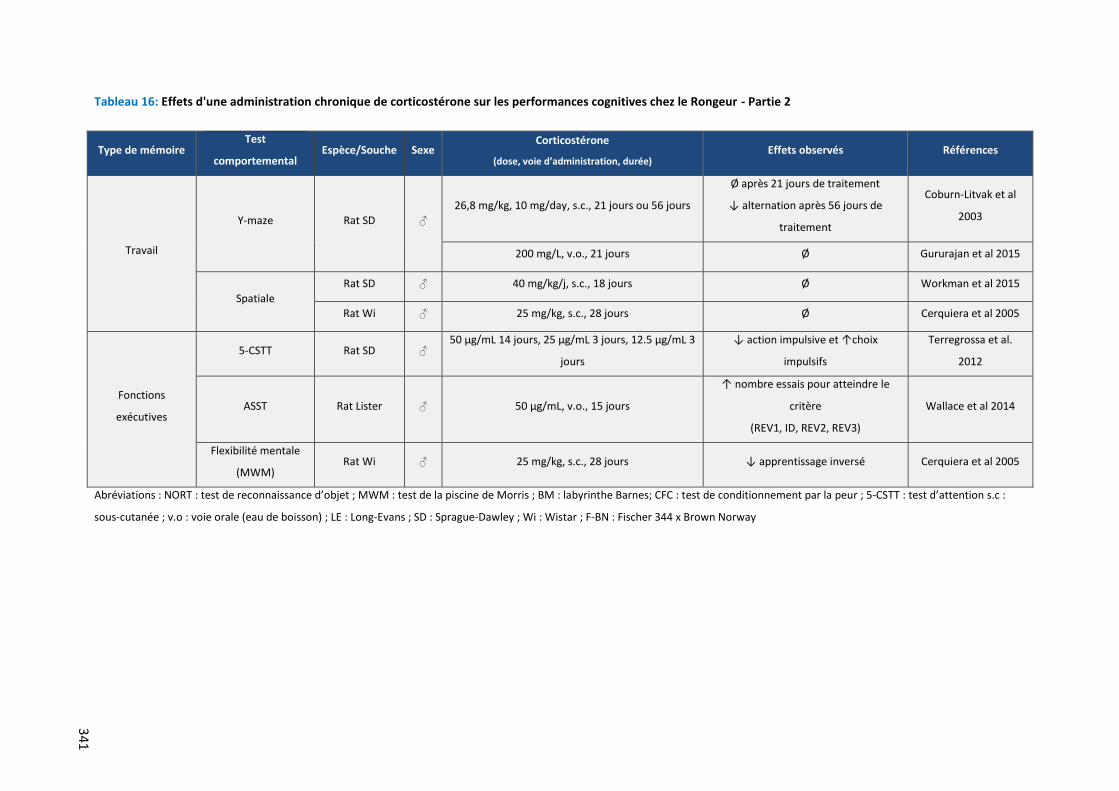
341
Tableau 16: Effets d'une administration chronique de corticostérone sur les performances cognitives chez le Rongeur - Partie 2
Type de mémoire Test
comportemental Espèce/Souche Sexe
Corticostérone
(dose, voie d’administration, durée) Effets observés Références
Travail
Y-maze Rat SD ♂
26,8 mg/kg, 10 mg/day, s.c., 21 jours ou 56 jours
Ø après 21 jours de traitement
↓ alternation après 56 jours de
traitement
Coburn-Litvak et al
2003
200 mg/L, v.o., 21 jours Ø Gururajan et al 2015
Spatiale
Rat SD ♂ 40 mg/kg/j, s.c., 18 jours Ø Workman et al 2015
Rat Wi ♂ 25 mg/kg, s.c., 28 jours Ø Cerquiera et al 2005
Fonctions
exécutives
5-CSTT Rat SD ♂ 50 µg/mL 14 jours, 25 µg/mL 3 jours, 12.5 µg/mL 3
jours
↓ action impulsive et ↑choix
impulsifs
Terregrossa et al.
2012
ASST Rat Lister ♂ 50 µg/mL, v.o., 15 jours
↑ nombre essais pour atteindre le
critère
(REV1, ID, REV2, REV3)
Wallace et al 2014
Flexibilité mentale
(MWM) Rat Wi ♂ 25 mg/kg, s.c., 28 jours ↓ apprentissage inversé Cerquiera et al 2005
Abréviations : NORT : test de reconnaissance d’objet ; MWM : test de la piscine de Morris ; BM : labyrinthe Barnes; CFC : test de conditionnement par la peur ; 5-CSTT : test d’attention s.c :
sous-cutanée ; v.o : voie orale (eau de boisson) ; LE : Long-Evans ; SD : Sprague-Dawley ; Wi : Wistar ; F-BN : Fischer 344 x Brown Norway
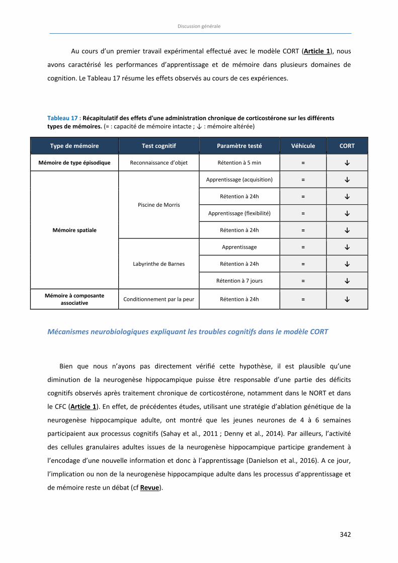
Discussion générale
342
Au cours d’un premier travail expérimental effectué avec le modèle CORT (Article 1), nous
avons caractérisé les performances d’apprentissage et de mémoire dans plusieurs domaines de
cognition. Le Tableau 17 résume les effets observés au cours de ces expériences.
Tableau 17 : Récapitulatif des effets d'une administration chronique de corticostérone sur les différents
types de mémoires. (= : capacité de mémoire intacte ; ↓ : mémoire altérée)
Type de mémoire Test cognitif Paramètre testé Véhicule CORT
Mémoire de type épisodique Reconnaissance d’objet Rétention à 5 min = ↓
Mémoire spatiale
Piscine de Morris
Apprentissage (acquisition) = ↓
Rétention à 24h = ↓
Apprentissage (flexibilité) = ↓
Rétention à 24h = ↓
Labyrinthe de Barnes
Apprentissage = ↓
Rétention à 24h = ↓
Rétention à 7 jours = ↓
Mémoire à composante
associative Conditionnement par la peur Rétention à 24h = ↓
Mécanismes neurobiologiques expliquant les troubles cognitifs dans le modèle CORT
Bien que nous n’ayons pas directement vérifié cette hypothèse, il est plausible qu’une
diminution de la neurogenèse hippocampique puisse être responsable d’une partie des déficits
cognitifs observés après traitement chronique de corticostérone, notamment dans le NORT et dans
le CFC (Article 1). En effet, de précédentes études, utilisant une stratégie d’ablation génétique de la
neurogenèse hippocampique adulte, ont montré que les jeunes neurones de 4 à 6 semaines
participaient aux processus cognitifs (Sahay et al., 2011 ; Denny et al., 2014). Par ailleurs, l’activité
des cellules granulaires adultes issues de la neurogenèse hippocampique participe grandement à
l’encodage d’une nouvelle information et donc à l’apprentissage (Danielson et al., 2016). A ce jour,
l’implication ou non de la neurogenèse hippocampique adulte dans les processus d’apprentissage et
de mémoire reste un débat (cf Revue).
Discussion générale
343
Même si la survie des jeunes neurones n’est pas affectée, il a été observé dans le modèle CORT
une diminution d’environ 30% de la prolifération cellulaire (David et al., 2009). Par ailleurs, de
récentes données obtenues au laboratoire avec une analyse de Sholl de l’arborisation dendritique
des jeunes neurones, montre une diminution de la longueur de l’arborisation dendritique et du
nombre d’intersections suite à une administration chronique de corticosterone. Cependant, il serait
insuffisant de résumer les déficits d’apprentissage ou de mémoire observés dans ce modèle à un
effet exclusivement neurogenèse-dépendant. D’autres modifications, par exemple des systèmes
glutamatergiques, gabaergiques ou cholinergiques centraux peuvent expliquer en partie l’apparition
de ces troubles cognitifs.
Des structures cérébrales autres que l’hippocampe peuvent voir leur fonctionnement altéré suite
à une exposition chronique à la CORT. Ainsi, la mémoire associative est en étroite relation avec
l’amygdale (Roozendaal et al., 2009), et la mémoire épisodique comme la discrimination d’objets est
aussi reliée au cortex entorhinal (Reagh and Yassa, 2014; Wilson et al., 2013). Des modifications
moléculaires et structurelles au sein de ces régions cérébrales peuvent également être à l’origine des
difficultés cognitives induites par un stress prolongé.
Récemment, il a été démontré que les glucocorticoïdes altéraient la mémoire spatiale et la
consolidation en diminuant l’excitabilité des synapses dans le subiculum (temporo-ammonique)-TA-
CA1 (Kvarta et al., 2015). Ces auteurs ont montré qu’un traitement chronique de corticostérone, en
mimant les effets d’un stress chronique, réduirait l’excitation induite par l’activation des récepteurs
AMPA du glutamate. Ces effets, dus à l’activation des récepteurs aux glucocorticoïdes, diminuent la
plasticité synaptique et affectent notamment la consolidation de la mémoire.
D’autre part, la neurotransmission glutamatergique joue un rôle majeur dans les processus de
PLT, de DLP et de mémorisation via l’activation des récepteurs NMDA du glutamate, suivie de la
cascade d’évènements provoqués par l’influx calcique (McEwen and Sapolsky, 1995; Riedel et al.,
2003). Or, si un stress aigu semble avoir des effets bénéfiques sur la transmission glutamatergique et
sur les performances cognitives, un stress chronique provoquerait à l’inverse des changements
structurels délétères tels qu’une perte et une simplification dendritique ou encore une réduction de
la densité d’épines dendritiques dans le cortex et dans l’hippocampe (Popoli et al., 2012; Sandi,
2011). Ces modifications quantitatives et qualitatives affectent la transmission synaptique et sont
alors responsables des déficits cognitifs comportementaux. Dans le cadre du travail expérimental de
l’Article 1, et comme viennent de le démontrer Kvarta et collaborateurs (2015), nous pouvons
émettre l’hypothèse que l’administration chronique de CORT freine la plasticité synaptique
notamment dans le circuit TA-CA1.
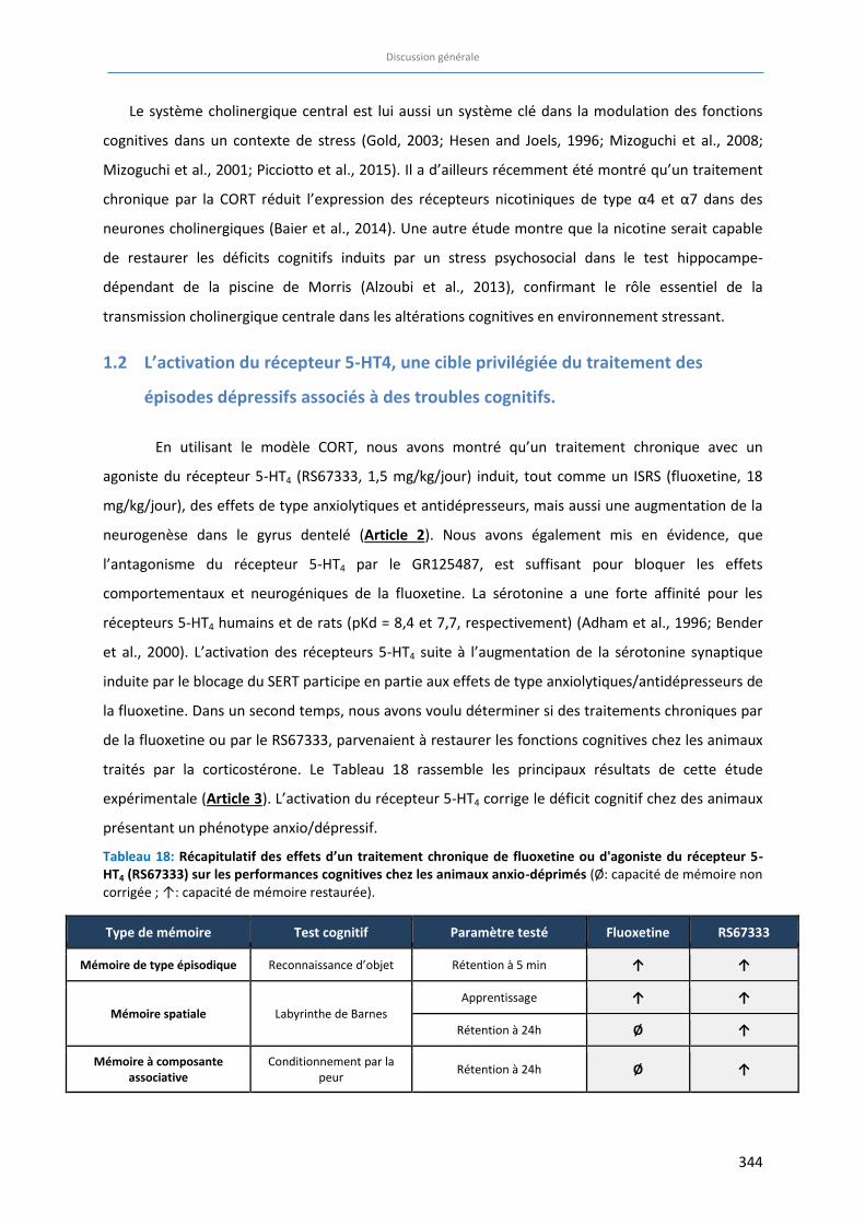
Discussion générale
344
Le système cholinergique central est lui aussi un système clé dans la modulation des fonctions
cognitives dans un contexte de stress (Gold, 2003; Hesen and Joels, 1996; Mizoguchi et al., 2008;
Mizoguchi et al., 2001; Picciotto et al., 2015). Il a d’ailleurs récemment été montré qu’un traitement
chronique par la CORT réduit l’expression des récepteurs nicotiniques de type α4 et α7 dans des
neurones cholinergiques (Baier et al., 2014). Une autre étude montre que la nicotine serait capable
de restaurer les déficits cognitifs induits par un stress psychosocial dans le test hippocampe-
dépendant de la piscine de Morris (Alzoubi et al., 2013), confirmant le rôle essentiel de la
transmission cholinergique centrale dans les altérations cognitives en environnement stressant.
1.2 L’activation du récepteur 5-HT4, une cible privilégiée du traitement des
épisodes dépressifs associés à des troubles cognitifs.
En utilisant le modèle CORT, nous avons montré qu’un traitement chronique avec un
agoniste du récepteur 5-HT4 (RS67333, 1,5 mg/kg/jour) induit, tout comme un ISRS (fluoxetine, 18
mg/kg/jour), des effets de type anxiolytiques et antidépresseurs, mais aussi une augmentation de la
neurogenèse dans le gyrus dentelé (Article 2). Nous avons également mis en évidence, que
l’antagonisme du récepteur 5-HT4 par le GR125487, est suffisant pour bloquer les effets
comportementaux et neurogéniques de la fluoxetine. La sérotonine a une forte affinité pour les
récepteurs 5-HT4 humains et de rats (pKd = 8,4 et 7,7, respectivement) (Adham et al., 1996; Bender
et al., 2000). L’activation des récepteurs 5-HT4 suite à l’augmentation de la sérotonine synaptique
induite par le blocage du SERT participe en partie aux effets de type anxiolytiques/antidépresseurs de
la fluoxetine. Dans un second temps, nous avons voulu déterminer si des traitements chroniques par
de la fluoxetine ou par le RS67333, parvenaient à restaurer les fonctions cognitives chez les animaux
traités par la corticostérone. Le Tableau 18 rassemble les principaux résultats de cette étude
expérimentale (Article 3). L’activation du récepteur 5-HT4 corrige le déficit cognitif chez des animaux
présentant un phénotype anxio/dépressif.
Tableau 18: Récapitulatif des effets d’un traitement chronique de fluoxetine ou d'agoniste du récepteur 5-
HT4 (RS67333) sur les performances cognitives chez les animaux anxio-déprimés (Ø: capacité de mémoire non corrigée ; ↑: capacité de mémoire restaurée).
Type de mémoire Test cognitif Paramètre testé Fluoxetine RS67333
Mémoire de type épisodique Reconnaissance d’objet Rétention à 5 min ↑ ↑
Mémoire spatiale Labyrinthe de Barnes Apprentissage ↑ ↑
Rétention à 24h Ø ↑
Mémoire à composante
associative Conditionnement par la
peur Rétention à 24h Ø ↑
Discussion générale
345
Comment expliquer une efficacité supérieure du RS67333 comparée à celle de la fluoxetine?
Alors que le traitement chronique par le RS67333 corrige la totalité des déficits induits par la
corticostérone, les effets du traitement chronique par la fluoxetine sont moins nombreux puisque
seuls les déficits de mémoire épisodique et d’apprentissage spatial sont corrigés. L’avantage principal
du RS67333 est d’être un composé sélectif qui active uniquement le récepteur 5-HT4. La fluoxetine,
quant à elle, inhibe la recapture de la sérotonine, provoquant alors une augmentation des taux de
sérotonine disponible dans la fente synaptique. Cette grande quantité de 5-HT se fixe alors de façon
non sélective sur les récepteurs sérotoninergiques pour exercer ses effets comportementaux. Il est
donc probable qu’il existe des effets inhibiteurs induits par la stimulation de récepteurs 5-HT qui
limitent ainsi l’efficacité de la fluoxetine dans certains tests de cognition. De façon à mieux
comprendre les mécanismes mis en jeu dans la réponse cognitive à la fluoxetine, il serait intéressant
d’identifier le ou les récepteur(s) sérotoninergique(s) postsynaptique(s) responsable(s) de l’absence
d’effets pro-cognitifs. Une fois ce(s) récepteur(s) déterminé(s), il faudrait alors identifier un
antagoniste de ce récepteur 5-HT qui, associé à la fluoxetine permettrait d'atteindre l'efficacité du
RS67333 dans les tests de cognition. Parmi les 14 sous-types de récepteurs sérotoninergiques
existants, l’activation des récepteurs 5-HT3, 5-HT6 et 5-HT7 semble être un frein à l’activité pro-
cognitive bien qu’ils soient tous les trois associés à une dépolarisation/excitation membranaire. Ainsi
des antagonistes de ces récepteurs pourraient être de bons candidats pour corriger les déficits
cognitifs (pour revue, (Meneses, 2015)).
Le récepteur 5-HT3, canal ionotropique, fait partie des récepteurs sérotoninergiques d’intérêts en
raison de son influence excitatrice sur la libération d’acétylcholine et de GABA suite à sa stimulation
par la sérotonine endogène (Giovannini et al., 1998). Dès les années 1990, des études ont montré
que le blocage du récepteur 5-HT3 par l’ondansetron améliorait certaines fonctions cognitives telle
que la navigation spatiale dans la piscine de Morris (Fontana et al., 1996). Plus récemment, des
travaux ont mis en exergue le rôle du tropisetron, antagoniste sélectif du récepteur 5-HT3, non
seulement dans le traitement des vomissements induits par la chimiothérapie mais aussi lors de
déficits cognitifs, et notamment dans le contexte clinique de la maladie d’Alzheimer (Fakhfouri et al.,
2015; Hashimoto, 2015).
Au même titre que le récepteur 5-HT4, l’activation du récepteur 5-HT6 mène à une activation
de la voie de signalisation de l’AMPc via une augmentation de l’adénylate cyclase. Pourtant, la
plupart des études rapportent un effet bénéfique du blocage du récepteur 5-HT6 sur les fonctions
cognitives, probablement dû à l’existence d’une voie alternative de type inhibitrice, activée par le
Discussion générale
346
récepteur 5-HT6. (Ramirez et al., 2014). Parmi ces travaux, la majorité est conduite dans un contexte
pathologique bien précis. Ainsi, le blocage du récepteur 5-HT6 par un antagoniste sélectif provoque
globalement des effets de type anti-amnésiques voire pro-cognitifs chez les animaux âgés (Callaghan
et al., 2012), chez les animaux présentant des déficits cognitifs induits par la scopolamine (Da Silva
Costa-Aze et al., 2012). Cela a aussi été observé chez les patients atteints de la maladie d’Alzheimer
(Claeysen et al., 2015; Wilkinson et al., 2014). Récemment, il a été démontré qu’une administration
chronique chez la souris naïve d’un antagoniste du récepteur 5-HT6, le SB271046, permettait
d’améliorer la capacité de discrimination des animaux dans le test de la reconnaissance d’objet
(Quiedeville et al., 2015). Cependant, à notre connaissance, aucune étude préclinique ou clinique n’a
encore exploré les propriétés pro-cognitives des antagonistes des récepteurs 5-HT6 dans le contexte
de la dépression majeure. L’ensemble de ces données fait donc du récepteur 5-HT6 une cible à
privilégier dans les perspectives de ce travail de thèse.
Enfin, une revue récente suggère des perspectives encourageantes concernant le rôle du
récepteur 5-HT7 dans différentes pathologies du SNC (anxiété, épilepsie, schizophrénie) (Nikiforuk,
2015). Des études précliniques ont d’ailleurs montré que l’utilisation d’antagonistes des récepteurs
5-HT7 entrainait des effets anti-amnésiques (Gasbarri and Pompili, 2014) voire pro-cognitifs (Gasbarri
et al., 2008) si la tâche cognitive présente un degré élevé de difficulté. Cependant, d’autres travaux
rapportent ces mêmes effets favorisant la mémoire suite à la stimulation des récepteurs 5-HT7 par un
agoniste (Di Pilato et al., 2014; Freret et al., 2014; Perez-Garcia and Meneses, 2005) (Figure 30). En
raison de ces résultats hétérogènes, le récepteur 5-HT7 ne constitue pas l’hypothèse prioritaire dans
notre recherche du (des) récepteur(s) impliqué(s) dans la réponse cognitive à la fluoxetine.
Il est important de disséquer le mécanisme d’action impliqué dans les effets cognitifs de la
fluoxetine induits par l’activation du récepteur 5-HT4. Les arguments précédemment développés
donnent quelques pistes à explorer pour de prochaines études. La Figure 30 ci-après récapitule les
conséquences de la liaison de la fluoxetine sur le transporteur du 5-HT, puis l’activation des
différents types de récepteurs sérotoninergiques par le 5-HT endogène sur le comportement cognitif.
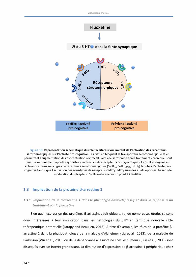
Discussion générale
347
Figure 30: Représentation schématique du rôle facilitateur ou limitant de l’activation des récepteurs
sérotoninergiques sur l’activité pro-cognitive. Les ISRS en bloquant le transporteur sérotoninergique et en permettant l’augmentation des concentrations extracellulaires de sérotonine après traitement chronique, sont
aussi communément appelés agonistes « indirects » des récepteurs postsynaptiques. La 5-HT endogène en activant certains sous types de récepteurs sérotoninergiques (5-HT1A, 5-HT1B/1D, 5-HT4) facilitera l’activité pro-cognitive tandis que l’activation des sous-types de récepteurs 5-HT3, 5-HT6 aura des effets opposés. Le sens de
modulation du récepteur 5-HT7 reste encore un point à identifier.
1.3 Implication de la protéine β-arrestine 1
1.3.1 Implication de la β-arrestine 1 dans le phénotype anxio-dépressif et dans la réponse à un traitement par la fluoxetine
Bien que l’expression des protéines β-arrestines soit ubiquitaire, de nombreuses études se sont
donc intéressées à leur implication dans les pathologies du SNC en tant que nouvelle cible
thérapeutique potentielle (Latapy and Beaulieu, 2013). A titre d’exemple, les rôles de la protéine β-
arrestine 1 dans la physiopathologie de la maladie d’Alzheimer (Liu et al., 2013), de la maladie de
Parkinson (Wu et al., 2013) ou de la dépendance à la nicotine chez les fumeurs (Sun et al., 2008) sont
disséqués avec un intérêt grandissant. La diminution d’expression de β-arrestine 1 périphérique chez
Discussion générale
348
les patients dépressifs a été, quant à elle, mise en évidence en 2004 (Avissar et al., 2004).
Rapidement, l’implication de la β-arrestine 1 dans la réponse aux traitements antidépresseurs a été
étudiée aussi bien en clinique (Avissar et al., 2004 ; Matuzany-Ruban et al., 2005) qu’en préclinique
(Mendez-David et al., 2015; Golan et al 2009). Dans le domaine préclinique, l’étude de l’implication
des protéines β-arrestines (1 et 2) peut faire appel à des lignées de souris génétiquement modifiées.
De manière intéressante, la double mutation KO ARRB1/ARRB2 est létale chez la Souris, alors que les
modèles de souris constitutives ARRB1-/- ou ARRB2-/- sont viables. Il semble donc que ces deux
protéines aient des propriétés chevauchantes.
Puisque la neurogenèse hippocampique et notamment les jeunes neurones du GD âgés de 4-6
semaines semblent jouer un rôle prépondérant dans l’activité cognitive ou antidépressive (Denny et
al., 2012; Sahay et al., 2011), nous avons souhaité étudier les conséquences de l’absence
d’expression de la protéine β-arrestine 1 dans les jeunes neurones nouvellement formés. Pour cela,
nous avons induit une délétion conditionnelle de la protéine β-arrestine 1 dans les cellules souches
et attendu 4/7 semaines avant de caractériser leur phénotype. Les résultats obtenus dans l’Article 4
sont regroupés dans le Tableau 19 et le Tableau 20. Au regard du score d’émotionalité, la délétion
conditionnelle, à l’âge adulte, de l’expression de la protéine β-arrestine 1 dans les jeunes neurones
issus de la neurogenèse du GD de l’hippocampe n’a pas modifié globalement le phénotype
émotionnel. Pourtant des différences apparaissent quand on analyse les résultats de chaque test
séparément. Un phénotype anxio/dépressif est apparu dans le labyrinthe en croix surélevée, le test
des 4 plaques et le Splash test. Ceci confirme que l’expression de la β-arrestine 1 au niveau des
jeunes neurones issus de la neurogenèse a des conséquences bien au-delà du GD (Bergami and
Berninger, 2012). Il apparaît que le score d’émotionalité est beaucoup plus pertinent dans le cadre
d’une étude de criblage que lors d’une étude mécanistique. De plus, l’absence d’expression de la β-
arrestine 1 dans les jeunes neurones issus de la neurogenèse, a bloqué les effets de type
anxiolytiques et antidépresseurs d’un traitement chronique par la fluoxetine, comme le révèle le
score d’émotionalité, même si là encore des disparités entre les tests existent (Tableau 20).
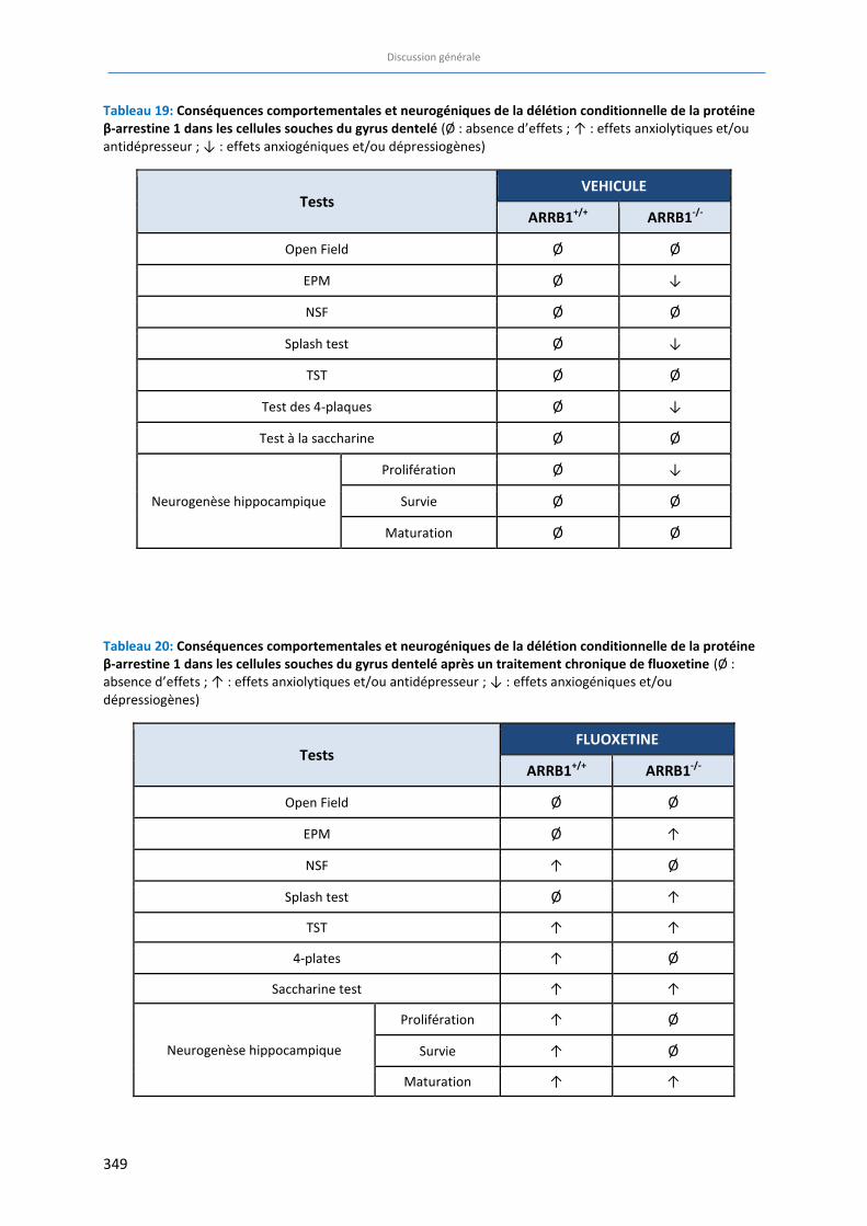
Discussion générale
349
Tableau 19: Conséquences comportementales et neurogéniques de la délétion conditionnelle de la protéine
β-arrestine 1 dans les cellules souches du gyrus dentelé (Ø : absence d’effets ; ↑ : effets anxiolytiques et/ou antidépresseur ; ↓ : effets anxiogéniques et/ou dépressiogènes)
Tests VEHICULE
ARRB1+/+
ARRB1-/-
Open Field Ø Ø
EPM Ø ↓
NSF Ø Ø
Splash test Ø ↓
TST Ø Ø
Test des 4-plaques Ø ↓
Test à la saccharine Ø Ø
Neurogenèse hippocampique
Prolifération Ø ↓
Survie Ø Ø
Maturation Ø Ø
Tableau 20: Conséquences comportementales et neurogéniques de la délétion conditionnelle de la protéine
β-arrestine 1 dans les cellules souches du gyrus dentelé après un traitement chronique de fluoxetine (Ø : absence d’effets ; ↑ : effets anxiolytiques et/ou antidépresseur ; ↓ : effets anxiogéniques et/ou dépressiogènes)
Tests FLUOXETINE
ARRB1+/+ ARRB1
-/-
Open Field Ø Ø
EPM Ø ↑
NSF ↑ Ø
Splash test Ø ↑
TST ↑ ↑
4-plates ↑ Ø
Saccharine test ↑ ↑
Neurogenèse hippocampique
Prolifération ↑ Ø
Survie ↑ Ø
Maturation ↑ ↑
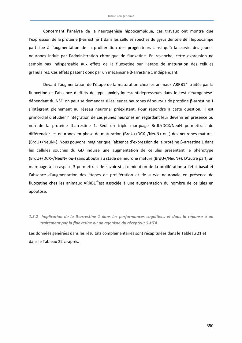
Discussion générale
350
Concernant l’analyse de la neurogenèse hippocampique, ces travaux ont montré que
l’expression de la protéine β-arrestine 1 dans les cellules souches du gyrus dentelé de l’hippocampe
participe à l’augmentation de la prolifération des progéniteurs ainsi qu’à la survie des jeunes
neurones induit par l’administration chronique de fluoxetine. En revanche, cette expression ne
semble pas indispensable aux effets de la fluoxetine sur l’étape de maturation des cellules
granulaires. Ces effets passent donc par un mécanisme β-arrestine 1 indépendant.
Devant l’augmentation de l’étape de la maturation chez les animaux ARRB1-/- traités par la
fluoxetine et l’absence d’effets de type anxiolytiques/antidépresseurs dans le test neurogenèse-
dépendant du NSF, on peut se demander si les jeunes neurones dépourvus de protéine β-arrestine 1
s’intègrent pleinement au réseau neuronal préexistant. Pour répondre à cette question, il est
primordial d’étudier l’intégration de ces jeunes neurones en regardant leur devenir en présence ou
non de la protéine β-arrestine 1. Seul un triple marquage BrdU/DCX/NeuN permettrait de
différencier les neurones en phase de maturation (BrdU+/DCX+/NeuN+ ou-) des neurones matures
(BrdU+/NeuN+). Nous pouvons imaginer que l’absence d’expression de la protéine β-arrestine 1 dans
les cellules souches du GD induise une augmentation de cellules présentant le phénotype
(BrdU+/DCX+/NeuN+ ou-) sans aboutir au stade de neurone mature (BrdU+/NeuN+). D’autre part, un
marquage à la caspase 3 permettrait de savoir si la diminution de la prolifération à l’état basal et
l’absence d’augmentation des étapes de prolifération et de survie neuronale en présence de
fluoxetine chez les animaux ARRB1-/-est associée à une augmentation du nombre de cellules en
apoptose.
1.3.2 Implication de la β-arrestine 1 dans les performances cognitives et dans la réponse à un traitement par la fluoxetine ou un agoniste du récepteur 5-HT4
Les données générées dans les résultats complémentaires sont récapitulées dans le Tableau 21 et
dans le Tableau 22 ci-après.
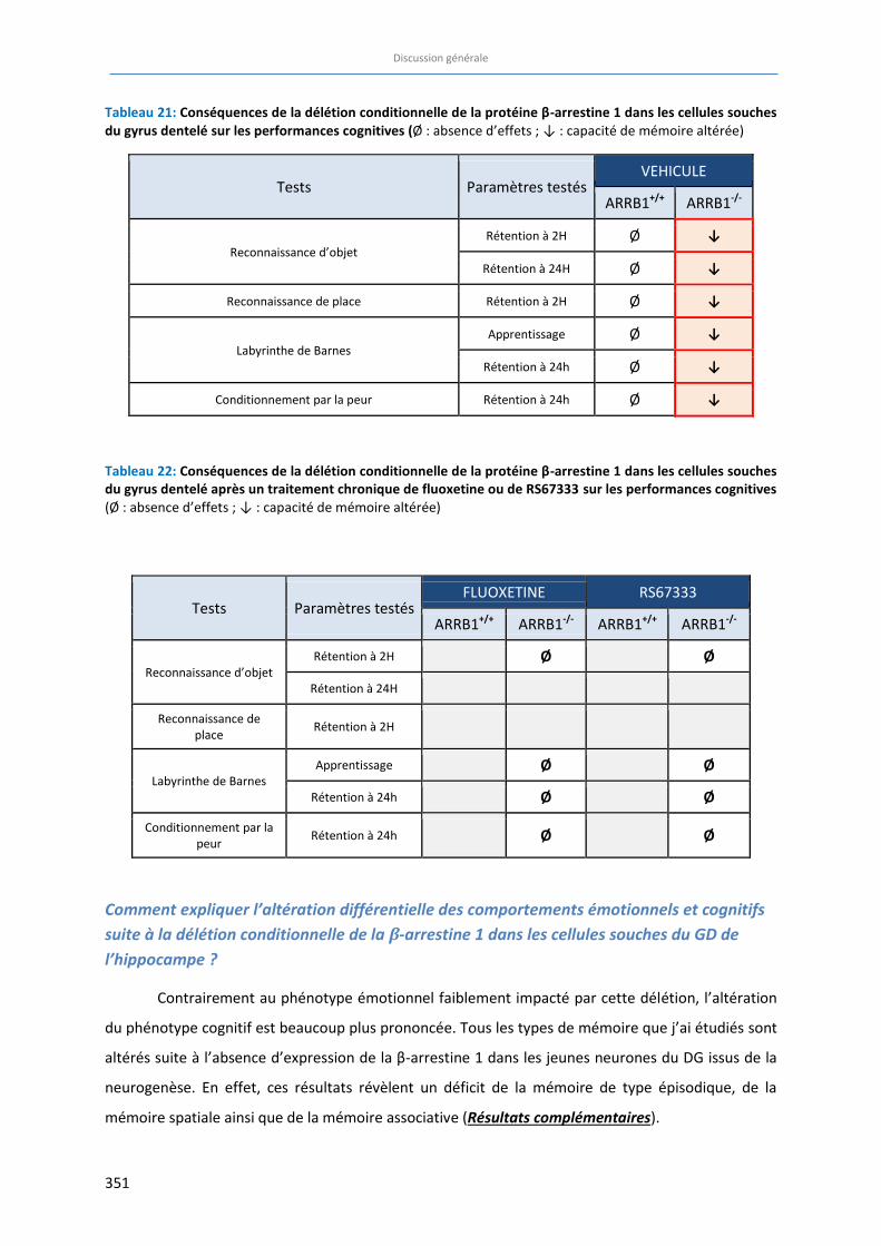
Discussion générale
351
Tableau 21: Conséquences de la délétion conditionnelle de la protéine β-arrestine 1 dans les cellules souches
du gyrus dentelé sur les performances cognitives (Ø : absence d’effets ; ↓ : capacité de mémoire altérée)
Tests Paramètres testés VEHICULE
ARRB1+/+ ARRB1-/-
Reconnaissance d’objet Rétention à 2H Ø ↓
Rétention à 24H Ø ↓
Reconnaissance de place Rétention à 2H Ø ↓
Labyrinthe de Barnes Apprentissage Ø ↓
Rétention à 24h Ø ↓
Conditionnement par la peur Rétention à 24h Ø ↓
Tableau 22: Conséquences de la délétion conditionnelle de la protéine β-arrestine 1 dans les cellules souches
du gyrus dentelé après un traitement chronique de fluoxetine ou de RS67333 sur les performances cognitives
(Ø : absence d’effets ; ↓ : capacité de mémoire altérée)
Tests Paramètres testés FLUOXETINE RS67333
ARRB1+/+ ARRB1-/- ARRB1+/+ ARRB1-/-
Reconnaissance d’objet Rétention à 2H Ø
Ø
Rétention à 24H
Reconnaissance de place
Rétention à 2H
Labyrinthe de Barnes Apprentissage Ø Ø
Rétention à 24h Ø Ø
Conditionnement par la peur
Rétention à 24h Ø Ø
Comment expliquer l’altération différentielle des comportements émotionnels et cognitifs suite à la délétion conditionnelle de la β-arrestine 1 dans les cellules souches du GD de l’hippocampe ?
Contrairement au phénotype émotionnel faiblement impacté par cette délétion, l’altération
du phénotype cognitif est beaucoup plus prononcée. Tous les types de mémoire que j’ai étudiés sont
altérés suite à l’absence d’expression de la β-arrestine 1 dans les jeunes neurones du DG issus de la
neurogenèse. En effet, ces résultats révèlent un déficit de la mémoire de type épisodique, de la
mémoire spatiale ainsi que de la mémoire associative (Résultats complémentaires).
Discussion générale
352
Dans une récente revue de la littérature, Miller et Hen concluent que si l’ablation de la
neurogenèse hippocampique induit un déficit cognitif relatif aux fonctions émotionnelles, une
minorité d’étude montre que cette ablation induit un phénotype anxio/dépressif (Miller and Hen,
2015). Nos résultats suggèrent que la délétion de la β-arrestine 1 dans les jeunes neurones
nouvellement formés n’est pas suffisante pour affecter le comportement émotionnel basal. Ce
constat est au final en accord avec une étude clinique montrant que l’altération de la neurogenèse
adulte n’est pas à l’origine de l’apparition des symptômes de la dépression (Cameron and McKay,
2001) et plus généralement avec la littérature. La protéine β-arrestine 1 serait donc impliquée de
façon plus importante dans les processus mnésiques plutôt que dans la physiopathologie des
troubles de l’humeur. Cependant, aucune étude clinique visant à préciser le rôle de la protéine β-
arrestine 1 dans les troubles cognitifs n’a été menée à ce jour. Finalement, très peu d’études
précliniques et cliniques se concentrent sur l’implication de la β-arrestine 1 dans les troubles
émotionnels ou cognitifs. Ces thématiques gagneraient à être plus largement examinées afin de
mieux comprendre les mécanismes mis en jeu dans ces pathologies mentales.
Considérations et limites méthodologiques
Impact de la souche de souris
Il est aujourd’hui bien reconnu que le fond génétique des animaux, et notamment chez la
Souris, influence les phénotypes comportementaux (Clement et al., 2002; Crawley et al., 1997). Dans
le cadre de ce travail conduit chez des animaux dans un contexte d’anxiété et de dépression, il est
impératif de faire la distinction entre des troubles du comportement d’un apprentissage réel versus
des altérations sensorielles, inhérentes aux différentes souches, pouvant mener à une performance
cognitive amoindrie. Par exemple, le paramètre d’acuité visuelle pour les tâches spatiales, la capacité
auditive dans les tests de conditionnement incluant un son, et le seuil de sensibilité au choc
électrique peuvent varier d’une souche à une autre. Les différences de motivation des animaux,
devant un stimulus appétant ou aversif, doivent être rigoureusement dissociées des troubles réels de
l’apprentissage et de la mémoire. D’autre part, les capacités d’apprentissage diffèrent selon les
souches de souris. Dans la piscine de Morris, les souris de souche C57BL/6J possèdent de meilleures
capacités d’apprentissage que la souche 129Sv/J (Crawley et al., 1997) tandis que les deux souches
semblent apprendre de la même manière dans le test de conditionnement par la peur (Crawley et al.,
1997). Une étude comparant les performances cognitives spatiales de 13 souches de souris
différentes dans le « Barnes maze », a montré que les souris 129S1/SvImJ mettent significativement
plus de temps à localiser le trou cible, à entrer dedans et commettent plus d’erreurs que les autres
souches. Ceci serait probablement plus dû à une hypoactivité plutôt qu’à une altération de
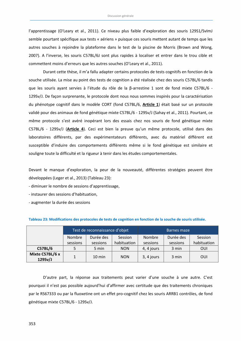
Discussion générale
353
l’apprentissage (O'Leary et al., 2011). Ce niveau plus faible d’exploration des souris 129S1/SvImJ
semble pourtant spécifique aux tests « aériens » puisque ces souris mettent autant de temps que les
autres souches à rejoindre la plateforme dans le test de la piscine de Morris (Brown and Wong,
2007). A l’inverse, les souris C57BL/6J sont plus rapides à localiser et entrer dans le trou cible et
commettent moins d’erreurs que les autres souches (O’Leary et al., 2011).
Durant cette thèse, il m’a fallu adapter certains protocoles de tests cognitifs en fonction de la
souche utilisée. La mise au point des tests de cognition a été réalisée chez des souris C57BL/6 tandis
que les souris ayant servies à l’étude du rôle de la β-arrestine 1 sont de fond mixte C57BL/6 -
129Sv/J. De façon surprenante, le protocole dont nous nous sommes inspirés pour la caractérisation
du phénotype cognitif dans le modèle CORT (fond C57BL/6, Article 1) était basé sur un protocole
validé pour des animaux de fond génétique mixte C57BL/6 - 129Sv/J (Sahay et al., 2011). Pourtant, ce
même protocole s’est avéré inopérant lors des essais chez nos souris de fond génétique mixte
C57BL/6 - 129Sv/J (Article 4). Ceci est bien la preuve qu’un même protocole, utilisé dans des
laboratoires différents, par des expérimentateurs différents, avec du matériel différent est
susceptible d’induire des comportements différents même si le fond génétique est similaire et
souligne toute la difficulté et la rigueur à tenir dans les études comportementales.
Devant le manque d’exploration, la peur de la nouveauté, différentes stratégies peuvent être
développées (Leger et al., 2013) (Tableau 23):
- diminuer le nombre de sessions d’apprentissage,
- instaurer des sessions d’habituation,
- augmenter la durée des sessions
Tableau 23: Modifications des protocoles de tests de cognition en fonction de la souche de souris utilisée.
Test de reconnaissance d’objet Barnes maze
Nombre sessions
Durée des sessions
Session habituation
Nombre sessions
Durée des sessions
Session habituation
C57BL/6 5 5 min NON 4, 4 jours 3 min OUI
Mixte C57BL/6 x
129Sv/J 1 10 min NON 3, 4 jours 3 min OUI
D’autre part, la réponse aux traitements peut varier d’une souche à une autre. C’est
pourquoi il n’est pas possible aujourd’hui d’affirmer avec certitude que des traitements chroniques
par le RS67333 ou par la fluoxetine ont un effet pro-cognitif chez les souris ARRB1 contrôles, de fond
génétique mixte C57BL/6 - 129Sv/J.
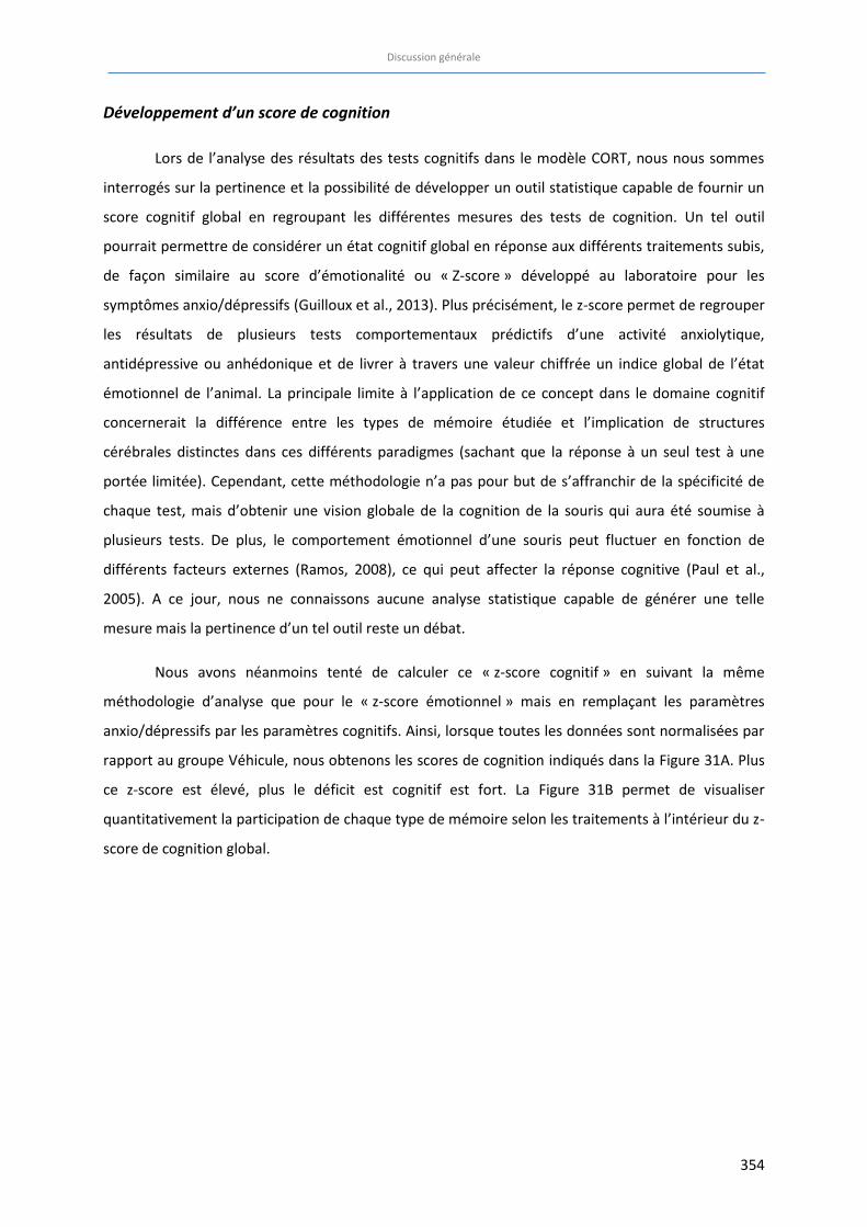
Discussion générale
354
Développement d’un score de cognition
Lors de l’analyse des résultats des tests cognitifs dans le modèle CORT, nous nous sommes
interrogés sur la pertinence et la possibilité de développer un outil statistique capable de fournir un
score cognitif global en regroupant les différentes mesures des tests de cognition. Un tel outil
pourrait permettre de considérer un état cognitif global en réponse aux différents traitements subis,
de façon similaire au score d’émotionalité ou « Z-score » développé au laboratoire pour les
symptômes anxio/dépressifs (Guilloux et al., 2013). Plus précisément, le z-score permet de regrouper
les résultats de plusieurs tests comportementaux prédictifs d’une activité anxiolytique,
antidépressive ou anhédonique et de livrer à travers une valeur chiffrée un indice global de l’état
émotionnel de l’animal. La principale limite à l’application de ce concept dans le domaine cognitif
concernerait la différence entre les types de mémoire étudiée et l’implication de structures
cérébrales distinctes dans ces différents paradigmes (sachant que la réponse à un seul test à une
portée limitée). Cependant, cette méthodologie n’a pas pour but de s’affranchir de la spécificité de
chaque test, mais d’obtenir une vision globale de la cognition de la souris qui aura été soumise à
plusieurs tests. De plus, le comportement émotionnel d’une souris peut fluctuer en fonction de
différents facteurs externes (Ramos, 2008), ce qui peut affecter la réponse cognitive (Paul et al.,
2005). A ce jour, nous ne connaissons aucune analyse statistique capable de générer une telle
mesure mais la pertinence d’un tel outil reste un débat.
Nous avons néanmoins tenté de calculer ce « z-score cognitif » en suivant la même
méthodologie d’analyse que pour le « z-score émotionnel » mais en remplaçant les paramètres
anxio/dépressifs par les paramètres cognitifs. Ainsi, lorsque toutes les données sont normalisées par
rapport au groupe Véhicule, nous obtenons les scores de cognition indiqués dans la Figure 31A. Plus
ce z-score est élevé, plus le déficit est cognitif est fort. La Figure 31B permet de visualiser
quantitativement la participation de chaque type de mémoire selon les traitements à l’intérieur du z-
score de cognition global.
A
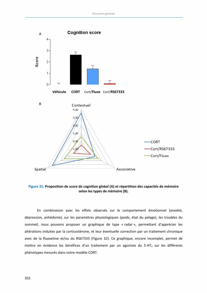
Discussion générale
355
Figure 31: Proposition de score de cognition global (A) et répartition des capacités de mémoire selon les types de mémoire (B).
En combinaison avec les effets observés sur le comportement émotionnel (anxiété,
dépression, anhédonie), sur les paramètres physiologiques (poids, état du pelage), les troubles du
sommeil, nous pouvons proposer un graphique de type « radar », permettant d’apprécier les
altérations induites par la corticostérone, et leur éventuelle correction par un traitement chronique
avec de la fluoxetine et/ou du RS67333 (Figure 32). Ce graphique, encore incomplet, permet de
mettre en évidence les bénéfices d’un traitement par un agoniste du 5-HT4 sur les différents
phénotypes mesurés dans notre modèle CORT.
Véhicule CORT Cort/Fluox Cort/RS67333
B
A
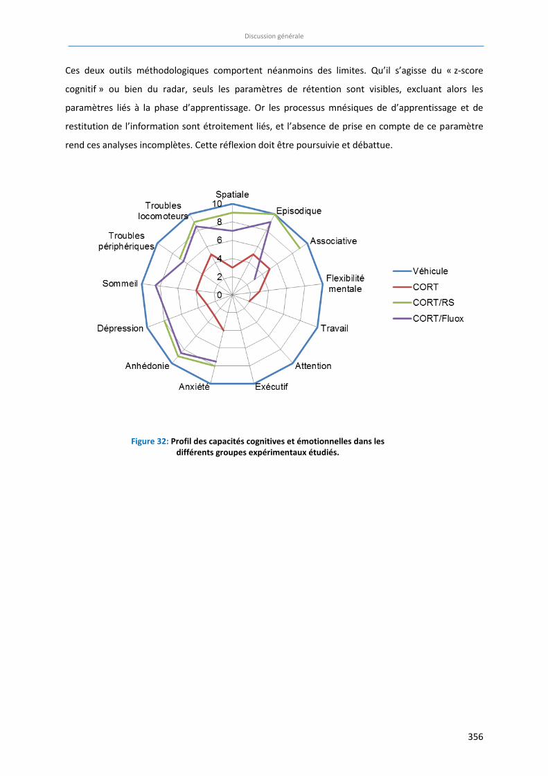
Discussion générale
356
Ces deux outils méthodologiques comportent néanmoins des limites. Qu’il s’agisse du « z-score
cognitif » ou bien du radar, seuls les paramètres de rétention sont visibles, excluant alors les
paramètres liés à la phase d’apprentissage. Or les processus mnésiques de d’apprentissage et de
restitution de l’information sont étroitement liés, et l’absence de prise en compte de ce paramètre
rend ces analyses incomplètes. Cette réflexion doit être poursuivie et débattue.
Figure 32: Profil des capacités cognitives et émotionnelles dans les différents groupes expérimentaux étudiés.
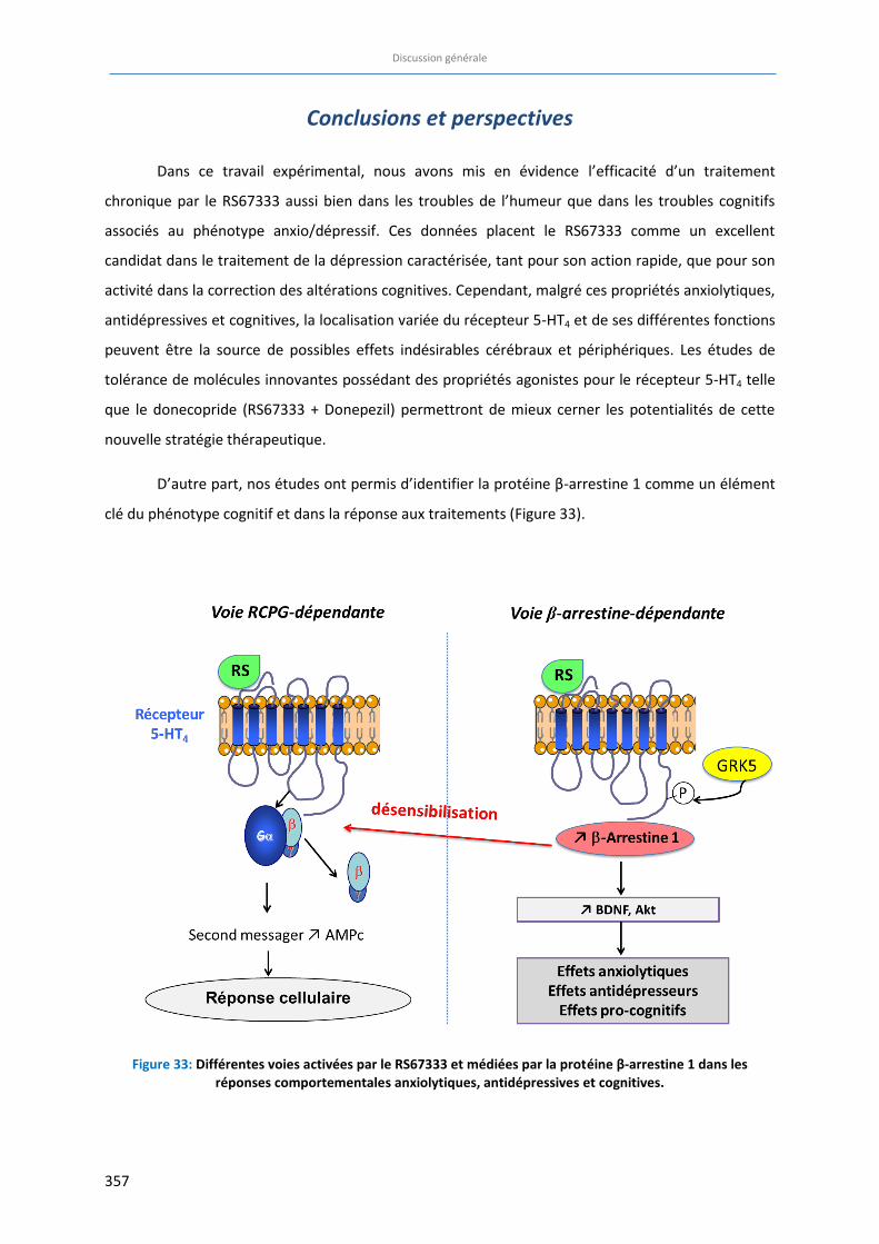
Discussion générale
357
Conclusions et perspectives
Dans ce travail expérimental, nous avons mis en évidence l’efficacité d’un traitement
chronique par le RS67333 aussi bien dans les troubles de l’humeur que dans les troubles cognitifs
associés au phénotype anxio/dépressif. Ces données placent le RS67333 comme un excellent
candidat dans le traitement de la dépression caractérisée, tant pour son action rapide, que pour son
activité dans la correction des altérations cognitives. Cependant, malgré ces propriétés anxiolytiques,
antidépressives et cognitives, la localisation variée du récepteur 5-HT4 et de ses différentes fonctions
peuvent être la source de possibles effets indésirables cérébraux et périphériques. Les études de
tolérance de molécules innovantes possédant des propriétés agonistes pour le récepteur 5-HT4 telle
que le donecopride (RS67333 + Donepezil) permettront de mieux cerner les potentialités de cette
nouvelle stratégie thérapeutique.
D’autre part, nos études ont permis d’identifier la protéine β-arrestine 1 comme un élément
clé du phénotype cognitif et dans la réponse aux traitements (Figure 33).
Figure 33: Différentes voies activées par le RS67333 et médiées par la protéine β-arrestine 1 dans les réponses comportementales anxiolytiques, antidépressives et cognitives.
Discussion générale
358
Les suites de ce travail peuvent s’articuler selon plusieurs axes :
x Etudier les mécanismes moléculaires et cellulaires différentiellement mis en jeu par un
traitement par la fluoxetine et le RS67333
x Vérifier si l’administration d’un antagoniste sélectif du récepteur 5-HT4, tel que le
GR125487, bloque la réponse cognitive de la fluoxetine
x Etudier les effets cognitifs des antagonistes sélectifs des récepteurs 5-HT3 et 5-HT6 en
association avec la fluoxetine
x Vérifier si la correction du déficit cognitif permet de prévenir la rechute des épisodes
dépressifs
Enfin, plusieurs questions restent également en suspens et mériteraient la mise en place et la
conception de nouvelles expérimentations :
x La correction des altérations cognitives est-elle préalable, associée ou consécutive à
l’amélioration du phénotype anxio/dépressif ?
x La réponse rapide observée après administration aigue de RS67333 induit elle également
une réponse cognitive ?
x Quels mécanismes expliquent les variations des différentes étapes de neurogenèse
hippocampique adulte observées après administration de fluoxétine chez les souris ARRB1-
/- ?

359

Références bibliographiques
360
Références bibliographiques

Références bibliographiques
361
Références bibliographiques
362
A Adham, N., Gerald, C., Schechter, L., Vaysse, P., Weinshank, R., Branchek, T., 1996. [3H]5-hydroxytryptamine
labels the agonist high affinity state of the cloned rat 5-HT4 receptor. European journal of pharmacology 304,
231-235.
Ago, Y., Arikawa, S., Yata, M., Yano, K., Abe, M., Takuma, K., Matsuda, T., 2008. Antidepressant-like effects of
the glucocorticoid receptor antagonist RU-43044 are associated with changes in prefrontal dopamine in mouse
models of depression. Neuropharmacology 55, 1355-1363.
Ahmad, I., Nirogi, R., 2011. 5-HT4 Receptor Agonists for the Treatment of Alzheimer's Disease. Neuroscience &
Medicine 2, 87-92.
Alam, F., Nayyar, S., Richie, W., Archibong, A., Nayyar, T., 2015. Beta-Arrestin1 Levels in Mononuclear
Leukocytes Support Depression Scores for Women with Premenstrual Dysphoric Disorder. International journal
of environmental research and public health 13.
Alzoubi, K.H., Srivareerat, M., Tran, T.T., Alkadhi, K.A., 2013. Role of alpha7- and alpha4beta2-nAChRs in the
neuroprotective effect of nicotine in stress-induced impairment of hippocampus-dependent memory. The
international journal of neuropsychopharmacology / official scientific journal of the Collegium Internationale
Neuropsychopharmacologicum 16, 1105-1113.
American Psychiatric Association., American Psychiatric Association. DSM-5 Task Force., 2013. Diagnostic and
statistical manual of mental disorders : DSM-5, 5th ed. American Psychiatric Association, Washington, D.C.
Ansanay, H., Dumuis, A., Sebben, M., Bockaert, J., Fagni, L., 1995. cAMP-dependent, long-lasting inhibition of a
K+ current in mammalian neurons. Proceedings of the National Academy of Sciences of the United States of
America 92, 6635-6639.
Ansanay, H., Sebben, M., Bockaert, J., Dumuis, A., 1992. Characterization of homologous 5-hydroxytryptamine4
receptor desensitization in colliculi neurons. Molecular pharmacology 42, 808-816.
Antonijevic, I., 2008. HPA axis and sleep: identifying subtypes of major depression. Stress 11, 15-27.
Aron, C., Simon, P., Larousse, C., Boissier, J.R., 1971. Evaluation of a rapid technique for detecting minor
tranquilizers. Neuropharmacology 10, 459-469.
Avissar, S., Matuzany-Ruban, A., Tzukert, K., Schreiber, G., 2004. Beta-arrestin-1 levels: reduced in leukocytes of
patients with depression and elevated by antidepressants in rat brain. The American journal of psychiatry 161,
2066-2072.
B Babu, H., Claasen, J.H., Kannan, S., Runker, A.E., Palmer, T., Kempermann, G., 2011. A protocol for isolation and
enriched monolayer cultivation of neural precursor cells from mouse dentate gyrus. Frontiers in neuroscience
5, 89.
Baier, C.J., Franco, D.L., Gallegos, C.E., Mongiat, L.A., Dionisio, L., Bouzat, C., Caviedes, P., Barrantes, F.J., 2014.
Corticosterone affects the differentiation of a neuronal cerebral cortex-derived cell line through modulation of
the nicotinic acetylcholine receptor. Neuroscience 274, 369-382.
Barnes, N.M., Sharp, T., 1999. A review of central 5-HT receptors and their function. Neuropharmacology 38,
1083-1152.
Barthet, G., Carrat, G., Cassier, E., Barker, B., Gaven, F., Pillot, M., Framery, B., Pellissier, L.P., Augier, J., Kang,
D.S., Claeysen, S., Reiter, E., Baneres, J.L., Benovic, J.L., Marin, P., Bockaert, J., Dumuis, A., 2009. Beta-arrestin1
phosphorylation by GRK5 regulates G protein-independent 5-HT4 receptor signalling. EMBO J 28, 2706-2718.
Références bibliographiques
363
Barthet, G., Gaven, F., Framery, B., Shinjo, K., Nakamura, T., Claeysen, S., Bockaert, J., Dumuis, A., 2005. Uncoupling and endocytosis of 5-hydroxytryptamine 4 receptors. Distinct molecular events with different GRK2 requirements. The Journal of biological chemistry 280, 27924-27934 . Bastos, A.G., Guimaraes, L.S., Trentini, C.M., 2013. Neurocognitive changes in depressed patients in psychodynamic psychotherapy, therapy with fluoxetine and combination therapy. Journal of affective disorders 151, 1066-1075. Beaulieu, J.M., Marion, S., Rodriguiz, R.M., Medvedev, I.O., Sotnikova, T.D., Ghisi, V., Wetsel, W.C., Lefkowitz, R.J., Gainetdinov, R.R., Caron, M.G., 2008. A beta-arrestin 2 signaling complex mediates lithium action on behavior. Cell 132, 125-136. Bender, E., Pindon, A., van Oers, I., Zhang, Y.B., Gommeren, W., Verhasselt, P., Jurzak, M., Leysen, J., Luyten, W., 2000. Structure of the human serotonin 5-HT4 receptor gene and cloning of a novel 5-HT4 splice variant. Journal of neurochemistry 74, 478-489. Bergami, M., Berninger, B., 2012. A fight for survival: the challenges faced by a newborn neuron integrating in the adult hippocampus. Developmental neurobiology 72, 1016-1031. Bockaert, J., Claeysen, S., Compan, V., Dumuis, A., 2004. 5-HT4 receptors. Curr Drug Targets CNS Neurol Disord 3, 39-51. Bockaert, J., Claeysen, S., Compan, V., Dumuis, A., 2008. 5-HT(4) receptors: history, molecular pharmacology and brain functions. Neuropharmacology 55, 922-931. Bohn, L.M., Schmid, C.L., 2010. Serotonin receptor signaling and regulation via beta-arrestins. Crit Rev Biochem Mol Biol 45, 555-566. Bonaventure, P., Hall, H., Gommeren, W., Cras, P., Langlois, X., Jurzak, M., Leysen, J.E., 2000. Mapping of serotonin 5-HT(4) receptor mRNA and ligand binding sites in the post-mortem human brain. Synapse 36, 35-46. Brodney, M.A., Johnson, D.E., Sawant-Basak, A., Coffman, K.J., Drummond, E.M., Hudson, E.L., Fisher, K.E., Noguchi, H., Waizumi, N., McDowell, L.L., Papanikolaou, A., Pettersen, B.A., Schmidt, A.W., Tseng, E., Stutzman-Engwall, K., Rubitski, D.M., Vanase-Frawley, M.A., Grimwood, S., 2012. Identification of multiple 5-HT(4) partial agonist clinical candidates for the treatment of Alzheimer's disease. Journal of medicinal chemistry 55, 9240-9254. Brown, R.E., Wong, A.A., 2007. The influence of visual ability on learning and memory performance in 13 strains of mice. Learning & memory 14, 134-144. Brunello, N., Mendlewicz, J., Kasper, S., Leonard, B., Montgomery, S., Nelson, J., Paykel, E., Versiani, M., Racagni, G., 2002. The role of noradrenaline and selective noradrenaline reuptake inhibition in depression. Eur Neuropsychopharmacol 12, 461-475. Buwalda, B., Kole, M.H., Veenema, A.H., Huininga, M., de Boer, S.F., Korte, S.M., Koolhaas, J.M., 2005. Long-term effects of social stress on brain and behavior: a focus on hippocampal functioning. Neuroscience and biobehavioral reviews 29, 83-97.
C Cai, X., Flores-Hernandez, J., Feng, J., Yan, Z., 2002. Activity-dependent bidirectional regulation of GABA(A) receptor channels by the 5-HT(4) receptor-mediated signalling in rat prefrontal cortical pyramidal neurons. J Physiol 540, 743-759. Caldieraro, M.A., Baeza, F.L., Pinheiro, D.O., Ribeiro, M.R., Parker, G., Fleck, M.P., 2013. Clinical differences between melancholic and nonmelancholic depression as defined by the CORE system. Compr Psychiatry 54, 11-15.
Références bibliographiques
364
Callaghan, C.K., Hok, V., Della-Chiesa, A., Virley, D.J., Upton, N., O'Mara, S.M., 2012. Age-related declines in delayed non-match-to-sample performance (DNMS) are reversed by the novel 5HT6 receptor antagonist SB742457. Neuropharmacology 63, 890-897. Cameron, H.A., McKay, R.D., 2001. Adult neurogenesis produces a large pool of new granule cells in the dentate gyrus. The Journal of comparative neurology 435, 406-417. Carroll, B.J., Cassidy, F., Naftolowitz, D., Tatham, N.E., Wilson, W.H., Iranmanesh, A., Liu, P.Y., Veldhuis, J.D., 2007. Pathophysiology of hypercortisolism in depression. Acta Psychiatr Scand Suppl, 90-103. Cattoretti, G., Becker, M.H., Key, G., Duchrow, M., Schluter, C., Galle, J., Gerdes, J., 1992. Monoclonal antibodies against recombinant parts of the Ki-67 antigen (MIB 1 and MIB 3) detect proliferating cells in microwave-processed formalin-fixed paraffin sections. The Journal of pathology 168, 357-363. Chaudhury, D., Liu, H., Han, M.H., 2015. Neuronal correlates of depression. Cellular and molecular life sciences : CMLS 72, 4825-4848. Cho, S., Hu, Y., 2007. Activation of 5-HT4 receptors inhibits secretion of beta-amyloid peptides and increases neuronal survival. Exp Neurol 203, 274-278. Chourbaji, S., Zacher, C., Sanchis-Segura, C., Dormann, C., Vollmayr, B., Gass, P., 2005. Learned helplessness: validity and reliability of depressive-like states in mice. Brain Res Brain Res Protoc 16, 70-78. Claeysen, S., Bockaert, J., Giannoni, P., 2015. Serotonin: A New Hope in Alzheimer's Disease? ACS chemical neuroscience 6, 940-943. Claeysen, S., Sebben, M., Becamel, C., Bockaert, J., Dumuis, A., 1999. Novel brain-specific 5-HT4 receptor splice variants show marked constitutive activity: role of the C-terminal intracellular domain. Mol Pharmacol 55, 910-920. Claeysen, S., Sebben, M., Journot, L., Bockaert, J., Dumuis, A., 1996. Cloning, expression and pharmacology of the mouse 5-HT(4L) receptor. FEBS Lett 398, 19-25. Clement, Y., Calatayud, F., Belzung, C., 2002. Genetic basis of anxiety-like behaviour: a critical review. Brain research bulletin 57, 57-71. Compan, V., Daszuta, A., Salin, P., Sebben, M., Bockaert, J., Dumuis, A., 1996. Lesion study of the distribution of serotonin 5-HT4 receptors in rat basal ganglia and hippocampus. The European journal of neuroscience 8, 2591-2598. Conductier, G., Dusticier, N., Lucas, G., Cote, F., Debonnel, G., Daszuta, A., Dumuis, A., Nieoullon, A., Hen, R., Bockaert, J., Compan, V., 2006. Adaptive changes in serotonin neurons of the raphe nuclei in 5-HT(4) receptor knock-out mouse. The European journal of neuroscience 24, 1053-1062. Consolo, S., Arnaboldi, S., Giorgi, S., Russi, G., Ladinsky, H., 1994. 5-HT4 receptor stimulation facilitates acetylcholine release in rat frontal cortex. Neuroreport 5, 1230-1232. Coryell, W., 2007. The facets of melancholia. Acta Psychiatr Scand Suppl, 31-36. Costall, B., Naylor, R.J., 1997. The influence of 5-HT2 and 5-HT4 receptor antagonists to modify drug induced disinhibitory effects in the mouse light/dark test. Br J Pharmacol 122, 1105-1118. Couillard-Despres, S., Winner, B., Schaubeck, S., Aigner, R., Vroemen, M., Weidner, N., Bogdahn, U., Winkler, J., Kuhn, H.G., Aigner, L., 2005. Doublecortin expression levels in adult brain reflect neurogenesis. The European journal of neuroscience 21, 1-14.
Références bibliographiques
365
Craft, C.M., Whitmore, D.H., Wiechmann, A.F., 1994. Cone arrestin identified by targeting expression of a functional family. The Journal of biological chemistry 269, 4613-4619. Craig, D.A., Clarke, D.E., 1991. Peristalsis evoked by 5-HT and renzapride: evidence for putative 5-HT4 receptor activation. Br J Pharmacol 102, 563-564. Crawley, J.N., Belknap, J.K., Collins, A., Crabbe, J.C., Frankel, W., Henderson, N., Hitzemann, R.J., Maxson, S.C., Miner, L.L., Silva, A.J., Wehner, J.M., Wynshaw-Boris, A., Paylor, R., 1997. Behavioral phenotypes of inbred mouse strains: implications and recommendations for molecular studies. Psychopharmacology 132, 107-124.
D Da Silva Costa-Aze, V., Quiedeville, A., Boulouard, M., Dauphin, F., 2012. 5-HT6 receptor blockade differentially affects scopolamine-induced deficits of working memory, recognition memory and aversive learning in mice. Psychopharmacology 222, 99-115. David, D.J., Samuels, B.A., Rainer, Q., Wang, J.W., Marsteller, D., Mendez, I., Drew, M., Craig, D.A., Guiard, B.P., Guilloux, J.P., Artymyshyn, R.P., Gardier, A.M., Gerald, C., Antonijevic, I.A., Leonardo, E.D., Hen, R., 2009. Neurogenesis-dependent and -independent effects of fluoxetine in an animal model of anxiety/depression. Neuron 62, 479-493. de Bodinat, C., Guardiola-Lemaitre, B., Mocaer, E., Renard, P., Munoz, C., Millan, M.J., 2010. Agomelatine, the first melatonergic antidepressant: discovery, characterization and development. Nat Rev Drug Discov 9, 628-642. De Foubert, G., Carney, S.L., Robinson, C.S., Destexhe, E.J., Tomlinson, R., Hicks, C.A., Murray, T.K., Gaillard, J.P., Deville, C., Xhenseval, V., Thomas, C.E., O'Neill, M.J., Zetterstrom, T.S., 2004. Fluoxetine-induced change in rat brain expression of brain-derived neurotrophic factor varies depending on length of treatment. Neuroscience 128, 597-604. Denny, C.A., Burghardt, N.S., Schachter, D.M., Hen, R., Drew, M.R., 2012. 4- to 6-week-old adult-born hippocampal neurons influence novelty-evoked exploration and contextual fear conditioning. Hippocampus 22, 1188-1201. DeWire, S.M., Ahn, S., Lefkowitz, R.J., Shenoy, S.K., 2007. Beta-arrestins and cell signaling. Annual review of physiology 69, 483-510. Di Pilato, P., Niso, M., Adriani, W., Romano, E., Travaglini, D., Berardi, F., Colabufo, N.A., Perrone, R., Laviola, G., Lacivita, E., Leopoldo, M., 2014. Selective agonists for serotonin 7 (5-HT7) receptor and their applications in preclinical models: an overview. Reviews in the neurosciences 25, 401-415. Donati, R.J., Rasenick, M.M., 2003. G protein signaling and the molecular basis of antidepressant action. Life Sci 73, 1-17. Dranovsky, A., Hen, R., 2006. Hippocampal neurogenesis: regulation by stress and antidepressants. Biological psychiatry 59, 1136-1143. Ducottet, C., Griebel, G., Belzung, C., 2003. Effects of the selective nonpeptide corticotropin-releasing factor receptor 1 antagonist antalarmin in the chronic mild stress model of depression in mice. Progress in neuro-psychopharmacology & biological psychiatry 27, 625-631. Duman, R.S., Malberg, J., Nakagawa, S., 2001. Regulation of adult neurogenesis by psychotropic drugs and stress. The Journal of pharmacology and experimental therapeutics 299, 401-407. Duman, R.S., Monteggia, L.M., 2006. A neurotrophic model for stress-related mood disorders. Biological psychiatry 59, 1116-1127.
Références bibliographiques
366
Dumuis, A., Bouhelal, R., Sebben, M., Cory, R., Bockaert, J., 1988. A nonclassical 5-hydroxytryptamine receptor positively coupled with adenylate cyclase in the central nervous system. Mol Pharmacol 34, 880-887. Dumuis, A., Sebben, M., Bockaert, J., 1989. The gastrointestinal prokinetic benzamide derivatives are agonists at the non-classical 5-HT receptor (5-HT4) positively coupled to adenylate cyclase in neurons. Naunyn Schmiedebergs Arch Pharmacol 340, 403-410. Dwivedi, Y., Rizavi, H.S., Conley, R.R., Roberts, R.C., Tamminga, C.A., Pandey, G.N., 2003. Altered gene expression of brain-derived neurotrophic factor and receptor tyrosine kinase B in postmortem brain of suicide subjects. Archives of general psychiatry 60, 804-815. E Egeland, M., Warner-Schmidt, J., Greengard, P., Svenningsson, P., 2011. Co-expression of serotonin 5-HT(1B) and 5-HT(4) receptors in p11 containing cells in cerebral cortex, hippocampus, caudate-putamen and cerebellum. Neuropharmacology 61, 442-450. Eglen, R.M., Cornett, C.M., Whiting, R.L., 1990. Interaction of p-F-HHSiD (p-Fluoro-hexahydrosila-difenidol) at muscarinic receptors in guinea-pig trachea. Naunyn Schmiedebergs Arch Pharmacol 342, 394-399. Eglen, R.M., Wong, E.H., Dumuis, A., Bockaert, J., 1995. Central 5-HT4 receptors. Trends in pharmacological sciences 16, 391-398. F Fakhfouri, G., Mousavizadeh, K., Mehr, S.E., Dehpour, A.R., Zirak, M.R., Ghia, J.E., Rahimian, R., 2015. From Chemotherapy-Induced Emesis to Neuroprotection: Therapeutic Opportunities for 5-HT3 Receptor Antagonists. Molecular neurobiology 52, 1670-1679. Fava, M., 2000. Management of nonresponse and intolerance: switching strategies. The Journal of clinical psychiatry 61 Suppl 2, 10-12. Ferguson, J.M., Wesnes, K.A., Schwartz, G.E., 2003. Reboxetine versus paroxetine versus placebo: effects on cognitive functioning in depressed patients. International clinical psychopharmacology 18, 9-14. Finamore, T.L., Port, R.L., 2000. Developmental stress disrupts habituation but spares prepulse inhibition in young rats. Physiol Behav 69, 527-530. Fisher, P.M., Holst, K.K., Mc Mahon, B., Haahr, M.E., Madsen, K., Gillings, N., Baare, W.F., Jensen, P.S., Knudsen, G.M., 2012. 5-HTTLPR status predictive of neocortical 5-HT4 binding assessed with [(11)C]SB207145 PET in humans. NeuroImage 62, 130-136. Fontana, D.J., Daniels, S.E., Eglen, R.M., Wong, E.H., 1996. Stereoselective effects of (R)- and (S)-zacopride on cognitive performance in a spatial navigation task in rats. Neuropharmacology 35, 321-327. Freret, T., Bouet, V., Quiedeville, A., Nee, G., Dallemagne, P., Rochais, C., Boulouard, M., 2012. Synergistic effect of acetylcholinesterase inhibition (donepezil) and 5-HT(4) receptor activation (RS67333) on object recognition in mice. Behavioural brain research 230, 304-308. Freret, T., Paizanis, E., Beaudet, G., Gusmao-Montaigne, A., Nee, G., Dauphin, F., Bouet, V., Boulouard, M., 2014. Modulation of 5-HT7 receptor: effect on object recognition performances in mice. Psychopharmacology 231, 393-400. G Gallassi, R., Di Sarro, R., Morreale, A., Amore, M., 2006. Memory impairment in patients with late-onset major depression: the effect of antidepressant therapy. Journal of affective disorders 91, 243-250.
Références bibliographiques
367
Gasbarri, A., Cifariello, A., Pompili, A., Meneses, A., 2008. Effect of 5-HT(7) antagonist SB-269970 in the modulation of working and reference memory in the rat. Behavioural brain research 195, 164-170. Gasbarri, A., Pompili, A., 2014. Serotonergic 5-HT7 receptors and cognition. Reviews in the neurosciences 25, 311-323. Gerald, C., Adham, N., Kao, H.T., Olsen, M.A., Laz, T.M., Schechter, L.E., Bard, J.A., Vaysse, P.J., Hartig, P.R., Branchek, T.A., et al., 1995. The 5-HT4 receptor: molecular cloning and pharmacological characterization of two splice variants. EMBO J 14, 2806-2815. Gillman, P.K., 2007. Tricyclic antidepressant pharmacology and therapeutic drug interactions updated. Br J Pharmacol 151, 737-748. Giovannini, M.G., Ceccarelli, I., Molinari, B., Cecchi, M., Goldfarb, J., Blandina, P., 1998. Serotonergic modulation of acetylcholine release from cortex of freely moving rats. The Journal of pharmacology and experimental therapeutics 285, 1219-1225. Golan, M., Schreiber, G., Avissar, S., 2009. Antidepressants, beta-arrestins and GRKs: from regulation of signal desensitization to intracellular multifunctional adaptor functions. Current pharmaceutical design 15, 1699-1708. Golan, M., Schreiber, G., Avissar, S., 2010. Antidepressants increase beta-arrestin 2 ubiquitinylation and degradation by the proteasomal pathway in C6 rat glioma cells. The Journal of pharmacology and experimental therapeutics 332, 970-976. Gold, P.E., 2003. Acetylcholine modulation of neural systems involved in learning and memory. Neurobiology of learning and memory 80, 194-210. Gorman, J.M., Kent, J.M., 1999. SSRIs and SNRIs: broad spectrum of efficacy beyond major depression. The Journal of clinical psychiatry 60 Suppl 4, 33-38; discussion 39. Gornall, A.G., Bardawill, C.J., David, M.M., 1949. Determination of serum proteins by means of the biuret reaction. The Journal of biological chemistry 177, 751-766. Gorzalka, B.B., Hanson, L.A., Hong, J.J., 2001. Ketanserin attenuates the behavioural effects of corticosterone: implications for 5-HT(2A) receptor regulation. European journal of pharmacology 428, 235-240. Gourley, S.L., Kedves, A.T., Olausson, P., Taylor, J.R., 2009. A history of corticosterone exposure regulates fear extinction and cortical NR2B, GluR2/3, and BDNF. Neuropsychopharmacology 34, 707-716. Gourley, S.L., Kiraly, D.D., Howell, J.L., Olausson, P., Taylor, J.R., 2008. Acute hippocampal brain-derived neurotrophic factor restores motivational and forced swim performance after corticosterone. Biological psychiatry 64, 884-890. Gourley, S.L., Taylor, J.R., 2009. Recapitulation and reversal of a persistent depression-like syndrome in rodents. Curr Protoc Neurosci Chapter 9, Unit 9 32. Gregus, A., Wintink, A.J., Davis, A.C., Kalynchuk, L.E., 2005. Effect of repeated corticosterone injections and restraint stress on anxiety and depression-like behavior in male rats. Behavioural brain research 156, 105-114. Griebel, G., Simiand, J., Serradeil-Le Gal, C., Wagnon, J., Pascal, M., Scatton, B., Maffrand, J.P., Soubrie, P., 2002. Anxiolytic- and antidepressant-like effects of the non-peptide vasopressin V1b receptor antagonist, SSR149415, suggest an innovative approach for the treatment of stress-related disorders. Proceedings of the National Academy of Sciences of the United States of America 99, 6370-6375. Grippo, A.J., Beltz, T.G., Weiss, R.M., Johnson, A.K., 2006. The effects of chronic fluoxetine treatment on chronic mild stress-induced cardiovascular changes and anhedonia. Biological psychiatry 59, 309-316.
Références bibliographiques
368
Grossman, C.J., Kilpatrick, G.J., Bunce, K.T., 1993. Development of a radioligand binding assay for 5-HT4 receptors in guinea-pig and rat brain. Br J Pharmacol 109, 618-624. Guidi, J., Tomba, E., Fava, G.A., 2016. The Sequential Integration of Pharmacotherapy and Psychotherapy in the Treatment of Major Depressive Disorder: A Meta-Analysis of the Sequential Model and a Critical Review of the Literature. The American journal of psychiatry 173, 128-137. Guilloux, J.P., Mendez-David, I., Pehrson, A., Guiard, B.P., Reperant, C., Orvoen, S., Gardier, A.M., Hen, R., Ebert, B., Miller, S., Sanchez, C., David, D.J., 2013. Antidepressant and anxiolytic potential of the multimodal antidepressant vortioxetine (Lu AA21004) assessed by behavioural and neurogenesis outcomes in mice. Neuropharmacology 73, 147-159. Gunschmann, C., Chiticariu, E., Garg, B., Hiz, M.M., Mostmans, Y., Wehner, M., Scharfenberger, L., 2014. Transgenic mouse technology in skin biology: inducible gene knockout in mice. The Journal of investigative dermatology 134, e22. Gurevich, E.V., Benovic, J.L., Gurevich, V.V., 2004. Arrestin2 expression selectively increases during neural differentiation. Journal of neurochemistry 91, 1404-1416.
H Haahr, M.E., Fisher, P., Holst, K., Madsen, K., Jensen, C.G., Marner, L., Lehel, S., Baare, W., Knudsen, G., Hasselbalch, S., 2013. The 5-HT4 receptor levels in hippocampus correlates inversely with memory test performance in humans. Human brain mapping 34, 3066-3074. Hajszan, T., Dow, A., Warner-Schmidt, J.L., Szigeti-Buck, K., Sallam, N.L., Parducz, A., Leranth, C., Duman, R.S., 2009. Remodeling of hippocampal spine synapses in the rat learned helplessness model of depression. Biological psychiatry 65, 392-400. Hamilton, M., 1967. Development of a rating scale for primary depressive illness. Br J Soc Clin Psychol 6, 278-296. Hashimoto, K., 2015. Tropisetron and its targets in Alzheimer's disease. Expert opinion on therapeutic targets 19, 1-5. Hasselbalch, B.J., 2015. Cognition and neuroplasticity in the remitted state of unipolar depressive disorder. Danish medical journal 62. Hasselbalch, B.J., Knorr, U., Kessing, L.V., 2011. Cognitive impairment in the remitted state of unipolar depressive disorder: a systematic review. Journal of affective disorders 134, 20-31. Hegde, S.S., Eglen, R.M., 1996. Peripheral 5-HT4 receptors. FASEB journal : official publication of the Federation of American Societies for Experimental Biology 10, 1398-1407. Herrera-Guzman, I., Herrera-Abarca, J.E., Gudayol-Ferre, E., Herrera-Guzman, D., Gomez-Carbajal, L., Pena-Olvira, M., Villuendas-Gonzalez, E., Joan, G.O., 2010. Effects of selective serotonin reuptake and dual serotonergic-noradrenergic reuptake treatments on attention and executive functions in patients with major depressive disorder. Psychiatry research 177, 323-329. Hesen, W., Joels, M., 1996. Cholinergic Responsiveness of Rat CA1 Hippocampal Neurons In Vitro: Modulation by Corticosterone and Stress. Stress 1, 65-72.
J Jacobsen, J.P., Mork, A., 2006. Chronic corticosterone decreases brain-derived neurotrophic factor (BDNF) mRNA and protein in the hippocampus, but not in the frontal cortex, of the rat. Brain research 1110, 221-225.
Références bibliographiques
369
K Kalynchuk, L.E., Gregus, A., Boudreau, D., Perrot-Sinal, T.S., 2004. Corticosterone increases depression-like behavior, with some effects on predator odor-induced defensive behavior, in male and female rats. Behav Neurosci 118, 1365-1377. Kemp, A., Manahan-Vaughan, D., 2004. Hippocampal long-term depression and long-term potentiation encode different aspects of novelty acquisition. Proceedings of the National Academy of Sciences of the United States of America 101, 8192-8197. Kemp, A., Manahan-Vaughan, D., 2005. The 5-hydroxytryptamine4 receptor exhibits frequency-dependent properties in synaptic plasticity and behavioural metaplasticity in the hippocampal CA1 region in vivo. Cereb Cortex 15, 1037-1043. Kennett, G.A., Bright, F., Trail, B., Blackburn, T.P., Sanger, G.J., 1997. Anxiolytic-like actions of the selective 5-HT4 receptor antagonists SB 204070A and SB 207266A in rats. Neuropharmacology 36, 707-712. Kessler, R.C., Chiu, W.T., Demler, O., Merikangas, K.R., Walters, E.E., 2005. Prevalence, severity, and comorbidity of 12-month DSM-IV disorders in the National Comorbidity Survey Replication. Archives of general psychiatry 62, 617-627. Kilbinger, H., Wolf, D., 1992. Effects of 5-HT4 receptor stimulation on basal and electrically evoked release of acetylcholine from guinea-pig myenteric plexus. Naunyn Schmiedebergs Arch Pharmacol 345, 270-275. King, M.V., Marsden, C.A., Fone, K.C., 2008. A role for the 5-HT(1A), 5-HT4 and 5-HT6 receptors in learning and memory. Trends in pharmacological sciences 29, 482-492. Kinney, J.L., 1985. Nomifensine maleate: a new second-generation antidepressant. Clin. Pharm. 4, 625-636. Konarski, J.Z., McIntyre, R.S., Kennedy, S.H., Rafi-Tari, S., Soczynska, J.K., Ketter, T.A., 2008. Volumetric neuroimaging investigations in mood disorders: bipolar disorder versus major depressive disorder. Bipolar disorders 10, 1-37. Krishnan, V., Han, M.H., Graham, D.L., Berton, O., Renthal, W., Russo, S.J., Laplant, Q., Graham, A., Lutter, M., Lagace, D.C., Ghose, S., Reister, R., Tannous, P., Green, T.A., Neve, R.L., Chakravarty, S., Kumar, A., Eisch, A.J., Self, D.W., Lee, F.S., Tamminga, C.A., Cooper, D.C., Gershenfeld, H.K., Nestler, E.J., 2007. Molecular adaptations underlying susceptibility and resistance to social defeat in brain reward regions. Cell 131, 391-404. Kulla, A., Manahan-Vaughan, D., 2002. Modulation by serotonin 5-HT(4) receptors of long-term potentiation and depotentiation in the dentate gyrus of freely moving rats. Cereb Cortex 12, 150-162. Kvarta, M.D., Bradbrook, K.E., Dantrassy, H.M., Bailey, A.M., Thompson, S.M., 2015. Corticosterone mediates the synaptic and behavioral effects of chronic stress at rat hippocampal temporoammonic synapses. Journal of neurophysiology 114, 1713-1724.
L Larsen, M.H., Hay-Schmidt, A., Ronn, L.C., Mikkelsen, J.D., 2008. Temporal expression of brain-derived neurotrophic factor (BDNF) mRNA in the rat hippocampus after treatment with selective and mixed monoaminergic antidepressants. European journal of pharmacology 578, 114-122. Latapy, C., Beaulieu, J.M., 2013. beta-Arrestins in the central nervous system. Progress in molecular biology and translational science 118, 267-295. Le Strat, Y., Gorwood, P., 2008. Agomelatine, an innovative pharmacological response to unmet needs. J Psychopharmacol 22, 4-8. Lecoutey, C., Hedou, D., Freret, T., Giannoni, P., Gaven, F., Since, M., Bouet, V., Ballandonne, C., Corvaisier, S., Malzert Freon, A., Mignani, S., Cresteil, T., Boulouard, M., Claeysen, S., Rochais, C., Dallemagne, P., 2014.
Références bibliographiques
370
Design of donecopride, a dual serotonin subtype 4 receptor agonist/acetylcholinesterase inhibitor with potential interest for Alzheimer's disease treatment. Proceedings of the National Academy of Sciences of the United States of America 111, E3825-3830. Lefebvre, H., Contesse, V., Delarue, C., Feuilloley, M., Hery, F., Grise, P., Raynaud, G., Verhofstad, A.A., Wolf, L.M., Vaudry, H., 1992. Serotonin-induced stimulation of cortisol secretion from human adrenocortical tissue is mediated through activation of a serotonin4 receptor subtype. Neuroscience 47, 999-1007. Lefkowitz, R.J., Whalen, E.J., 2004. beta-arrestins: traffic cops of cell signaling. Curr Opin Cell Biol 16, 162-168. Leger, M., Quiedeville, A., Bouet, V., Haelewyn, B., Boulouard, M., Schumann-Bard, P., Freret, T., 2013. Object recognition test in mice. Nature protocols 8, 2531-2537. Letty, S., Child, R., Dumuis, A., Pantaloni, A., Bockaert, J., Rondouin, G., 1997. 5-HT4 receptors improve social olfactory memory in the rat. Neuropharmacology 36, 681-687. Licht, C.L., Kirkegaard, L., Zueger, M., Chourbaji, S., Gass, P., Aznar, S., Knudsen, G.M., 2010. Changes in 5-HT4 receptor and 5-HT transporter binding in olfactory bulbectomized and glucocorticoid receptor heterozygous mice. Neurochemistry international 56, 603-610. Liu, M.T., Kuan, Y.H., Wang, J., Hen, R., Gershon, M.D., 2009. 5-HT4 receptor-mediated neuroprotection and neurogenesis in the enteric nervous system of adult mice. The Journal of neuroscience : the official journal of the Society for Neuroscience 29, 9683-9699. Liu, X., Zhao, X., Zeng, X., Bossers, K., Swaab, D.F., Zhao, J., Pei, G., 2013. beta-arrestin1 regulates gamma-secretase complex assembly and modulates amyloid-beta pathology. Cell research 23, 351-365. Lohse, M.J., Benovic, J.L., Codina, J., Caron, M.G., Lefkowitz, R.J., 1990. beta-Arrestin: a protein that regulates beta-adrenergic receptor function. Science 248, 1547-1550. Lucas, G., 2009. Serotonin receptors, type 4: a new hope? Curr Drug Targets 10, 1085-1095. Lucas, G., Debonnel, G., 2002. 5-HT4 receptors exert a frequency-related facilitatory control on dorsal raphe nucleus 5-HT neuronal activity. The European journal of neuroscience 16, 817-822. Lucas, G., Du, J., Romeas, T., Mnie-Filali, O., Haddjeri, N., Pineyro, G., Debonnel, G., 2010. Selective serotonin reuptake inhibitors potentiate the rapid antidepressant-like effects of serotonin4 receptor agonists in the rat. PLoS One 5, e9253. Lucas, G., Rymar, V.V., Du, J., Mnie-Filali, O., Bisgaard, C., Manta, S., Lambas-Senas, L., Wiborg, O., Haddjeri, N., Pineyro, G., Sadikot, A.F., Debonnel, G., 2007. Serotonin(4) (5-HT(4)) receptor agonists are putative antidepressants with a rapid onset of action. Neuron 55, 712-725.
M Maccari, S., Morley-Fletcher, S., 2007. Effects of prenatal restraint stress on the hypothalamus-pituitary-adrenal axis and related behavioural and neurobiological alterations. Psychoneuroendocrinology 32 Suppl 1, S10-15. Madsen, K., Torstensen, E., Holst, K.K., Haahr, M.E., Knorr, U., Frokjaer, V.G., Brandt-Larsen, M., Iversen, P., Fisher, P.M., Knudsen, G.M., 2015. Familial risk for major depression is associated with lower striatal 5-HT(4) receptor binding. The international journal of neuropsychopharmacology / official scientific journal of the Collegium Internationale Neuropsychopharmacologicum 18. Maeng, S., Zarate, C.A., Jr., Du, J., Schloesser, R.J., McCammon, J., Chen, G., Manji, H.K., 2008. Cellular mechanisms underlying the antidepressant effects of ketamine: role of alpha-amino-3-hydroxy-5-methylisoxazole-4-propionic acid receptors. Biological psychiatry 63, 349-352.
Références bibliographiques
371
Magarinos, A.M., Deslandes, A., McEwen, B.S., 1999. Effects of antidepressants and benzodiazepine treatments
on the dendritic structure of CA3 pyramidal neurons after chronic stress. European journal of pharmacology
371, 113-122.
Malberg, J.E., Eisch, A.J., Nestler, E.J., Duman, R.S., 2000. Chronic antidepressant treatment increases
neurogenesis in adult rat hippocampus. The Journal of neuroscience : the official journal of the Society for
Neuroscience 20, 9104-9110.
Marchetti, E., Chaillan, F.A., Dumuis, A., Bockaert, J., Soumireu-Mourat, B., Roman, F.S., 2004. Modulation of
memory processes and cellular excitability in the dentate gyrus of freely moving rats by a 5-HT4 receptors
partial agonist, and an antagonist. Neuropharmacology 47, 1021-1035.
Marchetti, E., Jacquet, M., Jeltsch, H., Migliorati, M., Nivet, E., Cassel, J.C., Roman, F.S., 2008. Complete
recovery of olfactory associative learning by activation of 5-HT4 receptors after dentate granule cell damage in
rats. Neurobiology of learning and memory 90, 185-191.
Marin, P., Becamel, C., Dumuis, A., Bockaert, J., 2012. 5-HT receptor-associated protein networks: new targets
for drug discovery in psychiatric disorders? Curr Drug Targets 13, 28-52.
Martinowich, K., Jimenez, D.V., Zarate, C.A., Jr., Manji, H.K., 2013. Rapid antidepressant effects: moving right
along. Molecular psychiatry 18, 856-863.
Matsumoto, M., Togashi, H., Mori, K., Ueno, K., Ohashi, S., Kojima, T., Yoshioka, M., 2001. Evidence for
involvement of central 5-HT(4) receptors in cholinergic function associated with cognitive processes:
behavioral, electrophysiological, and neurochemical studies. The Journal of pharmacology and experimental
therapeutics 296, 676-682.
Matuzany-Ruban, A., Avissar, S., Schreiber, G., 2005. Dynamics of beta-arrestin1 protein and mRNA levels
elevation by antidepressants in mononuclear leukocytes of patients with depression. Journal of affective
disorders 88, 307-312.
Mayberg, H.S., Lozano, A.M., Voon, V., McNeely, H.E., Seminowicz, D., Hamani, C., Schwalb, J.M., Kennedy, S.H.,
2005. Deep brain stimulation for treatment-resistant depression. Neuron 45, 651-660.
McAllister, A.K., 1999. Subplate neurons: a missing link among neurotrophins, activity, and ocular dominance
plasticity? Proceedings of the National Academy of Sciences of the United States of America 96, 13600-13602.
McEwen, B.S., Chattarji, S., Diamond, D.M., Jay, T.M., Reagan, L.P., Svenningsson, P., Fuchs, E., 2010. The
neurobiological properties of tianeptine (Stablon): from monoamine hypothesis to glutamatergic modulation.
Molecular psychiatry 15, 237-249.
McEwen, B.S., Sapolsky, R.M., 1995. Stress and cognitive function. Current opinion in neurobiology 5, 205-216.
Mendez-David, I., David, D.J., Guilloux, J.-P., Hen, R., Gardier, A.M., 2015a. 5-HT4 Receptor Subtype, β-Arrestin
Level, and Rapid-Onset Effects of Antidepressant Drugs, vol. 95, pp. 101-121.
Mendez-David, I., El-Ali, Z., Hen, R., Falissard, B., Corruble, E., Gardier, A.M., Kerdine-Romer, S., David, D.J.,
2013. A method for biomarker measurements in peripheral blood mononuclear cells isolated from anxious and
depressed mice: beta-arrestin 1 protein levels in depression and treatment. Frontiers in pharmacology 4, 124.
Mendez-David, I., Tritschler, L., Ali, Z.E., Damiens, M.H., Pallardy, M., David, D.J., Kerdine-Romer, S., Gardier,
A.M., 2015b. Nrf2-signaling and BDNF: A new target for the antidepressant-like activity of chronic fluoxetine
treatment in a mouse model of anxiety/depression. Neuroscience letters 597, 121-126.
Meneses, A., 2015. Serotonin, neural markers, and memory. Frontiers in pharmacology 6, 143.
Meneses, A., Hong, E., 1997. Effects of 5-HT4 receptor agonists and antagonists in learning. Pharmacology,
biochemistry, and behavior 56, 347-351.
Références bibliographiques
372
Micale, V., Leggio, G.M., Mazzola, C., Drago, F., 2006. Cognitive effects of SL65.0155, a serotonin 5-HT4
receptor partial agonist, in animal models of amnesia. Brain research 1121, 207-215.
Miller, B.R., Hen, R., 2015. The current state of the neurogenic theory of depression and anxiety. Current
opinion in neurobiology 30, 51-58.
Mineka, S., Watson, D., Clark, L.A., 1998. Comorbidity of anxiety and unipolar mood disorders. Annu Rev
Psychol 49, 377-412.
Mineur, Y.S., Prasol, D.J., Belzung, C., Crusio, W.E., 2003. Agonistic behavior and unpredictable chronic mild
stress in mice. Behav Genet 33, 513-519.
Mitra, R., Sapolsky, R.M., 2008. Acute corticosterone treatment is sufficient to induce anxiety and amygdaloid
dendritic hypertrophy. Proceedings of the National Academy of Sciences of the United States of America 105,
5573-5578.
Mizoguchi, K., Shoji, H., Ikeda, R., Tanaka, Y., Maruyama, W., Tabira, T., 2008. Suppression of glucocorticoid
secretion enhances cholinergic transmission in rat hippocampus. Brain research bulletin 76, 612-615.
Mizoguchi, K., Yuzurihara, M., Ishige, A., Sasaki, H., Tabira, T., 2001. Effect of chronic stress on cholinergic
transmission in rat hippocampus. Brain research 915, 108-111.
Mnie-Filali, O., Piñeyro, G., 2012. Desensitization and internalization mechanisms of the 5-HT4 receptors.
WIRES Membr Transp Signal 1, 779-788.
Montgomery, S.A., Asberg, M., 1979. A new depression scale designed to be sensitive to change. Br J Psychiatry
134, 382-389.
Murray, F., Smith, D.W., Hutson, P.H., 2008. Chronic low dose corticosterone exposure decreased hippocampal
cell proliferation, volume and induced anxiety and depression like behaviours in mice. European journal of
pharmacology 583, 115-127.
Musazzi, L., Cattaneo, A., Tardito, D., Barbon, A., Gennarelli, M., Barlati, S., Racagni, G., Popoli, M., 2009. Early
raise of BDNF in hippocampus suggests induction of posttranscriptional mechanisms by antidepressants. BMC
Neurosci 10, 48.
N Nakagawa, S., Kim, J.E., Lee, R., Malberg, J.E., Chen, J., Steffen, C., Zhang, Y.J., Nestler, E.J., Duman, R.S., 2002.
Regulation of neurogenesis in adult mouse hippocampus by cAMP and the cAMP response element-binding
protein. The Journal of neuroscience : the official journal of the Society for Neuroscience 22, 3673-3682.
Nemeroff, C.B., 1994. Evolutionary trends in the pharmacotherapeutic management of depression. The Journal
of clinical psychiatry 55 Suppl, 3-15; discussion 16-17.
Nestler, E.J., Gould, E., Manji, H., Buncan, M., Duman, R.S., Greshenfeld, H.K., Hen, R., Koester, S.,
Lederhendler, I., Meaney, M., Robbins, T., Winsky, L., Zalcman, S., 2002. Preclinical models: status of basic
research in depression. Biological psychiatry 52, 503-528.
Nestler, E.J., Hyman, S.E., 2010. Animal models of neuropsychiatric disorders. Nature neuroscience 13, 1161-
1169.
Neves, G., Cooke, S.F., Bliss, T.V., 2008. Synaptic plasticity, memory and the hippocampus: a neural network
approach to causality. Nature reviews. Neuroscience 9, 65-75.
Nibuya, M., Morinobu, S., Duman, R.S., 1995. Regulation of BDNF and trkB mRNA in rat brain by chronic
electroconvulsive seizure and antidepressant drug treatments. The Journal of neuroscience : the official journal
of the Society for Neuroscience 15, 7539-7547.
Références bibliographiques
373
Nikiforuk, A., 2015. Targeting the Serotonin 5-HT7 Receptor in the Search for Treatments for CNS Disorders: Rationale and Progress to Date. CNS drugs 29, 265-275. Nirogi, R., Mohammed, A.R., Shinde, A.K., Bogaraju, N., Gagginapalli, S.R., Ravella, S.R., Kota, L., Bhyrapuneni, G., Muddana, N.R., Benade, V., Palacharla, R.C., Jayarajan, P., Subramanian, R., Goyal, V.K., 2015. Synthesis and SAR of Imidazo[1,5-a]pyridine derivatives as 5-HT4 receptor partial agonists for the treatment of cognitive disorders associated with Alzheimer's disease. European journal of medicinal chemistry 103, 289-301. Nutt, D.J., Davidson, J.R., Gelenberg, A.J., Higuchi, T., Kanba, S., Karamustafalioglu, O., Papakostas, G.I., Sakamoto, K., Terao, T., Zhang, M., 2010. International consensus statement on major depressive disorder. The Journal of clinical psychiatry 71 Suppl E1, e08.
O O'Keane, V., Frodl, T., Dinan, T.G., 2012. A review of Atypical depression in relation to the course of depression and changes in HPA axis organization. Psychoneuroendocrinology 37, 1589-1599. O'Leary, T.P., Savoie, V., Brown, R.E., 2011. Learning, memory and search strategies of inbred mouse strains with different visual abilities in the Barnes maze. Behavioural brain research 216, 531-542. Ohtsuki, T., Ishiguro, H., Detera-Wadleigh, S.D., Toyota, T., Shimizu, H., Yamada, K., Yoshitsugu, K., Hattori, E., Yoshikawa, T., Arinami, T., 2002. Association between serotonin 4 receptor gene polymorphisms and bipolar disorder in Japanese case-control samples and the NIMH Genetics Initiative Bipolar Pedigrees. Molecular psychiatry 7, 954-961. Orsetti, M., Colella, L., Dellarole, A., Canonico, P.L., Ghi, P., 2007. Modification of spatial recognition memory and object discrimination after chronic administration of haloperidol, amitriptyline, sodium valproate or olanzapine in normal and anhedonic rats. The international journal of neuropsychopharmacology / official scientific journal of the Collegium Internationale Neuropsychopharmacologicum 10, 345-357. Orsetti, M., Dellarole, A., Ferri, S., Ghi, P., 2003. Acquisition, retention, and recall of memory after injection of RS67333, a 5-HT(4) receptor agonist, into the nucleus basalis magnocellularis of the rat. Learning & memory 10, 420-426.
P Parruti, G., Peracchia, F., Sallese, M., Ambrosini, G., Masini, M., Rotilio, D., De Blasi, A., 1993. Molecular analysis of human beta-arrestin-1: cloning, tissue distribution, and regulation of expression. Identification of two isoforms generated by alternative splicing. The Journal of biological chemistry 268, 9753-9761. Pascual-Brazo, J., Castro, E., Diaz, A., Valdizan, E.M., Pilar-Cuellar, F., Vidal, R., Treceno, B., Pazos, A., 2012. Modulation of neuroplasticity pathways and antidepressant-like behavioural responses following the short-term (3 and 7 days) administration of the 5-HT(4) receptor agonist RS67333. The international journal of neuropsychopharmacology / official scientific journal of the Collegium Internationale Neuropsychopharmacologicum 15, 631-643. Paul, E.S., Harding, E.J., Mendl, M., 2005. Measuring emotional processes in animals: the utility of a cognitive approach. Neuroscience and biobehavioral reviews 29, 469-491. Pego, J.M., Morgado, P., Pinto, L.G., Cerqueira, J.J., Almeida, O.F., Sousa, N., 2008. Dissociation of the morphological correlates of stress-induced anxiety and fear. The European journal of neuroscience 27, 1503-1516. Pellow, S., File, S.E., 1986. Anxiolytic and anxiogenic drug effects on exploratory activity in an elevated plus-maze: a novel test of anxiety in the rat. Pharmacology, biochemistry, and behavior 24, 525-529. Perez-Garcia, G.S., Meneses, A., 2005. Effects of the potential 5-HT7 receptor agonist AS 19 in an autoshaping learning task. Behavioural brain research 163, 136-140.
Références bibliographiques
374
Pfister, C., Chabre, M., Plouet, J., Tuyen, V.V., De Kozak, Y., Faure, J.P., Kuhn, H., 1985. Retinal S antigen identified as the 48K protein regulating light-dependent phosphodiesterase in rods. Science 228, 891-893. Picciotto, M.R., Lewis, A.S., van Schalkwyk, G.I., Mineur, Y.S., 2015. Mood and anxiety regulation by nicotinic acetylcholine receptors: A potential pathway to modulate aggression and related behavioral states. Neuropharmacology 96, 235-243. Pittenger, C., Duman, R.S., 2008. Stress, depression, and neuroplasticity: a convergence of mechanisms. Neuropsychopharmacology 33, 88-109. Ponimaskin, E.G., Heine, M., Joubert, L., Sebben, M., Bickmeyer, U., Richter, D.W., Dumuis, A., 2002. The 5-hydroxytryptamine(4a) receptor is palmitoylated at two different sites, and acylation is critically involved in regulation of receptor constitutive activity. The Journal of biological chemistry 277, 2534-2546. Popoli, M., Yan, Z., McEwen, B.S., Sanacora, G., 2012. The stressed synapse: the impact of stress and glucocorticoids on glutamate transmission. Nature reviews. Neuroscience 13, 22-37. Porsolt, R.D., Chermat, R., Lenegre, A., Avril, I., Janvier, S., Steru, L., 1987. Use of the automated tail suspension test for the primary screening of psychotropic agents. Archives internationales de pharmacodynamie et de therapie 288, 11-30. Pothion, S., Bizot, J.C., Trovero, F., Belzung, C., 2004. Strain differences in sucrose preference and in the consequences of unpredictable chronic mild stress. Behavioural brain research 155, 135-146. Preskorn, S.H., Othmer, S.C., 1984. Evaluation of bupropion hydrochloride: the first of a new class of atypical antidepressants. Pharmacotherapy 4, 20-34. Prut, L., Belzung, C., 2003. The open field as a paradigm to measure the effects of drugs on anxiety-like behaviors: a review. European journal of pharmacology 463, 3-33.
Q Qanbar, R., Bouvier, M., 2003. Role of palmitoylation/depalmitoylation reactions in G-protein-coupled receptor function. Pharmacol Ther 97, 1-33. Quiedeville, A., Boulouard, M., Hamidouche, K., Da Silva Costa-Aze, V., Nee, G., Rochais, C., Dallemagne, P., Fabis, F., Freret, T., Bouet, V., 2015. Chronic activation of 5-HT4 receptors or blockade of 5-HT6 receptors improve memory performances. Behavioural brain research 293, 10-17.
R Rainer, Q., Xia, L., Guilloux, J.P., Gabriel, C., Mocaer, E., Hen, R., Enhamre, E., Gardier, A.M., David, D.J., 2012. Beneficial behavioural and neurogenic effects of agomelatine in a model of depression/anxiety. The international journal of neuropsychopharmacology / official scientific journal of the Collegium Internationale Neuropsychopharmacologicum 15, 321-335. Ramirez, M.J., Lai, M.K., Tordera, R.M., Francis, P.T., 2014. Serotonergic therapies for cognitive symptoms in Alzheimer's disease: rationale and current status. Drugs 74, 729-736. Ramos, A., 2008. Animal models of anxiety: do I need multiple tests? Trends in pharmacological sciences 29, 493-498. Reagh, Z.M., Yassa, M.A., 2014. Object and spatial mnemonic interference differentially engage lateral and medial entorhinal cortex in humans. Proceedings of the National Academy of Sciences of the United States of America 111, E4264-4273. Reierson, G.W., Mastronardi, C.A., Licinio, J., Wong, M.L., 2009. Chronic imipramine downregulates cyclic AMP signaling in rat hippocampus. Neuroreport 20, 307-311.
Références bibliographiques
375
Riedel, G., Platt, B., Micheau, J., 2003. Glutamate receptor function in learning and memory. Behavioural brain research 140, 1-47. Rochais, C., Lecoutey, C., Gaven, F., Giannoni, P., Hamidouche, K., Hedou, D., Dubost, E., Genest, D., Yahiaoui, S., Freret, T., Bouet, V., Dauphin, F., Sopkova de Oliveira Santos, J., Ballandonne, C., Corvaisier, S., Malzert-Freon, A., Legay, R., Boulouard, M., Claeysen, S., Dallemagne, P., 2015. Novel multitarget-directed ligands (MTDLs) with acetylcholinesterase (AChE) inhibitory and serotonergic subtype 4 receptor (5-HT4R) agonist activities as potential agents against Alzheimer's disease: the design of donecopride. Journal of medicinal chemistry 58, 3172-3187. Roose, S.P., Glassman, A.H., Attia, E., Woodring, S., 1994. Comparative efficacy of selective serotonin reuptake inhibitors and tricyclics in the treatment of melancholia. The American journal of psychiatry 151, 1735-1739. Roozendaal, B., McEwen, B.S., Chattarji, S., 2009. Stress, memory and the amygdala. Nature reviews. Neuroscience 10, 423-433. Rosel, P., Arranz, B., Urretavizcaya, M., Oros, M., San, L., Navarro, M.A., 2004. Altered 5-HT2A and 5-HT4 postsynaptic receptors and their intracellular signalling systems IP3 and cAMP in brains from depressed violent suicide victims. Neuropsychobiology 49, 189-195. Roychowdhury, S., Haas, H., Anderson, E.G., 1994. 5-HT1A and 5-HT4 receptor colocalization on hippocampal pyramidal cells. Neuropharmacology 33, 551-557.
S Sahay, A., Scobie, K.N., Hill, A.S., O'Carroll, C.M., Kheirbek, M.A., Burghardt, N.S., Fenton, A.A., Dranovsky, A., Hen, R., 2011. Increasing adult hippocampal neurogenesis is sufficient to improve pattern separation. Nature 472, 466-470. Samuels, B.A., Leonardo, E.D., Gadient, R., Williams, A., Zhou, J., David, D.J., Gardier, A.M., Wong, E.H., Hen, R., 2011. Modeling treatment-resistant depression. Neuropharmacology 61, 408-413. Sandi, C., 2011. Glucocorticoids act on glutamatergic pathways to affect memory processes. Trends in neurosciences 34, 165-176. Santarelli, L., Saxe, M., Gross, C., Surget, A., Battaglia, F., Dulawa, S., Weisstaub, N., Lee, J., Duman, R., Arancio, O., Belzung, C., Hen, R., 2003. Requirement of hippocampal neurogenesis for the behavioral effects of antidepressants. Science 301, 805-809. Sapinho, D., Chan-Chee, C., Briffault, X., Guignard, R., Beck, F., 2008. Mesure de l’épisode dépressif majeur en population générale : apports et limites des outils, in: INV (Ed.), Bulletin Epidémiologique Hebdomadaire, Institut de Veille Sanitaire ed. Institut de Veille Sanitaire, France, pp. 314-317. Schatzberg, A.F., Nemeroff, C.B., 2009. The American Psychiatric Publishing textbook of psychopharmacology, 4th ed. American Psychiatric Pub., Washington, D.C. Schmidt, E.F., Warner-Schmidt, J.L., Otopalik, B.G., Pickett, S.B., Greengard, P., Heintz, N., 2012. Identification of the cortical neurons that mediate antidepressant responses. Cell 149, 1152-1163. Schreiber, G., Golan, M., Avissar, S., 2009. Beta-arrestin signaling complex as a target for antidepressants and as a depression marker. Drug News Perspect 22, 467-480. Schreiber, R., Melon, C., De Vry, J., 1998. The role of 5-HT receptor subtypes in the anxiolytic effects of selective serotonin reuptake inhibitors in the rat ultrasonic vocalization test. Psychopharmacology 135, 383-391. Seib, L.M., Wellman, C.L., 2003. Daily injections alter spine density in rat medial prefrontal cortex. Neuroscience letters 337, 29-32.
Références bibliographiques
376
Sen, S., Duman, R., Sanacora, G., 2008. Serum brain-derived neurotrophic factor, depression, and antidepressant medications: meta-analyses and implications. Biological psychiatry 64, 527-532. Shukla, A.K., Manglik, A., Kruse, A.C., Xiao, K., Reis, R.I., Tseng, W.C., Staus, D.P., Hilger, D., Uysal, S., Huang, L.Y., Paduch, M., Tripathi-Shukla, P., Koide, A., Koide, S., Weis, W.I., Kossiakoff, A.A., Kobilka, B.K., Lefkowitz, R.J., 2013. Structure of active beta-arrestin-1 bound to a G-protein-coupled receptor phosphopeptide. Nature 497, 137-141. Silvestre, J.S., Fernandez, A.G., Palacios, J.M., 1996. Effects of 5-HT4 receptor antagonists on rat behaviour in the elevated plus-maze test. European journal of pharmacology 309, 219-222. Siopi, E., Denizet, M., Gabellec, M.M., de Chaumont, F., Olivo-Marin, J.C., Guilloux, J.P., Lledo, P.M., Lazarini, F., 2016. Anxiety- and Depression-Like States Lead to Pronounced Olfactory Deficits and Impaired Adult Neurogenesis in Mice. The Journal of neuroscience : the official journal of the Society for Neuroscience 36, 518-531. Southwick, S.M., Vythilingam, M., Charney, D.S., 2005. The psychobiology of depression and resilience to stress: implications for prevention and treatment. Annual review of clinical psychology 1, 255-291. Srinivas, S., Watanabe, T., Lin, C.S., William, C.M., Tanabe, Y., Jessell, T.M., Costantini, F., 2001. Cre reporter strains produced by targeted insertion of EYFP and ECFP into the ROSA26 locus. BMC developmental biology 1, 4. Stahl, S.M., Lee-Zimmerman, C., Cartwright, S., Ann Morrissette, D., 2013. Serotonergic drugs for depression and beyond. Curr Drug Targets 14, 578-585. Steru, L., Chermat, R., Thierry, B., Simon, P., 1985. The tail suspension test: a new method for screening antidepressants in mice. Psychopharmacology 85, 367-370. Stimmel, G.L., Dopheide, J.A., Stahl, S.M., 1997. Mirtazapine: an antidepressant with noradrenergic and specific serotonergic effects. Pharmacotherapy 17, 10-21. Sun, D., Ma, J.Z., Payne, T.J., Li, M.D., 2008. Beta-arrestins 1 and 2 are associated with nicotine dependence in European American smokers. Molecular psychiatry 13, 398-406. Sun, M.Y., Yetman, M.J., Lee, T.C., Chen, Y., Jankowsky, J.L., 2014. Specificity and efficiency of reporter expression in adult neural progenitors vary substantially among nestin-CreER(T2) lines. The Journal of comparative neurology 522, 1191-1208.
T Terry, A.V., Jr., Buccafusco, J.J., Jackson, W.J., Prendergast, M.A., Fontana, D.J., Wong, E.H., Bonhaus, D.W., Weller, P., Eglen, R.M., 1998. Enhanced delayed matching performance in younger and older macaques administered the 5-HT4 receptor agonist, RS 17017. Psychopharmacology 135, 407-415. Thierry, B., Steru, L., Simon, P., Porsolt, R.D., 1986. The tail suspension test: ethical considerations. Psychopharmacology 90, 284-285. Tollefson, G.D., 1983. Monoamine oxidase inhibitors: a review. The Journal of clinical psychiatry 44, 280-288. Torres, G.E., Chaput, Y., Andrade, R., 1995. Cyclic AMP and protein kinase A mediate 5-hydroxytryptamine type 4 receptor regulation of calcium-activated potassium current in adult hippocampal neurons. Mol Pharmacol 47, 191-197. Trivedi, M.H., Rush, A.J., Wisniewski, S.R., Nierenberg, A.A., Warden, D., Ritz, L., Norquist, G., Howland, R.H., Lebowitz, B., McGrath, P.J., Shores-Wilson, K., Biggs, M.M., Balasubramani, G.K., Fava, M., Team, S.D.S., 2006. Evaluation of outcomes with citalopram for depression using measurement-based care in STAR*D: implications for clinical practice. The American journal of psychiatry 163, 28-40.
Références bibliographiques
377
Tsang, S.W., Keene, J., Hope, T., Spence, I., Francis, P.T., Wong, P.T., Chen, C.P., Lai, M.K., 2010. A serotoninergic basis for hyperphagic eating changes in Alzheimer's disease. J Neurol Sci 288, 151-155. Van den Wyngaert, I., Gommeren, W., Verhasselt, P., Jurzak, M., Leysen, J., Luyten, W., Bender, E., 1997. Cloning and expression of a human serotonin 5-HT4 receptor cDNA. Journal of neurochemistry 69, 1810-1819. Vaswani, M., Linda, F.K., Ramesh, S., 2003. Role of selective serotonin reuptake inhibitors in psychiatric disorders: a comprehensive review. Progress in neuro-psychopharmacology & biological psychiatry 27, 85-102. Vidal, R., Valdizan, E.M., Mostany, R., Pazos, A., Castro, E., 2009. Long-term treatment with fluoxetine induces desensitization of 5-HT4 receptor-dependent signalling and functionality in rat brain. Journal of neurochemistry 110, 1120-1127.
V Vidal, R., Valdizan, E.M., Vilaro, M.T., Pazos, A., Castro, E., 2010. Reduced signal transduction by 5-HT4 receptors after long-term venlafaxine treatment in rats. Br J Pharmacol 161, 695-706. Vilaro, M.T., Cortes, R., Gerald, C., Branchek, T.A., Palacios, J.M., Mengod, G., 1996. Localization of 5-HT4 receptor mRNA in rat brain by in situ hybridization histochemistry. Brain Res Mol Brain Res 43, 356-360. Vilaro, M.T., Cortes, R., Mengod, G., 2005. Serotonin 5-HT4 receptors and their mRNAs in rat and guinea pig brain: distribution and effects of neurotoxic lesions. The Journal of comparative neurology 484, 418-439. Vilaro, M.T., Domenech, T., Palacios, J.M., Mengod, G., 2002. Cloning and characterization of a novel human 5-HT4 receptor variant that lacks the alternatively spliced carboxy terminal exon. RT-PCR distribution in human brain and periphery of multiple 5-HT4 receptor variants. Neuropharmacology 42, 60-73.
W Waeber, C., Sebben, M., Grossman, C., Javoy-Agid, F., Bockaert, J., Dumuis, A., 1993. [3H]-GR113808 labels 5-HT4 receptors in the human and guinea-pig brain. Neuroreport 4, 1239-1242. Waeber, C., Sebben, M., Nieoullon, A., Bockaert, J., Dumuis, A., 1994. Regional distribution and ontogeny of 5-HT4 binding sites in rodent brain. Neuropharmacology 33, 527-541. Wang, H., Warner-Schmidt, J., Varela, S., Enikolopov, G., Greengard, P., Flajolet, M., 2015. Norbin ablation results in defective adult hippocampal neurogenesis and depressive-like behavior in mice. Proceedings of the National Academy of Sciences of the United States of America 112, 9745-9750. Wang, J.W., David, D.J., Monckton, J.E., Battaglia, F., Hen, R., 2008. Chronic fluoxetine stimulates maturation and synaptic plasticity of adult-born hippocampal granule cells. The Journal of neuroscience : the official journal of the Society for Neuroscience 28, 1374-1384. Warner-Schmidt, J.L., Flajolet, M., Maller, A., Chen, E.Y., Qi, H., Svenningsson, P., Greengard, P., 2009. Role of p11 in cellular and behavioral effects of 5-HT4 receptor stimulation. The Journal of neuroscience : the official journal of the Society for Neuroscience 29, 1937-1946. Watanabe, Y., Gould, E., Cameron, H.A., Daniels, D.C., McEwen, B.S., 1992. Phenytoin prevents stress- and corticosterone-induced atrophy of CA3 pyramidal neurons. Hippocampus 2, 431-435. Wilkinson, D., Windfeld, K., Colding-Jorgensen, E., 2014. Safety and efficacy of idalopirdine, a 5-HT6 receptor antagonist, in patients with moderate Alzheimer's disease (LADDER): a randomised, double-blind, placebo-controlled phase 2 trial. The Lancet. Neurology 13, 1092-1099. Willner, P., 1984. The validity of animal models of depression. Psychopharmacology 83, 1-16. Willner, P., 2005. Chronic mild stress (CMS) revisited: consistency and behavioural-neurobiological concordance in the effects of CMS. Neuropsychobiology 52, 90-110.
Références bibliographiques
378
Wilson, D.I., Watanabe, S., Milner, H., Ainge, J.A., 2013. Lateral entorhinal cortex is necessary for associative but not nonassociative recognition memory. Hippocampus 23, 1280-1290. Wong, M.L., Licinio, J., 2001. Research and treatment approaches to depression. Nature reviews. Neuroscience 2, 343-351. Wu, N., Song, L., Yang, X., Yuan, W., Liu, Z., 2013. NMDA receptor regulation of levodopa-induced behavior and changes in striatal G protein-coupled receptor kinase 6 and beta-arrestin-1 expression in parkinsonian rats. Clinical interventions in aging 8, 347-352.
Y Yamaguchi, T., Suzuki, M., Yamamoto, M., 1997. Facilitation of acetylcholine release in rat frontal cortex by indeloxazine hydrochloride: involvement of endogenous serotonin and 5-HT4 receptors. Naunyn Schmiedebergs Arch Pharmacol 356, 712-720. Yoshikawa, T., Yoshida, N., Mine, Y., Hosoki, K., 1998. Affinity of mosapride citrate, a new gastroprokinetic agent, for 5-HT4 receptors in guinea pig ileum. Japanese journal of pharmacology 77, 53-59.
Titre : Rôle du récepteur 5-HT4 et de la protéine β-arrestine 1 dans la modulation des processus émotionnels et cognitifs dans un modèle d’anxiété/dépression
Mots clés : récepteur 5-HT4, β-arrestine 1, cognition, modèle d’anxiété/dépression, neurogenèse
Résumé : Les troubles cognitifs constituent des symptômes quasi constants parmi les patients souffrant de dépression caractérisée. De récentes études indiquent que ces troubles mentaux pourraient bénéficier de la modulation de la signalisation du récepteur sérotoninergique 5-HT4.
Dans ce travail de thèse, nous avons dans un premier temps caractérisé les troubles cognitifs dans un modèle d’anxiété/dépression chez la Souris, le modèle CORT. Nous avons ensuite évalué l’efficacité d’un traitement chronique par un agoniste du récepteur 5-HT4, le RS 67333, en comparaison à la fluoxetine, un antidépresseur monoaminergique de référence sur les altérations émotionnelles et cognitives. D’autre part, des données de la littérature indiquent que la cascade de signalisation de la protéine β-arrestine 1 (impliquée dans la désensibilisation et l’internalisation du récepteur 5-HT4) serait un biomarqueur potentiel préclinique/clinique des états dépressifs et de la réponse au traitement antidépresseur.
Nous avons donc cherché à définir le phénotype anxio/dépressif et cognitif chez des souris tissus-spécifiques conditionnelles, dont l’expression de la protéine β-arrestine 1 dans les cellules souches du gyrus dentelé a été supprimée. L’ensemble de ces travaux de thèse met en avant le rôle prépondérant de l’activation du récepteur 5-HT4 non seulement dans sa capacité à corriger les symptômes d’anxiété/dépression mais aussi les troubles cognitifs associés à la dépression dans un modèle d’anxiété/dépression chez la Souris. D’un point de vue mécanistique, ces données confirment l’implication de la protéine β-arrestine 1 dans la réponse à la fluoxetine au niveau comportemental et directement au sein du processus de neurogenèse hippocampique chez la souris adulte. Enfin, l’expression de la protéine β-arrestine 1 dans les jeunes neurones de l’hippocampe se révèle aussi être un acteur déterminant dans les processus cognitifs et la réponse à la fluoxetine et au RS 67333.
Title : Role of 5-HT4 receptor and β-arrestin 1 protein in the modulation of emotional and cognitive processes in an anxiety/depression model.
Keywords : 5-HT4 receptor, β-arrestin 1, cognition, anxiety/depression model, neurogenesis
Abstract: Cognitive disturbances are often reported as serious incapacitating symptoms by patients suffering from major depressive disorders. Recent studies showed that these mood disorders could benefit from the modulation of 5-HT4 receptor pathway. Here, we performed a complete characterization of cognitive functions in a neuroendocrine mouse model of depression, the CORT model. We then evaluated emotional and cognitive effects of either a chronic 5-HT4 receptor agonist treatment, RS 67333 or fluoxetine, a classical monoaminergic antidepressant. Recent data indicate that β-arrestins proteins could be an important molecular determinant in depressive states and in the effects of antidepressants.
We determined emotional and cognitive phenotypes in conditional tissue-specific mice in which expression of β-arrestin 1 in adult hippocampal stem cells was deleted. This work suggests that the 5-HT4 receptor plays a major role to correct not only anxiety/depression-related symptoms but also cognitive alterations in a mouse model of anxiety/depression model. Moreover, these data confirm the involvement of β-arrestine 1 protein in behavioral and neurogenic responses to fluoxetine treatment in adult mice. Finally, β-arrestin 1 protein expression in adult born neuron is critical for cognition and also to fluoxetine and RS 67333 responses.
Copyright © 2022 FDOKUMEN