Cranial-Vertebral-Maxillary Morphological Integration in Down ...
ORIGINAL PAPER
Cranial pneumatization and auditory perceptionsof the oviraptorid dinosaur Conchoraptor gracilis(Theropoda, Maniraptora) from the LateCretaceous of Mongolia
Martin Kundrát & Jiří Janáček
Received: 9 March 2007 /Revised: 19 April 2007 /Accepted: 22 April 2007# Springer-Verlag 2007
Abstract The distribution of air-filled structures in thecraniofacial and neurocranial bones of the oviraptoridZPAL MgD-I/95, discovered at the Hermiin Tsav locality,Mongolia, is restored. Based on the complete obliteration ofmost of the cranial sutures, the specimen is identified as anadult individual of Conchoraptor gracilis Barsbold 1986.Except for the orbitosphenoids and epipterygoids, thepreserved bones of the neurocranium are hollow. Threetypes of tympanic recess are present in Conchoraptor, acharacteristic shared with troodontids, dromaeosaurids, andavian theropods. The contralateral middle ear cavities areinterconnected by the supraencephalic pathway that passesthrough the dorsal tympanic recesses, the posterodorsalprootic sinuses and the parietal sinus. The spatial arrange-ments of the middle ear cavity and a derived neurocranialpneumatic system in Conchoraptor indicate enhancementsof acoustic perception in the lower-frequency registers andof auditory directionality. We further speculate that thisimprovement of binaural hearing could be explained as anadaptation required for accurate detection of prey and/orpredators under conditions of low illumination. The other
potentially pneumatic structures of the Conchoraptorcranium include (1) recessus-like irregularities on the dorsalsurface of the nasal and frontal bones (a putative ovirap-torid synapomorphy; pos); (2) a subotic recess; (3) a sub-condylar recess; and (4) a posterior condylar recess (pos).
Keywords Theropoda . Oviraptorosauria .Conchoraptor .
Neurocranium . Pneumatic sinus . Acoustic perceptions .
Binaural hearing . Directionality
Introduction
The reconstruction of neurocranial anatomy has a potentialfor yielding information on the complex evolutionarytransition from non-avian to avian theropods. Neurocranialpneumatization is one of the most poorly understood aspectsof the non-avian theropod skeleton, despite considerableadvances in recent years. Available data are mostly based ondescriptions of more or less complete neurocrania ofmaniraptoran theropods, e.g., the oviraptorid Citipati (Clarket al. 2002) and the troodontids Saurornithoides (Barsbold1974), Troodon (Currie and Zhao 1993), Sinovenator (Xuet al. 2002), and Byronosaurus (Makovicky et al. 2003), aswell as the dromaeosaurids Dromaeosaurus (Currie 1995),Velociraptor (Barsbold and Osmólska 1999), and Tsaagan(Norell et al. 2006). In most of these taxa, the pneumatiza-tion of neurocranial bones has been considered, but nodetailed reconstruction of the extension and connection ofthe pneumatic recesses inside the theropod neurocraniumhas yet been performed.
The oviraptorid neurocranium studied herein had beendescribed as Oviraptor sp. by Osmólska (1976) but is nowassigned to Conchoraptor Barsbold 1986 (Barsbold 1986).
NaturwissenschaftenDOI 10.1007/s00114-007-0258-7
Electronic supplementary material The online version of this article(doi:10.1007/s00114-007-0258-7) contains supplementary material,which is available to authorized users.
M. Kundrát (*)Redpath Museum-Biology Department, McGill University,859 Sherbrooke Street West,Montreal, Quebec H3A 2K6, Canadae-mail: [email protected]
J. JanáčekInstitute of Physiology,The Academy of Sciences of the Czech Republic,Vídeňská 1083,142-20 Prague 4, Czech Republic

Here, we describe a CT-based 3D reconstruction of thepneumatization of this animal’s neurocranium and drawinferences from this reconstruction concerning the ovirap-torid’s ability to perceive acoustically.
Materials and methods
The oviraptorid specimen ZPAL MgD-I/95 (Fig. 1) ana-lyzed below comes from the collection of the Institute ofPalaeobiology of the Polish Academy of Sciences (ZPAL).It was collected by members of the Polish–MongolianPalaeontological Expedition in 1971 at the Hermiin Tsavlocality (Upper Cretaceous) in the Gobi Desert, Mongolia.The antorbital region is incompletely preserved. Thesupraoccipital, a posteriormost part of the parietal, andmost of the basicranium are missing. The left lateral wall ofthe braincase is considerably eroded. Otherwise, most ofthe braincase and premaxillary regions are fairly completeand undistorted.
The snout and the braincase of the specimen werescanned at the CT Facility of the NMR Unit (Pace Plus;General Electric) of the First Faculty of Medicine, CharlesUniversity in Prague, Czech Republic. The parameters ofthis CT scanning were 160 kV, 130 mA, 3 s (time of eachsingle slice scan), 1 mm (slice thickness), 1 mm (inter-slicespacing). The contrast of tonal values between bones andmatrix in the CT imagery was enhanced with AdobePhotoshop. Transverse CT images (see supplementary Figs.1, 2, and 3 in the electronic supplementary material) wereused for 3D reconstruction of the bones and their pneumaticsinuses. The contours of the pneumatic sinuses were
outlined using the Ellipse program (ViDiTo Systems,Slovakia). A virtual model of the Conchoraptor cranialpneumaticity was created by plotting the contours insuccessive planes of the 3D data set. To enhance thecontours of the bones, the images were processed by usingthe 2D top hat filter. The surfaces were constructed bydetection of triangulated iso-surfaces, properly colored, andcombined in the VRML model. The images of the modelswere made by rendering the resulting 3D constructs inVRML View program (Systems in Motion, Norway).
Results
A characteristic feature of the preserved cranial bones ofConchoraptor, except for the orbitosphenoid and theepipterygoid, is the presence of enlarged pneumatic sinuses.The description of the pneumatic system of Conchoraptorcan be organized under two headings: craniofacial (pre-maxilla, lacrimal, nasal, frontal) and neurocranial (parietal,laterosphenoid, prootic, opisthotic, supraoccipital, exocci-pital, basioccipital, parabasisphenoid). In this oviraptorid,the pneumatic sinuses of the craniofacial bones presumablyrepresent extensions of the nasal sinuses, whereas theneurocranial pneumatic sinuses are derived from thepharyngeal cavity through the Eustachian tube and middleear cavity. Pneumatic sinuses of both sources connect witheach other at the frontal/parietal interface in Conchoraptor.The quadrate pneumatic sinus is directly connected with themiddle ear cavity, and the pterygoid pneumatic sinus maybe connected with the tympanic cavity, possibly through theparabasisphenoid pneumatic sinus.
Fig. 1 Left lateral view of theneurocranium of Conchoraptorgracilis, ZPAL MgD-I/95, fromthe Late Cretaceous of HermiinTsav, Gobi, Mongolia
Naturwissenschaften

Craniofacial pneumatic sinuses
Premaxilla In Conchoraptor, the premaxillae are fusedwith each other, although the inter-premaxillary suture isnot obliterated. The external surfaces of the most rostralpart of the premaxillae display numerous pits, presumablyrepresenting vascular foramina. The interior of the premaxi-llae is almost completely hollow; however, numeroustrabeculae, concentrated mostly at the anterior and lateraledges, can be recognized. The trabeculae reinforce the oralarchitecture of the premaxillary and may increase supportfor its function in a biting action.
Lacrimal Only the upper portions of both lacrimals ofConchoraptor have been preserved (Figs. 1 and 2a).Present inside each is an air-filled cavity (Fig. 2a) thatopens into the orbit through the posterior lacrimal foramen(Fig. 1).
Nasal The two nasals meet in the midline to form a septum(Fig. 2a–c) separating left and right nasal pneumatic sinuses(Fig. 3). Dorsally, the nasals are formed by a thin bone thatshows surface irregularities (Fig. 2a), possibly of a naturalorigin. The considerable space between the dorsal andventral walls of the nasals shows no marks of septa or
Fig. 2 Cranium of C. gracilis, ZPAL MgD-I/95. a dorsal view of the fronto-nasal region; b latero-ventral view of the left otic region; c lateralview of the right otic region; d posterior view of the occipital region
Naturwissenschaften

Naturwissenschaften

trabeculae within the sediment that fills these sinuses. Thewhole nasal bone architecture is strengthened by the massivemedial septum and firm suturation between the nasals andfrontals.
Frontal The pneumatization of the frontals in Conchoraptorconsists of a main chamber (frontal sinus) and three pairs ofextensions: (1) anterolateral projections parallel with thenasal pneumatic sinus (anterolateral frontal sinus; Fig. 3),(2) posterolateral projections inside the orbital rim (pos-terolateral frontal sinus; Fig. 3), and (3) posteromedialprojections connecting to the parietal pneumatic sinus(posteromedial frontal sinus; Fig. 3a). The main chamberof the frontal pneumatization appears as a cavernous systeminside the anterior part of the frontals (Fig. 3b). Thiscavernous system might have been covered by a thincorrugated bone (Fig. 2a). It seems that the frontal sinuswas confluent with the nasal pneumatic sinuses (Fig. 3). Noconnection has been found between the anterolateral frontaland lacrimal pneumatic sinuses.
Neurocranial pneumatic sinuses
Parietal The parietals are fused into an arched, tunnel-likestructure covering the posterior part of the cerebral hemi-spheres and the anterior cerebellum (Kundrát 2007). Theparietal pneumatic sinus is a multichambered system withtwo antero-medial projections (anteromedial parietal sinus;Fig. 3) that surround the epiphysis (Kundrát 2007) andconnect the posteromedial frontal sinuses. The position ofthe anteromedial parietal sinuses is traceable from outsideby the presence of bony ridges that converge from theposterior points of the orbital rims to join one another about1 cm behind the anteriormost parietal point (Fig. 2a) andcontinue further posteriorly to form a blunt sagittal crest. Incross-section, both parietals in Conchoraptor are convex [inCitipati, they are sharply pointed, and in Oviraptorphiloceratops, they bear a tall midline crest (Clark et al.
2002)], and this convexity increases gradually in theposterior direction. No mesokinetic hinge between thefrontals and parietals can be confirmed for Conchoraptor,as had been suggested for O. philoceratops (AMNH 6517)by Smith (1993), because the frontoparietal suture isakinetic in this oviraptorid.
A complicated pneumatic labyrinth is found inside thefused parietals. The parietal pneumatic sinus was verylikely confluent with the frontal pneumatic sinuses near theepiphysial fossa. In contrast to Citipati (Clark et al. 2002),thin bony trabeculae reinforce the internal structure of theparietals of Conchoraptor. These form a labyrinth ofpneumatic passages proceeding longitudinally in the ante-rior part of the labyrinth. At some levels, a more openpneumatic space between clusters of sagittal and para-sagittal trabeculae can be observed. The parietal pneumaticsystem has its greatest maximum dorsoventral diameterbetween the level of the inter-laterosphenoid suture and theexit for the trigeminal nerve. Due to the incompleteness ofthe specimen, connection between the parietal and later-osphenoid pneumatic sinuses cannot be confirmed. Poste-riorly, the parietal pneumatic sinus is confluent with thedorsal tympanic recess (DTR) through the posterodorsalprootic pneumatic sinus (Fig. 4).
Laterosphenoid Each laterosphenoid surrounds the fenestrafor the optic nerves posteriorly and contributes to the canaltransmitting the abducens nerve laterally. Dorsally to theabducens canal, a groove for the ophthalmic branch of thetrigeminal nerve (V1) is seen. The laterosphenoid ispneumatized throughout (Fig. 3). Judging from the exposedinternal space on the left side, the laterosphenoid spaceshows no connection to the posterolateral frontal andparietal pneumatic sinuses. It is not clear if the laterosphe-noid pneumatic sinus was confluent with the prootic andparabasisphenoid pneumatic sinuses, although there may besome indications of such communication on the exposedposterior part of the right laterosphenoid in the specimen.
Prootic The prootics are preserved on both sides in thespecimen. Both are considerably eroded, and an exposedinternal relief of their pneumatic labyrinth allows us toreconstruct the extension of pneumaticity inside the prootic.The prootic pneumatic system seems to be divided into twomain labyrinths, the anteroventral and posterodorsalsinuses, which were probably confluent at the anterodorsalregion of the prootic. The anteroventral pneumatic sinus(Figs. 3e; 4c–e) is derived from the middle ear cavity(Fig. 2b), close to the place where the facial nerve exits theneurocranium. The posterodorsal prootic sinuses (Fig. 4)are much larger and participate in formation of the supra-encephalic passage projecting inside the parietal pneumaticsystem (Fig. 4c–e). The posterodorsal prootic sinus com-
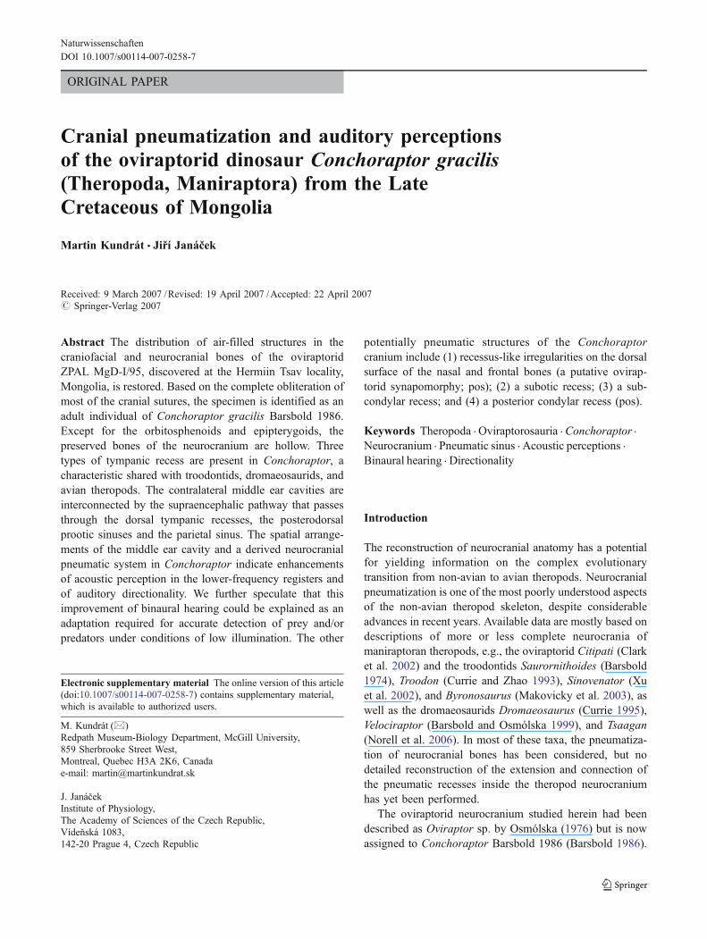
�Fig. 3 3D reconstruction of air-filled structures in the cranium of C.gracilis, ZPAL MgD-I/95. a,b Dorsal view; c,d Right lateral view; e,fLeft lateral view. alfrs Anterolateral frontal sinus; ampas antero-medial parietal sinus; ATR anterior tympanic recess; avpos antero-ventral prootic sinus; bos basioccipital sinus; dbos dorsal basioccipitalsinus; DTR dorsal tympanic recess; eos exoccipital sinus; EtEustachian tube passage; flc flocculus cerebelli; frs frontal sinus; ieinner ear region; las lacrimal sinus; lops lateral opisthotic sinus; lsslaterosphenoid sinus; mec medial ear cavity; mops medial opisthoticsinus; nas nasal sinus; ocs occipital condyle sinus; ops opisthoticsinus; pas parietal sinus; pbss parabasisphenoid sinus; pdpos postero-dorsal prootic sinus; plfrs posterolateral frontal sinus; pmfrs postero-medial frontal sinus; pos prootic sinus; ptgs pterygoid sinus; PTRposterior tympanic recess; qrs quadrate sinus; SEP supraencephalicpathway; sqs squamosal sinus; vbos ventral basioccipital sinus
Naturwissenschaften

Fig. 4 3D reconstruction of air-filled structures in the posterior neurocranium of C. gracilis, ZPAL MgD-I/95. a Posterior view; b Posterodorsalview; c–e Posterior part of the cranium in anterior and anterolateral perspectives. Abbreviations as in Fig. 3
Naturwissenschaften

municates with the dorsal and posterior tympanic recesses(PTR; Figs. 2b; 3c,d; 4).
The largely hollow auricular fossa is found posterodorsalto the acoustic fossa in Conchoraptor (Fig. 3f). On the outersurface of the neurocranium, the suture between the prooticand the opisthotic runs from the dorsal point of theinframetotic fissure and enters the posteriormost part ofthe inner ear recess. Further, it appears more anteriorly at amiddorsal point of the inner ear region and continuesdorsally along the canal-like DTR (Fig. 2c). The lateralmargin of the inner ear region (Fig. 2c) suggests that thepreserved opening was originally subdivided into theanterior oval opening, the fenestra ovalis, and a posteriorellipsoid opening, the foramen perilymphaticum, with theanterior opening three to four times larger than the posteriorone. These openings were separated by a thin interfenestralbony bar, which is not preserved in the ZPAL MgD-I/95specimen but has left indications on the dorsal and ventralmarginal relief of the inner ear recess. Below the ventralmargin of the inner ear region, a small fovea is foundwithin the groove running ventrally (Fig. 2b,c). This grooverepresents the dorso-medial bony wall of the Eustachiantube (Figs. 3c,f; 4d).
Opisthotic The opisthotic on the right side has been used todescribe the characteristics of this bone (Fig. 2c,d). Theopisthotic pneumatic sinus can be divided into medial andlateral pneumatic sinuses. The medial opisthotic sinus facestowards the supraoccipital region posterodorsally and isadjacent to medioventral portions of the posterodorsalprootic sinus (Fig. 4). The lateral opisthotic sinus occupiesthat part of the opisthotic communicating with the part ofthe middle ear cavity abutting the otic capitulum of thequadrate (Fig. 4a). The pneumatic sinus of the quadrate ofConchoraptor consists of dorsomedially confluent air-filledchambers separated by more or less thin horizontal bonylamellae (Figs. 3c,d; 4). The quadrate pneumatic sinusbecomes multichambered and is divided into lateral andmedial parts by the solid bony mass in the area of contactwith the articular bone. Both the pterygoid (Figs. 3c–f; 4)and the squamosal (Figs. 3a–d; 4) pneumatic sinuses arequite closely adjacent to the quadrate pneumatic sinuses,the last two probably communicating with each other.
The lateral opisthotic sinus also projects into theparoccipital process and faces onto the exoccipital(Fig. 2b–d). Based on the large cavity in the base of theparoccipital process (Fig. 2b), we speculate that theparoccipital process was highly pneumatized. In additionto the foregoing, the lateral opisthotic sinus opens into thePTR (Fig. 2d) behind a contact between the posterodorsalpneumatic prootic sinus and the DTR (Fig. 4a). However, itseems that the opisthotic and prootic pneumatic sinusescommunicated with one another directly, above the DTR,
through an opening between the lateral opisthotic andposterodorsal prootic sinuses. The lateral opisthotic sinusenters the exoccipital sinus ventrally (Fig. 2b). On the rightlateral side, the opisthotic shows a shallow concave area,the PTR (Fig. 2b,c).
Supraoccipital The supraoccipital is preserved only as athin bony sheet inclined between the prootic and opisthoticon the endoneurocranial side. Based on the topography ofthin bone fragments of the supraoccipital, it is very likelythat this bone was pneumatized and may have had apneumatic contact with the medial opisthotic sinus. Thesupraoccipital protected the posterior half of the dorsalsurface of the cerebellum.
Exoccipital The exoccipitals converge dorso-medially andgive a pyriform appearance to the foramen magnum(Fig. 2d). The exoccipitals are largely pneumatized exceptfor the part transmitting the roots of the hypoglossal nerve.The exoccipital sinus communicates with the lateralopisthotic sinus dorsally (Figs. 3e; 4a,b). It also communi-cates ventrally with a dorsal subdivision of the basioccipitalpneumatic sinus, and it opens externally into the suboticpneumatic recess (sensu Witmer 1997; Fig. 2b,c).
Basioccipital The basioccipital, which is incompletelypreserved, has a pneumatic vacuity that consists of a dorsalsubdivision (dorsal basioccipital sinus; Figs. 2b; 4a,d)connecting the exoccipital sinus (Fig. 2c), and a ventralsubdivision (ventral basioccipital sinus; Fig. 4a,f), occupy-ing most of the basioccipital bone, including the basicranialtubera. The subcondylar recess is seen on the posterior wallof the basioccipital (Fig. 2c). The basioccipital projectsbehind the dorsal and ventral margin of the foramenmagnum (Fig. 2c,d). The dorsal wall of the occipitalcondyle bends at right angles to the endoneurocranial floor.A shallow recess, the posterior condylar recess (Fig. 2c),occurs on the posterior surface of the occipital condyle. Thebase of the occipital condyle is pneumatized (Figs. 3c,d; 4a,b). The condyle itself is separated from the basioccipitalfloor by a laterally constricted condylar base (Fig. 2d).
Parabasisphenoid The parabasisphenoid is mostly absentin our specimen, but preserved fragments (Fig. 2d) suggestthat it was quite deep and pneumatized to a high degree.The parabasisphenoid pneumatic sinus makes contact withthe middle ear cavity at the same place as the antero-ventralprootic sinus does (Figs. 2b; 4c–e). The flat longitudinalbony element embedded in the sediment between themedial quadrate wall and lateral braincase wall (Fig. 2d)is partly hidden because it descends into the sediment,where it makes contact with the dorso-medial margin of thepterygoid. This element thus represents the sidewall of the
Naturwissenschaften

parabasisphenoid and gives an impression of a latero-ventral extension of this basicranial bone.
Discussion
Tympanic invasion of the neurocranial bones in theropods
The pattern of three tympanic recesses derived from themiddle ear (tympanic) cavity, a constant feature of birds(Witmer 1990; reviewed by Starck 1995) includingArchaeopteryx (Walker 1985) and known also in dromaeo-saurids (Norell et al. 2004) and troodontids (Currie andZhao 1993; Xu et al. 2002; Makovicky et al. 2003), ispresent in the oviraptorid Conchoraptor as well (Figs. 3a,b;4a,d). The anterior tympanic recess (ATR) and DTR seemto be plesiomorphic characteristics of tetanuran theropods(Rauhut 2004). It has been suggested that the PTR is aprimitive characteristic of coelurosaurs (Witmer 1997;Makovicky and Norell 1998). In Conchoraptor, the PTRinvades the base of the paroccipital process through thelateral opisthotic sinus. A very similar pattern is seen inArchaeopteryx (Walker 1985: Fig. 1). The DTR inConchoraptor occupies at first the posteroventral surfaceof the prootic and then enters the pneumatized interior ofthe bone as the posterodorsal prootic sinus. This sinus isinterconnected with its counterpart through the supra-encephalic pathway (SEP), a midline dorsal communicationbetween the contralateral middle ear cavities (Fig. 4c).There is an indication that the DTR might invade apneumatic labyrinth inside the quadrate of Conchoraptorthrough an opening on the medial side of the oticcapitulum. The ATR separately invades the anteroventralprootic and parabasisphenoid sinuses (Figs. 3c; 4d).Because the basicranium is incompletely preserved, it isunclear whether the interaural passage (IAP) was present inConchoraptor, although one other oviraptorid, Citipati,seems to possess this passage (Clark et al. 2002: Fig. 8).The other potential pneumatic structures of Conchoraptorinclude (1) recessus-like irregularities on the dorsal surfaceof the nasal and the frontal bones (a putative oviraptoridsynapomorphy); (2) the presence of a subotic recess [as introodontids (Barsbold 1974; Currie and Zhao 1993;Makovicky et al. 2003) and some ornithomimosaurs(Makovicky and Norell 1998)]; (3) the presence of a sub-condylar recess [unlike those in ornithomimosaurs(Makovicky and Norell 1998)], and (4) a posterior condylarrecess (another putative oviraptorid synapomorphy).
Acoustic perceptions of directionality in Conchoraptor
Auditory perception plays an important role in manydifferent kinds of avian behavior including individual and
species recognition, mate selection, territorial defense, songlearning, warning, and prey and predator detection(Dooling and Ryals 1997). The extent of auditory percep-tion is determined by the capacities of the external andmiddle ear for collecting sound energy and transforming itto provide optimal stimulation of the receptor organ (Kühneand Lewis 1985).
Acoustic abilities in the extinct ancestors of birds can bepartly estimated based on the specific characteristics oftheir middle ear regions. The well-preserved neurocraniumof Conchoraptor (Fig. 1) is a valuable source of informa-tion about the extension of the middle ear cavity-derivedtympanic sinuses into neurocranial bones and is, therefore,a basis for our inferences concerning sound propagationthrough this communicating pneumatic system (Fig. 3). AsKühne and Lewis (1985) pointed out, both the transformerfunction and the directionality of the ear are affected by thefinite physical dimensions of the head, on each side aloneor in combination. It is probable that, as in birds, thetympanic membrane in volant and arboreal theropodsgradually relocated from the surface of the head to a moreprotected position inside the acoustic meatus, where itacquired an increased surface area and may have becomethinner. This arrangement increases the sensitivity of thetympanic membrane, particularly to higher frequencies(Kühne and Lewis 1985). Reducing the bone mass of thetransmitting apparatus by trabeculation or hollowing alsoimproves the high-frequency sensitivity of the middle ear inbirds. At the other end of the acoustic spectrum, enlarge-ment of the avian middle ear cavity, derived tympanicrecesses, and pneumatic contralateral communication be-tween these structures decrease the impedance of themiddle ear at low frequencies and improve transmissionof low-frequency sounds to the cochlea (Saunders andJohnstone 1972).
The spacious middle ear cavity and derived tympanicrecesses, as well as extensive unilateral or contralateralcommunications between them, are found in birds withexcellent acoustic perception abilities, e.g., birds of prey(including owls) (Stellbogen 1930; Freye-Zumpfe 1953;Payne 1971; Norberg 1978). Among non-avian theropods,the most spacious middle ear cavity has been described introodontids (Barsbold 1974; Currie and Zhao 1993;Makovicky et al. 2003), and a similar extension is reportedhere for Conchoraptor (Figs. 3c,f; 4a,d). Apart from themiddle ear cavity, the most extensively pneumatizedneurocranial bones among theropods are found in ovi-raptorids, therizinosauroids, troodontids, and ornithomimo-saurs and may indicate enhanced acoustic abilities at thelower-frequency registers. The extension of neurocranialpneumaticity of Conchoraptor is comparable to that inpalaeognathous birds such as Struthio, Casuarius, andRhea. Starck (1995) proposed that the large air volumes
Naturwissenschaften

in the skulls of these birds result in sensitivity to very low-resonance frequencies, which might correlate with the lowfrequencies of the calls of most palaeognathous birds. Inview of the functional dependence of the hearing ability ofbirds on the volume capacity of the middle ear space andderived recesses and sinuses invading the neurocranialbones, it seems reasonable to hypothesize that Concho-raptor had enhanced acoustic abilities at the lower-frequency registers, presumably employed in intraspeciesacoustic communication.
In many birds, the middle ears are connected through anair-filled IAP consisting of an interaural canal andtrabeculated bone. Sound transmission through the IAPmay improve directional hearing. Experiments with modernbirds have demonstrated that the pneumatic sinuses providea passage (IAP) for the transmission of sounds from onemiddle ear to the other (Wada 1924; Stellbogen 1930;Schwartzkopff 1952; Payne 1971; Henson 1974; Lewis1983). Larsen et al. (2006) confirmed that at lowfrequencies, interaural sound propagation may cause oneeardrum to feel the vibrations of a sound considerablyearlier or later than the other, thus providing a possible cuefor directional hearing. Birds regulate their intracranial airpressure every few minutes to equilibrate with ambientpressure by opening their Eustachian tubes. If the intracra-nial space is not ventilated, the tympanal transfer functiongradually becomes high pass, with increasing air pressuredifference reducing both tympanal vibration amplitudes andsound transmission through the IAP (Larsen et al. 1996).
Larsen et al. (2006) revealed an interesting fact that incontemporary birds the feathers enlarge the acousticallyeffective size of the head compared with the dimensions ofthe skull. Similarly, the presence of the feathers (e.g., in abasal oviraptorosaur Caudipteryx; Ji et al. 1998) and theoccurrence of the IAP in non-avian theropods and earlybirds with a relatively small distance between ipsilateraleardrums might have a considerable acoustic effect andimprove directional hearing.
Birds and some troodontids (Currie and Zhao 1993) andoviraptorids (Clark et al. 2002) have been shown to possessan air space connecting their middle ear cavities; however,the considerable incompleteness of a parabasisphenoidprecludes any conclusion about the presence of an IAP inthe studied specimen of Conchoraptor.
The IAP represents a ventral contralateral communica-tion developed between the tympanic spaces and thepharyngotympanic tubes (Hill et al. 1980; Saiff 1988;Starck 1995). The middle ear cavities may be alsointerconnected through dorsal contralateral passages. ThePTRs may fuse in the dorsal midline of the supraoccipital.Furthermore, in Struthio and Casuarius, the spaciousparietal SEP connects the contralateral DTRs, invadingthe squamosal, the PTRs, and the supraoccipital (Starck
1995). The SEP was suggested to be present in Troodon(Currie and Zhao 1993), but until now, the SEP had beendocumented only in oviraptorids, such as Citipati (Clark etal. 2002). Here, we confirm the presence of the SEP inConchoraptor (Fig. 4d).
In Citipati, the middle ear spaces are broadly confluentwith the DTR and are thus directly interconnected insidethe parietal pneumatic cavity (Clark et al. 2002). Thismorphology implies 1) that the air pressure might havebeen the same on the interior of the two ear drums, and 2)that sound might propagate from one ear to the other. Thetiming of sound transmission through the SEP was likelymore delayed in Conchoraptor than in Citipati because theSEP in Conchoraptor consists of the trabeculated interior ofthe posterodorsal prootic sinus and the parietal pneumaticsystem, whereas there are no bony trabeculae in the parietalpneumatic system of Citipati (Clark et al. 2002). Dependingon the amount of sound transmission through the SEP, theears may therefore have been inherently directional. If anIAP was present, the SEP might have further amplified theperceptive capability of binaural hearing in Conchoraptor,allowing the animal to pinpoint the source of a soundextremely accurately.
The relatively large size of the orbital space and the eye(Fig. 1) in comparison to the cranium in Conchoraptor, aswell as its relatively large optic lobes (Kundrát 2007), mightsuggest an adaptation to a crepuscular or nocturnal life styleby this oviraptorid. Animals that are nocturnally active arefaced with the task of detecting and interpreting acousticstimuli correctly. To react in an appropriate manner toacoustic signals, Conchoraptor had to be able to recognizethe changing locations of sound sources, determining theirdirections and distances, in the dark. In such a scenario, theenhancement of Conchoraptor’s auditory directionality to ahigh degree could be explained as an adaptation offunctional requirements for the accurate detection of preyand/or predators in conditions of low illumination.
Acknowledgements The authors thank Ole N. Larsen, Gareth Dyke,and Kevin Padian for reading and commenting on an earlier version ofthis manuscript, Gerald Mayr for his valuable editing and editorialcomments, and Randy and Deb Lyons for their rigorous edit of thefinal version of the manuscript. Our special thanks to HalszkaOsmólska and Teresa Maryańska for providing access to the fossilspecimen and Zdeněk Seidl and Vladimír Smékal for providing accessto the CT facility.
References
Barsbold R (1974) Saurornithoididae, a new family of small theropoddinosaurs from Central Asia and North America. Palaeontol Pol30:5–22
Barsbold R (1986) Raubdinosaurier Oviraptoren. In: Vorobyeva EI (ed)Gerpetologičeskie issledovaniâ v Mongol’skoi Narodnoj Respub-
Naturwissenschaften

like, Institut Evolyucionnoy Morfologii i Ekologii Zhivotnikh im.A. N. Severcova, Akademia Nauk SSSR, Moscow, pp 210–223
Barsbold R, Osmólska H (1999) The skull of Velociraptor (Theropoda)from the Late Cretaceous of Mongolia. Acta Palaeontol Pol44:189–219
Clark JM, Norell MA, Rowe T (2002) Cranial anatomy of Citipatiosmolskae (Theropoda, Oviraptorosauria), and a reinterpretation ofthe holotype of Oviraptor philoceratops. Am Mus Novit 3364:1–24
Currie PJ (1995) New information on the anatomy and relationships ofDromaeosaurus albertensis (Dinosauria: Theropoda). J VertebrPaleontol 15:576–591
Currie PJ, Zhao X-J (1993) A new troodontid (Dinosauria, Theropoda)braincase from the Dinosaur Park Formation (Campanian) ofAlberta. Can J Earth Sci 30:2231–2247
Dooling RJ, Ryals BM (1997) Auditory perception and plasticity inthe avian auditory system. J Acoust Soc Am 101:3191
Freye-Zumpfe H (1953) Befunde im Mittelohr der Vögel. Wiss ZMartin-Luther-Univ Halle-Witten 2:445–461
Henson OW (1974) Comparative anatomy of the middle ear. In:Keidel WD, Neff WD (eds) Handbook of sensory physiology, volV. Springer, Berlin Heidelberg New York, pp 39–110
Hill KG, Lewis DB, Hutchings ME, Coles RB (1980) Directionalhearing in the Japanese Quail (Coturnix japonica). I. Acousticproperties of the auditory system. J Exp Biol 86:135–151
Ji Q, Currie PJ, Norell MA, Ji S-A (1998). Two feathered dinosaursfrom northeastern China. Nature 393:753–761
Kühne R, Lewis B (1985) External and middle ears. In: King AS,McLelland J (eds) Form and function in birds, vol 3. Academic,New York, pp 227–271
Kundrát M (2007) Avian-like attributes of a virtual brain model of theoviraptorid theropod Conhoraptor gracilis. Naturwissenschaften94:499–504
Larsen ON, Dooling RJ, Michelsen A (2006) The role of pressuredifference reception in the directional hearing of budgerigars(Melopsittacus undulatus). J Comp Physiol A 192(10):1063–1072
Larsen ON, Dooling RJ, Ryals BM (1996) Roles of intracranial airpressure in bird audition. In: Lewis ER, Miller GR (eds) Diversityin auditory mechanics. World Scientific, Singapore, pp 11–17
Lewis DB (1983) Directional cues for auditory localisation. In: LewisDB (ed) Bioacoustics. Academic, London, pp 233–257
Makovicky PJ, Norell MA (1998) A partial ornithomimid braincasefrom Ukhaa Tolgod (Upper Cretaceous, Mongolia). Am MusNovit 3247:1–16
Makovicky PJ, Norell MA, Clark JM, Rowe T (2003) Osteology andrelationships of Byronosaurus jaffei (Theropoda: Troodontidae).Am Mus Novit 3402:1–32
Norberg RÅ (1978) Skull asymmetry, ear structure and function, andauditory localization in Tengmalm’s owl, Aegolius funereus(Linné). Philos Trans R Soc Lond B 282:325–410
Norell MA, Clark JM, Turner AH, Makovicky PJ, Barsbold R, RoweT (2006) A new dromaeosaurid theropod from Ukhaa Tolgod(Ömnögov, Mongolia). Am Mus Novit 3545:1–51
Norell MA, Makovicky PJ, Clark JM (2004) The braincase ofVelociraptor. In: Currie PJ, Koppelhaus EB, Shugar MA, WrightJL (eds) Feathered dragons: studies on the transition fromdinosaurs to birds (life of the past). Indiana University Press,Bloomington, pp 133–143
Osmólska H (1976) New light on the skull anatomy and systematicposition of Oviraptor philoceratops. Nature 262:683–684
Payne RS (1971) Acoustic location of prey by barn owls (Tyto alba). JExp Biol 54:535–573
Rauhut OW (2004) Braincase structure of the Middle Jurassic theropoddinosaur Piatnitzkysaurus. Can J Earth Sci 41:1109–1122
Saiff EI (1988) The anatomy of the middle ear of the tinamiformes.J Morphol 196:107–116
Saunders JC, Johnstone BM (1972) A comparative analysis of middleear function in non-mammalian vertebrates. Acta Oto Laryngol73:353–361
Schwartzkopff J (1952) Untersuchungen über die Arbeitsweise desMittelohres und das Richtunshören der Singvögel unter Verwen-dung von Cochlea-Potentialen. Z Vgl Physiol 32:319–327
Smith DK (1993) The type specimen of Oviraptor philoceratops, atheropod dinosaur from the Upper Cretaceous of Mongolia.Neues Jharb Geol Paläontol Abh 186:365–388
Starck JM (1995) Comparative anatomy of the external and themiddle ear of palaeognathous birds. In: Beck F, Galveston WH,Kriz W, Pauly JE, Sano Y, Schlieber TH (eds) Advances inanatomy embryology and cell biology, vol 131. Springer, BerlinHeidelberg New York, pp 1–137
Stellbogen E (1930) Über das äussere und mittlere Ohr des Waldkauzes(Syrnium aluco, L.). Z Morphol Ökol Tiere 19:686–731
Wada Y (1924) Beiträge zur vergleichenden Physiologie des Gehör-organs. Pflügers Arch Gesamte Physiol Menschen Tiere 202:46–49
Walker AD (1985) The braincase of Archaeopteryx. In: Hecht MK,Ostrom JH, Viohl G, Wellnhofer P (eds) The beginnings of birds.Freunde des Jura-Museums, Eichstätt, pp 123–134
Witmer LM (1990) The craniofacial air sac system of Mesozoic birds(Aves). Zool J Linn Soc 100:327–378
Witmer LM (1997) Craniofacial air sinus system. In: Currie PJ, Padian K(eds) Encyclopedia of dinosaurs. Academic, San Diego, pp 151–159
Xu X, Norell MA, Wang X-L, Makovicky P, Wu X-C (2002) A basaltroodontid from the Early Cretaceous of China. Nature 415:780–784
Naturwissenschaften
Copyright © 2022 FDOKUMEN