Ecological correlates to cranial morphology in leporids (Mammalia, Lagomorpha)
20
Submitted 2 December 2014 Accepted 27 February 2015 Published 17 March 2015 Corresponding author Brian P. Kraatz, [email protected] Academic editor Jes ´ us Marug´ an-Lob ´ on Additional Information and Declarations can be found on page 15 DOI 10.7717/peerj.844 Copyright 2015 Kraatz et al. Distributed under Creative Commons CC-BY 4.0 OPEN ACCESS Ecological correlates to cranial morphology in Leporids (Mammalia, Lagomorpha) Brian P. Kraatz 1 , Emma Sherratt 2 , Nicholas Bumacod 3 and Mathew J. Wedel 1,4 1 College of Osteopathic Medicine of the Pacific, Western University of Health Sciences, Pomona, CA, USA 2 Department of Ecology, Evolution, and Organismal Biology, Iowa State University, Ames, IA, USA 3 College of Dental Medicine, Western University of Health Sciences, Pomona, CA, USA 4 College of Podiatric Medicine, Western University of Health Sciences, Pomona, CA, USA ABSTRACT The mammalian order Lagomorpha has been the subject of many morphometric studies aimed at understanding the relationship between form and function as it relates to locomotion, primarily in postcranial morphology. The leporid cranial skeleton, however, may also reveal information about their ecology, particularly locomotion and vision. Here we investigate the relationship between cranial shape and the degree of facial tilt with locomotion (cursoriality, saltation, and burrowing) within crown leporids. Our results suggest that facial tilt is more pronounced in cursors and saltators compared to generalists, and that increasing facial tilt may be driven by a need for expanded visual fields. Our phylogenetically informed analyses indicate that burrowing behavior, facial tilt, and locomotor behavior do not predict cranial shape. However, we find that variables such as bullae size, size of the splenius capitus fossa, and overall rostral dimensions are important components for understanding the cranial variation in leporids. Subjects Ecology, Zoology, Anatomy and Physiology Keywords Leporidae, Cranial morphology, Locomotion, Lagomorpha INTRODUCTION The relationship between form and function as it relates to locomotion has been extensively studied in a wide range of vertebrate groups (Webb, 1984; Hildebrand, 1988; Rayner, 1988; Aerts et al., 2000). The mammalian order Lagomorpha has been the subject of many morphometric studies aimed at understanding this relationship in postcranial morphology (e.g., Reese, Lanier & Sargis, 2013; Fostowicz-Frelik, 2007; Seckel & Janis, 2008; Young et al., 2014), and the impetus of these is largely to understand the high-speed form of leaping observed in some leporids (rabbits and hares). Leporids are peerless cursors for their size; some hares have been shown to achieve speeds greater than 70 km/h (Garland, 1983). Indeed, the leporid postcranial skeleton exhibits many derived features that are strongly associated with saltation and cursoriality, including limb element elongation (Szalay, 1985; Fostowicz-Frelik, 2007; Seckel & Janis, 2008). How to cite this article Kraatz et al. (2015), Ecological correlates to cranial morphology in Leporids (Mammalia, Lagomorpha). PeerJ 3:e844; DOI 10.7717/peerj.844
-
Upload
westernu-us -
Category
Documents
-
view
1 -
download
0
Transcript of Ecological correlates to cranial morphology in leporids (Mammalia, Lagomorpha)

Submitted 2 December 2014Accepted 27 February 2015Published 17 March 2015
Corresponding authorBrian P. Kraatz,[email protected]
Academic editorJesus Marugan-Lobon
Additional Information andDeclarations can be found onpage 15
DOI 10.7717/peerj.844
Copyright2015 Kraatz et al.
Distributed underCreative Commons CC-BY 4.0
OPEN ACCESS
Ecological correlates to cranialmorphology in Leporids (Mammalia,Lagomorpha)Brian P. Kraatz1, Emma Sherratt2, Nicholas Bumacod3 andMathew J. Wedel1,4
1 College of Osteopathic Medicine of the Pacific, Western University of Health Sciences, Pomona,CA, USA
2 Department of Ecology, Evolution, and Organismal Biology, Iowa State University, Ames, IA,USA
3 College of Dental Medicine, Western University of Health Sciences, Pomona, CA, USA4 College of Podiatric Medicine, Western University of Health Sciences, Pomona, CA, USA
ABSTRACTThe mammalian order Lagomorpha has been the subject of many morphometricstudies aimed at understanding the relationship between form and function as itrelates to locomotion, primarily in postcranial morphology. The leporid cranialskeleton, however, may also reveal information about their ecology, particularlylocomotion and vision. Here we investigate the relationship between cranial shapeand the degree of facial tilt with locomotion (cursoriality, saltation, and burrowing)within crown leporids. Our results suggest that facial tilt is more pronounced incursors and saltators compared to generalists, and that increasing facial tilt maybe driven by a need for expanded visual fields. Our phylogenetically informedanalyses indicate that burrowing behavior, facial tilt, and locomotor behavior do notpredict cranial shape. However, we find that variables such as bullae size, size of thesplenius capitus fossa, and overall rostral dimensions are important components forunderstanding the cranial variation in leporids.
Subjects Ecology, Zoology, Anatomy and PhysiologyKeywords Leporidae, Cranial morphology, Locomotion, Lagomorpha
INTRODUCTIONThe relationship between form and function as it relates to locomotion has been
extensively studied in a wide range of vertebrate groups (Webb, 1984; Hildebrand, 1988;
Rayner, 1988; Aerts et al., 2000). The mammalian order Lagomorpha has been the subject
of many morphometric studies aimed at understanding this relationship in postcranial
morphology (e.g., Reese, Lanier & Sargis, 2013; Fostowicz-Frelik, 2007; Seckel & Janis, 2008;
Young et al., 2014), and the impetus of these is largely to understand the high-speed form
of leaping observed in some leporids (rabbits and hares). Leporids are peerless cursors for
their size; some hares have been shown to achieve speeds greater than 70 km/h (Garland,
1983). Indeed, the leporid postcranial skeleton exhibits many derived features that are
strongly associated with saltation and cursoriality, including limb element elongation
(Szalay, 1985; Fostowicz-Frelik, 2007; Seckel & Janis, 2008).
How to cite this article Kraatz et al. (2015), Ecological correlates to cranial morphology in Leporids (Mammalia, Lagomorpha). PeerJ3:e844; DOI 10.7717/peerj.844

Figure 1 Disparity of leporid skulls. Disparity in facial tilt and cranial shape in selected leporids,including (A) Brachylagus idahoensis (LACM 447; SLD ∼50 mm), (B) Lepus capensis (LACM 40152; SLD∼82 mm), (C) Poelagus marjorita (AMNH 51056; SLD ∼80 mm), (D) Pronolagus crassicaudatus (AMNH89033; SLD ∼80 mm), (E) Lepus americanus (LACM 93737; SLD ∼75 mm), (F) Oryctolagus cuniculus(AMNH 166951; SLD ∼78 mm), (G) Nesolagus timminsi (AMNH 272419; SLD ∼78 mm), (H) Bunolagusmonticularis (AMNH 146662; SLD ∼78 mm), and (I) Romerolagus diazi (AMNH 148181; SLD ∼60 mm).All skull images are scaled to approximately the same skull length, skull length measurements are anapproximation based on our measured specimens.
The cranial skeleton is more often overlooked in studies of form and locomotion,
though there are biologically relevant associations between skull form and locomotor
behavior, such as the role of the skull in active headfirst burrowing (e.g., Gans, 1974; Barros,
Herrel & Kohlsdorf, 2011; Sherratt et al., 2014; Hopkins & Davis, 2009; and see Wake, 1993
for a review). In leporids, it has been suggested that morphological transformations of
the skull may also be related to their ecology, particularly locomotion and vision (DuBrul,
1950; Bramble, 1989). The leporid skull is highly transformed, exhibiting a combination
of features that clearly distinguish it from a more typical mammalian skull. A striking, yet
often overlooked, characteristic is the broad dorsal arching of the cranium (Thompson,
1942), which is achieved via expansion and folding of the supraoccipital, and a distinct
flexure near the basisphenoid/presphenoid suture (Fig. 1). A prominent ridge on the dorsal
portion of the posterior cranial roof, which is superficially similar to an occipital crest,
Kraatz et al. (2015), PeerJ, DOI 10.7717/peerj.844 2/20

Table 1 Leporid species studied. See Appendix S1 for specific specimens measured, and the text fordiscussion regarding the assessment of ecological variables.
Species Locomotion type Burrowing Abbreviation n
Romerolagus Saltatorial Yes Ro 7
Bunolagus Saltatorial Yes Bu 2
Caprolagus Generalized Yes Ca 2
Brachylagus Generalized Yes Br 10
Sylvilagus floridanus Saltatorial No Sfl 10
Sylvilagus palustris Generalized No Spal 10
Sylvilagus audobonii Saltatorial Yes Sau 10
Poelagus marjorita Saltatorial No Po 10
Pronolagus crossicaudatus Saltatorial No Pc 10
Oryctolagus cuninculus Saltatorial Yes Oc 10
Nesolagus timminsi Saltatorial Yes Nt 2
Lepus americanus Saltatorial No Lam 10
Lepus timidus Saltatorial Yes Lti 10
Lepus capensis Cursorial Yes Lcap 10
Lepus californicus Cursorial No Lcal 12
Lepus saxatilis Cursorial No Lsax 9
is actually a distinct flexure within the supraoccipital bone. Based on the position of the
rabbit skull in resting position (De Beer, 1947: Fig. 9; Vidal, Graf & Berthoz, 1986: Fig. 4B,
and see our Fig. 2), this flexure results in significant tilting of the facial region ventrally
relative to the basicranium, which we here refer to as Facial Tilt (FT). DuBrul (1950)
discusses this feature in detail within hares, and points out that the facial tilt of leporids is
likely related to their unique mode of locomotor behavior. DuBrul (1950) also discusses the
similarities in leporid skull transformations to those of our own lineage; in our hominin
relatives, increased basicranial flexion is associated with the onset of bipedal locomotion
(Strait & Ross, 1999).
The goal of this study is to investigate the relationship between cranial shape and
locomotion (cursoriality, saltation, and burrowing) within crown leporids. Our study
is driven by hypotheses previously stated (DuBrul, 1950; Bramble, 1989) but never
quantitatively tested. We use a large morphometric dataset spanning 16 phylogenetically
constrained extant taxa (Table 1) to evaluate hypotheses about the relationship between
skull shape and facial tilt with locomotor ecology.
STUDY SYSTEM AND HYPOTHESESThe mammalian order Lagomorpha is composed of two families, Leporidae (rabbits
and hares) and Ochotonidae (pikas). Ochotonids are represented by one living genus,
Ochotona, which includes two North American and 28 Eurasian species (Alves &
Hacklander, 2008). Leporids include 11 living genera with 62 species overall. The majority
of species are found within two genera (Alves & Hacklander, 2008); Lepus (hares, 32
species) and Sylvilagus (a portion of rabbits, 17 species). Of the remaining nine genera,
seven are monotypic, while two genera, Nesolagus and Pronolagus, only include two
Kraatz et al. (2015), PeerJ, DOI 10.7717/peerj.844 3/20

Figure 2 Facial tilt in leporids. The crania of (A) Caprolagus hispidus (AMNH 54852, above) and(B) Pronolagus crassicaudatus (AMNH 89033, below) are shown in left lateral view. Facial tilt (FT) isdefined herein as the angle between the upper diastema and the occipital plane, where increased valuesindicated a skull orientation closer the horizontal plane. The triangle indicates the position of the externaloccipital protuberance (EOP), and from that, both the dorsal (red) and occipital (blue) extent of thesupraoccipital bones is outlined.
Kraatz et al. (2015), PeerJ, DOI 10.7717/peerj.844 4/20

Figure 3 Phylogeny of Leporidae. The phylogenetic hypothesis of the 16 taxa used in this study, prunedfrom the supermatrix maximum likelihood phylogeny in Matthee et al. (2004). Locomotor styles fromTable 1.
and four species, respectively. Overall, sixteen leporids species are currently considered
endangered or critically endangered by the IUCN (Alves & Hacklander, 2008), and
conservation issues are compounded by the lack of natural history data for many of these
species. Leporids are found on every continent except Antarctica, from the high arctic
to dry, hot deserts (Chapman & Flux, 1990; Chapman & Flux , 2008). Some leporids are
nocturnal, some are social, and some live in dense cover as opposed to the open plains
often associated with these animals (Stoner, Bininda-Emonds & Caro, 2003). In terms
of size, our study includes (Appendix S1) the smallest leporid, Brachylagus (mean skull
length ∼50 mm) to one of the largest, Lepus timidus (mean skull length ∼90 mm). Genera
such as Pentalagus and Caprolagus have heavy, robust skulls, compared to the typically
gracile skulls of most taxa. These cranial differences manifest themselves morphometrically
via a wide range of snout lengths and marked differences in skull robustness and form
(Fig. 1). While leaping abilities are common among most leporid lineages, they are also
known to be facultatively semiaquatic, scansorial, fossorial, or exhibit a more generalized,
non-hopping form of locomotion (Chapman & Flux, 1990). We distinguish here between
the saltatory locomotion (i.e., hopping) most typical among leporids (Table 1 and Fig. 3),
and its cursorial form observed in some hare lineages (Gambaryan & Hardin, 1974;
Bramble, 1989). Generalists are recognized as those who don’t exhibit clear hopping, but
rather move in a more scampering habit.
Kraatz et al. (2015), PeerJ, DOI 10.7717/peerj.844 5/20

Hypothesis 1–facial tiltA high degree of facial tilting (e.g., ventral flexion of the facial region)should (a) be positively correlated with more active (e.g., saltatorial orcursorial) locomotor styles, and (b) show no correlation with burrowinghabitVariation in the degree of facial tilt among leporids has strong effects on orbital orientation
(Fig. 3). There is substantial literature discussing the relationship between orbit orientation
and ecology within vertebrates (Noble, Kowalski & Ravosa, 2000; Cox, 2008; Heesy, 2008;
Iwaniuk et al., 2008; Jeffery & Cox, 2010), and Cartmill (1970) established the terms ‘orbital
convergence’ and ‘frontation’ to understand these relationships. While orbit orientation
is influenced by brain size and jaw mastication (Lieberman, Ross & Ravosa, 2000; Cox,
2008), within primates, orbital convergence is also strongly associated with increased
binocular visual field overlap observed in nocturnal predatory species (Heesy, 2004; Heesy,
2008). Various groups exhibit a high degree of both orbital convergence and orbital
frontation (Cox & Jeffery , 2008), with hominids serving as an exemplar; orbital frontation
is strongly positively correlated with basicranial flexion (Ross, 1995). As DuBrul (1950)
points out, facial tilt transformations among leporids are nearly identical to basicranial
flexion observed within anthropoids; increased facial tilt and basicranial flexion both result
in increased orbital frontation (see Fig. 2 for changes in frontation related to increased FT).
Several workers have shown that increased frontation is positively correlated with arboreal
taxa (Cartmill, 1970; Heesy, 2008); increased frontation changes the visual field to allow
for better visualization of substrate. Jeffery & Cox (2010) show that leporids have relatively
low degrees of convergence and frontation. As we discuss below, however, when facial tilt is
taken into consideration, leporids actually demonstrate a relatively higher degree of fronta-
tion (as indicated by the orbital plane relative to the vertical plane). More importantly,
regardless of the absolute measure of frontation within leporids, we expect that frontation
will vary among leporids correlated with varying degrees of facial tilt. For this reason, we
expect that facial tilt (as a proxy for frontation) should be strongly correlated to locomotor
styles that would require enhanced substrate perception (saltatorial and cursorial), but we
do not expect that facial tilt will be related to burrowing habit.
Hypothesis 2—Skull shapeWe expect that there will be significant skull shape differences among(a) locomotor styles, and (b) burrowing habits
We have no a priori expectations about how overall skull shape might change with
locomotor mode or burrowing habit. Instead we will investigate the more fundamental
question of whether skull shape is related to locomotion and burrowing habit at all. Our
interest in this question is therefore more a form of exploratory data analysis than a test of a
specific hypothesis.
MATERIALS AND METHODSWe collected morphometric data (Table 2 and Appendix S1) from 140 leporid skulls
spanning 16 taxa (Table 1) housed in the departments of Mammalogy at the American
Kraatz et al. (2015), PeerJ, DOI 10.7717/peerj.844 6/20

Table 2 Skull measurements used. Variables used in this study and description; see Figs. 2 and 4 for illustrations of the measurement conventions.
Abbr. Variable Measurement convention
BLD Bulla diameter Maximum diameter (in any direction) of right bulla
BOL Basioccipital length Maximum midsagittal length from anterior basioccipital to foramen magnum
DIL Diastema length Maximum distance between right I2 and M1
IOW Interorbital width Minimum transverse width between dorsal rims of orbits
NL Nasal length Maximum parasagittal length of nasal bones (i.e., orthogonal antero-posterior but not along midline)
NW Nasal width Maximum transverse width across posterior nasal bones
PAL Parietal length Maximum midsagittal length of parietal bones
SCF Splenius capitis fossa Maximum parasagittal length from anterior margin of M. splenius capitis insertion fossa to opisthocranion
SLD Skull length dorsal Maximum midsagittal length from anterior nasal bones to Opisthocranion (just dorsal to incisors) toopisthocranion
SW Skull width Maximum transverse width across zygomatic processes
Table 3 PCA loadings. The first four principal component (PC) axes contribute to 90.2% of the totalvariation of the ten log normal variables. For each PC, the proportion of total variance (%) and theloadings on these are given. The variables with the highest loading are shown in bold and are discussedwithin the text.
PC1 PC2 PC3 PC4
Proportion of variance 43.5 24.4 13.2 9.1
BLD 0.85046873 0.02545804 0.016857617 0.272291215
BOL 0.159862063 0.161106564 0.012761305 −0.070830236
DIL −0.246528468 −0.026035317 −0.415136231 0.156169866
IOW −0.249260494 0.331660879 0.681589555 0.259415969
NL −0.175108112 0.062932441 −0.505939657 0.130829626
NW −0.296951523 0.01926985 0.131337875 0.224903873
PAL 0.027064391 0.140782388 0.078947604 −0.866213361SCF −0.050436754 −0.905402173 0.218089373 −0.039704014
SLD −0.064043373 0.114113325 −0.185926838 0.027064132
SW 0.044933539 0.076114004 −0.032580602 −0.093927069
Museum of Natural History (AMNH) and the Los Angeles County Museum of Natural
History (LACM). Care was made to use only adult specimens, characterized by fully fused
occipital sutures (Hoffmeister & Zimmerman, 1967). Ten linear measurements (Table 3 and
Fig. 3) were recorded per specimen using digital calipers by three authors (BPK, MW, and
NB), and a repeatability analysis (consisting of 10 specimens measured 3 times, results
not presented) was performed to ensure there was no intercollector bias introduced. The
ten cranial measurements were analyzed using the log-shape ratios approach (Mosimann,
1970; Mosimann & James, 1979). For each specimen, size was computed as the geometric
mean of all measurements, and then each measurement was divided by size to obtain the
shape ratios. We then used the log of this quantity as raw data for the subsequent analyses.
Facial tilt was measured by photographing each skull in lateral view using a
Nikon D80 digital camera (Nikon, Tokyo, Japan). The skulls were placed in a
Kraatz et al. (2015), PeerJ, DOI 10.7717/peerj.844 7/20
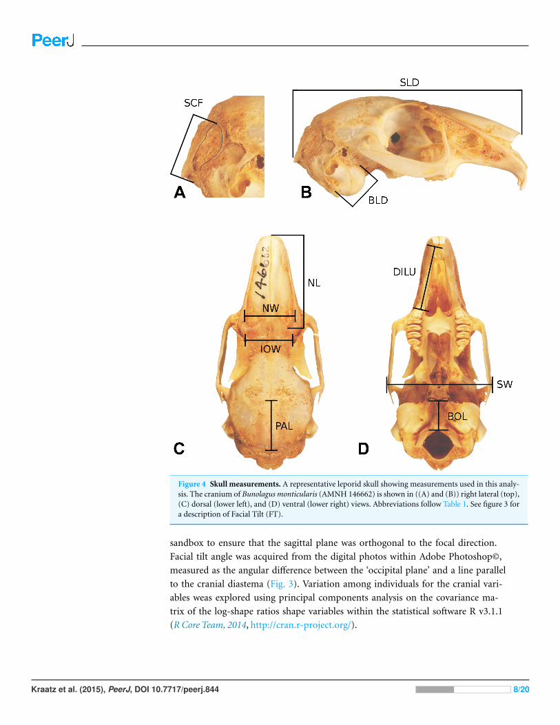
Figure 4 Skull measurements. A representative leporid skull showing measurements used in this analy-sis. The cranium of Bunolagus monticularis (AMNH 146662) is shown in ((A) and (B)) right lateral (top),(C) dorsal (lower left), and (D) ventral (lower right) views. Abbreviations follow Table 1. See figure 3 fora description of Facial Tilt (FT).
sandbox to ensure that the sagittal plane was orthogonal to the focal direction.
Facial tilt angle was acquired from the digital photos within Adobe Photoshop©,
measured as the angular difference between the ‘occipital plane’ and a line parallel
to the cranial diastema (Fig. 3). Variation among individuals for the cranial vari-
ables weas explored using principal components analysis on the covariance ma-
trix of the log-shape ratios shape variables within the statistical software R v3.1.1
(R Core Team, 2014, http://cran.r-project.org/).
Kraatz et al. (2015), PeerJ, DOI 10.7717/peerj.844 8/20

Phylogenetically informed analysesTo examine facial tilt angle and cranial shape in a phylogenetic context, we used the
phylogenetic relationships among species of Leporidae recently published by Matthee et
al. (2004). The original tree was constructed using seven genes (five nuclear and 2 mt)
for 25 ingroup taxa. We pruned the tree using Mesquite©(Maddison & Maddison, 2015)
to include only the 16 taxa studied here (Fig. 3), and retained the information on branch
lengths (details of which are in (Matthee et al., 2004)).
We first examined the amount of phylogenetic signal in the morphometric variables,
calculating the K statistic (Blomberg, Garland & Ives, 2003) for facial tilt angle, and the
multivariate equivalent Kmult (Adams, 2014a) for all log-shape ratios. The K statistics pro-
vide a measure of the strength of phylogenetic signal for univariate and multivariate traits
respectively, and in each case provides a single statistic. A value of less than one implies
that taxa resemble each other phenotypically less than expected under Brownian motion,
while values of more than 1 implies that close relatives are more similar to one another
phenotypically than expected under Brownian motion. Significance testing was performed
using a permutation procedure whereby the variables are randomized relative to the tree,
and 1000 permutations were performed for each test (Blomberg, Garland & Ives, 2003).
Log-shape ratios and facial tilt angle were compared to several key ecological indicators,
including locomotor type and burrowing habit (Table 1). Ecological data were obtained
from Chapman & Flux (1990) and Stoner, Bininda-Emonds & Caro (2003). We divided
leporids into three locomotor categories: generalized or ‘scramble’ locomotors, which tend
to be the slowest-moving; saltatory or hopping locomotors; and fast-moving taxa that
practice cursorial (leaping and bounding) locomotion, which is essentially a specialized
form of saltation. Regarding burrowing habits, some leporids dig their own burrows
(e.g., Oryctolagus and Romerolagus), whereas others simply occupy preexisting burrows
excavated by other animals. For the purposes of this study, we refer to leporids as burrowers
if they occupy burrows consistently, regardless of whether they dig the burrows.
To test whether or not the degree of facial tilt differs among the three locomotor
categories, we performed a one-way Analysis of Variance (ANOVA) in an evolutionary
context, under a Brownian motion model of evolution. This was done by using species
means of the FT angle in a distance-based phylogenetic generalized least squares analysis
(D-PGLS; Adams, 2014b). A distance-based approach provides numerically identical
estimates of evolutionary patterns to those obtained from standard implementations
of PGLS on univariate datasets, and was used here for consistency with analyses below
on the log-shape ratios. The statistical significance of each term in the D-PGLS was
assessed using 1000 permutations whereby the species means are shuffled among the
tips of the phylogeny. We performed a second ANOVA as above to test whether facial
tilt differs between taxa that utilize burrows (“burrowing”) and those that do not
(“non-burrowing”). Box and whisker plots were used to visualize the individual variation
in facial tilt angle among groups.
To test whether or not cranial shape, as represented by ten morphometric variables,
differs among the three locomotor types, we performed a multivariate analysis of variance
Kraatz et al. (2015), PeerJ, DOI 10.7717/peerj.844 9/20

in an evolutionary context under a Brownian motion model of evolution. This was done
as a D-PGLS with the species means of the ten log-shape ratios. The D-PGLS performs
better than a regular PGLS when the number of variables begins to approach the number
of species (Adams, 2014b). The statistical significance of each term in the D-PGLS was
assessed using 1,000 permutations of the species means. Similarly, we tested whether or
not cranial shape differs between burrowing and non-burrowing taxa using a D-PGLS as
above.
Finally, to test whether or not facial tilt is a significant predictor of cranial shape, we
performed a multivariate regression in an evolutionary context, under a Brownian motion
model of evolution, again using the D-PGLS approach. The statistical significance was
assessed using 1000 permutations of the species means of the log-shape ratios. All of the
phylogenetically informed analyses were done using the geomorph package (Adams et
al., 2014) in the statistical software R v3.1.1 (R Core Team, 2014). The ANOVAs on FT,
the MANOVAs on cranial log-shape rations, and the multivariate regression were done
using the procD.pgls function, and phylogenetic signal was calculated with the physignal
function.
RESULTSFacial tiltFacial tilt (FT) summarizes the broad dorsal arching of the skull roof that is prominent
among living leporids (Fig. 3). Across the species in this study, the measure of facial tilt
angle has a very low value for K, implying that the taxa resemble each other morphologi-
cally less than expected under Brownian motion, and the test is not significant (K = 0.62,
P = 0.53). Overall, there is a nearly 30◦ range of variation in FT among specimens of all
species in this sample (Appendix S1). We found a significant difference among locomotor
types for facial tilt angle (D-PGLS, F = 7.02, P = 0.016; Fig. 5A). The mean FT angle for
generalized locomotors (mean, µ = 44.0, standard deviation, σ = 5.48) is substantially
higher than that of cursorial (µ = 36.3, σ = 5.46) and saltatorial taxa (µ = 37.2, σ = 5.91)
(Fig. 5A). This indicates that taxa that are either saltatorial or cursorial tend to have facial
regions that are more ventrally deflected. By contrast, we found no significant difference
in FT angle between burrowing and non-burrowing taxa (Fig. 5B; D-PGLS, F = 0.0037,
P = 0.973; Fig. 5B).
Cranial shape analysesIn a principal components analysis of the ten log-shape ratios among individuals, the first
four PC axes account for 90.2% of total variance. PC1 accounts for 43.5% of cranial shape
differences, PC2 accounts for 24.4%, PC3 accounts for 13.2%, and PC4 accounts for 9.0%
of variance. (Table 3 and Fig. 6). The remaining PCs each contribute less than 10% of
the total variation. A PCA of the species means (not shown) produced equivalent results
(PC1 = 42.6%, PC2 = 33.4%, PC3 = 11.3%, PC4 = 5.9%), and with the same variables
contributing highly on each axis, and thus we present only one analysis for brevity.
Kraatz et al. (2015), PeerJ, DOI 10.7717/peerj.844 10/20

Figure 5 Facial tilt ANOVA. Box and whisker plot summarizing facial tilt angle for all specimens,showing how the angle differs between locomotor types (A) and burrowing behavior (B).
The loadings of the PCA (Table 3) show that bullae diameter (BD; 0.85) has the
strongest influence on PC1, substantially more than other variables. PC1 strongly separates
Brachylagus, Romerolagus, and Bunolagus (all with larger bullae diameters) from all other
leporid species (Fig. 6A). In terms of locomotor styles, cursorial species are isolated
towards the negative portion of the PC1 axis. Similarly, PC2 strongly shows the effects of
size of the splenius capitus fossa (SCF; −0.91). While this measure does not separate among
saltators (Fig. 6B), there is some separation between generalists and cursorial species.
Loadings for PC3 indicate that three variables are strongly affecting the variance along
that axis: interorbital width (IOW; 0.68), nasal length (NL; −0.51), and diastema length
(DILU; −0.42). PC3 shows separation of species (Fig. 6C), but no clear broader groupings.
It also does not clearly distinguish locomotor modes, but saltators do occupy the negative
portion of the axes where no generalists or cursors are found (Fig. 6D). Parietal length
(PAL; −0.87) loads strongly along PC4. This axes does help to distinguish species (Fig. 6C),
but shows little ability to distinguish among locomotor modes (Fig. 6D).
There is significant phylogenetic signal is cranial shape described by the ten log-shape
ratios (Kmult = 0.91, P = 0.035). The value of kappa is substantially higher than that for
facial tilt, but still below 1, implying that close relatives are moderately less similar to one
another phenotypically than expected under Brownian motion. Phylogenetically informed
analysis of variance (D-PGLS) indicates that there is no significant effect of locomotor
habit on cranial shape (F = 1.3712, P = 0.28). Likewise, there is no significant effect on
cranial shape by burrowing behavior (F = 1.2831, P = 0.56). Finally, a phylogenetically
informed multivariate regression suggests that facial tilt angle is not a significant predictor
of cranial shape (R2= 0.097, P = 0.413)
DISCUSSIONGiven a clear correlation between the degree of facial tilt (FT) and locomotor style, and
the lack of significant phylogenetic signal in FT angle, it is evident that this aspect of
Kraatz et al. (2015), PeerJ, DOI 10.7717/peerj.844 11/20
Figure 6 Multivariate PCAs. Principal components analysis of 10 log-shape ratios measurements de-scribing cranial shape for all specimens. Biplots show PC1 vs PC2 (top) and PC3 vs PC4 (bottom).(A) Colored symbols by species. (B) colored by LOC (with species symbols). Details of the loadings ofeach variable in the PCA are presented in Table 3.
cranial morphology is strongly influenced by ecological factors within Leporidae. While
the relationship between shape and function is established, the specific aspects of cranial
shape that inform ecological function are only partially resolved from our multivariate
analyses. Generalized locomotors exhibit less facial tilt, an anatomical condition that could
properly be thought to be primitive for the mammalian skull, and given their fossil record,
lagomorphs as well (Dice, 1933; Asher et al., 2005). Facial tilt within leporids is allowed via
the expansion of the supraoccipital bone on the dorsal skull (Fig. 2), and along the ventral
skull, there is a pronounced flexure near the basisphenoid/presphenoid juncture.
The complex architecture of the supraoccipital in leporids is the most marked change
related to the dorsal arching the skull roof, but there are additional effects on the
orientation of the orbit (Fig. 2). There is a vast literature on orbital orientation as it relates
to locomotion, visual acuity, brain size, and masticatory anatomy (Noble, Kowalski &
Ravosa, 2000; Heesy, 2005; Heesy, Ross & Demes, 2007; Iwaniuk et al., 2008; Heesy, 2008;
Cox, 2008; Jeffery & Cox, 2010), and perhaps most clearly, changes in orbit orientation
Kraatz et al. (2015), PeerJ, DOI 10.7717/peerj.844 12/20
have direct affects on the range of visual fields. Both orbital convergence and frontation
are commonly measured orbital variables that seem to be functionally predictive (Cox,
2008); increased convergence is thought to increase binocular field overlap within primates
(Ross & Martin, 2007; Heesy, Ross & Demes, 2007), and orbital frontation allows for better
substrate visualization (Cartmill, 1970; Heesy, 2008).
While Jeffery & Cox (2010) demonstrated that the rabbit shows low degrees convergence
and frontation, frontation in the rabbit is complicated by skull transformations associated
with facial tilt. Traditionally, frontation was considered as the degree to which the orbital
plane is aligned vertically (Cartmill, 1970; Ross, 1995); whereas, Jeffery & Cox (2010) used
angular differences between the lateral semicircular canal and the medial and lateral orbital
rectus muscles as a proxy for frontation. While these later measures are distinct for rabbits
(Cox, 2008; Jeffery & Cox, 2010) as compared to other mammals, due to the way rabbits
hold their heads (De Beer, 1947; Vidal, Graf & Berthoz, 1986), angular differences between
the lateral semi-circular canal and horizontal rectus muscles may not be a perfect summary
of the degree to which the orbital plane approaches vertical. Interestingly, as Jeffery &
Cox (2010) show, humans and rabbits are outliers with regard to this metric, as they both
demonstrate strong misalignment of semicircular canal and rectus muscle orientations.
This may be driven by the fact that both of these species exhibit skull shapes in which
the basicranium is highly transformed relative to the facial region (DuBrul, 1950). Most
importantly, and regardless of the absolute degree of frontation, facial tilting within
leporids would have the effect of changing the orientation of the orbit and increasing
frontation. Our data show that variation in facial tilt among leporids (∼30◦) is explained
by mode of locomotion. Presumably, pronounced facial tilt and the associated increase in
frontation improve substrate visibility in fast-moving taxa.
In contrast to FT angle, overall cranial morphology as described by ten log-shape ratio
measurements is not significantly different among locomotor modes or between burrowers
and non-burrowers. Instead, the PCA of individual variation in our cranial variables
clearly shows that among-species variation is a strong driver of morphospace organization
(Fig. 6). The phylogenetic structure evident in the cranial variables shown in our PCA is
supported by a high measure of phylogenetic signal. However, there is some separation of
the three locomotor modes in morphospace (Fig. 6). Saltatorial species have a wide-variety
of cranial morphologies, while the generalized locomotors are clustered in morphospace
(in the negative quadrant of PC1 and PC2), likely due to their close ancestry.
Bulla length contributes the most to the first PCA axis, separating out a group of
three species (larger bullae; Romerolagus, Bunolagus, and Brachylagus) from all other
leporids, and thus this morphological trait is a candidate feature of adaptive differences
between the different locomotors styles. The external bulla is a complicated structure,
which receives contributions from different bones across Mammalia (Novacek, 1977).
The external auditory bulla has been shown to be of significant systematic importance
within carnivorans (Hunt, 1974; Ivanoff, 2001), but the function of bulla size is unclear for
leporids. Pavlinov & Rogovin (2000) showed that bulla size is negatively correlated with
pinna size in specialized desert rodents. They specifically remark that faster, more agile
Kraatz et al. (2015), PeerJ, DOI 10.7717/peerj.844 13/20

rodents within these groups tend toward smaller bulla and larger pinnae. While our data
do not explicitly test this, our cursorial species appear toward the negative PC1 axes, which
are represented by smaller bullae. We also note that while Romerolagus and Brachylagus
exhibit large bullae and relatively small pinnae, Bunolagus does not fit this pattern as it
has both large bullae and large pinnae. Liao, Zhang & Liu (2007) shows that bulla size is
negatively correlated with altitude in the Daurian pika, Ochotona dauurica (Lagomorpha,
Ochotonidae). This patterns does not match our observations, as one of our large bullae
species, Romerolagus, is found at high elevations (Cervantes, Lorenzo & Hoffmann, 1990).
We think that our PC1 axis may also reflect the relative size of the basicranium to the facial
region within leporids, in addition to bullae size; we discuss this topic further below.
The second variable of interest, which loads strongly on PC2, is the splenius capitus
fossae. Lateral to the external occipital protuberance (EOP; i.e., Inion) are two large
fossae that extend to the parietal/occipital suture and allow for attachment of the splenius
capitus mm. (Barone, Pavaux & Blin, 1973), which are involved in head extension and
lateral rotation. The fossae can be clearly identified via the prominent superior nuchal line
that extends rostrally from the EOP. The longissimus capitus m. inserts with the splenius
capitus m. in the lateral, mastoid area, of the occipital region. A final long extensor muscle,
the semispinalus capitus m., attaches to the lateral portions of the EOP. Together, these
three long erectors serve to extend, stabilize, and laterally rotate the head (Igarashi et al.,
2000). Upon comparison of leporid skulls (Fig. 1), it is apparent that those with significant
facial tilt are expanding the rostral portions of the supraoccipital bone relative to the
caudal portion, and indeed, this seems to be reflected in PC2 as the variance along that
axis helps to separate out cursors (larger splenius capitus fossa) and generalists (smaller
splenius capitus fossa). The expansion of the splenius capitus fossa should serve to increase
the attachment area for the long extensor muscles, allowing for improved extension and
lateral rotation of the head.
It is worth noting that all variables strongly affecting PC1, PC2, and PC4 are
associated with the neurocranium, and variables affecting PC3 are all associated with
the splanchnocranium. It has been thoroughly demonstrated within our own lineage, and
mammals more broadly, that these basicranial and facial regions demonstrate strong levels
of phenotypic independence (see, for example, Porto et al., 2009; Drake & Klingenberg,
2010; Sanger et al., 2012; Klingenberg, 2013) While this pattern is debated within humans
and other great apes (Singh et al., 2012; Mitteroecker et al., 2012; Martınez-Abadıas et al.,
2012), some similarities in skull transformation between humans and rabbits have been
noted in the literature (DuBrul, 1950; Moore & Spence, 1969). Moore & Spence (1969)
highlighted that both humans and rabbits transform the facial regions relative to the
basicranium, but also pointed out that the transformation seems to be driven in the facial
regions within rabbits, whereas it seems to be driven from the basicranium in humans.
While our data do not directly address modularity or developmental pathways within
leporid skulls, it would be useful to understand how relative transformation between the
facial and basicranial regions within leporids, which seems to influence facial tilt, could be
explained mechanistically by these developmental trajectories.
Kraatz et al. (2015), PeerJ, DOI 10.7717/peerj.844 14/20

Most importantly, it is striking that facial tilt does distinguish generalist locomotors
clearly from more active taxa within leporids. This suggests that FT represents a
meaningful biological metric among leporids, but may also summarize a specific aspect
of cranial shape not recognized, but alluded to, within our linear variables. While our
linear measurements failed to strongly discern differences among locomotor groups, this
may be a function of the limited ability of these variables to capture important shape
differences among crania within leporids due to the highly transformed nature of their
skulls (e.g., pronounced dorsal arching). Nonetheless, our linear variables do separate
taxonomic groups, as has been done in other studies (see, for example, Palacios et al., 2008;
Pintur et al., 2014).
Our study demonstrates that the dorsal arching found within leporid skulls, mainly
represented here as facial tilt, has a strong relationship with how these animals moved.
Facial tilt is related to a complex transformation of nearly all aspects of the leporid skull,
including basicranial rearrangement and facial changes in the diastema region. Our linear
variables, in addition to distinguishing taxonomic groups, also capture some aspects of
these changes related to locomotion. Based on the changes in orbit orientation that are
associated with increased facial tilt, it is likely that skull transformations in crown leporids
are driven by a need for increased visual perception of substrate.
ACKNOWLEDGEMENTSWe are grateful to Neil Duncan and Eileen Westwig of the American Museum of Natural
History and Jim Dines of the Natural History Museum of Los Angeles County for access
to specimens in their care. We thank Christopher Heesy and Kevin Middleton for helpful
discussions, and Margaret Metz for assistance with R.
ADDITIONAL INFORMATION AND DECLARATIONS
FundingThis project was initiated under a fellowship from the AMNH awarded to Brian P. Kraatz.
Nicholas Brumacod completed work on this project while enrolled in Western University
of Health Sciences’ MSMS program, and received a summer fellowship from the Graduate
School at that university to continue that work. Travel to the AMNH by Matthew J. Wedel
was made possibly by funds from the Department of Anatomy, Western University of
Health Sciences. The funders had no role in study design, data collection and analysis,
decision to publish, or preparation of the manuscript.
Grant DisclosuresThe following grant information was disclosed by the authors:
AMNH.
Western University of Health Sciences’ MSMS.
Department of Anatomy, Western University of Health Sciences.
Kraatz et al. (2015), PeerJ, DOI 10.7717/peerj.844 15/20

Competing InterestsBrian P. Kraatz and Mathew J. Wedel are Academic Editors for PeerJ.
Author Contributions• Brian P. Kraatz conceived and designed the experiments, performed the experiments,
analyzed the data, contributed reagents/materials/analysis tools, wrote the paper,
prepared figures and/or tables, reviewed drafts of the paper.
• Emma Sherratt performed the experiments, analyzed the data, contributed
reagents/materials/analysis tools, wrote the paper, prepared figures and/or tables,
reviewed drafts of the paper.
• Nicholas Bumacod performed the experiments, analyzed the data, contributed
reagents/materials/analysis tools, wrote the paper, reviewed drafts of the paper.
• Mathew J. Wedel conceived and designed the experiments, contributed
reagents/materials/analysis tools, wrote the paper, prepared figures and/or tables,
reviewed drafts of the paper.
Supplemental InformationSupplemental information for this article can be found online at http://dx.doi.org/
10.7717/peerj.844#supplemental-information.
REFERENCESAdams DC. 2014a. A generalized K statistic for estimating phylogenetic signal from
shape and other high-dimensional multivariate data. Systematic Biology 63(5):685–697DOI 10.1093/sysbio/syu030.
Adams DC. 2014b. A method for assessing phylogenetic least squares models for shape and otherhigh-dimensional multivariate data. Evolution 68:2675–2688 DOI 10.1111/evo.12463.
Adams DC, Collyer ML, Otarola-Castillo E, Sherratt E. 2014. Geomorph: software for geometricmorphometric analyses. R package version 2.1. Available at http://cran.r-project.org/web/packages/geomorph/index.html.
Aerts P, Van Damme R, Vanhooydonck B, Zaaf A, Herrel A. 2000. Lizard locomotion: howmorphology meets ecology. Netherlands Journal of Zoology 50(2):261–277DOI 10.1163/156854200505865.
Alves PC, Hacklander K. 2008. Lagomorph species: geographical distribution and conservationstatus. In: Alves PC, Ferrand N, Hacklander K, eds. Lagomorph biology: evolution, ecology, andconservation. Heidelberg: Springer, 395–405.
Asher RJ, Meng J, Wible JR, McKenna MC, Rougier GW, Dashzeveg D, Novacek MJ.2005. Stem Lagomorpha and the antiquity of Glires. Science 307(5712):1091–1094DOI 10.1126/science.1107808.
Barone R, Pavaux PC, Blin B. 1973. Atlas d’anatomie du lapin [Atlas of rabbit anatomy]. Paris:Masson.
Barros F, Herrel A, Kohlsdorf T. 2011. Head shape evolution in Gymnophthalmidae: does habitatuse constrain the evolution of cranial design in fossorial lizards? Journal of Evolutionary Biology24(11):2423–2433 DOI 10.1111/j.1420-9101.2011.02372.x.
Kraatz et al. (2015), PeerJ, DOI 10.7717/peerj.844 16/20

Blomberg S, Garland Jr T, Ives AR. 2003. Testing for phylogenetic signal in comparative data:behavioral traits are more labile. Evolution 57(4):717–745DOI 10.1111/j.0014-3820.2003.tb00285.x.
Bramble DM. 1989. Axial-appendicular dynamics and the integration of breathing and gait inmammals. American Zoologist 29(1):171–186.
Cartmill M. 1970. The orbits of arboreal mammals. PhD dissertation, Chicago: University ofChicago.
Cervantes FA, Lorenzo C, Hoffmann RS. 1990. Romerolagus diazi. Mammalian Species 1–7DOI 10.2307/3504131.
Chapman JA, Flux JE (eds.) 1990. Rabbits, hares and pikas: status survey and conservation actionplan. Gland, Switzerland: International Union for Conservation of Nature and NaturalResources.
Chapman JA, Flux JE. 2008. Introduction to the Lagomorpha. In: Alves PC, Ferrand N,Hacklander K, eds. Lagomorph biology: evolution, ecology, and conservation. Heidelberg:Springer, 1–9.
Cox PG. 2008. A quantitative analysis of the Eutherian orbit: correlations with masticatoryapparatus. Biological Reviews 83(1):35–69 DOI 10.1111/j.1469-185X.2007.00031.x.
Cox PG, Jeffery N. 2008. Geometry of the semicircular canals and extraocular muscles in rodents,lagomorphs, felids and modern humans. Journal of Anatomy 213:583–596.
De Beer GR. 1947. How animals hold their heads. Proceedings of the Linnean Society of London159(2):125–139 DOI 10.1111/j.1095-8312.1947.tb00491.x.
Dice LR. 1933. Some characters of the skull and skeleton of the fossil hare, Palaeolagus haydeni.Papers of the Michigan Academy of Sciences and Arts and Letters 18:301–306.
Drake AG, Klingenberg CP. 2010. Large-scale diversification of skull shape in domestic dogs:disparity and modularity. The American Naturalist 175(3):289–301 DOI 10.1086/650372.
DuBrul EL. 1950. Posture, locomotion and the skull in Lagomorpha. American Journal of Anatomy87(2):277–313 DOI 10.1002/aja.1000870205.
Fostowicz-Frelik. 2007. Revision of Hypolagus (Mammalia: Lagomorpha) from thePlio-Pleistocene of Poland: qualitative and quantitative study. Annals of Zoology 57(3):541–590.
Gambaryan PP, Hardin H. 1974. How mammals run: anatomical adaptations. New York: Wiley,357.
Gans C. 1974. Biomechanics: an approach to vertebrate biology. Ann Arbor: University of MichiganPress, 272.
Garland Jr T. 1983. The relation between maximal running speed and body mass in terrestrialmammals. Journal of Zoology 199:157–170 DOI 10.1111/j.1469-7998.1983.tb02087.x.
Heesy CP. 2004. On the relationship between orbit orientation and binocular visual field overlapin mammals. Anatomical Record 281A:1104–1110 DOI 10.1002/ar.a.20116.
Heesy CP. 2005. Function of the mammalian postorbital bar. Journal of Morphology264(3):363–380 DOI 10.1002/jmor.10334.
Heesy CP. 2008. Ecomorphology of orbit orientation and the adaptive significance of binocularvision in primates and other mammals. Brain, Behavior and Evolution 71(1):54DOI 10.1159/000108621.
Heesy CP, Ross CF, Demes B. 2007. Oculomotor stability and the functions of the postorbital barand septum. In: Ravosa MJ, Dagosto M, eds. Primate origins: adaptations and evolution. NewYork: Springer, 257–283.
Kraatz et al. (2015), PeerJ, DOI 10.7717/peerj.844 17/20

Hildebrand M. 1988. Analysis of vertebrate structure. 3rd ed. New York: John Wiley and Sons.
Hoffmeister DF, Zimmerman EG. 1967. Growth of the skull in the cottontail (Sylvilagusfloridanus) and its application to age determination. American Midland Naturalist 78:198–206DOI 10.2307/2423380.
Hopkins SB, Davis EB. 2009. Quantitative morphological proxies for fossoriality in smallmammals. Journal of Mammalogy 90:1449–1460 DOI 10.1644/08-MAMM-A-262R1.1.
Hunt RM. 1974. The auditory bulla in Carnivora: an anatomical basis for reappraisal of carnivoreevolution. Journal of Morphology 143(1):21–75 DOI 10.1002/jmor.1051430103.
Igarashi N, Yamamura K, Yamada Y, Kohno S. 2000. Head movements and neck muscle activitiesassociated with the jaw movement during mastication in the rabbit authors. Brain Research871(1):151–155 DOI 10.1016/S0006-8993(00)02433-1.
Ivanoff DV. 2001. Partitions in the carnivoran auditory bulla: their formation and significance forsystematics. Mammal Review 31(1):1–16 DOI 10.1046/j.1365-2907.2001.00069.x.
Iwaniuk AN, Heesy CP, Hall MI, Wylie DR. 2008. Relative Wulst volume is correlated withorbit orientation and binocular visual field in birds. Journal of Comparative Physiology A194(3):267–282 DOI 10.1007/s00359-007-0304-0.
Jeffery N, Cox PG. 2010. Do agility and skull architecture influence the geometry of themammalian vestibulo-ocular reflex? Journal of Anatomy 216(4):496–509DOI 10.1111/j.1469-7580.2010.01211.x.
Klingenberg CP. 2013. Cranial integration and modularity: insights into evolution anddevelopment from morphometric data. Hystrix 24(1):43–58 DOI 10.4404/hystrix-24.1-6367.
Liao J, Zhang Z, Liu N. 2007. Effects of altitudinal change on the auditory bulla in Ochotonadaurica (Mammalia, Lagomorpha). Journal of Zoological Systematics and Evolutionary Research45(2):151–154 DOI 10.1111/j.1439-0469.2006.00401.x.
Lieberman DE, Ross CF, Ravosa MJ. 2000. The primate cranial base: ontogeny, function, andintegration. American Journal of Physical Anthropology 113(s 31):117–169DOI 10.1002/1096-8644(2000)43:31+<117::AID-AJPA5>3.3.CO;2-9.
Maddison WP, Maddison DR. 2015. Mesquite: a modular system for evolutionary analysis. Version3.02. Available at http://mesquiteproject.org.
Martınez-Abadıas N, Esparza M, Sjøvold T, Gonzalez-Jose R, Santos M, Hernandez M,Klingenberg CP. 2012. Pervasive genetic integration directs the evolution of human skullshape. Evolution 66(4):1010–1023 DOI 10.1111/j.1558-5646.2011.01496.x.
Matthee CA, Van Vuuren BJ, Bell D, Robinson TJ. 2004. A molecular supermatrix of the rabbitsand hares (Leporidae) allows for the identification of five intercontinental exchanges during theMiocene. Systematic Biology 53(3):433–447 DOI 10.1080/10635150490445715.
Mitteroecker P, Gunz P, Neubauer S, Muller G. 2012. How to explore morphologicalintegration in human evolution and development? Evolutionary Biology 39(4):536–553DOI 10.1007/s11692-012-9178-3.
Moore WJ, Spence TF. 1969. Age changes in the cranial base of the rabbit (Oryctolagus cuniculus).The Anatomical Record 165(3):355–361 DOI 10.1002/ar.1091650305.
Mosimann JE. 1970. Size allometry: size and shape variables with characterizations of thelognormal and generalized gamma distributions. Journal of the American Statistical Association65(330):930–945 DOI 10.1080/01621459.1970.10481136.
Mosimann JE, James FC. 1979. New statistical methods for allometry with application to Floridaredwinged blackbirds. Evolution 33:444–459 DOI 10.2307/2407633.
Kraatz et al. (2015), PeerJ, DOI 10.7717/peerj.844 18/20

Noble VE, Kowalski EM, Ravosa MJ. 2000. Orbit orientation and the function of the mammalianpostorbital bar. Journal of Zoology 250(3):405–418 DOI 10.1111/j.1469-7998.2000.tb00784.x.
Novacek MJ. 1977. Aspects of the problem of variation, origin and evolution of the eutherianauditory bulla. Mammal Review 7:131–150 DOI 10.1111/j.1365-2907.1977.tb00366.x.
Palacios F, Angelone C, Alonso G, Reig S. 2008. Morphological evidence of speciesdifferentiation within Lepus capensis Linnaeus, 1758 (Leporidae, Lagomorpha) in CapeProvince, South Africa. Mammalian Biology-Zeitschrift fur Saugetierkunde 73(5):358–370DOI 10.1016/j.mambio.2007.10.013.
Pavlinov II, Rogovin KA. 2000. Relation between size of pinna and auditory bulla in specializeddesert rodents. Zhurnal Obshchei Biologii 61:87–101 (in Russian).
Pintur K, Dancevic N, Stedul I, Popovic N, Slijepcevic V. 2014. Craniometric features ofEuropean hare (Lepus europaeus Pall.) from North-west Croatia and the island of Vir.Veterinarski arhiv 84(4):387–400.
Porto A, de Oliveira FB, Shirai LT, De Conto V, Marroig G. 2009. The evolution of modularityin the mammalian skull I: morphological integration patterns and magnitudes. EvolutionaryBiology 36(1):118–135 DOI 10.1007/s11692-008-9038-3.
Rayner JM. 1988. Form and function in avian flight. In: Johnston RF, ed. Current Ornithology.New York: Springer, 1–66.
R Core Team. 2014. R: A language and environment for statistical computing. Vienna: R Foundationfor Statistical Computing. Available at http://www.R-project.org/.
Reese AT, Lanier HC, Sargis EJ. 2013. Skeletal indicators of ecological specialization in pika(Mammalia, Ochotonidae). Journal of Morphology 274(5):585–602 DOI 10.1002/jmor.20127.
Ross CF. 1995. Allometric and functional influences on primate orbit orientation and the originsof the Anthropoidea. Journal of Human Evolution 29(3):201–227 DOI 10.1006/jhev.1995.1057.
Ross CF, Martin RD. 2007. The role of vision in the origin and evolution of primates. Evolution ofNervous Systems 4:59–78.
Sanger TJ, Mahler DL, Abzhanov A, Losos JB. 2012. Roles for modularity and constraintin the evolution of cranial diversity among Anolis lizards. Evolution 66(5):1525–1542DOI 10.1111/j.1558-5646.2011.01519.x.
Seckel L, Janis C. 2008. Convergences in scapula morphology among small cursorial mammals:an osteological correlate for locomotory specialization. Journal of Mammalian Evolution15:261–279 DOI 10.1007/s10914-008-9085-7.
Sherratt E, Gower DJ, Klingenberg CP, Wilkinson M. 2014. Evolution of cranialshape in caecilians (Amphibia: Gymnophiona). Evolutionary Biology 41(4):528–545DOI 10.1007/s11692-014-9287-2.
Singh N, Harvati K, Hublin JJ, Klingenberg CP. 2012. Morphological evolution throughintegration: a quantitative study of cranial integration in Homo, Pan, Gorilla and Pongo. Journalof Human Evolution 62(1):155–164 DOI 10.1016/j.jhevol.2011.11.006.
Stoner CJ, Bininda-Emonds ORP, Caro T. 2003. The adaptive significance of coloration inlagomorphs. Biological Journal of the Linnean Society 79(2):309–328DOI 10.1046/j.1095-8312.2003.00190.x.
Strait DS, Ross CF. 1999. Kinematic data on primate head and neck posture: implicationsfor the evolution of basicranial flexion and an evaluation of registration planesused in paleoanthropology. American Journal of Physical Anthropology 108(2):205–222DOI 10.1002/(SICI)1096-8644(199902)108:2<205::AID-AJPA6>3.0.CO;2-F.
Kraatz et al. (2015), PeerJ, DOI 10.7717/peerj.844 19/20

Szalay FS. 1985. Rodent and lagomorph morphotype adaptations, origins, and relationships:some postcranial attributes analyzed. In: Luckett WP, Hartenberger J-L, eds. EvolutionaryRelationships Among Rodents. New York: Springer, 83–132.
Thompson D W. 1942. On growth and form. 2nd ed. New York: Macmillan.
Vidal PP, Graf W, Berthoz A. 1986. The orientation of the cervical vertebral column in unre-strained awake animals. Experimental Brain Research 61:549–559 DOI 10.1007/BF00237580.
Wake MH. 1993. The skull as a locomotor organ. In: Hanken J, Hall BK, eds. The Skull, volume 3:Functional and Evolutionary Mechanisms. Chicago: University of Chicago Press, 197–240.
Webb PW. 1984. Body form, locomotion and foraging in aquatic vertebrates. American Zoologist24(1):107–120.
Young JW, Danczak R, Russo GA, Fellmann CD. 2014. Limb bone morphology, bone strength,and cursoriality in lagomorphs. Journal of Anatomy 225:403–418 DOI 10.1111/joa.12220.
Kraatz et al. (2015), PeerJ, DOI 10.7717/peerj.844 20/20