Neural Correlates of Reach Errors
29
Neural Correlates of Reach Errors Jörn Diedrichsen, Yasmin Hashambhoy, Tushar Rane, and Reza Shadmehr Laboratory for Computational Motor Control Department of Biomedical Engineering Johns Hopkins University Baltimore, MD 21205 Abstract Reach errors may be broadly classified into errors arising from unpredictable changes in target location, called target errors, and errors arising from miscalibration of internal models, called execution errors. Execution errors may be caused by miscalibration of dynamics (e.g.. when a force field alters limb dynamics) or by miscalibration of kinematics (e.g., when prisms alter visual feedback). While all types of errors lead to similar online corrections, we found that the motor system showed strong trial-by-trial adaptation in response to random execution errors but not in response to random target errors. We used fMRI and a compatible robot to study brain regions involved in processing each kind of error. Both kinematic and dynamic execution errors activated regions along the central and the post-central sulci and in lobules V, VI, and VIII of the cerebellum, making these areas possible sites of plastic changes in internal models for reaching. Only activity related to kinematic errors extended into parietal area 5. These results are inconsistent with the idea that kinematics and dynamics of reaching are computed in separate neural entities. In contrast, only target errors caused increased activity in the striatum and the posterior superior parietal lobule. The cerebellum and motor cortex were as strongly activated as with execution errors. These findings indicate a neural and behavioral dissociation between errors that lead to switching of behavioral goals, and errors that lead to adaptation of internal models of limb dynamics and kinematics. Keywords fMRI; visual rotation; target jump; target errors; execution errors; force field; internal models; motor learning Introduction Unpredictable environmental perturbations, errors in movement execution, or noise in motor production can prevent us from achieving our behavioral goals. Therefore, errors signals play an important role to help the motor system smoothly correct movements (Desmurget and Grafton, 2000). Errors may arise from different sources: For example, during reaching target errors may occur because the target moves to a new location during the reach. In this case, the error signal is due to a change in the behavioral goal of the task. Alternatively, execution errors may occur because the process of transforming the goal into motor commands relied on a faulty internal model, for example when holding a novel tool. It is not known whether the brain processes target errors differently from execution errors. Both types of errors produce smooth online corrections with kinematics that may be indistinguishable. However, execution errors strongly affect the internal models with which movements are controlled (Shadmehr and Correspondence to: Jörn Diedrichsen. Send correspondence to: Jörn Diedrichsen, Department of Biomedical Engineering, Johns Hopkins University, 720 Rutland Ave, 416 Traylor Building, Baltimore, MD 21205-2195. Phone: 410-614-3424, Email: [email protected] Fax: 410-614-9890. NIH Public Access Author Manuscript J Neurosci. Author manuscript; available in PMC 2006 June 16. Published in final edited form as: J Neurosci. 2005 October 26; 25(43): 9919–9931. NIH-PA Author Manuscript NIH-PA Author Manuscript NIH-PA Author Manuscript
-
Upload
independent -
Category
Documents
-
view
0 -
download
0
Transcript of Neural Correlates of Reach Errors

Neural Correlates of Reach Errors
Jörn Diedrichsen, Yasmin Hashambhoy, Tushar Rane, and Reza ShadmehrLaboratory for Computational Motor ControlDepartment of Biomedical EngineeringJohns Hopkins UniversityBaltimore, MD 21205
AbstractReach errors may be broadly classified into errors arising from unpredictable changes in targetlocation, called target errors, and errors arising from miscalibration of internal models, calledexecution errors. Execution errors may be caused by miscalibration of dynamics (e.g.. when a forcefield alters limb dynamics) or by miscalibration of kinematics (e.g., when prisms alter visualfeedback). While all types of errors lead to similar online corrections, we found that the motor systemshowed strong trial-by-trial adaptation in response to random execution errors but not in response torandom target errors. We used fMRI and a compatible robot to study brain regions involved inprocessing each kind of error. Both kinematic and dynamic execution errors activated regions alongthe central and the post-central sulci and in lobules V, VI, and VIII of the cerebellum, making theseareas possible sites of plastic changes in internal models for reaching. Only activity related tokinematic errors extended into parietal area 5. These results are inconsistent with the idea thatkinematics and dynamics of reaching are computed in separate neural entities. In contrast, only targeterrors caused increased activity in the striatum and the posterior superior parietal lobule. Thecerebellum and motor cortex were as strongly activated as with execution errors. These findingsindicate a neural and behavioral dissociation between errors that lead to switching of behavioralgoals, and errors that lead to adaptation of internal models of limb dynamics and kinematics.
KeywordsfMRI; visual rotation; target jump; target errors; execution errors; force field; internal models; motorlearning
IntroductionUnpredictable environmental perturbations, errors in movement execution, or noise in motorproduction can prevent us from achieving our behavioral goals. Therefore, errors signals playan important role to help the motor system smoothly correct movements (Desmurget andGrafton, 2000). Errors may arise from different sources: For example, during reaching targeterrors may occur because the target moves to a new location during the reach. In this case, theerror signal is due to a change in the behavioral goal of the task. Alternatively, executionerrors may occur because the process of transforming the goal into motor commands relied ona faulty internal model, for example when holding a novel tool. It is not known whether thebrain processes target errors differently from execution errors. Both types of errors producesmooth online corrections with kinematics that may be indistinguishable. However, executionerrors strongly affect the internal models with which movements are controlled (Shadmehr and
Correspondence to: Jörn Diedrichsen.Send correspondence to: Jörn Diedrichsen, Department of Biomedical Engineering, Johns Hopkins University, 720 Rutland Ave, 416Traylor Building, Baltimore, MD 21205-2195. Phone: 410-614-3424, Email: [email protected] Fax: 410-614-9890.
NIH Public AccessAuthor ManuscriptJ Neurosci. Author manuscript; available in PMC 2006 June 16.
Published in final edited form as:J Neurosci. 2005 October 26; 25(43): 9919–9931.
NIH
-PA Author Manuscript
NIH
-PA Author Manuscript
NIH
-PA Author Manuscript

Mussa-Ivaldi, 1994), while the same is not known for target errors. The adaptive response toexecution errors appears to depend on the cerebellum (Maschke et al., 2004; Smith andShadmehr, 2005) and the motor cortex (Li et al., 2001; Paz et al., 2003). In contrast, onlinecorrections during target errors depend on the integrity of the posterior parietal cortex(Desmurget et al., 1999) and the basal ganglia (Desmurget et al., 2004).
We hypothesized that the adaptive response to target errors might be fundamentally differentfrom the response to execution errors. Execution errors should result in adaptation of internalmodels and subsequently a change in motor commands, while target errors should not. Wecompared the adaptive response following movements that had very similar trajectories butsuffered from either random target errors or random execution errors. Execution errors weredue to alterations in the kinematics of the task (i.e., visual rotation), or its dynamics (i.e., forcefields). In previous work on force field perturbations, we had observed a consistent adaptiveresponse to errors, manifest on the subsequent trial, even when perturbations were random(Donchin et al., 2003). We confirmed this for execution errors that arose from visualperturbations. In contrast, target errors caused by discrete changes in target location led to littleor no adaptive response. Therefore, while both types of error produced similar patterns of onlinecorrection, they affected control of the subsequent reach differently.
We followed our psychophysical results with an imaging experiment. We constructed an fMRIcompatible robot to study activations during reaching movements suffering from either randomgoal or random execution errors. Despite very similar kinematics during reaching, activationpatterns clearly dissociated goal and execution errors. Secondly, we compared the neuralresponse to execution errors that were due to either a visual rotation (kinematics) or a forcefield (dynamics). Surprisingly, we found that the regions of neural activity were largelyoverlapping for these two different types of execution errors.
MethodsParticipants
A total of 39 neurologically healthy volunteers enrolled in the study. Their age ranged from18 to 45 (mean 26 years), with 18 females. All participants were right-handed. Of the 39volunteers, 10 participated in the behavioral experiment, 13 in imaging experiment 1, and 16in imaging experiment 2. Each participant took part in only one of the experiments. The JohnsHopkins School of Medicine Internal Review Board approved the study procedures.
ApparatusIn all experiments, participants held onto a two-joint robotic manipulandum, which allowedfree 2D movements in the horizontal plane and was capable of applying forces to the hand. Forthe fMRI studies a non-magnetic version of the manipulandum was used (Fig 1a). Forces wereapplied via air pistons supplied with an air pressure of 100 PSI from a compressor outside theMR room. Time constant of the pistons response was ca. 60 ms to a step-input. Linear opticalencoders on the elbow and shoulder joint provided position readings with endpoint accuracybetter than 0.01 mm to a control computer outside the room. A filter panel in the wall of thescanning room prevented leakage of RF-noise, both for ingoing and outgoing signals. Positionand velocity of the hand and the generated forces were recorded at 200 Hz. Numerous pilotstudies ensured that the presence and operation of the robotic arm near the scanner bore didnot impact the signal-to-noise ratio of the functional data. Targets and visual feedback on thehand position were projected from outside the scanner room onto a back screen, which wasviewed by the participants through a mirror.
Diedrichsen et al. Page 2
J Neurosci. Author manuscript; available in PMC 2006 June 16.
NIH
-PA Author Manuscript
NIH
-PA Author Manuscript
NIH
-PA Author Manuscript

Behavioral study outside the scannerThe goal of the behavioral study was to test the hypothesis that goal and execution errors wouldproduce different patterns of trial-to-trial adaptation. The study was conducted with theparticipants (n=10) sitting upright and holding the robot handle with the right hand. The elbowwas supported at shoulder height, bringing the arm into the plane of movement. Visual feedbackof the hand position was continuously provided by a white 5-mm cursor on a computer screen,which was arranged vertically, 50 cm in front of the participant. Movements were made onlyin the forward direction; the robot arm brought the hand of the participant passively back tothe start position. The target, a red 1.5-cm square, was presented 11 cm above the startingposition. When the target turned white, participants were instructed to move the arm quicklyand smoothly to the target. Movements with a peak-speed between 55 and 80 cm/s wererewarded by a visual “explosion” of the target if they were completed within a time limit. Thislimit was dynamically adjusted for each participant and condition, such that roughly 50% ofthe trials were successful. After each block of 81 trials, feedback was given to the participantconcerning the number of achieved explosions.
The experiment began with one training block of normal, unperturbed movements. Before theonset of the second block, participants were given the following instruction: “The apparatuswill perturb your movement in different ways. All perturbations are completely random. In alltrials, try to move as quickly as you can to the target. If you do not reach the target right away,try to get to the target as fast as possible”. The 12 experimental blocks switched sequentiallybetween three conditions: In the visual-rotation condition, the visual feedback was rotated by0, +12, or -12 degrees around the starting location. In the curl-field condition, a viscous forcefield (Shadmehr and MussaIvaldi, 1994) of 0, +13, or -13 N s/m was applied to the hand,exerting forces perpendicular to the movement direction. In the target-jump condition, thetarget was displaced when the tangential velocity of the hand crossed 4.5 cm/s and had movedat least 1 cm from the initial starting position. The new target positions were located at thesame distance and rotated by 0, +12, or -12 degrees around the starting position. In all threeconditions, the direction of perturbation was randomly selected with equal probability.
State space model of trial-by-trial adaptationWe asked whether target errors (due to target jump) or execution errors (due to visual rotationor force field) produced distinct patterns of adaptation as reflected in the feed-forward motorcommands in the subsequent trial. We used a state-space model to quantify the patterns of trial-by-trial adaptation for the behavioral data collected outside the scanner (Thoroughman andShadmehr, 2000; Donchin et al., 2003; Wainscott et al., 2005):
yn = Dun − zn + εnzn+1 = zn + Byn + ηn
(1)
The error on trial n is noted by yn and is expressed as a function of the perturbation un , theinternal state of the system zn , and the output noise εn . The error in each trial was measuredas the angle between the line connecting the starting point and the hand position at 200 ms andthe line connecting the starting point and the final hand position. The parameter D indicateshow much angular error is caused by a perturbation in a naïve participant (zn = 0). For theforce-field condition this parameter relates to the stiffness of the arm, while in the visual-rotation and target-jump conditions this parameter is close to one, because a 12 degree rotationof target or feedback induces an equally sized error. The second equation states that the internalstate (z) changes by a certain proportion of the experienced error, where the amount of changeis determined by the adaptation rate B, and by a random noise term, the state noise ηn . Theparameters D, B, and the noise variances σε
2 and ση2 were estimated for each participant and
condition using an Expectation-Maximization (EM) algorithm.
Diedrichsen et al. Page 3
J Neurosci. Author manuscript; available in PMC 2006 June 16.
NIH
-PA Author Manuscript
NIH
-PA Author Manuscript
NIH
-PA Author Manuscript
Scan acquisitionData were acquired on a 3T Philips Intera system (Philips Medical Systems, Best, Netherlands).For functional scans, we used an echoplanar imaging (EPI) sequence with Sensitivity-EncodedMRI (Pruessmann et al., 1999), and a SENSE-factor of 2. The whole brain was covered in 37axial slices (3 mm thickness, 0.5 mm gap, TR=2sec), each of which was acquired as an 80×80Matrix (FOV was 24.0 × 24.0 cm), with a voxel size of 3 × 3 × 3.5 mm. The image wasreconstructed to 128×128. Each scan consisted of 6 dummy images that were discarded, and176 (Experiment 1) or 144 (Experiment 2) data images. T1-weighted structural images wereacquired with 1×1×1mm resolution using a MPRAGE sequence without sensitivity-encodingfor higher signal to noise-ratio.
ArtifactsfMRI studies of overt arm movements with a robotic device pose significant technicalchallenges. At 3 Tesla, head movements of less than 1 mm can increase the noise variance ofindividual images by a factor of 3 (Diedrichsen and Shadmehr, 2005). Therefore, allparticipants used a custom-fit bite bar for head stabilization.
Even with the head perfectly stabilized, the dislocation of a mass near, but outside of the headcoil, can induce strong signal changes in EPI images. We confirmed this observation by placinga water-filled phantom into the head coil and positioning another water bottle at 30 cm distancebelow the head coil, either on the left or right side of the bed. Significant signal differenceswere observed depending on the position of the external bottle.
To address this issue for the current study, the movements were only 4 cm long, in contrast to11 cm in the behavioral study, and involved mostly the elbow joint, minimizing the amount ofmass that was displaced with the movement. The position of the hand during the acquisitionof baseline images was located between the two target positions. Therefore, the average handposition during movement phases and during baseline phases was the same, and locally linearcomponents of a static distortion effect would be averaged out.
To test whether non-linear changes induced a bias into the data, we directly studied theinfluence of hand position on the fMRI signal. For 21 of 29 participants we included a scan inwhich they were instructed to move the hand to the rest position, target 1, and target 2, andhold each position for 20 sec. Each position was repeated 4 times. We then computed thedifference between images taken when the hand was at a target vs. when the hand was at therest position. If the position-dependent artifact biased our results, this difference image shouldcorrelate with any movement vs. rest contrast image.
While this approach tested for the possibility that the artifact biased our results, it remainedpossible that the movements induce noise and increase the variance of the observations, leadingto a loss of sensitivity. We therefore developed a new statistical technique with which the noisevariance in each image can be estimated in an unbiased fashion, using Restricted MaximumLikelihood (ReML) estimation (Diedrichsen and Shadmehr, 2005). In a weighted-least-squaresapproach these images can be down-weighted by the inverse of that variance, minimizing theirinfluence on the result.
Imaging proceduresAll participants underwent a training session 1-5 days prior to scan acquisition. The sessionwas conducted in a mock scanner with a setup identical to the real scanning environment.Participants familiarized themselves with the bite bar and learned to perform movements inthe scanner. During training participants completed 4 blocks of 80 trials of unperturbed
Diedrichsen et al. Page 4
J Neurosci. Author manuscript; available in PMC 2006 June 16.
NIH
-PA Author Manuscript
NIH
-PA Author Manuscript
NIH
-PA Author Manuscript

movements. Depending on the experiment, the practice also included reaching trials in aresistive field or in the first visual control conditions (see below).
During the scan session, participants lay in a supine position, used a custom-fit bite bar, andgrasped the robot handle with their right hand, while the elbow was supported by a cushion(Fig 1a). The center target was adjusted for each participant such that his or her hand waspositioned right above the navel. Movements were performed in the horizontal plane.
To optimize experimental power, we chose a block design with task-phases of 10 movements.In these task phases, a new target appeared every 2 sec. There were two possible targetpositions, diagonally above or below the starting position (Fig 1b), arranged at an angle suchthat movements between targets 1 and 2 involved mostly elbow flexion and extension. Whileparticipants were moving in only one direction in the behavioral study, we chose to haveparticipants move in both directions in the imaging studies to avoid neural activity related tothe passive return of the arm to the starting position. Thus, the endpoint of each movement,which depended on the perturbation on that trial, became the starting position for the next one.This constraint precluded the possibility of using the state space model to measure the trial-by-trial adaptation rate, because we can not distinguish between the history of perturbationsand biomechanical difference between different movement directions. However, in a pilotstudy on n=8 subjects, using constant force fields with interspersed catch trials and twomovement directions, we observed a similar trial-to-trial adaptation inside the scanner asusually found outside the scanner (e.g., Thoroughman and Shadmehr, 2000).
Each trial began with the presentation of the target. Participants were instructed to move thecursor to the target immediately upon appearance. Each movement had to end in the target,and a movement was considered successful if the hand reached a velocity of greater than 20cm/s during the movement and the cursor was brought into the target in time. A visual targetexplosion indicated success. The movement duration criterion was flexibly adjusted such thatthe number of explosions was equal among all conditions. The cursor was continuously visible.The first movement of a block consisted of a 2-cm movement toward one of the targets,followed by eight 4-cm movements back and forth between the two targets and finally by a 2-cm movement back to the starting position. After this block of 10 movements (20 sec), therewas a 14 sec rest phase, in which participants were instructed to remain motionless and fixatethe cursor. The experiment design focused on making the kinematics of the reaching trials assimilar as possible between the various conditions (target jump, visual rotation, and curl field).
Imaging Experiment 1In this experiment (n=13), we compared the activation patterns due to target errors (targetjump) vs. execution errors (visual rotation). In the target-jump condition, the target wasdisplaced to a new location when the arm moved faster than 4.5 cm/s. The new location wasat the same distance from the start of the movement as the old location rotated clockwise orcounterclockwise by 25° (Fig. 1b). Displacements occurred randomly in 6 out of the middle 8movements, while 2 movements were unperturbed. In the visual-rotation condition, visualfeedback was rotated either clockwise or counter clockwise around the starting position. Toavoid discontinuous displacements of the cursor, two trials were performed under the samerotation condition. Interspersed in the middle 8 movements were 2 unperturbed movements,such that the center of rotation could change within a task-block from target 1 to target 2. Theamount of rotation was 25° degrees for the first five participants, such that the size of the errorswas comparable to the target-jump condition. In this case movement durations were longer inthe visual-rotation condition (Table 1). Thus, for the last eight participants, the amount ofrotation was adjusted, such that movement duration was roughly matched between the twoconditions.
Diedrichsen et al. Page 5
J Neurosci. Author manuscript; available in PMC 2006 June 16.
NIH
-PA Author Manuscript
NIH
-PA Author Manuscript
NIH
-PA Author Manuscript
We also included two visual control conditions without hand movements to account fordifferences in visual stimulation, shifts of attention, and eye movements. In the first visualcontrol condition, the sequence of target appearances mimicked exactly the visual-rotationcondition (including target explosions). In the second visual control condition, targets weredisplaced randomly on 6 of the 10 trials. The timing of displacements and possible explosionswere based on behavioral data from the target-jump condition. Participants were instructed tofollow these targets with their eyes as under normal movement conditions but not to move theirhand. As an instructional cue, the cursor turned blue in these conditions and remained stationaryat the center. We also included a normal movement condition where reaches were performedwithout perturbation.
Imaging Experiment 2In this experiment (n=16), we compared two different forms of execution errors: one due tovisual rotation and another due to a force field. The visual-rotation condition was identical toExperiment 1, but the rotation was set to +/- 25°. In the curl-field condition, a viscous curl field(+/-12 Ns/m) was applied to the hand. The force field was turned off when the hand velocitydropped below 4.5 cm/s. The experiment also included a normal movement condition betweenthe two standard targets. For half the participants, a resistive-field condition was added, inwhich a viscous force was applied opposite the principal movement direction. The resistivefield was designed to make the integrated shoulder and elbow torque equal to the curl-fieldcondition, roughly equating the total force produced in the two conditions.
Movement data analysisVelocity data were smoothed with a Gaussian kernel of 10 ms FWHM. Movement initiationwas defined as the time when the hand velocity crossed the threshold of 2cm/s and sustainedthis speed for at least 100 ms. Movement termination was the last time the hand velocity fellbelow 2cm/s for that trial. Thus, our measure of movement duration included all correctivesub-movements. All movements stopped in the target. Movement length was the total pathtraveled by the hand in that trial. The angular error was computed as the angle between the lineconnecting start and end point of the movement with the line connecting the start position andthe hand position at 200 ms.
Imaging data analysisFunctional imaging data were analyzed using Matlab and SPM2 (Friston et al., 1999). Forpreprocessing, data were temporally realigned to correct for the sequence of slice-acquisitionand then spatially aligned to the first scan with a 6-parameter rigid-body transform. Data werehigh-pass filtered with a cutoff frequency of 1/128s to remove slowly varying trends andnormalized by a single constant for each scan. The data of each voxel was fitted with a linearmodel that contained a separate regressor for each task-phase. Each regressor was a box-carfunction spanning a block of 10 movements, convolved with an estimated haemodynamicresponse function. The shape of this function was estimated based on a set of preliminaryexperiments on 8 subjects. To control for possible noise-artifacts in the data, we used aweighted-least-squares approach down-weighting images with high noise variance(Diedrichsen and Shadmehr, 2005). The resulting coefficient for each regressor was thentransformed into percent signal change by dividing the peak of the predicted response by themean signal intensity for that voxel.
We pursued two different strategies for inter-subject spatial normalization. For subcorticalareas, we normalized the individual anatomies to the MNI-template using a high-dimensionalnonlinear transform (Ashburner and Friston, 1999). For this purpose, the functional data weresmoothed with a 6mm Gaussian kernel. The functional data from the cerebellum was visualizedusing a surface-based representation of an individual cerebellum (Van Essen, 2002). For
Diedrichsen et al. Page 6
J Neurosci. Author manuscript; available in PMC 2006 June 16.
NIH
-PA Author Manuscript
NIH
-PA Author Manuscript
NIH
-PA Author Manuscript

cortical areas, we used the software Caret (http://brainmap.wustl.edu/caret, Van Essen, 2001)to segment the left and right hemisphere, reconstruct the cortical surface, and to inflate eachhemisphere to a spherical representation. The individual spheres were then aligned using 6anatomical landmarks to the new Population-Averaged, Landmark-, and Surface-based atlas(Van Essen, in press). The unsmoothed functional data for each condition and participant werethen projected onto this atlas surface and smoothed on the surface with an iterative procedureusing local averaging (10 iterations, strength 1). Group inferences were made from the 2-levelrandom-effects analysis from these smoothed images. The statistical threshold was fixed atn for each experiment (effect size of 1). To correct for multiple comparisons, we applied
random-field theory (Worsley et al., 1996) to the 2-D statistical maps (Diedrichsen, 2005) andused cluster-wise p-values for the given t-threshold.
ResultsWe began with a psychophysical experiment to compare consequences of random target errors(target jump) with random execution errors (visual rotation or force fields). Regardless of thetype of perturbation, all movements exhibited smooth corrections (Fig. 2a). The similarity inonline corrections following target jumps or visual rotations is apparent in the hand velocityperpendicular to the main movement direction (Fig. 2b). Relative to when the hand left thestarting position, the correction began at 171 ms for the target-jump and at 193 ms for thevisual-rotation condition, and followed similar time courses. The onset of this correctionoccurred before the main forward movement had ended (Fig 2c). Notice that the correctionsin the curl-field condition cannot be easily assessed using this measure due to the interactionbetween perturbation, arm stiffness and short-loop reflexes.
Despite the kinematic similarities in the online correction, goal and execution errors triggereddifferent adaptive responses. We quantified adaptation by estimating the effect of errorsexperienced in one trial on the internal state of the system that produced the motor commandsin the subsequent trial (Eq. 1). The adaptation rate B specifies the proportion of error in trialn that consistently affects the internal state for trial n+1. The strength of the model is that itallows for measurement of adaptation even when perturbations are random and do not lead tosustained changes in behavior.
We fit Eq. 1 to the sequence of trial-by-trial errors produced by each subject as they experiencedthe random perturbations. Fig. 2d shows the estimates for adaptation rates in the force-field,visual-rotation, and target-jump conditions. Replicating previous results (Donchin et al.,2003), the adaptation rates for the curl-field condition were on average 0.15. We also foundsimilar responses to random visual-rotation errors (curl field vs. visual rotation, t(9)=2.13, p=.062). In contrast, the errors in the target-jump condition produced significantly smalleradaptation (target jump vs. visual rotation, t(9)=11.32, p<.001).
We do not claim that participants cannot learn from target errors. In fact, if the target errorwere such that the target was displaced in a systematic fashion, participants would likely learnto change their motor plan accordingly. The main point is that trial-by-trial learning remainedsubstantial in response to randomly applied visual or force perturbations, while little or noadaptation was found in response to random target-jumps, indicating separate mechanisms foradaptation.
In the imaging experiments we exploited this difference in the adaptive response and studiedthe neural signatures of execution and target errors. We reasoned that any brain region thatundergoes adaptive changes in response to execution errors must receive this error signal.Because the BOLD signal is likely driven to a large degree by presynaptic activity (Logothetis,2003), these regions should show increased activity when execution errors occur.
Diedrichsen et al. Page 7
J Neurosci. Author manuscript; available in PMC 2006 June 16.
NIH
-PA Author Manuscript
NIH
-PA Author Manuscript
NIH
-PA Author Manuscript

We carefully tested our data for artifacts that could possibly arise from overt arm movementsin the MR environment. First, overt head movements were successfully reduced using thecustom-fit bite bar. The average translation between two adjacent images was 0.1 mm,accumulating to an average maximal translation over the course of the whole experiment of1.5 mm. Second, we tested whether the position of the arm induced a bias into the functionaldata: The average correlation between the effect of hand position, as determined by theadditional control scan, and our movement vs. rest contrast was 0.03 (SD=0.17). Thus, noconsistent bias due to changes in arm position was present. Finally, we estimated the noisevariance for each image. While our method identified head-movement, swallowing, blinking,and other related artifacts (Diedrichsen and Shadmehr, 2005), we did not find increased noise-variance during arm movement blocks compared to rest blocks. On the contrary, the noisevariance was estimated to be 5% lower during arm movement and 8% lower during visualcontrol conditions than during rest blocks. This suggests that participants showed more headmovement, swallowing, and blinking when not being engaged in a task. In comparison, noisearising from the arm movements themselves appeared to be modest. The imaging data isavailable in the Surface Management System Database at http://sumsdb.wustl.edu:8081/sums/directory.do?id=6406115.
Experiment 1: target errors vs. execution errorsIn Experiment 1, we compared neural activity in the target-jump and visual-rotation conditions.Because neural activity is likely to be affected by kinematics of the reach, we attempted tomatch as many variables as possible for the planned comparisons. We considered threevariables: movement duration, angular error, and movement length. We found that it was notpossible to match all three variables simultaneously in the two conditions. Therefore, wecompromised, divided our subjects into two groups, and attempted to match differentmovement parameters in different groups. In the first 5 subjects we positioned the targets toequate the angular error in the two conditions (Table1). This produced average movementerrors that differed by only 0.4°, but the length was 5mm and the movement duration was 45mslonger in the visual-rotation condition. For the subsequent eight participants we adjusted thevisual rotation angle dynamically to match movement length and duration between conditions.The average rotation angle was 20.2° compared to a 25° displacement in the target-jumpcondition. This led to relatively well-matched kinematics: the movement lengths and durationdiffered by only 1.4 mm and 17 ms, respectively (Table 1). For the imaging results we averagedthe first five and last eight participants together. However, for each region we checked that theresults were still significant when the difference in movement duration, length or angular errorbetween conditions was used as a covariate in the across-subject analysis. All reported regionspassed these tests.
To compare neural activity in the visual-rotation and target-jump conditions, we also had toconsider important differences in terms of visual stimuli and the resulting eye movements. Weincluded two visual control conditions. In visual control 1 (C1) condition, we programmed asequence of saccade targets that were identical in timing and position to the visual-rotationcondition. In visual control 2 (C2) condition, the programmed sequence of saccade targetsmatched the target-jump condition. Participants were instructed to saccade to every target whileholding the hand still. In the control conditions we did not show any cursor movements becauseobservation of reach errors in force fields, without any overt movements, can lead to adaptation(Mattar and Gribble, 2005). Although we did not monitor eye movements during imaging, weconfirmed in a control study that in visual control and movement conditions, participantsreliably made a single saccade to every new target and an additional saccade when the targetjumped. We also confirmed in the control study that in the normal-movement and the visual-rotation conditions, despite errors participants maintained fixation of the target during the entire
Diedrichsen et al. Page 8
J Neurosci. Author manuscript; available in PMC 2006 June 16.
NIH
-PA Author Manuscript
NIH
-PA Author Manuscript
NIH
-PA Author Manuscript

course of the reach (see also Neggers and Bekkering, 2001). Therefore, eye-movements werewell matched between the reach and the respective control conditions.
Fig. 3 shows the reach related activity (reach vs. rest) on a surface atlas representation of thecortical hemispheres. The arm area of the left primary motor cortex showed the highest peakof activity. Activity was also observed bilaterally in premotor cortex, and parietal areas alongthe postcentral sulcus and the medial bank of the intra parietal sulcus (IPS), extendingbackwards into the intersection of IPS and the transverse occipital sulcus (IPS/TOS). Latterarea is likely equivalent to V7 and V3a (Tootell et al., 1998). An area located in the ascendingbranch of the posterior inferior temporal sulcus (ITSp), likely the human homologue of MT(Sunaert et al., 1999), was also consistently activated, as well as lower level visual areas (notshown). Subcortical activity (Fig. 3d) was also observed in the striatum, globus pallidus, andthalamus. In the cerebellum, we found reach related activity mostly in ipsilateral anteriorcerebellum (V) extending into lobule VI and bilaterally in lobule VIII (Fig. 3e). Thesecerebellar areas have been shown to have reciprocal connection with primary motor cortex(Kelly and Strick, 2003).
The activation patterns in the two visual control conditions were similar to each other, withgenerally larger activations in the second visual control condition. Fig. 4 shows the C2 vs. restcontrast, which revealed a bilateral network consisting of the precentral sulcus, an area that isthought to be the human homologue of the frontal eye fields (hFEF) (Curtis et al., 2004;Koyamaet al., 2004), and areas in the medial bank of the IPS. Our results are consistent with the ideathat the human homologue of LIP is situated in the medial bank of the intra-parietal sulcus(Corbetta et al., 1998;Sereno et al., 2001;Koyama et al., 2004;Schluppeck et al., 2005). Theleft thalamus was the only subcortical area that showed increases in BOLD-signal in the visualcontrol conditions.
To arrive at a contrast that reveals neural activity specifically coding either for target errors orfor execution errors, we assumed that signal changes during normal reaching (N) were due totwo sources: activity arising from the execution of the reach movement (M1), and activityrelated to the visual stimulation, attention shifts, and eye movements (E1). That is,
N = M1 + E1.
In contrast, in the visual-rotation condition (VR) we assumed that signal changes were due tothree sources: neural activity related to the reach and online correction (M2), activity arisingfrom the visual stimulation and saccades (E1), and activity related to the processing of thekinematic execution error (εk). That is,
VR = M2 + E1 + εK
In the target-jump condition, the activity related to reach kinematics (M2) was approximatelythe same as in the visual-rotation condition (Table 1). The target jump, however, caused anadditional saccade compared to the normal condition. Thus, signal increases observed in thetarget-jump condition could be caused by the additional eye movement (E2), as well as by theprocessing of target errors (εT).
TJ = M2 + E2 + εT
The two visual control conditions C1 and C2 were designed to detect activity due to eyemovements and visual stimulation, i.e., C1∼E1 and C2∼E2. Therefore, to detect neural activitythat distinguished the two kinds of error, we used the following contrast:
εT − εK ∼ (TJ − C2) − (VR − C1) (2)
Diedrichsen et al. Page 9
J Neurosci. Author manuscript; available in PMC 2006 June 16.
NIH
-PA Author Manuscript
NIH
-PA Author Manuscript
NIH
-PA Author Manuscript

To exclude spurious activations, we restricted our search region to task-related areas. Thesewere defined as areas that showed a net positive signal change, averaged across all movementconditions. This lenient criterion led to a large search region that only excluded the inferiorprefrontal and inferior temporal cortices. Within this search area, we identified voxels in whichthe above contrast was significant at a threshold of t(12)=3.61. Cluster-wise p-values, derivedusing Gaussian field theory (Friston et al., 1994) were then used to correct for the size of thesearch region.
The red areas in Fig. 5 (see also Table 2) indicate clusters above threshold where activity wasdriven preferentially by target errors, while the blue areas indicate clusters that were drivenmore by execution errors. In the cerebral hemispheres, areas that were more activated by targeterrors were exclusively in the posterior parietal cortex. In the left hemisphere, target errorsactivated multiple regions in the depth of the intraparietal sulcus, only one of which wassignificant. This cluster (IPSp) showed activity related to eye movements and combined armand eye movements, but was especially active when the reach goal changed. A number ofregions in the medial wall of the right parietal cortex (SPLmed) and bilaterally in the parieto-occipital sulcus (POS) showed large increases in activity due to target errors. These areas werecharacterized by a very small signal change for normal movements. Finally, we also found anarea between the IPS and the transverse occipital sulcus (TOS) involved in the detection of thetarget error. This area is likely a homologue of V3A or V7 (Tootell et al., 1998;Medendorp etal., 2004;Schluppeck et al., 2005).
In contrast to the target errors, the execution errors due to visual rotation activated regionsmore anterior in the parietal lobe (blue regions of Fig. 5). Clusters with very strong activitywere found bilaterally in the postcentral sulcus, on the boundary between Brodman area 2 andarea 7 (Grefkes et al., 2001). In both hemispheres the activity extended in the superior-posteriordirection into area 5 (SPLa in Fig. 5a). Indeed, the area 5 cluster in the left hemisphere had thehighest negative t-value in the entire map, suggesting a strong drive due to execution errors.None of the clusters in area 5 and post-central sulcus were driven by the visual controlconditions. In contrast, the more posterior clusters in the middle superior parietal lobe (SPLm)were activated considerably in the visual control conditions. The interaction contrast (Eq. 2)indicated involvement in execution errors, despite the fact that these clusters were more activeduring the target-jump condition than during the visual-rotation condition. However, thedifference between the two visual control conditions was significantly larger, suggesting thatstronger activation during execution errors was overlaid with some weaker eye-movementrelated activity.
To look for subcortical correlates of goal and execution errors, we used a search area confinedto all subcortical and cerebellar voxels with movement related activity. In this search regionwe found only two significant clusters (using cluster-wise p-values, corrected for the searchvolume) activated due to target errors: a cluster in the left striatum (p=0.03) and a cluster inthe right striatum (p<0.001) (Fig. 6a). We also found a goal-error related cluster outside thegray matter in an area between superior colliculus, temporal lobe and anterior cerebellum.Given the location of this cluster, we believe it to be an artifact. Importantly, we found nosignificant clusters in the basal ganglia, the cerebellum, or any other sub-cortical region whereexecution errors produced larger increases in activity than target errors. The biggest clusterrelated to execution errors were two small sites in lobule VIII (Fig 6b) of the cerebellum (MNI:-16,-70,-56; MNI: 22, -78, -50), but both of these clusters were far from significant (p=0.418;p=0.294). To better reveal the activity patterns in the movement related areas in basal gangliaand cerebellum, we used regions of interest (ROIs) based on the averaged anatomical imageoutlining the putamen, caudate, globus pallidus, thalamus, cerebellar hemispheres I-V, VI,VIII, vermis V, VI, and dentate nucleus. In each region we identified movement-related voxelsbased on the within-participant fixed-effects analysis (t(12)=1.64, p=0.05). The proportions of
Diedrichsen et al. Page 10
J Neurosci. Author manuscript; available in PMC 2006 June 16.
NIH
-PA Author Manuscript
NIH
-PA Author Manuscript
NIH
-PA Author Manuscript

movement related voxels for each ROI are noted in Fig. 6c. For example, about half of thevoxels in both the left and right putamen, but about 80% of the voxels in the right cerebellumlobule V were found to be movement related. Next, we averaged the percent signal change inmovement-related voxel in each ROI across subjects. Thus, our selection criterion for voxels(anatomical criteria + movement > rest) was independent of the contrast of interest (Eq. 2).
The ROI-analysis confirmed the results of our voxel-based analysis. Of all subcortical ROIs,only the left putamen (t(12)=2.44, p=.03), the right putamen (t(12)=3.42, p=.005) and the rightcaudate (t(12)=2.22, p=.004) showed a significant interaction contrast (Eq. 2), indicating apreferential involvement in target errors. Strikingly, execution errors led to a suppression ofactivity in the putamen compared to normal movements (left putamen, t(12)=-2.13, p=.054,right putamen t(12)=3.29, p=.006). In contrast, both execution and target errors significantlyactivated the right anterior cerebellum (VR-N, t(12)=2.69, p=.019, TJ-N: t(12)=3.26, p=.006).
In summary, random target errors produced significantly increased activity in the posterioraspects of the superior parietal cortex, including areas along the left intraparietal sulcus (IPSpand V7), as well as regions in the medial wall (SPLmed, POS). Furthermore, target errorsproduced increased activity in the putamen and caudate nuclei of the basal ganglia. Randomvisual rotations resulted in reaches that had kinematic errors comparable to the target-jumpcondition. However, these execution errors led to large activity in the somatosensory cortexand the anterior aspects of the superior parietal cortex (area 5). The cerebellum showed signalincreases for both types of errors.
Experiment 2: kinematic vs. dynamic execution errorsThe second goal of our investigation was to compare areas involved in the correction ofkinematic and dynamic errors. It has been suggested that the computations underlying thecontrol of reaching differentiate between kinematics and dynamics (Atkeson, 1989; Flash andSejnowski, 2001; Shadmehr and Wise, 2004): First, the visual difference vector between thehand and the target is translated into a kinematic plan, i.e. a plan of how the joint-angles haveto change to bring the hand to the target. In a second step, the kinematic plan has to betransformed into the torques and muscle commands required to make the desired movement.It has been hypothesized that kinematic and dynamic errors lead to adaptation in two separateinverse models (Krakauer et al., 1999). Based on this hypothesis we predicted that kinematicand dynamic errors should activate neighboring, but spatially separate regions of the motorsystem.
Experiment 2 was designed to test this hypothesis. We identified regions that responded tokinematic errors (εK) or dynamic errors (εD) by comparing the neural response in the visual-rotation (VR) or the curl-field condition (CF) with normal, unperturbed movements (N).Additionally, this contrast was also sensitive to neural responses due to the additionalmovement corrections compared to normal movements (M2 - M1).
VR−N = εK + (M2−M1)CF−N = εD + (M2−M1)
The strength of the force field was adjusted so that the reaches in the two conditions weresimilar in terms of angular error, movement length, and movement duration (Table 1). Thus,a direct comparison between the two conditions would reveal regions that preferentiallyrespond to either dynamic or kinematic errors. The arm torques needed to correct for theperturbation, however, are higher in the curl-field than in the visual-rotation condition, whichcould lead to higher activity in the curl-field condition. For half the participants we thereforeadded a resistive-field condition to match the additional torques required in the curl-field
Diedrichsen et al. Page 11
J Neurosci. Author manuscript; available in PMC 2006 June 16.
NIH
-PA Author Manuscript
NIH
-PA Author Manuscript
NIH
-PA Author Manuscript

condition. By comparing this condition to normal movements, we determined which areas weresensitive to the higher torque requirements in the absence of errors.
In comparison to normal movements (reach without perturbations), there were significantlyactivated clusters for both the visual-rotation and curl-field conditions in the arm area of theleft primary motor cortex (M1), bilaterally in the secondary somatosensory cortex (SII), andbilaterally in SI along the post-central sulcus (Fig. 7a and b). We also found a significant clusterin the dorsal premotor cortex for the visual-rotation condition. In Experiment 1, we had foundslightly higher response to execution than to target errors in exactly the same region (Fig. 5),although these results were not significant on a cluster-wise level.
While kinematic and dynamic errors activated largely overlapping areas, it appeared that theactivity in the visual-rotation condition involved more posterior regions of the parietal cortex,particularly in area 5. To test for the difference between visual-rotation vs. curl-field condition,we began with a mask that included regions that were more active during movement than duringrest. We then looked within this area for regions that were activated due to execution errors,i.e., (visual rotation+curl field)/2 vs. normal movement. Within this final search region weidentified clusters that showed significant differences between the curl-field and visual-rotationconditions. Contrary to our prediction, we found no significant clusters that showed higherincreases in activity in response to dynamic errors compared to kinematic errors of the samesize. Rather, dynamic errors only produced neural activity in regions that were also activatedby kinematic errors. However, there was a single cluster in area 5 of the left hemisphere thatwas significantly more active in response to kinematic errors than to dynamic errors (correctedp=0.029, MNI: -26, -45, +68).
The resistive field condition (compared to normal movements) led to significant activationrestricted to the ipsilateral primary motor cortex and the bilateral SII (Fig. 7a). We thereforehave to consider the possibility that the activity observed in these areas during the visual-rotation condition and the curl-field condition is related to the higher torque requirementscompared to the normal movement condition, rather than to an error signal pre se.
We performed a similar search for error related voxels in the subcortical regions. Thecerebellum (right lobule V and bilaterally in VIII) showed strongly increased activity duringmovements that experienced either kinematic or dynamic errors (Fig. 8a). Again, there was aremarkable degree of overlap between these regions. There was only one small region on theborder of right lobule V and VI that showed higher activation in the visual-rotation than in thecurl-field condition. (MNI: 26, -46, -32, p=.006). No other subcortical sites were found thatshowed a significant difference between these two conditions.
We also conducted an anatomical ROI analysis for subcortical regions (Fig 8b, see Experiment1 and methods). First, we replicated our result that the visual-rotation condition suppressedactivity in the putamen compared to normal movement. This effect was significant in the rightputamen (t15=-2.16, p=.047), but not in the left putamen (t15=-1.22, p=.24). The suppressionof activity during movement error is even more remarkable, considering that the movementsin the normal condition were considerably shorter in length and duration than in the errorconditions (Table1).
In contrast, the cerebellar cortex showed increased activity with any type of error, both forkinematic or dynamic (all t>4.07, all p<.004). In contrast to primary motor cortex, the resistivefield condition did not lead to increased activity in the cerebellum, indicating that the signalincrease in the error conditions was not related to the higher torque requirements of thecorrective movements.
Diedrichsen et al. Page 12
J Neurosci. Author manuscript; available in PMC 2006 June 16.
NIH
-PA Author Manuscript
NIH
-PA Author Manuscript
NIH
-PA Author Manuscript

In summary, we found that the structures involved in the correction of errors due tomisestimation of dynamics (curl-field condition) were generally a subset of the neural areasinvolved in correction of movement errors due to misestimation of kinematics (visual-rotationcondition). The overlapping areas involved the motor cortex, SII, an area along the postcentralsulcus, area 5, and the cerebellum lobules V and VIII. Motor cortex and SII were also highlyactivated by the resistive-field condition, which led to a similar torque output as the curl-fieldcondition, but did not demand error correction. In contrast, none of the ROIs in the basal gangliawere significantly activated by kinematic or dynamic errors. Only target errors, as shown inExperiment 1, produced increased activity in the striatum.
DiscussionWe compared the neural response to different types of reach errors: target errors (induced bytarget displacements), kinematic errors (induced by novel visual feedback), and dynamic errors(induced by the application of force fields). The motor system showed trial-to-trial adaptationin response to random execution errors but not random target errors.
Target errors and goal changeTarget errors, but not execution errors, activated the posterior superior parietal lobule (SPL)and the striatum. More lateral posterior parietal regions (e.g., IPSp) overlapped spatially witharea “retIPS”, implicated in a gaze-centered representation of reach targets (Medendorp et al.,2003; Medendorp et al., 2004). Recently, medial regions (e.g., SPLmed and POS) have beenfound to be more active during planning of reaching than during planning of saccades (Astafievet al., 2003; Connolly et al., 2003), suggesting that these areas represent the current target forreaching movements. Given this functional role, the posterior SPL including the parieto-occipital sulcus constitutes a possible human homologue of the parietal-reach region(PRR,Snyder et al., 1998; Battaglia-Mayer et al., 2000; Snyder et al., 2000a, b). Congruentwith this hypothesis, we found that these areas showed increased activity when the target wasupdated during the reach.
In a classic PET-study, Desmurget et al. (2001) used a contrast similar to Eq. 2 to identifyregions involved in correction of reaching errors. They found an intraparietal site (approx. MNI-41, -48, 61) located anterior to the areas that we found active during target errors. It is possiblethat the errors in the PET study behaved like execution errors because the target displacementsoccurred during the saccade, and therefore went unnoticed due to saccadic suppression. Thishypothesis predicts that unlike the target errors that we studied, target jumps that occur duringa saccade would result in trial-to-trial adaptation.
We observed involvement of the striatum in the correction for target errors but not executionerrors. This result is consistent with impaired online correction in Parkinson’s disease whenreach target jumped during the reach (Desmurget et al., 2004). Evidence for the role of thebasal ganglia in online correction was also found in Huntington-disease patients, whichdemonstrated an inability to correct for large, externally imposed force pulses to the hand(Smith et al., 2000). While it is not clear whether to categorize such external perturbations asgoal or execution errors, it is possible that the observed deficits arose from an inability tomaintain the behavioral goal throughout the movement.
We propose that reach goals are represented in the parietal reach region (Snyder et al.,2000b), and have to be rapidly switched when a target jump is detected. Recent evidencesuggests the involvement of the striatum in this switching process (Zink et al., 2003; Cools etal., 2004). The selection and switching of the task-relevant goals may be achieved by thewinner-take-all property of the cortical-striatal-cortical loop (Redgrave et al., 1999). Multiplepossible target representations activate parallel subsets of striatal neurons, and inhibitory
Diedrichsen et al. Page 13
J Neurosci. Author manuscript; available in PMC 2006 June 16.
NIH
-PA Author Manuscript
NIH
-PA Author Manuscript
NIH
-PA Author Manuscript

interconnections lead to disinhibition of only the strongest of these inputs (Jiang et al., 2003).Our finding that activity increases during target jumps, but decreases during visual rotation,suggests that the role of the putamen is limited to changes in the reach goal, without extendingto online-error correction in general.
No response to errors was found in the anterior cingulate, an area that has been connected toerror detection and correction (Carter et al., 1998). We believe that errors that activate thecingulated are errors occurring when choosing a response under uncertainty (choice errors). Inour study there was never a choice, rather errors arose during the execution of a (correctly)chosen action.
Execution errors and adaptationWe found that execution errors produced strong adaptive responses that specifically activatedanterior aspects of the parietal cortex. Because we applied random perturbations to the arm,adaptation could never accumulate, leaving the participants in a constant state of early learning.Nonetheless, we found that execution errors led to adaptation on the next trial, while hardlyany adaptation occurred in response to target errors. This new method is advantageous ininvestigating learning because we can match the characteristics of errors and movements whiledissociating the adaptation produced by these errors.
In previous adaptation studies (Clower et al., 1996; Inoue et al., 1997; Inoue et al., 2000;Krakauer et al., 2004; Graydon et al., 2005) it was less clear whether increases in neural activityduring early learning were caused by error-driven adaptive processes or by changes inmovements kinematics. On the other hand, since learning did not accumulate in our study, ourresults do not speak to the question of whether neural representation shifts as a new internalmodel is formed (Shadmehr and Holcomb, 1997; Imamizu et al., 2000; Nezafat et al., 2001;Krakauer et al., 2004). Despite this limitation, our result shows that regions bilaterally alongthe postcentral sulcus and in area 5 receive execution error signals, making them candidatesfor areas that perform and learn new visuo-motor transformations.
While these conclusions are consistent with some findings (Inoue et al., 1997; Inoue et al.,2000; Imamizu et al., 2004), they contradict other studies reporting involvement of posteriorparietal regions in the acquisition of visuo-motor transformations (Clower et al., 1996; Grefkeset al., 2004; Graydon et al., 2005). The involvement of the posterior SPL in these studies maybe explained by additional eye movements or replanning of the movement (Graydon et al.,2005), or the need to attend to a target object (Grefkes et al., 2004).
Previous work in the non-human primate has also suggested the involvement of dorsal premotorcortex (Wise et al., 1998) and SMA (Padoa-Schioppa et al., 2004) in visuomotor adaptation.In Experiment 2, we found that the dorsal premotor cortex significantly responded to kinematicerrors. In Experiment 1, this region also showed slightly higher responses to kinematic than totarget errors, although this result was not significant on a cluster-wise level. In the SMA,however, we found no consistent response to errors.
The role of the cerebellum and motor cortex in error-correction and adaptationCerebellar lesions profoundly reduce the ability to adapt to both dynamic and kinematic errors(Martin et al., 1996; Baizer et al., 1999; Maschke et al., 2004; Diedrichsen et al., 2005; Smithand Shadmehr, 2005). In contrast, online corrections to large perturbations are partly preserved(Smith et al., 2000). Therefore, we expected increased activity in the cerebellar cortex inresponse to execution errors, but not to target errors. This hypothesis is reasonable given thatclimbing fibers are thought to carry error signals that cause adaptation (Kitazawa et al.,
Diedrichsen et al. Page 14
J Neurosci. Author manuscript; available in PMC 2006 June 16.
NIH
-PA Author Manuscript
NIH
-PA Author Manuscript
NIH
-PA Author Manuscript

1998), and climbing fiber activity contributes substantially to the metabolic signal from thecerebellar cortex (Zhang et al., 2003).
All task-related areas in the cerebellum (lobule V, VI, VIII and dentate) showed stronglyincreased activity to both execution and target errors. No area was selectively activated byexecution errors. It is unclear how to resolve the contradiction between patient and functionalimaging results. Given the fractured somatotopy of the cerebellum (Shambes et al., 1978), itis possible that we failed to detect small regions specific to execution errors. Based on thecurrent limitations in spatial resolution, however, we have to conclude that the cerebellarBOLD signal is driven equivalently by target and execution errors. This finding is consistentwith the observation of increases in cerebellar activity during early learning phases due toexecution errors (Imamizu et al., 2000), as well as when the need for feedback control isincreased (Seidler et al., 2004). Whether this activity only reflect online error correction, orpossibly also adaptive processes, is still a matter of debate (Seidler et al., 2002).
We also failed to detect a differential role for the motor cortex in execution vs. target errors.This is surprising because adaptation to force fields (Gandolfo et al., 2000; Li et al., 2001) orvisual rotations (Paz et al., 2003) lead to lasting changes in response properties of primarymotor cortex cells. It is possible that these changes would become apparent when adaptationis allowed to accumulate.
Comparisons of kinematic and dynamic errorsNeural responses specific to dynamic and kinematic errors were found in largely overlappingareas along the post-central sulcus. Only area 5, and the cerebellum on the border betweenlobule V and VI were more activated by kinematic than by dynamic errors. Thus, our resultsprovide evidence that kinematic and dynamic transformations are performed in a continuouscascade ranging from area 5, in which very little force-related activity is found (Kalaska et al.,1990), forward into sensory motor cortex.
These transformations may be performed in an overlapping manner, most likely by populationsof neurons that exhibit multiplicative tuning with respect to visual properties of hand and targetpositions, proprioceptive arm position, and dynamics (Hwang et al., 2003). Congruent withthis hypothesis is the observation that dynamic and kinematic adaptation processes can interferewith each other (Tong et al., 2002). Based on our results, it appears that kinematic and dynamictransformations are not performed in two anatomically separate areas, but rather in onecontinuous, overlapping cascade.
Acknowledgements
Acknowledgements: This work was supported through grants from the NIH (NS37422), the Human Frontiers ScienceProgram, and the Johns Hopkins General Clinical Research Center (RPN 02-08-15-03). The mechanical design of therobot was by Michael R. Turner, James Hartwell, and Reza Shadmehr. Robot controllers were designed by HaiyinChen, Maneesh Dewan, and Jörn Diedrichsen. Robot was constructed by Jay Burns. We are grateful to the staff of theF.M. Kirby Research Center for Functional Brain Imaging at Kennedy Krieger Institute for facilitating this study. ThisCenter is funded by an NIH/NCRR resource grant (RR15241). Finally, we thank David van Essen, Scott Grafton,Sarah Hemminger, and Maurice Smith for helpful comments.
ReferencesAshburner J, Friston KJ. Nonlinear spatial normalization using basis functions. Hum Brain Mapping
1999;7:254–266.Astafiev SV, Shulman GL, Stanley CM, Snyder AZ, Van Essen DC, Corbetta M. Functional organization
of human intraparietal and frontal cortex for attending, looking, and pointing. J Neurosci2003;23:4689–4699. [PubMed: 12805308]
Diedrichsen et al. Page 15
J Neurosci. Author manuscript; available in PMC 2006 June 16.
NIH
-PA Author Manuscript
NIH
-PA Author Manuscript
NIH
-PA Author Manuscript

Atkeson CG. Learning arm kinematics and dynamics. Annu Rev Neurosci 1989;12:157–183. [PubMed:2648948]
Baizer JS, Kralj-Hans I, Glickstein M. Cerebellar lesions and prism adaptation in macaque monkeys. JNeurophysiol 1999;81:1960–1965. [PubMed: 10200230]
Battaglia-Mayer A, Ferraina S, Mitsuda T, Marconi B, Genovesio A, Onorati P, Lacquaniti F, CaminitiR. Early coding of reaching in the parietooccipital cortex. J Neurophysiol 2000;83:2374–2391.[PubMed: 10758140]
Carter CS, Braver TS, Barch DM, Botvinick MM, Noll D, Cohen JD. Anterior cingulate cortex, errordetection, and the online monitoring of performance. Science 1998;280:747–749. [PubMed: 9563953]
Clower DM, Hoffman JM, Votaw JR, Faber TL, Woods RP, Alexander GE. Role of posterior parietalcortex in the recalibration of visually guided reaching. Nature 1996;383:618–621. [PubMed: 8857536]
Connolly JD, Andersen RA, Goodale MA. FMRI evidence for a 'parietal reach region' in the human brain.Exp Brain Res 2003;153:140–145. [PubMed: 12955383]
Cools R, Clark L, Robbins TW. Differential responses in human striatum and prefrontal cortex to changesin object and rule relevance. J Neurosci 2004;24:1129–1135. [PubMed: 14762131]
Corbetta M, Akbudak E, Conturo TE, Snyder AZ, Ollinger JM, Drury HA, Linenweber MR, PetersenSE, Raichle ME, Van Essen DC, Shulman GL. A common network of functional areas for attentionand eye movements. Neuron 1998;21:761–773. [PubMed: 9808463]
Curtis CE, Cole MW, Rao VY, D'Esposito M. Canceling Planned Action: An fMRI Study ofCountermanding Saccades. Cereb Cortex. 2004
Desmurget M, Grafton S. Forward modeling allows feedback control for fast reaching movements. TrendsCogn Sci 2000;4:423–431. [PubMed: 11058820]
Desmurget M, Epstein CM, Turner RS, Prablanc C, Alexander GE, Grafton ST. Role of the posteriorparietal cortex in updating reaching movements to a visual target. Nat Neurosci 1999;2:563–567.[PubMed: 10448222]
Desmurget M, Grea H, Grethe JS, Prablanc C, Alexander GE, Grafton ST. Functional anatomy ofnonvisual feedback loops during reaching: a positron emission tomography study. J Neurosci2001;21:2919–2928. [PubMed: 11306644]
Desmurget M, Gaveau V, Vindras P, Turner RS, Broussolle E, Thobois S. On-line motor control inpatients with Parkinson's disease. Brain 2004;127:1755–1773. [PubMed: 15215215]
Diedrichsen, J. Surface statistics using Caret. 2005.Diedrichsen J, Shadmehr R. Detecting and adjusting for artifacts in fMRI time series data. Neuroimage.
2005Diedrichsen J, Verstynen T, Lehman SL, Ivry RB. Cerebellar involvement in anticipating the
consequences of self-produced actions during bimanual movements. J Neurophysiol 2005;93:801–812. [PubMed: 15356182]
Donchin O, Francis JT, Shadmehr R. Quantifying generalization from trial-by-trial behavior of adaptivesystems that learn with basis functions: theory and experiments in human motor control. J Neurosci2003;23:9032–9045. [PubMed: 14534237]
Flash T, Sejnowski TJ. Computational approaches to motor control. Curr Opin Neurobiol 2001;11:655–662. [PubMed: 11741014]
Friston, K.; Holmes, AP.; Ashburner, J. Statistical Parameter mapping (SPM). 1999.Friston KJ, Worsley KJ, Frackowiak RSJ, Mazziotta JC, Evans AC. Assessing the significance of focal
activations using their spatial extent. Human Brain Mapping 1994;1:214–220.Gandolfo F, Li C, Benda BJ, Schioppa CP, Bizzi E. Cortical correlates of learning in monkeys adapting
to a new dynamical environment. Proc Natl Acad Sci U S A 2000;97:2259–2263. [PubMed:10681435]
Graydon, Fx; Friston, KJ.; Thomas, CG.; Brooks, VB.; Menon, RS. Learning-related fMRI activationassociated with a rotational visuo-motor transformation. Brain Res Cogn Brain Res 2005;22:373–383. [PubMed: 15722208]
Grefkes C, Ritzl A, Zilles K, Fink GR. Human medial intraparietal cortex subserves visuomotorcoordinate transformation. Neuroimage 2004;23:1494–1506. [PubMed: 15589113]
Diedrichsen et al. Page 16
J Neurosci. Author manuscript; available in PMC 2006 June 16.
NIH
-PA Author Manuscript
NIH
-PA Author Manuscript
NIH
-PA Author Manuscript

Grefkes C, Geyer S, Schormann T, Roland P, Zilles K. Human somatosensory area 2: observer-independent cytoarchitectonic mapping, interindividual variability, and population map. Neuroimage2001;14:617–631. [PubMed: 11506535]
Hwang EJ, Donchin O, Smith MA, Shadmehr R. A gain-field encoding of limb position and velocity inthe internal model of arm dynamics. PLoS Biol 2003;1:E25. [PubMed: 14624237]
Imamizu H, Kuroda T, Yoshioka T, Kawato M. Functional magnetic resonance imaging examination oftwo modular architectures for switching multiple internal models. J Neurosci 2004;24:1173–1181.[PubMed: 14762135]
Imamizu H, Miyauchi S, Tamada T, Sasaki Y, Takino R, Puetz B, Yoshoka T, Kawato M. Humancerebellar activity reflecting an acquired internal model of a new tool. Nature 2000;403:192–195.[PubMed: 10646603]
Inoue K, Kawashima R, Satoh K, Kinomura S, Goto R, Sugiura M, Ito M, Fukuda H. Activity in theparietal area during visuomotor learning with optical rotation. Neuroreport 1997;8:3979–3983.[PubMed: 9462478]
Inoue K, Kawashima R, Satoh K, Kinomura S, Sugiura M, Goto R, Ito M, Fukuda H. A PET study ofvisuomotor learning under optical rotation. Neuroimage 2000;11:505–516. [PubMed: 10806036]
Jiang H, Stein BE, McHaffie JG. Opposing basal ganglia processes shape midbrain visuomotor activitybilaterally. Nature 2003;423:982–986. [PubMed: 12827201]
Kalaska JF, Cohen DA, Prud'homme M, Hyde ML. Parietal area 5 neuronal activity encodes movementkinematics, not movement dynamics. Exp Brain Res 1990;80:351–364. [PubMed: 2113482]
Kelly RM, Strick PL. Cerebellar loops with motor cortex and prefrontal cortex of a nonhuman primate.J Neurosci 2003;23:8432–8444. [PubMed: 12968006]
Kitazawa S, Kimura T, Yin PB. Cerebellar complex spikes encode both destinations and errors in armmovements. Nature 1998;392:494–497. [PubMed: 9548253]
Koyama M, Hasegawa I, Osada T, Adachi Y, Nakahara K, Miyashita Y. Functional magnetic resonanceimaging of macaque monkeys performing visually guided saccade tasks: comparison of cortical eyefields with humans. Neuron 2004;41:795–807. [PubMed: 15003178]
Krakauer JW, Ghilardi MF, Ghez C. Independent learning of internal models for kinematic and dynamiccontrol of reaching. Nat Neurosci 1999;2:1026–1031. [PubMed: 10526344]
Krakauer JW, Ghilardi MF, Mentis M, Barnes A, Veytsman M, Eidelberg D, Ghez C. Differential corticaland subcortical activations in learning rotations and gains for reaching: a PET study. J Neurophysiol2004;91:924–933. [PubMed: 14523069]
Li CS, Padoa-Schioppa C, Bizzi E. Neuronal correlates of motor performance and motor learning in theprimary motor cortex of monkeys adapting to an external force field. Neuron 2001;30:593–607.[PubMed: 11395017]
Logothetis NK. The underpinnings of the BOLD functional magnetic resonance imaging signal. JNeurosci 2003;23:3963–3971. [PubMed: 12764080]
Martin TA, Keating JG, Goodkin HP, Bastian AJ, Thach WT. Throwing while looking through prisms:I. Focal olivocerebellar lesions impair adaptation. Brain 1996;119:1183–1198.
Maschke M, Gomez CM, Ebner TJ, Konczak J. Hereditary cerebellar ataxia progressively impairs forceadaptation during goal-directed arm movements. J Neurophysiol 2004;91:230–238. [PubMed:13679403]
Mattar AA, Gribble PL. Motor learning by observing. Neuron 2005;46:153–160. [PubMed: 15820701]Medendorp WP, Goltz HC, Vilis T, Crawford JD. Gaze-centered updating of visual space in human
parietal cortex. J Neurosci 2003;23:6209–6214. [PubMed: 12867504]Medendorp WP, Goltz HC, Crawford JD, Vilis T. Integration of target and effector information in human
posterior parietal cortex for the planning of action. J Neurophysiol. 2004Neggers SF, Bekkering H. Gaze anchoring to a pointing target is present during the entire pointing
movement and is driven by a non-visual signal. J Neurophysiol 2001;86:961–970. [PubMed:11495964]
Nezafat R, Shadmehr R, Holcomb HH. Long-term adaptation to dynamics of reaching movements: aPET study. Exp Brain Res 2001;140:66–76. [PubMed: 11500799]
Diedrichsen et al. Page 17
J Neurosci. Author manuscript; available in PMC 2006 June 16.
NIH
-PA Author Manuscript
NIH
-PA Author Manuscript
NIH
-PA Author Manuscript

Padoa-Schioppa C, Li CS, Bizzi E. Neuronal activity in the supplementary motor area of monkeysadapting to a new dynamic environment. J Neurophysiol 2004;91:449–473. [PubMed: 12968016]
Paz R, Boraud T, Natan C, Bergman H, Vaadia E. Preparatory activity in motor cortex reflects learningof local visuomotor skills. Nat Neurosci 2003;6:882–890. [PubMed: 12872127]
Pruessmann KP, Weiger M, Scheidegger MB, Boesiger P. SENSE: sensitivity encoding for fast MRI.Ma Reson Med 1999;42:952–962.
Redgrave P, Prescott TJ, Gurney K. The basal ganglia: a vertebrate solution to the selection problem?Neuroscience 1999;89:1009–1023. [PubMed: 10362291]
Schluppeck D, Glimcher PW, Heeger DJ. Topographic organization for delayed saccades in humanposterior parietal cortex. J Neurophysiol. 2005
Schmahmann, JD.; Doyon, J.; Toga, A.; Petrides, M.; Evans, A. MRI atlas of the human cerebellum.Academic Press; San Diego: 2000.
Seidler RD, Noll DC, Thiers G. Feedforward and feedback processes in motor control. Neuroimage2004;22:1775–1783. [PubMed: 15275933]
Seidler RD, Purushotham A, Kim SG, Ugurbil K, Willingham D, Ashe J. Cerebellum activationassociated with performance change but not motor learning. Science 2002;296:2043–2046. [PubMed:12065841]
Sereno MI, Pitzalis S, Martinez A. Mapping of contralateral space in retinotopic coordinates by a parietalcortical area in humans. Science 2001;294:1350–1354. [PubMed: 11701930]
Shadmehr R, Mussa-Ivaldi FA. Adaptive representation of dynamics during learning of a motor task. JNeurosci 1994;14:3208–3224. [PubMed: 8182467]
Shadmehr R, Holcomb HH. Neural correlates of motor memory consolidation. Science 1997;277:821–825. [PubMed: 9242612]
Shadmehr, R.; Wise, R. Motor Learning and Memory for Reaching and Pointing. In: Gazzaniga, MS.,editor. The Cognitive Neurosciences. Third Edition. MIT Press; Boston: 2004. p. 511-524.
Shambes GM, Gibson JM, Welker W. Fractured somatotopy in granule cell tactile areas of rat cerebellarhemispheres revealed by micromapping. Brain Behav Evol 1978;15:94–140. [PubMed: 638731]
Smith MA, Shadmehr R. Intact Ability to Learn Internal Models of Arm Dynamics in Huntington'sDisease But Not Cerebellar Degeneration. J Neurophysiol 2005;93:2809–2821. [PubMed:15625094]
Smith MA, Brandt J, Shadmehr R. Motor disorder in Huntington's disease begins as a dysfunction inerror feedback control. Nature 2000;403:544–549. [PubMed: 10676962]
Snyder LH, Batista AP, Andersen RA. Change in motor plan, without a change in the spatial locus ofattention, modulates activity in posterior parietal cortex. J Neurophysiol 1998;79:2814–2819.[PubMed: 9582248]
Snyder LH, Batista AP, Andersen RA. Saccade-related activity in the parietal reach region. JNeurophysiol 2000a;83:1099–1102. [PubMed: 10669521]
Snyder LH, Batista AP, Andersen RA. Intention-related activity in the posterior parietal cortex: a review.Vision Res 2000b;40:1433–1441. [PubMed: 10788650]
Sunaert S, Van Hecke P, Marchal G, Orban GA. Motion-responsive regions of the human brain. ExpBrain Res 1999;127:355–370. [PubMed: 10480271]
Thoroughman KA, Shadmehr R. Learning of action through adaptive combination of motor primitives.Nature 2000;407:742–747. [PubMed: 11048720]
Tong C, Wolpert DM, Flanagan JR. Kinematics and dynamics are not represented independently in motorworking memory: evidence from an interference study. J Neurosci 2002;22:1108–1113. [PubMed:11826139]
Tootell RB, Hadjikhani N, Hall EK, Marrett S, Vanduffel W, Vaughan JT, Dale AM. The retinotopy ofvisual spatial attention. Neuron 1998;21:1409–1422. [PubMed: 9883733]
Van Essen DC. Surface-based atlases of cerebellar cortex in the human, macaque, and mouse. Ann N YAcad Sci 2002;978:468–479. [PubMed: 12582074]
Van EssenDCA Population-Average, Landmark- and Surface-based (PALS) Atlas of the Human CerebralCortex. Neuroimagein press
Diedrichsen et al. Page 18
J Neurosci. Author manuscript; available in PMC 2006 June 16.
NIH
-PA Author Manuscript
NIH
-PA Author Manuscript
NIH
-PA Author Manuscript

Van Essen DC, Dickson, J, Harwell, J, Hanlon, D, Anderson, CH, Drury, HA. An Integrated SoftwareSystem for Surface-based Analyses of Cerebral Cortex. Journal of American Medical InformaticsAssociation 2001;41:1359–1378.
Wainscott SK, Donchin O, Shadmehr R. Internal models and contextual cues: encoding serial order anddirection of movement. J Neurophysiol 2005;93:786–800. [PubMed: 15385598]
Wise SP, Moody SL, Blomstrom KJ, Mitz AR. Changes in motor cortical activity during visuomotoradaptation. Exp Brain Res 1998;121:285–299. [PubMed: 9746135]
Worsley KJ, Marrett S, Neelin P, Vandal AC, Friston KJ, Evans AC. A unified statistical approach fordetermining significant voxels in images of cerebral activation. Human Brain Mapping 1996;12:900–918.
Zhang Y, Forster C, Milner TA, Iadecola C. Attenuation of activity-induced increases in cerebellar bloodflow by lesion of the inferior olive. Am J Physiol Heart Circ Physiol 2003;285:H1177–1182.[PubMed: 12750064]
Zink CF, Pagnoni G, Martin ME, Dhamala M, Berns GS. Human striatal response to salient nonrewardingstimuli. J Neurosci 2003;23:8092–8097. [PubMed: 12954871]
Diedrichsen et al. Page 19
J Neurosci. Author manuscript; available in PMC 2006 June 16.
NIH
-PA Author Manuscript
NIH
-PA Author Manuscript
NIH
-PA Author Manuscript
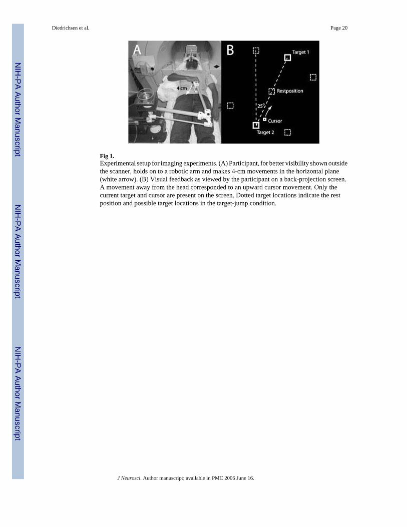
Fig 1.Experimental setup for imaging experiments. (A) Participant, for better visibility shown outsidethe scanner, holds on to a robotic arm and makes 4-cm movements in the horizontal plane(white arrow). (B) Visual feedback as viewed by the participant on a back-projection screen.A movement away from the head corresponded to an upward cursor movement. Only thecurrent target and cursor are present on the screen. Dotted target locations indicate the restposition and possible target locations in the target-jump condition.
Diedrichsen et al. Page 20
J Neurosci. Author manuscript; available in PMC 2006 June 16.
NIH
-PA Author Manuscript
NIH
-PA Author Manuscript
NIH
-PA Author Manuscript

Fig. 2.Results of the behavioral experiment. (A) Average hand position trace (solid line) and cursorposition trace (dashed line) in the target-jump, visual-rotation and curl-field condition. Allmovements start at the lower box. Gray triangle indicates average position and time of targetjump. (B) Hand velocities (cm/s) in the lateral direction and (C) in the forward direction fortrials perturbed to the left (black line), to the right (gray line), and unperturbed trials (dashedline). Black triangle marks the average onset of the correction. (D) Trial-to-trial adaptationrater estimated from the state-space model (Eq 1, see methods). Dots plot the estimates forindividual participants.
Diedrichsen et al. Page 21
J Neurosci. Author manuscript; available in PMC 2006 June 16.
NIH
-PA Author Manuscript
NIH
-PA Author Manuscript
NIH
-PA Author Manuscript

Fig 3.Population-averaged activity related to normal reaching movements compared to rest on(A) aflattened representation of the left and (B) right cortical hemisphere. The lateral and parts ofthe medial surface of parietal and frontal lobe are shown: gray scale indicates sulcucal depthwith sulci shown in dark gray, averaged across participants. SFS: Superior frontal sulcus, CS:Central sulcus, PoCS: Postcentral sulcus, IPS: Intraparietal sulcus, STS: Superior temporalsulcus, POS: Parietal-occipital sulcus. (C) Sulci (black lines) and the border of the flattenedrepresentation (red line) on an inflated representation of the left hemisphere. (D) Averagemovement related activity in basal ganglia and thalamus, and (E) on a flattened representationof the cerebellum. Roman digits denote cerebellar lobules according to the Larsell notation(Schmahmann et al., 2000). All maps are thresholded at t(13)>3.61.
Diedrichsen et al. Page 22
J Neurosci. Author manuscript; available in PMC 2006 June 16.
NIH
-PA Author Manuscript
NIH
-PA Author Manuscript
NIH
-PA Author Manuscript

Fig 4.Averaged activity in the visual control condition 2 vs. rest on a flat representation of the(A)left and (B) right cortical hemispheres. Threshold is t(13)>3.61. FEF. Human Frontal eye field.Other abbreviations as in Fig. 4.
Diedrichsen et al. Page 23
J Neurosci. Author manuscript; available in PMC 2006 June 16.
NIH
-PA Author Manuscript
NIH
-PA Author Manuscript
NIH
-PA Author Manuscript

Fig 5.Statistical t-map of areas activated more due to execution errors that lead to adaptation ofinternal models (blue) or more due to target errors that arise from a change in the reach target(red). Shown is the interaction contrast of Eq. 2 at a threshold of t(13)>3.61. Bar graphs withthe percent signal change relative to rest from significant cluster are shown on the margins.
Diedrichsen et al. Page 24
J Neurosci. Author manuscript; available in PMC 2006 June 16.
NIH
-PA Author Manuscript
NIH
-PA Author Manuscript
NIH
-PA Author Manuscript
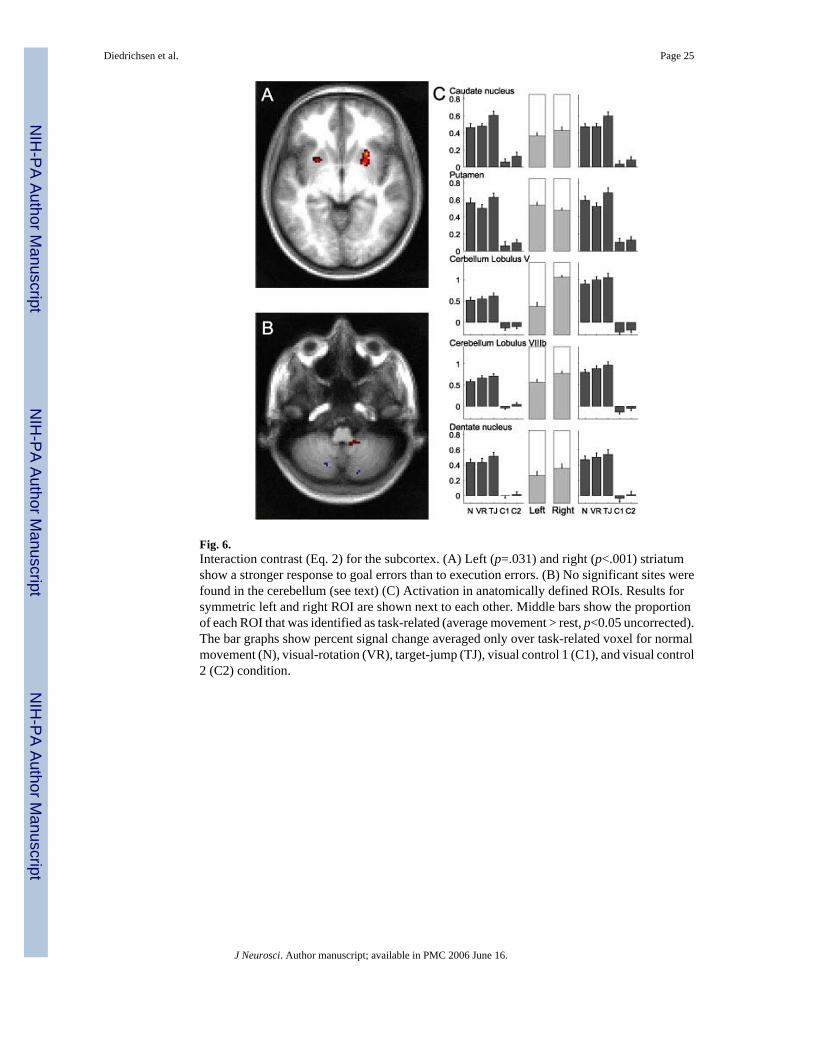
Fig. 6.Interaction contrast (Eq. 2) for the subcortex. (A) Left (p=.031) and right (p<.001) striatumshow a stronger response to goal errors than to execution errors. (B) No significant sites werefound in the cerebellum (see text) (C) Activation in anatomically defined ROIs. Results forsymmetric left and right ROI are shown next to each other. Middle bars show the proportionof each ROI that was identified as task-related (average movement > rest, p<0.05 uncorrected).The bar graphs show percent signal change averaged only over task-related voxel for normalmovement (N), visual-rotation (VR), target-jump (TJ), visual control 1 (C1), and visual control2 (C2) condition.
Diedrichsen et al. Page 25
J Neurosci. Author manuscript; available in PMC 2006 June 16.
NIH
-PA Author Manuscript
NIH
-PA Author Manuscript
NIH
-PA Author Manuscript

Fig. 7.Activation in Experiment 2 in the visual-rotation (blue), and curl-field (red) conditions relativeto normal movements at a threshold of t(16)>4 for the (A) left and (B) right hemisphere. Purpleareas indicate overlap. Bar graphs show average percent signal change in selected areas of theleft hemisphere for normal (N), visual-rotation (VR), curl-field (CF) and resistive-field (RF)conditions.
Diedrichsen et al. Page 26
J Neurosci. Author manuscript; available in PMC 2006 June 16.
NIH
-PA Author Manuscript
NIH
-PA Author Manuscript
NIH
-PA Author Manuscript

Fig. 8.(A) Activation in Experiment 2 in the visual-rotation (blue), and curl-field (red) conditionsrelative to normal movements at a threshold of t(16)>4 displayed on a surface representationof the cerebellum (Van Essen, 2002). (B) Percent signal change (dark bars) in subcortical ROIs,during normal (N), visual-rotation (VR), curl-field (CF), and resistive field (RF) conditions.Middle gray bars show the proportion of each ROI that was identified as movement related(average movement >rest, p<0.05 uncorrected).
Diedrichsen et al. Page 27
J Neurosci. Author manuscript; available in PMC 2006 June 16.
NIH
-PA Author Manuscript
NIH
-PA Author Manuscript
NIH
-PA Author Manuscript

NIH
-PA Author Manuscript
NIH
-PA Author Manuscript
NIH
-PA Author Manuscript
Diedrichsen et al. Page 28
Table 1Average movement parameters (SD) for the conditions of the imaging experiment 1 and 2.Movement duration(ms) encompasses all corrective sub-movements. Movement error is the absolute angular deviation of the initial(200ms) from the final movement direction. This measure is calculated only for the perturbed movements,whereas the other parameters are averaged over all movements (except first and last movement of a 10-movementblock).
Normal Movement Visual rotation Condition Curl field Target jump Resistive field
Experiment 1(Participant 1-5) Movement duration (ms) 417 614 - 569 -
(26) (68) - (57) - Movement error (deg) 4.6 25.1 - 25.5 -
(0.6) (3.9) - (2.1) - Movement length (cm) 4.3 5.9 - 5.4 -
(0.4) (0.5) - (0.5) -
Experiment 1(Participant 6-13) Movement duration (ms) 481 576 - 559 -
(26) (68) - (57) - Movement error (deg) 3.2 19.2 - 25.2 -
(0.6) (3.9) - (2.1) - Movement length (cm) 6.6 5.7 - 5.9 -
(0.4) (0.5) - (0.5) -
Experiment 2 Movement duration (ms) 422 648 631 - 491
(41) (45) (49) - (64) Movement error (deg) 6.5 25.3 25.4 - 6.6
(1.8) (1.6) (4.6) - (2.3) Movement length (cm) 4.8 6.7 6.9 - 4.4
(0.5) (0.7) (1.2) - (0.1)
J Neurosci. Author manuscript; available in PMC 2006 June 16.

NIH
-PA Author Manuscript
NIH
-PA Author Manuscript
NIH
-PA Author Manuscript
Diedrichsen et al. Page 29
Table 2Anatomical areas that showed a significant interaction contrast (Eq. 2) on a clusterwise level in Experiment 1.Height threshold was t(12)=3.61, p=0.002 (uncorrected). The total search volume was 110156 mm2 (2869 resels)with an estimated smoothness of FWHM = 6.2 (mm). The expected size of random clusters was 5.5 mm2. Allareas sizes are calculated on individual surfaces and averaged (Diedrichsen, 2005). The SPLa cluster on the leftside was not significant, but was reported because it contained the highest t-value and we found a homologousarea activated on the right side. The location of the maxima is listed in latitude/longitude for the population-averaged and mark- and surface-based atlas (PALS, Van Essen, in press) and the template of the Montrealneurological institute (MNI).
PALS MNI
Area SideExtendmm2 PeakT p (cluster) Lat Long x y z
More activated during target errorsIPSp L 39 4.71 0.028 1 157 -28 -63 54SPLmed R 112 7.71 <0.001 -30 131 7 -65 60SPLmedp R 71 7.79 <0.001 -33 143 11 -72 52IPS/TOS (V3a/V7) L 70 7.66 <0.001 -22 -180 -22 -87 22POS L 184 9.31 <0.001 -60 167 -16 -73 21POS R 80 4.86 <0.001 -49 152 17 -71 35POSinf R 41 5.50 0.019 -80 160 14 -55 14
More activated during execution errorsPoCSa (2) L 167 -6.11 <0.001 51 139 -55 -22 51PoCSp (2/7) L 128 -6.51 <0.001 41 151 -45 -31 45PoCS (2) R 60 -6.12 0.025 48 145 51 -23 50PoCS (2) R 39 -4.95 0.033 66 152 62 -12 39SPLa (5) L 17 -7.08 0.78 8 134 -30 -45 66SPLa (5) R 41 -4.16 0.026 24 147 37 -38 52SPLm L 50 -4.59 0.003 3 141 -32 -53 64ITS (hMT) R 100 -5.61 <0.001 -2 -144 46 -72 3
J Neurosci. Author manuscript; available in PMC 2006 June 16.