Atmospheric brown clouds reach the Tibetan Plateau by crossing the Himalayas
Upload
khangminh22Category
view
0download
0
SEPTEMBER 2021
In My ViewThe facts and myths of gender equity 14 – 15
NextGenMaking a splash with photoacoustic imaging
34 – 36
ProfessionZambia’s fight against blindness
44 – 46
Sitting Down With Retina specialist with a calling, Judy Kim 50 – 51
# 89
www.theophthalmologist.com
Within ReachExploring an ROP
screening program in southern India – in pictures
16– 24

• Single-use instruments offerprotection against infection,due to decreased risk ofcross-contamination1
• Surgeons benefit fromprecision and reliabilityof brand-new instrumentsfor each patient2
Providing Sterile Options for Your Operating Room
High Quality, Single-Use Instruments
bvimedical.com
BVI and all other trademarks (unless noted otherwise) are property of BVI. BVI ©2020-2021 1571364-01
US Tel: 866-906-8080 Fax: 866-906-4304Email: [email protected]
References1 Southworth, P.M. Infections and exposures: reported incidents associated with unsuccessful decontamination of reusable surgical instruments.
The Journal of Hospital Infection. September 17, 2014 2 Scheib, Christian; Single-use instruments deserve consideration for eye surgery: Advantages include cost savings, ease of use and high-quality
instruments; December 1, 2016
Call Your BVI Sales Representative About Malosa® Single-Use Today!
1571364-01 Global Single Use Cross Brand Ad for The Ophthalmologist FINAL.indd 1 4/19/21 5:45 PM

www.theophthalmologist.com
Edi tor ialLive and LearnFive personal anecdotes we would never have heard without The Ophthalmologist Power List
• Single-use instruments offer protection against infection, due to decreased risk of cross-contamination1
• Surgeons benefit from precision and reliability of brand-new instruments for each patient2
Providing Sterile Options for Your Operating Room
High Quality, Single-Use Instruments
bvimedical.com
BVI and all other trademarks (unless noted otherwise) are property of BVI. BVI ©2020-2021 1571364-01
US Tel: 866-906-8080 Fax: 866-906-4304Email: [email protected]
References1 Southworth, P.M. Infections and exposures: reported incidents associated with unsuccessful decontamination of reusable surgical instruments.
The Journal of Hospital Infection. September 17, 2014 2 Scheib, Christian; Single-use instruments deserve consideration for eye surgery: Advantages include cost savings, ease of use and high-quality
instruments; December 1, 2016
Call Your BVI Sales Representative About Malosa® Single-Use Today!
1571364-01 Global Single Use Cross Brand Ad for The Ophthalmologist FINAL.indd 1 4/19/21 5:45 PM
Following the publication of our 2021 Power List, which exclusively featured women in ophthalmology, I received a great deal of positive feedback – not only from the female frontrunners but also from their peers. The majority of
readers who spoke to me saw the decision to feature only women in the way it was intended: to redress the balance of “power” in the field after years of underrepresentation. Even though it is now easier than ever for women to enter the specialty, certain challenges remain firmly in place, as Tina Felfeli and Yvonne Buys comment on page 14.
Though the 2021 Power List will remain fresh and topical in our minds for some time, we must also look to the future – the 2022 edition. And in case you missed the announcement, nominations are already open: theophthalmologist.com/power-list/2022
I hope that our special focus on equity in 2021 will bear fruit in the form of a strong representation of women and other minorities. So please, go forth and nominate the individuals you feel deserve to be recognized and celebrated. To give you a friendly push in the right direction, I will leave you with five surprising things we learned from previous lists…
1. Serial Power Lister Carol Shields shared the moment she found out the baby she was treating for retinoblastoma lived in her old house in her western Philadelphia hometown
2. Terry Kim (aka DJ Special K) fondly remembered his musical contributions to the biggest ophthalmic meetings in the US – DJing together with Tony Aldave (aka DJ AJA) at major clubs to crowds of over 2,000 AAO and ASCRS attendees
3. David Chang humbly told the story of his young son – a keen baseball player – who suddenly and inexplicably started making mistakes on the court… until a pediatrician suggested the should perhaps check his son’s vision
4. Douglas Rhee described how his professional career shifted profoundly when he decided to reassess plans for a clinical fellowship abroad because his cat couldn’t travel with him (it all worked out for the best!)
5. And finally, we heard the story of the surprising and timely donation that allowed Sir Peng Tee Khaw to help a key researcher, who then went on to have a rather successful career in cellular regeneration.
What stories will we unearth in 2022? Well, in part, that’s up to you (theophthalmologist.com/power-list/2022).
Aleksandra JonesEditor

Contents
On The Cover
Aravind Eye Hospital’s outreach projects include a retinopathy of prematurity program.
03 Editorial Live and Learn by Aleksandra Jones
Upfront
08 The latest news, views and research – from the phone app that is improving eye health, to the magnetic protein in birds’ eyes that enables remarkable feats of migration
In My View
14 Equity: Facts and Future Tina Felfeli and Yvonne Buys lay out the facts on pay and practice disparity between male and female ophthalmologists and look to the future
16
08
38

OZURDEX® (dexamethasone intravitreal implant) acts fast1,2 and lasts3–5 with less treatment visitscompared with anti-VEGFs.5
Effective DME treatment doesn’t have to be a burden.6
The most commonly reported adverse events reported following treatment with OZURDEX® are those frequently observed with ophthalmic steroid treatment or intravitreal injections (elevated IOP, cataract formation and conjunctival or vitreal haemorrhage respectively). Less frequently reported, but more serious, adverse reactions include endophthalmitis, necrotizing retinitis, retinal detachment and retinal tear.
This advert is consistent with the UK marketing authorisation.Licences may vary by country, please refer to your local country SmPC.
DME, diabetic macular edema; IOP, intraocular pressure; VEGF, vascular endothelial growth factor.
1. Lo Giudice G et al. Eur J Ophthalmol 2018;28(1):74–79. 2. Veritti D et al. Ophthalmologica 2017;238(1–2):100–105. 3. Escobar-Barranco JJ et al. Ophthalmologica 2015;233(3–4):176–185. 4. Allergan. OZURDEX® Summary of Product Characteristics. 5. Kodjikian L et al. Biomed Res Int 2018:8289253. 6. Boyer DS et al. Ophthalmology 2014;121:(10):1904–1914.
The most commonly reported adverse events reported following treatment with OZURDEX® are those frequently observed with
OZURDEX® (Dexamethasone 700 micrograms intravitreal implant in applicator)Abbreviated Prescribing Information Presentation: Intravitreal implant in applicator. One implant contains 700 micrograms of dexamethasone. Disposable injection device, containing a rod-shaped implant which is not visible. The implant is approximately 0.46 mm in diameter and 6 mm in length. Indications:Treatment of adult patients: with macular oedema following either Branch Retinal Vein Occlusion (BRVO) or Central Retinal Vein Occlusion (CRVO), in� ammation of the posterior segment of the eye presenting as non-infectious uveitis and visual impairment due to diabetic macular oedema (DME) who are pseudophakic or who are considered insu� ciently responsive to, or unsuitable for non-corticosteroid therapy. Dosage and Administration: Please refer to the Summary of Product Characteristics before prescribing for full information. OZURDEX must be administered by a quali� ed ophthalmologist experienced in intravitreal injections. The recommended dose is one OZURDEX implant to be administered intra-vitreally to the a� ected eye. Administration to both eyes concurrently is not recommended. Repeat doses should be considered when a patient experiences a response to treatment followed subsequently by a loss in visual acuity and in the physician’s opinion may bene� t from retreatment without being exposed to signi� cant risk. Patients who experience and retain improved vision should not be retreated. Patients who experience a deterioration in vision, which is not slowed by OZURDEX, should not be retreated. In RVO and uveitis there is only very limited information on repeat dosing intervals less than 6 months. There is currently no experience of repeat administrations in posterior segment non-infectious uveitis or beyond 2 implants in Retinal Vein Occlusion. In DME there is no experience of repeat administration beyond 7 implants. Patients should be monitored following the injection to permit early treatment if an infection or increased intraocular pressure occurs. Single-use intravitreal implant in applicator for intravitreal use only. The intravitreal injection procedure should be carried out under controlled aseptic conditions as described in the Summary of Product Characteristics. The patient should be instructed to self-administer broad spectrum antimicrobial drops daily for 3 days before and after each injection. Contraindications: Hypersensitivity to the active substance or to any of the excipients. Active or suspected ocular or periocular infection including most viral diseases of the cornea and conjunctiva, including active epithelial herpes simplex keratitis (dendritic keratitis), vaccinia, varicella, mycobacterial infections, and fungal diseases. Advanced glaucoma which cannot be adequately controlled by medicinal products alone. Aphakic eyes with ruptured posterior lens capsule. Eyes with Anterior Chamber Intraocular Lens (ACIOL), iris or transscleral � xated intraocular lens and ruptured posterior lens capsule. Warnings/Precautions: Intravitreous injections, including OZURDEX can be associated with endophthalmitis, intraocular inflammation, increased intraocular pressure and retinal
detachment. Proper aseptic injection techniques must always be used. Patients should be monitored following the injection to permit early treatment if an infection or increased intraocular pressure occurs. Monitoring may consist of a check for perfusion of the optic nerve head immediately after the injection, tonometry within 30 minutes following the injection, and biomicroscopy between two and seven days following the injection. Patients must be instructed to report any symptoms suggestive of endophthalmitis or any of the above mentioned events without delay. All patients with posterior capsule tear, such as those with a posterior lens (e.g. due to cataract surgery), and/or those who have an iris opening to the vitreous cavity (e.g. due to iridectomy) with or without a history of vitrectomy, are at risk of implant migration into the anterior chamber. Implant migration to the anterior chamber may lead to corneal oedema. Persistent severe corneal oedema could progress to the need for corneal transplantation. Other than those patients contraindicated where OZURDEX should not be used, OZURDEX should be used with caution and only following a careful risk bene� t assessment. These patients should be closely monitored to allow for early diagnosis and management of device migration. Use of corticosteroids, including OZURDEX, may induce cataracts (including posterior subcapsular cataracts), increased IOP, steroid induced glaucoma and may result in secondary ocular infections. The rise in IOP is normally manageable with IOP lowering medication. Corticosteroids should be used cautiously in patients with a history of ocular herpes simplex and not be used in active ocular herpes simplex. OZURDEX is not recommended in patients with macular oedema secondary to RVO with signi� cant retinal ischemia. OZURDEX should be used with caution in patients taking anti-coagulant or anti-platelet medicinal products. OZURDEX administration to both eyes concurrently is not recommended. Visual disturbance may be reported with systemic and topical corticosteroid use. If a patient presents with symptoms such as blurred vision or other visual disturbances, consider evaluating for possible causes which may include cataract, glaucoma or rare diseases such as central serous chorioretinopathy (CSCR) which have been reported after use of systemic and topical corticosteroids. Interactions: No interaction studies have been performed. Systemic absorption is minimal and no interactions are anticipated. Pregnancy:There are no adequate data from the use of intravitreally administered dexamethasone in pregnant women. OZURDEX is not recommended during pregnancy unless the potential bene� t justi� es the potential risk to the foetus. Lactation: Dexamethasone is excreted in breast milk. No e� ects on the child are anticipated due to the route of administration and the resulting systemic levels. However OZURDEX is not recommended during breast-feeding unless clearly necessary. Driving/Use of Machines: Patients may experience temporarily reduced vision after receiving OZURDEX by intravitreal injection. They should not drive or use machines until this has resolved. Adverse E� ects: In clinical trials the most frequently reported adverse events were
increased intraocular pressure (IOP), cataract and conjunctival haemorrhage*. Increased IOP with OZURDEX peaked at day 60 and returned to baseline levels by day 180. The majority of elevations of IOP either did not require treatment or were managed with the temporary use of topical IOP-lowering medicinal products. 1% of patients (4/347 in DME and 3/421 in RVO) had surgical procedures in the study eye for the treatment of IOP elevation. The following adverse events were reported: Very Common (≥ 1/10): IOP increased, cataract, conjunctival haemorrhage*. Common (≥1/100 to <1/10): headache, ocular hypertension, cataract subcapsular, vitreous haemorrhage*, visual acuity reduced*, visual impairment/disturbance, vitreous detachment*, vitreous � oaters*, vitreous opacities*, blepharitis, eye pain*, photopsia*, conjunctival oedema*, conjunctival hyperaemia. Uncommon (≥1/1,000 to <1/100): migraine, necrotizing retinitis, endophthalmitis*, glaucoma, retinal detachment*, retinal tear*, hypotony of the eye*, anterior chamber in� ammation*, anterior chamber cells/� ares*, abnormal sensation in eye*, eyelids pruritus, scleral hyperaemia*, device dislocation* (migration of implant) with or without corneal oedema , complication of device insertion resulting in ocular tissue injury* (implant misplacement). (*Adverse reactions considered to be related to the intravitreous injection procedure rather than the dexamethasone implant). Please refer to Summary of Product Characteristics for full information on side e� ects. Basic NHS Price: £870 (ex VAT) per pack containing 1 implant. Marketing Authorisation Number: EU/1/10/638/001. Marketing Authorisation Holder: Allergan Pharmaceuticals Ireland, Castlebar Road, Westport, Co. Mayo, Ireland. Legal Category: POM. Date of Preparation: May 2019. UK/0288/2019
Adverse events should be reported. Reporting forms and information can be found at https://yellowcard.mhra.gov.uk/
Adverse events should also be reported to Allergan Ltd. [email protected] or 01628 494026
JOB CODE: INT-OZU-2050219DATE OF PREPARATION: DECEMBER 2020
008336_ALLOZU20_Refining Routines_The_Ophthalmologist_V2.2_FAW.indd 1008336_ALLOZU20_Refining Routines_The_Ophthalmologist_V2.2_FAW.indd 1 18/12/2020 12:1518/12/2020 12:15

WELCOME TO THE ZIEMER WORLD
www.ziemergroup.com/escrs
Visit us at booth #A22 at ESCRS to learn more!
ESCRS_World_of_Ziemer_Suite_Ad_210x266mm_The_Ophthalmologist.indd 1ESCRS_World_of_Ziemer_Suite_Ad_210x266mm_The_Ophthalmologist.indd 1 23.08.21 19:5323.08.21 19:53

WELCOME TO THE ZIEMER WORLD
www.ziemergroup.com/escrs
Visit us at booth #A22 at ESCRS to learn more!
ESCRS_World_of_Ziemer_Suite_Ad_210x266mm_The_Ophthalmologist.indd 1ESCRS_World_of_Ziemer_Suite_Ad_210x266mm_The_Ophthalmologist.indd 1 23.08.21 19:5323.08.21 19:53
Profession
38 Ain’t No Mountain High Enough How does climbing mountains translate to better quality of life for patients with AMD? Simon Prosser explains
44 Zambia’s Fight Against Blindness Lucia Nadaf discusses how ORBIS International is attempting to oust unnecessary blindness from Zambia – and how its efforts have been affected by the COVID-19 pandemic
Feature
16 Within Reach Terry Cooper’s amazing images from the outreach program of retinopathy of prematurity screening in Madurai, India, led by Renu P. Rajan from Aravind Eye Hospitals
Sitting Down With
50 Judy E. Kim, Professor, Ophthalmology and Visual Sciences, Director of Teleophthalmology and Research at the Medical College of Wisconsin, USA
NextGen
30 Serving One’s (Re)purpose Recycling in the pharmaceutical pipeline – the discovery that a class of HIV drugs can be used to treat dry AMD
34 Structurally Sound Parsin Haji Reza explains his novel photoacoustic imaging technique – PARS – and how it can revolutionize ophthalmic imaging and other medical disciplines
34
50
I S S U E 8 9 - S E P T E M B E R 2 0 2 1
Feel free to contact any one of us: [email protected]
Content TeamEditor - Aleksandra Jones
Geoffrey Potjewyd (Associate Editor) Commercial Team
Publishing Director - Neil HanleySam Blacklock (Associate Publisher)
Ross Terrone (Business Development Manager)Design Team
Head of Design - Marc BirdHannah Ennis (Senior Designer)
Charlotte Brittain (Designer) Digital Team
Digital Team Lead - David RobertsPeter Bartley (Digital Producer Web/Email)
Audience TeamAudience Growth Strategy Manager
– Brice AgamemnonCRM & Compliance
CRM & Compliance Manager - Tracey Nicholls Hayley Atiz (CRM Assistant)
Commercial Support TeamInternal Systems Manager - Jody Fryett
Dan Marr (Campaign Reporting Analyst) Commercial ServicesCommercial Service and
Social Media Manager- Matt Everett Lindsey Vickers (Sales Support Project Manager)
Julie Wheeler (Sales Support Coordinator)Marketing Team
Marketing Manager - Katy PearsonJo Baylay (Marketing Executive)
Accounts TeamKerri Benson (Accounts Assistant),
Emily Scragg (Accounts Apprentice) Human Resources
Human Resource Manager - Tara Higby Management Team
Chief Executive Officer - Andy Davies Chief Operating Officer - Tracey Peers
Senior Vice President (North America) - Fedra Pavlou
Financial Director - Phil Dale Commercial Director - Richard Hodson
Content Director - Rich Whitworth
Change of address [email protected] Atiz, The Ophthalmologist, Texere Publishing
Limited, Booths Park 1, Chelford Road, Knutsford, Cheshire, WA16 8GS, UK
General enquiries www.texerepublishing.com | info@
theophthalmologist.com +44 (0) 1565 745 200 | [email protected]
Distribution: The Ophthalmologist (ISSN 2051-4093), is published monthly by Texere Publishing
Limited, Booths Park 1, Chelford Road, Knutsford, Cheshire, WA16 8GS, UK. Single copy sales $15
(plus postage, cost available on request [email protected]). Non-qualified annual
subscription cost is available on request.
Reprints & Permissions – [email protected] copyright in the materials contained in this publication and the
typographical arrangement of this publication belongs to Texere Publishing Limited. No person may copy, modify, transmit, distribute, display,
reproduce, publish, licence or create works from any part of this material or typographical arrangement, or otherwise use it, for any public or commercial
use without the prior written consent of Texere Publishing Limited.The names, publication titles, logos, images and presentation style appearing
in this publication which identify Texere Publishing Limited and/or its products and services, including but without limitation Texere and The Ophthalmologist are proprietary marks of Texere Publishing Limited. Nothing contained in this publication shall be deemed to confer on any
person any licence or right on the part of Texere Publishing Limited with respect to any such name, title, logo, image or style.
The Ophthalmologist is printed using soy ink

Mice are crucial to research and development in ophthalmology, but how does mouse neuro-ophthalmology compare to that of humans? A recent study has revealed a region of the mouse brain responsible for enhanced spatial resolution – a feature of mouse eyesight not previously seen. Called the focea, this region is named after its closest human counterpart: the fovea, which is located in the retina and specializes in high-resolution vision. Because mice lack a fovea, researchers were unsure as to their spatial resolution capabilities – but the debate could soon be over. “We know that many species, including cats and monkeys, have better resolution in the center of the visual field,” explains co-lead researcher Pieter Roelfsema, “and this specialization may be even more widespread than anticipated.” Mice actually make compensatory eye movements to keep the region of eyesight that’s processed at the focea in front of them – similar to the human saccadic eye movements that focus the fovea on a point of interest.
This finding “increases the potential
usage of mouse models as a surrogate for human vision and opens new possibilities for studying how information is brought into the focea,” explains co-lead researcher Matthew Self, presenting the possibility of using mouse models to investigate the neural circuitry of high-detail vision.
The discovery of spatial bias in mice visual processing was serendipitous; originally, the group planned to map the mouse visual cortex. “Finding a region with enhanced resolution was a surprise. That is quite often
the case in fundamental research; you find something unexpected,” states Self.
The next steps for this research are already in place. “We are now studying how mice segregate objects from the background and how this form of vision is influenced by visual experience and learning,” says Roelfsema.
Reference1. EH van Beest et al., Nat Commun, 12, 4029
(2021). PMID: 34188047.
Of Mice and MenThe discovery of focea in mice implies that their vision may be more similar to human sight than previously thought
8 Upfront
UpfrontResearch
Innovation Trends
The seasonal migration of birds is fascinating – remarkable distances traveled with a mystifying ability to reach the intended
destination. But now, an international collaboration of researchers may have identified the source of magnetoreception – the biological “compass” that guides birds through the Earth’s magnetic fields. When isolated and tested in the lab, the team found that the light-sensitive protein cryptochrome 4 (CRY4) from the retina of the European robin is sensitive to magnetic fields (1). Is this the protein responsible for holding the GPS on their seasonal relocation marathon? Study author Peter
Hore, Professor at Oxford University, UK, explains: “To establish whether the cryptochrome hypothesis is the correct sensory mechanism, magnetic field effects on cryptochromes need to be measured in vivo to study the magnetic orientation of cryptochrome-deficient birds and/or to measure magnetic field effects on nerve impulses generated in the retina.”
See reference online at: top.txp.to/birds
The Robin’s Retinal CompassIs a light-sensitive eye protein the key to magnetoreception in bird migration?

• Vance Thompson is appointed Euclid Systems’ Chief Medical Officer. Thompson is founder of Vance Thompson Vision in Sioux Falls, South Dakota, and Professor of Ophthalmology at Sanford School of Medicine, at the University of South Dakota, Vermillion, USA. Euclid is a global leader in orthokeratology and myopia management
• Monty Montoya has been appointed the CEO of TherOptix. Montoya is
founder and CEO of CorneaGen and founder of Aurion Biotech – previously the CEO of SightLife. TherOptix develops a drug-eluting contact lens system and has received orphan drug designation for prevention of proliferative vitreoretinopathy
• NovaBay and ImprimisRX have partnered up to promote prescription of Avenova, an antimicrobial lid and lash solution used for numerous eye conditions. ImprimisRx is a leading ophthalmology-focused pharmaceutical business that will promote Avenova, and NovaBay Pharmaceuticals is focused on commercialization
• A nine-month preliminary extension pass has been granted to EyePoint
Pharmaceuticals to pass through payment status for DEXYCU. EyePoint develops and commercializes therapeutics for serious eye disorders. Without the nine-month extension, pass-through payment status for DEXYCU (a postoperative inflammation treatment) would have ended on March 31, 2022
• The refractive surgery council reports strong growth in H1 laser vision correction (LVC) procedures – an 82 percent increase on 2020 so far in 2021. “LVC’s current momentum is a reflection of consumers’ desire to take control of their vision in what has been an uncertain, yet optimistic, moment in time,” said Chairman, Jim Wachtman.
B U S I N E S S I N B R I E F The latest industry news – in no more than 60 words
9Upfront
Brought to you by Teleon

It’s a common story – a disease with a prime suspect, but no smoking gun. Knowing that a gene is likely responsible for AMD pathogenesis isn’t enough to show causation of the disease. Hard evidence, like finding the perpetrator at the scene of the crime, is necessary – and researchers at the Sharon Eccles Steele Center for Translational Medicine (SCTM) are on the case. Genes in chromosome 10q26 are associated with AMD, but the prime gene suspects – age-related maculopathy susceptibility 2 (ARMS2) and high temperature requirement A serine peptidase 1 (HTR A1) – have yet to be caught red handed. Do these genes cause the disease through an active mechanism or is something happening to them to affect their normal function?
Using human eye tissues, the SCTM investigators demonstrated that donors with risk-associated variants had a reduction of HTRA1 in the retinal
pigment epithelium (RPE), within the 10q26 locus, which was not the case in the retina or choroid tissues. The scene of the crime was established. This tissue-specific decrease is caused by the presence of an overlapping sequence of ARMS2, which contains a transcription factor binding site that is disrupted by the AMD risk variant rs36212733. Subsequently, HtrA1 protein was found to be reduced at the RPE-Bruch’s membrane – a crucial interface where HtrA1 would normally be responsible for driving clearance of waste and general maintenance. Notably, in a regular chromosome 10q26 locus, HtrA1 actually increases with age.
The investigators propose that modulating levels of HtrA1 may offer a new therapeutic avenue for AMD; in the meantime, they’re trying to gather more evidence. Translation from association to causation – finding the smoking gun – could be key to understanding and then mitigating RPE-specific and age-dependent drivers of AMD progression, ultimately helping to save the eyesight of those at high genetic risk of developing the disease.
Reference1. BL Williams et al., PNAS, 118, e2103617118
(2021). PMID: 34301870.
10 Upfront
Many low- and middle-income areas of the world have insufficient eye care provisions to meet the minimum
requirements set out by WHO, which leads to many people unnecessarily becoming or remaining visually impaired. Now, a smartphone app has almost tripled the number of people attending ophthalmic primary care appointments within the Kenyan Trans Nzoia County community. The Peek Community Eye Health system uses smartphone-based vision screening and referrals – allowing community volunteers to go door-to-door and perform initial eye screenings with the decision-guiding
app. Any eye problems identified at this stage result in an automatic referral for follow-up appointments, supported by SMS reminders to maximize patient attendance and treatment. This has improved access to care and maximized the time hospitals have to spend on serious cases – more effectively managing ophthalmologists’ limited time.
Reference1. H Rono et al., Lancet Digital Health, 3, e414
(2021). PMID: 34167763.
A Smoking GunHow an improved understanding of the genes that drive AMD pathogenesis could lead to new therapeutic options
Eye-Phone DiagnosisImproving screening and follow-up appointment management? There’s an app for that...
A Foreign Body
This month’s image shows a metallic intraocular foreign body very close to the optic nerve. It was removed safely, despite initial immediate postop vitreous inflammation, and the patient recovered completely within three weeks, with unaided visual acuity of 20/20.Credit: Costas H. Karabatsas, Assistant Professor of Ophthalmology at the Department of Biomedical Sciences
of the University of West Attica, Psachna, Greece.
Would you like your photo featured in Image of the Month? Send it to [email protected]
11Upfront
Q U O T E O F T H E M O N T H
“The pandemic has given me a chance to reflect, renew, and refocus. It gave me permission to stop and come up for breath,
above a life drowning in busyness. This period of reflection has helped me to refocus my priorities. And it will help me to put large rocks into my life’s bucket rather than filling it with
small pebbles or even grains of sand.”Judy E. Kim, Professor, Ophthalmology and Visual Sciences,
Graduate School of Biomedical Sciences, Director of Teleophthalmology and Research at the Medical College of
Wisconsin, Wauwatosa, USA
I M A G E O F T H E M O N T H
A new genetic test that factors in both polygenic and monogenic glaucoma risk variants could result in a 15-fold increase of people being identified as glaucoma suspects (1). Researchers analyzed the association of monogenic and polygenic variants with glaucoma risk – using clinical and genetic data from an Australian/New Zealand glaucoma database (ANZRAG, containing data from 2,507 individuals) and from the UK Biobank (data from 411,337 individuals). Both polygenic and monogenic variants have a comparable risk of developing glaucoma – over a 2.5-fold increase. But high-risk polygenic variants were six times more common than monogenic variants within the ANZRAG database – and more than 15 times more common within the general population (UK Biobank) data.
Although single gene testing is used clinically, a stratified approach of monogenic and polygenic testing will increase opportunities to save patients’ sight.
Reference1. OM Siggs et al., JAMA Ophthalmol, [Online
ahead of print] (2021). PMID: 34264281.
Diagnosis: GlaucomaSingle gene testing may not be sufficient to identify a high risk of glaucoma
K I N D L Y A N D U N C O N D I T I O N A L L Y S P O N S O R E D B Y
www.theophthalmologist.com

Have you ever wondered why we see faces in random objects? This phenomenon is called face pareidolia (FP) – and the face you see in the clock or house across the road is a false positive result of your brain’s evolutionary insistence on rapidly identifying faces and assigning intentions or emotions to them. Researchers at the University of Sydney are delving deeper into FP – establishing that we assign emotional weight to false faces in the same way that we do real ones (1). Lead researcher David Alais explains the rationale and results from their recent research publication…
What led to this research?Previous neuroimaging by co-authors Jessica Taubert and Susan Wardle showed that the brain region specialized for processing faces responded just as well – and just as quickly (<250 ms) – to FP images as to real face images (2).
That’s pretty speedy…The brain achieves such a fast response by applying a template-matching procedure – anything that fits the global configuration of a face (two eyes over a nose and a mouth). The downside of blindly applying a simple template is that it produces some false positives (such as seeing faces in objects!). The upside is that it is very fast and rarely misses a face.
Because we are the most sophisticated social animals on the planet, there is a strong evolutionary incentive to quickly detect faces and assess them for information: happy or sad, friend or foe, healthy or ill, and so on. Thus,
our current study asked: after the quick initial face detection, does the brain go on to process the emotional content of the FP images? Or, alternatively, does the brain realize it has made a mistake in detecting an object as a face and not process it for emotional expression?
How did you test for this?Our study used a kind of adaptation procedure in which exposure to a previous face biases perception of the current face. Specifically, using faces ranging from angry to happy, it has been shown that brief exposure to a happy face makes the next one look happier (and vice versa – one angry face makes the next look angrier). The FP images we used in our studies all had a clear face-like configuration. The ultra-fast response to images that satisfy this simple template is a quick and effective way to detect the presence of a face – as opposed to seeing faces in clouds, for example, which can be a slow or difficult cognitive process and is as much imagination as face response.
What did you find?We confirmed that the brain does process the emotional content of FP images – one happy FP image makes the next look happier. Critically, we also showed that this sequential effect occurred between categories. That is, a happy FP “face” made a real face look happier and vice versa. If this sequential effect for emotional expression occurs regardless of image category, then FP
and faces must both activate a common facial expression process.
Do you have a favorite “illusory face” example or stories?I like the angry handbag and the grumpy chocolate milk bottle – and also the pareidolia choir!
Is FP integrated into our lives in any useful way?If it were not for FP images activating the brain’s face emotion process, we wouldn’t have emojis. (And how could we live without them? They are so communicative of emotion when we text our friends, yet so economical!) Emojis began as simple templates, :-) for happy and :-( for sad. Our research shows that templates such as these activate face and emotion processing.
In fact, designers intuitively understand our emotion response to objects. For example, some motor cars are designed to look like friendly or happy faces (they satisfy the face template with their head lights (eyes), grille (mouth), and badge (nose). This applies to the design of many objects; even if they don’t all look like faces, many are designed to look soft, round, and fleshy, rather than harsh and angular.
References1. D Alais et al., Proc Biol Sci, 1, 234 (2020).
PMID: 34229489.2. SG Wardle et al., Nat Commun, 11, 4518
(2020). PMID: 32908146.
12 Upfront
CarfaceIs that car smiling at you? David Alais explains the phenomenon of face pareidolia
+ Ultra-Widefield of view up to 200˚+ Rich in details from center to periphery+ Imaging through media opacities
iCare’s unique combination of sharpness and TrueColor offers now even wider view on the retina from 120° with a single shot, up to 200° with Mosaic functionality, helping to detect subtle signs of pathologies further in the periphery.*
Discover the new Ultra-Widefield view of iCare EIDON Family!
www.icare-world.com
More information: [email protected]
EID
ON
-FAM
ILY-
006-
AD-R
EV01
- 2
021-
06
Wider field of view up to 200°Expanding iCare EIDON Family perspective
*Ultra-Widefield imaging is available with the optional EIDON UWF Module.
Centervue S.p.A. is the Legal Manufacturer of EIDON, EIDON AF and EIDON FA. iCare is a registered trademark of Icare Finland Oy. Centervue S.p.A., Icare Finland Oy and Icare USA, Inc. are parts of Revenio Group and represent the brand iCare.Not all products, services or offers referenced in this ad are approved or offered in every market and approved labeling and instructions may vary from one country to another.

14 In My V iew
We recently published a paper in Ophthalmology that featured a population-based evaluation of the differences in remuneration between female and male ophthalmologists compared with other physician specialty groups in Ontario, Canada, over three decades (1). We found that female ophthalmologists in a fee-for-service system had lower median payments than males despite their productivity based on the number of patients in their practice and the number of visits – and after adjusting for age and year of practice. Our results displaced the myth that lower pay is solely the outcome of less work. The same finding was observed amongst other specialty groups, including medical procedural, non-procedural specialties, and surgical – but the sex difference was more pronounced in ophthalmology (1). We are not the first to note these differences in remuneration. In fact, disparity in pay has also been noted for female ophthalmologists in New Zealand, where the pay gap between the sexes
remains significant even when adjusted for hours worked (2). Although the true underlying reason for these discrepancies remains to be elucidated, some suggest that the disparity exists even within the first year of starting clinical practice (3).
Another interesting finding from our paper is that female specialists have a smaller representation in ophthalmology than most other surgical and medical specialty groups. And previous studies had already pointed out the under-representation of women in surgical specialties, such as orthopedics, thoracic surgery, and cardiology (4, 5). Again, the reasons remain unknown, but may be related to the lack of mentors and system challenges (6, 7). The finding is particularly alarming when we consider that most medical schools now comprise more women than men (8) – but that the proportion of women in ophthalmology and some other surgical specialties has not increased comparably.
We wondered if the influx of newly graduated female physicians and the delay in the time from graduation to practice may play a role in pay differences, but our age-adjusted model revealed a persistent gap in pay between the two sexes. Some of our previous work has shown that women
make up 43 percent of all practicing family physicians in Ontario in 2013 compared with 20 percent in ophthalmology (9). As such, it is apparent that the majority of women are entering non-surgical specialties.
But the big question remains: what is driving the differences in remuneration between women and men? In our study, one striking difference was amongst top paid individuals. Men who were considered to be the most paid (higher than the 60th percentile) in ophthalmology in 2018 (the last year of the study) earned on average 17 percent (US$126,650) more than the top paid female ophthalmologists. In the same year, the top billing male ophthalmologists had a greater number of patient visits than their female colleagues despite having a fewer number of distinct patients. The male ophthalmologists belonging to this group also had 52 percent higher payments than other male ophthalmologists. As such, this cohort of top biller male ophthalmologists has a distinct practice pattern that disproportionately contributes to a large aspect of the healthcare billings in Ontario, Canada.
Some of the temporal trends in practice patterns of ophthalmologists, such as the frequency of patient visits with the rapid
Equity: Facts and Future What do we know about the pay and practice disparity amongst male and female ophthalmologists?
By Tina Felfeli, resident physician in the Department of Ophthalmology and Vision Sciences at the University of Toronto, Vanier Scholar and member of the Integrated physician-scientist training program at the Institute of Health Policy, Management and Evaluation, University of Toronto, and Yvonne Buys, clinician investigator at University Health Network’s Donald K. Johnson Eye Institute, Professor in the Department of Ophthalmology and Vision Sciences, University of Toronto, and Past President of the Canadian Ophthalmological Society, Toronto, Canada
In My View
Experts from across the world share a single strongly held opinion
or key idea.

www.theophthalmologist.com
15In My V iew
expansion of intravitreal injections after the approval of bevacizumab in 2005, may be driving these differences (10). Gender differences in practice patterns may be related to the inherent variations in subspecialty choices, practice set-up, and billing practices, which warrant further investigation. In a fee-for-service system under the single-payer healthcare system in Canada, those who spend more time on a patient consult for a complex medical or surgical case may be compensated less than those who see multiple routine patient visits during the same period of time. And that’s a key concept to bear in mind!
In pursuit of equity in medicine, we must all comprehensively consider the unique challenges and barriers for early-career women in ophthalmology and other specialties (11). Recent studies have suggested that female ophthalmology residents in the US perform fewer cataract operations and total procedures compared with their male counterparts (12). We previously found in a survey of Canadian ophthalmologists that women reported less operating time than men, with 51 percent of women operating less than two days per month compared to 36 percent of males (13). This was later confirmed in a study using billing data of Ontario ophthalmologists, where 68.6 percent of male compared with 57.9 percent of female ophthalmologists performed surgery – and, of those performing cataract surgery, male ophthalmologists had 1.7 times the volume of procedures compared with female ophthalmologists in 2013 (14).
In addition to differences in payments between men and women, disparities have been shown in senior authorship on scientific publications, relationships to the medical industry, and grant opportunities (13, 15, 16, 17). In a survey of Canadian ophthalmologists, women believed that childbearing slowed or markedly slowed career progress, as compared with men (13). Studies on female physicians and surgeons have also noted the challenges they face in terms of delays in childbearing
(18) and increased risks of infertility and pregnancy complications (19). Both real and perceived barriers regarding gender-based discrimination, lack of female role models, and challenges in balancing of personal and academic career in surgical specialties may be the major deterrents for recent medical school graduates (20, 21).
In an era when a growing number of women are choosing to enter medical school, addressing the barriers to progression for female physicians in surgical specialties will likely improve the appeal of ophthalmology as a profession for future generations of women (21).
References1. T Felfeli et al., “Pay gap amongst female and
male ophthalmologists compared to other specialties,” Ophthalmology, [Online ahead of print] (2021). PMID: 34271073.
2. HV Danesh-Meyer et al., “Differences in practice and personal profiles between male and female ophthalmologists,” Clin Exp Ophthalmol, 35, 318 (2007). PMID: 17539782.
3. JS Jia et al., “Gender compensation gap for ophthalmologists in the first year of clinical practice,” Ophthalmology, 128, 971 (2021). PMID: 33248156.
4. MA Hlatky, LJ Shaw, “Women in cardiology: very few, different work, different pay,” J Am Coll Cardiol, 67, 542 (2016). PMID: 26560678.
5. TY Wang et al., “Women in interventional cardiology: update in percutaneous coronary intervention practice patterns and outcomes of female operators from the National Cardiovascular Data Registry,” Catheter Cardiovasc Interv, 87, 663 (2016). PMID: 26255880.
6. LN Trinh et al., “Factors influencing female medical students’ decision to pursue surgical specialties: A systematic review,” J Surg Educ, 78, 836 (2021). PMID: 32933885.
7. A Dixon et al., “Female medical student retention in neurosurgery: A multifaceted approach,” World Neurosurg, 122, 245 (2019). PMID: 30391758.
8. IA Dhalla et al., “Characteristics of first-year students in Canadian medical schools,” CMAJ, 166, 1029 (2002). PMID: 12002979.
9. YM Buys et al., “Influence of age, sex, and
generation on physician payments and clinical activity in Ontario, Canada: An age-period-cohort analysis,” Am J Ophthalmol, 197, 23 (2019). PMID: 30236775.
10. RJ Cambpell et al., “Rapid expansion of intravitreal drug injection procedures, 2000 to 2008: a population-based analysis,” Arch Ophthalmol, 128, 359 (2010). PMID: 20212208.
11. T Bogler, “Female family physicians and the first 5 years: In pursuit of gender equity, work-life integration, and wellness,” Can Fam Physician, 65, 585 (2019). PMID: 31413031.
12. D Gong et al., “Gender differences in case volume among ophthalmology residents,” 137, 1015 (2019). PMID: 31318390.
13. R Jagsi et al., “Gender differences in the salaries of physician researchers,” 307, 2410 (2012). PMID: 22692173.
14. JA Micieli et al., “Gender gap and declining surgical activity among new graduates: cataract surgery in Ontario,” Can J Ophthalmol, 51, 154 (2016). PMID: 27316260.
15. M Kalavar et al., “Authorship gender composition in the ophthalmology literature from 2015 to 2019,” Ophthalmology, 128, 617 (2021). PMID: 32890547.
16. JA Eloy et al., “Association of gender with financial relationships between industry and academic otolaryngologists,” JAMA Otolaryngol Head Neck Surg, 143, 796 (2017). PMID: 28570741.
17. JK Weng et al., “Evaluation of sex distribution of industry payments among radiation oncologists,” JAMA Netw Open, 2, e187377 (2019). PMID: 30681710.
18. MC Cusimano et al., “Delay of Pregnancy Among Physicians vs Nonphysicians,” JAMA Intern Med, 181, 905 (2021). PMID: 33938909.
19. EL Rangel et al., “Incidence of Infertility and Pregnancy Complications in US Female Surgeons,” JAMA Surg, [Online ahead of print] (2021). PMID: 34319353.J Park et al., “Why are women deterred from general surgery training?” Am J Surg, 190, 141 (2005). PMID: 15972188.
20. EL Rangel et al., “Pregnancy and motherhood during surgical training,” JAMA Surg, 153, 644 (2018). PMID: 29562068.
21. AC Rogers et al., “Gender and specialty influences on personal and professional life among trainees,” Ann Surg, 269, 383 (2019). PMID: 29099401.


Feature 17
EXPLORING AN OUTREACH PROGRAM OF RETINOPATHY OF PREMATURITY
SCREENING IN MADURAI, INDIA — IN PICTURES
Images by Terry Cooper
W I T H I N R e a c h

Feature18

www.theophthalmologist.com
Just before the Aravind Eye Care System hospitals in southern India closed before the COVID-19 lockdown, Terry Cooper spent time with an outreach team screening for retinopathy of prematurity led by Renu P. Rajan from Aravind Eye Hospital in Madurai, India.
Aravind Eye Hospitals were founded by Govindappa Venkataswamy, better known as Dr V, who started running eye screening camps in early 1970s. He saw too many people needlessly going blind, mostly from cataracts. Rural areas in India have been especially vulnerable, with many locals unable to make the necessary journey to a large city like Madurai to get properly diagnosed and undergo cataract surgery, with costs also being an important consideration.
Optimized for aCombined Procedure
A silicone sleeve facilitates an incision size of 2.2-2.5mm in line with majority of Phaco incision sizes.
Visit us atESCRS 2021 Booth #C21
Combined Cataract &Glaucoma Surgery, Simplified
HelloTrabEx ProTM
www.microsurgical.com
The TrabEx Pro is pending 510(k), and not available for sale within the United States.

Feature20
Venkataswamy organized free outreach campaigns in rural areas, which identified patients who required surgery, arranged their transport in a hospital van the day before the planned surgery, and provided them with free food and accommodation while they waited and recovered; they would be taken back home after the first post-op check-up.

Enhanced Visibility
Fluidics tubing compatible with ALL phacoemulsifcation platforms, offering active I/A allowing for stable anterior chambers with controlled access to iridicorneal angle.[1]
Visit us atESCRS 2021 Booth #C21
Combined Cataract &Glaucoma Surgery, Simplified
HelloTrabEx ProTM
www.microsurgical.com
1. Wang C et al. Angle stability and outflow in dual blade ab interno trabeculectomy with active versus passive chamber management. PLoS One, 12(5):
e0177238 (2017). PMID: 28486513.
The TrabEx Pro is pending 510(k), and not available for sale within the United States.

To this day, this is how outreach camps function, with around 2,500 scans being taken annually in Tamil Nadu, the region where the current 14 Aravind Eye Hospitals operate. In the year ending in March 2020, the program screened over 500,000 people, with 92,000 people undergoing surgery as a result. Like in the early days, patients are provided with food and accommodation for the duration of their stay, and one-month follow-up visits are organized for them; in the rare instances of complications, patients are brought back to the hospital to address them.
Community sponsors are found for these events, providing community centers, churches, schools, wedding halls, and other locations free of charge. They also advertise the screening camps locally, posting flyers and making public announcements in the main squares. The sponsors have very strong links within the community, personally knowing all the families, so they can notify the people with existing issues. It is their responsibility to bring as many people as possible to the screening camps.
Feature22 Feature22

SerratedTrapezoidalDual Blade
For Complete Excision of Trabecular
Meshwork
Rounded footplate with a trapezoidal bladehead that features serrated blades is tailored to varying patient anatomies.
Visit us atESCRS 2021 Booth #C21
Combined Cataract &Glaucoma Surgery, Simplified
HelloTrabEx ProTM
www.microsurgical.com
The TrabEx Pro is pending 510(k), and not available for sale within the United States.
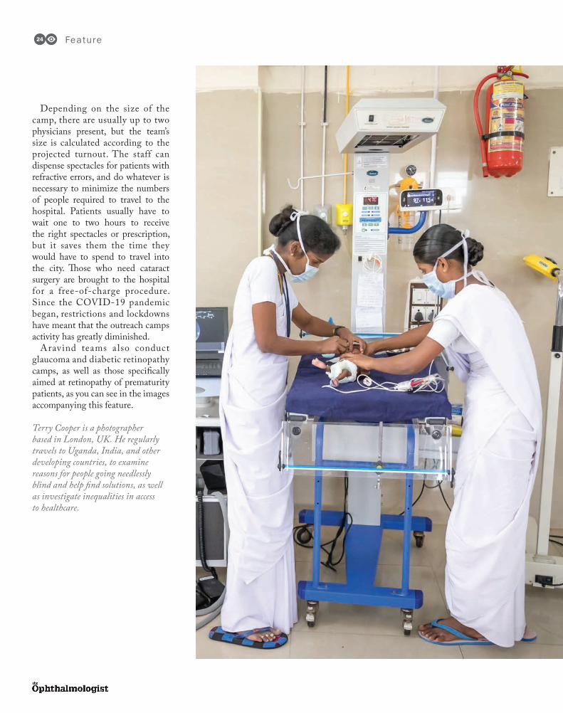
Depending on the size of the camp, there are usually up to two physicians present, but the team’s size is calculated according to the projected turnout. The staff can dispense spectacles for patients with refractive errors, and do whatever is necessary to minimize the numbers of people required to travel to the hospital. Patients usually have to wait one to two hours to receive the right spectacles or prescription, but it saves them the time they would have to spend to travel into the city. Those who need cataract surgery are brought to the hospital for a free-of-charge procedure. Since the COVID-19 pandemic began, restrictions and lockdowns have meant that the outreach camps activity has greatly diminished.
Aravind teams a lso conduct glaucoma and diabetic retinopathy camps, as well as those specifically aimed at retinopathy of prematurity patients, as you can see in the images accompanying this feature.
Terry Cooper is a photographer based in London, UK. He regularly travels to Uganda, India, and other developing countries, to examine reasons for people going needlessly blind and help find solutions, as well as investigate inequalities in access to healthcare.
Feature24

Topcon’s vision is to empower eye care providers with smart, value-driven, and efficient technologies for enhanced care. Keeping pace with the ever-changing landscape of the healthcare industry, Topcon Healthcare offers the latest integrated solutions, including advanced multimodal imaging, vendor-neutral data management, and ground-breaking remote diagnostic technology.
For the past year, world developments have taught us several lessons. We learned that Topcon’s products and service offerings are ahead of their time, so we were fully prepared to offer our customers solutions that fit the demands of unpredictable global events. However, the main lesson learned is that social interactions matter, and we missed personal interactions with our customers: we are thrilled to be able to meet you in person again.
Those visiting this year – either in person or virtually – will be able to see our portfolio solution by themselves, so be sure to stop by!
Booth Number A40
Mely Medel Director, Product Marketing & Marcom / EMEA/APACTopcon Healthcare
Back to Reality Your invite to ESCRS 2021 from eight leading companies who can’t wait to see you there!
After far too long (necessarily) living in a virtual world of Zoom meetings, Microsoft Teams, and Google Chats, many of you will be very much looking forward to catching up – in person – with friends and peers at ESCRS 2021. For those fortunate enough to make the trip, human faces will magically appear in a glorious third dimension alongside a possible sensory overload – the smell of freshly roasted coffee, a background hum of exciting discussions and exchanges, the protestations of your aching feet. For those unable to travel to Amsterdam, the hybrid nature of the event has you covered.
Here, representatives from a number of companies are also thrilled to be able to welcome you in person – or virtually – to this year’s ESCRS.
They’ve worked hard on inclusive programs that leave no one feeling left out. And however you attend, it’s fair to say this will be an ESCRS like no other.

Frank Ziemer President & CEOZiemer Ophthalmic Systems AG
At this year’s ESCRS, we will present the latest Ziemer devices, including i) the FEMTO Z8 NEO – our recently launched multipurpose laser platform for highly precise ophthalmic treatments, ii) our new diagnostic device – the GALILEI ColorZ – now with a Color TopView image, which is designed for greater surgical precision and reproducibility, and iii) our solid-state laser AQUARIUZ – a newly engineered, compact solid-state ablation laser for refractive surgery.
In the future, it will be possible to combine all three devices to create a Refractive Suite to meet all the needs of refractive, corneal, and cataract surgeons.
Internationally-renowned speakers will share their experiences at our Ziemer Satellite symposium, which will be recorded, so surgeons can watch it at their leisure.
The Ziemer booth is a place where surgeons can meet up, reconnect, and lead discussions, but that’s not all: we will offer CLEAR, LASIK, and Cataract wet labs with our FEMTO Z8 NEO Laser, and testing of our GALILEI ColorZ diagnostic device. We have put a lot of effort into the attendee journey for both our live and virtual Ziemer booth participants to ensure that they get the most out of this year’s ESCRS.
Booth Number A22Satellite Symposium:
Saturday, October 9, 11.45 am.
At the 2021 ESCRS Meeting, in-person visitors and virtual guests alike will see and experience a great variety of both new and well-established Teleon products. Indeed, we are excited to present our products through multiple channels – including digitally – tailored specifically to our customers, as we prepare for this year’s ESCRS.
Notably, our product portfolio will be complemented by an educational offering that spans a lunch symposium and several “Meet the Expert” sessions at our booth over the course of the exhibition.
As always, through personal connection and reunion, our customers can expect a fantastic experience with Teleon. In fact, my colleagues at Teleon cannot wait to connect with you again, present our new products and innovations, and offer professional education.
Perhaps even more exciting, we are delighted to invite our customers who can’t attend ESCRS to our International User Meeting. It will be held online, starting at 2pm on Friday, October 8, live from Teleon Surgical’s brand new factory in Spankeren – just one hour away from Amsterdam. For more details, check teleon-surgical.com.
Booth number A21Symposium
“LENTIS Quantum & LENTIS Comfort – Art and science behind the latest EDOF IOL
technologies,” October 9, 11.45 am-12.45 pm.
Mark Lansu CEO Teleon Surgical Group Teleon Surgical

Euan S. Thomson President of Ophthalmic Devices and Head of Digital Business of Carl Zeiss MeditecCarl Zeiss Meditec, Inc.
At ESCRS, visitors will have the opportunity to experience ZEISS’ fully connected environment, where a combination of devices, data management, software applications, and services enables our customers to stay connected to each other and to their patients, supporting a more streamlined workflow and optimizing clinical management. Our digital microscope and imaging systems for ophthalmology produce tens of millions of images each year. With the new ZEISS Medical Ecosystem, we connect our devices and bring that data together in one place.
The ZEISS booth will offer live, first-hand experiences with products along our integrated workflows. There will also be opportunities to meet with ZEISS experts and representatives, as well as hands-on wet lab demos. Our virtual showroom will offer a similar experience with virtual product demos and ZEISS experts available to answer any questions.
Surgeons will experience how the ZEISS Medical Ecosystem, with data managed in the cloud, opens possibilities for the creation of more streamlined workflow solutions. Data passes seamlessly from one ZEISS device to another with AI-powered applications and software tools to help make clinical management of patients easy and efficient.
Booth number D22Symposia: REF Symposium - “The whole is greater
than the sum of its parts – Digital workflows in refractive surgery today” October 9, 12:15-1:15 pm
SUR symposium “Stable Chamber – Stable Outcomes: Next Level Cataract Surgery in 2021”
October 10, 12:15-1:15 pm.
The ESCRS Meeting is a great opportunity for all attendees – both in person and virtual – to learn how iStent inject® W helps improve patients’ quality of life by reducing or eliminating the need for medications – both decreasing IOP and improving ocular surface outcomes. Additionally, we will show how iLinkTM V can slow or halt keratoconus when patients are treated with the KXL® System using VibeX® Rapid riboflavin. We will have our specialist team available to meet you at our booth in a safety-conscious environment.
Our dual lunchtime interactive symposia enable surgeons to attend both days’ seminars and benefit from the experience of international speakers specializing in mild to moderate glaucoma and CXL treatment.
Surgeons visiting the Glaukos booth will learn about our latest technologies for their mild to moderate glaucoma surgical options, and how to best improve their keratoconic patients’ quality of life through our KXL® treatment options combined with our Vibex® product range.
Booth Number A23Symposia: Preserve your Patients’ Quality of Life with iStent inject® W Trabecular Micro- Bypass,
October 9, 11:45 to 12:45pmTransforming the Standard of Care
for Keratoconus with iLinkTM V, October 10, 11.45 am-12.45 pm.
Andrew Geddes Vice President, Commercial – Europe, Middle East & IndiaGlaukos
Hamadi El-Ayari Vice President Sales & MarketingGeuder AG
Geuder is very proud to participate in the first in-person ESCRS after COVID-19-induced lockdowns, which coincides with our company’s 70th anniversary. To mark this milestone, we are inviting visitors and virtual participants to celebrate 70 years of precision and perfection in ophthalmology with us. Together, we will take a peek at the latest innovative products – from cornea to retina.
We hope you come to see us and marvel at the DMEK RAPID, the first CE-certified preloading DMEK device already available at many eye banks, and be thrilled by our vast portfolio of DMEK solutions, such as our ready-to-use EasyGas products and graft fixation. Experience the latest tools and techniques for minimally-invasive transscleral sutureless IOL fixation, especially the Yamane technique, check out the latest phaco pre-chopping and pre-slicing techniques, learn about the new suture-based glaucoma treatment, and see the full spectrum of visible light with the first RGB LED light source, SOLEA.
Booth Number A33 (Hall 12)Geuder’s 70th Anniversary
Milestones of Modern Ophthalmic Surgery
This year, Ace Vision is taking advantage of the hybrid format of the event and to ensure that all visitors will be equally well informed about our technology innovations, Ace Vision has worked hard to prepare both a virtual and live means of keeping abreast of its latest news and innovations.
There will be virtual and in-person opportunities for visitors to this year’s ESCRS Meeting to watch and take part in multiple presentations that introduce the new Laser Scleral Mircroporation procedure the company is developing, including preliminary results, modeling of accommodation, as well as presentations on the manifestation of presbyopia as a universal warning sign for age-related sight loss.
Visitors to Ace Vision’s presentations will also have the opportunity to see the latest innovations in addressing the etiology of presbyopia and to learn about the latest therapeutic solutions to restore the accommodation function of the eye.
AnnMarie Hipsley Founder & CEO Ace Vision Group, Inc.

High Quality Single-Use Instruments
2955C 7698C 7697C 2985C
bvimedical.com
1609204-01BVI and all other trademarks (unless noted otherwise) are property of BVI. BVI ©2021
Providing Sterile Options for Your Operating Room
• Single-use instruments offer protection against infection, due to decreased risk of cross-contamination1
• Surgeons benefi t from precision and reliability of brand-new instruments for each patient2
Also Available in Procedure Packs
• Single-use instruments offer protection against infection, due to decreased risk of cross-contamination
• Surgeons benefi t from precision and reliability of brand-new instruments for each patient
Also Available in Procedure Packs
Your Operating RoomYour Operating Room
References1 Southworth, P.M. Infections and exposures: reported incidents associated with unsuccessful
decontamination of reusable surgical instruments. The Journal of Hospital Infection. September 17, 2014
2 Scheib, Christian; Single-use instruments deserve consideration for eye surgery: Advantages include cost savings, ease of use and high-quality instruments; December 1, 2016
Speak With Your BVI Sales Representative About Malosa® Single-Use Today!
1609204-01 Malosa Single-Use The Ophthalmologist 08-25-21 Final.indd 1 8/25/21 7:48 PM
Steven Thomson Clinical Director Anterior SegmentHeidelberg Engineering
At ESCRS 2021, the main attraction at our booth will be the ANTERION® – a uniquely comprehensive device for optical biometry, IOL power calculation, and much more, which is based on spectacular images of the anterior segment. The high-quality swept-source OCT images enable precise biometric measurements that are fundamental for predictable outcomes in cataract and anterior segment surgery, and many other applications. We are excited to announce that ANTERION’s functionality has now been repeatedly validated by clinicians. At an afternoon ESCRS symposium, Oliver Findl will present the evidence on measurement accuracy and precision, and the new levels of prediction accuracy in challenging eyes will be summarized by Kjell Gundersen and Bjørn Gjerdrum, who will share their results and ideas for further enhancements. We have also worked with Damien Gatinel and Alan Saad to ensure that ANTERION data combined with SCORE, a corneal ectasia risk analysis application, has the potential to set new standards.
Beyond the benefits of ANTERION, surgeons and clinicians attending ESCRS will be able to hear about the latest SPECTRALIS® features and our healthcare IT solutions.
Booth Number B22ANTERION Symposium delivered by Oliver
Findl, Kjell Gunnar Gundersen, Bjørn Gjerdrum, and Damien Gatinel
Friday, October 8, Hall 11, 3:45-4:45 pm (also live streamed)

NextGen30
Dry AMD and geographic atrophy (GA) are subsequent stages of the same condition, with no current therapeutics available to stop the irreversible progression to blindness. For patients, it is an arduous wait in the hope that an effective treatment will be approved and save their eyesight. So how can we identify a treatment for this disease before it’s too late? Could the answer be a drug that already exists – but has not yet been applied to this condition?
Repurposing drugs that are already approved for other applications could knock years off the drug development pipeline – and millions of dollars off the cost of early-stage pre-clinical and clinical trials. My team and I have discovered that dry AMD and GA can be treated by repurposing the HIV drug class nucleoside reverse transcriptase inhibitors (NRTIs) (1) and we will be performing clinical trials for this purpose. This ties in with our discovery that Alu DNA causes death of the retinal pigment epithelium (RPE) and – crucially – is not produced when NRTIs are administered.
Digging for answersThis discovery tracks back to 2014, when we originally published research showing that NRTIs can block a
Serving One’s (Re)PurposeRepurposing HIV drugs may be an effective and time-efficient route to treating dry AMD By Jayakrishna Ambati
NextGenResearch advances
Experimental treatmentsDrug/device pipelines

NextGen 31
mouse model of dry AMD in mice (2) – a completely different application to NRTIs’ original purpose. All we knew at the time, in terms of the mechanism of action, was that these drugs inhibit inflammatory activity, which led us to investigate them further and decipher more details. Fortunately, the advent of big data archeology – which mines large databases of patient records for drug-disease associations – enabled us to test our hypothesis. The analysis of four distinctly different databases from both public and private health insurance carriers supported our notion that NRTIs are beneficial to treatment of dry AMD. We also identified (through big data archaeology) that Alu DNA, which we had previously identified as present in high levels in dry AMD
eyes, was made in the cytoplasm. These findings are the culmination of work that started a decade ago and drove us to seek a deeper understanding of the biology of the disease.
NRTIs work by two mechanisms: blocking reverse transcriptase and blocking the inflammasome of the immune system. Blocking reverse transcriptase is beneficial to dry AMD because it stops the conversion of Alu RNA to Alu cDNA, preventing the toxic Alu effect that causes RPE death in GA. The second mechanism of action, blocking the inflammasome, reduces the damage caused by the immune response to the disease – further increasing retinal protection. The combinat ion of these t wo mechanisms helps to explain NRTIs’
tremendous beneficial effects against dry AMD.
NRTIs are not perfect; they can be pretty nasty drugs, especially the early versions. Newer generations have been more tolerable, but still feature side effects that HIV patients tolerate due to the drugs’ survival benefits. Dry AMD patients are mostly middle-aged or elderly and may stand to lose their sight – but not their lives – so may be less tolerant of side effects. This led us to create NRTI derivatives – called Kamuvudines – that retains the beneficial aspects of the drug, but lacks its toxicity. It is these derivative drugs that we are now taking forward into clinical trials.
Looking to the futureThere are multiple benefits to repurposing
www.haag-streit.com
HAAG-STREIT SLIT LAMP10 Year Limited Warranty
The excellent optics and the superior mechanics make a Haag-Streit slit lamp one of the best investments you can ever make for your practice.

a well-classified drug class with copious amounts of real-world data. When starting from scratch, drug development may take 12 to 15 years and cost anywhere from US$2–3 billion to bring to market. This takes into account the immense failures inherent in this process; many drugs fail the expensive tests along the development pipeline, sometimes even when they are potentially beneficial, but may cause adverse effects. Repurposed drugs have already achieved this extensive, expensive, and time-consuming stamp of approval. Because they are already used in patients, we know that they meet reasonable safety and tolerability standards. We know the dosing regimen and pharmacokinetics of the drug. All of these factors shorten the
runway for launching a clinical trial and may allow researchers to skip phase I and II clinical trials (designed to establish safety) and initiate phase III trials as a first step.
The discovery of DNA synthesis in the cytoplasm also opens up a new chapter in biology and disease, teaching us that DNA is not produced in the nucleus alone. Our new understanding of Alu is similarly enlightening – with evidence that Alu is involved in the pathology of Alzheimer’s disease, multiple sclerosis, lupus, and several other diseases, it is perfectly reasonable to hypothesize that these diseases may see a similar benefit from Alu-targeting treatments. In fact, we are actively looking at wider disease applications in addition to dry AMD
– and our early results are intriguing.In addition to this work, I’ve formed
a company called Inf lammasome Therapeutics that has licensed this technology. Our next step is to run clinical trials through the company for dry AMD and some other diseases, including systemic conditions, to bring this drug into use. As the world slowly begins to move past COVID-19, we hope to advance our clinical trials as soon as possible.
Jayakrishna Ambati is Professor and Vice Chair for Research, and Founding Director of the Center for Advanced Vision Science at the University of Virginia, Charlottesville, Virginia, USA.
Disclosures: Jayakrishna Ambati is a co-founder of Inflammasome Therapeutics, iVeena Holdings, iVeena Delivery Systems, and DiceRx, and has been a consultant for Allergan, Boehringer-Ingelheim, Olix Pharmaceuticals, Retinal Solutions, and Saksin LifeSciences unrelated to this work.
See references online at: top.txp.to/repurpose
NextGen32
“When starting from scratch, drug development may
take 12 to 15 years and cost anywhere
from US$2–3 billion to bring to
market.”

Axial Length
Total Wavefront
Refraction
Scheimpflug Tomography
Retroillumination
Pentacam® AXL WaveYou position the patient. The new full sequence measuring assistant handles the rest.
Thanks to the newly developed measurement workflow and the automatic quality check, you are always on the safe side. Optimized workflows, satisfied patients, and the best possible clinical results: always achieved quickly, reliably, and without long training.
No risk, just fun – the new Pentacam® AXL Wave
Makes a Complex Process Simple, Automatic and Reliable
www.pentacam.com/axl-wave
The
avai
labi
lity
of p
rodu
cts
and
feat
ures
may
var
y by
cou
ntry
. O
CULU
S re
serv
es th
e rig
ht to
cha
nge
prod
uct s
peci
ficat
ions
and
des
ign.
Join us at the Satellite Meeting 9 October 2021 at ESCRS in Amsterdam or online!
The Ophthalmologist Pentacam AXL Wave Complex Simple 210x266 e 08.21.indd 1The Ophthalmologist Pentacam AXL Wave Complex Simple 210x266 e 08.21.indd 1 10.08.2021 18:55:3010.08.2021 18:55:30
Structurally SoundHow photoacoustic imaging may revolutionize ophthalmic imaging and diagnosis By Parsin Haji Reza
What could you do with better imaging? With better information at your fingertips, how many more ocular diseases would be diagnosed earlier – before damage became irreversible? I hope to help you find answers to these questions with the photoacoustic remote sensing microscopy (PARS) technology developed in my lab; PARS enables structural and functional imaging of the eye – with high resolution.
Given its strengths, there is virtually no limit to the potential applications of PARS in ophthalmology. We can use it for early diagnosis of most blinding diseases
and anything else that can be detected based on functional information, oxygen saturation, oxygen metabolism, and blood flow. And we can measure this information accurately down to a single capillary or – in some cases – down to a single red blood cell. This ability may extend to measuring the thickness of the RPE layer and melanin concentrations. We are also able to take advantage of the various optical absorptions of different contrast agents and drugs – thereby enabling measurement of drug concentrations in the eye, which could open a new field in pharmaceutical development.
Making wavesPhotoacoustic imaging, in general, uses light energy to generate ultrasonic sound waves in a sample – the waves can be translated into an image – much like the ultrasound technology we’re all familiar with.
So why isn’t it already being used for eye imaging? Probably the biggest issue is the need for direct contact, with both gel and transducer touching the eye; needless to say, this is not ideal – both because of patient discomfort and the increased risk of infection from the physical contact.

Furthermore, contact-based imaging directly affects the balance of vascular function and oxygen diffusion from the pressure applied to the eye, which means we are unable to study dynamic processes under conditions that are close to normality.
PARS uses an excitation laser to generate sound waves that are collected by a novel remote laser sensor that completely avoids physical contact, and collects the initial pressure right at the source. A traditional ultrasound transducer can only collect sound waves that travel to the surface of the tissue, requiring constant physical contact. To put the process in other words, when you throw a stone into a lake, shockwaves or initial pressures are generated from the moment that the stone hits the water, and these waves travel to the shore. The further you are from the place of impact, the less pressure you detect in the waves – as is the case with some transducer and traditional photoacoustic imaging. In PARS, instead of collecting the waves that have travelled to the shore, we only collect the initial pressures – the immediate pressure in the first few hundred nanoseconds after the stone (laser) hits the water (eye).
In short, PARS gives us a non-contact imaging method that, in addition to optical absorption, can provide additional imaging contrast, including scattering.
Better togetherPARS is a powerful technique but, by harmonizing it with OCT, we are able to access the best of both worlds. PARS provides accurate measurements directly from optical absorption and OCT provides optical scattering, so they work in concert with each other. PARS can also provide optical scattering (and a few other light-matter interactions), but OCT is the gold standard in the field of ophthalmology – and the images are easy to understand – so we wanted to make sure that PARS images were collected from the same exact location to provide verification for what we are imaging and how we are imaging it. In future, PARS will be able to function as a stand-alone technology and for many applications OCT won’t be needed.
The imaging penetration depth is related to the wavelength and the tissue type we are looking at – which makes the eye especially advantageous. With visible wavelengths, we cannot penetrate more than two or three millimeters into scattering tissues. As the eye is transparent to most of the wavelengths we use, there is no limit to the depth we can achieve. We can look at the retina and the structures at the back of the eye easily. The RPE layer may bring a unique challenge given that the role of this tissue is to absorb light – and that may limit the ability to image the choroid and beyond. As most of the light will be absorbed by the RPE layer, not enough photons will get through to generate the photoacoustic waves that create the image; however, we are actively working on techniques to overcome this.
EYE SURGERY. SWISS MADE.
The new OS 4 marks the beginning of the next generation of retina, glaucoma and cataract surgery. The all-in-one platform has received numerous exciting features that provide even more comfort, precision and safety.
Not
ava
ilabl
e fo
r sal
es in
the
US
*Oer
tli d
ata
on fi
le
Laser integration: More safety, fully automated userprotection filter
Light: 45% more power*, maximum visibility
Pedal: Multifunctional withover 100 setting options
Phaco: Speedier readiness, greater controllability
User comfort: Even more user- friendly and communicative
Make the difference – with the new OS 4: www.oertli-instruments.com
OS 4TM
THE NEXTGENERATION
NEW FEATURE
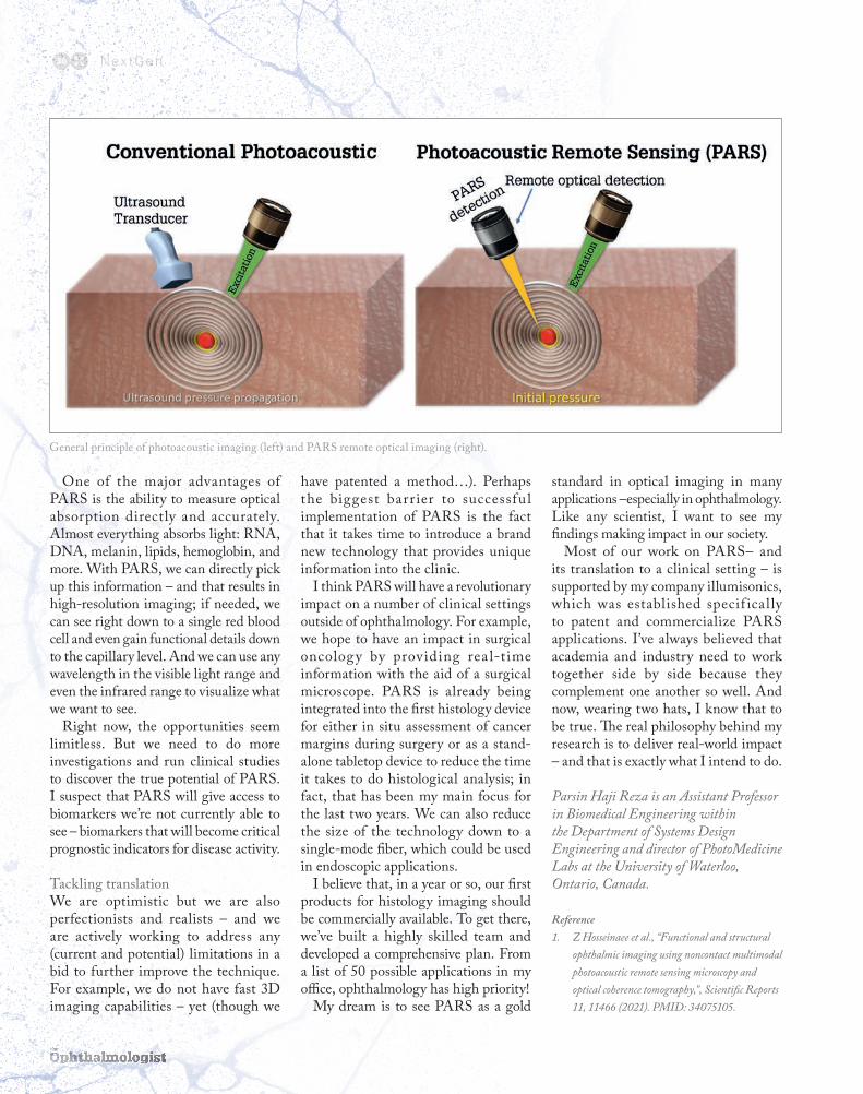
NextGen36
One of the major advantages of PARS is the ability to measure optical absorption directly and accurately. Almost everything absorbs light: RNA, DNA, melanin, lipids, hemoglobin, and more. With PARS, we can directly pick up this information – and that results in high-resolution imaging; if needed, we can see right down to a single red blood cell and even gain functional details down to the capillary level. And we can use any wavelength in the visible light range and even the infrared range to visualize what we want to see.
Right now, the opportunities seem limitless. But we need to do more investigations and run clinical studies to discover the true potential of PARS. I suspect that PARS will give access to biomarkers we’re not currently able to see – biomarkers that will become critical prognostic indicators for disease activity.
Tackling translationWe are optimistic but we are also perfectionists and realists – and we are actively working to address any (current and potential) limitations in a bid to further improve the technique. For example, we do not have fast 3D imaging capabilities – yet (though we
have patented a method…). Perhaps the biggest barrier to successful implementation of PARS is the fact that it takes time to introduce a brand new technology that provides unique information into the clinic.
I think PARS will have a revolutionary impact on a number of clinical settings outside of ophthalmology. For example, we hope to have an impact in surgical oncology by providing rea l-t ime information with the aid of a surgical microscope. PARS is already being integrated into the first histology device for either in situ assessment of cancer margins during surgery or as a stand-alone tabletop device to reduce the time it takes to do histological analysis; in fact, that has been my main focus for the last two years. We can also reduce the size of the technology down to a single-mode fiber, which could be used in endoscopic applications.
I believe that, in a year or so, our first products for histology imaging should be commercially available. To get there, we’ve built a highly skilled team and developed a comprehensive plan. From a list of 50 possible applications in my office, ophthalmology has high priority!
My dream is to see PARS as a gold
standard in optical imaging in many applications –especially in ophthalmology. Like any scientist, I want to see my findings making impact in our society.
Most of our work on PARS– and its translation to a clinical setting – is supported by my company illumisonics, which was established specif ically to patent and commercialize PARS applications. I’ve always believed that academia and industry need to work together side by side because they complement one another so well. And now, wearing two hats, I know that to be true. The real philosophy behind my research is to deliver real-world impact – and that is exactly what I intend to do.
Parsin Haji Reza is an Assistant Professor in Biomedical Engineering within the Department of Systems Design Engineering and director of PhotoMedicine Labs at the University of Waterloo, Ontario, Canada.
Reference1. Z Hosseinaee et al., “Functional and structural
ophthalmic imaging using noncontact multimodal photoacoustic remote sensing microscopy and optical coherence tomography,”, Scientific Reports 11, 11466 (2021). PMID: 34075105.
General principle of photoacoustic imaging (left) and PARS remote optical imaging (right).

www.newworldmedical.com
Beyond Comparison Experienced surgeons discuss excisional goniotomy using the Kahook Dual Blade – and make comparisons with the iStent inject
Mario Economou, Consultant Ophthalmologist at Queen Sofia Hospital and Praktikertjänst in Stockholm, Sweden, and Achyut Mukherjee, Consultant Ophthalmologist at Oaks Hospital and East Suffolk and North Essex NHS Foundation Trust, Colchester, UK, describe their experience with the Kahook Dual Blade® (KDB) and offer a comparison with the iStent inject®.
Economou started using the KDB and iStent inject in 2016 and has since incorporated them in his clinical practice, using both regularly in combination with cataract surgery or as a standalone procedure. He says: “I use both the KDB and the iStent for ocular hypertension and mild to moderate glaucoma patients. I have also successfully used the KDB in advanced glaucoma when a trabeculectomy is not an option for various reasons. I think that canal-based procedures, such as KDB goniotomy and iStent inject, are safe and effective options for managing glaucoma and should be offered as a first option in appropriate patients. They can produce sufficient IOP reduction for a large portion of glaucoma patients and/or reduce the number of glaucoma medications without compromising safety. I would say that surgical glaucoma treatment needs to become more stepwise, allowing an early intervention with the canal-based procedures that would allow for a more optimal IOP level for a longer period of time
with less dependence on therapeutics.”Mukherjee was one of the first surgeons
in the UK to integrate the KDB into their practice back in 2016, and has since used it in a wide range of open angle glaucoma, primary angle closure, and secondary glaucoma patients. He comments: “I have developed a modification to the KDB
procedure using indirect gonioscopy to extend the treated area, which
my team has called KDB-XL, and which enhances the effect of the KDB procedure.” He has also been using the iStent, starting with the iStent inject
and more recently moving to the iStent inject W.
Hard dataEconomou and Thorsteinn Arnljots recently published data retrospectively analyzing the results of procedures
using KDB or iStent inject as a standalone procedure or in
combination with cataract surgery (1). He comments: “Both the KDB and the iStent, when combined with cataract surgery, showed to be effective in reducing the IOP by more than 20-30 percent while also reducing the number of glaucoma medications. The KDB as a standalone procedure reduced the IOP by 28 percent, which is very useful information when offering a new surgical option for patients who are already pseudophakic and have uncontrolled IOP. The study also showed that the KDB had a good effect on eyes with pseudoexfoliative glaucoma, which is often considered to be more difficult to treat. Both the KDB and the iStent inject proved to be very safe procedures. IOP spikes were very rare, while hyphemas self-resolved within a few days.”
Mukherjee’s group recently published clinical results of an extended ab interno trabeculotomy using the KDB, and found that 77 percent of all eyes treated with phaco KDB achieved IOP-related success
and reduced medication, compared with 5 percent of eyes treated with just phaco (2).
Patient selectionMukherjee sees the procedures using the KDB and the iStent inject as appropriate to different patient groups, based on several important differences. In his experience of working with both devices, the KDB lowers the IOP with a greater effect. He uses it up to a 180 degree angle (using indirect gonioscopy with extended KDB-XL), whereas his iStent procedures are usually limited to 40-70 degree angles. In terms of corneal opacity, the KDB requires a good angle view, whereas iStent can be safely performed when the cornea limits the view. The risk of hyphema is, in his experience, greater with the KDB, but hyphemas usually resolve spontaneously. The iStent has a shorter operative time – a few seconds in many cases – whereas with the KDB it is longer, which may be problematic in less cooperative patients.
From one surgeon to anotherEconomou asserts that his experience with the KDB and the iStent inject is very positive. He elaborates: “Since the introduction of these devices, my clinical and surgical armamentarium has been enriched and I can now offer these two options to my patients with ocular hypertension, mild to moderate glaucoma who also have cataracts and have either a suboptimal IOP or are on glaucoma drops that have negative side effects or compromise compliance.”
Mukherjee concludes: “In my experience, the KDB is highly efficacious in a wide range of glaucomas. My clinical experience is that the IOP lowering effect is greater than that of trabecular bypass stents.”
References
1. TS Arnljots, MA Economou, Clin Ophthalmol,
15, 541 (2021). PMID: 33603332.
2. M Hamza et al., Medical and Research
Publications (2021). Available at:
https://bit.ly/3hmmEQV.
Sponsored Feature 37
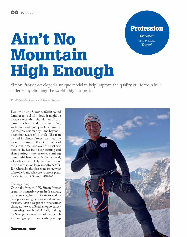
Does the name Summits4Sight sound familiar to you? If it does, it might be because recently a foundation of this name has been making some noise, with more and more people within the ophthalmic community – and beyond – becoming aware of its goals. The man behind it, Simon Prosser, has had the vision of Summits4Sight in his head for a long time, and over the past few months, he has been busy training and then putting it into practice climbing some the highest mountains in the world, all with a view to help improve lives of people with vision loss caused by AMD. But where did the idea come from, what is involved, and what are Prosser’s plans for the future of Summits4Sight?
The beginningsOriginally from the UK, Simon Prosser spent his formative years in Germany, before moving back to Britain to work as an application engineer for an automotive business. After a couple of further career changes, he was offered an opportunity of entering the ophthalmic field, working for Synergetics, now part of the Bausch + Lomb group. He successfully set up
Profession38
Ain’t No Mountain High EnoughSimon Prosser developed a unique model to help improve the quality of life for AMD sufferers by climbing the world’s highest peaks By Aleksandra Jones, with Simon Prosser
ProfessionYour career
Your businessYour life

Profession 3939
the company’s European office and helped build its business. He then began working for DORC – Dutch Ophthalmic Research Center International – in Germany; he spearheaded the company’s US operations in Dallas, and worked in South America ahead of and immediately after the company’s sale to Montagu. After a stint at Vitrek, the time came for him to look for something he felt really passionate about. He began working for Eye Tech, a company developing eye tracking systems for people with disabilities, which at the time started to branch out into ophthalmology and neurology – two areas he was familiar with – where he met Michael Freeman, the CEO and Founder of Ocutrx, a company Prosser now works for.
At Ocutrx, he became involved in the company’s project using a wearable augmented reality (AR) device to help people with severe central vision loss due to
AMD. Freeman, whose father had suffered from AMD at a time when no treatment was available, was inspired to try to develop new capabilities for people afflicted with the condition. He sought Prosser out thanks to his experience with eye tracking systems, as well as retinal surgery, and asked for his help in bringing the AR vision aid to market, and designing a surgical platform.
As Prosser had already had an idea to found a non-profit that would fundraise money to help people by climbing peaks, making Ocutrx’s AR technology available to as many AMD sufferers as possible seemed like the perfect cause to focus on. He became the company’s Global Director, working on developing its surgical platform, and he has also been working on developing the foundation to distribute the Ocutrx platform widely to people who need it – Summits4Sight, a foundation that Ocutrx is now also supporting financially.
The causeProsser began to look into “giving back” and setting up a charitable foundation a while ago, and over the months and years the idea started to take shape in his head. While he saw many organizations and foundations focused on funding research in the field of retina, there
“Prosser saw an opportunity in
bringingtogether existing low
vision centers andgroups.”
www.theophthalmologist.com

Profession40
wasn’t much that focused directly on patients, and improving their quality of life immediately. The latest technologies improving quality of life for people with vision loss are not usually commercially available to all those who need them. Selected few wealthy individuals can access certain resources, but most useful innovations are not available to most people as they are not affordable and/or not covered by insurance.
Prosser saw an opportunity in bringing together existing low vision centers and groups, and developing criteria to select people who would benefit from using the technology Ocutrx has been working on, at no cost to themselves. This is still work in progress, and for now, Prosser is focusing on raising awareness of the cause in order to raise funds to purchase the needed technology from interested, relevant vendors – including Ocutrx – at production cost, to maximize the technology’s future reach and availability. The idea is to fund real-life
solutions that improve quality of life, which low vision centers and groups will be able to distribute among their members, according to their needs.
The fundraisingProsser is looking for the ophthalmic industry’s wide support, with many companies already helping out with donation. His network, built up over 20 years or more, includes many physicians and leading industry figures. What’s more, some automotive and engineering companies he worked with at the beginning of his career are also involved in supporting the foundation, without the perspective of gains in the ophthalmic field. Some wealthy individuals in the US and in Europe are throwing their weight behind him, so the initial goal of raising US$ 1 million seems well within reach in the first several months of the fundraising efforts. This could help hundreds of people, with many more that could be supported over the coming years.
For now, it has been a tough job of navigating the legal and administrative issues between intensive daily workouts, and climbing training t weekends. It would be made easier with extra paid help, but Prosser is determined for that
The peaks Prosser is planning to climb as part of this fundraising campaign.
“It was a fantastic experience. I
wanted to prove to myself that I could
live up to the expectations of all
the people who have supported me,
trusted me.”
not to impact the fundraising total before absolutely necessary. The four climbs are already fully-funded, and Prosser has made sure that the necessary funds to begin the distribution of the device to AMD groups in the fourth quarter of 2021 are in place.
The preparationsThe ambitious plan of conquering the “summits” of Summits4Sight means that when Prosser completes it, having climbed the four mountains he is attacking, he will have climbed twice the height that he has ever climbed before – and he has already done his fair share. As the mountain heights increase, so do the demands and dangers of the climbs.
Prosser first came to contact with rock and mountain climbing as a young child, through his father, although he didn’t enjoy it to begin with. He then became a semi-professional skier, and fell in love with mountains and hiking, climbing Alpine peaks when living in Europe. In the last decade or so, he has turned it into a real passion, and traveling with work allowed him to explore mountains in different parts of the world, including South America, in his free time. As a result, he had a few close calls, and has become accustomed to pushing his limits.
To develop his Summits4Sight plan, he got in touch with the International Mountain Guides (IMG) who helped him come up with an appropriate training schedule and advice on staggering the climbs, so that the whole idea of reaching the four peaks became realistic. As he points out, he has never been a professional alpinist and spent most of his life in an office or an OR, so this project is way out of his comfort zone and requires a lot of preparation.
In September 2020, Prosser embarked on a strict training and diet regimen. He has been very active throughout his life – apart from skiing, he is also a keen diver, and he got involved in bodybuilding, so he already had the knowledge of nutrition, appropriate training, and the discipline required. He typically trains three to four hours a day, shared between treadmills, cross trainers, stairs, and bicycles, and when he’s not on the machines, he’s training in the mountains, both in the US (in Arizona and Colorado) and in Switzerland. The pandemic has occasionally put a spanner in the works with imposed quarantines – and Prosser, who only takes a day off every week from his training, gets a bit anxious if he misses any more of it – but it also allowed him to focus on his training while staying at home for months, without the feeling of missing out on much happening outside.
The first climbFollowing this intense training, which luckily didn’t result in any injuries, in April 2021, Prosser made his first trip to Nepal,
www.optotek-medical.com
OptoSLT nanoSophis�cated Technology Evolved under Alpine Peaks
pulse to pulse stability: DPSS vs. conven�onal

Profession42
to climb Lobuche. He was prepared to have to quarantine for 10 days upon his arrival, but the requirement was dropped 24 hours before his planned arrival, which really helped keep his fitness levels up. He made it to the base camp three days ahead of schedule and didn’t experience any altitude sickness – a success he ascribes to his training in high peaks of Arizona ahead of the Nepal trip. As the weather window was coming to an end, with a storm coming, it was the last opportunity to reach the summit. On April 21, at 7.30 am, after a record ascent time, Prosser and his team reached the summit of Lobuche. He says: “It was a fantastic experience. I wanted to prove to myself that I could live up to the expectations of all the people who have supported me, trusted me, and made this climb happen, so to get to the top without a struggle meant a lot to me. On the way down, I saw people using
Lobuche as training ahead of climbing Everest, and even though they were struggling, they were still planning to attempt the world’s highest peak in a few weeks’ time – something that shocked me, as I don’t consider it to be a responsible approach.” It took the team around 3.5 hours to get down to the high camp, where they were going to rest for a few hours and “charge the batteries,” but the guides, seeing the rapid change in the weather, insisted on coming down much faster. Prosser continues: “Immediately after leaving the camp, snow started coming in from all directions. I was wearing all my hard-shell protective clothing, with just a little gap for my eyes, but the snow still found its way in and crammed in my eyes. I had no clue where the path was, and there were huge drops on either side. Luckily, with the Sherpa guides’ experience, we made our
way down, although it took us two days to make the descent.” Having gained five days on his original schedule, Prosser then had a wait for his return flight from Kathmandu, with the added difficulty of having to get PCR test results ahead of flying home at a time when the city was going into a new lockdown.
Most of the corporate sponsors
“Immediately after leaving the camp,
snow started coming in from all
directions.”

C
M
Y
CM
MY
CY
CMY
K
100x266 AAFF.pdf 1 26/07/2021 8:51:58
who have already agreed to support the foundation have guaranteed a base sponsorship, but they are also reserving bonus sponsorships for reaching the summits, which is a real incentive for Prosser to push to his absolute limits, as he knows that not making the summit will cost the foundation money, which would ultimately limit the number of people he can help. This pressure keeps him going, both during his training, and during the climbs, and ensures that he plans things well. He trusts the IMG’s guidance – the guides have helped hundreds of people get to the top of Everest and have never lost anyone, and even though the risks that high up the mountain are always substantial, they can be mitigated with good preparation, team work, and knowing how hard one can and should push themselves.
As Prosser says himself, the biggest challenge for him are not the climbs itself: it is getting enough people on board to make Summits4Sight as successful as it can be, and exceed everyone’s expectations. He has been off to a great start, having already secured good funding to see the project through, and being able to stick to his climbing schedule, so he is convinced that the foundation is going to make a big difference.
Still, he does have a level of fear of the climbing itself – healthy fear, as he says. At any point during a climb, many things can go wrong, and the threat to life is constantly present. Only a naïve person would go into these circumstances with no fear, and it is necessary to make sure you don’t make a mistake that could cost your or somebody else’s life.
The futureFor now, between Arizona and Switzerland, Prosser is busy training for the next scheduled climb – Putha Hiunchuli, for which the expedition will begin at the end of September. For this, together with his guide, he decided to make his own team, rather than attach himself to an existing one. This means having to organize and fund the logistics – down to booking and paying for eight yaks. The normal time to climb the peak is 31-34 days, and Prosser will attempt to do it in 24 days, trying to break another speed record.
Once he’s done all the four climbs (see the graph below) he has planned, is that going to be it for Prosser’s work on Summits4Sight? It seems very unlikely, as he already has fresh ideas, such as going to Antarctica, and perhaps writing a book about his experiences, to raise even more funds. It seems obvious that after this chapter, Summits4Sight will carry on helping the AMD patients with vision loss, but, as Prosser comments, he is focusing on his current goals and not looking too far ahead – the plan is ambitious in both the climbing scope, and financial targets, and it needs his full attention to come to fruition.

Profession44
In Zambia, we have a blindness prevalence of two percent. One person’s preventable blindness is a big problem – so the Zambian figure is a massive problem and a clear unmet need. The biggest reason for this is a lack of trained eye care staff in the country, which often results in waits of months or years before treatment becomes available. Although the government can sponsor a patient’s travel to another country for
treatment, this is expensive – and money is not always available. Other barriers include myths and misconceptions regarding eye health, with education and poverty the major limiting factors in people’s looking after their own eye health and seeking medical treatment when necessary.
I work at ORBIS International as country director for Zambia. The role consists of managing our work portfolio
in comprehensive and pediatric eye health, with a focus on adult cataracts and glaucoma prevention. ORBIS is an international non-profit organization that aims to treat and prevent avoidable blindness and has been working in Zambia for just over a decade now – with our work located in three of the country’s 10 provinces. This started in the northern area on the copper belt, working out of a tertiary facility that has grown from
Zambia’s Fight Against BlindnessORBIS International is working to combat unnecessary blindness across Zambia By Lucia Nadaf

Profession46
a single unit to an entire eye hospital. Initially providing pediatric eye health services, it then became a training facility for upcoming ophthalmologists, as well as strengthening the faculty through quality ophthalmic training and providing free treatment for children. Now, its focus has grown to include strengthening existing centers – with equipment, supplies, and training referrals for staff, – and setting up new ones. These improvements have enabled centers to work at full capacity, rather than opening only one or a few days a week. General physicians and nurses at primary health care centers have also been trained in basic eye health, so that eye conditions can be recognized in their early stages and either treated there or referred to a center that can assess further or treat the patient. District health facilities and community volunteers have also been trained to identify cases so that they can refer children to eye care centers if they see an issue. Community outreach is very important to ORBIS. We do this in several ways, but we try never to lecture, because we don’t want to be “in charge”; we want to form partnerships and work with the community for the betterment of eye health. Eyesight is
crucial to education and people’s ability to provide for their families – so the work we do is important.
My work at ORBIS centers on treating and preventing blindness. Working with the ministries of health in multiple countries, we aim to build their capacity to provide quality eye care – through training, equipment, and medical supplies. Additionally, we work with these communities directly; the figures that appear in reports and the eyes that we treat are people and families, so our work needs this interaction and engagement.
Pandemic eye careOphthalmology was hit hard by COVID-19 (as we’re all aware), but what about countries who already had a disproportionately high rate of blindness and eye health issues before the pandemic hit? Working through COVID-19 has required lots of adaptation in our processes. Keeping our staff, partners, and communities safe has been important, but it has also meant that we haven’t been able to see patients as usual – especially during the first and second waves, when we were only able to address emergency cases. This has had a major impact on our previous work to increase awareness of eye health in Zambian communities, with many reverting back to old habits – not looking after their eyes and avoiding coming into the clinic if issues arose.
The pandemic has forced us to be more innovative with our work. Because we can’t be in the communities, we’ve had to ride with ongoing government programs along the copper belt – often using COVID-19 programs as a vehicle to do our eye care work. In parts of northern Zambia, for instance, COVID-19 and eye health screening have been added to the standard child health activity programs. This has extended to training midwives and general nurses in basic eye health so that they can identify cases
or risks in a child or mother. Another way we’ve adapted to the situation is by taking advantage of Zambia’s widespread radio to broadcast our messaging about eye health and COVID-19. We have also produced educational material on handwashing and what to look for in terms of eye health; this has enabled us to educate the community
indirectly on eye care and the link between COVID-19
and conjunctivitis.The work also
stretches out to communities with no access to health services, where people would go needlessly blind
without our help. One such community
is an older population, mainly farmers who only
grow food for their own consumption and therefore don’t earn an income. For them, travel isn’t an option, so we developed a project to allow an ophthalmic professional to meet them and educate the community on eye health through screenings and treatment, including surgery.
We also have staff working on the frontlines. Two ophthalmic clinical officers working on the copper belt border between Zambia and the Democratic Republic of the Congo (DRC) have been screening truck drivers for both COVID-19 and eye conditions. There is also an eye health center on the copper belt where both COVID-19 and eye health are being screened.
We have worked hard to provide education (to both local communities and healthcare professionals), eye screening, and treatment to the people of Zambia – it is my hope that this eventually translates to the eradication of unnecessary blindness for the country’s population.
Lucia Nadaf, Orbis International Country Director for Zambia, Lusaka, Zambia.
“Keeping our staff, partners, and communities safe has been important, but it has also meant that we haven’t been able to see patients as usual.”

Optimized performance
Smallest details resolved
Upgradeable platform
2006
13-0
01 IN
T.A
E21
© H
eide
lber
g En
gine
erin
g G
mbH More than meets the eye
Every new component contributes to the powerful synergies in the next generation SPECTRALIS multimodal imaging platform.The changes may only be recognizable on the inside, but the improved workfl ow is unmistakable.
Experience increasing effi ciency without sacrifi cing image quality. Benefi t from outstanding performance while preserving existing data.
Uncover more detail: www.he-lounge.com

PolyPhotonix, the manufacturer of the Noctura 400 – a treatment for diabetic retinopathy – recently completed a real-world study at an NHS hospital in the UK, which began in 2019. The study analyzed the effects of enhanced patient interaction with and use of the Noctura 400 sleep mask in a group of 26 diabetics displaying diabetic retinopathy (DR) and diabetic macular oedema (DMO).
The results of the study were remarkable: 94 percent of the patients who completed the study saw clinical improvement or stability. But the results are even more impressive when considering that the patients taking part in the study were a vulnerable population advised by the UK government and the NHS not to attend hospital for treatment during the COVID-19 pandemic.
The purpose of the studyThe study aimed to find if patient compliance could be maintained and determine the anatomical and functional consequences of consistent mask wear. Throughout the whole study, outcomes were positive, with a high level of consistent patient use of the mask, above
74 percent up to and beyond one year. Even during the first COVID-19 lockdown in England, patients maintained a 65 percent nightly light mask compliance. Statistically significant improvements in maculopathy and visual acuity (VA) were maintained to the end of the study (see details below). Anatomical improvement or stability was recorded in all but one of the study eyes. Moroever, the investigation shows that, given appropriate interactions with patients who are self-treating in a home environment, a high level of patient compliance can be maintained – even with disruptions to the normal hospital clinic setup.
Mechanism of action The Noctura 400 Sleep Mask prevents dark adaptation in the rod cells, which reduces the oxygen demand of the eye at night, reducing retinal hypoxia – a strain that not only affects choroidal circulation, but also compromises retinal vessels, potentially leading to DR and DMO.
The mask features an organic light emitting diode (OLED) housed inside a soft cushioned fabric mask – designed, of course, to be worn at night – that
administers a precise dose of low-intensity light of a specific wavelength during a patient’s normal hours of sleep every night as part of a continuing therapy. Importantly, the mask also measures patient compliance; at the end of the allocated period, the mask is returned for analysis and a replacement is provided. Under normal circumstances collection and replacement happens in the clinic, but during pandemic-related disruption, new masks are posted to patients. The compliance data allows clinicians to compare how regularly the mask has been worn, which can then be matched with changes in vision and disease status.
Currently, DR is treated with laser and/or injections of anti-VEGF pharmaceuticals directly into the eye. These treatments are expensive, demand substantial clinician time, and require patients to attend clinics on a regular basis. Visual healthcare and eye department budgets
Sleeping Off Diabetic RetinopathySharing results from a real-world single center study of patients with diabetic macular oedema (DMO) who wore a home-use sleep mask (Noctura 400) for 12 months
By Ulrich Meyer-Bothling, Consultant Ophthalmic Surgeon, Ashford and St Peter’s Hospitals NHS Foundation Trust,and Ian Grierson, CCO of PolyPhotonix, Emeritus Professor of Ophthalmology, University of Liverpool, UK
Sponsored Feature48
www.noctura.com
Journal of Ophthalmology - https://www.hindawi.com/journals/joph/2021/6612126/

Sponsored Feature 49
of many countries have been stretched by the success and rapid expansion of anti-VEGF injection therapy for DMO. But it is also true that anti-VEGF injections and steroid implants are not without side effects and some level of patient distress. There is, therefore, a need for earlier treatment of limited-to-zero risk that should preferably be moved away from the clinic to the home – especially during COVID-19 outbreaks.
Study resultsA total of 26 patients were recruited to the study; 24 (92.3 percent) reached their exit visit. Clinical assessment of DR and DMO, based on fundus examination and OCT, showed that in two patients both eyes improved; in 11 patients one eye improved while the other remained stable; 10 patients remained stable in both eyes; and in one patient one eye improved
while the other developed proliferative diabetic retinopathy (PDR). Thus in 16/48 eyes (33.3 percent) the condition improved, in 31/48 eyes (64.6 percent) status remained the same at study end as at baseline, and only 1/48 eyes (2.1 percent) suffered deterioration.
Notably, the majority of patients saw functional improvement based on VA. The waterfall plots (see Figure 1) show that almost all patients improved their acuity measures (VA improved in 47 eyes but deteriorated in one eye). Specifically, for 42/48 eyes (87.5 percent) the improvement was in excess of five letters; for 15/48 eyes (31.2 percent) the improvement was 15 letters or more. The mean and SD for improvement comparing baseline versus exit visit was highly significant by both paired t-test and signed rank with a mean best VA improvement of 12.4 +/-7.2 letters and a median of 14.
Maculopathy improvements were apparent af ter approximately six months. And on OCT examination, patients showed clinically significant anatomical improvements, such as a marked reduction in macular cysts (56.4 percent of eyes with cysts had clear cyst shrinkage), and the vast majority of patients exhibited anatomic stability or improvement during the study period, eliminating their need to engage with additional hospital treatments.
Today’s treatment of DMO is costly, invasive, and hospital based. The Noctura 400 is dramatically less expensive than current treatments and allows patients to take control of their treatment at home under the overall charge of their optometrists or hospital ophthalmologists. Noctura 400 is expected to profoundly change the future management of diabetic eye disease.
www.polyphotonix.com

The Happiness EquationSitting Down With… Judy E. Kim, Professor, Ophthalmology and Visual Sciences, Graduate School of Biomedical Sciences, Director of Teleophthalmology and Research at the Medical College of Wisconsin, Milwaukee, Wisconsin, USA

www.theophthalmologist.com
51Sit t ing Down With
Which traits of your character have brought you to where you are? Creativity and curiosity have been strong traits in me since I was a child. As I got older, I found the importance of spirituality and developed a strong desire to help others, reduce suffering, and improve our current state of being. While I was born in South Korea, my family lived in Japan briefly and then immigrated to the United States when I was 11 years old, not speaking a word of English. My parents always told me and my siblings that their inheritance to us are education and faith. Perhaps because I have lived in the East as well as the West, I tend to have a global view and a desire to bridge the two cultures. I am curious about people, love to learn about others and their experiences, and have an open mind, wanting to understand rather than to judge.
When I was younger, I wanted to be a teacher and then a scientist – to pursue intellectual endeavors and find answers that I may be able to impart to others to improve their lives. I must admit, there was also a brief moment when I entertained the thought of becoming a nun – and I’m not even a Catholic! Throughout my career, I thrived on scientific inquiry and exploring the unknown. It has been this quest for knowledge that demanded creative methods to find answers and to translate scientific findings from the bench to the bedside to improve people’s lives. While working alongside many students, post-docs, and mentors in the laboratories as a college student, I realized that a career in medicine possessed even greater options. All the qualities that I found to be meaningful – especially relieving suffering – are required in this profession on an immediate and daily basis. Therefore, I became a medical doctor to provide compassionate care. Furthermore, working in academic medicine has allowed me to combine patient care, teaching, researching, and serving – all the things that fulfil me.
What inspired you to choose ophthalmology?I fell into ophthalmology by chance. When I returned from researching at the NIH between second and third year of medical school, l took ophthalmology elective as an “easy” rotation to ease into clinical training. However, when I realized the importance of sight in people’s lives, the delicate nature of ophthalmic surgery, and the intricacies of the ocular tissues and their function, I changed my goal from becoming an immunologist to an ophthalmologist.
Of all the senses we possess, I believe sight is the most important. It overwhelmingly contributes to our sense of reality and perspective, which in turn makes up who we are, how we think, and how we interact with the world around us. Furthermore, through our eyes, we can tell what is going on inside our body… How cool is that?
What has kept you fascinated with the retina field?The field of retina has kept me fulfilled, entertained, excited, curious, and challenged – and it has propelled me forward right from the beginning of my ophthalmic career. Retina had the greatest promise for innovation, both surgically and medically. Over the years, the advances made in retina, such as pharmacologic treatments, ophthalmic imaging, surgical instruments, and gene therapy, among others, have confirmed that my initial impressions were correct. I also love being in the operating room. Due to the complexity of the surgeries, we often have to go into the battlefield with a clear battle plan in our heads, sometimes with two or three alternative plans, and even have to think quickly and improvise as the battle takes unexpected turns. The biggest thrill I get, even to this day, is seeing a closed macular hole or attached retina post-operatively, or a swollen retina return
to normal anatomy after intravitreal injections. The most significant source of satisfaction is hearing my patients tell me how much the restoration of sight has meant to them and their families.
What impact has the COVID-19 pandemic had on your life?The pandemic has given me a chance to reflect, renew, and refocus. And that’s been a huge change, as my head is usually like New York’s Times Square ticker tape: constantly running! It gave me permission to stop and come up for breath, above a life drowning in “busyness.” For some uncanny reason, I have always been acutely aware of our limited time on this earth. Even if we live to be 80 years old, that’s only 4,160 weeks! This realization of brevity of time has driven me to accomplish as much as possible and take on more than I should at times. The pandemic period of reflection has helped me to refocus my priorities. And it will help me to put large rocks into my life’s bucket rather than filling it with small pebbles or even grains of sand.
I have a Happiness Equation: Happiness = (Reality - Expectations) x Gratitude. During the pandemic, I have exercised the act of Gratitude much more than before – being thankful for my family, friends, colleagues, health, another sunrise, rose bushes in my garden, among other things – while decreasing Expectations – learning to accept the uncertainty, letting go, going with the flow. I have been trying to elevate the state of Reality – seemingly bleak, terrible, and depressing not only from the pandemic with so many lives lost and sickened but also from social, economic, and health inequities and political unrest around the world – with love, kindness, and compassion for others and myself. After all, living is about relationships. The pandemic has reaffirmed my desire to be His instrument to improve people’s lives to the best of my abilities until my expiration date.

#JNJVISIONSURGICAL#JNJVISIONEMEA #JNJVISION
Your surgical day, Just got easier.1
Scan this QR code andsay YES to discover more:
Reference: 1. VERITAS Claims matrix EMEA 2021. PP2021CT4706.For Healthcare Professionals Only. Please reference the Instructions for Use for a complete list of Indications and Important Safety Information and contact our specialists in case of any question.
© Johnson & Johnson Surgical Vision, Inc. 2021. PP2021CT5349
Join us on Social Media
PP2021CT5349 VERITAS Ad v9_PRINT266mm x 210mm Ophthalmologist.indd 1PP2021CT5349 VERITAS Ad v9_PRINT266mm x 210mm Ophthalmologist.indd 1 5/8/21 20:285/8/21 20:28
Copyright © 2022 FDOKUMEN