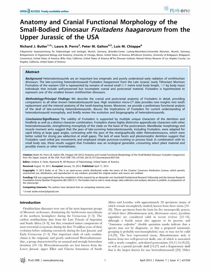
Anatomy and Cranial Functional Morphology of the Small-Bodied Dinosaur Fruitadens haagarorum from...
31
Anatomy and Cranial Functional Morphology of the Small-Bodied Dinosaur Fruitadens haagarorum from the Upper Jurassic of the USA Richard J. Butler 1,2 *, Laura B. Porro 3 , Peter M. Galton 4,5 , Luis M. Chiappe 6 1 Bayerische Staatssammlung fu ¨r Pala ¨ontologie und Geologie, Munich, Germany, 2 GeoBio-Center, Ludwig-Maximilians-Universita ¨t Mu ¨ nchen, Munich, Germany, 3 Department of Organismal Biology and Anatomy, University of Chicago, Illinois, United States of America, 4 Professor Emeritus, University of Bridgeport, Bridgeport, Connecticut, United States of America, 5 Rio Vista, California, United States of America, 6 The Dinosaur Institute, Natural History Museum of Los Angeles County, Los Angeles, California, United States of America Abstract Background: Heterodontosaurids are an important but enigmatic and poorly understood early radiation of ornithischian dinosaurs. The late-surviving heterodontosaurid Fruitadens haagarorum from the Late Jurassic (early Tithonian) Morrison Formation of the western USA is represented by remains of several small (,1 metre total body length, ,1 kg body mass) individuals that include well-preserved but incomplete cranial and postcranial material. Fruitadens is hypothesized to represent one of the smallest known ornithischian dinosaurs. Methodology/Principal Findings: We describe the cranial and postcranial anatomy of Fruitadens in detail, providing comparisons to all other known heterodontosaurid taxa. High resolution micro-CT data provides new insights into tooth replacement and the internal anatomy of the tooth-bearing bones. Moreover, we provide a preliminary functional analysis of the skull of late-surviving heterodontosaurids, discuss the implications of Fruitadens for current understanding of heterodontosaurid monophyly, and briefly review the evolution and biogeography of heterodontosaurids. Conclusions/Significance: The validity of Fruitadens is supported by multiple unique characters of the dentition and hindlimb as well as a distinct character combination. Fruitadens shares highly distinctive appendicular characters with other heterodontosaurids, strengthening monophyly of the clade on the basis of the postcranium. Mandibular morphology and muscle moment arms suggest that the jaws of late-surviving heterodontosaurids, including Fruitadens, were adapted for rapid biting at large gape angles, contrasting with the jaws of the stratigraphically older Heterodontosaurus, which were better suited for strong jaw adduction at small gapes. The lack of wear facets and plesiomorphic dentition suggest that Fruitadens used orthal jaw movements and employed simple puncture-crushing to process food. In combination with its small body size, these results suggest that Fruitadens was an ecological generalist, consuming select plant material and possibly insects or other invertebrates. Citation: Butler RJ, Porro LB, Galton PM, Chiappe LM (2012) Anatomy and Cranial Functional Morphology of the Small-Bodied Dinosaur Fruitadens haagarorum from the Upper Jurassic of the USA. PLoS ONE 7(4): e31556. doi:10.1371/journal.pone.0031556 Editor: Andrew A. Farke, Raymond M. Alf Museum of Paleontology, United States of America Received August 19, 2011; Accepted January 10, 2012; Published April 11, 2012 Copyright: ß 2012 Butler et al. This is an open-access article distributed under the terms of the Creative Commons Attribution License, which permits unrestricted use, distribution, and reproduction in any medium, provided the original author and source are credited. Funding: RJB was supported during the completion of this research by an Alexander von Humboldt Postdoctoral Research Fellowship and the German Research Foundation Emmy Noether Programme (BU 2587/3-1). The funders had no role in study design, data collection and analysis, decision to publish, or preparation of the manuscript. Competing Interests: The authors have declared that no competing interests exist. * E-mail: [email protected] Introduction Ornithischian dinosaurs were one of the most important groups of Mesozoic archosaurs, dominating the herbivorous macrofauna of the northern hemisphere during the Cretaceous [1–3]. The earliest ornithischians date from the Late Triassic of Argentina and South Africa [4–9], but they remained minor components of most terrestrial ecosystems during the first 70 million years of their evolution before radiating extensively during the Late Jurassic and Early Cretaceous [1–3]. One important clade of early ornithis- chians is the enigmatic and poorly understood Heterodontosaur- idae, a group characterized by an unusual and strongly heterodont dentition [10–14]. Heterodontosaurids are best known from the Lower Jurassic upper Elliot and Clarens formations of South Africa and Lesotho, with approximately 20 specimens (many of which remain incompletely studied) known from these strata [10– 29]. These specimens form the basis for five monospecific genera, of which three (Heterodontosaurus tucki, Abrictosaurus consors, Lycorhinus angustidens) are considered valid in recent reviews [12–14], although a fourth taxon also appears to be present, and ‘‘Lanasaurus scalpridens’’ (double quotation marks indicate that a species may not be diagnostic, or that a proposed taxonomic grouping is probably non-monophyletic) may or may not be valid [14,29]. The best represented taxon, Heterodontosaurus tucki, is known from two well-preserved skulls, one of which is associated with a nearly complete, articulated postcranium [10,11,14,18,22], as well as a partial juvenile skull [14,27] and a fragmentary skull that is the largest known for any heterodontosaurid [29]. Other PLoS ONE | www.plosone.org 1 April 2012 | Volume 7 | Issue 4 | e31556
Transcript of Anatomy and Cranial Functional Morphology of the Small-Bodied Dinosaur Fruitadens haagarorum from...
Anatomy and Cranial Functional Morphology of theSmall-Bodied Dinosaur Fruitadens haagarorum from theUpper Jurassic of the USARichard J. Butler1,2*, Laura B. Porro3, Peter M. Galton4,5, Luis M. Chiappe6
1 Bayerische Staatssammlung fur Palaontologie und Geologie, Munich, Germany, 2 GeoBio-Center, Ludwig-Maximilians-Universitat Munchen, Munich, Germany,
3 Department of Organismal Biology and Anatomy, University of Chicago, Illinois, United States of America, 4 Professor Emeritus, University of Bridgeport, Bridgeport,
Connecticut, United States of America, 5 Rio Vista, California, United States of America, 6 The Dinosaur Institute, Natural History Museum of Los Angeles County, Los
Angeles, California, United States of America
Abstract
Background: Heterodontosaurids are an important but enigmatic and poorly understood early radiation of ornithischiandinosaurs. The late-surviving heterodontosaurid Fruitadens haagarorum from the Late Jurassic (early Tithonian) MorrisonFormation of the western USA is represented by remains of several small (,1 metre total body length, ,1 kg body mass)individuals that include well-preserved but incomplete cranial and postcranial material. Fruitadens is hypothesized torepresent one of the smallest known ornithischian dinosaurs.
Methodology/Principal Findings: We describe the cranial and postcranial anatomy of Fruitadens in detail, providingcomparisons to all other known heterodontosaurid taxa. High resolution micro-CT data provides new insights into toothreplacement and the internal anatomy of the tooth-bearing bones. Moreover, we provide a preliminary functional analysisof the skull of late-surviving heterodontosaurids, discuss the implications of Fruitadens for current understanding ofheterodontosaurid monophyly, and briefly review the evolution and biogeography of heterodontosaurids.
Conclusions/Significance: The validity of Fruitadens is supported by multiple unique characters of the dentition andhindlimb as well as a distinct character combination. Fruitadens shares highly distinctive appendicular characters with otherheterodontosaurids, strengthening monophyly of the clade on the basis of the postcranium. Mandibular morphology andmuscle moment arms suggest that the jaws of late-surviving heterodontosaurids, including Fruitadens, were adapted forrapid biting at large gape angles, contrasting with the jaws of the stratigraphically older Heterodontosaurus, which werebetter suited for strong jaw adduction at small gapes. The lack of wear facets and plesiomorphic dentition suggest thatFruitadens used orthal jaw movements and employed simple puncture-crushing to process food. In combination with itssmall body size, these results suggest that Fruitadens was an ecological generalist, consuming select plant material andpossibly insects or other invertebrates.
Citation: Butler RJ, Porro LB, Galton PM, Chiappe LM (2012) Anatomy and Cranial Functional Morphology of the Small-Bodied Dinosaur Fruitadens haagarorumfrom the Upper Jurassic of the USA. PLoS ONE 7(4): e31556. doi:10.1371/journal.pone.0031556
Editor: Andrew A. Farke, Raymond M. Alf Museum of Paleontology, United States of America
Received August 19, 2011; Accepted January 10, 2012; Published April 11, 2012
Copyright: � 2012 Butler et al. This is an open-access article distributed under the terms of the Creative Commons Attribution License, which permitsunrestricted use, distribution, and reproduction in any medium, provided the original author and source are credited.
Funding: RJB was supported during the completion of this research by an Alexander von Humboldt Postdoctoral Research Fellowship and the German ResearchFoundation Emmy Noether Programme (BU 2587/3-1). The funders had no role in study design, data collection and analysis, decision to publish, or preparation ofthe manuscript.
Competing Interests: The authors have declared that no competing interests exist.
* E-mail: [email protected]
Introduction
Ornithischian dinosaurs were one of the most important groups
of Mesozoic archosaurs, dominating the herbivorous macrofauna
of the northern hemisphere during the Cretaceous [1–3]. The
earliest ornithischians date from the Late Triassic of Argentina
and South Africa [4–9], but they remained minor components of
most terrestrial ecosystems during the first 70 million years of their
evolution before radiating extensively during the Late Jurassic and
Early Cretaceous [1–3]. One important clade of early ornithis-
chians is the enigmatic and poorly understood Heterodontosaur-
idae, a group characterized by an unusual and strongly heterodont
dentition [10–14]. Heterodontosaurids are best known from the
Lower Jurassic upper Elliot and Clarens formations of South
Africa and Lesotho, with approximately 20 specimens (many of
which remain incompletely studied) known from these strata [10–
29]. These specimens form the basis for five monospecific genera,
of which three (Heterodontosaurus tucki, Abrictosaurus consors, Lycorhinus
angustidens) are considered valid in recent reviews [12–14],
although a fourth taxon also appears to be present, and
‘‘Lanasaurus scalpridens’’ (double quotation marks indicate that a
species may not be diagnostic, or that a proposed taxonomic
grouping is probably non-monophyletic) may or may not be valid
[14,29]. The best represented taxon, Heterodontosaurus tucki, is
known from two well-preserved skulls, one of which is associated
with a nearly complete, articulated postcranium [10,11,14,18,22],
as well as a partial juvenile skull [14,27] and a fragmentary skull
that is the largest known for any heterodontosaurid [29]. Other
PLoS ONE | www.plosone.org 1 April 2012 | Volume 7 | Issue 4 | e31556
heterodontosaurids include Tianyulong confuciusi, known from a
single exceptionally preserved specimen that preserves filamentous
integumentary structures from the Middle–Late Jurassic of China
[30] (previously reported as late Early Cretaceous, but see Lu et al.
[31]), the historical taxon Echinodon becklesii from the earliest
Cretaceous of the UK [1,13,32–39], a jaw fragment from the Late
Triassic of Argentina [6], an undescribed specimen from the Early
Jurassic of the USA [1,40], the recently described Manidens
condorensis from the Middle Jurassic of Argentina [41], and possibly
the oldest known ornithischian, Pisanosaurus mertii, from the Late
Triassic (late Carnian: see Martinez et al. [42]) of Argentina
[5,8,12,13]. Specimens previously assigned to Heterodontosaur-
idae from the Late Triassic of Switzerland [43], the Early Jurassic
of China [44], and the Early Cretaceous of Spain [45] have been
recently removed from the clade [46–48].
Recent work has demonstrated the importance of heterodonto-
saurids for an understanding of ornithischian dinosaur evolution,
particularly global patterns of ornithischian phylogeny, origination
dates for major clades, and diversity patterns [7,49,50], as well as
broader questions relating to early dinosaur evolution [51,52] and
the early evolution of feathers [30,51,53]. Although Ornithischia
was one of the first fossil reptile groups to which cladistic
approaches were applied [54–59], the study of global patterns of
ornithischian interrelationships subsequently lagged behind that of
the other major clades of dinosaurs, Theropoda and Sauropodo-
morpha. However, an increasing number of analyses of basal
ornithischian phylogeny have been carried out in recent years
[2,7,30,41,49,50,60,61], with broad agreement on major ornith-
ischian interrelationships. One key point on which these analyses
disagree is the phylogenetic position of Heterodontosauridae.
Sereno ([2,56,59]; see also [12,13,62]) has argued that hetero-
dontosaurids represent the most basal grouping within Ornitho-
poda, a clade that also includes ‘hypsilophodontids’, iguanodon-
tians, and hadrosaurs. By contrast, Butler et al. ([7,50,61]; see also
[30,41,63]) have argued that heterodontosaurids are the most
basal radiation of ornithischians, a position that appears to be
more concordant with stratigraphic evidence, while several
authors [13,49,54,57,64] have found support for a link between
marginocephalians (Pachycephalosauria+Ceratopsia) and Hetero-
dontosauridae. Finally, Heterodontosauridae has also been placed
as a sister taxon to Cerapoda (Ornithopoda+Marginocephalia
[58,60]). The character evidence supporting these alternative
placements was discussed critically by Norman et al. [14].
Inconsistencies regarding the phylogenetic position of hetero-
dontosaurids are likely to be resolved with increased taxonomic
sampling and a better understanding of the postcranial anatomy of
these dinosaurs. The morphology of Fruitadens haagarorum [61] from
the Late Jurassic Morrison Formation of the western USA fills an
important gap in our knowledge of heterodontosaurid anatomy.
Given its well-preserved postcranial material, the morphology of
Fruitadens thus sheds new light into the monophyly, phylogenetic
position, and evolutionary patterns of the clade. The preserved
cranial material of Fruitadens also provides new information on
heterodontosaurid functional morphology and craniodental vari-
ation. Furthermore, being the first heterodontosaurid for which
published histological data are available, Fruitadens provides
insights into the ontogeny and body size distribution of
ornithischians. Because of the importance of this taxon for
understanding heterodontosaurid evolution, we provide here a
full description of its anatomy with detailed comparisons to other
heterodontosaurids. Moreover, we provide initial analyses of the
cranial functional morphology of late-surviving small-bodied
heterodontosaurids, review characters supporting heterodonto-
saurid monophyly, and provide an overview of the evolutionary
history of the group.
Taxonomic backgroundThe ornithischian Echinodon becklesii Owen, 1861 [65] is based
upon fragmentary cranial material from the lowermost Cretaceous
(Middle Purbeck Beds, Purbeck Limestone Group: Berriasian) of
southern England, UK [32,33,37,39,65]. This material was
originally described as ‘lacertilian’ (i.e. as a lizard) by Owen
[65]; however, the dinosaurian nature of Echinodon was later noted
by Owen ([66]: 9) (as ‘the small Purbeck Dinosaur [Echinodon]’) and
Lydekker [67]. Echinodon has since been assigned to multiple
phylogenetically disparate groups within Ornithischia, including
Stegosauria, Thyreophora, ‘‘Hypsilophodontidae’’, and ‘‘Fabro-
sauridae’’ (see review in Norman & Barrett [37]). Most recently,
Echinodon has been proposed to represent a Cretaceous hetero-
dontosaurid [1,13,33–38] and Norman & Barrett [37] cited three
potential synapomorphies supporting this referral: 1) a wedge-
shaped predentary; 2) teeth from the midpoint of the maxillary/
dentary tooth rows have denticles restricted to the apical-most
third of their crowns; 3) absence of replacement foramina on the
medial surface of the maxilla and dentary.
Callison & Quimby ([68]: figs 3B, C) figured a femoral shaft and
a distal tibia with an articulated astragalus and calcaneum as those
of a small ‘‘fabrosaurid’’ ornithischian dinosaur. These bones came
from the Upper Jurassic Morrison Formation of the Fruita
Paleontological Area (FPA), northwest of Grand Junction,
Colorado, USA. The material was subsequently identified as
Echinodon sp. on the basis of an initial assessment of the
morphology of another specimen, consisting of associated jaws
with teeth [34,69]. Galton [36] proposed several autapomorphies
for Echinodon based upon the English and Fruita material, such as
the form of the dentary symphysis and the presence of an
anteromedially directed edge on the distal part of the tibia. In
addition, he listed several cranial and postcranial character
synapomorphies shared by Echinodon (postcranial characters based
on Fruita material) and Heterodontosaurus.
Galton [38] compared the morphology of the dentition of the
Fruita material to Echinodon, and noted several differences.
Subsequently, Butler et al. [61] erected the new taxon Fruitadens
haagarorum for the Fruita material.
Geological backgroundGeneral accounts of the Fruita Paleontological Area (FPA –
land administered by the Bureau of Land Management of the
USA), including the history of discovery, geology, taphonomy and
paleoenvironments, and fauna, are given by Callison [69],
Kirkland [70,71], and Foster [72]. Fossils at the FPA were
collected from a geographically small area, covering approximate-
ly one square kilometer ([71]: fig. 4A). The sediments at the FPA
are interpreted as representing a number of depositional
environments, including low-sinuosity anastomosing river chan-
nels, levees, floodplains, and ponds [71]. Vertebrate fossils occur in
nearly all of these facies, but the small-bodied vertebrate remains
for which the FPA is famous occur in the so-called ‘drab
floodplain’ and ‘alkaline pond’ facies, the former representing a
poorly drained floodplain with poorly developed paleosols and the
latter representing ephemeral floodplain ponds [71]. Preservation
of these small vertebrate taxa is attributed to the alkaline nature of
the enclosing sediments [71].
The vertebrate fauna documented from the FPA is diverse and
includes dipnoan fish (Ceratodus guentheri), an amioid, the actinop-
terygian Hulettia hawesi, the chelonian Glyptops, the rhynchocepha-
lians Opisthias and Eilenodon robustus, lizards including the
Anatomy of the Dinosaur Fruitadens
PLoS ONE | www.plosone.org 2 April 2012 | Volume 7 | Issue 4 | e31556

anguimorphs Parviraptor gilmorei and Dorsetisaurus and the scinco-
morphs Paramacellodus and Saurillodon, a small cursorial mesosu-
chian crocodiliform, the sphenosuchian crocodiliform Macelog-
nathus vagans, the mammals Priacodon fruitaensis, Glirodon grandis and
Fruitafossor windsheffeli, the theropod dinosaurs Ceratosaurus magni-
cornis and Allosaurus, the sauropod dinosaurs Camarasaurus and
Apatosaurus, and the large-bodied ornithischians Stegosaurus and
Dryosaurus [71–85].
Institutional abbreviationsNHMUK [formerly NHM, BMNH], Natural History Museum,
London, UK; IVPP, Institute of Vertebrate Paleontology and
Paleoanthropology, Bejing, People’s Republic of China; LACM,
Dinosaur Institute of the Natural History Museum of Los Angeles
County, Los Angeles, California, USA; MNA, Museum of
Northern Arizona, Flagstaff, Arizona, USA; SAM-PK, Iziko
South African Museum, Cape Town, South Africa.
Results
Systematic PaleontologyDinosauria Owen, 1842 [86]
Ornithischia Seeley, 1887 [87]
Heterodontosauridae Kuhn, 1966 [88]
Phylogenetic definition. The most inclusive clade
containing Heterodontosaurus tucki Crompton & Charig, 1962 [10]
but not Parasaurolophus walkeri Parks, 1922 [89], Pachycephalosaurus
wyomingensis (Gilmore, 1931) [90], Triceratops horridus Marsh, 1889
[91], Ankylosaurus magniventris Brown, 1908 [92] (Sereno [93]).
Diagnosis. Small-bodied ornithischians diagnosed by the
following unique combination of characters [61]: (1) three
premaxillary teeth; (2) arched and recessed diastema between
the premaxilla and the maxilla; (3) wedge-shaped predentary; (4)
constriction on the proximal surface of the humerus, between the
head and the medial tubercle; (5) ‘rod-like’ (with near parallel
sides) fourth trochanter on the femur; (6) very slender distal fibula;
(7) fused astragalus and calcaneum (astragalocalcaneum); (8)
proximal phalanges of pedal digits II–IV with extensor pits on
distal heads.
Fruitadens haagarorum Butler, Galton, Porro, Chiappe, Henderson
& Erickson, 2010 [61]
Figs. 1, 2, 3, 4, 5, 6, 7, 8A, B, 9, 10, 11, 12, 13, 14, 15, 16
‘‘cf. Coelurosaurus’’; Callison & Rasmussen, 1980:151 [94]
‘‘cf. Fabrosaurus’’; Callison & Rasmussen, 1980:153 [94]
‘‘Fruita fabrosaurid’’; Callison & Quimby, 1984:figs 3B, C [68]
‘‘Echinodon sp.’’; Callison, 1987:95, fig. 4 [69]
‘‘Gen. & sp. nov., Morrison Formation’’; Olshevsky & Ford,
1994:93, fig. 43 [34]
‘‘Echinodon sp.’’; Galton, 2002:55–56A [36]
‘‘Fruita jaws’’; Galton, 2006:26, 28, fig. 2.7A–G [38]
Diagnosis. Small heterodontosaurid ornithischian
characterised by the following unique combination of characters,
including autapomorphies (* indicates character that is
autapomorphic within Heterodontosauridae; ** indicates
character that is autapomorphic within Ornithischia): (1)
premaxillary crowns small and subequal to one another in size,
expanded labiolingually and mesiodistally above the root; (2)
maxillary caniniform absent; (3) maxillary and dentary crowns
apicobasally low and triangular in lingual and labial views, with
symmetrically distributed enamel; (4*) mesial and distal denticles
extend over half of the apicobasal height of maxillary and dentary
crowns, not restricted to apical third; (5*) dentary caniniform
present but erupted apicobasal height does not exceed that of the
crown of the largest dentary ‘cheek’ (post-caniniform) tooth; (6**)
small, unserrated, peg-like and procumbent tooth present anterior
to dentary caniniform; (7**) small foramen on anteroventral aspect
of the medial dentary, ventral to the Meckelian groove and
beneath dentary crowns 3 and 4; (8) distal end of tibia with
anteromedial flange; (9**) apex of the ascending process of
astragalus is formed by a separate ossification; (10**) two large
foramina pierce anterior surface of ascending process of astragalus
(modified from Butler et al. [61]).
Etymology. Fruitadens, from Fruita (hypodigm locality) and
dens (Latin, tooth); haagarorum, for Paul Haaga, Jr, Heather Haaga,
Blythe Haaga, Paul Haaga III, and Catalina Haaga, to honour
their support of the Natural History Museum of Los Angeles
County (LACM, Los Angeles, USA), where the specimens of
Fruitadens haagarorum are held.
Holotype. LACM 115747, associated jaws, vertebrae and
limb bones of a near full-grown individual (Butler et al. [61]: figs
2B, E, I, 3A–C; cf. Fabrosaurus of Callison & Rasmussen [94]: 153).
Includes maxillae (both incomplete), partial right dentary, and
anterior end of left dentary, disarticulated vertebrae including two
partial cervicals, six partial dorsals, six sacrals and numerous
caudals, proximal end of the right femur, proximal and distal ends
of both tibiae, partial metatarsal. Collected by J. M. Clark, August
1977, at ‘‘Locality Number 4’’ ([94]: 153), Fruita Paleontological
Area (FPA). The specimen is currently catalogued as from locality
LACM 4684: LACM 4684 is a ‘‘general locality for specimens
from FPA with poor specific locality data’’ ([70]: 95).
Referred specimens. LACM 115727, proximal ends of both
femora, proximal and distal ends of left tibia with attached
astragalocalcaneum, bone fragments ([61]: figs 2G, H, 3F; [67]:
fig. 3C) (referred to as cf. Coelurosaurus by Callison & Rasmussen
([94]: 151). Collected by G. L. Callison and party (July/August
1979). Callison & Rasmussen ([94]: 151) give the locality as
‘‘Locality Number 4’’ but it is currently catalogued as collected
from locality LACM 5576, ‘‘George’s ‘Coelurosaur’ Site’’ ([71]:
93).
LACM 120478, left humerus, partial left femur, and articulated
left tibia, fibula, and astragalocalcaneum ([61]: fig. 2J–P; [68]: fig.
3B) (referred to as cf. Coelurosaurus by Callison & Rasmussen [94]:
151). Collected July/August 1979 by G. L. Callison and party, also
at ‘‘Locality Number 4’’ ([94]: 151) and now catalogued as
collected from LACM 5572, the ‘‘Main Callison Quarry’’ ([71]:
94).
LACM 120602, distal caudal vertebra, left astragalocalcaneum
and elements of the metatarsus and pes. Collected 10th June 1985
by G. L. Callison and party from locality LACM 4684 (see above).
LACM 128258, right premaxilla, partial left maxilla, originally
articulated dentaries, dorsal vertebra, distal caudal vertebra ([34]:
79, fig. 14; [38]: figs 2.7A, B; [61]: figs 2A, C, D, F, 3D). A cast of
the jaws of the dentaries (including the anteriorly positioned
caniniform, which is no longer preserved in the original specimen)
is held at the LACM. Collected by G. L. Callison and party (no
date given) from locality LACM 4684 (see above).
LACM 128303, poorly preserved anterior left dentary (con-
taining five crowns and four empty alveoli). Collected in 1981 by
G. L. Callison and party from locality LACM 4684 (see above).
Horizon and type locality. All specimens came from the
Morrison Formation at the LACM Fruita Paleontological Area
(FPA), west of Fruita, 19 km northwest of Grand Junction, Mesa
County, Colorado, USA ([61]: fig. 1.) The approximate latitude
and longitude of the FPA is 39.2uN, 108.8uW. Specimens were
collected in the late 1970s and early 1980s from the ‘drab flood-
plain facies’ at the base of the Brushy Basin Member of the
Morrison Formation immediately above the ‘clay change’ horizon
([61]: fig. S1; [71]). The ‘clay change’ horizon is commonly used
Anatomy of the Dinosaur Fruitadens
PLoS ONE | www.plosone.org 3 April 2012 | Volume 7 | Issue 4 | e31556
for regional correlation of the Morrison Formation [95]. Turner &
Peterson ([95]: fig. 7) placed the localities (listed as CO-33 in their
stratigraphic sections and their Appendix 3) yielding Fruitadens
within the Kimmeridgian, and within their ‘Dinosaur Zone 2’ and
charophyte-ostracode Zone 4. Stratigraphic horizons closely
equivalent to the Fruita quarries yield 40Ar/39Ar isotopic dates
of 150.360.3 Ma and 150.260.5 Ma [95,96]. This would suggest
an early Tithonian age for the Fruita quarries based upon the most
recent geological time scales [97,98] that place the Tithonian at
150.8–145.5 Ma.
Notes on associations of specimens. No data on the
original field associations of the holotype and referred specimens is
currently available at the LACM. Within the holotype, all of the
preserved material is generally consistent (in terms of size,
morphology, lack of duplication of elements, and preservation)
with belonging to a single individual. A distal femur of the
crocodylomorph Macelognathus was previously included within
LACM 115747, but has now been removed from this specimen.
Each of the other specimens referred to Fruitadens likely represents
a single individual, based upon consistent morphology and lack of
overlapping elements.
Description and comparisonsSkull anatomy: general comments. The skull is
represented in the holotype by fragmentary maxillae (with only
a few crowns preserved), most of the right dentary, and the
anterior end of the left dentary. Most of the dentary crowns are
missing, with only crown bases and/or just tooth roots preserved.
In LACM 128258, the incomplete right premaxilla, left maxilla
and dentaries are preserved, and most of the crowns are preserved
(although damaged). LACM 128303 is a poorly preserved anterior
left dentary. Other referred specimens lack cranial elements, and
the morphology of the remainder of the skull is unknown. A
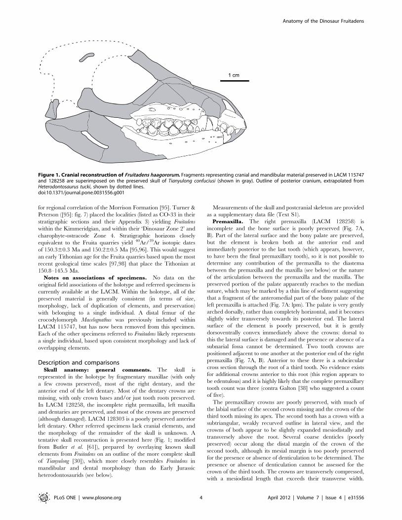
tentative skull reconstruction is presented here (Fig. 1; modified
from Butler et al. [61]), prepared by overlaying known skull
elements from Fruitadens on an outline of the more complete skull
of Tianyulong [30]), which more closely resembles Fruitadens in
mandibular and dental morphology than do Early Jurassic
heterodontosaurids (see below).
Measurements of the skull and postcranial skeleton are provided
as a supplementary data file (Text S1).
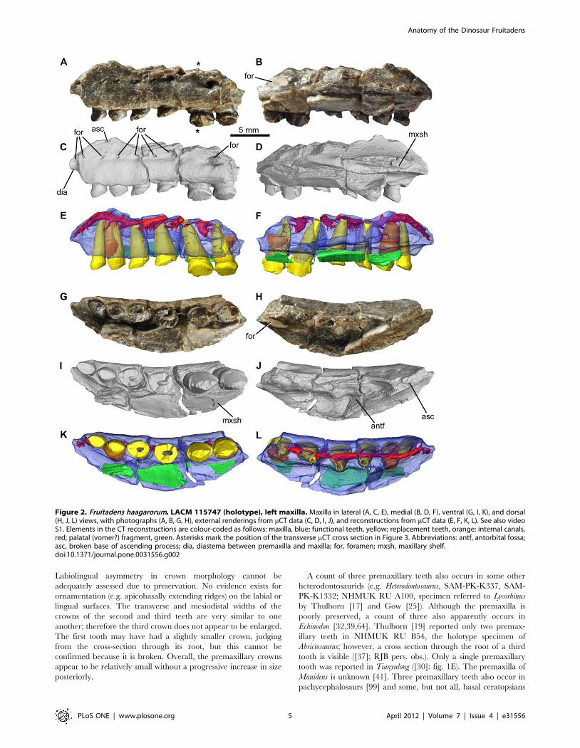
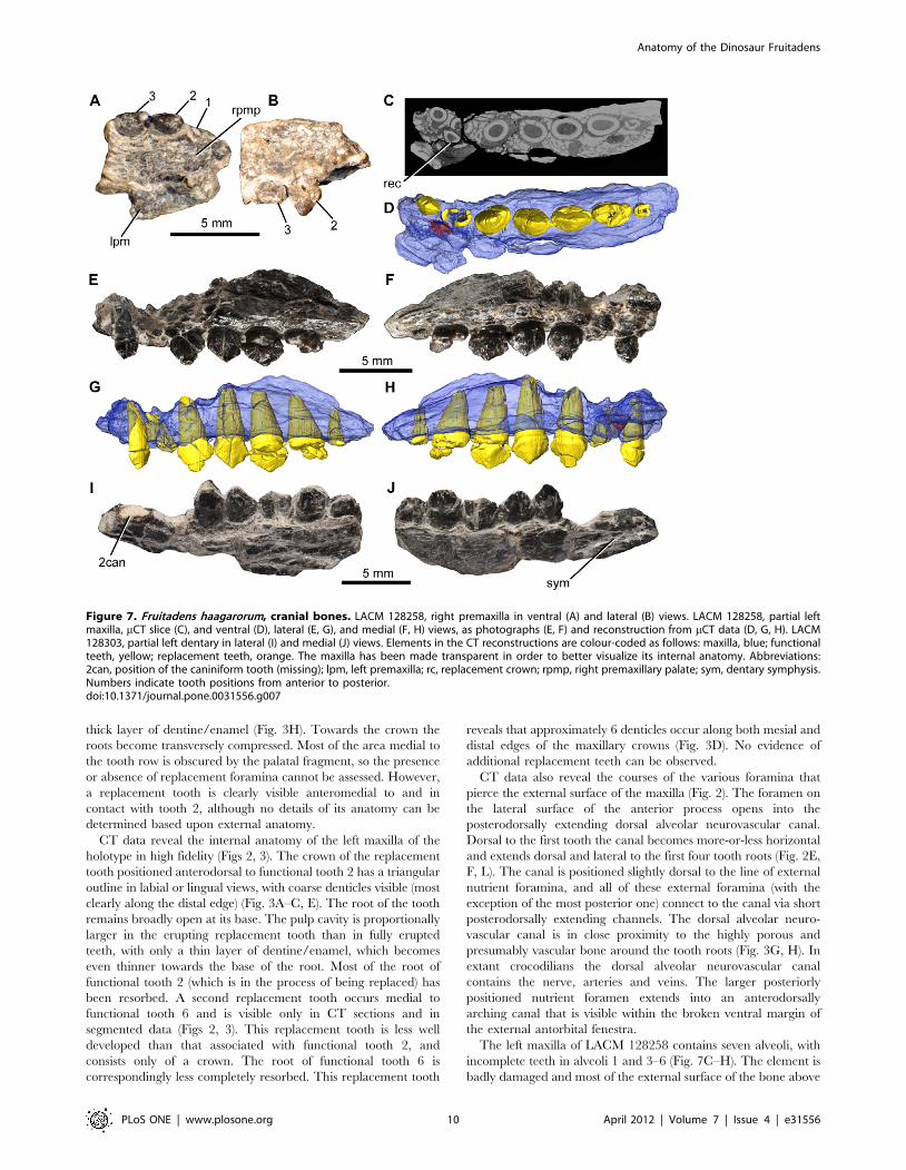
Premaxilla. The right premaxilla (LACM 128258) is
incomplete and the bone surface is poorly preserved (Fig. 7A,
B). Part of the lateral surface and the bony palate are preserved,
but the element is broken both at the anterior end and
immediately posterior to the last tooth (which appears, however,
to have been the final premaxillary tooth), so it is not possible to
determine any contribution of the premaxilla to the diastema
between the premaxilla and the maxilla (see below) or the nature
of the articulation between the premaxilla and the maxilla. The
preserved portion of the palate apparently reaches to the median
suture, which may be marked by a thin line of sediment suggesting
that a fragment of the anteromedial part of the bony palate of the
left premaxilla is attached (Fig. 7A: lpm). The palate is very gently
arched dorsally, rather than completely horizontal, and it becomes
slightly wider transversely towards its posterior end. The lateral
surface of the element is poorly preserved, but it is gently
dorsoventrally convex immediately above the crowns: dorsal to
this the lateral surface is damaged and the presence or absence of a
subnarial fossa cannot be determined. Two tooth crowns are
positioned adjacent to one another at the posterior end of the right
premaxilla (Fig. 7A, B). Anterior to these there is a subcircular
cross section through the root of a third tooth. No evidence exists
for additional crowns anterior to this root (this region appears to
be edentulous) and it is highly likely that the complete premaxillary
tooth count was three (contra Galton [38] who suggested a count
of five).
The premaxillary crowns are poorly preserved, with much of
the labial surface of the second crown missing and the crown of the
third tooth missing its apex. The second tooth has a crown with a
subtriangular, weakly recurved outline in lateral view, and the
crowns of both appear to be slightly expanded mesiodistally and
transversely above the root. Several coarse denticles (poorly
preserved) occur along the distal margin of the crown of the
second tooth, although its mesial margin is too poorly preserved
for the presence or absence of denticulation to be determined. The
presence or absence of denticulation cannot be assessed for the
crown of the third tooth. The crowns are transversely compressed,
with a mesiodistal length that exceeds their transverse width.
Figure 1. Cranial reconstruction of Fruitadens haagororum. Fragments representing cranial and mandibular material preserved in LACM 115747and 128258 are superimposed on the preserved skull of Tianyulong confuciusi (shown in gray). Outline of posterior cranium, extrapolated fromHeterodontosaurus tucki, shown by dotted lines.doi:10.1371/journal.pone.0031556.g001
Anatomy of the Dinosaur Fruitadens
PLoS ONE | www.plosone.org 4 April 2012 | Volume 7 | Issue 4 | e31556
Labiolingual asymmetry in crown morphology cannot be
adequately assessed due to preservation. No evidence exists for
ornamentation (e.g. apicobasally extending ridges) on the labial or
lingual surfaces. The transverse and mesiodistal widths of the
crowns of the second and third teeth are very similar to one
another; therefore the third crown does not appear to be enlarged.
The first tooth may have had a slightly smaller crown, judging
from the cross-section through its root, but this cannot be
confirmed because it is broken. Overall, the premaxillary crowns
appear to be relatively small without a progressive increase in size
posteriorly.
A count of three premaxillary teeth also occurs in some other
heterodontosaurids (e.g. Heterodontosaurus, SAM-PK-K337, SAM-
PK-K1332; NHMUK RU A100, specimen referred to Lycorhinus
by Thulborn [17] and Gow [25]). Although the premaxilla is
poorly preserved, a count of three also apparently occurs in
Echinodon [32,39,64]. Thulborn [19] reported only two premax-
illary teeth in NHMUK RU B54, the holotype specimen of
Abrictosaurus; however, a cross section through the root of a third
tooth is visible ([37]; RJB pers. obs.). Only a single premaxillary
tooth was reported in Tianyulong ([30]: fig. 1E). The premaxilla of
Manidens is unknown [41]. Three premaxillary teeth also occur in
pachycephalosaurs [99] and some, but not all, basal ceratopsians
Figure 2. Fruitadens haagarorum, LACM 115747 (holotype), left maxilla. Maxilla in lateral (A, C, E), medial (B, D, F), ventral (G, I, K), and dorsal(H, J, L) views, with photographs (A, B, G, H), external renderings from mCT data (C, D, I, J), and reconstructions from mCT data (E, F, K, L). See also videoS1. Elements in the CT reconstructions are colour-coded as follows: maxilla, blue; functional teeth, yellow; replacement teeth, orange; internal canals,red; palatal (vomer?) fragment, green. Asterisks mark the position of the transverse mCT cross section in Figure 3. Abbreviations: antf, antorbital fossa;asc, broken base of ascending process; dia, diastema between premaxilla and maxilla; for, foramen; mxsh, maxillary shelf.doi:10.1371/journal.pone.0031556.g002
Anatomy of the Dinosaur Fruitadens
PLoS ONE | www.plosone.org 5 April 2012 | Volume 7 | Issue 4 | e31556
[49,100–102]. By contrast, 5–6 premaxillary teeth occur in many
other early ornithischians, including Lesothosaurus [33], early
thyreophorans [103–104], Agilisaurus [105], and basal ornithopods
[106–108]. Four or more premaxillary teeth typically occur in
ornithischian outgroups [109,110].
The morphology of the premaxillary crowns in Fruitadens differs
from all other heterodontosaurids with the apparent exception of
Echinodon [39], although this morphology may represent a retained
plesiomorphy at the level of Ornithischia (due to the similarities
with premaxillary crowns of other basal ornithischians). In
Heterodontosaurus the premaxillary dentition consists of three crowns
that increase in size posteriorly (SAM-PK-K337, K1332). The
anterior two crowns are comparatively small, conical, recurved,
lack serrations and are not markedly expanded above their roots.
The caniniform third crown is greatly enlarged, recurved, with
serrations along the distal surface, and is unexpanded above its
root. This condition differs substantially from that of Fruitadens and
Echinodon, in which the premaxillary crowns do not increase in size
posteriorly, are expanded above their roots, and a caniniform is
absent. A similar premaxillary dentition to that of Heterodontosaurus
occurs in NHMUK RU A100 ([17]: fig. 2), although the
caniniform third crown is not as enlarged (relative to the more
anterior crowns) as in Heterodontosaurus. In Abrictosaurus (NHMUK
RU B54; [19]: fig. 2) the crowns are conical and unexpanded
above their roots, and increase in size posteriorly, although the last
crown is relatively small compared to the caniniform teeth of
Heterodontosaurus and NHMUK RU A100. The single, posteriorly
positioned, premaxillary tooth of Tianyulong is enlarged, caniniform
and not expanded above the root ([30]: fig. 1E).
The premaxillary crowns of Fruitadens and Echinodon appear to
be most similar to those of basal ornithischians such as Lesothosaurus
[33] and Scutellosaurus [103], which are also expanded labiolin-
gually and mesiodistally above their roots and do not increase in
size posteriorly. Similar premaxillary teeth occur in some basal
ornithopods [106–108], ceratopsians (e.g. Liaoceratops, IVPP
V12738) and pachycephalosaurs [111].
Maxilla. The holotype includes an incomplete left maxilla,
containing six tooth positions (Figs 2, 3, supplementary video;
identified as the posterior right maxilla by Galton [38]: fig. 2.7A).
A fragment containing two teeth represents the anterior end of the
right maxilla; a second fragment contains three crowns that
represent right maxillary teeth 4–6 (based upon the large foramen
on the posterolateral surface of this fragment). A small section of
the right maxilla separating these fragments is therefore missing.
The fragments from the right maxilla are missing most of their
medial surfaces (including most of the medial part of the antorbital
fossa) and do not add anatomical information that is not evident in
the left maxilla; they will therefore not be described in detail.
Additionally, there are two small fragments in the holotype that
could be from either the maxillary or dentary tooth rows: one of
them has a single partial crown and the second has two partial
crowns (one of which is very small). The complete tooth count for
the maxilla of the holotype is unknown (although a reconstruction
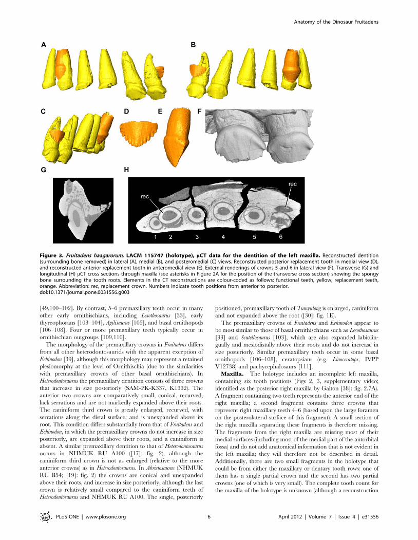
Figure 3. Fruitadens haagarorum, LACM 115747 (holotype), mCT data for the dentition of the left maxilla. Reconstructed dentition(surrounding bone removed) in lateral (A), medial (B), and posteromedial (C) views. Reconstructed posterior replacement tooth in medial view (D),and reconstructed anterior replacement tooth in anteromedial view (E). External renderings of crowns 5 and 6 in lateral view (F). Transverse (G) andlongitudinal (H) mCT cross sections through maxilla (see asterisks in Figure 2A for the position of the transverse cross section) showing the spongybone surrounding the tooth roots. Elements in the CT reconstructions are colour-coded as follows: functional teeth, yellow; replacement teeth,orange. Abbreviation: rec, replacement crown. Numbers indicate tooth positions from anterior to posterior.doi:10.1371/journal.pone.0031556.g003
Anatomy of the Dinosaur Fruitadens
PLoS ONE | www.plosone.org 6 April 2012 | Volume 7 | Issue 4 | e31556

suggests that a count of 7–8 is plausible: Fig. 1), as is the nature of
the midline contact (if one occurred) between the maxillae.
The left maxilla of the holotype is broken anteriorly, posteriorly
and dorsally (Fig. 2). In ventral view, the maxillary tooth row is
arched inwards along its length, such that the lateral surface of the
element is concave anteroposteriorly (Fig. 2G, I, K). The teeth are
set laterally (with no buccal emargination) at the anterior end;
posteriorly they are inset a short distance (equivalent to
approximately half of their transverse width). The buccal
emargination is therefore very weakly developed in Fruitadens.
Dorsal to the crowns and ventral to the line of nutrient foramina
(see below), the lateral surface of the maxilla is dorsoventrally
convex; this convexity becomes more pronounced posteriorly,
forming a low rounded shelf dorsal to the weak buccal
emargination. Immediately beneath the broken base of the
ascending process of the maxilla and the ventral margin of the
Figure 4. Fruitadens haagarorum, LACM 115747 (holotype), right dentary. Dentary in lateral (A, C, E), medial (B, D, F), and dorsal (G, I, J) views,with photographs (A, B, G), external renderings from mCT data (C, D, I), and reconstructions from mCT data (E, F, J). Reconstructed and extracteddentition in medial view (H). Close-up of the reconstructed and extracted posterior replacement teeth in lateral view (K). Close-up of the externalrendering showing the symphyseal region in medial view (L). Longitudinal CT slice (M) through the entire element and sagittal CT slice (N) throughthe anterior part of the mandible. See also video S2. Elements in the CT reconstructions are colour-coded as follows: dentary, blue; functional teeth,yellow; replacement teeth, orange; internal canals, red. The dentary has been made transparent in order to better visualize its internal anatomy.Abbreviations: 1pc, position of the ‘pre-caniniform’ (missing in this specimen); 2can, caniniform tooth in second tooth position; adf, anterior dentaryforamen; con, concavity dorsal to the symphyseal surface; for, foramina; mc, mandibular canal within the dentary; mgr, Meckelian groove; rec,replacement crown; rfor, replacement foramen; sym, symphyseal surface; syri, curved ridge marking dorsal margin of symphysis. Numbers indicatetooth positions from anterior to posterior.doi:10.1371/journal.pone.0031556.g004
Anatomy of the Dinosaur Fruitadens
PLoS ONE | www.plosone.org 7 April 2012 | Volume 7 | Issue 4 | e31556
external antorbital fenestra, the lateral surface is dorsoventrally
concave. An anteroposteriorly extending line of nutrient foramina
occurs within this concavity: these begin anterodorsal to the first
tooth, and at least eight are visible on the left maxilla (Fig. 2A, C).
The foramina generally increase in size posteriorly, with notably
large foramina above teeth 4 and 6. The number, sizes and
positions of nutrient foramina vary between the right and left
maxillae and are not symmetrical.
Anterior to the first tooth, the ventral edge of the maxilla arches
dorsally and forms a short (but anteriorly broken) wedge-shaped
anterior process, the lateral surface of which is depressed relative
to the lateral surface of the main body of the maxilla (Fig. 2A, C:
dia). This anterior process represents the contribution of the
maxilla to an arched diastema between the maxillary and
premaxillary tooth rows (the process is also visible on the anterior
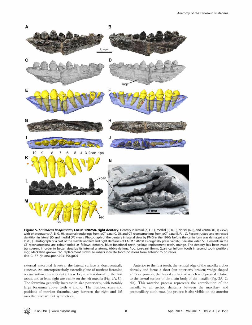
Figure 5. Fruitadens haagarorum, LACM 128258, right dentary. Dentary in lateral (A, C, E), medial (B, D, F), dorsal (G, I), and ventral (H, J) views,with photographs (A, B, G, H), external renderings from mCT data (C, D), and CT reconstructions from mCT data (E, F, I, J). Reconstructed and extracteddentition in lateral (K) and medial (M) views. Photograph of the dentary in lateral view by PMG in the 1980s before the caniniform was damaged andlost (L). Photograph of a cast of the maxilla and left and right dentaries of LACM 128258 as originally preserved (N). See also video S3. Elements in theCT reconstructions are colour-coded as follows: dentary, blue; functional teeth, yellow; replacement teeth, orange. The dentary has been madetransparent in order to better visualize its internal anatomy. Abbreviations: 1pc, ‘pre-caniniform’; 2can, caniniform tooth in second tooth position;mgr, Meckelian groove; rec, replacement crown. Numbers indicate tooth positions from anterior to posterior.doi:10.1371/journal.pone.0031556.g005
Anatomy of the Dinosaur Fruitadens
PLoS ONE | www.plosone.org 8 April 2012 | Volume 7 | Issue 4 | e31556
fragment of the right maxilla). A large foramen occurs on the
lateral surface of this process on both maxillae.
Posterodorsal to the diastema is the broken base of the
ascending process of the maxilla (Fig. 2A, C, H, J: asc). Above
the second tooth, the base of the ascending process splits into two
branches, with the lateral branch forming the damaged ventral
margin of the external antorbital fenestra, and the medial branch
forming the medial wall (also broken) of the antorbital fossa.
Between the ventral margin of the external antorbital fenestra and
the medial wall of the antorbital fossa, the antorbital fossa is deeply
excavated into the body of the maxilla. This excavation, visible
only in dorsal view, has a subtriangular outline (with the apex of
the triangle directed medially), reaching a maximum transverse
width dorsal and medial to tooth 4 (Fig. 2H, J). A large, sediment-
filled, dorsomedially facing foramen opens in the posterolateral
corner of the antorbital fossa (visible in dorsal and medial views),
dorsal to tooth 6 (Fig. 2H: for). Anteromedial to the antorbital
fossa is a medially extending maxillary shelf, the anteromedial
margin of which is grooved for articulation with the opposing
maxilla or another palatal bone (Fig. 2: mxsh). A palatal fragment
(possibly part of the vomer) is attached by sediment to the medial
surface of the maxilla (Fig. 2). The nature of the contacts with the
surrounding bones (lacrimal, jugal, nasal, premaxilla) is unknown.
All of the crowns of the holotype left maxilla are damaged, with
those of teeth 5 and 6 being the best preserved (Fig. 3). The
mesiodistal length and labiolingual width of the erupted crowns
increases to a maximum in teeth 5 and 6, with the crown of tooth
1 being considerably smaller than those positioned more
posteriorly. The crown of the replacement tooth positioned medial
to functional tooth 2 and visible in CT sections (see below) is
approximately the same size as the more posterior ones (Fig. 3). All
of the crowns are expanded at their base both mesiodistally and
labiolingually: the basal expansion is similar on labial and lingual
surfaces. The apex of the crown is slightly offset lingually, so the
crowns are slightly asymmetrical in mesial or distal views. Coarse
denticles occur along the mesial and distal edges (a denticle count
is not possible due to incomplete preservation), and extend over at
least 50% of the crown, rather than being limited to the apical
third as in all other heterodontosaurids. The mesial- and
distalmost denticles are each supported on both labial and lingual
surfaces by a thickened ridge that merges with the basal expansion
(‘cingulum’). Numerous subtle apicobasally extending lineations
occur on the labial and lingual crown surfaces, although distinctly
raised ridges are absent. Packing of the crowns is difficult to judge
due to their incomplete preservation, but those of teeth 5 and 6
have a small point contact with one another (Fig. 3F). The roots of
the teeth are elongate and tapering, and are inclined anterodor-
sally: the bases of the roots of teeth 3–5 are visible within the
lateral part of the antorbital fossa. They have a subcircular cross
section and are composed of a large pulp cavity surrounded by a
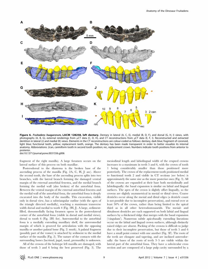
Figure 6. Fruitadens haagarorum, LACM 128258, left dentary. Dentary in lateral (A, C, E), medial (B, D, F), and dorsal (G, H, I) views, withphotographs (A, B, G), external renderings from mCT data (C, D, H), and CT reconstructions from mCT data (E, F, I). Reconstructed and extracteddentition in lateral (J) and medial (K) views. Elements in the CT reconstructions are colour-coded as follows: dentary, dark blue; fragment of coronoid,light blue; functional teeth, yellow; replacement teeth, orange. The dentary has been made transparent in order to better visualize its internalanatomy. Abbreviations: 2can, caniniform tooth in second tooth position; rec, replacement crown. Numbers indicate tooth positions from anterior toposterior.doi:10.1371/journal.pone.0031556.g006
Anatomy of the Dinosaur Fruitadens
PLoS ONE | www.plosone.org 9 April 2012 | Volume 7 | Issue 4 | e31556
thick layer of dentine/enamel (Fig. 3H). Towards the crown the
roots become transversely compressed. Most of the area medial to
the tooth row is obscured by the palatal fragment, so the presence
or absence of replacement foramina cannot be assessed. However,
a replacement tooth is clearly visible anteromedial to and in
contact with tooth 2, although no details of its anatomy can be
determined based upon external anatomy.
CT data reveal the internal anatomy of the left maxilla of the
holotype in high fidelity (Figs 2, 3). The crown of the replacement
tooth positioned anterodorsal to functional tooth 2 has a triangular
outline in labial or lingual views, with coarse denticles visible (most
clearly along the distal edge) (Fig. 3A–C, E). The root of the tooth
remains broadly open at its base. The pulp cavity is proportionally
larger in the erupting replacement tooth than in fully erupted
teeth, with only a thin layer of dentine/enamel, which becomes
even thinner towards the base of the root. Most of the root of
functional tooth 2 (which is in the process of being replaced) has
been resorbed. A second replacement tooth occurs medial to
functional tooth 6 and is visible only in CT sections and in
segmented data (Figs 2, 3). This replacement tooth is less well
developed than that associated with functional tooth 2, and
consists only of a crown. The root of functional tooth 6 is
correspondingly less completely resorbed. This replacement tooth
reveals that approximately 6 denticles occur along both mesial and
distal edges of the maxillary crowns (Fig. 3D). No evidence of
additional replacement teeth can be observed.
CT data also reveal the courses of the various foramina that
pierce the external surface of the maxilla (Fig. 2). The foramen on
the lateral surface of the anterior process opens into the
posterodorsally extending dorsal alveolar neurovascular canal.
Dorsal to the first tooth the canal becomes more-or-less horizontal
and extends dorsal and lateral to the first four tooth roots (Fig. 2E,
F, L). The canal is positioned slightly dorsal to the line of external
nutrient foramina, and all of these external foramina (with the
exception of the most posterior one) connect to the canal via short
posterodorsally extending channels. The dorsal alveolar neuro-
vascular canal is in close proximity to the highly porous and
presumably vascular bone around the tooth roots (Fig. 3G, H). In
extant crocodilians the dorsal alveolar neurovascular canal
contains the nerve, arteries and veins. The larger posteriorly
positioned nutrient foramen extends into an anterodorsally
arching canal that is visible within the broken ventral margin of
the external antorbital fenestra.
The left maxilla of LACM 128258 contains seven alveoli, with
incomplete teeth in alveoli 1 and 3–6 (Fig. 7C–H). The element is
badly damaged and most of the external surface of the bone above
Figure 7. Fruitadens haagarorum, cranial bones. LACM 128258, right premaxilla in ventral (A) and lateral (B) views. LACM 128258, partial leftmaxilla, mCT slice (C), and ventral (D), lateral (E, G), and medial (F, H) views, as photographs (E, F) and reconstruction from mCT data (D, G, H). LACM128303, partial left dentary in lateral (I) and medial (J) views. Elements in the CT reconstructions are colour-coded as follows: maxilla, blue; functionalteeth, yellow; replacement teeth, orange. The maxilla has been made transparent in order to better visualize its internal anatomy. Abbreviations:2can, position of the caniniform tooth (missing); lpm, left premaxilla; rc, replacement crown; rpmp, right premaxillary palate; sym, dentary symphysis.Numbers indicate tooth positions from anterior to posterior.doi:10.1371/journal.pone.0031556.g007
Anatomy of the Dinosaur Fruitadens
PLoS ONE | www.plosone.org 10 April 2012 | Volume 7 | Issue 4 | e31556
the tooth row has been lost. The available morphology is
consistent with that of the holotype. The crowns of teeth 3–5
are relatively complete and have a low triangular outline: denticles
are not preserved along mesial or distal edges. As in the holotype,
the most mesial crown is considerably smaller than those
positioned more distally. A replacement tooth is visible in CT
data medial to alveolus 2 (Fig. 7C, D), but no other replacement
teeth are observed.
An arched and recessed diastema between the premaxilla and
maxilla occurs in other heterodontosaurids, including Heterodonto-
saurus [13,14], ‘‘Lanasaurus scalpridens’’ [20], Abrictosaurus ([19];
NHMUK RU B54] and Tianyulong [30], although the condition
for this character is uncertain in Echinodon and the recess is
reportedly absent in Manidens [41]. A similar recess is absent in
most other ornithischians, with the exception of some pachyce-
phalosaurs [99,112]. The extensive border of the external
antorbital fenestra preserved in Fruitadens indicates the presence
of a large and deeply excavated antorbital fossa, similar to that of
Heterodontosaurus [13,14], ‘‘Lanasaurus’’ [20], Abrictosaurus (NHMUK
RU B54), and Tianyulong [30], as well as the basal ornithischian
Lesothosaurus [33], some basal ornithopods [107], and most
ornithischian outgroups. By contrast, the antorbital fossa is
typically reduced in size in ceratopsians [49] and pachycephalo-
saurs [99], as well as most thyreophorans and derived ornithopods.
A weak buccal emargination similar to that of Fruitadens also
occurs in Abrictosaurus (NHMUK RU B54), Echinodon [37], and
Tianyulong [30], as well as the basal ornithischian Lesothosaurus and
the basal thyreophoran Scutellosaurus [32,33,37,60,103]. By con-
trast, a well-developed buccal emargination occurs in many other
ornithischians [113], including the heterodontosaurids ‘‘Lana-
saurus’’ [20], Manidens [41] and Heterodontosaurus. The buccal
emargination is particularly well developed in the latter two taxa,
in which it is demarcated dorsally by a sharp ridge that defines the
ventral margin of the external antorbital fenestra [13,14,41].
Unlike the condition in Echinodon [37,39], there is no caniniform
tooth in the maxilla of Fruitadens.
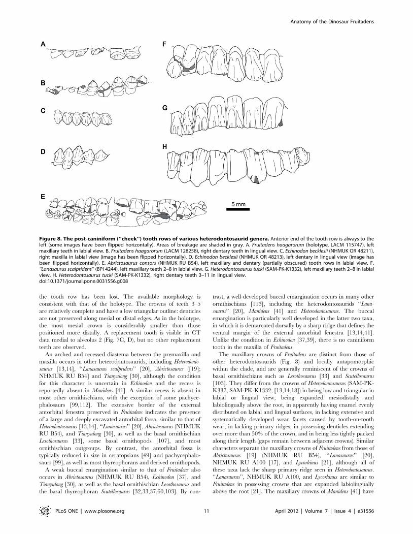
The maxillary crowns of Fruitadens are distinct from those of
other heterodontosaurids (Fig. 8) and locally autapomorphic
within the clade, and are generally reminiscent of the crowns of
basal ornithischians such as Lesothosaurus [33] and Scutellosaurus
[103]. They differ from the crowns of Heterodontosaurus (SAM-PK-
K337, SAM-PK-K1332; [13,14,18]) in being low and triangular in
labial or lingual view, being expanded mesiodistally and
labiolingually above the root, in apparently having enamel evenly
distributed on labial and lingual surfaces, in lacking extensive and
systematically developed wear facets caused by tooth-on-tooth
wear, in lacking primary ridges, in possessing denticles extending
over more than 50% of the crown, and in being less tightly packed
along their length (gaps remain between adjacent crowns). Similar
characters separate the maxillary crowns of Fruitadens from those of
Abrictosaurus [19] (NHMUK RU B54), ‘‘Lanasaurus’’ [20],
NHMUK RU A100 [17], and Lycorhinus [21], although all of
these taxa lack the sharp primary ridge seen in Heterodontosaurus.
‘‘Lanasaurus’’, NHMUK RU A100, and Lycorhinus are similar to
Fruitadens in possessing crowns that are expanded labiolingually
above the root [21]. The maxillary crowns of Manidens [41] have
Figure 8. The post-caniniform (‘‘cheek’’) tooth rows of various heterodontosaurid genera. Anterior end of the tooth row is always to theleft (some images have been flipped horizontally). Areas of breakage are shaded in gray. A. Fruitadens haagarorum (holotype, LACM 115747), leftmaxillary teeth in labial view. B. Fruitadens haagarorum (LACM 128258), right dentary teeth in lingual view. C. Echinodon becklesii (NHMUK OR 48211),right maxilla in labial view (image has been flipped horizontally). D. Echinodon becklesii (NHMUK OR 48213), left dentary in lingual view (image hasbeen flipped horizontally). E. Abrictosaurus consors (NHMUK RU B54), left maxillary and dentary (partially obscured) tooth rows in labial view. F.‘‘Lanasaurus scalpridens’’ (BPI 4244), left maxillary teeth 2–8 in labial view. G. Heterodontosaurus tucki (SAM-PK-K1332), left maxillary teeth 2–8 in labialview. H. Heterodontosaurus tucki (SAM-PK-K1332), right dentary teeth 3–11 in lingual view.doi:10.1371/journal.pone.0031556.g008
Anatomy of the Dinosaur Fruitadens
PLoS ONE | www.plosone.org 11 April 2012 | Volume 7 | Issue 4 | e31556
not yet been described. The maxillary crowns of Tianyulong [30]
are similar to those of Fruitadens in being low and subtriangular,
lacking primary ridges, lacking systematic wear facets, and not
being closely packed, but differ by reportedly lacking denticles on
mesial and distal crown edges [30]. The ‘cheek’ teeth (maxillary
and dentary crowns) of Fruitadens also resemble those of Echinodon
in that they exhibit low, triangular crowns that are expanded
above the roots, lack primary ridges and systematic wear, feature
symmetrically-distributed enamel, and are widely-spaced com-
pared to the ‘cheek’ teeth of Heterodontosaurus [37]. However, the
maxillary crowns of Fruitadens differ from those of Echinodon
primarily in the possession of mesial and distal denticles that
extend over more than 50% of the apicobasal height of the crown
(rather than the apical third) and possessing subtle apicobasally
extending lineations on labial and lingual crown surfaces [38,39].
The possession of mesial and distal denticles extending over more
than 50% of the apicobasal height of the crown appears to be
autapomorphic for Fruitadens within Heterodontosauridae, al-
though it may represent a retained plesiomorphy at the level of
Ornithischia.
Both Fruitadens individuals for which skull material is preserved
and has been CT-scanned show evidence of active tooth
replacement; in contrast, of the five known specimens of
Heterodontosaurus, only one (SAM-PK-K1334) shows unambiguous
evidence of replacement [14].
Dentary. The right dentary of the holotype is the most
complete, containing nine alveoli (with the anterior tip of a tenth
alveolus at the posterior end) but lacking the anterior tip (including
the articular surface for the predentary, assuming that this element
was present as in all other ornithischians [50,59]) and the posterior
and posteroventral regions of the element (Fig. 4; supplementary
video). The left dentary of the holotype is represented only by the
anterior end (alveoli 1–4), although a greater proportion of the
symphyseal region is preserved than in its counterpart.
The holotype right dentary possesses a curved ventral margin in
lateral view that increases in dorsoventral depth posteriorly.
Although the point of maximum depth is unknown due to the
incompleteness of the posteroventral region, it is clear that the
dorsoventral depth of the posterior dentary is substantially greater
than that of the anterior dentary (this morphology is also evident in
right dentary of LACM 128258). The posterior end of the right
holotype dentary (as well as both LACM 128258 dentaries) is
upturned, suggesting the presence of a substantial coronoid
eminence. The lateral surface of the dentary is pierced by
numerous nutrient foramina. The foramina are placed at
irregularly spaced intervals in an anteroposteriorly extending line,
ventral to the tooth row (Fig. 4A, C). The most anterior of these
foramina is slightly enlarged and communicates with an anteriorly
extending channel (Fig. 4A, C: adf): this foramen is probably
equivalent to the ‘anterior dentary foramen’ noted by Sereno [33]
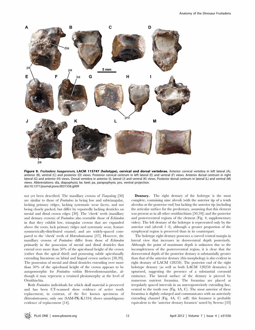
Figure 9. Fruitadens haagarorum, LACM 115747 (holotype), cervical and dorsal vertebrae. Anterior cervical vertebra in left lateral (A),anterior (B), ventral (C) and posterior (D) views. Posterior cervical centrum in left lateral (E) and ventral (F) views. Anterior dorsal centrum in rightlateral (G) and anterior (H) views. Dorsal vertebra in anterior (I), lateral (J) and ventral (K) views. Posterior dorsal centrum in lateral (L) and ventral (M)views. Abbreviations: dia, diapophysis; ke, keel; pa, parapophysis; pro, ventral projection.doi:10.1371/journal.pone.0031556.g009
Anatomy of the Dinosaur Fruitadens
PLoS ONE | www.plosone.org 12 April 2012 | Volume 7 | Issue 4 | e31556
in Lesothosaurus diagnosticus. Three additional foramina pierce the
anteroventral region of the lateral surface more ventrally on the
right dentary. The number, position and size of the nutrient
foramina on the left and right dentaries are asymmetrical. The
lateral surface of the dentary is generally convex dorsoventrally,
but the tooth row is not inset at its anterior end or at the level of
tooth 9. Between teeth 6 and 8 the tooth row is very slightly inset,
to an even lesser extent than in the maxilla. In lateral view the
tooth row is straight. Medially, the dentary is flat to gently convex
dorsoventrally beneath the tooth row. Ventrally, the Meckelian
groove is dorsoventrally narrow, and becomes very shallow
towards its anterior termination, fading out at a point approx-
imately level with the caniniform (Fig. 4B, D). Ventral to the
Meckelian groove, beneath the gap between teeth 3 and 4, there is
an elliptical foramen (with the long axis of the ellipse oriented
anteroposteriorly: Fig. 4B, D, L): the Meckelian groove curves
dorsally over this foramen. A similar foramen has not been
described in other ornithischians, including other heterodonto-
saurids, and may be autapomorphic for Fruitadens.
The small symphyseal region is positioned anteroventrally, with
its dorsal margin marked by a low curved ridge (Fig. 4B, D, L). At
its anterior end (anterior to the inferred position of tooth 1) this
ridge is nearly horizontal; ventral to tooth 1 the ridge curves
posterodorsally, fading out ventral to the anterior part of the
caniniform (tooth 2). This ridge is positioned about two thirds of
the way down the bone, so that the symphysis is limited to the
ventral third of the element. Ventral to the ridge, the symphyseal
surface is divided into anterior and posterior concavities, which are
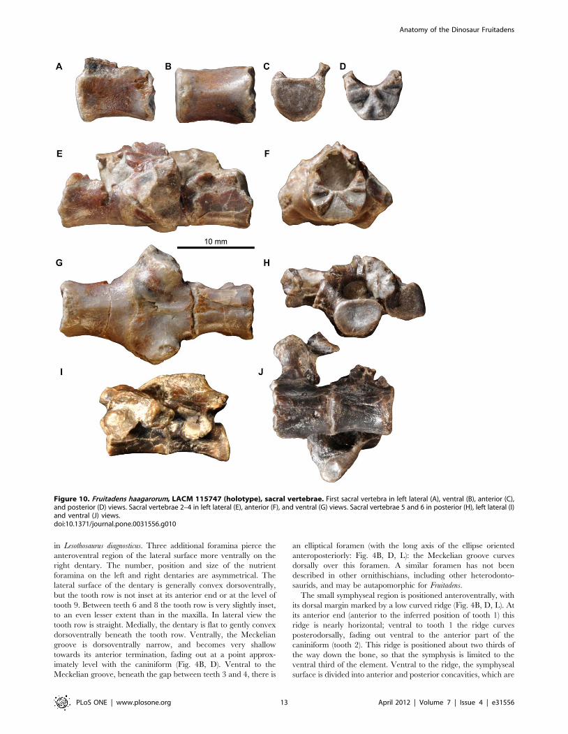
Figure 10. Fruitadens haagarorum, LACM 115747 (holotype), sacral vertebrae. First sacral vertebra in left lateral (A), ventral (B), anterior (C),and posterior (D) views. Sacral vertebrae 2–4 in left lateral (E), anterior (F), and ventral (G) views. Sacral vertebrae 5 and 6 in posterior (H), left lateral (I)and ventral (J) views.doi:10.1371/journal.pone.0031556.g010
Anatomy of the Dinosaur Fruitadens
PLoS ONE | www.plosone.org 13 April 2012 | Volume 7 | Issue 4 | e31556
separated by a saddle-like convexity (Fig. 4B, D, L). A small
foramen occurs dorsally within the anterior concavity. Dorsal to
the ridge and the symphyseal region, the medial surface of the
anterior end is depressed and covered by a large oval concavity.
The symphysis is not developed into a medially directed ‘spout-
like’ process as occurs in most ornithischians [59]. The contact
between the dentary and the predentary is not preserved in any
specimen.
Parts of teeth are preserved in eight of the nine tooth positions
(teeth 2–9) of the holotype right dentary, but in all cases the crowns
are entirely, or almost entirely, missing. The second tooth position
has an alveolus that is expanded transversely and anteroposteri-
orly, and the crown of the tooth (tooth 2) contained within it was
presumably caniniform (see below), although only the root of this
tooth is preserved (Fig. 4). The caniniform did not exceed the
maximum mesiodistal and labiolingual diameters of the largest
post-caniniform teeth (crowns of teeth 6–7). CT data show that the
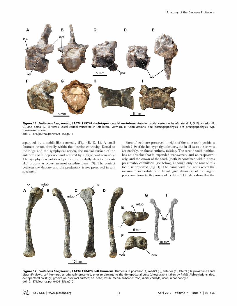
Figure 11. Fruitadens haagarorum, LACM 115747 (holotype), caudal vertebrae. Anterior caudal vertebrae in left lateral (A, D, F), anterior (B,G), and dorsal (C, E) views. Distal caudal vertebrae in left lateral view (H, I). Abbreviations: poz, postzygapophysis; prz, prezygapophysis; tvp,transverse process.doi:10.1371/journal.pone.0031556.g011
Figure 12. Fruitadens haagarorum, LACM 120478, left humerus. Humerus in posterior (A) medial (B), anterior (C), lateral (D), proximal (E) anddistal (F) views. Left humerus as originally preserved, prior to damage to the deltopectoral crest (photographs taken by PMG). Abbreviations: dpc,deltopectoral crest; gr, groove on proximal surface; he, head; mtub, medial tubercle; rcon, radial condyle; ucon, ulnar condyle.doi:10.1371/journal.pone.0031556.g012
Anatomy of the Dinosaur Fruitadens
PLoS ONE | www.plosone.org 14 April 2012 | Volume 7 | Issue 4 | e31556
root of the caniniform was compressed transversely with an oval
outline (Fig. 4M, N), and extended posteroventrally at a distinct
angle to the alveolar margin, with the root tapering mesiodistally
and labiolingually towards its base and reaching almost to the
ventral surface of the dentary. It extends below the root of tooth 3.
Anterior to the caniniform there is a pit with an oval (right
dentary: Fig. 4G, I), or circular (left dentary), outline, which (by
comparison with LACM 128258; see below) probably represents
an additional small alveolus (for tooth 1). CT data show no trace of
a tooth in this alveolus, and that the alveolus is relatively shallow.
This first alveolus is separated by a short bony margin from the
anterior end of the element (as preserved) on the left side: a short
diastema therefore separated the predentary from the first dentary
tooth.
No diastema occurs between the caniniform and tooth 3. Tooth
3 is similar in size to the alveolus of tooth 1, but is less than 50% of
the anteroposterior length of teeth 4–10. Tooth 3 has a root that
extends for less than 50% of the depth of the dentary. Posterior to
tooth 3, the remaining teeth increase in size to a maximum in
tooth 6, and then decrease in size, with tooth 9 being similar in size
to tooth 5 (based upon alveolar dimensions). Although the dentary
is broken posterior to tooth 9, the cross-section of the break
indicates the presence of at least one additional tooth, meaning
that the dentary tooth count in the holotype was at least 10.
All of the erupted crowns are badly damaged (Fig. 4). The
crowns are expanded mesiodistally and labiolingually above the
root. This expansion is similar on labial and lingual sides. On the
lingual side of the crown of tooth 6, basal ridges that connect to the
mesial and distal most denticles are evident. A similar, weakly
developed ridge is visible on the distal margin of the labial surface
of this crown, but it is unclear if a mesial ridge occurs. Several
subtle apicobasally extending ridges are visible on the lingual
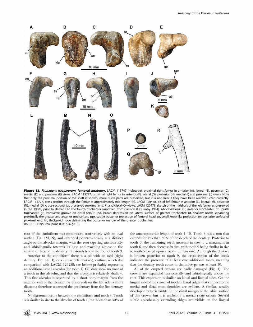
Figure 13. Fruitadens haagarorum, femoral anatomy. LACM 115747 (holotype), proximal right femur in anterior (A), lateral (B), posterior (C),medial (D) and proximal (E) views. LACM 115727, proximal right femur in anterior (F), lateral (G), posterior (H), medial (I) and proximal (J) views. Notethat only the proximal portion of the shaft is shown; more distal parts are preserved, but it is not clear if they have been reconstructed correctly.LACM 115727, cross section through the femur at approximately mid-length (K). LACM 120478, distal left femur in anterior (L), lateral (M), posterior(N), medial (O), cross-sectional (at preserved proximal end: P) and distal (Q) views. LACM 120478, sketch of the midshaft of the left femur as preservedin the 1980s, prior to damage to the fourth trochanter (modified from Callison & Quimby 1984). Abbreviations: atr, anterior trochanter; ftr, fourthtrochanter; gr, transverse groove on distal femur; lpd, broad depression on lateral surface of greater trochanter; nt, shallow notch separatingproximally the greater and anterior trochanters; ppr, subtle posterior projection of femoral head; pr, small knob-like projection on posterior surface ofproximal end; tri, thickened ridge delimiting the posterior margin of the greater trochanter.doi:10.1371/journal.pone.0031556.g013
Anatomy of the Dinosaur Fruitadens
PLoS ONE | www.plosone.org 15 April 2012 | Volume 7 | Issue 4 | e31556

surface of the base of the crown of tooth 7. The crowns are
moderately closely packed (adjacent alveoli are continuous with
one another), and would likely have slightly overlapped one
another or at least contacted one another. The roots of the crowns
are transversely compressed with oval cross-sections and are
vertically oriented (Fig. 4H). Each tapers towards its base and has a
broad pulp cavity that is surrounded by a thick layer of dentine/
enamel (Fig. 4M), and each root generally extends for about two-
thirds of the dorsoventral height of the dentary (Fig. 4E, F).
Figure 14. Fruitadens haagarorum, distal hindlimb. LACM 120478, articulated left tibia, fibula and astragalocalcaneum in anterior (A), medial (B),posterior (C), lateral (D), proximal (E) and distal (F) views. LACM 115747 (holotype), proximal left tibia in lateral (G) and medial (H) views. LACM 115727,distal left tibia with attached astragalocalcaneum in anterior (I), medial (J), posterior (K), lateral (L) and distal (M) views. LACM 115747 (holotype), distalright tibia in distal (N) and anterior (O) views. LACM 120602, left astragalocalcaneum in anterior (P) and proximal (Q) views. Abbreviations: amsh,anteromedial sheet of tibia; asp, ascending process; cal, calcaneum; cnc, cnemial crest; fibc, fibular condyle; for, foramen; innc, inner condyle; int,notch between inner condyle and fibular condyle.doi:10.1371/journal.pone.0031556.g014
Anatomy of the Dinosaur Fruitadens
PLoS ONE | www.plosone.org 16 April 2012 | Volume 7 | Issue 4 | e31556
Clear evidence of tooth replacement is visible in the right
dentary. Medial to the tooth row, a thin strip of bone separates the
alveoli from the medial surface of the dentary. Slit-like
replacement foramina are visible within this bony strip ventrome-
dial to the crowns of teeth 5 and 8 (Fig. 4B, D). These teeth appear
to be among the most heavily erupted (i.e. a large section of the
root is visible beneath the crowns). This bony strip is absent
beneath the crown of tooth 6, which is the most recently erupted
(none of its root is visible). The crown of tooth 9 is in the process of
replacement: only a very small fragment of the erupted crown is
preserved, and a replacement crown is partially visible in medial
view. This replacement crown has coarse denticles along mesial
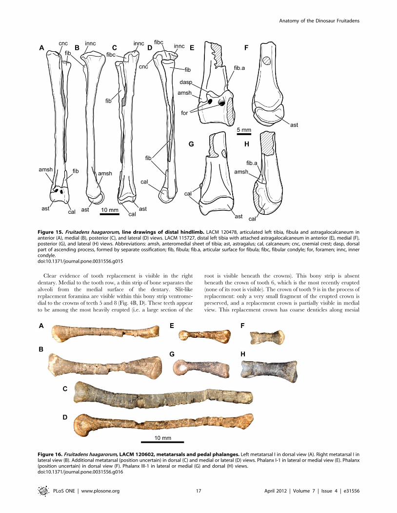
Figure 15. Fruitadens haagarorum, line drawings of distal hindlimb. LACM 120478, articulated left tibia, fibula and astragalocalcaneum inanterior (A), medial (B), posterior (C), and lateral (D) views. LACM 115727, distal left tibia with attached astragalocalcaneum in anterior (E), medial (F),posterior (G), and lateral (H) views. Abbreviations: amsh, anteromedial sheet of tibia; ast, astragalus; cal, calcaneum; cnc, cnemial crest; dasp, dorsalpart of ascending process, formed by separate ossification; fib, fibula; fib.a, articular surface for fibula; fibc, fibular condyle; for, foramen; innc, innercondyle.doi:10.1371/journal.pone.0031556.g015
Figure 16. Fruitadens haagarorum, LACM 120602, metatarsals and pedal phalanges. Left metatarsal I in dorsal view (A). Right metatarsal I inlateral view (B). Additional metatarsal (position uncertain) in dorsal (C) and medial or lateral (D) views. Phalanx I-1 in lateral or medial view (E). Phalanx(position uncertain) in dorsal view (F). Phalanx III-1 in lateral or medial (G) and dorsal (H) views.doi:10.1371/journal.pone.0031556.g016
Anatomy of the Dinosaur Fruitadens
PLoS ONE | www.plosone.org 17 April 2012 | Volume 7 | Issue 4 | e31556
and distal surfaces, and the mesial and distal most denticles are
particularly pronounced and supported by ridges (Fig. 4H, K). CT
data provide additional information on tooth replacement, with
the three replacement teeth clearly visible (Fig. 4H). The
replacement teeth are positioned medial and slightly posterior to
the functional tooth they are replacing. The replacement teeth for
functional teeth 5 and 8 consist only of partial crowns, with coarse
denticles along mesial and distal surfaces, and the roots of
functional teeth 5 and 8 are partially resorbed. Replacement teeth
are not observed elsewhere in the dentary.
As in the maxilla, CT sections clarify the courses of the
foramina that pierce the external surface of the dentary (Fig. 4E, F,
J). The ‘anterior dentary foramen’ opens into the mandibular
canal that runs within the dentary, ventral and lateral to the tooth
roots. All other foramina positioned on the lateral, ventrolateral
and medial surfaces of the dentary connect to the mandibular
canal via smaller channels. The mandibular canal extends
posteriorly lateral to the root of the caniniform. Ventral to tooth
4 it expands in size and becomes more centrally positioned, close
to the ventral margin of the dentary. The canal continues at least
as far as tooth 7; beyond this point the posteroventral margin of
the dentary is broken away. The canal very closely approaches the
bases of the tooth roots, and in some cases communicates with the
space surrounding the roots. The canal contained the inferior
alveolar nerve as well as vasculature.
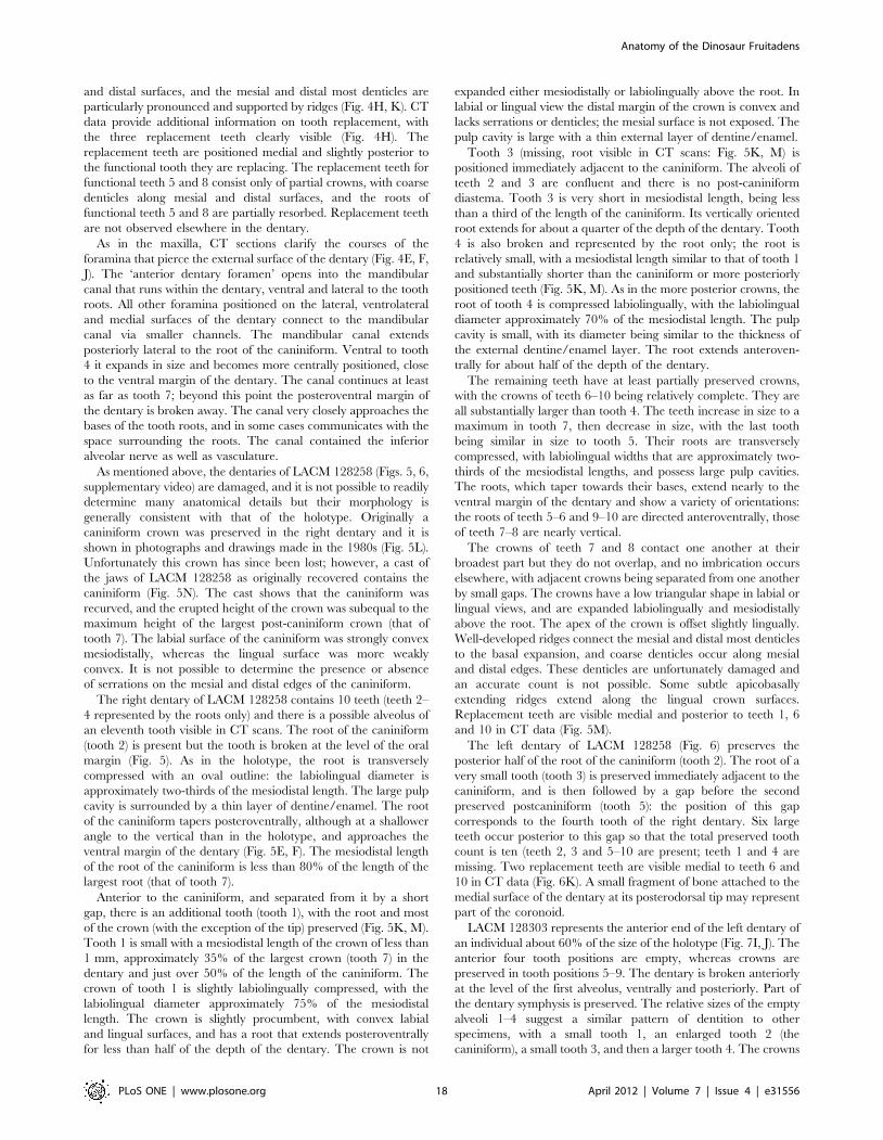
As mentioned above, the dentaries of LACM 128258 (Figs. 5, 6,
supplementary video) are damaged, and it is not possible to readily
determine many anatomical details but their morphology is
generally consistent with that of the holotype. Originally a
caniniform crown was preserved in the right dentary and it is
shown in photographs and drawings made in the 1980s (Fig. 5L).
Unfortunately this crown has since been lost; however, a cast of
the jaws of LACM 128258 as originally recovered contains the
caniniform (Fig. 5N). The cast shows that the caniniform was
recurved, and the erupted height of the crown was subequal to the
maximum height of the largest post-caniniform crown (that of
tooth 7). The labial surface of the caniniform was strongly convex
mesiodistally, whereas the lingual surface was more weakly
convex. It is not possible to determine the presence or absence
of serrations on the mesial and distal edges of the caniniform.
The right dentary of LACM 128258 contains 10 teeth (teeth 2–
4 represented by the roots only) and there is a possible alveolus of
an eleventh tooth visible in CT scans. The root of the caniniform
(tooth 2) is present but the tooth is broken at the level of the oral
margin (Fig. 5). As in the holotype, the root is transversely
compressed with an oval outline: the labiolingual diameter is
approximately two-thirds of the mesiodistal length. The large pulp
cavity is surrounded by a thin layer of dentine/enamel. The root
of the caniniform tapers posteroventrally, although at a shallower
angle to the vertical than in the holotype, and approaches the
ventral margin of the dentary (Fig. 5E, F). The mesiodistal length
of the root of the caniniform is less than 80% of the length of the
largest root (that of tooth 7).
Anterior to the caniniform, and separated from it by a short
gap, there is an additional tooth (tooth 1), with the root and most
of the crown (with the exception of the tip) preserved (Fig. 5K, M).
Tooth 1 is small with a mesiodistal length of the crown of less than
1 mm, approximately 35% of the largest crown (tooth 7) in the
dentary and just over 50% of the length of the caniniform. The
crown of tooth 1 is slightly labiolingually compressed, with the
labiolingual diameter approximately 75% of the mesiodistal
length. The crown is slightly procumbent, with convex labial
and lingual surfaces, and has a root that extends posteroventrally
for less than half of the depth of the dentary. The crown is not
expanded either mesiodistally or labiolingually above the root. In
labial or lingual view the distal margin of the crown is convex and
lacks serrations or denticles; the mesial surface is not exposed. The
pulp cavity is large with a thin external layer of dentine/enamel.
Tooth 3 (missing, root visible in CT scans: Fig. 5K, M) is
positioned immediately adjacent to the caniniform. The alveoli of
teeth 2 and 3 are confluent and there is no post-caniniform
diastema. Tooth 3 is very short in mesiodistal length, being less
than a third of the length of the caniniform. Its vertically oriented
root extends for about a quarter of the depth of the dentary. Tooth
4 is also broken and represented by the root only; the root is
relatively small, with a mesiodistal length similar to that of tooth 1
and substantially shorter than the caniniform or more posteriorly
positioned teeth (Fig. 5K, M). As in the more posterior crowns, the
root of tooth 4 is compressed labiolingually, with the labiolingual
diameter approximately 70% of the mesiodistal length. The pulp
cavity is small, with its diameter being similar to the thickness of
the external dentine/enamel layer. The root extends anteroven-
trally for about half of the depth of the dentary.
The remaining teeth have at least partially preserved crowns,
with the crowns of teeth 6–10 being relatively complete. They are
all substantially larger than tooth 4. The teeth increase in size to a
maximum in tooth 7, then decrease in size, with the last tooth
being similar in size to tooth 5. Their roots are transversely
compressed, with labiolingual widths that are approximately two-
thirds of the mesiodistal lengths, and possess large pulp cavities.
The roots, which taper towards their bases, extend nearly to the
ventral margin of the dentary and show a variety of orientations:
the roots of teeth 5–6 and 9–10 are directed anteroventrally, those
of teeth 7–8 are nearly vertical.
The crowns of teeth 7 and 8 contact one another at their
broadest part but they do not overlap, and no imbrication occurs
elsewhere, with adjacent crowns being separated from one another
by small gaps. The crowns have a low triangular shape in labial or
lingual views, and are expanded labiolingually and mesiodistally
above the root. The apex of the crown is offset slightly lingually.
Well-developed ridges connect the mesial and distal most denticles
to the basal expansion, and coarse denticles occur along mesial
and distal edges. These denticles are unfortunately damaged and
an accurate count is not possible. Some subtle apicobasally
extending ridges extend along the lingual crown surfaces.
Replacement teeth are visible medial and posterior to teeth 1, 6
and 10 in CT data (Fig. 5M).
The left dentary of LACM 128258 (Fig. 6) preserves the
posterior half of the root of the caniniform (tooth 2). The root of a
very small tooth (tooth 3) is preserved immediately adjacent to the
caniniform, and is then followed by a gap before the second
preserved postcaniniform (tooth 5): the position of this gap
corresponds to the fourth tooth of the right dentary. Six large
teeth occur posterior to this gap so that the total preserved tooth
count is ten (teeth 2, 3 and 5–10 are present; teeth 1 and 4 are
missing. Two replacement teeth are visible medial to teeth 6 and
10 in CT data (Fig. 6K). A small fragment of bone attached to the
medial surface of the dentary at its posterodorsal tip may represent
part of the coronoid.
LACM 128303 represents the anterior end of the left dentary of
an individual about 60% of the size of the holotype (Fig. 7I, J). The
anterior four tooth positions are empty, whereas crowns are
preserved in tooth positions 5–9. The dentary is broken anteriorly
at the level of the first alveolus, ventrally and posteriorly. Part of
the dentary symphysis is preserved. The relative sizes of the empty
alveoli 1–4 suggest a similar pattern of dentition to other
specimens, with a small tooth 1, an enlarged tooth 2 (the
caniniform), a small tooth 3, and then a larger tooth 4. The crowns
Anatomy of the Dinosaur Fruitadens
PLoS ONE | www.plosone.org 18 April 2012 | Volume 7 | Issue 4 | e31556

of teeth 5–9 are poorly preserved, but generally similar to those of
other Fruitadens dentaries: they are low, subtriangular, expanded
mesiodistally and labiolingually above the root, with coarse
denticles, and apicobasally extending ridges occur on the lingual
crown surface.
The dentary of Fruitadens strongly resembles that of Echinodon
(NHMUK OR 48213, 48215a, 48215b), which also tapers
anteriorly, possesses a strongly upturned posterior end, lacks a
well-developed buccal emargination, and possesses an anterior
dentary foramen associated with an anteriorly extending groove
[37]. Moreover, although the dentaries of Echinodon are trans-
versely crushed, the form of the symphyseal area (NHMUK OR
48215b) is essentially identical to that of Fruitadens. By contrast, the
dentary of Heterodontosaurus is slightly dorsoventrally expanded at its
anterior end, has a nearly straight ventral margin, a deep buccal
emargination, the anterior dentary foramen is not associated with
a laterally positioned groove, and the symphyseal surface is not
restricted to the ventral third of the element [14]. The dentaries of
Tianyulong and Abrictosaurus are also slightly expanded at their
anterior ends and do not taper anteriorly [19,30]. Both have weak
buccal emarginations similar to that of Fruitadens. The dentary of
Manidens appears to be much more robust than that of Fruitadens
[41].
The presence of a dentary caniniform is a common hetero-
dontosaurid character that is absent in almost all other
ornithischians, with the exception of some, but not all,
pachycephalosaurs [112] and which is also absent in Echinodon
[37] and Abrictosaurus [19]. An enlarged caniniform tooth
positioned anteriorly within the dentary and a ‘subnarial gap’
occurs in basal theropods [61,114], and the possible homology of
this character with the dentary caniniform of heterodontosaurids is
worthy of future examination. The dentary caniniform of Fruitadens
differs from those of Heterodontosaurus [14], Lycorhinus [21],
NHMUK RU A100 [17], and Tianyulong [30] in being relatively
short apicobasally, and not exceeding in size the largest
postcaniniform teeth. The presence of an apicobasally short
caniniform appears therefore to be autapomorphic for Fruitadens
within Heterodontosauridae, although the caniniform of Manidens
may also be relatively short [41]. Moreover, the dentary
caniniform of Heterodontosaurus further differs from those of
Fruitadens and Tianyulong [30] in being less strongly recurved [14].
The presence of an additional tooth anterior to the dentary
caniniform is a unique character of Fruitadens, being absent in all
other heterodontosaurids [13,14,21,30] and other ornithischians
that possess a dentary caniniform [112], although two additional
teeth occur anterior to the similar enlarged anterior caniniform of
basal theropods [114]. Interestingly, this additional tooth in
Fruitadens is only preserved in the smaller dentary (LACM 128258)
and the first alveolus is empty on both sides in the larger holotype
(LACM 115747) despite the fact that teeth (or at least tooth roots)
are preserved in all other tooth positions. Further specimens are
required to determine the significance of this observation with
regard to sexual dimorphism, individual variation and ontogeny:
e.g. is it possible that the ‘pre-caniniform’ occurs only in early
ontogenetic stages of Fruitadens and is later lost?
The morphology of the postcaniniform dentary teeth (i.e. the
‘cheek’ teeth) is highly similar to that of the maxillary teeth, and
the same characters that distinguish the maxillary teeth of
Fruitadens from other heterodontosaurids also distinguish the
dentary teeth (Fig. 8). The dentary teeth of Manidens are highly
distinct from those of Fruitadens in being apicobasally tall and (in
more distal crowns) highly asymmetrical in labial view, with
subdivided denticles [41].
Vertebral column: general comments. The vertebrae of
the holotype are disarticulated and there is no information
concerning their preserved positions. Callison (in [115]: 13)
incorrectly suggested that the neural arches were separated from
the centra prior to burial; parts of the neural arch are in fact
preserved in all elements. In every vertebra (with the exception of
the caudal vertebrae, in which the neural arch and centrum are
indistinguishably fused) the neurocentral sutural line is visible as a
thin line of matrix, so these elements are not fused together.
Cervical vertebrae. The most complete cervical in the
holotype is probably from the anterior end of the postaxial
column (Fig. 9A–D), based upon the wedge-shaped centrum, with
the two articular ends converging ventrally in lateral view, and the
posterior one offset ventrally. As preserved, this vertebra lacks the
zygapophyses, neural spine and the articular ends of the
parapophyses and diapophyses. The roof of the neural arch is
almost flat, and the centrum is pinched in transversely with a
ventral median ridge that is not clearly delimited from the rest of
the centrum. The flat articular ends of the centrum are
subtriangular in outline. The neural canal is proportionately
large, being as broad as the centrum. The parapophysis is
positioned anterodorsally upon the lateral surface of the centrum,
and the diapophysis is positioned at the midpoint of the neural
arch. Although the postzygapophyses are broken at their bases, a
faint ridge on the base of the dorsal surface of the right
postzygapophysis may represent an epipophysis.
One additional cervical vertebra occurs in the holotype
(probably from the posterior end of the cervical column, possibly
cervical 7 or 8), but the majority of the neural arch has broken
away above its base and only the centrum is well preserved
(Fig. 9E, F). The centrum has a prominent parapophysis on its
anterodorsal margin, positioned mostly on the base of the neural
arch but also extending onto the neurocentral suture. The
centrum has articular ends that converge dorsally in lateral view.
The anterior face of the centrum is diamond shaped and flat to
slightly convex; the posterior face is triangular in outline and flat to
slightly concave. The lateral surfaces of the centrum are strongly
pinched inwards with one large nutrient foramen (as well as
multiple smaller foramina) on each surface. A well-developed,
keel-like, ventral ridge is transversely narrow and sharp edged
posteriorly, and becomes progressively wider and more rounded
towards its anterior end (Fig. 9F). The ventral margin of the
centrum in lateral view is strongly convex (Fig. 9E).
The cervical vertebrae are generally similar to those of small-
bodied ornithischians, including Heterodontosaurus (SAM-PK-
K1332; [11]) and Hypsilophodon [106]. The anterior cervical
vertebra of Fruitadens compares well with cervical 3 of these taxa,
sharing with them the transversely pinched centrum and the
ventrally convergent and offset articular faces. The posterior
cervical centrum of Fruitadens is similar in its proportions to the
posterior cervical centra of Heterodontosaurus and Hypsilophodon;
however, the posterior cervicals of these taxa differ in possessing
concave-to-flat ventral margins in lateral view. Convex ventral
margins of cervical centra are seen in several other ornithischian
taxa, including the small ornithopods Changchunsaurus, Jeholosaurus,
and Othnielosaurus, and the basal ceratopsian Chaoyangsaurus [116].
Dorsal vertebrae. Two isolated centra in the holotype are
reminiscent of cervical vertebrae in possessing moderately pinched
lateral surfaces and a ventral keel. No trace of a parapophysis on
occurs on the anterodorsal part of either centrum, and it is
therefore likely that these vertebrae represent anterior dorsals. The
anterior articular face in both is flat to slightly convex, whereas the
posterior surface is flat to slightly concave. One of these centra
possesses a laterally compressed ventral projection at its anterior
Anatomy of the Dinosaur Fruitadens
PLoS ONE | www.plosone.org 19 April 2012 | Volume 7 | Issue 4 | e31556

end that extends 2 mm ventral to the anterior articular surface
(Fig. 9G, H). In the second centrum the ventral keel is broken.
Tiny nutrient foramina occur on the lateral surfaces of both
centra.
A third anterior dorsal vertebra in the holotype lacks the
diapophyses, most of the neural spine and zygopophyses, and the
anteroventral corner of the centrum (Fig. 9I–K). The posterior
articular face is wider than deep and gently concave. The lateral
surfaces of the centrum are gently concave anteroposteriorly (not
strongly pinched), and ventrally there is no midline keel. Nutrient
foramina occur on the lateral surfaces of the centrum. The
parapophysis is mainly positioned on the anteroventral part of the
neural arch, but also extends slightly onto the anterodorsal corner
of the centrum. The neural canal is broader than deep, with an
oval outline (Fig. 9I). The base of the neural spine is elongate,
extending for the full length of the neural arch. The left
postzygapophysis faces lateroventrally at approximately 45u to
the horizontal. Another incomplete dorsal vertebra in the holotype
has a centrum with a very similar morphology, but there is no
trace of the parapophysis on the preserved part of the neural arch.
There are two additional isolated centra in the holotype, both
with a very small part of the neural arch attached (Fig. 9L, M).
These centra are the same light grey to slightly brownish color as
the anterior section of the sacrum, so they were probably
preserved adjacent to it, and may represent the last two dorsal
vertebrae. The centra are spool-shaped, lacking ventral keels, with
lateral surfaces that are gently concave anteroposteriorly. The
articular ends are broader than deep, and are flat to slightly
concave.
A single poorly preserved dorsal vertebra occurs in LACM
128258. It has a spool-shaped centrum, with no trace of a ventral
keel, and gently concave articular faces that are broader than
deep. The neural canal is large, being as deep and nearly as broad
as the centrum. The transverse processes, prezygapophyses and
the neural spine are all damaged and incomplete. The base of the
neural spine is elongate, extending for the full length of the neural
arch. The elongate postzygapophyses extend beyond the posterior
margin of the centrum and have articular faces that face
ventrolaterally at around 10u to the horizontal.
The fragmentary nature of the dorsal column of Fruitadens, and
the extremely limited data available on the dorsal column of other
heterodontosaurids (even in Heterodontosaurus, in which the
complete dorsal column is not well exposed [11]), prevents
detailed comparisons, although the dorsal vertebrae appear to be
generally similar to those of other small-bodied ornithischians.
One unusual feature is the ventral projection on the anterior end
of the centrum of one of the anterior dorsals. A similar feature
occurs in the anterior four dorsal vertebrae of the basal
ornithopods Changchunsaurus and Jeholosaurus [116], but appears
to be absent elsewhere within Ornithischia (including Hypsilopho-
don), although the condition for this character is unknown for most
other heterodontosaurids. The ventral surfaces of the centra are
generally not exposed in the dorsal vertebrae of Heterodontosaurus
described by Santa Luca [11], with the exception of the first dorsal
which appears to lack a ventral projection.
Sacral vertebrae. The sacrum of the holotype is preserved in
three parts (Fig. 10). The first is an isolated centrum (with a very
small attached part of the neural arch), similar in morphology to
the posterior dorsals discussed above (i.e. spool-shaped with gently
excavated lateral surfaces) (Fig. 10A–D). Two features suggest that
this centrum formed part of the sacrum. First, the posterior part of
the centrum is deeply excavated by the neural canal (Fig. 10D), in
a manner similar to that seen in the other sacrals (see below), and
this excavation becomes much shallower anteriorly (Fig. 10C).
Second, the posterior articular face of the centrum has a radiating
pattern of ridges and grooves, similar to that seen in the other
sacrals (Fig. 10D).
The second part of the sacrum consists of centra 2–4 (Fig. 10E–
G), with only fragments of the neural arches preserved. The
anterior face of sacral 2 has a U-shaped outline, with the dorsal
surface deeply excavated by the neural canal (Fig. 10F). The
articular face has a series of ridges and grooves, similar to those of
sacral 1. The centrum of sacral 2 is expanded transversely at its
posterior end to help support the massive sacral rib of sacral 3
(Fig. 10G). The sinuous suture between sacral centra 2 and 3 (and
the similar suture between sacral centra 3 and 4) is visible, so the
elements are not indistinguishably fused. A fragment of the left
postzygapophysis is preserved in articulation with (but not fused to)
the prezygapophysis of sacral 3, but the rest of the neural arch of
sacral 2 is missing. Sacral 3 has a centrum with a flattened ventral
margin and a subtle midline ridge, and bears a massive,
posterolaterally directed sacral rib on each lateral surface
(Fig. 10E–G). These ribs attach to almost the entire lateral surface
of the centrum as well as to the lateral surface of the neural arch.
The neural arch is poorly preserved: only the left prezygapophysis
is visible. The centrum of sacral 4 has a sinuous suture with sacral
3, a flattened ventral margin (with a very weak midline groove),
and a deeply excavated neural canal. The posterior articular
surface has a U-shaped outline, and is covered by a series of ridges
and grooves. The neural arch of this vertebra is almost entirely
missing and the morphology of the sacral rib is unknown.
The third part of the sacrum consists of two articulated
vertebrae (Fig. 10H–J). The first of these sacrals (sacral 5) has a
centrum with a U-shaped anterior articular face, a deeply
excavated neural canal, and a slightly flattened ventral surface.
The anterior articular face has a pattern of ridges and grooves that
appears to match that of the posterior surface of sacral 4,
suggesting that the two were originally in articulation. A large
laterally directed sacral rib is preserved on the left side, borne on
the anterodorsal corner of the centrum, and partially supported by
the preceding sacral. This sacral rib does not appear tobe present
on the right side: instead, a massive sacral rib contacts the
posterodorsal end of the centrum (Fig. 10J), as well as most of the
lateral surface of the neural arch of the succeeding centrum (sacral
6). The morphology of the sacrum is therefore asymmetrical in this
region, although it is possible that the right side is damaged, with
the sacral rib being posteriorly displaced. Sacral 6 has a smooth
and gently concave posterior articular face (Fig. 10H) with an oval
outline (being wider transversely than dorsoventrally deep), and
the neural canal is small and does not extensively excavate the
centrum. The neural arch is incomplete but carries a large sacral
rib on each lateral surface.
Information on the sacra of other heterodontosaurids is limited
to a single specimen of Heterodontosaurus in which the sacrum is
largely hidden by the articulated ilia [11], the sacrum of Manidens,
which is only briefly described [41], and the incompletely
prepared postcranium of the holotype of Abrictosaurus (NHMUK
RU B54). Fruitadens is similar to Heterodontosaurus [11], Manidens
[41], most basal ornithopods [106], and marginocephalians
[99,101] in possessing six sacral vertebrae. By contrast, basal
ornithischians such as Lesothosaurus [33] and Agilisaurus [105]
possess five sacral vertebrae, and five sacrals may also occur in
Abrictosaurus (NHMUK RU B54). Although Heterodontosaurus and
Fruitadens possess a sacral count of six, there are differences
between the sacra of these taxa and those of most basal
ornithopods, ceratopsians and pachycephalosaurs. In most repre-
sentatives of the latter clades, sacrals 2–6 possess large sacral ribs
on the lateral surfaces of their centra that contact the main body of
Anatomy of the Dinosaur Fruitadens
PLoS ONE | www.plosone.org 20 April 2012 | Volume 7 | Issue 4 | e31556

the ilium [99,106] (Archaeoceratops, IVPP V11114). By contrast, in
Heterodontosaurus [11] and Fruitadens, only four large sacral ribs
attach to the lateral surfaces of the centra of sacrals 3–6. In
Heterodontosaurus, the ribs of sacrals 1–2 are both slender and attach
to the preacetabular process of the ilium [11], whereas there is
only a single slender rib in basal ornithopods and this rib is
attached to the preacetabular process in ceratopsians and
pachycephalosaurids. These differences in morphology suggest
that the evolution of six sacral vertebrae in heterodontosaurids
may have occurred independently to that in ornithopods,
ceratopsians and pachycephalosaurs, via the incorporation of an
additional dorsal vertebra into the sacrum.
Caudal vertebrae. The holotype contains six incompletely
preserved proximal caudal vertebrae (Fig. 11), as well as three
complete or mostly complete distal caudals. Numerous additional
vertebral fragments probably belong to the caudal series.
Neurocentral sutures appear to be indistinguishably fused in all
caudal vertebrae. A caudal vertebra with a complete and highly
elongate neural spine may represent the first caudal (Fig. 11A–C),
or one of the first few caudals, based on comparisons to
Heterodontosaurus in which the neural spine of the first caudal is
elongate and slender [11]. This vertebra has a centrum that is
longer than deep, with gently excavated lateral surfaces. Its
articular faces are broader than high, with the anterior face being
gently concave to flat and the posterior face being concave. The
posterior articular face is offset ventrally relative to the anterior
face in lateral view. The transverse processes are broken but their
bases are positioned at the approximate level of the neurocentral
suture and the processes would have projected laterally. The
postzygapophyses are positioned relatively high on the neural
arch, are widely divergent, and have large articular surfaces that
face ventrolaterally at around 25u to the horizontal. The neural
spine is transversely compressed, anteroposteriorly narrow, and
positioned posteriorly on the neural arch.
Other proximal caudals (Fig. 11D–G) are generally similar in
morphology to the first, although their articular faces tend to be
deeper and more shield-like. All of the other proximal caudals are
missing their neural spines and prezygapophyses, and most lack
the postzygapophyses. One proximal caudal possesses an elongate,
laterally projecting transverse process that exceeds the centrum in
length (Fig. 11D, E). Well-developed chevron facets do not appear
to be present.
Two of the three distal caudals are very poorly preserved
(Fig. 11I); the other is well preserved (Fig. 11H) and has a long and
low centrum (more than 2.5 times as long as high), with articular
faces that have an approximately hexagonal outline. The anterior
articular face is flattened; the posterior face is concave. The ventral
surface of the centrum is flat to slightly concave transversely, and is
separated by an anteroposteriorly extending ridge from the lateral
faces of the centrum. The lateral faces are gently concave
anteroposteriorly, and are subdivided by an anteroposteriorly
extending ridge positioned at midheight. Dorsal and ventral to this
ridge the lateral surface is concave dorsoventrally. The anterior
and posterior ends of the centrum are not beveled for articulation
with chevrons. No traces of transverse processes occur and a
neural spine is absent (although a very subtle and broad midline
ridge extends for the entire length of the neural arch). The pre-
and postzygapophyses extend only a very short distance anterior
and posterior to the faces of the centrum, and have articular faces
set at about 20–30u to the horizontal. A similar distal caudal is
preserved in LACM 120602.
Comparisons are limited due to the incomplete preservation of
the caudal vertebrae; however, all preserved caudals are closely
comparable to those of Heterodontosaurus tucki [11] as well as other
small-bodied ornithischians [106].
Humerus. The shaft of the almost complete left humerus of
LACM 120478 (Fig. 12) has been repaired in several places; as a
result the original orientation of the proximal and distal ends
relative to one another is unknown. The proximal and distal ends
of the element are expanded transversely, and coarse
endochondral bone trabeculae are visible at both ends, as in the
femur and other limb bones of this specimen. The articular head is
subspherical and positioned laterally, and is set off from the
prominent medial tuberosity by a groove and a constriction in
proximal view (Fig. 12E). The deltopectoral crest is unfortunately
missing in the specimen as currently preserved; however, earlier
photographs of the specimen (Fig. 12G, H) show that the apex of
the crest was positioned at about 30% of length, and was directed
anteriorly and slightly laterally. The anterior edge of the crest was
gently concave proximodistally in lateral view. Transversely, the
anterior surface of the proximal third of the element is concave
whereas the posterior surface is convex; the shaft beyond this is
subcircular in cross section for most of its length. At the distal end,
near the condyles, the shaft becomes expanded transversely and
thus has a subrectangular cross-section. The long-axis of the
medial ulnar condyle is oriented anteroposteriorly and the long-
axis of the radial condyle is oriented anterolaterally-to-
posteromedially (Fig. 12F). Anteriorly there is a shallow
depression above the condyles on the distal end. There are no
well-developed ent- or ectepicondyles on the medial and lateral
surfaces of the distal end; however, there are shallow depressions
on the articular surfaces of the radial and ulnar condyles (Fig. 12F).
The humerus shares with that of Heterodontosaurus the presence of
a constriction in proximal view between the head and the medial
tubercle [11]. This constriction seems to be absent in other
ornithischians, and may therefore represent a heterodontosaurid
synapomorphy [61]. However, the deltopectoral crest of Fruitadens
is proportionally smaller than the well-developed crest of
Heterodontosaurus, and the distal humerus lacks the well-developed
ectepicondyle that occurs in Heterodontosaurus [11]. With the
exception of the proximal constriction, there are no characters
that clearly distinguish the humerus of Fruitadens from that of most
other small-bodied ornithischians (e.g. Hypsilophodon [106]).
Femur. The holotype includes the proximal end of the right
femur, broken distal to the base of the anterior trochanter
(Fig. 13A–E); LACM 115727 contains the proximal end and most
of the shaft of the right femur (Fig. 13F–K) and the proximal end
(missing the head) of the left femur; and LACM 120478 includes
the distal 75% (including the distal end) of the left femur (Fig. 13L–
R). The proximal femora of LACM 115747 and LACM 115727
are similar in size (LACM 115747 being a slightly larger
individual), while LACM 120478 is considerably smaller in size.
The distal end of LACM 120478 shows longitudinally oriented
coarse endochondral bone trabeculae consistent with a juvenile
that is undergoing extensive longitudinal growth at the time of
death [61: ESM].
The shaft is arched anteriorly along its length in lateral view
(LACM 120478; Fig. 13M). The shaft ventral to the position of the
fourth trochanter curves laterally, so that in anterior and posterior
views the lateral surface is gently concave and the medial surface
gently convex. Immediately proximal to the fourth trochanter the
shaft of LACM 120478 is broken, but appears to be beginning to
curve medially. In LACM 115727 the femur is arched strongly
laterally along its length in anterior and posterior views, but the
shaft has been broken and repaired in several positions and may
not reflect exactly the original morphology. As a result, the
Anatomy of the Dinosaur Fruitadens
PLoS ONE | www.plosone.org 21 April 2012 | Volume 7 | Issue 4 | e31556
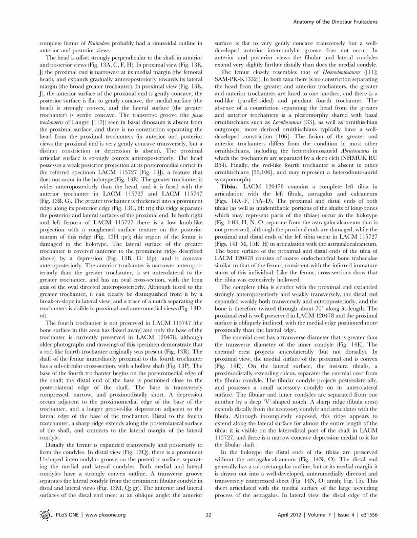
complete femur of Fruitadens probably had a sinusoidal outline in
anterior and posterior views.
The head is offset strongly perpendicular to the shaft in anterior
and posterior views (Fig. 13A, C, F, H). In proximal view (Fig. 13E,
J) the proximal end is narrowest at its medial margin (the femoral
head), and expands gradually anteroposteriorly towards its lateral
margin (the broad greater trochanter). In proximal view (Fig. 13E,
J), the anterior surface of the proximal end is gently concave, the
posterior surface is flat to gently concave, the medial surface (the
head) is strongly convex, and the lateral surface (the greater
trochanter) is gently concave. The transverse groove (the fossa
trochanteris of Langer [117]) seen in basal dinosaurs is absent from
the proximal surface, and there is no constriction separating the
head from the proximal trochanters (in anterior and posterior
views the proximal end is very gently concave transversely, but a
distinct constriction or depression is absent). The proximal
articular surface is strongly convex anteroposteriorly. The head
possesses a weak posterior projection at its posteromedial corner in
the referred specimen LACM 115727 (Fig. 13J), a feature that
does not occur in the holotype (Fig. 13E). The greater trochanter is
wider anteroposteriorly than the head, and it is fused with the
anterior trochanter in LACM 115727 and LACM 115747
(Fig. 13B, G). The greater trochanter is thickened into a prominent
ridge along its posterior edge (Fig. 13C, H: tri); this ridge separates
the posterior and lateral surfaces of the proximal end. In both right
and left femora of LACM 115727 there is a low knob-like
projection with a roughened surface texture on the posterior
margin of this ridge (Fig. 13H :pr); this region of the femur is
damaged in the holotype. The lateral surface of the greater
trochanter is covered (anterior to the prominent ridge described
above) by a depression (Fig. 13B, G: ldp), and is concave
anteroposteriorly. The anterior trochanter is narrower anteropos-
teriorly than the greater trochanter, is set anterolateral to the
greater trochanter, and has an oval cross-section, with the long
axis of the oval directed anteroposteriorly. Although fused to the
greater trochanter, it can clearly be distinguished from it by a
break-in-slope in lateral view, and a trace of a notch separating the
trochanters is visible in proximal and anteromedial views (Fig. 13D:
nt).
The fourth trochanter is not preserved in LACM 115747 (the
bone surface in this area has flaked away) and only the base of the
trochanter is currently preserved in LACM 120478, although
older photographs and drawings of this specimen demonstrate that
a rod-like fourth trochanter originally was present (Fig. 13R). The
shaft of the femur immediately proximal to the fourth trochanter
has a sub-circular cross-section, with a hollow shaft (Fig. 13P). The
base of the fourth trochanter begins on the posteromedial edge of
the shaft; the distal end of the base is positioned close to the
posterolateral edge of the shaft. The base is transversely
compressed, narrow, and proximodistally short. A depression
occurs adjacent to the proximomedial edge of the base of the
trochanter, and a longer groove-like depression adjacent to the
lateral edge of the base of the trochanter. Distal to the fourth
tranchanter, a sharp ridge extends along the posterolateral surface
of the shaft, and connects to the lateral margin of the lateral
condyle.
Distally the femur is expanded transversely and posteriorly to
form the condyles. In distal view (Fig. 13Q), there is a prominent
U-shaped intercondylar groove on the posterior surface, separat-
ing the medial and lateral condyles. Both medial and lateral
condyles have a strongly convex outline. A transverse groove
separates the lateral condyle from the prominent fibular condyle in
distal and lateral views (Fig. 13M, Q: gr). The anterior and lateral
surfaces of the distal end meet at an oblique angle: the anterior
surface is flat to very gently concave transversely but a well-
developed anterior intercondylar groove does not occur. In
anterior and posterior views the fibular and lateral condyles
extend very slightly further distally than does the medial condyle.
The femur closely resembles that of Heterodontosaurus ([11];
SAM-PK-K1332]). In both taxa there is no constriction separating
the head from the greater and anterior trochanters, the greater
and anterior trochanters are fused to one another, and there is a
rod-like (parallel-sided) and pendant fourth trochanter. The
absence of a constriction separating the head from the greater
and anterior trochanters is a plesiomorphy shared with basal
ornithischians such as Lesothosaurus [33], as well as ornithischian
outgroups; more derived ornithischians typically have a well-
developed constriction [106]. The fusion of the greater and
anterior trochanters differs from the condition in most other
ornithischians, including the heterodontosaurid Abrictosaurus in
which the trochanters are separated by a deep cleft (NHMUK RU
B54). Finally, the rod-like fourth trochanter is absent in other
ornithischians [33,106], and may represent a heterodontosaurid
synapomorphy.
Tibia. LACM 120478 contains a complete left tibia in
articulation with the left fibula, astragalus and calcaneum
(Figs. 14A–F, 15A–D). The proximal and distal ends of both
tibiae (as well as unidentifiable portions of the shafts of long-bones
which may represent parts of the tibiae) occur in the holotype
(Fig. 14G, H, N, O; separate from the astragalocalcaneum that is
not preserved), although the proximal ends are damaged, while the
proximal and distal ends of the left tibia occur in LACM 115727
(Figs. 14I–M, 15E–H) in articulation with the astragalocalcaneum.
The bone surface of the proximal and distal ends of the tibia of
LACM 120478 consists of coarse endochondral bone trabeculae
similar to that of the femur, consistent with the inferred immature
status of this individual. Like the femur, cross-sections show that
the tibia was extensively hollowed.
The complete tibia is slender with the proximal end expanded
strongly anteroposteriorly and weakly transversely, the distal end
expanded weakly both transversely and anteroposteriorly, and the
bone is therefore twisted through about 70u along its length. The
proximal end is well preserved in LACM 120478 and the proximal
surface is obliquely inclined, with the medial edge positioned more
proximally than the lateral edge.
The cnemial crest has a transverse diameter that is greater than
the transverse diameter of the inner condyle (Fig. 14E). The
cnemial crest projects anterolaterally (but not dorsally). In
proximal view, the medial surface of the proximal end is convex
(Fig. 14E). On the lateral surface, the insisura tibialis, a
proximodistally extending sulcus, separates the cnemial crest from
the fibular condyle. The fibular condyle projects posterolaterally,
and possesses a small accessory condyle on its anterolateral
surface. The fibular and inner condyles are separated from one
another by a deep ‘V’-shaped notch. A sharp ridge (fibula crest)
extends distally from the accessory condyle and articulates with the
fibula. Although incompletely exposed, this ridge appears to
extend along the lateral surface for almost the entire length of the
tibia: it is visible on the laterodistal part of the shaft in LACM
115727, and there is a narrow concave depression medial to it for
the fibular shaft.
In the holotype the distal ends of the tibiae are preserved
without the astragalocalcaneum (Fig. 14N, O). The distal end
generally has a sub-rectangular outline, but at its medial margin it
is drawn out into a well-developed, anteromedially directed and
transversely compressed sheet (Fig. 14N, O: amsh; Fig. 15). This
sheet articulated with the medial surface of the large ascending
process of the astragalus. In lateral view the distal edge of the
Anatomy of the Dinosaur Fruitadens
PLoS ONE | www.plosone.org 22 April 2012 | Volume 7 | Issue 4 | e31556
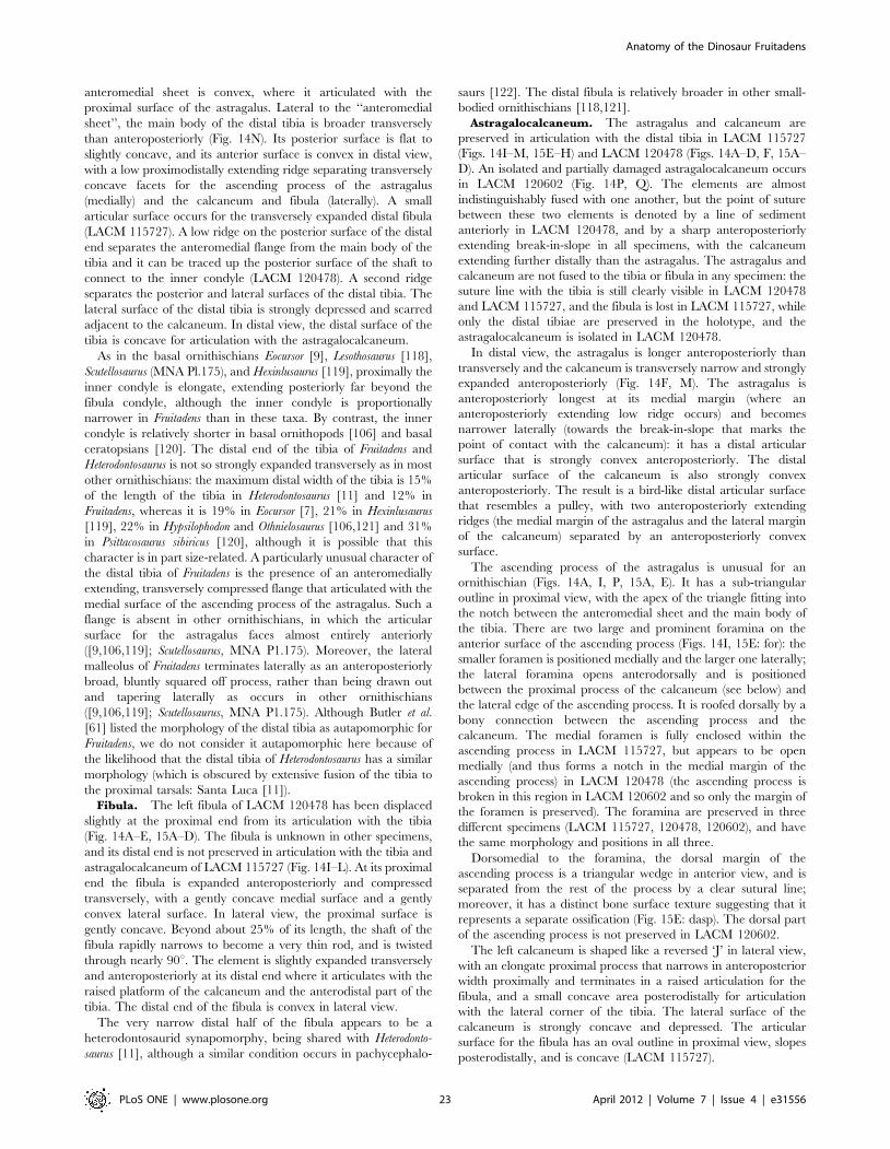
anteromedial sheet is convex, where it articulated with the
proximal surface of the astragalus. Lateral to the ‘‘anteromedial
sheet’’, the main body of the distal tibia is broader transversely
than anteroposteriorly (Fig. 14N). Its posterior surface is flat to
slightly concave, and its anterior surface is convex in distal view,
with a low proximodistally extending ridge separating transversely
concave facets for the ascending process of the astragalus
(medially) and the calcaneum and fibula (laterally). A small
articular surface occurs for the transversely expanded distal fibula
(LACM 115727). A low ridge on the posterior surface of the distal
end separates the anteromedial flange from the main body of the
tibia and it can be traced up the posterior surface of the shaft to
connect to the inner condyle (LACM 120478). A second ridge
separates the posterior and lateral surfaces of the distal tibia. The
lateral surface of the distal tibia is strongly depressed and scarred
adjacent to the calcaneum. In distal view, the distal surface of the
tibia is concave for articulation with the astragalocalcaneum.
As in the basal ornithischians Eocursor [9], Lesothosaurus [118],
Scutellosaurus (MNA Pl.175), and Hexinlusaurus [119], proximally the
inner condyle is elongate, extending posteriorly far beyond the
fibula condyle, although the inner condyle is proportionally
narrower in Fruitadens than in these taxa. By contrast, the inner
condyle is relatively shorter in basal ornithopods [106] and basal
ceratopsians [120]. The distal end of the tibia of Fruitadens and
Heterodontosaurus is not so strongly expanded transversely as in most
other ornithischians: the maximum distal width of the tibia is 15%
of the length of the tibia in Heterodontosaurus [11] and 12% in
Fruitadens, whereas it is 19% in Eocursor [7], 21% in Hexinlusaurus
[119], 22% in Hypsilophodon and Othnielosaurus [106,121] and 31%
in Psittacosaurus sibiricus [120], although it is possible that this
character is in part size-related. A particularly unusual character of
the distal tibia of Fruitadens is the presence of an anteromedially
extending, transversely compressed flange that articulated with the
medial surface of the ascending process of the astragalus. Such a
flange is absent in other ornithischians, in which the articular
surface for the astragalus faces almost entirely anteriorly
([9,106,119]; Scutellosaurus, MNA P1.175). Moreover, the lateral
malleolus of Fruitadens terminates laterally as an anteroposteriorly
broad, bluntly squared off process, rather than being drawn out
and tapering laterally as occurs in other ornithischians
([9,106,119]; Scutellosaurus, MNA P1.175). Although Butler et al.
[61] listed the morphology of the distal tibia as autapomorphic for
Fruitadens, we do not consider it autapomorphic here because of
the likelihood that the distal tibia of Heterodontosaurus has a similar
morphology (which is obscured by extensive fusion of the tibia to
the proximal tarsals: Santa Luca [11]).
Fibula. The left fibula of LACM 120478 has been displaced
slightly at the proximal end from its articulation with the tibia
(Fig. 14A–E, 15A–D). The fibula is unknown in other specimens,
and its distal end is not preserved in articulation with the tibia and
astragalocalcaneum of LACM 115727 (Fig. 14I–L). At its proximal
end the fibula is expanded anteroposteriorly and compressed
transversely, with a gently concave medial surface and a gently
convex lateral surface. In lateral view, the proximal surface is
gently concave. Beyond about 25% of its length, the shaft of the
fibula rapidly narrows to become a very thin rod, and is twisted
through nearly 90u. The element is slightly expanded transversely
and anteroposteriorly at its distal end where it articulates with the
raised platform of the calcaneum and the anterodistal part of the
tibia. The distal end of the fibula is convex in lateral view.
The very narrow distal half of the fibula appears to be a
heterodontosaurid synapomorphy, being shared with Heterodonto-
saurus [11], although a similar condition occurs in pachycephalo-
saurs [122]. The distal fibula is relatively broader in other small-
bodied ornithischians [118,121].
Astragalocalcaneum. The astragalus and calcaneum are
preserved in articulation with the distal tibia in LACM 115727
(Figs. 14I–M, 15E–H) and LACM 120478 (Figs. 14A–D, F, 15A–
D). An isolated and partially damaged astragalocalcaneum occurs
in LACM 120602 (Fig. 14P, Q). The elements are almost
indistinguishably fused with one another, but the point of suture
between these two elements is denoted by a line of sediment
anteriorly in LACM 120478, and by a sharp anteroposteriorly
extending break-in-slope in all specimens, with the calcaneum
extending further distally than the astragalus. The astragalus and
calcaneum are not fused to the tibia or fibula in any specimen: the
suture line with the tibia is still clearly visible in LACM 120478
and LACM 115727, and the fibula is lost in LACM 115727, while
only the distal tibiae are preserved in the holotype, and the
astragalocalcaneum is isolated in LACM 120478.
In distal view, the astragalus is longer anteroposteriorly than
transversely and the calcaneum is transversely narrow and strongly
expanded anteroposteriorly (Fig. 14F, M). The astragalus is
anteroposteriorly longest at its medial margin (where an
anteroposteriorly extending low ridge occurs) and becomes
narrower laterally (towards the break-in-slope that marks the
point of contact with the calcaneum): it has a distal articular
surface that is strongly convex anteroposteriorly. The distal
articular surface of the calcaneum is also strongly convex
anteroposteriorly. The result is a bird-like distal articular surface
that resembles a pulley, with two anteroposteriorly extending
ridges (the medial margin of the astragalus and the lateral margin
of the calcaneum) separated by an anteroposteriorly convex
surface.
The ascending process of the astragalus is unusual for an
ornithischian (Figs. 14A, I, P, 15A, E). It has a sub-triangular
outline in proximal view, with the apex of the triangle fitting into
the notch between the anteromedial sheet and the main body of
the tibia. There are two large and prominent foramina on the
anterior surface of the ascending process (Figs. 14I, 15E: for): the
smaller foramen is positioned medially and the larger one laterally;
the lateral foramina opens anterodorsally and is positioned
between the proximal process of the calcaneum (see below) and
the lateral edge of the ascending process. It is roofed dorsally by a
bony connection between the ascending process and the
calcaneum. The medial foramen is fully enclosed within the
ascending process in LACM 115727, but appears to be open
medially (and thus forms a notch in the medial margin of the
ascending process) in LACM 120478 (the ascending process is
broken in this region in LACM 120602 and so only the margin of
the foramen is preserved). The foramina are preserved in three
different specimens (LACM 115727, 120478, 120602), and have
the same morphology and positions in all three.
Dorsomedial to the foramina, the dorsal margin of the
ascending process is a triangular wedge in anterior view, and is
separated from the rest of the process by a clear sutural line;
moreover, it has a distinct bone surface texture suggesting that it
represents a separate ossification (Fig. 15E: dasp). The dorsal part
of the ascending process is not preserved in LACM 120602.
The left calcaneum is shaped like a reversed ‘J’ in lateral view,
with an elongate proximal process that narrows in anteroposterior
width proximally and terminates in a raised articulation for the
fibula, and a small concave area posterodistally for articulation
with the lateral corner of the tibia. The lateral surface of the
calcaneum is strongly concave and depressed. The articular
surface for the fibula has an oval outline in proximal view, slopes
posterodistally, and is concave (LACM 115727).
Anatomy of the Dinosaur Fruitadens
PLoS ONE | www.plosone.org 23 April 2012 | Volume 7 | Issue 4 | e31556
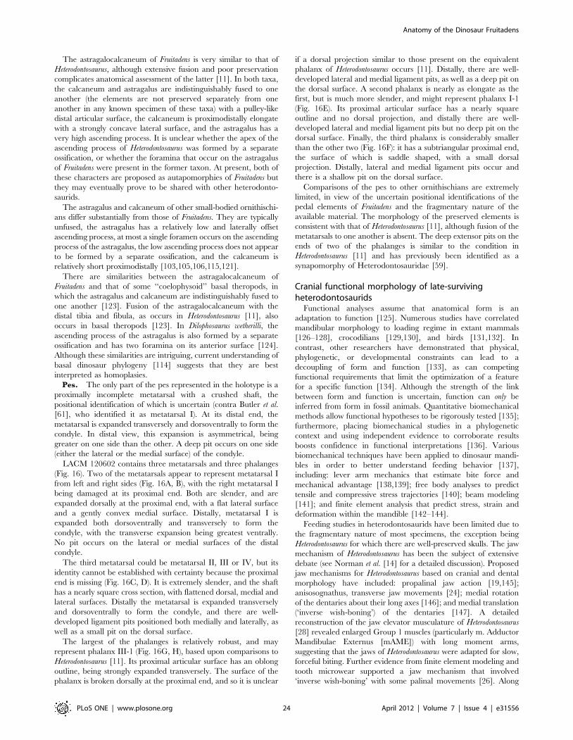
The astragalocalcaneum of Fruitadens is very similar to that of
Heterodontosaurus, although extensive fusion and poor preservation
complicates anatomical assessment of the latter [11]. In both taxa,
the calcaneum and astragalus are indistinguishably fused to one
another (the elements are not preserved separately from one
another in any known specimen of these taxa) with a pulley-like
distal articular surface, the calcaneum is proximodistally elongate
with a strongly concave lateral surface, and the astragalus has a
very high ascending process. It is unclear whether the apex of the
ascending process of Heterodontosaurus was formed by a separate
ossification, or whether the foramina that occur on the astragalus
of Fruitadens were present in the former taxon. At present, both of
these characters are proposed as autapomorphies of Fruitadens but
they may eventually prove to be shared with other heterodonto-
saurids.
The astragalus and calcaneum of other small-bodied ornithischi-
ans differ substantially from those of Fruitadens. They are typically
unfused, the astragalus has a relatively low and laterally offset
ascending process, at most a single foramen occurs on the ascending
process of the astragalus, the low ascending process does not appear
to be formed by a separate ossification, and the calcaneum is
relatively short proximodistally [103,105,106,115,121].
There are similarities between the astragalocalcaneum of
Fruitadens and that of some ‘‘coelophysoid’’ basal theropods, in
which the astragalus and calcaneum are indistinguishably fused to
one another [123]. Fusion of the astragalocalcaneum with the
distal tibia and fibula, as occurs in Heterodontosaurus [11], also
occurs in basal theropods [123]. In Dilophosaurus wetherilli, the
ascending process of the astragalus is also formed by a separate
ossification and has two foramina on its anterior surface [124].
Although these similarities are intriguing, current understanding of
basal dinosaur phylogeny [114] suggests that they are best
interpreted as homoplasies.
Pes. The only part of the pes represented in the holotype is a
proximally incomplete metatarsal with a crushed shaft, the
positional identification of which is uncertain (contra Butler et al.
[61], who identified it as metatarsal I). At its distal end, the
metatarsal is expanded transversely and dorsoventrally to form the
condyle. In distal view, this expansion is asymmetrical, being
greater on one side than the other. A deep pit occurs on one side
(either the lateral or the medial surface) of the condyle.
LACM 120602 contains three metatarsals and three phalanges
(Fig. 16). Two of the metatarsals appear to represent metatarsal I
from left and right sides (Fig. 16A, B), with the right metatarsal I
being damaged at its proximal end. Both are slender, and are
expanded dorsally at the proximal end, with a flat lateral surface
and a gently convex medial surface. Distally, metatarsal I is
expanded both dorsoventrally and transversely to form the
condyle, with the transverse expansion being greatest ventrally.
No pit occurs on the lateral or medial surfaces of the distal
condyle.
The third metatarsal could be metatarsal II, III or IV, but its
identity cannot be established with certainty because the proximal
end is missing (Fig. 16C, D). It is extremely slender, and the shaft
has a nearly square cross section, with flattened dorsal, medial and
lateral surfaces. Distally the metatarsal is expanded transversely
and dorsoventrally to form the condyle, and there are well-
developed ligament pits positioned both medially and laterally, as
well as a small pit on the dorsal surface.
The largest of the phalanges is relatively robust, and may
represent phalanx III-1 (Fig. 16G, H), based upon comparisons to
Heterodontosaurus [11]. Its proximal articular surface has an oblong
outline, being strongly expanded transversely. The surface of the
phalanx is broken dorsally at the proximal end, and so it is unclear
if a dorsal projection similar to those present on the equivalent
phalanx of Heterodontosaurus occurs [11]. Distally, there are well-
developed lateral and medial ligament pits, as well as a deep pit on
the dorsal surface. A second phalanx is nearly as elongate as the
first, but is much more slender, and might represent phalanx I-1
(Fig. 16E). Its proximal articular surface has a nearly square
outline and no dorsal projection, and distally there are well-
developed lateral and medial ligament pits but no deep pit on the
dorsal surface. Finally, the third phalanx is considerably smaller
than the other two (Fig. 16F): it has a subtriangular proximal end,
the surface of which is saddle shaped, with a small dorsal
projection. Distally, lateral and medial ligament pits occur and
there is a shallow pit on the dorsal surface.
Comparisons of the pes to other ornithischians are extremely
limited, in view of the uncertain positional identifications of the
pedal elements of Fruitadens and the fragmentary nature of the
available material. The morphology of the preserved elements is
consistent with that of Heterodontosaurus [11], although fusion of the
metatarsals to one another is absent. The deep extensor pits on the
ends of two of the phalanges is similar to the condition in
Heterodontosaurus [11] and has previously been identified as a
synapomorphy of Heterodontosauridae [59].
Cranial functional morphology of late-survivingheterodontosaurids
Functional analyses assume that anatomical form is an
adaptation to function [125]. Numerous studies have correlated
mandibular morphology to loading regime in extant mammals
[126–128], crocodilians [129,130], and birds [131,132]. In
contrast, other researchers have demonstrated that physical,
phylogenetic, or developmental constraints can lead to a
decoupling of form and function [133], as can competing
functional requirements that limit the optimization of a feature
for a specific function [134]. Although the strength of the link
between form and function is uncertain, function can only be
inferred from form in fossil animals. Quantitative biomechanical
methods allow functional hypotheses to be rigorously tested [135];
furthermore, placing biomechanical studies in a phylogenetic
context and using independent evidence to corroborate results
boosts confidence in functional interpretations [136]. Various
biomechanical techniques have been applied to dinosaur mandi-
bles in order to better understand feeding behavior [137],
including: lever arm mechanics that estimate bite force and
mechanical advantage [138,139]; free body analyses to predict
tensile and compressive stress trajectories [140]; beam modeling
[141]; and finite element analysis that predict stress, strain and
deformation within the mandible [142–144].
Feeding studies in heterodontosaurids have been limited due to
the fragmentary nature of most specimens, the exception being
Heterodontosaurus for which there are well-preserved skulls. The jaw
mechanism of Heterodontosaurus has been the subject of extensive
debate (see Norman et al. [14] for a detailed discussion). Proposed
jaw mechanisms for Heterodontosaurus based on cranial and dental
morphology have included: propalinal jaw action [19,145];
anisosognathus, transverse jaw movements [24]; medial rotation
of the dentaries about their long axes [146]; and medial translation
(‘inverse wish-boning’) of the dentaries [147]. A detailed
reconstruction of the jaw elevator musculature of Heterodontosaurus
[28] revealed enlarged Group 1 muscles (particularly m. Adductor
Mandibulae Externus [mAME]) with long moment arms,
suggesting that the jaws of Heterodontosaurus were adapted for slow,
forceful biting. Further evidence from finite element modeling and
tooth microwear supported a jaw mechanism that involved
‘inverse wish-boning’ with some palinal movements [26]. Along
Anatomy of the Dinosaur Fruitadens
PLoS ONE | www.plosone.org 24 April 2012 | Volume 7 | Issue 4 | e31556
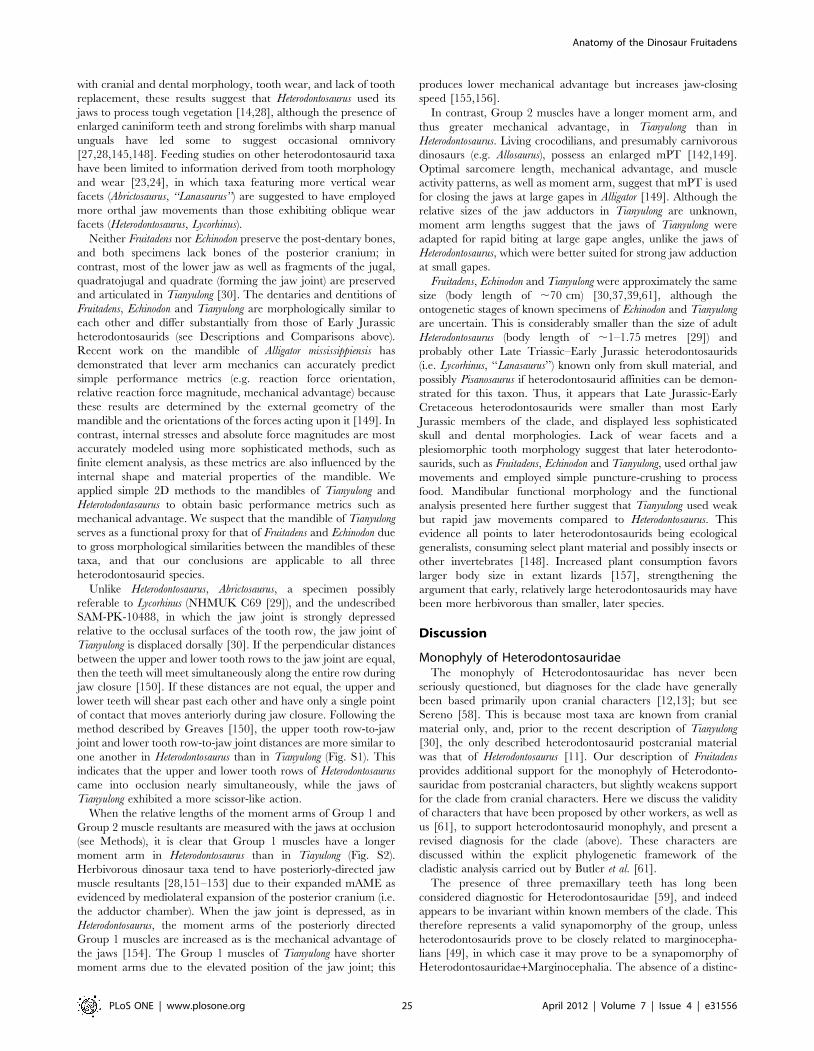
with cranial and dental morphology, tooth wear, and lack of tooth
replacement, these results suggest that Heterodontosaurus used its
jaws to process tough vegetation [14,28], although the presence of
enlarged caniniform teeth and strong forelimbs with sharp manual
unguals have led some to suggest occasional omnivory
[27,28,145,148]. Feeding studies on other heterodontosaurid taxa
have been limited to information derived from tooth morphology
and wear [23,24], in which taxa featuring more vertical wear
facets (Abrictosaurus, ‘‘Lanasaurus’’) are suggested to have employed
more orthal jaw movements than those exhibiting oblique wear
facets (Heterodontosaurus, Lycorhinus).
Neither Fruitadens nor Echinodon preserve the post-dentary bones,
and both specimens lack bones of the posterior cranium; in
contrast, most of the lower jaw as well as fragments of the jugal,
quadratojugal and quadrate (forming the jaw joint) are preserved
and articulated in Tianyulong [30]. The dentaries and dentitions of
Fruitadens, Echinodon and Tianyulong are morphologically similar to
each other and differ substantially from those of Early Jurassic
heterodontosaurids (see Descriptions and Comparisons above).
Recent work on the mandible of Alligator mississippiensis has
demonstrated that lever arm mechanics can accurately predict
simple performance metrics (e.g. reaction force orientation,
relative reaction force magnitude, mechanical advantage) because
these results are determined by the external geometry of the
mandible and the orientations of the forces acting upon it [149]. In
contrast, internal stresses and absolute force magnitudes are most
accurately modeled using more sophisticated methods, such as
finite element analysis, as these metrics are also influenced by the
internal shape and material properties of the mandible. We
applied simple 2D methods to the mandibles of Tianyulong and
Heterotodontasaurus to obtain basic performance metrics such as
mechanical advantage. We suspect that the mandible of Tianyulong
serves as a functional proxy for that of Fruitadens and Echinodon due
to gross morphological similarities between the mandibles of these
taxa, and that our conclusions are applicable to all three
heterodontosaurid species.
Unlike Heterodontosaurus, Abrictosaurus, a specimen possibly
referable to Lycorhinus (NHMUK C69 [29]), and the undescribed
SAM-PK-10488, in which the jaw joint is strongly depressed
relative to the occlusal surfaces of the tooth row, the jaw joint of
Tianyulong is displaced dorsally [30]. If the perpendicular distances
between the upper and lower tooth rows to the jaw joint are equal,
then the teeth will meet simultaneously along the entire row during
jaw closure [150]. If these distances are not equal, the upper and
lower teeth will shear past each other and have only a single point
of contact that moves anteriorly during jaw closure. Following the
method described by Greaves [150], the upper tooth row-to-jaw
joint and lower tooth row-to-jaw joint distances are more similar to
one another in Heterodontosaurus than in Tianyulong (Fig. S1). This
indicates that the upper and lower tooth rows of Heterodontosaurus
came into occlusion nearly simultaneously, while the jaws of
Tianyulong exhibited a more scissor-like action.
When the relative lengths of the moment arms of Group 1 and
Group 2 muscle resultants are measured with the jaws at occlusion
(see Methods), it is clear that Group 1 muscles have a longer
moment arm in Heterodontosaurus than in Tiayulong (Fig. S2).
Herbivorous dinosaur taxa tend to have posteriorly-directed jaw
muscle resultants [28,151–153] due to their expanded mAME as
evidenced by mediolateral expansion of the posterior cranium (i.e.
the adductor chamber). When the jaw joint is depressed, as in
Heterodontosaurus, the moment arms of the posteriorly directed
Group 1 muscles are increased as is the mechanical advantage of
the jaws [154]. The Group 1 muscles of Tianyulong have shorter
moment arms due to the elevated position of the jaw joint; this
produces lower mechanical advantage but increases jaw-closing
speed [155,156].
In contrast, Group 2 muscles have a longer moment arm, and
thus greater mechanical advantage, in Tianyulong than in
Heterodontosaurus. Living crocodilians, and presumably carnivorous
dinosaurs (e.g. Allosaurus), possess an enlarged mPT [142,149].
Optimal sarcomere length, mechanical advantage, and muscle
activity patterns, as well as moment arm, suggest that mPT is used
for closing the jaws at large gapes in Alligator [149]. Although the
relative sizes of the jaw adductors in Tianyulong are unknown,
moment arm lengths suggest that the jaws of Tianyulong were
adapted for rapid biting at large gape angles, unlike the jaws of
Heterodontosaurus, which were better suited for strong jaw adduction
at small gapes.
Fruitadens, Echinodon and Tianyulong were approximately the same
size (body length of ,70 cm) [30,37,39,61], although the
ontogenetic stages of known specimens of Echinodon and Tianyulong
are uncertain. This is considerably smaller than the size of adult
Heterodontosaurus (body length of ,1–1.75 metres [29]) and
probably other Late Triassic–Early Jurassic heterodontosaurids
(i.e. Lycorhinus, ‘‘Lanasaurus’’) known only from skull material, and
possibly Pisanosaurus if heterodontosaurid affinities can be demon-
strated for this taxon. Thus, it appears that Late Jurassic-Early
Cretaceous heterodontosaurids were smaller than most Early
Jurassic members of the clade, and displayed less sophisticated
skull and dental morphologies. Lack of wear facets and a
plesiomorphic tooth morphology suggest that later heterodonto-
saurids, such as Fruitadens, Echinodon and Tianyulong, used orthal jaw
movements and employed simple puncture-crushing to process
food. Mandibular functional morphology and the functional
analysis presented here further suggest that Tianyulong used weak
but rapid jaw movements compared to Heterodontosaurus. This
evidence all points to later heterodontosaurids being ecological
generalists, consuming select plant material and possibly insects or
other invertebrates [148]. Increased plant consumption favors
larger body size in extant lizards [157], strengthening the
argument that early, relatively large heterodontosaurids may have
been more herbivorous than smaller, later species.
Discussion
Monophyly of HeterodontosauridaeThe monophyly of Heterodontosauridae has never been
seriously questioned, but diagnoses for the clade have generally
been based primarily upon cranial characters [12,13]; but see
Sereno [58]. This is because most taxa are known from cranial
material only, and, prior to the recent description of Tianyulong
[30], the only described heterodontosaurid postcranial material
was that of Heterodontosaurus [11]. Our description of Fruitadens
provides additional support for the monophyly of Heterodonto-
sauridae from postcranial characters, but slightly weakens support
for the clade from cranial characters. Here we discuss the validity
of characters that have been proposed by other workers, as well as
us [61], to support heterodontosaurid monophyly, and present a
revised diagnosis for the clade (above). These characters are
discussed within the explicit phylogenetic framework of the
cladistic analysis carried out by Butler et al. [61].
The presence of three premaxillary teeth has long been
considered diagnostic for Heterodontosauridae [59], and indeed
appears to be invariant within known members of the clade. This
therefore represents a valid synapomorphy of the group, unless
heterodontosaurids prove to be closely related to marginocepha-
lians [49], in which case it may prove to be a synapomorphy of
Heterodontosauridae+Marginocephalia. The absence of a distinc-
Anatomy of the Dinosaur Fruitadens
PLoS ONE | www.plosone.org 25 April 2012 | Volume 7 | Issue 4 | e31556
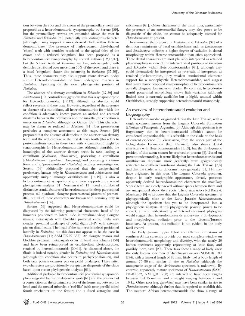
tion between the root and the crown of the premaxillary teeth was
proposed as a heterodontosaurid synapomorphy by Sereno [59],
but the premaxillary crowns are expanded above the root in
Fruitadens and Echinodon [39], potentially invalidating this character
(although it may support a more derived clade within Hetero-
dontosauridae). The presence of high-crowned, chisel-shaped
‘cheek’ teeth with denticles restricted to the apical third of the
crown and a reduced ‘cingulum’ has been proposed as a
heterodontosaurid synapomorphy by several authors [12,13,37],
but the ‘cheek’ teeth of Fruitadens are low, subtriangular, with
denticles distributed over more than 50% of the crown and there is
a basal ‘cingulum’ (latter also occurring in Echinodon [37,39]).
Thus, these characters may also support more derived nodes
within Heterodontosauridae, or have undergone reversals in
Fruitadens, depending on the exact phylogenetic position of
Fruitadens.
The absence of a dentary caniniform in Echinodon [37,39] and
Abrictosaurus [19] contradicts suggestions that this is synapomorphic
for Heterodontosauridae [12,13], although its absence could
reflect reversals in these taxa. However, regardless of the presence
or absence of a caniniform, all heterodontosaurids for which the
condition is adequately known possess an arched and recessed
diastema between the premaxilla and the maxilla (the condition is
uncertain in Echinodon, although see Galton [39]). This character
may be absent or reduced in Manidens [41], but preservation
precludes a complete assessment at this stage. Sereno [59]
proposed that the absence of denticles in the anterior two dentary
teeth and the reduced size of the first dentary tooth (this refers to
post-caniniform teeth in those taxa with a caniniform) might be
synapomorphic for Heterodontosauridae. Although plausible, the
homologies of the anterior dentary teeth in taxa lacking a
caniniform (Echinodon, Abrictosaurus), possessing a caniniform
(Heterodontosaurus, Lycorhinus, Tianyulong), and possessing a canini-
form and a ‘pre-caniniform’ (Fruitadens) requires reinvestigation.
Sereno [59] additionally proposed that the wedge-shaped
predentary, known only in Heterodontosaurus and Abrictosaurus and
apparently unique amongst ornithischians [14,19], is also a
heterodontosaurid synapomorphy, a view supported by recent
phylogenetic analyses [61]. Norman et al. [13] noted a number of
distinctive cranial features of heterodontosaurids (deep paroccipital
process, tall quadrate, elongate posterolateral process of premax-
illa), but all of these characters are known with certainly only in
Heterodontosaurus [14].
Sereno [59] suggested that Heterodontosauridae could be
diagnosed by the following postcranial characters: head of the
humerus positioned to lateral side in proximal view; elongate
manus; metacarpals with blocklike proximal ends; fibula very
slender; proximal phalanges of pedal digits II–IV with extensor
pits on distal heads. The head of the humerus is indeed positioned
laterally in Fruitadens, but this does not appear to be the case in
Heterodontosaurus [11; SAM-PK-K1332]. An elongate manus and
blocklike proximal metacarpals occur in basal saurischians [158]
and have been reinterpreted as ornithischian plesiomorphies,
retained by heterodontosaurids [50,61]. As discussed above, the
fibula is indeed notably slender in Fruitadens and Heterodontosaurus
(although this condition also occurs in pachycephalosaurs), and
both taxa possess extensor pits on pedal phalanges. These latter
two characters are provisionally accepted as diagnostic of the clade
based upon recent phylogenetic analyses [61].
Additional probable heterodontosaurid postcranial synapomor-
phies suggested by our phylogenetic work [61] are: the presence of
a constriction on the proximal surface of the humerus, between the
head and the medial tubercle; a ‘rod-like’ (with near parallel sides)
fourth trochanter on the femur; and a fused astragalus and
calcaneum [61]. Other characters of the distal tibia, particularly
the presence of an anteromedial flange, may also prove to be
diagnostic of the clade, but cannot be adequately assessed for
Heterodontosaurus at present.
In summary, the presence in Fruitadens and Echinodon [37] of a
dentition reminiscent of basal ornithischians such as Lesothosaurus
and Scutellosaurus indicates a higher degree of variation in dental
morphology within Heterodontosauridae than often appreciated.
These dental characters are most plausibly interpreted as retained
plesiomorphies in view of the inferred basal positions of Fruitadens
and Echinodon within Heterodontosauridae [61], although they
could alternatively be interpreted as reversals. If interpreted as
retained plesiomorphies, they weaken craniodental character
support for a monophyletic Heterodontosauridae, and suggest
that many classic proposed synapomorphies of heterodontosaurids
actually diagnose less inclusive clades. By contrast, heterodonto-
saurid postcranial morphology shows little variation (although
limited data is currently available) but is highly unusual within
Ornithischia, strongly supporting heterodontosaurid monophyly.
An overview of heterodontosaurid evolution andbiogeography
Heterodontosauridae originated during the Late Triassic, with a
single specimen known from the Laguna Colorada Formation
(?Norian) of Patagonia, Argentina [6]. Although this specimen is so
fragmentary that its heterodontosaurid affinities cannot be
considered unquestionable, it is referable to the clade on the basis
of current evidence [8]. Pisanosaurus, from the upper part of the
Ischigualasto Formation (late Carnian), also shares dental
characters with Heterodontosauridae [5,13], but the phylogenetic
position of this taxon cannot be resolved at present [8]. Based on
present understanding, it seems likely that heterodontosaurids (and
ornithischian dinosaurs more generally) were geographically
restricted to southern Gondwana during the Late Triassic [7,8]
and that the clade, as for dinosaurs more generally [51,114], may
have originated in this area. The Laguna Colorada specimen,
despite its early stratigraphic appearance, already possesses
apparently derived heterodontosaurid characters: the maxillary
‘cheek’ teeth are closely packed without spaces between them and
are unexpanded above their roots. These similarities led Baez &
Marsicano [6] to propose that the Laguna Colorada specimen is
phylogenetically close to the Early Jurassic Heterodontosaurus,
although the specimen has yet to be incorporated into a
phylogenetic analysis. If this phylogenetic position proves to be
correct, current understanding of heterodontosaurid phylogeny
would suggest that heterodontosaurids underwent a phylogenetic
and morphological radiation prior to the Triassic/Jurassic
boundary. At present, this radiation is not evident in the body
fossil record.
The Early Jurassic upper Elliot and Clarens formations of
southern Africa currently provide our most complete window on
heterodontosaurid morphology and diversity, with the nearly 20
known specimens apparently representing at least four, and
possibly more, taxa [29]. These taxa show a range of body sizes:
the only known specimen of Abrictosaurus consors (NHMUK RU
B54), with a femoral length of 78 mm, likely had a body length of
around 75–80 cm, similar in size to Fruitadens (although the
ontogenetic stage of the Abrictosaurus specimen is unknown). By
contrast, apparently mature specimens of Heterodontosaurus (SAM-
PK-K1332, NM QR 1788) are inferred to have body lengths
between 1–1.75 metres, and a weight ranging between 2 and
10 kg. Other taxa (e.g. Lycorhinus) may have been similar in size to
Heterodontosaurus, although further data is required to establish this.
The southern African heterodontosaurids also show a range of
Anatomy of the Dinosaur Fruitadens
PLoS ONE | www.plosone.org 26 April 2012 | Volume 7 | Issue 4 | e31556

craniodental morphologies that potentially imply niche partition-
ing and slightly different dietary adaptations. In the context of
current understanding of heterodontosaurid phylogeny, there
appears to have been a trend towards increasing craniodental
specializations within the southern African faunal assemblage (see
also Pol. et al. [41]), with Heterodontosaurus showing greater
specializations (e.g. more closely packed ‘cheek’ teeth, more
extensive tooth-on-tooth wear, loss of the ‘cingulum’) than
Lycorhinus or Abrictosaurus. The relative stratigraphic positions of
the southern African heterodontosaurid specimens within the
upper Elliot and Clarens formations is extremely poorly resolved,
and it is possible that some of this variation could ultimately prove
to represent anagenetic evolution rather than multiple coexisting
species.
A single heterodontosaurid specimen is known from the Early
Jurassic Kayenta Formation (Sinemurian–Pliensbachian) of Ar-
izona [1,40]. Because this specimen remains undescribed we do
not discuss it in detail here. However, it indicates that
heterodontosaurids achieved a broader geographical distribution
during the Early Jurassic. Although the Kayenta Formation has
been relatively well sampled, with numerous specimens of the
basal thyreophoran Scutellosaurus known [103,159], only a single
heterodontosaurid specimen is currently known, and heterodon-
tosaurids are currently absent from the approximately contempo-
raneous faunal assemblage of the Lufeng Formation of southern
China [46]. This may suggest that heterodontosaurids remained
uncommon outside of southern Gondwana during the Early
Jurassic. In addition, although currently unknown, ghost lineages
indicate that basal heterodontosaurids similar to Echinodon and
Fruitadens must have occurred in the Early Jurassic (see also Pol et
al. [41]).
Middle Jurassic heterodontosaurids are known only from
Argentina [41] and apparently China [30], although the exact
stratigraphic position and age of Tianyulong is currently unclear.
However, numerous tiny ornithischian teeth are known from
microvertebrate sites within the Middle Jurassic of Europe
[160,161]. Although generally referred to ‘‘Fabrosauridae’’ or
Ornithischia indet., at least some of these teeth may ultimately
prove to belong to tiny Echinodon- or Fruitadens-like heterodonto-
saurids. These microvertebrate sites hint at a substantial
undiscovered diversity of small-bodied ornithischian dinosaurs. A
missing diversity of heterodontosaurids is also suggested by ghost
lineages that pass through the Middle Jurassic [61].
All currently known Late Jurassic–earliest Cretaceous hetero-
dontosaurid sites (the type localities of Echinodon, Fruitadens, and
possibly Tianyulong) are Laurasian, although contemporaneous
geological formations in Gondwana are poorly sampled. Thus,
heterodontosaurids have been recovered from nearly all continents
(with the exception of Australia, India, and Antarctica, all of which
have poorly sampled faunas), indicating that they were probably a
cosmopolitan group. Although phylogenetic analysis does not
suggest that they form a monophyletic grouping [61], these late-
surviving and apparently phylogenetically basal heterodontosaur-
ids do show a number of similarities to one another when
compared to the currently known Early Jurassic heterodontosaur-
ids, particularly Heterodontosaurus. First, Fruitadens, Echinodon, Tianyu-
long and Manidens are all remarkably small, with maximum known
body lengths of around 70–80 cm [30,41,61] and body masses of
,1 kg [39,61]. Although Early Jurassic heterodontosaurids were
also small within the context of Ornithischia as a whole,
Heterodontosaurus at least reached notably larger body masses [29].
As discussed above, late-surviving heterodontosaurids show
relatively unsophisticated craniodental feeding adaptations when
compared to the Early Jurassic Heterodontosaurus, and are therefore
interpreted as more ecologically generalized, and possibly
incorporated a greater proportion of animal matter (e.g. small
invertebrates) into their diet. These more generalized dietary
preferences probably represent retained plesiomorphies, with
other early ornithischians also interpreted as omnivores [148].
Although these temporal patterns do not equate into evolution-
ary trends when viewed in a phylogenetic context [61], they do
suggest that those heterodontosaurid lineages that persisted
through the Jurassic and into the earliest Cretaceous were small-
bodied ecological generalists, and that moderately larger and
ecologically more specialized taxa such as Lycorhinus and Hetero-
dontosaurus were temporally limited to the Early Jurassic (and
possibly the Late Triassic, based upon the Laguna Colorada
heterodontosaurid). No evidence for trends towards larger body
sizes (i.e. Cope’s rule) exists when the entirety of heterodontosaurid
evolution is examined.
Although the heterodontosaurid fossil record has improved
markedly in recent years due to new discoveries and reevaluation
of historical taxa, it is still extremely patchy in time and space. A
rigorous understanding of heterodontosaurid evolution requires
further discoveries, as well as a better-constrained phylogenetic
hypothesis. What is clear, however, is that the evolutionary lineage
of heterodontosaurids extends for more than 55 million years,
making them one of the longest lived of all early dinosaur clades,
and implying a substantial diversity that remains largely
unsampled by paleontologists.
Methods
Micro-CT dataFive of the skull elements described (LACM 115747, left maxilla
and right dentary; LACM 128258, left maxilla and left and right
dentaries) here were micro-CT scanned at NHMUK by SA Walsh
using a HMX-ST CT 225 System (Metris X-Tek, Tring, UK) in
February 2009. Data were reconstructed using CT-PRO software
version 2.0 (Metris X-Tek). 2000 transverse slices were taken of the
left maxilla and left dentary of LACM 115747, and the left maxilla
and both dentaries of LACM 128258. Image size and resolution
are variable; average voxel size is 0.013 mm. Contrast between
fossil material and matrix is excellent. CT data were segmented (to
extract bones, teeth, and cavities) and visualized by LBP using
Amira 5.3.0 (Visage Imaging, Berlin, Germany; www.amiravis.
com). Some additional visualization and generation of rendered
images was carried out by RJB using VG Studio MAX 2.0
(Volume Graphics, Heidelberg, Germany). Videos of three of the
reconstructions of the cranial elements are available as supple-
mentary material.
Cranial functional morphologyLateral reconstructions of the skulls of Heterodontosaurus (based on
SAM-PK-K1332) and Tianyulong [30] were used in the methods
described below (Figs. S1 and S2). Due to the incomplete nature of
Fruitadens and Echinodon cranial and mandibular material, the skull
of Tianyulong was used to represent small-bodied, Middle
Jurassic—Early Cretaceous heterodontosaurids in this functional
analysis.
To understand how the upper and lower tooth rows came
together during jaw closure (Fig. S1), skull reconstructions were
scaled to actual size, the upper ‘cheek’ tooth row was set horizontal
and the mandible of each taxon was rotated to a gape angle of 15u(measured between the ‘cheek’ tooth rows). Perpendicular
distances were measured between the center of the quadrate-
articular jaw joint and: 1) a line parallel to the the occusal surface
of the maxillary tooth row; 2) a line parallel to the occlusal surface
Anatomy of the Dinosaur Fruitadens
PLoS ONE | www.plosone.org 27 April 2012 | Volume 7 | Issue 4 | e31556
of the dentary ‘cheek’ tooth row, following methods described by
Greaves [150].
To compare the relative length of the moment arms for jaw
adductor muscles, the skulls were scaled to the same basal skull
length and the jaws set at occlusion. 2D vectors (Fig. S2)
representing muscle resultants for Group 1 (defined here as the
three portions of M. adductor mandibulae externus [mAME],
two portions of M. pseudotemporalis [mPST], and M. adductor
mandibular posterior [mAMP]) and Group 2 muscles (M.
pterygoideus dorsalis [mPTd] and M. pterygoideus ventralis
[mPTv]) were mapped onto each skull. Among extant diapsids,
Group 1 muscles originate within the supratemporal fossa, on the
supratemporal bar and on the anterior surface of the quadrate,
and insert on the surangular, coronoid process and mandibular
adductor fossa; thus these muscles are directed dorsally and
posteriorly relative to the mandible. Group 2 muscles originate
on the palate and insert on the posteroventral aspect of the
mandible; thus, these muscles are oriented dorsally and anteriorly
relative to the mandible [162]. For consistency, Group 1 muscle
resultants were drawn between the highest point of the coronoid
process and the center of the supratemporal bar on lateral
reconstructions of the Heterodontosaurus and Tianyulong skulls.
Group 2 muscle resultants were drawn between the postero-
ventral margin of the lower jaw and the dorsal surface of the
palate. Moment arm lengths for Group 1 and 2 muscles in Figure
S2 were divided by mandible length for ease of comparison
between the two taxa.
It must be emphasized that these calculations are approximate
and involve numerous simplifications. Furthermore, we compare
the lengths of the muscle moment arms, not the muscle moments,
which account for both moment arm length and muscle force. An
accurate functional analysis of the skull of generalized, small-
bodied heterodontosaurids (such as Tianyulong, Echinodon or
Fruitadens) is not possible at this time due to the fact that the
posterior cranium (which serves as the origin for most of the jaw
elevator muscles) is missing or deformed in all specimens, making
it impossible to estimate muscle size and, therefore, actual muscle
or bite force.
Data archivingAll locality data and taxonomic data and opinions provided by
Butler et al. [61] and this paper for Fruitadens haagarorum have been
checked, modified or added to the Paleobiology Database (http://
paleodb.science.mq.edu.au) by RJB (most original data entered by
MT Carrano). Fruitadens localities within the Paleobiology Database
are numbered 53040, 71476 and 92360. Micro-CT data have
been reposited with the specimen in the collections of the LACM.
Supporting Information
Figure S1 Skull reconstructions of Heterodontosaurusand Tianyulong illustrating the differing nature ofcontact between upper and lower tooth rows duringjaw closure. Lateral reconstructions of the skulls of Hetero-
dontosaurus tucki (A; based on SAM-PK-K1332) and Tianyulong
confuciusi (B; redrawn from [30], areas of breakage shown in gray).
Skulls are scaled to relative size (scale bar equals 1 cm). Using
methods developed by Greaves [85], the jaws have been set at a
gape angle of 15u between the tooth rows, the perpendicular
distance (indicated by red arrows) was measured between the jaw
joint and occlusal surfaces of the upper and lower tooth rows.
The small difference between these distances (due to a depressed
jaw joint) in Heterodontosaurus indicate simultaneous occlusion of
the upper and lower tooth rows; the larger (relative) difference in
Tianyulong indicate the jaws closed with a scissor-like action.
(TIF)
Figure S2 Skull reconstructions of Heterodontosaurusand Tianyulong documenting moment arm lengths forGroup 1 and 2 muscles. Heterodontosaurus tucki (A; based on
SAM-PK-K1332) and Tianyulong confuciusi (B; redrawn from [30],
areas of breakage shown in gray). Skulls are scaled to the same size
(basal skull length). Black arrows indicate orientation of Group 1
(pointing posterodorsally) and Group 2 (pointing anterodorsally)
muscles; see text for explanation of muscle groups and orientation
of muscle vectors. Red lines indicate perpendicular moment arms
between the jaw joint and Group 1 and 2 muscle vectors (or
projections from these vectors, shown by dotted lines). Moment
arms were then scaled by mandibular length to produce relative
moment arm length for each muscle group in both taxa.
(TIF)
Video S1 Fruitadens haagororum, LACM 115747 (holo-type) left maxilla CT reconstruction. Maxilla is shown
rotating about its longitudinal axis; anterior end is to the left.
Elements in the CT reconstructions are colour-coded as follows:
maxilla, blue; palatal fragment (vomer?), green; functional teeth,
yellow; replacement teeth, orange; internal canals, red. The
maxilla and palatal bones have been made transparent in order to
better visualize internal anatomy.
(MPG)
Video S2 Fruitadens haagororum, LACM 115747 (holo-type) right dentary CT reconstruction. Dentary is shown
rotating about its longitudinal axis; anterior end is to the left.
Elements in the CT reconstructions are colour-coded as follows:
dentary, blue; functional teeth, yellow; replacement teeth, orange;
internal canals, red. The dentary bone has been made transparent
in order to better visualize its internal anatomy.
(MPG)
Video S3 Fruitadens haagororum, LACM 128258 rightdentary CT reconstruction. Dentary is shown rotating about
its longitudinal axis; anterior end is to the left. Elements in the CT
reconstructions are colour-coded as follows: dentary, blue;
functional teeth, yellow; replacement teeth, orange. The dentary
bone has been made transparent in order to better visualize its
internal anatomy.
(MPG)
Text S1 Measurements of holotype and referred spec-imens of Fruitadens haagororum.
(DOC)
Acknowledgments
PMG is extremely grateful to George Callison (then at California State
University, Long Beach, now retired to Grand Junction opposite the FPA),
to whom he dedicates this paper, for forwarding the material of Fruitadens
for study in the early 1980s. Some of this material was collected by the
1977 summer field party that was funded by the National Science
Foundation award DEB 77-14647 to Callinson. The following individuals
provided assistance while studying material in their respective institutions:
Sandra Chapman, Paul Barrett, Angela Milner and Cyril Walker
(NHMUK), Paige Johnson, Samuel McLeod and Kenneth Campbell
(LACM), A. W. ‘‘Fuzz’’ Crompton and Charles Schaff (MCZ), Sheena
Kaal and Roger Smith (SAM-PK), Bernard Zipfel, Michael Raath and
Adam Yates (BPI), and Paul Sereno (University of Chicago). We thank Stig
Walsh and Richard Abel (NHMUK), and Casey Holliday (University of
Missouri School of Medicine), for assistance with CT data, and Stephanie
Abramowicz (LACM) for photography and illustrations. We thank Greg
Erickson and Donald Henderson for collaboration on an earlier part of this
Anatomy of the Dinosaur Fruitadens
PLoS ONE | www.plosone.org 28 April 2012 | Volume 7 | Issue 4 | e31556
project, Jim Kirkland (Utah Geological Survey) for information on the
Fruita Paleontological Area, and Oliver Rauhut (BSPG, Munich) for
discussion. Clint Boyd and Paul Barrett provided helpful review comments
on an earlier version of this manuscript.
Author Contributions
Conceived and designed the experiments: RJB LBP PMG LMC. Analyzed
the data: RJB LBP PMG. Wrote the paper: RJB LBP PMG LMC.
Prepared figures: RJB LBP. Analysed CT data and functional morphology:
LBP.
References
1. Sereno PC (1997) The origin and evolution of dinosaurs. Ann Rev EarthPlanet Sci 25: 435–489.
2. Sereno PC (1999) The evolution of dinosaurs. Science 284: 2137–2147.
3. Weishampel DB, Dodson P, Osmolska H (2004) The Dinosauria. 2nd ed.
Berkeley: University of California Press. 861 p.
4. Casamiquela RM (1967) Un nuevo dinosaurio ornitischio Triasico (Pisanosaurus
mertii: Ornithopoda) de la Formacion Ischigualasto, Argentina. Ameghiniana 4:47–64.
5. Bonaparte JF (1976) Pisanosaurus mertii Casamiquela and the origin of the
Ornithischia. J Paleontol 50: 808–820.
6. Baez AM, Marsicano CA (2001) A heterodontosaurid ornithischian dinosaur
from the Upper Triassic of Patagonia. Ameghiniana 38: 271–279.
7. Butler RJ, Smith RMH, Norman DB (2007) A primitive ornithischian dinosaurfrom the Late Triassic of South Africa, and the early evolution and
diversification of Ornithischia. Proc Roy Soc B 274: 2041–2046.
8. Irmis RB, Parker WG, Nesbitt SJ, Liu J (2007) Early ornithischian dinosaurs:
the Triassic record. Hist Biol 19: 3–22.
9. Butler RJ (2010) The anatomy of the basal ornithischian dinosaur Eocursor
parvus from the lower Elliot Formation (Late Triassic) of South Africa.
Zoo J Linn Soc 160: 648–684.
10. Crompton AW, Charig AJ (1962) A new ornithischian from the Upper Triassic
of South Africa. Nature 196: 1074–1077.
11. Santa Luca AP (1980) The postcranial skeleton of Heterodontosaurus tucki
(Reptilia, Ornithischia) from the Stormberg of South Africa. Ann South Afr
Mus 79: 159–211.
12. Weishampel DB, Witmer LM (1990) Heterodontosauridae. In:
Weishampel DB, Dodson P, Osmolska H, eds. The Dinosauria. Berkeley:University of California Press. pp 486–497.
13. Norman DB, Sues H-D, Witmer LM, Coria RA (2004) Basal Ornithopoda. In:
Weishampel DB, Dodson P, Osmolska H, eds. The Dinosauria, 2nd edn.
Berkeley: University of California Press. pp 393–412.
14. Norman DB, Crompton AW, Butler RJ, Porro LB, Charig AJ (2011) TheLower Jurassic ornithischian dinosaur Heterodontosaurus tucki Crompton and
Charig, 1962: cranial anatomy, functional morphology, taxonomy and
relationships. Zoo J Linn Soc 163: 182–276.
15. Broom R (1911) On the dinosaurs of the Stormberg, South Africa. Ann SouthAfr Mus 7: 291–308.
16. Haughton SH (1924) The fauna and stratigraphy of the Stormberg Series. Ann
South Afr Mus 12: 323–497.
17. Thulborn RA (1970) The systematic position of the Triassic ornithischian
dinosaur Lycorhinus angustidens. Zoo J Linn Soc 49: 235–245.
18. Charig AJ, Crompton AW (1974) The alleged synonymy of Lycorhinus andHeterodontosaurus. Ann South Afr Mus 64: 167–189.
19. Thulborn RA (1974) A new heterodontosaurid dinosaur (Reptilia: Or-nithischia) from the Upper Triassic Red Beds of Lesotho. Zoo J Linn Soc
55: 151–175.
20. Gow CE (1975) A new heterodontosaurid from the Red Beds of South Africashowing clear evidence of tooth replacement. Zoo J Linn Soc 57: 335–339.
21. Hopson JA (1975) On the generic separation of the ornithischian dinosaursLycorhinus and Heterodontosaurus from the Stormberg Series (Upper Triassic of
South Africa). South Afr J Sci 71: 302–305.
22. Santa Luca AP, Crompton AW, Charig AJ (1976) A complete skeleton of thelate Triassic ornithischian Heterodontosaurus tucki. Nature 264: 324–328.
23. Thulborn RA (1978) Aestivation among ornithopod dinosaurs of the AfricanTriassic. Lethaia 11: 185–198.
24. Hopson JA (1980) Tooth function and replacement in early Mesozoic
ornithischian dinosaurs: implications for aestivation. Lethaia 13: 93–105.
25. Gow CE (1990) A tooth-bearing maxilla referable to Lycorhinus angustidens
Haughton, 1924 (Dinosauria, Ornithischia). Ann South Afr Mus 99: 367–380.
26. Porro LB (2007) Feeding and jaw mechanism in Heterodontosaurus tucki usingfinite-element analysis. J Vert Paleontol 27(suppl. to 3): 131A.
27. Butler RJ, Porro LB, Norman DB (2008) A juvenile skull of the primitive
ornithischian dinosaur Heterodontosaurus tucki from the ‘Stormberg’ of southern
Africa. J Vert Paleontol 28: 702–711.
28. Porro LB (2009) Cranial biomechanics in the early dinosaur Heterodontosaurus.Unpublished PhD thesis. University of Cambridge.
29. Porro LB, Butler RJ, Barrett PM, Moore-Fay S, Abel RL (2011) Newheterodontosaurid specimens from the Lower Jurassic of southern Africa and
the early ornithischian dinosaur radiation. Earth Envir Sci Trans Roy SocEdinburgh 101: 351–356.
30. Zheng X-T, You H-L, Xu X, Dong X-M (2009) An Early Cretaceous
heterodontosaurid dinosaur with integumentary structures. Nature 458:
333–336.
31. Lu J, Unwin DM, Deeming DC, Jin X, Liu Y, Ji Q (2011) An egg-adult
association, gender, and reproduction in pterosaurs. Science 331: 321–324.
32. Galton PM (1978) Fabrosauridae, the basal family of ornithischian dinosaurs
(Reptilia: Ornithopoda). Palaontol Zeit 52: 138–159.
33. Sereno PC (1991) Lesothosaurus, ‘‘fabrosaurids,’’ and the early evolution ofOrnithischia. J Vert Paleontol 11: 168–197.
34. Olshevsky G, Ford T (1994) The origin and evolution of the heterodontosaur-ians. Kyoryugaku Saizensen (Dino-Frontline) 8: 74–97. [in Japanese].
35. Barrett PM (1999) A reassessment of the enigmatic ornithischian Echinodon
becklesii. J Vert Paleontol 19(3, Suppl.): 31A.
36. Galton PM (2002) New material of ornithischian (?heterodontosaurid) dinosaur
Echinodon (Early Cretaceous, southern England) from the Late Jurassic of Fruitanear Grand Junction, Colorado, USA. J Vert Paleontol 22(3, Suppl.):
55A–56A.
37. Norman DB, Barrett PM (2002) Ornithischian dinosaurs from the Lower
Cretaceous (Berriasian) of England. Spec Papers Palaeontol 68: 161–189.
38. Galton PM (2006) Teeth of ornithischian dinosaurs (mostly Ornithopoda) from
the Morrison Formation (Upper Jurassic) of western USA. In: Carpenter K, ed.
Horns and Beaks: Ceratopsian and Ornithopod Dinosaurs. Bloomington:Indiana University Press. pp 17–47.
39. Galton PM (in press) Hypsilophodon foxii and other smaller bipedal ornithischiandinosaurs from the Lower Cretaceous of southern England. In: Godefroit P,
Lambert O, eds. Bernissart Dinosaurs and Early Cretaceous Terrestrial
Ecosystems. Bloomington: Indiana University Press.
40. Attridge J, Crompton AW, Jenkins FA (1985) The southern African Liassic
prosauropod Massospondylus discovered in North America. J Vert Paleontol 5:128–132.
41. Pol D, Rauhut OWM, Becerra M (2011) A Middle Jurassic heterodontosauriddinosaur from Patagonia and the evolution of heterodontosaurids. Naturwis-
senschaften 98: 369–379.
42. Martinez RN, Sereno PC, Alcobar OA, Colombi CE, Renne PR, et al. (2011)A basal dinosaur from the dawn of the dinosaur era in southwestern Pangaea.
Science 331: 206–210.
43. Tatarinov LP (1985) New data on a collection of tetrapod teeth from the
Rhaetian of Hallau (Switzerland). Paleontol J 19: 138–140.
44. Yang Z-J (1982) [A new genus of dinosaur from Lufeng County, Yunnan]. In:
Zhou M-Z, ed. [Collected works of Yang Zhongjian]. Beijing: Academica
Sinica. pp 38–42. [In Chinese].
45. Sanchez-Hernandez B, Benton MJ, Naish D (2007) Dinosaurs and other fossil
vertebrates from the Late Jurassic and Early Cretaceous of the Galve area, NESpain. Palaeogeog Palaeoclim Palaeoecol 249: 180–215.
46. Barrett PM, Xu X (2005) A reassessment of Dianchungosaurus lufengensis Yang,1982a, an enigmatic reptile from the Lower Lufeng Formation (Lower Jurassic)
of Yunnan Province, People’s Republic of China. J Paleontol 79: 991–996.
47. Butler RJ, Porro LB, Heckert AB (2006) A supposed heterodontosaurid toothfrom the Rhaetian of Switzerland and a reassessment of the European Late
Triassic record of Ornithischia (Dinosauria). Neues Jahrb Geol P-M 10:613–633.
48. Galton PM (2009) Notes on Neocomian (Lower Cretaceous) ornithopod
dinosaurs from England - Hypsilophodon, Valdosaurus, ‘‘Camptosaurus’’, ‘‘Iguanodon’’- and referred specimens from Romania and elsewhere. Revue Paleobiol 28:
211–273.
49. Xu X, Forster CA, Clark JM, Mo J (2006) A basal ceratopsian with transitional
features from the Late Jurassic of northwestern China. Proc Roy Soc B 273:2135–2140.
50. Butler RJ, Upchurch P, Norman DB (2008) The phylogeny of the ornithischian
dinosaurs. J Syst Palaeontol 6: 1–40.
51. Brusatte SL, Nesbitt SJ, Irmis RB, Butler RJ, Benton MJ, Norell MA (2010)
The origin and early radiation of dinosaurs. Earth-Sci Rev 101: 68–100.
52. Langer MC, Ezcurra MD, Bittencourt JS, Novas FE (2010) The origin and
early evolution of dinosaurs. Biol Rev 85: 55–110.
53. Witmer LM (2009) Dinosaurs: fuzzy origins for feathers. Nature 458: 295–295.
54. Maryanska T, Osmolska H (1984) Phylogenetic classification of ornithischian
dinosaurs. In: Abstracts of 27th International Geological Congress. Moscow:Nauka Press. pp 286–287.
55. Norman DB (1984) A systematic reappraisal of the reptile order Ornithischia.In: Reif W-E, Westphal F, eds. Third symposium on Mesozoic terrestrial
ecosystems, short papers. Tubingen: Attempto Verlag. pp 157–162.
56. Sereno PC (1984) The phylogeny of Ornithischia; a reappraisal. In: Reif W-E,Westphal F, eds. Third symposium on Mesozoic terrestrial ecosystems, short
papers. Tubingen: Attempto Verlag. pp 219–226.
57. Cooper MR (1985) A revision of the ornithischian dinosaur Kangnasaurus coetzeei
Haughton, with a classification of the Ornithischia. Ann South Afr Mus 95:281–317.
Anatomy of the Dinosaur Fruitadens
PLoS ONE | www.plosone.org 29 April 2012 | Volume 7 | Issue 4 | e31556
58. Maryanska T, Osmolska H (1985) On ornithischian phylogeny. ActaPalaeontol Polonica 30: 137–150.
59. Sereno PC (1986) Phylogeny of the bird-hipped dinosaurs (Order Or-
nithischia). Nat Geo Res 2: 234–256.
60. Butler RJ (2005) The ‘fabrosaurid’ ornithischian dinosaurs of the Upper Elliot
Formation (Lower Jurassic) of South Africa and Lesotho. Zoo J Linn Soc 145:
175–218.
61. Butler RJ, Galton PM, Porro LB, Chiappe LM, Henderson DM, Erickson GM(2010) Lower limits of ornithischian dinosaur body size inferred from a
diminutive new Upper Jurassic heterodontosaurid from North America. Proc
Roy Soc B 277: 375–381.
62. Gauthier J (1986) Saurischian monophyly and the origin of birds. MemCalifornia Acad Sci 8: 1–55.
63. Bakker RT, Galton PM (1974) Dinosaur monophyly and a new class ofvertebrates. Nature 248: 168–172.
64. You H-L, Xu X, Wang X-L (2003) A new genus of Psittacosauridae(Dinosauria: Ornithopoda) and the origin and early evolution of margin-
ocephalian dinosaurs. Acta Geol Sinica 77: 15–20.
65. Owen R (1861) Monograph of the fossil Reptilia of the Wealden and Purbeck
formations. Part V. Lacertilia. Palaeontograph Soc Monographs 12: 31–39.
66. Owen R (1874) Monograph of the fossil Reptilia of the Wealden and Purbeckformations. Supplement V. Dinosauria (Iguanodon). Palaeontograph Soc
Monographs 27: 1–18.
67. Lydekker R (1888) Catalogue of the Fossil Reptilia in the British Museum
(Natural History), London (Part 1). London: Trustees of the British Museum
(Natural History). 309 p.
68. Callison G, Quimby H (1984) Tiny dinosaurs: are they fully grown? J VertPaleontol 3: 200–209.
69. Callison G (1987) Fruita: a place for wee fossils. In: Averett WR, ed.Paleontology and Geology of the Dinosaur Triangle, Guidebook for 1987 Field
Trip. Grand Junction: Museum of Western Colorado. pp 91–96.
70. Kirkland JI (1997) Fruita Paleontological Area. In: Currie PJ, Padian K, eds.
Encyclopedia of Dinosaurs. London: Academic Press. pp 254–256.
71. Kirkland JI (2006) Fruita Paleontological Area (Upper Jurassic, Morrison
Formation), western Colorado: an example of terrestrial taphofacies analysis.In: Foster JR, Lucas SG, eds. Paleontology and Geology of the Upper Jurassic
Morrison Formation. New Mexico: Museum of Natural History & Science,Bulletin 36. pp 67–95.
72. Foster JR (2007) Jurassic West: the Dinosaurs of the Morrison Formation andtheir World. Bloomington: Indiana University Press. 387 p.
73. Rasmussen TE, Callison G (1981) A new herbivorous sphenodontid
(Rhynchocephalia: Reptilia) from the Jurassic of Colorado. J Paleontol 55:
1109–1116.
74. Rasmussen TE, Callison G (1981) A new species of triconodont mammal from
the Upper Jurassic of Colorado. J Paleontol 55: 628–634.
75. Clark JM (1985) A new crocodilian from the Late Jurassic Morrison Formationof western Colorado with a discussion of the relationships within the
Mesosuchia. Unpublished MSc thesis, University of California, Berkeley.
76. Kirkland JI (1994) Predation of dinosaur nests by terrestrial crocodilians. In:
Carpenter K, Hirsch KF, Horner JR, eds. Dinosaur Eggs and Babies.Cambridge: Cambridge University Press. pp 124–133.
77. Evans SE (1996) Parviraptor (Squamata: Anguimorpha) and other lizards fromthe Morrison Formation at Fruita, Colorado. Mus North Arizona Bull 60:
243–248.
78. Rougier GW, Wible JR, Hopson JA (1996) Basicranial anatomy of Priacodon
fruitaensis (Triconodontidae, Mammalia) from the Late Jurassic of Colorado,and a reappraisal of mammalian interrelationships. Am Mus Novi 3183: 1–38.
79. Engelmann GF, Callison G (1998) Mammalian faunas of the Upper MorrisonFormation. Modern Geol 23: 343–379.
80. Kirkland JI (1998) Morrison fishes. Modern Geol 22: 503–533.
81. Engelmann GF, Callison G (1999) Glirodon grandis, a new multitiberculatemammal from the Upper Jurassic Morrison Formation. In Gillette DD, ed.
Vertebrate Paleontology in Utah, Utah Geological Survey Miscellaneous
Publications 99–1. pp 161–183.
82. Madsen JH, Welles SP (2000) Ceratosaurus (Dinosauria, Theropoda): a revisedosteology. Utah Geol Survey, Misc Publ 00-2: 1–80.
83. Foster JR (2003) New specimens of Eilenodon (Reptilia, Sphenodontia) from theMorrison Formation (Upper Jurassic) of Colorado and Utah. Brigham Young
Uni Geol Stud 47: 17–22.
84. Gohlich UB, Chiappe LM, Clark JM, Sues H-D (2005) The systematic position
of the Late Jurassic alleged dinosaur Macelognathus (Crocodylomorpha:Sphenosuchia). Can J Earth Sci 42: 307–321.
85. Luo Z-X, Wible JR (2005) A late Jurassic digging mammal and earlymammalian diversification. Science 308: 103–107.
86. Owen R (1842) Report on British fossil reptiles. Part II. Brit Assoc
Advancement Sci Ann Report 1841, 9: 60–204.
87. Seeley HG (1887) On the classification of the fossil animals commonly named
Dinosauria. Proc Roy Soc London 43: 165–171.
88. Kuhn O (1966) Die Reptilien, System und Stammesgeschichte. Krailling bei
Munchen, Verlag Oeben.
89. Parks WA (1922) Parasaurolophus walkeri, a new genus and species of crested
trachodont dinosaur. Univ Toronto Stud (Geol Series) 13: 1–32.
90. Gilmore CW (1931) A new species of troodont dinosaur from the Lance
Formation of Wyoming. Proc US Natl Mus 79: 1–6.
91. Marsh OC (1889) Notice of gigantic horned Dinosauria from the Cretaceous.
Am J Sci (Series 3) 38: 173–175.
92. Brown B (1908) The Ankylosauridae, a new family of armored dinosaurs from
the Upper Cretaceous. Bull Am Mus Nat Hist 24: 187–201.
93. Sereno PC (2005) Stem Archosauria – Taxonsearch. http://www.taxonsearch.
org/Archive/stem-archosauria-1.0.php.
94. Callison G, Rasmussen TE (1980) Faunal list. Fruita Paleontological Area, S 1/
2 Sec. 13, N 1/2 Sec. 24, T. 1N., R. 3W., Ute P.M., Mesa County, Colorado.
Late Jurassic, Morrison Formation. In: Armstrong HJ, Kihm AJ, eds. AnEvaluation of the Fossil Vertebrate Resources of the Glade Park and Mount
Garfield Planning Areas, Grand Junction District of the Bureau of Land
Management, Western Colorado. pp 142–156.
95. Turner CE, Peterson F (1999) Biostratigraphy of dinosaurs in the Upper
Jurassic Morrison Formation of the western interior, U.S.A. Utah Geological
Survey, Miscellaneous Publications 99-1: 75–120.
96. Kowallis BJ, Christiansen EH, Deino AL, Peterson F, Turner CE, Kunk MJ,
Obradovich JD (1998) The age of the Morrison Formation. In Carpenter K,
Chure DJ, Kirkland JI, eds. The Upper Jurassic Morrison Formation: An
Interdisciplinary Study, Modern Geology 22. pp 235–260.
97. Gradstein FM, Ogg JG, Smith AG (2004) A Geologic Time Scale 2004.
Cambridge University Press, Cambridge.
98. Walker J, Geissman J (2009) 2009 GSA Geologic Time Scale. GSA Today 19:
60–61.
99. Maryanska T, Osmolska H (1974) Pachycephalosauria, a new suborder of
ornithischian dinosaurs. Palaeontol Polonica 30: 45–102.
100. Xu X, Makovicky PJ, Wang XL, Norell MA, You HL (2002) A ceratopsian
dinosaur from China and the early evolution of Ceratopsia. Nature 416:
314–317.
101. You H-L, Dodson P (2003) Redescription of neoceratopsian dinosaur
Archaeoceratops and early evolution of Neoceratopsia. Acta Palaeontol Polonica
48: 261–272.
102. Zhao X, Cheng Z, Xu X, Makovicky PJ (2006) A new ceratopsian from the
Upper Jurassic Houcheng Formation. Acta Geol Sinica 80: 467–473.
103. Colbert EH (1981) A primitive ornithischian dinosaur from the Kayenta
Formation of Arizona. Bull Mus Northern Arizona 53: 1–61.
104. Haubold H (1990) Ein neuer Dinosaurier (Ornithischia, Thyreophora) aus
dem unteren Jura des nordlichen Mitteleuropa. Rev Paleobiol 9: 149–177.
105. Peng G (1992) Jurassic ornithopod Agilisaurus louderbacki (Ornithopoda:
Fabrosauridae) from Zigong, Sichuan, China. Vert PalAs 30: 39–51. [In
Chinese with English summary].
106. Galton PM (1974) The ornithischian dinosaur Hypsilophodon from the Wealden
of the Isle of Wight. Brit Mus (Nat Hist) Geol Bull 25: 1–152c.
107. Barrett PM, Han F-L (2009) Cranial anatomy of Jeholosaurus shangyuanensis
(Dinosauria: Ornithischia) from the Early Cretaceous of China. Zootaxa 2072:
31–55.
108. Jin L, Chen J, Zan S, Butler RJ, Godefroit P (2010) Cranial anatomy of the
small ornithischian dinosaur Changchunsaurus parvus from the Quantou
Formation (Cretaceous: Aptian–Cenomanian) of Jilin Province, northeastern
China. J Vert Paleontol 30: 196–214.
109. Sereno PC, Novas FE (1994) The skull and neck of the basal theropod
Herrerasaurus ischigualastensis. J Vert Paleontol 13: 451–476.
110. Dzik J (2003) A beaked herbivorous archosaur with dinosaur affinities from the
early Late Triassic of Poland. J Vert Paleontol 23: 556–574.
111. Sues H-D, Galton PM (1987) Anatomy and classification of the North
American Pachycephalosauria (Dinosauria: Ornithischia). Palaeontograph
Abt A 198: 1–40.
112. Perle A, Maryanska T, Osmolska H (1982) Goyocephale lattimorei gen. et sp. n., a
new flat-headed pachycephalosaur (Ornithischia, Dinosauria) from the Upper
Cretaceous of Mongolia. Acta Palaeontol Polonica 27: 115–127.
113. Galton PM (1973) The cheeks of ornithischian dinosaurs. Lethaia 6: 67–89.
114. Nesbitt SJ, Smith ND, Irmis RB, Turner AH, Downs A, Norell MA (2009) A
complete skeleton of a Late Triassic saurischian and the early evolution of
dinosaurs. Science 326: 1530–1533.
115. Bakker RT, Galton PM, Siegwarth J, Filla J (1990) A new latest Jurassic
vertebrate fauna, from the highest levels of the Morrison Formation at Como
Bluff, Wyoming. Part IV. The dinosaurs: a new Othnielia-like hypsilophodon-
toid. Hunteria 2: 8–14.
116. Butler RJ, Jin L, Chen J, Godefroit P (2011) The postcranial osteology and
phylogenetic position of the small ornithischian dinosaur Changchunsaurus parvus
from the Quantou Formation (Cretaceous: Aptian–Cenomanian) of Jilin
Province, northeastern China. Palaeontol 54: 667–683.
117. Langer MC (2003) The pelvic and hind limb anatomy of the stem-
sauropodomorph Saturnalia tupiniquim (Late Triassic, Brazil). PaleoBios 23:
1–40.
118. Thulborn RA (1972) The postcranial skeleton of the Triassic ornithischian
dinosaur Fabrosaurus australis. Palaeontol 15: 29–60.
119. He X-L, Cai K (1984) [The Middle Jurassic Dinosaurian Fauna from
Dashanpu, Zigong, Sichuan. Vol 1, The Ornithopod Dinosaurs]. Chengdu:
Sichuan Scientific and Technological Publishing House. 71 p. [In Chinese with
English summary].
120. Averianov AO, Voronkevich AV, Leshchinskiy SV, Fayngertz AV (2006) A
ceratopsian dinosaur Psittacosaurus sibiricus from the Early Cretaceous of West
Siberia, Russia and its phylogenetic relationships. J Syst Palaeontol 4: 359–395.
Anatomy of the Dinosaur Fruitadens
PLoS ONE | www.plosone.org 30 April 2012 | Volume 7 | Issue 4 | e31556
121. Galton PM, Jensen JA (1973) Skeleton of a hypsilophodontid dinosaur
(Nanosaurus (?) rex) from the Upper Jurassic of Utah. Brigham Young Uni GeolStud 20: 137–157.
122. Butler RJ, Zhao Q (2009) The small-bodied ornithischian dinosaurs
Micropachycephalosaurus hongtuyanensis and Wannanosaurus yansiensis from the LateCretaceous of China. Cret Res 30: 63–77.
123. Tykoski RS (1998) The osteology of Syntarsus kayentakatae and its implications forceratosaurid phylogeny. MSc thesis, University of Texas, TX.
124. Welles SP (1984) Dilophosaurus wetherilli (Dinosauria, Theropoda) osteology and
comparisons. Palaeontograph Abt A 185: 85–180.125. Bock WJ, Von Wahlert G (1965) Adaptation and the form-function complex.
Evolution 19: 269–299.126. Biknevicius AR, Ruff CB (1992) The structure of the mandibular corpus and its
relationship to feeding behaviours in extant carnivorans. J Zool Lond 228:479–507.
127. Hylander WL, Ravosa MJ, Ross CF, Johnson KR (1998) Mandibular corpus
strain in primates: further evidence for a functional link between symphysealfusion and jaw-adductor muscle force. Am J Phys Anthropol 107: 257–271.
128. Liu ZJ, Herring SW (2000) Bone surface strains and internal bony pressures atthe jaw joint of the miniature pig during masticatory muscle contraction. Arch
Oral Biol 45: 95–112.
129. Dullemeijer P (1974) Concepts and Approaches in Animal Morphology.Netherlands: Van Gorcum and Comp. 264 p.
130. van Drongelen W, Dullemeijer P (1982) The feeding apparatus of Caiman
crocodilus : a functional-morphological study. Anat Anz 151: 337–366.
131. Bock WJ (1966) An approach to the functional analysis of bill shape. Auk 83:10–51.
132. Bock WJ, Kummer B (1968) The avian mandible as a structural girder.
J Biomechan 1: 89–96.133. Smith KK (1993) The form of the feeding apparatus in terrestrial vertebrates:
studies of adaptation and constraint. In: Hanken J, Hall BK, eds. The Skull:Functional and Evolutionary Mechanisms v.3. Chicago: University of Chicago
Press. pp 150–196.
134. Pierce SE, Angielczyk KID, Rayfield EJ (2008) Patterns of morphospaceoccuptation and mechanical performance in extant crocodilian skulls: a
combined geometric morphometrics and finite element modeling approach.J Morphol 269: 840–864.
135. Plotnick RE, Baumiller TZ (2000) Invention by evolution: functional analysisin paleobiology. In: Erwin DH, Wing SL, eds. Deep Time; Paleobiology’s
Perspective. Lawrence: The Paleontological Society. pp 305–323.
136. Lauder GV (1982) Historical biology and the problem of design. J TheoreticalBiol 97: 57–67.
137. Barrett PM, Rayfield EJ (2006) Dinosaur feeding: recent advances andevolutionary implications. TREE 21: 217–224.
138. Ostrom JH (1964) A functional analysis of jaw mechanics in the dinosaur
Triceratops. Postilla 88: 1–35.139. Mazzetta GV, Farina RA, Vizcaino SF (1998) On the paleobiology of the
South American horned theropod Carnotaurus sastrei Bonaparte. Gaia 15:185–192.
140. Molnar RE (1998) Mechanical factors in the design of the skull of Tyrannosaurus
rex. Gaia 15: 193–218.
141. Therrien F, Henderson DM, Ruff CB (2005) Bite me: biomechanical models of
theropod mendibles and implications for feeding behaviour. In: Carpenter K,ed. The Carnivorous Dinosaurs. Bloomington: Indiana University Press. pp
179–237.
142. Rayfield EJ (2001) Cranial design and function in a large theropod dinosaur: a
study using finite element analysis. Unpublished PhD thesis. University ofCambridge.
143. Mazetta GV, Cisilina AP, Blanco RE (2004) Distribucion de tensiones durante
la mordida en la mandıbula de Carnotaurus sastrei Bonaparte, 1985 (Theropoda:Abelisauridae). Ameghiniana 41: 605–617.
144. Bell PR, Snively E, Shychoski L (2009) A comparison of the jaw mechanics inhadrosaurid and ceratopsid dinosaurs using finite element analysis. Anatom
Rec 292: 1338–1351.
145. Barrett PM (1998) Herbivory in the non-avian dinosaurs. Unpublished PhDthesis. University of Cambridge.
146. Weishampel DB (1984) Evolution of jaw mechanisms in ornithopod dinosaurs.Adv Anat Embry Cell Biol 87: 1–110.
147. Crompton AW, Attridge J (1986) Masticatory apparatus of the largerherbivores during the Late Triassic and Early Jurassic times. In: Padian K,
ed. The Beginning of the Age of the Dinosaurs: Faunal Change Across the
Triassic-Jurassic Boundary. Cambridge: Cambridge University Press. pp223–236.
148. Barrett PM (2000) Prosauropod dinosaurs and iguanas: speculations on thediets of extinct reptiles. In: Sues H-D, ed. Evolution of Herbivory in Terrestrial
Vertebrates. Perspectives from the Fossil Record. Cambridge: Cambridge
University Press. pp 42–78.149. Porro LB, Holliday CM, Anapol F, Ontiveros LC, Ontiveros LT, Ross CF
(2011b) Free body analysis, beam mechanics, and finite element modeling ofthe mandible of Alligator mississippiensis. J Morph 272: 910–937.
150. Greaves WS (1974) Functional implications of mammalian jaw position. FormaFunctio 7: 363–376.
151. Haas G (1955) The jaw musculature in Protoceratops and in other ceratopsians.
Am Mus Novit 1729: 1–24.152. Haas G (1963) A proposed reconstruction of the musculature of Diplodocus. Ann
Carnegie Mus 36: 139–157.153. Ostrom JH (1964) A functional analysis of jaw mechanics in the dinosaur
Triceratops. Postilla 88: 1–35.
154. Greaves WS (1991) The orientation of the force of the jaw muscles and thelength of the mandible in mammals. Zoo J Linn Soc 102: 367–374.
155. Maynard Smith J, Savage RJG (1959) The mechanics of mammalian jaws. SchSci Rev 141: 289–301.
156. Westneat MW (2004) Evolution of levers and linkages in the feedingmechanism of fishes. Integrative Comparative Biol 44: 378–389.
157. Cooper WE, Vitt LJ (2002) Distribution, extent, and evolution of plant
consumption by lizards. J Zool 257: 487–517.158. Sereno PC (1993) The pectoral girdle and forelimb of the basal theropod
Herrerasaurus ischigualastensis. J Vert Paleontol 13: 425–450.159. Tykoski RS (2005) Vertebrate paleontology in the Arizona Jurassic. Mesa
Southwest Mus Bull 11: 72–93.
160. Evans SE, Milner AR (1994) Middle Jurassic microvertebrate assemblagesfrom the British Isles. In: Fraser NC, Sues H-D, eds. In the Shadow of the
Dinosaurs: Early Mesozoic Tetrapods. Cambridge: Cambridge UniversityPress. pp 303–321.
161. Thulborn RA (1973) Teeth of ornithischian dinosaurs from the Upper Jurassicof Portugal, with description of a hypsilophodontid (Phyllodon henkeli gen. et sp.
nov.) from the Guimarota Lignite. Mem Serv Geol Port 22: 89–134.
162. Holliday CM, Witmer LM (2007) Archosaur adductor chamber evolution:Intergration of musculoskeletal and topological criteria in jaw muscle
homology. J Morph 268: 457–484.
Anatomy of the Dinosaur Fruitadens
PLoS ONE | www.plosone.org 31 April 2012 | Volume 7 | Issue 4 | e31556