Multi-Analytical Assessment of Bodied Drying Oil Varnishes ...
Upload
independentCategory
view
6download
0
J N E R JOURNAL OF NEUROENGINEERINGAND REHABILITATION
Kumar et al. Journal of NeuroEngineering and Rehabilitation 2013, 10:50http://www.jneuroengrehab.com/content/10/1/50
RESEARCH Open Access
Towards identification of finger flexions usingsingle channel surface electromyography – ablebodied and amputee subjectsDinesh Kant Kumar1†, Sridhar Poosapadi Arjunan1*† and Vijay Pal Singh2†
Abstract
Background: This research has established a method for using single channel surface electromyogram (sEMG)recorded from the forearm to identify individual finger flexion. The technique uses the volume conductionproperties of the tissues and uses the magnitude and density of the singularities in the signal as a measure ofstrength of the muscle activity.
Methods: SEMG was recorded from the flexor digitorum superficialis muscle during four different finger flexions.Based on the volume conduction properties of the tissues, sEMG was decomposed into wavelet maxima andgrouped into four groups based on their magnitude. The mean magnitude and the density of each group were theinputs to the twin support vector machines (TSVM). The algorithm was tested on 11 able-bodied and onetrans-radial amputated volunteer to determine the accuracy, sensitivity and specificity. The system was alsotested to determine inter-experimental variations and variations due to difference in the electrode location.
Results: Accuracy and sensitivity of identification of finger actions from single channel sEMG signal was 93% and94% for able-bodied and 81% and 84% for trans-radial amputated respectively, and there was only a smallinter-experimental variation.
Conclusions: Volume conduction properties based sEMG analysis provides a suitable basis for identifying fingerflexions from single channel sEMG. The reported system requires supervised training and automatic classification.
BackgroundSurface electromyogram (sEMG) is the non-invasive re-cording of the electrical activity of the muscle. It isclosely related to muscle contraction and an indicator ofthe associated actions. For an amputee, sEMG of the re-sidual muscles becomes an obvious choice for naturalcontrol of the prosthetic hand. This requires the classifi-cation of sEMG signals to identify the desired fingermovements and obtain the command for controlling theprosthetic hand. Some of the earlier attempts to identifyfinger actions from sEMG were based on an estimate ofthe amplitude [1] and the rate of change of the sEMG[2]. More recent studies [3-19], have reported significant
* Correspondence: [email protected]†Equal contributors1Bio-signals Lab, School of Electrical and Computer Engineering, RMITUniversity, GPO Box 2476, Melbourne, Victoria 3001, AustraliaFull list of author information is available at the end of the article
© 2013 Kumar et al.; licensee BioMed CentralCommons Attribution License (http://creativecreproduction in any medium, provided the or
developments in the identification of movements formyoelectric control systems.Researchers have reported success in the use of multiple
channels sEMG recording for controlling the prosthetichand [3-10]. Tenore et al. [7,10] have investigated the ef-fectiveness of different configurations of electrode arrays(19 and 32) on the performance of the prosthetic control,both on able – bodied and trans-radial amputees. How-ever, such systems are complex and the variation in elec-trode placement during sEMG recording can alter thesignal and the outcomes significantly [17,20] making thetechnology unsuitable for self-administration by the useror their carer. There is also significant variation of sEMGmagnitude and spectrum between different experimentsdue to a number of factors that cannot be controlled[12,21,22]. A single channel system that can reliably iden-tify the finger actions and in which the location of elec-trodes is not critical, is highly desirable. Smith et al. [8]and Chen et al. [9] attempted to minimise the number of
Ltd. This is an Open Access article distributed under the terms of the Creativeommons.org/licenses/by/2.0), which permits unrestricted use, distribution, andiginal work is properly cited.
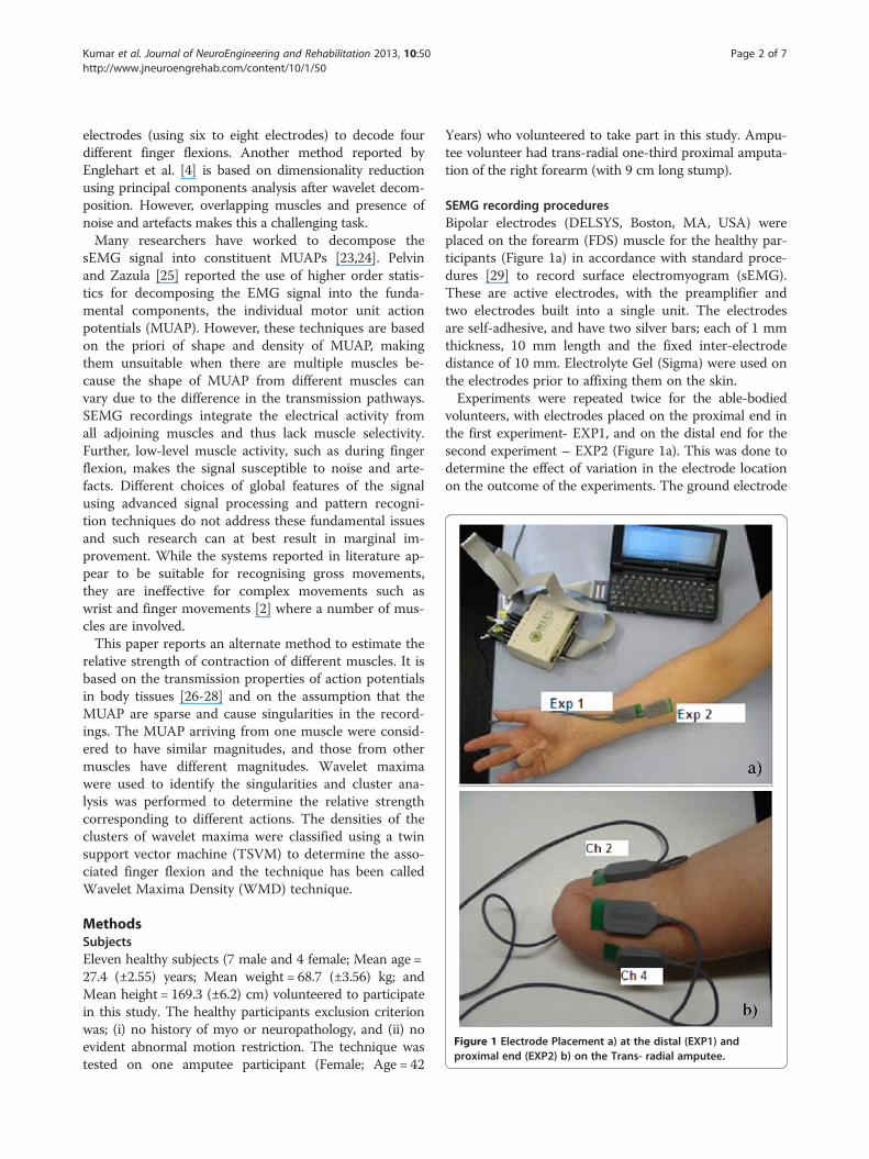
Figure 1 Electrode Placement a) at the distal (EXP1) andproximal end (EXP2) b) on the Trans- radial amputee.
Kumar et al. Journal of NeuroEngineering and Rehabilitation 2013, 10:50 Page 2 of 7http://www.jneuroengrehab.com/content/10/1/50
electrodes (using six to eight electrodes) to decode fourdifferent finger flexions. Another method reported byEnglehart et al. [4] is based on dimensionality reductionusing principal components analysis after wavelet decom-position. However, overlapping muscles and presence ofnoise and artefacts makes this a challenging task.Many researchers have worked to decompose the
sEMG signal into constituent MUAPs [23,24]. Pelvinand Zazula [25] reported the use of higher order statis-tics for decomposing the EMG signal into the funda-mental components, the individual motor unit actionpotentials (MUAP). However, these techniques are basedon the priori of shape and density of MUAP, makingthem unsuitable when there are multiple muscles be-cause the shape of MUAP from different muscles canvary due to the difference in the transmission pathways.SEMG recordings integrate the electrical activity fromall adjoining muscles and thus lack muscle selectivity.Further, low-level muscle activity, such as during fingerflexion, makes the signal susceptible to noise and arte-facts. Different choices of global features of the signalusing advanced signal processing and pattern recogni-tion techniques do not address these fundamental issuesand such research can at best result in marginal im-provement. While the systems reported in literature ap-pear to be suitable for recognising gross movements,they are ineffective for complex movements such aswrist and finger movements [2] where a number of mus-cles are involved.This paper reports an alternate method to estimate the
relative strength of contraction of different muscles. It isbased on the transmission properties of action potentialsin body tissues [26-28] and on the assumption that theMUAP are sparse and cause singularities in the record-ings. The MUAP arriving from one muscle were consid-ered to have similar magnitudes, and those from othermuscles have different magnitudes. Wavelet maximawere used to identify the singularities and cluster ana-lysis was performed to determine the relative strengthcorresponding to different actions. The densities of theclusters of wavelet maxima were classified using a twinsupport vector machine (TSVM) to determine the asso-ciated finger flexion and the technique has been calledWavelet Maxima Density (WMD) technique.
MethodsSubjectsEleven healthy subjects (7 male and 4 female; Mean age =27.4 (±2.55) years; Mean weight = 68.7 (±3.56) kg; andMean height = 169.3 (±6.2) cm) volunteered to participatein this study. The healthy participants exclusion criterionwas; (i) no history of myo or neuropathology, and (ii) noevident abnormal motion restriction. The technique wastested on one amputee participant (Female; Age = 42
Years) who volunteered to take part in this study. Ampu-tee volunteer had trans-radial one-third proximal amputa-tion of the right forearm (with 9 cm long stump).
SEMG recording proceduresBipolar electrodes (DELSYS, Boston, MA, USA) wereplaced on the forearm (FDS) muscle for the healthy par-ticipants (Figure 1a) in accordance with standard proce-dures [29] to record surface electromyogram (sEMG).These are active electrodes, with the preamplifier andtwo electrodes built into a single unit. The electrodesare self-adhesive, and have two silver bars; each of 1 mmthickness, 10 mm length and the fixed inter-electrodedistance of 10 mm. Electrolyte Gel (Sigma) were used onthe electrodes prior to affixing them on the skin.Experiments were repeated twice for the able-bodied
volunteers, with electrodes placed on the proximal end inthe first experiment- EXP1, and on the distal end for thesecond experiment – EXP2 (Figure 1a). This was done todetermine the effect of variation in the electrode locationon the outcome of the experiments. The ground electrode
Kumar et al. Journal of NeuroEngineering and Rehabilitation 2013, 10:50 Page 3 of 7http://www.jneuroengrehab.com/content/10/1/50
was placed on the volar aspect of the wrist. For the ampu-tee participant, the electrodes were placed on theremaining stump of the participant as shown in Figure 1b.DELSYS (Boston, MA, USA) sEMG acquisition system
was used to record the signal. The system gain was 1000,CMRR was 92 dB, and bandwidth was 20–450 Hz, with12 dB/ octave roll-off. The input impedance of the systemwas 115 Pico-farad in parallel with 1 K-ohm. The samplingrate of the system was 1024 samples/ second for eachchannel and the resolution were 16 bits/ sample. Prior tothe placement of electrodes, the skin of the participantwas prepared by shaving (if required) and exfoliation to re-move dead skin. Skin was cleaned with 70% v/v alcoholswab to remove any oil or dust from the skin surface.
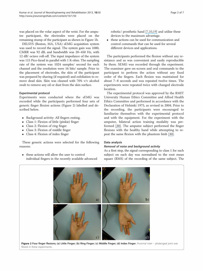
Experimental protocolExperiments were conducted where the sEMG wasrecorded while the participants performed four sets ofgeneric finger flexion actions (Figure 2) labelled and de-scribed below.
� Background activity: All fingers resting.� Class 1: Flexion of little (pinkie) finger� Class 2: Flexion of ring finger� Class 3: Flexion of middle finger� Class 4: Flexion of index finger
These generic actions were selected for the followingreasons:
� these actions will allow the user to controlindividual fingers in the recently available advanced
Figure 2 Four finger flexions; (a) Little Finger; (b) Ring Finger; (c) Midflexed in these experiments.
robotic/ prosthetic hand [7,10,19] and utilise thesedevices to the maximum advantage.
� these actions can be used for communication andcontrol commands that can be used for severaldifferent devices and applications.
The participants performed the flexion without any re-sistance and as was convenient and easily reproducibleby them. SEMG was recorded through the experiment.The examiner gave on-screen and oral commands to theparticipant to perform the action without any fixedorder of the fingers. Each flexion was maintained forabout 7–8 seconds and was repeated twelve times. Theexperiments were repeated twice with changed electrodelocation.The experimental protocol was approved by the RMIT
University Human Ethics Committee and Alfred HealthEthics Committee and performed in accordance with theDeclaration of Helsinki 1975, as revised in 2004. Prior tothe recording, the participants were encouraged tofamiliarize themselves with the experimental protocoland with the equipment. For the experiment with theamputee, bilateral action training modality was per-formed [30]. The amputee subject performed the fingerflexions with the healthy hand while attempting to re-peat the same flexion with the phantom limb [30].
Data analysisRemoval of noise and background activityAs a first step, the signal corresponding to class 1 for eachsubject on each day was normalised to the root meansquare (RMS) of the recording of the same subject. The
dle Finger; (d) Index Finger. Proximal inter – phalangeal joint was
Kumar et al. Journal of NeuroEngineering and Rehabilitation 2013, 10:50 Page 4 of 7http://www.jneuroengrehab.com/content/10/1/50
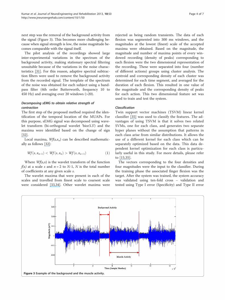
next step was the removal of the background activity fromthe signal (Figure 3). This becomes more challenging be-cause when signal strength is low, the noise magnitude be-comes comparable with the signal itself.The pilot analysis of the recordings showed large
inter-experimental variations in the spectrum of thebackground activity, making stationary spectral filteringunsuitable because of the variations in the noise charac-teristics [31]. For this reason, adaptive spectral subtrac-tion filters were used to remove the background activityfrom the recorded signal. The template of the spectrumof the noise was obtained for each subject using a band-pass filter (4th order Butterworth, frequency 10 to450 Hz) and averaging over 20 windows (>20).
Decomposing sEMG to obtain relative strength ofcontractionThe first step of the proposed method required the iden-tification of the temporal location of the MUAPs. Forthis purpose, sEMG signal was decomposed using wave-let transform (bi-orthogonal wavelet ‘bior3.3’) and themaxima were identified based on the change of sign[32].Local maxima, Wf(s,xn) can be described mathematic-
ally as follows [32]:
Wf s; xn−1ð Þ < Wf s; xnð Þ > Wf s; xnþ1ð Þ ð1Þ
Where Wf(s,x) is the wavelet transform of the functionf(x) at a scale s and n = 2 to N-1, N is the total numberof coefficients at any given scale s.The wavelet maxima that were present in each of the
scales and travelled from finest scale to coarsest scalewere considered [33,34]. Other wavelet maxima were
Figure 3 Example of the background and the muscle activity.
rejected as being random transients. The data of eachflexion was segmented into 300 ms windows, and themagnitudes at the lowest (finest) scale of the acceptedmaxima were obtained. Based on the magnitude, themagnitude and number of maxima points of every win-dowed recording (density of peaks) corresponding toeach flexion were the two dimensional representation ofthe recording. These were separated into four (numberof different actions) groups using cluster analysis. Thecentroid and corresponding density of each cluster wasdetermined for each time segment, and averaged for theduration of each flexion. This resulted in one value ofthe magnitude and the corresponding density of peaksfor each action. This two dimensional feature set wasused to train and test the system.
ClassificationTwin support vector machines (TSVM) linear kernelclassifier [35] was used to classify the features. The ad-vantages of using TSVM is that it solves two relatedSVMs, one for each class, and generates two separatehyper planes without the assumption that patterns ineach class arise from similar distributions. It allows theuse of a different kernel for each class which can beseparately optimized based on the data. This data de-pendent kernel optimization for each class is particu-larly useful in this study. For more details, please referto [15,35].The vectors corresponding to the four densities and
four magnitudes were the input to the classifier. Duringthe training phase the associated finger flexion was thetarget. After the system was trained, the system accuracywas validated using ten-fold cross – validation andtested using Type I error (Specificity) and Type II error

Table 2 Mean classification accuracy (%) of differentclasses recorded from Ch 2 and Ch 4 from amputeesubject using WMD method
Class Channel 2mean (±SD)%
Channel 4mean (±SD)%
Class 1 96.67 (±9.44) 71.67 (±8.08)
Class 2 75 (±9.92) 62.42 (±8.74)
Class 3 66.67 (±10.67) 72.15 (±11.24)
Class 4 89.15 (±8.26) 92.14 (±7.52)
Overall mean (±SD) 81.87 (±13.54) 74.59 (±12.52)
Kumar et al. Journal of NeuroEngineering and Rehabilitation 2013, 10:50 Page 5 of 7http://www.jneuroengrehab.com/content/10/1/50
(Sensitivity) [15]. The training dataset had 100 datapoints, and testing was done using 30 data points. Thetraining data set and test data set was chosen randomlyusing random sub-sampling method. Ten-cross valid-ation method was used to determine the accuracy, sensi-tivity and specificity of the system. Classification wasperformed on the data from the individual subject. Thesystem was tested individually for the two experimentsto determine the inter-experimental variations.
ResultsThe average classification accuracy for able-bodied sub-jects is reported in Table 1 and for amputee subject inTable 2. The sensitivity and specificity results for theable bodied subjects are reported in Table 3 and for theamputee subject in Table 4.
Accuracy of identifying sEMG of able bodied participantsFrom Table 1, the overall accuracy of the detection offlexion of four classes of fingers (digits 2 to 5) usingWMD was found to be 93.41(± 1.45)% when sEMG wasrecorded from the distal end (experiment 1) of the FDSmuscle. When sEMG was recorded from the proximalend (experiment 2) of the FDS muscle, there was only asmall variation (1% decrease) in the overall accuracy(92.4 ± 3.23)%.
Accuracy of identifying sEMG of amputeeThe accuracy of identification of movements from theamputee is tabulated in Table 2. The results show thatthe average accuracy of the detection of flexion of fourclasses of fingers (digits 2 to 5) performed based on thebilateral learning was found to be 81.87 (± 13.54)% fromsEMG electrode location 2 (Figure 1b). The average ac-curacy was found to be 74.59 (± 12.52)% from the sEMGelectrode location 4. The classifier did not converge forrecordings from the electrode locations 1 and 3 andhence has not been reported. This demonstrated thatthe electrode location had a significant effect on the sys-tem accuracy for the amputee subject. For further ana-lysis, electrode location 2 was considered.
Table 1 Mean classification accuracy (%) of differentclasses recorded from distal end (EXP1) and proximal end(EXP2) from able-bodied all subjects using WMD method
ClassDistal end (EXP1) Proximal end (EXP 2)
Mean (±SD)% Mean (±SD)%
Class 1 92.84 (±4.47) 94.23 (±5.08)
Class 2 95.47 (±4.07) 95.96 (±3.08)
Class 3 92.10 (±5.98) 90.37 (±5.58)
Class 4 93.21 (±5.96) 89.06 (±8.17)
Overall mean (±SD) 93.41 (±1.54) 92.4 (±3.23)
Sensitivity and specificity analysisFrom Table 3, the sensitivity of the results for able bod-ied subjects was 94% while the average specificity of allclasses was ~ 97% for experiment 1 and ~ 93% for experi-ment 2. The results indicate low Type I and Type IIerror. From Table 4, the average specificity was 85% forchannel 2 and 84.5% for channel 4, while the averagesensitivity was 85% for channel 2 and 83% for channel 4.
DiscussionThe results indicate that WMD sEMG signal analysis,which is based on the volume conduction properties ofthe tissues, can accurately identify individual finger ac-tions. Based on the volume conduction model reportedby [26,27], the muscle tissue forms the major portion ofthe volume conductor. This is an anisotropic conductingmedium, having different conductivities along differentaxes, relatively higher conductivity in the longitudinaldirection and slower in the transverse direction. There-fore electric potential travelling in transverse directionof a muscle is attenuated more rapidly as compared tolongitudinal direction [26,27]. Thus the action potentialoriginating from co-located muscle fibres would havesimilar magnitude and the assumption is that the mus-cles of the forearm have relatively small cross-sectionalarea. The magnitude of the singularities of the signal isinversely proportional to the distance between the elec-trodes and the muscle, while the density of the singular-ities corresponds to the relative number of motor unitaction potentials (MUAP), thus corresponding to thestrength of muscle contraction.In this method, sEMG was represented by the wavelet
maxima and these were then grouped based on theirmagnitude into four groups corresponding to the fourfinger actions. The average magnitude and density ofeach of the four groups for a time-window of 300 milli-seconds [4,13,16] were the input to a support vector ma-chine that was trained to associate these with the fingeraction. This time window is the permissible delay forreal time operation of the system. The method was also
Table 3 Sensitivity (%) and specificity (%) for each class recorded from distal end (EXP1) and proximal end (EXP2) fromable-bodied all subjects using WMD method
ClassDistal end (EXP 1) Proximal end (EXP 2)
Sensitivity mean (±SD)% Specificity mean (±SD)% Sensitivity mean (±SD)% Specificity mean (±SD)%
Class 1 92 (±0.05) 98 (±0.02) 94 (±0.07) 98 (±0.02)
Class 2 95 (±0.06) 97 (±0.03) 95 (±0.04) 98 (±0.01)
Class 3 92 (±0.06) 98 (±0.03) 91 (±0.05) 96 (±0.02)
Class 4 93 (±0.06) 98 (±0.02) 90 (±0.06) 97 (±0.03)
Overall mean (±SD) 93 (±0.01) 98 (±0.01) 93 (±0.02) 97 (±0.01)
Kumar et al. Journal of NeuroEngineering and Rehabilitation 2013, 10:50 Page 6 of 7http://www.jneuroengrehab.com/content/10/1/50
tested for the 200 ms (< 300 ms) and no significantchange in the classification accuracy was observed.Two sets of experiments with the electrode placement
distinctly different between the two experiments wereconducted to determine the reproducibility of the tech-nique. The classification accuracy for able-bodied sub-jects was in the range between 89% and 96%, while forthe trans-radial amputee was between 62% and 96%.The lower accuracy for the trans-radial amputee may bedue to number of reasons such as disuse the muscles ordamage to the muscle [22], or movement of the inser-tion point, which makes it difficult to identify suitablelocation for electrode placement.The recognition accuracy shows that this technique
is significantly better compared with the techniquereported by Momen et al. (56 ± 13%) [13], even thoughthe proposed technique has used single channel whileMomen et al. [13] have used two channels. The othersignificance is that this method has identified genericfinger actions, which makes this suitable for control-ling each finger of a prosthetic hand, while Momenet al. [13] and other researchers [10,11,14,20] haveconsidered user defined actions. The other strength ofthis technique is that it is repeatable and not sensitiveto differences in electrode placement.The system requires supervised training, while the
classification phase is suitable for being automated with-out manual supervision. The technique could be appliedfor prosthetic hand control or for human computerinterface.
Table 4 Sensitivity (%) and specificity (%) for each class recorWMD method
ClassChannel 2
Sensitivity (%) Specif
Class 1 90
Class 2 82
Class 3 79
Class 4 89
Overall mean (±SD) 85 (±0.05) 85 (
ConclusionIn this study, we have investigated and reported a novelmethod to estimate the relative strength of contraction ofdifferent muscles to identify individual finger flexions. Thismethod is based on the volume transmission properties ofaction potentials in body tissues [16], leading to the MUAPof different muscles having different amplitudes. Duringthe low level of muscle contraction such as isometric fingerflexion, MUAP are sparse and thus the measure of densityof MUAP in terms of the amplitude leads to a measure ofstrength of contraction of different muscles.Wavelet maxima detection is a suitable technique to
identify the local peaks in the sEMG. Identifying therange of the amplitude of peaks using clustering tech-niques using the wavelet maxima density (WMD) is anindicator of the muscle activity of different muscles. Thistechnique uses WMD as the set of features that can beclassified using twin support vector machine (TSVM) todetermine the associated finger flexion. The advantageof the TSVM over other classifier techniques is that it issuitable for unbalanced data sets.The results show that using WMD, single channel
EMG is suitable for accurately identifying individual fin-ger flexion. This technique was tested for able bodiedand trans-radial amputee subject and also for variationsin the placement of the electrodes. This work has dem-onstrated that it is possible to accurately identify the in-tent of individual finger flexion using single channelEMG recorded from the surface of the stump of atransradial amputee patient.
ded from Ch 2 and Ch 4 from amputee subject using
Channel 4
icity (%) Sensitivity (%) Specificity (%)
95 81 80
81 80 79
80 84 81
85 93 92
±0.06) 85 (±0.05) 83 (±0.06)

Kumar et al. Journal of NeuroEngineering and Rehabilitation 2013, 10:50 Page 7 of 7http://www.jneuroengrehab.com/content/10/1/50
Competing interestsThe authors declare that they have no competing interests.
Authors’ contributionsDKK has designed the experiment, discussed and developed the underlyingconcepts for the technique and finalised the manuscript. SPA has alsodesigned and conducted the experiments on the amputee subject,performed data analysis and written part of the manuscript. VPS hasconducted the experiment with able bodies and developed the waveletbased signal processing technique. All authors read and approved the finalmanuscript.
AcknowledgementsThe authors would like to acknowledge Caulfield Alfred Hospital forproviding permission to conduct experiment on amputee volunteer andProf. Jayadeva, for providing us with the Twin SVM classifier. The authorswould also like to thank Dr Michael Chou, Mr. David Wilson-Brown and Ms.Karen Roberts of the Amputee Unit, Caulfield Hospital, Alfred Health forproviding assistance and help on the conducting experiment with amputeevolunteer.
Author details1Bio-signals Lab, School of Electrical and Computer Engineering, RMITUniversity, GPO Box 2476, Melbourne, Victoria 3001, Australia. 2IP Australia,Canberra, Australia.
Received: 30 April 2012 Accepted: 25 May 2013Published: 7 June 2013
References1. Dorcas D, Scott RN: A three state myoelectric control. Med Biol Eng 1966,
4:367–372.2. Childress DA: A myoelectric three state controller using rate sensitivity. In
Proceedings of 8th International Conference on Medical and BiologicalEngineering conference(ICMBE). Chicago: Palmer House; 1969:4–5.
3. Hudgins B, Parker PA, Scott RN: A new strategy for multifunctionmyoelectric control. IEEE Trans Biomed Eng 1993, 40:82–94.
4. Englehart K, Hudgins B, Parker PA: A wavelet-based continuousclassification scheme for multifunction myoelectric control. IEEE TransBiomed Eng 2001, 48(3):302–311.
5. Karlik B, Tokhi O, Musa A: A fuzzy clustering neural network architecturefor multifunction upper-limb prosthesis. IEEE Trans Biomed Eng 2003,50(11):1255–1261.
6. Nagata K, Adno K, Magatani K, Yamada M: A classification method of handmovements using multi channel electrode. In Proceedings of 27th annualinternational conference of the engineering in medicine and biology society.Shanghai: IEEE; 2005:2375–2378.
7. Tenore F, Ramos A, Fahmy A, Acharya S, Etienne-Cummings R, Thakor NV:Decoding of individuated finger movements using surfaceelectromyography. IEEE Trans Biomed Eng 2009, 56(5):1427–1434.
8. Smith JR, Huberdeau D, Tenore F, Thakor NV: Real- time myoelectricdecoding of individual finger movements for a virtual target task. InProceedings of 31st annual IEEE EMBS international conference. Minneapolis,Minnesota, USA: IEEE; 2009:2376–2379.
9. Chen X, Lantz V, Kong-Qiao W, Zhang-Yan Z, Xu Z, Ji-Hai Y: Feasibility ofbuilding robust surface electromyography-based hand gestureinterfaces. In Proceedings of 31st annual IEEE EMBS international conference.Minneapolis, Minnesota, USA: IEEE; 2009:2983–2986.
10. Tenore F, Ramos A, Fahmy A, Acharya S, Etienne-Cummings R, Thakor NV:Towards the control of individual fingers of a prosthetic hand usingsurface EMG signals. In Proceedings of 29th annual IEEE EMBS internationalconference. Lyon, France: IEEE; 2007:6145–6148.
11. Chan FHY, Yong-Sheng Y, Lam FK, Yuan-Ting Z, Parker PA: Fuzzy EMGclassification for prosthesis control. IEEE Trans Rehabil Engin 2000,8(3):305–311.
12. Englehart K, Hudgins B, Parker PA, Stevenson M: Classification of themyoelectric signal using time-frequency based representations.Med Engin and Physics 1999, 21:431–438.
13. Momen K, Krishnan S, Chau T: Real time classification of forearmelectrmyographic signals corresponding to user-selected intentional
movements for multifunction prosthesis control. IEEE Trans. NeuralSystems and Rehab Engin 2007, 15(4):535–542.
14. Arjunan SP, Kumar DK: Decoding subtle forearm flexions using fractalfeatures of surface electromyogram from single and multiple sensors.J Neuroeng Rehabil 2010, 7:53.
15. Naik GR, Kumar DK, Jayadeva J: Twin SVM for gesture classification using thesurface electromyogram. IEEE Trans Inf Technol Biomed 2010, 14(2):301–308.
16. Englehart K, Hudgins B: A robust, real-time control scheme for multifunctionmyoelectric control. IEEE Trans Biomed Eng 2003, 50(7):848–854.
17. Kyberd J, Holland OE, Chappel PH, Smith S, Tregdigo R, Bagwell PJ, SnaithM: Marcus: A two degree of freedom hand prosthesis with hierarchicalgrip control. IEEE Trans Rehabil Eng 1995, 3(1):70–76.
18. Cipriani C, Antfolk C, Controzzi M, Lundborg G, Rosen B, Carrozza MC, SebeliusF: Online myoelectric control of a dexterous hand prosthesis by transradialamputees. IEEE Trans Neural Syst Rehabil Eng 2011, 19(3):260–270.
19. Cipriani C, Controzzi M, Carrozza MC: The SmartHand transradialprosthesis. J Neuroeng Rehabil 2011, 8:29.
20. Cram GS, Kasman J, Holtz J: Introduction to surface electromyography.Gaithersburg, Maryland: Aspen Publishers, Inc; 1998.
21. Kumar DK, Alemu M: Time-frequency analysis of SEMG- with specialconsideration to interelectrode spacing. IEEE Trans Neural Syst Rehabil Eng2004, 11(4):341–345.
22. Kumar DK, Pah ND: Thresholding wavelet networks for signal classification.Intl J Wavelets, Multiresolution and Info Process 2009, 2:243–261.
23. Zhou P, Rymer WZ, Suresh N, Zhang L: A study of surface motor unitaction potentials in first dorsal interosseus (FDI) muscle. In Proceedings of23rd annual IEEE EMBS international conference. Istanbul, Turkey: IEEE;2001:1074–1077.
24. Kleine BU, van Dijk JP, Lapatki BG, Zwarts MJ, Stegeman DF: Using two-dimensional spatial information in decomposition of surface EMGsignals. J Electromyogr Kinesiol 2007, 17:535–548.
25. Plevin E, Zazula D: Decomposition of surface EMG signals using non-linear LMS optimisation of higher-order cumulants. In Proceedings of the15th IEEE Symposium on Computer-Based Medical Systems volume 32.Maribor, Slovenia; 2002:149–154.
26. Blok JH, Stegeman DF, Oosterom AV: Three-layer volume conductormodel and software package for applications in surfaceelectromyography. Ann Biomed Eng 2002, 30:566–577.
27. Roeleveld K, Blok J, Stegeman DF, Oosterom AV: Volume conductionmodels for surface EMG; confrontation with measurements.J Electromyogr Kinesiol 1997, 7(4):221–232.
28. Merletti R, Botter A, Troiano A, Merlo E, Minetto MA: Technology andinstrumentation for detection and conditioning of the surfaceelectromyographic signal: state of the art. Clin Biomech 2009, 24:122–134.
29. Hermens HJ, Freriks B, Disselhorst-Klug C, Rau G: Development ofrecommendations for SEMG sensors and sensor placement procedures.J Electromyogr Kinesiol 2000, 10:361–374.
30. Castellini S, Gruppioni E, Davalli A, Sandini G: Fine detection of grasp forceand posture by amputees via surface electromyography. J Physiol Paris2009, 103(3–5):255–262.
31. Andrade OA, Nasuto S, Kyberd P, Sweeney- Reed CM, Van Kanijn FR: EMGsignal filtering based on empirical mode decomposition. Biomed SignalProces and Control 2006, 1(1):44–55.
32. Mallat S: A wavelet tour of signal processing. London: Academic Press; 1999.33. Abel EW, Meng H, Forster A, Holder D: Singularity characteristics of needle
EMG IP signals. IEEE Trans Biomed Eng 2006, 53(2):219–225.34. Arikidis NS, Abel EW, Forster A: Interscale wavelet maximum—a fine to
coarse algorithm for wavelet analysis of the EMG interference pattern.IEEE Trans Biomed Eng 2002, 49(4):337–344.
35. Jayadeva J, Khemchandani R, Chandra S: Twin support vector machinesfor pattern classification. IEEE Trans. Pattern Analysis and MachineIntelligence 2007, 29(5):905–910.
doi:10.1186/1743-0003-10-50Cite this article as: Kumar et al.: Towards identification of finger flexionsusing single channel surface electromyography – able bodied andamputee subjects. Journal of NeuroEngineering and Rehabilitation 201310:50.
Copyright © 2022 FDOKUMEN