Photosynthetic limitations in Mediterranean plants: A review
Upload
independentCategory
view
3download
0
Electromyography and the evolution of motor control:limitations and insightsAnthony Herrel,*,†,1 Vicky Schaerlaeken,† Callum Ross,‡ Jay Meyers,§ Kiisa Nishikawa,�
Virginia Abdala,k Adriana Manzano# and Peter Aerts†
�Department of Organismic & Evolutionary Biology, Harvard University, 26 Oxford St, Cambridge, MA 02138, USA;†Department of Biology, University of Antwerp, Universiteitsplein 1, B-2610 Antwerpen, Belgium; ‡Organismal Biology
and Anatomy, University of Chicago, 1027 E. 57th Street, Chicago, IL 60637, USA; §Department of Biology, 221 Morrill
Science Center, University of Massachusetts at Amherst, Amherst, MA 01003, USA; �Department of Biological Sciences,
Northern Arizona University, Flagstaff AZ 86011-5640, USA; kInstituto de Herpetologıa, Fundacion Miguel
Lillo-CONICET, Fac. de Cs. Naturales (UNT) Miguel Lillo 251 4000 Tucuman, Argentina; #CICyTTP-CONICET,
Matteri y Espana (3105), Diamante, Entre Rıos, Argentina
Synopsis Electromyography (EMG), or the study of muscle activation patterns, has long been used to infer central
nervous system (CNS) control of the musculoskeletal system and the evolution of that control. As the activation of the
muscles at the level of the periphery is a reflection of the interaction of descending influences and local reflex control,
EMG is an important tool in integrated investigations of the evolution of coordination in complex, musculoskeletal
systems. Yet, the use of EMG as a tool to understand the evolution of motor control has its limitations. We here review
the potential limitations and opportunities of the use of EMG in studying the evolution of motor control in vertebrates
and provide original previously unpublished data to illustrate this. The relative timing of activation of a set of muscles
can be used to evaluate CNS coordination of the components in a musculoskeletal system. Studies of relative timing
reveal task-dependent variability in the recruitment of different populations of muscle fibers (i.e., different fiber types)
within a single muscle, and left–right asymmetries in activation that need to be taken into account in comparative
studies. The magnitude of muscle recruitment is strongly influenced by the instantaneous demands imposed on the
system, and is likely determined by local reflex-control systems. Consequently, using EMG to make meaningful inferences
about evolutionary changes in musculoskeletal control requires comparisons across similar functional tasks. Moreover, our
data show that inferences about the evolution of motor control are limited in their explanatory power without proper
insights into the kinematics and dynamics of a system.
Introduction
Electromyography (EMG), or the study of activity
patterns of muscles, is a relatively old technique and
has been used extensively as a physiological and clinical
research tool since the early 1900s (see Ashley-Ross and
Gillis 2002 for an overview). However, the use of EMG
as a tool to gain insight into motor control and its
evolution has been restricted to the past four decades.
EMG has been an important tool in the study of motor
control as it provides insights into how the muscles are
controlled by the nervous system as evidenced by the
coordination of muscles in a given movement (Loeb
and Gans 1986). In this context, EMG has been used
extensively to try to understand motor-control strate-
gies used by the central nervous system (CNS), whether
it be from higher control centers, or local reflex arcs,
and to understand the functional role of muscles in
generating movements (Loeb and Gans 1986).
Movements are remarkably diverse as are the under-
lying bones and muscles that are responsible for gener-
ating those movements. Moreover, although some
movements can be understood as relatively simple
lever systems, others are the result of the coordination
of many monoarticular and multiarticular muscles.
Understanding how muscles are controlled to generate
the movements, and understanding how the control
changes as the underlying morphology changes has
been of interest to functional morphologists. Given
the apparent complexity associated with the control
of multi-jointed musculoskeletal systems, previous
authors have suggested that the central control is
highly conserved in evolution despite the obvious
From the symposium ‘‘Electromyography: Interpretation and Limitations in Functional Analyses of Musculoskeletal Systems’’ presented at theannual meeting of the Society for Integrative and Comparative Biology, January 2–6, 2008, at San Antonio, Texas.1E-mail: [email protected]
261
Integrative and Comparative Biology, volume 48, number 2, pp. 261–271
doi:10.1093/icb/icn025
Advanced Access publication April 28, 2008
� The Author 2008. Published by Oxford University Press on behalf of the Society for Integrative and Comparative Biology. All rights reserved.
For permissions please email: [email protected].
changes in the musculoskeletal systems across taxa
(Bramble and Wake 1985; Alfaro and Herrel 2001;
Wainwright 2002). More recently, however, an impor-
tant role for the intrinsic mechanics in simplifying
control has been advocated (Aerts et al. 2001; Van der
Leeuw et al. 2001; Nishikawa et al. 2007).
EMG has often been used as a tool to gain insights
into these issues as it provides a relatively direct repre-
sentation of how the muscles are activated by the
brainstem, spinal cord, and higher control centers.
By comparing which muscles are activated, and how
these muscles are activated (i.e., the sequence, dura-
tion, and magnitude of activation) in order to execute
a given movement or functional task, we can gain
insights into basic neuromuscular control. The com-
plexity in doing so lies in deciding which aspects to
study and how to treat them so as to make meaningful
comparisons across individuals and species; the
intrinsic variability of the activation patterns provided
the greatest obstacle.
Here, we use examples from recently collected, as
well as previously published electromyographic data
on feeding and locomotory systems in vertebrates, to
illustrate a number of potential pitfalls and short-
comings of using electromyographic data in a com-
parative context. Given these limitations, however, we
do believe meaningful insights can be gained from the
interpretation of muscle activation patterns. Finally,
we discuss the importance of the dynamics of any
musculoskeletal system in understanding its control,
and we illustrate this using examples from recent and
previously published work.
Materials and methods
Animals
Lizards
Three adult male Pogona vitticeps (Bearded dragon)
were used in the feeding experiments. Animals were
housed individually in a climatically controlled room
set at 248C and were provided with a basking spot
of 508C. Lizards were fed with vegetables, crickets,
mealworms, waxworms, superworms, and newborn
mice twice weekly. Water was always available to the
animals. Two additional adults, housed at the animal
care and use facility of Northern Arizona University
and maintained under similar conditions, were used
in the experiments on muscle physiology.
Frogs
One adult Phyllomedusa bicolor (Giant leaf frog) and
three adult Litoria caerulea (White’s treefrog) were
used in the experiments on locomotion. The animals
were maintained in a glass terrarium with dense
vegetation and high relative humidity. The tempera-
ture fluctuated between 268C (day) and 208C (night).
A large reservoir of water was present in the terrarium
and the animals were fed with waxworms and large
grasshoppers ad libitum.
Electomyographic recordings in P. vitticeps
Small metal markers were inserted on the right side or
along the medial plane at the anterior tip of the upper
and lower jaw, at the posterior end of the tooth row on
the upper and lower jaw, at the base and top of the
quadrate, at the anterior and posterior aspect of the
parietal and frontal bones, in the neck at the level of
cervical vertebrae 2 and 6, in the anterior and posterior
regions of the tongue,and at the intersection of the
basihyal and hypohyal. Animals were filmed in lateral
view while feeding on waxworms, mealworms, super-
worms, small and large crickets, and grasshoppers
using Redlake MotionPro2000 set at 250 Hz and
attached to a Philips image intensifier. X-rays were
generated using a Philips optimus M200 X-ray
generator at 50 KV.
Four bipolar stainless steel twisted-hook electrodes
were inserted percutaneously into both the left and
right m. adductor mandibulae externus medialis. During
implantation of the markers and electrodes, animals
were anesthetized using ketamine (200 mg/kg body
mass). The first electrode on each side was inserted
starting at the anteriormost border and electrodes were
equally spaced from there to the posteriormost border
of the muscle. Additionally, a bipolar Ni-Cr twisted-
hook electrode was inserted percutaneously into the
left and right m. depressor mandibulae. Signals were
amplified 10,000 times using Gould Universal pream-
plifiers with notch filter and Honeywell Accudata
117DC amplifiers. Signals were recorded digitally on
tape using a TEAC 145T DAT recorder. To allow syn-
chronization between the X-ray video recordings and
muscle activity patterns, a synchronization signal from
the X-ray generator was recorded on tape. Data were
transferred digitally to a PC using the TEAC QuickVu
software and were quantified in Microsoft Excel.
X-ray recordings and EMG in P. bicolor and L. caerulea
Small metal markers were inserted subcutaneously
at the proximal and distal ends of the humerus, at the
proximal and distal ends of the radius, at the base
of the carpals, at the base of the phalanges, and at the
last phalanx of digit II. During implantation of radio-
opaque markers, animals were anesthetized using
a buffered MS222 solution. Upon recovery, animals
were filmed in lateral view while moving on a narrow
dowel (17 mm). X-rays were generated using a Philips
262 A. Herrel et al.
optimus M200 X-ray generator and recorded at 200 Hz
using a Philips image intensifier attached to a Redlake
MotionPro2000 camera.
Bipolar Ni-Cr twisted-hook electrodes were inserted
percutaneously into the m. palmaris profundus and the
m. flexor digitorum communis longus in P. bicolor, and
only into the latter muscle in L. caerulea. Signals were
amplified 10 000 times using Gould Universal pre-
amplifiers with notch filter and Honeywell Accudata
117DC amplifiers. Signals were recorded digitally on
tape using a TEAC 145T DAT recorder. To allow
synchronization between the X-ray video recordings
and muscle activity patterns, a synchronization signal
from the X-ray generator was recorded on tape.
Data were transferred digitally to a PC using TEAC
QuickVu software, and the onset and duration of
muscular activity relative to substrate contact was
quantified in Microsoft Excel.
Physiological properties of them. sternohyoideusin P. vitticeps
The length/tension properties of the hyoid retractor
muscle were investigated in two live, anaesthetized
adult P. vitticeps (snout-to-vent length 99.07 mm and
107.85 mm). In this experiment, the animals were
deeply anaesthetized with ketamine (200 mg/kg body
mass), and bipolar stainless-steel electrodes were
implanted bilaterally into the hyoid retractor muscle
(m. sternohyoideus). The animals were kept under
deep anesthesia by administering additional ketamine
(half the original dose) every 2–3 h. In the experi-
ments, the animal was mounted upside-down in
a purpose-built holder, the hyoid was sutured to a
muscle lever (Cambridge Technology model 6650
force lever connected to an Aurora Scientific Series
305B lever system controller). Initially, the muscle
was twitch-stimulated (Grass S48 stimulator con-
nected to a Grass SIU5 stimulus isolation unit), and
stimulation voltage was increased until maximal
force output was obtained (at 12 V). In all sub-
sequent recordings, muscles were stimulated at 15 V
to ensure maximal muscle recruitment.
Next, the muscle was kept at resting length and
stimulated with tetanic trains of 300 ms (2 ms pulse
duration) of increasing frequency. The fusion freq-
uency (60 Hz) and tension at fusion were determined.
Subsequently, muscle length was varied and the passive
tension recorded; the muscle was then stimulated with
300 ms tetanic trains at 60 Hz and the active tension
recorded. Throughout the experiment, the tempera-
ture of the animal was kept at 328C by a heat lamp and
continuously monitored with a YSI telethermometer
and thermocouple. After the recordings, the animals
were killed by injection of a lethal dose of ketamine
(twice the anesthetic dose). ‘‘Resting length’’ of the
muscle was defined as its length when the hyoid was
lying at rest in the mouth. Extensions of the muscle,
thus involve stretching of the muscle beyond this
length and correspond to protraction of the hyoid as
observed during capture and transport of prey in vivo
(Herrel et al. 1995, 1997a).
Stimulation experiment—P. bicolor and L. caerulea
Bipolar twisted Ni-Cr electrodes were inserted in the
m. flexor digitorum communis longus in both species
and also in the m. palmaris profundus in P. bicolor.
Stimulations were performed on one P. bicolor and
two L. caerulea. The animals were brought under deep
anesthesia using Ketamine (225 mg/kg body mass)
and the muscles of the right forelimb were exposed.
Electrodes were inserted in the middle of the respective
muscle bellies and connected to a stimulator (Grass
S48). The stimulation circuit was charge balanced by
a coupling capacitor and bleed resistor (Loeb and Gans
1986) to avoid muscle damage and undue fatigue.
Muscles were stimulated at 12 V with a pulse train of
500 ms at 70Hz, and three ms pulse duration. Animals
were positioned on their back on a custom-made
platform and the lower arm was immobilized to allow
visualization of movements at the wrist and hand.
Animals were filmed in combined ventral and lateral
view by using a mirror positioned at an angle of 458 to
the horizontal at the level of the arm. Muscles were
stimulated one by one and movements were recorded.
Next, combined stimulations were performed to
understand the consequences of coactivation of the
different muscles.
All experiments were approved by the animal
ethics committee at the University of Antwerp and
by the IACUC at Northern Arizona University.
Results and discussion
Electrode placement
Careful placement of electrodes and verification of
their positions are important in order to accurately
categorize and explain inter-electrode variance in
EMG activity. Heterogeneity in timing of activity in
different parts of ‘‘single’’ muscles (e.g., masseter or
temporalis) is well documented in mammals and is
of fundamental importance for generating the trans-
verse movements characteristic of mammalian masti-
cation (Hylander et al. 1987; Weijs 1994). In primates,
for example, the masseter has two distinct functional
components: superficial and deep. On the chewing
(or working) side in anthropoid primates (monkeys),
the deep masseter shows onset and peak activity before
Electromyography and the evolution of motor control 263
the superficial masseter does, whereas on the non-
chewing (balancing) side, the deep masseter is active
after the superficial masseter (Hylander and Johnson
1994; Hylander et al. 2000). The kinematic conse-
quence of these activity patterns is that it is the
balancing-side deep masseter that pulls the teeth
medially through the late part of the power stroke.
This late activity in the balancing-side deep masseter
also has consequences for mandibular loading; in
combination with laterally directed reaction forces on
the working-side teeth, deep masseter ‘‘wishbones’’ the
mandible, stressing the symphysis, thereby necessitat-
ing fusion of the symphysis to strengthen the joint
(Hylander 1984, 1985). Significantly, late balancing-
side deep masseter activity is also associated with
symphyseal fusion in alpacas and horses, whereas
earlier cessation of balancing-side deep masseter is
associated with an unfused symphysis in goats
(Williams et al. 2007). Without careful placement of
electrodes matched with prior dissection of conspecific
individuals, these important aspects of the function
of mammalian chewing muscle would not have been
revealed.
In many mammals, heterogeneity in timing of
different parts of the temporalis are responsible for
rotating the mandible about a vertical axis during
chewing. The posteriorly directed working-side poster-
ior temporalis (PT) fires first, rotating the mandible
over to the working side, whereas the balancing-side
PT fires last, helping to drive the mandible transversely
during the power stroke (Weijs 1994). Anteroposterior
heterogeneity in muscle EMG is also found in some
lizards, but is not associated with transverse move-
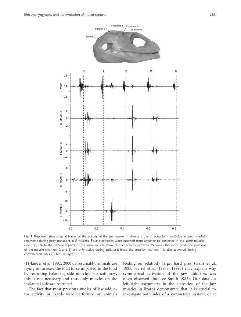
ments of the mandible. Figure 1 shows the results of
an experiment when multiple electrodes were inserted
from anterior to posterior into a single muscle of
a lizard, P. vitticeps. Whereas the more posteriorly
situated electrodes only picked up a signal during
ipsilateral bites, the anteriormost electrode picked up
a signal during both ipsilateral and contralateral
bites when chewing on soft foods (Fig. 1). These
results were highly consistent across individuals and
are likely associated with the presence of populations
of different fiber types within the muscle, with the
anteriormost compartment containing slower fiber
types (Throckmorton and Saubert 1982; Herrel et al.
1999b). Indeed, previous authors have demonstrated
that many of the jaw adductors in lizards consist
of regions with slow fibers surrounded by an area
consisting mostly of faster fiber types. Similar results
have been observed in primates. Baboon superficial
anterior temporalis (SAT) and PT muscles have more
fast twitch fibers than do the deep anterior temporalis
(DAT). SAT and PT also are relatively inactive during
chewing of soft foods and exhibit faster times for rise
and fall than does the DAT (Wall et al. 2005, 2006).
The differences in recruitment pattern of the differ-
ent regions in these muscles may be a reflection of
Henneman’s size principle (Henneman 1968). Slower
fibers will depolarize first because of their lower
threshold, and hence are recruited sooner (Milner-
Brown et al. 1973). Recruiting only areas with slow
fibers for contralateral bites on soft food makes intui-
tive sense as high forces are not needed to crush such
prey. Moreover, slow fibers are oxidative and can be
recruited longer or more often without experiencing
fatigue. Similar reasoning may be used to explain
differences in muscle recruitment during transport of
prey. As the food item gets reduced and transported
towards the back of the oral cavity, more and more of
the jaw adductors become silent (Herrel et al. 1997a,
1999a). Presumably, only those muscles with a high
proportion of slow oxidative (SO) fibers are recruited
towards the end of the transport stage (but see Herrel
et al. 1999b).
Location-specific recruitment of different parts
of the muscle with different populations of types of
fibers is not unique to jaw muscles. Recent studies have
reported similar site-specific recruitment for loco-
motor muscles (Hodson-Tole and Wakeling 2007;
Higham et al. 2008). Differences in the recruitment of
different components of the muscle may have major
consequences on the mechanics of musculoskeletal
systems (Higham et al. 2008). Moreover, caution
should be exerted in comparing motor patterns across
individuals, species, or higher-level taxa, as apparent
differences in strategies of motor control may just be
the reflection of differences in electrode position, or
the population of fiber type being recorded.
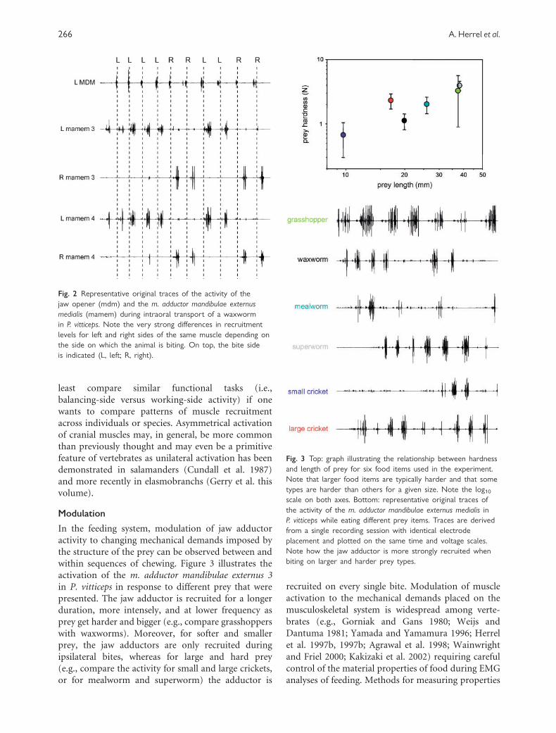
Left–right asymmetry
Figure 2 illustrates the activity of the medial jaw
adductor in P. vitticeps while chewing and transporting
a waxworm. Note how the jaw adductors are not
activated, or are only marginally so, when the prey is
positioned on the contralateral side. Thus, in contrast
to what was previously thought (Bramble and Wake
1985; Herrel et al. 2001), the recruitment of muscles in
bilaterally symmetrical systems need not be synchro-
nous, as demonstrated here by data from P. vitticeps.
Depending on the type of food that is being chewed,
both in P. vitticeps and in Tupinambis merrianae
(Ross and Herrel unpublished), the muscles on the
contralateral side may remain silent. In both species,
however, harder foods are associated with more
balancing-side recruitment of the jaw adductors as
has been demonstrated previously for some mammals
264 A. Herrel et al.
(Hylander et al. 1992, 2000). Presumably, animals are
trying to increase the total force imparted to the food
by recruiting balancing-side muscles. For soft prey,
this is not necessary and thus only muscles on the
ipsilateral side are recruited.
The fact that most previous studies of jaw adduc-
tor activity in lizards were performed on animals
feeding on relatively large, hard prey (Gans et al.
1985; Herrel et al. 1997a, 1999a) may explain why
symmetrical activation of the jaw adductors was
often observed (but see Smith 1982). Our data on
left–right asymmetry in the activation of the jaw
muscles in lizards demonstrate that it is crucial to
investigate both sides of a symmetrical system, or at
Fig. 1 Representative original traces of the activity of the jaw opener (mdm) and the m. adductor mandibulae externus medialis
(mamem) during prey transport in P. vitticeps. Four electrodes were inserted from anterior to posterior in the same muscle
(see top). Note that different parts of the same muscle show distinct activity patterns. Whereas the more posterior portions
of the muscle (mamem 2 and 3) are only active during ipsilateral bites, the anterior mamem 1 is also activated during
contralateral bites (L, left; R, right).
Electromyography and the evolution of motor control 265
least compare similar functional tasks (i.e.,
balancing-side versus working-side activity) if one
wants to compare patterns of muscle recruitment
across individuals or species. Asymmetrical activation
of cranial muscles may, in general, be more common
than previously thought and may even be a primitive
feature of vertebrates as unilateral activation has been
demonstrated in salamanders (Cundall et al. 1987)
and more recently in elasmobranchs (Gerry et al. this
volume).
Modulation
In the feeding system, modulation of jaw adductor
activity to changing mechanical demands imposed by
the structure of the prey can be observed between and
within sequences of chewing. Figure 3 illustrates the
activation of the m. adductor mandibulae externus 3
in P. vitticeps in response to different prey that were
presented. The jaw adductor is recruited for a longer
duration, more intensely, and at lower frequency as
prey get harder and bigger (e.g., compare grasshoppers
with waxworms). Moreover, for softer and smaller
prey, the jaw adductors are only recruited during
ipsilateral bites, whereas for large and hard prey
(e.g., compare the activity for small and large crickets,
or for mealworm and superworm) the adductor is
recruited on every single bite. Modulation of muscle
activation to the mechanical demands placed on the
musculoskeletal system is widespread among verte-
brates (e.g., Gorniak and Gans 1980; Weijs and
Dantuma 1981; Yamada and Yamamura 1996; Herrel
et al. 1997b, 1997b; Agrawal et al. 1998; Wainwright
and Friel 2000; Kakizaki et al. 2002) requiring careful
control of the material properties of food during EMG
analyses of feeding. Methods for measuring properties
Fig. 2 Representative original traces of the activity of the
jaw opener (mdm) and the m. adductor mandibulae externus
medialis (mamem) during intraoral transport of a waxworm
in P. vitticeps. Note the very strong differences in recruitment
levels for left and right sides of the same muscle depending on
the side on which the animal is biting. On top, the bite side
is indicated (L, left; R, right).
Fig. 3 Top: graph illustrating the relationship between hardness
and length of prey for six food items used in the experiment.
Note that larger food items are typically harder and that some
types are harder than others for a given size. Note the log10scale on both axes. Bottom: representative original traces of
the activity of the m. adductor mandibulae externus medialis in
P. vitticeps while eating different prey items. Traces are derived
from a single recording session with identical electrode
placement and plotted on the same time and voltage scales.
Note how the jaw adductor is more strongly recruited when
biting on larger and harder prey types.
266 A. Herrel et al.
of food in the field and laboratory are available and
should be more widely used (Lucas et al. 2001;
Williams et al. 2005). It is of interest that the intensity
of muscle recruitment (reflected in the amplitude of
the electromyographic signal) seems especially prone
to variation as amplitude depends on the instanta-
neous demands imposed on the system and may
largely be governed by proprioceptive feedback loops
(Ross et al. 2007). The distribution and functioning
of these feedback loops among vertebrates are poorly
surveyed. In mammals the proprioceptors in the peri-
odontal ligament act together with the muscle spindles
to modulate EMG activity in response to variation in
the material properties of food (Hidaka et al. 1997,
1999). Nontetrapods, however, are said to lack muscle
spindles altogether, and mammals (and possibly birds)
are the only vertebrates shown to have gamma moto-
neurons that modulate the response properties of
the spindles. The implications of these differences
for the evolution of vertebrate neuromechanics have
only begun to be explored (e.g., Houk 1972; Ross
et al. 2007).
One additional issue with using the intensity of
muscle recruitment in comparative studies is that the
amplitude of the signal is highly dependent on the
number of motor units being recorded, and that in
turn is dependent on the position of the electrode
within the muscle. Even if electrodes are positioned in
compartments with similar fiber types, their distance
to the motor endplate is likely different and, thus, so
will be the amplitude of the signal recorded (Loeb and
Gans 1986). Standardization of the amplitude of
muscle recruitment relative to a ‘‘maximum’’ recruit-
ment for a given electrode position during a given
recording session may alleviate some of these issues.
Additionally, time-averaging (i.e., binning) the ampli-
tude component of the signal may take out some of the
intrinsic variability in the spike patterns (Larson and
Stern 2007). As the coordination of different muscles
and temporal components of muscle activation appear
less variable, these have often been used by researchers
in comparative studies (see Wainwright 2002 for an
overview). Temporal aspects of the pattern of muscle
activation are, however, still affected by left–right
asymmetries in activation and the potential for
differences of electrode position with respect to
variation in fiber types in a muscle.
Importance of dynamics andmuscle mechanics
Interpretation of EMG activity patterns for the func-
tion of musculoskeletal control is crucially dependent
on the mechanical context of the muscle activity. To
illustrate this, we provide two examples that show
how an understanding of the context of the patterns
of muscle activation can be crucial in interpretation.
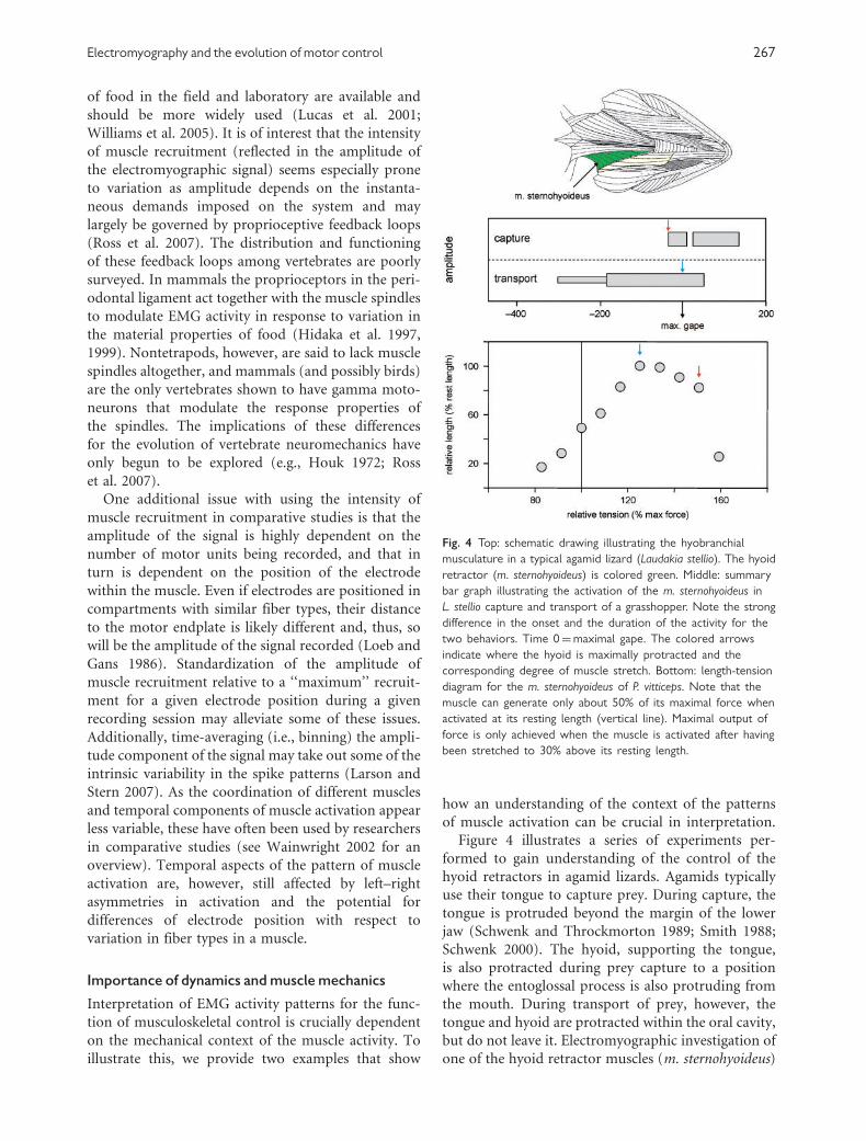
Figure 4 illustrates a series of experiments per-
formed to gain understanding of the control of the
hyoid retractors in agamid lizards. Agamids typically
use their tongue to capture prey. During capture, the
tongue is protruded beyond the margin of the lower
jaw (Schwenk and Throckmorton 1989; Smith 1988;
Schwenk 2000). The hyoid, supporting the tongue,
is also protracted during prey capture to a position
where the entoglossal process is also protruding from
the mouth. During transport of prey, however, the
tongue and hyoid are protracted within the oral cavity,
but do not leave it. Electromyographic investigation of
one of the hyoid retractor muscles (m. sternohyoideus)
Fig. 4 Top: schematic drawing illustrating the hyobranchial
musculature in a typical agamid lizard (Laudakia stellio). The hyoid
retractor (m. sternohyoideus) is colored green. Middle: summary
bar graph illustrating the activation of the m. sternohyoideus in
L. stellio capture and transport of a grasshopper. Note the strong
difference in the onset and the duration of the activity for the
two behaviors. Time 0¼maximal gape. The colored arrows
indicate where the hyoid is maximally protracted and the
corresponding degree of muscle stretch. Bottom: length-tension
diagram for the m. sternohyoideus of P. vitticeps. Note that the
muscle can generate only about 50% of its maximal force when
activated at its resting length (vertical line). Maximal output of
force is only achieved when the muscle is activated after having
been stretched to 30% above its resting length.
Electromyography and the evolution of motor control 267

during the two behaviors shows strong differences
in the activation patterns; whereas during transport
the hyoid retractors are activated about 300 ms
before maximal gape (Herrel et al. 1997a), during
prey capture the same muscle is activated immediately
before, or at, maximal gape (Herrel et al. 1995).
Although this difference seems puzzling at first sight,
investigations into the physiological properties of the
hyoid retractors may shed light on the observed
differences in activation pattern.
Physiological experiments show that at resting
length the muscle can only generate about 50% of its
maximal force, a situation similar to that observed for
the tongue retractor muscle in these lizards (Herrel
et al. 2002). As the muscle is stretched, however, it is
pulled onto the plateau of its length-tension curve that
lies at about 120–150% of resting length. As the hyoid
retractors originate on the pectoral girdle, the muscle
is stretched to different degrees during both prey
capture and prey transport. Indeed, whereas during
prey capture the hyoid retractors are stretched to about
150% of their resting length, during prey transport the
hyoid retractors are stretched to about 120–130% of
resting length. Given the physiological properties of
the muscle, this implies that the muscle can optimally
contract upon prey contact during prey capture
and work largely on the plateau of its length-tension
curve as it is pulling back the hyoid and tongue with
adhering prey. During transport, however, the tongue
is stretched less and only just reaches the plateau of
its length-tension curve. The long preactivation of
the muscle as the hyoid is being pulled forward
(i.e., lengthening activation) means that the muscle is
undergoing an eccentric contraction. This may help it
to contract forcefully despite operating on the ascend-
ing limb of the length-tension diagram, and may
allow the muscle to behave with spring-like properties
(Lindstedt et al. 2001, 2002). These data illustrate how
highly divergent activation patterns may be used to
execute the same functional task: forceful retraction
of hyoid and tongue with adhering prey.
One other example illustrating the relevance of
understanding the mechanical context of the activity
of a muscle comes from a recent study of the role of
the distal forelimb muscles during walking on narrow
substrates in arboreal frogs of the genus Phyllomedusa
(Manzano et al. in press). In species of this genus, the
forearm muscles are highly individualized and appear
to control each digit individually. High speed video
and cineradiographic recordings show how the frogs
wrap their hands around narrow branches while
walking upon them. This allows them to generate a
stabilizing moment at the hands and feet and prevents
them from toppling sideways as the branches are much
narrower than the width of the body. Electromyo-
graphic recordings of one of the major hand and finger
flexors, the m. flexor digitorum communis longus shows
that this muscle is active during the stance phase in
the two species of arboreal frog studied. This muscle
may thus actively contribute to the establishment of
the gripping posture of the hand (Fig. 5). Moreover,
experiments show that upon stimulation of the muscle
a pronounced flexion at the wrist and digits is observed
in both Litoria and Phyllomedusa. However, full
closure of the hand is not observed.
In Phyllomedusa, a superficial hand muscle, the
m. palmaris profundus, actually inserts onto the tendon
of the m. flexor digitorum communis longus, and upon
stimulation, pulls the tendon of the latter muscle
laterally for about 2–3 mm. Stimulation of this muscle
by itself does not, however, cause any flexion at the
wrist or digits. Consequently, it can be considered
as not contributing to flexion of the wrist and finger.
Yet, electromyographic recordings show that this
muscle is active in vivo during the stance phase in
Phyllomedusa. Insights into its functional role come
from an experiment in which a combined stimulation
of both the m. palmaris profundus and the m. flexor
digitorum communis longus was performed. Because
the m. palmaris profundus pulls on the tendon of the
flexor digitorum communis longus it actually increases
the moment arm and kinematic advantage of the latter
that results in full closure of the hand upon stimula-
tion. Consequently, the electromyographic data show
how the frog actively changes the moment arm of the
m. flexor digitorum communis longus by activating
the m. palmaris profundus to generate a grip. Thus,
although no changes in the activity pattern of the
muscle occurred in the evolution towards a specialized
grip in Phyllomedusa, this has gone hand in hand with
the cooption of an additional muscle. Without under-
standing the context of the activity patterns of the
muscle, it would be impossible to understand the
musculoskeletal control of the system and to appreci-
ate the evolution of the motor control of the system.
Insights into motor control
Electromyographic data show that motor control is
dynamic and complex. Activity patterns of muscles can
vary within a ‘‘single’’ muscle, defined anatomically as,
e.g., masseter or temporalis. Muscle activity patterns
can vary with the functional demands imposed upon
the muscle and the mechanical context within which it
is operating. In the context of this flexibility, the fact
that temporal components of the activation patterns of
muscle sometimes appear to be conserved across major
changes in morphology and mechanics is of interest
268 A. Herrel et al.

(Jenkins and Goslow 1983; Goslow et al. 1989; Dial
et al. 1991; Wainwright 2002; Larson and Stern 1989).
However, the conservation of muscle activation
patterns and their apparent constraint in some cases
may be largely due to the intrinsic capacity of the
nervous system to modulate activation patterns,
depending on the functional task. The presence of
asymmetrical activation in systems with mechanical
symmetry (this article; Gerry et al. this volume) and
the evolution of derived activation patterns (Grubich
2000; Alfaro et al. 2001), which may even evolve
convergently in systems with similar mechanical
demands (Konow and Sanford 2008), suggests no
intrinsic constraint on the evolution of motor patterns.
Moreover, distinguishing between stabilizing selection
on a successful control circuit or constraint on the
evolution of the control systems is intrinsically difficult
(see also Smith 1994). Our data clearly demonstrate
that care has to be taken in comparing motor patterns
by focusing on functionally similar tasks, and that an
understanding of the mechanics of the muscle is
crucial for interpreting the control of the musculoske-
letal system studied. Given these limitations, EMG
appears to be a powerful tool for investigating the
evolution of motor control in complex integrated
systems such as the vertebrate feeding and locomotory
systems.
Acknowledgments
The authors would like to thank Nicolai Konow and
Shannon Gerry for inviting us to present this article
at the symposium ‘‘Electromyography: Interpretation
and limitations in functional analyses of musculo-
skeletal systems’’ at the 2008 annual SICB meeting,
and SICB for financial support. The research was
Fig. 5 Top left: image taken from a high-speed movie of a P. bicolor walking on a narrow branch. Note how the fingers are
extended (right hand) and subsequently wrapped around the branch in a grasping motion. Top right: schematic drawing illustrating the
flexor muscles of the hand and fingers in P. bicolor. The m. flexor digitorum communis longis is in yellow and the m. palmaris profundus is
in blue. Activity of the m. palmaris profundus pulls the tendon of the m. flexor digitorum communis longus 2–3mm to the right, thus
changing the moment, arm and the action of the muscle around the wrist joint (red circle). Bottom: representative original traces of
the m. flexor digitorum communis longus in L . caerulea, and the m. palmaris profundus during locomotion in P. bicolor. The m. flexor
digitorum communis longus is active during stance in all arboreal frogs, including P. bicolor. In this species, however, the m. palmaris
profundus is active during stance as well and thus affects the function of the m. flexor digitorum communis longus in vivo and allows
P. bicolor to close its hand more fully than can any other frog studied to date.
Electromyography and the evolution of motor control 269

funded by a PhD grant from the Institute for the
Promotion of Innovation through Science and Tech-
nology in Flanders (IWT-Vlaanderen) to V.S. and by
a Research Grant of the Research Foundation—
Flanders (FWO) to A.H.
References
Aerts P, Van Damme J, Herrel A. 2001. Intrinsic mechanics
and control of fast cranio-cervical movements in aquatic
feeding turtles. Am Zool 41:1299–1310.
Agrawal KR, Lucas PW, Bruce IC, Prinz JF. 1998. Food
properties that influence neuromuscular activity during
human mastication. J Dent Res 77:1931–1938.
Alfaro ME, Herrel A. 2001. Introduction: major issues of
feeding motor control in vertebrates. Am Zool 41:
1243–1247.
Alfaro ME, Janovetz J, Westneat MW. 2001. Motor control
across trophic strategies: muscle activity of biting and
suction feeding fishes. Am Zool 41:1266–1279.
Ashley-Ross MA, Gillis G. 2002. A brief history of vertebrate
functional morphology. Integ Comp Biol 42:183–189.
Bramble DM, DB Wake. 1985. Feeding mechanisms of lower
tetrapods. In: Hildebrand M, Bramble DM, Liem K, and
Wake D (editors). Functional vertebrate morphology.
Cambridge: Harvard University Press. p 230–261.
Cundall D, Lorenz-Elwood J, Groves JD. 1987. Asymmetric
suction feeding in primitive salamanders. Experientia
43:1229–1231.
Dial KP, Goslow GE, Jenkins FA. 1991. The functional
anatomy of the shoulder in the European starling (Sturnus
vulgaris). J Morph 207:327–344.
Gans C, De Vree F, Carrier D. 1985. Usage pattern of the
complex masticatory muscles in the shingleback lizard,
Trachydosaurus rugosus: a model for muscle placement. Am
J Anat 173:219–240.
Gorniak GC, Gans C. 1980. Quantitative assay of electro-
myograms during mastication in domestic cats (Felis catus).
J Morph 163:253–281.
Goslow GE, Dial KP, Jenkins FA. 1989. The avian shoulder:
an experimental approach. Amer Zool 29:287–301.
Grubich JR. 2000. Crushing motor patterns in drum
(Teleostei: Sciaenidae): functional novelties associated
with molluscivory. J Exp Biol 203:3161–3176.
Henneman E. 1968. Peripheral mechanisms involved in the
control of muscle. In: Mountcastle VB, editor. Medical
physiology. St Louis: CV Mosby Co. p 1697–1716.
Herrel A, Cleuren J, De Vree F. 1995. Prey capture in the
lizard Agama stellio. J. Morphol 224:313–329.
Herrel A, Cleuren P, Fret J, De Vree F. 1999b. Morphology of
the feeding system in agamid lizards; ecological correlates.
Anat Rec 254:496–507.
Herrel A, Cleuren J, De Vree F. 1997a. Quantitative analysis
of jaw and hyolingual muscle activity during feeding in the
lizard Agama stellio. J Exp Biol 200:101–115.
Herrel A, Meyers JJ, Nishikawa KC, De Vree F. 2001. The
evolution of feeding motor patterns in lizards: modulatory
complexity and constraints. Am Zool 41:1311–1320.
Herrel A, Meyers JJ, Timmermans JP, Nishikawa KC. 2002.
Supercontracting muscle: producing tension over extreme
muscle lengths. J Exp Biol 205:2167–2173.
Herrel A, Verstappen M, De Vree F. 1999a. Modulatory
complexity of the feeding repertoire in scincid lizards.
J Comp Physiol A 184:501–518.
Herrel A, Wauters I, Aerts P, De Vree F. 1997b. The
mechanics of ovophagy in the beaded lizard (Heloderma
horridum). J Herpetol 31:383–393.
Hidaka O, Morimoto T, Kato T, Masuda Y, Inoue T,
Takada K. 1999. Behavior of jaw muscle spindle afferents
during cortically induced rhythmic jaw Movements in the
anesthetized rabbit. J Neurophysiol 82:2633–2640.
Hidaka O, Morimoto T, Masuda Y, Kato T, Matsuo R, Inoue T,
Kobayashi M, Takada K. 1997. Regulation of masticatory
force during cortically induced rhythmic jaw movements in
the anesthetized rabbit. J Neurophysiol 77:3168–3179.
Higham TE, Biewener AA, Wakeling JM. 2008. Functional
diversification within and between muscle synergists during
locomotion. Biol Lett 4:41–44.
Hodson-Tole EF, Wakeling JM. 2007. Variations in motor
unit recruitment patterns occur within and between
muscles in the running rat (Rattus norvegicus). J Exp Biol
210:2333–2345.
Houk JC. 1972. The phylogeny of muscular control config-
uration. Biocybernetics 2:125–144.
Hylander WL. 1984. Stress and strain in the mandibular
symphysis of primates: a test of competing hypotheses.
Am J Phys Anthropol 64:1–46.
Hylander WL. 1985. Mandibular function and biomechanical
stress and scaling. Am Zool 25:315–330.
Hylander WL, Johnson KR. 1994. Jaw muscle function and
wishboning of the mandible during mastication in
macaques and baboons. Am J Phys Anthropol 94:523–547.
Hylander WL, Johnson KR, Crompton AW. 1987. Loading
patterns and jaw movements during mastication in
Macaca fascicularis: A bone-strain, electromyographic,
and cineradiographic analysis. Am J Phys Anthropol
72:287–314.
Hylander WL, Johnson KR, Crompton AW. 1992. Muscle
force recruitment and biomechanical modeling: an analysis
of masseter muscle function during mastication in Macaca
fascicularis. Am J Phys Anthropol 88:365–387.
Hylander WL, Ravosa MJ, Ross CF, Wall CE, Johnson KR.
2000. Symphyseal fusion and jaw-adductor muscle force: an
EMG study. Am J Phys Anthropol 112:469–492.
Jenkins F A, Goslow GE. 1983. The functional anatomy of the
shoulder of the savannah monitor lizard (Varanus exanthe-
maticus). J Morph 175:195–216.
Kakizaki Y, Uchida K, Yamamura K, Yamada Y. 2002.
Coordination between the masticatory and tongue muscles
as seen with different foods in consistency and in reflex
activities during natural chewing. Brain Res 929:210–217.
270 A. Herrel et al.

Konow N, Sanford CPJ. 2008. Is a convergently derived
muscle-activity pattern driving novel raking behaviours in
teleost fishes? J Exp Biol 211:989–999.
Larson SG, Stern JT Jr. 1989. The role of propulsive muscles
of the shoulder during quadrupedalism in vervet monkeys
(Cercopithecus aethiops): Implications for neural control of
locomotion in primates. Journal of Motor Behavior
21:457–472.
Larson SG, Stern JT Jr. 2007. Humeral retractor EMG during
quadrupedal walking in primates. J Exp Biol 210:
1204–1215.
Lindstedt SL, LaStayo PC, Reich TE. 2001. When active
muscles lengthen: properties and consequences of eccentric
contractions. News Physiol Sci 16:256–261.
Lindstedt SL, Reich TE, Keim P, LaStayo PC. 2002. Do
muscles function as adaptable locomotor springs? J Exp
Biol 205:2211–2216.
Leob GE, Gans C. 1986. Electromyography for experimental-
ists. Chicago: University of Chicago Press. p 394.
Lucas PW, Beta T, Darvell BW, Dominy NJ, Essackjee HC,
Lee PKD, Osorio D, Ramsden L, Yamashita N, Yuen TDB.
2001. Field kit to characterize physical, chemical and spatial
aspects of potential primate foods. Folia Primatologica
72:11–25.
Manzano AS, Abdala V, Herrel A. Morphology and
function of the forelimb in arboreal frogs: specializa-
tions for grasping ability? J Anat (in press).
Milner-Brown HS, Stein RB, Yemm R. 1973. The orderly
recruitment of human motor units during voluntary iso-
metric contractions. J Physiol 230:359–370.
Nishikawa K, et al. 2007. Neuromechanics: an integrative
approach for understanding motor control. Integr Comp
Biol 47:16–54.
Ross CF, Eckhardt A, Herrel A, Hylander WL, Metzger KA,
Schaerlaeken V, Washington RL, Williams SH. 2007.
Modulation of intra-oral processing in mammals and
lepidosaurs. Integr Comp Biol 47:118–136.
Schwenk K. 2000. Feeding in lepidosaurians. Feeding.
San Diego: Academic Press. p 175–291.
Schwenk K, Throckmorton GS. 1989. Functional and evolu-
tionary morphology of lingual feeding in squamate reptiles:
phylogenetics and kinematics. J Zool 219:153–175.
Smith KK. 1982. An electromyographic study of the function
of the jaw adducting muscles in Varanus exanthemathicus.
J Morphol 173:137–158.
Smith KK. 1988. Form and function of the tongue in agamid
lizards with comments on its phylogenetic significance.
J Morphol 196:157–171.
Smith KK. 1994. Are neuromotor systems conserved in
evolution? Brain Behav Evol 43:293–305.
Throckmorton GS, Saubert CW IV. 1982. Histochemical
properties of some jaw muscles of the lizard Tupinambis
nigropunctatus. Anat Rec 203:345–352.
Van der Leeuw A, Bout RG, Zweers GA. 2001. Control of the
cranio-cervical system during feeding in birds. Amer Zool
41:1352–1363.
Wainwright PC. 2002. The evolution of feeding motor
patterns in vertebrates. Curr Op Neurobiol 12:691–695.
Wainwright PC, Friel JP. 2000. Effects of prey type on motor
pattern variance in tetraodontiform fishes. J Exp Zool
286:563–571.
Wall CE, Vinyard C, Johnson KR, Williams SH, Hylander WL.
2005. Functional heterogeneity of the temporalis muscle
of male and female baboons. Am J Phys Anthropol Suppl
40:217.
Wall CE, Briggs M, Schachat F, Vinyard C, Williams SH,
Hylander WL. 2006. Anatomical and functional specializa-
tions of the anterior temporalis muscle of baboons. Comp
Biochem Physiol 134A:S70.
Wall CE, Vinyard CJ, Williams SH, Johnson KR, Hylander WL.
2008. Specialization of the superficial anterior temporalis
muscle for hard-object feeding in baboons. In: Vinyard CJ,
Ravosa MJ, Wall CE, editors. Primate craniofacial function
and biology. New York: Springer Academic Publishers.
Weijs WA. 1994. Evolutionary approach to masticatory motor
patterns in mammals. In: Bels VL, Chardon M, Vandewalle
P (editors). Advances in Comparative and Environmental
Physiology. 8. Berlin: 55 Springer-Verlag. p 281–320.
Weijs WA, Dantuma R. 1981. Functional anatomy of
the masticatory apparatus in the rabbit (Oryctolagus
cuniculus L.). Neth J Zool 31:99–147.
Williams SH, Vinyard C, Wall CE, Hylander WL. 2007.
Masticatory motor patterns in ungulates: a quantitative
assessment of jaw-muscle coordination in goats, alpacas
and horses. J Exp Zool A 307:226–240.
Williams SH, Wright B, Truong VD, Daubert CR, Vinyard C.
2005. Mechanical properties of foods used in experimental
studies of primate masticatory function. Am J Primatol
67:329–346.
Yamada Y, Yamamura K. 1996. Possible factors which may
affect phase durations in the natural chewing rhythm. Brain
Res 706:237–242.
Electromyography and the evolution of motor control 271
Copyright © 2022 FDOKUMEN