Comparative binding energy analysis of haloalkane dehalogenase substrates: modelling of...
13
Journal of Computer-Aided Molecular Design 17: 299–311, 2003. © 2003 Kluwer Academic Publishers. Printed in the Netherlands. 299 Comparative binding energy analysis of haloalkane dehalogenase substrates: Modelling of enzyme-substrate complexes by molecular docking and quantum mechanical calculations Jan Kmun´ ıˇ cek 1 , Michal Boh´ aˇ c 1 , Santos Luengo 2 , Federico Gago 2 , Rebecca C. Wade 3,4 & Jiˇ r´ ı Damborsk´ y 1,∗ 1 National Centre for Biomolecular Research, Masaryk University, Kotlarska 2, 611 37 Brno, Czech Republic; 2 Department of Pharmacology, University of Alcal´ a, E-28871 Alcal´ a de Henares, Madrid, Spain; 3 European Me- dia Laboratory, Villa Bosch, Schloss-Wolfsbrunnenweg 33, D-69118 Heidelberg, Germany; 4 European Molecular Biology Laboratory, Meyerhofstr. 1, D-69012 Heidelberg, Germany Received 14 October 2002; Accepted 22 April 2003 Summary We evaluate the applicability of automated molecular docking techniques and quantum mechanical calculations to the construction of a set of structures of enzyme-substrate complexes for use in Comparative binding energy (COMBINE) analysis to obtain 3D structure-activity relationships. The data set studied consists of the complexes of eighteen substrates docked within the active site of haloalkane dehalogenase (DhlA) from Xanthobacter auto- trophicus GJ10. The results of the COMBINE analysis are compared with previously reported data obtained for the same dataset from modelled complexes that were based on an experimentally determined structure of the DhlA- dichloroethane complex. The quality of fit and the internal predictive power of the two COMBINE models are comparable, but better external predictions are obtained with the new approach. Both models show a similar composition of the principal components. Small differences in the relative contributions that are assigned to important residues for explaining binding affinity differences can be directly linked to structural differences in the modelled enzyme-substrate complexes: (i) rotation of all substrates in the active site about their longitudinal axis, (ii) repositioning of the ring of epihalohydrines and the halogen substituents of 1,2-dihalopropanes, and (iii) altered conformation of the long-chain molecules (halobutanes and halohexanes). For external validation, both a novel substrate not included in the training series and two different mutant proteins were used. The results obtained can be useful in the future to guide the rational engineering of substrate specificity in DhlA and other related enzymes. Abbreviations: COMBINE – Comparative binding energy; DhlA – haloalkane dehalogenase from Xanthobacter autotrophicus GJ10; E a – activation energy; H – change in reaction enthalpy; K m – apparent dissociation constant; PLS – Partial Least-Squares Projection to Latent Structures; Q 2 – cross-validated correlation coefficient; R 2 – correlation coefficient; RMSEP – root mean square error of predictions; SDEP – standard deviation of error of predictions. Introduction Comparative binding energy (COMBINE) analysis is a computational technique for deriving detailed quant- itative structure-activity relationships (QSAR) from a ∗ To whom correspondence should be addressed. E-mail: [email protected] set of three-dimensional structures of protein-ligand complexes [1]. COMBINE analysis systematically investigates the relationships between experimental binding affinities and the interaction energies for a set of enzyme-ligand complexes. COMBINE ana- lysis was originally developed for drug design ap- plications and nowadays it is well established as a
Transcript of Comparative binding energy analysis of haloalkane dehalogenase substrates: modelling of...
Journal of Computer-Aided Molecular Design 17: 299–311, 2003.© 2003 Kluwer Academic Publishers. Printed in the Netherlands.
299
Comparative binding energy analysis of haloalkane dehalogenasesubstrates: Modelling of enzyme-substrate complexes by moleculardocking and quantum mechanical calculations
Jan Kmunıcek1, Michal Bohac1, Santos Luengo2, Federico Gago2, Rebecca C. Wade3,4 & JirıDamborsky1,∗1National Centre for Biomolecular Research, Masaryk University, Kotlarska 2, 611 37 Brno, Czech Republic;2Department of Pharmacology, University of Alcala, E-28871 Alcala de Henares, Madrid, Spain; 3European Me-dia Laboratory, Villa Bosch, Schloss-Wolfsbrunnenweg 33, D-69118 Heidelberg, Germany; 4European MolecularBiology Laboratory, Meyerhofstr. 1, D-69012 Heidelberg, Germany
Received 14 October 2002; Accepted 22 April 2003
Summary
We evaluate the applicability of automated molecular docking techniques and quantum mechanical calculationsto the construction of a set of structures of enzyme-substrate complexes for use in Comparative binding energy(COMBINE) analysis to obtain 3D structure-activity relationships. The data set studied consists of the complexesof eighteen substrates docked within the active site of haloalkane dehalogenase (DhlA) from Xanthobacter auto-trophicus GJ10. The results of the COMBINE analysis are compared with previously reported data obtained for thesame dataset from modelled complexes that were based on an experimentally determined structure of the DhlA-dichloroethane complex. The quality of fit and the internal predictive power of the two COMBINE models arecomparable, but better external predictions are obtained with the new approach. Both models show a similarcomposition of the principal components. Small differences in the relative contributions that are assigned toimportant residues for explaining binding affinity differences can be directly linked to structural differences inthe modelled enzyme-substrate complexes: (i) rotation of all substrates in the active site about their longitudinalaxis, (ii) repositioning of the ring of epihalohydrines and the halogen substituents of 1,2-dihalopropanes, and (iii)altered conformation of the long-chain molecules (halobutanes and halohexanes). For external validation, both anovel substrate not included in the training series and two different mutant proteins were used. The results obtainedcan be useful in the future to guide the rational engineering of substrate specificity in DhlA and other relatedenzymes.
Abbreviations: COMBINE – Comparative binding energy; DhlA – haloalkane dehalogenase from Xanthobacterautotrophicus GJ10; Ea – activation energy; �H – change in reaction enthalpy; Km – apparent dissociationconstant; PLS – Partial Least-Squares Projection to Latent Structures; Q2 – cross-validated correlation coefficient;R2 – correlation coefficient; RMSEP – root mean square error of predictions; SDEP – standard deviation of errorof predictions.
Introduction
Comparative binding energy (COMBINE) analysis isa computational technique for deriving detailed quant-itative structure-activity relationships (QSAR) from a
∗To whom correspondence should be addressed. E-mail:[email protected]
set of three-dimensional structures of protein-ligandcomplexes [1]. COMBINE analysis systematicallyinvestigates the relationships between experimentalbinding affinities and the interaction energies for aset of enzyme-ligand complexes. COMBINE ana-lysis was originally developed for drug design ap-plications and nowadays it is well established as a
300
Figure 1. Scheme of the reaction mechanism of hydrolytic dehalogenation catalyzed by the haloalkane dehalogenases. Enz - enzyme.
standard 3D-QSAR method [1–12]. The applicationof this methodology in the protein-engineering fieldwas first described in a study of the substrate spe-cificity of haloalkane dehalogenase DhlA (Figure 1)from Xanthobacter autotrophicus GJ10 [8]. As thecrystal structure of the DhlA-dichloroethane complexwas known [13], the Michaelis complexes for the othersubstrates studied could be constructed by structuralalignment to this structure [8]. However, a broaderapplication of COMBINE analysis can be hinderedby the fact that there is a large number of enzymesfor which the experimental structure of an enzyme-substrate complex is unknown and positioning of thesubstrate in the active site may not be straightforward.This problem can become especially important forbroad-specificity enzymes with a large active site inwhich a large variety of ligands can be accommodatedin different binding modes. This is the case, for ex-ample, for the haloalkane dehalogenases DhaA fromRhodococcus sp. [14] and LinB from Sphingomonaspaucomobilis UT26 [15], both of which have activesites between 2 and 2.5 times larger than that in DhlA[16].
The problem outlined above indicates the necessityfor a method capable of finding favorable orientationsfor a set of substrates inside the active site of an en-zyme that will lead to the construction of robust QSARmodels by COMBINE analysis. Automated molecu-lar docking methods may provide a solution to thisproblem but validation is an important issue. The pur-pose of this study is to evaluate the applicability ofa well-established docking program for the prepara-tion of structures of DhlA-substrate complexes andcomplementary quantum mechanical calculations forthe selection of binding modes that are suitable for aCOMBINE-type analysis.
Methods
Experimental data
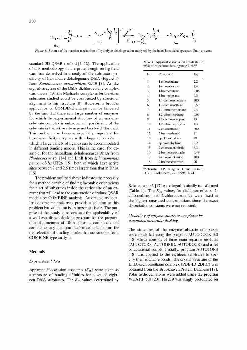
Apparent dissociation constants (Km) were taken asa measure of binding affinities for a set of eight-een DhlA substrates. The Km values determined by
Table 1. Apparent dissociation constants (inmM) of haloalkane dehalogenase DhlAa
No Compound Km
1 1-chlorobutane 2,2
2 1-chlorohexane 1,4
3 1-bromobutane 0,06
4 1-bromohexane 0,3
5 1,1-dichloromethane 100
6 1,2-dichloroethane 0,53
7 1,1-dibromomethane 2,4
8 1,2-dibromoethane 0,01
9 1,2-dichloropropane 13
10 1,2-dibromopropane 1,3
11 2-chloroethanol 400
12 2-bromoethanol 11
13 epichlorohydrine 48
14 epibromohydrine 2,2
15 2-chloroacetonitrile 6,3
16 2-bromoacetonitrile 0,49
17 2-chloroacetamide 100
18 2-bromoacetamide 20
aSchanstra, J.P., Kingma, J. and Janssen,D.B., J. Biol. Chem., 271 (1996) 14747.
Schanstra et al. [17] were logarithmically transformed(Table 1). The Km values for dichloromethane, 2-chloroethanol and 2-chloroacetamide were fixed atthe highest measured concentrations since the exactdissociation constants were not reported.
Modelling of enzyme-substrate complexes byautomated molecular docking
The structures of the enzyme-substrate complexeswere modelled using the program AUTODOCK 3.0[18] which consists of three main separate modules(AUTOTORS, AUTOGRID, AUTODOCK) and a setof additional scripts. Initially, program AUTOTORS[18] was applied to the eighteen substrates to spe-cify their rotatable bonds. The crystal structure of theDhlA-dichloroethane complex (PDB-ID 2DHC) wasobtained from the Brookhaven Protein Database [19].Polar hydrogen atoms were added using the programWHATIF 5.0 [20]. His289 was singly protonated on
301
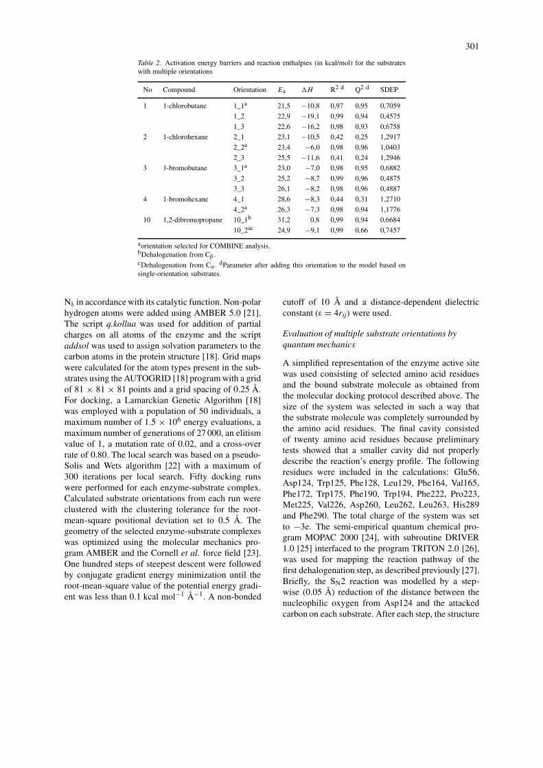
Table 2. Activation energy barriers and reaction enthalpies (in kcal/mol) for the substrateswith multiple orientations
No Compound Orientation Ea �H R2 d Q2 d SDEP
1 1-chlorobutane 1_1a 21,5 −10,8 0,97 0,95 0,7059
1_2 22,9 −19,1 0,99 0,94 0,4575
1_3 22,6 −16,2 0,98 0,93 0,6758
2 1-chlorohexane 2_1 23,1 −10,5 0,42 0,25 1,2917
2_2a 23,4 −6,0 0,98 0,96 1,0403
2_3 25,5 −11,6 0,41 0,24 1,2946
3 1-bromobutane 3_1a 23,0 −7,0 0,98 0,95 0,6882
3_2 25,2 −8,7 0,99 0,96 0,4875
3_3 26,1 −8,2 0,98 0,96 0,4887
4 1-bromohexane 4_1 28,6 −8,3 0,44 0,31 1,2710
4_2a 26,3 −7,3 0,98 0,94 1,1776
10 1,2-dibromopropane 10_1b 31,2 0,8 0,99 0,94 0,6684
10_2ac 24,9 −9,1 0,99 0,66 0,7457
aorientation selected for COMBINE analysis.bDehalogenation from Cβ.cDehalogenation from Cα . dParameter after adding this orientation to the model based onsingle-orientation substrates.
Nδ in accordance with its catalytic function. Non-polarhydrogen atoms were added using AMBER 5.0 [21].The script q.kollua was used for addition of partialcharges on all atoms of the enzyme and the scriptaddsol was used to assign solvation parameters to thecarbon atoms in the protein structure [18]. Grid mapswere calculated for the atom types present in the sub-strates using the AUTOGRID [18] program with a gridof 81 × 81 × 81 points and a grid spacing of 0.25 Å.For docking, a Lamarckian Genetic Algorithm [18]was employed with a population of 50 individuals, amaximum number of 1.5 × 10⁶ energy evaluations, amaximum number of generations of 27 000, an elitismvalue of 1, a mutation rate of 0.02, and a cross-overrate of 0.80. The local search was based on a pseudo-Solis and Wets algorithm [22] with a maximum of300 iterations per local search. Fifty docking runswere performed for each enzyme-substrate complex.Calculated substrate orientations from each run wereclustered with the clustering tolerance for the root-mean-square positional deviation set to 0.5 Å. Thegeometry of the selected enzyme-substrate complexeswas optimized using the molecular mechanics pro-gram AMBER and the Cornell et al. force field [23].One hundred steps of steepest descent were followedby conjugate gradient energy minimization until theroot-mean-square value of the potential energy gradi-ent was less than 0.1 kcal mol−1 Å−1. A non-bonded
cutoff of 10 Å and a distance-dependent dielectricconstant (ε = 4rij) were used.
Evaluation of multiple substrate orientations byquantum mechanics
A simplified representation of the enzyme active sitewas used consisting of selected amino acid residuesand the bound substrate molecule as obtained fromthe molecular docking protocol described above. Thesize of the system was selected in such a way thatthe substrate molecule was completely surrounded bythe amino acid residues. The final cavity consistedof twenty amino acid residues because preliminarytests showed that a smaller cavity did not properlydescribe the reaction’s energy profile. The followingresidues were included in the calculations: Glu56,Asp124, Trp125, Phe128, Leu129, Phe164, Val165,Phe172, Trp175, Phe190, Trp194, Phe222, Pro223,Met225, Val226, Asp260, Leu262, Leu263, His289and Phe290. The total charge of the system was setto −3e. The semi-empirical quantum chemical pro-gram MOPAC 2000 [24], with subroutine DRIVER1.0 [25] interfaced to the program TRITON 2.0 [26],was used for mapping the reaction pathway of thefirst dehalogenation step, as described previously [27].Briefly, the SN2 reaction was modelled by a step-wise (0.05 Å) reduction of the distance between thenucleophilic oxygen from Asp124 and the attackedcarbon on each substrate. After each step, the structure
302
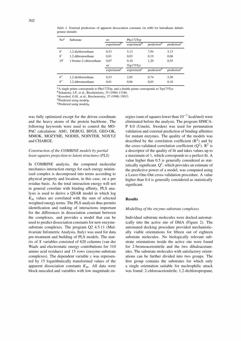
Table 3. External predictions of apparent dissociation constants (in mM) for haloalkane dehalo-genase mutants
Noa Substrate wt Phe172Trp
experimentb experimentb predictiond predictione
6′ 1,2-dichloroethane 0,53 5,13 7,94 5,13
8′ 1,2-dibromoethane 0,01 0,03 0,19 0,06
19′ 1-bromo-2-chloroethane 0,07 0,10 1,20 0,55
wt Trp175Tyr
experimentc experimentc predictiond predictione
6′′ 1,2-dichloroethane 0,53 2,85 0,74 5,50
8′′ 1,2-dibromoethane 0,01 0,06 0,03 0,10
aA single prime corresponds to Phe172Trp, and a double prime corresponds to Trp175Tyr.bSchanstra, J.P., et al., Biochemistry, 35 (1996) 13186.cKrooshof, G.H., et al., Biochemistry, 37 (1998) 15013.dPredicted using modelP.ePredicted using modelN.
was fully optimized except for the driven coordinateand the heavy atoms of the protein backbone. Thefollowing keywords were used to control the MO-PAC calculation: AM1, DEBUG, BFGS, GEO-OK,MMOK, MOZYME, NODIIS, NOINTER, NOXYZand CHARGE.
Construction of the COMBINE models by partialleast-squares projection to latent structures (PLS)
In COMBINE analysis, the computed molecularmechanics interaction energy for each energy minim-ized complex is decomposed into terms according tophysical property and location, in this case, on a perresidue basis. As the total interaction energy will notin general correlate with binding affinity, PLS ana-lysis is used to derive a QSAR model in which logKm values are correlated with the sum of selectedweighted energy terms. The PLS analysis thus permitsidentification and ranking of interactions importantfor the differences in dissociation constant betweenthe complexes, and provides a model that can beused to predict dissociation constants for new enzyme-substrate complexes. The program Q2 4.5.11 (Mul-tivariate Infometric Analysis, Italy) was used for datapre-treatment and building of PLS models. The mat-rix of X variables consisted of 620 columns (van derWaals and electrostatic energy contributions for 310amino acid residues) and 15 rows (enzyme-substratecomplexes). The dependent variable y was represen-ted by 15 logarithmically transformed values of theapparent dissociation constants Km. All data wereblock-unscaled and variables with low magnitude en-
ergies (sum of squares lower than 10−7 kcal/mol) wereeliminated before the analysis. The program SIMCA-P 8.0 (Umetri, Sweden) was used for permutationvalidation and external prediction of binding affinitiesfor mutant enzymes. The quality of the models wasdescribed by the correlation coefficient (R2) and bythe cross-validated correlation coefficient (Q2). R2 isa descriptor of the quality of fit and takes values up toa maximum of 1, which corresponds to a perfect fit. Avalue higher than 0.5 is generally considered as stat-istically significant. Q2, which provides an estimate ofthe predictive power of a model, was computed usinga Leave-One-Out cross-validation procedure. A valuehigher than 0.4 is generally considered as statisticallysignificant.
Results
Modelling of the enzyme-substrate complexes
Individual substrate molecules were docked automat-ically into the active site of DhlA (Figure 2). Theautomated docking procedure provided mechanistic-ally viable orientations for fifteen out of eighteensubstrate molecules. No biologically relevant sub-strate orientations inside the active site were foundfor 2-bromoacetonitrile and the two dihaloacetam-ides. The substrate molecules with satisfactory orient-ations can be further divided into two groups. Thefirst group contains the substrates for which onlya single orientation suitable for nucleophilic attackwas found: 2-chloroacetonitrile, 1,2-dichloropropane,

303
Figure 2. Active site cavity of DhlA with docked substrate 1-bromohexane. The electrophilic carbon of 1-bromohexane is oriented towardsthe nucleophilic oxygen of Asp124 in a position suitable for SN2 attack. Asp124 together with Asp260 and His289 constitutes the catalytictriad (13). The scissile bond is pointing towards the halide-binding pocket made of primary halide-stabilizing residues Trp125 and Trp175, andsecondary halide-stabilizing residues Phe172, Pro223 and Val226 (35).
dihalomethanes, dihaloethanes, haloethanols and epi-halohydrines. The second group is composed of sub-strates for which several satisfactory orientations werefound: 1,2-dibromopropane, halobutanes and halohex-anes. The two binding modes that were obtained for1,2-dibromopropane represented orientations suitablefor dehalogenation from either the α or the β carbonatom.
Selection of correct binding modes
The orientations of 1,2-dibromopropane, the halobu-tanes and the halohexanes were further studied byquantum-mechanical calculations to select one uniqueenzyme-substrate complex per molecule. Semi-empirical modelling of the SN2 dehalogenation reac-tion was conducted for each of these enzyme-substratecomplexes (Table 2). The change in heat of forma-tion of the system during the reaction was taken asa criterion to select the binding mode with the mostappropriate geometry for nucleophilic substitution, i.e.the initial reaction step. The activation energy (Ea)of the reaction was approximated by the differencein heat of formation between the Michaelis complexand the transition state structures. The enthalpy change(�H ) was expressed as the difference in heat of form-
ation between the Michaelis complex and the enzyme-product structures. The activation energy barriers andthe reaction enthalpies are listed in Table 2. Stabiliz-ation of the released halide anion by two tryptophanresidues located in the enzyme active site (Trp125 andTrp175) is critical for the first reaction step of DhlA,as demonstrated in previous experimental [28–31] andtheoretical [27, 32–35] studies. The magnitude of thisstabilization, as quantitated by �H , was therefore thefirst criterion for evaluation of different substrate ori-entations. If tryptophan stabilization was missing, then�H displayed more positive values. Stabilization wasfulfilled by all binding modes except the mode that as-sumed the nucleophilic attack on the β carbon atom of1,2-dibromopropane (orientation 10_1). For this ori-entation, a positive enthalpy change of the reaction(0.8 kcal/mol) was found. Orientation 10_2, whichassumes dehalogenation from the α carbon atom of1,2-dibromopropane, was therefore selected for theCOMBINE analysis. The orientations with the lowestactivation barrier (1_1, 3_1 and 4_2) were selectedfor inclusion in the COMBINE model for substrates1-chlorobutane, 1-bromobutane and 1-bromohexane,respectively. Two out of three binding modes for1-chlorohexane (2_1 and 2_2) showed similar activ-
304
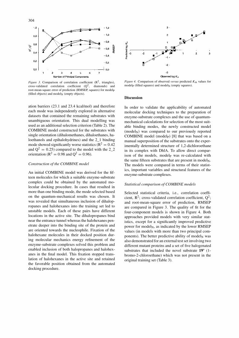
Figure 3. Comparison of correlation coefficient (R2, triangles),cross-validated correlation coefficient (Q2, diamonds) androot-mean-square error of prediction (RMSEP, squares) for modelP(filled objects) and modelN (empty objects).
ation barriers (23.1 and 23.4 kcal/mol) and thereforeeach mode was independently explored in alternativedatasets that contained the remaining substrates withunambiguous orientation. This dual modelling wasused as an additional selection criterion (Table 2). TheCOMBINE model constructed for the substrates withsingle orientation (dihalomethanes, dihaloethanes, ha-loethanols and epihalohydrines) and the 2_1 bindingmode showed significantly worse statistics (R2 = 0.42and Q2 = 0.25) compared to the model with the 2_2orientation (R2 = 0.98 and Q2 = 0.96).
Construction of the COMBINE model
An initial COMBINE model was derived for the fif-teen molecules for which a suitable enzyme-substratecomplex could be obtained by the automated mo-lecular docking procedure. In cases that resulted inmore than one binding mode, the mode selected basedon the quantum-mechanical results was chosen. Itwas revealed that simultaneous inclusion of dihalop-ropanes and halohexanes into the training set led tounstable models. Each of these pairs have differentlocations in the active site. The dihalopropanes bindnear the entrance tunnel whereas the halohexanes pen-etrate deeper into the binding site of the protein andare oriented towards the nucleophile. Fixation of thehalohexane molecules in their docked position dur-ing molecular mechanics energy refinement of theenzyme-substrate complexes solved this problem andenabled inclusion of both halopropanes and halohex-anes in the final model. This fixation stopped trans-lation of halohexanes in the active site and retainedthe favorable position obtained from the automateddocking procedure.
Figure 4. Comparison of observed versus predicted Km values formodelP (filled squares) and modelN (empty squares).
Discussion
In order to validate the applicability of automatedmolecular docking techniques to the preparation ofenzyme-substrate complexes and the use of quantum-mechanical calculations for selection of the most suit-able binding modes, the newly constructed model(modelN) was compared to our previously reportedCOMBINE model (modelP) [8] that was based on amanual superposition of the substrates onto the exper-imentally determined structure of 1,2-dichloroethanein its complex with DhlA. To allow direct compar-ison of the models, modelP was re-calculated withthe same fifteen substrates that are present in modelN.The models were compared in terms of their statist-ics, important variables and structural features of theenzyme-substrate complexes.
Statistical comparison of COMBINE models
Selected statistical criteria, i.e., correlation coeffi-cient, R2; cross-validated correlation coefficient, Q2;and root-mean-square error of prediction, RMSEPare compared in Figure 3. The quality of fit for thefour-component models is shown in Figure 4. Bothapproaches provided models with very similar stat-istics, except for a significantly improved predictivepower for modelN, as indicated by the lower RMSEPvalues (in models with more than two principal com-ponents). The better predictive ability of modelN wasalso demonstrated for an external test set involving twodifferent mutant proteins and a set of five halogenatedsubstrates that included the novel substrate 19’ (1-bromo-2-chloroethane) which was not present in theoriginal training set (Table 3).
305
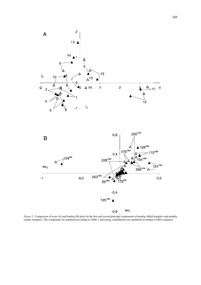
Figure 5. Comparison of score (A) and loading (B) plots for the first and second principal components of modelP (filled triangles) and modelN(empty triangles). The compounds are numbered according to Table 1 and energy contributions are numbered according to DhlA sequence.
306
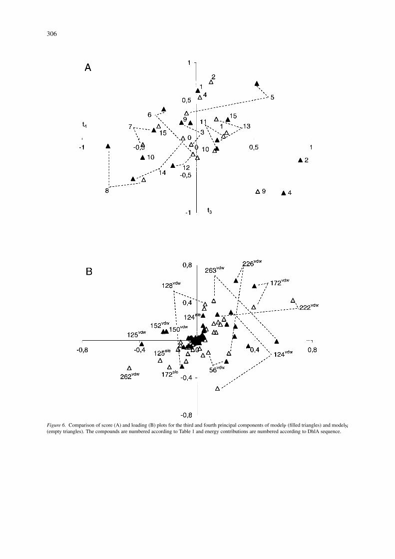
Figure 6. Comparison of score (A) and loading (B) plots for the third and fourth principal components of modelP (filled triangles) and modelN(empty triangles). The compounds are numbered according to Table 1 and energy contributions are numbered according to DhlA sequence.
307
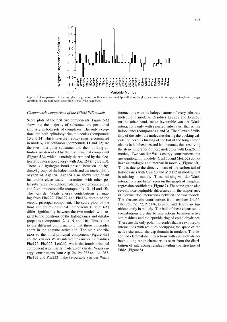
Figure 7. Comparison of the weighted regression coefficients for modelP (filled rectangles) and modelN (empty rectangles). Energycontributions are numbered according to the DhlA sequence.
Chemometric comparison of the COMBINE models
Score plots of the first two components (Figure 5A)show that the majority of substrates are positionedsimilarly in both sets of complexes. The only excep-tions are both epihalohydrine molecules (compounds13 and 14) which have their epoxy rings re-orientatedin modelN. Haloethanols (compounds 11 and 12) arethe two most polar substrates and their binding af-finities are described by the first principal component(Figure 5A), which is mainly determined by the elec-trostatic interaction energy with Asp124 (Figure 5B).There is a hydrogen bond present between the hy-droxyl groups of the haloethanols and the nucleophilicoxygen of Asp124. Asp124 also shows significantfavourable electrostatic interactions with other po-lar substrates: 2-epichlorohydrine, 2-epibromohydrineand 2-chloroacetonitrile (compounds 13, 14 and 15).The van der Waals energy contributions emanat-ing from Phe222, Phe172 and Phe164 dominate thesecond principal component. The score plots of thethird and fourth principal components (Figure 6A)differ significantly between the two models with re-gard to the positions of the halohexanes and dihalo-propanes (compounds 2, 4, 9 and 10). This is dueto the different conformations that these moleculesadopt in the enzyme active site. The main contrib-utors to the third principal component (Figure 6B)are the van der Waals interactions involving residuesPhe172, Phe222, Leu262, while the fourth principalcomponent is primarily made up of van der Waals en-ergy contributions from Asp124, Phe222 and Leu263.Phe172 and Phe222 make favourable van der Waals
interactions with the halogen atoms of every substratemolecule in modelN. Residues Leu262 and Leu263,on the other hand, make favourable van der Waalsinteractions only with selected substrates, that is, thehalobutanes (compounds 1 and 3). The allowed flexib-ility of the substrate molecules during the docking cal-culation permits turning of the tail of the long carbonchains in halohexanes and halobutanes, thus resolvingthe steric hindrance of these molecules with Leu263 inmodelP. Two van der Waals energy contributions thatare significant in modelP (Cys150 and Met152) do nothave an analogous counterpart in modelN (Figure 6B).This is due to the direct contact of the carbon tail ofhalohexanes with Cys150 and Met152 in modelP thatis missing in modelN. These missing van der Waalsinteractions are better seen on the graph of weightedregression coefficients (Figure 7). The same graph alsoreveals non-negligible differences in the importanceof electrostatic interactions between the two models.The electrostatic contributions from residues Glu56,Phe128, Phe172, Phe176, Leu263, and His289 are sig-nificant only in modelN. The bulk of these electrostaticcontributions are due to interactions between activesite residues and the epoxide ring of epihalohydrines.These are the only polar molecules that are exposed tointeractions with residues occupying the space of theactive site under the cap domain in modelN. The de-scribed electrostatic interactions with epihalohydrineshave a long-range character, as seen from the distri-bution of interacting residues within the structure ofDhlA (Figure 8).
308
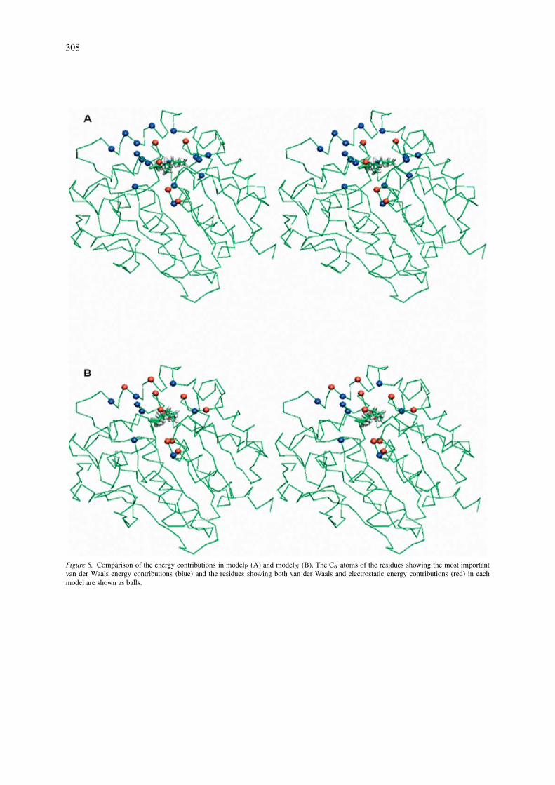
Figure 8. Comparison of the energy contributions in modelP (A) and modelN (B). The Cα atoms of the residues showing the most importantvan der Waals energy contributions (blue) and the residues showing both van der Waals and electrostatic energy contributions (red) in eachmodel are shown as balls.
309
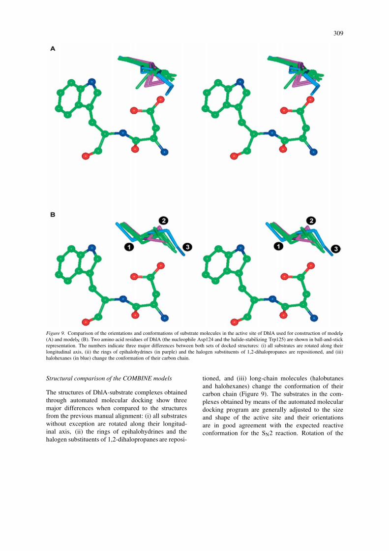
Figure 9. Comparison of the orientations and conformations of substrate molecules in the active site of DhlA used for construction of modelP(A) and modelN (B). Two amino acid residues of DhlA (the nucleophile Asp124 and the halide-stabilizing Trp125) are shown in ball-and-stickrepresentation. The numbers indicate three major differences between both sets of docked structures: (i) all substrates are rotated along theirlongitudinal axis, (ii) the rings of epihalohydrines (in purple) and the halogen substituents of 1,2-dihalopropanes are repositioned, and (iii)halohexanes (in blue) change the conformation of their carbon chain.
Structural comparison of the COMBINE models
The structures of DhlA-substrate complexes obtainedthrough automated molecular docking show threemajor differences when compared to the structuresfrom the previous manual alignment: (i) all substrateswithout exception are rotated along their longitud-inal axis, (ii) the rings of epihalohydrines and thehalogen substituents of 1,2-dihalopropanes are reposi-
tioned, and (iii) long-chain molecules (halobutanesand halohexanes) change the conformation of theircarbon chain (Figure 9). The substrates in the com-plexes obtained by means of the automated moleculardocking program are generally adjusted to the sizeand shape of the active site and their orientationsare in good agreement with the expected reactiveconformation for the SN2 reaction. Rotation of the
310
substrate molecules along their longitudinal axes po-sitions their electrophilic carbon atoms closer (byabout 1Å) to the attacking oxygen of Asp124 whileretaining the released halide ion in a position inter-mediate between two stabilizing amino acids, Trp125and Trp175 (Figure 9). This rotation places the sub-strate molecules in both a position and a conformation(e.g., gauche for 1,2-dichlororethane) perfectly suitedfor SN2 displacement. In line with this observation,the geometry of the Michaelis complexes evolvedsmoothly to that of the enzyme-product complexeswithout any additional conformational change duringthe quantum mechanical calculations. The only ex-ceptions were the 1,2-dihalopropanes, which changedtheir conformation from trans to gauche during theSN2 displacement reaction (gauche conformationswere never obtained from the docking calculations).Epihalohydrines and 1,2-dihalopropanes were dockedin orientations such that their favorable interactionswith surrounding active site residues were maxim-ized. Long-chain substrates adopted conformationsthat were adjusted to the relatively small size of theenzyme active site. The tail of the carbon chain distantfrom the cleaved carbon-halogen bond is rotated toprevent steric hindrance with the active site residues(Figure 9).
Conclusions
An automated molecular docking procedure providedstructures for a set of DhlA-substrate complexes thatwas used to derive a robust COMBINE model. Theprocedure was not devoid of problems, however, asmultiple binding modes were apparent for some sub-strates whereas biologically relevant binding modeswere missing for others. Quantum-mechanical calcu-lations were successfully used as an additional andcomplementary computational tool for selection ofsuitable binding modes, even though we note thatthis approach may not be equally applicable to en-zyme inhibitors or other protein ligands. Fixation ofthe substrate molecule in the docked conformationduring structural refinement of the enzyme-substratecomplexes by energy minimization was found to be auseful alternative to full minimization in certain cases.Comparison of the present COMBINE model witha previous one based on an experimentally derivedenzyme-substrate structure solved at 2.4 Å resolutionand manual superposition of the substrates revealedthat, despite significant differences in substrate ori-
entations and conformations, both models are similarin terms of overall fit and internal predictive power.It thus appears that positioning of the substrate mo-lecules relative to each other is more crucial for con-struction of robust COMBINE models than absolutepositioning and orientation of the substrates insidethe active site. However, the new COMBINE modelderived from the automatically docked structures, per-formed notably better in external prediction.
We expect that COMBINE-type models can aidin the rational engineering of substrate specificity ofDhlA and related dehalogenases. For this purpose, theactive site residues of DhlA can be conveniently di-vided into two groups. The first group contains theresidues that are essential for catalysis: the catalytictriad (Asp124, Asp260 and His289), the oxyanion hole(Glu56 and Trp125) and the primary halide-stabilizingresidues (Trp125 and Trp175). Modification of theseresidues is not recommended because it could leadto enzyme inactivation. The second group containsthe residues involved in substrate binding: Phe222,Pro223, Val226, Leu262, and Leu263 (the first shell)and Lys176, Leu176 and Arg229 (the second shell).We would expect these second-shell residues to be es-pecially suitable targets for site-directed mutagenesisstudies.
Acknowledgements
This work was supported by the NATO LinkageGrant MTECH.LG.974701 and grants from the CzechMinistry of Education J07/98:143100005 and ME551(JD).
Supporting information
The datasets used for COMBINE analysis andthe reaction paths calculated for multiple bindingmodes. This material is available via Internet athttp://ncbr.chemi.muni.cz/∼jiri.
References
1. Ortiz, A.R., Pisabarro, M.T., Gago, F., and Wade, R.C., J.Med. Chem., 38 (1995) 2681.
2. Ortiz, A.R., Pastor, M., Palomer, A., Cruciani, G., Gago, F.,and Wade, R.C., J. Med. Chem., 40 (1997) 1136.
3. Pastor, M., Perez, C., and Gago, F., J. Mol. Graph. Model., 15(1997) 364.
311
4. Perez, C., Pastor, M., Ortiz, A.R., and Gago, F., J. Med.Chem., 41 (1998) 836.
5. Lozano, J.J., Pastor, M., Cruciani, G., Gaedt, K., Centeno,N.B., Gago, F., and Sanz, F., J. Comput.-Aid. Mol. Design, 14(2000) 341.
6. Tomic, S., Nilsson, L., and Wade, C.R., J. Med. Chem., 43(2000) 1780.
7. Cuevas, C., Pastor, M., Perez, C., and Gago, F., Combin.Chem. High Through. Screen., 4 (2001) 627.
8. Kmunicek, J., Luengo, S., Gago, F., Ortiz, A.R., Wade, R.C.,and Damborsky, J., Biochemistry, 40 (2001) 8905.
9. Wade, R.C. 2001. Derivation of QSARs using 3D structuralmodels of protein-ligand complexes by COMBINE analysis,In Holtje, H.-D. and Sippl, W. (eds.), Rational approachesto drug design: 13th European symposium on QuantitativeStructure-Activity Relationships, Prous Science, Barcelona, p.23.
10. Wang, T., and Wade, R.C., J. Med. Chem., 44 (2001) 961.11. Wang, T., and Wade, R.C., J. Med. Chem., 45 (2002) 4828.12. Damborsky, J., Kmunicek, J., Jedlicka, T., Luengo, S., Gago,
F., Ortiz, A.R., and Wade, R.C. 2003. Rational re-design ofhaloalkane dehalogenases guided by comparative binding en-ergy analysis, In Svendsen, A. (ed.), Enzyme functionality:design, engineering and screening, Marcel Dekker, New York,in press.
13. Verschueren, K.H.G., Seljee, F., Rozeboom, H.J., Kalk, K.H.,and Dijkstra, B.W., Nature, 363 (1993) 693.
14. Newman, J., Peat, T.S., Richard, R., Kan, L., Swanson, P.E.,Affholter, J.A., Holmes, I.H., Schindler, J.F., Unkefer, C.J.,and Terwilliger, T.C., Biochemistry, 38 (1999) 16105.
15. Marek, J., Vevodova, J., Kuta-Smatanova, I., Nagata, Y.,Svensson, L.A., Newman, J., Takagi, M., and Damborsky, J.,Biochemistry, 39 (2000) 14082.
16. Damborsky, J., and Koca, J., Prot. Engng., 12 (1999) 989.17. Schanstra, J.P., Kingma, J., and Janssen, D.B., J. Biol. Chem.,
271 (1996) 14747.18. Morris, G.M., Goodsell, D.S., Halliday, R.S., Huey, R., Hart,
W.E., Belew, R.K., and Olson, A.J., J. Comput. Chem., 19(1998) 1639.
19. Berman, H.M., Westbrook, J., Feng, Z., Gilliland, G., Bhat,
T.N., Weissig, H., Shindyalov, I.N., and Bourne, P.E., Nucl.Acid Res., 28 (2000) 235.
20. Vriend, G., J. Mol. Graphics, 8 (1990) 52.21. Case, D.A., Pearlman, D.A., Caldwell, J.W., Cheatham III,
T.E., Ross, W.S., Simmerling, C.L., Darden, T.A., Merz,K.M., Stanton, R.V., Cheng, A.L., Vincent, J.J., Crowley,M., Ferguson, D.M., Radmer, R.J., Seibel, G.L., Singh, U.C.,Weiner, P.K., and Kollman, P.A. AMBER 5.0, University ofCalifornia, San Francisco (1997).
22. Solis, F.J., and Wets, R.J.B., Math. Oper. Res., 6 (1981) 19.23. Cornell, W.D., Cieplak, P., Bayly, C.I., Gould, I.R., Merz,
K.M., Ferguson, D.M., Spellmeyer, D.C., Fox, T., Caldwell,J.W., and Kollman, P.A., J. Am. Chem. Soc., 117 (1995) 5179.
24. Stewart, J.J.P., J. Comput.-Aid. Mol. Design, 4 (1990) 1.25. Cernohorsky, M., Kuty, M., and Koca, J., Comp. Chem., 21
(1996) 35.26. Damborsky, J., Prokop, M., and Koca, J., Trends Biochem.
Sci., 26 (2001) 71.27. Damborsky, J., Kuty, M., Nemec, M., and Koca, J., J. Chem.
Inf. Comp. Sci., 37 (1997) 562.28. Verschueren, K.H.G., Kingma, J., Rozeboom, H.J., Kalk,
K.H., Janssen, D.B., and Dijkstra, B.W., Biochemistry, 32(1993) 9031.
29. Kennes, C., Pries, F., Krooshof, G.H., Bokma, E., Kingma, J.,and Janssen, D.B., Eur. J. Biochem., 228 (1995) 403.
30. Krooshof, G.H., Ridder, I.S., Tepper, A.W.J.W., Vos, G.J.,Rozeboom, H.J., Kalk, K.H., Dijkstra, B.W., and Janssen,D.B., Biochemistry, 37 (1998) 15013.
31. Schindler, J.F., Naranjo, P.A., Honaberger, D.A., Chang,C.-H., Brainard, J.R., Vanderberg, L.A., and Unkefer, C.J.,Biochemistry, 38 (1999) 5772.
32. Lightstone, F.C., Zheng, Y.-J., Maulitz, A.H., and Bruice,T.C., Proc. Natl. Acad. Sci. USA, 94 (1997) 8417.
33. Shurki, A., Strajbl, M., Villa, J., and Warshel, A., J. Am.Chem. Soc., 124 (2002) 4097.
34. Bohac, M., Nagata, Y., Prokop, Z., Prokop, M., Monincova,M., Koca, J., Tsuda, M., and Damborsky, J., Biochemistry, 41(2002) 14272.
35. Devi-Kesavan, L.S., and Gao, J., J. Am. Chem. Soc., 125(2003) 1532.